EP3080299B1 - Biomarkers of rapid progression in advanced non-small cell lung cancer - Google Patents
Biomarkers of rapid progression in advanced non-small cell lung cancer Download PDFInfo
- Publication number
- EP3080299B1 EP3080299B1 EP14869355.9A EP14869355A EP3080299B1 EP 3080299 B1 EP3080299 B1 EP 3080299B1 EP 14869355 A EP14869355 A EP 14869355A EP 3080299 B1 EP3080299 B1 EP 3080299B1
- Authority
- EP
- European Patent Office
- Prior art keywords
- biomarkers
- biomarker
- igfbp
- subject
- treatment
- Prior art date
- Legal status (The legal status is an assumption and is not a legal conclusion. Google has not performed a legal analysis and makes no representation as to the accuracy of the status listed.)
- Active
Links
Images




Classifications
-
- G—PHYSICS
- G01—MEASURING; TESTING
- G01N—INVESTIGATING OR ANALYSING MATERIALS BY DETERMINING THEIR CHEMICAL OR PHYSICAL PROPERTIES
- G01N33/00—Investigating or analysing materials by specific methods not covered by groups G01N1/00 - G01N31/00
- G01N33/48—Biological material, e.g. blood, urine; Haemocytometers
- G01N33/50—Chemical analysis of biological material, e.g. blood, urine; Testing involving biospecific ligand binding methods; Immunological testing
- G01N33/53—Immunoassay; Biospecific binding assay; Materials therefor
- G01N33/575—Immunoassay; Biospecific binding assay; Materials therefor for cancer
- G01N33/5752—Immunoassay; Biospecific binding assay; Materials therefor for cancer of the lungs
Definitions
- the present invention relates to methods and kits for identifying patients with rapidly progressing disease, and in particular to methods and kits for identifying patients with rapidly progressing non-small cell lung cancer and for determining optimal treatment plans for patients with rapidly progressing disease and for monitoring treatments.
- Lung cancer is leading cause of cancer-related mortality worldwide, with a projected 159,480 patients succumbing to the disease in the US in 2014.
- Lung cancer is typically characterized as being quite aggressive with poor clinical outcomes that stem from the very rapid proliferation rates, high metastatic potential, and general insensitivity to available treatment strategies.
- NSCLC Non-small cell lung cancer
- NSCLC presents unique challenges to health care providers because of its common late stage of presentation and the poor median overall survival of advanced disease.(2, 3) Patients often become too ill to receive second line treatment as noted by a recent phase III clinical trial where only 37% of the patients randomized to docetaxel at disease progression received treatment.(4)
- the method includes assaying a level of a biomarker in a biomarker panel in a biological sample obtained from a subject where the panel includes at least one biomarker selected from Table I or Table II.
- the method is dependent on whether the subject is treatment na ⁇ ve or has received at least one treatment; and comparing the level of the biomarker in the subject's sample to a cutoff value listed in Table I for treatment na ⁇ ve subjects or Table II for previously treated subjects.
- the method further includes determining whether the subject's level is above or below the cutoff value to determine whether the subject has rapidly progressing lung cancer.
- the present invention will utilize at least one biomarker measured in a biological sample obtained from a subject to identify rapidly progressing lung cancer, and in some embodiments in subjects having rapidly progressing NSCLC.
- the at least one biomarker may be selected from a panel of biomarkers.
- one or more biomarkers from a panel of biomarkers are used to identify subjects having rapidly progressing NSCLC in subjects that are treatment na ⁇ ve or that have been previously treated.
- biomarker refers to any biological compound that can be measured as an indicator of the physiological status of a biological system.
- a biomarker may comprise an amino acid sequence, a nucleic acid sequence and fragments thereof.
- Exemplary biomarkers include, but are not limited to cytokines, chemokines, growth and angiogenic factors, metastasis related molecules, cancer antigens, apoptosis related proteins, proteases, adhesion molecules, cell signaling molecules and hormones.
- Measurement means assessing the presence, absence, quantity or amount (which can be an effective amount) of a given substance within a sample, including the derivation of qualitative or quantitative concentration levels of such substances, or otherwise evaluating the values or categorization of a subject's clinical parameters.
- detecting or “detection” may be used and is understood to cover all measuring or measurement as described herein.
- sample refers to a sample of biological fluid in a healthy and/or pathological state obtained from a subject.
- samples include, but are not limited to, blood, bronchial lavage fluid, sputum, saliva, urine, amniotic fluid, lymph fluid, peritoneal fluid, and cerebrospinal fluid.
- the whole blood sample is further processed into serum or plasma samples.
- the sample includes blood spotting tests.
- subject refers to a mammal, preferably a human.
- rapidly progressing refers to cases of disease that were observed to not respond to chemotherapy or targeted agents and advance (evidence of nascent metastases, increasing tumor volume, etc.) within a defined time interval. Thresholds for rapid progression were set to 90 days after the first treatment for the treatment na ⁇ ve patients and 45 days after the second or subsequent treatment for the previously treated patients. Circulating levels of 27 biomarkers were found to be significantly associated (p ⁇ 0.05) with progression within 90 days of treatment initiation in treatment naive patients. Circulating levels of 34 biomarkers were found to be significantly associated (p ⁇ 0.05) with progression within 45 days of treatment initiation in previously treated patients.
- Biomarkers that may be used include but are not limited to cytokines, chemokines, growth and angiogenic factors, metastasis related molecules, cancer antigens, apoptosis related proteins, proteases, adhesion molecules, cell signaling molecules and hormones.
- the biomarkers are proteins that are circulating in the subject that may be detected from a fluid sample obtained from the subject.
- the fluid sample may be serum or plasma.
- one or more biomarkers from a panel of biomarkers may be used.
- one or more biomarkers may be measured in a biomarker panel.
- the biomarker panel may include a plurality of biomarkers.
- the biomarker panel may include ten or fewer biomarkers.
- the biomarker panel may include 1, 2, 3, 4, 5, 6 or 7 biomarkers.
- the biomarker panel may be optimized from a candidate pool of biomarkers.
- the biomarker or biomarker panel may be configured for determining whether a treatment na ⁇ ve subject is likely to have rapidly progressing disease.
- the biomarker or biomarker panel may be configured for determining whether a previously treated subject is likely to have rapidly progressing disease.
- the biomarker panel may include biomarkers from several biological pathways.
- the biomarkers may be associated the tumor necrosis factor (TNF) family, the epidermal growth factor (EGF) family, the vascular endothelial growth factor (VEGF) family, the Insulin-like growth factor (IGF) family and/or associated with angiogenesis.
- TNF tumor necrosis factor
- EGF epidermal growth factor
- VEGF vascular endothelial growth factor
- IGF Insulin-like growth factor
- the TNF family may include, but is not limited to TNF-RI, TNF-RII, TNF- ⁇ and TRAIL.
- the EGF family may include but is not limited to betacellulin, amphiregulin, and soluble-EGFR.
- the VEGF family may include but is not limited to VEGF-A, VEGF-C, and soluble-VEGFR3.
- the IGF family may include but is not limited to IGF-I, IGF-II, IGFBPs-2,-3, and -7.
- the biomarkers associated with angiogenesis may include follistatin, IL-6, endoglin, PDGF-BB, IGF-1, and endothelin-1, PLGF, IL-8, MMP-2, HGF, sVEGFR2, VEGF-A, leptin, PDGF-AA, and others.
- the biomarker panel may include one or more biomarkers from a panel of biomarkers.
- the one or more biomarkers may be selected from the list of biomarkers in Table I.
- the one or more biomarkers may be selected from the list of biomarkers in Table II.
- other biomarkers may be used and may be combined with the biomarkers listed in Tables I and II.
- patients with rapid disease progression in a treatment na ⁇ ve group may be identified using one or more biomarkers selected from a panel of biomarkers listed Table I.
- the one or more biomarkers may be selected from the group of biomarkers identified in Table I as having a p-value of 0.01 or less.
- the one or more biomarkers may include at least one biomarker from Table I having a p-value of 0.01 or less and at least one biomarker from Table I having a p-value of 0.05 or less.
- the one or more biomarkers may include biomarkers selected from the group consisting of sTNFRI, sTNFRII, CA 19-9, Follistatin, Total PSA, TNF- ⁇ and IL-6.
- the biomarkers may include 3, 4, 5, 6 or 7 biomarkers selected from the group consisting of sTNFRI, sTNFRII, CA 19-9, Follistatin, Total PSA, TNF-a and IL-6 and may also include additional biomarkers.
- patients with rapid disease progression in a treatment na ⁇ ve group may be identified using a panel of one or more biomarkers selected from Table I where the panel may include 1, 2, 3, 4, 5, 6, 7, 8, 9, 10, 11, 12, 13, 14, 15, 16, 17, 18, 19, 20, 21, 22, 23, 24, 25, 26 or 27 biomarkers.
- patients with rapid disease progression in a previously treated group may be identified using one or more biomarkers selected from a panel of biomarkers selected from Table II.
- the one or more biomarkers may be selected from the group of biomarkers identified in Table II as having a p-value of 0.01 or less.
- the one or more biomarkers may include at least one biomarker from Table II having a p-value of 0.01 or less and at least one biomarker form Table II having a p-value of 0.05 or less.
- the one or more biomarkers may be selected from the group consisting of TRAIL, sTNFRI, IGFBP-1, sEGFR, IGF-1, TGF- ⁇ , HGF, MMP-7, MMP-2, ⁇ -fetoprotein, Osteopontin, sVEGFR2 and IL-6.
- the one or more biomarkers may include 1, 2, 3, 4, 5, 6, 7, 8, 9, 10, 11, 12 or 13 biomarkers selected from the group consisting of TRAIL, sTNFRI, IGFBP-1, sEGFR, IGF-1, TGF- ⁇ , HGF, MMP-7, MMP-2, ⁇ -fetoprotein, Osteopontin, sVEGFR2 and IL-6 and may also include additional biomarkers.
- patients with rapid disease progression in a previously treated group may be identified using one or more biomarkers selected from Table II where the panel may include 1, 2, 3, 4, 5, 6, 7, 8, 9, 10, 11, 12, 13, 14, 15, 16, 17, 18, 19, 20, 21, 22, 23, 24, 25, 26, 27, 28, 29, 30, 31, 32, 33 or 34 biomarkers.
- the biomarker panel may be selected using a reference profile that can be made in conjunction with a statistical algorithm used with a computer to implement the statistical algorithm to sort the subject into a group.
- the statistical algorithm is a learning statistical classifier system.
- the learning statistical classifier system can be selected from the following list of non-limiting examples, including Random Forest (RF), Classification and Regression Tree (CART), boosted tree, neural network (NN), support vector machine (SVM), general chi-squared automatic interaction detector model, interactive tree, multiadaptive regression spline, machine learning classifier, and combinations thereof.
- exemplary tools for selecting a biomarker panel may be found in WO 2012/054732 and US Provisional Application No. 61/792, 710 .
- Measurement of a biomarker generally relates to a quantitative measurement of an expression product, which is typically a protein or polypeptide.
- the measurement of a biomarker may relate to a quantitative or qualitative measurement of nucleic acids, such as DNA or RNA.
- the measurement of the biomarker of the subject detects expression levels of one or more biomarkers in subjects having lung cancer and in some embodiments, compares the expression level of each biomarker measured to a cutoff value listed in Table I or in Table II.
- Expression of the biomarkers may be measured using any method known to one skilled in the art.
- Methods for measuring protein expression include, but are not limited to Western blot, immunoprecipitation, immunohistochemistry, Enzyme-linked immunosorbent assay (ELISA), Radio Immuno Assay (RIA), radioreceptor assay, proteomics methods, mass-spectrometry based detection (SRM or MRM) or quantitative immunostaining methods.
- Methods for measuring nucleic acid expression or levels may be any techniques known to one skilled in the art. Expression levels from the one or more biomarkers are measured in the subject and compared to the levels of the one or more biomarkers obtained from a cohort of subjects described below.
- MILLIPLEX® MAP multiplex assays may be used to determine the expression levels of the one or more biomarkers in a panel of biomarkers.
- EMD Millipore, Billlerica, MA. EMD Millipore, Billlerica, MA.
- Luminex-based xMAP® multiplexed immunoassays may be used to determine the expression levels of the panel of biomarkers.
- biomarker concentrations may be calculated based on 7-point standard curves using a five-parametric fit algorithm in xPONENT v4.0.3 (Luminex Corp.) Other measurement systems and techniques may also be used.
- a kit is provided with reagents to measure a plurality of biomarkers.
- the kit includes reagents to measure a panel of biomarkers for subjects that are treatment na ⁇ ve.
- the kit includes reagents to measure a panel of biomarkers for subjects that have been previously treated.
- methods for determining whether a subject has rapidly progressing lung cancer may be based upon the biomarker measurements from the subject compared to a reference cutoff level for each biomarker measured.
- the reference cutoff level for a plurality of biomarkers is listed in Tables I-IV.
- the analysis of the biomarker panel may be used to determine a treatment regime for the subject.
- the measurement of one or more biomarkers in the panel may be used to determine whether to begin a treatment, to continue the same treatment or to modify the treatment regime for a subject.
- the treatment may be modified by changing the drug administered to the subject or to add an additional drug to the existing drug treatment regime, to change the dosage or other changes.
- other types of treatment regimes may be used such as radiation.
- the identification of patients with rapidly progressing disease who are insensitive to standard platinum doublet-based chemotherapy may have multiple clinical implications.
- the identification of patients with rapidly progressing disease using the biomarker level may place the patient in a specific treatment, a different treatment or an earlier treatment in the overall treatment strategy.
- a specific targeted chemotherapeutic agent may be selected based on the identification of rapidly progressing disease.
- the specific chemotherapeutic agent may be changed based in the biomarker level measured relative to the cutoff value.
- VEGF-A levels in patients taking bevacizumab may be monitored and the treatment regime may be changed or not changed based on the level of VEGF-A measured and compared to the cutoff level in either Table I for treatment na ⁇ ve patients or Table II for previously treated patients.
- Serum was evaluated using the following biomarker panels: the MILLIPLEX® MAP Human Angiogenesis/ Growth Factor Panel (EMD Millipore, Billerica, MA) and included the following assays: epidermal growth factor (EGF), angiopoietin-2, granulocyte colony-stimulating factor (G-CSF), bone morphogenic protein 9 (BMP-9), endoglin, endothelin-1, leptin, fibroblast growth factor-1 (FGF-1), FGF-2, follistatin, interleukin-8 (IL-8), hepatocyte growth factor (HGF), heparin-binding epidermal growth factor (HB-EGF), placental growth factor (PLGF), vascular endothelial growth factor-A (VEGF-A), VEGF-C and VEGF-D; the MILLIPLEX® MAP Human Soluble Cytokine Receptor Panel which includes sVEGFR1, sVEGFR2, sVEGFR3, s
- biomarkers were identified for identifying advanced stage NSCLC patients that were chemotherapy na ⁇ ve with rapidly progressing disease.
- the specific cutoff values, number of patients in each arm, and optimal p-values are all provided in Table I, with the distribution of these classifications for select representative biomarkers provided in Figure 1 .
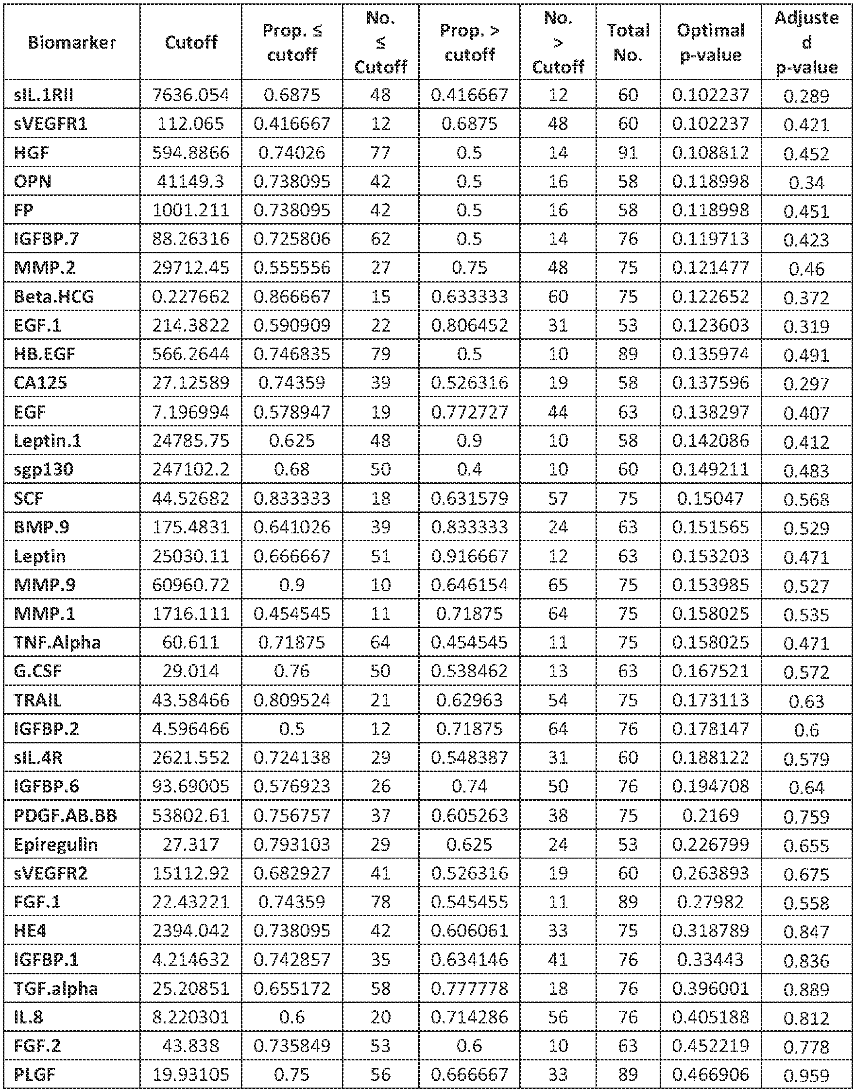
- a complete account for all 76 biomarkers is provided in Table III.
- biomarkers with optimal p-value ⁇ 0.05 representing the following processes: angiogenesis (sTNFRI, sTNFRII, Follistatin, TNF- ⁇ , Betacellulin, sVEGFR3, VEGF-A, Endoglin, MMP-10, PDGF-BB, VEGF-C, IGF-I, IGFBP-3, IGFBP-5, Endothelin-1, and Amphiregulin), cancer cachexia (TNF-a, sTNFRI, sTNFRII, IGF-I, IGFBP-3, IGFBP-5, IL-6, IL-6R), and phenotypic transdifferentiation (betacellulin, IGF-I, IGFBP-3, IGFBP-5, sEGFR, and prolactin).
- angiogenesis sTNFRI, sTNFRII, Follistatin, TNF- ⁇ , Betacellulin, sVEGFR3, VEGF-A, Endoglin, MMP-10,
- a total of 34 biomarkers were identified for identifying advanced stage NSCLC patients that failed frontline chemotherapy with rapidly progressing disease.
- the specific cutoff values, number of patients in each arm, and optimal p-values are all provided in Table II, with the distribution of these classifications for select representative biomarkers provided in Figure 2 .
- a complete account for all 76 biomarkers are provided in Table IV.
- biomarkers with optimal p-value ⁇ 0.05 representing the following processes: angiogenesis (sVEGFR2, follistatin, IGF-I, IGF-II, IGFBPs -1,-2,-3,-5,-7, IL-8, MMP-2, MMP-7, PLGF, Leptin, PDGF-AA, TNF- ⁇ , sTNFRI, sTNFRII, and VEGF-A), cancer cachexia (HGF, IGF-I, IGF-II, IGFBPs -1,-2,-3,-5,-7, IL-6, Leptin, TRAIL, sCD30, TNF- ⁇ , sTNFRI, and sTNFRII), phenotypic transdifferentiation (beta-HCG, ⁇ -fetoprotein, HE4, HGF, IGF-I, IGF-II, IGFBPs -1,-2,-3,-5,-7, osteopontin, PLGF, TGF- ⁇ ,
- the practice of the present invention will employ, unless otherwise indicated, conventional methods for measuring the level of the biomarker within the skill of the art.
- the techniques may include, but are not limited to, molecular biology and immunology. Such techniques are explained fully in the literature. See, e.g., Sambrook, et al. Molecular Cloning: A Laboratory Manual (Current Edition, Cold Spring Harbor Laboratory Press, Cold Spring Harbor, N.Y .); Current Protocols in Molecular Biology (Eds. A Ausubel et al., NY: John Wiley & Sons, Current Editi on); DNA Cloning: A Practical Approach, vol. I & II (D. Glover, ed .); Oligonucleotide Synthesis (N.
Landscapes
- Health & Medical Sciences (AREA)
- Life Sciences & Earth Sciences (AREA)
- Immunology (AREA)
- Engineering & Computer Science (AREA)
- Molecular Biology (AREA)
- Biomedical Technology (AREA)
- Chemical & Material Sciences (AREA)
- Hematology (AREA)
- Urology & Nephrology (AREA)
- Biotechnology (AREA)
- Biochemistry (AREA)
- Cell Biology (AREA)
- Food Science & Technology (AREA)
- Medicinal Chemistry (AREA)
- Physics & Mathematics (AREA)
- Analytical Chemistry (AREA)
- Microbiology (AREA)
- General Health & Medical Sciences (AREA)
- General Physics & Mathematics (AREA)
- Pathology (AREA)
- Investigating Or Analysing Biological Materials (AREA)
- Measuring Or Testing Involving Enzymes Or Micro-Organisms (AREA)
- Peptides Or Proteins (AREA)
Description
- The present invention relates to methods and kits for identifying patients with rapidly progressing disease, and in particular to methods and kits for identifying patients with rapidly progressing non-small cell lung cancer and for determining optimal treatment plans for patients with rapidly progressing disease and for monitoring treatments.
- Lung cancer is leading cause of cancer-related mortality worldwide, with a projected 159,480 patients succumbing to the disease in the US in 2014.(1) Lung cancer is typically characterized as being quite aggressive with poor clinical outcomes that stem from the very rapid proliferation rates, high metastatic potential, and general insensitivity to available treatment strategies. Non-small cell lung cancer (NSCLC) presents unique challenges to health care providers because of its common late stage of presentation and the poor median overall survival of advanced disease.(2, 3) Patients often become too ill to receive second line treatment as noted by a recent phase III clinical trial where only 37% of the patients randomized to docetaxel at disease progression received treatment.(4)
- An objective of this study was reveal circulating biomarkers to identify patients with rapidly progressing NSCLC. This study examined 76 biomarkers that are surrogates for several pathophysiological processes associated with aggressive disease in both frontline (chemo naïve) and second-line and greater (pretreated) patients. A total of 186 patient serum specimens were evaluated. Processes evaluated include angiogenesis, phenotypic transdifferentiation (i.e. EMT, cancer stem cells), cancer cachexia, chronic inflammation, and immune system response.
- Identification of patients with rapidly-progressing disease who are insensitive to standard platinum double-based chemotherapy will provide clinical implications.
- There is a need in the art for screening methods and kits that identify patients with rapidly progressing disease in patients that are treatment naïve and in patients that have received a treatment.
- Methods and kits for identifying rapidly progressing lung cancer in a subject are provided. The method includes assaying a level of a biomarker in a biomarker panel in a biological sample obtained from a subject where the panel includes at least one biomarker selected from Table I or Table II. The method is dependent on whether the subject is treatment naïve or has received at least one treatment; and comparing the level of the biomarker in the subject's sample to a cutoff value listed in Table I for treatment naïve subjects or Table II for previously treated subjects. The method further includes determining whether the subject's level is above or below the cutoff value to determine whether the subject has rapidly progressing lung cancer.
-
-
Figure 1 illustrates representative 'Box and Whisker' plots indicating distribution of biomarker levels in the frontline chemotherapy cohort, separated based on a 90 day cutoff for rapid disease progression. Shown are TNF-α (panel A), sTNFRII (panel B), IL-6 (panel C), sVEGFR3 (panel D), betacellulin (Panel E) and Total PSA (panel F). All biomarker levels are provided in pg/mL. -
Figure 2 illustrates representative 'Box and Whisker plots indicating distribution of biomarker levels in the treated chemotherapy cohort, separated based on a 45 day cutoff for rapid disease progression. Shown are sEGFR (panel A), sTNFRI (panel B), TRAIL (panel C), IGFBP-1 (panel D), IGFBP-2 (Panel E) and HGF (panel F). All biomarker levels are provided in pg/mL. - The present invention will utilize at least one biomarker measured in a biological sample obtained from a subject to identify rapidly progressing lung cancer, and in some embodiments in subjects having rapidly progressing NSCLC. In some embodiments, the at least one biomarker may be selected from a panel of biomarkers. In some embodiments, one or more biomarkers from a panel of biomarkers are used to identify subjects having rapidly progressing NSCLC in subjects that are treatment naïve or that have been previously treated.
- The term "biomarker" as used herein, refers to any biological compound that can be measured as an indicator of the physiological status of a biological system. A biomarker may comprise an amino acid sequence, a nucleic acid sequence and fragments thereof. Exemplary biomarkers include, but are not limited to cytokines, chemokines, growth and angiogenic factors, metastasis related molecules, cancer antigens, apoptosis related proteins, proteases, adhesion molecules, cell signaling molecules and hormones.
- "Measuring" or "measurement" means assessing the presence, absence, quantity or amount (which can be an effective amount) of a given substance within a sample, including the derivation of qualitative or quantitative concentration levels of such substances, or otherwise evaluating the values or categorization of a subject's clinical parameters. Alternatively, the term "detecting" or "detection" may be used and is understood to cover all measuring or measurement as described herein.
- The terms "sample" or "biological sample" as used herein, refers to a sample of biological fluid in a healthy and/or pathological state obtained from a subject. Such samples include, but are not limited to, blood, bronchial lavage fluid, sputum, saliva, urine, amniotic fluid, lymph fluid, peritoneal fluid, and cerebrospinal fluid. In some embodiments, the whole blood sample is further processed into serum or plasma samples. In some embodiments, the sample includes blood spotting tests.
- The term "subject" or "patient" as used herein, refers to a mammal, preferably a human.
- The term "rapid progression" or "rapidly progressing" as used herein, refers to cases of disease that were observed to not respond to chemotherapy or targeted agents and advance (evidence of nascent metastases, increasing tumor volume, etc.) within a defined time interval. Thresholds for rapid progression were set to 90 days after the first treatment for the treatment naïve patients and 45 days after the second or subsequent treatment for the previously treated patients. Circulating levels of 27 biomarkers were found to be significantly associated (p≤0.05) with progression within 90 days of treatment initiation in treatment naive patients. Circulating levels of 34 biomarkers were found to be significantly associated (p≤0.05) with progression within 45 days of treatment initiation in previously treated patients.
- Biomarkers that may be used include but are not limited to cytokines, chemokines, growth and angiogenic factors, metastasis related molecules, cancer antigens, apoptosis related proteins, proteases, adhesion molecules, cell signaling molecules and hormones. The biomarkers are proteins that are circulating in the subject that may be detected from a fluid sample obtained from the subject. In some embodiments, the fluid sample may be serum or plasma. In some embodiments, one or more biomarkers from a panel of biomarkers may be used.
- In some embodiments, one or more biomarkers may be measured in a biomarker panel. The biomarker panel may include a plurality of biomarkers. In some embodiments, the biomarker panel may include ten or fewer biomarkers. In yet other embodiments, the biomarker panel may include 1, 2, 3, 4, 5, 6 or 7 biomarkers. In some embodiments, the biomarker panel may be optimized from a candidate pool of biomarkers. By way of non-limiting example, the biomarker or biomarker panel may be configured for determining whether a treatment naïve subject is likely to have rapidly progressing disease. In some embodiments, the biomarker or biomarker panel may be configured for determining whether a previously treated subject is likely to have rapidly progressing disease.
- In some embodiments, the biomarker panel may include biomarkers from several biological pathways. By way of non-limiting example, the biomarkers may be associated the tumor necrosis factor (TNF) family, the epidermal growth factor (EGF) family, the vascular endothelial growth factor (VEGF) family, the Insulin-like growth factor (IGF) family and/or associated with angiogenesis. In some embodiments, the TNF family may include, but is not limited to TNF-RI, TNF-RII, TNF-α and TRAIL. In some embodiments, the EGF family may include but is not limited to betacellulin, amphiregulin, and soluble-EGFR. In some embodiments, the VEGF family may include but is not limited to VEGF-A, VEGF-C, and soluble-VEGFR3. In some embodiments, the IGF family may include but is not limited to IGF-I, IGF-II, IGFBPs-2,-3, and -7. In some embodiments, the biomarkers associated with angiogenesis may include follistatin, IL-6, endoglin, PDGF-BB, IGF-1, and endothelin-1, PLGF, IL-8, MMP-2, HGF, sVEGFR2, VEGF-A, leptin, PDGF-AA, and others. In some embodiments, the biomarker panel may include one or more biomarkers from a panel of biomarkers. In some embodiments, the one or more biomarkers may be selected from the list of biomarkers in Table I. In some embodiments, the one or more biomarkers may be selected from the list of biomarkers in Table II. In some embodiments, other biomarkers may be used and may be combined with the biomarkers listed in Tables I and II.
- In some embodiments, patients with rapid disease progression in a treatment naïve group may be identified using one or more biomarkers selected from a panel of biomarkers listed Table I. In some embodiments, the one or more biomarkers may be selected from the group of biomarkers identified in Table I as having a p-value of 0.01 or less. In some embodiments, the one or more biomarkers may include at least one biomarker from Table I having a p-value of 0.01 or less and at least one biomarker from Table I having a p-value of 0.05 or less. In some embodiments, the one or more biomarkers may include biomarkers selected from the group consisting of sTNFRI, sTNFRII, CA 19-9, Follistatin, Total PSA, TNF-α and IL-6. In some embodiments, the biomarkers may include 3, 4, 5, 6 or 7 biomarkers selected from the group consisting of sTNFRI, sTNFRII, CA 19-9, Follistatin, Total PSA, TNF-a and IL-6 and may also include additional biomarkers. In some embodiments, patients with rapid disease progression in a treatment naïve group may be identified using a panel of one or more biomarkers selected from Table I where the panel may include 1, 2, 3, 4, 5, 6, 7, 8, 9, 10, 11, 12, 13, 14, 15, 16, 17, 18, 19, 20, 21, 22, 23, 24, 25, 26 or 27 biomarkers.
- In some embodiments, patients with rapid disease progression in a previously treated group may be identified using one or more biomarkers selected from a panel of biomarkers selected from Table II. In some embodiments, the one or more biomarkers may be selected from the group of biomarkers identified in Table II as having a p-value of 0.01 or less. In some embodiments, the one or more biomarkers may include at least one biomarker from Table II having a p-value of 0.01 or less and at least one biomarker form Table II having a p-value of 0.05 or less. In some embodiments, the one or more biomarkers may be selected from the group consisting of TRAIL, sTNFRI, IGFBP-1, sEGFR, IGF-1, TGF-β, HGF, MMP-7, MMP-2, α-fetoprotein, Osteopontin, sVEGFR2 and IL-6. In some embodiments, the one or more biomarkers may include 1, 2, 3, 4, 5, 6, 7, 8, 9, 10, 11, 12 or 13 biomarkers selected from the group consisting of TRAIL, sTNFRI, IGFBP-1, sEGFR, IGF-1, TGF-β, HGF, MMP-7, MMP-2, α-fetoprotein, Osteopontin, sVEGFR2 and IL-6 and may also include additional biomarkers. In some embodiments, patients with rapid disease progression in a previously treated group may be identified using one or more biomarkers selected from Table II where the panel may include 1, 2, 3, 4, 5, 6, 7, 8, 9, 10, 11, 12, 13, 14, 15, 16, 17, 18, 19, 20, 21, 22, 23, 24, 25, 26, 27, 28, 29, 30, 31, 32, 33 or 34 biomarkers.
- In some embodiments, the biomarker panel may be selected using a reference profile that can be made in conjunction with a statistical algorithm used with a computer to implement the statistical algorithm to sort the subject into a group. In some embodiments, the statistical algorithm is a learning statistical classifier system. The learning statistical classifier system can be selected from the following list of non-limiting examples, including Random Forest (RF), Classification and Regression Tree (CART), boosted tree, neural network (NN), support vector machine (SVM), general chi-squared automatic interaction detector model, interactive tree, multiadaptive regression spline, machine learning classifier, and combinations thereof. By way of non-limiting example, exemplary tools for selecting a biomarker panel may be found in
WO 2012/054732 andUS Provisional Application No. 61/792, 710 - Measurement of a biomarker generally relates to a quantitative measurement of an expression product, which is typically a protein or polypeptide. In some embodiments, the measurement of a biomarker may relate to a quantitative or qualitative measurement of nucleic acids, such as DNA or RNA. The measurement of the biomarker of the subject detects expression levels of one or more biomarkers in subjects having lung cancer and in some embodiments, compares the expression level of each biomarker measured to a cutoff value listed in Table I or in Table II.
- Expression of the biomarkers may be measured using any method known to one skilled in the art. Methods for measuring protein expression include, but are not limited to Western blot, immunoprecipitation, immunohistochemistry, Enzyme-linked immunosorbent assay (ELISA), Radio Immuno Assay (RIA), radioreceptor assay, proteomics methods, mass-spectrometry based detection (SRM or MRM) or quantitative immunostaining methods. Methods for measuring nucleic acid expression or levels may be any techniques known to one skilled in the art. Expression levels from the one or more biomarkers are measured in the subject and compared to the levels of the one or more biomarkers obtained from a cohort of subjects described below.
- In some embodiments, MILLIPLEX® MAP multiplex assays may be used to determine the expression levels of the one or more biomarkers in a panel of biomarkers. (EMD Millipore, Billlerica, MA.) In some embodiments, Luminex-based xMAP® multiplexed immunoassays may be used to determine the expression levels of the panel of biomarkers. (Luminex Corp.; Austin, TX.) In some embodiments, biomarker concentrations may be calculated based on 7-point standard curves using a five-parametric fit algorithm in xPONENT v4.0.3 (Luminex Corp.) Other measurement systems and techniques may also be used.
- A kit is provided with reagents to measure a plurality of biomarkers. The kit includes reagents to measure a panel of biomarkers for subjects that are treatment naïve. The kit includes reagents to measure a panel of biomarkers for subjects that have been previously treated.
- In some embodiments, methods for determining whether a subject has rapidly progressing lung cancer may be based upon the biomarker measurements from the subject compared to a reference cutoff level for each biomarker measured. The reference cutoff level for a plurality of biomarkers is listed in Tables I-IV.
- In some embodiments, the analysis of the biomarker panel may be used to determine a treatment regime for the subject. In some embodiments, the measurement of one or more biomarkers in the panel may be used to determine whether to begin a treatment, to continue the same treatment or to modify the treatment regime for a subject. The treatment may be modified by changing the drug administered to the subject or to add an additional drug to the existing drug treatment regime, to change the dosage or other changes. In some embodiments, other types of treatment regimes may be used such as radiation. In some embodiments, the identification of patients with rapidly progressing disease who are insensitive to standard platinum doublet-based chemotherapy may have multiple clinical implications. The identification of patients with rapidly progressing disease using the biomarker level may place the patient in a specific treatment, a different treatment or an earlier treatment in the overall treatment strategy. In some embodiments, a specific targeted chemotherapeutic agent may be selected based on the identification of rapidly progressing disease. In some embodiments, the specific chemotherapeutic agent may be changed based in the biomarker level measured relative to the cutoff value. By way of non-limiting example, VEGF-A levels in patients taking bevacizumab may be monitored and the treatment regime may be changed or not changed based on the level of VEGF-A measured and compared to the cutoff level in either Table I for treatment naïve patients or Table II for previously treated patients.
- Between 2004 and 2011, 186 patients at Rush University Medical Center (Chicago, IL) were enrolled and divided into the following cohorts: patients with advanced lung adenocarcinoma naïve to previous chemotherapy (n=76) and patients with advanced lung adenocarcinoma that have failed at least 1 line of chemotherapy (n=110). All stage classifications were determined according to the American Joint Committee on Cancer (AJCC) seventh edition criteria and confirmed by pathological evaluation (5, 6). All patient data was obtained after informed consent was given by the patient. The study was conducted in absolute compliance with the Institutional Review Board at Rush University Medical Center.
- All peripheral blood was collected pre-treatment and processed into serum using standard phlebotomy protocols. Serum was archived at -80 °C in aliquots; and no evaluable specimen were subjected to more than two freeze-thaw cycles (7-10). Serum was evaluated using the following biomarker panels: the MILLIPLEX® MAP Human Angiogenesis/ Growth Factor Panel (EMD Millipore, Billerica, MA) and included the following assays: epidermal growth factor (EGF), angiopoietin-2, granulocyte colony-stimulating factor (G-CSF), bone morphogenic protein 9 (BMP-9), endoglin, endothelin-1, leptin, fibroblast growth factor-1 (FGF-1), FGF-2, follistatin, interleukin-8 (IL-8), hepatocyte growth factor (HGF), heparin-binding epidermal growth factor (HB-EGF), placental growth factor (PLGF), vascular endothelial growth factor-A (VEGF-A), VEGF-C and VEGF-D; the MILLIPLEX® MAP Human Soluble Cytokine Receptor Panel which includes sVEGFR1, sVEGFR2, sVEGFR3, sIL-6R, sgp130, sTNFRI, and sTNFRII; the MILLIPLEX® MAP Human Circulating Cancer Biomarker Panel 1 which includes sFasL, IL-6, prolactin, SCF, TGF-α, and TNF-α; the MILLIPLEX® MAP Human MMP1 and MMP2 panels which combine to provide MMPs -1,-2,-3,-7,-9, and -10; and the MILLIPLEX® MAP Human Cytokine/ Chemokine Panel II, which includes SDF-1 (α+β). All assays were performed according to the manufacturer's recommended protocols and in a blinded fashion. All data was collected on a Luminex FlexMAP 3D system with concentrations calculated based on 7-point standard curves using a five-parametric fit algorithm in xPONENT v4.0.3 (Luminex Corp., Austin, TX).
- One endpoint of the investigation was to evaluate associations of the circulating biomarkers tested with clinical outcome measures for patients determined to have rapidly progressing disease. Progression status values were classified as 'slow' or 'rapid' based on chosen clinically-relevant cutoff (45 days for frontline and 90 days for those second-line and above) value. Association of the slow/ rapid progression state was then accomplished with low and high values of a biomarker based on cutoff values obtained from a grid search for an optimal cutoff within the potential range of the biomarker values that maximizes the p-value for disease progression via Fisher's exact test. Additionally, an adjusted p-value, which adjusts for the grid search, is also obtained. These analyses were performed regardless of regimen type. All statistical analyses were completed using the R Statistical Package.
- A total of 27 biomarkers were identified for identifying advanced stage NSCLC patients that were chemotherapy naïve with rapidly progressing disease. The specific cutoff values, number of patients in each arm, and optimal p-values are all provided in Table I, with the distribution of these classifications for select representative biomarkers provided in
Figure 1 . A complete account for all 76 biomarkers is provided in Table III. Included in the findings were biomarkers with optimal p-value ≤0.05 representing the following processes: angiogenesis (sTNFRI, sTNFRII, Follistatin, TNF-α, Betacellulin, sVEGFR3, VEGF-A, Endoglin, MMP-10, PDGF-BB, VEGF-C, IGF-I, IGFBP-3, IGFBP-5, Endothelin-1, and Amphiregulin), cancer cachexia (TNF-a, sTNFRI, sTNFRII, IGF-I, IGFBP-3, IGFBP-5, IL-6, IL-6R), and phenotypic transdifferentiation (betacellulin, IGF-I, IGFBP-3, IGFBP-5, sEGFR, and prolactin).Table I: Treatment Naïve Cohort Biomarker Cutoff Value (pg/mL) Cases ≤ cutoff Cases > Cutoff p-Value Adjusted p-value sTNFRI 1879.8 48 12 0.0003 0.001 sTNFRII 8493.9 49 11 0.0011 0.006 CA 19-9 20.9 62 13 0.0029 0.018 Follistatin 990.0 48 15 0.0033 0.007 Total PSA 67.1 47 28 0.0042 0.027 TNF-α 0.61 12 45 0.0050 0.029 IL-6 13.1 48 10 0.0100 0.021 TGF β1 30665.9 64 11 0.0132 0.083 Betacellulin 14.9 25 28 0.0162 0.058 CA 15.3 55.0 62 13 0.0203 0.077 sCD30 101.5 40 20 0.0219 0.111 sVEGFR3 2930.8 48 12 0.0221 0.094 VEGF-A 727.1 49 27 0.0222 0.092 Endoglin 993.8 48 15 0.0228 0.084 MMP-10 272.6 43 32 0.0244 0.122 sRAGE 84.2 39 21 0.0244 0.121 PDGF-BB 13336.6 12 40 0.0248 0.075 VEGF-C 32.9 10 53 0.0255 0.105 IGF-I 31266.8 46 30 0.0264 0.133 IGFBP-3 1452.8 53 23 0.0308 0.168 sEGFR 25073.2 29 43 0.0392 0.177 IGFBP-5 151.1 48 28 0.0428 0.192 Endothelin-1 8.0 39 24 0.0430 0.124 Amphiregulin 38.7 16 36 0.0445 0.222 GRO 3960.3 47 28 0.0453 0.181 sIL-6R 10666.7 20 40 0.0493 0.203 Prolactin 5353.7 30 28 0.0497 0.152 - A total of 34 biomarkers were identified for identifying advanced stage NSCLC patients that failed frontline chemotherapy with rapidly progressing disease. The specific cutoff values, number of patients in each arm, and optimal p-values are all provided in Table II, with the distribution of these classifications for select representative biomarkers provided in
Figure 2 . A complete account for all 76 biomarkers are provided in Table IV. Included in the findings were biomarkers with optimal p-value ≤0.05 representing the following processes: angiogenesis (sVEGFR2, follistatin, IGF-I, IGF-II, IGFBPs -1,-2,-3,-5,-7, IL-8, MMP-2, MMP-7, PLGF, Leptin, PDGF-AA, TNF-α, sTNFRI, sTNFRII, and VEGF-A), cancer cachexia (HGF, IGF-I, IGF-II, IGFBPs -1,-2,-3,-5,-7, IL-6, Leptin, TRAIL, sCD30, TNF-α, sTNFRI, and sTNFRII), phenotypic transdifferentiation (beta-HCG, α-fetoprotein, HE4, HGF, IGF-I, IGF-II, IGFBPs -1,-2,-3,-5,-7, osteopontin, PLGF, TGF-β, and sEGFR), and inflammation or immune response (CYFRA 21-1, GRO, IL-6, IL-8, sIL-2Rα, sIL-4R, TRAIL, sCD30, TNF-α, sTNFRI, and sTNFRII).Table II: Post Treatment Cohort Biomarker Cutoff Value (pg/mL) Cases ≤ cutoff Cases > Cutoff p-Value Adjusted p-value TRAIL 60.3 35 76 0.0005 0.003 sTNFRI 1506.6 62 34 0.0007 0.005 IGFBP-1 9.2 88 22 0.0009 0.005 sEGFR 50565.2 36 68 0.0012 0.007 IGF-I 7807.7 32 78 0.0021 0.008 TGF-β1 9073.7 10 77 0.0028 0.016 HGF 239.7 81 30 0.0035 0.034 MMP-7 8326.9 68 43 0.0043 0.024 MMP-2 33782.5 31 80 0.0044 0.027 a-fetoprotein 964.1 63 40 0.0050 0.031 Osteopontin 34067.2 49 54 0.0058 0.034 sVEGFR2 10846.5 13 83 0.0073 0.049 IL-6 8.0 66 37 0.0098 0.035 CYFRA 21.1 768.7 51 52 0.0119 0.03 IGF-II 507.7 37 68 0.0129 0.083 sTNFRII 7588.9 65 31 0.0151 0.093 CA 15.3 34.5 83 28 0.0156 0.056 sCD30 129.7 73 23 0.0177 0.081 sIL-4R 2096.7 14 82 0.0201 0.103 sIL-2Ralpha 836.6 55 41 0.0208 0.118 IGFBP-7 53.7 18 92 0.0211 0.137 TNF-α 91.1 93 17 0.0213 0.114 VEGF-A 1297.7 98 14 0.0227 0.05 Follistatin 968.9 11 10 0.0237 0.013 Leptin-1 11840.0 55 48 0.0239 0.154 IGFBP-3 1486.8 90 20 0.0263 0.161 PLGF 86.8 69 10 0.0268 0.127 IGFBP-5 193.5 44 66 0.0296 0.166 GRO 4254.6 80 30 0.0333 0.162 IL-8 63.9 96 15 0.0334 0.178 HE4 2993.4 88 23 0.0343 0.201 IGFBP-2 22.9 64 46 0.0344 0.169 PDGF-AA 10851.4 12 98 0.0375 0.218 B-HCG 0.16 26 85 0.0446 0.239 



- The practice of the present invention will employ, unless otherwise indicated, conventional methods for measuring the level of the biomarker within the skill of the art. The techniques may include, but are not limited to, molecular biology and immunology. Such techniques are explained fully in the literature. See, e.g., Sambrook, et al. Molecular Cloning: A Laboratory Manual (Current Edition, Cold Spring Harbor Laboratory Press, Cold Spring Harbor, N.Y.); Current Protocols in Molecular Biology (Eds. A Ausubel et al., NY: John Wiley & Sons, Current Edition); DNA Cloning: A Practical Approach, vol. I & II (D. Glover, ed.); Oligonucleotide Synthesis (N. Gait, ed., Current Edition); Nucleic Acid Hybridization (B. Hames & S. Higgins, eds., Current Edition); Transcription and Translation (B. Hames & S. Higgins, eds., Current Edition).
- The above Figures and disclosure are intended to be illustrative and not exhaustive. This description will suggest many variations and alternatives to one of ordinary skill in the art.
-
- 1. ACS. American Cancer Society. Cancer Facts & Figures 2013. 2013 [cited 2013 06/06/2013]; Available from: http://www.cancer.org/acs/groups/content/@epidemiologysurveilance/documents/ document/acspc-036845.pdf
- 2. Jemal A, Center MM, Ward E, Thun MJ. Cancer occurrence. Methods Mol Biol 2009:471: 3-29.
- 3. Jemal A, Thun MJ, Ries LA, et al. Annual report to the nation on the status of cancer, 1975-2005, featuring trends in lung cancer, tobacco use, and tobacco control. J Natl Cancer Inst 2008;100: 1672-94.
- 4. Fidias PM, Dakhil SR, Lyss AP, et al. Phase III study of immediate compared with delayed docetaxel after front-line therapy with gemcitabine plus carboplatin in advanced non-small-cell lung cancer. J Clin Oncol 2009;27: 591-8.
- 5. Goldstraw P, Crowley J, Chansky K, et al. The IASLC Lung Cancer Staging Project: proposals for the revision of the TNM stage groupings in the forthcoming (seventh) edition of the TNM Classification of malignant tumours. J Thorac Oncol 2007;2: 706-14.
- 6. Groome PA, Bolejack V, Crowley JJ, et al. The IASLC Lung Cancer Staging Project: validation of the proposals for revision of the T, N, and M descriptors and consequent stage groupings in the forthcoming (seventh) edition of the TNM classification of malignant tumours. J Thorac Oncol 2007;2: 694-705.
- 7. Borgia JA, Basu S, Faber LP, et al. Establishment of a multi-analyte serum biomarker panel to identify lymph node metastases in non-small cell lung cancer. J Thorac Oncol 2009;4: 338-47.
- 8. Farlow EC, Patel K, Basu S, at al. Development of a multiplexed tumor-associated autoantibody-based blood test for the detection of non-small cell lung cancer. Clin Cancer Res 2010;16: 3452-62.
- 9. Patel K, Farlow EC, Kim AW, et al. Enhancement of a multianalyte serum biomarker panel to identify lymph node metastases in non-small cell lung cancer with circulating autoantibody biomarkers. Int J Cancer 2010;129: 133-42.
- 10. Shersher DD, Vercillo MS, Fhied C, et al. Biomarkers of the Insulin-Like Growth Factor Pathway Predict Progression and Outcome in Lung Cancer. Ann Thorac Surg 2011.
- 11. Borgia JA, Basu S, Faber LP, et al. Establishment of a multi-analyte serum biomarker panel to identify lymph node metastases in non-small cell lung cancer. J Thorac Oncol 2009;4: 338-47.
- 12. R Core Team (2013). R: A language and environment for statistical computing. R Foundation for Statistical Computing, Vienna, Austria URL http://www.R-project.org /
Claims (13)
- A method for identifying rapidly progressing lung cancer in a subject, the method comprising:measuring a level of at least one circulating biomarker in a biological sample obtained from a subject, wherein the at least one biomarker is selected from Table I or Table II;
Table I: Biomarker Cutoff Value (pg/ml) Cases ≤ cutoff Cases > Cutoff p-Value Adjusted p-value sTNFRI 1879.8 48 12 0.0003 0.001 sTNFRII 8493.9 49 11 0.0011 0.006 CA 19-9 20.9 62 13 0.0029 0.018 Follistatin 990.0 48 15 0.0033 0.007 Total PSA 67.1 47 28 0.0042 0.027 TNF-α 0.61 12 45 0.0050 0.029 IL-6 13.1 48 10 0.0100 0.021 TGF β1 30665.9 64 11 0.0132 0.083 Betacellulin 14.9 25 28 0.0162 0.058 CA 15.3 55.0 62 13 0.0203 0.077 sCD30 101.5 40 20 0.0219 0.111 sVEGFR3 2930.8 48 12 0.0221 0.094 VEGF-A 727.1 49 27 0.0222 0.092 Endoglin 993.8 48 15 0.0228 0.084 MMP-10 272.6 43 32 0.0244 0.122 sRAGE 84.2 39 21 0.0244 0.121 PDGF-BB 13336.6 12 40 0.0248 0.075 VEGF-C 32.9 10 53 0.0255 0.105 IGF-I 31266.8 46 30 0.0264 0.133 IGFBP-3 1452.8 53 23 0.0308 0.168 sEGFR 25073.2 29 43 0.0392 0.177 IGFBP-5 151.1 48 28 0.0428 0.192 Endothelin-1 8.0 39 24 0.0430 0.124 Amphiregulin 38.7 16 36 0.0445 0.222 GRO 3960.3 47 28 0.0453 0.181 sIL-6R 10666.7 20 40 0.0493 0.203 Prolactin 5353.7 30 28 0.0497 0.152 Table II Biomarker Cutoff Value (pg/mL) Cases ≤ cutoff Cases > Cutoff p-Value Adjusted p-value TRAIL 60.3 35 76 0.0005 0.003 sTNFRI 1506.6 62 34 0.0007 0.005 IGFBP-1 9.2 88 22 0.0009 0.005 sEGFR 50565.2 36 68 0.0012 0.007 IGF-I 7807.7 32 78 0.0021 0.008 TGF-β1 9073.7 10 77 0.0028 0.016 HGF 239.7 81 30 0.0035 0.034 MMP-7 8326.9 68 43 0.0043 0.024 MMP-2 33782.5 31 80 0.0044 0.027 α-fetoprotein 964.1 63 40 0.0050 0.031 Osteopontin 34067.2 49 54 0.0058 0.034 sVEGFR2 10846.5 13 83 0.0073 0.049 IL-6 8.0 66 37 0.0098 0.035 CYFRA 21.1 768.7 51 52 0.0119 0.03 IGF-II 507.7 37 68 0.0129 0.083 sTNFRII 7588.9 65 31 0.0151 0.093 CA 15.3 34.5 83 28 0.0156 0.056 sCD30 129.7 73 23 0.0177 0.081 sIL-4R 2096.7 14 82 0.0201 0.103 sIL-2Ralpha 836.6 55 41 0.0208 0.118 IGFBP-7 53.7 18 92 0.0211 0.137 TNF-α 91.1 93 17 0.0213 0.114 VEGF-A 1297.7 98 14 0.0227 0.05 Follistatin 968.9 11 10 0.0237 0.013 Leptin-1 11840.0 55 48 0.0239 0.154 IGFBP-3 1486.8 90 20 0.0263 0.161 PLGF 86.8 69 10 0.0268 0.127 IGFBP-5 193.5 44 66 0.0296 0.166 GRO 4254.6 80 30 0.0333 0.162 IL-8 63.9 96 15 0.0334 0.178 HE4 2993.4 88 23 0.0343 0.201 IGFBP-2 22.9 64 46 0.0344 0.169 PDGF-AA 10851.4 12 98 0.0375 0.218 B-HCG 0.16 26 85 0.0446 0.239 determining whether the subject is treatment naïve or has received at least one treatment;comparing the level of the at least one biomarker in the subject's sample to a cutoff value listed in Table I for treatment naïve subjects or Table II for previously treated subjects;determining whether the subject's level is above or below the cutoff value to determine whether the subject has rapidly progressing lung cancer, wherein the threshold for rapid progression is 90 days after the first treatment for the treatment naïve subjects and 45 days after the second or subsequent treatment for the previously treated subjects. - The method according to claim 1, wherein the at least one biomarker measured has a p-value where p≤0.05.
- The method according to claim 1, wherein the at least one biomarker measured has a p-value where p≤0.01.
- The method according to any one of claims 1-3, comprising determining the level of at least two biomarkers selected from Table I for treatment naïve subjects or from Table II for previously treated subjects.
- The method according to any one of claims 1-4, comprising determining the level of the at least one biomarker wherein the at least one biomarker is selected from sTNFRI, sTNFRII, CA 19-9, Follistatin, Total PSA, TNF-α and IL-6 for treatment naïve subjects.
- The method according to any one of claims 1-4, comprising determining the level of the at least one biomarker wherein the at least one biomarker comprises TRAIL, sTNFRI, IGFBP-1, sEGFR, IGF-1, TGF-β, HGF, MMP-7, MMP-2, α-fetoprotein, Osteopontin, sVEGFR2 and IL-6 for the pretreated subjects.
- The method according to any one of claims 1-6, wherein the lung cancer is non-small cell lung cancer.
- The method according to any one of claims 1-7, wherein the biological sample comprises plasma sample or serum sample.
- The method according to any one of claims 1-8, further comprising modifying a treatment regime for the subject when the comparison indicates that the subject has rapidly progressing lung cancer.
- The method according to any one of claims 1-9, wherein a plurality of biomarkers are measured, wherein the plurality of biomarkers comprises sTNFRI, sTNFRII, CA 19-9, Follistatin, Total PSA, TNF-α and IL-6 for treatment naïve subjects.
- The method according to any one of claims 1-9, wherein a plurality of biomarkers are measured, wherein the plurality of biomarkers comprises TRAIL, sTNFRI, IGFBP-1, sEGFR, IGF-1, TGF-β, HGF, MMP-7, MMP-2, α-fetoprotein, Osteopontin, sVEGFR2 and IL-6 for the pretreated subjects.
- A kit for performing the measurement of the level of a plurality of biomarkers of the subject in claim 1, wherein the kit comprises reagents for measuring the plurality of biomarkers wherein the plurality of biomarkers include sTNFRI, sTNFRII, CA 19-9, Follistatin, Total PSA, TNF-α and IL-6 for treatment naïve subjects or wherein the plurality of biomarkers include TRAIL, sTNFRI, IGFBP-1, sEGFR, IGF-1, TGF-β, HGF, MMP-7, MMP-2, α-fetoprotein, Osteopontin, sVEGFR2 and IL-6 for the pretreated subjects.
- The kit according to claim 12, wherein the kit comprises reagents for measuring the circulating biomarker in serum or plasma.
Applications Claiming Priority (2)
| Application Number | Priority Date | Filing Date | Title |
|---|---|---|---|
| US201361913740P | 2013-12-09 | 2013-12-09 | |
| PCT/US2014/069009 WO2015088947A1 (en) | 2013-12-09 | 2014-12-08 | Biomarkers of rapid progression in advanced non-small cell lung cancer |
Publications (3)
| Publication Number | Publication Date |
|---|---|
| EP3080299A1 EP3080299A1 (en) | 2016-10-19 |
| EP3080299A4 EP3080299A4 (en) | 2017-06-21 |
| EP3080299B1 true EP3080299B1 (en) | 2020-09-30 |
Family
ID=53371729
Family Applications (1)
| Application Number | Title | Priority Date | Filing Date |
|---|---|---|---|
| EP14869355.9A Active EP3080299B1 (en) | 2013-12-09 | 2014-12-08 | Biomarkers of rapid progression in advanced non-small cell lung cancer |
Country Status (3)
| Country | Link |
|---|---|
| US (2) | US10365281B2 (en) |
| EP (1) | EP3080299B1 (en) |
| WO (1) | WO2015088947A1 (en) |
Families Citing this family (6)
| Publication number | Priority date | Publication date | Assignee | Title |
|---|---|---|---|---|
| US11656229B2 (en) * | 2015-06-26 | 2023-05-23 | Biomark Cancer Systems Inc. | Method of detecting lung cancer |
| EP3607089A4 (en) * | 2017-04-04 | 2020-12-30 | Lung Cancer Proteomics, LLC | Plasma based protein profiling for early stage lung cancer prognosis |
| CN108680745B (en) * | 2018-01-03 | 2021-06-15 | 湖州市中心医院 | Application of serum lipid biomarkers in early diagnosis of NSCLC |
| EP4278186A1 (en) * | 2021-01-14 | 2023-11-22 | F. Hoffmann-La Roche AG | Prognostic value of biomarkers in patients with non-small cell lung cancer having stable disease |
| US11940450B2 (en) | 2021-03-16 | 2024-03-26 | University Of Connecticut | Biomarker panel for non-invasive diagnosis of congenital renal dysfunction |
| JP2024535694A (en) * | 2021-09-10 | 2024-10-02 | グレイル インコーポレイテッド | Method for analyzing target molecules in biological fluids |
Family Cites Families (19)
| Publication number | Priority date | Publication date | Assignee | Title |
|---|---|---|---|---|
| AU2002236507A1 (en) * | 2000-11-29 | 2002-06-11 | Cangen International | Dap-kinase and hoxa9, two human genes associated with genesis, progression, and aggressiveness of non-small cell lung cancer |
| CN1547617A (en) | 2001-06-25 | 2004-11-17 | 2 | Medicine innovation for oncology |
| US20040091857A1 (en) | 2001-07-20 | 2004-05-13 | Nallur Girish N. | Gene expression profiling |
| EP1520032A4 (en) | 2001-11-02 | 2007-07-18 | Pfizer Prod Inc | THERAPEUTIC AND DIAGNOSTIC MEANS APPLICABLE TO LUNG CANCER |
| US8747867B2 (en) | 2004-09-30 | 2014-06-10 | Ifom Fondazione Instituto Firc Di Oncologia Molecolare | Cancer markers |
| US9347945B2 (en) | 2005-12-22 | 2016-05-24 | Abbott Molecular Inc. | Methods and marker combinations for screening for predisposition to lung cancer |
| US20090047689A1 (en) | 2007-06-20 | 2009-02-19 | John Kolman | Autoantigen biomarkers for early diagnosis of lung adenocarcinoma |
| WO2009028580A1 (en) | 2007-08-24 | 2009-03-05 | Oncotherapy Science, Inc. | Ebi3, dlx5, nptx1 and cdkn3 for target genes of lung cancer therapy and diagnosis |
| US8541183B2 (en) | 2007-09-11 | 2013-09-24 | Cancer Prevention And Cure, Ltd. | Methods of identification, assessment, prevention and therapy of lung diseases and kits thereof |
| WO2009067546A2 (en) | 2007-11-19 | 2009-05-28 | Celera Corpration | Lung cancer markers and uses thereof |
| CN101896818B (en) | 2007-12-10 | 2016-01-20 | 霍夫曼-拉罗奇有限公司 | Seprase as a marker of cancer |
| US8338189B2 (en) | 2008-03-05 | 2012-12-25 | Axela Inc. | Detection of biomarkers and biomarker complexes |
| WO2010045573A2 (en) | 2008-10-17 | 2010-04-22 | University Of Miami | Tumor vaccine |
| US20100176228A1 (en) | 2009-01-14 | 2010-07-15 | Michilin Prosperity Co., Ltd. | Safety switch of a shredder |
| BRPI1015179A2 (en) | 2009-06-19 | 2016-04-19 | Merck Patent Gmbh | Biomarkers and Methods for Determining Efficacy of Anti-Egfr Antibodies in Cancer Therapy |
| US20120178111A1 (en) * | 2009-09-23 | 2012-07-12 | Diamandis Eleftherios P | Methods and compositions for the detection of lung cancers |
| US20110318336A1 (en) | 2010-03-29 | 2011-12-29 | George Mason Intellectual Properties, Inc. | Identification and Treatment of Aggressive Lung Cancer Tumors |
| US20120115745A1 (en) | 2010-05-07 | 2012-05-10 | Mckeegan Evelyn M | Methods for predicting sensitivity to treatment with a targeted tyrosine kinase inhibitor |
| AU2011316986A1 (en) * | 2010-10-20 | 2013-06-06 | Rush University Medical Center | Lung cancer tests |
-
2014
- 2014-12-08 WO PCT/US2014/069009 patent/WO2015088947A1/en not_active Ceased
- 2014-12-08 EP EP14869355.9A patent/EP3080299B1/en active Active
- 2014-12-08 US US15/102,647 patent/US10365281B2/en active Active
-
2019
- 2019-06-20 US US16/447,634 patent/US20190331683A1/en not_active Abandoned
Non-Patent Citations (1)
| Title |
|---|
| None * |
Also Published As
| Publication number | Publication date |
|---|---|
| EP3080299A4 (en) | 2017-06-21 |
| WO2015088947A1 (en) | 2015-06-18 |
| US10365281B2 (en) | 2019-07-30 |
| EP3080299A1 (en) | 2016-10-19 |
| US20190331683A1 (en) | 2019-10-31 |
| US20160313336A1 (en) | 2016-10-27 |
Similar Documents
| Publication | Publication Date | Title |
|---|---|---|
| US20190331683A1 (en) | Biomarkers of Rapid Progression in Advanced Non-Small Cell Lung Cancer | |
| Staff et al. | Growth differentiation factor-15 as a prognostic biomarker in ovarian cancer | |
| US10613091B2 (en) | Methods of developing a prognosis for pancreatic cancer and predicting responsiveness to cancer therapeutics | |
| JP5750152B2 (en) | Test method using signature gene associated with hepatocellular carcinoma, and array or kit used in the method | |
| JP6198752B2 (en) | Biomarkers for gastric cancer and uses thereof | |
| WO2021076036A1 (en) | Apparatuses and methods for detection of pancreatic cancer | |
| Xu et al. | Identification of blood protein biomarkers that aid in the clinical assessment of patients with malignant glioma | |
| US20150198601A1 (en) | Methods of diagnosing and prognosing colonic polyps | |
| AU2016214140A1 (en) | Biomarkers for pancreatic cancer | |
| JP6250600B2 (en) | Methods for detecting non-small cell lung cancer biomarkers | |
| JP2021144052A (en) | Methods of detecting relapse of lung adenocarcinoma based on marker human epididymis protein 4 (he4) and related uses | |
| EP3835789A1 (en) | Biomarker panel for diagnosing colorectal cancer | |
| Fidler et al. | Differential expression of circulating biomarkers of tumor phenotype and outcomes in previously treated non-small cell lung cancer patients receiving erlotinib vs. cytotoxic chemotherapy | |
| US20190101540A1 (en) | Angiogenesis Biomarkers Associated With Disease Progression in Lung Cancer | |
| JP2012526543A5 (en) | ||
| EP2895863B1 (en) | New biomarkers for the diagnosis and/or prognosis of clear cell renal cell carcinoma | |
| JP2021170021A (en) | Testing method that enables specific diagnosis of early pathology of diabetic nephropathy | |
| KR102737876B1 (en) | Biomarker for predicting prognosis post-neoadjuvant chemotherapy of breast cancer patient and use thereof | |
| JP7660077B2 (en) | Diagnostic methods | |
| KR20200045131A (en) | Diagnostic Biomarker For Prognosis of Intestinal Type Gastric Cancer | |
| US20150276747A1 (en) | Biomarkers for ovarian cancer | |
| WO2026068798A1 (en) | Sox9 for assessing colorectal cancer (crc) or a precancerous condition thereof | |
| US20210231664A1 (en) | Eosinophil Cationic Protein (ECP) as a Tumor Marker for Malignant Tumors | |
| WO2026068795A2 (en) | Clec3b for assessing colorectal cancer (crc) or a precancerous condition thereof | |
| Charrier et al. | Tu1495 Connective Tissue Growth Factor Production by Pancreatic Acinar Cells is Associated With Regulation of Immune Function in a Mouse Model of Alcoholic Chronic Pancreatitis |
Legal Events
| Date | Code | Title | Description |
|---|---|---|---|
| PUAI | Public reference made under article 153(3) epc to a published international application that has entered the european phase |
Free format text: ORIGINAL CODE: 0009012 |
|
| 17P | Request for examination filed |
Effective date: 20160709 |
|
| AK | Designated contracting states |
Kind code of ref document: A1 Designated state(s): AL AT BE BG CH CY CZ DE DK EE ES FI FR GB GR HR HU IE IS IT LI LT LU LV MC MK MT NL NO PL PT RO RS SE SI SK SM TR |
|
| AX | Request for extension of the european patent |
Extension state: BA ME |
|
| RIN1 | Information on inventor provided before grant (corrected) |
Inventor name: BASU, SANJIB Inventor name: BORGIA, JEFFREY, A. |
|
| DAX | Request for extension of the european patent (deleted) | ||
| REG | Reference to a national code |
Ref country code: DE Ref legal event code: R079 Ref document number: 602014070863 Country of ref document: DE Free format text: PREVIOUS MAIN CLASS: C12Q0001680000 Ipc: G01N0033574000 |
|
| RIC1 | Information provided on ipc code assigned before grant |
Ipc: G01N 33/574 20060101AFI20170509BHEP |
|
| A4 | Supplementary search report drawn up and despatched |
Effective date: 20170519 |
|
| STAA | Information on the status of an ep patent application or granted ep patent |
Free format text: STATUS: EXAMINATION IS IN PROGRESS |
|
| 17Q | First examination report despatched |
Effective date: 20180424 |
|
| GRAP | Despatch of communication of intention to grant a patent |
Free format text: ORIGINAL CODE: EPIDOSNIGR1 |
|
| STAA | Information on the status of an ep patent application or granted ep patent |
Free format text: STATUS: GRANT OF PATENT IS INTENDED |
|
| INTG | Intention to grant announced |
Effective date: 20200507 |
|
| GRAS | Grant fee paid |
Free format text: ORIGINAL CODE: EPIDOSNIGR3 |
|
| GRAA | (expected) grant |
Free format text: ORIGINAL CODE: 0009210 |
|
| STAA | Information on the status of an ep patent application or granted ep patent |
Free format text: STATUS: THE PATENT HAS BEEN GRANTED |
|
| AK | Designated contracting states |
Kind code of ref document: B1 Designated state(s): AL AT BE BG CH CY CZ DE DK EE ES FI FR GB GR HR HU IE IS IT LI LT LU LV MC MK MT NL NO PL PT RO RS SE SI SK SM TR |
|
| REG | Reference to a national code |
Ref country code: GB Ref legal event code: FG4D Ref country code: CH Ref legal event code: EP |
|
| REG | Reference to a national code |
Ref country code: AT Ref legal event code: REF Ref document number: 1319333 Country of ref document: AT Kind code of ref document: T Effective date: 20201015 |
|
| REG | Reference to a national code |
Ref country code: DE Ref legal event code: R096 Ref document number: 602014070863 Country of ref document: DE |
|
| REG | Reference to a national code |
Ref country code: IE Ref legal event code: FG4D |
|
| PG25 | Lapsed in a contracting state [announced via postgrant information from national office to epo] |
Ref country code: NO Free format text: LAPSE BECAUSE OF FAILURE TO SUBMIT A TRANSLATION OF THE DESCRIPTION OR TO PAY THE FEE WITHIN THE PRESCRIBED TIME-LIMIT Effective date: 20201230 Ref country code: FI Free format text: LAPSE BECAUSE OF FAILURE TO SUBMIT A TRANSLATION OF THE DESCRIPTION OR TO PAY THE FEE WITHIN THE PRESCRIBED TIME-LIMIT Effective date: 20200930 Ref country code: HR Free format text: LAPSE BECAUSE OF FAILURE TO SUBMIT A TRANSLATION OF THE DESCRIPTION OR TO PAY THE FEE WITHIN THE PRESCRIBED TIME-LIMIT Effective date: 20200930 Ref country code: SE Free format text: LAPSE BECAUSE OF FAILURE TO SUBMIT A TRANSLATION OF THE DESCRIPTION OR TO PAY THE FEE WITHIN THE PRESCRIBED TIME-LIMIT Effective date: 20200930 Ref country code: BG Free format text: LAPSE BECAUSE OF FAILURE TO SUBMIT A TRANSLATION OF THE DESCRIPTION OR TO PAY THE FEE WITHIN THE PRESCRIBED TIME-LIMIT Effective date: 20201230 Ref country code: GR Free format text: LAPSE BECAUSE OF FAILURE TO SUBMIT A TRANSLATION OF THE DESCRIPTION OR TO PAY THE FEE WITHIN THE PRESCRIBED TIME-LIMIT Effective date: 20201231 |
|
| REG | Reference to a national code |
Ref country code: AT Ref legal event code: MK05 Ref document number: 1319333 Country of ref document: AT Kind code of ref document: T Effective date: 20200930 |
|
| PG25 | Lapsed in a contracting state [announced via postgrant information from national office to epo] |
Ref country code: LV Free format text: LAPSE BECAUSE OF FAILURE TO SUBMIT A TRANSLATION OF THE DESCRIPTION OR TO PAY THE FEE WITHIN THE PRESCRIBED TIME-LIMIT Effective date: 20200930 Ref country code: RS Free format text: LAPSE BECAUSE OF FAILURE TO SUBMIT A TRANSLATION OF THE DESCRIPTION OR TO PAY THE FEE WITHIN THE PRESCRIBED TIME-LIMIT Effective date: 20200930 |
|
| REG | Reference to a national code |
Ref country code: NL Ref legal event code: MP Effective date: 20200930 |
|
| REG | Reference to a national code |
Ref country code: LT Ref legal event code: MG4D |
|
| PG25 | Lapsed in a contracting state [announced via postgrant information from national office to epo] |
Ref country code: CZ Free format text: LAPSE BECAUSE OF FAILURE TO SUBMIT A TRANSLATION OF THE DESCRIPTION OR TO PAY THE FEE WITHIN THE PRESCRIBED TIME-LIMIT Effective date: 20200930 Ref country code: RO Free format text: LAPSE BECAUSE OF FAILURE TO SUBMIT A TRANSLATION OF THE DESCRIPTION OR TO PAY THE FEE WITHIN THE PRESCRIBED TIME-LIMIT Effective date: 20200930 Ref country code: SM Free format text: LAPSE BECAUSE OF FAILURE TO SUBMIT A TRANSLATION OF THE DESCRIPTION OR TO PAY THE FEE WITHIN THE PRESCRIBED TIME-LIMIT Effective date: 20200930 Ref country code: EE Free format text: LAPSE BECAUSE OF FAILURE TO SUBMIT A TRANSLATION OF THE DESCRIPTION OR TO PAY THE FEE WITHIN THE PRESCRIBED TIME-LIMIT Effective date: 20200930 Ref country code: NL Free format text: LAPSE BECAUSE OF FAILURE TO SUBMIT A TRANSLATION OF THE DESCRIPTION OR TO PAY THE FEE WITHIN THE PRESCRIBED TIME-LIMIT Effective date: 20200930 Ref country code: LT Free format text: LAPSE BECAUSE OF FAILURE TO SUBMIT A TRANSLATION OF THE DESCRIPTION OR TO PAY THE FEE WITHIN THE PRESCRIBED TIME-LIMIT Effective date: 20200930 Ref country code: PT Free format text: LAPSE BECAUSE OF FAILURE TO SUBMIT A TRANSLATION OF THE DESCRIPTION OR TO PAY THE FEE WITHIN THE PRESCRIBED TIME-LIMIT Effective date: 20210201 |
|
| PG25 | Lapsed in a contracting state [announced via postgrant information from national office to epo] |
Ref country code: AL Free format text: LAPSE BECAUSE OF FAILURE TO SUBMIT A TRANSLATION OF THE DESCRIPTION OR TO PAY THE FEE WITHIN THE PRESCRIBED TIME-LIMIT Effective date: 20200930 Ref country code: AT Free format text: LAPSE BECAUSE OF FAILURE TO SUBMIT A TRANSLATION OF THE DESCRIPTION OR TO PAY THE FEE WITHIN THE PRESCRIBED TIME-LIMIT Effective date: 20200930 Ref country code: ES Free format text: LAPSE BECAUSE OF FAILURE TO SUBMIT A TRANSLATION OF THE DESCRIPTION OR TO PAY THE FEE WITHIN THE PRESCRIBED TIME-LIMIT Effective date: 20200930 Ref country code: IS Free format text: LAPSE BECAUSE OF FAILURE TO SUBMIT A TRANSLATION OF THE DESCRIPTION OR TO PAY THE FEE WITHIN THE PRESCRIBED TIME-LIMIT Effective date: 20210130 Ref country code: PL Free format text: LAPSE BECAUSE OF FAILURE TO SUBMIT A TRANSLATION OF THE DESCRIPTION OR TO PAY THE FEE WITHIN THE PRESCRIBED TIME-LIMIT Effective date: 20200930 |
|
| PG25 | Lapsed in a contracting state [announced via postgrant information from national office to epo] |
Ref country code: SK Free format text: LAPSE BECAUSE OF FAILURE TO SUBMIT A TRANSLATION OF THE DESCRIPTION OR TO PAY THE FEE WITHIN THE PRESCRIBED TIME-LIMIT Effective date: 20200930 |
|
| REG | Reference to a national code |
Ref country code: DE Ref legal event code: R097 Ref document number: 602014070863 Country of ref document: DE |
|
| REG | Reference to a national code |
Ref country code: CH Ref legal event code: PL |
|
| PLBE | No opposition filed within time limit |
Free format text: ORIGINAL CODE: 0009261 |
|
| STAA | Information on the status of an ep patent application or granted ep patent |
Free format text: STATUS: NO OPPOSITION FILED WITHIN TIME LIMIT |
|
| PG25 | Lapsed in a contracting state [announced via postgrant information from national office to epo] |
Ref country code: MC Free format text: LAPSE BECAUSE OF FAILURE TO SUBMIT A TRANSLATION OF THE DESCRIPTION OR TO PAY THE FEE WITHIN THE PRESCRIBED TIME-LIMIT Effective date: 20200930 Ref country code: DK Free format text: LAPSE BECAUSE OF FAILURE TO SUBMIT A TRANSLATION OF THE DESCRIPTION OR TO PAY THE FEE WITHIN THE PRESCRIBED TIME-LIMIT Effective date: 20200930 |
|
| REG | Reference to a national code |
Ref country code: BE Ref legal event code: MM Effective date: 20201231 |
|
| 26N | No opposition filed |
Effective date: 20210701 |
|
| PG25 | Lapsed in a contracting state [announced via postgrant information from national office to epo] |
Ref country code: LU Free format text: LAPSE BECAUSE OF NON-PAYMENT OF DUE FEES Effective date: 20201208 Ref country code: IT Free format text: LAPSE BECAUSE OF FAILURE TO SUBMIT A TRANSLATION OF THE DESCRIPTION OR TO PAY THE FEE WITHIN THE PRESCRIBED TIME-LIMIT Effective date: 20200930 Ref country code: IE Free format text: LAPSE BECAUSE OF NON-PAYMENT OF DUE FEES Effective date: 20201208 |
|
| PG25 | Lapsed in a contracting state [announced via postgrant information from national office to epo] |
Ref country code: LI Free format text: LAPSE BECAUSE OF NON-PAYMENT OF DUE FEES Effective date: 20201231 Ref country code: SI Free format text: LAPSE BECAUSE OF FAILURE TO SUBMIT A TRANSLATION OF THE DESCRIPTION OR TO PAY THE FEE WITHIN THE PRESCRIBED TIME-LIMIT Effective date: 20200930 Ref country code: CH Free format text: LAPSE BECAUSE OF NON-PAYMENT OF DUE FEES Effective date: 20201231 |
|
| PG25 | Lapsed in a contracting state [announced via postgrant information from national office to epo] |
Ref country code: IS Free format text: LAPSE BECAUSE OF FAILURE TO SUBMIT A TRANSLATION OF THE DESCRIPTION OR TO PAY THE FEE WITHIN THE PRESCRIBED TIME-LIMIT Effective date: 20210130 Ref country code: TR Free format text: LAPSE BECAUSE OF FAILURE TO SUBMIT A TRANSLATION OF THE DESCRIPTION OR TO PAY THE FEE WITHIN THE PRESCRIBED TIME-LIMIT Effective date: 20200930 Ref country code: MT Free format text: LAPSE BECAUSE OF FAILURE TO SUBMIT A TRANSLATION OF THE DESCRIPTION OR TO PAY THE FEE WITHIN THE PRESCRIBED TIME-LIMIT Effective date: 20200930 Ref country code: CY Free format text: LAPSE BECAUSE OF FAILURE TO SUBMIT A TRANSLATION OF THE DESCRIPTION OR TO PAY THE FEE WITHIN THE PRESCRIBED TIME-LIMIT Effective date: 20200930 |
|
| PG25 | Lapsed in a contracting state [announced via postgrant information from national office to epo] |
Ref country code: MK Free format text: LAPSE BECAUSE OF FAILURE TO SUBMIT A TRANSLATION OF THE DESCRIPTION OR TO PAY THE FEE WITHIN THE PRESCRIBED TIME-LIMIT Effective date: 20200930 |
|
| PG25 | Lapsed in a contracting state [announced via postgrant information from national office to epo] |
Ref country code: BE Free format text: LAPSE BECAUSE OF NON-PAYMENT OF DUE FEES Effective date: 20201231 |
|
| P01 | Opt-out of the competence of the unified patent court (upc) registered |
Effective date: 20230526 |
|
| REG | Reference to a national code |
Ref country code: DE Ref legal event code: R079 Ref document number: 602014070863 Country of ref document: DE Free format text: PREVIOUS MAIN CLASS: G01N0033574000 Ipc: G01N0033575000 |
|
| PGFP | Annual fee paid to national office [announced via postgrant information from national office to epo] |
Ref country code: GB Payment date: 20251229 Year of fee payment: 12 |
|
| PGFP | Annual fee paid to national office [announced via postgrant information from national office to epo] |
Ref country code: FR Payment date: 20251226 Year of fee payment: 12 |
|
| PGFP | Annual fee paid to national office [announced via postgrant information from national office to epo] |
Ref country code: DE Payment date: 20251229 Year of fee payment: 12 |