EP3075743A1 - Isolation of nucleosomes having multiple-modified histone protein octamers - Google Patents
Isolation of nucleosomes having multiple-modified histone protein octamers Download PDFInfo
- Publication number
- EP3075743A1 EP3075743A1 EP15161621.6A EP15161621A EP3075743A1 EP 3075743 A1 EP3075743 A1 EP 3075743A1 EP 15161621 A EP15161621 A EP 15161621A EP 3075743 A1 EP3075743 A1 EP 3075743A1
- Authority
- EP
- European Patent Office
- Prior art keywords
- histone modification
- domain
- histone
- binding
- protein
- Prior art date
- Legal status (The legal status is an assumption and is not a legal conclusion. Google has not performed a legal analysis and makes no representation as to the accuracy of the status listed.)
- Withdrawn
Links
- 108010047956 Nucleosomes Proteins 0.000 title claims abstract description 133
- 210000001623 nucleosome Anatomy 0.000 title claims abstract description 133
- 102100039869 Histone H2B type F-S Human genes 0.000 title claims abstract description 52
- 101001035372 Homo sapiens Histone H2B type F-S Proteins 0.000 title claims abstract description 52
- 238000002955 isolation Methods 0.000 title description 9
- 108010033040 Histones Proteins 0.000 claims abstract description 312
- 230000004048 modification Effects 0.000 claims abstract description 303
- 238000012986 modification Methods 0.000 claims abstract description 303
- 230000027455 binding Effects 0.000 claims abstract description 233
- 108090000623 proteins and genes Proteins 0.000 claims abstract description 162
- 102000004169 proteins and genes Human genes 0.000 claims abstract description 162
- 150000001413 amino acids Chemical class 0.000 claims abstract description 71
- 238000000034 method Methods 0.000 claims abstract description 30
- 210000004027 cell Anatomy 0.000 claims abstract description 14
- 108020004707 nucleic acids Proteins 0.000 claims abstract description 13
- 102000039446 nucleic acids Human genes 0.000 claims abstract description 13
- 150000007523 nucleic acids Chemical class 0.000 claims abstract description 13
- 238000000338 in vitro Methods 0.000 claims abstract description 8
- 102000009353 PWWP domains Human genes 0.000 claims description 50
- 108050000223 PWWP domains Proteins 0.000 claims description 50
- 102000017589 Chromo domains Human genes 0.000 claims description 47
- 108050005811 Chromo domains Proteins 0.000 claims description 47
- 102100024812 DNA (cytosine-5)-methyltransferase 3A Human genes 0.000 claims description 47
- 108050002829 DNA (cytosine-5)-methyltransferase 3A Proteins 0.000 claims description 42
- 102100023268 M-phase phosphoprotein 8 Human genes 0.000 claims description 30
- 101001115417 Homo sapiens M-phase phosphoprotein 8 Proteins 0.000 claims description 29
- 238000007481 next generation sequencing Methods 0.000 claims description 21
- 238000003753 real-time PCR Methods 0.000 claims description 13
- 102000010195 ADD domains Human genes 0.000 claims description 11
- 108050001756 ADD domains Proteins 0.000 claims description 11
- 102000009322 Tudor domains Human genes 0.000 claims description 10
- 108050000178 Tudor domains Proteins 0.000 claims description 10
- 230000015572 biosynthetic process Effects 0.000 claims description 8
- 230000011987 methylation Effects 0.000 claims description 8
- 238000007069 methylation reaction Methods 0.000 claims description 8
- 230000021736 acetylation Effects 0.000 claims description 6
- 238000006640 acetylation reaction Methods 0.000 claims description 6
- 238000004949 mass spectrometry Methods 0.000 claims description 6
- 102000001805 Bromodomains Human genes 0.000 claims description 5
- 108050009021 Bromodomains Proteins 0.000 claims description 5
- 102000001421 BRCT domains Human genes 0.000 claims description 4
- 108050009608 BRCT domains Proteins 0.000 claims description 4
- -1 chromobarrel Proteins 0.000 claims description 4
- 230000026731 phosphorylation Effects 0.000 claims description 4
- 238000006366 phosphorylation reaction Methods 0.000 claims description 4
- 238000010798 ubiquitination Methods 0.000 claims description 4
- 102000008102 Ankyrins Human genes 0.000 claims description 2
- 108010049777 Ankyrins Proteins 0.000 claims description 2
- 102100022108 Aspartyl/asparaginyl beta-hydroxylase Human genes 0.000 claims description 2
- 101000901030 Homo sapiens Aspartyl/asparaginyl beta-hydroxylase Proteins 0.000 claims description 2
- 101000937826 Rhodococcus erythropolis Barbiturase Proteins 0.000 claims description 2
- HCHKCACWOHOZIP-UHFFFAOYSA-N Zinc Chemical compound [Zn] HCHKCACWOHOZIP-UHFFFAOYSA-N 0.000 claims description 2
- 239000011701 zinc Substances 0.000 claims description 2
- 229910052725 zinc Inorganic materials 0.000 claims description 2
- 235000018102 proteins Nutrition 0.000 description 139
- 108020004414 DNA Proteins 0.000 description 53
- 235000001014 amino acid Nutrition 0.000 description 47
- 108010077544 Chromatin Proteins 0.000 description 18
- 102100026680 Chromobox protein homolog 7 Human genes 0.000 description 18
- 101000910835 Homo sapiens Chromobox protein homolog 7 Proteins 0.000 description 18
- 210000003483 chromatin Anatomy 0.000 description 18
- 101000625376 Homo sapiens Transcription initiation factor TFIID subunit 3 Proteins 0.000 description 13
- 102100025042 Transcription initiation factor TFIID subunit 3 Human genes 0.000 description 13
- 238000004458 analytical method Methods 0.000 description 13
- 238000002487 chromatin immunoprecipitation Methods 0.000 description 13
- 230000035772 mutation Effects 0.000 description 13
- 102000006947 Histones Human genes 0.000 description 11
- FAPWRFPIFSIZLT-UHFFFAOYSA-M Sodium chloride Chemical compound [Na+].[Cl-] FAPWRFPIFSIZLT-UHFFFAOYSA-M 0.000 description 10
- 102100023931 Transcriptional regulator ATRX Human genes 0.000 description 9
- 108010051779 histone H3 trimethyl Lys4 Proteins 0.000 description 9
- KCXVZYZYPLLWCC-UHFFFAOYSA-N EDTA Chemical compound OC(=O)CN(CC(O)=O)CCN(CC(O)=O)CC(O)=O KCXVZYZYPLLWCC-UHFFFAOYSA-N 0.000 description 8
- 108700042462 X-linked Nuclear Proteins 0.000 description 8
- 238000001556 precipitation Methods 0.000 description 8
- 239000000872 buffer Substances 0.000 description 7
- 201000010099 disease Diseases 0.000 description 7
- 208000037265 diseases, disorders, signs and symptoms Diseases 0.000 description 7
- 239000012528 membrane Substances 0.000 description 7
- 230000009870 specific binding Effects 0.000 description 7
- 101000613625 Homo sapiens Lysine-specific demethylase 4A Proteins 0.000 description 6
- 102100040863 Lysine-specific demethylase 4A Human genes 0.000 description 6
- 125000003588 lysine group Chemical group [H]N([H])C([H])([H])C([H])([H])C([H])([H])C([H])([H])C([H])(N([H])[H])C(*)=O 0.000 description 6
- 239000007858 starting material Substances 0.000 description 6
- 238000005406 washing Methods 0.000 description 6
- 238000001262 western blot Methods 0.000 description 6
- 238000003556 assay Methods 0.000 description 5
- 238000010494 dissociation reaction Methods 0.000 description 5
- 230000005593 dissociations Effects 0.000 description 5
- 238000003752 polymerase chain reaction Methods 0.000 description 5
- 108090000765 processed proteins & peptides Proteins 0.000 description 5
- 239000011780 sodium chloride Substances 0.000 description 5
- 101100477411 Dictyostelium discoideum set1 gene Proteins 0.000 description 4
- 241000588724 Escherichia coli Species 0.000 description 4
- 102000005720 Glutathione transferase Human genes 0.000 description 4
- 108010070675 Glutathione transferase Proteins 0.000 description 4
- 125000000539 amino acid group Chemical group 0.000 description 4
- 239000011324 bead Substances 0.000 description 4
- 239000012634 fragment Substances 0.000 description 4
- RWSXRVCMGQZWBV-WDSKDSINSA-N glutathione Chemical compound OC(=O)[C@@H](N)CCC(=O)N[C@@H](CS)C(=O)NCC(O)=O RWSXRVCMGQZWBV-WDSKDSINSA-N 0.000 description 4
- 230000003993 interaction Effects 0.000 description 4
- KWGKDLIKAYFUFQ-UHFFFAOYSA-M lithium chloride Chemical compound [Li+].[Cl-] KWGKDLIKAYFUFQ-UHFFFAOYSA-M 0.000 description 4
- 238000000746 purification Methods 0.000 description 4
- 238000002415 sodium dodecyl sulfate polyacrylamide gel electrophoresis Methods 0.000 description 4
- 108010024491 DNA Methyltransferase 3A Proteins 0.000 description 3
- PEDCQBHIVMGVHV-UHFFFAOYSA-N Glycerine Chemical compound OCC(O)CO PEDCQBHIVMGVHV-UHFFFAOYSA-N 0.000 description 3
- MTCFGRXMJLQNBG-UHFFFAOYSA-N Serine Natural products OCC(N)C(O)=O MTCFGRXMJLQNBG-UHFFFAOYSA-N 0.000 description 3
- 239000006180 TBST buffer Substances 0.000 description 3
- 229920004890 Triton X-100 Polymers 0.000 description 3
- 239000013504 Triton X-100 Substances 0.000 description 3
- 125000000637 arginyl group Chemical group N[C@@H](CCCNC(N)=N)C(=O)* 0.000 description 3
- 239000000463 material Substances 0.000 description 3
- 239000013612 plasmid Substances 0.000 description 3
- 230000008569 process Effects 0.000 description 3
- 238000011084 recovery Methods 0.000 description 3
- 150000003839 salts Chemical class 0.000 description 3
- 239000000243 solution Substances 0.000 description 3
- 239000006228 supernatant Substances 0.000 description 3
- MTCFGRXMJLQNBG-REOHCLBHSA-N (2S)-2-Amino-3-hydroxypropansäure Chemical compound OC[C@H](N)C(O)=O MTCFGRXMJLQNBG-REOHCLBHSA-N 0.000 description 2
- 102220472224 APOBEC1 complementation factor_F59A_mutation Human genes 0.000 description 2
- 108091093088 Amplicon Proteins 0.000 description 2
- 208000023275 Autoimmune disease Diseases 0.000 description 2
- 238000001712 DNA sequencing Methods 0.000 description 2
- 208000012239 Developmental disease Diseases 0.000 description 2
- 108010067770 Endopeptidase K Proteins 0.000 description 2
- 108010024636 Glutathione Proteins 0.000 description 2
- DHMQDGOQFOQNFH-UHFFFAOYSA-N Glycine Chemical compound NCC(O)=O DHMQDGOQFOQNFH-UHFFFAOYSA-N 0.000 description 2
- 101000909242 Homo sapiens DNA (cytosine-5)-methyltransferase 3A Proteins 0.000 description 2
- 108090000364 Ligases Proteins 0.000 description 2
- 102000003960 Ligases Human genes 0.000 description 2
- 206010028980 Neoplasm Diseases 0.000 description 2
- 229920002684 Sepharose Polymers 0.000 description 2
- 239000007984 Tris EDTA buffer Substances 0.000 description 2
- 238000002835 absorbance Methods 0.000 description 2
- 238000003491 array Methods 0.000 description 2
- 230000008901 benefit Effects 0.000 description 2
- 201000011510 cancer Diseases 0.000 description 2
- 238000012512 characterization method Methods 0.000 description 2
- 150000005829 chemical entities Chemical class 0.000 description 2
- 210000000349 chromosome Anatomy 0.000 description 2
- 238000010367 cloning Methods 0.000 description 2
- NKLPQNGYXWVELD-UHFFFAOYSA-M coomassie brilliant blue Chemical compound [Na+].C1=CC(OCC)=CC=C1NC1=CC=C(C(=C2C=CC(C=C2)=[N+](CC)CC=2C=C(C=CC=2)S([O-])(=O)=O)C=2C=CC(=CC=2)N(CC)CC=2C=C(C=CC=2)S([O-])(=O)=O)C=C1 NKLPQNGYXWVELD-UHFFFAOYSA-M 0.000 description 2
- 230000029087 digestion Effects 0.000 description 2
- 238000002474 experimental method Methods 0.000 description 2
- 229960003180 glutathione Drugs 0.000 description 2
- 238000011068 loading method Methods 0.000 description 2
- 230000004807 localization Effects 0.000 description 2
- 238000004519 manufacturing process Methods 0.000 description 2
- 230000001404 mediated effect Effects 0.000 description 2
- 239000000203 mixture Substances 0.000 description 2
- 210000004940 nucleus Anatomy 0.000 description 2
- 230000004481 post-translational protein modification Effects 0.000 description 2
- 238000002331 protein detection Methods 0.000 description 2
- 238000001742 protein purification Methods 0.000 description 2
- 230000014493 regulation of gene expression Effects 0.000 description 2
- 230000000717 retained effect Effects 0.000 description 2
- 238000012163 sequencing technique Methods 0.000 description 2
- 238000002741 site-directed mutagenesis Methods 0.000 description 2
- 125000000341 threoninyl group Chemical group [H]OC([H])(C([H])([H])[H])C([H])(N([H])[H])C(*)=O 0.000 description 2
- 238000012546 transfer Methods 0.000 description 2
- 230000009261 transgenic effect Effects 0.000 description 2
- JKMHFZQWWAIEOD-UHFFFAOYSA-N 2-[4-(2-hydroxyethyl)piperazin-1-yl]ethanesulfonic acid Chemical compound OCC[NH+]1CCN(CCS([O-])(=O)=O)CC1 JKMHFZQWWAIEOD-UHFFFAOYSA-N 0.000 description 1
- 230000005730 ADP ribosylation Effects 0.000 description 1
- 201000002434 Alpha-thalassemia-X-linked intellectual disability syndrome Diseases 0.000 description 1
- 101100408455 Arabidopsis thaliana PLC7 gene Proteins 0.000 description 1
- 241000894006 Bacteria Species 0.000 description 1
- 102100032919 Chromobox protein homolog 1 Human genes 0.000 description 1
- 101710173115 Chromobox protein homolog 1 Proteins 0.000 description 1
- 108700029231 Developmental Genes Proteins 0.000 description 1
- 206010013710 Drug interaction Diseases 0.000 description 1
- WHUUTDBJXJRKMK-UHFFFAOYSA-N Glutamic acid Natural products OC(=O)C(N)CCC(O)=O WHUUTDBJXJRKMK-UHFFFAOYSA-N 0.000 description 1
- 239000004471 Glycine Substances 0.000 description 1
- 239000007995 HEPES buffer Substances 0.000 description 1
- 102100033636 Histone H3.2 Human genes 0.000 description 1
- 101001090860 Homo sapiens Myeloblastin Proteins 0.000 description 1
- 101000990908 Homo sapiens Neutrophil collagenase Proteins 0.000 description 1
- 101000904868 Homo sapiens Transcriptional regulator ATRX Proteins 0.000 description 1
- 108010001336 Horseradish Peroxidase Proteins 0.000 description 1
- ONIBWKKTOPOVIA-BYPYZUCNSA-N L-Proline Chemical compound OC(=O)[C@@H]1CCCN1 ONIBWKKTOPOVIA-BYPYZUCNSA-N 0.000 description 1
- QNAYBMKLOCPYGJ-REOHCLBHSA-N L-alanine Chemical compound C[C@H](N)C(O)=O QNAYBMKLOCPYGJ-REOHCLBHSA-N 0.000 description 1
- WHUUTDBJXJRKMK-VKHMYHEASA-N L-glutamic acid Chemical compound OC(=O)[C@@H](N)CCC(O)=O WHUUTDBJXJRKMK-VKHMYHEASA-N 0.000 description 1
- ZDXPYRJPNDTMRX-VKHMYHEASA-N L-glutamine Chemical compound OC(=O)[C@@H](N)CCC(N)=O ZDXPYRJPNDTMRX-VKHMYHEASA-N 0.000 description 1
- KDXKERNSBIXSRK-YFKPBYRVSA-N L-lysine Chemical compound NCCCC[C@H](N)C(O)=O KDXKERNSBIXSRK-YFKPBYRVSA-N 0.000 description 1
- 241000877463 Lanio Species 0.000 description 1
- KDXKERNSBIXSRK-UHFFFAOYSA-N Lysine Natural products NCCCCC(N)C(O)=O KDXKERNSBIXSRK-UHFFFAOYSA-N 0.000 description 1
- 239000004472 Lysine Substances 0.000 description 1
- 101710126845 M-phase phosphoprotein 8 Proteins 0.000 description 1
- 108010059724 Micrococcal Nuclease Proteins 0.000 description 1
- 241000699666 Mus <mouse, genus> Species 0.000 description 1
- 102100034681 Myeloblastin Human genes 0.000 description 1
- 102100030411 Neutrophil collagenase Human genes 0.000 description 1
- 239000000020 Nitrocellulose Substances 0.000 description 1
- 108091028043 Nucleic acid sequence Proteins 0.000 description 1
- ONIBWKKTOPOVIA-UHFFFAOYSA-N Proline Natural products OC(=O)C1CCCN1 ONIBWKKTOPOVIA-UHFFFAOYSA-N 0.000 description 1
- 238000011529 RT qPCR Methods 0.000 description 1
- 241000700159 Rattus Species 0.000 description 1
- 240000004808 Saccharomyces cerevisiae Species 0.000 description 1
- 241000235347 Schizosaccharomyces pombe Species 0.000 description 1
- 238000003646 Spearman's rank correlation coefficient Methods 0.000 description 1
- 239000007983 Tris buffer Substances 0.000 description 1
- 238000005411 Van der Waals force Methods 0.000 description 1
- YVNQAIFQFWTPLQ-UHFFFAOYSA-O [4-[[4-(4-ethoxyanilino)phenyl]-[4-[ethyl-[(3-sulfophenyl)methyl]amino]-2-methylphenyl]methylidene]-3-methylcyclohexa-2,5-dien-1-ylidene]-ethyl-[(3-sulfophenyl)methyl]azanium Chemical compound C1=CC(OCC)=CC=C1NC1=CC=C(C(=C2C(=CC(C=C2)=[N+](CC)CC=2C=C(C=CC=2)S(O)(=O)=O)C)C=2C(=CC(=CC=2)N(CC)CC=2C=C(C=CC=2)S(O)(=O)=O)C)C=C1 YVNQAIFQFWTPLQ-UHFFFAOYSA-O 0.000 description 1
- 239000002253 acid Substances 0.000 description 1
- 235000004279 alanine Nutrition 0.000 description 1
- 238000013459 approach Methods 0.000 description 1
- 230000001580 bacterial effect Effects 0.000 description 1
- 230000008827 biological function Effects 0.000 description 1
- 125000001246 bromo group Chemical group Br* 0.000 description 1
- 230000006242 butyrylation Effects 0.000 description 1
- 238000010514 butyrylation reaction Methods 0.000 description 1
- 210000004899 c-terminal region Anatomy 0.000 description 1
- 230000024245 cell differentiation Effects 0.000 description 1
- 238000005119 centrifugation Methods 0.000 description 1
- 230000006329 citrullination Effects 0.000 description 1
- 238000010614 crotonylation reaction Methods 0.000 description 1
- 239000013078 crystal Substances 0.000 description 1
- 229960003964 deoxycholic acid Drugs 0.000 description 1
- 238000001514 detection method Methods 0.000 description 1
- 229940042399 direct acting antivirals protease inhibitors Drugs 0.000 description 1
- 230000009977 dual effect Effects 0.000 description 1
- 230000009881 electrostatic interaction Effects 0.000 description 1
- 239000012149 elution buffer Substances 0.000 description 1
- 210000001671 embryonic stem cell Anatomy 0.000 description 1
- 238000000605 extraction Methods 0.000 description 1
- 230000004927 fusion Effects 0.000 description 1
- 230000014509 gene expression Effects 0.000 description 1
- 235000013922 glutamic acid Nutrition 0.000 description 1
- 239000004220 glutamic acid Substances 0.000 description 1
- ZDXPYRJPNDTMRX-UHFFFAOYSA-N glutamine Natural products OC(=O)C(N)CCC(N)=O ZDXPYRJPNDTMRX-UHFFFAOYSA-N 0.000 description 1
- 235000004554 glutamine Nutrition 0.000 description 1
- 230000013595 glycosylation Effects 0.000 description 1
- 238000006206 glycosylation reaction Methods 0.000 description 1
- 239000012145 high-salt buffer Substances 0.000 description 1
- 210000005260 human cell Anatomy 0.000 description 1
- 229910052739 hydrogen Inorganic materials 0.000 description 1
- 239000001257 hydrogen Substances 0.000 description 1
- 230000002209 hydrophobic effect Effects 0.000 description 1
- 238000011534 incubation Methods 0.000 description 1
- 235000018977 lysine Nutrition 0.000 description 1
- 238000002703 mutagenesis Methods 0.000 description 1
- 231100000350 mutagenesis Toxicity 0.000 description 1
- 210000000653 nervous system Anatomy 0.000 description 1
- 229920001220 nitrocellulos Polymers 0.000 description 1
- 239000002773 nucleotide Substances 0.000 description 1
- 239000000137 peptide hydrolase inhibitor Substances 0.000 description 1
- 229920000642 polymer Polymers 0.000 description 1
- 239000000047 product Substances 0.000 description 1
- 235000013930 proline Nutrition 0.000 description 1
- 230000006289 propionylation Effects 0.000 description 1
- 238000010515 propionylation reaction Methods 0.000 description 1
- 108020001580 protein domains Proteins 0.000 description 1
- 230000004850 protein–protein interaction Effects 0.000 description 1
- 238000010379 pull-down assay Methods 0.000 description 1
- NGVDGCNFYWLIFO-UHFFFAOYSA-N pyridoxal 5'-phosphate Chemical compound CC1=NC=C(COP(O)(O)=O)C(C=O)=C1O NGVDGCNFYWLIFO-UHFFFAOYSA-N 0.000 description 1
- 238000011897 real-time detection Methods 0.000 description 1
- 238000011160 research Methods 0.000 description 1
- 108091008146 restriction endonucleases Proteins 0.000 description 1
- 238000007480 sanger sequencing Methods 0.000 description 1
- 230000035945 sensitivity Effects 0.000 description 1
- 235000020183 skimmed milk Nutrition 0.000 description 1
- FHHPUSMSKHSNKW-SMOYURAASA-M sodium deoxycholate Chemical compound [Na+].C([C@H]1CC2)[C@H](O)CC[C@]1(C)[C@@H]1[C@@H]2[C@@H]2CC[C@H]([C@@H](CCC([O-])=O)C)[C@@]2(C)[C@@H](O)C1 FHHPUSMSKHSNKW-SMOYURAASA-M 0.000 description 1
- 239000007787 solid Substances 0.000 description 1
- 241000894007 species Species 0.000 description 1
- 238000010186 staining Methods 0.000 description 1
- 230000010741 sumoylation Effects 0.000 description 1
- 230000002195 synergetic effect Effects 0.000 description 1
- 238000012360 testing method Methods 0.000 description 1
- 230000014616 translation Effects 0.000 description 1
- LENZDBCJOHFCAS-UHFFFAOYSA-N tris Chemical compound OCC(N)(CO)CO LENZDBCJOHFCAS-UHFFFAOYSA-N 0.000 description 1
- 238000012982 x-ray structure analysis Methods 0.000 description 1
Images





Classifications
-
- C—CHEMISTRY; METALLURGY
- C07—ORGANIC CHEMISTRY
- C07K—PEPTIDES
- C07K14/00—Peptides having more than 20 amino acids; Gastrins; Somatostatins; Melanotropins; Derivatives thereof
- C07K14/435—Peptides having more than 20 amino acids; Gastrins; Somatostatins; Melanotropins; Derivatives thereof from animals; from humans
-
- C—CHEMISTRY; METALLURGY
- C07—ORGANIC CHEMISTRY
- C07K—PEPTIDES
- C07K14/00—Peptides having more than 20 amino acids; Gastrins; Somatostatins; Melanotropins; Derivatives thereof
- C07K14/435—Peptides having more than 20 amino acids; Gastrins; Somatostatins; Melanotropins; Derivatives thereof from animals; from humans
- C07K14/46—Peptides having more than 20 amino acids; Gastrins; Somatostatins; Melanotropins; Derivatives thereof from animals; from humans from vertebrates
- C07K14/47—Peptides having more than 20 amino acids; Gastrins; Somatostatins; Melanotropins; Derivatives thereof from animals; from humans from vertebrates from mammals
- C07K14/4701—Peptides having more than 20 amino acids; Gastrins; Somatostatins; Melanotropins; Derivatives thereof from animals; from humans from vertebrates from mammals not used
- C07K14/4702—Regulators; Modulating activity
-
- G—PHYSICS
- G01—MEASURING; TESTING
- G01N—INVESTIGATING OR ANALYSING MATERIALS BY DETERMINING THEIR CHEMICAL OR PHYSICAL PROPERTIES
- G01N33/00—Investigating or analysing materials by specific methods not covered by groups G01N1/00 - G01N31/00
- G01N33/48—Biological material, e.g. blood, urine; Haemocytometers
- G01N33/50—Chemical analysis of biological material, e.g. blood, urine; Testing involving biospecific ligand binding methods; Immunological testing
- G01N33/53—Immunoassay; Biospecific binding assay; Materials therefor
- G01N33/573—Immunoassay; Biospecific binding assay; Materials therefor for enzymes or isoenzymes
-
- G—PHYSICS
- G01—MEASURING; TESTING
- G01N—INVESTIGATING OR ANALYSING MATERIALS BY DETERMINING THEIR CHEMICAL OR PHYSICAL PROPERTIES
- G01N33/00—Investigating or analysing materials by specific methods not covered by groups G01N1/00 - G01N31/00
- G01N33/48—Biological material, e.g. blood, urine; Haemocytometers
- G01N33/50—Chemical analysis of biological material, e.g. blood, urine; Testing involving biospecific ligand binding methods; Immunological testing
- G01N33/68—Chemical analysis of biological material, e.g. blood, urine; Testing involving biospecific ligand binding methods; Immunological testing involving proteins, peptides or amino acids
- G01N33/6803—General methods of protein analysis not limited to specific proteins or families of proteins
- G01N33/6842—Proteomic analysis of subsets of protein mixtures with reduced complexity, e.g. membrane proteins, phosphoproteins, organelle proteins
-
- C—CHEMISTRY; METALLURGY
- C07—ORGANIC CHEMISTRY
- C07K—PEPTIDES
- C07K2319/00—Fusion polypeptide
- C07K2319/20—Fusion polypeptide containing a tag with affinity for a non-protein ligand
-
- C—CHEMISTRY; METALLURGY
- C07—ORGANIC CHEMISTRY
- C07K—PEPTIDES
- C07K2319/00—Fusion polypeptide
- C07K2319/80—Fusion polypeptide containing a DNA binding domain, e.g. Lacl or Tet-repressor
-
- G—PHYSICS
- G01—MEASURING; TESTING
- G01N—INVESTIGATING OR ANALYSING MATERIALS BY DETERMINING THEIR CHEMICAL OR PHYSICAL PROPERTIES
- G01N2440/00—Post-translational modifications [PTMs] in chemical analysis of biological material
- G01N2440/12—Post-translational modifications [PTMs] in chemical analysis of biological material alkylation, e.g. methylation, (iso-)prenylation, farnesylation
Definitions
- the present invention relates to the use of an artificial protein for isolating a nucleosome, the nucleosome comprising a multiple-modified histone protein octamer.
- the invention also relates to a nucleic acid encoding an artificial protein, a host cell comprising the nucleic acid and a kit for isolating a nucleosome.
- the invention further relates to an in-vitro method for isolating a nucleosome having a first and a second histone modification.
- histone proteins Post-translational modifications of histone proteins, such as methylation and acetylation, play an important role in the regulation of gene expression and other chromatin-associated processes. They may also be involved in various diseases such as autoimmune diseases, developmental disorders and cancer. To date, more than 100 histone modifications are known. They occur in complex patterns, forming the so-called "histone code". With a view to deciphering this code and gain a better understanding of its role in human disease, the identification and characterisation of co-occurring histone modifications is an area of intense research.
- nucleosomes having multiple co-occurring histone modifications is used to detect the presence of co-occurring histone modifications. It also facilitates their further analysis.
- Nucleosomes having multiple-modified histone protein octamers can be isolated by consecutive chromatin immunoprecipitation (ChIP) assays: First, a ChIP assay using an antibody directed to a first histone modification is performed. The precipitated nucleosomes are then eluted and subjected to a second ChIP assay using an antibody directed to a second histone modification. Consecutive ChIP assays are not only time-consuming; they also require a lot of starting material and are technically difficult to perform. Moreover, consecutive ChIP assays have a poor sensitivity and the DNA recovered from the isolated nucleosomes cannot be analysed by next-generation sequencing to date.
- the invention relates to the use of an artificial protein for isolating a nucleosome, the nucleosome comprising a multiple-modified histone protein octamer, wherein the artificial protein comprises a first histone modification binding domain of 50 to 200 amino acids binding to a first histone modification, a second histone modification binding domain of 50 to 200 amino acids binding to a second histone modification, a linker of 5 to 50 amino acids connecting the first and the second histone modification binding domain, and an affinity tag.
- the present invention relates to a nucleic acid encoding an artificial protein, wherein the artificial protein comprises a first histone modification binding domain of 50 to 200 amino acids binding to a first histone modification, a second histone modification binding domain of 50 to 200 amino acids binding to a second histone modification, a linker of 5 to 50 amino acids connecting the first and the second histone modification binding domain, and an affinity tag.
- the present invention relates to a host cell comprising the nucleic acid of the invention.
- the invention relates to a kit for isolating a nucleosome, the nucleosome comprising a multiple-modified histone protein octamer, wherein the kit comprises an artificial protein, wherein the artificial protein comprises a first histone modification binding domain of 50 to 200 amino acids binding to a first histone modification, a second histone modification binding domain of 50 to 200 amino acids binding to a second histone modification, a linker of 5 to 50 amino acids connecting the first and the second histone modification binding domain, and an affinity tag.
- the invention relates to an in-vitro method for isolating a nucleosome having a first and a second histone modification, the method comprising the steps of (a) providing an artificial protein, wherein the artificial protein comprises a first histone modification binding domain of 50 to 200 amino acids binding to the first histone modification, a second histone modification binding domain of 50 to 200 amino acids binding to the second histone modification, a linker of 5 to 50 amino acids connecting the first and the second histone modification binding domain, and an affinity tag; (b) contacting the artificial protein with a sample comprising nucleosomes to allow formation of a complex of the artificial protein and a nucleosome having the first and the second histone modification; and (c) isolating the complex.
- the invention relates to the use of an artificial protein for isolating a nucleosome, the nucleosome comprising a multiple-modified histone protein octamer, wherein the artificial protein comprises a first histone modification binding domain of 50 to 200 amino acids binding to a first histone modification, a second histone modification binding domain of 50 to 200 amino acids binding to a second histone modification, a linker of 5 to 50 amino acids connecting the first and the second histone modification binding domain, and an affinity tag.
- histone protein octamer of a nucleosome is composed of two copies of histone proteins H3, H4, H2A and H2B, respectively.
- histone modification refers to any covalently bound chemical entity that is post-translationally added to an amino acid residue of a histone protein in the histone protein octamer as well as combinations thereof that can be bound by a given histone modification binding domain.
- Common histone modifications comprise methylation, acetylation and ubiquitylation of one or more lysine residues, methylation of one or more arginine residues and phosphorylation of one or more serine and threonine residues.
- histone modifications comprise SUMOylation, crotonylation, butyrylation and propionylation of lysine residues, citrullination and ADP-ribosylation of arginine residues, and glycosylation of serine and threonine residues.
- Lysine residues can be mono-, di- or trimethylated while arginine residues can be mono- or dimethylated. Therefore, the term "methylation" as used herein comprises monomethylation, dimethylation and trimethylation.
- Histone modifications may co-occur in complex patterns, such that nucleosomes often comprise a multiple-modified histone protein octamer.
- the artificial protein comprises a first histone modification binding domain and a second histone modification binding domain of 50 to 200 amino acids each.
- histone modification binding domain refers to any amino acid polymer that, when folded into its three-dimensional structure, forms a binding pocket that can specifically interact with one or more histone modifications.
- the term comprises naturally occurring as well as modified, engineered and/or de novo designed histone modification binding domains.
- the histone modification binding domain may be based on a human histone modification binding domain or on a histone modification binding domain from another species such as mouse, rat or yeast (e.g. Schizosaccharomyces pombe).
- Histone modification binding domains are also known as reading domains or histone modification interacting domains (HMIDs).
- Histone modification binding domains specifically bind one or more histone modifications in a non-covalent manner.
- Non-covalent binding is mediated for example by van der Waals forces, hydrophobic interactions or electrostatic interactions such as formation of hydrogen bonds.
- the binding comprises specific protein-protein-interactions between the modified histone proteins and the artificial protein.
- Histone modification binding domains differ in their binding profiles. There are domains that bind a single histone modification such as bromodomains binding to an acetylated lysine residue. The binding of histone modification binding domains may also depend on more than a single histone modification. For example, the HP1 beta chromodomain binds H3K9me2/3 only if H3S10 is not phosphorylated. The ADD domain of ATRX binds H3K9me3 only if H3K4 is not modified. Further, histone modification binding domains binding to methylated lysine residues often bind to dimethylated as well as trimethylated lysine residues such as H3K9me2 and H3K9me3. Binding profiles may be even more manifold. For example, the PHD domain binds H3K4me3, H3K4me2 and H3K9me3.
- histone modification also refers to combinations of chemical entities that are covalently bound to amino acid residues of a histone protein in the histone protein octamer and that can be bound by a given histone modification binding domain.
- the first and the second histone modification binding domain allow the artificial protein to interact with both the first and the second histone modification at the same time, thereby facilitating a dual readout of the two histone modifications. Accordingly, the artificial protein can be used to detect the presence of multiple histone modifications on one single nucleosome.
- the first and the second histone modification binding domain may be copies of the same domain or different domains giving rise to homodimeric or heterodimeric artificial proteins, respectively.
- the artificial protein further comprises a linker of 5 to 50 amino acids connecting the first and the second histone modification binding domain.
- the linker may be an artificially designed amino acid sequence or a linker derived from a naturally occurring amino acid sequence connecting two protein domains in a protein as for example the linker that connects the PWWP domain and the ADD domain in the DNA (cytosine-5-)-methyltransferase 3 alpha (Dnmt3a) protein.
- the linker can also be composed of a first portion corresponding to a naturally occurring amino acid sequence and a second portion being an artificially designed amino acid sequence.
- the linker preferably facilitates a flexible movement of the first and the second histone modification binding domain with respect to one another.
- the linker should be sufficiently flexible to allow simultaneous entry of the first and the second histone modification from the same nucleosome in the respective histone modification binding pockets of the first and the second histone modification binding domain. This may be advantageous for the use of the artificial protein since histone modifications are generally located in flexible regions of histone proteins (so-called "histone tails") that stick out from the surface of the core of the nucleosome. In case the first and the second histone modification are located close to each other within the histone protein octamer, the linker may also be rigid.
- the possibility of simultaneous entry of the first and the second histone modification from the same nucleosome in the respective histone modification binding pockets of the artificial protein can be verified for example by side by side comparison of next-generation sequencing data of DNA recovered from isolated nucleosomes.
- the latter may be obtained using the artificial protein and control proteins which comprise only one of the first and the second histone modification binding domains. Determination of the three-dimensional structure of the artificial protein, for example by X-ray structure analysis, may also be applied to determine the flexibility of the first and the second histone modification binding domain with respect to one another.
- the artificial protein further comprises an affinity tag.
- affinity tag refers to any amino acid sequence that is suitable for protein purification and/or protein detection using an affinity technique.
- the affinity tag can be a glutathione-S-transferase (GST) tag or a polyhistidine-tag.
- GST glutathione-S-transferase
- the affinity tag is preferably located at the N-terminus or at the C-terminus of the artificial protein.
- the artificial protein comprises more than one affinity tag. Additional affinity tags may enhance protein purification and/or protein detection.
- the artificial protein preferably comprises at least two different affinity tags. This broadens the range of affinity techniques that can be employed for purifying and/or detecting the artificial protein.
- the inventors have successfully isolated nucleosomes comprising two defined histone modifications. To do so, the artificial protein was expressed in Escherichia coli and shown to be able to interact with native nucleosomes. It was found that the artificial protein favors binding to nucleosomes when both the first and the second histone modification are present. In line with this finding, the inventors successfully used the artificial protein for isolating nucleosomes which comprise both the first and the second histone modification in a single chromatin precipitation assay at the same time. This confirms that the artificial protein is a useful tool for detecting combinations of histone modifications that co-occur on the same nucleosome.
- one advantage of the invention compared to consecutive ChIP assays is that the use of the artificial protein allows the detection of two or more co-occurring histone modifications on the same nucleosome at the same time in a single step. This is facilitated by specific binding of the artificial protein to multiple-modified histone protein octamers. Specific binding to multiple-modified histone protein octamers means that the artificial protein does not bind histone protein octamers having only one of the first and the second histone modification or neither of the two histone modifications.
- the specific binding of the artificial protein to nucleosomes comprising both the first and the second histone modification can be verified for example by side by side comparison of next-generation sequencing data of DNA recovered from isolated nucleosomes.
- the latter may be obtained using the artificial protein and control proteins which comprise only one of the first and the second histone modification binding domains.
- Specific binding of the artificial protein to nucleosomes comprising both the first and the second histone modification allows the specific isolation of nucleosomes comprising multiple-modified histone protein octamers.
- nucleosome-comprising starting material are required.
- small amount refers to an amount of 10-30 ⁇ g of nucleosomes based on DNA absorbance. This is equivalent to chromatin isolated from 1-4 million human cells and typically used in a single ChIP assay. For consecutive ChIP assays, higher amounts of starting material are needed. Since the use of the artificial protein allows the isolation of a nucleosome having two or more co-occurring histone modifications in a single step, material is lost only once. Consecutive ChIP assays comprise at least two consecutive steps so that loss of material occurs at least twice. Thus, the use according to the invention requires less nucleosomecomprising starting material compared to consecutive ChIP assays.
- nucleosomes Due to the use of the artificial protein, the isolation of nucleosomes is also technically easy to perform, for example by chromatin precipitation.
- DNA recovered from the isolated nucleosomes can be analysed by commonly used methods such as quantitative PCR.
- the DNA can also be analysed by next-generation sequencing, which is not possible when nucleosomes are isolated by consecutive ChIP assays. Consecutive ChIP assays do not yield sufficient amounts of DNA for performing next-generation sequencing.
- the sequenced DNA can be mapped and used to study the co-occurrence of histone modifications on a genome-wide or locus-specific scale.
- DNA analysis by quantitative PCR leads to quantitatively accurate results, however, it is limited to pre-selected DNA loci of around 150 base pairs. Therefore, quantitative PCR is almost always hypothesis-driven since the DNA loci for which information is generated need to be determined beforehand when primer sequences are selected. The same applies to simple sequencing techniques such as Sanger sequencing. In contrast, next-generation sequencing facilitates hypothesis-free DNA analysis as a signal can be present anywhere in the genome. In this way, a genome-wide overview of the co-occurrence of histone modifications is obtained.
- the use according to the invention provides an improved strategy for analysing complex patterns of histone modifications that co-occur on the same nucleosome. Given that specific combinations of histone modifications may be associated with unique biological functions, the use according to the invention will aid efforts to uncover the role of multiple-modified histone protein octamers in the regulation of gene expression and other chromatin-associated processes as well as in human disease.
- the first and the second histone modification binding domain are different from each other.
- This provides a heterodimeric artificial protein recognizing two different histone modifications, for example on one single histone protein (in cis) or on different copies of the respective histone protein (in trans) in the histone protein octamer.
- homodimeric artificial proteins can only detect the presence of a certain histone modification in trans, namely on the two different copies of the respective histone protein in the histone protein octamer.
- the first and the second histone modification binding domain are copies of the same domain.
- This provides a homodimeric artificial protein for isolating nucleosomes comprising the respective histone modification in multiple copies.
- homodimeric artificial proteins are particularly useful if an enhanced binding strength and/or an enhanced specificity of a given histone modification binding domain are desired. Since histone modifications generally occur in clusters, homodimeric artificial proteins bind stronger and with a higher specificity to the respective histone modification compared to the corresponding single histone modification binding domain.
- the dissociation constants of histone modification binding domains for binding to their respective histone modification are generally in the high nanomolar to low micromolar range.
- Antibodies have dissociation constants ranging from low nanomolar to low micromolar range. Nevertheless, the inventors found that the dissociation constants of histone modification binding domains are strong enough for isolating nucleosomes.
- the dissociation constants of the first and the second histone modification binding domain may be considerably different from each other.
- the inventors found that a difference in dissociation constants of about 100-fold between the first and the second histone modification binding domain does not interfere with the use according to the invention.
- the artificial protein comprises a linker of 14 to 35 amino acids, more preferred 21 to 27 amino acids.
- linkers of this length are particularly suitable for the use of the artificial protein according to the invention.
- the inventors have used linkers having 14, 21 and 27 amino acids, respectively, for obtaining a flexible connection of the two histone modification binding domains. Since histone modifications are generally located in flexible regions of histone proteins, a flexible linker may expedite simultaneous binding of the artificial protein to the first and the second histone modification.
- the necessary minimum length of the linker will depend on the spatial arrangement of the histone modification binding pockets in the three-dimensional structure of the artificial protein and on the position at which the linker emerges from each three-dimensional histone modification binding domain. In general, one amino acid can bridge at most about 3.5 Angstrom (this is the case in beta-strands of proteins in which the peptide bonds between two amino acids are almost fully extended).
- the linker may further serve to improve the solubility of the artificial protein.
- the amino acid sequence of the linker preferably comprises proline, alanine, glutamine, glutamic acid, lysine and/or serine.
- the solubility of the artificial protein is particularly important for efficient recombinant production and purification of the artificial protein.
- the first and/or the second histone modification is selected from the group consisting of methylation, phosphorylation, acetylation, and ubiquitylation.
- Methylation, phosphorylation, acetylation, and ubiquitylation of amino acid residues are common histone modifications. For many of these modifications respective histone modification binding domains are known.
- the first and/or the second histone modification binding domain is selected from the group consisting of 14-3-3 domain, ADD domain, ankyrin, BAH domain, BIR domain, BRCT domain, tandem BRCT domain, bromodomain, double bromodomain, chromobarrel, chromodomain, double chromodomain, double PHD finger domain, MBT domain, PID domain, PHD domain, double PH domain, PWWP domain, royal family domain, Vietnamese domain, tandem Vietnamese domain, WD40 domain, and zinc finger CW domain.
- BAH domain refers to bromo adjacent homology domain.
- royal family domain refers to a subclass of histone modification binding domains comprising Vietnamese domains, chromodomains, MBT domains and PWWP domains.
- the first histone modification binding domain is the PWWP domain of Dnmt3a and the second histone modification binding domain is the chromodomain of MPP8.
- Table 1 Preferred combinations of the first and the second histone modification binding domain No. First histone modification binding domain Second histone modification binding domain 1 PWWP domain of Dnmt3a chromodomain of MPP8 2 chromodomain of MPP8 PWWP domain of Dnmt3a 3 PWWP domain of Dnmt3a PWWP domain of Dnmt3a 4 chromodomain of MPP8 chromodomain of MPP8 5 chromodomain of CBX7 PHD domain of TAF3 6 PHD domain of TAF3 chromodomain of CBX7 7 PWWP domain of Dnmt3a PHD domain of TAF3 8 PHD domain of TAF3 PWWP domain of Dnmt3a 9 PWWP domain of Dnmt3a chromodomain of CBX7 10 chromodomain of CBX7 PWP domain
- Dnmt3a refers to DNA (cytosine-5-)-methyltransferase 3 alpha.
- MPP8 refers to M-Phase Phosphoprotein 8.
- CBX7 refers to chromobox homolog 7.
- TAF3 refers to TATA Box Binding Protein-Associated Factor 3.
- ATRX refers to Alpha thalassemia/mental retardation syndrome X-linked.
- JMJD2A is a member of the Jumonji domain 2 (JMJD2) family.
- the artificial protein comprising the chromodomain of CBX7 and the PHD domain of TAF3 binds to H3K27me3 and H3K4me3.
- This combination of histone modifications is also known as "bivalent" state and has a high medical relevance. It was found to occur at developmental genes in embryonic stem cells and to be important for cell differentiation.
- the first and the second histone modification are chemically different.
- the first histone modification is a methylation while the second histone modification is an acetylation. This is particularly useful to further reveal the complex patterns of histone modifications that co-occur on the same nucleosome.
- the use according to the invention may also be applied for identifying novel combinations of co-occurring histone modifications by selecting the first and the second histone modification binding domain accordingly. Therefore, in a preferred embodiment, the co-occurrence of the first and the second histone modification has not been described yet.
- the artificial protein comprising the PWWP domain of Dnmt3a and the chromodomain of CBX7 binds to H3K36me3 and H3K27me3. These two histone modifications were previously considered mutually exclusive.
- the present invention relates to a nucleic acid encoding an artificial protein, wherein the artificial protein comprises a first histone modification binding domain of 50 to 200 amino acids binding to a first histone modification, a second histone modification binding domain of 50 to 200 amino acids binding to a second histone modification, a linker of 5 to 50 amino acids connecting the first and the second histone modification binding domain, and an affinity tag.
- the nucleic acid may be DNA or RNA.
- the nucleic acid is used to produce the artificial protein in transgenic host cells or transgenic organisms such as bacteria.
- the present invention relates to a host cell comprising the nucleic acid of the invention.
- the host cell is used to produce the artificial protein. Therefore, the term "host cell” as used herein refers to any cell that is suitable for protein production.
- the host cell is preferably a bacterial cell such as Escherichia coli ( E. co / i ).
- the invention relates to a kit for isolating a nucleosome, the nucleosome comprising a multiple-modified histone protein octamer, wherein the kit comprises an artificial protein, wherein the artificial protein comprises a first histone modification binding domain of 50 to 200 amino acids binding to a first histone modification, a second histone modification binding domain of 50 to 200 amino acids binding to a second histone modification, a linker of 5 to 50 amino acids connecting the first and the second histone modification binding domain, and an affinity tag.
- the kit of the invention provides a tool for detecting two or more co-occurring histone modifications on the same nucleosome at the same time in a single step. Due to the higher binding affinity of the artificial protein to nucleosomes having both the first and the second histone modification compared to nucleosomes having only one of the two histone modifications, the kit facilitates the specific isolation of nucleosomes comprising multiple-modified histone protein octamers.
- the isolation of nucleosomes using the kit of the invention is technically easy to perform, for example by chromatin precipitation, and requires only small amounts of starting material. Accordingly, the kit of the invention is particularly suited for a comprehensive analysis of complex patterns of histone modifications that co-occur on the same nucleosome. The analysis can be complemented by next-generation sequencing of the DNA recovered from the isolated nucleosomes.
- the invention relates to an in-vitro method for isolating a nucleosome having a first and a second histone modification, the method comprising the steps of (a) providing an artificial protein, wherein the artificial protein comprises a first histone modification binding domain of 50 to 200 amino acids binding to the first histone modification, a second histone modification binding domain of 50 to 200 amino acids binding to the second histone modification, a linker of 5 to 50 amino acids connecting the first and the second histone modification binding domain, and an affinity tag; (b) contacting the artificial protein with a sample comprising nucleosomes to allow formation of a complex of the artificial protein and a nucleosome having the first and the second histone modification; and (c) isolating the complex.
- the method of the invention is based on the inventors' finding that the artificial protein can be used for specifically isolating nucleosomes having multiple histone modifications at the same time in a single step with high efficiency. This is facilitated by the specific binding of the artificial protein to nucleosomes having both the first and the second histone modification compared to nucleosomes having only one of two histone modifications.
- the method of the invention requires only small amounts of nucleosome-comprising starting material and is technically easy to perform. Further, DNA recovered from the isolated nucleosomes can be analysed by commonly used methods such as quantitative PCR, but also by next-generation sequencing.
- the complex is formed by binding of the first histone modification binding domain to the first histone modification and binding of the second histone modification binding domain to the second histone modification.
- Complex-forming conditions may be adjusted, for example depending on the binding affinity of the histone modification binding domains and/or the technical approach used.
- the salt concentration of the solution in which the artificial protein is contacted with the sample and/or the salt concentration of the washing solutions used for removing unbound nucleosomes before isolating the complex may be adjusted. This also allows adapting the complex-forming conditions to a desired degree of stringency.
- the complex is immobilized on a solid support such as beads.
- the complex can be isolated by a simple pull-down assay well known in the art. Immobilization of the complex is preferably mediated by the affinity tag of the artificial protein.
- the sample is obtained from a patient suffering from a disease.
- the disease is preferably an autoimmune disease, a developmental disorder, a disease of the nervous system or cancer. Histone modifications are believed to play a role in various human diseases. Thus the analysis of the presence of the first and the second histone modification on nucleosomes from a respective patient is of particular interest.
- the method further comprises the steps of (d) providing a first control protein and a second control protein, each control protein comprising a single histone modification binding domain, wherein the single histone modification binding domain of the first control protein is the same as the first histone modification binding domain of the artificial protein and the single histone modification binding domain of the second control protein is the same as the second histone modification binding domain of the artificial protein; and (e) contacting the first and the second control protein with a sample comprising nucleosomes to allow formation of a complex of the first control protein and a nucleosome having the first histone modification and/or formation of a complex of the second control protein and a nucleosome having the second histone modification; and (f) isolating the complex.
- steps (d) to (f) are performed side-by-side with steps (a) to (c).
- the control proteins allow verifying the binding specificity of the artificial protein to double-modified nucleosomes by comparing the complex isolated in step (f) with the complex isolated in step (c). In this way, the specific binding of the artificial protein to nucleosomes having both the first and the second histone modification compared to nucleosomes having only one of two histone modifications can be confirmed.
- control proteins may also be used to verify that the artificial protein allows simultaneous entry of the first and the second histone modification from the same nucleosome in the respective histone modification binding pockets of the first and the second histone modification binding domain.
- the method further comprises the step of (g) analysing the isolated complex.
- the isolated complex is analysed by mass spectrometry.
- Mass spectrometry can be used to confirm the presence of the first and the second histone modification. More importantly, mass spectrometry can be used to identify further histone modifications which co-occur with the first and the second histone modification on the same nucleosome. It also facilitates the identification of proteins which are associated with nucleosomes having the first and the second histone modification. Taken together, information about the typical composition of chromatin comprising the double-modified nucleosomes may be obtained by mass spectrometry of the isolated complex.
- the isolated complex is analysed by recovering DNA from the nucleosome and analysing the DNA.
- DNA can be easily recovered from the nucleosome by any method suitable for DNA purification from chromatin. DNA recovery is not affected by the additional presence of the artificial protein in the isolated complex.
- the isolated complex can also be analysed by both mass spectrometry and DNA analysis of DNA recovered from the nucleosome of the isolated complex.
- the DNA is analysed by quantitative PCR and/or next-generation sequencing.
- Quantitative PCR polymerase chain reaction
- the amount of recovered DNA is a measure for the amount of nucleosomes that have been isolated, i.e. for the amount of nucleosomes having the first and the second histone modification in the sample.
- next-generation sequencing the total number of times a DNA fragment is read during the sequencing process allows to determine the enrichment of a corresponding DNA region by the method according to the invention.
- the sequenced DNA can be mapped and used to study the co-occurrence of histone modifications on a genome-wide or a locus-specific scale.
- an artificial protein for determining whether a first and a second histone modification co-occur on the same copy of a histone protein or on different copies of the same histone protein in a nucleosome, wherein the artificial protein comprises a first histone modification binding domain of 50 to 200 amino acids binding to the first histone modification, a second histone modification binding domain of 50 to 200 amino acids binding to the second histone modification, wherein the first and the second histone modification binding domain are different from each other, a linker of 5 to 50 amino acids connecting the first and the second histone modification binding domain, and an affinity tag.
- Modifications occurring at different amino acid residues in a given histone protein may co-occur either on the same copy of the histone protein, i.e. on one single histone protein (in cis) or on different copies of the same histone protein (in trans) in the histone protein octamer.
- a heterodimeric artificial protein i.e. an artificial protein in which the first and the second histone modification binding domain are different from each other, the positioning of the first and the second histone modification in cis or in trans can be determined. To do so, the artificial protein is constructed in a way that allows specific binding of the two histone modifications in cis or in trans only.
- the histone modification binding domains can be oriented in a manner in which their binding pockets are facing towards the same side of the artificial protein. If this orientation is combined with a rather rigid linker, the artificial protein will specifically bind to histone modifications co-occurring in cis.
- the rigid linker ensures that the spatial orientation of the two histone modification binding domains is maintained.
- the histone modification binding domains may also be oriented in a manner in which their binding pockets are facing towards opposite sides of the artificial protein. When combined with a rather rigid linker, this artificial protein will specifically bind to histone modifications co-occurring in trans.
- the artificial protein presents a useful tool for determining whether the first and the second histone modification co-occur in cis or in trans.
- nucleic acids encoding the artificial proteins of interest were generated, they were digested with specific restriction enzymes ( Eco RI and Xma I) and ligated with digested empty pGEX-6P-2 vector to obtain the final plasmids. The correct sizes of the inserts were confirmed by colony PCR and the correct sequences by Sanger DNA sequencing.
- the sequence selected for cloning also comprises a portion located at the C-terminus of the PWWP domain that is part of the naturally occurring flexible linker that connects the PWWP domain and the ADD domain in the Dnmt3a protein. This is derived from the crystal structure of the PWWP domain.
- the linker is formed by the C-terminal portion of the PWWP domain. This linker has the following amino acid sequence:
- the artificial linker is also flexible and has the following amino acid sequence:
- Site-directed mutagenesis was used to insert a defined mutation in the binding pocket of the relevant histone modification binding domain in order to generate a domain unable to bind its respective histone modification.
- Mutated histone modification binding domains served as controls.
- the mutation is an F59A exchange that renders the domain unable to bind to H3K9me3.
- the mutation is a D329A exchange that renders the domain unable to bind to H3K36me3.
- the peptide array protocol was adapted. Native histones were isolated by acid extraction from HEK293 cells and recombinant histones H3 and H4 were purchased from New England Biolabs (New England Biolabs, Frankfurt a.M., Germany) or purified from E.coli. Five micrograms of native histones and 2.5 ⁇ g of recombinant histones were loaded and electrophoresed on 16% SDS-PAGE and transferred on nitrocellulose membranes by semi-dry western blotting with transfer buffer (300 mM Tris, 300 mM glycine, pH 9.2) for 10-15 minutes. The membrane was stained with Ponceau S for around 15 minutes to assess the quality of the transfer.
- transfer buffer 300 mM Tris, 300 mM glycine, pH 9.2
- the membrane was incubated overnight with 5% skim milk at 4°C. The next day the membrane was washed two times with TTBS and once with interaction buffer (100 mM KCI, 20 mM HEPES pH 7.5, 1 mM EDTA, 0.1 mM DTT and 10% glycerol). The membrane was incubated with artificial protein or the respective single histone modification binding domains in inter-action buffer for 2 hours at room temperature, washed three times with TTBS and incubated with anti-GST antibody for 1 hour. After three washings with TTBS the membrane was incubated with horseradish peroxidase conjugated with anti-goat antibody for 1 hour at room temperature. After three times washing with TBS, the membrane was immersed in ECL solution and chemiluminescence was detected.
- interaction buffer 100 mM KCI, 20 mM HEPES pH 7.5, 1 mM EDTA, 0.1 mM DTT and 10% glycerol.
- the membrane was incubated with artificial
- nClDOP native chromatin interacting domain precipitation
- Native chromatin comprising nucleosomes were isolated from around 20 million HepG2 cells (which was sufficient for 5-15 CIDOP/ChIP experiments) by micro-coccal nuclease digestion of nuclei as described in Brand et al. 2008 with minor modifications. In brief, following MNase digestion, the nuclei were centrifuged at 13000 g for 10 minutes and the resulting supernatant which contained the soluble nucleosomal fraction was collected and snap frozen.
- a sample of native chromatin (10-30 ⁇ g based on DNA absorbance) was pre-cleared for 1 hour at 4°C with 20 ⁇ l glutathione sepharose 4B beads (GE Healthcare, Solingen, Germany) in DP buffer (16.7 mM Tris-Cl, 167 mM NaCl, 1.1% Triton X-100, 1.2 mM EDTA and protease inhibitors) filled up to 500 ⁇ l.
- the beads were removed and the supernatant (pre-cleared chromatin) was incubated overnight with the artificial protein (10-30 ⁇ g or equimolar concentration when compared to single domains with different size) at 4°C.
- the artificial protein-nucleosome complexes were immobilized for 2 hours on 20 ⁇ l glutathione sepharose 4B beads (GE Healthcare, Solingen, Germany) with rotation at 4°C and washed for 10 minutes with rotation under stringent conditions with: 1x Low Salt Buffer (20 mM Tris-Cl pH 8.0, 150 mM NaCl, 1% Triton X-100, 0.1% SDS and 2 mM EDTA), 1x High Salt Buffer (20 mM Tris-Cl pH 8.0, 500 mM NaCl, 1% Triton X-100, 0.1% SDS and 2 mM EDTA), 1x LiCl buffer (10 mM Tris-Cl pH 8.0, 250 mM LiCl, 1% NP-40, 1% sodium deoxycholate and 1 mM EDTA) and 2x TE buffer (10 mM Tris-Cl pH 8.0 and 1 mM EDTA).
- 1x Low Salt Buffer (20 mM Tri
- washing was performed with: 3x PB buffer (50 mM Tris-Cl, 200 mM NaCl, 1 mM EDTA, 0.5% NP-40 and 2 mM DTT) and 2x TE buffer. Between each washing step, the complexes were spun down for 2 min at 2000 g at 4°C. Bound nucleosomes were eluted in 200 ⁇ l elution buffer (50 mM Tris-Cl, 50 mM NaCl, 1 mM EDTA and 1% SDS) and 1 ⁇ l proteinase K (20 mg/ml) for 45 minutes at room temperature with rotation.
- 3x PB buffer 50 mM Tris-Cl, 200 mM NaCl, 1 mM EDTA, 0.5% NP-40 and 2 mM DTT
- the recovered DNA was quantified by real-time PCR.
- the quantitative PCR assays were performed on a CFX96 Touch or CFX96 Real-Time detection system (Bio-Rad, Kunststoff, Germany) using SYBR fast qPCR mix (Kapa Biosystems, London, UK) or SsoFast EvaGreen supermix (Bio-Rad, Kunststoff, Germany).
- the PCR protocol used was: 3 minutes at 95°C, 39 cycles of 95°C for 3 seconds, followed by 20 seconds at 58-60°C and 72°C for 3 seconds.
- the primers used are listed in Table 2. A standard curve was generated to calculate the percent of precipitated DNA and test the efficiency of each primer set.
- the genomic regions have the first and the second histone modification (H3K36me3 and H3K9me3, genomic regions designated PM-PM), only the first histone modification (H3K36me3, genomic regions designated PM-P) or only the second histone modification (H3K9me3, genomic regions designated PM-M).
- PM is an artificial protein comprising the PWWP domain of Dnmt3a binding to H3K36me3, the chromodomain of MPP8 binding to H3K9me3, a linker connecting the PWWP domain and the chromodomain, and a GST tag.
- Variants of the artificial protein PM with pocket mutations in the first or the second histone modification binding domain were also produced and are listed in Table 4.
- the mutated domain is indicated by a "*".
- the mutated variants were used as controls since the mutation inactivated binding of the domain to its respective histone modification.
- Each artificial protein produced comprises an N-terminal GST tag as affinity tag.
- the naturally occurring linker is derived from the linker connecting the PWWP domain and the ADD domain in the Dnmt3a protein.
- this linker has 21 amino acids (SEQ ID NO.: 1).
- PT it has 14 amino acids (SEQ ID NO.: 2).
- the proteins from which the histone modification binding domains have been derived have the following protein identifiers in Universal Protein Resource (UniProt) databases (amino acids in the protein sequence that correspond to the respective histone modification binding domain are also indicated): Dnmt3a Q9Y6K1 (PWWP domain: amino acids 292-350); MMP8 Q99549 (chromodomain: amino acids 59-118); CBX7 095931 (chromodomain: amino acids 11-69); TAF3 Q5VWG9 (PHD domain: amino acids 865-915); JMJD2A 075164 (double6.1 domain: amino acids 897-1011); and ATRX P46100 (ADD domain: amino acids 159-296).
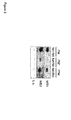
- Figure 1 shows an SDS-PAGE of purified artificial proteins and variants of PM stained with Coomassie Brilliant Blue.
- C7LT harbors the specificity of C7 (binding to H3K9me3 and H3K27me3, however, binding to H3K9me3 is only observed in vitro) and T (binding to H3K4me3), while PT harbors the specificity of P (binding to H3K36me2/3) and T (binding to H3K4me3). Binding specificity of MLM to H3K9me3 and H3K27me3 was also confirmed, however, binding to H3K27me3 is only observed in vitro.
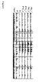
- Figure 2 shows two far-western blot analyses (designated WB1 and WB2) using the artificial protein PM and its respective variants with a pocket mutation in the first (P*M) or the second (PM*) histone modification binding domain.
- L.c. designates the loading control (Ponceau S staining). In contrast to native histones, recombinant histones do not have any post-translational modifications. Accordingly, none of PM, P*M and PM* bound to recombinant histones. As expected, binding to native histones was observed.
- Binding of PM is strongest, indicating enhanced binding of PM to histone modifications compared to its variants P*M and PM*. This is likely due to multi-dentate binding of the two (non-mutated) binding domains in PM to their target histone modifications, which in turn leads to an increased avidity of PM to its target histone modifications. Binding of PM* is weakest due to the weak binding of P to its respective histone modification.
- nCIDOP native chromatin interacting domain precipitation
- Nucleosomes comprising the first and/or the second histone modification were isolated by chromatin interacting domain precipitation using PM, P*M and PM* as well as the single histone modification binding domains P (PWWP domain of Dnmt3a) and M (chromodomain of MPP8). Washing was performed under stringent conditions except for P and PM* which were washed under less stringent conditions due to the weak binding affinity of P to its target histone modification compared to M.
- DNA was recovered from the isolated complexes and analysed by quantitative PCR using amplicons associated with both H3K9me3 and H3K36me3 modifications (based on H3K9me3 and H3K36me3 peak overlap) ("K9me3 + K36me3”) and amplicons associated with H3K9me3 only (“K9me3”) or H3K36me3 only (“K36me3”).
- the single domains P and M differ in their binding affinities. M has a higher binding affinity to its target histone modification than P on a peptide level. This may explain the differences observed on the chromatin level such as the different values on the y-axis between PM, PM* and P*M.
- PM is highly selective for doubly modified nucleosomes which are not as common as singly modified nucleosomes. This might explain the lower value on the y-axis when compared to P*M.
- the artificial protein favors binding to nucleosomes when both the first and the second histone modification are present.
- Figure 4 shows the Spearman correlation coefficient which was calculated in bins of 10-kb and indicates that the profile of PM is different from the profile of P*M (M active) and P (which is analogous to PM*).
- Figure 5 shows genome browser tracks of sections of chromosome 19 obtained by next-generation sequencing using PM (in two repeats designated PM_1 and PM_2), P*M (in two repeats designated P*M_1 and P*M_2), M and P.
- the y-axis indicates the number of reads.
- the signal obtained with PM is only present when both P*M (and its analogous single domain M) and P (analogous to PM*) overlap, i.e. when the first and the second histone modification co-occur. If only one of the two histone modifications is present, PM only yields a background signal. This can be seen for example in the regions highlighted by the black boxes. Thus, the presence of only one of H3K36me3 and H3K9me3 is not sufficient for binding of PM to the nucleosome.
- the inventors further analyzed the next-generation sequencing results obtained from nucleosomes isolated with PM by chromatin interacting domain precipitation at whole genome level.
- the genome was segmented into pieces of 3000 base pairs, the obtained reads averaged and background subtracted.
- Analysis of the M and P data allows to annotate regions of the whole genome that contain only H3K9me3 (M signal but not P signal), only H3K36me3 (P signal but no M signal), none of the two histone modifications or both of them. It is thus possible to define the fraction of recovered DNA located in the respective regions.
- the inventors found that 50% of the genomic fragments identified in next-generation sequencing of DNA recovered from control nucleosomes are located in regions that are annotated as H3K9me3-only regions (first column on the left). Based on DNA recovered from control nucleosomes, the inventors further found that 16.6% of the genome carries both H3K9me3 and H3K36me3. Analysis of DNA recovered from nucleosomes isolated with PM shows that nucleosomes having both H3K9me3 and H3K36me3 are strongly enriched. This demonstrates successful use of PM for isolating nucleosomes comprising the double-modified histone protein octamer having both H3K9me3 and H3K36me3.
Landscapes
- Health & Medical Sciences (AREA)
- Life Sciences & Earth Sciences (AREA)
- Chemical & Material Sciences (AREA)
- Molecular Biology (AREA)
- Engineering & Computer Science (AREA)
- Organic Chemistry (AREA)
- Immunology (AREA)
- Biophysics (AREA)
- General Health & Medical Sciences (AREA)
- Biochemistry (AREA)
- Medicinal Chemistry (AREA)
- Physics & Mathematics (AREA)
- Hematology (AREA)
- Urology & Nephrology (AREA)
- Biomedical Technology (AREA)
- Proteomics, Peptides & Aminoacids (AREA)
- Toxicology (AREA)
- Zoology (AREA)
- Genetics & Genomics (AREA)
- Gastroenterology & Hepatology (AREA)
- Biotechnology (AREA)
- Food Science & Technology (AREA)
- Pathology (AREA)
- Cell Biology (AREA)
- General Physics & Mathematics (AREA)
- Analytical Chemistry (AREA)
- Microbiology (AREA)
- Bioinformatics & Cheminformatics (AREA)
- Bioinformatics & Computational Biology (AREA)
- Peptides Or Proteins (AREA)
Abstract
The invention discloses the use of an artificial protein for isolating a nucleosome, the nucleosome comprising a multiple-modified histone protein octamer, wherein the artificial protein comprises a first histone modification binding domain of 50 to 200 amino acids binding to a first histone modification, a second histone modification binding domain of 50 to 200 amino acids binding to a second histone modification, a linker of 5 to 50 amino acids connecting the first and the second histone modification binding domain, and an affinity tag. Further disclosed are a nucleic acid encoding the artificial protein, a host cell comprising the nucleic acid and a kit for isolating a nucleosome, the nucleosome comprising a multiple-modified histone protein octamer. Further disclosed is an in-vitro method for isolating a nucleosome having a first and a second histone modification.
Description
- The present invention relates to the use of an artificial protein for isolating a nucleosome, the nucleosome comprising a multiple-modified histone protein octamer. The invention also relates to a nucleic acid encoding an artificial protein, a host cell comprising the nucleic acid and a kit for isolating a nucleosome. The invention further relates to an in-vitro method for isolating a nucleosome having a first and a second histone modification.
- Post-translational modifications of histone proteins, such as methylation and acetylation, play an important role in the regulation of gene expression and other chromatin-associated processes. They may also be involved in various diseases such as autoimmune diseases, developmental disorders and cancer. To date, more than 100 histone modifications are known. They occur in complex patterns, forming the so-called "histone code". With a view to deciphering this code and gain a better understanding of its role in human disease, the identification and characterisation of co-occurring histone modifications is an area of intense research.
- The isolation of nucleosomes having multiple co-occurring histone modifications is used to detect the presence of co-occurring histone modifications. It also facilitates their further analysis. Nucleosomes having multiple-modified histone protein octamers can be isolated by consecutive chromatin immunoprecipitation (ChIP) assays: First, a ChIP assay using an antibody directed to a first histone modification is performed. The precipitated nucleosomes are then eluted and subjected to a second ChIP assay using an antibody directed to a second histone modification. Consecutive ChIP assays are not only time-consuming; they also require a lot of starting material and are technically difficult to perform. Moreover, consecutive ChIP assays have a poor sensitivity and the DNA recovered from the isolated nucleosomes cannot be analysed by next-generation sequencing to date.
- Therefore, new tools and methods for isolating nucleosomes having multiple-modified histone protein octamers are needed that overcome the current limitations.
- In a first aspect, the invention relates to the use of an artificial protein for isolating a nucleosome, the nucleosome comprising a multiple-modified histone protein octamer, wherein the artificial protein comprises a first histone modification binding domain of 50 to 200 amino acids binding to a first histone modification, a second histone modification binding domain of 50 to 200 amino acids binding to a second histone modification, a linker of 5 to 50 amino acids connecting the first and the second histone modification binding domain, and an affinity tag.
- In a second aspect, the present invention relates to a nucleic acid encoding an artificial protein, wherein the artificial protein comprises a first histone modification binding domain of 50 to 200 amino acids binding to a first histone modification, a second histone modification binding domain of 50 to 200 amino acids binding to a second histone modification, a linker of 5 to 50 amino acids connecting the first and the second histone modification binding domain, and an affinity tag.
- In a third aspect, the present invention relates to a host cell comprising the nucleic acid of the invention.
- In a further aspect, the invention relates to a kit for isolating a nucleosome, the nucleosome comprising a multiple-modified histone protein octamer, wherein the kit comprises an artificial protein, wherein the artificial protein comprises a first histone modification binding domain of 50 to 200 amino acids binding to a first histone modification, a second histone modification binding domain of 50 to 200 amino acids binding to a second histone modification, a linker of 5 to 50 amino acids connecting the first and the second histone modification binding domain, and an affinity tag.
- In a further aspect, the invention relates to an in-vitro method for isolating a nucleosome having a first and a second histone modification, the method comprising the steps of (a) providing an artificial protein, wherein the artificial protein comprises a first histone modification binding domain of 50 to 200 amino acids binding to the first histone modification, a second histone modification binding domain of 50 to 200 amino acids binding to the second histone modification, a linker of 5 to 50 amino acids connecting the first and the second histone modification binding domain, and an affinity tag; (b) contacting the artificial protein with a sample comprising nucleosomes to allow formation of a complex of the artificial protein and a nucleosome having the first and the second histone modification; and (c) isolating the complex.
-
-
Figure 1 shows an SDS-PAGE of purified artificial proteins stained with Coomassie Brilliant Blue. -
Figure 2 shows far-western blot analyses of the binding of the artificial protein PM in which the first histone modification binding domain is the PWWP domain of Dnmt3a (P) and the second histone modification binding domain is the chromodomain of MPP8 (M) to native and recombinant histone proteins (NH, native histone proteins; RH, recombinant histone proteins; L.c., loading control). Analysis of variants of PM having a pocket mutation either in the first histone modification binding domain (P*M) or in the second histone modification binding domain (PM*) are also shown. -
Figure 3 shows the results of analysing recovered DNA by quantitative PCR. The DNA was recovered from nucleosomes isolated using the artificial protein PM or one of its variants having a pocket mutation. -
Figure 4 shows the Spearman correlation coefficient of next-generation sequencing of recovered DNA. The DNA was recovered from nucleosomes isolated using the artificial protein PM, its variant having a pocket mutation in the first histone modification binding domain (P*M) or a single PWWP domain of Dnmt3a (P). -
Figure 5 shows genome browser tracks of sections of chromosome 19 obtained by next-generation sequencing of recovered DNA. The DNA was recovered from nucleosomes isolated using the artificial protein PM, its variant having a pocket mutation in the first histone modification binding domain (P*M), a single chromodomain of MPP8 (M), or a single PWWP domain of Dnmt3a (P). -
Figure 6 shows the genomic localization of DNA recovered from nucleosomes isolated using PM compared to DNA recovered from control nucleosomes. DNA was analysed by next-generation sequencing. -
Figure 7 shows the recovery of peaks obtained with PM based on next-generation sequencing of DNA recovered from nucleosomes isolated using PM. - In a first aspect, the invention relates to the use of an artificial protein for isolating a nucleosome, the nucleosome comprising a multiple-modified histone protein octamer, wherein the artificial protein comprises a first histone modification binding domain of 50 to 200 amino acids binding to a first histone modification, a second histone modification binding domain of 50 to 200 amino acids binding to a second histone modification, a linker of 5 to 50 amino acids connecting the first and the second histone modification binding domain, and an affinity tag.
- The histone protein octamer of a nucleosome is composed of two copies of histone proteins H3, H4, H2A and H2B, respectively. The term "histone modification" as used herein refers to any covalently bound chemical entity that is post-translationally added to an amino acid residue of a histone protein in the histone protein octamer as well as combinations thereof that can be bound by a given histone modification binding domain. Common histone modifications comprise methylation, acetylation and ubiquitylation of one or more lysine residues, methylation of one or more arginine residues and phosphorylation of one or more serine and threonine residues. Further histone modifications comprise SUMOylation, crotonylation, butyrylation and propionylation of lysine residues, citrullination and ADP-ribosylation of arginine residues, and glycosylation of serine and threonine residues. Lysine residues can be mono-, di- or trimethylated while arginine residues can be mono- or dimethylated. Therefore, the term "methylation" as used herein comprises monomethylation, dimethylation and trimethylation. Histone modifications may co-occur in complex patterns, such that nucleosomes often comprise a multiple-modified histone protein octamer.
- The artificial protein comprises a first histone modification binding domain and a second histone modification binding domain of 50 to 200 amino acids each. The term "histone modification binding domain" as used herein refers to any amino acid polymer that, when folded into its three-dimensional structure, forms a binding pocket that can specifically interact with one or more histone modifications. The term comprises naturally occurring as well as modified, engineered and/or de novo designed histone modification binding domains. The histone modification binding domain may be based on a human histone modification binding domain or on a histone modification binding domain from another species such as mouse, rat or yeast (e.g. Schizosaccharomyces pombe). Histone modification binding domains are also known as reading domains or histone modification interacting domains (HMIDs). Histone modification binding domains specifically bind one or more histone modifications in a non-covalent manner. Non-covalent binding is mediated for example by van der Waals forces, hydrophobic interactions or electrostatic interactions such as formation of hydrogen bonds. The binding comprises specific protein-protein-interactions between the modified histone proteins and the artificial protein.
- Histone modification binding domains differ in their binding profiles. There are domains that bind a single histone modification such as bromodomains binding to an acetylated lysine residue. The binding of histone modification binding domains may also depend on more than a single histone modification. For example, the HP1 beta chromodomain binds H3K9me2/3 only if H3S10 is not phosphorylated. The ADD domain of ATRX binds H3K9me3 only if H3K4 is not modified. Further, histone modification binding domains binding to methylated lysine residues often bind to dimethylated as well as trimethylated lysine residues such as H3K9me2 and H3K9me3. Binding profiles may be even more manifold. For example, the PHD domain binds H3K4me3, H3K4me2 and H3K9me3.
- Therefore, the term "histone modification" also refers to combinations of chemical entities that are covalently bound to amino acid residues of a histone protein in the histone protein octamer and that can be bound by a given histone modification binding domain.
- The first and the second histone modification binding domain allow the artificial protein to interact with both the first and the second histone modification at the same time, thereby facilitating a dual readout of the two histone modifications. Accordingly, the artificial protein can be used to detect the presence of multiple histone modifications on one single nucleosome.
- The first and the second histone modification binding domain may be copies of the same domain or different domains giving rise to homodimeric or heterodimeric artificial proteins, respectively.
- The artificial protein further comprises a linker of 5 to 50 amino acids connecting the first and the second histone modification binding domain. The linker may be an artificially designed amino acid sequence or a linker derived from a naturally occurring amino acid sequence connecting two protein domains in a protein as for example the linker that connects the PWWP domain and the ADD domain in the DNA (cytosine-5-)-methyltransferase 3 alpha (Dnmt3a) protein. The linker can also be composed of a first portion corresponding to a naturally occurring amino acid sequence and a second portion being an artificially designed amino acid sequence. The linker preferably facilitates a flexible movement of the first and the second histone modification binding domain with respect to one another. Depending on the spatial arrangement of the first and the second histone modification within the histone protein octamer, a flexible connection of the two histone modification binding domains will enable independent interaction of the first and the second histone modification binding domain with their respective histone modifications on a single nucleosome. In other words, the linker should be sufficiently flexible to allow simultaneous entry of the first and the second histone modification from the same nucleosome in the respective histone modification binding pockets of the first and the second histone modification binding domain. This may be advantageous for the use of the artificial protein since histone modifications are generally located in flexible regions of histone proteins (so-called "histone tails") that stick out from the surface of the core of the nucleosome. In case the first and the second histone modification are located close to each other within the histone protein octamer, the linker may also be rigid.
- The possibility of simultaneous entry of the first and the second histone modification from the same nucleosome in the respective histone modification binding pockets of the artificial protein can be verified for example by side by side comparison of next-generation sequencing data of DNA recovered from isolated nucleosomes. The latter may be obtained using the artificial protein and control proteins which comprise only one of the first and the second histone modification binding domains. Determination of the three-dimensional structure of the artificial protein, for example by X-ray structure analysis, may also be applied to determine the flexibility of the first and the second histone modification binding domain with respect to one another.
- The artificial protein further comprises an affinity tag. The term "affinity tag" as used herein refers to any amino acid sequence that is suitable for protein purification and/or protein detection using an affinity technique. For example, the affinity tag can be a glutathione-S-transferase (GST) tag or a polyhistidine-tag. The affinity tag is preferably located at the N-terminus or at the C-terminus of the artificial protein. In a preferred embodiment, the artificial protein comprises more than one affinity tag. Additional affinity tags may enhance protein purification and/or protein detection. The artificial protein preferably comprises at least two different affinity tags. This broadens the range of affinity techniques that can be employed for purifying and/or detecting the artificial protein.
- The inventors have successfully isolated nucleosomes comprising two defined histone modifications. To do so, the artificial protein was expressed in Escherichia coli and shown to be able to interact with native nucleosomes. It was found that the artificial protein favors binding to nucleosomes when both the first and the second histone modification are present. In line with this finding, the inventors successfully used the artificial protein for isolating nucleosomes which comprise both the first and the second histone modification in a single chromatin precipitation assay at the same time. This confirms that the artificial protein is a useful tool for detecting combinations of histone modifications that co-occur on the same nucleosome.
- Thus, one advantage of the invention compared to consecutive ChIP assays is that the use of the artificial protein allows the detection of two or more co-occurring histone modifications on the same nucleosome at the same time in a single step. This is facilitated by specific binding of the artificial protein to multiple-modified histone protein octamers. Specific binding to multiple-modified histone protein octamers means that the artificial protein does not bind histone protein octamers having only one of the first and the second histone modification or neither of the two histone modifications. The inventors found that the bivalent binding of the artificial protein to multiple-modified histone protein octamers leads to a synergistic effect, which results in higher binding affinity of the artificial protein to nucleosomes comprising both the first and the second histone modification compared to nucleosomes comprising only one of two histone modifications. This is believed to be due to multi-dentate binding of the first and the second histone modification binding domain to their respective target histone modifications, which in turn leads to an increased avidity of the artificial protein to the first and the second histone modification.
- The specific binding of the artificial protein to nucleosomes comprising both the first and the second histone modification can be verified for example by side by side comparison of next-generation sequencing data of DNA recovered from isolated nucleosomes. The latter may be obtained using the artificial protein and control proteins which comprise only one of the first and the second histone modification binding domains. Specific binding of the artificial protein to nucleosomes comprising both the first and the second histone modification allows the specific isolation of nucleosomes comprising multiple-modified histone protein octamers.
- Another advantage of the use according to the invention is that only small amounts of nucleosome-comprising starting material are required. The term "small amount" as used herein refers to an amount of 10-30 µg of nucleosomes based on DNA absorbance. This is equivalent to chromatin isolated from 1-4 million human cells and typically used in a single ChIP assay. For consecutive ChIP assays, higher amounts of starting material are needed. Since the use of the artificial protein allows the isolation of a nucleosome having two or more co-occurring histone modifications in a single step, material is lost only once. Consecutive ChIP assays comprise at least two consecutive steps so that loss of material occurs at least twice. Thus, the use according to the invention requires less nucleosomecomprising starting material compared to consecutive ChIP assays.
- Due to the use of the artificial protein, the isolation of nucleosomes is also technically easy to perform, for example by chromatin precipitation.
- Further, DNA recovered from the isolated nucleosomes can be analysed by commonly used methods such as quantitative PCR. Importantly, the DNA can also be analysed by next-generation sequencing, which is not possible when nucleosomes are isolated by consecutive ChIP assays. Consecutive ChIP assays do not yield sufficient amounts of DNA for performing next-generation sequencing. The sequenced DNA can be mapped and used to study the co-occurrence of histone modifications on a genome-wide or locus-specific scale.
- DNA analysis by quantitative PCR leads to quantitatively accurate results, however, it is limited to pre-selected DNA loci of around 150 base pairs. Therefore, quantitative PCR is almost always hypothesis-driven since the DNA loci for which information is generated need to be determined beforehand when primer sequences are selected. The same applies to simple sequencing techniques such as Sanger sequencing. In contrast, next-generation sequencing facilitates hypothesis-free DNA analysis as a signal can be present anywhere in the genome. In this way, a genome-wide overview of the co-occurrence of histone modifications is obtained.
- Taken together, the use according to the invention provides an improved strategy for analysing complex patterns of histone modifications that co-occur on the same nucleosome. Given that specific combinations of histone modifications may be associated with unique biological functions, the use according to the invention will aid efforts to uncover the role of multiple-modified histone protein octamers in the regulation of gene expression and other chromatin-associated processes as well as in human disease.
- In a preferred embodiment, the first and the second histone modification binding domain are different from each other. This provides a heterodimeric artificial protein recognizing two different histone modifications, for example on one single histone protein (in cis) or on different copies of the respective histone protein (in trans) in the histone protein octamer. In contrast, homodimeric artificial proteins can only detect the presence of a certain histone modification in trans, namely on the two different copies of the respective histone protein in the histone protein octamer.
- In a preferred embodiment, the first and the second histone modification binding domain are copies of the same domain. This provides a homodimeric artificial protein for isolating nucleosomes comprising the respective histone modification in multiple copies. In addition, homodimeric artificial proteins are particularly useful if an enhanced binding strength and/or an enhanced specificity of a given histone modification binding domain are desired. Since histone modifications generally occur in clusters, homodimeric artificial proteins bind stronger and with a higher specificity to the respective histone modification compared to the corresponding single histone modification binding domain.
- The dissociation constants of histone modification binding domains for binding to their respective histone modification are generally in the high nanomolar to low micromolar range. Antibodies have dissociation constants ranging from low nanomolar to low micromolar range. Nevertheless, the inventors found that the dissociation constants of histone modification binding domains are strong enough for isolating nucleosomes.
- The dissociation constants of the first and the second histone modification binding domain may be considerably different from each other. For example, the inventors found that a difference in dissociation constants of about 100-fold between the first and the second histone modification binding domain does not interfere with the use according to the invention.
- In a preferred embodiment, the artificial protein comprises a linker of 14 to 35 amino acids, more preferred 21 to 27 amino acids. The inventors found that linkers of this length are particularly suitable for the use of the artificial protein according to the invention. For example, the inventors have used linkers having 14, 21 and 27 amino acids, respectively, for obtaining a flexible connection of the two histone modification binding domains. Since histone modifications are generally located in flexible regions of histone proteins, a flexible linker may expedite simultaneous binding of the artificial protein to the first and the second histone modification.
- The necessary minimum length of the linker will depend on the spatial arrangement of the histone modification binding pockets in the three-dimensional structure of the artificial protein and on the position at which the linker emerges from each three-dimensional histone modification binding domain. In general, one amino acid can bridge at most about 3.5 Angstrom (this is the case in beta-strands of proteins in which the peptide bonds between two amino acids are almost fully extended).
- The linker may further serve to improve the solubility of the artificial protein. To this end, the amino acid sequence of the linker preferably comprises proline, alanine, glutamine, glutamic acid, lysine and/or serine. The solubility of the artificial protein is particularly important for efficient recombinant production and purification of the artificial protein.
- In a preferred embodiment, the first and/or the second histone modification is selected from the group consisting of methylation, phosphorylation, acetylation, and ubiquitylation. Methylation, phosphorylation, acetylation, and ubiquitylation of amino acid residues are common histone modifications. For many of these modifications respective histone modification binding domains are known.
- In a preferred embodiment, the first and/or the second histone modification binding domain is selected from the group consisting of 14-3-3 domain, ADD domain, ankyrin, BAH domain, BIR domain, BRCT domain, tandem BRCT domain, bromodomain, double bromodomain, chromobarrel, chromodomain, double chromodomain, double PHD finger domain, MBT domain, PID domain, PHD domain, double PH domain, PWWP domain, royal family domain, Tudor domain, tandem Tudor domain, WD40 domain, and zinc finger CW domain.
- The term "BAH domain" refers to bromo adjacent homology domain. The term "royal family domain" refers to a subclass of histone modification binding domains comprising Tudor domains, chromodomains, MBT domains and PWWP domains.
- In a preferred embodiment, the first histone modification binding domain is the PWWP domain of Dnmt3a and the second histone modification binding domain is the chromodomain of MPP8.
- An overview of preferred combinations of the first and the second histone modification binding domain is shown in Table 1.
Table 1: Preferred combinations of the first and the second histone modification binding domain No. First histone modification binding domain Second histone modification binding domain 1 PWWP domain of Dnmt3a chromodomain of MPP8 2 chromodomain of MPP8 PWWP domain of Dnmt3a 3 PWWP domain of Dnmt3a PWWP domain of Dnmt3a 4 chromodomain of MPP8 chromodomain of MPP8 5 chromodomain of CBX7 PHD domain of TAF3 6 PHD domain of TAF3 chromodomain of CBX7 7 PWWP domain of Dnmt3a PHD domain of TAF3 8 PHD domain of TAF3 PWWP domain of Dnmt3a 9 PWWP domain of Dnmt3a chromodomain of CBX7 10 chromodomain of CBX7 PWWP domain of Dnmt3a 11 chromodomain of CBX7 chromodomain of MPP8 12 chromodomain of MPP8 chromodomain of CBX7 13 PWWP domain of Dnmt3a ADD domain of ATRX 14 ADD domain of ATRX PWWP domain of Dnmt3a 15 chromodomain of MPP8 double Tudor domain of JMJD2A 16 double Tudor domain of JMJD2A chromodomain of MPP8 - Dnmt3a refers to DNA (cytosine-5-)-methyltransferase 3 alpha. MPP8 refers to M-
Phase Phosphoprotein 8. CBX7 refers to chromobox homolog 7. TAF3 refers to TATA Box Binding Protein-Associated Factor 3. ATRX refers to Alpha thalassemia/mental retardation syndrome X-linked. JMJD2A is a member of the Jumonji domain 2 (JMJD2) family. - The artificial protein comprising the chromodomain of CBX7 and the PHD domain of TAF3 binds to H3K27me3 and H3K4me3. This combination of histone modifications is also known as "bivalent" state and has a high medical relevance. It was found to occur at developmental genes in embryonic stem cells and to be important for cell differentiation.
- In a preferred embodiment, the first and the second histone modification are chemically different. For example, the first histone modification is a methylation while the second histone modification is an acetylation. This is particularly useful to further reveal the complex patterns of histone modifications that co-occur on the same nucleosome.
- The use according to the invention may also be applied for identifying novel combinations of co-occurring histone modifications by selecting the first and the second histone modification binding domain accordingly. Therefore, in a preferred embodiment, the co-occurrence of the first and the second histone modification has not been described yet. As an example, the artificial protein comprising the PWWP domain of Dnmt3a and the chromodomain of CBX7 binds to H3K36me3 and H3K27me3. These two histone modifications were previously considered mutually exclusive.
- In a second aspect, the present invention relates to a nucleic acid encoding an artificial protein, wherein the artificial protein comprises a first histone modification binding domain of 50 to 200 amino acids binding to a first histone modification, a second histone modification binding domain of 50 to 200 amino acids binding to a second histone modification, a linker of 5 to 50 amino acids connecting the first and the second histone modification binding domain, and an affinity tag.
- The nucleic acid may be DNA or RNA. The nucleic acid is used to produce the artificial protein in transgenic host cells or transgenic organisms such as bacteria.
- In a third aspect, the present invention relates to a host cell comprising the nucleic acid of the invention.
- The host cell is used to produce the artificial protein. Therefore, the term "host cell" as used herein refers to any cell that is suitable for protein production. The host cell is preferably a bacterial cell such as Escherichia coli (E. co/i).
- In a further aspect, the invention relates to a kit for isolating a nucleosome, the nucleosome comprising a multiple-modified histone protein octamer, wherein the kit comprises an artificial protein, wherein the artificial protein comprises a first histone modification binding domain of 50 to 200 amino acids binding to a first histone modification, a second histone modification binding domain of 50 to 200 amino acids binding to a second histone modification, a linker of 5 to 50 amino acids connecting the first and the second histone modification binding domain, and an affinity tag.
- The kit of the invention provides a tool for detecting two or more co-occurring histone modifications on the same nucleosome at the same time in a single step. Due to the higher binding affinity of the artificial protein to nucleosomes having both the first and the second histone modification compared to nucleosomes having only one of the two histone modifications, the kit facilitates the specific isolation of nucleosomes comprising multiple-modified histone protein octamers. The isolation of nucleosomes using the kit of the invention is technically easy to perform, for example by chromatin precipitation, and requires only small amounts of starting material. Accordingly, the kit of the invention is particularly suited for a comprehensive analysis of complex patterns of histone modifications that co-occur on the same nucleosome. The analysis can be complemented by next-generation sequencing of the DNA recovered from the isolated nucleosomes.
- In a further aspect, the invention relates to an in-vitro method for isolating a nucleosome having a first and a second histone modification, the method comprising the steps of (a) providing an artificial protein, wherein the artificial protein comprises a first histone modification binding domain of 50 to 200 amino acids binding to the first histone modification, a second histone modification binding domain of 50 to 200 amino acids binding to the second histone modification, a linker of 5 to 50 amino acids connecting the first and the second histone modification binding domain, and an affinity tag; (b) contacting the artificial protein with a sample comprising nucleosomes to allow formation of a complex of the artificial protein and a nucleosome having the first and the second histone modification; and (c) isolating the complex.
- The method of the invention is based on the inventors' finding that the artificial protein can be used for specifically isolating nucleosomes having multiple histone modifications at the same time in a single step with high efficiency. This is facilitated by the specific binding of the artificial protein to nucleosomes having both the first and the second histone modification compared to nucleosomes having only one of two histone modifications.
- The method of the invention requires only small amounts of nucleosome-comprising starting material and is technically easy to perform. Further, DNA recovered from the isolated nucleosomes can be analysed by commonly used methods such as quantitative PCR, but also by next-generation sequencing.
- The complex is formed by binding of the first histone modification binding domain to the first histone modification and binding of the second histone modification binding domain to the second histone modification. Complex-forming conditions may be adjusted, for example depending on the binding affinity of the histone modification binding domains and/or the technical approach used. In particular, the salt concentration of the solution in which the artificial protein is contacted with the sample and/or the salt concentration of the washing solutions used for removing unbound nucleosomes before isolating the complex may be adjusted. This also allows adapting the complex-forming conditions to a desired degree of stringency.
- In a preferred embodiment, the complex is immobilized on a solid support such as beads. In this case, the complex can be isolated by a simple pull-down assay well known in the art. Immobilization of the complex is preferably mediated by the affinity tag of the artificial protein.
- In a preferred embodiment, the sample is obtained from a patient suffering from a disease. The disease is preferably an autoimmune disease, a developmental disorder, a disease of the nervous system or cancer. Histone modifications are believed to play a role in various human diseases. Thus the analysis of the presence of the first and the second histone modification on nucleosomes from a respective patient is of particular interest.
- In a preferred embodiment, the method further comprises the steps of (d) providing a first control protein and a second control protein, each control protein comprising a single histone modification binding domain, wherein the single histone modification binding domain of the first control protein is the same as the first histone modification binding domain of the artificial protein and the single histone modification binding domain of the second control protein is the same as the second histone modification binding domain of the artificial protein; and (e) contacting the first and the second control protein with a sample comprising nucleosomes to allow formation of a complex of the first control protein and a nucleosome having the first histone modification and/or formation of a complex of the second control protein and a nucleosome having the second histone modification; and (f) isolating the complex.
- In a preferred embodiment, steps (d) to (f) are performed side-by-side with steps (a) to (c). The control proteins allow verifying the binding specificity of the artificial protein to double-modified nucleosomes by comparing the complex isolated in step (f) with the complex isolated in step (c). In this way, the specific binding of the artificial protein to nucleosomes having both the first and the second histone modification compared to nucleosomes having only one of two histone modifications can be confirmed.
- The control proteins may also be used to verify that the artificial protein allows simultaneous entry of the first and the second histone modification from the same nucleosome in the respective histone modification binding pockets of the first and the second histone modification binding domain.
- In a preferred embodiment, the method further comprises the step of (g) analysing the isolated complex.
- In a preferred embodiment, the isolated complex is analysed by mass spectrometry. Mass spectrometry can be used to confirm the presence of the first and the second histone modification. More importantly, mass spectrometry can be used to identify further histone modifications which co-occur with the first and the second histone modification on the same nucleosome. It also facilitates the identification of proteins which are associated with nucleosomes having the first and the second histone modification. Taken together, information about the typical composition of chromatin comprising the double-modified nucleosomes may be obtained by mass spectrometry of the isolated complex.
- In a preferred embodiment, the isolated complex is analysed by recovering DNA from the nucleosome and analysing the DNA. DNA can be easily recovered from the nucleosome by any method suitable for DNA purification from chromatin. DNA recovery is not affected by the additional presence of the artificial protein in the isolated complex.
- The isolated complex can also be analysed by both mass spectrometry and DNA analysis of DNA recovered from the nucleosome of the isolated complex.
- It is further preferred that the DNA is analysed by quantitative PCR and/or next-generation sequencing.
- Quantitative PCR (polymerase chain reaction) can be used to determine the amount of recovered DNA. The amount of recovered DNA is a measure for the amount of nucleosomes that have been isolated, i.e. for the amount of nucleosomes having the first and the second histone modification in the sample.
- In next-generation sequencing, the total number of times a DNA fragment is read during the sequencing process allows to determine the enrichment of a corresponding DNA region by the method according to the invention. The sequenced DNA can be mapped and used to study the co-occurrence of histone modifications on a genome-wide or a locus-specific scale.
- Further described is the use of an artificial protein for determining whether a first and a second histone modification co-occur on the same copy of a histone protein or on different copies of the same histone protein in a nucleosome, wherein the artificial protein comprises a first histone modification binding domain of 50 to 200 amino acids binding to the first histone modification, a second histone modification binding domain of 50 to 200 amino acids binding to the second histone modification, wherein the first and the second histone modification binding domain are different from each other, a linker of 5 to 50 amino acids connecting the first and the second histone modification binding domain, and an affinity tag.
- Modifications occurring at different amino acid residues in a given histone protein such as H3K9me3 and H3K36me3 (both located on histone H3) may co-occur either on the same copy of the histone protein, i.e. on one single histone protein (in cis) or on different copies of the same histone protein (in trans) in the histone protein octamer. Using a heterodimeric artificial protein, i.e. an artificial protein in which the first and the second histone modification binding domain are different from each other, the positioning of the first and the second histone modification in cis or in trans can be determined. To do so, the artificial protein is constructed in a way that allows specific binding of the two histone modifications in cis or in trans only. This can be achieved by adjusting the spatial orientation of the two histone modification binding domains and/or the characteristics of the linker. For example, the histone modification binding domains can be oriented in a manner in which their binding pockets are facing towards the same side of the artificial protein. If this orientation is combined with a rather rigid linker, the artificial protein will specifically bind to histone modifications co-occurring in cis. The rigid linker ensures that the spatial orientation of the two histone modification binding domains is maintained. The histone modification binding domains may also be oriented in a manner in which their binding pockets are facing towards opposite sides of the artificial protein. When combined with a rather rigid linker, this artificial protein will specifically bind to histone modifications co-occurring in trans. Simultaneous entry of two histone modifications into their respective binding pockets will not be feasible when the histone modifications are located in cis but the two binding pockets are facing towards opposite sides of the artificial protein. Therefore, only histone modifications co-occurring in trans will be bound. Thus, the artificial protein presents a useful tool for determining whether the first and the second histone modification co-occur in cis or in trans.
- Further aspects of the invention will be apparent to the person skilled in the art by the enclosed description of the examples, in particular the scientific results.
- The sequences encoding the chromodomain of MPP8 (also known as MPHOSPH8) (57-111 of NP_059990.2), the PWWP domain of DNMT3A (279-420 of NP_001258682) and other, with or without an artificial linker of 27 amino acids were subcloned from in house plasmids and cloned by overlap assembly of DNA fragments fusion into pGEX-6P-2 vector (GE Healthcare, Solingen, Germany) using ligase or ligase-independent methods. In brief, each neighboring fragment to be cloned shared an overlapping region. The overlapping regions were hybridized and extended by normal PCR steps to form a linear sequence. Once nucleic acids encoding the artificial proteins of interest were generated, they were digested with specific restriction enzymes (EcoRI and XmaI) and ligated with digested empty pGEX-6P-2 vector to obtain the final plasmids. The correct sizes of the inserts were confirmed by colony PCR and the correct sequences by Sanger DNA sequencing.
- Artificial proteins comprising an N-terminal GST-tag as affinity tag were overex-pressed in E.coli BL21 cells carrying the corresponding plasmids at 18°C and purified essentially as described in Rathert et al. 2008, electrophoresed on 12% SDS-PAGE and stained with colloidal Coomassie Brilliant Blue G-250.
- In case the first histone modification binding domain is the PWWP domain of Dnmt3a, the sequence selected for cloning also comprises a portion located at the C-terminus of the PWWP domain that is part of the naturally occurring flexible linker that connects the PWWP domain and the ADD domain in the Dnmt3a protein. This is derived from the crystal structure of the PWWP domain. Thus, in artifical proteins in which the first histone modification binding domain is the PWWP domain of Dnmt3a, such as PM, the linker is formed by the C-terminal portion of the PWWP domain. This linker has the following amino acid sequence:
- GGFQPSGPKGLEPPLERPHRD (SEQ ID NO.: 1); or
- GGFQPSGPKGLEPP (SEQ ID NO.: 2)
- The artificial linker is also flexible and has the following amino acid sequence:
- SSGNSNANSRGPSFSSGLVPLSLRGSH (SEQ ID NO.: 3)
- Site-directed mutagenesis was used to insert a defined mutation in the binding pocket of the relevant histone modification binding domain in order to generate a domain unable to bind its respective histone modification. Mutated histone modification binding domains served as controls. In the mutated chromodomain of MPP8 (designated M*), the mutation is an F59A exchange that renders the domain unable to bind to H3K9me3. In the mutated PWWP domain of Dnmt3a (designated P*), the mutation is a D329A exchange that renders the domain unable to bind to H3K36me3.
- Mutations were introduced using the megaprimer method described in Jeltsch and Lanio 2002 and successful mutagenesis was confirmed by restriction analysis and Sanger DNA sequencing.
- Experiments to determine the binding specificity of the artificial proteins were performed using the CelluSpots peptide array platform as described in Bock et al. 2011 a and Bock et al. 2011 b.
- For western blot, the peptide array protocol was adapted. Native histones were isolated by acid extraction from HEK293 cells and recombinant histones H3 and H4 were purchased from New England Biolabs (New England Biolabs, Frankfurt a.M., Germany) or purified from E.coli. Five micrograms of native histones and 2.5 µg of recombinant histones were loaded and electrophoresed on 16% SDS-PAGE and transferred on nitrocellulose membranes by semi-dry western blotting with transfer buffer (300 mM Tris, 300 mM glycine, pH 9.2) for 10-15 minutes. The membrane was stained with Ponceau S for around 15 minutes to assess the quality of the transfer. Afterwards the membrane was incubated overnight with 5% skim milk at 4°C. The next day the membrane was washed two times with TTBS and once with interaction buffer (100 mM KCI, 20 mM HEPES pH 7.5, 1 mM EDTA, 0.1 mM DTT and 10% glycerol). The membrane was incubated with artificial protein or the respective single histone modification binding domains in inter-action buffer for 2 hours at room temperature, washed three times with TTBS and incubated with anti-GST antibody for 1 hour. After three washings with TTBS the membrane was incubated with horseradish peroxidase conjugated with anti-goat antibody for 1 hour at room temperature. After three times washing with TBS, the membrane was immersed in ECL solution and chemiluminescence was detected.
- The amounts of artificial proteins (wild type and single domain mutants) used in CelluSpots and western blot analyses were equimolar (10 nM).
- Native chromatin comprising nucleosomes were isolated from around 20 million HepG2 cells (which was sufficient for 5-15 CIDOP/ChIP experiments) by micro-coccal nuclease digestion of nuclei as described in Brand et al. 2008 with minor modifications. In brief, following MNase digestion, the nuclei were centrifuged at 13000 g for 10 minutes and the resulting supernatant which contained the soluble nucleosomal fraction was collected and snap frozen. Then, a sample of native chromatin (10-30 µg based on DNA absorbance) was pre-cleared for 1 hour at 4°C with 20 µl glutathione sepharose 4B beads (GE Healthcare, Solingen, Germany) in DP buffer (16.7 mM Tris-Cl, 167 mM NaCl, 1.1% Triton X-100, 1.2 mM EDTA and protease inhibitors) filled up to 500 µl. The beads were removed and the supernatant (pre-cleared chromatin) was incubated overnight with the artificial protein (10-30 µg or equimolar concentration when compared to single domains with different size) at 4°C. The next day, the artificial protein-nucleosome complexes were immobilized for 2 hours on 20 µl glutathione sepharose 4B beads (GE Healthcare, Solingen, Germany) with rotation at 4°C and washed for 10 minutes with rotation under stringent conditions with: 1x Low Salt Buffer (20 mM Tris-Cl pH 8.0, 150 mM NaCl, 1% Triton X-100, 0.1% SDS and 2 mM EDTA), 1x High Salt Buffer (20 mM Tris-Cl pH 8.0, 500 mM NaCl, 1% Triton X-100, 0.1% SDS and 2 mM EDTA), 1x LiCl buffer (10 mM Tris-Cl pH 8.0, 250 mM LiCl, 1% NP-40, 1% sodium deoxycholate and 1 mM EDTA) and 2x TE buffer (10 mM Tris-Cl pH 8.0 and 1 mM EDTA). In case of less stringent conditions, washing was performed with: 3x PB buffer (50 mM Tris-Cl, 200 mM NaCl, 1 mM EDTA, 0.5% NP-40 and 2 mM DTT) and 2x TE buffer. Between each washing step, the complexes were spun down for 2 min at 2000 g at 4°C. Bound nucleosomes were eluted in 200 µl elution buffer (50 mM Tris-Cl, 50 mM NaCl, 1 mM EDTA and 1% SDS) and 1 µl proteinase K (20 mg/ml) for 45 minutes at room temperature with rotation. After incubation and centrifugation for 3 minutes at 3000 g the supernatant was transferred into a new tube and incubated for 1 hour at 55°C with additional 1 µl proteinase K. The DNA was recovered from the nucleosomes using Chromatin IP DNA purification columns (Active Motif, La Hulpe, Belgium).
- The recovered DNA was quantified by real-time PCR. The quantitative PCR assays were performed on a CFX96 Touch or CFX96 Real-Time detection system (Bio-Rad, Munich, Germany) using SYBR fast qPCR mix (Kapa Biosystems, London, UK) or SsoFast EvaGreen supermix (Bio-Rad, Munich, Germany). The PCR protocol used was: 3 minutes at 95°C, 39 cycles of 95°C for 3 seconds, followed by 20 seconds at 58-60°C and 72°C for 3 seconds. The primers used are listed in Table 2. A standard curve was generated to calculate the percent of precipitated DNA and test the efficiency of each primer set.
Table 2: Primers used in quantitative PCR assays Genome region Associated histone modifications Sequence of forward primer Sequence of reverse primer Size of PCR product (bp) PM-PM1 chr2 H3K36me3 and H3K9me3 GTCTTAACCGTCTGCCTAGA (SEQ ID NO.: 4) TGGTAGATTGGCAAATGGAA (SEQ ID NO.: 5) 127 PM-PM2 chr12 H3K36me3 and H3K9me3 GCCTCTGCATTCAGCATTTC (SEQ ID NO.: 6) AGGTTGGCCAAAGACACATC (SEQ ID NO.: 7) 124 PM-PM3 chr7 H3K36me3 and H3K9me3 CCAGACCAGTGCAATAAGGAA (SEQ ID NO.: 8) TGACCTTTGAGGGTTCAAG (SEQ ID NO.: 9) 132 PM-PM5 chr5 H3K36me3 and H3K9me3 CTGCTCCCATGTCTGCTACA (SEQ ID NO.: 10) TGGAAGGACTGCAGAGAAAAA (SEQ ID NO.: 11) 119 PM-P1 chr12 H3K36me3 CAATGACCCCTTCATTGACC (SEQ ID NO.: 12) GGGGGAATACGTGAGGGTAT (SEQ ID NO.: 13) 119 PM-P3 chr1 H3K36me3 TGCAAAGAAAAAGGAGCAGAA (SEQ ID NO.: 14) CCAACAAGCAAAAGAAGGAAA (SEQ ID NO.: 15) 90 PM-P5 chr9 H3K36me3 TGCTCCTTTTTCCCATCTTTT (SEQ ID NO.: 16) GCAAAACCAAGTCGAATGCT (SEQ ID NO.: 17) 99 PM-M2 chr5 H3K9me3 TGCATGATGTTTTCCTCAGC (SEQ ID NO.: 18) ATCTTGCGCAAATGCTCTG (SEQ ID NO.: 19) 119 PM-M3 chr19 H3K9me3 TTGTCACCACTGTCCAGGAA (SEQ ID NO.: 20) CAGCTGCCTCAGAGACACAC (SEQ ID NO.: 21) 123 PM-M4 chr5 H3K9me3 AGAACACCATGGACCACCAG (SEQ ID NO.: 22) TTTCTGAATTGGTTCTGGGTTT (SEQ ID NO.: 23) 113 - The genomic regions have the first and the second histone modification (H3K36me3 and H3K9me3, genomic regions designated PM-PM), only the first histone modification (H3K36me3, genomic regions designated PM-P) or only the second histone modification (H3K9me3, genomic regions designated PM-M).
- Around 50 million, 100-nucleotide sequence reads obtained with Illumina's HiSeq 2500 were mapped to the human reference genome hg19 with Bowtie (Langmead et al. 2009) from the Chipster software tool (Kallio et al. 2011). Only uniquely mapped reads were retained and all duplicates were removed. The genome coverage files normalized to reads per kilobase per million (RPKM) and the Spearman's rank correlation coefficient in window sizes of 10 kb were calculated with deepTools (Ramirez et al. 2014). The genome browser snapshots were taken with the Integrative Genomics Viewer (IGV).
- Artificial proteins produced are listed in Table 3. For example, PM is an artificial protein comprising the PWWP domain of Dnmt3a binding to H3K36me3, the chromodomain of MPP8 binding to H3K9me3, a linker connecting the PWWP domain and the chromodomain, and a GST tag.
- Variants of the artificial protein PM with pocket mutations in the first or the second histone modification binding domain were also produced and are listed in Table 4. The mutated domain is indicated by a "*". The mutated variants were used as controls since the mutation inactivated binding of the domain to its respective histone modification.
- Each artificial protein produced comprises an N-terminal GST tag as affinity tag.
- The naturally occurring linker is derived from the linker connecting the PWWP domain and the ADD domain in the Dnmt3a protein. In some proteins such as PM this linker has 21 amino acids (SEQ ID NO.: 1). In some proteins such as PT it has 14 amino acids (SEQ ID NO.: 2).
Table 3: Artificial proteins Name First histone modification binding domain First histone modification Linker (number of amino acids) Second histone modification binding domain Second histone modification DNA sequence encoding the artificial protein Amino acid sequence of artificial protein PM PWWP domain of Dnmt3a H3K36me3 Naturally occurring linker (21) chromodomain of MPP8 H3K9me3 SEQ ID NO.: 24 SEQ ID NO.: 25 C7LT chromodomain of CBX7 H3K27me3 Artificial linker (27) PHD domain of TAF3 H3K4me3 SEQ ID NO.: 26 SEQ ID NO.: 27 PT PWWP domain of Dnmt3a H3K36me3 Naturally occurring linker (14) PHD domain of TAF3 H3K4me3 SEQ ID NO.: 28 SEQ ID NO.: 29 MLM chromodomain of MPP8 H3K9me3 Artificial linker (27) chromodomain of MPP8 H3K9me3 SEQ ID NO.: 30 SEQ ID NO.: 31 MLP chromodomain of MPP8 H3K9me3 Artificial linker (27) PWWP domain of Dnmt3a H3K36me3 SEQ ID NO.: 32 SEQ ID NO.: 33 PLM PWWP domain of Dnmt3a H3K36me3 Artificial linker (27) chromodomain of MPP8 H3K9me3 SEQ ID NO.: 34 SEQ ID NO.: 35 PLP PWWP domain of Dnmt3a H3K36me3 Artificial linker (27) PWWP domain of Dnmt3a H3K36me3 SEQ ID NO.: 36 SEQ ID NO.: 37 PP PWWP domain of Dnmt3a H3K36me3 Naturally occurring linker (21) PWWP domain of Dnmt3a H3K36me3 SEQ ID NO.: 38 SEQ ID NO.: 39 MLdT chromodomain of MPP8 H3K9me3 Artificial linker (27) double Tudor domain of JMJD2A H4K20me3 SEQ ID NO.: 40 SEQ ID NO.: 41 dTLM double Tudor domain of JMJD2A H4K20me3 Artificial linker (27) chromodomain of MPP8 H3K9me3 SEQ ID NO.: 42 SEQ ID NO.: 43 PC7 PWWP domain of Dnmt3a H3K36me3 Naturally occurring linker (14) chromodomain of CBX7 H3K27me3 SEQ ID NO.: 44 SEQ ID NO.: 45 C7LP chromodomain of CBX7 H3K27me3 Artificial linker (27) PWWP domain of Dnmt3a H3K36me3 SEQ ID NO.: 46 SEQ ID NO.: 47 C7LM chromodomain of CBX7 H3K27me3 Artificial linker (27) chromodomain of MPP8 H3K9me3 SEQ ID NO.: 48 SEQ ID NO.: 49 MLC7 chromodomain of MPP8 H3K9me3 Artificial linker (27) chromodomain of CBX7 H3K27me3 SEQ ID NO.: 50 SEQ ID NO.: 51 TLC7 PHD domain of TAF3 H3K4me3 Artificial linker (27) chromodomain of CBX7 H3K27me3 SEQ ID NO.: 52 SEQ ID NO.: 53 PA PWWP domain of Dnmt3a H3K36me3 Naturally occurring linker (14) ADD domain of ATRX H3K9me3 when H3K4 unmodified SEQ ID NO.: 54 SEQ ID NO.: 55 PLA PWWP domain of Dnmt3a H3K36me3 Artificial linker (27) ADD domain of ATRX H3K9me3 when H3K4 unmodified SEQ ID NO.: 56 SEQ ID NO.: 57 ALP ADD domain of ATRX H3K9me3 when H3K4 unmodified Artificial linker (27) PWWP domain of Dnmt3a H3K36me3 SEQ ID NO.: 58 SEQ ID NO.: 59 TLP PHD domain of TAF3 H3K4me3 Artificial linker (27) PWWP domain of Dnmt3a H3K36me3 SEQ ID NO.: 60 SEQ ID NO.: 61 PLT PWWP domain of Dnmt3a H3K36me3 Artificial linker (27) PHD domain of TAF3 H3K4me3 SEQ ID NO.: 62 SEQ ID NO.: 63 PLC7 PWWP domain of Dnmt3a H3K36me3 Artificial linker (27) chromodomain of CBX7 H3K27me3 SEQ ID NO.: 64 SEQ ID NO.: 65 Table 4: Variants of PM with pocket mutations Name First histone modification binding domain First histone modification Linker (number of amino acids) Second histone modification binding domain Second histone modification P*M Mutated PWWP domain of Dnmt3a (D329A) -- Naturally occurring linker (21) chromodomain of MPP8 H3K9me3 PM* PWWP domain of Dnmt3a H3K36me3 Naturally occurring linker (21) Mutated chromodomain of MPP8 (F59A) -- - The proteins from which the histone modification binding domains have been derived have the following protein identifiers in Universal Protein Resource (UniProt) databases (amino acids in the protein sequence that correspond to the respective histone modification binding domain are also indicated):
Dnmt3a Q9Y6K1 (PWWP domain: amino acids 292-350); MMP8 Q99549 (chromodomain: amino acids 59-118); CBX7 095931 (chromodomain: amino acids 11-69); TAF3 Q5VWG9 (PHD domain: amino acids 865-915); JMJD2A 075164 (double Tudor domain: amino acids 897-1011); and ATRX P46100 (ADD domain: amino acids 159-296). -
Figure 1 shows an SDS-PAGE of purified artificial proteins and variants of PM stained with Coomassie Brilliant Blue. - The binding specificity of the artificial proteins and variants with a pocket mutation in the first or the second histone modification binding domain to the first and/or the second histone modification was confirmed by peptide arrays. It was confirmed that PM harbors the binding specificity of P (binding to H3K36me2/3) and M (binding to H3K9me2/3 and H3K27me3, however, binding to H3K27me3 is only observed in vitro). As expected, P*M retained binding to H3K9me2/3 and H3K27me3 and exhibited loss of binding to H3K36me3. Likewise, PM* showed binding to H3K36me2/3 only. This is in agreement with the binding profiles of the non-mutated corresponding single domains.
- It was further confirmed that C7LT harbors the specificity of C7 (binding to H3K9me3 and H3K27me3, however, binding to H3K9me3 is only observed in vitro) and T (binding to H3K4me3), while PT harbors the specificity of P (binding to H3K36me2/3) and T (binding to H3K4me3). Binding specificity of MLM to H3K9me3 and H3K27me3 was also confirmed, however, binding to H3K27me3 is only observed in vitro.
-
Figure 2 shows two far-western blot analyses (designated WB1 and WB2) using the artificial protein PM and its respective variants with a pocket mutation in the first (P*M) or the second (PM*) histone modification binding domain. Binding to native histone proteins (NH) and to recombinant histone proteins (RH) was tested. L.c. designates the loading control (Ponceau S staining). In contrast to native histones, recombinant histones do not have any post-translational modifications. Accordingly, none of PM, P*M and PM* bound to recombinant histones. As expected, binding to native histones was observed. Binding of PM is strongest, indicating enhanced binding of PM to histone modifications compared to its variants P*M and PM*. This is likely due to multi-dentate binding of the two (non-mutated) binding domains in PM to their target histone modifications, which in turn leads to an increased avidity of PM to its target histone modifications. Binding of PM* is weakest due to the weak binding of P to its respective histone modification. - Using the same assay, binding to native histones but not to recombinant histones was also found for C7LT, PT and MLM. Of interest, MLM bound to histones having its target modifications with much stronger affinity compared to the single domain counterpart M.
- Nucleosomes comprising the first and/or the second histone modification were isolated by chromatin interacting domain precipitation using PM, P*M and PM* as well as the single histone modification binding domains P (PWWP domain of Dnmt3a) and M (chromodomain of MPP8). Washing was performed under stringent conditions except for P and PM* which were washed under less stringent conditions due to the weak binding affinity of P to its target histone modification compared to M.
- DNA was recovered from the isolated complexes and analysed by quantitative PCR using amplicons associated with both H3K9me3 and H3K36me3 modifications (based on H3K9me3 and H3K36me3 peak overlap) ("K9me3 + K36me3") and amplicons associated with H3K9me3 only ("K9me3") or H3K36me3 only ("K36me3"). Four genome regions associated with both H3K9me3 and H3K36me3 (PM-PM1 chr2, PM-PM2 chr12, PM-PM3 chr 7 and PM-PM5 chr 5), three regions associated with H3K9me3 only (PM-M2 chr5, PM-M3 chr19 and PM-M4 chr5) and three regions associated with H3K36me3 only (PM-P1 chr12, PM-P3 chr1 and PM-P5 chr9) were tested.
- The results obtained for PM, P*M and PM* are shown in
Figure 3 . Error bars represent the standard error of mean (SEM). The results show that artificial proteins are able to interact with native nucleosomes. The results further indicate that PM has a different binding profile than its respective variants P*M and PM*. PM shows preferred binding to nucleosomes comprising both the first and the second histone modification. The variants P*M and PM* bind roughly equally to nucleosomes comprising both histone modifications and to nucleosomes comprising only H3K9me3 in the case of P*M and H3K36me3 in the case of PM*. - The single domains P and M differ in their binding affinities. M has a higher binding affinity to its target histone modification than P on a peptide level. This may explain the differences observed on the chromatin level such as the different values on the y-axis between PM, PM* and P*M.
- PM is highly selective for doubly modified nucleosomes which are not as common as singly modified nucleosomes. This might explain the lower value on the y-axis when compared to P*M.
- Using the same assay, stronger binding to nucleosomes comprising both the first and the second histone modification compared to nucleosomes having only one of the two histone modifications was also found for C7LT and PT.
- Thus, the artificial protein favors binding to nucleosomes when both the first and the second histone modification are present.
- DNA recovered from the isolated nucleosomes was further analysed by next-generation sequencing.
Figure 4 shows the Spearman correlation coefficient which was calculated in bins of 10-kb and indicates that the profile of PM is different from the profile of P*M (M active) and P (which is analogous to PM*). -
Figure 5 shows genome browser tracks of sections of chromosome 19 obtained by next-generation sequencing using PM (in two repeats designated PM_1 and PM_2), P*M (in two repeats designated P*M_1 and P*M_2), M and P. The y-axis indicates the number of reads. The signal obtained with PM is only present when both P*M (and its analogous single domain M) and P (analogous to PM*) overlap, i.e. when the first and the second histone modification co-occur. If only one of the two histone modifications is present, PM only yields a background signal. This can be seen for example in the regions highlighted by the black boxes. Thus, the presence of only one of H3K36me3 and H3K9me3 is not sufficient for binding of PM to the nucleosome. - This indicates that PM is highly selective for nucleosomes having both the first and the second histone modification. It also confirms that the single histone modification binding domains have distinct binding profiles compared to the artificial protein.
- The inventors further analyzed the next-generation sequencing results obtained from nucleosomes isolated with PM by chromatin interacting domain precipitation at whole genome level. The genome was segmented into pieces of 3000 base pairs, the obtained reads averaged and background subtracted. Analysis of the M and P data allows to annotate regions of the whole genome that contain only H3K9me3 (M signal but not P signal), only H3K36me3 (P signal but no M signal), none of the two histone modifications or both of them. It is thus possible to define the fraction of recovered DNA located in the respective regions.
- In
Figure 6 , the genomic localization of DNA recovered from nucleosomes isolated with PM ("PM", black bars) is compared to DNA recovered from control nucleosomes ("control", grey bars). Control nucleosomes were directly subjected to next-generation sequencing, i.e. without any native chromatin interacting domain precipitation. The fraction of total reads in regions that are annotated as H3K9me3-only regions ("K9me3"), H3K36me3-only regions ("K36me3"), regions annotated as having both H3K9me3 and H3K36me3 ("K9me3 + K36me3") and regions annotated as having neither H3K9me3 nor H3K36me3 ("--") is shown. - For example, the inventors found that 50% of the genomic fragments identified in next-generation sequencing of DNA recovered from control nucleosomes are located in regions that are annotated as H3K9me3-only regions (first column on the left). Based on DNA recovered from control nucleosomes, the inventors further found that 16.6% of the genome carries both H3K9me3 and H3K36me3. Analysis of DNA recovered from nucleosomes isolated with PM shows that nucleosomes having both H3K9me3 and H3K36me3 are strongly enriched. This demonstrates successful use of PM for isolating nucleosomes comprising the double-modified histone protein octamer having both H3K9me3 and H3K36me3.
- Recovery of the peaks obtained with PM was also analyzed. Results are shown in
Figure 7 . 80.87% of all regions of the genome known to have both H3K9me3 and H3K36me3 modifications ("K9me3 + K36me3") were detected by PM. Only 6.2% of the H3K9me3-only regions ("K9me3"), 6.66% of the H3K36me3-only regions ("K36me3") and 0.04% of the regions having neither H3K9me3 nor H3K36me3 ("--") were isolated by PM. Therefore, PM is a powerful tool for the specific readout of nucleosomes comprising the double-modified histone protein octamer at the whole genome level. -
- Bock, I.; Kudithipudi, S.; Tamas, R.; Kungulovski, G.; Dhayalan, A.; Jeltsch, A., Application of Celluspots peptide arrays for the analysis of the binding specificity of epigenetic reading domains to modified histone tails. .
- Bock, I.; Dhayalan, A.; Kudithipudi, S.; Brandt, O.; Rathert, P.; Jeltsch, A., Detailed specificity analysis of antibodies binding to modified histone tails with peptide arrays. Epigenefics 2011, 6(2), 256-263.
- Brand, M.; Rampalli, S.; Chaturvedi, C.-P.; F Jeffrey Dilworth, F.J., Analysis of epigenetic modifications of chromatin at specific gene loci by native chromatin immunoprecipitation of nucleosomes isolated using hydroxyapatite chromatography. Nature Protocols 2008, 3(3), 398-409.
- Jeltsch, A. and Lanio, T., Site-Directed Mutagenesis by Polymerase Chain Reaction. Methods in Molecular Biology 2002, 182: In Vitro Mutagenesis Protocols, 2nd ed., 85-94.
- Kallio M.A.; Tuimala, J.T.; Hupponen, T.; Klemelä, P.; Gentile, M.; Scheinin, I.; Koski, M.; Käki, J.; Korpelainen, E.I., Chipster: user-friendly analysis software for microarray and other high-throughput data. .
- Langmead, B.; Trapnell, C.; Pop, M.; Salzberg, S.L., Ultrafast and memory-efficient alignment of short DNA sequences to the human genome. Genome Biology 2009, 10, R25.
- Ramirez, F.; Dündar, F.; Diehl, S.; Grüning, B.A.; Manke, T., deepTools: a flexible platform for exploring deep-sequencing data. Nucleic Acids Research 2014, 42, W187-W191.
- Rathert, P.; Dhayalan, A.; Murakami, M.; Zhang, X.; Tamas, R.; Jurkowska, R.; Komatsu, Y.; Shinkai, Y.; Cheng, X.; Jeltsch, A., Protein lysine methyltransferase G9a acts on non-histone targets. Nature Chemical Biology 2008, 4(6), 344-346.


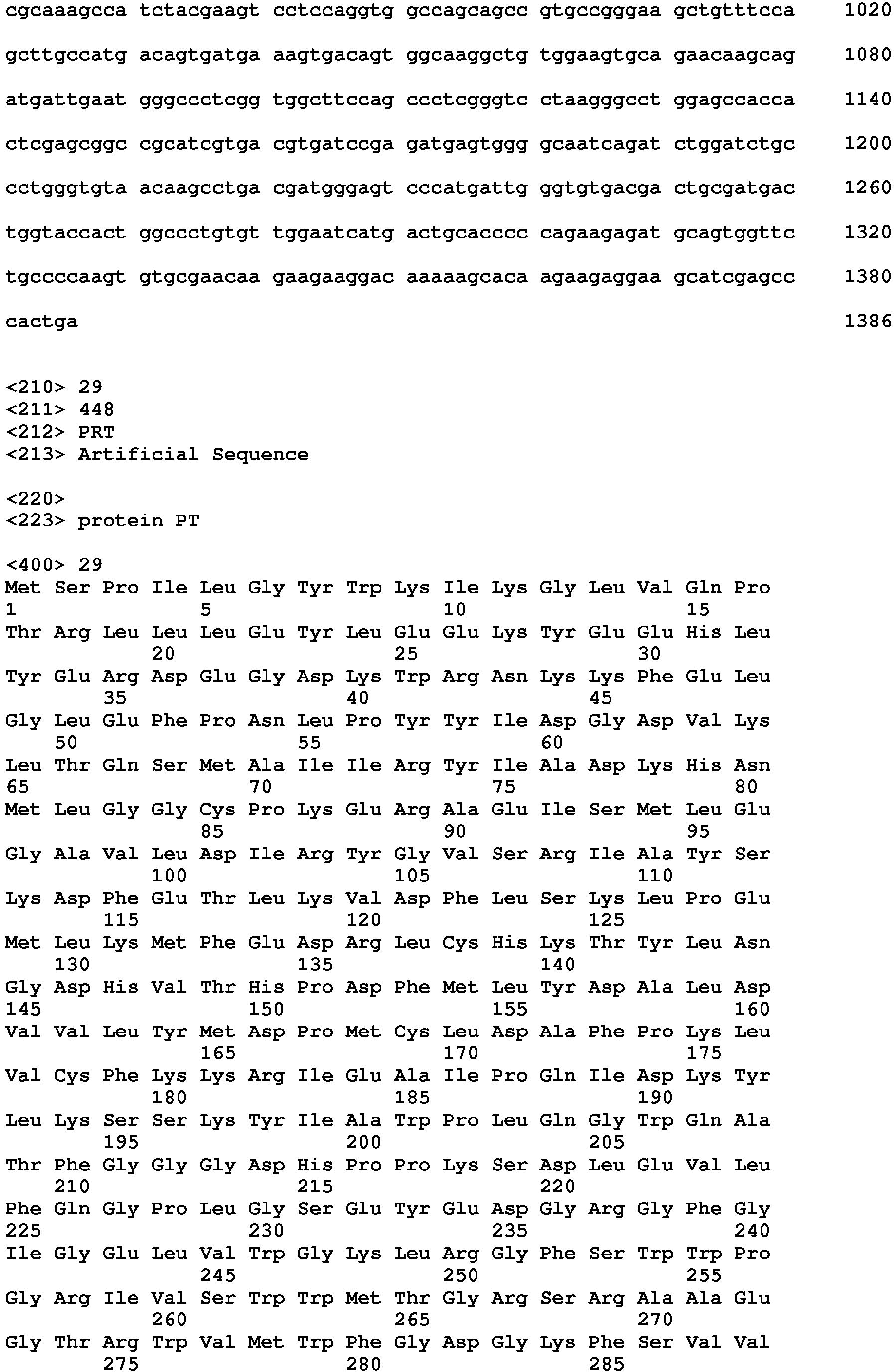


Claims (15)
- Use of an artificial protein for isolating a nucleosome, the nucleosome comprising a multiple-modified histone protein octamer, wherein the artificial protein comprises
a first histone modification binding domain of 50 to 200 amino acids binding to a first histone modification,
a second histone modification binding domain of 50 to 200 amino acids binding to a second histone modification,
a linker of 5 to 50 amino acids connecting the first and the second histone modification binding domain, and
an affinity tag. - Use of claim 1, wherein the first and the second histone modification binding domain are different from each other.
- Use of claim 1, wherein the first and the second histone modification binding domain are copies of the same domain.
- Use of any of claims 1 to 3, wherein the first and/or the second histone modification is selected from the group consisting of methylation, phosphorylation, acetylation, and ubiquitylation.
- Use of any of claims 1 to 4, wherein the first and/or the second histone modification binding domain is selected from the group consisting of 14-3-3 domain, ADD domain, ankyrin, BAH domain, BIR domain, BRCT domain, tandem BRCT domain, bromodomain, double bromodomain, chromobarrel, chromodomain, double chromodomain, double PHD finger domain, MBT domain, PID domain, PHD domain, double PH domain, PWWP domain, royal family domain, Tudor domain, tandem Tudor domain, WD40 domain, and zinc finger CW domain.
- Use of any of claims 1 to 5, wherein the first histone modification binding domain is the PWWP domain of Dnmt3a and the second histone modification binding domain is the chromodomain of MPP8.
- A nucleic acid encoding an artificial protein, wherein the artificial protein comprises
a first histone modification binding domain of 50 to 200 amino acids binding to a first histone modification,
a second histone modification binding domain of 50 to 200 amino acids binding to a second histone modification,
a linker of 5 to 50 amino acids connecting the first and the second histone modification binding domain, and
an affinity tag. - A host cell comprising the nucleic acid of claim 7.
- A kit for isolating a nucleosome, the nucleosome comprising a multiple-modified histone protein octamer, wherein the kit comprises an artificial protein, wherein the artificial protein comprises
a first histone modification binding domain of 50 to 200 amino acids binding to a first histone modification,
a second histone modification binding domain of 50 to 200 amino acids binding to a second histone modification,
a linker of 5 to 50 amino acids connecting the first and the second histone modification binding domain, and
an affinity tag. - An in-vitro method for isolating a nucleosome having a first and a second histone modification, the method comprising the steps of:(a) providing an artificial protein, wherein the artificial protein comprises a first histone modification binding domain of 50 to 200 amino acids binding to the first histone modification,
a second histone modification binding domain of 50 to 200 amino acids binding to the second histone modification,
a linker of 5 to 50 amino acids connecting the first and the second histone modification binding domain, and
an affinity tag;(b) contacting the artificial protein with a sample comprising nucleosomes to allow formation of a complex of the artificial protein and a nucleosome having the first and the second histone modification; and(c) isolating the complex. - The method of claim 10, further comprising the steps of:(d) providing a first control protein and a second control protein, each control protein comprising a single histone modification binding domain, wherein the single histone modification binding domain of the first control protein is the same as the first histone modification binding domain of the artificial protein and the single histone modification binding domain of the second control protein is the same as the second histone modification binding domain of the artificial protein; and(e) contacting the first and the second control protein with a sample comprising nucleosomes to allow formation of a complex of the first control protein and a nucleosome having the first histone modification and/or formation of a complex of the second control protein and a nucleosome having the second histone modification; and(f) isolating the complex.
- The method of claim 10 or 11, further comprising the step of:(g) analysing the isolated complex.
- The method of claim 12, wherein the isolated complex is analysed by mass spectrometry.
- The method of claim 12, wherein the isolated complex is analysed by recovering DNA from the nucleosome and analysing the DNA.
- The method of claim 14, wherein the DNA is analysed by quantitative PCR and/or next-generation sequencing.
Priority Applications (4)
| Application Number | Priority Date | Filing Date | Title |
|---|---|---|---|
| EP15161621.6A EP3075743A1 (en) | 2015-03-30 | 2015-03-30 | Isolation of nucleosomes having multiple-modified histone protein octamers |
| US15/561,486 US10711045B2 (en) | 2015-03-30 | 2016-03-15 | Isolation of nucleosomes having multiple-modified histone protein octamers |
| EP16711206.9A EP3277710B1 (en) | 2015-03-30 | 2016-03-15 | Isolation of nucleosomes having multiple-modified histone protein octamers |
| PCT/EP2016/055605 WO2016156033A1 (en) | 2015-03-30 | 2016-03-15 | Isolation of nucleosomes having multiple-modified histone protein octamers |
Applications Claiming Priority (1)
| Application Number | Priority Date | Filing Date | Title |
|---|---|---|---|
| EP15161621.6A EP3075743A1 (en) | 2015-03-30 | 2015-03-30 | Isolation of nucleosomes having multiple-modified histone protein octamers |
Publications (1)
| Publication Number | Publication Date |
|---|---|
| EP3075743A1 true EP3075743A1 (en) | 2016-10-05 |
Family
ID=52810999
Family Applications (2)
| Application Number | Title | Priority Date | Filing Date |
|---|---|---|---|
| EP15161621.6A Withdrawn EP3075743A1 (en) | 2015-03-30 | 2015-03-30 | Isolation of nucleosomes having multiple-modified histone protein octamers |
| EP16711206.9A Active EP3277710B1 (en) | 2015-03-30 | 2016-03-15 | Isolation of nucleosomes having multiple-modified histone protein octamers |
Family Applications After (1)
| Application Number | Title | Priority Date | Filing Date |
|---|---|---|---|
| EP16711206.9A Active EP3277710B1 (en) | 2015-03-30 | 2016-03-15 | Isolation of nucleosomes having multiple-modified histone protein octamers |
Country Status (3)
| Country | Link |
|---|---|
| US (1) | US10711045B2 (en) |
| EP (2) | EP3075743A1 (en) |
| WO (1) | WO2016156033A1 (en) |
Families Citing this family (1)
| Publication number | Priority date | Publication date | Assignee | Title |
|---|---|---|---|---|
| GB202208728D0 (en) | 2022-06-14 | 2022-07-27 | Cambridge Entpr Ltd | Methods for spatial genomic, epigenomic and multi-omic profiling using transposases and light-activated spatial barcoding |
Citations (3)
| Publication number | Priority date | Publication date | Assignee | Title |
|---|---|---|---|---|
| WO2003070894A2 (en) * | 2002-02-20 | 2003-08-28 | University Of Virginia Patent Foundation | A non-invasive diagnostic test utilizing histone modification markers |
| WO2004044168A2 (en) * | 2002-11-12 | 2004-05-27 | Massachusetts Institute Of Technology | Genetically encoded fluorescent reporters of kinase, methyltransferase, and acetyl-transferase activities |
| WO2014144303A1 (en) * | 2013-03-15 | 2014-09-18 | Constellation Pharmaceuticals, Inc. | Fusion proteins and methods for identifying bromodomain inhibiting compounds |
Family Cites Families (1)
| Publication number | Priority date | Publication date | Assignee | Title |
|---|---|---|---|---|
| US20030049649A1 (en) * | 2000-04-28 | 2003-03-13 | Wolffe Alan P. | Targeted modification of chromatin structure |
-
2015
- 2015-03-30 EP EP15161621.6A patent/EP3075743A1/en not_active Withdrawn
-
2016
- 2016-03-15 EP EP16711206.9A patent/EP3277710B1/en active Active
- 2016-03-15 WO PCT/EP2016/055605 patent/WO2016156033A1/en not_active Ceased
- 2016-03-15 US US15/561,486 patent/US10711045B2/en active Active
Patent Citations (3)
| Publication number | Priority date | Publication date | Assignee | Title |
|---|---|---|---|---|
| WO2003070894A2 (en) * | 2002-02-20 | 2003-08-28 | University Of Virginia Patent Foundation | A non-invasive diagnostic test utilizing histone modification markers |
| WO2004044168A2 (en) * | 2002-11-12 | 2004-05-27 | Massachusetts Institute Of Technology | Genetically encoded fluorescent reporters of kinase, methyltransferase, and acetyl-transferase activities |
| WO2014144303A1 (en) * | 2013-03-15 | 2014-09-18 | Constellation Pharmaceuticals, Inc. | Fusion proteins and methods for identifying bromodomain inhibiting compounds |
Non-Patent Citations (9)
| Title |
|---|
| BOCK, I.; DHAYALAN, A.; KUDITHIPUDI, S.; BRANDT, 0.; RATHERT, P.; JELTSCH, A.: "Detailed specificity analysis of antibodies binding to modified histone tails with peptide arrays", EPIGENETICS, vol. 6, no. 2, 2011, pages 256 - 263 |
| BOCK, I.; KUDITHIPUDI, S.; TAMAS, R.; KUNGULOVSKI, G.; DHAYALAN, A.; JELTSCH, A.: "Application of Celluspots peptide arrays for the analysis of the binding specificity of epigenetic reading domains to modified histone tails", BMC BIOCHEMISTRY, vol. 12, 2011, pages 48 - 59 |
| BRAND, M.; RAMPALLI, S.; CHATURVEDI, C.-P.; F JEFFREY DILWORTH, F.J.: "Analysis of epigenetic modifications of chromatin at specific gene loci by native chromatin immunoprecipitation of nucleosomes isolated using hydroxyapatite chromatography", NATURE PROTOCOLS, vol. 3, no. 3, 2008, pages 398 - 409 |
| JELTSCH, A; LANIO, T.: "Methods in Molecular Biology", vol. 182, 2002, article "Site-Directed Mutagenesis by Polymerase Chain Reaction", pages: 85 - 94 |
| KALLIO M.A.; TUIMALA, J.T.; HUPPONEN, T.; KLEMEL5, P.; GENTILE, M.; SCHEININ, I.; KOSKI, M.; KAKI, J.; KORPELAINEN, E.I.: "Chipster: user-friendly analysis software for microarray and other high-throughput data", BMC GENOMICS, vol. 12, 2011, pages 507 - 521 |
| LANGMEAD, B.; TRAPNELL, C.; POP, M.; SALZBERG, S.L.: "Ultrafast and memory-efficient alignment of short DNA sequences to the human genome", GENOME BIOLOGY, vol. 10, 2009, pages R25 |
| LAU PRISCILLA NGA IENG ET AL: "Elucidating combinatorial histone modifications and crosstalks by coupling histone-modifying enzyme with biotin ligase activity", NUCLEIC ACIDS RESEARCH, vol. 41, no. 3, February 2013 (2013-02-01), pages Article No.: e49, XP002744719, ISSN: 0305-1048(print) * |
| RAMIREZ, F.; DÜNDAR, F.; DIEHL, S; GRUNING, B.A.; MANKE, T.: "deepTools: a flexible platform for exploring deep-sequencing data", NUCLEIC ACIDS RESEARCH, vol. 42, 2014, pages W187 - W191 |
| RATHERT, P.; DHAYALAN, A.; MURAKAMI, M.; ZHANG, X.; TAMAS, R.; JURKOWSKA, R.; KOMATSU, Y.; SHINKAI, Y.; CHENG, X.; JELTSCH, A.: "Protein lysine methyltransferase G9a acts on non-histone targets", NATURE CHEMICAL BIOLOGY, vol. 4, no. 6, 2008, pages 344 - 346 |
Also Published As
| Publication number | Publication date |
|---|---|
| WO2016156033A1 (en) | 2016-10-06 |
| EP3277710A1 (en) | 2018-02-07 |
| US10711045B2 (en) | 2020-07-14 |
| EP3277710B1 (en) | 2020-03-11 |
| US20180066029A1 (en) | 2018-03-08 |
Similar Documents
| Publication | Publication Date | Title |
|---|---|---|
| US20220282336A1 (en) | Biomarkers for non-hodgkin lymphomas and uses thereof | |
| Morgan et al. | A cryptic Tudor domain links BRWD2/PHIP to COMPASS-mediated histone H3K4 methylation | |
| Gorasia et al. | Porphyromonas gingivalis type IX secretion substrates are cleaved and modified by a sortase-like mechanism | |
| Chapman et al. | Mapping PARP-1 auto-ADP-ribosylation sites by liquid chromatography–tandem mass spectrometry | |
| Filippakopoulos et al. | Histone recognition and large-scale structural analysis of the human bromodomain family | |
| US9045801B2 (en) | Biomarkers for non-hodgkin lymphomas and uses thereof | |
| US9353161B2 (en) | Streptavidin mutein exhibiting reversible binding for biotin and streptavidin binding peptide tagged proteins | |
| Angelini et al. | Membrane binding of the bacterial signal recognition particle receptor involves two distinct binding sites | |
| Vega et al. | Isolation and characterization of soluble human full‐length TDP‐43 associated with neurodegeneration | |
| WO2014125668A1 (en) | Method for isolating specific genomic region using molecule binding specifically to endogenous dna sequence | |
| Shimazu et al. | Histidine N1-position-specific methyltransferase CARNMT1 targets C3H zinc finger proteins and modulates RNA metabolism | |
| Slater et al. | Structural variation in PWWP domains | |
| EP3277710B1 (en) | Isolation of nucleosomes having multiple-modified histone protein octamers | |
| Li et al. | Functional analysis of affinity-purified polyhistidine-tagged DnaA protein | |
| Garg et al. | Bromodomain proteins IBD1 and IBD2 link histone acetylation to SWR1-and INO80-mediated H2A. Z regulation in Tetrahymena | |
| Kölbel et al. | Peptide backbone conformation affects the substrate preference of protein arginine methyltransferase I | |
| Käkönen et al. | Purification and characterization of recombinant osteocalcin fusion protein expressed inEscherichia coli | |
| Cushman et al. | Using peptide arrays to define nuclear carrier binding sites on nucleoporins | |
| Huang et al. | Proteomic analysis of stable protein methylation in lymphoblastoid cells | |
| CN104419756B (en) | Detect the method and its application of mammalian genome trinucleotide repeats sequence | |
| Saitoh et al. | The PBII gene of the human salivary proline-rich protein PB produces another protein, Q504X8, with an opiorphin homolog, QRGPR | |
| Ganesan et al. | Progressive polysomal association of Upf1, Upf2, and Upf3 as ribosomes traverse mRNA coding regions | |
| Bestehorn et al. | Pre-mRNA fate decision safeguards the fidelity of the inflammatory response | |
| Viñals Guitart et al. | Engineered HMGB1 construct with tandem HMG B domains promotes tissue regeneration without potential for deleterious inflammation or thrombosis | |
| Zhang et al. | Bromodomain protein IBD1 bridges histone acetylation and H2A. Z deposition to fine-tune transcription and prevent hyperactivation in Tetrahymena thermophila |
Legal Events
| Date | Code | Title | Description |
|---|---|---|---|
| PUAI | Public reference made under article 153(3) epc to a published international application that has entered the european phase |
Free format text: ORIGINAL CODE: 0009012 |
|
| AK | Designated contracting states |
Kind code of ref document: A1 Designated state(s): AL AT BE BG CH CY CZ DE DK EE ES FI FR GB GR HR HU IE IS IT LI LT LU LV MC MK MT NL NO PL PT RO RS SE SI SK SM TR |
|
| AX | Request for extension of the european patent |
Extension state: BA ME |
|
| STAA | Information on the status of an ep patent application or granted ep patent |
Free format text: STATUS: THE APPLICATION IS DEEMED TO BE WITHDRAWN |
|
| 18D | Application deemed to be withdrawn |
Effective date: 20170406 |