EP3069669B1 - Double-joint sickle knife for endoscopy therapy - Google Patents
Double-joint sickle knife for endoscopy therapy Download PDFInfo
- Publication number
- EP3069669B1 EP3069669B1 EP16160727.0A EP16160727A EP3069669B1 EP 3069669 B1 EP3069669 B1 EP 3069669B1 EP 16160727 A EP16160727 A EP 16160727A EP 3069669 B1 EP3069669 B1 EP 3069669B1
- Authority
- EP
- European Patent Office
- Prior art keywords
- knife
- head
- base
- joint
- sliding rod
- Prior art date
- Legal status (The legal status is an assumption and is not a legal conclusion. Google has not performed a legal analysis and makes no representation as to the accuracy of the status listed.)
- Active
Links
Images


Classifications
-
- A—HUMAN NECESSITIES
- A61—MEDICAL OR VETERINARY SCIENCE; HYGIENE
- A61B—DIAGNOSIS; SURGERY; IDENTIFICATION
- A61B17/00—Surgical instruments, devices or methods
- A61B17/32—Surgical cutting instruments
- A61B17/320016—Endoscopic cutting instruments, e.g. arthroscopes, resectoscopes
-
- A—HUMAN NECESSITIES
- A61—MEDICAL OR VETERINARY SCIENCE; HYGIENE
- A61B—DIAGNOSIS; SURGERY; IDENTIFICATION
- A61B17/00—Surgical instruments, devices or methods
- A61B17/32—Surgical cutting instruments
- A61B17/3209—Incision instruments
- A61B17/3211—Surgical scalpels, knives; Accessories therefor
-
- A—HUMAN NECESSITIES
- A61—MEDICAL OR VETERINARY SCIENCE; HYGIENE
- A61B—DIAGNOSIS; SURGERY; IDENTIFICATION
- A61B17/00—Surgical instruments, devices or methods
- A61B17/00234—Surgical instruments, devices or methods for minimally invasive surgery
- A61B2017/00238—Type of minimally invasive operation
- A61B2017/00269—Type of minimally invasive operation endoscopic mucosal resection EMR
-
- A—HUMAN NECESSITIES
- A61—MEDICAL OR VETERINARY SCIENCE; HYGIENE
- A61B—DIAGNOSIS; SURGERY; IDENTIFICATION
- A61B17/00—Surgical instruments, devices or methods
- A61B17/00234—Surgical instruments, devices or methods for minimally invasive surgery
- A61B2017/00292—Surgical instruments, devices or methods for minimally invasive surgery mounted on or guided by flexible, e.g. catheter-like, means
- A61B2017/0034—Surgical instruments, devices or methods for minimally invasive surgery mounted on or guided by flexible, e.g. catheter-like, means adapted to be inserted through a working channel of an endoscope
-
- A—HUMAN NECESSITIES
- A61—MEDICAL OR VETERINARY SCIENCE; HYGIENE
- A61B—DIAGNOSIS; SURGERY; IDENTIFICATION
- A61B17/00—Surgical instruments, devices or methods
- A61B17/32—Surgical cutting instruments
- A61B17/320016—Endoscopic cutting instruments, e.g. arthroscopes, resectoscopes
- A61B2017/32004—Endoscopic cutting instruments, e.g. arthroscopes, resectoscopes having a laterally movable cutting member at its most distal end which remains within the contours of said end
-
- A—HUMAN NECESSITIES
- A61—MEDICAL OR VETERINARY SCIENCE; HYGIENE
- A61B—DIAGNOSIS; SURGERY; IDENTIFICATION
- A61B17/00—Surgical instruments, devices or methods
- A61B17/32—Surgical cutting instruments
- A61B2017/320052—Guides for cutting instruments
Definitions
- the present invention relates to a double-joint sickle knife for an endoscopy therapy.
- ESD endoscopic submucosal dissection
- Patent WO 96/35381 A1 discloses a known endosurgical cutting instrument with a radially moveable sickle-knife for ESD.
- Existing HOOK knifes and IT knifes are an important tool to promote technological development.
- shortcomings still exist, for example: the angle between knife body and the plane of the therapy view field is a larger angle (approximately vertical), and electrosurgical excision operation easily causes to cut too deeply or even causes perforation, so that the surgical operation is very difficult, and has a higher risk.
- the knife body is rod-shaped, so that passive cutting and movement of the knife head is achieved by necessarily moving the endoscope controlled by a surgeon. The above mentioned difficulties highly require a high skill and proficiency for the surgeon.
- the present invention provides a double-joint sickle knife for an endoscopy therapy, which will be achieved by the following technical solutions:
- the double-joint sickle knife for an endoscopy therapy wherein, the knife head comprises a knife edge, a knife back and a knife head root.
- the double-joint sickle knife for an endoscopy therapy wherein, the knife back is made of an insulating ceramic material; and the knife edge is made from a metallic material.
- the double-joint sickle knife for an endoscopy therapy wherein: the head end of the guide wire forms the knife edge; one end of the knife base connected with the knife head is divided into two up and down clamping pieces which are parallel to each other; and the space between the two clamping pieces forms a knife groove; one end of the knife head connected with the knife base is a knife head root; and the knife head root is putted into the knife groove; and the knife head root is equipped with a knife head shaft; and the knife head shaft is embedded with the two clamping pieces; a side of the knife head root near the knife back is connected with the knife base by the compression spring; and all the above-mentioned structures form the transverse joint.
- the double-joint sickle knife for an endoscopy therapy wherein: one end of the knife body connected with the knife base is equipped with two shaft bases; a concave base shaft groove is arranged in the middle of the tail end of the knife base; the knife base shaft crosses the one end of the knife body connected with the knife base and protrudes outwardly; and both ends of the knife base shaft are inserted into the two shaft bases respectively; a torsion spring wraps around the knife base shaft and is put into the base shaft groove; two forwardly and backwardly protruding branches of the torsion spring are inserted into the knife base and the knife body respectively; all the above mentioned structures form the longitudinal joint.
- the double-joint sickle knife for an endoscopy therapy wherein: a guide-wire cavity is arranged in the knife body, and the guide wire is arranged in the guide-wire cavity.
- the double-joint sickle knife for an endoscopy therapy wherein, a guide-wire channel is arranged on the knife base and the knife body, and the guide-wire channel is connected with the guide-wire cavity.
- the double-joint sickle knife for an endoscopy therapy wherein, an outer sheath is sleeved on the outside of the knife body, and the outer sheath can move on the knife body and wrap the knife head.
- the double-joint sickle knife for an endoscopy therapy wherein, the sliding rod is H-shaped; and the middle part of the sliding rod is annular and cylindrical, and covers around the outside of the handle; a hollow sliding groove is arranged in the handle; and a rectangular column sliding rod core is arranged in the middle of the sliding rod; and the sliding rod core crosses into the sliding groove; and a guide-wire outlet is arranged at the top of the sliding rod core; and the guide wire passes through the upper end of the sliding rod by the guide-wire outlet and is connected with the power port.
- the double-joint sickle knife for an endoscopy therapy wherein, and the other end of the handle is connected with a round pull ring.
- a multi-angle movement of a cutting knife blade of the endoscopy knife can be achieved, wherein the movement includes a circumstance cutting by the knife blade in parallel with mucosal tissues.
- the knife blade angle is not limited by the lens of the endoscope, so that the speed and the quality of the endoscopy therapy are greatly improved, and subsidiary injuries potentially occurring to peripheral tissues are minimized to the furthest.
- a double-joint sickle knife for an endoscopy therapy of one embodiment.
- the knife comprises a knife head 1, a knife base 2, a knife body 5, a guide wire 15, a handle 7 and a sliding rod 8.
- the knife head 1 is connected with one end of the knife base 2 by a transverse joint 3.
- the other end of the knife base 2 is connected with a head end of the knife body 5 by a longitudinal joint 4.
- the tail end of the knife body 5 is connected with one end of the handle 7, and the sliding rod 8 for adjusting the transverse joint 3 is arranged on the handle 7.
- One end of the guide wire 15 is connected with the knife head 1, and the guide wire 15 passes through the knife base 2, the knife body 5, the handle 7 and the sliding rod 8 successively.
- the transverse joint 3 can be rotated by controlling the sliding rod 8, so that the knife head 1 moves transversely to achieve a cutting operation.
- the knife head 1 comprises a knife edge 11, a knife back 12 and a knife head root 16.
- the knife back 12 is made of an insulating ceramic material.
- the knife edge 11 is made up by embedding the electric conductive guide wire 15 into the knife back 12.
- the head end of the guide wire 15 forms the knife edge 11.
- One end of the knife base 2 connected with the knife head 1 is divided into two up and down clamping pieces 13 which are parallel to each other. And the space between the two clamping pieces 13 forms a knife groove 14.
- One end of the knife head 1 connected with the knife base 2 is a knife head root 16.
- the knife head root 16 is putted into the knife groove 14. And the knife head root 16 is equipped with a knife head shaft 17. And the knife head shaft 17 is embedded with the two clamping pieces 13. A side of the knife head root 16 near the knife back 12 is connected with the knife base 2 by the compression spring 18. And all the above-mentioned structures form the transverse joint 3. All the above-mentioned structures form the transverse joint 3. A rotated transverse joint can be pulled back to a functional position by the compression spring 18 in the transverse joint 3.
- One end of the knife body 5 connected with the knife base 2 is equipped with two shaft bases 19.
- a concave base shaft groove 22 is arranged in the middle of the tail end of the knife base 2.
- the knife base shaft 20 crosses the one end of the knife body 5 connected with the knife base 2 and protrudes outwardly. And both ends of the knife base shaft 20 are inserted into the two shaft bases 19 respectively.
- a torsion spring 21 wraps around the knife base shaft 20 and is put into the base shaft groove 2. Two forwardly and backwardly protruding branches of the torsion spring 21 are inserted into the knife base 2 and the knife body 5 respectively. All the above mentioned structures form the longitudinal joint 4.
- the torsion spring 21 in the longitudinal joint 4 has features such as resilience and damping.
- a guide-wire cavity 24 is arranged in the knife body 5, and the guide wire 15 is arranged in the guide-wire cavity 24.
- a guide-wire channel 23 is arranged on the knife base 2 and a head end of the knife body 5, and the guide-wire channel 23 is connected with the guide-wire cavity 24.
- An outer sheath 6 is sleeved on the outside of the knife body 5, and the outer sheath 6 can move on the knife body 5 and wrap the knife head 1, so as to choose whether to expose the knife head 1 according to the requirement to avoid injuring an endoscope tube or a mucosa in the therapy view field.
- the sliding rod 8 is H-shaped. And the middle part of the sliding rod 8 is annular and cylindrical, and covers around the outside of the handle 7.
- a hollow sliding groove 27 is arranged in the handle 7. And a rectangular column sliding rod core 25 is arranged in the middle of the sliding rod 8. And the sliding rod core 25 crosses into the sliding groove 27. And a guide-wire outlet 26 is arranged at the top of the sliding rod core 25. And the guide wire 15 passes through the upper end of the sliding rod 8 by the guide-wire outlet 26 and is connected with the power port 10. And the other end of the handle 7 is connected with a round pull ring 9.
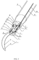
- FIG. 2 illustrates in detail the knife head 1, the knife base 2, a knife body 5, the transverse joint 3 and the longitudinal joint 4 of the present invention.
- the knife head 1 comprises a knife edge 11, a knife back 12 and a knife head root 16.
- the knife back 12 is made of an insulating ceramic material.
- the knife edge 11 is embedded into the knife back 12 in the horizontal half plane of the cross section by the electronic guide wire 15.
- the head end of knife base 2 is divided into two up and down clamping pieces 13 which are parallel to each other, the space between the two clamping pieces 13 forms a knife groove 14.
- the tail end of knife head 1 is a T-shaped knife head root 16. And the knife head root 16 is putted into the knife groove 14.
- the knife head shaft 17 lies at the middle of the knife head root 16, and is embedded into the two up and down clamping pieces 13. And a side of the knife head root 16 near the knife back 12 is connected with the knife base 2 by the compression spring 18.
- the above-mentioned structures form the transverse joint 3.
- the cylindrical knife base shaft 20 crosses the tail end of the knife base 2, and its two ends protrude outwardly. And the cylindrical knife base shaft 20 is inserted into the two shaft bases 19 protruding outwardly from a head end of the knife body 5.
- a concave base shaft groove 22 is arranged in the middle of the tail end of the knife base 2.
- the torsion spring 21 wraps around the knife base shaft 20; and two forwardly and backwardly protruding branches of the torsion spring 21 are inserted into the knife base 2 and the knife body 5 respectively. And the above mentioned structures form the longitudinal joint 4.
- a longitudinal strip guide-wire channel 23 is arranged in the cross section of the head end of the knife body 5. And the guide-wire channel 23 extends to the tail end narrowly from the internal of the knife body 5, and is connected with the guide-wire cavity 24.
- FIG. 3 illustrates in detail a part of a handle 7 and a sliding rod 8 of the present invention
- the tail end of the knife body 5 is connected with the head end of the handle 7, and the head end of handle 7 is a little thicker than the outer sheath 6, in order to avoid the outer sheath 6 from sliding and slipping.
- a hollow sliding groove 27 is arranged in the handle 7.
- the sliding rod 8 is H-shaped.
- the middle part of the sliding rod 8 is annular and cylindrical, and covers around the outside of the handle 7.
- the rectangular column sliding rod core 25 is arranged in the middle of the sliding rod 8.
- the sliding rod core 25 crosses into the groove 27.
- a guide-wire outlet 26 is arranged at the top of the sliding rod core 25.
- a round pull ring 9 is arranged at the tail end of the handle 7.
- the guide-wire cavity 24 passes through from the internal of the knife body 5 to the head end of the handle 7, and ends at the top of the sliding rod groove 27 in the handle 7. Please refer to FIG.2 and FIG.3 , the guide wire 15 passes through the knife head root 16, the knife base 2, the guide-wire channel 23, the guide-wire cavity 24, the handle 7, and the sliding rod core 25 successively from the knife edge 11 to the tail part, until the guide wire 15 is connected with the power port 10.
- FIG. 4 illustrates a plane d relative to the knife protruding outwardly from the endoscope biopsy channel of the present invention.
- the knife body 5 forms an ⁇ (alpha) Angle relateive to a plane a.
- the plane where the ⁇ Angle is in perpendicular to the plane d.
- the knife head 1 forms a ⁇ (Beta) angle relative to the knife base 2, and a proper angle is arranged between the plane d and the plane where the ⁇ Angle is, or the plane d is in parallel with the plane where the ⁇ Angle is.
- the knife head 1 and knife base 2 of the present double-joint sickle knife can be sent to the surgical site through the endoscope biopsy channel.
- the power port 10 on the sliding rod 8 of the external handle is connected with a high frequency power generator.
- the knife edge 11 generates a cutting action by being powered on through the conductive guide wire.
- the knife head 1 is sickle-shaped, and only has a cutting function on the knife edge 11 in the working surface.
- the knife back 12 is made of a ceramic material, so as to prevent subsidiary injuries effectively.
- the knife base 2 is pressed down to force the longitudinal joint 4 to rotate so as to adjust the ⁇ Angle by operating the endoscope (as shown FIG.4 ), so that the knife head 1 can be placed at different angles relative to the plane of the therapy view field, and even in parallel with the plane of the therapy view field.
- the torsion spring 21 in the internal of the longitudinal joint 4 provides the joint with functions of resilience and damping in a proper intensity.
- the guide wire 15 can be pulled back by the sliding rod 8 so as to force knife head 1 to carry a rotating movement around the knife head shaft 17, so that the transverse joint 3 can rotate to adjust the angle ⁇ (as show in FIG. 4 ).
- the axial movement of knife head 1 can be controlled just by the index finger and the middle finger to pull back the sliding rod 8 so as to achieve a intricate cutting action.
- a rotated transverse joint can be pulled back to the functional position by the compressed spring 18 in the transverse joint 3 (the state as shown in FIG.1 and FIG.2 ).
Landscapes
- Health & Medical Sciences (AREA)
- Life Sciences & Earth Sciences (AREA)
- Surgery (AREA)
- General Health & Medical Sciences (AREA)
- Veterinary Medicine (AREA)
- Biomedical Technology (AREA)
- Heart & Thoracic Surgery (AREA)
- Medical Informatics (AREA)
- Molecular Biology (AREA)
- Animal Behavior & Ethology (AREA)
- Nuclear Medicine, Radiotherapy & Molecular Imaging (AREA)
- Public Health (AREA)
- Engineering & Computer Science (AREA)
- Orthopedic Medicine & Surgery (AREA)
- Surgical Instruments (AREA)
- Physics & Mathematics (AREA)
- Biophysics (AREA)
- Optics & Photonics (AREA)
- Pathology (AREA)
- Radiology & Medical Imaging (AREA)
Description
- The present invention relates to a double-joint sickle knife for an endoscopy therapy.
- With the development of the techniques of digestion endoscope, adaptable indications for endoscopic microsurgeries extends continuously, and relevant minimally invasive techniques such as EMR, ESD, POEM and so on are emerging and developing continuously. The endoscopic submucosal dissection (ESD) therapy is currently an important means of surgical treatment of early gastrointestinal cancers and precancerous lesions. ESD has shown great advantages such as a tiny trauma, a maintenance of organ functions, a quick recovery after the surgery. However, ESD has a high demand in technology, and is prone to come out with complications such as perforation, so that ESD is greatly limited and has not been widely applied. Patent
WO 96/35381 A1 - In order to solve the above problems in the prior art, the present invention provides a double-joint sickle knife for an endoscopy therapy, which will be achieved by the following technical solutions:
- A double-joint sickle knife for an endoscopy therapy, wherein, the knife comprises a knife head, a knife base, a knife body, a guide wire, a handle and a sliding rod; wherein the knife head is connected with one end of the knife base by a transverse joint, and the other end of the knife base is connected with a head end of the knife body by a longitudinal joint; the tail end of the knife body is connected with one end of the handle, and the sliding rod is arranged on the handle; one end of the guide wire is connected with the knife head, and the guide wire passes through the knife base, the knife body, the handle and the sliding rod successively; and
- the other end of the guide wire is connected with a power port.
- The double-joint sickle knife for an endoscopy therapy, wherein, the knife head comprises a knife edge, a knife back and a knife head root.
- The double-joint sickle knife for an endoscopy therapy, wherein, the knife back is made of an insulating ceramic material; and the knife edge is made from a metallic material.
- The double-joint sickle knife for an endoscopy therapy, wherein: the head end of the guide wire forms the knife edge; one end of the knife base connected with the knife head is divided into two up and down clamping pieces which are parallel to each other; and the space between the two clamping pieces forms a knife groove; one end of the knife head connected with the knife base is a knife head root; and the knife head root is putted into the knife groove; and the knife head root is equipped with a knife head shaft; and the knife head shaft is embedded with the two clamping pieces; a side of the knife head root near the knife back is connected with the knife base by the compression spring; and all the above-mentioned structures form the transverse joint.
- The double-joint sickle knife for an endoscopy therapy, wherein: one end of the knife body connected with the knife base is equipped with two shaft bases; a concave base shaft groove is arranged in the middle of the tail end of the knife base; the knife base shaft crosses the one end of the knife body connected with the knife base and protrudes outwardly; and both ends of the knife base shaft are inserted into the two shaft bases respectively; a torsion spring wraps around the knife base shaft and is put into the base shaft groove; two forwardly and backwardly protruding branches of the torsion spring are inserted into the knife base and the knife body respectively; all the above mentioned structures form the longitudinal joint.
- The double-joint sickle knife for an endoscopy therapy, wherein: a guide-wire cavity is arranged in the knife body, and the guide wire is arranged in the guide-wire cavity.
- The double-joint sickle knife for an endoscopy therapy, wherein, a guide-wire channel is arranged on the knife base and the knife body, and the guide-wire channel is connected with the guide-wire cavity.
- The double-joint sickle knife for an endoscopy therapy, wherein, an outer sheath is sleeved on the outside of the knife body, and the outer sheath can move on the knife body and wrap the knife head.
- The double-joint sickle knife for an endoscopy therapy, wherein, the sliding rod is H-shaped; and the middle part of the sliding rod is annular and cylindrical, and covers around the outside of the handle; a hollow sliding groove is arranged in the handle; and a rectangular column sliding rod core is arranged in the middle of the sliding rod; and the sliding rod core crosses into the sliding groove; and a guide-wire outlet is arranged at the top of the sliding rod core; and the guide wire passes through the upper end of the sliding rod by the guide-wire outlet and is connected with the power port.
- The double-joint sickle knife for an endoscopy therapy, wherein, and the other end of the handle is connected with a round pull ring.
- Compared with the prior art, the advantages of the present invention using the above-mentioned technical solutions is that:
- 1. The sickle knife has two rotary joints. The longitudinal joint can cut tissues by the knife head in parallel with the therapy view field. Therefore, it is highly safe, and can reduce the probability of perforation. The transverse joint facilitates the knife head to actively cut and move in control of the handle and the sliding rod, so that the surgeon finishes cutting tissues without controlling the movement of the lens end of the endoscope in a large range, and thus the difficulty of the therapy is decreased significantly, the efficiency of the therapy is enhanced, and the surgery becomes more easier;
- 2. The knife head is shaped like a sickle, and just one side of the knife head has a cutting function, and any other side is wrapped around by a ceramic material, so that the knife head can cut mucosal tissues quickly and safely. And thus, subsidiary injuries potentially occurring to peripheral tissues are minimized to the furthest;
- 3. Subsidiary injuries in endoscopy microsurgeries such as endoscopic submucosal dissection (ESD) can be greatly reduced. And the present invention becomes more safe and effective, and the knife is convenient for promotion and application too;
- 4. The present invention can also be used in other advanced techniques such as natural orifice transluminal endoscopic surgeries (NOTES). And the present invention has an extensive application prospect and may bring huge economic and social benefits.
- By means of the knife, a multi-angle movement of a cutting knife blade of the endoscopy knife can be achieved, wherein the movement includes a circumstance cutting by the knife blade in parallel with mucosal tissues. And the knife blade angle is not limited by the lens of the endoscope, so that the speed and the quality of the endoscopy therapy are greatly improved, and subsidiary injuries potentially occurring to peripheral tissues are minimized to the furthest.
-
-
FIG. 1 is a structure diagram of the present invention; -
FIG. 2 illustrates in detail a knife head, a knife base, a knife body, a transverse joint and a longitudinal joint of the present invention; -
FIG. 3 illustrates in detail a handle and a sliding rod of the present invention; -
FIG. 4 illustrates the plane d relative to the knife protruding outwardly from the endoscope biopsy channel of the present invention. - Reference signs of drawings: 1-knife head, 2-knife base, 3-transverse joint, 4-longitudinal joint, 5-knife body, 6-outer sheath, 7-handle, 8-sliding rod, 9-pull ring, 10-power port, 11-knife edge, 12-knife back, 13-clamping pieces, 14-knife groove, 15-guide wire, 16-knife head root, 17-knife head shaft, 18-compression spring, 19-shaft base, 20-knife base shaft, 21-torsion spring, 22-base shaft groove, 23-guide-wire channel, 24-guide-wire cavity, 25-sliding rod core, 26-guide-wire outlet, 27-sliding groove.
- The double-joint sickle knife for an endoscopy therapy is described in detail with reference to the accompanying drawings and the embodiments as follows.
- As shown in
FIG. 1 to FIG.4 , a double-joint sickle knife for an endoscopy therapy of one embodiment. The knife comprises aknife head 1, aknife base 2, aknife body 5, aguide wire 15, ahandle 7 and asliding rod 8. Theknife head 1 is connected with one end of theknife base 2 by atransverse joint 3. And the other end of theknife base 2 is connected with a head end of theknife body 5 by alongitudinal joint 4. The tail end of theknife body 5 is connected with one end of thehandle 7, and thesliding rod 8 for adjusting thetransverse joint 3 is arranged on thehandle 7. One end of theguide wire 15 is connected with theknife head 1, and theguide wire 15 passes through theknife base 2, theknife body 5, thehandle 7 and thesliding rod 8 successively. And the other end of theguide wire 15 is connected with apower port 10. Thetransverse joint 3 can be rotated by controlling thesliding rod 8, so that theknife head 1 moves transversely to achieve a cutting operation. Theknife head 1 comprises aknife edge 11, aknife back 12 and aknife head root 16. Theknife back 12 is made of an insulating ceramic material. Theknife edge 11 is made up by embedding the electricconductive guide wire 15 into theknife back 12. Moreover, the head end of theguide wire 15 forms theknife edge 11. One end of theknife base 2 connected with theknife head 1 is divided into two up and down clampingpieces 13 which are parallel to each other. And the space between the twoclamping pieces 13 forms aknife groove 14. One end of theknife head 1 connected with theknife base 2 is aknife head root 16. And theknife head root 16 is putted into theknife groove 14. And theknife head root 16 is equipped with aknife head shaft 17. And theknife head shaft 17 is embedded with the two clampingpieces 13. A side of theknife head root 16 near the knife back 12 is connected with theknife base 2 by thecompression spring 18. And all the above-mentioned structures form thetransverse joint 3. All the above-mentioned structures form thetransverse joint 3. A rotated transverse joint can be pulled back to a functional position by thecompression spring 18 in thetransverse joint 3. - One end of the
knife body 5 connected with theknife base 2 is equipped with two shaft bases 19. A concavebase shaft groove 22 is arranged in the middle of the tail end of theknife base 2. Theknife base shaft 20 crosses the one end of theknife body 5 connected with theknife base 2 and protrudes outwardly. And both ends of theknife base shaft 20 are inserted into the twoshaft bases 19 respectively. Atorsion spring 21 wraps around theknife base shaft 20 and is put into thebase shaft groove 2. Two forwardly and backwardly protruding branches of thetorsion spring 21 are inserted into theknife base 2 and theknife body 5 respectively. All the above mentioned structures form thelongitudinal joint 4. Thetorsion spring 21 in thelongitudinal joint 4 has features such as resilience and damping. A guide-wire cavity 24 is arranged in theknife body 5, and theguide wire 15 is arranged in the guide-wire cavity 24. A guide-wire channel 23 is arranged on theknife base 2 and a head end of theknife body 5, and the guide-wire channel 23 is connected with the guide-wire cavity 24. Anouter sheath 6 is sleeved on the outside of theknife body 5, and theouter sheath 6 can move on theknife body 5 and wrap theknife head 1, so as to choose whether to expose theknife head 1 according to the requirement to avoid injuring an endoscope tube or a mucosa in the therapy view field. The slidingrod 8 is H-shaped. And the middle part of the slidingrod 8 is annular and cylindrical, and covers around the outside of thehandle 7. A hollow slidinggroove 27 is arranged in thehandle 7. And a rectangular column slidingrod core 25 is arranged in the middle of the slidingrod 8. And the slidingrod core 25 crosses into the slidinggroove 27. And a guide-wire outlet 26 is arranged at the top of the slidingrod core 25. And theguide wire 15 passes through the upper end of the slidingrod 8 by the guide-wire outlet 26 and is connected with thepower port 10. And the other end of thehandle 7 is connected with around pull ring 9. -
FIG. 2 illustrates in detail theknife head 1, theknife base 2, aknife body 5, the transverse joint 3 and thelongitudinal joint 4 of the present invention. Theknife head 1 comprises aknife edge 11, a knife back 12 and aknife head root 16. The knife back 12 is made of an insulating ceramic material. And theknife edge 11 is embedded into the knife back 12 in the horizontal half plane of the cross section by theelectronic guide wire 15. The head end ofknife base 2 is divided into two up and down clampingpieces 13 which are parallel to each other, the space between the two clampingpieces 13 forms aknife groove 14. The tail end ofknife head 1 is a T-shapedknife head root 16. And theknife head root 16 is putted into theknife groove 14. Theknife head shaft 17 lies at the middle of theknife head root 16, and is embedded into the two up and down clampingpieces 13. And a side of theknife head root 16 near the knife back 12 is connected with theknife base 2 by thecompression spring 18. The above-mentioned structures form thetransverse joint 3. The cylindricalknife base shaft 20 crosses the tail end of theknife base 2, and its two ends protrude outwardly. And the cylindricalknife base shaft 20 is inserted into the twoshaft bases 19 protruding outwardly from a head end of theknife body 5. A concavebase shaft groove 22 is arranged in the middle of the tail end of theknife base 2. In the concavebase shaft groove 22, thetorsion spring 21 wraps around theknife base shaft 20; and two forwardly and backwardly protruding branches of thetorsion spring 21 are inserted into theknife base 2 and theknife body 5 respectively. And the above mentioned structures form thelongitudinal joint 4. A longitudinal strip guide-wire channel 23 is arranged in the cross section of the head end of theknife body 5. And the guide-wire channel 23 extends to the tail end narrowly from the internal of theknife body 5, and is connected with the guide-wire cavity 24. -
FIG. 3 illustrates in detail a part of ahandle 7 and a slidingrod 8 of the present invention, The tail end of theknife body 5 is connected with the head end of thehandle 7, and the head end ofhandle 7 is a little thicker than theouter sheath 6, in order to avoid theouter sheath 6 from sliding and slipping. And a hollow slidinggroove 27 is arranged in thehandle 7. And the slidingrod 8 is H-shaped. The middle part of the slidingrod 8 is annular and cylindrical, and covers around the outside of thehandle 7. And the rectangular column slidingrod core 25 is arranged in the middle of the slidingrod 8. The slidingrod core 25 crosses into thegroove 27. And a guide-wire outlet 26 is arranged at the top of the slidingrod core 25. And around pull ring 9 is arranged at the tail end of thehandle 7. The guide-wire cavity 24 passes through from the internal of theknife body 5 to the head end of thehandle 7, and ends at the top of the slidingrod groove 27 in thehandle 7. Please refer toFIG.2 andFIG.3 , theguide wire 15 passes through theknife head root 16, theknife base 2, the guide-wire channel 23, the guide-wire cavity 24, thehandle 7, and the slidingrod core 25 successively from theknife edge 11 to the tail part, until theguide wire 15 is connected with thepower port 10. -
FIG. 4 illustrates a plane d relative to the knife protruding outwardly from the endoscope biopsy channel of the present invention. Theknife body 5 forms an α (alpha) Angle relateive to a plane a. The plane where the α Angle is in perpendicular to the plane d. And theknife head 1 forms a β (Beta) angle relative to theknife base 2, and a proper angle is arranged between the plane d and the plane where the β Angle is, or the plane d is in parallel with the plane where the β Angle is. - In a digestive endoscopy minimally invasive surgery, the
knife head 1 andknife base 2 of the present double-joint sickle knife can be sent to the surgical site through the endoscope biopsy channel. Thepower port 10 on the slidingrod 8 of the external handle is connected with a high frequency power generator. Theknife edge 11 generates a cutting action by being powered on through the conductive guide wire. Theknife head 1 is sickle-shaped, and only has a cutting function on theknife edge 11 in the working surface. The knife back 12 is made of a ceramic material, so as to prevent subsidiary injuries effectively. Theknife base 2 is pressed down to force the longitudinal joint 4 to rotate so as to adjust the α Angle by operating the endoscope (as shownFIG.4 ), so that theknife head 1 can be placed at different angles relative to the plane of the therapy view field, and even in parallel with the plane of the therapy view field. Thetorsion spring 21 in the internal of thelongitudinal joint 4 provides the joint with functions of resilience and damping in a proper intensity. - The
guide wire 15 can be pulled back by the slidingrod 8 so as to forceknife head 1 to carry a rotating movement around theknife head shaft 17, so that the transverse joint 3 can rotate to adjust the angle β (as show inFIG. 4 ). The axial movement ofknife head 1 can be controlled just by the index finger and the middle finger to pull back the slidingrod 8 so as to achieve a intricate cutting action. A rotated transverse joint can be pulled back to the functional position by thecompressed spring 18 in the transverse joint 3 (the state as shown inFIG.1 andFIG.2 ). When the fingers loose the slidingrod 8, by adjusting the two joints in different directions and quadrants, the cutting angle of theknife edge 11 can be wider in the range, the endoscopic view can be clearer, and it can be easily and quickly to cut and peel lesions. - The description above are merely illustrative, is not intended to limit the invention. The description of the present invention is readily appreciated by those skilled in the art can that, any modification, improvement, or equivalent replacement of the present invention within the principle of the present invention should be deemed to fall within the protection scope of the present invention, as defined by the appended claims.
Claims (10)
- A double-joint sickle knife for an endoscopy therapy, wherein the knife comprises a knife head (1), a knife base (2), a knife body (5), a guide wire (15), a handle (7) and a sliding rod (8); wherein the knife head (1) is connected with one end of the knife base (2) by a transverse joint (3), and the other end of the knife base (2) is connected with a head end of the knife body (5) by a longitudinal joint (4); the tail end of the knife body (5) is connected with one end of the handle (7), and the sliding rod (8) is arranged on the handle (7); one end of the guide wire (15) is connected with the knife head (1), and the guide wire (15) passes through the knife base (2), the knife body (5), the handle (7) and the sliding rod (8) successively; and the other end of the guide wire (15) is connected with a power port (10) which is connected to power generator.
- The double-joint sickle knife for an endoscopy therapy as claimed in claim 1, wherein the knife head (1) comprises a knife edge (11), a knife back (12) and a knife head root (16).
- The double-joint sickle knife for an endoscopy therapy as claimed in claim 2, wherein the knife back (12) is made of an insulating ceramic material; and the knife edge (11) is made from a metallic material.
- The double-joint sickle knife for an endoscopy therapy as claimed in claim 2, wherein: the head end of the guide wire (15) forms the knife edge (11); one end of the knife base (2) connected with the knife head (1) is divided into two up and down clamping pieces (13) which are parallel to each other; and the space between the two clamping pieces (13) forms a knife groove (14); one end of the knife head (1) connected with the knife base (2) is a knife head root (16); and the knife head root (16) is putted into the knife groove (14); and the knife head root (16) is equipped with a knife head shaft (17); and the knife head shaft (17) is embedded with the two clamping pieces (13); a side of the knife head root (16) near the knife back (12) is connected with the knife base (2) by the compression spring (18); and all the above-mentioned structures form the transverse joint (3).
- The double-joint sickle knife for an endoscopy therapy as claimed in claim 1, wherein: one end of the knife body (5) connected with the knife base (2) is equipped with two shaft bases (19); a concave base shaft groove (22) is arranged in the middle of the tail end of the knife base (2); the knife base shaft (20) crosses the one end of the knife body (5) connected with the knife base (2) and protrudes outwardly; and both ends of the knife base shaft (20) are inserted into the two shaft bases (19) respectively; a torsion spring (21) wraps around the knife base shaft (20) and is put into the base shaft groove (22); two forwardly and backwardly protruding branches of the torsion spring (21) are inserted into the knife base (2) and the knife body (5) respectively; all the above mentioned structures form the longitudinal joint (4).
- The double-joint sickle knife for an endoscopy therapy as claimed in claim 1, wherein: a guide-wire cavity (24) is arranged in the knife body (5), and the guide wire (15) is arranged in the guide-wire cavity (24).
- The double-joint sickle knife for an endoscopy therapy as claimed in claim 6, wherein a guide-wire channel (23) is arranged on the knife base (2) and the knife body (5), and the guide-wire channel (23) is connected with the guide-wire cavity (24).
- The double-joint sickle knife for an endoscopy therapy as claimed in claim 1, wherein an outer sheath (6) is sleeved on the outside of the knife body (5), and the outer sheath (6) can move on the knife body (5) and wrap the knife head (1).
- The double-joint sickle knife for an endoscopy therapy as claimed in claim 1, wherein the sliding rod (8) is H-shaped; and the middle part of the sliding rod (8) is annular and cylindrical, and covers around the outside of the handle (7); a hollow sliding groove (27) is arranged in the handle (7); and a rectangular column sliding rod core (25) is arranged in the middle of the sliding rod (8); and the sliding rod core (25) crosses into the sliding groove (27); and a guide-wire outlet (26) is arranged at the top of the sliding rod core (25); and the guide wire (15) passes through the upper end of the sliding rod (8) by the guide-wire outlet (26) and is connected with the power port (10).
- The double-joint sickle knife for an endoscopy therapy as claimed in claim 1, wherein and the other end of the handle (7) is connected with a round pull ring (9).
Applications Claiming Priority (1)
| Application Number | Priority Date | Filing Date | Title |
|---|---|---|---|
| CN201510121464.2A CN104799909B (en) | 2015-03-19 | 2015-03-19 | Digestive endoscopy Minimally Invasive Surgery doublejointed sickle knife |
Publications (2)
| Publication Number | Publication Date |
|---|---|
| EP3069669A1 EP3069669A1 (en) | 2016-09-21 |
| EP3069669B1 true EP3069669B1 (en) | 2017-11-29 |
Family
ID=53685420
Family Applications (1)
| Application Number | Title | Priority Date | Filing Date |
|---|---|---|---|
| EP16160727.0A Active EP3069669B1 (en) | 2015-03-19 | 2016-03-16 | Double-joint sickle knife for endoscopy therapy |
Country Status (5)
| Country | Link |
|---|---|
| US (1) | US10524818B2 (en) |
| EP (1) | EP3069669B1 (en) |
| JP (1) | JP6147384B2 (en) |
| KR (1) | KR101776126B1 (en) |
| CN (1) | CN104799909B (en) |
Families Citing this family (7)
| Publication number | Priority date | Publication date | Assignee | Title |
|---|---|---|---|---|
| CN107638198A (en) * | 2017-09-30 | 2018-01-30 | 四川大学华西医院 | Endoscopic mucosal dissection instrument with dovetail-shaped cutter head |
| CN113208698B (en) * | 2021-04-19 | 2024-07-26 | 北京大学深圳医院 | Multifunctional bone joint cutting and peeling device |
| CN113171180B (en) * | 2021-06-02 | 2024-10-01 | 上海生知医疗科技有限公司 | A portable manual surgical robot |
| CN113243885B (en) * | 2021-06-16 | 2021-10-01 | 上海宇度医学科技股份有限公司 | Electronic hysteroscope with adjustable lens angle |
| EP4444200A4 (en) * | 2021-12-06 | 2025-11-12 | George G Kihiczak | MEDICAL CUTTING DEVICES AND METHODS FOR USE THEM |
| CN115836911A (en) * | 2022-12-09 | 2023-03-24 | 安瑞医疗器械(杭州)有限公司 | Multifunctional high-frequency electrical instrument used with endoscope |
| CN118121271B (en) * | 2024-03-27 | 2024-08-27 | 江苏唯德康医疗科技有限公司 | Dredging type mucous membrane incision sword in tool bit |
Family Cites Families (18)
| Publication number | Priority date | Publication date | Assignee | Title |
|---|---|---|---|---|
| DE3918720A1 (en) | 1989-06-08 | 1990-12-20 | Wolf Gmbh Richard | RETROGRAD CUTTING HOOK PUNCH |
| US5306284A (en) * | 1992-02-19 | 1994-04-26 | John Agee | Surgical instrument |
| US5308358A (en) * | 1992-08-25 | 1994-05-03 | Bond Albert L | Rigid-shaft surgical instruments that can be disassembled for improved cleaning |
| US5586990A (en) | 1995-05-08 | 1996-12-24 | Symbiosis Corporation | Endosurgical instrument with a radially movable end effector |
| JP4388172B2 (en) * | 1999-08-26 | 2009-12-24 | オリンパス株式会社 | Medical treatment tool |
| US20060174898A1 (en) * | 2005-02-10 | 2006-08-10 | Team Brown Enterprises, Llc | Defibrillator insertion device and method |
| US20070225740A1 (en) * | 2006-02-22 | 2007-09-27 | Loubert Suddaby | Endoscopic Pulley Knife Instrument for Transecting Ligaments or Fascia |
| US20080029574A1 (en) * | 2006-08-02 | 2008-02-07 | Shelton Frederick E | Pneumatically powered surgical cutting and fastening instrument with actuator at distal end |
| PL2077774T3 (en) * | 2006-10-05 | 2015-02-27 | Erbe Elektromedizin | Tubular shaft instrument |
| US20080262492A1 (en) * | 2007-04-11 | 2008-10-23 | Cambridge Endoscopic Devices, Inc. | Surgical Instrument |
| CN101317777B (en) | 2007-06-05 | 2012-01-04 | 伊西康内外科公司 | Medical instrument with needle and knife |
| GB0718721D0 (en) * | 2007-09-25 | 2007-11-07 | Medical Device Innovations Ltd | Surgical resection apparatus |
| EP2486876B1 (en) * | 2009-10-08 | 2020-04-15 | River Seiko Corporation | Scissors for endoscope |
| JP5385930B2 (en) * | 2011-02-22 | 2014-01-08 | 富士フイルム株式会社 | Ultrasonic surgical device |
| KR101711361B1 (en) * | 2012-01-06 | 2017-03-02 | 지완 스티븐 싱 | An insert and insert system for a laparoscopic instrument |
| WO2013116282A1 (en) | 2012-01-31 | 2013-08-08 | Boston Scientific Scimed, Inc. | An endoscopic instrument having a cutting tool |
| WO2014027548A1 (en) * | 2012-08-13 | 2014-02-20 | オリンパスメディカルシステムズ株式会社 | Treatment device for endoscope |
| CN203988348U (en) * | 2014-06-20 | 2014-12-10 | 杭州安杰思医学科技有限公司 | A kind of combination electric knife |
-
2015
- 2015-03-19 CN CN201510121464.2A patent/CN104799909B/en active Active
-
2016
- 2016-03-15 KR KR1020160030953A patent/KR101776126B1/en not_active Expired - Fee Related
- 2016-03-16 JP JP2016052523A patent/JP6147384B2/en not_active Expired - Fee Related
- 2016-03-16 EP EP16160727.0A patent/EP3069669B1/en active Active
- 2016-03-17 US US15/072,471 patent/US10524818B2/en active Active
Non-Patent Citations (1)
| Title |
|---|
| None * |
Also Published As
| Publication number | Publication date |
|---|---|
| JP6147384B2 (en) | 2017-06-14 |
| US20160270812A1 (en) | 2016-09-22 |
| JP2016174901A (en) | 2016-10-06 |
| EP3069669A1 (en) | 2016-09-21 |
| CN104799909A (en) | 2015-07-29 |
| KR101776126B1 (en) | 2017-09-07 |
| US10524818B2 (en) | 2020-01-07 |
| KR20160112987A (en) | 2016-09-28 |
| CN104799909B (en) | 2016-06-29 |
Similar Documents
| Publication | Publication Date | Title |
|---|---|---|
| US10524818B2 (en) | Double-joint sickle knife for endoscopy therapy | |
| US11039851B2 (en) | Endoscopic submucosal dissection hood | |
| US8425509B2 (en) | Surgical instrument for performing controlled myotomies | |
| CN105307583B (en) | Minimally Invasive Surgery component and method | |
| US20210068805A1 (en) | Traction apparatus and traction ring for traction apparatus | |
| US11717342B2 (en) | Medical device | |
| CN209032622U (en) | A high frequency cutter with injection needle for endoscope | |
| WO2020002988A3 (en) | Medical instrument for endoscopic mucosal resection and endoscopic submucosal dissection | |
| EP3243469A1 (en) | Mucosa dissector | |
| CN202568437U (en) | ESD mucosa scissor | |
| US11109908B2 (en) | RF surgical resection snare for flexible endoscopy | |
| CN117017468B (en) | High-frequency electric knife and use method thereof | |
| CN202554057U (en) | Surgical instrument accompanied with unilateral scissors special for endoscopic mucosal resection | |
| CN219397552U (en) | Asymmetric snare with electrotome function | |
| CN113907865B (en) | A cutting knife | |
| CN117338392A (en) | rotary cutter | |
| CN204468206U (en) | Mucosa-cutting knife under endoscope | |
| JP7076130B2 (en) | Bipolar scissors knife | |
| CN215192169U (en) | A warped high-frequency electric knife | |
| CN203710130U (en) | Multifunctional high-frequency cutting knife | |
| CN209122366U (en) | An endoscope operation device |
Legal Events
| Date | Code | Title | Description |
|---|---|---|---|
| PUAI | Public reference made under article 153(3) epc to a published international application that has entered the european phase |
Free format text: ORIGINAL CODE: 0009012 |
|
| AK | Designated contracting states |
Kind code of ref document: A1 Designated state(s): AL AT BE BG CH CY CZ DE DK EE ES FI FR GB GR HR HU IE IS IT LI LT LU LV MC MK MT NL NO PL PT RO RS SE SI SK SM TR |
|
| AX | Request for extension of the european patent |
Extension state: BA ME |
|
| STAA | Information on the status of an ep patent application or granted ep patent |
Free format text: STATUS: REQUEST FOR EXAMINATION WAS MADE |
|
| 17P | Request for examination filed |
Effective date: 20161129 |
|
| RBV | Designated contracting states (corrected) |
Designated state(s): AL AT BE BG CH CY CZ DE DK EE ES FI FR GB GR HR HU IE IS IT LI LT LU LV MC MK MT NL NO PL PT RO RS SE SI SK SM TR |
|
| RIC1 | Information provided on ipc code assigned before grant |
Ipc: A61B 17/32 20060101AFI20170223BHEP Ipc: A61B 17/94 20060101ALN20170223BHEP |
|
| RIC1 | Information provided on ipc code assigned before grant |
Ipc: A61B 17/94 20060101ALN20170323BHEP Ipc: A61B 17/32 20060101AFI20170323BHEP |
|
| RIC1 | Information provided on ipc code assigned before grant |
Ipc: A61B 17/94 20060101ALN20170420BHEP Ipc: A61B 17/32 20060101AFI20170420BHEP |
|
| GRAP | Despatch of communication of intention to grant a patent |
Free format text: ORIGINAL CODE: EPIDOSNIGR1 |
|
| STAA | Information on the status of an ep patent application or granted ep patent |
Free format text: STATUS: GRANT OF PATENT IS INTENDED |
|
| RIC1 | Information provided on ipc code assigned before grant |
Ipc: A61B 17/94 20060101ALN20170519BHEP Ipc: A61B 17/32 20060101AFI20170519BHEP |
|
| RIC1 | Information provided on ipc code assigned before grant |
Ipc: A61B 17/94 20060101ALN20170602BHEP Ipc: A61B 17/32 20060101AFI20170602BHEP |
|
| INTG | Intention to grant announced |
Effective date: 20170623 |
|
| RIN1 | Information on inventor provided before grant (corrected) |
Inventor name: LIU, SHUI Inventor name: DU, YAJU Inventor name: LIU, BINGRONG Inventor name: ZHAO, LIXIA Inventor name: KONG, LINGJIAN Inventor name: DU, BING |
|
| RAP1 | Party data changed (applicant data changed or rights of an application transferred) |
Owner name: HARBIN MEDICAL UNIVERSITY |
|
| RIN1 | Information on inventor provided before grant (corrected) |
Inventor name: LIU, SHUI Inventor name: DU, BING Inventor name: KONG, LINGJIAN Inventor name: LIU, BINGRONG Inventor name: DU, YAJU Inventor name: ZHAO, LIXIA |
|
| GRAS | Grant fee paid |
Free format text: ORIGINAL CODE: EPIDOSNIGR3 |
|
| GRAA | (expected) grant |
Free format text: ORIGINAL CODE: 0009210 |
|
| STAA | Information on the status of an ep patent application or granted ep patent |
Free format text: STATUS: THE PATENT HAS BEEN GRANTED |
|
| AK | Designated contracting states |
Kind code of ref document: B1 Designated state(s): AL AT BE BG CH CY CZ DE DK EE ES FI FR GB GR HR HU IE IS IT LI LT LU LV MC MK MT NL NO PL PT RO RS SE SI SK SM TR |
|
| REG | Reference to a national code |
Ref country code: CH Ref legal event code: EP |
|
| REG | Reference to a national code |
Ref country code: AT Ref legal event code: REF Ref document number: 949662 Country of ref document: AT Kind code of ref document: T Effective date: 20171215 |
|
| REG | Reference to a national code |
Ref country code: IE Ref legal event code: FG4D |
|
| REG | Reference to a national code |
Ref country code: DE Ref legal event code: R096 Ref document number: 602016000922 Country of ref document: DE |
|
| REG | Reference to a national code |
Ref country code: FR Ref legal event code: PLFP Year of fee payment: 3 |
|
| REG | Reference to a national code |
Ref country code: NL Ref legal event code: MP Effective date: 20171129 |
|
| REG | Reference to a national code |
Ref country code: LT Ref legal event code: MG4D |
|
| REG | Reference to a national code |
Ref country code: AT Ref legal event code: MK05 Ref document number: 949662 Country of ref document: AT Kind code of ref document: T Effective date: 20171129 |
|
| PG25 | Lapsed in a contracting state [announced via postgrant information from national office to epo] |
Ref country code: NO Free format text: LAPSE BECAUSE OF FAILURE TO SUBMIT A TRANSLATION OF THE DESCRIPTION OR TO PAY THE FEE WITHIN THE PRESCRIBED TIME-LIMIT Effective date: 20180228 Ref country code: LT Free format text: LAPSE BECAUSE OF FAILURE TO SUBMIT A TRANSLATION OF THE DESCRIPTION OR TO PAY THE FEE WITHIN THE PRESCRIBED TIME-LIMIT Effective date: 20171129 Ref country code: SE Free format text: LAPSE BECAUSE OF FAILURE TO SUBMIT A TRANSLATION OF THE DESCRIPTION OR TO PAY THE FEE WITHIN THE PRESCRIBED TIME-LIMIT Effective date: 20171129 Ref country code: ES Free format text: LAPSE BECAUSE OF FAILURE TO SUBMIT A TRANSLATION OF THE DESCRIPTION OR TO PAY THE FEE WITHIN THE PRESCRIBED TIME-LIMIT Effective date: 20171129 Ref country code: FI Free format text: LAPSE BECAUSE OF FAILURE TO SUBMIT A TRANSLATION OF THE DESCRIPTION OR TO PAY THE FEE WITHIN THE PRESCRIBED TIME-LIMIT Effective date: 20171129 |
|
| PG25 | Lapsed in a contracting state [announced via postgrant information from national office to epo] |
Ref country code: LV Free format text: LAPSE BECAUSE OF FAILURE TO SUBMIT A TRANSLATION OF THE DESCRIPTION OR TO PAY THE FEE WITHIN THE PRESCRIBED TIME-LIMIT Effective date: 20171129 Ref country code: BG Free format text: LAPSE BECAUSE OF FAILURE TO SUBMIT A TRANSLATION OF THE DESCRIPTION OR TO PAY THE FEE WITHIN THE PRESCRIBED TIME-LIMIT Effective date: 20180228 Ref country code: AT Free format text: LAPSE BECAUSE OF FAILURE TO SUBMIT A TRANSLATION OF THE DESCRIPTION OR TO PAY THE FEE WITHIN THE PRESCRIBED TIME-LIMIT Effective date: 20171129 Ref country code: HR Free format text: LAPSE BECAUSE OF FAILURE TO SUBMIT A TRANSLATION OF THE DESCRIPTION OR TO PAY THE FEE WITHIN THE PRESCRIBED TIME-LIMIT Effective date: 20171129 Ref country code: RS Free format text: LAPSE BECAUSE OF FAILURE TO SUBMIT A TRANSLATION OF THE DESCRIPTION OR TO PAY THE FEE WITHIN THE PRESCRIBED TIME-LIMIT Effective date: 20171129 Ref country code: GR Free format text: LAPSE BECAUSE OF FAILURE TO SUBMIT A TRANSLATION OF THE DESCRIPTION OR TO PAY THE FEE WITHIN THE PRESCRIBED TIME-LIMIT Effective date: 20180301 |
|
| PG25 | Lapsed in a contracting state [announced via postgrant information from national office to epo] |
Ref country code: NL Free format text: LAPSE BECAUSE OF FAILURE TO SUBMIT A TRANSLATION OF THE DESCRIPTION OR TO PAY THE FEE WITHIN THE PRESCRIBED TIME-LIMIT Effective date: 20171129 |
|
| PG25 | Lapsed in a contracting state [announced via postgrant information from national office to epo] |
Ref country code: SK Free format text: LAPSE BECAUSE OF FAILURE TO SUBMIT A TRANSLATION OF THE DESCRIPTION OR TO PAY THE FEE WITHIN THE PRESCRIBED TIME-LIMIT Effective date: 20171129 Ref country code: CZ Free format text: LAPSE BECAUSE OF FAILURE TO SUBMIT A TRANSLATION OF THE DESCRIPTION OR TO PAY THE FEE WITHIN THE PRESCRIBED TIME-LIMIT Effective date: 20171129 Ref country code: CY Free format text: LAPSE BECAUSE OF FAILURE TO SUBMIT A TRANSLATION OF THE DESCRIPTION OR TO PAY THE FEE WITHIN THE PRESCRIBED TIME-LIMIT Effective date: 20171129 Ref country code: DK Free format text: LAPSE BECAUSE OF FAILURE TO SUBMIT A TRANSLATION OF THE DESCRIPTION OR TO PAY THE FEE WITHIN THE PRESCRIBED TIME-LIMIT Effective date: 20171129 Ref country code: EE Free format text: LAPSE BECAUSE OF FAILURE TO SUBMIT A TRANSLATION OF THE DESCRIPTION OR TO PAY THE FEE WITHIN THE PRESCRIBED TIME-LIMIT Effective date: 20171129 |
|
| REG | Reference to a national code |
Ref country code: DE Ref legal event code: R097 Ref document number: 602016000922 Country of ref document: DE |
|
| PG25 | Lapsed in a contracting state [announced via postgrant information from national office to epo] |
Ref country code: PL Free format text: LAPSE BECAUSE OF FAILURE TO SUBMIT A TRANSLATION OF THE DESCRIPTION OR TO PAY THE FEE WITHIN THE PRESCRIBED TIME-LIMIT Effective date: 20171129 Ref country code: RO Free format text: LAPSE BECAUSE OF FAILURE TO SUBMIT A TRANSLATION OF THE DESCRIPTION OR TO PAY THE FEE WITHIN THE PRESCRIBED TIME-LIMIT Effective date: 20171129 Ref country code: SM Free format text: LAPSE BECAUSE OF FAILURE TO SUBMIT A TRANSLATION OF THE DESCRIPTION OR TO PAY THE FEE WITHIN THE PRESCRIBED TIME-LIMIT Effective date: 20171129 |
|
| PLBE | No opposition filed within time limit |
Free format text: ORIGINAL CODE: 0009261 |
|
| STAA | Information on the status of an ep patent application or granted ep patent |
Free format text: STATUS: NO OPPOSITION FILED WITHIN TIME LIMIT |
|
| 26N | No opposition filed |
Effective date: 20180830 |
|
| PG25 | Lapsed in a contracting state [announced via postgrant information from national office to epo] |
Ref country code: SI Free format text: LAPSE BECAUSE OF FAILURE TO SUBMIT A TRANSLATION OF THE DESCRIPTION OR TO PAY THE FEE WITHIN THE PRESCRIBED TIME-LIMIT Effective date: 20171129 |
|
| REG | Reference to a national code |
Ref country code: BE Ref legal event code: MM Effective date: 20180331 |
|
| PG25 | Lapsed in a contracting state [announced via postgrant information from national office to epo] |
Ref country code: BE Free format text: LAPSE BECAUSE OF NON-PAYMENT OF DUE FEES Effective date: 20180331 |
|
| PG25 | Lapsed in a contracting state [announced via postgrant information from national office to epo] |
Ref country code: MT Free format text: LAPSE BECAUSE OF NON-PAYMENT OF DUE FEES Effective date: 20180316 |
|
| PG25 | Lapsed in a contracting state [announced via postgrant information from national office to epo] |
Ref country code: TR Free format text: LAPSE BECAUSE OF FAILURE TO SUBMIT A TRANSLATION OF THE DESCRIPTION OR TO PAY THE FEE WITHIN THE PRESCRIBED TIME-LIMIT Effective date: 20171129 |
|
| PG25 | Lapsed in a contracting state [announced via postgrant information from national office to epo] |
Ref country code: PT Free format text: LAPSE BECAUSE OF FAILURE TO SUBMIT A TRANSLATION OF THE DESCRIPTION OR TO PAY THE FEE WITHIN THE PRESCRIBED TIME-LIMIT Effective date: 20171129 |
|
| PG25 | Lapsed in a contracting state [announced via postgrant information from national office to epo] |
Ref country code: HU Free format text: LAPSE BECAUSE OF FAILURE TO SUBMIT A TRANSLATION OF THE DESCRIPTION OR TO PAY THE FEE WITHIN THE PRESCRIBED TIME-LIMIT; INVALID AB INITIO Effective date: 20160316 Ref country code: MK Free format text: LAPSE BECAUSE OF NON-PAYMENT OF DUE FEES Effective date: 20171129 |
|
| PG25 | Lapsed in a contracting state [announced via postgrant information from national office to epo] |
Ref country code: AL Free format text: LAPSE BECAUSE OF FAILURE TO SUBMIT A TRANSLATION OF THE DESCRIPTION OR TO PAY THE FEE WITHIN THE PRESCRIBED TIME-LIMIT Effective date: 20171129 Ref country code: IS Free format text: LAPSE BECAUSE OF FAILURE TO SUBMIT A TRANSLATION OF THE DESCRIPTION OR TO PAY THE FEE WITHIN THE PRESCRIBED TIME-LIMIT Effective date: 20180329 |
|
| PGFP | Annual fee paid to national office [announced via postgrant information from national office to epo] |
Ref country code: MC Payment date: 20250325 Year of fee payment: 10 |
|
| PGFP | Annual fee paid to national office [announced via postgrant information from national office to epo] |
Ref country code: DE Payment date: 20250326 Year of fee payment: 10 |
|
| PGFP | Annual fee paid to national office [announced via postgrant information from national office to epo] |
Ref country code: LU Payment date: 20250320 Year of fee payment: 10 |
|
| PGFP | Annual fee paid to national office [announced via postgrant information from national office to epo] |
Ref country code: IE Payment date: 20250319 Year of fee payment: 10 |
|
| PGFP | Annual fee paid to national office [announced via postgrant information from national office to epo] |
Ref country code: FR Payment date: 20250325 Year of fee payment: 10 |
|
| PGFP | Annual fee paid to national office [announced via postgrant information from national office to epo] |
Ref country code: GB Payment date: 20250321 Year of fee payment: 10 Ref country code: IT Payment date: 20250325 Year of fee payment: 10 |
|
| PGFP | Annual fee paid to national office [announced via postgrant information from national office to epo] |
Ref country code: CH Payment date: 20250401 Year of fee payment: 10 |