EP3064942A2 - Methods, reagents and kits for detecting minimal residual disease - Google Patents
Methods, reagents and kits for detecting minimal residual disease Download PDFInfo
- Publication number
- EP3064942A2 EP3064942A2 EP16166647.4A EP16166647A EP3064942A2 EP 3064942 A2 EP3064942 A2 EP 3064942A2 EP 16166647 A EP16166647 A EP 16166647A EP 3064942 A2 EP3064942 A2 EP 3064942A2
- Authority
- EP
- European Patent Office
- Prior art keywords
- markers
- cells
- reagent composition
- antibodies
- panel
- Prior art date
- Legal status (The legal status is an assumption and is not a legal conclusion. Google has not performed a legal analysis and makes no representation as to the accuracy of the status listed.)
- Granted
Links
Images



Classifications
-
- A—HUMAN NECESSITIES
- A61—MEDICAL OR VETERINARY SCIENCE; HYGIENE
- A61K—PREPARATIONS FOR MEDICAL, DENTAL OR TOILETRY PURPOSES
- A61K38/00—Medicinal preparations containing peptides
- A61K38/16—Peptides having more than 20 amino acids; Gastrins; Somatostatins; Melanotropins; Derivatives thereof
- A61K38/164—Peptides having more than 20 amino acids; Gastrins; Somatostatins; Melanotropins; Derivatives thereof from bacteria
-
- A—HUMAN NECESSITIES
- A61—MEDICAL OR VETERINARY SCIENCE; HYGIENE
- A61K—PREPARATIONS FOR MEDICAL, DENTAL OR TOILETRY PURPOSES
- A61K38/00—Medicinal preparations containing peptides
- A61K38/16—Peptides having more than 20 amino acids; Gastrins; Somatostatins; Melanotropins; Derivatives thereof
- A61K38/17—Peptides having more than 20 amino acids; Gastrins; Somatostatins; Melanotropins; Derivatives thereof from animals; from humans
- A61K38/1703—Peptides having more than 20 amino acids; Gastrins; Somatostatins; Melanotropins; Derivatives thereof from animals; from humans from vertebrates
- A61K38/1709—Peptides having more than 20 amino acids; Gastrins; Somatostatins; Melanotropins; Derivatives thereof from animals; from humans from vertebrates from mammals
- A61K38/1716—Amyloid plaque core protein
-
- A—HUMAN NECESSITIES
- A61—MEDICAL OR VETERINARY SCIENCE; HYGIENE
- A61K—PREPARATIONS FOR MEDICAL, DENTAL OR TOILETRY PURPOSES
- A61K39/00—Medicinal preparations containing antigens or antibodies
- A61K39/02—Bacterial antigens
- A61K39/025—Enterobacteriales, e.g. Enterobacter
-
- A—HUMAN NECESSITIES
- A61—MEDICAL OR VETERINARY SCIENCE; HYGIENE
- A61K—PREPARATIONS FOR MEDICAL, DENTAL OR TOILETRY PURPOSES
- A61K39/00—Medicinal preparations containing antigens or antibodies
- A61K39/02—Bacterial antigens
- A61K39/09—Lactobacillales, e.g. aerococcus, enterococcus, lactobacillus, lactococcus, streptococcus
-
- G—PHYSICS
- G01—MEASURING; TESTING
- G01N—INVESTIGATING OR ANALYSING MATERIALS BY DETERMINING THEIR CHEMICAL OR PHYSICAL PROPERTIES
- G01N15/00—Investigating characteristics of particles; Investigating permeability, pore-volume or surface-area of porous materials
- G01N15/10—Investigating individual particles
- G01N15/14—Optical investigation techniques, e.g. flow cytometry
-
- G—PHYSICS
- G01—MEASURING; TESTING
- G01N—INVESTIGATING OR ANALYSING MATERIALS BY DETERMINING THEIR CHEMICAL OR PHYSICAL PROPERTIES
- G01N33/00—Investigating or analysing materials by specific methods not covered by groups G01N1/00 - G01N31/00
- G01N33/48—Biological material, e.g. blood, urine; Haemocytometers
- G01N33/50—Chemical analysis of biological material, e.g. blood, urine; Testing involving biospecific ligand binding methods; Immunological testing
- G01N33/53—Immunoassay; Biospecific binding assay; Materials therefor
- G01N33/575—Immunoassay; Biospecific binding assay; Materials therefor for cancer
- G01N33/57505—Immunoassay; Biospecific binding assay; Materials therefor for cancer of the blood, e.g. leukaemia
-
- G—PHYSICS
- G01—MEASURING; TESTING
- G01N—INVESTIGATING OR ANALYSING MATERIALS BY DETERMINING THEIR CHEMICAL OR PHYSICAL PROPERTIES
- G01N33/00—Investigating or analysing materials by specific methods not covered by groups G01N1/00 - G01N31/00
- G01N33/48—Biological material, e.g. blood, urine; Haemocytometers
- G01N33/50—Chemical analysis of biological material, e.g. blood, urine; Testing involving biospecific ligand binding methods; Immunological testing
- G01N33/53—Immunoassay; Biospecific binding assay; Materials therefor
- G01N33/575—Immunoassay; Biospecific binding assay; Materials therefor for cancer
- G01N33/5758—Immunoassay; Biospecific binding assay; Materials therefor for cancer involving compounds serving as markers for tumours, cancers or neoplasias, e.g. cellular determinants, receptors, heat shock/stress proteins, A-protein, oligosaccharides or metabolites
-
- G—PHYSICS
- G01—MEASURING; TESTING
- G01N—INVESTIGATING OR ANALYSING MATERIALS BY DETERMINING THEIR CHEMICAL OR PHYSICAL PROPERTIES
- G01N15/00—Investigating characteristics of particles; Investigating permeability, pore-volume or surface-area of porous materials
- G01N15/10—Investigating individual particles
- G01N2015/1006—Investigating individual particles for cytology
Definitions
- the invention relates to the field of cancer diagnosis, more specifically to means and method for the monitoring of disease development during and after treatment or for the detection of minimal disseminated disease.
- Cytostatic or cytotoxic treatment induces remission in the majority of patients with lymphoid malignancies. Nevertheless many of these patients relapse.
- the current cytostatic or cytotoxic treatment protocols are not capable of killing all malignant cells in these relapsing patients, although they reached so-called complete remission according to cytomorphological criteria. Since the detection limit of cytomorphological techniques is not lower than 1-5% malignant cells, it is obvious that such techniques can only provide superficial information about the effectiveness of treatment, up to 10 10 tumor cells still potentially remaining in the body
- MRD minimal residual disease
- this new approach is not anymore limited to individual patients, but is applicable to every patient of a specific disease category, such B-cell precursor acute lymphoblastic leukemia (BCP-ALL), B-cell chronic lymphocytic leukemia (B-CLL) and multiple myeloma (MM).
- BCP-ALL B-cell precursor acute lymphoblastic leukemia
- B-CLL B-cell chronic lymphocytic leukemia
- MM multiple myeloma
- One or two ⁇ 8-color combinations per patient will allow careful MRD monitoring with sensitivities of at least 10 -4 .
- the provided 10-color and 12-color antibody combinations can even better discriminate between normal cells and their malignant counterparts, thereby allowing for MRD detection with sensitivities down to 10 -5 .
- the invention provides unique reagent compositions for flow cytometric detection of' MRD, comprising a combination of at least eight distinct fluorochrome-conjugated antibodies.
- the reagent compositions are of use for detecting MRD in patients with BCP-ALL, B-CLL or MM/PCD.
- the composition comprises monoclonal antibodies against a given CD antigen.
- CD stands for cluster designation and is a nomenclature for the identification of specific cell surface antigens or intracellular antigens defined by monoclonal antibodies.
- the invention provides a reagent composition for flow cytometric detection of BCP-ALL cells in a human subject, comprising a panel of at least eight distinct fluorochrome-conjugated antibodies.
- the BCP-ALL panel comprises antibodies against the four "core markers" CD10, CD19, CD20, CD34 and CD45.
- the panel further comprises one or more antibodies selected from the group of antibodies against CD38, CD81, CyIg ⁇ , and deoxynucleotidyl transferase (NuTdT).
- the panel further comprises one or more sets of antibodies selected from (a) set of antibodies against CD66c and CD123; (b) set of antibodies against CD304 and CD73; and (c) set of antibodies against SmIgk and SmIg ⁇ , wherein the antibodies within each set are conjugated to the same fluorochrome.
- the BCP-ALL panel comprises antibodies against CD10, CD19, CD20, CD34, CD45, one or more antibodies selected from the group of antibodies against CD38, CD81, CyIg ⁇ , NuTdT, and two or more sets of antibodies selected from (a) set of antibodies against CD66c and CD123; (b) set of antibodies against CD304 and CD73; and (c) set of antibodies against SmIgk and SmIg ⁇ , wherein the antibodies within each set are conjugated to the same fluorochrome.
- a reagent composition comprises distinct fluorochrome-conjugated antibodies directed against one of the following combinations of markers:
- it comprises distinct fluorochrome-conjugated antibodies directed against the markers CD20, CD45, CD81, NuTdT, CD34, CD19, CD10 and CD38, and one or more sets of antibodies selected from (a) set of antibodies against CD66c and CD123; (b) set of antibodies against CD304 and CD73; and (c) set of antibodies against SmIgk and SmIg ⁇ , wherein the antibodies within each set are conjugated to the same fluorochrome. See for instance the 10-color tube in Panel 1B comprising antibodies against the markers CD20, CD45, CD81, NuTdT, CD66c, CD123, CD304, CD73, CD34, CD19, CD10 and CD38.
- the composition comprises a combination of fluorochrome-conjugated antibodies directed against the markers CD20, CD45, CD81, NuTdT, CD66c, CD123, CD304, CD73, SmIgk, SmIg ⁇ , CyIg ⁇ , CD34, CD19, CD10 and CD38, wherein the antibodies against each of the sets CD66c/CD123, CD304/CD73 and SmIgk/ SmIg ⁇ are conjugated to the same fluorochrome. See for instance the 12-color tube in panel 1C.
- Suitable fluorochromes for conjugating antibodies for use in the present invention against the recited markers are known in the art. As will be understood, the fluorochromes used within a reagent composition should be distinguishable from each other by flow cytometry. The fluorochromes are preferably selected for brightness, limited spectral overlap and limited need for compensation, stability, etc (see: Kalina et al. Leukemia 2012: 26: 1986-2010 ).
- the following panel of fluorochromes is of particular use in a BCP-ALL reagent composition according to the invention: (1) pacific blue (PacB), brilliant violet 421 (BV421) or Horizon V450, (2) pacific orange (PacO), Horizon V500 (HV500), BV510, Khrome orange (KO) or OC515, (3) fluorescein isothiocyanate (FITC) or Alexa488, (4) phycoerythrin (PE), (5) peridinin chlorophyl protein/cyanine 5.5 (PerCP-Cy5.5), PerCP or PE-TexasRed, (6) phycoerythrin/cyanine7 (PE-Cy7), (7) allophycocyanine (APC) or Alexa647, and (8) allophycocyanine/hilite 7 (APC-H7), APC-Cy7, Alexa680, APC-A750, APC-C750 or Alexa700.
- PacB pacific blue
- the present inventors observed that very good results can be obtained if the following fluorochromes are chosen: Pacific Blue, brilliant violet 421 or Horizon V450, PacO or Horizon V500, FITC, PE, PerCP-Cy5.5, PE-Cy7, APC, and APC-H7 or APC-A750 or APC-C750.
- the invention provides for a reagent composition shown in Table 1, panel 1A, panel 1B or panel 1C. Table 1. Exemplary reagent compositions for MRD detection in BCP-ALL.. Panel 1A.
- Marker Composition of 8-color BCP-ALL MRD panels of the invention tube PacB PacO FITC PE PerCPCy5.5 PECy7 APC APCC750 1 CD20 CD45 CD81 CD66c and CD123 CD34 CD19 CD10 CD38 2 CD20 CD45 CD81 CD304 and CD73 CD34 CD19 CD10 CD38 2 CD20 CD45 NuTdT SmIg ⁇ and SmIg ⁇ Cylg ⁇ CD19 CD34 CD10 Panel 1B.
- Marker Composition of 10-color BCP-ALL MRD panel of the invention F1 F2 F3 F4 F5 F6 F7 F8 F9 F10 CD20 CD45 CD81 NuTdT CD66c and CD 123 CD304 and CD73 CD34 CD19 CD10 CD38 Panel 1C.
- the invention provides a reagent composition for flow cytometric detection of B-cell chronic lymphocytic leukemia (B-CLL) in a human subject, comprising a panel of at least eight distinct fluorochrome-conjugated antibodies, the panel comprising at least antibodies against the seven "core markers” CD5, CD27, CD79b, CD3 , CD200, CD81 and CD19. It was found that CD22 and/or Receptor tyrosine kinase-like orphan receptor 1 (ROR1) can be used as valuable additional marker(s). Very good results were obtained in combination with the markers CD43 and CD38.
- B-CLL B-cell chronic lymphocytic leukemia
- Preferred marker combinations for detecting B-CLL are as follows:
- the following panel of fluorochromes is of particular use in a CLL reagent composition according to the invention: (1) pacific blue (PacB), brilliant violet 421 (BV421) or Horizon V450, (2) pacific orange (PacO), Horizon V500 (HV500), BV510, Khrome orange (KO) or OC515, (3) fluorescein isothiocyanate (FITC) or Alexa488, (4) phycoerythrin (PE), (5) peridinin chlorophyl protein/cyanine 5.5 (PerCP-Cy5.5), PerCP or PE-TexasRed, (6) phycoerythrin/cyanine7 (PE-Cy7), (7) allophycocyanine (APC) or Alexa647, and (8) allophycocyanine/hilite 7 (APC-H7), APC-Cy7, Alexa680, APC-A750, APC-C750 or Alexa700.
- PacB pacific blue
- fluorochromes are chosen: Pacific Blue, brilliant violet 421 or Horizon V450, PacO or Horizon V500, FITC, PE, PerCP-Cy5.5, PE-Cy7, APC, and APC-H7 or APC-A750 or APC-C750.
- the invention provides for a reagent composition shown in Table 2.
- Table 2. Exemplary reagent compositions for MRD detection in B-CLL Panel 2A.
- MM/PCD multiple myeloma/plasma cell disease
- a still further aspect of the invention relates to a reagent composition for detecting MM or PCD cells.
- the panel comprises antibodies against the four "core markers" CD 138, CD38, CD56 and CD 19, supplemented with at least four additional markers selected from the group consisting of CD27, CD117, CD81, CD229, CD45, CyIgk and CyIg ⁇ .
- CD45 is a preferred fifth marker, preferably in combination with CD27, CD117 and CD81 or CD229, CyIgk and CyIg ⁇ .
- a reagent composition for flow cytometric detection of MM or PCD in a human subject comprising a panel of at least eight distinct fluorochrome-conjugated antibodies, the panel comprising at least antibodies against the core markers CD138, CD38, CD56 and CD19, supplemented with at least four additional markers selected from the group consisting of CD27, CD117, CD81, CD229, CD45, CyIg ⁇ and CyIg ⁇ .
- CD45 is the fifth marker, more preferably in combination with the markers CD27, CD117 and CD81, or in combination with the markers CD229, CyIg ⁇ and CyIg ⁇ .
- Preferred reagent compositions comprise distinct fluorochrome-conjugated antibodies directed against one of the following combinations of markers: (iv) CD45, CD138, CD38, CD56, CD27, CD19, CD117 and CD81 (v) CD45, CD138, CD38, CD56, CD229, CD19, CyIg ⁇ and CyIg ⁇ (vi) CD138, CD27, CD38, CD56, CD45, CD19,CD117 and CD81 (vii) CD138, CD27, CD38, CD56, CD229, CD19, CyIg ⁇ and CyIg ⁇ (viii) CD138, CD27, CD38, CD56, CD45, CD19, CyIg ⁇ and CyIg ⁇ See for instance the 8-color PCD MRD Panel 3A.
- the following panel of fluorochromes is of particular use in a MM/PCD reagent composition according to the invention: (1) pacific blue (PacB), brilliant violet 421 (BV421) or Horizon V450, (2) pacific orange (PacO), Horizon V500 (HV500), BV510, Khrome orange (KO) or OC515, (3) fluorescein isothiocyanate (FITC) or Alexa488, (4) phycoerythrin (PE), (5) peridinin chlorophyl protein/cyanine 5.5 (PerCP-Cy5.5), PerCP or PE-TexasRed, (6) phycoerythrin/cyanine7 (PE-Cy7), (7) allophycocyanine (APC) or Alexa647, and (8) allopycocyanine/hilite 7 (APC-H7), APC-Cy7, Alexa680, APC-A750, APC-C750 or Alexa700.
- PacB pacific blue
- fluorochromes are chosen: Pacific Blue, brilliant violet 421 or Horizon V450, PacO or Horizon V500, FITC, PE, PerCP-Cy5.5, PE-Cy7, APC, and APC-H7 or APC-A750 or APC-C750.
- Table 3 Exemplary reagent compositions for MRD detection in MM/PCD Panel 3A.
- a further aspect of the invention relates to a diagnostic kit for detecting MRD, in particular BCP-ALL MRD, CLL MRD or MM/PCD MRD comprising one or more of the reagent compositions described herein above, optionally together with instructions for use, buffer, and/or control samples (see: Kalina et al. Leukemia 2012: 26: 1986-2010 ).
- a BCP-ALL kit comprising one or more reagent compositions of Table 1.
- a CLL kit comprising one or more reagent compositions of Table 2.
- a PCD kit comprising one or more reagent compositions of Table 3.
- the invention also relates to a method for flow cytometric detection of MRD, comprising the steps of providing a biological sample from a human subject and contacting at least a portion (aliquot) of the sample with a reagent composition provided herein.
- a biological sample from a human subject
- a reagent composition provided herein.
- Any type of sample known or suspected to contain leukocytes may be used directly, or after lysing non-nucleated red cells, or after density centrifugation, or after cell sorting procedures.
- the sample is peripheral blood, bone marrow, tissue sample such as lymph nodes, adenoid, spleen, or liver, or other type of body fluid such as cerebrospinal fluid, vitreous fluid, synovial fluid, pleural effusions or ascitis.
- MRD minimal residual disease
- MRD minimal residual disease
- MRD minimal residual disease
- the power of the EuroFlow approach disclosed herein is based on the combination of sets of markers and the usage of multivariate analyses for both the identification of normal cells (e.g. normal precursor B-cells, normal B-lymphocytes and normal plasma cells) and the distinction between normal/reactive cells vs. clonal/malignant cells.
- normal cells e.g. normal precursor B-cells, normal B-lymphocytes and normal plasma cells
- Such a strategy was used to evaluate the selected combinations of most discriminating markers in multiple sequential rounds of experimental testing. Because of this the final proposed antibody combinations became extremely strong when used in combination with the principal component analysis, specifically with the automated population separation (APS) tool of the Infinicyt software, so that the added (independent) value of each marker is used in a single step of analysis.
- APS automated population separation
- Pre-gating using the CD19 marker is essential for identifying a pure B-cell population.
- BCP normal B-cell precursors
- CD45-negativity or weak positivity can be used to discriminate BCP from CD45-positive mature B-cells.
- CD19 might be replaced by CD22.
- SSC sideward light scatter
- FSC forward light scatter
- markers like CD10, CD20, CD38 and CD34, which are used for discriminating BCP-ALL cells from normal BCP cells (see below), may also contribute to the gating of the total BCP cell population (e.g. CD34+, CD10+, CD20-to dim, CD38+).
- EXAMPLE 2 Antibody panels and diagnostic method for MRD detection in CLL patients.
- Pre-gating using this marker combination is essential for identifying a pure B-cell population, and removing T-cell/B-cell doublets.
- These markers may be used in combination also with sideward light scatter (SSC) or forward light scatter (FSC) or both FSC and SSC to identify B-cells in peripheral blood or bone marrow or other types of samples (e.g. tissue biopsy, spinal fluid).
- SSC sideward light scatter
- FSC forward light scatter
- both CD5 and CD27 may be used.
- any combination of the three markers in any fluorochrome position works; also it is possible to use any combinations of two of the three markers or in a subset of cases (not all) even one of the three markers alone. Preferable combinations are order as follows: 1) CD138/CD38/CD229; 2) CD138/CD38, 3) CD138/CD229; 4) CD38/CD229; 5) CD138; 6) CD38); 7) CD229. Note that any of these markers individually and in combination may be used in combination also with sideward light scatter (SSC) or forward light scatter (FSC) or both FSC and SSC to identify plasma cells in the bone marrow or other types of samples (e.g. peripheral blood, tissue biopsy, spinal fluid).
- SSC sideward light scatter
- FSC forward light scatter
Landscapes
- Health & Medical Sciences (AREA)
- Life Sciences & Earth Sciences (AREA)
- Immunology (AREA)
- Engineering & Computer Science (AREA)
- Chemical & Material Sciences (AREA)
- General Health & Medical Sciences (AREA)
- Medicinal Chemistry (AREA)
- Hematology (AREA)
- Biomedical Technology (AREA)
- Molecular Biology (AREA)
- Urology & Nephrology (AREA)
- Microbiology (AREA)
- Public Health (AREA)
- Pharmacology & Pharmacy (AREA)
- Veterinary Medicine (AREA)
- Animal Behavior & Ethology (AREA)
- Epidemiology (AREA)
- General Physics & Mathematics (AREA)
- Physics & Mathematics (AREA)
- Pathology (AREA)
- Biochemistry (AREA)
- Analytical Chemistry (AREA)
- Cell Biology (AREA)
- Food Science & Technology (AREA)
- Biotechnology (AREA)
- Proteomics, Peptides & Aminoacids (AREA)
- Gastroenterology & Hepatology (AREA)
- Bioinformatics & Cheminformatics (AREA)
- Mycology (AREA)
- Marine Sciences & Fisheries (AREA)
- Zoology (AREA)
- Dispersion Chemistry (AREA)
- Investigating Or Analysing Biological Materials (AREA)
- Measuring Or Testing Involving Enzymes Or Micro-Organisms (AREA)
- Peptides Or Proteins (AREA)
Abstract
Description
- The invention relates to the field of cancer diagnosis, more specifically to means and method for the monitoring of disease development during and after treatment or for the detection of minimal disseminated disease. Cytostatic or cytotoxic treatment induces remission in the majority of patients with lymphoid malignancies. Nevertheless many of these patients relapse. Apparently the current cytostatic or cytotoxic treatment protocols are not capable of killing all malignant cells in these relapsing patients, although they reached so-called complete remission according to cytomorphological criteria. Since the detection limit of cytomorphological techniques is not lower than 1-5% malignant cells, it is obvious that such techniques can only provide superficial information about the effectiveness of treatment, up to 1010 tumor cells still potentially remaining in the body
- Techniques with a higher sensitivity to detect "minimal
residual disease" or minimal disease (MRD) are needed to obtain better insight in the reduction of tumor mass during induction treatment and further eradication of the malignant cells during maintenance treatment from one or more than one tissue. The application of flow cytometry for detection of MRD is traditionally based on discrimination between malignant cells and normal leukocytes via malignancy-associated phenotypic characteristics, such as aberrant expression, overexpression, and cross-lineage expression of antigens. - Current 4-color and 6-color flow cytometry reaches a fair sensitivity of 10-3 (to 10-4) in most patients with a hematological malignancy. However, it should be noted that the detection of low frequencies of malignant cells in blood, bone marrow and other body fluids such as cerebrospinal fluid during and after therapy and after hematopoietic stem cell transplantation can be hampered by high frequencies of normal regenerating cells. The extent and the pattern of regeneration differs per treatment protocol, per phase of treatment, per time of sampling, and seems to be dependent on the intensity of the preceding treatment: the more intensive the treatment, the more prominent the regeneration of hematopoietic cells.
- Logically, both the background of regenerating cells and the drug-induced immunophenotypic shifts reduce the sensitivity and specificity of the existing 4-color and 6-color flow cytometric MRD methods. This has mainly lead to approaches in which either multiple combinations of markers are used to evaluate MRD in a patient or, alternatively to use one or a few patient-specific combinations of markers. Recognizing the need for improved diagnostic methods for MRD, the present inventors set out to identify additional markers which could be used to obtain a more sensitive and reliable assay for detecting MRD, particularly based on a fully integrated approach, in which information of multiple markers is combined via multivariate analysis. In addition, this new approach is not anymore limited to individual patients, but is applicable to every patient of a specific disease category, such B-cell precursor acute lymphoblastic leukemia (BCP-ALL), B-cell chronic lymphocytic leukemia (B-CLL) and multiple myeloma (MM).
- After careful selection of the relevant markers, design of appropriate combinations of antibodies in multi-color tubes, and the selection of suited fluorochromes (based on need for brightness, compensation, stability, etc.), a set of antibody reagents was developed. The studies were complemented with extensive multicentric evaluation of the consensus panels in order to reshape and achieve an optimal efficiency. The inventors designed novel ≥ 8-color stainings with carefully selected and thoroughly tested combinations of antibodies, which can reach sensitivities of 10-4 to 10-5 Based on design-testing-redesign-retesting-redesign (etc.), specific combinations of fluorochrome-conjugated antibodies have been developed per disease category, such as BCP-ALL, B-CLL and MM. One or two ≥ 8-color combinations per patient will allow careful MRD monitoring with sensitivities of at least 10-4. The provided 10-color and 12-color antibody combinations can even better discriminate between normal cells and their malignant counterparts, thereby allowing for MRD detection with sensitivities down to 10-5.
- Here we present novel 8-color 10-color and 12-color antibody combinations for detection of MRD in a sample, e.g. blood or bone marrow, isolated from patients with:
- B-cell precursor acute lymphoblastic leukemia (BCP-ALL)
- B-ccll chronic lymphocytic leukemia (B-CLL) or
- Multiple myeloma (MM) and related plasma cell disorders (PCD).
- These multi-color immunostainings can be performed according to the so-called EuroFlow protocols as described by Van Dongen et al. Leukemia 2012; 26: 1908-1075 and by Kalina et al. Leukemia 2012; 26: 1986-2010.
- Accordingly, the invention provides unique reagent compositions for flow cytometric detection of' MRD, comprising a combination of at least eight distinct fluorochrome-conjugated antibodies. In particular, the reagent compositions are of use for detecting MRD in patients with BCP-ALL, B-CLL or MM/PCD. In a preferred embodiment, the composition comprises monoclonal antibodies against a given CD antigen. CD stands for cluster designation and is a nomenclature for the identification of specific cell surface antigens or intracellular antigens defined by monoclonal antibodies. (Monoclonal) antibodies against the indicated markers can be commercially obtained from various companies, including Becton/Dickinson (BD) Biosciences, Dako, Beckman Coulter, CYTOGNOS, Caltag, Pharmingen, Exbio, Sanquin, Invitrogen, and the like.
- In one embodiment, the invention provides a reagent composition for flow cytometric detection of BCP-ALL cells in a human subject, comprising a panel of at least eight distinct fluorochrome-conjugated antibodies. The BCP-ALL panel comprises antibodies against the four "core markers" CD10, CD19, CD20, CD34 and CD45. Preferably, the panel further comprises one or more antibodies selected from the group of antibodies against CD38, CD81, CyIgµ, and deoxynucleotidyl transferase (NuTdT). Very good results are obtained if the panel further comprises one or more sets of antibodies selected from (a) set of antibodies against CD66c and CD123; (b) set of antibodies against CD304 and CD73; and (c) set of antibodies against SmIgk and SmIgλ, wherein the antibodies within each set are conjugated to the same fluorochrome. In a specific aspect, the BCP-ALL panel comprises antibodies against CD10, CD19, CD20, CD34, CD45, one or more antibodies selected from the group of antibodies against CD38, CD81, CyIgµ, NuTdT, and two or more sets of antibodies selected from (a) set of antibodies against CD66c and CD123; (b) set of antibodies against CD304 and CD73; and (c) set of antibodies against SmIgk and SmIgλ, wherein the antibodies within each set are conjugated to the same fluorochrome.
For example, a reagent composition comprises distinct fluorochrome-conjugated antibodies directed against one of the following combinations of markers: - (i) CD20, CD45, CD81, CD66c, CD123, CD34, CD19, CD10 and CD38, wherein the antibodies against CD66c and CD123 are conjugated to the same fluorochrome;
- (ii) CD20, CD45, CD81, CD304, CD73, CD34, CD19, CD10 and CD38, wherein the antibodies against CD304 and CD73 are conjugated to the same fluorochrome;
- (iii) CD20, CD45, NuTdT, SmIgκ, SmIgλ, Cylgµ, CD19, CD34 and CD10,
- As another example, it comprises distinct fluorochrome-conjugated antibodies directed against the markers CD20, CD45, CD81, NuTdT, CD34, CD19, CD10 and CD38, and one or more sets of antibodies selected from (a) set of antibodies against CD66c and CD123; (b) set of antibodies against CD304 and CD73; and (c) set of antibodies against SmIgk and SmIgλ, wherein the antibodies within each set are conjugated to the same fluorochrome. See for instance the 10-color tube in Panel 1B comprising antibodies against the markers CD20, CD45, CD81, NuTdT, CD66c, CD123, CD304, CD73, CD34, CD19, CD10 and CD38.
- In a further specific aspect, the composition comprises a combination of fluorochrome-conjugated antibodies directed against the markers CD20, CD45, CD81, NuTdT, CD66c, CD123, CD304, CD73, SmIgk, SmIgλ, CyIgµ, CD34, CD19, CD10 and CD38, wherein the antibodies against each of the sets CD66c/CD123, CD304/CD73 and SmIgk/ SmIgλ are conjugated to the same fluorochrome. See for instance the 12-color tube in panel 1C.
- Suitable fluorochromes for conjugating antibodies for use in the present invention against the recited markers are known in the art. As will be understood, the fluorochromes used within a reagent composition should be distinguishable from each other by flow cytometry. The fluorochromes are preferably selected for brightness, limited spectral overlap and limited need for compensation, stability, etc (see: Kalina et al. Leukemia 2012: 26: 1986-2010).
- The following panel of fluorochromes is of particular use in a BCP-ALL reagent composition according to the invention: (1) pacific blue (PacB), brilliant violet 421 (BV421) or Horizon V450, (2) pacific orange (PacO), Horizon V500 (HV500), BV510, Khrome orange (KO) or OC515, (3) fluorescein isothiocyanate (FITC) or Alexa488, (4) phycoerythrin (PE), (5) peridinin chlorophyl protein/cyanine 5.5 (PerCP-Cy5.5), PerCP or PE-TexasRed, (6) phycoerythrin/cyanine7 (PE-Cy7), (7) allophycocyanine (APC) or Alexa647, and (8) allophycocyanine/hilite 7 (APC-H7), APC-Cy7, Alexa680, APC-A750, APC-C750 or Alexa700. After multiple testing rounds, the present inventors observed that very good results can be obtained if the following fluorochromes are chosen: Pacific Blue, brilliant violet 421 or Horizon V450, PacO or Horizon V500, FITC, PE, PerCP-Cy5.5, PE-Cy7, APC, and APC-H7 or APC-A750 or APC-C750. In a specific aspect, the invention provides for a reagent composition shown in Table 1, panel 1A, panel 1B or panel 1C.
Table 1. Exemplary reagent compositions for MRD detection in BCP-ALL.. Panel 1A. Marker Composition of 8-color BCP-ALL MRD panels of the invention tube PacB PacO FITC PE PerCPCy5.5 PECy7 APC APCC750 1 CD20 CD45 CD81 CD66c and CD123 CD34 CD19 CD10 CD38 2 CD20 CD45 CD81 CD304 and CD73 CD34 CD19 CD10 CD38 2 CD20 CD45 NuTdT SmIgκ and SmIgλ Cylgµ CD19 CD34 CD10 Panel 1B. Marker Composition of 10-color BCP-ALL MRD panel of the invention F1 F2 F3 F4 F5 F6 F7 F8 F9 F10 CD20 CD45 CD81 NuTdT CD66c and CD 123 CD304 and CD73 CD34 CD19 CD10 CD38 Panel 1C. Marker Composition of 12-color BCP-ALL MRD panel of the invention F1 F2 F3 F4 F5 F6 F7 F8 F9 F10 F11 F12 CD20 CD45 CD81 NuTdT CD66c and CD123 CD304 and CD73 SmIgx and SmIgλ Cylg µ CD34 CD19 CD10 CD38 - In another embodiment, the invention provides a reagent composition for flow cytometric detection of B-cell chronic lymphocytic leukemia (B-CLL) in a human subject, comprising a panel of at least eight distinct fluorochrome-conjugated antibodies, the panel comprising at least antibodies against the seven "core markers" CD5, CD27, CD79b, CD3 , CD200, CD81 and CD19. It was found that CD22 and/or Receptor tyrosine kinase-like orphan receptor 1 (ROR1) can be used as valuable additional marker(s). Very good results were obtained in combination with the markers CD43 and CD38.
- Preferred marker combinations for detecting B-CLL are as follows:
- (a) CD27, CD3, CD79b, CD5, CD22, CD19, CD200 and CD81
- (b) CD5, CD3, CD79b, ROR1, CD27, CD19, CD200 and CD81
- (c) CD27, CD3, CD79b, ROR1, CD5, CD22, CD19, CD20, CD200 and CD81
- (d) CD27, CD3, CD79b, ROR1, CD5, CD22, CD19, CD20, CD200, CD43, CD81 and CD38.
- The following panel of fluorochromes is of particular use in a CLL reagent composition according to the invention: (1) pacific blue (PacB), brilliant violet 421 (BV421) or Horizon V450, (2) pacific orange (PacO), Horizon V500 (HV500), BV510, Khrome orange (KO) or OC515, (3) fluorescein isothiocyanate (FITC) or Alexa488, (4) phycoerythrin (PE), (5) peridinin chlorophyl protein/cyanine 5.5 (PerCP-Cy5.5), PerCP or PE-TexasRed, (6) phycoerythrin/cyanine7 (PE-Cy7), (7) allophycocyanine (APC) or Alexa647, and (8) allophycocyanine/hilite 7 (APC-H7), APC-Cy7, Alexa680, APC-A750, APC-C750 or Alexa700. After multiple testing rounds, the present inventors observed that very good results can be obtained if the following fluorochromes are chosen: Pacific Blue, brilliant violet 421 or Horizon V450, PacO or Horizon V500, FITC, PE, PerCP-Cy5.5, PE-Cy7, APC, and APC-H7 or APC-A750 or APC-C750.
- In a specific aspect, the invention provides for a reagent composition shown in Table 2.
Table 2. Exemplary reagent compositions for MRD detection in B-CLL Panel 2A. Composition of 8-color CLL MRD panel BV421 BV510 FITC PE PerCPCy5. 5 PECy 7 APC APCC750 CD27 CD3 CD79b CD5 CD22 CD19 CD200 CD81 CD5 CD3 CD79b ROR1 CD27 CD19 CD200 CD81 Panel 2B. Composition of 10-color CLL MRD tube F1 F2 F3 F4 F5 F6 F7 F8 F9 F10 CD27 CD3 CD79b ROR1 CD5 CD22 CD19 CD20 CD200 CD81 Panel 2C. Composition of 12-color CLL MRD tube F1 F2 F3 F4 F5 F6 F7 F8 F9 F10 F11 F12 CD27 CD3 CD79b ROR1 CD5 CD22 CD19 CD20 CD200 CD43 CD81 CD38 - A still further aspect of the invention relates to a reagent composition for detecting MM or PCD cells. The panel comprises antibodies against the four "core markers" CD 138, CD38, CD56 and CD 19, supplemented with at least four additional markers selected from the group consisting of CD27, CD117, CD81, CD229, CD45, CyIgk and CyIgλ. CD45 is a preferred fifth marker, preferably in combination with CD27, CD117 and CD81 or CD229, CyIgk and CyIgλ.
- Provided is a reagent composition with distinct fluorochrome-conjugated antibodies against either one of the following panels of markers:
(n) CD45 CD138 CD38 CD56 CD27 CD19 CD117 CD81 (o) CD45 CD138 CD38 CD56 CD22 9CD19 CyIgκ CyIgλ (p) CD138 CD27 CD38 CD56 CD45 CD19 CD117 CD81 (q) CD138 CD27 CD38 CD56 CD229 CD19 CyIgκ CyIgλ (r) CD138 CD 27 CD38 CD56 CD45 CD19 CyIgκ CyIgλ - For example, provided is a reagent composition for flow cytometric detection of MM or PCD in a human subject, comprising a panel of at least eight distinct fluorochrome-conjugated antibodies, the panel comprising at least antibodies against the core markers CD138, CD38, CD56 and CD19, supplemented with at least four additional markers selected from the group consisting of CD27, CD117, CD81, CD229, CD45, CyIgκ and CyIgλ. Preferably, CD45 is the fifth marker, more preferably in combination with the markers CD27, CD117 and CD81, or in combination with the markers CD229, CyIgκ and CyIgλ. Preferred reagent compositions comprise distinct fluorochrome-conjugated antibodies directed against one of the following combinations of markers:
(iv) CD45, CD138, CD38, CD56, CD27, CD19, CD117 and CD81 (v) CD45, CD138, CD38, CD56, CD229, CD19, CyIgκ and CyIgλ (vi) CD138, CD27, CD38, CD56, CD45, CD19,CD117 and CD81 (vii) CD138, CD27, CD38, CD56, CD229, CD19, CyIgκ and CyIgλ (viii) CD138, CD27, CD38, CD56, CD45, CD19, CyIgκ and CyIgλ - Very good results were obtained using fluorochrome-conjugated antibodies directed against the markers CD138, CD27, CD38, CD56, CD45, CD19, CD117, CD81 and one or both set(s) of antibodies selected from (a) set of antibodies against CD229 and CD28: and (b) set of antibodies against CyIgκ and CyIgλ. See for instance the 10-color tube in Panel 3B and the 12-color tube in Panel 3C.
- The following panel of fluorochromes is of particular use in a MM/PCD reagent composition according to the invention: (1) pacific blue (PacB), brilliant violet 421 (BV421) or Horizon V450, (2) pacific orange (PacO), Horizon V500 (HV500), BV510, Khrome orange (KO) or OC515, (3) fluorescein isothiocyanate (FITC) or Alexa488, (4) phycoerythrin (PE), (5) peridinin chlorophyl protein/cyanine 5.5 (PerCP-Cy5.5), PerCP or PE-TexasRed, (6) phycoerythrin/cyanine7 (PE-Cy7), (7) allophycocyanine (APC) or Alexa647, and (8) allopycocyanine/hilite 7 (APC-H7), APC-Cy7, Alexa680, APC-A750, APC-C750 or Alexa700. After multiple testing rounds, the present inventors observed that very good results can be obtained if the following fluorochromes are chosen: Pacific Blue, brilliant violet 421 or Horizon V450, PacO or Horizon V500, FITC, PE, PerCP-Cy5.5, PE-Cy7, APC, and APC-H7 or APC-A750 or APC-C750.
Table 3. Exemplary reagent compositions for MRD detection in MM/PCD Panel 3A. Composition of PCD MRD panel Tube PacB or BV421 or HV450 HV500 or PacO FITC PE PerCPCy5.5 pECy7 APC APCH7 or APCA750 or APCC750 1 CD45 CD138 CD38 CD56 CD27 CD19 CD117 CD81 2 CD45 CD138 CD38 CD56 CD229 CD19 CyIgκ CyIgλ 3 CD138 CD27 CD38 CD56 CD45 CD19 CD117 CD81 4 PCD138 CD27 CD38 CD56 CD229 CD19 CyIgκ CyIgλ 5 PCD138 CD27 CD38 CD56 CD45 CD19 CyIgκ CyIgλ Panel 3B. Composition of 10-color PCD MRD panel F1 F2 F3 F4 F5 F6 F7 F8 F9 F10 CD138 CD27 PCD38 CD56 CD45 CD19 CD117 CD81 CD229 CD28 CD138 CD27 CD38 CD56 CD45 CD19 CD117 CD81 CyIgκ CyIgλ Panel 3C. Composition of 12-color PCD MRD tube F1 F2 F3 F4 F5 F6 F7 F8 F9 F10 F11 F12 CD138 CD27 CD38 CD56 CD45 CD19 CD117 CD81 CD229 CD28 CyIgκ cyIgλ - A further aspect of the invention relates to a diagnostic kit for detecting MRD, in particular BCP-ALL MRD, CLL MRD or MM/PCD MRD comprising one or more of the reagent compositions described herein above, optionally together with instructions for use, buffer, and/or control samples (see: Kalina et al. Leukemia 2012: 26: 1986-2010). In one embodiment, there is provided a BCP-ALL kit comprising one or more reagent compositions of Table 1. In another embodiment, there is provided a CLL kit comprising one or more reagent compositions of Table 2. In still a further embodiment, there is provided a PCD kit comprising one or more reagent compositions of Table 3.
- The invention also relates to a method for flow cytometric detection of MRD, comprising the steps of providing a biological sample from a human subject and contacting at least a portion (aliquot) of the sample with a reagent composition provided herein. Any type of sample known or suspected to contain leukocytes may be used directly, or after lysing non-nucleated red cells, or after density centrifugation, or after cell sorting procedures. For example, the sample is peripheral blood, bone marrow, tissue sample such as lymph nodes, adenoid, spleen, or liver, or other type of body fluid such as cerebrospinal fluid, vitreous fluid, synovial fluid, pleural effusions or ascitis. Peripheral blood or bone marrow is preferred.
Provided is a multi-color flow cytometric method for detecting minimal residual disease (MRD) in a biological sample comprising cells, preferably B-lineage cells (B-cell precursors, B-lymphocytes, and plasma cells), comprising the steps of: - (i) staining the sample with a reagent composition according to the invention,
- (ii) subjecting the sample to flow cytometry; (iii) gating on cells for expression of the selected markers detected by the antibodies present in the reagent composition;
- (iv) distinguishing between normal and malignant cells, based on the expression profile of the multiple markers.
- In one embodiment, there is provided is a multi-color flow cytometric method for detecting minimal residual disease (MRD) in a biological sample comprising lymphocytes, wherein MRD is BCP-ALL, comprising the steps of:
- (i) staining the sample with a BCP-ALL reagent composition according to the invention, preferably a composition selected from any one of panels 1A, 1B or 1C,;
- (ii) subjecting the sample to flow cytometry; (iii) gating on mature B cells and BCP-cells for expression of the markers detected by the antibodies present in the reagent composition; (iv) distinction between normal and malignant BCP cells, based on the application of multiple markers, each having added value in the distinction process via the principal component analysis. See examples in Example 1 and
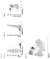
Figures 1 and2 for an exemplary analysis allowing for detection of MRD in BCP-ALL patients. - In another embodiment, there is provided is a multi-color flow cytometric method for detecting minimal residual disease (MRD) in a biological sample comprising lymphocytes, wherein MRD is CLL, comprising the steps of:
- (i) staining the sample with a CLL reagent composition according to the invention, preferably a composition selected from any one of panels 2A, 2B or 2C;
- (ii) subjecting the sample to flow cytometry; (iii) gating B-lymphocytes for expression of the markers detected by the antibodies present in the reagent composition; (iv) distinction between normal and malignant B cells, based on the application of multiple markers, each having added value in the distinction process via the principal component analysis.
- In yet another embodiment, there is provided is a multi-color flow cytometric method for detecting minimal residual disease (MRD) in a biological sample comprising lymphocytes, wherein MRD is MM/PCD, comprising the steps of:
- (i) staining the sample with a MM/PCD reagent composition according to the invention, preferably a composition selected from any one of panels 3A, 3B or 3C;
- (ii) subjecting the sample to flow cytometry; (iii) gating plasma cells for expression of the markers detected by the antibodies present in the reagent composition; (iv) distinction between normal and malignant plasma cells, based on the application of multiple markers, each having added value in the distinction process via the principal component analysis.
-
-
Figure 1 . Typical example of how to use the CD19 and CD45 identification markers in combination with SSC (Panel A to C) for the distinction between BCP cells and other nucleated cells in a bone marrow sample from a BCP-ALL patient during therapy. In each plot, light grey events correspond to non-B cells in the sample, while dark grey events are mature B-cells and black events BCP cells. In the multivariate analysis representation (APS1) ofprincipal component 1 vs. principal component 2 (Panel D), both mature B cells and BCP cells are clearly separated from all other events based on all informative parameters (e.g. CD19, CD45, SSC). -
Figure 2 .- Illustrating example of how to use immunophenotypic characterization markers CD10, CD20, CD34, CD66c/CD123, and CD38 in combination with SSC for the distinction between BCP-ALL cells and normal residual B-cells in a bone marrow sample from a BCP-ALL patient during therapy (Panels A to D). Only bone marrow B-cells are displayed, after gated/selected as described inFigure 1 . In each plot, black dots correspond to BCP-ALL cells in the sample, while grey dots are normal B-cells. Compared to normal B-cells, BCP-ALL cells show overexpression of CD81 (Panel D), CD 10 (Panels A and D), and CD66c/CD123 (Panel C). In the APS1 (principal component 1 versus principal component 2) representation based on all immunophenotypic markers and scatter characteristics evaluated (Panel E), normal residual B-cells (grey) are clearly discriminated from BCP-ALL cells (black). -
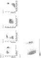
Figure 3 .- Illustrating example of how to use the CD19 and CD3 identification markers in combination with SSC (Panels A to C) for the distinction between mature B-cells and other nucleated cells in a peripheral blood sample from a CLL patient. In each plot, grey events correspond to non-B-cells in the sample, while black events are total peripheral blood B-cells. In the multivariate analysis representation (APS1) ofprincipal component 1 vs. principal component 2 (Panel D), B-cells are clearly separated from all other events based on all informative parameters (e.g. CD19, CD3, SSC). -
Figure 4 .- Illustrating example of how to use immunophenotypic characterization markers CD27, CD5, CD22, CD200 and CD79b (panels A to C) for the distinction between CLL cells and normal mature B-cells in a peripheral blood sample from a CLL patient. Only peripheral blood B-cells are displayed, after gated/selected as described inFigure 3 . In each plot, black dots correspond to CLL cells in the sample, while grey dots are normal peripheral blood B-cells. Compared to normal B-cells, CLL cells show underexpression of CD22 (Panel B) and CD79b (Panel C) together with overexpression of CD200 (Panel B) and CD5 (Panels A and C). In the APS1 (principal component 1 vs. principal component 2) representation based on all immunophenotypic markers and scatter characteristics evaluated (Panel D), CLL cells are clearly discriminated from normal B-cells, while this degree of discrimination could not be achieved based on individual markers. -
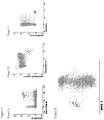
Figure 5 .- Illustrating example of how to use the CD38 and CD 138 identification markers in combination with SSC (Panel A to C) for the distinction between plasma cells and other nucleated cells in a bone marrow sample of an MM patient after therapy. In each plot, grey dots correspond to non-plasma cells in the sample, while black dots are total bone marrow plasma cells. In the multivariate analysis representation (APS1) ofprincipal component 1 vs. principal component 2 (Panel D), plasma cells (black dots) are clearly separated from all other events (grey dots) based on all informative parameters (e.g. CD138, CD38, SSC). -
Figure 6 .- Illustrating example of how to use immunophenotypic characterization markers CD81, CD19, CD45, CD56, CD27, CD117 and CD38, in combination with SSC (Panels A to D) for the distinction between myeloma/malignant plasma cells and normal residual plasma cells in a bone marrow sample of an MM patient after therapy. Only bone marrow plasma cells are displayed, after gated/selected as described inFigure 5 . In each plot, black dots correspond to myeloma/clonal plasma cells in the sample, while grey dots are normal residual bone marrow plasma cells. Compared to normal plasma cells, myeloma/clonal plasma cells show underexpression of CD81, CD19, CD45, CD27 and CD38 together with higher SSC and overexpression of CD56 and CD117. In the APS1 (principal component 1 vs. principal component 2) representation (Panel E) based on all immunophenotypic markers and scatter characteristics evaluated normal residual plasma cells (grey dots) are clearly discriminated from myeloma/malignant plasma cells (black dots), while this degree of discrimination could not be achieved based on individual markers. - The power of the EuroFlow approach disclosed herein is based on the combination of sets of markers and the usage of multivariate analyses for both the identification of normal cells (e.g. normal precursor B-cells, normal B-lymphocytes and normal plasma cells) and the distinction between normal/reactive cells vs. clonal/malignant cells. For this purpose, a powerful multivariate analysis of the contribution of individual markers for inclusion and exclusion of each marker in the panel according to its contribution over all other markers in the combination. Such a strategy was used to evaluate the selected combinations of most discriminating markers in multiple sequential rounds of experimental testing. Because of this the final proposed antibody combinations became extremely strong when used in combination with the principal component analysis, specifically with the automated population separation (APS) tool of the Infinicyt software, so that the added (independent) value of each marker is used in a single step of analysis.
- Herewith we provided the summary of the results of the extensive experimental studies for MRD detection in blood and bone marrow of patients with BCP-ALL (Example 1), CLL (Example 2) and Multiple Myeloma (Example 3).
- In the Examples below, lists of markers are provided together with the most frequent phenotypic aberration of these markers in case of BCP-ALL, CLL, and MM/PCD. However, it should be noted that the real discrimination power between normal and malignant cells is based on combinations of markers in the corresponding n-dimensional space, as is clearly visible in the principle component analyses in the figures of Examples 1 to 3. In fact, minor differences of several markers add up to a larger difference in principle component analysis. Therefore the current invention is not about single marker studies for MRD detection, but about carefully selected sets of markers, that allow excellent discrimination between normal cells and their malignant counterparts, such as normal BCP cells vs. BCP-ALL blasts, normal B-lymphocytes vs. B-CLL cells, and normal plasma cells vs. MM/PCD plasma cells.
- How to use them: Pre-gating using the CD19 marker is essential for identifying a pure B-cell population. To focus on normal B-cell precursors (BCP), CD45-negativity or weak positivity can be used to discriminate BCP from CD45-positive mature B-cells. In case of CD19-directed therapies, CD19 might be replaced by CD22. These markers may be used in combination also with sideward light scatter (SSC) or forward light scatter (FSC) or both FSC and SSC to identify B-cells in peripheral blood or bone marrow or other types of samples (e.g. bone marrow, tissue biopsy, spinal fluid). Of note, other markers, like CD10, CD20, CD38 and CD34, which are used for discriminating BCP-ALL cells from normal BCP cells (see below), may also contribute to the gating of the total BCP cell population (e.g. CD34+, CD10+, CD20-to dim, CD38+).
-
- CD38:
- underexpressed in BCP-ALL/malignant vs. normal B-cell precursor cells
- CD10:
- over- or underexpressed in BCP-ALL/malignant B-cell precursor cells
- CD45:
- underexpressed (usually negative) in BCP-ALL/malignant vs. normal B-cell precursor cells
- CD20:
- under- or overexpressed in BCP-ALL/ malignant vs. normal B-cell precursor cells
- CD81:
- over- or underexpressed in BCP-ALL/ malignant vs. normal B-cell precursor cells
- CD66c:
- overexpressed in BCP-ALL/ malignant vs. normal B-cell precursor cells (particularly BCR-ABL positive ALL; generally negative in TEL-AML1-posiitve or MLL-AF4-positive ALL)
- CD123:
- overexpressed in BCP-ALL/ malignant vs. normal B-cell precursor cells (particularly in hyperdiploid ALL)
- CD304:
- overexpressed in BCP-ALL/ malignant vs. normal B-cell precursor cells
- CD73:
- overexpressed in BCP-ALL/ malignant vs. normal B-cell precursor cells
- CD34:
- under- or overexpressed in BCP-ALL/ malignant vs. normal B-cell precursor cells
- SSC:
- increased or decreased intensity in BCP-ALL/ malignant vs. normal B-cell precursor cells.
- FSC:
- increased or decreased intensity in BCP-ALL/ malignant vs. normal B-cell precursor cells.
- How to use them: Pre-gating using this marker combination is essential for identifying a pure B-cell population, and removing T-cell/B-cell doublets. These markers may be used in combination also with sideward light scatter (SSC) or forward light scatter (FSC) or both FSC and SSC to identify B-cells in peripheral blood or bone marrow or other types of samples (e.g. tissue biopsy, spinal fluid). For a more refined gating with better enrichment of CLL cells, both CD5 and CD27 may be used.
-
- CD27: positive on CLL cells and a small fraction of normal B-cells
- CD5: positive on CLL cells and a small fraction of normal B-cells
- CD79b: underexpressed on CLL cells as compared to normal transitional and mature B-lymphocytes
- CD22: underexpressed on CLL cells as compared to normal transitional and mature B-lymphocytes
- CD20: underexpressed on CLL cells as compared to normal transitional and mature B-lymphocytes
- CD200: overexpressed on CLL cells as compared to normal transitional and mature B-lymphocytes
- ROR1: overexpressed on CLL cells as compared to normal transitional and mature B-lymphocytes
- CD43: overexpressed on CLL cells as compared to normal transitional and mature B-lymphocytes
- CD81: underexpressed on CLL cells as compared to B-cell precursors and both transitional and mature B-lymphocytes
- CD38: underexpressed on CLL cells as compared to B-cell precursors
- How to use them: Any combination of the three markers in any fluorochrome position works; also it is possible to use any combinations of two of the three markers or in a subset of cases (not all) even one of the three markers alone. Preferable combinations are order as follows: 1) CD138/CD38/CD229; 2) CD138/CD38, 3) CD138/CD229; 4) CD38/CD229; 5) CD138; 6) CD38); 7) CD229. Note that any of these markers individually and in combination may be used in combination also with sideward light scatter (SSC) or forward light scatter (FSC) or both FSC and SSC to identify plasma cells in the bone marrow or other types of samples (e.g. peripheral blood, tissue biopsy, spinal fluid).
-
- CD38: underexpressed in malignant plasma cells compared to normal plasma cells
- CD27: underexpressed in malignant plasma cells compared to normal plasma cells
- CD45: underexpressed in malignant plasma cells compared to normal plasma cells
- CD19: underexpressed (usually negative) in malignant plasma cells compared to normal plasma cells
- CD81: underexpressed in malignant plasma cells compared to normal plasma cells
- CD56: overexpressed in malignant plasma cells compared to normal plasma cells
- CD28: overexpressed in malignant plasma cells compared to normal plasma cells
- CD117:overexpressed in malignant plasma cells compared to normal plasma cells
- CyIgk and CyIglambda: expression restricted to either one or the other Ig light chains in malignant plasma cells while showing a balanced distribution (CyIgk/CyIglambda ratio in normal plasma cells usually ranging between
ratios 3 and 0.5). - SSC: increased or decreased intensity in malignant plasma cells compared to normal plasma cells.
- FSC: increased or decreased intensity in malignant plasma cells compared to normal plasma cells.
PCA is a mathematical procedure that uses an orthogonal transformation to convert a set of observations of possibly correlated variables into a set of values of uncorrelated variables called principal components. The number of principal components is less than or equal to the number of original variables. This transformation is defined in such a way that the first principal component has as high a variance as possible (that is, accounts for as much of the variability in the data as possible), and each succeeding component in turn has the highest variance possible under the constraint that it be orthogonal to (uncorrelated with) the preceding components. Principal components are guaranteed to be independent only if the data set is jointly normally distributed. PCA is sensitive to the relative scaling of the original variables. Depending on the field of application, it is also named the discrete Karhunen-Loève transform (KLT), the Hotelling transform or proper orthogonal decomposition (POD). Alternatively to PCA, MDS or any other type of well-established multivariate analysis, can be used (see: Pedreira et al. Trends Biotechnol 2013).
Claims (12)
- A reagent composition for flow cytometric detection of multiple myeloma (MM) or plasma cell disorders (PCD) in a human subject, comprising a panel of at least eight distinct fluorochrome-conjugated antibodies, the panel comprising at least antibodies against the core markers CD138, CD38, CD56 and CD19, supplemented with at least four additional markers selected from the group consisting of CD27, CD 117, CD81, CD229, CD45, CyIgκ and CyIgλ.
- Reagent composition according to claim 1, wherein CD45 is the fifth marker.
- Reagent composition according to claim 2, further comprising antibodies against the markers CD27, CD117 and CD81, or further comprising antibodies against markers CD229, CyIgκ and CyIgλ.
- Reagent composition according to any one of claims 1 to 3, comprising distinct fluorochrome-conjugated antibodies directed against one of the following combinations of markers:
(iv) CD45, CD138, CD38, CD56, CD27,CD19, CD117and CD81 (v) CD45, CD138, CD38, CD56, CD229, CD19, CyIgκ and CyIgλ (vi) CD138, CD27, CD38, CD56, CD45, CD19,CD117 and CD81 (vii) CD138, CD27, CD38, CD56, CD229, CD19, CyIgκ and CyIgλ (viii) CD138, CD27, CD38, CD56, CD45, CD19, CyIgκ and CyIgλ - Reagent composition according to claim 4, comprising distinct fluorochrome-conjugated antibodies directed against the markers CD138, CD27, CD38, CD56, CD45, CD19, CD117, CD81 and one or both set(s) of antibodies selected from(a) set of antibodies against CD229 and CD28;(b) set of antibodies against CyIgκ and CyIgλ.
- Reagent composition according to claim 5, comprising distinct fluorochrome-conjugated antibodies directed against the markers CD138, CD27, CD38, CD56, CD45, CD19, CD117, CD81, CD229, CD28, CyIgκ and CyIgλ.
- A reagent composition for flow cytometric detection of chronic lymphocytic leukemia (CLL) in a human subject, comprising a panel of at least eight distinct fluorochrome-conjugated antibodies, the panel comprising at least antibodies against the core markers CD5, CD27, CD79b, CD3, CD200, CD81 and CD19.
- Reagent composition according to claim 7, wherein CD22 and/or Receptor tyrosine kinase-like orphan receptor 1 (ROR1) is used as additional marker(s), preferably in combination with CD43 and CD38.
- A diagnostic kit for flow cytometric detection of minimal residual disease (MRD), comprising at least one reagent compositions according to claims 1-8, optionally together with instructions for use, buffer, and/or control samples.
- A multi-color flow cytometric method for detecting minimal residual disease (MRD) in a biological sample comprising cells, preferably lymphocytes, comprising the steps of:(i) staining the sample with a reagent composition according to any one of claims 1-8,(ii) subjecting the sample to flow cytometry;(iii) gating on cells for expression of the selected markers detected by the antibodies present in the reagent composition; and(iv) distinguishing between normal and malignant cells, based on the expression profile of the multiple markers.
- Method according to claim 10, wherein step (iv) involves multivariate analysis, preferably principal component analysis (PCA).
- Method according to claim 10 or 11, wherein the biological sample is a peripheral blood sample or a bone marrow sample.
Applications Claiming Priority (2)
| Application Number | Priority Date | Filing Date | Title |
|---|---|---|---|
| US201261659524P | 2012-06-14 | 2012-06-14 | |
| EP13733084.1A EP2861987B1 (en) | 2012-06-14 | 2013-06-14 | Methods, reagents and kits for detecting minimal residual disease |
Related Parent Applications (1)
| Application Number | Title | Priority Date | Filing Date |
|---|---|---|---|
| EP13733084.1A Division EP2861987B1 (en) | 2012-06-14 | 2013-06-14 | Methods, reagents and kits for detecting minimal residual disease |
Publications (3)
| Publication Number | Publication Date |
|---|---|
| EP3064942A2 true EP3064942A2 (en) | 2016-09-07 |
| EP3064942A3 EP3064942A3 (en) | 2016-10-19 |
| EP3064942B1 EP3064942B1 (en) | 2017-12-20 |
Family
ID=48741458
Family Applications (2)
| Application Number | Title | Priority Date | Filing Date |
|---|---|---|---|
| EP16166647.4A Active EP3064942B1 (en) | 2012-06-14 | 2013-06-14 | Methods, reagents and kits for detecting minimal residual disease |
| EP13733084.1A Active EP2861987B1 (en) | 2012-06-14 | 2013-06-14 | Methods, reagents and kits for detecting minimal residual disease |
Family Applications After (1)
| Application Number | Title | Priority Date | Filing Date |
|---|---|---|---|
| EP13733084.1A Active EP2861987B1 (en) | 2012-06-14 | 2013-06-14 | Methods, reagents and kits for detecting minimal residual disease |
Country Status (9)
| Country | Link |
|---|---|
| US (3) | US20150160226A1 (en) |
| EP (2) | EP3064942B1 (en) |
| JP (2) | JP6262218B2 (en) |
| CN (2) | CN104471393B (en) |
| AU (2) | AU2013274965B2 (en) |
| CA (2) | CA2876405C (en) |
| DK (2) | DK3064942T3 (en) |
| ES (1) | ES2660429T3 (en) |
| WO (1) | WO2013187765A2 (en) |
Cited By (1)
| Publication number | Priority date | Publication date | Assignee | Title |
|---|---|---|---|---|
| WO2026017800A1 (en) | 2024-07-19 | 2026-01-22 | Medical University – Plovdiv | Panels and reagent kits for minimal residual disease measurement in pediatric b-cell precursor acute lymphoblastic leukemia via immunophenotyping |
Families Citing this family (25)
| Publication number | Priority date | Publication date | Assignee | Title |
|---|---|---|---|---|
| US20150160226A1 (en) | 2012-06-14 | 2015-06-11 | Erasmus University Medical Center Rotterdam | Method, reagents, and kits for detecting minimal residual disease |
| WO2015160226A1 (en) * | 2014-04-17 | 2015-10-22 | 주식회사 건강사랑 | Female menopause alleviation use of composition containing composite extract of red clover and pomegranate as active ingredient |
| WO2017027391A2 (en) * | 2015-08-07 | 2017-02-16 | Dana-Farber Cancer Institute, Inc. | Genetic abnormalities in plasma cell dyscrasias |
| CN105223360B (en) * | 2015-08-28 | 2017-03-22 | 北京大学人民医院 | Kit for detection and identification of normal plasma cell and clonal plasma cell and application thereof |
| CN106290812A (en) * | 2016-09-08 | 2017-01-04 | 北京海思特临床检验所有限公司 | B cell malignant tumor related antigen expression amount detection kit and detection method |
| JP7084695B2 (en) * | 2017-03-28 | 2022-06-15 | シスメックス株式会社 | Reagent selection support equipment, methods, programs and recording media as well as sample measurement equipment |
| CN108458964A (en) * | 2017-10-31 | 2018-08-28 | 天津协和华美医学诊断技术有限公司 | A kind of antibody compositions and its application in the detection of lymphoma lymphoplasmacytic minimal residual |
| AU2019265019B2 (en) | 2018-05-11 | 2025-11-06 | Beam Therapeutics Inc. | Methods of substituting pathogenic amino acids using programmable base editor systems |
| CN109655616B (en) * | 2018-12-19 | 2022-05-06 | 广州金域医学检验中心有限公司 | Combined reagent and system for detection of acute myeloid leukemia cells |
| CN109752548B (en) * | 2019-02-01 | 2022-05-06 | 广州金域医学检验中心有限公司 | Combined reagent and system for evaluating prognosis of chronic lymphocytic leukemia |
| CN109884313A (en) * | 2019-02-22 | 2019-06-14 | 武汉康圣达医学检验所有限公司 | The detection kit of B-lineage Acute Lymphocyte Leukemia minimal residual |
| EP4110351A4 (en) | 2020-02-25 | 2024-07-03 | Seema Singhal | IMPROVING LIQUID BIOPSY PERFORMANCE |
| CN114487422A (en) * | 2020-10-26 | 2022-05-13 | 苏州云泰生物医药科技有限公司 | Flow cytometry kit for monitoring minimal residual focus of acute B lymphocyte leukemia and monitoring method |
| CN112578117B (en) * | 2021-02-22 | 2021-05-25 | 信纳克(北京)生化标志物检测医学研究有限责任公司 | Antibody composition and its application in screening post-transplantation lymphoproliferative diseases |
| CN112552407B (en) * | 2021-02-24 | 2021-05-25 | 信纳克(北京)生化标志物检测医学研究有限责任公司 | Antibody composition and application thereof in detecting acute B lymphocyte leukemia |
| CN112698037B (en) * | 2021-03-25 | 2021-06-25 | 北京积水潭医院 | An antibody composition for detecting the therapeutic effect of multiple myeloma and its kit and application |
| CN113419066B (en) | 2021-08-23 | 2021-11-02 | 信纳克(北京)生化标志物检测医学研究有限责任公司 | Reagent composition for one-step screening and/or diagnosis of clonal disease and application thereof |
| CN113777327B (en) * | 2021-09-13 | 2022-09-02 | 北京大学人民医院 | Antibody composition for leukemia/lymphoma immunophenotyping primary screening and application thereof |
| CN113933511B (en) * | 2021-09-18 | 2024-03-22 | 广州金域医学检验中心有限公司 | Antibody composition and method for detecting acute B lymphocyte leukemia tiny residue |
| CN115197322B (en) * | 2022-07-21 | 2024-01-30 | 江苏省人民医院(南京医科大学第一附属医院) | Antibody composition for detecting micro residual focus of chronic lymphocytic leukemia and application thereof |
| CN115491423A (en) * | 2022-09-23 | 2022-12-20 | 珠海横琴铂华医学检验有限公司 | Gene combination, kit and application for monitoring MRD of B cell lymphoma |
| CN116087518B (en) * | 2022-11-02 | 2025-01-07 | 北京海思特医学检验实验室有限公司 | An MRD detection product |
| CN115902216B (en) * | 2022-12-22 | 2025-03-18 | 苏州元德维康生物科技有限公司 | Reagent composition for screening and/or diagnosing plasma cell tumors and its application |
| CN115856302B (en) * | 2023-03-02 | 2023-06-02 | 北京大学人民医院 | Antibody composition for mature B cell tumor immunophenotyping and application thereof |
| EP4732014A1 (en) | 2023-10-12 | 2026-04-29 | Stichting EuroFlow | Methods, reagents and kits for detecting minimal/measurable disease in chronic lymphocytic leukemia (cll) |
Family Cites Families (10)
| Publication number | Priority date | Publication date | Assignee | Title |
|---|---|---|---|---|
| ES2051651B1 (en) | 1992-12-10 | 1995-01-01 | Univ Salamanca | PROCEDURE FOR THE SIMULTANEOUS QUANTIFICATION, IN A SINGLE MEASUREMENT, OF THE MAIN TYPES OF HUMAN LYMPHOCYTES AND THEIR SUB-POPULATIONS. |
| US6939670B2 (en) * | 2001-03-12 | 2005-09-06 | Monogen, Inc. | Cell-based detection and differentiation of lung cancer |
| JPWO2003085399A1 (en) * | 2002-04-09 | 2005-08-11 | 協和醗酵工業株式会社 | Determination method and diagnostic agent for leukemia, preleukemia or non-leukemic malignant blood disease |
| US7507548B2 (en) * | 2003-03-04 | 2009-03-24 | University Of Salamanca | Multidimensional detection of aberrant phenotypes in neoplastic cells to be used to monitor minimal disease levels using flow cytometry measurements |
| US7601355B2 (en) * | 2005-06-01 | 2009-10-13 | Northwestern University | Compositions and methods for altering immune function |
| CA2717656A1 (en) * | 2007-02-23 | 2008-08-28 | Baylor Research Institute | Activation of human antigen-presenting cells through clec-6 |
| EP2259065A1 (en) * | 2009-06-03 | 2010-12-08 | Erasmus University Medical Center Rotterdam | Methods, reagents and kits for flow cytometric immunophenotyping |
| US9677125B2 (en) * | 2009-10-21 | 2017-06-13 | General Electric Company | Detection of plurality of targets in biological samples |
| CN102353793B (en) * | 2011-07-07 | 2014-01-29 | 山东轻工业学院 | Binary flow cytometry liquid array detection method for fusion protein in leukemia cells |
| US20150160226A1 (en) | 2012-06-14 | 2015-06-11 | Erasmus University Medical Center Rotterdam | Method, reagents, and kits for detecting minimal residual disease |
-
2013
- 2013-06-14 US US14/407,268 patent/US20150160226A1/en not_active Abandoned
- 2013-06-14 DK DK16166647.4T patent/DK3064942T3/en active
- 2013-06-14 DK DK13733084.1T patent/DK2861987T3/en active
- 2013-06-14 CA CA2876405A patent/CA2876405C/en active Active
- 2013-06-14 JP JP2015517213A patent/JP6262218B2/en active Active
- 2013-06-14 AU AU2013274965A patent/AU2013274965B2/en active Active
- 2013-06-14 CN CN201380037624.4A patent/CN104471393B/en active Active
- 2013-06-14 EP EP16166647.4A patent/EP3064942B1/en active Active
- 2013-06-14 WO PCT/NL2013/050420 patent/WO2013187765A2/en not_active Ceased
- 2013-06-14 CA CA3022975A patent/CA3022975C/en active Active
- 2013-06-14 CN CN201710436056.5A patent/CN107422122B/en active Active
- 2013-06-14 ES ES16166647.4T patent/ES2660429T3/en active Active
- 2013-06-14 EP EP13733084.1A patent/EP2861987B1/en active Active
-
2017
- 2017-11-14 US US15/812,384 patent/US10874710B2/en active Active
- 2017-12-13 JP JP2017238962A patent/JP6524197B2/en active Active
-
2018
- 2018-06-20 AU AU2018204429A patent/AU2018204429B2/en active Active
-
2020
- 2020-10-30 US US17/085,840 patent/US11571457B2/en active Active
Non-Patent Citations (3)
| Title |
|---|
| KALINA ET AL., LEUKEMIA, vol. 26, 2012, pages 1986 - 2010 |
| PEDREIRA ET AL., TRENDS BIOTECHNOL, 2013 |
| VAN DONGEN ET AL., LEUKEMIA, vol. 26, 2012, pages 1908 - 1075 |
Cited By (1)
| Publication number | Priority date | Publication date | Assignee | Title |
|---|---|---|---|---|
| WO2026017800A1 (en) | 2024-07-19 | 2026-01-22 | Medical University – Plovdiv | Panels and reagent kits for minimal residual disease measurement in pediatric b-cell precursor acute lymphoblastic leukemia via immunophenotyping |
Also Published As
| Publication number | Publication date |
|---|---|
| US20180140664A1 (en) | 2018-05-24 |
| WO2013187765A2 (en) | 2013-12-19 |
| US20210177936A1 (en) | 2021-06-17 |
| EP2861987A2 (en) | 2015-04-22 |
| AU2013274965B2 (en) | 2018-07-19 |
| CA2876405A1 (en) | 2013-12-19 |
| CN107422122A (en) | 2017-12-01 |
| JP6524197B2 (en) | 2019-06-05 |
| WO2013187765A3 (en) | 2014-04-24 |
| EP2861987B1 (en) | 2016-04-27 |
| CN104471393A (en) | 2015-03-25 |
| CN107422122B (en) | 2020-06-26 |
| JP6262218B2 (en) | 2018-01-17 |
| AU2013274965A1 (en) | 2015-01-22 |
| AU2018204429B2 (en) | 2020-04-02 |
| CA3022975A1 (en) | 2013-12-19 |
| US20150160226A1 (en) | 2015-06-11 |
| CN104471393B (en) | 2017-07-07 |
| CA3022975C (en) | 2019-10-01 |
| EP3064942A3 (en) | 2016-10-19 |
| DK2861987T3 (en) | 2016-07-04 |
| JP2015521733A (en) | 2015-07-30 |
| DK3064942T3 (en) | 2018-03-05 |
| US10874710B2 (en) | 2020-12-29 |
| US11571457B2 (en) | 2023-02-07 |
| CA2876405C (en) | 2020-01-07 |
| EP3064942B1 (en) | 2017-12-20 |
| ES2660429T3 (en) | 2018-03-22 |
| JP2018066752A (en) | 2018-04-26 |
| AU2018204429A1 (en) | 2018-07-05 |
Similar Documents
| Publication | Publication Date | Title |
|---|---|---|
| US11571457B2 (en) | Methods, reagents and kits for detecting minimal residual disease | |
| US12066441B2 (en) | Methods, reagents and kits for flow cytometric immunophenotyping | |
| Dong et al. | B-cell lymphomas with coexpression of CD5 and CD10 | |
| Dworzak et al. | CD99 (MIC2) expression in paediatric B‐lineage leukaemia/lymphoma reflects maturation‐associated patterns of normal B‐lymphopoiesis | |
| Nguyen et al. | FCM data analysis on heterogeneous specimens |
Legal Events
| Date | Code | Title | Description |
|---|---|---|---|
| PUAI | Public reference made under article 153(3) epc to a published international application that has entered the european phase |
Free format text: ORIGINAL CODE: 0009012 |
|
| 17P | Request for examination filed |
Effective date: 20160422 |
|
| AC | Divisional application: reference to earlier application |
Ref document number: 2861987 Country of ref document: EP Kind code of ref document: P |
|
| AK | Designated contracting states |
Kind code of ref document: A2 Designated state(s): AL AT BE BG CH CY CZ DE DK EE ES FI FR GB GR HR HU IE IS IT LI LT LU LV MC MK MT NL NO PL PT RO RS SE SI SK SM TR |
|
| RIN1 | Information on inventor provided before grant (corrected) |
Inventor name: BOETTCHER, SEBASTIAN Inventor name: ALMEIDA PARRA, JULIA MARIA Inventor name: SZCZEPANSKI, TOMASZ Inventor name: MEJSTRIKOVA, ESTER Inventor name: MONTEIRO DA SILVA LUCIO, PAULO JORGE Inventor name: FLORES MONTERO, JUAN ALEJANDRO Inventor name: RITGEN, MATTHIAS Inventor name: VAN DONGEN, JACOBUS JOHANNES MARIA Inventor name: ORFAO DE MATOS CORREIA E VALE, JOSE ALBERTO Inventor name: VAN DER VELDEN, VINCENT HENRICUS JOHANNES Inventor name: LANGERAK, ANTHONIE WILLEM |
|
| PUAL | Search report despatched |
Free format text: ORIGINAL CODE: 0009013 |
|
| AK | Designated contracting states |
Kind code of ref document: A3 Designated state(s): AL AT BE BG CH CY CZ DE DK EE ES FI FR GB GR HR HU IE IS IT LI LT LU LV MC MK MT NL NO PL PT RO RS SE SI SK SM TR |
|
| RIC1 | Information provided on ipc code assigned before grant |
Ipc: G01N 33/574 20060101ALI20160912BHEP Ipc: G01N 33/50 20060101AFI20160912BHEP |
|
| RIC1 | Information provided on ipc code assigned before grant |
Ipc: G01N 33/574 20060101ALI20170517BHEP Ipc: G01N 33/50 20060101AFI20170517BHEP |
|
| GRAP | Despatch of communication of intention to grant a patent |
Free format text: ORIGINAL CODE: EPIDOSNIGR1 |
|
| INTG | Intention to grant announced |
Effective date: 20170703 |
|
| GRAS | Grant fee paid |
Free format text: ORIGINAL CODE: EPIDOSNIGR3 |
|
| GRAA | (expected) grant |
Free format text: ORIGINAL CODE: 0009210 |
|
| AC | Divisional application: reference to earlier application |
Ref document number: 2861987 Country of ref document: EP Kind code of ref document: P |
|
| AK | Designated contracting states |
Kind code of ref document: B1 Designated state(s): AL AT BE BG CH CY CZ DE DK EE ES FI FR GB GR HR HU IE IS IT LI LT LU LV MC MK MT NL NO PL PT RO RS SE SI SK SM TR |
|
| REG | Reference to a national code |
Ref country code: GB Ref legal event code: FG4D |
|
| REG | Reference to a national code |
Ref country code: CH Ref legal event code: EP |
|
| REG | Reference to a national code |
Ref country code: IE Ref legal event code: FG4D |
|
| REG | Reference to a national code |
Ref country code: AT Ref legal event code: REF Ref document number: 956829 Country of ref document: AT Kind code of ref document: T Effective date: 20180115 |
|
| REG | Reference to a national code |
Ref country code: DE Ref legal event code: R096 Ref document number: 602013031256 Country of ref document: DE |
|
| REG | Reference to a national code |
Ref country code: DK Ref legal event code: T3 Effective date: 20180226 |
|
| REG | Reference to a national code |
Ref country code: NL Ref legal event code: FP |
|
| REG | Reference to a national code |
Ref country code: ES Ref legal event code: FG2A Ref document number: 2660429 Country of ref document: ES Kind code of ref document: T3 Effective date: 20180322 |
|
| PG25 | Lapsed in a contracting state [announced via postgrant information from national office to epo] |
Ref country code: NO Free format text: LAPSE BECAUSE OF FAILURE TO SUBMIT A TRANSLATION OF THE DESCRIPTION OR TO PAY THE FEE WITHIN THE PRESCRIBED TIME-LIMIT Effective date: 20180320 Ref country code: SE Free format text: LAPSE BECAUSE OF FAILURE TO SUBMIT A TRANSLATION OF THE DESCRIPTION OR TO PAY THE FEE WITHIN THE PRESCRIBED TIME-LIMIT Effective date: 20171220 Ref country code: LT Free format text: LAPSE BECAUSE OF FAILURE TO SUBMIT A TRANSLATION OF THE DESCRIPTION OR TO PAY THE FEE WITHIN THE PRESCRIBED TIME-LIMIT Effective date: 20171220 Ref country code: FI Free format text: LAPSE BECAUSE OF FAILURE TO SUBMIT A TRANSLATION OF THE DESCRIPTION OR TO PAY THE FEE WITHIN THE PRESCRIBED TIME-LIMIT Effective date: 20171220 |
|
| REG | Reference to a national code |
Ref country code: LT Ref legal event code: MG4D |
|
| REG | Reference to a national code |
Ref country code: AT Ref legal event code: MK05 Ref document number: 956829 Country of ref document: AT Kind code of ref document: T Effective date: 20171220 |
|
| PG25 | Lapsed in a contracting state [announced via postgrant information from national office to epo] |
Ref country code: BG Free format text: LAPSE BECAUSE OF FAILURE TO SUBMIT A TRANSLATION OF THE DESCRIPTION OR TO PAY THE FEE WITHIN THE PRESCRIBED TIME-LIMIT Effective date: 20180320 Ref country code: LV Free format text: LAPSE BECAUSE OF FAILURE TO SUBMIT A TRANSLATION OF THE DESCRIPTION OR TO PAY THE FEE WITHIN THE PRESCRIBED TIME-LIMIT Effective date: 20171220 Ref country code: GR Free format text: LAPSE BECAUSE OF FAILURE TO SUBMIT A TRANSLATION OF THE DESCRIPTION OR TO PAY THE FEE WITHIN THE PRESCRIBED TIME-LIMIT Effective date: 20180321 Ref country code: RS Free format text: LAPSE BECAUSE OF FAILURE TO SUBMIT A TRANSLATION OF THE DESCRIPTION OR TO PAY THE FEE WITHIN THE PRESCRIBED TIME-LIMIT Effective date: 20171220 Ref country code: HR Free format text: LAPSE BECAUSE OF FAILURE TO SUBMIT A TRANSLATION OF THE DESCRIPTION OR TO PAY THE FEE WITHIN THE PRESCRIBED TIME-LIMIT Effective date: 20171220 |
|
| REG | Reference to a national code |
Ref country code: FR Ref legal event code: PLFP Year of fee payment: 6 |
|
| PG25 | Lapsed in a contracting state [announced via postgrant information from national office to epo] |
Ref country code: SK Free format text: LAPSE BECAUSE OF FAILURE TO SUBMIT A TRANSLATION OF THE DESCRIPTION OR TO PAY THE FEE WITHIN THE PRESCRIBED TIME-LIMIT Effective date: 20171220 Ref country code: EE Free format text: LAPSE BECAUSE OF FAILURE TO SUBMIT A TRANSLATION OF THE DESCRIPTION OR TO PAY THE FEE WITHIN THE PRESCRIBED TIME-LIMIT Effective date: 20171220 Ref country code: CY Free format text: LAPSE BECAUSE OF FAILURE TO SUBMIT A TRANSLATION OF THE DESCRIPTION OR TO PAY THE FEE WITHIN THE PRESCRIBED TIME-LIMIT Effective date: 20171220 |
|
| PGFP | Annual fee paid to national office [announced via postgrant information from national office to epo] |
Ref country code: CZ Payment date: 20180611 Year of fee payment: 6 |
|
| PG25 | Lapsed in a contracting state [announced via postgrant information from national office to epo] |
Ref country code: IS Free format text: LAPSE BECAUSE OF FAILURE TO SUBMIT A TRANSLATION OF THE DESCRIPTION OR TO PAY THE FEE WITHIN THE PRESCRIBED TIME-LIMIT Effective date: 20180420 Ref country code: RO Free format text: LAPSE BECAUSE OF FAILURE TO SUBMIT A TRANSLATION OF THE DESCRIPTION OR TO PAY THE FEE WITHIN THE PRESCRIBED TIME-LIMIT Effective date: 20171220 Ref country code: AT Free format text: LAPSE BECAUSE OF FAILURE TO SUBMIT A TRANSLATION OF THE DESCRIPTION OR TO PAY THE FEE WITHIN THE PRESCRIBED TIME-LIMIT Effective date: 20171220 Ref country code: SM Free format text: LAPSE BECAUSE OF FAILURE TO SUBMIT A TRANSLATION OF THE DESCRIPTION OR TO PAY THE FEE WITHIN THE PRESCRIBED TIME-LIMIT Effective date: 20171220 Ref country code: PL Free format text: LAPSE BECAUSE OF FAILURE TO SUBMIT A TRANSLATION OF THE DESCRIPTION OR TO PAY THE FEE WITHIN THE PRESCRIBED TIME-LIMIT Effective date: 20171220 |
|
| REG | Reference to a national code |
Ref country code: DE Ref legal event code: R097 Ref document number: 602013031256 Country of ref document: DE |
|
| PLBE | No opposition filed within time limit |
Free format text: ORIGINAL CODE: 0009261 |
|
| STAA | Information on the status of an ep patent application or granted ep patent |
Free format text: STATUS: NO OPPOSITION FILED WITHIN TIME LIMIT |
|
| PGFP | Annual fee paid to national office [announced via postgrant information from national office to epo] |
Ref country code: DK Payment date: 20180620 Year of fee payment: 6 |
|
| 26N | No opposition filed |
Effective date: 20180921 |
|
| PG25 | Lapsed in a contracting state [announced via postgrant information from national office to epo] |
Ref country code: SI Free format text: LAPSE BECAUSE OF FAILURE TO SUBMIT A TRANSLATION OF THE DESCRIPTION OR TO PAY THE FEE WITHIN THE PRESCRIBED TIME-LIMIT Effective date: 20171220 |
|
| REG | Reference to a national code |
Ref country code: BE Ref legal event code: MM Effective date: 20180630 |
|
| REG | Reference to a national code |
Ref country code: IE Ref legal event code: MM4A |
|
| PG25 | Lapsed in a contracting state [announced via postgrant information from national office to epo] |
Ref country code: MC Free format text: LAPSE BECAUSE OF FAILURE TO SUBMIT A TRANSLATION OF THE DESCRIPTION OR TO PAY THE FEE WITHIN THE PRESCRIBED TIME-LIMIT Effective date: 20171220 Ref country code: LU Free format text: LAPSE BECAUSE OF NON-PAYMENT OF DUE FEES Effective date: 20180614 |
|
| PG25 | Lapsed in a contracting state [announced via postgrant information from national office to epo] |
Ref country code: IE Free format text: LAPSE BECAUSE OF NON-PAYMENT OF DUE FEES Effective date: 20180614 |
|
| PG25 | Lapsed in a contracting state [announced via postgrant information from national office to epo] |
Ref country code: BE Free format text: LAPSE BECAUSE OF NON-PAYMENT OF DUE FEES Effective date: 20180630 |
|
| REG | Reference to a national code |
Ref country code: DK Ref legal event code: EBP Effective date: 20190630 |
|
| PG25 | Lapsed in a contracting state [announced via postgrant information from national office to epo] |
Ref country code: CZ Free format text: LAPSE BECAUSE OF NON-PAYMENT OF DUE FEES Effective date: 20190614 Ref country code: MT Free format text: LAPSE BECAUSE OF NON-PAYMENT OF DUE FEES Effective date: 20180614 |
|
| PG25 | Lapsed in a contracting state [announced via postgrant information from national office to epo] |
Ref country code: TR Free format text: LAPSE BECAUSE OF FAILURE TO SUBMIT A TRANSLATION OF THE DESCRIPTION OR TO PAY THE FEE WITHIN THE PRESCRIBED TIME-LIMIT Effective date: 20171220 |
|
| PG25 | Lapsed in a contracting state [announced via postgrant information from national office to epo] |
Ref country code: PT Free format text: LAPSE BECAUSE OF FAILURE TO SUBMIT A TRANSLATION OF THE DESCRIPTION OR TO PAY THE FEE WITHIN THE PRESCRIBED TIME-LIMIT Effective date: 20171220 |
|
| PG25 | Lapsed in a contracting state [announced via postgrant information from national office to epo] |
Ref country code: HU Free format text: LAPSE BECAUSE OF FAILURE TO SUBMIT A TRANSLATION OF THE DESCRIPTION OR TO PAY THE FEE WITHIN THE PRESCRIBED TIME-LIMIT; INVALID AB INITIO Effective date: 20130614 Ref country code: MK Free format text: LAPSE BECAUSE OF NON-PAYMENT OF DUE FEES Effective date: 20171220 |
|
| PG25 | Lapsed in a contracting state [announced via postgrant information from national office to epo] |
Ref country code: DK Free format text: LAPSE BECAUSE OF NON-PAYMENT OF DUE FEES Effective date: 20190630 Ref country code: AL Free format text: LAPSE BECAUSE OF FAILURE TO SUBMIT A TRANSLATION OF THE DESCRIPTION OR TO PAY THE FEE WITHIN THE PRESCRIBED TIME-LIMIT Effective date: 20171220 |
|
| P01 | Opt-out of the competence of the unified patent court (upc) registered |
Effective date: 20230530 |
|
| PGFP | Annual fee paid to national office [announced via postgrant information from national office to epo] |
Ref country code: DE Payment date: 20250618 Year of fee payment: 13 |
|
| PGFP | Annual fee paid to national office [announced via postgrant information from national office to epo] |
Ref country code: GB Payment date: 20250618 Year of fee payment: 13 |
|
| PGFP | Annual fee paid to national office [announced via postgrant information from national office to epo] |
Ref country code: NL Payment date: 20250618 Year of fee payment: 13 |
|
| PGFP | Annual fee paid to national office [announced via postgrant information from national office to epo] |
Ref country code: FR Payment date: 20250625 Year of fee payment: 13 |
|
| PGFP | Annual fee paid to national office [announced via postgrant information from national office to epo] |
Ref country code: ES Payment date: 20250728 Year of fee payment: 13 |
|
| PGFP | Annual fee paid to national office [announced via postgrant information from national office to epo] |
Ref country code: IT Payment date: 20250624 Year of fee payment: 13 |
|
| PGFP | Annual fee paid to national office [announced via postgrant information from national office to epo] |
Ref country code: CH Payment date: 20250701 Year of fee payment: 13 |