EP3057591B1 - 4-[5-(3-chloro-phenoxy)-oxazolo[5,4-d]pyrimidin-2-yl]-2,6-dimethyl-phenoxy}-acetic acid for use in the prevention or treatment of acute kidney injury - Google Patents
4-[5-(3-chloro-phenoxy)-oxazolo[5,4-d]pyrimidin-2-yl]-2,6-dimethyl-phenoxy}-acetic acid for use in the prevention or treatment of acute kidney injury Download PDFInfo
- Publication number
- EP3057591B1 EP3057591B1 EP14784452.6A EP14784452A EP3057591B1 EP 3057591 B1 EP3057591 B1 EP 3057591B1 EP 14784452 A EP14784452 A EP 14784452A EP 3057591 B1 EP3057591 B1 EP 3057591B1
- Authority
- EP
- European Patent Office
- Prior art keywords
- compound
- cells
- phenoxy
- baf
- aki
- Prior art date
- Legal status (The legal status is an assumption and is not a legal conclusion. Google has not performed a legal analysis and makes no representation as to the accuracy of the status listed.)
- Active
Links
Images




Classifications
-
- A—HUMAN NECESSITIES
- A61—MEDICAL OR VETERINARY SCIENCE; HYGIENE
- A61K—PREPARATIONS FOR MEDICAL, DENTAL OR TOILETRY PURPOSES
- A61K31/00—Medicinal preparations containing organic active ingredients
- A61K31/33—Heterocyclic compounds
- A61K31/395—Heterocyclic compounds having nitrogen as a ring hetero atom, e.g. guanethidine or rifamycins
- A61K31/495—Heterocyclic compounds having nitrogen as a ring hetero atom, e.g. guanethidine or rifamycins having six-membered rings with two or more nitrogen atoms as the only ring heteroatoms, e.g. piperazine or tetrazines
- A61K31/505—Pyrimidines; Hydrogenated pyrimidines, e.g. trimethoprim
- A61K31/519—Pyrimidines; Hydrogenated pyrimidines, e.g. trimethoprim ortho- or peri-condensed with heterocyclic rings
-
- A—HUMAN NECESSITIES
- A61—MEDICAL OR VETERINARY SCIENCE; HYGIENE
- A61K—PREPARATIONS FOR MEDICAL, DENTAL OR TOILETRY PURPOSES
- A61K47/00—Medicinal preparations characterised by the non-active ingredients used, e.g. carriers or inert additives; Targeting or modifying agents chemically bound to the active ingredient
- A61K47/06—Organic compounds, e.g. natural or synthetic hydrocarbons, polyolefins, mineral oil, petrolatum or ozokerite
- A61K47/26—Carbohydrates, e.g. sugar alcohols, amino sugars, nucleic acids, mono-, di- or oligo-saccharides; Derivatives thereof, e.g. polysorbates, sorbitan fatty acid esters or glycyrrhizin
-
- A—HUMAN NECESSITIES
- A61—MEDICAL OR VETERINARY SCIENCE; HYGIENE
- A61K—PREPARATIONS FOR MEDICAL, DENTAL OR TOILETRY PURPOSES
- A61K47/00—Medicinal preparations characterised by the non-active ingredients used, e.g. carriers or inert additives; Targeting or modifying agents chemically bound to the active ingredient
- A61K47/30—Macromolecular organic or inorganic compounds, e.g. inorganic polyphosphates
- A61K47/36—Polysaccharides; Derivatives thereof, e.g. gums, starch, alginate, dextrin, hyaluronic acid, chitosan, inulin, agar or pectin
- A61K47/38—Cellulose; Derivatives thereof
-
- A—HUMAN NECESSITIES
- A61—MEDICAL OR VETERINARY SCIENCE; HYGIENE
- A61K—PREPARATIONS FOR MEDICAL, DENTAL OR TOILETRY PURPOSES
- A61K9/00—Medicinal preparations characterised by special physical form
- A61K9/0012—Galenical forms characterised by the site of application
- A61K9/0053—Mouth and digestive tract, i.e. intraoral and peroral administration
-
- A—HUMAN NECESSITIES
- A61—MEDICAL OR VETERINARY SCIENCE; HYGIENE
- A61K—PREPARATIONS FOR MEDICAL, DENTAL OR TOILETRY PURPOSES
- A61K9/00—Medicinal preparations characterised by special physical form
- A61K9/10—Dispersions; Emulsions
-
- A—HUMAN NECESSITIES
- A61—MEDICAL OR VETERINARY SCIENCE; HYGIENE
- A61P—SPECIFIC THERAPEUTIC ACTIVITY OF CHEMICAL COMPOUNDS OR MEDICINAL PREPARATIONS
- A61P13/00—Drugs for disorders of the urinary system
- A61P13/12—Drugs for disorders of the urinary system of the kidneys
-
- Y—GENERAL TAGGING OF NEW TECHNOLOGICAL DEVELOPMENTS; GENERAL TAGGING OF CROSS-SECTIONAL TECHNOLOGIES SPANNING OVER SEVERAL SECTIONS OF THE IPC; TECHNICAL SUBJECTS COVERED BY FORMER USPC CROSS-REFERENCE ART COLLECTIONS [XRACs] AND DIGESTS
- Y02—TECHNOLOGIES OR APPLICATIONS FOR MITIGATION OR ADAPTATION AGAINST CLIMATE CHANGE
- Y02P—CLIMATE CHANGE MITIGATION TECHNOLOGIES IN THE PRODUCTION OR PROCESSING OF GOODS
- Y02P20/00—Technologies relating to chemical industry
- Y02P20/50—Improvements relating to the production of bulk chemicals
- Y02P20/582—Recycling of unreacted starting or intermediate materials
Definitions
- the present invention relates to ⁇ 4-[5-(3-chloro-phenoxy)-oxazolo[5,4-d]pyrimidin-2-yl]-2,6-dimethyl-phenoxy ⁇ -acetic acid or a pharmaceutically acceptable salt thereof for use in the prevention or treatment of acute kidney injury (AKI), and a pharmaceutical composition thereof.
- the compound ⁇ 4-[5-(3-chloro-phenoxy)-oxazolo[5,4-d]pyrimidin-2-yl]-2,6-dimethyl-phenoxy ⁇ -acetic acid or a pharmaceutically acceptable salt thereof and a preparative method thereof are described in the patent application WO2011/086079 . This compound has affinity for S1P 1 /EDG1 receptors.
- Acute kidney injury formerly known as "acute renal failure” is a frequent and potentially severe disorder that occurs in a variety of settings with clinical manifestations ranging from transient elevation in serum creatinine (SCr) to anuric definitive renal failure.
- AKI has been traditionally described as an abrupt (within 48 hours) decrease in kidney function as measured by increases in serum creatinine.
- KDIGO Kidney Disease Improving Global Outcomes
- AKI occurs in approximately 7% of all hospitalized patients ( Nash et al., 2002, Am J Kidney Dis 39: 930-936 ) and up to 36-67% of critically ill patients, depending on the definition used. Severe AKI occurs in 4-25% of all Intensive Care Unit (ICU) admissions and about 5% of ICU patients with AKI require renal replacement therapy ( Hoste et al.
- ICU Intensive Care Unit
- Crit Care 10 R73 , Mehta et al., 2004, Kidney Int 66: 1613-1621 ; Uchino et al., 2005, Jama 294: 813-818 ; Uchino et al., 2006, Crit Care Med 34: 1913-1917 ; Ostermann et al., 2007, Crit Care Med 35: 1837-1843 ).
- the most common causes of AKI are sepsis, major surgery, hypovolemia and medications.
- Risk factors for development of AKI include old age (>75 yrs), chronic kidney disease (CKD, eGFR (estimated Glomerular Filtration Rate) ⁇ 60 mL/min/1.73m 2 ), atherosclerotic peripheral vascular disease, cardiac failure, liver disease, diabetes mellitus and nephrotoxic medications.
- CKD chronic kidney disease
- eGFR estimated Glomerular Filtration Rate
- AKI continues to be associated with high morbidity and mortality, particularly in those admitted to the ICU, where in-hospital mortality rates may exceed 50%.
- chronic consequences which include a high risk of developing chronic kidney disease (CKD) and hastened development of end-stage renal disease (ESRD) ( Hsu et al., 2009, Clin J Am Soc Nephrol 4: 891-898 ; Ishani et al., 2009, JASN 20: 223-228 ; Coca et al., 2009, Am J Kidney Dis 53: 961-973 ).
- CKD chronic kidney disease
- ESRD end-stage renal disease
- Morbidity is also associated with increased costs and increased length of hospital stay.
- Sphingosine-1-phosphate is a lipid mediator that binds to five GPCRs (G Protein-Coupled Receptors) termed S1P 1-5 ( Brinkman et al., 2007, Pharmacol Ther 115: 84-105 ). Circulating S1P originates mainly from endothelial cells, platelets and erythrocytes and is found highly bound to plasma proteins including ApoM (Apolipoprotein M) in HDL (High Density Lipoproteins) ( Hammad et al., 2012, J Lipids ; Karuna et al., 2011, Atherosclerosis 219: 855-863 ) and albumin.
- ApoM Polipoprotein M
- HDL High Density Lipoproteins
- S1P 1 is widely expressed, including in endothelial, immune, and renal epithelial cells. S1P 1 regulates many physiological functions including maintenance of endothelial barrier integrity (cytoskeleton rearrangements), cell growth, survival, differentiation, angiogenesis and immune cell trafficking. S1P 1 is highly expressed in the renal medulla ( Zhu et al., 2011, Am J Physiol Renal Physiol 301: F35-F41 ), a region of the kidney where blood flow and oxygen supply are restricted by the tubulo-vascular anatomy specifically designed for urinary concentration. Since cells in this region have high oxygen consumption, the medulla is particularly vulnerable to hypoxic damage.
- S1P 1 is up-regulated following renal ischemia reperfusion injury ( Awad et al., 2006, Am J Physiol Renal Physiol 290: F1516-F1524 ) and its activation is expected to preserve renal function by three main mechanisms:
- S1P-S1P 1 pathway The role of the S1P-S1P 1 pathway in regulating endothelial barrier integrity has been extensively documented ( Wang and Dudek, 2009, Microvasc Res 77: 39-45 ; Mc Verry and Garcia, 2005, Cell signalling 17: 131-139 ; Lucke and Levkau, 2010, Cell Phys Biochem 26: 87-96 ). Activation of S1P 1 improves endothelial barrier function through PLC/Ca 2+ /FAK/pRac pathway activation ( Belvitch and Dudek, 2012, Microvasc Res 83: 22-30 ).
- HDL-ApoM associated S1P was shown to prolong endothelial barrier function through eNOS activation ( Wilkerson et al., 2012, JBC 287: 44645-44653 ; Christoffersen et al., 2011, PNAS 108: 9613-9618 ; Argraves et al., 2008, JBC 283: 25074-25081 ).
- low plasma HDL levels have been associated with a risk of developing AKI in patients undergoing vascular surgery ( Miller GJ and Miller NE., 1975, Lancet 1:16-19 ; Castelli WP, Garrison RJ, Wilson PW, et al., 1986, JAMA; 256:2835-2838 )
- S1P 1 Global or endothelial-specific deletion of S1P 1 induces embryo-lethality due to a massive haemorrhage affecting multiple organs ( Kono et al., 2004, JBC 279: 29367-29373 ). Identical defects are seen in knockout mice with deletion of the enzymes responsible for S1P biosynthesis (sphingosine kinases 1 and 2) ( Mizugishi et al., 2005, Mol Cell Biol 25: 11113-11121 ). Similarly, S1P 1 antagonists also increase vascular permeability.
- cytokines like TNF- ⁇ are released from endothelial cells and up-regulate adhesion molecules such as ICAM-1, VCAM-1 and P/E-selectins which contribute to inflammatory cell infiltration into the tubulo-interstitial parenchyma ( Bonventre et al., 2003, JASN 14: 2199-2210 ; Singbartl et al., 2000, Crit Care Med 28: 2507-2514 ; Sadik et al, 2012, Mol Cell Biochem 359: 73-81 ).
- S1P 1 Stimulation of S1P 1 activates endothelial nitric oxide synthase (eNOS) through Akt phosphorylation, which is a key enzyme regulating local vasorelaxation via the production of NO ( Morales-Ruiz et al., 2001, JBC 276: 19672-19677 ; Igarashi et al, 2000, JBC 275: 32363-32370 ; Igarashi et al, 2008, BBA 1781: 489-495 ). Therefore, S1P induces vasorelaxation in an endothelium and NO-dependent manner ( Roviezzo et al., 2006, FASEB J 20: 340 ).
- eNOS endothelial nitric oxide synthase
- NO has a protective effect on renal function in animal models of I/R-induced AKI ( Garcia-Criado et al, 1998, Transplantation 66: 982-990 ).
- eNOS inhibition reduces renal blood flow by 35%, with coupled decreases in GFR and increased renal vascular resistance ( Cao et al, 2010, Am J Physiol Renal Physiol 299: F1056-F1064 ).
- S1P is regarded as one of the most effective activators of endothelial nitric oxide, which is known to improve local blood flow, but also to limit platelet activation and vascular congestion, particularly in the vulnerable cortico-medullary region. This mechanism could be of importance to preserve renal function during AKI.
- nitric oxide bioavailability is associated with AKI in cardio-pulmonary bypass (CPB) and sepsis patients ( Lema et al., 2009, J Cardio Thorac Vasc Anesth 23:188-194 ; Sadik et al., 2012, Mol Cell Biochem 359: 73-81 ).
- a 786C polymorphism in the human eNOS promoter exhibits reduced transcriptional activity and is associated with renal dysfunction in cardiac surgery patients with cardio-pulmonary bypass ( Popov et al., 2009, Eur J Cardio-Thorac Surgery 36: 651-656 ; Nakayama et al., 1999, Circulation 99: 2864-2870 ).
- the polymorphism frequency is around 50% and might represent a significant sub-population that is not only more at risk of developing AKI but also more responsive to a treatment that restores local NO levels, such as through activation of S1P 1 .
- S1P 1 limits directly apoptosis of proximal tubular epithelial cells by activating pERK and pAkt survival pathways.
- Specific deletion of S1P 1 in proximal tubular epithelial cells worsens renal dysfunction following ischemia-reperfusion injury ( Bajwa et al., 2010, JASN 21: 955-965 ), as well as tubular necrosis, which underline an endogenous S1P tone buffering the severity of reperfusion injury.
- S1P is responsible for the egress of lymphocytes from the lymph node to the blood through S1P 1 activation.
- This S1P 1 activation causes receptor internalization followed by recycling of the receptor back to the cell surface, allowing re-activation.
- S1P 1 functional antagonists or antagonists are believed to cause lymphopenia by either inhibiting the recycling of internalised S1P 1 receptors back to the cell surface or blocking S1P 1 activation, and hence cause dramatic and sustained reductions in cell surface S1P 1 . Consequently, the latter agents show a large and sustained drop in blood lymphocytes as observed preclinically and clinically.
- lymphopenia is a predisposing factor for opportunistic infections, particularly in very sensitive, debilitated patients such as patients at high risk of AKI (old age, cardiac diseases, diabetes and chronic kidney disease).
- AKI old age, cardiac diseases, diabetes and chronic kidney disease.
- S1P 1 agonist which induces limited lymphopenia, or in the best case, non lymphopenia, and has therapeutic utility in the treatment of AKI.
- the compound ⁇ 4-[5-(3-chloro-phenoxy)-oxazolo[5,4-d]pyrimidin-2-yl]-2,6-dimethyl-phenoxy ⁇ -acetic acid differs from other S1P 1 modulators, by the fact that it causes AKI protection in mammals at doses that are non-lymphopenic. It is believed that this property of ⁇ 4-[5-(3-chloro-phenoxy)-oxazolo[5,4-d]pyrimidin-2-yl]-2,6-dimethyl-phenoxy ⁇ -acetic acid might be related to its biased agonism at the S1P 1 receptor, with minimal functional antagonism or desensitization effects.
- the present invention concerns ⁇ 4-[5-(3-chloro-phenoxy)-oxazolo[5,4-d]pyrimidin-2-yl]-2,6-dimethyl-phenoxy ⁇ -acetic acid or a pharmaceutically acceptable salt thereof for use in the prevention or treatment of AKI (acute kidney injury).
- the present invention also concerns ⁇ 4-[5-(3-chloro-phenoxy)-oxazolo[5,4-d]pyrimidin-2-yl]-2,6-dimethyl-phenoxy ⁇ -acetic acid or a pharmaceutically acceptable salt thereof for use as a non lymphopenic agent in the prevention or treatment of AKI.
- Lymphopenia mediated by S1P 1 activation refers to a lymphopenic condition caused by using an S1P 1 modulator. Since S1P is responsible for S1P 1 -mediated egress of lymphocytes from the lymph node to the blood, the latter agents are believed to cause lymphopenia by either inhibiting the recycling of internalised S1P 1 receptors back to the cell surface or blocking S1P 1 activation.
- a first embodiment is ⁇ 4-[5-(3-chloro-phenoxy)-oxazolo[5,4-d]pyrimidin-2-yl]-2,6-dimethyl-phenoxy ⁇ -acetic acid or a pharmaceutically acceptable salt thereof for use in the prevention or treatment of AKI, wherein ⁇ 4-[5-(3-chloro-phenoxy)-oxazolo[5,4-d]pyrimidin-2-yl]-2,6-dimethyl-phenoxy ⁇ -acetic acid is non-lymphopenic at AKI protective doses.
- a second embodiment is ⁇ 4-[5-(3-chloro-phenoxy)-oxazolo[5,4-d]pyrimidin-2-yl]-2,6-dimethyl-phenoxy ⁇ -acetic acid or a pharmaceutically acceptable salt thereof for use in the prevention or treatment of AKI, wherein the protective effects are mediated by S1P 1 activation without inducing lymphopenia.
- Figure 1 Effect of a single or repeated administration of compound A and/or BAF-312 in rat Ischemia-Reperfusion (I/R) model.
- Fischer rats were subjected to sham surgery or to 25 min bilateral renal artery occlusion and plasma creatinine measured following 24h reperfusion.
- Fig 1-A Compound A was evaluated at 0.3, 1 and 3 mg/kg p.o. given 1 hour before occlusion.
- Fig 1-B BAF-312 was evaluated at 3, 10 and 30 mg/kg p.o. given 1 hour before occlusion. The no compound control group was given vehicle 1 hour before occlusion. *** p ⁇ 0.001 I/R-vehicle vs Sham group.
- Kidney samples were collected to evaluate the effect of compound A on cortical albumin content following rat I/R injury. Albumin was measured by Western Blot in cortical area of representative kidneys from sham-operated, I/R-vehicle-treated or I/R-compound A (3 mg/kg p.o.)-treated rats.
- Fig 1-D Compound A (3 mg/kg p.o.) or vehicle were administrated twice a day for 5 days. The last administration was given 1 hour before occlusion. ** p ⁇ 0.01 I/R-vehicle vs Sham group. ### p ⁇ 0.001 compound A-treated vs vehicle group. Bars are mean +/- s.e.m.
- Figure 2 Effect of compound A on rhabdomyolysis-induced AKI injury in mice.
- Compound A 0.3, 1, 3 and 10 mg/kg or vehicle were administered orally 1 hour prior to intramuscular injection (hindlimbs) of glycerol (50% in PBS v/v) at 8 mL/kg in male CD1 mice.
- the sham group received the corresponding volume of PBS.
- Plasma creatinine was evaluated 24h later. ** p ⁇ 0.01 glycerol-vehicle vs Sham group. # p ⁇ 0.05 compound A-treated vs vehicle group. Bars are mean +/- s.e.m.
- Figure 3 Peripheral blood lymphocytes in Fischer rats after single administration ( Fig 3- A) or Sprague-Dawley rats after ( Fig 3-B ) single administration or ( Fig 3-C ) repeated administration.
- Compound A was administered orally either at 1, 3, 10 and 30 mg/kg in Fischer rats or 3 mg/kg in Sprague-Dawley rats.
- compounds were administered for 5 days b.i.d.
- Blood lymphocytes are either expressed as area under curve (AUC) ( Fig 3-A ) or as a percentage of total white blood cells at each time point ( Fig 3-B/C ), and represented as mean +/- s.e.m. ** p ⁇ 0.01 vs baseline.
- Fig 3-D Relationship between AKI protection and lymphopenia in Fischer rats. Note boxes which denote lack of lymphopenia and full AKI protection for compound A at 3 mg/kg, whereas BAF-312 is lymphopenic and shows partial AKI protection.
- Fig 6-A Vascular permeability evaluated using Evan's blue extravasation.
- Fig 6-B Lung weight. Rats were treated for 7 days with compound A (1, 3, 10, 30 and 100 mg/kg p.o.
- Fig 6-C Vascular permeability evaluated using Evan's blue extravasation. * p ⁇ 0.05; ** p ⁇ 0.01; *** p ⁇ 0.001 vs control (no compound) for mice or vehicle (methylcellulose 0.6 % - tween 80 0.5 % in water) for rats. Bars are mean +/- s.e.m.
- FIG. 7 S1P 1 receptor desensitization on human dermal microvascular endothelial cells (HDMEC) using an impedance assay.
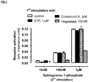
- HDMECs were 1 st stimulated for 1h with either S1P (1 ⁇ M), fingolimod (100 nM) or compound A (1 ⁇ M), followed by a 30 min washout period.
- FIG 8 S1P 1 receptor desensitization on human umbilical vein endothelial cells (HUVEC) using an impedance assay.
- HUVECs were 1 st stimulated for 1 h with either S1P (1 ⁇ M), compound A (1 ⁇ M), fingolimod (100 nM) or SEW2871 (1 ⁇ M), followed by a 30 min washout period.
- FIG. 9 S1P 1 receptor desensitization on CHO cells overexpressing S1P 1 (G-fusion) using a FlipR assay.
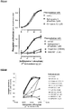
- Fig 9-A The protocol involved a 5 minutes 1 st stimulation with S1P (100 nM), compound A, fingolimod, BAF-312, ponesimod at the indicated concentrations. All 1 st stimulation concentrations were chosen to be > EC 90 . After varying washout periods, receptor desensitization was assessed using a 2 nd stimulation with S1P (100 nM).
- FIG 10 S1P 1 receptor desensitization in human hepatic stellate cells (HSC) using a FlipR assay.
- Fig 10-A The protocol involved a 1 st stimulation with compound A (10 ⁇ M) or BAF-312 (300 nM). All 1 st stimulation concentrations were chosen to be > EC 90 . After either 1 hour or 3 hours washout periods, receptor desensitization was assessed using a 2 nd stimulation with the same compound.
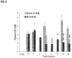
- FIG 11 Time-course of plasma S1P following I/R injury in C57BI6/J mice.
- Plasma S1P levels (Elisa kit from Echelon) were evaluated following sham surgery or after 22 min bilateral renal artery occlusion followed by reperfusion. The control group (CTRL) did not undergo surgery.
- Compound A has been synthesized according to methods described in WO2011/086079 document.
- Recombinant CHO Choinese Hamster Ovary
- human ERG ether- ⁇ -go-go related gene potassium channel
- the medium has been renewed after 24 h and 1 % geneticin (G418, Gibco 10131) has been added to prevent cellular derivation.
- the cells were sown at 1.5x10 6 cells by T25 flask.
- the flasks were placed in 37°C 5% CO2 incubator during 1 hour; after they have been placed at 28 °C in a 5 % CO 2 incubator during 2 days.
- the procedure for cell isolation was carried out after 2 days incubation at 28°C.
- Cells were harvested with Accumax (Sigma; 1:4) or versene (Gibco) and placed in extracellular medium.
- the cells were superfused with the extracellular solution PATCH (NaCl, 138 mM; KCI, 4 mM; CaCl 2 , 1.8 mM; MgCl 2 , 1 mM; glucose, 5.6 mM, HEPES, 10 mM).
- the pH is adjusted to 7.3 with NaOH.
- the osmolarity is adjusted to 285 mOsm.
- the internal solution was prepared as following: KCI, 60 mM; KF, 70 mM; NaCl, 15 mM; HEPES, 5 mM; EGTA(K), 5 mM; pH was adjusted to 7.25 with KOH.
- the osmolarity is adjusted to 290 mOsm.
- the hERG current was activated in response to voltage steps (2s) from a holding potential of -80 mV to a test potential of + 20 mV followed by a repolarization to -100 mV. Voltage steps were applied at 20s intervals. hERG current was measured as the tail deactivating current at - 100 mV. After a 3 minutes period for baseline measurements, compound A effects were measured 4-6 minutes after the change of solution. At the end of each experiment, the superfusion of a specific hERG-blocker, i.e. Risperidone (which exhibits an IC 50 around 0.7 ⁇ M), at a 10 ⁇ M concentration allowed to measure a 100% inhibition of hERG.
- Risperidone which exhibits an IC 50 around 0.7 ⁇ M
- I was the current measured after drug effect
- Irisperidone was the current measured after the effect of the specific hERG-blocker
- Ibaseline was the current measured before the superfusion of drug.
- Compound A was first dissolved into DMSO to obtain a 12 mmol/L stock solution. This solution was further diluted into DMSO and then added into the physiological solution to obtain the appropriate nominal concentrations of 0.3, 1, 3, 10 and 30 ⁇ mol/L (ie, 0.1, 0.5, 1.4, 4.5 and 13.6 ⁇ g/mL of active ingredient, respectively).
- the final concentration of DMSO in the test formulation was kept constant at 0.25 % (v/v) in the physiological solution.
- the low stimulation rate was used to favour the occurrence of abnormal electrical events during the repolarization phase of the action potential and to facilitate the development of Early After Depolarization's (EADs).
- the high stimulation rate was used to evaluate the use-dependent sodium channel blockade. After the highest concentration, the physiological solution was superfused again to evaluate the reversibility of the drug effect (washout).
- Conscious freely moving dogs received an oral or intravenous dose of the negative control article (ie, aqueous solution of 0.5 % (w/w) hydroxyethylcellulose / 0.6 % (w/w) polysorbate 80 for oral route, or aqueous solution of 20 % Captisol, pH 7.5 to 8) followed by compound A at 30 and 100 mg/kg for oral route as suspensions in negative control article, or 10 and 30 mg/kg for i.v.
- the negative control article ie, aqueous solution of 0.5 % (w/w) hydroxyethylcellulose / 0.6 % (w/w) polysorbate 80 for oral route, or aqueous solution of 20 % Captisol, pH 7.5 to 8
- the whole ECG signal was examined beat to beat for any rhythm disturbances and waveform morphology from 30 min to 6 h post dosing. On each treatment day, clinical observations were conducted. A video recording was reviewed to identify potential gross behavioural changes and potential interference with cardiovascular parameters. After dosing, the time-points for analysis were as follows: 0.5, 1, 2, 3, 4, 6, 8, 12, 16 and 24 hours.
- rats were anesthetized using an i.p. injection of pentobarbital (50 mg/kg). Evans blue was injected i.v. in the retro-orbital sinus at 30 mg/kg in 1 mL/kg.
- Conscious freely moving male dogs received an oral dose of the negative control article (ie, aqueous solution of 0.5 % (w/w) hydroxyethylcellulose / 0.6 % (w/w) polysorbate 80) or compound A at 3, 10 and 30 mg/kg or BAF-312 at 10 mg/kg, as suspensions in negative control article, according to a rising dose study design with at least a 7-day washout period between each administration (5 mL/kg).
- Blood was collected at 0.5, 1, 2, 3, 4, 5, 6, 24, 48 and 168h for both compounds (and up to 66 days for BAF-312) for haematology analysis to determine whole blood lymphocytes count. Results are expressed as mean +/- s.e.m.
- the activation of the S1P 1 receptor by the compounds was quantified by their effect on S1P 1 receptor-related calcium liberation in a cell-based calcium fluorescence assay by use of CHO cells in which the human S1P 1 receptor was stably overexpressed (Flp-In system, Invitrogen).
- the overexpressed receptor additionally had a C-terminal sequence of a modified G-protein (G ⁇ i4qi4 ) ( WO 02/04665 ). Changes in intracellular calcium were determined by fluorescence measurement with the calcium-sensitive dye fluo-4 (Invitrogen) in a fluorescence imaging plate reader (FlipR, Molecular Dynamics).
- CHO cells stably overexpressing the human S1P 1 receptor were seeded (40.000 per well) in black clear-bottomed poly-D-lysine-coated 96 well plates (Becton Dickinson, Biocoat cellware) approximately 18-24 h prior to the experiments.
- Cells were grown in an incubator at 37°C, 5 % carbon dioxide and 95% humidity in cell culture media based on F-12 glutamax media (Gibco #31765) supplemented with 1 % (vol/vol) penicilline/streptomycine (PAN, #P06-07100), 10 % (vol/vol) fetal calf serum (FCS; Hyclone Charcoal / Dextran treated FBS #SH30068) and hygromycin B (Invitrogen, #10687-010) 300 mg/L (final concentrations).
- F-12 glutamax media Gibco #31765
- PAN penicilline/streptomycine
- FCS fetal calf serum
- FCS Hyclone Charcoal / Dextran treated FBS #SH30068
- hygromycin B Invitrogen, #10687-010
- fluo-4 AM is cleaved by intracellular esterase resulting in trapping of the dye fluo-4 within the cells.
- Loading was terminated by washing of the cells in a cell washer (Tecan Power washer) three times with the buffer specified afore but without fluo-4 AM and BSA. This latter buffer was also used as the buffer in the subsequent cell fluorescence measurements.
- the dye-loaded and washed cells were then stimulated with respective compounds with various concentrations added as a solution in DMSO (0.3 % vol/vol maximum final concentration of DMSO), or with S1P (100 nM final concentration) in DMSO in the respective concentration only (positive control).
- Compounds which activated the S1P receptor lead to liberation of intracellular calcium from internal stores resulting in a large transient increase of the fluo-4 fluorescence signal which was monitored over approximately 3 min.
- the percent activation caused by a test compound was determined from the maximum fluorescence response as compared to the maximum fluorescence response to the S1P positive control.
- the GPCRProfiler® assay from Millipore was used to determine the EC 50 activity of compound A, BAF-312 and fingolimod on S1P 1 , S1P 2 , S1P 3 , S1P 4 or S1P 5 receptors. These assays used ChemiScreen GPCR stable cell lines.
- Human HSC (Sciencell) were seeded at 25x10 3 cells per well in 96 multiwell plates, 100 ⁇ L/well, and allowed to adhere for 24 hours in complete medium (Sciencell SC5301) with 1 % supplements (Sciencell SC5352) and 2% FCS. Culture medium was replaced by Sciencell Medium with 2% FCS but without supplements for another 24 hour period. Cells were washed and placed in 100 ⁇ L assay buffer (HBSS, 0.8 mM MgSO 4 , 20 mM Hepes, 3.3 mM Na 2 CO 3 , 1 mM CaCl 2 , 10% BSA). Cells were loaded with Fluo4-AM in the presence of pluronic acid for 1 hour at 37°C in the dark.
- HBSS assay buffer
- the PathHunter® ⁇ -Arrestin Assay from DiscoverX was used to determine the EC 50 activity of compound A, BAF-312 and fingolimod on S1P 1 , S1P 2 , S1P 3 , S1P 4 or S1P 5 receptors. The compounds were tested in assay medium containing 0.5% FBS.
- the PathHunter® Activated GPCR Internalization Assay from DiscoverX was used to determine the EC 50 activity of compound A, BAF-312 and fingolimod on the S1P 1 receptor. The compounds were tested in assay medium containing 0.5% FBS.
- the Hit Hunter® cAMP Assay from DiscoverX was used to determine the EC 50 activity of compound A, BAF-312 and fingolimod on S1P 1 .
- the compounds were tested in assay medium containing 0.5% FBS.
- HUVEC Human Umbilical Venous Endothelial Cells
- Cyclic AMP in response to compound A, BAF-312 or fingolimod was assessed in Forskolin-treated HUVEC cells by the mean of the cAMP HTRF® kit (Cisbio). Briefly, HUVEC (Lonza) were seeded at 5x10 3 cells per well into a white 96 multiwell plate (half volume) for 24 hours in EGM2 medium (Lonza). At the beginning of the experiment, EGM2 medium was replaced by assay buffer consisting in HBSS containing Hepes (10 mM), BSA (0.1 %) and IBMX (0.5 mM). Cells were allowed to recover during 15 min.
- Compound A, BAF-312 or fingolimod (10 nM - 100 ⁇ M) were added to the cells for 15 min followed by addition of forskolin (FSK, 10 ⁇ M). After 45 min of FSK treatment, reaction was stopped by addition of MAb anti-cAMP-cryptate and cAMP-D2 for 1 hour. At the end of the incubation, fluorescence was read (Envision, Perkin Elmer) and FRET ratio calculated according to assay kit protocol. The potency of compound A, BAF-312 or fingolimod (EC 50 ) was calculated using the Sanofi BIOST@T-SPEED software according to the logistic equation model.
- RPTEC cells (Lonza) were seeded at 0.3x10 6 cells per well in 6 multiwell plates in 2 mL per well complete REBM/REGM medium (Lonza) for 24 hours. Cells were starved with serum for an additional 24 hour period in 900 ⁇ L quiescent medium. Increasing concentrations of compound A (10X) were added under the volume of 100 ⁇ L in each well for 10 minutes at 37°C. When used, tunicamycin (100 ⁇ L) was added 30 min prior to compound A addition. After medium removal, cells were rinsed with cold PBS and lysed on ice in 100 ⁇ L cold RIPA buffer containing 1 % triton, proteases and phosphatases inhibitors.
- HUVEC cells (Lonza) were seeded at 0.3x10 6 cells per well in 6 multiwell plates in 2 mL per well complete EGM2 medium (Lonza) for 24 hours. Cells were starved with serum for an additional 24 hour period in 900 ⁇ L quiescent medium. Increasing concentrations of compound A, BAF-312 or fingolimod (10X) were added under the volume of 100 ⁇ L in each well for 10 minutes at 37°C. After medium removal, cells were rinsed with cold PBS and cells lysed on ice in 100 ⁇ L RIPA buffer containing 1% triton, proteases and phosphatases inhibitors.
- RPTEC cells (Lonza) were seeded in 96 multiwell white plates at 30x10 3 cells per well in REGM (REBM medium plus 0.5% FCS plus Single quots, Lonza) and allowed to adhere for 24h. After 24h, complete medium was replaced by serum and supplements free medium (56 ⁇ L/well). Cells were pre-treated for 30 min by compound A (final concentrations 0.3-30 ⁇ M, 7 ⁇ L) followed by addition of tunicamycin (TN, final concentration, 0.1 ⁇ g/mL, 7 ⁇ L).
- HUVEC cells were seeded in 96 multiwell plates at 25x10 3 cells per well under the volume of 100 ⁇ L in EGM2 medium (Lonza) and allowed to adhere for 24h. After 24h, complete medium was replaced by quiescent medium (EGM2 without supplements and serum) for 3 hours. Following this, cells were pre-treated with compound A or BAF-312 (1-30 ⁇ M) for 18h. Cells were then treated with TNF- ⁇ (3 ng/mL) in growth-factors free medium for an additional 6h period.
- the Impedance Assay from CEREP was used to determine the EC 50 activity of compound A, BAF-312 and fingolimod on S1P 1 .
- Compound A, BAF-312, fingolimod, SEW2871 and S1P (used as positive control) were concentration-dependent tested on their effect on shape change which is detected by changes in electrical impedance (X-celligence system), to monitor S1P 1 receptor desensitization both on human dermal microvascular endothelial cells (HDMEC) and human umbilical vein endothelial cells (HUVEC).
- HDMEC human dermal microvascular endothelial cells
- HUVEC human umbilical vein endothelial cells
- HDMEC and HUVEC were 1 st stimulated for 1h with either S1P (1 ⁇ M), compound A (1 ⁇ M for HDMEC; 0.1, 1, 10 ⁇ M for HUVEC), BAF-312 (1, 10, 100 nM only in HUVEC), fingolimod (100 nM for HDMEC; 1, 10, 100 nM for HUVEC) or SEW2871 (0.1, 1, 10 ⁇ M, only in HUVEC).
- S1P 1 ⁇ M
- compound A 1 ⁇ M for HDMEC; 0.1, 1, 10 ⁇ M for HUVEC
- BAF-312 1, 10, 100 nM only in HUVEC
- fingolimod 100 nM for HDMEC
- SEW2871 0.1, 1, 10 ⁇ M, only in HUVEC
- compound A was administrated b.i.d. during 5 days to assess potential tachyphylaxia. Briefly, animals were subjected to sham surgery (i.e.
- tissue albumin anti-albumin, Santa Cruz
- tissue HSP70 monoclonal anti-HSP70 antibody from Santa Cruz #SC32239
- Acute tubular necrosis is expressed as percentage of tubules that displayed cell necrosis in 12-15 fields in renal cortico-medullary region.
- CD68 and PECAM immunolabelling are expressed as percentage of positive pixel count (Aperio algorithm) on whole section.
- Plasma creatinine is expressed as mean +/- s.e.m.
- the compound according to the invention namely compound A, was the subject of various in vitro and in vivo experiments in order to demonstrate its activity in AKI settings, and its differentiation over S1P 1 functional antagonists.
- compound A is a S1P 1 agonist that lacks functional antagonism
- potential target-based concerns could be similar to the S1P 1 functional antagonists.
- the greatest clinical experience to date has been generated with the mixed S1P 1/3/4/5 compound, fingolimod, which induces atrio-ventricular block and bradycardia ( Schmouder et al., 2006, J Clin Pharmacol 46: 895-904 ).
- S1P 3 There is much preclinical evidence implicating S1P 3 in these cardio-toxicity findings, hence the field has moved to identification of selective S1P 1 compounds to overcome these limitations.
- a rat model of renal ischemia reperfusion (I/R) injury was used to mimic the renal injury arising after cardiac surgery in patients.
- compound A reduced markedly (85-90%) the severity of AKI, as reflected by limiting the rise of serum creatinine (a clinically-validated biomarker) ( Fig 1A is representative of 5 independent studies). The effect was dose-dependent and statistically significant at 1 and 3 mg/kg p.o. ( figure 1A ). Histological analysis showed that compound A had direct effects on the vasculature by preventing albumin extravasation ( figure 1C ) and preserving capillaries.
- Compound A also protected renal proximal tubules from necrosis, reduced macrophages infiltration and increased renal HSP70 protein (a marker associated with repair following renal ischemic injury). Compound A showed no sign of tachyphylaxis as similar activity was retained following 5 days of repeated BID administration compared to a single administration ( figure 1D ).
- BAF-312 is at least 10-fold more potent in most S1P 1 in in vitro assays (including endothelial assays - see table 1) and has similar plasma/kidney exposure to compound A in rats.
- BAF-312 failed to show more than a 40 % reduction in serum creatinine in the ischemia reperfusion model, even at doses up to 30 mg/kg p.o. ( figure 1B ).
- S1P is responsible for the egress of lymphocytes from the lymph node to the blood through S1P 1 activation.
- This S1P 1 activation causes receptor internalization followed by recycling of the receptor back to the cell surface, allowing re-activation.
- S1P 1 functional antagonists (fingolimod, BAF-312) cause degradation of the internalised S1P 1 and hence cause dramatic and sustained reductions in cell surface S1P 1 . Consequently, S1P 1 functional antagonists show a large and sustained drop in blood lymphocytes as observed preclinically and clinically ( Mandala et al., 2002, Science 296: 346-349 ; Gergely et al., 2012, BJP 167: 1035-1047 ).
- BAF-312 induced a profound (-80%) and sustained lymphopenia in rats ( figure 3A, 3B ). This was evident even at doses as low as 1 mg/kg p.o. (the lowest dose tested).
- the 2 doses that were partially effective in the rat AKI model were higher doses (10 and 30 mg/kg p.o., figure 1B ) and represent highly lymphopenic doses.
- compound A at 1 and 3 mg/kg p.o. showed no lymphocyte reduction, even following 5 days of repeated BID administration (3 mg/kg p.o) in rat ( figure 3C ). Higher doses of compound A revealed a dose-dependent lymphopenia, but these represent higher than required doses for full AKI protection.
- Compound A was non-lymphopenic in dogs at 3 mg/kg p.o. ( figure 5A ). This non-lymphopenic dose in dogs already provides an exposure (Cmax and AUC) that exceeds the exposure required for full AKI protection in rats (3 mg/kg) (table 4). At the higher doses of 10 and 30 mg/kg p.o in dogs compound A induced only a transient lymphopenia ( figure 5A ). In contrast, BAF-312 induced a profound (I max ⁇ 80%) reduction in lymphocytes at 3 mg/kg p.o.. which took at least 60 days to recover ( figure 5B ).
- Compound A is a unique S1P 1 agonist because it demonstrates for the first time a strong AKI protection at non-lymphopenic doses ( figure 3D ).
- S1P 1 compounds such as fingolimod and SEW2871, are also active in AKI models, their effects occur at lymphopenic doses ( Awad et al., 2006, Am J Physiol Renal Physiol 290: F1516-F1524 ; Sanna et al., 2004, JBC 279: 13839-13848 ; Lai et al., 2007, Kidney Int 71: 1223-1231 ).
- compound A acts as a S1P 1 agonist inducing AKI protection at non-lymphopenic doses.
- Compound A differs notably from BAF-312, as described herein, as well as other S1P 1 functional antagonists which are specifically designed to be lymphopenic agents for example for autoimmune diseases.
- S1P 1 functional antagonism is associated with endothelial-damaging effects.
- Macular and lung oedema are prominent adverse events in multiple sclerosis patients treated with fingolimod (Jain and Bhatti, 2012, 78 : 672-680).
- compound A showed no increase in vascular leakage (at 3 and 10 mg/kg (representing doses providing up to the full AKI protective effect in mice) ( figure 6A, 6B ).
- compound A did not induce lung vascular leakage at 1 and 3 mg/kg (representing AKI effective doses) or even one dose higher (10 mg/kg).
- BAF-312 induced vascular leakage in rats at both 3 mg/kg (non-effective AKI dose) as well as 30 mg/kg (partially effective AKI dose).
- Compound A was shown to be a potent S1P 1 agonist.
- EC 50 was 31 nM in a calcium mobilization assay, 656 nM in a ⁇ -arrestin assay, 206 nM in GTP ⁇ S assay and 213 nM in an internalization assay (table 1).
- Table 1 EC 50 values for compound A, BAF-312 and fingolimod using various in vitro assays.
- Chem* and CHO cells were engineered to over-express human S1P 1 .
- HUVECs endogenously express S1P 1 . # ChemiScreen assay. "-" means not determined.
- ⁇ -arrestin assay Cpd A - 800 & 511, BAF-312 - 7.9 & 2.83, S1P - 41 & 20.8; internalisation assay: Cpd A - 160 & 266, BAF-312 - ⁇ 0.5 & 0.67, fingolimod-P - 0.7 & 4.74, S1P - 56 & 40.2.
- Compound A displayed different relative potencies on S1P 1 signalling pathways (table 1) compared to BAF-312 or fingolimod. Since the assay conditions with respect to protein differed between the tests, we excluded the possible influence of protein binding on the relative potencies by comparing the potencies in CHO assays for cAMP, beta arrestin and internalisation using the same media conditions. As shown in table 2, compound A (in relation to BAF-312 and S1P) is more biased on its activation of cAMP compared to internalisation or ⁇ -arrestin. Table 2: Relative EC 50 values of compound A, BAF-312 and S1P (normalized to cAMP assay). Media contained identical protein concentrations (0.5% FBS) in the 3 tests cAMP Internalization ⁇ -Arrestin CHO CHO CHO Compound A 1 9.4 29 BAF-312 1 1.3 11.5 S1P 1 0.62 0.40
- RPTEC human renal tubular epithelial cells
- tunicamycin injury EC 50 ⁇ 10 ⁇ M
- these pro-survival effects were more potent in human endothelial cells after serum starvation, with an EC 50 of 101 nM on P-Akt, and 190 nM on P-ERK 1/2 .
- compound A inhibited tunicamycin-induced apoptosis in RPTEC evaluated by the caspase 3/7 activity with an EC 50 around 3-10 ⁇ M.
- Compound A unlike BAF-312, reduced TNF ⁇ -induced over-expression of adhesion molecules, including ICAM-I, VCAM-I, and P/E selectins, on three human endothelial cell types (HPAEC, HUVEC and HRGEC). These endothelial dysfunction markers are known to be upregulated in plasma samples from AKI patients ( Sadik et al., 2012, Mol Cell Biochem 359:73-81 ), and involved in the infiltration process of inflammatory cells into the tubulo-interstitium space.
- Compound A had an attractive selectivity profile on the other receptors in the S1PR family (table 3), on over 110 targets in a CEREP panel, on over 216 kinases and on over 5 targets in an ion channel panel.
- Compound A is a more selective S1P 1 agonist compared to fingolimod.
- the lack of activity on S1P 2 and S1P 3 is particularly important as these receptors are reported to oppose the function of S1P 1 on endothelial cells.
- Table 3 Selectivity of compound A, BAF-312 and fingolimod against members of the sphingosine receptor family.
- S1P 1 compounds which have a different signalling profile and are i) S1P 1 functional antagonists, ii) endothelial-damaging and iii) show only limited activity in I/R-induced AKI at doses that are lymphopenic.
- Compound A provides the novel opportunity to treat AKI patients without inducing lymphopenia and hence avoiding the corresponding side effects (including infections). Consequently, this compound has the potential to be a transformative therapy in a field where there are currently no drugs available to patients.
Landscapes
- Health & Medical Sciences (AREA)
- Chemical & Material Sciences (AREA)
- Life Sciences & Earth Sciences (AREA)
- Pharmacology & Pharmacy (AREA)
- Veterinary Medicine (AREA)
- Public Health (AREA)
- General Health & Medical Sciences (AREA)
- Medicinal Chemistry (AREA)
- Animal Behavior & Ethology (AREA)
- Epidemiology (AREA)
- General Chemical & Material Sciences (AREA)
- Chemical Kinetics & Catalysis (AREA)
- Engineering & Computer Science (AREA)
- Inorganic Chemistry (AREA)
- Dispersion Chemistry (AREA)
- Nutrition Science (AREA)
- Physiology (AREA)
- Biochemistry (AREA)
- Molecular Biology (AREA)
- Oil, Petroleum & Natural Gas (AREA)
- Urology & Nephrology (AREA)
- Bioinformatics & Cheminformatics (AREA)
- Nuclear Medicine, Radiotherapy & Molecular Imaging (AREA)
- Organic Chemistry (AREA)
- Pharmaceuticals Containing Other Organic And Inorganic Compounds (AREA)
- Nitrogen And Oxygen Or Sulfur-Condensed Heterocyclic Ring Systems (AREA)
Description
- The present invention relates to {4-[5-(3-chloro-phenoxy)-oxazolo[5,4-d]pyrimidin-2-yl]-2,6-dimethyl-phenoxy}-acetic acid or a pharmaceutically acceptable salt thereof for use in the prevention or treatment of acute kidney injury (AKI), and a pharmaceutical composition thereof. The compound {4-[5-(3-chloro-phenoxy)-oxazolo[5,4-d]pyrimidin-2-yl]-2,6-dimethyl-phenoxy}-acetic acid or a pharmaceutically acceptable salt thereof and a preparative method thereof are described in the patent application
WO2011/086079 . This compound has affinity for S1P1/EDG1 receptors. - Acute kidney injury (AKI), formerly known as "acute renal failure" is a frequent and potentially severe disorder that occurs in a variety of settings with clinical manifestations ranging from transient elevation in serum creatinine (SCr) to anuric definitive renal failure. AKI has been traditionally described as an abrupt (within 48 hours) decrease in kidney function as measured by increases in serum creatinine. The recent KDIGO (Kidney Disease Improving Global Outcomes) guidelines established consensus around the diagnostic criteria based on laboratory and clinical values listed below (Working group of ERBP (European Renal Best Practice), 2012, NDT 27: 4263-4272). AKI is defined and staged as any of the following:
- Increase in SCr by ≥0.3 mg/dL (≥26.5 µmol/L) within 48 hours; or
- Increase in SCr to ≥1.5 times baseline, which is known or presumed to have occurred within the prior 7 days; or
- Urine volume < 0.5 mL/kg/h for 6 hours.
- AKI occurs in approximately 7% of all hospitalized patients (Nash et al., 2002, Am J Kidney Dis 39: 930-936) and up to 36-67% of critically ill patients, depending on the definition used. Severe AKI occurs in 4-25% of all Intensive Care Unit (ICU) admissions and about 5% of ICU patients with AKI require renal replacement therapy (Hoste et al. 2006, Crit Care 10: R73, Mehta et al., 2004, Kidney Int 66: 1613-1621; Uchino et al., 2005, Jama 294: 813-818; Uchino et al., 2006, Crit Care Med 34: 1913-1917; Ostermann et al., 2007, Crit Care Med 35: 1837-1843). The most common causes of AKI are sepsis, major surgery, hypovolemia and medications. Risk factors for development of AKI include old age (>75 yrs), chronic kidney disease (CKD, eGFR (estimated Glomerular Filtration Rate) < 60 mL/min/1.73m2), atherosclerotic peripheral vascular disease, cardiac failure, liver disease, diabetes mellitus and nephrotoxic medications.
- Despite advances in preventive strategies and support measures, AKI continues to be associated with high morbidity and mortality, particularly in those admitted to the ICU, where in-hospital mortality rates may exceed 50%. In addition to mortality rates, there are chronic consequences which include a high risk of developing chronic kidney disease (CKD) and hastened development of end-stage renal disease (ESRD) (Hsu et al., 2009, Clin J Am Soc Nephrol 4: 891-898; Ishani et al., 2009, JASN 20: 223-228; Coca et al., 2009, Am J Kidney Dis 53: 961-973). Morbidity is also associated with increased costs and increased length of hospital stay.
- There is a clear unmet medical need since there is no approved therapy for the prevention or for the treatment of AKI whatever the aetiology. Management of the condition is primarily supportive, with Renal Replacement Therapy (RRT) as the central component of care for patients with severe AKI. Hydration remains the most appropriate preventative measure for contrast induced nephropathy (CIN).
- Sphingosine-1-phosphate (S1P) is a lipid mediator that binds to five GPCRs (G Protein-Coupled Receptors) termed S1P1-5 (Brinkman et al., 2007, Pharmacol Ther 115: 84-105). Circulating S1P originates mainly from endothelial cells, platelets and erythrocytes and is found highly bound to plasma proteins including ApoM (Apolipoprotein M) in HDL (High Density Lipoproteins) (Hammad et al., 2012, J Lipids; Karuna et al., 2011, Atherosclerosis 219: 855-863) and albumin. S1P1 is widely expressed, including in endothelial, immune, and renal epithelial cells. S1P1 regulates many physiological functions including maintenance of endothelial barrier integrity (cytoskeleton rearrangements), cell growth, survival, differentiation, angiogenesis and immune cell trafficking. S1P1 is highly expressed in the renal medulla (Zhu et al., 2011, Am J Physiol Renal Physiol 301: F35-F41), a region of the kidney where blood flow and oxygen supply are restricted by the tubulo-vascular anatomy specifically designed for urinary concentration. Since cells in this region have high oxygen consumption, the medulla is particularly vulnerable to hypoxic damage. Cortical tubular damage is a consequence of both medulla injury and direct insults on proximal tubular cells. S1P1 is up-regulated following renal ischemia reperfusion injury (Awad et al., 2006, Am J Physiol Renal Physiol 290: F1516-F1524) and its activation is expected to preserve renal function by three main mechanisms:
- Maintenance of endothelial barrier function. This barrier is damaged in AKI leading to impaired vascular permeability and adhesion properties,
- Limitation of apoptosis of proximal tubular epithelial cells,
- Reduction of inflammatory cell infiltration. Inflammation in AKI is often a consequence of the combined injury to tubular epithelial cells and the endothelial barrier.
- The role of the S1P-S1P1 pathway in regulating endothelial barrier integrity has been extensively documented (Wang and Dudek, 2009, Microvasc Res 77: 39-45; Mc Verry and Garcia, 2005, Cell signalling 17: 131-139; Lucke and Levkau, 2010, Cell Phys Biochem 26: 87-96). Activation of S1P1 improves endothelial barrier function through PLC/Ca2+/FAK/pRac pathway activation (Belvitch and Dudek, 2012, Microvasc Res 83: 22-30). HDL-ApoM associated S1P was shown to prolong endothelial barrier function through eNOS activation (Wilkerson et al., 2012, JBC 287: 44645-44653; Christoffersen et al., 2011, PNAS 108: 9613-9618; Argraves et al., 2008, JBC 283: 25074-25081). In this regard, low plasma HDL levels have been associated with a risk of developing AKI in patients undergoing vascular surgery (Miller GJ and Miller NE., 1975, Lancet 1:16-19 ; Castelli WP, Garrison RJ, Wilson PW, et al., 1986, JAMA; 256:2835-2838)
- Global or endothelial-specific deletion of S1P1 induces embryo-lethality due to a massive haemorrhage affecting multiple organs (Kono et al., 2004, JBC 279: 29367-29373). Identical defects are seen in knockout mice with deletion of the enzymes responsible for S1P biosynthesis (
sphingosine kinases 1 and 2) (Mizugishi et al., 2005, Mol Cell Biol 25: 11113-11121). Similarly, S1P1 antagonists also increase vascular permeability. Despite acute protective effects of S1P1 functional antagonists in AKI models (Bajwa et al., 2010, JASN 21: 955-965; Awad et al., 2006, Am J Physiol Renal Physiol 290: F1516-F1524), chronic treatment with these S1P1 functional antagonists dramatically worsens vascular leak in lung injury models and disrupts endothelial barrier integrity in vitro (Shea et al., 2010, Am J Respir Cell Mol Biol 43: 662-673). Similar endothelial defects are also mimicked in S1P less mice (Camerer et al., 2009, JCI 119: 1871-1879). - In patients and AKI models, cytokines like TNF-α are released from endothelial cells and up-regulate adhesion molecules such as ICAM-1, VCAM-1 and P/E-selectins which contribute to inflammatory cell infiltration into the tubulo-interstitial parenchyma (Bonventre et al., 2003, JASN 14: 2199-2210; Singbartl et al., 2000, Crit Care Med 28: 2507-2514; Sadik et al, 2012, Mol Cell Biochem 359: 73-81). Moreover, in cultured human endothelial cells, several of these endothelial adhesion molecules are up-regulated after hypoxia/re-oxygenation (Lutz et al., 2008, J Mol Med 86: 1329-1339). S1P1 activation down-regulates these adhesion molecules and therefore reduces inflammatory cells infiltration (Lien et al., 2006, Kidney Int 69: 1601-1608). An essential role for S1P in preserving endothelial function in acute kidney injury has been demonstrated using a conditional deletion of the endothelial S1P1 receptor in mice (Ham A., 2013, Kidney Int, Sept).
- Stimulation of S1P1 activates endothelial nitric oxide synthase (eNOS) through Akt phosphorylation, which is a key enzyme regulating local vasorelaxation via the production of NO (Morales-Ruiz et al., 2001, JBC 276: 19672-19677; Igarashi et al, 2000, JBC 275: 32363-32370; Igarashi et al, 2008, BBA 1781: 489-495). Therefore, S1P induces vasorelaxation in an endothelium and NO-dependent manner (Roviezzo et al., 2006, FASEB J 20: 340). NO has a protective effect on renal function in animal models of I/R-induced AKI (Garcia-Criado et al, 1998, Transplantation 66: 982-990). In rats, eNOS inhibition reduces renal blood flow by 35%, with coupled decreases in GFR and increased renal vascular resistance (Cao et al, 2010, Am J Physiol Renal Physiol 299: F1056-F1064). S1P is regarded as one of the most effective activators of endothelial nitric oxide, which is known to improve local blood flow, but also to limit platelet activation and vascular congestion, particularly in the vulnerable cortico-medullary region. This mechanism could be of importance to preserve renal function during AKI. Importantly, reduced nitric oxide bioavailability is associated with AKI in cardio-pulmonary bypass (CPB) and sepsis patients (Lema et al., 2009, J Cardio Thorac Vasc Anesth 23:188-194; Sadik et al., 2012, Mol Cell Biochem 359: 73-81). Interestingly, a 786C polymorphism in the human eNOS promoter exhibits reduced transcriptional activity and is associated with renal dysfunction in cardiac surgery patients with cardio-pulmonary bypass (Popov et al., 2009, Eur J Cardio-Thorac Surgery 36: 651-656; Nakayama et al., 1999, Circulation 99: 2864-2870). The polymorphism frequency is around 50% and might represent a significant sub-population that is not only more at risk of developing AKI but also more responsive to a treatment that restores local NO levels, such as through activation of S1P1.
- Moreover, activation of S1P1 limits directly apoptosis of proximal tubular epithelial cells by activating pERK and pAkt survival pathways. Specific deletion of S1P1 in proximal tubular epithelial cells worsens renal dysfunction following ischemia-reperfusion injury (Bajwa et al., 2010, JASN 21: 955-965), as well as tubular necrosis, which underline an endogenous S1P tone buffering the severity of reperfusion injury. These data underscore the importance of a protective role for the S1P-S1P1 system in AKI, both on endothelial and epithelial axes.
- S1P is responsible for the egress of lymphocytes from the lymph node to the blood through S1P1 activation. This S1P1 activation causes receptor internalization followed by recycling of the receptor back to the cell surface, allowing re-activation. However, S1P1 functional antagonists or antagonists are believed to cause lymphopenia by either inhibiting the recycling of internalised S1P1 receptors back to the cell surface or blocking S1P1 activation, and hence cause dramatic and sustained reductions in cell surface S1P1. Consequently, the latter agents show a large and sustained drop in blood lymphocytes as observed preclinically and clinically.
- Several reports show that T lymphocytes contribute to acute kidney injury. The observation that injury is ameliorated by depletion of T cells and reconstituted with adoptive transfer of CD4+ T cells is evidence that it is dependent on CD4+ T cells (Burne et al., 2001, J. Clin Invest 108: 1283-1290; Ysebaert et al., 2004, Kidney Int 66: 491-496). It is therefore not surprising that S1P1 functional antagonists, including fingolimod and SEW2871, are protective in acute kidney injury models at lymphopenic doses (Lien et al., 2006, Kidney Int 69: 1601-1608).
- However, lymphopenia is a predisposing factor for opportunistic infections, particularly in very sensitive, debilitated patients such as patients at high risk of AKI (old age, cardiac diseases, diabetes and chronic kidney disease).There is consequently a need for a selective S1P1 agonist which induces limited lymphopenia, or in the best case, non lymphopenia, and has therapeutic utility in the treatment of AKI. Surprisingly, the compound {4-[5-(3-chloro-phenoxy)-oxazolo[5,4-d]pyrimidin-2-yl]-2,6-dimethyl-phenoxy}-acetic acid differs from other S1P1 modulators, by the fact that it causes AKI protection in mammals at doses that are non-lymphopenic. It is believed that this property of {4-[5-(3-chloro-phenoxy)-oxazolo[5,4-d]pyrimidin-2-yl]-2,6-dimethyl-phenoxy}-acetic acid might be related to its biased agonism at the S1P1 receptor, with minimal functional antagonism or desensitization effects. This would represent a significant clinical safety advantage for the use of {4-[5-(3-chloro-phenoxy)-oxazolo[5,4-d]pyrimidin-2-yl]-2,6-dimethyl-phenoxy}-acetic acid in the treatment of AKI.
- The present invention concerns {4-[5-(3-chloro-phenoxy)-oxazolo[5,4-d]pyrimidin-2-yl]-2,6-dimethyl-phenoxy}-acetic acid or a pharmaceutically acceptable salt thereof for use in the prevention or treatment of AKI (acute kidney injury). The present invention also concerns {4-[5-(3-chloro-phenoxy)-oxazolo[5,4-d]pyrimidin-2-yl]-2,6-dimethyl-phenoxy}-acetic acid or a pharmaceutically acceptable salt thereof for use as a non lymphopenic agent in the prevention or treatment of AKI.
- For convenience reasons and to facilitate reading, the compound {4-[5-(3-chloro-phenoxy)-oxazolo[5,4-d]pyrimidin-2-yl]-2,6-dimethyl-phenoxy}-acetic acid has been renamed as "compound A" in the chapters of the current application and in the figures.
- For the present invention the following words are to be understood accordingly:
- Selective S1P1 agonist is a compound that is more potent on S1P1 compared to the other 4 S1P receptors, S1P2-5. In addition the compound should have minimal or no activity in a broad range of receptor, enzyme and ion channel assays.
- Lymphopenia is the condition of having an abnormal drop of blood lymphocytes, a white blood cell with important functions in the immune system. In adults, a lymphocyte level below 1,500 cells/microliter is diagnostic (proof of the condition), and in children, a lymphocyte level below 3,000 cells/microliter is diagnostic.
- Lymphopenia mediated by S1P1 activation refers to a lymphopenic condition caused by using an S1P1 modulator. Since S1P is responsible for S1P1-mediated egress of lymphocytes from the lymph node to the blood, the latter agents are believed to cause lymphopenia by either inhibiting the recycling of internalised S1P1 receptors back to the cell surface or blocking S1P1 activation.
- Limited lymphopenia is a transient reduction of blood lymphocytes (absolute lymphocyte count falling outside of the normal range before treatment). Transient refers to a temporal change in lymphocytes that returns to the normal range within 24 h or 48 h.
- Non-lymphopenic or no lymphopenia refers to a lack of transient or sustained reduction of blood lymphocytes (absolute lymphocyte count falling outside of the normal range before treatment). Transient refers to a temporal change in lymphocytes that returns to the normal range within 24h, whereas sustained refers to a reduction that persists beyond 24 h.
- AKI protective dose: a dose of {4-[5-(3-chloro-phenoxy)-oxazolo[5,4-d]pyrimidin-2-yl]-2,6-dimethyl-phenoxy}-acetic acid which provides a significant reduction of plasma creatinine of at least 30%.
- Therapeutically effective amount, as used herein, means an amount of a pharmaceutical compound, such as {4-[5-(3-chloro-phenoxy)-oxazolo[5,4-d]pyrimidin-2-yl]-2,6-dimethyl-phenoxy}-acetic acid that produces an effect on AKI.
- Patient means a human patient.
- Prophylactic: which serves to prevent or protect against an undesired effect and is intended to prevent a medical condition from occurring, especially a disease.
- A first embodiment is {4-[5-(3-chloro-phenoxy)-oxazolo[5,4-d]pyrimidin-2-yl]-2,6-dimethyl-phenoxy}-acetic acid or a pharmaceutically acceptable salt thereof for use in the prevention or treatment of AKI, wherein {4-[5-(3-chloro-phenoxy)-oxazolo[5,4-d]pyrimidin-2-yl]-2,6-dimethyl-phenoxy}-acetic acid is non-lymphopenic at AKI protective doses.
- A second embodiment is {4-[5-(3-chloro-phenoxy)-oxazolo[5,4-d]pyrimidin-2-yl]-2,6-dimethyl-phenoxy}-acetic acid or a pharmaceutically acceptable salt thereof for use in the prevention or treatment of AKI, wherein the protective effects are mediated by S1P1 activation without inducing lymphopenia.
-
- AKI
- Acute Kidney Injury
- ApoM
- Apolipoprotein M
- CIN
- Contrast Induced Nephropathy
- CKD
- Chronic Kidney Disease
- DMSO
- Dimethylsulfoxide
- ERBP
- European Renal Best Practice
- eGFR
- estimated Glomerular Filtration Rate
- eNOS
- endothelial Nitric Oxide Synthase
- ESRD
- End-Stage Renal Disease
- FlipR
- Fluorescence Imaging Plate Reader
- GPCRs
- G Protein-Coupled Receptors
- HDL
- High Density Lipoproteins
- HDMEC
- Human Dermal Microvascular Endothelial Cells
- HSC
- Hepatic Stellate Cells
- ICU
- Intensive Care Unit
- I/R
- Ischemia/Reperfusion
- KDIGO
- Kidney Disease Improving Global Outcomes
- PBS
- Phosphate Buffer Saline
- RRT
- Renal Replacement Therapy
- S1P
- Sphingosine-1-Phosphate
- S1P1
- Sphingosine-1-
Phosphate 1 - SCr
- Serum Creatinine
- s.e.m
- standard error of mean
- The following examples further illustrate the present invention and are not intended to limit the invention. For convenient reasons and to facilitate reading, the compound {4-[5-(3-chloro-phenoxy)-oxazolo[5,4-d]pyrimidin-2-yl]-2,6-dimethyl-phenoxy}-acetic acid has been renamed as "compound A" into the figures and the results.
- Statistical analyses and p values calculation have been performed and are symbolized either by "*" or "#" symbols in the figures, depending on the experimental conditions.
-
Figure 1 : Effect of a single or repeated administration of compound A and/or BAF-312 in rat Ischemia-Reperfusion (I/R) model. Fischer rats were subjected to sham surgery or to 25 min bilateral renal artery occlusion and plasma creatinine measured following 24h reperfusion. (Fig 1-A ) Compound A was evaluated at 0.3, 1 and 3 mg/kg p.o. given 1 hour before occlusion. (Fig 1-B ) BAF-312 was evaluated at 3, 10 and 30 mg/kg p.o. given 1 hour before occlusion. The no compound control group was givenvehicle 1 hour before occlusion. *** p<0.001 I/R-vehicle vs Sham group. # p<0.05; ## p<0.01 compound-treated vs vehicle group. Bars are mean +/s.e.m. (Fig 1-C ) Kidney samples were collected to evaluate the effect of compound A on cortical albumin content following rat I/R injury. Albumin was measured by Western Blot in cortical area of representative kidneys from sham-operated, I/R-vehicle-treated or I/R-compound A (3 mg/kg p.o.)-treated rats. (Fig 1-D ) Compound A (3 mg/kg p.o.) or vehicle were administrated twice a day for 5 days. The last administration was given 1 hour before occlusion. ** p<0.01 I/R-vehicle vs Sham group. ### p<0.001 compound A-treated vs vehicle group. Bars are mean +/- s.e.m. -
Figure 2 : Effect of compound A on rhabdomyolysis-induced AKI injury in mice. Compound A (0.3, 1, 3 and 10 mg/kg) or vehicle were administered orally 1 hour prior to intramuscular injection (hindlimbs) of glycerol (50% in PBS v/v) at 8 mL/kg in male CD1 mice. The sham group received the corresponding volume of PBS. Plasma creatinine was evaluated 24h later. ** p<0.01 glycerol-vehicle vs Sham group. # p<0.05 compound A-treated vs vehicle group. Bars are mean +/- s.e.m. -
Figure 3 : Peripheral blood lymphocytes in Fischer rats after single administration (Fig 3- A) or Sprague-Dawley rats after (Fig 3-B ) single administration or (Fig 3-C ) repeated administration. Compound A was administered orally either at 1, 3, 10 and 30 mg/kg in Fischer rats or 3 mg/kg in Sprague-Dawley rats. BAF-312 was given orally either at 1 and 10 mg/kg in Fischer rats or 3 mg/kg in Sprague-Dawley rats (n=8). In the repeated administration protocol, compounds were administered for 5 days b.i.d. Blood lymphocytes are either expressed as area under curve (AUC) (Fig 3-A ) or as a percentage of total white blood cells at each time point (Fig 3-B/C ), and represented as mean +/- s.e.m. ** p<0.01 vs baseline. (Fig 3-D ) Relationship between AKI protection and lymphopenia in Fischer rats. Note boxes which denote lack of lymphopenia and full AKI protection for compound A at 3 mg/kg, whereas BAF-312 is lymphopenic and shows partial AKI protection. -
Figure 4 : Peripheral blood lymphocytes in C57BI6/J mice. Compound A was administered at 1, 3, 10 and 30 mg/kg p.o. (n=8). Blood lymphocytes at each time point are expressed as a percentage of total white blood cells and represented as mean +/- s.e.m. *** p<0.001 vs baseline. -
Figure 5 : Peripheral blood lymphocytes in beagle dogs. Baseline lymphocyte counts were taken and then dogs were administered a single oral dose of compound A, at 3, 10 and 30 mg/kg (n=8), or BAF-312, at 10 mg/kg (n=4). Animals were monitored for 7 days (Fig 5-A ) and, in the case of BAF-312, for up to 66 days (Fig 5-B ). * p< 0.05; ** p<0.01; *** p<0.001 vs baseline. Bars are mean +/- s.e.m. Note compound A did not induce any significant lymphopenia at any of the time points measured for 3 mg/kg dosing. -
Figure 6 : Lung vascular leak in healthy C57BI6/J mice and Fischer rats. Mice were treated for 7 days with compound A (3 and 10 mg/kg p.o. b.i.d.), BAF-312 (1 and 3 mg/kg p.o. b.i.d.) and fingolimod (0.1 and 1 mg/kg p.o. b.i.d.), n=5-8. (Fig 6-A ) Vascular permeability evaluated using Evan's blue extravasation. (Fig 6-B ) Lung weight. Rats were treated for 7 days with compound A (1, 3, 10, 30 and 100 mg/kg p.o. b.i.d.) and BAF-312 (0.03, 0.1, 0.3, 3 and 30 mg/kg p.o. b.i.d.). (Fig 6-C ) Vascular permeability evaluated using Evan's blue extravasation. * p<0.05; ** p<0.01; *** p<0.001 vs control (no compound) for mice or vehicle (methylcellulose 0.6 % -tween 80 0.5 % in water) for rats. Bars are mean +/- s.e.m. -
Figure 7 : S1P1 receptor desensitization on human dermal microvascular endothelial cells (HDMEC) using an impedance assay. HDMECs were 1st stimulated for 1h with either S1P (1 µM), fingolimod (100 nM) or compound A (1 µM), followed by a 30 min washout period. The effect of the 1st stimulation on receptor desensitization was assessed using a 2nd stimulation of S1P (3 concentrations) and measuring the resulting impedance (n=3 experiments). All 1st stimulation concentrations were > EC90. Bars are mean +/- s.e.m. -
Figure 8 : S1P1 receptor desensitization on human umbilical vein endothelial cells (HUVEC) using an impedance assay. HUVECs were 1st stimulated for 1 h with either S1P (1 µM), compound A (1 µM), fingolimod (100 nM) or SEW2871 (1 µM), followed by a 30 min washout period. The effect of the 1st stimulation on receptor desensitization was assessed using a 2nd stimulation of S1P (4 concentrations) and measuring the resulting impedance (n=3 experiments). All 1st stimulation concentrations were > EC90. Bars are mean +/- s.e.m. -
Figure 9 : S1P1 receptor desensitization on CHO cells overexpressing S1P1 (G-fusion) using a FlipR assay. (Fig 9-A ) The protocol involved a 5minutes 1st stimulation with S1P (100 nM), compound A, fingolimod, BAF-312, ponesimod at the indicated concentrations. All 1st stimulation concentrations were chosen to be > EC90. After varying washout periods, receptor desensitization was assessed using a 2nd stimulation with S1P (100 nM). (Fig 9-B ) The S1P-induced FlipR response, expressed as a percentage of the basal response is displayed for each compound with varying wash-out periods (15 to 60 min, n=4 experiments). Bars are mean +/s.e.m. -
Figure 10 : S1P1 receptor desensitization in human hepatic stellate cells (HSC) using a FlipR assay. (Fig 10-A ) The protocol involved a 1st stimulation with compound A (10 µM) or BAF-312 (300 nM). All 1st stimulation concentrations were chosen to be > EC90. After either 1 hour or 3 hours washout periods, receptor desensitization was assessed using a 2nd stimulation with the same compound. (Fig 10-B ) The compound A or BAF-312 induced FlipR response, expressed as fluorescent units is displayed for each compound with varying wash-out periods (n=3 experiments). Bars are mean +/- s.e.m. -
Figure 11 : Time-course of plasma S1P following I/R injury in C57BI6/J mice. Plasma S1P levels (Elisa kit from Echelon) were evaluated following sham surgery or after 22 min bilateral renal artery occlusion followed by reperfusion. The control group (CTRL) did not undergo surgery. ### p<0.001 vs sham group (time-paired). * p<0.05; ** p<0.001; *** p<0.001 vs control (CTRL). Bars are mean +/- s.e.m. -
- Fingolimod and SEW2871 are commercially available. BAF-312 and ponesimod were synthesized according to published methods:
-
WO04/103306 -
WO05/054215
-
- Compound A has been synthesized according to methods described in
WO2011/086079 document. - All in vivo studies were performed in accordance with the European Community Standards on the Care and Use of Laboratory Animals and approved by the Animal Care and Use Committee of Sanofi Research & Development.
- On the basis of the normality of the distribution and the homogeneity of variance (Levene test), a Student test or Wilcoxon test was used to compare sham/control group to vehicle-treated group in order to assess differences on the evaluated parameter. Then a one-way analysis of variance (ANOVA) or two-way ANOVA was performed, using Everst@t V6 software, to compare vehicle-treated group to compound-treated group(s). This primary analysis was followed by a Dunnett post hoc test when appropriate. In case of non-homogeneity of variance, a Kruskal-Wallis test was applied. Differences between groups were considered significant if p<0.05.
- Recombinant CHO (Chinese Hamster Ovary) cell line expressing the human ERG (ether-α-go-go related gene) potassium channel (Cytomyx Catalogue Number: CYL3002) has been cultured according to the following protocol. When necessary a culture has been restarted from frozen aliquots. The aliquots of frozen cells (1 mL) were rapidly warmed to 37°C and first replated in a 75 cm2 flask (Corning). The volume was then adjusted to 10 mL with the α-Minimum Essential Medium (Gibco 32571-028) containing 10 % fetal calf serum. The medium has been renewed after 24 h and 1 % geneticin (G418, Gibco 10131) has been added to prevent cellular derivation. After two or three days, the cells were sown at 1.5x10⁶ cells by T25 flask. The flasks were placed in 37°
C 5% CO2 incubator during 1 hour; after they have been placed at 28 °C in a 5 % CO2 incubator during 2 days. The procedure for cell isolation was carried out after 2 days incubation at 28°C. Cells were harvested with Accumax (Sigma; 1:4) or versene (Gibco) and placed in extracellular medium. - The cells were superfused with the extracellular solution PATCH (NaCl, 138 mM; KCI, 4 mM; CaCl2, 1.8 mM; MgCl2, 1 mM; glucose, 5.6 mM, HEPES, 10 mM). The pH is adjusted to 7.3 with NaOH. The osmolarity is adjusted to 285 mOsm. The internal solution was prepared as following: KCI, 60 mM; KF, 70 mM; NaCl, 15 mM; HEPES, 5 mM; EGTA(K), 5 mM; pH was adjusted to 7.25 with KOH. The osmolarity is adjusted to 290 mOsm.
- The hERG current was activated in response to voltage steps (2s) from a holding potential of -80 mV to a test potential of + 20 mV followed by a repolarization to -100 mV. Voltage steps were applied at 20s intervals. hERG current was measured as the tail deactivating current at - 100 mV. After a 3 minutes period for baseline measurements, compound A effects were measured 4-6 minutes after the change of solution. At the end of each experiment, the superfusion of a specific hERG-blocker, i.e. Risperidone (which exhibits an IC50 around 0.7 µM), at a 10 µM concentration allowed to measure a 100% inhibition of hERG.
- The currents were analyzed using a database and analysis software DataXpress 1.0 (Axon Instruments/Molecular Devices). The linear rundown of currents was extrapolated from baseline measurement period and the percentage of hERG current inhibition after drug effect has been calculated as follows:

- Where "I" was the current measured after drug effect, "Irisperidone" was the current measured after the effect of the specific hERG-blocker and "Ibaseline" was the current measured before the superfusion of drug.
- The effects of compound A on resting membrane potential and action potential parameters recorded from isolated rabbit Purkinje fibers (male, New Zealand rabbits; 1.3 to 1.5 kg; 9-12 weeks of age) were evaluated through a microelectrode technique. The following parameters were measured: resting potential (RP in mV), action potential amplitude (APA in mV), maximal rate of rise of action potential (Vmax in V/s), action potential duration at 50 and 90% of repolarization (APD50 and APD90 in ms). The fibers were superfused with an oxygenated physiological solution containing (in mmol/L): NaCl, 120; KCI, 4; MgCl2, 1; NaH2PO4, 1.8; NaHCO3, 25; glucose, 11; CaCl2, 1.8; pH = 7.4, at 36±1 °C. Compound A was first dissolved into DMSO to obtain a 12 mmol/L stock solution. This solution was further diluted into DMSO and then added into the physiological solution to obtain the appropriate nominal concentrations of 0.3, 1, 3, 10 and 30 µmol/L (ie, 0.1, 0.5, 1.4, 4.5 and 13.6 µg/mL of active ingredient, respectively). The final concentration of DMSO in the test formulation was kept constant at 0.25 % (v/v) in the physiological solution. Purkinje fibers (n=4) were first superfused by the physiological solution. After a 30-minute control period, test compound was evaluated at increasing concentrations sequentially applied, every 30 minutes. For each tested concentration, the fibers were stimulated at the basal rate of 1 pulse per second (1 Hz). In addition, stimulation rate was decreased from 1 pulse per second (1 Hz) to 1 pulse every 4 seconds (0.25 Hz) for 3 minutes, increased again to 1 pulse per second for 1 minute and finally increased to 3 pulses per second (3 Hz) for 2 additional minutes (between the 19th and the 25th minute), as described below:

- The low stimulation rate was used to favour the occurrence of abnormal electrical events during the repolarization phase of the action potential and to facilitate the development of Early After Depolarization's (EADs). The high stimulation rate was used to evaluate the use-dependent sodium channel blockade. After the highest concentration, the physiological solution was superfused again to evaluate the reversibility of the drug effect (washout).
- The purpose of this study was to assess the potential effect of compound A on the cardiovascular function (blood pressure, heart rate and ECG) in conscious telemetered dogs over a 24-hour period post dosing.
- Conscious freely moving dogs (Marshall Farms, n = 4, 2 males and 2 females, 6.9 to 10.8 kg body weight and 44 to 64-months) received an oral or intravenous dose of the negative control article (ie, aqueous solution of 0.5 % (w/w) hydroxyethylcellulose / 0.6 % (w/w)
polysorbate 80 for oral route, or aqueous solution of 20 % Captisol, pH 7.5 to 8) followed by compound A at 30 and 100 mg/kg for oral route as suspensions in negative control article, or 10 and 30 mg/kg for i.v. route as solution in negative control article, according to a rising dose study design with at least a 4-day washout period between each administration (5 mL/kg for oral route or for 30-min infusion into the jugular vein). Arterial blood pressure and lead II ECG signals (Transmitters from Data Science, USA) were continuously recorded on each treatment day from approximately 2 hours before dosing up to 24 hours after dosing. The following parameters were analyzed at several time points: arterial blood pressure (systolic, diastolic and mean), heart rate, electrocardiographic parameters of RR, PQ (=PR), QT interval durations and QRS complex duration as well as body temperature. QT interval was corrected for heart rate variations according to both Fridericia's and van de Water's formulae. The whole ECG signal was examined beat to beat for any rhythm disturbances and waveform morphology from 30 min to 6 h post dosing. On each treatment day, clinical observations were conducted. A video recording was reviewed to identify potential gross behavioural changes and potential interference with cardiovascular parameters. After dosing, the time-points for analysis were as follows: 0.5, 1, 2, 3, 4, 6, 8, 12, 16 and 24 hours. - Male C57BI6/J mice (n = 5-8, 25-30 g) (Charles River Laboratory, France) were treated, orally and twice a day, for 7 days with compound A at 3 and 10 mg/kg, BAF-312 at 1 and 3 mg/kg, fingolimod at 0.1 and 1 mg/kg or vehicle article (methylcellulose 0.6 % -
tween 80 0.5 % in water) within a volume of administration of 10 mL/kg. The number of mice included in each group was between 5 and 8. After 7 days, mice were anesthetized using an i.p. injection of ketamine (100 mg/kg) and xylasine (10 mg/kg) mixture. Evans blue was injected i.v. in the jugular vein at 15 mg/kg in 1 mL/kg. Ten minutes later, physiological serum was injected by the left ventricle to rinse the body from residual intravascular Evans blue. Lung tissue was collected and weighted and then immersed in a pure formamide solution during 24h to extract tissue Evans blue and therefore evaluate vascular permeability. Evans blue concentration was evaluated at 630 nM using a standard curve and normalized with lung weight. Results are expressed as means +/- s.e.m. - Male Fischer rats (n = 4-8, 280-350 g) (Charles River Laboratory, France) were treated, orally and twice a day, for 7 days with compound A (1, 3, 10, 30 and 100 mg/kg), BAF-312 (0.03, 0.1, 0.3, 3 and 30 mg/kg) or vehicle article (methylcellulose 0.6 % -
tween 80 0.5 % in water) within a volume of administration of 2 mL/kg. After 7 days, rats were anesthetized using an i.p. injection of pentobarbital (50 mg/kg). Evans blue was injected i.v. in the retro-orbital sinus at 30 mg/kg in 1 mL/kg. Fifteen minutes later, physiological serum was injected by the left ventricle to rinse the body from residual intravascular Evans blue. Lung tissue was collected and weighted and then immersed in a pure formamide solution (4 mL/g tissue weight) during 24h to extract tissue Evans blue and therefore evaluate vascular permeability. Evans blue concentration was evaluated at 630 nM using a standard curve. Results are expressed as means +/- s.e.m. - Male Sprague-Dawley rats (n = 8, 250 to 350 g) (Charles River Laboratory, France) received oral administration of compound A, BAF-312 at 3 mg/kg or vehicle article (methylcellulose 0.6 % -
tween 80 0.5 % in water) within a volume of administration of 2 mL/kg. Two protocols were used: single administration or repeated b.i.d. administration during 5 consecutive days. Blood was collected at 2, 6 and 24h following compound administration (last administration for the repeated protocol) for haematology analysis to determine whole blood lymphocyte count. Results are expressed as mean +/- s.e.m. - Male Fischer rats (n = 8, 250 to 350 g) (Charles River Laboratory, France) received a single oral administration of compound A (1, 3, 10 and 30 mg/kg), BAF-312 (1 and 10 mg/kg) or vehicle article (methylcellulose 0.6 % -
tween 80 0.5 % in water) within a volume of administration of 2 mL/kg. Blood was collected at 2, 6 and 24h following compound administration for haematology analysis to determine whole blood lymphocyte count. Results are expressed as mean +/- s.e.m. - Male C57BI6/J mice (n = 8, 25-30 g) (Charles River Laboratory, France) received a single oral administration of compound A at 1, 3, 10 and 30 mg/kg or vehicle article (methylcellulose 0.6 % -
tween 80 0.5 % in water) within a volume of administration of 10 mL/kg. Blood was collected at 2, 6 and 24h following compound administration for haematology analysis to determine whole blood lymphocyte count. Results are expressed as mean +/- s.e.m. - Conscious freely moving male dogs (Marshall Farms, n = 8, 10.5 to 14.3 kg body weight) received an oral dose of the negative control article (ie, aqueous solution of 0.5 % (w/w) hydroxyethylcellulose / 0.6 % (w/w) polysorbate 80) or compound A at 3, 10 and 30 mg/kg or BAF-312 at 10 mg/kg, as suspensions in negative control article, according to a rising dose study design with at least a 7-day washout period between each administration (5 mL/kg). Blood was collected at 0.5, 1, 2, 3, 4, 5, 6, 24, 48 and 168h for both compounds (and up to 66 days for BAF-312) for haematology analysis to determine whole blood lymphocytes count. Results are expressed as mean +/- s.e.m.
- Activities of Compound A, BAF-312 and fingolimod were tested on Flp-In stable transfection with hS1P1-G-fusion construction in CHO cells (proprietary), and selectivity tested over hS1P2- and hS1P3- G-fusion constructions (proprietary). Cell line generation is described in
WO2011/086079 . - The activation of the S1P1 receptor by the compounds was quantified by their effect on S1P1 receptor-related calcium liberation in a cell-based calcium fluorescence assay by use of CHO cells in which the human S1P1 receptor was stably overexpressed (Flp-In system, Invitrogen). In order to enforce G-Protein coupling and to direct signalling towards Ca2+ liberation, the overexpressed receptor additionally had a C-terminal sequence of a modified G-protein (Gαi4qi4) (
WO 02/04665 - CHO cells stably overexpressing the human S1P1 receptor were seeded (40.000 per well) in black clear-bottomed poly-D-lysine-coated 96 well plates (Becton Dickinson, Biocoat cellware) approximately 18-24 h prior to the experiments. Cells were grown in an incubator at 37°C, 5 % carbon dioxide and 95% humidity in cell culture media based on F-12 glutamax media (Gibco #31765) supplemented with 1 % (vol/vol) penicilline/streptomycine (PAN, #P06-07100), 10 % (vol/vol) fetal calf serum (FCS; Hyclone Charcoal / Dextran treated FBS #SH30068) and hygromycin B (Invitrogen, #10687-010) 300 mg/L (final concentrations).
- Prior to the FlipR experiment, cells were loaded with fluo-4 acetoxymethyl ester (fluo-4 AM, Invitrogen, #F14202) for 60 min in an incubator at 37 °C, 5 % carbon dioxide and 95 % humidity in dye-loading buffer consisting of Hanks' Balanced Salt Solution (HBSS; Invitrogen #14065049) supplemented with fluo-4 AM at 2 µM (all data given for final concentration), Pluronic® F-127 0.05% (vol/vol) (Invitrogen, #P-3000MP),
HEPES 20 mM (Gibco #15630), probenecid 2.5 mM (Sigma #P-8761) and bovine serum albumin (BSA) 0.05 % (Sigma # A-6003), adjusted to pH 7.5 with sodium hydroxide. During cell loading, fluo-4 AM is cleaved by intracellular esterase resulting in trapping of the dye fluo-4 within the cells. Loading was terminated by washing of the cells in a cell washer (Tecan Power washer) three times with the buffer specified afore but without fluo-4 AM and BSA. This latter buffer was also used as the buffer in the subsequent cell fluorescence measurements. - The dye-loaded and washed cells were then stimulated with respective compounds with various concentrations added as a solution in DMSO (0.3 % vol/vol maximum final concentration of DMSO), or with S1P (100 nM final concentration) in DMSO in the respective concentration only (positive control). Compounds which activated the S1P receptor lead to liberation of intracellular calcium from internal stores resulting in a large transient increase of the fluo-4 fluorescence signal which was monitored over approximately 3 min. The percent activation caused by a test compound was determined from the maximum fluorescence response as compared to the maximum fluorescence response to the S1P positive control. All fluorescence values were corrected for the baseline fluorescence values obtained with cells which were pre-incubated with DMSO only and were not treated with S1P (baseline control). All measurements were performed in triplicate. From the activation at various concentrations, the EC50 value was calculated.
- The GPCRProfiler® assay from Millipore was used to determine the EC50 activity of compound A, BAF-312 and fingolimod on S1P1, S1P2, S1P3, S1P4 or S1P5 receptors. These assays used ChemiScreen GPCR stable cell lines.
- Human HSC (Sciencell) were seeded at 25x103 cells per well in 96 multiwell plates, 100 µL/well, and allowed to adhere for 24 hours in complete medium (Sciencell SC5301) with 1 % supplements (Sciencell SC5352) and 2% FCS. Culture medium was replaced by Sciencell Medium with 2% FCS but without supplements for another 24 hour period. Cells were washed and placed in 100 µL assay buffer (HBSS, 0.8 mM MgSO4, 20 mM Hepes, 3.3 mM Na2CO3, 1 mM CaCl2, 10% BSA). Cells were loaded with Fluo4-AM in the presence of pluronic acid for 1 hour at 37°C in the dark. Loading medium was removed and replaced by 200 µL/well assay buffer. Cells were allowed to stabilize for 20 min at room temperature in the dark. Culture plate was placed in the FlipR instrument, compound A or BAF-312 were added under a volume of 50 µl (5X solution,
final concentration 10 µM) and calcium fluorescence was continuously recorded for 6 minutes. For desensitization experiments, after the first injection of compound A or BAF-312, culture plate was washed once and allowed to recover for 1 or 3 hours in assay buffer before the second addition of compound A or BAF-312. For control experiments, first injection of compound A or BAF-312 were replaced by assay buffer and the second injection with compound A or BAF-312 were taken as reference. Results are expressed as Fluorescence Units (FU). - The PathHunter® β-Arrestin Assay from DiscoverX was used to determine the EC50 activity of compound A, BAF-312 and fingolimod on S1P1, S1P2, S1P3, S1P4 or S1P5 receptors. The compounds were tested in assay medium containing 0.5% FBS.
- This method is described in
WO2011/086079 . - The PathHunter® Activated GPCR Internalization Assay from DiscoverX was used to determine the EC50 activity of compound A, BAF-312 and fingolimod on the S1P1 receptor. The compounds were tested in assay medium containing 0.5% FBS.
- The Hit Hunter® cAMP Assay from DiscoverX was used to determine the EC50 activity of compound A, BAF-312 and fingolimod on S1P1. The compounds were tested in assay medium containing 0.5% FBS.
- Cyclic AMP in response to compound A, BAF-312 or fingolimod was assessed in Forskolin-treated HUVEC cells by the mean of the cAMP HTRF® kit (Cisbio). Briefly, HUVEC (Lonza) were seeded at 5x10³ cells per well into a white 96 multiwell plate (half volume) for 24 hours in EGM2 medium (Lonza). At the beginning of the experiment, EGM2 medium was replaced by assay buffer consisting in HBSS containing Hepes (10 mM), BSA (0.1 %) and IBMX (0.5 mM). Cells were allowed to recover during 15 min. Compound A, BAF-312 or fingolimod (10 nM - 100µM) were added to the cells for 15 min followed by addition of forskolin (FSK, 10 µM). After 45 min of FSK treatment, reaction was stopped by addition of MAb anti-cAMP-cryptate and cAMP-D2 for 1 hour. At the end of the incubation, fluorescence was read (Envision, Perkin Elmer) and FRET ratio calculated according to assay kit protocol. The potency of compound A, BAF-312 or fingolimod (EC50) was calculated using the Sanofi BIOST@T-SPEED software according to the logistic equation model.
- RPTEC cells (Lonza) were seeded at 0.3x10⁶ cells per well in 6 multiwell plates in 2 mL per well complete REBM/REGM medium (Lonza) for 24 hours. Cells were starved with serum for an additional 24 hour period in 900 µL quiescent medium. Increasing concentrations of compound A (10X) were added under the volume of 100 µL in each well for 10 minutes at 37°C. When used, tunicamycin (100 µL) was added 30 min prior to compound A addition. After medium removal, cells were rinsed with cold PBS and lysed on ice in 100 µL cold RIPA buffer containing 1 % triton, proteases and phosphatases inhibitors. For western blotting, 30 µg of total protein lysate were loaded in 4-12 % bis-tris gels (Invitrogen). Following migration and transfer, nitrocellulose membranes were probed with anti phospho-Akt (Phospho-Ser473, Cst #9271) and anti phospho-Erk1/2 (phopsho-Tyr202/Tyr204, Cst #4377). Tubuline labelling with anti α-Tubuline (Cst #2144) was used for normalization after densitometric analysis of the films.
- HUVEC cells (Lonza) were seeded at 0.3x10⁶ cells per well in 6 multiwell plates in 2 mL per well complete EGM2 medium (Lonza) for 24 hours. Cells were starved with serum for an additional 24 hour period in 900 µL quiescent medium. Increasing concentrations of compound A, BAF-312 or fingolimod (10X) were added under the volume of 100 µL in each well for 10 minutes at 37°C. After medium removal, cells were rinsed with cold PBS and cells lysed on ice in 100 µL RIPA buffer containing 1% triton, proteases and phosphatases inhibitors. For western blotting, 30 µg of total protein lysate were loaded in 4-12% bis-tris gels (Invitrogen). Following migration and transfer, nitrocellulose membranes were probed with anti phospho-Akt (Phospho-Ser473, Cst #2965) and anti phospho-Erk1/2 (Phospho Tyr202/Tyr204, Cst #4377). Tubuline labelling with anti α-Tubuline (Cst #2144) was used for normalization after densitometric analysis of the films.
- The effect of compound A on tunicamycin (TN)-induced apoptosis in RPTEC was measured by the mean of the Caspase-
Glo 3/7 assay kit (Promega). Briefly, RPTEC cells (Lonza) were seeded in 96 multiwell white plates at 30x103 cells per well in REGM (REBM medium plus 0.5% FCS plus Single quots, Lonza) and allowed to adhere for 24h. After 24h, complete medium was replaced by serum and supplements free medium (56 µL/well). Cells were pre-treated for 30 min by compound A (final concentrations 0.3-30 µM, 7 µL) followed by addition of tunicamycin (TN, final concentration, 0.1 µg/mL, 7µL). After 24 hours at 37°C 5% CO2, 70 µL of Caspase-Glo reagent were added and culture plate placed under shaking for 1 hour. Luminescence was recorded by the mean of the Envision reader (Perkin Elmer). Results are expressed as percent inhibition of TN-induced apoptosis. - ICAM-1, VCAM-1 and P/E-selectins expression were measured in HUVEC by ELISA. HUVEC cells were seeded in 96 multiwell plates at 25x103 cells per well under the volume of 100 µL in EGM2 medium (Lonza) and allowed to adhere for 24h. After 24h, complete medium was replaced by quiescent medium (EGM2 without supplements and serum) for 3 hours. Following this, cells were pre-treated with compound A or BAF-312 (1-30 µM) for 18h. Cells were then treated with TNF-α (3 ng/mL) in growth-factors free medium for an additional 6h period. Medium was removed and cells were washed and fixed with 100µL RLC2 solution per well (Alphelys #01-RLC2-RTU30) for 20 min at 4°C. Fixed cells were washed twice with 100µL HBSS before addition of antibodies anti-ICAM-1 (#BBA3, R&D System), anti-VCAM-1 (#BBA5, R&D System) and anti-P/E-Selectins (#BBA1, R&D system) for 1 hour. Following extensive wash, anti-mouse IgG HRP (#NA931, Amersham) was added for 2 hours. Optical density was read at 450 nm (Envision) after washing and addition of HRP substrate (OPD, Sigma #P9187). Results are expressed as percent inhibition of TNF-α -induced adhesion molecule expression.
- The Impedance Assay from CEREP was used to determine the EC50 activity of compound A, BAF-312 and fingolimod on S1P1.
- Compound A, BAF-312, fingolimod, SEW2871 and S1P (used as positive control) were concentration-dependent tested on their effect on shape change which is detected by changes in electrical impedance (X-celligence system), to monitor S1P1 receptor desensitization both on human dermal microvascular endothelial cells (HDMEC) and human umbilical vein endothelial cells (HUVEC). On
day 1, primary cells were seeded into 96-well e-plates pre-coated with collagen-I with 20,000 cells per well. After 24h of adherence and proliferation, HDMEC and HUVEC were 1st stimulated for 1h with either S1P (1 µM), compound A (1 µM for HDMEC; 0.1, 1, 10 µM for HUVEC), BAF-312 (1, 10, 100 nM only in HUVEC), fingolimod (100 nM for HDMEC; 1, 10, 100 nM for HUVEC) or SEW2871 (0.1, 1, 10 µM, only in HUVEC). After incubation with the S1PR agonists, the cells were carefully washed twice with medium followed by a 5.5 h recovery period. Then the effect of the preceding 1st stimulation on receptor desensitization was assessed using a 2nd stimulation of increasing S1P concentrations (0.1, 1 and 10 µM) and measuring the resulting impedance. All measurements were done at least in triplicates. All 1st stimulation concentrations were > EC90 in HDMEC. Results are expressed in arbitrary impedance unit as mean +/- s.e.m. - Male Fischer rats (n = 3-9, 250 to 300 g) (Charles River Laboratory, France) received oral administration of compound A at 0.3, 1 and 3 mg/kg or BAF-312 at 3, 10 and 30 mg/kg or vehicle article (methylcellulose 0.6 % -
tween 80 0.5 % in water) within a volume of administration of 2 mL/kg, one hour before renal ischemia. In a second set of experiment, compound A was administrated b.i.d. during 5 days to assess potential tachyphylaxia. Briefly, animals were subjected to sham surgery (i.e. laparotomy, renal arteries isolation) or to 25 min bilateral renal artery occlusion under pentobarbital (50 mg/kg i.p.) anaesthesia. Body temperature (37 °C-38 °C) and hydration (peritoneal injection of physiological serum) were carefully monitored to standardize the surgery procedure and limit inter-individual variability. At the end of the ischemic period, kidneys were reperfused by removing clamps, and the quality of reperfusion was controlled before suturing muscular and cutaneous plan (animals with poor reperfusion were excluded immediately). Twenty-four hours later, blood and kidneys were collected. Blood was centrifuged (3000 g, 10 min) and heparinised plasma frozen for creatinine evaluation using a biochemical analyser (P400, Horiba, France). One kidney was frozen for western blot analyses for tissue albumin (anti-albumin, Santa Cruz) and tissue HSP70 (monoclonal anti-HSP70 antibody from Santa Cruz #SC32239) evaluation and the second one fixed (10% neutral-buffered formalin) and paraffin-embedded for histological analyses on 5 µm-thick slice (acute tubular necrosis quantification using classical hematoxylin-erythrosin-Saffran, modified Masson's trichrome and periodic acid-Schiff associated blue alcine) and immunohistochemistry analyses (macrophage staining with monoclonal anti-CD68 antibody from Acris #BM4000, and capillary staining with monoclonal ant-PECAM antibody from Santa Cruz #SC1506, using Ventana robot, VMS Inc.). Acute tubular necrosis is expressed as percentage of tubules that displayed cell necrosis in 12-15 fields in renal cortico-medullary region. CD68 and PECAM immunolabelling are expressed as percentage of positive pixel count (Aperio algorithm) on whole section. Plasma creatinine is expressed as mean +/- s.e.m. - Swiss (CD1) male mice (n = 5-15, 13-14 weeks old) (Charles River Laboratory, France) received oral administration of compound A at 0.3, 1, 3 and 10 mg/kg or vehicle article (methylcellulose 0.6 % -
tween 80 0.5 % in water) within a volume of administration of 10 mL/kg, one hour before glycerol injection. Intramuscular injection of glycerol (50% in PBS v/v) or vehicle article (PBS) was performed into hindlimbs (2 injections per leg, gastrocnemius and rectus femoris) at 8 mL/kg under pentobarbital (33 mg/kg) and ketamine (40 mg/kg) anaesthesia. Twenty-four hours later, blood was collected, centrifuged (3000 g, 10 min) and heparinised plasma frozen for creatinine evaluation using a biochemical analyser (P400, Horiba, France). Results are expressed as mean +/- s.e.m. - The compound according to the invention, namely compound A, was the subject of various in vitro and in vivo experiments in order to demonstrate its activity in AKI settings, and its differentiation over S1P1 functional antagonists.
- Although compound A is a S1P1 agonist that lacks functional antagonism, potential target-based concerns could be similar to the S1P1 functional antagonists. The greatest clinical experience to date has been generated with the mixed S1P1/3/4/5 compound, fingolimod, which induces atrio-ventricular block and bradycardia (Schmouder et al., 2006, J Clin Pharmacol 46: 895-904). There is much preclinical evidence implicating S1P3 in these cardio-toxicity findings, hence the field has moved to identification of selective S1P1 compounds to overcome these limitations. However, selective S1P1 compounds, that follow into the clinical phase appear to also induce bradycardia (Gergely et al., 2012, BJP 167: 1035-1047), but their effects on atrio-ventricular block are currently unknown. Thus a number of studies were conducted in addition to the usual studies to evaluate potential cardiotoxicity of compound A. In the hERG assay, the IC50 for compound A was >30 µM. In the rabbit Purkinje fiber assay, there was no significant effect of compound A tested from 0.3 to 10 µM, although a shortening of the action potential was observed at 26 µM.
- No effect on heart rate or atrioventricular block or any other ECG parameters was observed in a dog telemetry study, at 30 or 100 mg/kg p.o. and 10 or 30 mg/kg i.v. (infusion over 30 min). There was no significant change in blood pressure at both doses.
- A rat model of renal ischemia reperfusion (I/R) injury was used to mimic the renal injury arising after cardiac surgery in patients. In this model, compound A reduced markedly (85-90%) the severity of AKI, as reflected by limiting the rise of serum creatinine (a clinically-validated biomarker) (
Fig 1A is representative of 5 independent studies). The effect was dose-dependent and statistically significant at 1 and 3 mg/kg p.o. (figure 1A ). Histological analysis showed that compound A had direct effects on the vasculature by preventing albumin extravasation (figure 1C ) and preserving capillaries. Compound A also protected renal proximal tubules from necrosis, reduced macrophages infiltration and increased renal HSP70 protein (a marker associated with repair following renal ischemic injury). Compound A showed no sign of tachyphylaxis as similar activity was retained following 5 days of repeated BID administration compared to a single administration (figure 1D ). - Rhabdomyolysis is another significant cause of AKI in patients and is reproduced in mice by the intramuscular injection of glycerol. Glycerol induces a progressive muscle injury with release of myoglobin and subsequent renal dysfunction. As observed in the I/R model, compound A prevented markedly (-85 % at 10 mg/kg p.o) and dose-dependently the deterioration of renal function in this model (n=3 independent studies) (
figure 2 ). - The effects of compound A were compared in the renal ischemia reperfusion model with a S1P1-selective functional antagonist, BAF-312. BAF-312 is at least 10-fold more potent in most S1P1 in in vitro assays (including endothelial assays - see table 1) and has similar plasma/kidney exposure to compound A in rats. However, despite the improved potency/exposure properties of BAF-312, it failed to show more than a 40 % reduction in serum creatinine in the ischemia reperfusion model, even at doses up to 30 mg/kg p.o. (
figure 1B ). - S1P is responsible for the egress of lymphocytes from the lymph node to the blood through S1P1 activation. This S1P1 activation causes receptor internalization followed by recycling of the receptor back to the cell surface, allowing re-activation. However, S1P1 functional antagonists (fingolimod, BAF-312) cause degradation of the internalised S1P1 and hence cause dramatic and sustained reductions in cell surface S1P1. Consequently, S1P1 functional antagonists show a large and sustained drop in blood lymphocytes as observed preclinically and clinically (Mandala et al., 2002, Science 296: 346-349; Gergely et al., 2012, BJP 167: 1035-1047).
- As expected, BAF-312 induced a profound (-80%) and sustained lymphopenia in rats (
figure 3A, 3B ). This was evident even at doses as low as 1 mg/kg p.o. (the lowest dose tested). The 2 doses that were partially effective in the rat AKI model were higher doses (10 and 30 mg/kg p.o.,figure 1B ) and represent highly lymphopenic doses. In contrast, compound A at 1 and 3 mg/kg p.o. showed no lymphocyte reduction, even following 5 days of repeated BID administration (3 mg/kg p.o) in rat (figure 3C ). Higher doses of compound A revealed a dose-dependent lymphopenia, but these represent higher than required doses for full AKI protection. - Similarly, no lymphopenic activity of compound A was observed in mice at 3 and 10 mg/kg (
figure 4 ), doses which were active in the glycerol-induced rhabdomyolysis model (figure 2 ). - Compound A was non-lymphopenic in dogs at 3 mg/kg p.o. (
figure 5A ). This non-lymphopenic dose in dogs already provides an exposure (Cmax and AUC) that exceeds the exposure required for full AKI protection in rats (3 mg/kg) (table 4). At the higher doses of 10 and 30 mg/kg p.o in dogs compound A induced only a transient lymphopenia (figure 5A ). In contrast, BAF-312 induced a profound (Imax ∼80%) reduction in lymphocytes at 3 mg/kg p.o.. which took at least 60 days to recover (figure 5B ). - Compound A is a unique S1P1 agonist because it demonstrates for the first time a strong AKI protection at non-lymphopenic doses (
figure 3D ). Although other S1P1 compounds, such as fingolimod and SEW2871, are also active in AKI models, their effects occur at lymphopenic doses (Awad et al., 2006, Am J Physiol Renal Physiol 290: F1516-F1524; Sanna et al., 2004, JBC 279: 13839-13848; Lai et al., 2007, Kidney Int 71: 1223-1231). - These in vivo data support that compound A acts as a S1P1 agonist inducing AKI protection at non-lymphopenic doses. Compound A differs notably from BAF-312, as described herein, as well as other S1P1 functional antagonists which are specifically designed to be lymphopenic agents for example for autoimmune diseases.
- S1P1 functional antagonism is associated with endothelial-damaging effects. Macular and lung oedema are prominent adverse events in multiple sclerosis patients treated with fingolimod (Jain and Bhatti, 2012, 78: 672-680).
- We found that chronic oral administration in healthy mice of fingolimod was able to produce significant vascular leakage (
figure 6A, 6B ), in line with the clinical findings. The selective S1P1 agonist, BAF-312 at 1 and 3 mg/kg b.i.d. induced an even greater vascular leakage in the lung (figure 6A, 6B ). These findings are in agreement with previous reports demonstrating that S1P1 functional antagonists altered endothelial barrier integrity and promoted vascular protein extravasation in the lung (Shea et al., 2010, Am J Respir Cell Mol Biol 43: 662-673). In contrast compound A showed no increase in vascular leakage (at 3 and 10 mg/kg (representing doses providing up to the full AKI protective effect in mice) (figure 6A, 6B ). Similarly in Fisher rats, compound A did not induce lung vascular leakage at 1 and 3 mg/kg (representing AKI effective doses) or even one dose higher (10 mg/kg). In contrast, BAF-312 induced vascular leakage in rats at both 3 mg/kg (non-effective AKI dose) as well as 30 mg/kg (partially effective AKI dose). - Given the observed endothelial-damaging properties of BAF-312 (
figure 6 ), the protective properties of BAF-312 could be counteracted by its endothelial deleterious effects resulting in the partial protective effect observed (figure 1B ). Endothelial damage is a notable feature of multiple S1P1 functional antagonists (Figure 6 and published data). The novel profile of compound A therefore provides the opportunity to protect against acute kidney injury at doses that are endothelial protective rather than damaging. - Compound A was shown to be a potent S1P1 agonist. In CHO cells over-expressing human S1P1 (G-fusion), EC50 was 31 nM in a calcium mobilization assay, 656 nM in a β-arrestin assay, 206 nM in GTPγS assay and 213 nM in an internalization assay (table 1).
Table 1: EC50 values for compound A, BAF-312 and fingolimod using various in vitro assays. Chem* and CHO cells were engineered to over-express human S1P1. HUVECs endogenously express S1P1. # ChemiScreen assay. "-" means not determined. Assay Cell type Proteins in assay media EC50 in nM Compound A BAF-312 Fingolimod phosphate S1P SEW2871 FLIPR Chem# 0% 86 29 26 9.6 - CHO 0% 31 2.8 0.14 7.5 24 GTPγS CHO 0% 206 6.7 5 120 795 β-arrestin CHO 0.5% FBS 656* 5.4* 7 30.9* - Internalization CHO 0.5% FBS 213* < 0.59* 2.72* 48.1* - cAMP CHO 0.5% FBS 22.6 0.47 - 78 - cAMP HUVEC 0.1% BSA 15,600 74 132 - - pERK1/2 HUVEC 0.1% FBS 190 4.9 9.4 - - pAkt HUVEC 0.1% FBS 101 4.3 14.5 - - Impedance CHO 10% FBS +0.1% BSA 65 0.4 - 1.5 - Impedance HUVEC 2% FBS 172 0.21 0.12 - 54 * Values are means of 2 separate experiments. β-arrestin assay: Cpd A - 800 & 511, BAF-312 - 7.9 & 2.83, S1P - 41 & 20.8; internalisation assay: Cpd A - 160 & 266, BAF-312 - <0.5 & 0.67, fingolimod-P - 0.7 & 4.74, S1P - 56 & 40.2. - Compound A displayed no functional antagonism properties nor induced receptor desensitization (
figures 7-10 ). This differentiation was demonstrated in four independent in vitro systems, two systems using impedance measurement in endothelial cells HDMEC (human dermal microvascular endothelial cells) and HUVEC (human umbilical vein endothelial cells), and two systems using FlipR assay in CHO cells and HSC cells (hepatic stellate cells). Pre-incubation with compound A (followed by a wash step) allowed an almost complete 2nd stimulation with S1P (figures 7-9 ) or compound A (figure 10 ). However, pre-incubation with the functional antagonists prevented a full 2nd stimulation with S1P (figures 7-9 ) or BAF-312 (figure 10 ). In the HUVEC impedance assay, the concentration in that each compound began desensitizing the S1P1-induced impedance response was compared to its absolute EC50 in the assay. BAF-312 began desensitization at 5x the EC50, SEW2871 at 20x the EC50, while no desensitization was observed for compound A up to 60x the EC50. - The data in
figures 7-10 indicate that the functional antagonists (fingolimod, BAF-312, SEW2871 and ponesimod) induced sustained receptor desensitization. - Compound A displayed different relative potencies on S1P1 signalling pathways (table 1) compared to BAF-312 or fingolimod. Since the assay conditions with respect to protein differed between the tests, we excluded the possible influence of protein binding on the relative potencies by comparing the potencies in CHO assays for cAMP, beta arrestin and internalisation using the same media conditions. As shown in table 2, compound A (in relation to BAF-312 and S1P) is more biased on its activation of cAMP compared to internalisation or β-arrestin.
Table 2: Relative EC50 values of compound A, BAF-312 and S1P (normalized to cAMP assay). Media contained identical protein concentrations (0.5% FBS) in the 3 tests cAMP Internalization β-Arrestin CHO CHO CHO Compound A 1 9.4 29 BAF-312 1 1.3 11.5 S1P 1 0.62 0.40 - Compound A induced survival markers, pERK and pAKt, in human renal tubular epithelial cells (RPTEC) with an EC50 around 3 µM under basal conditions, or after tunicamycin injury (EC50 ∼ 10 µM) in a concentration-dependent manner. These pro-survival effects were more potent in human endothelial cells after serum starvation, with an EC50 of 101 nM on P-Akt, and 190 nM on P-ERK1/2. Moreover compound A inhibited tunicamycin-induced apoptosis in RPTEC evaluated by the
caspase 3/7 activity with an EC50 around 3-10 µM. - Compound A, unlike BAF-312, reduced TNFα-induced over-expression of adhesion molecules, including ICAM-I, VCAM-I, and P/E selectins, on three human endothelial cell types (HPAEC, HUVEC and HRGEC). These endothelial dysfunction markers are known to be upregulated in plasma samples from AKI patients (Sadik et al., 2012, Mol Cell Biochem 359:73-81), and involved in the infiltration process of inflammatory cells into the tubulo-interstitium space.
Compound A had an attractive selectivity profile on the other receptors in the S1PR family (table 3), on over 110 targets in a CEREP panel, on over 216 kinases and on over 5 targets in an ion channel panel. As shown in table 3, Compound A is a more selective S1P1 agonist compared to fingolimod. The lack of activity on S1P2 and S1P3 is particularly important as these receptors are reported to oppose the function of S1P1 on endothelial cells.Table 3: Selectivity of compound A, BAF-312 and fingolimod against members of the sphingosine receptor family. EC50 in µM S1P2 S1P3 S1P4 S1P5 FlipR (Chem) β-arrestin FlipR (Chem) β-arrestin FlipR (Chem) β-arrestin FlipR (Chem) β-arrestin Compound A > 30 > 10 > 30 > 10 > 30 2.5 1.0 1.3 BAF-312 > 30 > 10 > 30 > 10 > 30 2.1 0.30 0.035 Fingolimod > 30 - 0.82 - 0.12 - 0.12 - "-" means not determined. - The preferential activation of some signalling pathways by compound A (e.g. cAMP), while having weaker potency on others (e.g. internalisation, β-arrestin), provides the desired profile for AKI (i.e. minimal receptor desensitization, no sustained lymphopenia, endothelial protection). This is in contrast to that desired for the S1P1 functional antagonists BAF-312 and fingolimod in multiple sclerosis (high receptor desensitization, strong lymphopenia - with endothelial damage being the undesired side effect). This profile defines compound A as a novel class of biased S1P1 agonist.
Table 4: Pharmacokinetic properties of compound A at 3 mg/kg p.o. in male Fisher rat and beagle dog. Rat Dog Cmax (µg/mL) 2.56 2.91 AUC (µg.h/mL) 13 41.3 - These preclinical data demonstrate a profound impact of compound A on multiple mechanistic endpoints in AKI, including a marked maintenance of the endothelial barrier function, as well as a reduction of tubular necrosis and macrophage inflammation. All AKI-protective doses of compound A (in both mice and rats) were non-lymphopenic and when the corresponding exposures were achieved in dogs, compound A was still non-lymphopenic. Since compound A was non-lymphopenic at all AKI-protective doses, its mechanism of AKI protection is independent of lymphopenia. Compound A showed direct protective effects on endothelial and epithelial cells, which are likely the mechanism of action in AKI.
- This is in contrast to existing S1P1 compounds which have a different signalling profile and are i) S1P1 functional antagonists, ii) endothelial-damaging and iii) show only limited activity in I/R-induced AKI at doses that are lymphopenic.
- Compound A provides the novel opportunity to treat AKI patients without inducing lymphopenia and hence avoiding the corresponding side effects (including infections). Consequently, this compound has the potential to be a transformative therapy in a field where there are currently no drugs available to patients.
Claims (2)
- {4-[5-(3-chloro-phenoxy)-oxazolo[5,4-d]pyrimidin-2-yl]-2,6-dimethyl-phenoxy}-acetic acid or a pharmaceutically acceptable salt thereof for use in the prevention or treatment of AKI (acute kidney injury).
- {{4-[5-(3-chloro-phenoxy)-oxazolo[5,4-d]pyrimidin-2-yl]-2,6-dimethyl-phenoxy}-acetic acid or a pharmaceutically acceptable salt thereof for use according to claim 1 as a non lymphopenic agent.
Priority Applications (1)
| Application Number | Priority Date | Filing Date | Title |
|---|---|---|---|
| EP14784452.6A EP3057591B1 (en) | 2013-10-15 | 2014-10-15 | 4-[5-(3-chloro-phenoxy)-oxazolo[5,4-d]pyrimidin-2-yl]-2,6-dimethyl-phenoxy}-acetic acid for use in the prevention or treatment of acute kidney injury |
Applications Claiming Priority (3)
| Application Number | Priority Date | Filing Date | Title |
|---|---|---|---|
| EP20130306417 EP2862574A1 (en) | 2013-10-15 | 2013-10-15 | {4-[5-(3-chloro-phenoxy)-oxazolo[5,4 d]pyrimidin-2-yl]-2,6-dimethyl-phenoxy}-acetic acid for use in the prevention or treatment of acute kidney injury |
| PCT/EP2014/072078 WO2015055694A1 (en) | 2013-10-15 | 2014-10-15 | 4-[5-(3-chloro-phenoxy)-oxazolo[5,4-d]pyrimidin-2-yl]-2,6-dimethyl-phenoxy}-acetic acid for use in the prevention or treatment of acute kidney injury |
| EP14784452.6A EP3057591B1 (en) | 2013-10-15 | 2014-10-15 | 4-[5-(3-chloro-phenoxy)-oxazolo[5,4-d]pyrimidin-2-yl]-2,6-dimethyl-phenoxy}-acetic acid for use in the prevention or treatment of acute kidney injury |
Publications (2)
| Publication Number | Publication Date |
|---|---|
| EP3057591A1 EP3057591A1 (en) | 2016-08-24 |
| EP3057591B1 true EP3057591B1 (en) | 2021-09-08 |
Family
ID=49488539
Family Applications (2)
| Application Number | Title | Priority Date | Filing Date |
|---|---|---|---|
| EP20130306417 Withdrawn EP2862574A1 (en) | 2013-10-15 | 2013-10-15 | {4-[5-(3-chloro-phenoxy)-oxazolo[5,4 d]pyrimidin-2-yl]-2,6-dimethyl-phenoxy}-acetic acid for use in the prevention or treatment of acute kidney injury |
| EP14784452.6A Active EP3057591B1 (en) | 2013-10-15 | 2014-10-15 | 4-[5-(3-chloro-phenoxy)-oxazolo[5,4-d]pyrimidin-2-yl]-2,6-dimethyl-phenoxy}-acetic acid for use in the prevention or treatment of acute kidney injury |
Family Applications Before (1)
| Application Number | Title | Priority Date | Filing Date |
|---|---|---|---|
| EP20130306417 Withdrawn EP2862574A1 (en) | 2013-10-15 | 2013-10-15 | {4-[5-(3-chloro-phenoxy)-oxazolo[5,4 d]pyrimidin-2-yl]-2,6-dimethyl-phenoxy}-acetic acid for use in the prevention or treatment of acute kidney injury |
Country Status (24)
| Country | Link |
|---|---|
| US (1) | US9782411B2 (en) |
| EP (2) | EP2862574A1 (en) |
| JP (1) | JP6458016B2 (en) |
| KR (1) | KR102348608B1 (en) |
| CN (1) | CN105658220B (en) |
| AR (1) | AR098028A1 (en) |
| AU (1) | AU2014336254B9 (en) |
| BR (1) | BR112016007669B1 (en) |
| CA (1) | CA2926211C (en) |
| CL (1) | CL2016000724A1 (en) |
| CR (1) | CR20160172A (en) |
| DK (1) | DK3057591T3 (en) |
| EA (1) | EA032012B1 (en) |
| ES (1) | ES2898403T3 (en) |
| FI (1) | FI3057591T3 (en) |
| IL (2) | IL244686B (en) |
| MA (1) | MA39032B2 (en) |
| MX (1) | MX383252B (en) |
| PH (1) | PH12016500563A1 (en) |
| PT (1) | PT3057591T (en) |
| SG (1) | SG11201602263TA (en) |
| TN (1) | TN2016000110A1 (en) |
| TW (1) | TWI685340B (en) |
| WO (1) | WO2015055694A1 (en) |
Families Citing this family (2)
| Publication number | Priority date | Publication date | Assignee | Title |
|---|---|---|---|---|
| MY192385A (en) * | 2016-10-05 | 2022-08-18 | Mitobridge Inc | Methods of treating acute kidney injury |
| CN106905348B (en) * | 2017-02-13 | 2018-12-04 | 牡丹江医学院 | A kind of drug and its preparation method and application preventing and treating acute kidney injury |
Family Cites Families (4)
| Publication number | Priority date | Publication date | Assignee | Title |
|---|---|---|---|---|
| DE10033353A1 (en) | 2000-07-08 | 2002-01-24 | Aventis Pharma Gmbh | Widely applicable method for the identification of modulators of G-protein coupled receptors |
| MY150088A (en) | 2003-05-19 | 2013-11-29 | Irm Llc | Immunosuppressant compounds and compositions |
| US7435828B2 (en) | 2003-11-21 | 2008-10-14 | Actelion Pharmaceuticals Ltd. | Thiazolidin-4-one derivatives |
| PH12012501265A1 (en) * | 2010-01-14 | 2016-07-27 | Sanofi Sa | Carboxylic acid derivatives having a 2,5-substituted oxazolopyrimidine ring |
-
2013
- 2013-10-15 EP EP20130306417 patent/EP2862574A1/en not_active Withdrawn
-
2014
- 2014-10-15 BR BR112016007669-9A patent/BR112016007669B1/en active IP Right Grant
- 2014-10-15 US US15/027,964 patent/US9782411B2/en active Active
- 2014-10-15 EA EA201690762A patent/EA032012B1/en not_active IP Right Cessation
- 2014-10-15 MA MA39032A patent/MA39032B2/en unknown
- 2014-10-15 EP EP14784452.6A patent/EP3057591B1/en active Active
- 2014-10-15 AR ARP140103825A patent/AR098028A1/en unknown
- 2014-10-15 AU AU2014336254A patent/AU2014336254B9/en active Active
- 2014-10-15 ES ES14784452T patent/ES2898403T3/en active Active
- 2014-10-15 MX MX2016004778A patent/MX383252B/en unknown
- 2014-10-15 KR KR1020167011272A patent/KR102348608B1/en active Active
- 2014-10-15 FI FIEP14784452.6T patent/FI3057591T3/en active
- 2014-10-15 JP JP2016521603A patent/JP6458016B2/en active Active
- 2014-10-15 TN TN2016000110A patent/TN2016000110A1/en unknown
- 2014-10-15 DK DK14784452.6T patent/DK3057591T3/en active
- 2014-10-15 CA CA2926211A patent/CA2926211C/en active Active
- 2014-10-15 CN CN201480056551.8A patent/CN105658220B/en active Active
- 2014-10-15 WO PCT/EP2014/072078 patent/WO2015055694A1/en not_active Ceased
- 2014-10-15 TW TW103135715A patent/TWI685340B/en active
- 2014-10-15 PT PT147844526T patent/PT3057591T/en unknown
- 2014-10-15 SG SG11201602263TA patent/SG11201602263TA/en unknown
-
2016
- 2016-03-21 IL IL244686A patent/IL244686B/en active IP Right Grant
- 2016-03-28 PH PH12016500563A patent/PH12016500563A1/en unknown
- 2016-03-29 CL CL2016000724A patent/CL2016000724A1/en unknown
- 2016-04-14 CR CR20160172A patent/CR20160172A/en unknown
-
2021
- 2021-04-06 IL IL282097A patent/IL282097A/en unknown
Also Published As
Similar Documents
| Publication | Publication Date | Title |
|---|---|---|
| Acin‐Perez et al. | Inhibition of ATP synthase reverse activity restores energy homeostasis in mitochondrial pathologies | |
| Bhalla et al. | Serum-and glucocorticoid-regulated kinase 1 regulates ubiquitin ligase neural precursor cell-expressed, developmentally down-regulated protein 4-2 by inducing interaction with 14-3-3 | |
| Fauconnier et al. | Insulin and inositol 1, 4, 5-trisphosphate trigger abnormal cytosolic Ca2+ transients and reveal mitochondrial Ca2+ handling defects in cardiomyocytes of ob/ob mice | |
| Llanos et al. | The cholesterol-lowering agent methyl-β-cyclodextrin promotes glucose uptake via GLUT4 in adult muscle fibers and reduces insulin resistance in obese mice | |
| Criollo et al. | Polycystin-2-dependent control of cardiomyocyte autophagy | |
| Shojaiefard et al. | Stimulation of the creatine transporter SLC6A8 by the protein kinases SGK1 and SGK3 | |
| Henrotin et al. | Physiological effects of oral glucosamine on joint health: current status and consensus on future research priorities | |
| Capera et al. | A novel mitochondrial Kv1. 3–caveolin axis controls cell survival and apoptosis | |
| Chiang et al. | Protective effects of high-molecular weight polyethylene glycol (PEG) in human lung endothelial cell barrier regulation: role of actin cytoskeletal rearrangement | |
| Manna et al. | Constitutive endocytic recycling and protein kinase C-mediated lysosomal degradation control KATP channel surface density | |
| Visentin et al. | Octreotide inhibits the bilirubin carriers organic anion transporting polypeptides 1B1 and 1B3 and the multidrug resistance-associated protein 2 | |
| US20190336483A1 (en) | Autophagy enhancer and use thereof | |
| US9782411B2 (en) | {4-[5-(3-chloro-phenoxy)-oxazolo[5,4-d]pyrimidin-2-yl]-2,6-dimethyl-phenoxy}-acetic acid for use in the prevention or treatment of acute kidney injury | |
| Ruan et al. | Chronic palmitic acid-induced lipotoxicity correlates with defective trafficking of ATP sensitive potassium channels in pancreatic β cells | |
| AU2014336254A1 (en) | {4-[5-(3-chloro-phenoxy)-oxazolo[5,4-d]pyrimidin-2-yl]-2,6-dimethyl-phenoxy}-acetic acid for use in the prevention or treatment of acute kidney injury | |
| Itagaki et al. | Sphingosine 1-phosphate has dual functions in the regulation of endothelial cell permeability and Ca2+ metabolism | |
| HK1225611B (en) | {4-[5-(3-chloro-phenoxy)-oxazolo[5,4-d]pyrimidin-2-yl]-2,6-dimethyl-phenoxy}-acetic acid for use in the prevention or treatment of acute kidney injury | |
| NZ719089B2 (en) | {4-[5-(3-chloro-phenoxy)-oxazolo[5,4-d]pyrimidin-2-yl]-2,6-dimethyl-phenoxy}-acetic acid for use in the prevention or treatment of acute kidney injury | |
| Kashihara et al. | β2-Adrenergic and M2-muscarinic receptors decrease basal t-tubular L-type Ca2+ channel activity and suppress ventricular contractility in heart failure | |
| RS67861B1 (en) | Methods of treating fabry patients having renal impairment | |
| Blbas et al. | Dexamethasone acutely suppresses the anabolic SNAT2/SLC38A2 amino acid transporter protein in L6‐G8C5 rat skeletal muscle cells | |
| Krajčová et al. | Effect of noradrenaline on propofol-induced mitochondrial dysfunction in human skeletal muscle cells | |
| Carlein et al. | Adaptive mechanisms in pancreatic islets counteract mitochondrial dysfunction in Barth syndrome | |
| Wu et al. | Transient receptor potential canonical (TRPC) channels in diabetes and associated complications | |
| Mokhonova et al. | Sarcospan protects against LGMD R5 via remodeling of the sarcoglycan complex composition in dystrophic mice |
Legal Events
| Date | Code | Title | Description |
|---|---|---|---|
| PUAI | Public reference made under article 153(3) epc to a published international application that has entered the european phase |
Free format text: ORIGINAL CODE: 0009012 |
|
| 17P | Request for examination filed |
Effective date: 20160517 |
|
| AK | Designated contracting states |
Kind code of ref document: A1 Designated state(s): AL AT BE BG CH CY CZ DE DK EE ES FI FR GB GR HR HU IE IS IT LI LT LU LV MC MK MT NL NO PL PT RO RS SE SI SK SM TR |
|
| AX | Request for extension of the european patent |
Extension state: BA ME |
|
| DAX | Request for extension of the european patent (deleted) | ||
| REG | Reference to a national code |
Ref country code: HK Ref legal event code: DE Ref document number: 1225611 Country of ref document: HK |
|
| STAA | Information on the status of an ep patent application or granted ep patent |
Free format text: STATUS: EXAMINATION IS IN PROGRESS |
|
| 17Q | First examination report despatched |
Effective date: 20180418 |
|
| GRAP | Despatch of communication of intention to grant a patent |
Free format text: ORIGINAL CODE: EPIDOSNIGR1 |
|
| STAA | Information on the status of an ep patent application or granted ep patent |
Free format text: STATUS: GRANT OF PATENT IS INTENDED |
|
| INTG | Intention to grant announced |
Effective date: 20210514 |
|
| GRAS | Grant fee paid |
Free format text: ORIGINAL CODE: EPIDOSNIGR3 |
|
| GRAA | (expected) grant |
Free format text: ORIGINAL CODE: 0009210 |
|
| STAA | Information on the status of an ep patent application or granted ep patent |
Free format text: STATUS: THE PATENT HAS BEEN GRANTED |
|
| AK | Designated contracting states |
Kind code of ref document: B1 Designated state(s): AL AT BE BG CH CY CZ DE DK EE ES FI FR GB GR HR HU IE IS IT LI LT LU LV MC MK MT NL NO PL PT RO RS SE SI SK SM TR |
|
| REG | Reference to a national code |
Ref country code: GB Ref legal event code: FG4D |
|
| REG | Reference to a national code |
Ref country code: CH Ref legal event code: EP Ref country code: AT Ref legal event code: REF Ref document number: 1427953 Country of ref document: AT Kind code of ref document: T Effective date: 20210915 |
|
| REG | Reference to a national code |
Ref country code: DE Ref legal event code: R096 Ref document number: 602014080008 Country of ref document: DE |
|
| REG | Reference to a national code |
Ref country code: IE Ref legal event code: FG4D |
|
| REG | Reference to a national code |
Ref country code: PT Ref legal event code: SC4A Ref document number: 3057591 Country of ref document: PT Date of ref document: 20211209 Kind code of ref document: T Free format text: AVAILABILITY OF NATIONAL TRANSLATION Effective date: 20211202 |
|
| REG | Reference to a national code |
Ref country code: DK Ref legal event code: T3 Effective date: 20211207 |
|
| REG | Reference to a national code |
Ref country code: LT Ref legal event code: MG9D |
|
| REG | Reference to a national code |
Ref country code: SE Ref legal event code: TRGR |
|
| REG | Reference to a national code |
Ref country code: NL Ref legal event code: MP Effective date: 20210908 |
|
| PG25 | Lapsed in a contracting state [announced via postgrant information from national office to epo] |
Ref country code: HR Free format text: LAPSE BECAUSE OF FAILURE TO SUBMIT A TRANSLATION OF THE DESCRIPTION OR TO PAY THE FEE WITHIN THE PRESCRIBED TIME-LIMIT Effective date: 20210908 Ref country code: RS Free format text: LAPSE BECAUSE OF FAILURE TO SUBMIT A TRANSLATION OF THE DESCRIPTION OR TO PAY THE FEE WITHIN THE PRESCRIBED TIME-LIMIT Effective date: 20210908 Ref country code: FI Free format text: LAPSE BECAUSE OF FAILURE TO SUBMIT A TRANSLATION OF THE DESCRIPTION OR TO PAY THE FEE WITHIN THE PRESCRIBED TIME-LIMIT Effective date: 20210908 Ref country code: BG Free format text: LAPSE BECAUSE OF FAILURE TO SUBMIT A TRANSLATION OF THE DESCRIPTION OR TO PAY THE FEE WITHIN THE PRESCRIBED TIME-LIMIT Effective date: 20211208 Ref country code: LT Free format text: LAPSE BECAUSE OF FAILURE TO SUBMIT A TRANSLATION OF THE DESCRIPTION OR TO PAY THE FEE WITHIN THE PRESCRIBED TIME-LIMIT Effective date: 20210908 |
|
| REG | Reference to a national code |
Ref country code: NO Ref legal event code: T2 Effective date: 20210908 |
|
| PG25 | Lapsed in a contracting state [announced via postgrant information from national office to epo] |
Ref country code: LV Free format text: LAPSE BECAUSE OF FAILURE TO SUBMIT A TRANSLATION OF THE DESCRIPTION OR TO PAY THE FEE WITHIN THE PRESCRIBED TIME-LIMIT Effective date: 20210908 Ref country code: GR Free format text: LAPSE BECAUSE OF FAILURE TO SUBMIT A TRANSLATION OF THE DESCRIPTION OR TO PAY THE FEE WITHIN THE PRESCRIBED TIME-LIMIT Effective date: 20211209 |
|
| REG | Reference to a national code |
Ref country code: ES Ref legal event code: FG2A Ref document number: 2898403 Country of ref document: ES Kind code of ref document: T3 Effective date: 20220307 |
|
| PG25 | Lapsed in a contracting state [announced via postgrant information from national office to epo] |
Ref country code: IS Free format text: LAPSE BECAUSE OF FAILURE TO SUBMIT A TRANSLATION OF THE DESCRIPTION OR TO PAY THE FEE WITHIN THE PRESCRIBED TIME-LIMIT Effective date: 20220108 Ref country code: SM Free format text: LAPSE BECAUSE OF FAILURE TO SUBMIT A TRANSLATION OF THE DESCRIPTION OR TO PAY THE FEE WITHIN THE PRESCRIBED TIME-LIMIT Effective date: 20210908 Ref country code: SK Free format text: LAPSE BECAUSE OF FAILURE TO SUBMIT A TRANSLATION OF THE DESCRIPTION OR TO PAY THE FEE WITHIN THE PRESCRIBED TIME-LIMIT Effective date: 20210908 Ref country code: RO Free format text: LAPSE BECAUSE OF FAILURE TO SUBMIT A TRANSLATION OF THE DESCRIPTION OR TO PAY THE FEE WITHIN THE PRESCRIBED TIME-LIMIT Effective date: 20210908 Ref country code: PL Free format text: LAPSE BECAUSE OF FAILURE TO SUBMIT A TRANSLATION OF THE DESCRIPTION OR TO PAY THE FEE WITHIN THE PRESCRIBED TIME-LIMIT Effective date: 20210908 Ref country code: NL Free format text: LAPSE BECAUSE OF FAILURE TO SUBMIT A TRANSLATION OF THE DESCRIPTION OR TO PAY THE FEE WITHIN THE PRESCRIBED TIME-LIMIT Effective date: 20210908 Ref country code: EE Free format text: LAPSE BECAUSE OF FAILURE TO SUBMIT A TRANSLATION OF THE DESCRIPTION OR TO PAY THE FEE WITHIN THE PRESCRIBED TIME-LIMIT Effective date: 20210908 Ref country code: CZ Free format text: LAPSE BECAUSE OF FAILURE TO SUBMIT A TRANSLATION OF THE DESCRIPTION OR TO PAY THE FEE WITHIN THE PRESCRIBED TIME-LIMIT Effective date: 20210908 Ref country code: AL Free format text: LAPSE BECAUSE OF FAILURE TO SUBMIT A TRANSLATION OF THE DESCRIPTION OR TO PAY THE FEE WITHIN THE PRESCRIBED TIME-LIMIT Effective date: 20210908 |
|
| REG | Reference to a national code |
Ref country code: DE Ref legal event code: R097 Ref document number: 602014080008 Country of ref document: DE |
|
| REG | Reference to a national code |
Ref country code: BE Ref legal event code: MM Effective date: 20211031 |
|
| PG25 | Lapsed in a contracting state [announced via postgrant information from national office to epo] |
Ref country code: MC Free format text: LAPSE BECAUSE OF FAILURE TO SUBMIT A TRANSLATION OF THE DESCRIPTION OR TO PAY THE FEE WITHIN THE PRESCRIBED TIME-LIMIT Effective date: 20210908 |
|
| PLBE | No opposition filed within time limit |
Free format text: ORIGINAL CODE: 0009261 |
|
| STAA | Information on the status of an ep patent application or granted ep patent |
Free format text: STATUS: NO OPPOSITION FILED WITHIN TIME LIMIT |
|
| PG25 | Lapsed in a contracting state [announced via postgrant information from national office to epo] |
Ref country code: LU Free format text: LAPSE BECAUSE OF NON-PAYMENT OF DUE FEES Effective date: 20211015 Ref country code: BE Free format text: LAPSE BECAUSE OF NON-PAYMENT OF DUE FEES Effective date: 20211031 |
|
| 26N | No opposition filed |
Effective date: 20220609 |
|
| PG25 | Lapsed in a contracting state [announced via postgrant information from national office to epo] |
Ref country code: SI Free format text: LAPSE BECAUSE OF FAILURE TO SUBMIT A TRANSLATION OF THE DESCRIPTION OR TO PAY THE FEE WITHIN THE PRESCRIBED TIME-LIMIT Effective date: 20210908 |
|
| REG | Reference to a national code |
Ref country code: AT Ref legal event code: UEP Ref document number: 1427953 Country of ref document: AT Kind code of ref document: T Effective date: 20210908 |
|
| PG25 | Lapsed in a contracting state [announced via postgrant information from national office to epo] |
Ref country code: HU Free format text: LAPSE BECAUSE OF FAILURE TO SUBMIT A TRANSLATION OF THE DESCRIPTION OR TO PAY THE FEE WITHIN THE PRESCRIBED TIME-LIMIT; INVALID AB INITIO Effective date: 20141015 |
|
| P01 | Opt-out of the competence of the unified patent court (upc) registered |
Effective date: 20230413 |
|
| P02 | Opt-out of the competence of the unified patent court (upc) changed |
Effective date: 20230503 |
|
| PG25 | Lapsed in a contracting state [announced via postgrant information from national office to epo] |
Ref country code: CY Free format text: LAPSE BECAUSE OF FAILURE TO SUBMIT A TRANSLATION OF THE DESCRIPTION OR TO PAY THE FEE WITHIN THE PRESCRIBED TIME-LIMIT Effective date: 20210908 |
|
| PG25 | Lapsed in a contracting state [announced via postgrant information from national office to epo] |
Ref country code: MK Free format text: LAPSE BECAUSE OF FAILURE TO SUBMIT A TRANSLATION OF THE DESCRIPTION OR TO PAY THE FEE WITHIN THE PRESCRIBED TIME-LIMIT Effective date: 20210908 |
|
| PG25 | Lapsed in a contracting state [announced via postgrant information from national office to epo] |
Ref country code: TR Free format text: LAPSE BECAUSE OF FAILURE TO SUBMIT A TRANSLATION OF THE DESCRIPTION OR TO PAY THE FEE WITHIN THE PRESCRIBED TIME-LIMIT Effective date: 20210908 |
|
| PG25 | Lapsed in a contracting state [announced via postgrant information from national office to epo] |
Ref country code: MT Free format text: LAPSE BECAUSE OF FAILURE TO SUBMIT A TRANSLATION OF THE DESCRIPTION OR TO PAY THE FEE WITHIN THE PRESCRIBED TIME-LIMIT Effective date: 20210908 |
|
| REG | Reference to a national code |
Ref country code: FI Ref legal event code: FGE |
|
| PG25 | Lapsed in a contracting state [announced via postgrant information from national office to epo] |
Ref country code: FI Free format text: LAPSE BECAUSE OF FAILURE TO SUBMIT A TRANSLATION OF THE DESCRIPTION OR TO PAY THE FEE WITHIN THE PRESCRIBED TIME-LIMIT Effective date: 20210908 |
|
| PGRI | Patent reinstated in contracting state [announced from national office to epo] |
Ref country code: FI Effective date: 20230105 |
|
| REG | Reference to a national code |
Ref country code: CH Ref legal event code: U11 Free format text: ST27 STATUS EVENT CODE: U-0-0-U10-U11 (AS PROVIDED BY THE NATIONAL OFFICE) Effective date: 20251101 |
|
| PGFP | Annual fee paid to national office [announced via postgrant information from national office to epo] |
Ref country code: PT Payment date: 20251002 Year of fee payment: 12 |
|
| PGFP | Annual fee paid to national office [announced via postgrant information from national office to epo] |
Ref country code: DE Payment date: 20251021 Year of fee payment: 12 |
|
| PGFP | Annual fee paid to national office [announced via postgrant information from national office to epo] |
Ref country code: GB Payment date: 20251022 Year of fee payment: 12 |
|
| PGFP | Annual fee paid to national office [announced via postgrant information from national office to epo] |
Ref country code: NO Payment date: 20251024 Year of fee payment: 12 |
|
| PGFP | Annual fee paid to national office [announced via postgrant information from national office to epo] |
Ref country code: AT Payment date: 20251022 Year of fee payment: 12 |
|
| PGFP | Annual fee paid to national office [announced via postgrant information from national office to epo] |
Ref country code: FI Payment date: 20251028 Year of fee payment: 12 Ref country code: DK Payment date: 20251027 Year of fee payment: 12 Ref country code: IT Payment date: 20251024 Year of fee payment: 12 |
|
| PGFP | Annual fee paid to national office [announced via postgrant information from national office to epo] |
Ref country code: FR Payment date: 20251030 Year of fee payment: 12 |
|
| PGFP | Annual fee paid to national office [announced via postgrant information from national office to epo] |
Ref country code: CH Payment date: 20251101 Year of fee payment: 12 Ref country code: SE Payment date: 20251021 Year of fee payment: 12 |
|
| PGFP | Annual fee paid to national office [announced via postgrant information from national office to epo] |
Ref country code: IE Payment date: 20251024 Year of fee payment: 12 |
|
| PGFP | Annual fee paid to national office [announced via postgrant information from national office to epo] |
Ref country code: ES Payment date: 20251210 Year of fee payment: 12 |