EP3055410B1 - Medium for preparing a neural cell and usage thereof - Google Patents
Medium for preparing a neural cell and usage thereof Download PDFInfo
- Publication number
- EP3055410B1 EP3055410B1 EP14898951.0A EP14898951A EP3055410B1 EP 3055410 B1 EP3055410 B1 EP 3055410B1 EP 14898951 A EP14898951 A EP 14898951A EP 3055410 B1 EP3055410 B1 EP 3055410B1
- Authority
- EP
- European Patent Office
- Prior art keywords
- medium
- cells
- cell
- inhibitor
- neural
- Prior art date
- Legal status (The legal status is an assumption and is not a legal conclusion. Google has not performed a legal analysis and makes no representation as to the accuracy of the status listed.)
- Active
Links
Images




Classifications
-
- C—CHEMISTRY; METALLURGY
- C12—BIOCHEMISTRY; BEER; SPIRITS; WINE; VINEGAR; MICROBIOLOGY; ENZYMOLOGY; MUTATION OR GENETIC ENGINEERING
- C12N—MICROORGANISMS OR ENZYMES; COMPOSITIONS THEREOF; PROPAGATING, PRESERVING, OR MAINTAINING MICROORGANISMS; MUTATION OR GENETIC ENGINEERING; CULTURE MEDIA
- C12N5/00—Undifferentiated human, animal or plant cells, e.g. cell lines; Tissues; Cultivation or maintenance thereof; Culture media therefor
- C12N5/06—Animal cells or tissues; Human cells or tissues
- C12N5/0602—Vertebrate cells
- C12N5/0618—Cells of the nervous system
- C12N5/0619—Neurons
-
- A—HUMAN NECESSITIES
- A61—MEDICAL OR VETERINARY SCIENCE; HYGIENE
- A61K—PREPARATIONS FOR MEDICAL, DENTAL OR TOILETRY PURPOSES
- A61K31/00—Medicinal preparations containing organic active ingredients
- A61K31/185—Acids; Anhydrides, halides or salts thereof, e.g. sulfur acids, imidic, hydrazonic or hydroximic acids
- A61K31/19—Carboxylic acids, e.g. valproic acid
- A61K31/20—Carboxylic acids, e.g. valproic acid having a carboxyl group bound to a chain of seven or more carbon atoms, e.g. stearic, palmitic, arachidic acids
- A61K31/203—Retinoic acids ; Salts thereof
-
- A—HUMAN NECESSITIES
- A61—MEDICAL OR VETERINARY SCIENCE; HYGIENE
- A61K—PREPARATIONS FOR MEDICAL, DENTAL OR TOILETRY PURPOSES
- A61K31/00—Medicinal preparations containing organic active ingredients
- A61K31/33—Heterocyclic compounds
- A61K31/335—Heterocyclic compounds having oxygen as the only ring hetero atom, e.g. fungichromin
- A61K31/365—Lactones
- A61K31/366—Lactones having six-membered rings, e.g. delta-lactones
-
- A—HUMAN NECESSITIES
- A61—MEDICAL OR VETERINARY SCIENCE; HYGIENE
- A61K—PREPARATIONS FOR MEDICAL, DENTAL OR TOILETRY PURPOSES
- A61K31/00—Medicinal preparations containing organic active ingredients
- A61K31/33—Heterocyclic compounds
- A61K31/395—Heterocyclic compounds having nitrogen as a ring hetero atom, e.g. guanethidine or rifamycins
- A61K31/435—Heterocyclic compounds having nitrogen as a ring hetero atom, e.g. guanethidine or rifamycins having six-membered rings with one nitrogen as the only ring hetero atom
- A61K31/4353—Heterocyclic compounds having nitrogen as a ring hetero atom, e.g. guanethidine or rifamycins having six-membered rings with one nitrogen as the only ring hetero atom ortho- or peri-condensed with heterocyclic ring systems
- A61K31/436—Heterocyclic compounds having nitrogen as a ring hetero atom, e.g. guanethidine or rifamycins having six-membered rings with one nitrogen as the only ring hetero atom ortho- or peri-condensed with heterocyclic ring systems the heterocyclic ring system containing a six-membered ring having oxygen as a ring hetero atom, e.g. rapamycin
-
- A—HUMAN NECESSITIES
- A61—MEDICAL OR VETERINARY SCIENCE; HYGIENE
- A61K—PREPARATIONS FOR MEDICAL, DENTAL OR TOILETRY PURPOSES
- A61K31/00—Medicinal preparations containing organic active ingredients
- A61K31/33—Heterocyclic compounds
- A61K31/395—Heterocyclic compounds having nitrogen as a ring hetero atom, e.g. guanethidine or rifamycins
- A61K31/435—Heterocyclic compounds having nitrogen as a ring hetero atom, e.g. guanethidine or rifamycins having six-membered rings with one nitrogen as the only ring hetero atom
- A61K31/44—Non condensed pyridines; Hydrogenated derivatives thereof
- A61K31/4409—Non condensed pyridines; Hydrogenated derivatives thereof only substituted in position 4, e.g. isoniazid, iproniazid
-
- A—HUMAN NECESSITIES
- A61—MEDICAL OR VETERINARY SCIENCE; HYGIENE
- A61K—PREPARATIONS FOR MEDICAL, DENTAL OR TOILETRY PURPOSES
- A61K31/00—Medicinal preparations containing organic active ingredients
- A61K31/33—Heterocyclic compounds
- A61K31/395—Heterocyclic compounds having nitrogen as a ring hetero atom, e.g. guanethidine or rifamycins
- A61K31/495—Heterocyclic compounds having nitrogen as a ring hetero atom, e.g. guanethidine or rifamycins having six-membered rings with two or more nitrogen atoms as the only ring heteroatoms, e.g. piperazine or tetrazines
- A61K31/505—Pyrimidines; Hydrogenated pyrimidines, e.g. trimethoprim
- A61K31/506—Pyrimidines; Hydrogenated pyrimidines, e.g. trimethoprim not condensed and containing further heterocyclic rings
-
- A—HUMAN NECESSITIES
- A61—MEDICAL OR VETERINARY SCIENCE; HYGIENE
- A61K—PREPARATIONS FOR MEDICAL, DENTAL OR TOILETRY PURPOSES
- A61K31/00—Medicinal preparations containing organic active ingredients
- A61K31/33—Heterocyclic compounds
- A61K31/395—Heterocyclic compounds having nitrogen as a ring hetero atom, e.g. guanethidine or rifamycins
- A61K31/535—Heterocyclic compounds having nitrogen as a ring hetero atom, e.g. guanethidine or rifamycins having six-membered rings with at least one nitrogen and one oxygen as the ring hetero atoms, e.g. 1,2-oxazines
- A61K31/5375—1,4-Oxazines, e.g. morpholine
- A61K31/5377—1,4-Oxazines, e.g. morpholine not condensed and containing further heterocyclic rings, e.g. timolol
-
- A—HUMAN NECESSITIES
- A61—MEDICAL OR VETERINARY SCIENCE; HYGIENE
- A61K—PREPARATIONS FOR MEDICAL, DENTAL OR TOILETRY PURPOSES
- A61K35/00—Medicinal preparations containing materials or reaction products thereof with undetermined constitution
- A61K35/12—Materials from mammals; Compositions comprising non-specified tissues or cells; Compositions comprising non-embryonic stem cells; Genetically modified cells
- A61K35/30—Nerves; Brain; Eyes; Corneal cells; Cerebrospinal fluid; Neuronal stem cells; Neuronal precursor cells; Glial cells; Oligodendrocytes; Schwann cells; Astroglia; Astrocytes; Choroid plexus; Spinal cord tissue
-
- A—HUMAN NECESSITIES
- A61—MEDICAL OR VETERINARY SCIENCE; HYGIENE
- A61K—PREPARATIONS FOR MEDICAL, DENTAL OR TOILETRY PURPOSES
- A61K45/00—Medicinal preparations containing active ingredients not provided for in groups A61K31/00 - A61K41/00
- A61K45/06—Mixtures of active ingredients without chemical characterisation, e.g. antiphlogistics and cardiaca
-
- A—HUMAN NECESSITIES
- A61—MEDICAL OR VETERINARY SCIENCE; HYGIENE
- A61P—SPECIFIC THERAPEUTIC ACTIVITY OF CHEMICAL COMPOUNDS OR MEDICINAL PREPARATIONS
- A61P25/00—Drugs for disorders of the nervous system
-
- A—HUMAN NECESSITIES
- A61—MEDICAL OR VETERINARY SCIENCE; HYGIENE
- A61P—SPECIFIC THERAPEUTIC ACTIVITY OF CHEMICAL COMPOUNDS OR MEDICINAL PREPARATIONS
- A61P25/00—Drugs for disorders of the nervous system
- A61P25/14—Drugs for disorders of the nervous system for treating abnormal movements, e.g. chorea, dyskinesia
- A61P25/16—Anti-Parkinson drugs
-
- A—HUMAN NECESSITIES
- A61—MEDICAL OR VETERINARY SCIENCE; HYGIENE
- A61P—SPECIFIC THERAPEUTIC ACTIVITY OF CHEMICAL COMPOUNDS OR MEDICINAL PREPARATIONS
- A61P25/00—Drugs for disorders of the nervous system
- A61P25/28—Drugs for disorders of the nervous system for treating neurodegenerative disorders of the central nervous system, e.g. nootropic agents, cognition enhancers, drugs for treating Alzheimer's disease or other forms of dementia
-
- C—CHEMISTRY; METALLURGY
- C12—BIOCHEMISTRY; BEER; SPIRITS; WINE; VINEGAR; MICROBIOLOGY; ENZYMOLOGY; MUTATION OR GENETIC ENGINEERING
- C12N—MICROORGANISMS OR ENZYMES; COMPOSITIONS THEREOF; PROPAGATING, PRESERVING, OR MAINTAINING MICROORGANISMS; MUTATION OR GENETIC ENGINEERING; CULTURE MEDIA
- C12N2500/00—Specific components of cell culture medium
- C12N2500/30—Organic components
- C12N2500/32—Amino acids
-
- C—CHEMISTRY; METALLURGY
- C12—BIOCHEMISTRY; BEER; SPIRITS; WINE; VINEGAR; MICROBIOLOGY; ENZYMOLOGY; MUTATION OR GENETIC ENGINEERING
- C12N—MICROORGANISMS OR ENZYMES; COMPOSITIONS THEREOF; PROPAGATING, PRESERVING, OR MAINTAINING MICROORGANISMS; MUTATION OR GENETIC ENGINEERING; CULTURE MEDIA
- C12N2500/00—Specific components of cell culture medium
- C12N2500/30—Organic components
- C12N2500/38—Vitamins
-
- C—CHEMISTRY; METALLURGY
- C12—BIOCHEMISTRY; BEER; SPIRITS; WINE; VINEGAR; MICROBIOLOGY; ENZYMOLOGY; MUTATION OR GENETIC ENGINEERING
- C12N—MICROORGANISMS OR ENZYMES; COMPOSITIONS THEREOF; PROPAGATING, PRESERVING, OR MAINTAINING MICROORGANISMS; MUTATION OR GENETIC ENGINEERING; CULTURE MEDIA
- C12N2501/00—Active agents used in cell culture processes, e.g. differentation
- C12N2501/01—Modulators of cAMP or cGMP, e.g. non-hydrolysable analogs, phosphodiesterase inhibitors, cholera toxin
-
- C—CHEMISTRY; METALLURGY
- C12—BIOCHEMISTRY; BEER; SPIRITS; WINE; VINEGAR; MICROBIOLOGY; ENZYMOLOGY; MUTATION OR GENETIC ENGINEERING
- C12N—MICROORGANISMS OR ENZYMES; COMPOSITIONS THEREOF; PROPAGATING, PRESERVING, OR MAINTAINING MICROORGANISMS; MUTATION OR GENETIC ENGINEERING; CULTURE MEDIA
- C12N2501/00—Active agents used in cell culture processes, e.g. differentation
- C12N2501/10—Growth factors
- C12N2501/105—Insulin-like growth factors [IGF]
-
- C—CHEMISTRY; METALLURGY
- C12—BIOCHEMISTRY; BEER; SPIRITS; WINE; VINEGAR; MICROBIOLOGY; ENZYMOLOGY; MUTATION OR GENETIC ENGINEERING
- C12N—MICROORGANISMS OR ENZYMES; COMPOSITIONS THEREOF; PROPAGATING, PRESERVING, OR MAINTAINING MICROORGANISMS; MUTATION OR GENETIC ENGINEERING; CULTURE MEDIA
- C12N2501/00—Active agents used in cell culture processes, e.g. differentation
- C12N2501/10—Growth factors
- C12N2501/115—Basic fibroblast growth factor (bFGF, FGF-2)
-
- C—CHEMISTRY; METALLURGY
- C12—BIOCHEMISTRY; BEER; SPIRITS; WINE; VINEGAR; MICROBIOLOGY; ENZYMOLOGY; MUTATION OR GENETIC ENGINEERING
- C12N—MICROORGANISMS OR ENZYMES; COMPOSITIONS THEREOF; PROPAGATING, PRESERVING, OR MAINTAINING MICROORGANISMS; MUTATION OR GENETIC ENGINEERING; CULTURE MEDIA
- C12N2501/00—Active agents used in cell culture processes, e.g. differentation
- C12N2501/10—Growth factors
- C12N2501/13—Nerve growth factor [NGF]; Brain-derived neurotrophic factor [BDNF]; Cilliary neurotrophic factor [CNTF]; Glial-derived neurotrophic factor [GDNF]; Neurotrophins [NT]; Neuregulins
-
- C—CHEMISTRY; METALLURGY
- C12—BIOCHEMISTRY; BEER; SPIRITS; WINE; VINEGAR; MICROBIOLOGY; ENZYMOLOGY; MUTATION OR GENETIC ENGINEERING
- C12N—MICROORGANISMS OR ENZYMES; COMPOSITIONS THEREOF; PROPAGATING, PRESERVING, OR MAINTAINING MICROORGANISMS; MUTATION OR GENETIC ENGINEERING; CULTURE MEDIA
- C12N2501/00—Active agents used in cell culture processes, e.g. differentation
- C12N2501/10—Growth factors
- C12N2501/135—Platelet-derived growth factor [PDGF]
-
- C—CHEMISTRY; METALLURGY
- C12—BIOCHEMISTRY; BEER; SPIRITS; WINE; VINEGAR; MICROBIOLOGY; ENZYMOLOGY; MUTATION OR GENETIC ENGINEERING
- C12N—MICROORGANISMS OR ENZYMES; COMPOSITIONS THEREOF; PROPAGATING, PRESERVING, OR MAINTAINING MICROORGANISMS; MUTATION OR GENETIC ENGINEERING; CULTURE MEDIA
- C12N2501/00—Active agents used in cell culture processes, e.g. differentation
- C12N2501/30—Hormones
- C12N2501/375—Thyroid stimulating hormone [TSH]
-
- C—CHEMISTRY; METALLURGY
- C12—BIOCHEMISTRY; BEER; SPIRITS; WINE; VINEGAR; MICROBIOLOGY; ENZYMOLOGY; MUTATION OR GENETIC ENGINEERING
- C12N—MICROORGANISMS OR ENZYMES; COMPOSITIONS THEREOF; PROPAGATING, PRESERVING, OR MAINTAINING MICROORGANISMS; MUTATION OR GENETIC ENGINEERING; CULTURE MEDIA
- C12N2501/00—Active agents used in cell culture processes, e.g. differentation
- C12N2501/30—Hormones
- C12N2501/38—Hormones with nuclear receptors
- C12N2501/385—Hormones with nuclear receptors of the family of the retinoic acid recptor, e.g. RAR, RXR; Peroxisome proliferator-activated receptor [PPAR]
-
- C—CHEMISTRY; METALLURGY
- C12—BIOCHEMISTRY; BEER; SPIRITS; WINE; VINEGAR; MICROBIOLOGY; ENZYMOLOGY; MUTATION OR GENETIC ENGINEERING
- C12N—MICROORGANISMS OR ENZYMES; COMPOSITIONS THEREOF; PROPAGATING, PRESERVING, OR MAINTAINING MICROORGANISMS; MUTATION OR GENETIC ENGINEERING; CULTURE MEDIA
- C12N2501/00—Active agents used in cell culture processes, e.g. differentation
- C12N2501/40—Regulators of development
- C12N2501/41—Hedgehog proteins; Cyclopamine (inhibitor)
-
- C—CHEMISTRY; METALLURGY
- C12—BIOCHEMISTRY; BEER; SPIRITS; WINE; VINEGAR; MICROBIOLOGY; ENZYMOLOGY; MUTATION OR GENETIC ENGINEERING
- C12N—MICROORGANISMS OR ENZYMES; COMPOSITIONS THEREOF; PROPAGATING, PRESERVING, OR MAINTAINING MICROORGANISMS; MUTATION OR GENETIC ENGINEERING; CULTURE MEDIA
- C12N2501/00—Active agents used in cell culture processes, e.g. differentation
- C12N2501/70—Enzymes
- C12N2501/72—Transferases [EC 2.]
- C12N2501/727—Kinases (EC 2.7.)
-
- C—CHEMISTRY; METALLURGY
- C12—BIOCHEMISTRY; BEER; SPIRITS; WINE; VINEGAR; MICROBIOLOGY; ENZYMOLOGY; MUTATION OR GENETIC ENGINEERING
- C12N—MICROORGANISMS OR ENZYMES; COMPOSITIONS THEREOF; PROPAGATING, PRESERVING, OR MAINTAINING MICROORGANISMS; MUTATION OR GENETIC ENGINEERING; CULTURE MEDIA
- C12N2501/00—Active agents used in cell culture processes, e.g. differentation
- C12N2501/998—Proteins not provided for elsewhere
-
- C—CHEMISTRY; METALLURGY
- C12—BIOCHEMISTRY; BEER; SPIRITS; WINE; VINEGAR; MICROBIOLOGY; ENZYMOLOGY; MUTATION OR GENETIC ENGINEERING
- C12N—MICROORGANISMS OR ENZYMES; COMPOSITIONS THEREOF; PROPAGATING, PRESERVING, OR MAINTAINING MICROORGANISMS; MUTATION OR GENETIC ENGINEERING; CULTURE MEDIA
- C12N2506/00—Differentiation of animal cells from one lineage to another; Differentiation of pluripotent cells
- C12N2506/13—Differentiation of animal cells from one lineage to another; Differentiation of pluripotent cells from connective tissue cells, from mesenchymal cells
- C12N2506/1307—Differentiation of animal cells from one lineage to another; Differentiation of pluripotent cells from connective tissue cells, from mesenchymal cells from adult fibroblasts
Definitions
- the present disclosure relates to biomedical field, particularly to a method for preparing an oligodendrocyte progenitor cell.
- Central nervous system (CNS) disorders such as Alzheimer's Disease (AD), Parkinsin's Disease (PD), Amyotrophic Lateral Sclerosis (ALS), Multiple Sclerosis (MS), and Leukodystrophies, affect millions of people worldwide and most of CNS diseases currently have no cure.
- Transplantation of neural cells has been demonstrated in non-human primates and rodents to be a very promising therapeutic strategy for treating CNS disorders.
- cell therapy requires high quality and large quantities of primary neural cells that are not readily obtainable.
- Different methods have been developed during last two decades to produce neural cells in vitro. These technologies include generation of neural cells from human embryonic stem cells (hESC), from induced human pluripotent stem cells (hiPSC), and from direct conversion of somatic cells.
- hiPSC can be obtained from specific patients and thus alleviate the immunorejection issue, but like hESC, it still encounters the safety issue.
- Direct conversion or reprogramming has a number of advantages, including: the time required to generate, expand and differentiate pluripotent cells is avoided, and the postmitotic state of induced neural cells has a much lower risk of cancer and teratoma formation. Therefore, neural cells derived from direct conversion of fibroblasts have been favored for autologous transplantation. However, all reports on direct conversion of somatic cells into neural cells have been using transcription factors, which often involve lengthy conversion procedure and are regarded not safe due to integration of viral vector sequence into the genome.
- one purpose of present disclosure is to provide a method for efficiently preparing oligodendrocyte progenitor cells with high safety and suitable for autotransplantation using protein kinase inhibitor.
- a protein kinase inhibitor for use in preparing a oligodendrocyte progenitor cell from a fibroblast cell.
- the inventors have surprisingly found that in the presence of protein kinase inhibitors, it is not necessary to use transcription factors, and fibroblast cells can be efficiently transformed into oligodendrocyte progenitor cells.
- the protein kinase inhibitor maybe one selected from ROCK inhibitor, AKT/mTOR inhibitor, PI3K inhibitor, FAK inhibitor and immunosuppresants. Thereby, it is possible to effectively induce non-neural cells to transform into neural cells.
- the protein kinase inhibitor is at least one selected from ROCK inhibitor Y27632, AKT/mTOR inhibitor Palomid 529, PI3K inhibitor LY294002, FAK inhibitor PF562271 and immunosuppresants rapamycin.
- the prepared oligodendrocyte progenitor cells can be effectively used for autologous transplantation, treatment of central nervous system disorders and spinal cord injury with high safety while without immune rejection.
- a method for preparing an oligodendrocyte progenitor cell may comprise: culturing a fibroblast cell in the present of a protein kinase inhibitor.
- the inventors have found that by the method of present disclosure, the oligodendrocyte progenitor cells can be prepared rapidly and efficiently without requirement of the transcription factors.
- the prepared oligodendrocyte progenitor cells are suitable for autologous transplantation, treatment of central nervous system disorders and spinal cord injury with high safety while without immune rejection.
- the protein kinase inhibitor maybe selected from ROCK inhibitor, AKT/mTOR inhibitor, PI3K inhibitor, FAK inhibitor and immunosuppresants. Thereby, it is possible to effectively induce non-neural cells to transform into neural cells.
- the protein kinase inhibitor is at least one selected from ROCK inhibitor Y27632, AKT/mTOR inhibitor Palomid 529, PI3K inhibitor LY294002, FAK inhibitor PF562271 and immunosuppresants rapamycin.
- kit may comprise: a first medium, the first medium may comprise: a first basic medium, a protein kinase inhibitor, brain-like neurotrophic factor (BDNF), neurotrophic factor 3 (NT3), sodium valproate (VPA), dibutyryl cyclic adenosine monophosphate (dbcAMP), and retinoic acid (RA), wherein the protein kinase inhibitor maybe selected from ROCK inhibitor, AKT/mTOR inhibitor, PI3K inhibitor, FAK inhibitor and immunosuppresants.
- BDNF brain-like neurotrophic factor
- NT3 neurotrophic factor 3
- VPA sodium valproate
- dbcAMP dibutyryl cyclic adenosine monophosphate
- RA retinoic acid
- the kit may further comprises: a second medium, or/and, a third medium, wherein the second medium comprises: a second basic medium, a protein kinase inhibitor, N2 cell culture medium additive (N-2), B27 cell culture medium additives (B-27), glutamine (Glutamax), hedgehog protein (SHH), fibroblast growth factor (FGF2), recombinant human platelet derived growth factor (PDGF-AA), and retinoic acid, or/and a third medium, wherein the third medium comprises: a third basic medium, a protein kinase inhibitor, N2 cell culture medium additive (N-2), B27 cell culture medium additives (B-27), glutamine (Glutamax), hedgehog protein (SHH), noggin protein (Noggin), dibutyryl cyclic adenosine monophosphate, insulin-like growth factor (IGF), neurotrophic factor 3, and retinoic acid, wherein the protein kinase maybe selected from ROCK inhibitor, AKT/mTOR inhibitor
- the first basic medium is neuronal medium
- the second basic medium is DMEM/F12 medium
- the third basic medium is DMEM/F12 medium.
- the protein kinase inhibitor is at least one selected from ROCK inhibitor Y27632, AKT/mTOR inhibitor Palomid 529, PI3K inhibitor LY294002, FAK inhibitor FPF562271 and immunosuppresants rapamycin.
- he protein kinase inhibitor is ROCK inhibitor Y27632.
- the first medium may comprise: 5 ⁇ M - 20 ⁇ M Y27632, 5 ng/ml - 20 ng/ml BDNF, 20 ng/ml - 80 ng/ml NT3, 0.5 mM - 1.5 mM VPA, 25 ⁇ M -100 ⁇ M dbcAMP, and 0.5 ⁇ M - 1 ⁇ M retinoic acid;
- the second medium may comprise: 5 ⁇ M - 20 ⁇ M Y27632, 1x N-2, 1x B-27, 1x Glutamax, 100 ng/ml - 400 ng/ml SHH, 10 ng/ml - 40 ng/ml FGF2, 10 ng/ml - 40 ng/ml PDGF-AA, and 0.5 ⁇ M - 1 ⁇ M retinoic acid;
- the third medium may comprise: 5 ⁇ M - 20 ⁇ M Y27632, 1x N-2, 1x B-27, 1x Glutamax, 100 ng/ml
- the first medium may comprise: 10 ⁇ M Y27632, 10 ng/ml BDNF, 10 ng/ml NT3, 1 mM VPA, 50 ⁇ M dbcAMP, and 0.5 ⁇ M retinoic acid;
- the second medium may comprise: 10 ⁇ M Y27632, 1x N-2, 1x B-27, 1x Glutamax, 200 ng/ml SHH, 20 ng/ml FGF2, 20 ng/ml PDGF-AA, and 0.5 ⁇ M retinoic acid;
- the third medium may comprise: 10 ⁇ M Y27632, 1x N-2, 1x B-27, 1x Glutamax, 200 ng/ml SHH, 100 ng/ml Noggin, 100 ng/ml dbcAMP, 100 ng/ml IGF, 10 ng/ml NT3, and 0.5 ⁇ M retinoic acid.
- the kit for use in preparing a neural cell from a non-neural cell is described.
- the inventors have found that the non-neural cell may be induced to convert into neural cells with the above kit efficiently, and the prepared neural cells may be effectively used for autologous transplantation, treatment of central nervous system disorders and spinal cord injury with high safety while without immune rejection.
- a method for preparing an oligodendrocyte progenitor cell is provided, and according to embodiments of present disclosure, the method may comprise:
- the first basic medium is neuronal medium
- the second basic medium is DMEM/F12 medium
- the third basic medium is DMEM/F12 medium.
- the protein kinase inhibitor is at least one selected from ROCK inhibitor Y27632, AKT/mTOR inhibitor Palomid 529, PI3K inhibitor LY294002, FAK inhibitor PF562271 and immunosuppresants rapamycin.
- the first medium may comprise: 5 ⁇ M - 20 ⁇ M Y27632, 5 ng/ml - 20 ng/ml BDNF, 20 ng/ml - 80 ng/ml NT3, 0.5 mM - 1.5 mM VPA, 25 ⁇ M -100 ⁇ M dbcAMP, and 0.5 ⁇ M - 1 ⁇ M retinoic acid;
- the second medium may comprise: 5 ⁇ M - 20 ⁇ M Y27632, 1x N-2, 1x B-27, 1x Glutamax, 100 ng/ml - 400 ng/ml SHH, 10 ng/ml - 40 ng/ml FGF2, 10 ng/ml - 40 ng/ml PDGF-AA, and 0.5 ⁇ M - 1 ⁇ M retinoic acid;
- the third medium may comprise: 5 ⁇ M - 20 ⁇ M Y27632, 1x N-2, 1x B-27, 1x Glutamax, 100 ng/ml
- the first medium may comprise: 10 ⁇ M Y27632, 10 ng/ml BDNF, 10 ng/ml NT3, 1 mM VPA, 50 ⁇ M dbcAMP, and 0.5 ⁇ M retinoic acid;
- the second medium may comprise: 10 ⁇ M Y27632, 1x N-2, 1x B-27, 1x Glutamax, 200 ng/ml SHH, 20 ng/ml FGF2, 20 ng/ml PDGF-AA, and 0.5 ⁇ M retinoic acid;
- the third medium may comprise: 10 ⁇ M Y27632, 1x N-2, 1x B-27, 1x Glutamax, 200 ng/ml SHH, 100 ng/ml Noggin, 100 ng/ml dbcAMP, 100 ng/ml IGF, 10 ng/ml NT3, and 0.5 ⁇ M retinoic acid.
- oligodendrocyte progenitor cell or their derivative.
- the oligodendrocyte progenitor cell maybe prepared by the method mentioned above.
- the inventors have found that the oligodendrocyte progenitor cell or their derivative of present disclosure maybe suitable for autologous transplantation, treatment of central nervous system disorders and spinal cord injury with high safety while without immune rejection.
- oligodendrocyte progenitor cell or their derivative for use in preparing a drug for the treatment of central nervous system disorder and spinal cord injury is provided.
- the Central nervous system disorder is at least one selected from Alzheimer's Disease, Parkinsin's Disease, Amyotrophic Lateral Sclerosis, Multiple Sclerosis, and Leukodystrophies. Thereby, the treatment effect is better.
- a protein kinase inhibitor for use in preparing a oligodendrocyte progenitor cell from a fibroblast cell. Inventors found that, the protein kinase inhibitor is able to convert the fibroblast cell into the oligodendrocyte progenitor cell efficiently, without using any transcription factor.
- the protein kinase inhibitor is at least one selected from ROCK inhibitor Y27632, AKT/mTOR inhibitor Palomid 529, PI3K inhibitor LY294002, FAK inhibitor PF562271 and immunosuppresants rapamycin.
- the protein kinase inhibitor is sufficient to reprogram the non-neural cell into the neural cell.
- a method for preparing a oligodendrocyte progenitor cell is provided, and according to embodiments of present disclosure, the method may comprise: culturing a fibroblast cell in the present of a protein kinase inhibitor.
- the inventors have found that by the method of present disclosure, the oligodendrocyte progenitor cells can be prepared rapidly and efficiently without requirement of the transcription factors.
- the prepared oligodendrocyte progenitor cells are suitable for autologous transplantation, treatment of central nervous system disorders and spinal cord injury with high safety while without immune rejection.
- the protein kinase inhibitor maybe one selected from ROCK inhibitor, AKT/mTOR inhibitor, PI3K inhibitor, FAK inhibitor and immunosuppresants. Using the protein kinase inhibitor can convert the non-neural cell into the neural cell efficiently.
- the protein kinase inhibitor is at least one selected from ROCK inhibitor Y27632, AKT/mTOR inhibitor Palomid 529, PI3K inhibitor LY294002, FAK inhibitor PF562271 and immunosuppresants rapamycin.
- the protein kinase inhibitor is sufficient to reprogram the non-neural cell into the neural cell.
- the kit may comprise: a first medium, the first medium may comprise: a first basic medium, a protein kinase inhibitor, BDNF, NT3, VPA, dbcAMP, and retinoic acid, according to embodiments of present disclosure, the protein kinase inhibitor is at least one selected from ROCK inhibitor, AKT/mTOR inhibitor, PI3K inhibitor, FAK inhibitor and immunosuppresants.
- the non-neural cell can be induced into the neural cell directly with high efficiency and convenience.
- the first basic medium is neuronal medium.
- the first medium may comprise: 5 ⁇ M - 20 ⁇ M Y27632, 5 ng/ml - 20 ng/ml BDNF, 20 ng/ml - 80 ng/ml NT3, 0.5 mM - 1.5 mM VPA, 25 ⁇ M -100 ⁇ M dbcAMP, and 0.5 ⁇ M - 1 ⁇ M retinoic acid.
- the non-neural cell can be directly induced to convert into neural cell rapidly and effectively with high efficiency.
- the first medium may comprise: 10 ⁇ M Y27632, 10 ng/ml BDNF, 10 ng/ml NT3, 1 mM VPA, 50 ⁇ M dbcAMP, and 0.5 ⁇ M retinoic acid.
- the non-neural cell can be directly induced to convert into neural cell rapidly and effectively with high efficiency.
- the prepared neural cells can be effectively used for autologous transplantation, treatment of central nervous system disorders and spinal cord injury with high safety while without immune rejection.
- the kit may future comprises: a second medium, or/and, a third medium.
- the second medium comprises: a second basic medium, a protein kinase inhibitor, N-2, B-27, Glutamax, SHH, FGF2, PDGF-AA, and retinoic acid, or/and a third medium.
- the third medium comprises: a third basic medium, a protein kinase inhibitor, N-2, B-27, Glutamax, SHH, Noggin, dbcAMP, IGF, NT3, and retinoic acid.
- the protein kinase inhibitor is at least one selected from ROCK inhibitor, AKT/mTOR inhibitor, PI3K inhibitor, FAK inhibitor and immunosuppresants.
- the neural cell maybe one selected from oligodendrocyte progenitor cell, mature oligodendrocyte, neuron and astrocyte.
- the prepared neural cells may be effectively used for autologous transplantation, treatment of central nervous system disorders and spinal cord injury with high safety while without immune rejection.
- the second basic medium is DMEM/F12 medium
- the third basic medium is DMEM/F12 medium.
- the basic mediums mentioned above are in favor of the conversion from the non-neural cell to the neural cell.
- the protein kinase inhibitor is at least one selected from of Y27632, Palomid 529, LY294002, PF562271 and rapamycin.
- the protein kinase inhibitor is sufficient to reprogram the non-neural cell into the neural cell.
- the second medium may comprise: 5 ⁇ M - 20 ⁇ M Y27632, 1x N-2, 1x B-27, 1x Glutamax, 100 ng/ml - 400 ng/ml SHH, 10 ng/ml - 40 ng/ml FGF2, 10 ng/ml - 40 ng/ml PDGF-AA, and 0.5 ⁇ M - 1 ⁇ M retinoic acid;
- the third medium may comprise: 5 ⁇ M - 20 ⁇ M Y27632, 1x N-2, 1x B-27, 1x Glutamax, 100 ng/ml - 400 ng/ml SHH, 50 ng/ml - 200 ng/ml Noggin, 50 ng/ml - 200 ng/ml dbcAMP, 50 ng/ml - 200 ng/ml IGF, 20 ng/ml - 80 ng/ml NT3, and 0.5 ⁇ M - 1 ⁇ M retinoic acid.
- the third medium
- the second medium may comprise: 10 ⁇ M Y27632, 1x N-2, 1x B-27, 1x Glutamax, 200 ng/ml SHH, 20 ng/ml FGF2, 20 ng/ml PDGF-AA, and 0.5 ⁇ M retinoic acid;
- the third medium may comprise: 10 ⁇ M Y27632, 1x N-2, 1x B-27, 1x Glutamax, 200 ng/ml SHH, 100 ng/ml Noggin, 100 ng/ml dbcAMP, 100 ng/ml IGF, 10 ng/ml NT3, and 0.5 ⁇ M retinoic acid.
- the non-neural cell can be directly induced to convert into neural cell rapidly and effectively with high efficiency.
- the prepared neural cells may be effectively used for autologous transplantation, treatment of central nervous system disorders and spinal cord injury with high safety while without immune rejection.
- the kit for use in preparing a neural cell may be induced to convert into neural cells efficiently, and the neural cell maybe suitable for autologous transplantation to treat central nervous system disorder and spinal cord injury with high safety, and there is no immune rejection.
- a method for preparing a oligodendrocyte progenitor cell is provided, and according to embodiments of present disclosure, the method may comprise:
- the second medium comprises: a second basic medium, a protein kinase inhibitor, N-2, B-27, Glutamax, SHH, FGF2, PDGF-AA, and retinoic acid
- the third medium comprises: a third basic medium, a protein kinase inhibitor, N-2, B-27, Glutamax, SHH, Noggin, dbcAMP, IGF, NT3, and retinoic acid.
- the kind of the second basic medium is not particularly limited, and one skilled in the art can select it flexibly according to the actual situation.
- the second basic medium is DMEM / F12 medium.
- the protein kinase inhibitor is at least one selected from Y27632, Palomid 529, LY294002, PF562271 and rapamycin, preferably is Y27632.
- the protein kinase inhibitor is sufficient to reprogram the non-neural cell into the neural cell.
- the second medium may comprise: 5 ⁇ M - 20 ⁇ M ROCK inhibitor (Y27632), 1x N-2, 1x B-27, 1x Glutamax, 100 ng/ml - 400 ng/ml SHH, 10 ng/ml - 40 ng/ml FGF2, 10 ng/ml - 40 ng/ml PDGF-AA, and 0.5 ⁇ M - 1 ⁇ M retinoic acid.
- the non-neural cell can be directly induced to convert into neural cell rapidly and effectively with high efficiency.
- the second medium may comprise: 10 ⁇ M Y27632, 1x N-2, 1x B-27, 1x Glutamax, 200 ng/ml SHH, 20 ng/ml FGF2, 20 ng/ml PDGF-AA, and 0.5 ⁇ M retinoic acid.
- the non-neural cell can be gradually induced to convert into neural cell under most suitable conditions with high efficiency.
- the prepared neural cells can be effectively used for autologous transplantation, treatment of central nervous system disorders and spinal cord injury with high safety while without immune rejection.
- the third medium comprises: a third basic medium, a protein kinase inhibitor, N-2, B-27, Glutamax, SHH, Noggin, dbcAMP, IGF, NT3, and retinoic acid.
- the kind of the third basic medium is not particularly limited, and one skilled in the art can select it flexibly according to the actual situation.
- the third basic medium is DMEM / F12 medium.
- the protein kinase inhibitor is at least one selected from Y27632, Palomid 529, LY294002, PF562271 and rapamycin, preferably is Y27632.
- the non-neural cell can directly convert into the neural cell with high efficiency and convention rate.
- the third medium may comprise: 5 ⁇ M - 20 ⁇ M ROCK inhibitor (Y27632), 1x N-2, 1x B-27, 1x Glutamax, 100 ng/ml - 400 ng/ml SHH, 50 ng/ml - 200 ng/ml Noggin, 50 ng/ml - 200 ng/ml dbcAMP, 50 ng/ml - 200 ng/ml IGF, 20 ng/ml - 80 ng/ml NT3, and 0.5 ⁇ M - 1 ⁇ M retinoic acid.
- the non-neural cell will convert into the neural cell quickly and efficiently when cultured with the medium mentioned above, and the conversion rate is very high.
- the third medium may comprise: 10 ⁇ M Y27632, 1x N-2, 1x B-27, 1x Glutamax, 200 ng/ml SHH, 100 ng/ml Noggin, 100 ng/ml dbcAMP, 100 ng/ml IGF, 10 ng/ml NT3, and 0.5 ⁇ M retinoic acid.
- the non-neural cell will convert gradually into the neural cell quickly and efficiently when cultured with the medium mentioned above under most suitable conditions, and the conversion rate is very high.
- the neural cell is suitable for autologous transplantation to treat central nervous system disorder and spinal cord injury with high safety, and there is no immune rejection.
- the neural cell maybe one selected from oligodendrocyte progenitor cell, mature oligodendrocyte, neuron and astrocyte.
- the prepared neural cells may be effectively used for autologous transplantation, treatment of central nervous system disorders and spinal cord injury with high safety while without immune rejection.
- the neural cells which are oligodendrocyte progenitor cells
- the neural cells can be prepared rapidly and efficiently without requirement of the transcription factors.
- the prepared oligodendrocyte progenitor cells are suitable for autologous transplantation, treatment of central nervous system disorders and spinal cord injury with high safety while without immune rejection.
- the neural cell maybe prepared by the method mentioned above.
- the inventors have found that the neural cell or their derivative described herewith are suitable for autologous transplantation, treatment of central nervous system disorders and spinal cord injury with high safety while without immune rejection.
- neural cell or their derivative for use in preparing a drug for the treatment of central nervous system disorder and spinal cord injury.
- the type of the central nervous system disorder is not particularly limited.
- the aforementioned medicament maybe suitable for use in the treatment of the central nervous system disorder selected from Alzheimer's Disease, Parkinsin's Disease, Amyotrophic Lateral Sclerosis, Multiple Sclerosis, and Leukodystrophies. Thereby, the treatment effect is better.
- EXAMPLE 1 screening of protein kinase inhibitor library
- the Basic Medium comprises: DMEM high glucose (HyClone, SH30022), 10% FBS (Fisher Scientific, SH3007003), 10 mM HEPES (CORNING, 25060-CL), 1x MEM NEAA (100X) (GIBCO, 11140) (MEM Non-Essential Amino Acids Solution), 1 mM Na Pyruvate (GIBCO, 25000), 1x 2-Mercaptoethanol (1000X) (GIBCO, 21985), 1x Glutamax (100X) (GIBCO, 35050), penicillin/streptomycin. Aspirated Basic Medium from the wells. Washed with PBS once. Fed wells with Induction Medium.
- the Induction Medium comprises: Neuronal Medium (ScienCell, 1521), 10 ng/ml BDNF (PROSPEC, CYT-207) (Brain-Derived Neurotrophic Factor), 10 ng/ml NT3 (PROSPEC, CYT-257) (neurotrophin 3), 1 mM VPA (sigma, P4543) (Valproic acid sodium salt), 50 ⁇ M dbcAMP (sigma, D0627) (Dibutyryl cAMP sodium salt), penicillin/streptomycin, 0.5 ⁇ M RA (Retinoic acid) (sigma, R2625), 2 ⁇ M protein inhibitor (protein kinase inhibitor library, Library I ,II, III: 240 inhibitors, Calbiochem, Cat# 539744,539745 & 539746).
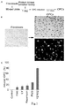
- Fig.1a is a schematic diagram of the flow of screening process.
- Fig.1b are images of cells in the control and experimental groups at 7 days of culture, wherein the left panel is image of cells in control group, while the right panel is the image of cells in experimental group in which ROCK inhibitor Y27632 (Rock inhibitors Y-27632 and Y-27632 were used interchangeably herein) was added.
- Fig.1c is the result of conversion rate in the control and experimental groups in which different protein inhibitors was add at 7 days of culture. Control represent control group.
- OPC oligodendrocyte precursor cells
- DMEM/F12,Invitrogen, 11320 1x N-2 (R&D Systems), 1x B-27 without vitamin A (Invitrogen), 1x Glutamax (GIBCO, 35050)
- 200 ng/ml SHH (R&D Systems) 20 ng/ml FGF2 (R&D Systems)
- 20 ng/ml PDGF-AA (R&D Systems) penicillin/streptomycin, 10 ⁇ M Y-27632 (Enzo Life Sciences, ALX-270-333-M005), 0.5 ⁇ M RA (Sigma, R2625) for induction of OPCs.
- fibroblasts As shown in Fig.1 , addition of ROCK inhibitors Y-27632 can induce human adult dermal fibroblasts to convert into neural cells with high efficiency, whereas no conversion of fibroblasts to neural cells was observed in control group without kinase inhibitor.
- inventors observed that a subpopulation of fibroblasts underwent a marked morphological change in only 3-7 days in neural induction medium with kinase inhibitor treatments, from large, flat, spindle shaped cells (fibroblasts) to small, bi- of multi-polar cells, termed iOPCs (right panel in Fig. 1b ).
- Fig.1c when the culture medium containing ROCK inhibitor Y27632 was cultured into fibroblasts, the conversion rate was the highest, reaching about 80%.
- inventors selected the top 5 candidate inhibitors with the highest conversation rates, namely the ROCK inhibitor Y27632 (Y27632), the AKT / mTOR inhibitor Palomid 529 (P529), the PI3K inhibitor LY294002 (LY), the FAK inhibitor PF562271 (FAKi), and Immunosuppressant rapamycin for further analysis.
- fiber cell line IMR90 and WI38 were induced ROCK inhibitors Y27632, Palomid 529, LY294002, PF562271 or rapamycin.
- the result showed that: cultured for a period of time with neural induction medium including the protein kinase inhibitor, in both cell lines, fibroblasts were converted into oligodendrocyte precursor cells.
- Results of the IMR90 cells induced by the ROCK inhibitor Y27632 and the conversion results are shown in Figure 1 .
- iOPCs prepared by inducing can be differentiated into mature oligodendrocytes.
- mature medium (10 ⁇ M of ROCK inhibitor Y27632 (Enzo Life Sciences, ALX-270-333-M005) with thyroid hormone (a known oligodendrocyte differentiation inducer) while without growth factor , 1x N2 cell culture medium additive, 1x B27 cell culture additive, 1x glutamine, 200 ng / ml hedgehog protein, 100 ng / ml Noggin (R & D systems), 100 ng / ml dibutyryl cyclic adenosine (Sigma, D0627), 100 ng / ml insulin-like growth factor (R & D systems), 10 ng / ml neurotrophic factor 3 (PROSPEC, CYT-257), and 0.5 ⁇ M retinoic acid (sigma, R2625)) to differentiated culture iOPCs obtained after 7 days in a neuronal
- iOPCs and iOLs were subjected to immunostaining experiments before and after differentiation culture as following steps: 5 ⁇ 10 4 fibroblasts were planted on coated glass coverslips on the day before induction. The induction was carried out in the same manner as in Example 1.
- IPOCs were fixed for 20 min at room temperature in 4% paraformaldehyde in PBS, and then permeabilized for 30 min in PBS containing 0.2% Triton X-100 and 10% goat serum (NGS), and incubated overnight at 4 °C in PBS containing 10% NGS and primary antibodies (mouse anti-O4 (Millipore, 1:50), rabbit anti-NF (Sigma-Aldrich, 1:1000), mouse anti-A2B5 (Millipore, 1:50), MBP (1:100, Covance; 1:100, Abcam), MAG (Millipore, 1:50), MOG (Millipore, 1:50).
- NGS Triton X-100 and 10% goat serum
- Fig.2 where Merge is the coincidence map.
- Fig.2A showed the immunostaining results of iOPCs before differentiation culture
- Fig.2B showed the immunostaining results of iOLs after differentiation culture.
- Fig.2A showed that about 80% of fibroblasts were converted to O4-positive cells with characteristic morphology of oligodendrocyte progenitor cells (OPC). Furthermore, these O4 positive cells also showed positive staining with two additional OPC markers, A2B5 (oligodendrocyte precursor cell surface antigen A2B5) and S100 ⁇ (glial specific protein). In contrast, after 4 weeks in the same induction medium without ROCK inhibitor, cells kept fibroblast morphology with negative staining of O4 (oligodendrocyte precursor cell surface antigen), A2B5, and S100 ⁇ . These results suggest that ROCK inhibitor leads to direct conversion of human fibroblasts to iOPCs.
- OPC oligodendrocyte progenitor cells
- Fig.2B showed that about 70% of the iOPCs differentiated into cells with a multiprocessed morphology typical of oligodendrocytes within 3-7 days).
- the oligodendrocytes expressed myelin basic protein (MBP), and other specific markers of mature oligodendrocytes, including myelin-associated glycoprotein (MAG) and myelin oligodendrocyte glycoprotein (MOG).
- MBP myelin basic protein
- MAG myelin-associated glycoprotein
- MOG myelin oligodendrocyte glycoprotein
- ROCK is a kinase belonging to the AGC (PKA/ PKG/PKC) family of serine-threonine kinases, including ROCK1 and ROCK2 in mammals (human, rat, mouse).
- ROCK1 is mainly expressed in the lung, liver, spleen, kidney and testis, while ROCK2 is distributed mostly in the brain and heart.
- PKA/ PKG/PKC AGC
- ROCK1 is mainly expressed in the lung, liver, spleen, kidney and testis
- ROCK2 is distributed mostly in the brain and heart.
- Rho-ROCK pathway is involved in fibroblast-OPC conversion
- inventors examined the effects of ROCK knockdown on fibroblast-OPC conversion as follows: Lentiviral constructs expressing short hairpin RNAs including shRNA1 and shRNA2 (Sigma-Aldrich, TRCN0000342532, TRCN0000342473) against ROCK2 gene were provided.
- shRNA1 and shRNA2 were transfected to human adult dermal fibroblasts, respectively. And the transfected cells were subjected to western blot analysis. Next, the transfected cells were cultured with a neutral-inducing medium without a protein kinase inhibitor for 7 days to obtain iOPCs, and then the iOPCs were subjected to immunostaining experiments using the same procedure as in Example 3, followed by differentiated culturing the obtained iOPCs by using a mature medium to obtain iOLs. The obtained iOLs then were subjected to immunostaining experiments in the same manner as in Example 3. The results are shown in Fig. 3. Fig.
- FIG. 3A is the result of Western blot analysis, where Tubulin is the tubulin, and Control is the control group.
- Fig. 3B is the result of iOPCs immunostaining test
- Fig. 3C is the result of iOLs immunostaining test.
- Fig. 3A showed that transfected cells showed significantly reduced ROCK2 expression, indicating that shRNA1 and shRNA2 are effective in inhibiting the expression of ROCK2.
- the transfected cells gave a conversion rate (60-80%) essentially same to that of ROCK inhibitor treatment.
- Fig.3B showed that the iOPCs induced by ROCK2 knockdown showed positive staining for OPC markers O4, A2B5 and S100 ⁇ .
- Fig.3C showed that these iOPCs obtained by ROCK2 gene silencing can differentiate into cells with typical oligodendrocytes morphology after differentiation culture, and the differentiated cells (iOLs) expressed MBP, MAG, and MOG ( Fig. 3c ). The results dindicated that ROCK2 kinase is involved in conversion of fibroblast to neural cells.
- ROCK inhibitor induces fibroblast-OPC conversion by regulating a set of neural transcription factors.
- iOPCs obtained with Y27632 treatment
- human brain-derived OPCs, iOLs and parental fibroblasts inventors generated comparative global gene expression data by microarray analysis.
- Microarray analysis was performed at the Washington University Genome Center. Specific steps are as follows, Illumina HumanHT-12 v4 Expression BeadChip was used and samples from above cells were labeled by biotin. The Direct Hybridization Assay was performed and the data were scanned on the BeadArray Reader. Scanned images were quantitated by Illumina Beadscan, v3.
- Quantitative data was imported into Illumina GenomeStudio software and normalized by Illumina's quantile method.
- the quantile-normalized background-subtracted data were calculated in excel.
- the data were filtered based on the average signal, and set the baseline as 50 so that only the genes of which average signal >50 could be used for further analysis.
- maximum and minimum signal values were selected, and then divided between them. Only the genes of the division value >3 were selected and considered as differently expressed. All of the differently expressed genes were clustered by using MeV software.
- microarray analysis was performed on each sample separately. The obtained data were filtered based on average signal and baseline was set as 50 to increase the sensitivity.
- all OPC differentiation genes from Gene Ontology Website (http://www.geneontology.org/, GO: 0030182) were compared with the microarray data.
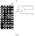
- inventors used web-based Gorilla program (http://cbl-gorilla.cs.technion.ac.il/). The test results are shown in Fig.4 , where WT represents wild type.
- Fig.4A shows the gene differential expression profiles between iOPCs, human brain-derived OPCs, iOLs and parental fibroblasts
- Fig.4B shows the results of cluster analysis of differentially expressed genes between iOPCs, human brain-derived OPCs, iOLs and parental fibroblasts.
- Fig. 4 cluster analysis revealed a significant difference of gene expression profiles between iOPC cells and their parental fibroblasts ( Fig. 4A ), suggesting that iOPCs are clearly distinct from parental fibroblasts.
- Clustering analysis of differential gene expression revealed that the transcription profile of ROCK inhibitor-induced iOPCs were tightly clustered with the transcription profile of human brain-derived OPCs and were distant from the profile of the parental fibroblasts ( Figs. 4A and B ).
- iOPCs and brain-derived OPCs showed significant gene expression overlap for neural transcription factors.
- mice were transplanted into the demyelinating brain corpus callosum of mice treated with cuprizone to evaluate the functionality of iOPCs in vivo and the potential to form myelin as follows: C57BL/6 mice were put on a diet of 0.2% (w/w) cuprizone (Sigma Aldrich), a copper chelator. Feeding cuprizone diet for 12 weeks results in depletion of the pool of endogenous OPCs in the corpus callosum and finally leads to its complete demyelination. At 12 weeks after the start of the cuprizone diet, mice were divided into three groups.
- the concentration of the injected cells is 25,000 cells / ⁇ l.
- the stereotactic co-ordinates of the injection position are 10.98 mm (anteroposterior axis), 21.75 mm (lateromedial axis), 22.25mm (vertical axis) (Copray et al., 2006; Sher et al.,2009).
- mice were taken off the cuprizone diet and put back on normal diet to avoid degeneration of the implanted cells by cuprizone. After 4 weeks of transplantation, the mice were sacrificed and the brain was resected. Then, Immunohistochemical staining (MBP staining) was performed on the brain tissue sections.
- Fig.5 where NF represents the neurofilament protein.
- Fig.5A are images of the immunofluorescence results and H & E staining results for the site of iOPs injection of the same brain tissue sections.
- Figure 5B are images of the immunostaining results for the site of iOPCs injection of brain tissue sections.
- Figure 5C are confocal microscopic photographs of the iOPCs site of injection of the brain tissue sections.
- Fig.5A shows that only about 20% of the implanted cells survived.
- Fig.5B shows that the surviving iOPCs developed into mature MBP-expressing OLs that contributed to the remyelination of the corpus callosum axons.
- Confocal microscopy in Fig.5C revealed that MBP+ tube-like structures and the nerve fibers co-coat the structure of the nerve fibers, indicating the implanted iOPCs promote the host axon to produce myelin. No similar structures were observed in nontransplanted brains.
- Aspirate the solution from the well wash with basic medium and plate fibroblast cells (ATCC, PCS-201-012) with basic medium at 2x10 ⁇ 4 cells per well, and culture with the above basic medium for 24 hours.
- Aspirate basic medium from the well wash with PBS once, and feed cells with induction medium containing 5 ⁇ M ROCK inhibitor Y27632 and 2 ⁇ M mTOR inhibitor P529.
- induction medium containing 5 ⁇ M ROCK inhibitor Y27632 and 2 ⁇ M mTOR inhibitor P529.
- cells change their morphology into neural-like cells, and then culture the obtained neural-like cells with mature medium or OPC medium, and induced neuronal cells (iNCs) and astrocytes were obtained.
- immunostaining experiments and patch-clamp experiments were performed on the induced neurons (iNCs) and astrocytes.
- Fig.7a shows current traces (upper panel) recorded in the process in which cells were depolarized by voltage steps from -60 to +60 mV in 10 mV increments and the relationship (lower panel) between the current-voltage (I-V) and sodium current.
- Fig.7b shows traces of membrane currents recorded in the process of a voltage ramp from -80mV to +60mV over 500ms.
- Fig.7c shows traces of spontaneous synaptic currentsrecorded at a holding potential of -80 mV.
- the the immunostaining results of astrocyte cells induced by Y27632and mTOR inhibitors were shown in Fig.8 .
- Neural cells and astrocytes induced by Y27632 and mTOR inhibitors showed typical cell types morphology and biomarkers.
- iNCs induced neural cells
- Tuj microtubule associated protein
- MAP2 microtubule associated protein 2
- Synaptotagnin synaptic binding protein
- astrocyte-specific markers GFAP Glial fibrillary acidic protein
Landscapes
- Health & Medical Sciences (AREA)
- Life Sciences & Earth Sciences (AREA)
- Chemical & Material Sciences (AREA)
- Engineering & Computer Science (AREA)
- General Health & Medical Sciences (AREA)
- Biomedical Technology (AREA)
- Pharmacology & Pharmacy (AREA)
- Veterinary Medicine (AREA)
- Public Health (AREA)
- Animal Behavior & Ethology (AREA)
- Medicinal Chemistry (AREA)
- Epidemiology (AREA)
- Neurology (AREA)
- Bioinformatics & Cheminformatics (AREA)
- Neurosurgery (AREA)
- Organic Chemistry (AREA)
- Zoology (AREA)
- Biotechnology (AREA)
- Cell Biology (AREA)
- Wood Science & Technology (AREA)
- Genetics & Genomics (AREA)
- Developmental Biology & Embryology (AREA)
- Chemical Kinetics & Catalysis (AREA)
- Nuclear Medicine, Radiotherapy & Molecular Imaging (AREA)
- General Chemical & Material Sciences (AREA)
- General Engineering & Computer Science (AREA)
- Microbiology (AREA)
- Biochemistry (AREA)
- Immunology (AREA)
- Ophthalmology & Optometry (AREA)
- Virology (AREA)
- Psychiatry (AREA)
- Hospice & Palliative Care (AREA)
- Psychology (AREA)
- Medicines Containing Material From Animals Or Micro-Organisms (AREA)
- Micro-Organisms Or Cultivation Processes Thereof (AREA)
- Medicines That Contain Protein Lipid Enzymes And Other Medicines (AREA)
- Pharmaceuticals Containing Other Organic And Inorganic Compounds (AREA)
Description
- The present disclosure relates to biomedical field, particularly to a method for preparing an oligodendrocyte progenitor cell.
- Central nervous system (CNS) disorders, such as Alzheimer's Disease (AD), Parkinsin's Disease (PD), Amyotrophic Lateral Sclerosis (ALS), Multiple Sclerosis (MS), and Leukodystrophies, affect millions of people worldwide and most of CNS diseases currently have no cure. Transplantation of neural cells has been demonstrated in non-human primates and rodents to be a very promising therapeutic strategy for treating CNS disorders. However, cell therapy requires high quality and large quantities of primary neural cells that are not readily obtainable. Different methods have been developed during last two decades to produce neural cells in vitro. These technologies include generation of neural cells from human embryonic stem cells (hESC), from induced human pluripotent stem cells (hiPSC), and from direct conversion of somatic cells. There are several limitations to existing art. For example, neural cells derived from hESC encounter the safety issue due to potential tumorgenesis caused by residual hESC in the products as well as immune rejection issue due to allogeneic transplantation. hiPSC can be obtained from specific patients and thus alleviate the immunorejection issue, but like hESC, it still encounters the safety issue.
- Recently, the field of direct somatic lineage conversion has attracted much attention. In 2010, Wernig's group first demonstrated that a set of transcription factors can convert fibroblasts into neurons. Several laboratories have used various neural factors and microRNAs to generate fibroblast-neuron conversion. Very recently, it was reported that over-expression transcription factors can mediate reprogramming of mouse fibroblasts to myelinogenic OPCs. Thus, direct fibroblast-OPC conversion provides an alternative, potentially complementary, tool to many of the proposed applications of hESC/hiPSC technology for both disease modeling and development of cell-based therapies. Direct conversion or reprogramming has a number of advantages, including: the time required to generate, expand and differentiate pluripotent cells is avoided, and the postmitotic state of induced neural cells has a much lower risk of cancer and teratoma formation. Therefore, neural cells derived from direct conversion of fibroblasts have been favored for autologous transplantation. However, all reports on direct conversion of somatic cells into neural cells have been using transcription factors, which often involve lengthy conversion procedure and are regarded not safe due to integration of viral vector sequence into the genome.
- The present disclosure directs to solve at least one of the problems existing in the prior art. Therefore, one purpose of present disclosure is to provide a method for efficiently preparing oligodendrocyte progenitor cells with high safety and suitable for autotransplantation using protein kinase inhibitor.
- In one aspect of present disclosure, a protein kinase inhibitor for use in preparing a oligodendrocyte progenitor cell from a fibroblast cell is provided. The inventors have surprisingly found that in the presence of protein kinase inhibitors, it is not necessary to use transcription factors, and fibroblast cells can be efficiently transformed into oligodendrocyte progenitor cells.
- As described in the present disclosure, the protein kinase inhibitor maybe one selected from ROCK inhibitor, AKT/mTOR inhibitor, PI3K inhibitor, FAK inhibitor and immunosuppresants. Thereby, it is possible to effectively induce non-neural cells to transform into neural cells.
- According to embodiments of present disclosure, the protein kinase inhibitor is at least one selected from ROCK inhibitor Y27632, AKT/mTOR inhibitor Palomid 529, PI3K inhibitor LY294002, FAK inhibitor PF562271 and immunosuppresants rapamycin. Thereby, it is possible to effectively induce non-neural cells to transform into neural cells with high convention efficiency.
- Thereby, the prepared oligodendrocyte progenitor cells can be effectively used for autologous transplantation, treatment of central nervous system disorders and spinal cord injury with high safety while without immune rejection.
- In one aspect of present disclosure, a method for preparing an oligodendrocyte progenitor cell is provided, and according to embodiments of present disclosure, the method may comprise: culturing a fibroblast cell in the present of a protein kinase inhibitor. The inventors have found that by the method of present disclosure, the oligodendrocyte progenitor cells can be prepared rapidly and efficiently without requirement of the transcription factors. The prepared oligodendrocyte progenitor cells are suitable for autologous transplantation, treatment of central nervous system disorders and spinal cord injury with high safety while without immune rejection.
- As described in the present disclosure, the protein kinase inhibitor maybe selected from ROCK inhibitor, AKT/mTOR inhibitor, PI3K inhibitor, FAK inhibitor and immunosuppresants. Thereby, it is possible to effectively induce non-neural cells to transform into neural cells.
- According to embodiments of present disclosure, the protein kinase inhibitor is at least one selected from ROCK inhibitor Y27632, AKT/mTOR inhibitor Palomid 529, PI3K inhibitor LY294002, FAK inhibitor PF562271 and immunosuppresants rapamycin. Thereby, it is possible to effectively induce fibroblast cells to convert into oligodendrocyte progenitor cells with high convention efficiency.
- The application further describes a kit, the kit described herewith may comprise: a first medium, the first medium may comprise: a first basic medium, a protein kinase inhibitor, brain-like neurotrophic factor (BDNF), neurotrophic factor 3 (NT3), sodium valproate (VPA), dibutyryl cyclic adenosine monophosphate (dbcAMP), and retinoic acid (RA), wherein the protein kinase inhibitor maybe selected from ROCK inhibitor, AKT/mTOR inhibitor, PI3K inhibitor, FAK inhibitor and immunosuppresants.
- As described herewith, the kit may further comprises: a second medium, or/and, a third medium, wherein the second medium comprises: a second basic medium, a protein kinase inhibitor, N2 cell culture medium additive (N-2), B27 cell culture medium additives (B-27), glutamine (Glutamax), hedgehog protein (SHH), fibroblast growth factor (FGF2), recombinant human platelet derived growth factor (PDGF-AA), and retinoic acid, or/and a third medium, wherein the third medium comprises: a third basic medium, a protein kinase inhibitor, N2 cell culture medium additive (N-2), B27 cell culture medium additives (B-27), glutamine (Glutamax), hedgehog protein (SHH), noggin protein (Noggin), dibutyryl cyclic adenosine monophosphate, insulin-like growth factor (IGF), neurotrophic factor 3, and retinoic acid, wherein the protein kinase maybe selected from ROCK inhibitor, AKT/mTOR inhibitor, PI3K inhibitor, FAK inhibitor and immunosuppresants. Thereby, it is possible to effectively prepare mature different types of nerual cells.
- According to embodiments of present disclosure, the first basic medium is neuronal medium, the second basic medium is DMEM/F12 medium, and the third basic medium is DMEM/F12 medium. Thereby, it is advantageous to convert non-neural cells into neural cells.
- According to embodiments of present disclosure, the protein kinase inhibitor is at least one selected from ROCK inhibitor Y27632, AKT/mTOR inhibitor Palomid 529, PI3K inhibitor LY294002, FAK inhibitor FPF562271 and immunosuppresants rapamycin. Preferably he protein kinase inhibitor is ROCK inhibitor Y27632.
- According to embodiments of present disclosure, the first medium may comprise: 5µM - 20µM Y27632, 5 ng/ml - 20 ng/ml BDNF, 20 ng/ml - 80 ng/ml NT3, 0.5 mM - 1.5 mM VPA, 25µM -100 µM dbcAMP, and 0.5µM - 1µM retinoic acid; the second medium may comprise: 5µM - 20µM Y27632, 1x N-2, 1x B-27, 1x Glutamax, 100 ng/ml - 400 ng/ml SHH, 10 ng/ml - 40 ng/ml FGF2, 10 ng/ml - 40 ng/ml PDGF-AA, and 0.5µM - 1µM retinoic acid; the third medium may comprise: 5µM - 20µM Y27632, 1x N-2, 1x B-27, 1x Glutamax, 100 ng/ml - 400 ng/ml SHH, 50 ng/ml - 200 ng/ml Noggin, 50 ng/ml - 200 ng/ml dbcAMP, 50 ng/ml - 200 ng/ml IGF, 20 ng/ml - 80 ng/ml NT3, and 0.5µM - 1µM retinoic acid. Thereby, the non-neural cell can be directly induced to convert into neural cell rapidly and effectively with high efficiency.
- According to embodiments of present disclosure, the first medium may comprise: 10µM Y27632, 10 ng/ml BDNF, 10 ng/ml NT3, 1 mM VPA, 50 µM dbcAMP, and 0.5µM retinoic acid; the second medium may comprise: 10µM Y27632, 1x N-2, 1x B-27, 1x Glutamax, 200 ng/ml SHH, 20 ng/ml FGF2, 20 ng/ml PDGF-AA, and 0.5µM retinoic acid; the third medium may comprise: 10µM Y27632, 1x N-2, 1x B-27, 1x Glutamax, 200 ng/ml SHH, 100 ng/ml Noggin, 100 ng/ml dbcAMP, 100 ng/ml IGF, 10 ng/ml NT3, and 0.5µM retinoic acid. Thereby, the non-neural cell can be directly induced to convert into neural cell rapidly and effectively with high efficiency. The prepared neural cells can be effectively used for autologous transplantation, treatment of central nervous system disorders and spinal cord injury with high safety while without immune rejection.
- Further, the kit for use in preparing a neural cell from a non-neural cell is described. The inventors have found that the non-neural cell may be induced to convert into neural cells with the above kit efficiently, and the prepared neural cells may be effectively used for autologous transplantation, treatment of central nervous system disorders and spinal cord injury with high safety while without immune rejection.
- In another aspect of present disclosure, a method for preparing an oligodendrocyte progenitor cell is provided, and according to embodiments of present disclosure, the method may comprise:
- (1) culturing a fibroblast cell with a first medium,
wherein the first medium may comprise: a first basic medium, a protein kinase inhibitor, BDNF, NT3, VPA, dbcAMP, and retinoic acid, wherein the protein kinase inhibitor is at least one selected from ROCK inhibitor, AKT/mTOR inhibitor, PI3K inhibitor, FAK inhibitor and immunosuppresants. The inventors have found that by the method of present disclosure, the neural cells can be prepared rapidly and efficiently without requirement of the transcription factors. The prepared neural cells are suitable for autologous transplantation, treatment of central nervous system disorders and spinal cord injury with high safety while without immune rejection.
According to embodiments of present disclosure, the method for preparing a oligodendrocyte progenitor cell further comprises: - (2) Culturing the cell with a second medium or a third medium after the culturing with the first medium.
wherein the second medium comprises: a second basic medium, a protein kinase inhibitor, N-2, B-27, Glutamax, SHH, FGF2, PDGF-AA, and retinoic acid, the third medium comprises: a third basic medium, a protein kinase inhibitor, N-2, B-27, Glutamax, SHH, Noggin, dbcAMP, IGF, NT3, and retinoic acid. According to embodiments of present disclosure, the protein kinase inhibitor is selected from ROCK inhibitor, AKT/mTOR inhibitor, PI3K inhibitor, FAK inhibitor and immunosuppresants. By the method of present disclosure, the neural cells can be prepared rapidly and efficienly without requirement of the transcription factors. The prepared oligodendrocyte progenitor cells are suitable for autologous transplantation, treatment of central nervous system disorders and spinal cord injury with high safety while without immune rejection. - According to embodiments of present disclosure, the first basic medium is neuronal medium, the second basic medium is DMEM/F12 medium, the third basic medium is DMEM/F12 medium. Thereby, it is advantageous to convert non-neural cells into neural cells.
- According to embodiments of present disclosure, the protein kinase inhibitor is at least one selected from ROCK inhibitor Y27632, AKT/mTOR inhibitor Palomid 529, PI3K inhibitor LY294002, FAK inhibitor PF562271 and immunosuppresants rapamycin. Thereby, the non-neural cell can be induced to convert into neural cell with high convention efficiency.
- According to embodiments of present disclosure, the first medium may comprise: 5µM - 20µM Y27632, 5 ng/ml - 20 ng/ml BDNF, 20 ng/ml - 80 ng/ml NT3, 0.5 mM - 1.5 mM VPA, 25µM -100 µM dbcAMP, and 0.5µM - 1µM retinoic acid; the second medium may comprise: 5µM - 20µM Y27632, 1x N-2, 1x B-27, 1x Glutamax, 100 ng/ml - 400 ng/ml SHH, 10 ng/ml - 40 ng/ml FGF2, 10 ng/ml - 40 ng/ml PDGF-AA, and 0.5µM - 1µM retinoic acid; the third medium may comprise: 5µM - 20µM Y27632, 1x N-2, 1x B-27, 1x Glutamax, 100 ng/ml - 400 ng/ml SHH, 50 ng/ml - 200 ng/ml Noggin, 50 ng/ml - 200 ng/ml dbcAMP, 50 ng/ml - 200 ng/ml IGF, 20 ng/ml - 80 ng/ml NT3, and 0.5µM - 1µM retinoic acid. Thereby, the non-neural cell can be directly induced to convert into neural cell rapidly and effectively with high efficiency.
- According to embodiments of present disclosure, the first medium may comprise: 10µM Y27632, 10 ng/ml BDNF, 10 ng/ml NT3, 1 mM VPA, 50 µM dbcAMP, and 0.5µM retinoic acid; the second medium may comprise: 10µM Y27632, 1x N-2, 1x B-27, 1x Glutamax, 200 ng/ml SHH, 20 ng/ml FGF2, 20 ng/ml PDGF-AA, and 0.5µM retinoic acid; the third medium may comprise: 10µM Y27632, 1x N-2, 1x B-27, 1x Glutamax, 200 ng/ml SHH, 100 ng/ml Noggin, 100 ng/ml dbcAMP, 100 ng/ml IGF, 10 ng/ml NT3, and 0.5µM retinoic acid. Thereby, the non-neural cell can be directly induced to convert into neural cell rapidly and effectively with high efficiency. The prepared neural cells can be effectively used for autologous transplantation, treatment of central nervous system disorders and spinal cord injury with high safety while without immune rejection.
- Further described in this invention is a oligodendrocyte progenitor cell or their derivative. The oligodendrocyte progenitor cell maybe prepared by the method mentioned above. The inventors have found that the oligodendrocyte progenitor cell or their derivative of present disclosure maybe suitable for autologous transplantation, treatment of central nervous system disorders and spinal cord injury with high safety while without immune rejection.
- Further described in the present disclosure is the oligodendrocyte progenitor cell or their derivative for use in preparing a drug for the treatment of central nervous system disorder and spinal cord injury is provided.
- According to description of present disclosure, the Central nervous system disorder is at least one selected from Alzheimer's Disease, Parkinsin's Disease, Amyotrophic Lateral Sclerosis, Multiple Sclerosis, and Leukodystrophies. Thereby, the treatment effect is better.
-
-
Fig.1 is a graph showing the immunostaining results of iOPCs according to an embodiment of the present disclosure, in which -
Fig.1A is a schematic diagram of the flow of screening process, -
Fig.1B are images of cells in the control and experimental groups at 7 days of culture, -
Fig.1C is conversion rate in the control and experimental groups at 7 days of culture; -
Fig.2 is a graph showing the immunostaining results of iOPCs and iOLs according to example 3 of the present disclosure, in which -
Fig.2A shows the immunostaining results of iOPCs before differentiation culture, -
Fig.2B shows the immunostaining results of iOLs after differentiation culture; -
Fig.3 is a graph showing the results of immunostaining and Western blot according to example 4 of the present disclosure, in which -
Fig.3A is the result of the Western blot analysis, -
Fig.3B is the immunostaining results of iOPCs, -
Fig.3C is the immunostaining results of iOLs; -
Fig.4 is a graph showing the results of a microarray analysis according to example 5 of the present disclosure, in which -
Fig.4A shows the differential expression profiles of iOPCs, human brain-derived OPCs, iOLs and parental fibroblasts, -
Fig.4B shows the results of cluster analysis of differentially expressed genes between iOPCs, human brain-derived OPCs, iOLs and parental fibroblasts; -
Fig.5 is a graph showing the result of immuno-histochemical detection according to example 6 of the present disclosure, in which -
Fig.5A are images of the immunofluorescence results and H & E staining results for the site of iOPs injection of the same brain tissue sections, -
Figure 5B are images of the immunostaining results for the site of iOPCs injection of brain tissue sections, -
Figure 5C are confocal microscopic photographs of the iOPCs site of injection of the brain tissue sections; -
Fig.6 is a graph showing the immunostaining results of neutral cells converted from IMR90 fibroblasts according to example 7 of the present disclosure, -
Fig.7 is a graph showing the electrophysiological examination results neutral cells converted from IMR90 fibroblasts according to example 7 of the present disclosure, -
Fig.7a shows current traces (upper panel) recorded in the process in which cells were depolarized by voltage steps from -60 to +60 mV in 10 mV increments and the relationship (lower panel) between the current-voltage (I-V) and sodium current, -
Fig.7b shows traces of membrane currents recorded in the process of a voltage ramp from -80mV to +60mV over 500ms, -
Fig.7c shows traces of spontaneous synaptic currentsrecorded at a holding potential of -80 mV; and -
Fig.8 is a graph showing the immunostaining results of astrocyte cells converted from IMR90 fibroblasts according to example 7 of the present disclosure, - The embodiments of the present invention will be described in detail below. The embodiments described below are exemplary and are intended to be illustrative of the invention and are not to be construed as limiting the invention. The specific technical or conditions not indicated in the examples, are either in accordance with the techniques or conditions described in the literature in the art or in accordance with the product specifications. The reagents or instruments used not indicate the manufacturer are available through the purchase of conventional products.
- In one aspect of present disclosure, a protein kinase inhibitor for use in preparing a oligodendrocyte progenitor cell from a fibroblast cell is provided. Inventors found that, the protein kinase inhibitor is able to convert the fibroblast cell into the oligodendrocyte progenitor cell efficiently, without using any transcription factor.
- According to embodiments of present disclosure, the protein kinase inhibitor is at least one selected from ROCK inhibitor Y27632, AKT/mTOR inhibitor Palomid 529, PI3K inhibitor LY294002, FAK inhibitor PF562271 and immunosuppresants rapamycin.The protein kinase inhibitor is sufficient to reprogram the non-neural cell into the neural cell.
- In a second broad aspect of present disclosure, a method for preparing a oligodendrocyte progenitor cell is provided, and according to embodiments of present disclosure, the method may comprise: culturing a fibroblast cell in the present of a protein kinase inhibitor. The inventors have found that by the method of present disclosure, the oligodendrocyte progenitor cells can be prepared rapidly and efficiently without requirement of the transcription factors. The prepared oligodendrocyte progenitor cells are suitable for autologous transplantation, treatment of central nervous system disorders and spinal cord injury with high safety while without immune rejection.
- As described in the present disclosure, the protein kinase inhibitor maybe one selected from ROCK inhibitor, AKT/mTOR inhibitor, PI3K inhibitor, FAK inhibitor and immunosuppresants. Using the protein kinase inhibitor can convert the non-neural cell into the neural cell efficiently.
- According to embodiments of present disclosure, the protein kinase inhibitor is at least one selected from ROCK inhibitor Y27632, AKT/mTOR inhibitor Palomid 529, PI3K inhibitor LY294002, FAK inhibitor PF562271 and immunosuppresants rapamycin. The protein kinase inhibitor is sufficient to reprogram the non-neural cell into the neural cell.
- Further described in the present invention is a kit, the kit may comprise: a first medium, the first medium may comprise: a first basic medium, a protein kinase inhibitor, BDNF, NT3, VPA, dbcAMP, and retinoic acid, according to embodiments of present disclosure, the protein kinase inhibitor is at least one selected from ROCK inhibitor, AKT/mTOR inhibitor, PI3K inhibitor, FAK inhibitor and immunosuppresants. Using the kit of present disclosure, the non-neural cell can be induced into the neural cell directly with high efficiency and convenience.
- According to the description, the first basic medium is neuronal medium.
- According to the description, the first medium may comprise: 5µM - 20µM Y27632, 5 ng/ml - 20 ng/ml BDNF, 20 ng/ml - 80 ng/ml NT3, 0.5 mM - 1.5 mM VPA, 25µM -100 µM dbcAMP, and 0.5µM - 1µM retinoic acid. Thereby, the non-neural cell can be directly induced to convert into neural cell rapidly and effectively with high efficiency.
- According to the description, the first medium may comprise: 10µM Y27632, 10 ng/ml BDNF, 10 ng/ml NT3, 1 mM VPA, 50 µM dbcAMP, and 0.5µM retinoic acid. Thereby, the non-neural cell can be directly induced to convert into neural cell rapidly and effectively with high efficiency. The prepared neural cells can be effectively used for autologous transplantation, treatment of central nervous system disorders and spinal cord injury with high safety while without immune rejection.
- According to the description, the kit may future comprises: a second medium, or/and, a third medium. According to embodiments of present disclosure, the second medium comprises: a second basic medium, a protein kinase inhibitor, N-2, B-27, Glutamax, SHH, FGF2, PDGF-AA, and retinoic acid, or/and a third medium. The third medium comprises: a third basic medium, a protein kinase inhibitor, N-2, B-27, Glutamax, SHH, Noggin, dbcAMP, IGF, NT3, and retinoic acid. According to embodiments of present disclosure, the protein kinase inhibitor is at least one selected from ROCK inhibitor, AKT/mTOR inhibitor, PI3K inhibitor, FAK inhibitor and immunosuppresants. Thereby, it is possible to effectively prepare mature different types of nerual cells.
- According to the description, the neural cell maybe one selected from oligodendrocyte progenitor cell, mature oligodendrocyte, neuron and astrocyte. Thereby, the prepared neural cells may be effectively used for autologous transplantation, treatment of central nervous system disorders and spinal cord injury with high safety while without immune rejection.
- According to the description, the second basic medium is DMEM/F12 medium, the third basic medium is DMEM/F12 medium. The basic mediums mentioned above are in favor of the conversion from the non-neural cell to the neural cell.
- According to the description, the protein kinase inhibitor is at least one selected from of Y27632, Palomid 529, LY294002, PF562271 and rapamycin. The protein kinase inhibitor is sufficient to reprogram the non-neural cell into the neural cell.
- According to the description, the second medium may comprise: 5µM - 20µM Y27632, 1x N-2, 1x B-27, 1x Glutamax, 100 ng/ml - 400 ng/ml SHH, 10 ng/ml - 40 ng/ml FGF2, 10 ng/ml - 40 ng/ml PDGF-AA, and 0.5µM - 1µM retinoic acid; the third medium may comprise: 5µM - 20µM Y27632, 1x N-2, 1x B-27, 1x Glutamax, 100 ng/ml - 400 ng/ml SHH, 50 ng/ml - 200 ng/ml Noggin, 50 ng/ml - 200 ng/ml dbcAMP, 50 ng/ml - 200 ng/ml IGF, 20 ng/ml - 80 ng/ml NT3, and 0.5µM - 1µM retinoic acid. Thus, the non-neural cell will convert into the neural cell quickly and efficiently when cultured with the medium mentioned above, and the conversion rate is very high.
- According to the description, the second medium may comprise: 10µM Y27632, 1x N-2, 1x B-27, 1x Glutamax, 200 ng/ml SHH, 20 ng/ml FGF2, 20 ng/ml PDGF-AA, and 0.5µM retinoic acid; the third medium may comprise: 10µM Y27632, 1x N-2, 1x B-27, 1x Glutamax, 200 ng/ml SHH, 100 ng/ml Noggin, 100 ng/ml dbcAMP, 100 ng/ml IGF, 10 ng/ml NT3, and 0.5µM retinoic acid. Thereby, the non-neural cell can be directly induced to convert into neural cell rapidly and effectively with high efficiency. The prepared neural cells may be effectively used for autologous transplantation, treatment of central nervous system disorders and spinal cord injury with high safety while without immune rejection.
- Further described is the kit for use in preparing a neural cell. Using the kit as described above, the non-neural cell may be induced to convert into neural cells efficiently, and the neural cell maybe suitable for autologous transplantation to treat central nervous system disorder and spinal cord injury with high safety, and there is no immune rejection.
- In a further aspect of present disclosure, a method for preparing a oligodendrocyte progenitor cell is provided, and according to embodiments of present disclosure, the method may comprise:
- (1) Culturing a fibroblast cell with a first medium,
According to embodiments of present disclosure, the first medium may comprise: a first basic medium, a protein kinase inhibitor, BDNF, NT3, VPA, dbcAMP, and retinoic acid.
According to embodiments of the present invention, the type of the first basic medium is not particularly limited, and can be advantageously selected by a person skilled in the art, as long as it can facilitate the induction of non-neuronal cells into neutral cells. According to an embodiment of the present invention, the first basic medium is neuronal medium. Thus, it is advantageous to convert non-neuronal cells into neutral cells.
According to embodiments of present disclosure, the protein kinase inhibitor is at least one selected from Y27632, Palomid 529, LY294002, PF562271 and rapamycin, preferably is Y27632. Thereby the non-neural cell, which is a fibroblast cell, can directly convert into the neural cell, which is oligodendrocyte progenitor cell, with high efficiency and convention rate.
According to embodiments of present disclosure, the first medium may comprise: 5µM - 20µM Y27632, 5 ng/ml - 20 ng/ml BDNF, 20 ng/ml - 80 ng/ml NT3, 0.5 mM - 1.5 mM VPA, 25µM -100 µM dbcAMP, and 0.5µM - 1µM retinoic acid. Thereby, the non-neural cell can be directly induced to convert into neural cell rapidly and effectively with high efficiency. If the concentration of each component is too high or too low, the conversion effect is not ideal.
According to embodiments of present disclosure, the first medium may comprise: 10µM Y27632, 10 ng/ml BDNF, 10 ng/ml NT3, 1 mM VPA, 50 µM dbcAMP, and 0.5µM retinoic acid. Thereby, the non-neural cell, which is fibroblast cell, can be gradually induced to convert into neural cell, which is oligodendrocyte progenitor cell, under most suitable conditions with high efficiency. The prepared oligodendrocyte progenitor cells can be effectively used for autologous transplantation, treatment of central nervous system disorders and spinal cord injury with high safety while without immune rejection.
As described in the present disclosure, the non-neural cell is human cell. The human cell maybe one selected from fibroblast cell, epithelial cell, adult cell and neonatal cell. Thus, the non-neural cell, which is fibroblast cell, can convert into the neural cell, which is oligodendrocyte progenitor cell, efficiently in the present of the protein kinase inhibitor. - (2) Culturing the cell with a second medium or a third medium after the culturing with the first medium.
- According to embodiments of present disclosure, the second medium comprises: a second basic medium, a protein kinase inhibitor, N-2, B-27, Glutamax, SHH, FGF2, PDGF-AA, and retinoic acid, the third medium comprises: a third basic medium, a protein kinase inhibitor, N-2, B-27, Glutamax, SHH, Noggin, dbcAMP, IGF, NT3, and retinoic acid. Thereby, the non-neural cell, which is fibroblast cell, can be gradually induced to convert into neural cell, which is oligodendrocyte progenitor cell, under most suitable conditions with high efficiency. The prepared neural cells, which are oligodendrocyte progenitor cells, can be effectively used for autologous transplantation, treatment of central nervous system disorders and spinal cord injury with high safety while without immune rejection.
- According to embodiments of the present invention, the kind of the second basic medium is not particularly limited, and one skilled in the art can select it flexibly according to the actual situation. According to an embodiment of the present invention, the second basic medium is DMEM / F12 medium.
- According to embodiments of present disclosure, the protein kinase inhibitor is at least one selected from Y27632, Palomid 529, LY294002, PF562271 and rapamycin, preferably is Y27632. The protein kinase inhibitor is sufficient to reprogram the non-neural cell into the neural cell.
- According to embodiments of present disclosure, the second medium may comprise: 5µM - 20µM ROCK inhibitor (Y27632), 1x N-2, 1x B-27, 1x Glutamax, 100 ng/ml - 400 ng/ml SHH, 10 ng/ml - 40 ng/ml FGF2, 10 ng/ml - 40 ng/ml PDGF-AA, and 0.5µM - 1µM retinoic acid. Thereby, the non-neural cell can be directly induced to convert into neural cell rapidly and effectively with high efficiency.
- According to embodiments of present disclosure, the second medium may comprise: 10µM Y27632, 1x N-2, 1x B-27, 1x Glutamax, 200 ng/ml SHH, 20 ng/ml FGF2, 20 ng/ml PDGF-AA, and 0.5µM retinoic acid. Thereby, the non-neural cell can be gradually induced to convert into neural cell under most suitable conditions with high efficiency. The prepared neural cells can be effectively used for autologous transplantation, treatment of central nervous system disorders and spinal cord injury with high safety while without immune rejection.
- According to embodiments of present disclosure, the third medium comprises: a third basic medium, a protein kinase inhibitor, N-2, B-27, Glutamax, SHH, Noggin, dbcAMP, IGF, NT3, and retinoic acid.
- According to embodiments of the present invention, the kind of the third basic medium is not particularly limited, and one skilled in the art can select it flexibly according to the actual situation. According to an embodiment of the present invention, the third basic medium is DMEM / F12 medium.
- According to embodiments of present disclosure, the protein kinase inhibitor is at least one selected from Y27632, Palomid 529, LY294002, PF562271 and rapamycin, preferably is Y27632. Thereby the non-neural cell can directly convert into the neural cell with high efficiency and convention rate.
- According to embodiments of present disclosure, the third medium may comprise: 5µM - 20µM ROCK inhibitor (Y27632), 1x N-2, 1x B-27, 1x Glutamax, 100 ng/ml - 400 ng/ml SHH, 50 ng/ml - 200 ng/ml Noggin, 50 ng/ml - 200 ng/ml dbcAMP, 50 ng/ml - 200 ng/ml IGF, 20 ng/ml - 80 ng/ml NT3, and 0.5µM - 1µM retinoic acid. Thus, the non-neural cell will convert into the neural cell quickly and efficiently when cultured with the medium mentioned above, and the conversion rate is very high.
- According to embodiments of present disclosure, the third medium may comprise: 10µM Y27632, 1x N-2, 1x B-27, 1x Glutamax, 200 ng/ml SHH, 100 ng/ml Noggin, 100 ng/ml dbcAMP, 100 ng/ml IGF, 10 ng/ml NT3, and 0.5µM retinoic acid. Thus, the non-neural cell will convert gradually into the neural cell quickly and efficiently when cultured with the medium mentioned above under most suitable conditions, and the conversion rate is very high. Besides, the neural cell is suitable for autologous transplantation to treat central nervous system disorder and spinal cord injury with high safety, and there is no immune rejection.
- As described herewith, the neural cell maybe one selected from oligodendrocyte progenitor cell, mature oligodendrocyte, neuron and astrocyte. Thereby, the prepared neural cells may be effectively used for autologous transplantation, treatment of central nervous system disorders and spinal cord injury with high safety while without immune rejection.
- By the method of present disclosure, the neural cells, which are oligodendrocyte progenitor cells, can be prepared rapidly and efficiently without requirement of the transcription factors. The prepared oligodendrocyte progenitor cells are suitable for autologous transplantation, treatment of central nervous system disorders and spinal cord injury with high safety while without immune rejection.
- Further described is a neural cell or their derivative, the neural cell maybe prepared by the method mentioned above. The inventors have found that the neural cell or their derivative described herewith are suitable for autologous transplantation, treatment of central nervous system disorders and spinal cord injury with high safety while without immune rejection.
- Further described is the neural cell or their derivative for use in preparing a drug for the treatment of central nervous system disorder and spinal cord injury.
- According to the description, the type of the central nervous system disorder is not particularly limited. According to the description, the aforementioned medicament maybe suitable for use in the treatment of the central nervous system disorder selected from Alzheimer's Disease, Parkinsin's Disease, Amyotrophic Lateral Sclerosis, Multiple Sclerosis, and Leukodystrophies. Thereby, the treatment effect is better.
- Coated cell culture 24-well plate with 0.1 mg/ml Poly-L-ornithine solution (sigma, P4957) for at least 3 hours. Then washed wells 3 times with autoclaved MilliQ water, 5 mins per time. Added 500ul/well of 2 µg/ml Fibronectin (sigma, F0556) and 10 µg/ml Laminin (ROCHE, 11243217001) solution in IX PBS and placed in incubator overnight to coat. Aspirated the Fibronectin/Laminin solution from the wells, washed with culture medium. Plated fibroblast cells with Basic Medium at 2x10^4 cells (human adult dermal fibroblasts, ATCC, PCS-201-012) per well, cultured overnight. The Basic Medium comprises: DMEM high glucose (HyClone, SH30022), 10% FBS (Fisher Scientific, SH3007003), 10 mM HEPES (CORNING, 25060-CL), 1x MEM NEAA (100X) (GIBCO, 11140) (MEM Non-Essential Amino Acids Solution), 1 mM Na Pyruvate (GIBCO, 25000), 1x 2-Mercaptoethanol (1000X) (GIBCO, 21985), 1x Glutamax (100X) (GIBCO, 35050), penicillin/streptomycin. Aspirated Basic Medium from the wells. Washed with PBS once. Fed wells with Induction Medium. The Induction Medium comprises: Neuronal Medium (ScienCell, 1521), 10 ng/ml BDNF (PROSPEC, CYT-207) (Brain-Derived Neurotrophic Factor), 10 ng/ml NT3 (PROSPEC, CYT-257) (neurotrophin 3), 1 mM VPA (sigma, P4543) (Valproic acid sodium salt), 50 µM dbcAMP (sigma, D0627) (Dibutyryl cAMP sodium salt), penicillin/streptomycin, 0.5 µM RA (Retinoic acid) (sigma, R2625), 2 µM protein inhibitor (protein kinase inhibitor library, Library I ,II, III: 240 inhibitors, Calbiochem, Cat# 539744,539745 & 539746). The Induction Medium without protein kinase inhibitor was added to the control group. Changed medium every two days. The morphology of the cells was observed during the culture and the conversion rate was calculated at day 7 (conversion rate = number of induced neural cells / number of total cells). The results of several screening experiments are shown in
Fig.1. Fig.1a is a schematic diagram of the flow of screening process.Fig.1b are images of cells in the control and experimental groups at 7 days of culture, wherein the left panel is image of cells in control group, while the right panel is the image of cells in experimental group in which ROCK inhibitor Y27632 (Rock inhibitors Y-27632 and Y-27632 were used interchangeably herein) was added.Fig.1c is the result of conversion rate in the control and experimental groups in which different protein inhibitors was add at 7 days of culture. Control represent control group. - When cells changed their morphology into neural-like cells (about 7 days from fibroblasts seeding), cells were cultured with OPC (oligodendrocyte precursor cells) medium (DMEM/F12,Invitrogen, 11320), 1x N-2 (R&D Systems), 1x B-27 without vitamin A (Invitrogen), 1x Glutamax (GIBCO, 35050), 200 ng/ml SHH (R&D Systems), 20 ng/ml FGF2 (R&D Systems), 20 ng/ml PDGF-AA (R&D Systems), penicillin/streptomycin, 10 µM Y-27632 (Enzo Life Sciences, ALX-270-333-M005), 0.5 µM RA (Sigma, R2625) for induction of OPCs.
- As shown in
Fig.1 , addition of ROCK inhibitors Y-27632 can induce human adult dermal fibroblasts to convert into neural cells with high efficiency, whereas no conversion of fibroblasts to neural cells was observed in control group without kinase inhibitor. In particular, inventors observed that a subpopulation of fibroblasts underwent a marked morphological change in only 3-7 days in neural induction medium with kinase inhibitor treatments, from large, flat, spindle shaped cells (fibroblasts) to small, bi- of multi-polar cells, termed iOPCs (right panel inFig. 1b ). As can be seen fromFig.1c , when the culture medium containing ROCK inhibitor Y27632 was cultured into fibroblasts, the conversion rate was the highest, reaching about 80%. After the initial screening, inventors selected the top 5 candidate inhibitors with the highest conversation rates, namely the ROCK inhibitor Y27632 (Y27632), the AKT / mTOR inhibitor Palomid 529 (P529), the PI3K inhibitor LY294002 (LY), the FAK inhibitor PF562271 (FAKi), and Immunosuppressant rapamycin for further analysis. - Using the same method with EXAMPLE 1, fiber cell line IMR90 and WI38 were induced ROCK inhibitors Y27632, Palomid 529, LY294002, PF562271 or rapamycin. The result showed that: cultured for a period of time with neural induction medium including the protein kinase inhibitor, in both cell lines, fibroblasts were converted into oligodendrocyte precursor cells. Results of the IMR90 cells induced by the ROCK inhibitor Y27632 and the conversion results are shown in
Figure 1 . - In this example, whether the iOPCs prepared by inducing can be differentiated into mature oligodendrocytes can be assessed as follows:
Using mature medium (10 µM of ROCK inhibitor Y27632 (Enzo Life Sciences, ALX-270-333-M005) with thyroid hormone (a known oligodendrocyte differentiation inducer) while without growth factor , 1x N2 cell culture medium additive, 1x B27 cell culture additive, 1x glutamine, 200 ng / ml hedgehog protein, 100 ng / ml Noggin (R & D systems), 100 ng / ml dibutyryl cyclic adenosine (Sigma, D0627), 100 ng / ml insulin-like growth factor (R & D systems), 10 ng / ml neurotrophic factor 3 (PROSPEC, CYT-257), and 0.5 µM retinoic acid (sigma, R2625)) to differentiated culture iOPCs obtained after 7 days in a neuronal induction medium containing 2 µM of ROCK inhibitor Y27632 in Example 1 to obtain iOLs (induced oligodendrocytes). iOPCs and iOLs were subjected to immunostaining experiments before and after differentiation culture as following steps:
5 × 10⁴ fibroblasts were planted on coated glass coverslips on the day before induction. The induction was carried out in the same manner as in Example 1. After induction, the obtained IPOCs were fixed for 20 min at room temperature in 4% paraformaldehyde in PBS, and then permeabilized for 30 min in PBS containing 0.2% Triton X-100 and 10% goat serum (NGS), and incubated overnight at 4 °C in PBS containing 10% NGS and primary antibodies (mouse anti-O4 (Millipore, 1:50), rabbit anti-NF (Sigma-Aldrich, 1:1000), mouse anti-A2B5 (Millipore, 1:50), MBP (1:100, Covance; 1:100, Abcam), MAG (Millipore, 1:50), MOG (Millipore, 1:50). Then cells were washed three times with PBS and incubated for 2 h at room temperature with anti-rabbit or anti-mouse secondary antibodies Alexa Fluor-488 or Alexa Fluor-594 (1:500, Invitrogen). The cells were obtained by using immunofluorescence microscope or Zeiss LSM 510 META confocal microscope. The results were shown inFig.2 , where Merge is the coincidence map.Fig.2A showed the immunostaining results of iOPCs before differentiation culture, whileFig.2B showed the immunostaining results of iOLs after differentiation culture. - The result in
Fig.2A showed that about 80% of fibroblasts were converted to O4-positive cells with characteristic morphology of oligodendrocyte progenitor cells (OPC). Furthermore, these O4 positive cells also showed positive staining with two additional OPC markers, A2B5 (oligodendrocyte precursor cell surface antigen A2B5) and S100β (glial specific protein). In contrast, after 4 weeks in the same induction medium without ROCK inhibitor, cells kept fibroblast morphology with negative staining of O4 (oligodendrocyte precursor cell surface antigen), A2B5, and S100β. These results suggest that ROCK inhibitor leads to direct conversion of human fibroblasts to iOPCs. The results ofFig.2B showed that about 70% of the iOPCs differentiated into cells with a multiprocessed morphology typical of oligodendrocytes within 3-7 days). The oligodendrocytes expressed myelin basic protein (MBP), and other specific markers of mature oligodendrocytes, including myelin-associated glycoprotein (MAG) and myelin oligodendrocyte glycoprotein (MOG). Thus, it is concluded that ROCK inhibitor-induced iOPCs can be further differentiated into mature functional oligodendrocytes. - ROCK is a kinase belonging to the AGC (PKA/ PKG/PKC) family of serine-threonine kinases, including ROCK1 and ROCK2 in mammals (human, rat, mouse). ROCK1 is mainly expressed in the lung, liver, spleen, kidney and testis, while ROCK2 is distributed mostly in the brain and heart. To determine if the Rho-ROCK pathway is involved in fibroblast-OPC conversion, inventors examined the effects of ROCK knockdown on fibroblast-OPC conversion as follows:
Lentiviral constructs expressing short hairpin RNAs including shRNA1 and shRNA2 (Sigma-Aldrich, TRCN0000342532, TRCN0000342473) against ROCK2 gene were provided. And shRNA1 and shRNA2 were transfected to human adult dermal fibroblasts, respectively. And the transfected cells were subjected to western blot analysis. Next, the transfected cells were cultured with a neutral-inducing medium without a protein kinase inhibitor for 7 days to obtain iOPCs, and then the iOPCs were subjected to immunostaining experiments using the same procedure as in Example 3, followed by differentiated culturing the obtained iOPCs by using a mature medium to obtain iOLs. The obtained iOLs then were subjected to immunostaining experiments in the same manner as in Example 3. The results are shown inFig. 3. Fig. 3A is the result of Western blot analysis, where Tubulin is the tubulin, and Control is the control group.Fig. 3B is the result of iOPCs immunostaining test, andFig. 3C is the result of iOLs immunostaining test. -
Fig. 3A showed that transfected cells showed significantly reduced ROCK2 expression, indicating that shRNA1 and shRNA2 are effective in inhibiting the expression of ROCK2. After culturing in neural-inducing medium, the transfected cells gave a conversion rate (60-80%) essentially same to that of ROCK inhibitor treatment.Fig.3B showed that the iOPCs induced by ROCK2 knockdown showed positive staining for OPC markers O4, A2B5 and S100β. Moreover,Fig.3C showed that these iOPCs obtained by ROCK2 gene silencing can differentiate into cells with typical oligodendrocytes morphology after differentiation culture, and the differentiated cells (iOLs) expressed MBP, MAG, and MOG (Fig. 3c ). The results dindicated that ROCK2 kinase is involved in conversion of fibroblast to neural cells. - Inventors hypothesize that ROCK inhibitor induces fibroblast-OPC conversion by regulating a set of neural transcription factors. To analyze the similarities among iOPCs obtained with Y27632 treatment, human brain-derived OPCs, iOLs and parental fibroblasts, inventors generated comparative global gene expression data by microarray analysis. Microarray analysis was performed at the Washington University Genome Center. Specific steps are as follows, Illumina HumanHT-12 v4 Expression BeadChip was used and samples from above cells were labeled by biotin. The Direct Hybridization Assay was performed and the data were scanned on the BeadArray Reader. Scanned images were quantitated by Illumina Beadscan, v3. Quantitative data was imported into Illumina GenomeStudio software and normalized by Illumina's quantile method. The quantile-normalized background-subtracted data were calculated in excel. The data were filtered based on the average signal, and set the baseline as 50 so that only the genes of which average signal >50 could be used for further analysis. To each gene, maximum and minimum signal values were selected, and then divided between them. Only the genes of the division value >3 were selected and considered as differently expressed. All of the differently expressed genes were clustered by using MeV software.
- To determine the relationship among fibroblasts and iOPC, brain-derived OPC and iOL cells, microarray analysis was performed on each sample separately. The obtained data were filtered based on average signal and baseline was set as 50 to increase the sensitivity. To examine the potential OPC differentiation pathways relevant to the immortalization, all OPC differentiation genes from Gene Ontology Website (http://www.geneontology.org/, GO: 0030182) were compared with the microarray data. For gene enrichment analysis, inventors used web-based Gorilla program (http://cbl-gorilla.cs.technion.ac.il/).The test results are shown in
Fig.4 , where WT represents wild type.Fig.4A shows the gene differential expression profiles between iOPCs, human brain-derived OPCs, iOLs and parental fibroblasts, andFig.4B shows the results of cluster analysis of differentially expressed genes between iOPCs, human brain-derived OPCs, iOLs and parental fibroblasts. - As can be seen from
Fig. 4 , cluster analysis revealed a significant difference of gene expression profiles between iOPC cells and their parental fibroblasts (Fig. 4A ), suggesting that iOPCs are clearly distinct from parental fibroblasts. Clustering analysis of differential gene expression revealed that the transcription profile of ROCK inhibitor-induced iOPCs were tightly clustered with the transcription profile of human brain-derived OPCs and were distant from the profile of the parental fibroblasts (Figs. 4A and B ). By analysis of the microarray data of the genes of 4-folds differential expression, iOPCs and brain-derived OPCs showed significant gene expression overlap for neural transcription factors. These above data suggest that there may be permanent down-regulation of a set of fibroblast specific genes during induced conversion. Taken together, these findings indicate that the genetic transfer-differentiation erased the majority of the evident expression hallmarks of the cell of origin, whereas specifically inducing the OPC phenotypic expression. - The iOPCs obtained by the ROCK inhibitor Y27632 in Example 1 were transplanted into the demyelinating brain corpus callosum of mice treated with cuprizone to evaluate the functionality of iOPCs in vivo and the potential to form myelin as follows:
C57BL/6 mice were put on a diet of 0.2% (w/w) cuprizone (Sigma Aldrich), a copper chelator. Feeding cuprizone diet for 12 weeks results in depletion of the pool of endogenous OPCs in the corpus callosum and finally leads to its complete demyelination. At 12 weeks after the start of the cuprizone diet, mice were divided into three groups. In the first group, 4 µl of PBS solution containing iOPCs induced by ROCK inhibitor was injected into the right side of the brain corpus callosum of cuprizone-mice (n = 6, left as untransfected control group, where n represents the number of mice). In the second group, 4 µl of PBS solution was injected into the right side of the brain corpus callosum of cuprizone-mice (n = 3, left as untransfected control group, where n represents the number of mice). In the third group, 4 µl of PBS solution containing parental fibroblasts was injected into the right side of the brain corpus callosum of cuprizone-mice (n = 3, left as untransfected control group, where n represents the number of mice). The concentration of the injected cells is 25,000 cells / µl. The stereotactic co-ordinates of the injection position are 10.98 mm (anteroposterior axis), 21.75 mm (lateromedial axis), 22.25mm (vertical axis) (Copray et al., 2006; Sher et al.,2009). After the implantation, mice were taken off the cuprizone diet and put back on normal diet to avoid degeneration of the implanted cells by cuprizone. After 4 weeks of transplantation, the mice were sacrificed and the brain was resected. Then, Immunohistochemical staining (MBP staining) was performed on the brain tissue sections. The results are shown inFig.5 , where NF represents the neurofilament protein.Fig.5A are images of the immunofluorescence results and H & E staining results for the site of iOPs injection of the same brain tissue sections.Figure 5B are images of the immunostaining results for the site of iOPCs injection of brain tissue sections.Figure 5C are confocal microscopic photographs of the iOPCs site of injection of the brain tissue sections. -
Fig.5A shows that only about 20% of the implanted cells survived.Fig.5B shows that the surviving iOPCs developed into mature MBP-expressing OLs that contributed to the remyelination of the corpus callosum axons. Confocal microscopy inFig.5C revealed that MBP+ tube-like structures and the nerve fibers co-coat the structure of the nerve fibers, indicating the implanted iOPCs promote the host axon to produce myelin. No similar structures were observed in nontransplanted brains. These results demonstrated that iOPCs induced by ROCK inhibitors can be transformed into oligodendrocytes that can produce myelin in vivo. - To assess the general application of ROCK inhibitor for direct conversion of different neural cells, inventors tweaked the protocol for induction of iOPCs using the following procedure:
Coat tissue culture 24-well plate with 0.1 mg/ml Poly-L-ornithine solution (sigma, P4957) for at least 3 hours; wash wells 3 times with autoclaved MilliQ water, 5 mins each; add 500ul/well of 2 µg/ml Fibronectin (sigma, F0556) and 10 µg/ml Laminin (ROCHE, 11243217001) solution in IX PBS and place in incubator overnight to coat. Aspirate the solution from the well, wash with basic medium and plate fibroblast cells (ATCC, PCS-201-012) with basic medium at 2x10^4 cells per well, and culture with the above basic medium for 24 hours. Aspirate basic medium from the well, wash with PBS once, and feed cells with induction medium containing 5 µM ROCK inhibitor Y27632 and 2 µM mTOR inhibitor P529. After culturing for 3-7 days, cells change their morphology into neural-like cells, and then culture the obtained neural-like cells with mature medium or OPC medium, and induced neuronal cells (iNCs) and astrocytes were obtained. Furthermore, immunostaining experiments and patch-clamp experiments were performed on the induced neurons (iNCs) and astrocytes. The results of the immunostaining assay of iNCs induced by Y27632 and mTOR inhibitors are shown inFigure 6 . The results of the patch-clamp experiment of iNCs induced by Y27632 and mTOR inhibitors are shown inFigure 7. Fig.7a shows current traces (upper panel) recorded in the process in which cells were depolarized by voltage steps from -60 to +60 mV in 10 mV increments and the relationship (lower panel) between the current-voltage (I-V) and sodium current.Fig.7b shows traces of membrane currents recorded in the process of a voltage ramp from -80mV to +60mV over 500ms.Fig.7c shows traces of spontaneous synaptic currentsrecorded at a holding potential of -80 mV. The the immunostaining results of astrocyte cells induced by Y27632and mTOR inhibitors were shown inFig.8 . - From the results of the above experiments, the inventors succeeded in converting fibroblasts into typical neural cells and astrocytes. Neural cells and astrocytes induced by Y27632 and mTOR inhibitors showed typical cell types morphology and biomarkers. For example, it can be seen from
Fig.6 that the induced neural cells (iNCs) express neural specific markers Tuj (microtubule associated protein), MAP2 (microtubule associated protein 2) and Synaptotagnin (synaptic binding protein) The results ofFig.7 show that the induced neural cells exhibit typical electrophysiological properties of neural cells. As can be seen fromFig.8 , the resulting astrocytes are induced to express astrocyte-specific markers GFAP (Glial fibrillary acidic protein). The above results indicate that protein kinase inhibitors are capable of inducing various neural cells, such as neuronal cells, astrocytes, etc., from non-neural cells.
Claims (4)
- A method for preparing a oligodendrocyte progenitor cell, comprising:culturing a fibroblast cell with a first medium,wherein the first medium consists of:a first basic medium,a first protein kinase inhibitor,BDNF,NT3,VPA,dbcAMP, andRetinoic acid,wherein the first protein kinase inhibitor is at least one selected from the group consisting of Y27632, Palomid 529, LY294002, PF562271 and rapamycin.
- The method of claim 1, further comprising:culturing the cell with a second medium or a third medium after the culturing with the first medium,wherein the second medium comprises:a second basic medium,a second protein kinase inhibitor,N-2,B-27 without vitamin A,Glutamax,SHH,FGF2,PDGF-AA, andRetinoic acid,
andthe third medium comprises:a third basic medium,a third protein kinase inhibitor,N-2,B-27 without vitamin A,Glutamax,SHH,Noggin,dbcAMP,IGF,NT3, andRetinoic acid,wherein the first basic medium is neuronal medium, the second basic medium is DMEM/F12 medium, the third basic medium is DMEM/F12 medium,preferably the second protein kinase inhibitor is at least one selected from the group consisting ofY27632, Palomid 529, LY294002, PF562271 and rapamycin, andthe third protein kinase inhibitor is at least one selected from the group consisting of Y27632, Palomid 529, LY294002, PF562271 and rapamycin. - The method of claim 2, wherein the first medium comprises:5µM - 20µM ROCK inhibitor (Y27632),5 ng/ml - 20 ng/ml BDNF,5 ng/ml - 20 ng/ml NT3,0.5 mM- 1.5 mM VPA,25µM -100 µM dbcAMP, and0.5µM - 1µM retinoic acid;the second medium comprises5µM - 20µM Y27632,1x N-2,1x B-27 without vitamin A,1x Glutamax,100 ng/ml - 400 ng/ml SHH,10 ng/ml - 40 ng/ml FGF2,10 ng/ml - 40 ng/ml PDGF-AA, and0.5µM - 1µM retinoic acid; andthe third medium comprises:5µM - 20µM Y27632,1x N-2,1x B-27 without vitamin A,1x Glutamax,100 ng/ml - 400 ng/ml SHH,50 ng/ml - 200 ng/ml Noggin,50 ng/ml - 200 ng/ml dbcAMP,50 ng/ml - 200 ng/ml IGF,5 ng/ml - 20 ng/ml NT3, and0.5µM - 1µM retinoic acid
- The method of claim 3, wherein the first medium comprises:10µM Y27632,10 ng/ml BDNF,10 ng/ml NT3,1 mM VPA,50 µM dbcAMP, and0.5µM retinoic acid;the second medium comprises:10µM Y27632,1x N-2,1x B-27 without vitamin A,1x Glutamax,200 ng/ml SHH,20 ng/ml FGF2,20 ng/ml PDGF-AA, and0.5µM retinoic acid;the third medium comprises:10µM Y27632,1x N-2,1x B-27 without vitamin A,1x Glutamax,200 ng/ml SHH,100 ng/ml Noggin,100 ng/ml dbcAMP,100 ng/ml IGF,10 ng/ml NT3, and0.5µM retinoic acid.
Applications Claiming Priority (2)
| Application Number | Priority Date | Filing Date | Title |
|---|---|---|---|
| CN201410367556.4A CN104195108B (en) | 2014-07-29 | 2014-07-29 | Purposes of the kinases inhibitor in nerve cell is prepared from non-neuronal cells |
| PCT/CN2014/087828 WO2016015378A1 (en) | 2014-07-29 | 2014-09-29 | Medium for preparing a neural cell and usage thereof |
Publications (3)
| Publication Number | Publication Date |
|---|---|
| EP3055410A1 EP3055410A1 (en) | 2016-08-17 |
| EP3055410A4 EP3055410A4 (en) | 2017-04-26 |
| EP3055410B1 true EP3055410B1 (en) | 2019-11-06 |
Family
ID=52080478
Family Applications (1)
| Application Number | Title | Priority Date | Filing Date |
|---|---|---|---|
| EP14898951.0A Active EP3055410B1 (en) | 2014-07-29 | 2014-09-29 | Medium for preparing a neural cell and usage thereof |
Country Status (4)
| Country | Link |
|---|---|
| US (1) | US10053666B2 (en) |
| EP (1) | EP3055410B1 (en) |
| CN (1) | CN104195108B (en) |
| WO (1) | WO2016015378A1 (en) |
Families Citing this family (9)
| Publication number | Priority date | Publication date | Assignee | Title |
|---|---|---|---|---|
| CN104894060B (en) * | 2014-03-03 | 2018-11-06 | 中国科学院上海生命科学研究院 | Inducing somatic transdifferentiation is the method and its application of neural stem cell |
| CN105331583B (en) * | 2015-12-08 | 2019-01-29 | 广州医科大学附属第三医院 | A method of promote people to induce multi-potent stem cell and breaks up to Neuronal induction |
| CN106479980B (en) * | 2016-07-04 | 2019-12-31 | 上海爱萨尔生物科技有限公司 | Neural stem cell culture medium and culture method |
| CN106367390B (en) * | 2016-10-12 | 2019-12-31 | 上海爱萨尔生物科技有限公司 | Neuron cell culture medium and culture method |
| CN111471653B (en) * | 2017-03-01 | 2022-11-25 | 中国科学院动物研究所 | Method for converting non-neuron cells into neuron cells |
| CN109868258B (en) * | 2017-12-27 | 2023-12-05 | 华南师范大学 | Compositions and methods for inducing astrocytes to become functional neurons |
| US12553028B2 (en) * | 2018-06-14 | 2026-02-17 | Academia Sinica | Method to generate induced oligodendrocyte-lineage cells and treatment using such cells |
| CN111621525B (en) * | 2020-06-18 | 2021-04-23 | 中赛干细胞基因工程有限公司 | Application of STX1B gene in promoting growth and differentiation of human adipose-derived mesenchymal stem cells |
| WO2023060820A1 (en) * | 2021-10-15 | 2023-04-20 | 合肥中科普瑞昇生物医药科技有限公司 | Culture medium and culture method for gastric carcinoma primary cells |
Family Cites Families (10)
| Publication number | Priority date | Publication date | Assignee | Title |
|---|---|---|---|---|
| GB0606392D0 (en) * | 2006-03-30 | 2006-05-10 | Univ Court Of The Univeresity | Culture medium containing kinase inhibitors, and uses thereof |
| US8455251B2 (en) * | 2007-11-09 | 2013-06-04 | Rnl Bio Co., Ltd. | Method for isolating and culturing adult stem cells derived from human amniotic epithelium |
| WO2010033906A2 (en) * | 2008-09-19 | 2010-03-25 | President And Fellows Of Harvard College | Efficient induction of pluripotent stem cells using small molecule compounds |
| KR101213279B1 (en) * | 2010-04-30 | 2012-12-18 | 서울대학교산학협력단 | Composition for cryopreservation of human embryonic stem cell-derived cardiomyocytes |
| KR101788903B1 (en) | 2010-08-19 | 2017-10-23 | 에프. 호프만-라 로슈 아게 | Conversion of somatic cells to induced reprogrammed neural stem cells (irnscs) |
| SG189269A1 (en) | 2010-10-26 | 2013-05-31 | Univ Case Western Reserve | Differentiation methods for production of glial cell populations |
| KR101357402B1 (en) * | 2012-01-20 | 2014-02-07 | 주식회사 스템랩 | Method for induction neuroepithelial cell and neural stem cell from somatic cells using direct reprogramming strategy without genetic modification |
| CN102604894B (en) | 2012-02-29 | 2014-07-30 | 中国科学院广州生物医药与健康研究院 | Culture medium for preparing neural stem cells and application thereof |
| WO2014082096A1 (en) * | 2012-11-26 | 2014-05-30 | The Trustees Of Columbia University In The City Of New York | Method for culture of human and mouse prostate organoids and uses thereof |
| CN104894060B (en) | 2014-03-03 | 2018-11-06 | 中国科学院上海生命科学研究院 | Inducing somatic transdifferentiation is the method and its application of neural stem cell |
-
2014
- 2014-07-29 CN CN201410367556.4A patent/CN104195108B/en active Active
- 2014-09-29 EP EP14898951.0A patent/EP3055410B1/en active Active
- 2014-09-29 US US15/026,117 patent/US10053666B2/en active Active
- 2014-09-29 WO PCT/CN2014/087828 patent/WO2016015378A1/en not_active Ceased
Non-Patent Citations (1)
| Title |
|---|
| None * |
Also Published As
| Publication number | Publication date |
|---|---|
| EP3055410A4 (en) | 2017-04-26 |
| EP3055410A1 (en) | 2016-08-17 |
| US20160244720A1 (en) | 2016-08-25 |
| CN104195108A (en) | 2014-12-10 |
| US10053666B2 (en) | 2018-08-21 |
| WO2016015378A1 (en) | 2016-02-04 |
| CN104195108B (en) | 2018-02-06 |
Similar Documents
| Publication | Publication Date | Title |
|---|---|---|
| US10053666B2 (en) | Medium for preparing a neural cell and usage thereof | |
| Tyler et al. | Activation of the mammalian target of rapamycin (mTOR) is essential for oligodendrocyte differentiation | |
| Xiao et al. | Direct reprogramming of fibroblasts into neural stem cells by single non-neural progenitor transcription factor Ptf1a | |
| Hernández et al. | Electrical stimulation promotes cardiac differentiation of human induced pluripotent stem cells | |
| Dong et al. | Ascl1 regulates electric field-induced neuronal differentiation through PI3K/Akt pathway | |
| KR101870240B1 (en) | Differentiation method of adipose-derived mesenchymal stem cells into neural stem cells, neural cells, and GABAergic neural cells | |
| Su et al. | Direct conversion of fibroblasts into neural progenitor-like cells by forced growth into 3D spheres on low attachment surfaces | |
| Fukushima et al. | Lysophosphatidic acid stimulates neuronal differentiation of cortical neuroblasts through the LPA1–Gi/o pathway | |
| KR101870125B1 (en) | Method for Producing Reprogrammed Induced Neural Stem Cells from Non-Neuronal Cells Using HMGA2 | |
| Markert et al. | Transcriptional comparison of adult human primary Retinal Pigment Epithelium, human pluripotent stem cell-derived Retinal Pigment Epithelium, and ARPE19 cells | |
| Liu et al. | BMSCs differentiated into neurons, astrocytes and oligodendrocytes alleviated the inflammation and demyelination of EAE mice models | |
| Czekaj et al. | In vitro expanded stem cells from the developing retina fail to generate photoreceptors but differentiate into myelinating oligodendrocytes | |
| Chew et al. | Timed notch inhibition drives photoreceptor fate specification in human retinal organoids | |
| US10231999B2 (en) | Process for preparing cholinergic neurons | |
| US7635591B2 (en) | Method for differentiating mesenchymal stem cell into neural cell and pharmaceutical composition containing the neural cell for neurodegenerative disease | |
| Sanchez-Petidier et al. | Toll-like receptors 2 and 4 differentially regulate the self-renewal and differentiation of spinal cord neural precursor cells | |
| Abdouh et al. | In vivo reactivation of a quiescent cell population located in the ocular ciliary body of adult mammals | |
| Martínez-Morales et al. | Stem Cells as In Vitro Model of Parkinson′ s Disease | |
| Kanao et al. | Capacity of human dental follicle cells to differentiate into neural cells in vitro | |
| Habekost et al. | MicroRNAs and Ascl1 facilitate direct conversion of porcine fibroblasts into induced neurons | |
| Evans et al. | Human iPSC-derived myelinating organoids and globoid cells to study Krabbe disease | |
| Yan et al. | Ectopic expression of DAZL gene in goat bone marrow‐derived mesenchymal stem cells enhances the trans‐differentiation to putative germ cells compared to the exogenous treatment of retinoic acid or bone morphogenetic protein 4 signalling molecules | |
| Krajka et al. | Ventral telencephalic patterning protocols for induced pluripotent stem cells | |
| Piscopo et al. | The use of a SOX10 reporter toward ameliorating oligodendrocyte lineage differentiation from human induced pluripotent stem cells | |
| Bohl et al. | Directed evolution of motor neurons from genetically engineered neural precursors |
Legal Events
| Date | Code | Title | Description |
|---|---|---|---|
| PUAI | Public reference made under article 153(3) epc to a published international application that has entered the european phase |
Free format text: ORIGINAL CODE: 0009012 |
|
| 17P | Request for examination filed |
Effective date: 20160422 |
|
| AK | Designated contracting states |
Kind code of ref document: A1 Designated state(s): AL AT BE BG CH CY CZ DE DK EE ES FI FR GB GR HR HU IE IS IT LI LT LU LV MC MK MT NL NO PL PT RO RS SE SI SK SM TR |
|
| AX | Request for extension of the european patent |
Extension state: BA ME |
|
| A4 | Supplementary search report drawn up and despatched |
Effective date: 20170323 |
|
| RIC1 | Information provided on ipc code assigned before grant |
Ipc: A61P 25/28 20060101ALI20170317BHEP Ipc: A61K 31/5377 20060101ALI20170317BHEP Ipc: A61P 25/16 20060101ALI20170317BHEP Ipc: A61K 31/506 20060101ALI20170317BHEP Ipc: C12N 5/0793 20100101ALI20170317BHEP Ipc: A61K 35/12 20150101ALI20170317BHEP Ipc: A61K 31/203 20060101ALI20170317BHEP Ipc: C12N 5/079 20100101AFI20170317BHEP Ipc: A61K 31/366 20060101ALI20170317BHEP Ipc: A61K 35/30 20150101ALI20170317BHEP Ipc: A61P 25/00 20060101ALI20170317BHEP Ipc: A61K 31/436 20060101ALI20170317BHEP Ipc: A61K 45/06 20060101ALI20170317BHEP |
|
| DAX | Request for extension of the european patent (deleted) | ||
| STAA | Information on the status of an ep patent application or granted ep patent |
Free format text: STATUS: EXAMINATION IS IN PROGRESS |
|
| 17Q | First examination report despatched |
Effective date: 20180822 |
|
| GRAP | Despatch of communication of intention to grant a patent |
Free format text: ORIGINAL CODE: EPIDOSNIGR1 |
|
| STAA | Information on the status of an ep patent application or granted ep patent |
Free format text: STATUS: GRANT OF PATENT IS INTENDED |
|
| INTG | Intention to grant announced |
Effective date: 20190521 |
|
| GRAS | Grant fee paid |
Free format text: ORIGINAL CODE: EPIDOSNIGR3 |
|
| GRAA | (expected) grant |
Free format text: ORIGINAL CODE: 0009210 |
|
| STAA | Information on the status of an ep patent application or granted ep patent |
Free format text: STATUS: THE PATENT HAS BEEN GRANTED |
|
| AK | Designated contracting states |
Kind code of ref document: B1 Designated state(s): AL AT BE BG CH CY CZ DE DK EE ES FI FR GB GR HR HU IE IS IT LI LT LU LV MC MK MT NL NO PL PT RO RS SE SI SK SM TR |
|
| REG | Reference to a national code |
Ref country code: GB Ref legal event code: FG4D |
|
| REG | Reference to a national code |
Ref country code: CH Ref legal event code: EP Ref country code: AT Ref legal event code: REF Ref document number: 1198747 Country of ref document: AT Kind code of ref document: T Effective date: 20191115 |
|
| REG | Reference to a national code |
Ref country code: DE Ref legal event code: R096 Ref document number: 602014056590 Country of ref document: DE |
|
| REG | Reference to a national code |
Ref country code: IE Ref legal event code: FG4D |
|
| REG | Reference to a national code |
Ref country code: NL Ref legal event code: MP Effective date: 20191106 |
|
| REG | Reference to a national code |
Ref country code: LT Ref legal event code: MG4D |
|
| PG25 | Lapsed in a contracting state [announced via postgrant information from national office to epo] |
Ref country code: FI Free format text: LAPSE BECAUSE OF FAILURE TO SUBMIT A TRANSLATION OF THE DESCRIPTION OR TO PAY THE FEE WITHIN THE PRESCRIBED TIME-LIMIT Effective date: 20191106 Ref country code: BG Free format text: LAPSE BECAUSE OF FAILURE TO SUBMIT A TRANSLATION OF THE DESCRIPTION OR TO PAY THE FEE WITHIN THE PRESCRIBED TIME-LIMIT Effective date: 20200206 Ref country code: GR Free format text: LAPSE BECAUSE OF FAILURE TO SUBMIT A TRANSLATION OF THE DESCRIPTION OR TO PAY THE FEE WITHIN THE PRESCRIBED TIME-LIMIT Effective date: 20200207 Ref country code: LT Free format text: LAPSE BECAUSE OF FAILURE TO SUBMIT A TRANSLATION OF THE DESCRIPTION OR TO PAY THE FEE WITHIN THE PRESCRIBED TIME-LIMIT Effective date: 20191106 Ref country code: NL Free format text: LAPSE BECAUSE OF FAILURE TO SUBMIT A TRANSLATION OF THE DESCRIPTION OR TO PAY THE FEE WITHIN THE PRESCRIBED TIME-LIMIT Effective date: 20191106 Ref country code: PL Free format text: LAPSE BECAUSE OF FAILURE TO SUBMIT A TRANSLATION OF THE DESCRIPTION OR TO PAY THE FEE WITHIN THE PRESCRIBED TIME-LIMIT Effective date: 20191106 Ref country code: NO Free format text: LAPSE BECAUSE OF FAILURE TO SUBMIT A TRANSLATION OF THE DESCRIPTION OR TO PAY THE FEE WITHIN THE PRESCRIBED TIME-LIMIT Effective date: 20200206 Ref country code: LV Free format text: LAPSE BECAUSE OF FAILURE TO SUBMIT A TRANSLATION OF THE DESCRIPTION OR TO PAY THE FEE WITHIN THE PRESCRIBED TIME-LIMIT Effective date: 20191106 Ref country code: SE Free format text: LAPSE BECAUSE OF FAILURE TO SUBMIT A TRANSLATION OF THE DESCRIPTION OR TO PAY THE FEE WITHIN THE PRESCRIBED TIME-LIMIT Effective date: 20191106 Ref country code: PT Free format text: LAPSE BECAUSE OF FAILURE TO SUBMIT A TRANSLATION OF THE DESCRIPTION OR TO PAY THE FEE WITHIN THE PRESCRIBED TIME-LIMIT Effective date: 20200306 |
|
| PG25 | Lapsed in a contracting state [announced via postgrant information from national office to epo] |
Ref country code: IS Free format text: LAPSE BECAUSE OF FAILURE TO SUBMIT A TRANSLATION OF THE DESCRIPTION OR TO PAY THE FEE WITHIN THE PRESCRIBED TIME-LIMIT Effective date: 20200306 Ref country code: RS Free format text: LAPSE BECAUSE OF FAILURE TO SUBMIT A TRANSLATION OF THE DESCRIPTION OR TO PAY THE FEE WITHIN THE PRESCRIBED TIME-LIMIT Effective date: 20191106 Ref country code: HR Free format text: LAPSE BECAUSE OF FAILURE TO SUBMIT A TRANSLATION OF THE DESCRIPTION OR TO PAY THE FEE WITHIN THE PRESCRIBED TIME-LIMIT Effective date: 20191106 |
|
| PG25 | Lapsed in a contracting state [announced via postgrant information from national office to epo] |
Ref country code: AL Free format text: LAPSE BECAUSE OF FAILURE TO SUBMIT A TRANSLATION OF THE DESCRIPTION OR TO PAY THE FEE WITHIN THE PRESCRIBED TIME-LIMIT Effective date: 20191106 |
|
| PG25 | Lapsed in a contracting state [announced via postgrant information from national office to epo] |
Ref country code: ES Free format text: LAPSE BECAUSE OF FAILURE TO SUBMIT A TRANSLATION OF THE DESCRIPTION OR TO PAY THE FEE WITHIN THE PRESCRIBED TIME-LIMIT Effective date: 20191106 Ref country code: RO Free format text: LAPSE BECAUSE OF FAILURE TO SUBMIT A TRANSLATION OF THE DESCRIPTION OR TO PAY THE FEE WITHIN THE PRESCRIBED TIME-LIMIT Effective date: 20191106 Ref country code: CZ Free format text: LAPSE BECAUSE OF FAILURE TO SUBMIT A TRANSLATION OF THE DESCRIPTION OR TO PAY THE FEE WITHIN THE PRESCRIBED TIME-LIMIT Effective date: 20191106 Ref country code: EE Free format text: LAPSE BECAUSE OF FAILURE TO SUBMIT A TRANSLATION OF THE DESCRIPTION OR TO PAY THE FEE WITHIN THE PRESCRIBED TIME-LIMIT Effective date: 20191106 Ref country code: DK Free format text: LAPSE BECAUSE OF FAILURE TO SUBMIT A TRANSLATION OF THE DESCRIPTION OR TO PAY THE FEE WITHIN THE PRESCRIBED TIME-LIMIT Effective date: 20191106 |
|
| REG | Reference to a national code |
Ref country code: DE Ref legal event code: R097 Ref document number: 602014056590 Country of ref document: DE |
|
| REG | Reference to a national code |
Ref country code: AT Ref legal event code: MK05 Ref document number: 1198747 Country of ref document: AT Kind code of ref document: T Effective date: 20191106 |
|
| PG25 | Lapsed in a contracting state [announced via postgrant information from national office to epo] |
Ref country code: SK Free format text: LAPSE BECAUSE OF FAILURE TO SUBMIT A TRANSLATION OF THE DESCRIPTION OR TO PAY THE FEE WITHIN THE PRESCRIBED TIME-LIMIT Effective date: 20191106 Ref country code: SM Free format text: LAPSE BECAUSE OF FAILURE TO SUBMIT A TRANSLATION OF THE DESCRIPTION OR TO PAY THE FEE WITHIN THE PRESCRIBED TIME-LIMIT Effective date: 20191106 |
|
| PLBE | No opposition filed within time limit |
Free format text: ORIGINAL CODE: 0009261 |
|
| STAA | Information on the status of an ep patent application or granted ep patent |
Free format text: STATUS: NO OPPOSITION FILED WITHIN TIME LIMIT |
|
| 26N | No opposition filed |
Effective date: 20200807 |
|
| PG25 | Lapsed in a contracting state [announced via postgrant information from national office to epo] |
Ref country code: AT Free format text: LAPSE BECAUSE OF FAILURE TO SUBMIT A TRANSLATION OF THE DESCRIPTION OR TO PAY THE FEE WITHIN THE PRESCRIBED TIME-LIMIT Effective date: 20191106 Ref country code: SI Free format text: LAPSE BECAUSE OF FAILURE TO SUBMIT A TRANSLATION OF THE DESCRIPTION OR TO PAY THE FEE WITHIN THE PRESCRIBED TIME-LIMIT Effective date: 20191106 |
|
| PG25 | Lapsed in a contracting state [announced via postgrant information from national office to epo] |
Ref country code: IT Free format text: LAPSE BECAUSE OF FAILURE TO SUBMIT A TRANSLATION OF THE DESCRIPTION OR TO PAY THE FEE WITHIN THE PRESCRIBED TIME-LIMIT Effective date: 20191106 |
|
| REG | Reference to a national code |
Ref country code: DE Ref legal event code: R119 Ref document number: 602014056590 Country of ref document: DE |
|
| PG25 | Lapsed in a contracting state [announced via postgrant information from national office to epo] |
Ref country code: MC Free format text: LAPSE BECAUSE OF FAILURE TO SUBMIT A TRANSLATION OF THE DESCRIPTION OR TO PAY THE FEE WITHIN THE PRESCRIBED TIME-LIMIT Effective date: 20191106 |
|
| REG | Reference to a national code |
Ref country code: CH Ref legal event code: PL |
|
| REG | Reference to a national code |
Ref country code: BE Ref legal event code: MM Effective date: 20200930 |
|
| PG25 | Lapsed in a contracting state [announced via postgrant information from national office to epo] |
Ref country code: LU Free format text: LAPSE BECAUSE OF NON-PAYMENT OF DUE FEES Effective date: 20200929 |
|
| PG25 | Lapsed in a contracting state [announced via postgrant information from national office to epo] |
Ref country code: DE Free format text: LAPSE BECAUSE OF NON-PAYMENT OF DUE FEES Effective date: 20210401 Ref country code: FR Free format text: LAPSE BECAUSE OF NON-PAYMENT OF DUE FEES Effective date: 20200930 |
|
| PG25 | Lapsed in a contracting state [announced via postgrant information from national office to epo] |
Ref country code: LI Free format text: LAPSE BECAUSE OF NON-PAYMENT OF DUE FEES Effective date: 20200930 Ref country code: IE Free format text: LAPSE BECAUSE OF NON-PAYMENT OF DUE FEES Effective date: 20200929 Ref country code: BE Free format text: LAPSE BECAUSE OF NON-PAYMENT OF DUE FEES Effective date: 20200930 Ref country code: CH Free format text: LAPSE BECAUSE OF NON-PAYMENT OF DUE FEES Effective date: 20200930 |
|
| PG25 | Lapsed in a contracting state [announced via postgrant information from national office to epo] |
Ref country code: TR Free format text: LAPSE BECAUSE OF FAILURE TO SUBMIT A TRANSLATION OF THE DESCRIPTION OR TO PAY THE FEE WITHIN THE PRESCRIBED TIME-LIMIT Effective date: 20191106 Ref country code: MT Free format text: LAPSE BECAUSE OF FAILURE TO SUBMIT A TRANSLATION OF THE DESCRIPTION OR TO PAY THE FEE WITHIN THE PRESCRIBED TIME-LIMIT Effective date: 20191106 Ref country code: CY Free format text: LAPSE BECAUSE OF FAILURE TO SUBMIT A TRANSLATION OF THE DESCRIPTION OR TO PAY THE FEE WITHIN THE PRESCRIBED TIME-LIMIT Effective date: 20191106 |
|
| PG25 | Lapsed in a contracting state [announced via postgrant information from national office to epo] |
Ref country code: MK Free format text: LAPSE BECAUSE OF FAILURE TO SUBMIT A TRANSLATION OF THE DESCRIPTION OR TO PAY THE FEE WITHIN THE PRESCRIBED TIME-LIMIT Effective date: 20191106 |
|
| PGFP | Annual fee paid to national office [announced via postgrant information from national office to epo] |
Ref country code: GB Payment date: 20250911 Year of fee payment: 12 |