EP3043182A1 - Monoclonal abtibodies to hemoglobin variants c, s, e and d - Google Patents
Monoclonal abtibodies to hemoglobin variants c, s, e and d Download PDFInfo
- Publication number
- EP3043182A1 EP3043182A1 EP16155015.7A EP16155015A EP3043182A1 EP 3043182 A1 EP3043182 A1 EP 3043182A1 EP 16155015 A EP16155015 A EP 16155015A EP 3043182 A1 EP3043182 A1 EP 3043182A1
- Authority
- EP
- European Patent Office
- Prior art keywords
- hemoglobin
- antibody
- glycated
- hbs
- binds
- Prior art date
- Legal status (The legal status is an assumption and is not a legal conclusion. Google has not performed a legal analysis and makes no representation as to the accuracy of the status listed.)
- Granted
Links
Images
Classifications
-
- G—PHYSICS
- G01—MEASURING; TESTING
- G01N—INVESTIGATING OR ANALYSING MATERIALS BY DETERMINING THEIR CHEMICAL OR PHYSICAL PROPERTIES
- G01N33/00—Investigating or analysing materials by specific methods not covered by groups G01N1/00 - G01N31/00
- G01N33/48—Biological material, e.g. blood, urine; Haemocytometers
- G01N33/50—Chemical analysis of biological material, e.g. blood, urine; Testing involving biospecific ligand binding methods; Immunological testing
- G01N33/72—Chemical analysis of biological material, e.g. blood, urine; Testing involving biospecific ligand binding methods; Immunological testing involving blood pigments, e.g. haemoglobin, bilirubin or other porphyrins; involving occult blood
- G01N33/721—Haemoglobin
-
- C—CHEMISTRY; METALLURGY
- C07—ORGANIC CHEMISTRY
- C07K—PEPTIDES
- C07K16/00—Immunoglobulins [IG], e.g. monoclonal or polyclonal antibodies
- C07K16/18—Immunoglobulins [IG], e.g. monoclonal or polyclonal antibodies against material from animals or humans
-
- G—PHYSICS
- G01—MEASURING; TESTING
- G01N—INVESTIGATING OR ANALYSING MATERIALS BY DETERMINING THEIR CHEMICAL OR PHYSICAL PROPERTIES
- G01N33/00—Investigating or analysing materials by specific methods not covered by groups G01N1/00 - G01N31/00
- G01N33/48—Biological material, e.g. blood, urine; Haemocytometers
- G01N33/50—Chemical analysis of biological material, e.g. blood, urine; Testing involving biospecific ligand binding methods; Immunological testing
- G01N33/53—Immunoassay; Biospecific binding assay; Materials therefor
- G01N33/531—Production of immunochemical test materials
-
- G—PHYSICS
- G01—MEASURING; TESTING
- G01N—INVESTIGATING OR ANALYSING MATERIALS BY DETERMINING THEIR CHEMICAL OR PHYSICAL PROPERTIES
- G01N33/00—Investigating or analysing materials by specific methods not covered by groups G01N1/00 - G01N31/00
- G01N33/48—Biological material, e.g. blood, urine; Haemocytometers
- G01N33/50—Chemical analysis of biological material, e.g. blood, urine; Testing involving biospecific ligand binding methods; Immunological testing
- G01N33/58—Chemical analysis of biological material, e.g. blood, urine; Testing involving biospecific ligand binding methods; Immunological testing involving labelled substances
- G01N33/582—Chemical analysis of biological material, e.g. blood, urine; Testing involving biospecific ligand binding methods; Immunological testing involving labelled substances with fluorescent label
-
- G—PHYSICS
- G01—MEASURING; TESTING
- G01N—INVESTIGATING OR ANALYSING MATERIALS BY DETERMINING THEIR CHEMICAL OR PHYSICAL PROPERTIES
- G01N33/00—Investigating or analysing materials by specific methods not covered by groups G01N1/00 - G01N31/00
- G01N33/48—Biological material, e.g. blood, urine; Haemocytometers
- G01N33/50—Chemical analysis of biological material, e.g. blood, urine; Testing involving biospecific ligand binding methods; Immunological testing
- G01N33/72—Chemical analysis of biological material, e.g. blood, urine; Testing involving biospecific ligand binding methods; Immunological testing involving blood pigments, e.g. haemoglobin, bilirubin or other porphyrins; involving occult blood
- G01N33/721—Haemoglobin
- G01N33/723—Glycosylated haemoglobin
-
- C—CHEMISTRY; METALLURGY
- C07—ORGANIC CHEMISTRY
- C07K—PEPTIDES
- C07K2317/00—Immunoglobulins specific features
- C07K2317/30—Immunoglobulins specific features characterized by aspects of specificity or valency
- C07K2317/34—Identification of a linear epitope shorter than 20 amino acid residues or of a conformational epitope defined by amino acid residues
-
- C—CHEMISTRY; METALLURGY
- C07—ORGANIC CHEMISTRY
- C07K—PEPTIDES
- C07K2317/00—Immunoglobulins specific features
- C07K2317/90—Immunoglobulins specific features characterized by (pharmaco)kinetic aspects or by stability of the immunoglobulin
- C07K2317/92—Affinity (KD), association rate (Ka), dissociation rate (Kd) or EC50 value
-
- Y—GENERAL TAGGING OF NEW TECHNOLOGICAL DEVELOPMENTS; GENERAL TAGGING OF CROSS-SECTIONAL TECHNOLOGIES SPANNING OVER SEVERAL SECTIONS OF THE IPC; TECHNICAL SUBJECTS COVERED BY FORMER USPC CROSS-REFERENCE ART COLLECTIONS [XRACs] AND DIGESTS
- Y10—TECHNICAL SUBJECTS COVERED BY FORMER USPC
- Y10T—TECHNICAL SUBJECTS COVERED BY FORMER US CLASSIFICATION
- Y10T436/00—Chemistry: analytical and immunological testing
- Y10T436/10—Composition for standardization, calibration, simulation, stabilization, preparation or preservation; processes of use in preparation for chemical testing
- Y10T436/101666—Particle count or volume standard or control [e.g., platelet count standards, etc.]
-
- Y—GENERAL TAGGING OF NEW TECHNOLOGICAL DEVELOPMENTS; GENERAL TAGGING OF CROSS-SECTIONAL TECHNOLOGIES SPANNING OVER SEVERAL SECTIONS OF THE IPC; TECHNICAL SUBJECTS COVERED BY FORMER USPC CROSS-REFERENCE ART COLLECTIONS [XRACs] AND DIGESTS
- Y10—TECHNICAL SUBJECTS COVERED BY FORMER USPC
- Y10T—TECHNICAL SUBJECTS COVERED BY FORMER US CLASSIFICATION
- Y10T436/00—Chemistry: analytical and immunological testing
- Y10T436/25—Chemistry: analytical and immunological testing including sample preparation
- Y10T436/25125—Digestion or removing interfering materials
Definitions
- This invention lies in the field of assays for glycated hemoglobin.
- HbA 1c assays are also used in the screening of individuals for diabetes.
- HbA 1c measurements for both patient monitoring and screening are taken as an average over the lifetime of an erythrocyte. This average is compromised by several physiological conditions, notable among which are the presence of hemoglobin variants and thalassemias in the patient's blood. Hemoglobin variants are prevalent among certain ethnic groups and in certain geographical regions. Of the over 800 variants known worldwide, the most common are HbS, HbC, HbD, and HbE. HbS is most prevalent among individuals of African descent, HbD among individuals of Punjabi Indian descent, and HbE among individuals of Southeast Asia.
- HbF fetal hemoglobin
- HbA2 fetal hemoglobin
- thalassemia a relatively common condition characterized by an imbalance of hemoglobin alpha and beta subunits.
- Beta thalassemias can also occur in the presence of HbE and HbS, and the combined sickle/beta thalassemia trait occurs most frequently among individuals of Mediterranean descent.
- Variants and thalassemias can cause inaccuracies in HbA 1c measurements by affecting such factors as red blood cell survival and glycosylation rates.
- Variants also affect immunologically determined levels of glycated hemoglobin since immunoreactivity differs from one glycated variant to the next and also between glycated variants and HbA itself. Health care professionals must therefore know of the presence of variants and their proportions relative to HbA as well as the presence of thalassemias to achieve a proper determination of glycemic control.
- hemoglobin variants are typically done separately from determinations of HbA 1c regardless of whether a variant is actually known to be present.
- Antibodies to specific variants have been developed for this purpose, and the following is a sampling of reports on such antibodies:
- HPLC high performance liquid chromatography
- electrophoresis determinations of variants are presently performed by either high performance liquid chromatography (HPLC) or electrophoresis.
- HPLC can indeed be a rapid means of obtaining the HbA 1c level, but extended HPLC gradients are needed for detecting and quantifying the variants and thalassemias, since in HPLC impurities co-elute with the variants, and different variants tend to co-elute with each other.
- certain variants cannot be resolved by HPLC, even with the most optimized HPLC gradients.
- separate HPLC methods for rapid A 1c measurements and variant and thalassemia testing are used, therefore making it impossible to simultaneously determine the A 1c level and variant or thalassemia status by HPLC, much less in a rapid manner.
- multiplex assays Assays that provide simultaneous detection of multiple analytes are termed "multiplex" assays, and disclosures of multiplex assays using affinity-type binding reactions on the surfaces of beads that are then detected by flow cytometry are disclosed in the following patents:
- multiplex assays involve a plurality of different immunoreactants in intimate mixture in a common reaction medium, which creates competition among the immunoreactants for the different analytes, more so than in media where a single immunoreactant is present, and the cross-reactivities occur in multiple directions.
- the bead sets themselves must also be differentiated at the same time as the immunoassays are performed. This differentiation, whether by the use of different dyes on different bead sets, a different size for each bead set, or other known differentiation factors, adds a further level of complexity and further opportunities for cross-talk.
- the present invention resides in the discovery that hemoglobin variants can be differentiated from each other and from HbA 1c and from total hemoglobin, and the levels of each measured, in a multiplex immunoassay.
- the assay can, e.g., detect a single variant in addition to HbA lc and total hemoglobin or two or more variants and total hemoglobin. When two or more variants are detected, different combinations of variants can be selected, although preferably the assay will include the four most common variants, HbS, HbC, HbE, and HbD.
- the assay may also include the measurement of HbA 2 and HbF.
- the invention thus resides in a method for detecting and identifying the presence of hemoglobin variants in a patient's blood.
- the invention also resides in a method for measuring the level of HbA1c relative to total hemoglobin while correcting the result for the presence of variants that may also be present.
- the correction can be for individual variants or combinations of variants.
- the invention also resides in a method for the simultaneous detection of A 1c and hemoglobin variants without correction, which is useful in certain cases.
- a still further aspect of the invention is the measurement of levels of particular variants in glycated form. When a variant is known to be present, the glycated version of that variant can be measured and added to the level of HbA 1c to obtain an accurate indication of total glycated hemoglobin.
- the invention provides antibodies having selective binding affinity for hemoglobin variants that can be used in the methods of the invention.
- the invention provides a monoclonal antibody that has selective binding affinity for HbC and glycated HbC, wherein the monoclonal antibody binds to an HbC minimal epitope 4 TPKEKSAVT 12 ; or to an HbC minimal epitope comprising the amino acid sequence TX 1 KE or LTX 1 KE, wherein X 1 is one of the 20 common naturally occurring amino acids.
- the invention provides a monoclonal antibody having selective binding affinity for HbS and glycated HbS, wherein the monoclonal antibody binds to an HbS minimal epitope 3 LTPVEKSAVT 12 ; or to an HbS minimal epitope comprising the amino acid sequence PVEX 2 X 3 A or LTPVEX 2 X 3 A, wherein each of X 2 and X 3 is an amino acid independently selected from the 20 common naturally occurring amino acids.
- the invention provides a monoclonal antibody having selective binding affinity for HbE and glycated HbE, wherein the antibody binds to an HbE minimal epitope 22 EVGGK 26 ; or to an HbE minimal epitope comprising the amino acid sequence DEVGGK or EVX 4 X 5 K, wherein each of X 4 and X 5 is an amino acid independently selected from the 20 common naturally occurring amino acids.
- the invention provides a monoclonal antibody having selective binding affinity for HbD and glycated HbD, where the monoclonal antibody binds to an HbD minimal epitope 121 QFTPP 125 ; or to an HbD minimal epitope comprising the amino acid sequence GX 6 QFX 7 PP or QFX 7 PP, wherein each of X 6 and X 7 is an amino acid independently selected from the 20 common naturally occurring amino acids.
- the invention also may employ an antibody, either a polyclonal antibody or monoclonal antibody that selectively binds total hemoglobin (in comparison to non-hemoglobin polypeptides).
- a pan-reactive polyclonal antibody for use in the invention binds to one ore more epitopes present in the following regions of alpha globin and beta globin: 49 SHGSAQVKGHGKKVADALTNAVAHVDDMPNALSALSDHLHA HKLRRVDPV 96 , beta globin l5 WGKVNVDEVGGEALG 30 , 45 FGDLSTP 51 , and 76 AHLDNLKGTFAT 87 .
- a pan reactive antibody is a monoclonal or polyclonal antibody that binds to the epitope 9 SAVTALWGKVNV 20 (beta globin) or 8 KSAVTALWGKVNV 20 or 11 VTALW 15 or to a beta globin minimal epitope that comprises the sequence ALWG or VTX 9 LW, wherein X 9 is one of the 20 common naturally occurring amino acids.
- An antibody for use in the invention may bind to an epitope on beta globin, e.g. 8 KSAVTALWGKVNV 20 , 58 PKVKAHGKKVLGAF 71 or 87 TLSELHCDKLHVDPENFR 104 (beta globin).
- the invention further provides monoclonal antibodies that selectively bind to glycated forms of hemoglobin, including binding to both normal and variant hemoglobins, but do not bind to non-glycated forms of hemoglobin.
- a glycosylated residue 1 V and residue 2 H are typically important for binding for such antibodies.
- the method of the invention may additionally comprise detecting other hemoglobin variants using antibodies, e.g., monoclonal antibodies, that selectively bind such variants.
- antibodies e.g., monoclonal antibodies
- an antibody for use in the invention has a K D that is anywhere in the range of from about 10 -6 M to about 10 -12 M. In some embodiments, the antibody has a K D that is anywhere in the range of from about 10 -7 M to about 10 -11 M. In other embodiments, the antibody has a K D anywhere in the range of about 10 -8 M to about 10 -10 M. Typically the K D is in the nM range, e.g., anywhere from about 10 -9 M to about 10 -10 M.
- the hemoglobin variants to be detected by the present invention are any of the known variants reported in the literature or otherwise known to clinicians and researchers skilled in technology of hemoglobin, glycated hemoglobin, and diabetes.
- the four most common hemoglobin variants are HbS, HbC, HbE, and HbD, although other variants can be detected in addition to these four or in place of one or more of them.
- two variants that are elevated in beta thalassemia are HbF and HbA 2 .
- the binding members used for each of these variants in the multiplex assay are generally monoclonal antibodies, preferably those that are developed expressly for the multiplex assay.
- the antibodies preferably bind to epitopes on the variants that distinguish each variant from the other variants to minimize cross-reactivity, and most importantly that distinguish the variants from the wild-type hemoglobin A0.
- concentration can be determined either by an immunoassay method or a non-immunoassay method.
- An example of a non-immunoassay method is the determination of optical density. Other examples will be readily apparent to those skilled in the hemoglobin art.
- the determination can be performed as part of the multiplex assay.
- the antibody for total hemoglobin in the multiplex assay can be either a monoclonal antibody or a polyclonal antibody, and the antibody for HbA 1c can be either a polyclonal antibody or a monoclonal antibody, preferably a monoclonal antibody.
- an “antibody” refers to a protein functionally defined as a binding protein and structurally defined as comprising an amino acid sequence that is recognized by one of skill as being derived from the framework region of an immunoglobulin-encoding gene of an animal that produces antibodies.
- An antibody can consist of one or more polypeptides substantially encoded by immunoglobulin genes or fragments of immunoglobulin genes.
- the recognized immunoglobulin genes include the kappa, lambda, alpha, gamma, delta, epsilon and mu constant region genes, as well as myriad immunoglobulin variable region genes.
- Light chains are classified as either kappa or lambda.
- Heavy chains are classified as gamma, mu, alpha, delta, or epsilon, which in turn define the immunoglobulin classes, IgG, IgM, IgA, IgD and IgE, respectively.
- a typical immunoglobulin (antibody) structural unit is known to comprise a tetramer.
- Each tetramer is composed of two identical pairs of polypeptide chains, each pair having one "light” (about 25 kD) and one "heavy” chain (about 50 kD).
- the N-terminus of each chain defines a variable region of about 100 to 110 or more amino acids primarily responsible for antigen recognition.
- the terms variable light chain (V L ) and variable heavy chain (V H ) refer to these light and heavy chains, respectively.
- antibody as used herein includes antibody fragments that retain binding specificity. For example, there are a number of well characterized antibody fragments.
- pepsin digests an antibody C-terminal to the disulfide linkages in the hinge region to produce F(ab)'2, a dimer of Fab which itself is a light chain joined to VH-CH1 (Fd) by a disulfide bond.
- the F(ab)'2 may be reduced under mild conditions to break the disulfide linkage in the hinge region thereby converting the (Fab')2 dimer into an Fab' monomer.
- the Fab' monomer is essentially a Fab with all or part of the hinge region (see, Fundamental Immunology, W.E.
- antibody also includes antibody fragments produced either by the modification of whole antibodies or synthesized using recombinant DNA methodologies.
- Antibodies include dimers such as V H -V L dimers, V H dimers, or V L dimers, including single chain antibodies.
- the antibody can be another fragment, such as a disulfide-stabilized Fv (dsFv).
- antibodies include those that have been displayed on phage or generated by recombinant technology using vectors where the chains are secreted as soluble proteins, e.g., scFv, Fv, Fab, (Fab')2 or generated by recombinant technology using vectors where the chains are secreted as soluble proteins.
- an "immunological binding member having selective binding affinity" for an antigen is typically an antibody.
- a binding member having selective binding affinity for an antigen may be a peptide, e.g., that can be identified by screening peptide libraries, that has a selective binding interaction with the antigen.
- the invention provides monoclonal antibodies that bind to Hb A 1c as well as monoclonal antibodies that specifically bind to hemoglobin variants HbS, HbC, HbE, and HbD.
- the sequence of hemoglobin beta chain is as follows: The positions of amino acid residues in the hemoglobin beta chain referred to herein is with reference to this amino acid sequence unless otherwise specified.
- Hb A 1c is glycated at the N-terminal valine.
- the most prevalent beta-chain point mutations are HbS (Glu 6 ⁇ Val); HbC (Glu 6 ⁇ Lys); HbE (Glu 26 ⁇ Lys) and HbD (Glu 121 ⁇ Gln).
- the Glu 6, Glu 26, and Glu 121 positions are indicated in the beta chain sequence by underline.
- HbA 2 and HbF can also be determined in the assay of the present invention.
- Hemoglobin A 2 has two alpha chains and two delta chains; and hemoglobin F has two alpha and two gamma chains.
- the term “specifically binds” or “specifically (or selectively) immunoreactive with,” or “having a selective affinity for” refers to a binding reaction where the antibody binds to the antigen of interest.
- the antibody binds to the antigen of interest, e.g., HbS, including the glycated form of HbS, with an affinity that is at least 100-fold better than its affinity for other antigens, e.g., other hemoglobin variants such as HbA 0 or HbC.
- Reactivity refers to the relative binding signal from the reactions of an antibody with the antigen to which it specifically binds versus other antigens, such as other hemoglobin variants and or wild-type HbA 0 . Reactivity is assessed using appropriate buffers that permit the antigen and antibody to bind. Reactivity can be determined, e.g., using a direct or sandwich ELISA assay.
- a direct format assay for determining reactivity with wildtype hemoglobin and/or hemoglobin variants can be used in which the antigen is directly bound to the ELISA plate, and the various antibodies are added to see which ones bind, followed by interrogation using a labeled anti-mouse antibody such as a phycoerythrin-labeled antibody.
- a labeled anti-mouse antibody such as a phycoerythrin-labeled antibody.
- the monoclonal antibody is bound to the bead, followed by addition of antigen, followed by interrogation with phycoerythrin-labeled universal detection antibody, e.g., a phycoerythrin-labeled universal detection antibody, that binds all hemoglobin species.
- reactivity can be defined as the relative fluorescent signal produced when the specific antigen, e.g., an HbS hemoglobin variant, is bound versus another antigen, e.g., a wildtype hemoglobin.
- An antibody is considered to be specific for an antigen if it exhibits a 2-fold, typically at least a 3- or 4-fold increase, in reactivity for the reference antigen compared to another antigen that is tested.
- Epitope or “antigenic determinant” refers to a site on an antigen to which an antibody binds.
- Epitopes can be formed both from contiguous amino acids or noncontiguous amino acids juxtaposed by tertiary folding of a protein. Epitopes formed from contiguous amino acids are typically retained on exposure to denaturing solvents whereas epitopes formed by tertiary folding are typically lost on treatment with denaturing solvents.
- An epitope typically includes at least 3, and more usually, at least 5 or 8-10 amino acids in a unique spatial conformation. Methods of epitope mapping are well known in the art (see, e.g., Epitope Mapping Protocols in Methods in Molecular Biology, Vol. 66, Glenn E.
- a “minimal" epitope in the current invention is typically determined by measuring binding of the antibody to overlapping peptides covering the entire amino acid sequence of beta or alpha globin and identifying the amino acid sequence shared by all bound peptides. Important amino acids in the "minimal" epitope are typically identified by alanine scanning.
- a "minimal" epitope may include substitutions, e.g., at positions that are not important for binding, e.g., as determined using alanine scanning. Such substitutions include conservative substitutions where the alteration results in the substitution of an amino acid with a chemically similar amino acid. Conservative substitution tables providing functionally similar amino acids are well known in the art. The following are examples from among the twenty common naturally occurring amino acids of amino acids that may be substituted for one another: alanine and glycine; aspartic acid and glutamic acid; asparagine and glutamine; arginine and lysine; serine and threonine.
- substitutions of one amino acid in the following group with another amino acid in the group isoleucine, leucine, methionine, and valine.
- Phenylalanine, tyrosine, and tryptophan are also examples of residues that may be substituted for one another.
- Table 1 provides examples of immunogens utilized to generate specific monoclonal antibodies to hemoglobin and hemoglobin variants.
- Table 1 Examples of Peptide Immunogens Hemoglobin target Peptide Name sequence Hemoglobin and variants H1 H2N-VHLTPEEKSAVTALW-C-CONH2 H2 H2N-VHLTPEEASASTASW-C-CONH2 H2bis H2N-VHLTPEEKSASTASW-C-CONH2 HbS H3 H2N-VHLTPVEKSAVTALW-C-CONH2 HbC H4 H2N-VHLTPKEKSAVTALW-C-CONH2 HbE H5 H2N-CYG-NVDEVGGKALGRLLV-CONH2 H5bis H2N-CYG-VTALWGKVNVDEVGGK-CONH2 H10 H2N-C-Hx-EVGGKALG-CONH2 H10bis H2N-E V GGKALG-Hx-C-CON H
- Table 2 provides examples of immunization regimens utilized to generate specific monoclonal antibodies to hemoglobin and hemoglobin variants.
- Table 2 Examples of immunization regimens Injection sequence HbS HbC HbE HbD HbA1c HbA and variants 1 native HbS antigen H4-KLH H5bis-KLH denatured HbD + H6-KLH GP3-KLH native HbA0 2 H3-KLH H4-KLH H5bis-KLH denatured HbD + H6-KLH GP3-KLH H1-KLH 3 H3-KLH H4-KLH H5bis-KLH denatured HbD + H6-KLH GP3-KLH native HbA0 4 denatured HbS + H3-KLH denatured HbC H5bis-KLH denatured HbD + H6-KLH GP3-KLH H1
- Hemoglobin variant HbS has a point mutation in which glutamic acid at position 6 of the hemoglobin beta chain is mutated to a valine.
- Anti-HbS antibodies of the invention that are selective for HbS have the following binding characteristics: the antibody bind to HbS with an affinity that is at least 100-fold lower (i.e., better) than its affinity for HbC and HbA0. In some embodiments, the antibody binds to the minimal HbS epitope 5 PVEKSAVT 12 . Such an antibody may have a reactivity in which the reactivity is such that the valine at position 6 can be replaced by an isoleucine, but replacement with other amino acids at that position results in a 2-fold, often a three-fold or greater decrease in reactivity. In some embodiments, 3 LTP 5 , 7 E, and 10 A are also important for binding.
- the antibody binds to a minimal epitope 3 LTPVEKSAVT 12 .
- the antibody may have a reactivity where the valine at position 6 can be replaced by an isoleucine or alanine, but substitution with other amino acids at that positions results in a two-fold, often a three-fold or greater decrease in reactivity.
- 2 HLTPVEK 8 and 10 A are also important for binding.
- an antibody that binds to an HbS minimal epitope may bind to variants of the HbS minimal epitope that have the valine at position 6, such as a minimal epitope comprising 5 PVEX 2 X 3 A 10 , where X 2 and X 3 can be independently selected from the 20 common naturally occurring amino acids, e.g., conservative substitutions of K and S, respectively.
- the antibody typically is an IgG, e.g., the antibody may have an IgG1, IgG2, or IgG3 isotype.
- the light chain constant region is a kappa chain. In other embodiments, the light chain constant region may be a lambda chain.
- an HbS antibody of the invention is raised against the immunogen HbS and H3-KLH : H 2 N-VHLTPVEKSAVTALW-C-CONH 2 .
- the immunogen is either a combination of H3-KLH : H 2 N-VHLTPVEKSAVTALW-C-CONH 2 and purified native and/or denatured HbS protein, or sequential or serial immunizations using the individual components of the above immunogens.
- Carrier proteins other than KLH can also be used. Examples are albumin and ovalbumin, and further examples will be readily apparent to those skilled in the art.
- peptide immunogen KLH H 2 N-VHLTPVEKSAVTALW-C-CONH 2 may also have a C-terminal carboxylate, rather than a C-terminal carboxamide.
- the cysteine linker moiety may be spaced with a Hx residue, which is 6-amino hexanoic acid, or a spacer, such as a gly-gly spacer sequence may be employed. Further, the peptide sequence may also vary.
- an anti-HbS antibody typically binds to both glycated and nonglycated forms of HbS with similar affinity.
- an anti-HbS antibody typically selectively binds to both glycated and non-glycated HbS with a binding reactivity in which there is less than a three-fold reactivity difference, typically less than a two-fold reactivity difference, between binding to glycated vs. non-glycated HbS.
- Hemoglobin variant HbC has a lysine substituted for the glutamic acid at position 6 of the hemoglobin beta chain.
- An anti-HbC monoclonal antibody for use in the invention typically binds to HbC with an affinity that is at least 100 times greater that the affinity of the antibody for HbS and HbA0.
- the monoclonal antibody binds to the minimal epitope 4 TPKEKSAVT 12 .
- the antibody has a binding specificity such that residues important for binding are residues 3 LT 4 and 6 K.
- residues important for binding may be 3 LT 4 and 6 KE 7 .
- the binding specificity also allows for substitution of lysine by arginine or histidine at position 6, but substitution of other amino acids results in at least a 2-fold, typically a 3-fold or greater loss in reactivity.
- the reactivity of the HbC antibody is such that the lysine at position 6 may be substituted with an arginine, tyrosine, asparagine, glutamine or glycine, but substitution with other amino acids residues results in a loss of reactivity.
- an antibody that binds to a HbC minimal epitope may bind to variants of the HbC minimal epitope that have the K at position 6, such as a minimal epitope comprising 4 TX 1 KE 7 or 3 LTX 1 KE 7 where X 1 can be one of the 20 common naturally occurring amino acids.
- the antibody typically is an IgG, e.g., the antibody may have an IgG1, IgG2, or IgG3 isotype.
- the light chain constant region is a kappa chain. In other embodiments, the light chain constant region may be a lambda chain.
- An antibody of the invention may be raised against the immunogen H4-KLH : H 2 N-VHLTPKEKSAVTALW-C-CONH 2 .
- examples of other peptide immunogens are listed in Table 1, and here again, other common carrier proteins can be used in place of KLH.
- the immunization is performed using a combination of the peptide and purified native and/or denatured HbC protein.
- sequential or serial immunizations are performed using the individual components of the above immunogens.
- An exemplary immunization protocol is shown in Table 2. As explained above in the section relating to anti-HbS antibodies, one of skill can readily design other immunogenic peptides to obtain an antibody having the desired HbC binding specificity.
- an anti-HbC antibody typically binds to both glycated and nonglycated forms of HbC with similar affinity.
- an anti-HbC antibody typically selectively binds to both glycated and non-glycated HbC with a binding reactivity in which there is less than a three-fold reactivity difference, typically less than a two-fold reactivity difference, between binding to glycated vs. non-glycated HbC.
- HbE has a lysine substituted for the glutamic acid at position 26 of the hemoglobin beta chain.
- An anti-HbE monoclonal antibody of the invention is typically at least 4-fold or 5-fold more reactive, often at least 10-fold more reactive, with HbE in comparison to HbA.
- the monoclonal antibody binds to the minimal epitope 22 EVGGK 26 .
- such an anti-Hb-E antibody has a binding specificity for 22 EVGGK 26 that is dependent on 22 E and in which 21 D, 23 V, and 26 K are important for binding.
- the antibody has a binding specificity that is dependent on E22 and in which D21, V23 and K26 are important for binding.
- the antibody has a binding specificity such that substitution of the K at position 26 with S, T A, R, Q or G preserves at least 50%, typically at least 70% or greater of the binding activity. In some embodiments, substitution of the K at position 26 with S, T, A, R or V preserves at least 50%, typically at least 70% or greater, of the binding activity.
- an antibody that binds to a HbE minimal epitope may bind to variants of the HbE minimal epitope that have the K at position 26, such as a minimal epitope comprising 21 DEVGGK 26 or 22 EVX 4 X 5 K 26 , where X 4 and X 5 can be independently selected from the 20 common naturally occurring amino acids, e.g., conservative substitutions of G.
- the antibody typically is an IgG, e.g., the antibody may have an IgG1, IgG2, or IgG3 isotype.
- the light chain constant region is a kappa chain. In other embodiments, the light chain constant region may be a lambda chain.
- an anti-HbE antibody of the invention can be obtained, e.g., using the immunogen H5bis-KLH : H 2 N-CYG-VTALWGKVNVDEVGGK-CONH 2 -
- the antibody is raised against an immunogen H5bis-KLH : H 2 N-CYG-VTALWGKVNVDEVGGKCONH 2 with mixtures or sequential injections of peptide, native HbE antigen, and HbE denatured antigen.
- Examples of peptide immunogens are provided in Table 1.
- Peptide immunogens can be used in combination with one another, either with or without denatured or native HbE.
- Exemplary immunization protocols are provided in Table 2.
- Table 1 for examples of other peptide immunogens.
- an anti-HbE antibody typically binds to both glycated and nonglycated forms of HbE with similar affinity.
- an anti-HbE antibody typically selectively binds to both glycated and non-glycated HbE with a binding reactivity in which there is less than a three-fold reactivity difference, typically less than a two-fold reactivity difference, between binding to glycated vs. non-glycated HbE.
- HbD has a glutamine substituted for a glutamic acid at position 121 of the hemoglobin beta chain.
- An anti-HbD monoclonal antibody of the invention is typically at least 3-fold, or greater more reactive with HbD in comparison to HbA.
- the antibody binds to the minimal epitope 121 QFTPP 125
- the antibody has a binding specificity where residues 119 G, 121 QF 122 , and 124 PP 125 are important for binding.
- an antibody that binds to a HbD minimal epitope may bind to variants of the HbE minimal epitope that have the Q at position 121, such as a minimal epitope comprising 119 GX 6 QFX 7 PP 125 or 121 QFX 7 PP 125 , where X 6 and X 7 can be independently selected from the 20 common naturally occurring amino acids, e.g., conservative substitutions of K and T, respectively.
- the antibody typically is an IgG , e.g., the antibody may have an IgG1 or IgG2 isotype.
- the light chain constant region is a kappa chain. In other embodiments, the light chain constant region may be a lambda chain.
- An anti-HbD antibody of the invention can be raised, for example, against the immunogen H6-KLH : H 2 N-CYGVLAHHFGKQFTPPVQAA-CONH 2 , or against mixtures of native and/or denatured HbD and H6-KLH : H 2 N-CYGVLAHHFGKQFTPPVQAA-CONH 2 , or by using combinations of, or sequential injections of, the various immunogens.
- Other immunogenic peptides useful in obtaining an antibody having the desired HbD binding specificity will be readily apparent to those skilled in the art.
- an anti-HbD antibody typically binds to both glycated and nonglycated forms of HbD with similar affinity.
- an anti-HbD antibody typically selectively binds to both glycated and non-glycated HbD with a binding reactivity in which there is less than a three-fold reactivity difference, typically less than a two-fold reactivity difference, between binding to glycated vs. non-glycated HbD.
- pan-reactive antibodies for use in the invention.
- Such antibodies bind to multiple forms of hemoglobin.
- Pan-reactive antibodies can be produced using a number of different immunogens, including H5bis-KLH : H 2 N-CYGVTALWGKVNVDEVGGK-CONH 2 or H1-KLH : H 2 N-VHLTPEEKSAVTALW-C-CONH 2 .
- Such peptide immunogens can be injected either in a mixture with native or denatured HbA 0 , or sequentially with native and/or denatured HbA0.
- any number of Hb immunogens can be used to obtain a Hb antibody that selectively binds to HbA 0 as well as Hb variants.
- Pan-reactive antibodies may be monoclonal or polyclonal. Pan-reactive antibodies can also be obtained by immunization with native or denatured hemoglobin without using peptide immunogens.
- a pan-reactive polyclonal antibody for use in the invention binds to one ore more epitopes present in the following regions of alpha globin and beta globin: alpha globin 49 SHGSAQVKGHGKKVADALTNAVAHVDDMPNALSALSDHLHAHKLRRVDPV 96 , beta globin 15 WGKVNVDEVGGEALG 29 45 FGDLSTP 51 and 76 AHLDNLKGTFAT 87 .
- a pan-reactive antibody binds to the beta globin epitope 9 SAVTALWGKVNV 20 . In some embodiments, the antibody binds to the beta globin epitope 11 VTALW 15 . In some embodiments 11 VT 12 and 14 LW 15 are important for binding.
- a pan-reactive antibody binds to beta and alpha globin eptiopes that contain the following sequences: a beta globin minimal epitope 8 KSAVTALWGKVNV 20 , a beta globin minimal epitope 58 PKVKAHGKKVLGAF 71 and a beta globin minimal epitope 87 TLSELHCDKLHVDPENFR 104 .
- residues 13 ALWG 16 are important for binding.
- the antibody typically is an IgG, e.g., the antibody may have an IgG1, IgG2, or IgG3 isotype.
- the light chain constant region is a kappa chain. In other embodiments, the light chain constant region may be a lambda chain.
- a pan-reactive antibody used in the invention is broadly reactive to hemoglobin and binds to both glycated and non-glycated forms of hemoglobin A and variants such as HbS, HbC, HbD, and HbE.
- a pan-reactive antibody that binds to multiple forms of hemoglobin is used as a labeled binding member that binds to all of the analytes, thereby labeling the bound analytes.
- a labeled pan-reactive polyclonal antibody that binds to the epitopes: alpha globin 49 SHGSAQVKGHGKKVADALTNAVAHVDDMPNALSALSDHLHAHKLRRVDPV 96 , beta globin 16 GKVNVDEVGGEALG 29 , 46 GDLSTP 51 , and 78 LDNLKGTFAT 87 can be used as a universal detection antibody that binds to all of the analytes (forms of hemoglobin) being assayed, thereby labeling the bound analytes.
- the invention additionally provides anti-HbA 1c antibodies that have a binding specificity for glycated hemoglobin.
- Such antibodies can be produced using an immunogen such as GP3-KLH : 1-deoxyfructopyranosyl-HN-VHLTPEE-Hx-C-CONH 2 .
- the antibody can be an IgG, for example, and can have an IgG 1 , IgG 2 , or IgG 3 isotype.
- the light chain constant region is a lambda chain. In other embodiments, the light chain constant region is a kappa chain.
- An anti-HbA 1c , antibody of the invention is highly specific for glycated hemoglobin, including HbA 1c , HbS 1c , HbD 1c , HbE 1c , and HbC 1c , and does not recognize non-glycated forms of hemoglobin (i.e., the antibody has at least a 100-fold greater affinity for HbA 1c , HbS 1c , HbD 1c , HbE 1c , and HbC 1c than for the non-glycated forms).
- Such antibodies typically have a binding specificity for glycated N-terminal peptide where both glycated valine 1 and histidine 2 are important residues for binding.
- An A 1c monoclonal antibody has a binding specificity in competitive binding experiments such that the glycated peptide GP3 (1-deoxyfructopyranosyl-HN-VHLTPEE-Hx-C-CONH 2 ) competes for binding to native HbA 1c , but unglycated peptides such as RW1a (VHLTPEE-CONH) do not.
- HbF and HbA 2 can also be assayed using the methods of the invention.
- Antibodies that selectively bind to HbF relative to HbA 0 or other Hb proteins; or to HbA 2 relative to HbA 0 or other Hb proteins, can be obtained using immunogens comprising peptide sequences that are specific to HbF or sequences that are specific to HbA 2 , as there are multiple differences in the delta and gamma chains relative to the A 0 beta chain.
- the anti-hemoglobin antibodies of the invention can be raised against hemoglobin proteins, or fragments, or produced recombinantly. Any number of techniques well known int he art can be used to determine antibody binding specificity. See, e.g., Harlow & Lane, Antibodies, A Laboratory Manual (1988 ) for a description of immunoassay formats and conditions that can be used to determine specific immunoreactivity of an antibody
- an antibody for use in the invention e.g., a hemoglobin antibody that binds various forms of hemoglobin, a hemoglobin antibody specific for a variant, or a hemoglobin antibody specific for glycated hemoglobin
- a polyclonal antibody e.g., an antibody specific for a hemoglobin variant can be an affinity-purified monospecific polyclonal antibody.
- Methods of preparing polyclonal antibodies are known to the skilled artisan (e.g., Harlow & Lane, Antibodies, A Laboratory manual (1988 ); Methods in Immunology). Polyclonal antibodies can be raised in a mammal by one or more injections of an immunizing agent and, if desired, an adjuvant.
- the antibody for use in the invention e.g., an antibody that binds to multiple forms of hemoglobin, an antibody that is specific for a hemoglobin variant (and the glycated hemoglobin variant), or an antibody that is specific for glycated hemoglobin, is a monoclonal antibody.
- Monoclonal antibodies may be prepared using hybridoma methods, such as those described by Kohler & Milstein, Nature 256:495 (1975 ). In a hybridoma method, a mouse, rat, rabbit, or other appropriate host animal, is typically immunized with an immunizing agent to elicit lymphocytes that produce or are capable of producing antibodies that will specifically bind to the immunizing agent. Alternatively, the lymphocytes may be immunized in vitro.
- antibodies of the invention can be generated using any number of immunogens and immunization protocols.
- the immunogen is a peptide that is administered in combination with a native or denatured hemoglobin protein.
- an immunogen may be administered multiple times.
- the combination of antigens may be administered concurrently, or sequentially, in any order.
- a peptide immunogen is a KLH conjugate, however, carrier proteins other than KLH can be used, e.g., BSA conjugates can be used.
- hemoglobin A 1c hemoglobin A 1c
- hemoglobin variants and total hemoglobin
- sandwich format in which a specific antibody to the analyte is attached to the solid phase bead, and detection is accomplished by interrogating the beads with one or more antibodies to the hemoglobin species.
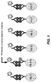
- a universal antibody which binds to all hemoglobin species can be used for the interrogation, but two or more detection antibodies can also be used. An example is shown in FIG.
- the assay is of particular value to samples of human blood.
- Blood samples are prepared for the assay by lysis of the cells and dilution of the lysate to a concentration suitable for immunoassay. Each of these steps is performed by methods known in the art. Dilution can be achieved with water, solutions containing saponin, or any other diluent that will not affect the hemoglobins or their immunological binding activity, and the degree of dilution can vary widely. In most cases, the dilution will be within the range of about 1:25 to about 1:3000.
- the hemoglobins in the lysate can be denatured before or after dilution of the lysate and used in the assay, or the lysate can be used without denaturation of the hemoglobins. In most cases, denaturation is preferred, and can be performed by methods known in the art.
- the levels of HbA 1c and each of the variants are preferably each expressed as a percentage of total hemoglobin in the sample.
- the invention offers three options. One option, which is compatible with the currently accepted method, is the determination of the HbA 1c , level by the result from the HbA 1c bead only, normalized to total hemoglobin. The second option is the determination of the total hemoglobin glycation by adding the percent of the glycated form of the variant to the percent HbA 1c .
- the third option which is useful in the event that the determination of HbA 1c is adversely affected by the presence of the variant, is to adjust the as-measured percent HbA 1c by a correction factor that is a function of the detected level of the variant.
- the function can be determined empirically by a relation that can be independently determined by separate assays, including non-multiplex assays.
- the correction factor can be one that is applied either to the HbA 1c concentration after the concentration has been normalized with respect to total hemoglobin, or to the concentration prior to normalization.
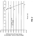
- the mathematical model used in this example is a simple linear regression model. Using this model, the values obtained from the BioPlex 2200 immunoassay can be corrected to yield a result comparable to the reference method. This is demonstrated by the average difference of the adjusted BioPlex 2200 percent HbA lc value relative to the target percent HbA 1c , value determined by the reference Variant II method. In Table 3 and FIG. 2 , the average difference is zero for the adjusted HbA 1c values compared to -0.33 for the corresponding unadjusted values. The corrected HbA 1c value shown in the table provides a better estimate of the glycemic index of the individual.
- the BioPlex 2200 bead-based immunoassay used in the obtaining the data in Table 3 and FIG. 2 utilizes an antibody that binds HbA 1c and all hemoglobin variants including HbS, HbC, HbD, and HbE. All glycated variants and HbA 0 are bound with approximately the same affinity and avidity by the antibody.
- the immunoassay result thus represents total glycated hemoglobin, which in the case of a heterozygous hemoglobin AS variant, is the combined value that includes both the HbA 1c and HbS 1c species.
- the proportion of the glycated hemoglobin corresponding to the variant provides an improved measure of glycemic status.
- the percent HbS 1c in the sample is obtained by multiplying the total percent HbA lc plus HbS 1c value by the proportion of HbS in the sample. For example, for a patient sample with a total glycated hemoglobin value of 5.54% (consisting of HbA lc and HbS 1c ), multiplying this value by the proportion of HbS in the sample of 38.8% yields a value for HbS 1c of 2.14%.
- the remainder of the glycated material is HbA 1c at 3.4%. This again is but one mathematical model; more sophisticated mathematical models can be used to provide more accurate results as needed.
- the beads that provide the surfaces on which the binding reactions occur in the practice of this invention can be formed of any material that is inert to the assay materials and to the components of the sample itself, and that is solid and insoluble in the sample and in any other solvents or carriers used in the assay. Polymers are preferred, and the beads are preferably microparticles.
- the polymeric can be any material that can be formed into a microparticle and is capable of coupling to an antibody at a region on the antibody that does not interfere with the antigen-binding regions of the antibody. In embodiments in which fluorescent labels are used, preferred polymers are also those that produce at most a minimal level of autofluorescence.
- suitable polymers are polyesters, polyethers, polyolefins, polyalkylene oxides, polyamides, polyurethanes, polysaccharides, celluloses, and polyisoprenes.
- Crosslinking is useful in many polymers for imparting structural integrity and rigidity to the microparticle. Magnetic beads can also be used.
- Attachment of the antibodies to the surfaces of the beads can be achieved by electrostatic attraction, specific affinity interaction, hydrophobic interaction, or covalent bonding. Covalent bonding is preferred.
- Functional groups for covalent bonding can be incorporated into the polymer structure by conventional means, such as the use of monomers that contain the functional groups, either as the sole monomer or as a co-monomer. Examples of suitable functional groups are amine groups (-NH 2 ), ammonium groups (-NH 3 + or-NR3 + ), hydroxyl groups (-OH), carboxylic acid groups (-COOH), and isocyanate groups (-NCO).
- Useful monomers for introducing carboxylic acid groups into polyolefins, for example, are acrylic acid and methacrylic acid.
- Linking groups can also be used for increasing the density of the antibodies on the solid phase surface and for decreasing steric hindrance to increase the range and sensitivity of the assay.
- suitable useful linking groups are polylysine, polyaspartic acid, polyglutamic acid and polyarginine.
- the size range of the beads can vary and particular size ranges are not critical to the invention. In most cases, the aggregated size range of the beads lies within the range of from about 0.3 micrometers to about 100 micrometers in diameter, and preferably within the range of from about 0.5 micrometers to about 40 micrometers.
- Multiplexing with the use of beads in accordance with this invention is achieved by assigning the beads to two or more groups, also referred to herein as bead sets or subpopulations.
- Each group will have affixed thereto an antibody selected for either a hemoglobin variant, a glycated variant, HbA 1c , or total hemoglobin, and will be separable or at least distinguishable from the other group(s) by a "differentiation parameter.”
- the "differentiation parameter" can be any distinguishable characteristic that permits separate detection of the assay result in one group from those in the other groups.
- One example of a differentiation parameter is the particle size, with each group having a size range that does not overlap with the size ranges of the other groups.
- the widths of the size ranges and the spacing between mean diameters of different size ranges are selected to permit differentiation of the groups by flow cytometry according to size, and will be readily apparent to those skilled in the use of and instrumentation for flow cytometry.
- the term "mean diameter" refers to a number average diameter.
- a preferred size range width is one with a CV of about ⁇ 5% or less of the mean diameter, where CV is the coefficient of variation and is defined as the standard deviation of the particle diameter divided by the mean particle diameter times 100 percent.
- the minimum spacing between mean diameters among the various size ranges can vary depending on the size distribution, the ease of segregating beads by size for purposes of attaching different antibodies, and the type and sensitivity of the flow cytometry equipment.
- the mean diameters of different size ranges are spaced apart by at least about 6% of the mean diameter of one of the size ranges, preferably at least about 8% of the mean diameter of one of the size ranges and most preferably at least about 10% of the mean diameter of one of the size ranges.
- Another preferred size range width relation is that in which the standard deviation of the particle diameters within each size range is less than one third of the separation of the mean diameters of adjacent size ranges.
- Another example of a differentiation parameter that can be used to distinguish among the various groups of beads is fluorescence. Differentiation by fluorescence is accomplished by incorporating fluorescent materials in the beads, the materials having different fluorescent emission spectra for each group of beads and being distinguishable on this basis.
- Fluorescence can thus be used both as a differentiation parameter and as a means for detecting that binding has occurred in the assays performed on the beads.
- the latter can be achieved by fluorescent labels serving as assay reporters.
- individual groups can be distinguished by emitting different emission spectra, and the emission spectra used for group differentiation purposes can themselves differ from the emission spectra of the assay reporters.
- An example of a fluorescent substance that can be used as a differentiation parameter is fluorescein and an example of a substance that can be used for the assay detection is phycoerythrin.
- Different bead groups can be distinguished from each other by being dyed with different concentrations of fluorescein.
- Different bead groups can be distinguished by using fluorescent materials that have different fluorescence intensities or that emit fluorescence at different wavelengths.
- the dyes can also be used in combinations to produce a plurality of fluorescent emissions at different wavelengths, and the wavelength difference can be used both as the differentiation parameter and as a means of distinguishing the differentiation parameter from the assay reporter.
- differentiation parameters are light scatter, light emission, or combinations of light scatter and emission.
- Side-angle light scatter varies with particle size, granularity, absorbance and surface roughness, while forward-angle light scatter is mainly affected by size and refractive index. Any of these qualities can thus be used as the differentiation parameter.
- the beads will have two or more fluorochromes incorporated within them so that each bead in the array will have at least three distinguishable parameters associated with it, i.e., side scatter together with fluorescent emissions at two separate wavelengths.
- a red fluorochrome such as Cy5 can thus be used together with an orange fluorochrome such as Cy5.5. Additional fluorochromes can be used to expand the system further.
- Each bead can thus contain a plurality of fluorescent dyes at varying wavelengths.
- Still another example of a differentiation parameter that can be used to distinguish among the various groups of beads is absorbance.
- absorbance When light is applied to beads the absorbance of the light by the beads is indicated mostly by the strength of the laterally (side-angle) scattered light while the strength of the forward-scattered light is relatively unaffected. Consequently, the difference in absorbance between various colored dyes associated with the beads is determined by observing differences in the strength of the laterally scattered light.
- a still further example of a differentiation parameter that can be used to distinguish among the various groups of beads is the number of beads in each group.
- the number of beads of each group in an assay is varied in a known way, and the count of beads having various assay responses is determined.
- the various responses are associated with a particular assay by the number of beads having each response.
- the differentiation parameters may arise from size, composition, physical characteristics that affect light scattering, excitable fluorescent or colored dyes that impart different emission spectra and/or scattering characteristics to the beads, or different concentrations of one or more fluorescent dyes.
- the differentiation parameter is a fluorescent dye or color, it can be coated on the surface of the beads, embedded in the beads, or bound to the molecules of the bead material.
- fluorescent beads can be manufactured by combining the polymer material with the fluorescent dye, or by impregnating the beads with the dye.
- Beads with dyes already incorporated and thereby suitable for use in the present invention are commercially available, from suppliers such as Spherotech, Inc. (Libertyville, Illinois, USA) and Molecular Probes, Inc. (Eugene, Oregon, USA).
- a list of vendors of flow cytometric products can be found on the Internet, e.g., at the world wide web molbio.princeton.edu/facs/FCMsites.html site.
- Detection and differentiation in accordance with this invention are performed by flow cytometry.
- Methods of and instrumentation for flow cytometry are known in the art, and those that are known can be used in the practice of the present invention.
- Flow cytometry in general resides in the passage of a suspension of beads or microparticles as a stream past a light beam and electro-optical sensors, in such a manner that only one particle at a time passes through the region. As each particle passes this region, the light beam is perturbed by the presence of the particle, and the resulting scattered and fluorescent light are detected.
- the optical signals are used by the instrumentation to identify the subgroup to which each particle belongs, along with the presence and amount of label, so that individual assay results are achieved.
- This example presents the binding activities of six hemoglobin candidate antibodies for use in the practice of this inveniton.
- the six antibodies are: 19E10-E7 (HbS specific) 7B3-2C3-1G10 (HbD specific) 12C8-A11 (HbC specific) 13G7-E8-3H3 (HbA 1c specific) 4A10-2D6-2G8 (HbE specific) 3E5-DLE10-3A3 (pan-reactive).
- 19E10-E7 binds to a beta globin minimal epitope 5 PVEKSAVT 12 .
- 5 PVE 7 and A 10 are important for binding.
- L 3 and T 4 also contribute to binding activity. Additional epitope mapping experiments showed that V 6 can be replaced with I without loss of binding activity.
- T 4 and K 6 are important for binding.
- L 3 also contributes to binding. Additional epitope mapping experiments showed that K 6 can be replaced with R without reducing binding activity.
- 4A10-2D6-2G8 binds a beta globin minimal epitope 22 EVGGK 26 .
- 22 EV 23 and K 26 are important for binding.
- D 21 also contributes to binding.
- Additional epitope mapping experiments showed that K 26 can be replaced with S or T without loss of binding activity.
- 7B3-2C3-1G10 binds to a beta globin minimal eptiope 121 QFTPP 125 .
- G 119 also contributes to binding.
- the antibody binding kinetics were analyzed using the ProteOn XPR36 (Bio-Rad Laboratories, Inc.) for protein-protein interactions.
- the different antibodies (10 ⁇ g of each) were amine-coupled to the sensor chip such that one antibody was immobilized per channel.
- Antigen was employed in the range of from 200 to 13 nM. The results of the kinetic analysis are summarized in Table 4 below.
- Each antibody had a high affinity for its specific hemoglobin.
- the pan-reactive antibody also had good affinity constants to the different antigens, but lower than the affinity constant exhibited by the specific variant antibodies for their respective antigens. Except for 19E10-E7, all of the variant antibodies did not bind HbA0, so the affinity constants were essentially zero. For the 19E10-E7 anti HbS antibody, a low level of binding to HbA0 was observed, with an affinity constant of 6.5 10 -7 M, which is 2 logs less than the affinity constant for HbS.
- Antibodies to each of the six target antigens were coupled covalently to paramagnetic beads. Each bead was dyed to contain a specific fluorescent signal that was unique to each antibody, to enable subsequent differentiation in a flow cytometry detector. The six antibody-coupled beads were mixed to create a multiplex bead reagent. A detection antibody reagent was prepared using a phycoerythrin-labelled polyclonal antibody with reactivity to all hemoglobin species. A diagram of the various beads and the species bound to each in the assay is shown in FIG. 1 .
- the assay was performed by adding samples of whole blood and calibrators (5 ⁇ L) to a solution of buffered denaturant (10 ⁇ L), to expose all of the epitopes of the hemoglobin species present in the samples in order to make them available for binding by the solid phase antibodies. After denaturation for 10 minutes at 37 degrees, the bead reagent (250 ⁇ L) was added to the samples, followed by an additional incubation for 20 minutes at 37 degrees. The reaction mixture was washed four times with phosphate-buffered saline containing 0.1% Tween-20 (PBST, 100 ⁇ L each) detergent to remove all of the unbound proteins from the sample, leaving the beads with their bound hemoglobin targets. The beads were re-suspended in PBST containing phycoerythrin-labelled antibody reagent (25 ⁇ L), and incubated for 20 minutes at 37 degrees Celsius.
- PBST phosphate-buffered saline containing 0.1% Tween-20
- the beads were resuspended in PBST and processed through a Luminex flow cytometry detector to interrogate the beads for binding of the individual hemoglobin species present in the samples.
- samples from homozygous hemoglobin AA individuals exhibited signal from the HbA1c and HbAO beads
- samples from heterozygous hemoglobin variant individuals exhibited signal from the HbA 0 , HbA 1c and the specific hemoglobin variant beads.
- the phycoerythrin-derived fluorescent signal of each bead was measured for the samples and calibrators.
- a calibration curve was constructed for each hemoglobin analyte using the signal from the bead and the known dose of the respective calibrators.
- the concentration of the hemoglobin analytes in each sample was determined from their fluorescent signal and the established dose-response of the calibration curve. Percent HbA 1c and percent hemoglobin variant, if any, in the samples were determined by dividing the concentration of hemoglobin A 1c or variant hemoglobin protein by the concentration of HbA 0 , each derived from their respective bead. In the case of heterozygous hemoglobin variant-containing samples (such as HbAS, for example), the percent A 1c value derived from the ratio of the A 1c to HbA 0 concentrations was adjusted when needed using the concentration of the hemoglobin variant present in the sample, to provide a value that best reflected the true glycemic index of the individual.
- a method for individually detecting a plurality of hemoglobin-containing analytes comprising HbA 1c , and a plurality of hemoglobin variants in a single sample of blood cell lysate comprising:
- said plurality of hemoglobin-containing analytes consists of total hemoglobin, HbA 1c , and said plurality of hemoglobin variants.
- the immunological binding member having selective binding affinity for HbA 1c is a monoclonal antibody.
- said plurality of hemoglobin variants includes at least one of HbS, HbC, HbE, or HbD.
- said immunological binding member having selective affinity for hemoglobin variant HbC, HbS, HbD, or HbE is a monoclonal antibody that selectively binds the variant and the glycated variant.
- the variant is HbC and the immunological binding member having selective affinity for HbC and glycated HbC is a monoclonal antibody that binds to a HbC minimal epitope 4 TPKEKSAVT 12 .
- the variant is HbE and the immunological binding member having selective binding affinity for HbE and glycated HbE is a monoclonal antibody that binds to a HbE minimal epitope 22 EVGGK 26 or 21 DEVGGK 26 .
- said method further comprises determining total hemoglobin by a non-immunoassay method.
- said plurality of bead subpopulations comprises a subpopulation binding to total hemoglobin, wherein said immunological binding member having selective binding affinity toward total hemoglobin is a monoclonal antibody.
- said monoclonal antibody binds to a beta globin chain minimal epitope 9 SAVTALWGKVNV 20 , 8 KSAVTALWGKVNV 20 , or 11 VTALW 15 .
- said plurality of bead subpopulations comprises a subpopulation binding to total hemoglobin, wherein said immunological binding member having selective binding affinity toward total hemoglobin is a polyclonal antibody.
- the blood cell lysate is a denatured blood cell lysate.
- the sample is from a diabetic patient.
- said plurality of analytes further comprises total hemoglobin.
- said plurality of bead subpopulations does not include a subpopulation binding to total hemoglobin, and said method further comprises determining total hemoglobin by a non-immunoassay method
- the immunological binding member having selective binding affinity for HbA 1c is a monoclonal antibody.
- said plurality of bead subpopulations comprises a subpopulation binding to total hemoglobin, wherein said immunological binding member having selective binding affinity toward total hemoglobin is a monoclonal antibody.
- said monoclonal antibody binds to a beta globin chain minimal epitope 9 SAVTALWGKVNV 20 , 8 KSAVTALWGKVNV 20 , or 11 VTALW 15 .
- said plurality of bead subpopulations comprises a subpopulation binding to total hemoglobin, and wherein said immunological binding member having selective binding affinity toward total hemoglobin is a polyclonal antibody.
- the blood cell lysate is a denatured blood cell lysate.
- said hemoglobin variant is selected from the group consisting of HbC, HbS, HbD, and HbE.
- the immunological binding member that selectively binds to hemoglobin variant HbC, HbS, HbD, or HbE is a monoclonal antibody that binds to the hemoglobin variant and the glycated variant.
- said hemoglobin variant is HbC and said immunological binding member having selective binding affinity for HbC is a monoclonal antibody that binds to a HbC minimal epitope 4 TPKEKSAVT 12 .
- said hemoglobin variant is HbS and the immunological binding member having selective binding affinity for the variant is a monoclonal antibody that binds to a HbS minimal epitope 3 LTPVEKSAVT 12 .
- hemoglobin variant is HbE and the immunological binding member having selective binding affinity for the variant is a monoclonal antibody that binds to a HbE minimal epitope 22 EVGGK 26 or 21 DEVGGK 26 .
- said hemoglobin variant is HbD and the immunological binding member having selective binding affinity for the variant is a monoclonal antibody that binds to a HbD minimal epitope 121 QFTPP 125 or 119 GKQFTPP 125 .
- the sample is from a diabetic patient.
- a method for determining the proportion of a glycated hemoglobin variant relative to total hemoglobin in a sample of blood cell lysate comprising:
Landscapes
- Health & Medical Sciences (AREA)
- Life Sciences & Earth Sciences (AREA)
- Engineering & Computer Science (AREA)
- Chemical & Material Sciences (AREA)
- Immunology (AREA)
- Hematology (AREA)
- Molecular Biology (AREA)
- Biomedical Technology (AREA)
- Urology & Nephrology (AREA)
- Medicinal Chemistry (AREA)
- General Health & Medical Sciences (AREA)
- Biochemistry (AREA)
- Physics & Mathematics (AREA)
- Pathology (AREA)
- Microbiology (AREA)
- Analytical Chemistry (AREA)
- Cell Biology (AREA)
- Biotechnology (AREA)
- General Physics & Mathematics (AREA)
- Food Science & Technology (AREA)
- Organic Chemistry (AREA)
- Biophysics (AREA)
- Genetics & Genomics (AREA)
- Proteomics, Peptides & Aminoacids (AREA)
- Peptides Or Proteins (AREA)
- Investigating Or Analysing Biological Materials (AREA)
- Preparation Of Compounds By Using Micro-Organisms (AREA)
- Measuring Or Testing Involving Enzymes Or Micro-Organisms (AREA)
Abstract
Description
- This application claims the benefit of United States Provisional Patent Application No.
61/262,488, filed November 18, 2009 - This invention lies in the field of assays for glycated hemoglobin.
- For individuals suffering from type 1 or type 2 diabetes mellitus, maintenance of glycemic control is of prime importance, and such maintenance requires the determination of the level of hemoglobin A1c in the blood of these individuals. With diabetes reaching global epidemic proportions, it is particularly important to have accurate and reproducible HbA1c assays. HbA1c assays are also used in the screening of individuals for diabetes.
- HbA1c measurements for both patient monitoring and screening are taken as an average over the lifetime of an erythrocyte. This average is compromised by several physiological conditions, notable among which are the presence of hemoglobin variants and thalassemias in the patient's blood. Hemoglobin variants are prevalent among certain ethnic groups and in certain geographical regions. Of the over 800 variants known worldwide, the most common are HbS, HbC, HbD, and HbE. HbS is most prevalent among individuals of African descent, HbD among individuals of Punjabi Indian descent, and HbE among individuals of Southeast Asia. Other known forms of hemoglobin are HbF (fetal hemoglobin) and HbA2, both of which can be elevated in thalassemia, a relatively common condition characterized by an imbalance of hemoglobin alpha and beta subunits. Beta thalassemias can also occur in the presence of HbE and HbS, and the combined sickle/beta thalassemia trait occurs most frequently among individuals of Mediterranean descent. Variants and thalassemias can cause inaccuracies in HbA1c measurements by affecting such factors as red blood cell survival and glycosylation rates. Variants also affect immunologically determined levels of glycated hemoglobin since immunoreactivity differs from one glycated variant to the next and also between glycated variants and HbA itself. Health care professionals must therefore know of the presence of variants and their proportions relative to HbA as well as the presence of thalassemias to achieve a proper determination of glycemic control.
- Determinations of hemoglobin variants are typically done separately from determinations of HbA1c regardless of whether a variant is actually known to be present. Antibodies to specific variants have been developed for this purpose, and the following is a sampling of reports on such antibodies:
- HbS: Jensen, R.H., el al., "Monoclonal antibodies specific for sickle cell hemoglobin," Hemoglobin 9(4), 349-362 (1985)
- HbS: Epstein, N., et al., "Monoclonal antibody-based methods for quantitation of hemoglobins: application to evaluating patients with sickle cell anemia treated with hydroxyurea," Eur. J. Haemotol. 57(1), 17-24 (1996)
- HbA: Rosenthal, M.A., et al., "Binding specificity of a monoclonal antibody to human HbA," Hemoglobin 19(3-4), 191-196 (1995)
- HbS and HbC: Garver, E.A., et al., "Screening for hemoglobins S and C in newborn and adult blood with a monoclonal antibody in an ELISA procedure," Annals of Hematology 60(6), 334-338 (1990)
- Hb with single amino acid substitutions: Stanker, L.H., et al., "Monoclonal antibodies recognizing single amino acid substitutions in hemoglobin," J. Immunol. 136 (11), 4174-4180 (1986)
- Hb variants: Moscoso. H., et al., "Enzyme immunoassay for the identification of hemoglobin variants," Hemoglobin 14(4), 389-98 (1990)
- Hb variants: Schultz, J.C., "Utilization of monoclonal antibody-based assay HemoCard in screening for and differentiating between genotypes of sickle cell disease and other hemoglobinopathies," J. Clin. Lab. Anal. 9(6), 366-374 (1995)
- Despite these reports and others, determinations of variants are presently performed by either high performance liquid chromatography (HPLC) or electrophoresis. HPLC can indeed be a rapid means of obtaining the HbA1c level, but extended HPLC gradients are needed for detecting and quantifying the variants and thalassemias, since in HPLC impurities co-elute with the variants, and different variants tend to co-elute with each other. In fact, certain variants cannot be resolved by HPLC, even with the most optimized HPLC gradients. Typically, separate HPLC methods for rapid A1c measurements and variant and thalassemia testing are used, therefore making it impossible to simultaneously determine the A1c level and variant or thalassemia status by HPLC, much less in a rapid manner.
- Assays that provide simultaneous detection of multiple analytes are termed "multiplex" assays, and disclosures of multiplex assays using affinity-type binding reactions on the surfaces of beads that are then detected by flow cytometry are disclosed in the following patents:
- Watkins, M.I., et al., "Magnetic particles as solid phase for multiplex flow assays,"
US 6,280,618 B2, issued August 28, 2001 - Watkins, M.I., et al., "Magnetic particles as solid phase for multiplex flow assays,"
US 6,872,578 B2, issued March 29, 2005 - Thomas, N., "Multiple assay method,"
US 6,913,935 B1, issued July 5, 2005 - Hechinger, M., "Platelet immunoglobulin bead suspension and flow cytometry,"
US 6,933,106 B1, issued August 23, 2005 - Hechinger, M., "Anti-platelet immunoglobulin bead positive control,"
US 6,951,716 B1, issued October 4, 2005 - Watkins, M.I., et al., "Multi-analyte diagnostic test for thyroid disorders,"
US 7,271,009 B1, issued September 8, 2007 - Bell, M.L., "Assay procedures and apparatus,"
US 7,326,573 B2, issued February 5, 2008 - Song, Y., et al., "Multiplex protein interaction determinations using glutathione-GST binding,"
US 2002/0115116 A1, published August 22, 2002 - The success of multiplex assays for certain combinations of analytes does not however provide assurance, or even a high level of expectation, that similar success will be achieved for all combinations of analytes, particularly combinations with a high level of homology among the analytes. Hemoglobin and its variants and glycated forms are one such combination. Multiplex assays involve a plurality of different immunoreactants in intimate mixture in a common reaction medium, which creates competition among the immunoreactants for the different analytes, more so than in media where a single immunoreactant is present, and the cross-reactivities occur in multiple directions. The bead sets themselves must also be differentiated at the same time as the immunoassays are performed. This differentiation, whether by the use of different dyes on different bead sets, a different size for each bead set, or other known differentiation factors, adds a further level of complexity and further opportunities for cross-talk.
- The present invention resides in the discovery that hemoglobin variants can be differentiated from each other and from HbA1c and from total hemoglobin, and the levels of each measured, in a multiplex immunoassay. The assay can, e.g., detect a single variant in addition to HbAlc and total hemoglobin or two or more variants and total hemoglobin. When two or more variants are detected, different combinations of variants can be selected, although preferably the assay will include the four most common variants, HbS, HbC, HbE, and HbD. The assay may also include the measurement of HbA2 and HbF. The invention thus resides in a method for detecting and identifying the presence of hemoglobin variants in a patient's blood. The invention also resides in a method for measuring the level of HbA1c relative to total hemoglobin while correcting the result for the presence of variants that may also be present. Here as well, the correction can be for individual variants or combinations of variants. The invention also resides in a method for the simultaneous detection of A1c and hemoglobin variants without correction, which is useful in certain cases. A still further aspect of the invention is the measurement of levels of particular variants in glycated form. When a variant is known to be present, the glycated version of that variant can be measured and added to the level of HbA1c to obtain an accurate indication of total glycated hemoglobin.
- In a further aspect, the invention provides antibodies having selective binding affinity for hemoglobin variants that can be used in the methods of the invention. In some embodiments, the invention provides a monoclonal antibody that has selective binding affinity for HbC and glycated HbC, wherein the monoclonal antibody binds to an HbC minimal epitope 4TPKEKSAVT12; or to an HbC minimal epitope comprising the amino acid sequence TX1KE or LTX1KE, wherein X1 is one of the 20 common naturally occurring amino acids. In some embodiments, the invention provides a monoclonal antibody having selective binding affinity for HbS and glycated HbS, wherein the monoclonal antibody binds to an HbS minimal epitope 3LTPVEKSAVT12; or to an HbS minimal epitope comprising the amino acid sequence PVEX2X3A or LTPVEX2X3A, wherein each of X2 and X3 is an amino acid independently selected from the 20 common naturally occurring amino acids. In some embodiments, the invention provides a monoclonal antibody having selective binding affinity for HbE and glycated HbE, wherein the antibody binds to an HbE minimal epitope 22EVGGK26; or to an HbE minimal epitope comprising the amino acid sequence DEVGGK or EVX4X5K, wherein each of X4 and X5 is an amino acid independently selected from the 20 common naturally occurring amino acids. In some embodiments, the invention provides a monoclonal antibody having selective binding affinity for HbD and glycated HbD, where the monoclonal antibody binds to an HbD minimal epitope 121QFTPP125; or to an HbD minimal epitope comprising the amino acid sequence GX6QFX7PP or QFX7PP, wherein each of X6 and X7 is an amino acid independently selected from the 20 common naturally occurring amino acids.
- The invention also may employ an antibody, either a polyclonal antibody or monoclonal antibody that selectively binds total hemoglobin (in comparison to non-hemoglobin polypeptides). In some embodiments, a pan-reactive polyclonal antibody for use in the invention binds to one ore more epitopes present in the following regions of alpha globin and beta globin: 49SHGSAQVKGHGKKVADALTNAVAHVDDMPNALSALSDHLHA HKLRRVDPV96, beta globin l5WGKVNVDEVGGEALG30, 45FGDLSTP51, and 76AHLDNLKGTFAT87. In some embodiments, a pan reactive antibody is a monoclonal or polyclonal antibody that binds to the epitope 9SAVTALWGKVNV20 (beta globin) or 8KSAVTALWGKVNV20 or 11VTALW15 or to a beta globin minimal epitope that comprises the sequence ALWG or VTX9LW, wherein X9 is one of the 20 common naturally occurring amino acids. An antibody for use in the invention may bind to an epitope on beta globin, e.g. 8KSAVTALWGKVNV20, 58PKVKAHGKKVLGAF71 or 87TLSELHCDKLHVDPENFR104 (beta globin).
- The invention further provides monoclonal antibodies that selectively bind to glycated forms of hemoglobin, including binding to both normal and variant hemoglobins, but do not bind to non-glycated forms of hemoglobin. A glycosylated residue 1V and residue 2H are typically important for binding for such antibodies.
- The method of the invention may additionally comprise detecting other hemoglobin variants using antibodies, e.g., monoclonal antibodies, that selectively bind such variants.
- In typical embodiments, an antibody for use in the invention has a KD that is anywhere in the range of from about 10-6 M to about 10-12 M. In some embodiments, the antibody has a KD that is anywhere in the range of from about 10-7 M to about 10-11 M. In other embodiments, the antibody has a KD anywhere in the range of about 10-8 M to about 10-10 M. Typically the KD is in the nM range, e.g., anywhere from about 10-9 M to about 10-10 M.
- These and other features, objects, and advantages of the invention will be better understood from the description that follows.
-
-
FIG. 1 provides a schematic depicting an example of a sandwich immunoassay for measuring glycated hemoglobin and hemoglobin variants. -
FIG. 2 is a plot of comparative data between a series of multiplex assays in accordance with the present invention and a series of HPLC assays. - The hemoglobin variants to be detected by the present invention are any of the known variants reported in the literature or otherwise known to clinicians and researchers skilled in technology of hemoglobin, glycated hemoglobin, and diabetes. As noted above, the four most common hemoglobin variants are HbS, HbC, HbE, and HbD, although other variants can be detected in addition to these four or in place of one or more of them. For example, two variants that are elevated in beta thalassemia are HbF and HbA2. The binding members used for each of these variants in the multiplex assay are generally monoclonal antibodies, preferably those that are developed expressly for the multiplex assay. The antibodies preferably bind to epitopes on the variants that distinguish each variant from the other variants to minimize cross-reactivity, and most importantly that distinguish the variants from the wild-type hemoglobin A0. In embodiments of the invention requiring the use of a value for the concentration of total hemoglobin in the sample, the concentration can be determined either by an immunoassay method or a non-immunoassay method. An example of a non-immunoassay method is the determination of optical density. Other examples will be readily apparent to those skilled in the hemoglobin art. In embodiments where total hemoglobin is determined by immunoassay, the determination can be performed as part of the multiplex assay. The antibody for total hemoglobin in the multiplex assay can be either a monoclonal antibody or a polyclonal antibody, and the antibody for HbA1c can be either a polyclonal antibody or a monoclonal antibody, preferably a monoclonal antibody.
- As used herein, an "antibody" refers to a protein functionally defined as a binding protein and structurally defined as comprising an amino acid sequence that is recognized by one of skill as being derived from the framework region of an immunoglobulin-encoding gene of an animal that produces antibodies. An antibody can consist of one or more polypeptides substantially encoded by immunoglobulin genes or fragments of immunoglobulin genes. The recognized immunoglobulin genes include the kappa, lambda, alpha, gamma, delta, epsilon and mu constant region genes, as well as myriad immunoglobulin variable region genes. Light chains are classified as either kappa or lambda. Heavy chains are classified as gamma, mu, alpha, delta, or epsilon, which in turn define the immunoglobulin classes, IgG, IgM, IgA, IgD and IgE, respectively.
- A typical immunoglobulin (antibody) structural unit is known to comprise a tetramer. Each tetramer is composed of two identical pairs of polypeptide chains, each pair having one "light" (about 25 kD) and one "heavy" chain (about 50 kD). The N-terminus of each chain defines a variable region of about 100 to 110 or more amino acids primarily responsible for antigen recognition. The terms variable light chain (VL) and variable heavy chain (VH) refer to these light and heavy chains, respectively.
- The term antibody as used herein includes antibody fragments that retain binding specificity. For example, there are a number of well characterized antibody fragments. Thus, for example, pepsin digests an antibody C-terminal to the disulfide linkages in the hinge region to produce F(ab)'2, a dimer of Fab which itself is a light chain joined to VH-CH1 (Fd) by a disulfide bond. The F(ab)'2 may be reduced under mild conditions to break the disulfide linkage in the hinge region thereby converting the (Fab')2 dimer into an Fab' monomer. The Fab' monomer is essentially a Fab with all or part of the hinge region (see, Fundamental Immunology, W.E. Paul, ed., Raven Press, N.Y. (1993), for a more detailed description of other antibody fragments). While various antibody fragments are defined in terms of the digestion of an intact antibody, one of skill will appreciate that fragments can be synthesized de novo either chemically or by utilizing recombinant DNA methodology. Thus, the term "antibody" also includes antibody fragments produced either by the modification of whole antibodies or synthesized using recombinant DNA methodologies. Antibodies include dimers such as VH-VL dimers, VH dimers, or VL dimers, including single chain antibodies. Alternatively, the antibody can be another fragment, such as a disulfide-stabilized Fv (dsFv). Other fragments can also be generated using known techniques, including using recombinant techniques. In some embodiments, antibodies include those that have been displayed on phage or generated by recombinant technology using vectors where the chains are secreted as soluble proteins, e.g., scFv, Fv, Fab, (Fab')2 or generated by recombinant technology using vectors where the chains are secreted as soluble proteins.
- As used here, an "immunological binding member having selective binding affinity" for an antigen, e.g., a hemoglobin variant, is typically an antibody. In some embodiments, a binding member having selective binding affinity for an antigen may be a peptide, e.g., that can be identified by screening peptide libraries, that has a selective binding interaction with the antigen.
- In one aspect, the invention provides monoclonal antibodies that bind to Hb A1c as well as monoclonal antibodies that specifically bind to hemoglobin variants HbS, HbC, HbE, and HbD. The sequence of hemoglobin beta chain is as follows:The positions of amino acid residues in the hemoglobin beta chain referred to herein is with reference to this amino acid sequence unless otherwise specified.

- Hb A1c is glycated at the N-terminal valine. The most prevalent beta-chain point mutations are HbS (Glu 6 → Val); HbC (Glu 6 → Lys); HbE (Glu 26 → Lys) and HbD (Glu 121 → Gln). The Glu 6, Glu 26, and Glu 121 positions are indicated in the beta chain sequence by underline.
- HbA2 and HbF can also be determined in the assay of the present invention. Hemoglobin A2 has two alpha chains and two delta chains; and hemoglobin F has two alpha and two gamma chains.
- In the context of this invention, the term "specifically binds" or "specifically (or selectively) immunoreactive with," or "having a selective affinity for" refers to a binding reaction where the antibody binds to the antigen of interest. In the context of this invention, the antibody binds to the antigen of interest, e.g., HbS, including the glycated form of HbS, with an affinity that is at least 100-fold better than its affinity for other antigens, e.g., other hemoglobin variants such as HbA0 or HbC.
- "Reactivity" as used herein refers to the relative binding signal from the reactions of an antibody with the antigen to which it specifically binds versus other antigens, such as other hemoglobin variants and or wild-type HbA0. Reactivity is assessed using appropriate buffers that permit the antigen and antibody to bind. Reactivity can be determined, e.g., using a direct or sandwich ELISA assay. For example, a direct format assay for determining reactivity with wildtype hemoglobin and/or hemoglobin variants, can be used in which the antigen is directly bound to the ELISA plate, and the various antibodies are added to see which ones bind, followed by interrogation using a labeled anti-mouse antibody such as a phycoerythrin-labeled antibody. In the sandwich format, the monoclonal antibody is bound to the bead, followed by addition of antigen, followed by interrogation with phycoerythrin-labeled universal detection antibody, e.g., a phycoerythrin-labeled universal detection antibody, that binds all hemoglobin species. Thus, in an example using the sandwich format, reactivity can be defined as the relative fluorescent signal produced when the specific antigen, e.g., an HbS hemoglobin variant, is bound versus another antigen, e.g., a wildtype hemoglobin. An antibody is considered to be specific for an antigen if it exhibits a 2-fold, typically at least a 3- or 4-fold increase, in reactivity for the reference antigen compared to another antigen that is tested.
- "Epitope" or "antigenic determinant" refers to a site on an antigen to which an antibody binds. Epitopes can be formed both from contiguous amino acids or noncontiguous amino acids juxtaposed by tertiary folding of a protein. Epitopes formed from contiguous amino acids are typically retained on exposure to denaturing solvents whereas epitopes formed by tertiary folding are typically lost on treatment with denaturing solvents. An epitope typically includes at least 3, and more usually, at least 5 or 8-10 amino acids in a unique spatial conformation. Methods of epitope mapping are well known in the art (see, e.g., Epitope Mapping Protocols in Methods in Molecular Biology, Vol. 66, Glenn E. Morris, Ed (1996)). A "minimal" epitope in the current invention is typically determined by measuring binding of the antibody to overlapping peptides covering the entire amino acid sequence of beta or alpha globin and identifying the amino acid sequence shared by all bound peptides. Important amino acids in the "minimal" epitope are typically identified by alanine scanning.
- As understood in the art, a "minimal" epitope may include substitutions, e.g., at positions that are not important for binding, e.g., as determined using alanine scanning. Such substitutions include conservative substitutions where the alteration results in the substitution of an amino acid with a chemically similar amino acid. Conservative substitution tables providing functionally similar amino acids are well known in the art. The following are examples from among the twenty common naturally occurring amino acids of amino acids that may be substituted for one another: alanine and glycine; aspartic acid and glutamic acid; asparagine and glutamine; arginine and lysine; serine and threonine. Other conservative substitutions include substitutions of one amino acid in the following group with another amino acid in the group: isoleucine, leucine, methionine, and valine. Phenylalanine, tyrosine, and tryptophan are also examples of residues that may be substituted for one another.
- Table 1 provides examples of immunogens utilized to generate specific monoclonal antibodies to hemoglobin and hemoglobin variants.
Table 1 Examples of Peptide Immunogens Hemoglobin target Peptide Name sequence Hemoglobin and variants H1 H2N-VHLTPEEKSAVTALW-C-CONH2 H2 H2N-VHLTPEEASASTASW-C-CONH2 H2bis H2N-VHLTPEEKSASTASW-C-CONH2 HbS H3 H2N-VHLTPVEKSAVTALW-C-CONH2 HbC H4 H2N-VHLTPKEKSAVTALW-C-CONH2 HbE H5 H2N-CYG-NVDEVGGKALGRLLV-CONH2 H5bis H2N-CYG-VTALWGKVNVDEVGGK-CONH2 H10 H2N-C-Hx-EVGGKALG-CONH2 H10bis H2N-E V GGKALG-Hx-C-CON H2 HbD H6 H2N-CYG-VLAHHFGKQFTPPVQAA-CONH2 H6bis H2N-QFTPPVQAAYQKVVAGV-GYC-CONH2 H9 H2N-GKQFTGKQFTGKQFT-GYC-CONH2 H11 H2N-C-Hx-HFGKQFTP-CONH2 H11bis H2N-HFGKQFTP-Hx-C-CONH2 HbA1c GP1 Glucose-HN-VHLTPEE-Hx-C-CONH2 GP3 1-deoxyfructopyranosyl-HN-VHLTPEE-Hx-C-CONH2 Glycated H2 Glucose-HN-VHLTPEEASASTASW-C-CONH2 - Table 2 provides examples of immunization regimens utilized to generate specific monoclonal antibodies to hemoglobin and hemoglobin variants.
Table 2 Examples of immunization regimens Injection sequence HbS HbC HbE HbD HbA1c HbA and variants 1 native HbS antigen H4-KLH H5bis-KLH denatured HbD + H6-KLH GP3-KLH native HbA0 2 H3-KLH H4-KLH H5bis-KLH denatured HbD + H6-KLH GP3-KLH H1-KLH 3 H3-KLH H4-KLH H5bis-KLH denatured HbD + H6-KLH GP3-KLH native HbA0 4 denatured HbS + H3-KLH denatured HbC H5bis-KLH denatured HbD + H6-KLH GP3-KLH H1- KLH 5 denatured HbS + H3-KLH H4-KLH denatured HbE denatured HbD + H6-KLH 6 denatured HbS + H3-KLH H4-KLH denatured HbE denatured HbD 7 denatured HbE denatured HbD + H6-KLH 8 H5bis-KLH denatured HbD + H6-KLH 9 H5bis-KLH denatured HbD + H6- KLH 10 denatured HbE + H5bis-KLH denatured HbD + H6-KLH 11 denatured HbE + H5bis-KLH 12 denatured HbE + H5bis-KLH Route of injection Subcutaneous And intraperitoneal Subcutaneous and intraperitoneal Subcutaneous and intraperitoneal Subcutaneous and intraperitoneal Intraperitoneal Subcutaneous and intraperitoneal - Hemoglobin variant HbS has a point mutation in which glutamic acid at position 6 of the hemoglobin beta chain is mutated to a valine.
- Anti-HbS antibodies of the invention that are selective for HbS have the following binding characteristics: the antibody bind to HbS with an affinity that is at least 100-fold lower (i.e., better) than its affinity for HbC and HbA0. In some embodiments, the antibody binds to the minimal HbS epitope 5PVEKSAVT12. Such an antibody may have a reactivity in which the reactivity is such that the valine at position 6 can be replaced by an isoleucine, but replacement with other amino acids at that position results in a 2-fold, often a three-fold or greater decrease in reactivity. In some embodiments, 3LTP5, 7E, and 10A are also important for binding.
- In some embodiments, the antibody binds to a minimal epitope 3LTPVEKSAVT12. In some embodiments, the antibody may have a reactivity where the valine at position 6 can be replaced by an isoleucine or alanine, but substitution with other amino acids at that positions results in a two-fold, often a three-fold or greater decrease in reactivity. In some embodiments, 2HLTPVEK8 and 10A are also important for binding.
- In some embodiments, an antibody that binds to an HbS minimal epitope, e.g., 5PVEKSAVT12, may bind to variants of the HbS minimal epitope that have the valine at position 6, such as a minimal epitope comprising 5PVEX2X3A10, where X2 and X3 can be independently selected from the 20 common naturally occurring amino acids, e.g., conservative substitutions of K and S, respectively.
- The antibody typically is an IgG, e.g., the antibody may have an IgG1, IgG2, or IgG3 isotype. In some embodiments, the light chain constant region is a kappa chain. In other embodiments, the light chain constant region may be a lambda chain.
- In one embodiment, an HbS antibody of the invention is raised against the immunogen HbS and H3-KLH : H2N-VHLTPVEKSAVTALW-C-CONH2. In other embodiments, the immunogen is either a combination of H3-KLH : H2N-VHLTPVEKSAVTALW-C-CONH2 and purified native and/or denatured HbS protein, or sequential or serial immunizations using the individual components of the above immunogens. Carrier proteins other than KLH can also be used. Examples are albumin and ovalbumin, and further examples will be readily apparent to those skilled in the art.
- As understood in the art and illustrated by Table 1 above, many variations of immunogens can be used to obtain the desired antibody. For example, peptide immunogen KLH : H2N-VHLTPVEKSAVTALW-C-CONH2 may also have a C-terminal carboxylate, rather than a C-terminal carboxamide. In some embodiments, the cysteine linker moiety may be spaced with a Hx residue, which is 6-amino hexanoic acid, or a spacer, such as a gly-gly spacer sequence may be employed. Further, the peptide sequence may also vary.
- An anti-HbS antibody typically binds to both glycated and nonglycated forms of HbS with similar affinity. For example, an anti-HbS antibody typically selectively binds to both glycated and non-glycated HbS with a binding reactivity in which there is less than a three-fold reactivity difference, typically less than a two-fold reactivity difference, between binding to glycated vs. non-glycated HbS.
- Hemoglobin variant HbC has a lysine substituted for the glutamic acid at position 6 of the hemoglobin beta chain. An anti-HbC monoclonal antibody for use in the invention typically binds to HbC with an affinity that is at least 100 times greater that the affinity of the antibody for HbS and HbA0. In some embodiments, the monoclonal antibody binds to the minimal epitope 4TPKEKSAVT12. In some embodiments, the antibody has a binding specificity such that residues important for binding are residues 3LT4 and 6K. In some embodiments, residues important for binding may be 3LT4 and 6KE7. The binding specificity also allows for substitution of lysine by arginine or histidine at position 6, but substitution of other amino acids results in at least a 2-fold, typically a 3-fold or greater loss in reactivity. In other embodiments, the reactivity of the HbC antibody is such that the lysine at position 6 may be substituted with an arginine, tyrosine, asparagine, glutamine or glycine, but substitution with other amino acids residues results in a loss of reactivity.
- In some embodiments, an antibody that binds to a HbC minimal epitope, e.g., 4TPKEKSAVT12, may bind to variants of the HbC minimal epitope that have the K at position 6, such as a minimal epitope comprising 4TX1KE7 or 3LTX1KE7 where X1 can be one of the 20 common naturally occurring amino acids.
- The antibody typically is an IgG, e.g., the antibody may have an IgG1, IgG2, or IgG3 isotype. In some embodiments, the light chain constant region is a kappa chain. In other embodiments, the light chain constant region may be a lambda chain.
- An antibody of the invention may be raised against the immunogen H4-KLH : H2N-VHLTPKEKSAVTALW-C-CONH2. Examples of other peptide immunogens are listed in Table 1, and here again, other common carrier proteins can be used in place of KLH. In some embodiments, the immunization is performed using a combination of the peptide and purified native and/or denatured HbC protein. In some embodiments, sequential or serial immunizations are performed using the individual components of the above immunogens. An exemplary immunization protocol is shown in Table 2. As explained above in the section relating to anti-HbS antibodies, one of skill can readily design other immunogenic peptides to obtain an antibody having the desired HbC binding specificity.
- An anti-HbC antibody typically binds to both glycated and nonglycated forms of HbC with similar affinity. For example, an anti-HbC antibody typically selectively binds to both glycated and non-glycated HbC with a binding reactivity in which there is less than a three-fold reactivity difference, typically less than a two-fold reactivity difference, between binding to glycated vs. non-glycated HbC.
- HbE has a lysine substituted for the glutamic acid at position 26 of the hemoglobin beta chain. An anti-HbE monoclonal antibody of the invention is typically at least 4-fold or 5-fold more reactive, often at least 10-fold more reactive, with HbE in comparison to HbA. In some embodiments, the monoclonal antibody binds to the minimal epitope 22EVGGK26. In some embodiments such an anti-Hb-E antibody has a binding specificity for 22EVGGK26 that is dependent on 22E and in which 21D, 23V, and 26K are important for binding. In some embodiments, the antibody has a binding specificity that is dependent on E22 and in which D21, V23 and K26 are important for binding. In some embodiments, the antibody has a binding specificity such that substitution of the K at position 26 with S, T A, R, Q or G preserves at least 50%, typically at least 70% or greater of the binding activity. In some embodiments, substitution of the K at position 26 with S, T, A, R or V preserves at least 50%, typically at least 70% or greater, of the binding activity.
- In some embodiments, an antibody that binds to a HbE minimal epitope, e.g., 22EVGGK26 may bind to variants of the HbE minimal epitope that have the K at position 26, such as a minimal epitope comprising 21DEVGGK26 or 22EVX4X5K26, where X4 and X5 can be independently selected from the 20 common naturally occurring amino acids, e.g., conservative substitutions of G.
- The antibody typically is an IgG, e.g., the antibody may have an IgG1, IgG2, or IgG3 isotype. In some embodiments, the light chain constant region is a kappa chain. In other embodiments, the light chain constant region may be a lambda chain.
- An anti-HbE antibody of the invention can be obtained, e.g., using the immunogen H5bis-KLH : H2N-CYG-VTALWGKVNVDEVGGK-CONH2- In some embodiments, the antibody is raised against an immunogen H5bis-KLH : H2N-CYG-VTALWGKVNVDEVGGKCONH2 with mixtures or sequential injections of peptide, native HbE antigen, and HbE denatured antigen. Examples of peptide immunogens are provided in Table 1. Peptide immunogens can be used in combination with one another, either with or without denatured or native HbE. Exemplary immunization protocols are provided in Table 2. As explained above in the section relating to anti-HbS antibodies, one of skill can readily design other immunogenic peptides to obtain an antibody having the desired HbE binding specificity. The reader is again referred to Table 1 for examples of other peptide immunogens.
- An anti-HbE antibody typically binds to both glycated and nonglycated forms of HbE with similar affinity. For example, an anti-HbE antibody typically selectively binds to both glycated and non-glycated HbE with a binding reactivity in which there is less than a three-fold reactivity difference, typically less than a two-fold reactivity difference, between binding to glycated vs. non-glycated HbE.
- HbD has a glutamine substituted for a glutamic acid at position 121 of the hemoglobin beta chain. An anti-HbD monoclonal antibody of the invention is typically at least 3-fold, or greater more reactive with HbD in comparison to HbA. In some embodiments, the antibody binds to the minimal epitope 121QFTPP125 In some embodiments, the antibody has a binding specificity where residues 119G, 121QF122, and 124PP125 are important for binding.
- In some embodiments, an antibody that binds to a HbD minimal epitope, e.g., 121QFTPP125, may bind to variants of the HbE minimal epitope that have the Q at position 121, such as a minimal epitope comprising 119GX6QFX7PP125 or 121QFX7PP125, where X6 and X7 can be independently selected from the 20 common naturally occurring amino acids, e.g., conservative substitutions of K and T, respectively.
- The antibody typically is an IgG , e.g., the antibody may have an IgG1 or IgG2 isotype. In some embodiments, the light chain constant region is a kappa chain. In other embodiments, the light chain constant region may be a lambda chain.
- An anti-HbD antibody of the invention can be raised, for example, against the immunogen H6-KLH : H2N-CYGVLAHHFGKQFTPPVQAA-CONH2, or against mixtures of native and/or denatured HbD and H6-KLH : H2N-CYGVLAHHFGKQFTPPVQAA-CONH2, or by using combinations of, or sequential injections of, the various immunogens. Other immunogenic peptides useful in obtaining an antibody having the desired HbD binding specificity will be readily apparent to those skilled in the art.
- An anti-HbD antibody typically binds to both glycated and nonglycated forms of HbD with similar affinity. For example, an anti-HbD antibody typically selectively binds to both glycated and non-glycated HbD with a binding reactivity in which there is less than a three-fold reactivity difference, typically less than a two-fold reactivity difference, between binding to glycated vs. non-glycated HbD.
- The invention also provides pan-reactive antibodies for use in the invention. Such antibodies bind to multiple forms of hemoglobin. Pan-reactive antibodies can be produced using a number of different immunogens, including H5bis-KLH : H2N-CYGVTALWGKVNVDEVGGK-CONH2 or H1-KLH : H2N-VHLTPEEKSAVTALW-C-CONH2. Such peptide immunogens can be injected either in a mixture with native or denatured HbA0, or sequentially with native and/or denatured HbA0. As understood in the art, any number of Hb immunogens can be used to obtain a Hb antibody that selectively binds to HbA0 as well as Hb variants. Pan-reactive antibodies may be monoclonal or polyclonal. Pan-reactive antibodies can also be obtained by immunization with native or denatured hemoglobin without using peptide immunogens.
- In some embodiments, a pan-reactive polyclonal antibody for use in the invention binds to one ore more epitopes present in the following regions of alpha globin and beta globin: alpha globin 49SHGSAQVKGHGKKVADALTNAVAHVDDMPNALSALSDHLHAHKLRRVDPV96, beta globin 15WGKVNVDEVGGEALG29 45FGDLSTP51 and 76AHLDNLKGTFAT87.
- In one embodiment, a pan-reactive antibody binds to the beta globin epitope 9SAVTALWGKVNV20. In some embodiments, the antibody binds to the beta globin epitope 11VTALW15. In some embodiments 11VT12 and 14LW15 are important for binding.
- In some embodiments, a pan-reactive antibody binds to beta and alpha globin eptiopes that contain the following sequences: a beta globin minimal epitope 8KSAVTALWGKVNV20, a beta globin minimal epitope 58PKVKAHGKKVLGAF71 and a beta globin minimal epitope 87TLSELHCDKLHVDPENFR104. In some embodiments residues 13ALWG16 are important for binding.
- The antibody typically is an IgG, e.g., the antibody may have an IgG1, IgG2, or IgG3 isotype. In some embodiments, the light chain constant region is a kappa chain. In other embodiments, the light chain constant region may be a lambda chain.
- A pan-reactive antibody used in the invention is broadly reactive to hemoglobin and binds to both glycated and non-glycated forms of hemoglobin A and variants such as HbS, HbC, HbD, and HbE.
- In some embodiments of the invention, a pan-reactive antibody that binds to multiple forms of hemoglobin is used as a labeled binding member that binds to all of the analytes, thereby labeling the bound analytes. Thus, for example, a labeled pan-reactive polyclonal antibody that binds to the epitopes: alpha globin 49SHGSAQVKGHGKKVADALTNAVAHVDDMPNALSALSDHLHAHKLRRVDPV96, beta globin 16GKVNVDEVGGEALG29, 46 GDLSTP51, and 78LDNLKGTFAT87 can be used as a universal detection antibody that binds to all of the analytes (forms of hemoglobin) being assayed, thereby labeling the bound analytes.
- The invention additionally provides anti-HbA1c antibodies that have a binding specificity for glycated hemoglobin. Such antibodies can be produced using an immunogen such as GP3-KLH : 1-deoxyfructopyranosyl-HN-VHLTPEE-Hx-C-CONH2. The antibody can be an IgG, for example, and can have an IgG1, IgG2, or IgG3 isotype. In some embodiments, the light chain constant region is a lambda chain. In other embodiments, the light chain constant region is a kappa chain.
- An anti-HbA1c, antibody of the invention is highly specific for glycated hemoglobin, including HbA1c, HbS1c, HbD1c, HbE1c, and HbC1c, and does not recognize non-glycated forms of hemoglobin (i.e., the antibody has at least a 100-fold greater affinity for HbA1c, HbS1c, HbD1c, HbE1c, and HbC1c than for the non-glycated forms). Such antibodies typically have a binding specificity for glycated N-terminal peptide where both glycated valine 1 and histidine 2 are important residues for binding.
- An A1c monoclonal antibody has a binding specificity in competitive binding experiments such that the glycated peptide GP3 (1-deoxyfructopyranosyl-HN-VHLTPEE-Hx-C-CONH2) competes for binding to native HbA1c, but unglycated peptides such as RW1a (VHLTPEE-CONH) do not.
- HbF and HbA2 can also be assayed using the methods of the invention. Antibodies that selectively bind to HbF relative to HbA0 or other Hb proteins; or to HbA2 relative to HbA0 or other Hb proteins, can be obtained using immunogens comprising peptide sequences that are specific to HbF or sequences that are specific to HbA2, as there are multiple differences in the delta and gamma chains relative to the A0 beta chain.
- The anti-hemoglobin antibodies of the invention can be raised against hemoglobin proteins, or fragments, or produced recombinantly. Any number of techniques well known int he art can be used to determine antibody binding specificity. See, e.g., Harlow & Lane, Antibodies, A Laboratory Manual (1988) for a description of immunoassay formats and conditions that can be used to determine specific immunoreactivity of an antibody
- In some embodiments, an antibody for use in the invention, e.g., a hemoglobin antibody that binds various forms of hemoglobin, a hemoglobin antibody specific for a variant, or a hemoglobin antibody specific for glycated hemoglobin, is a polyclonal antibody. For example, an antibody specific for a hemoglobin variant can be an affinity-purified monospecific polyclonal antibody. Methods of preparing polyclonal antibodies are known to the skilled artisan (e.g., Harlow & Lane, Antibodies, A Laboratory manual (1988); Methods in Immunology). Polyclonal antibodies can be raised in a mammal by one or more injections of an immunizing agent and, if desired, an adjuvant.
- In some embodiments, the antibody for use in the invention, e.g., an antibody that binds to multiple forms of hemoglobin, an antibody that is specific for a hemoglobin variant (and the glycated hemoglobin variant), or an antibody that is specific for glycated hemoglobin, is a monoclonal antibody. Monoclonal antibodies may be prepared using hybridoma methods, such as those described by Kohler & Milstein, Nature 256:495 (1975). In a hybridoma method, a mouse, rat, rabbit, or other appropriate host animal, is typically immunized with an immunizing agent to elicit lymphocytes that produce or are capable of producing antibodies that will specifically bind to the immunizing agent. Alternatively, the lymphocytes may be immunized in vitro.
- As stated above, antibodies of the invention can be generated using any number of immunogens and immunization protocols. In some embodiments, the immunogen is a peptide that is administered in combination with a native or denatured hemoglobin protein. As understood in the art, an immunogen may be administered multiple times. In embodiments in which a combination is employed, the combination of antigens may be administered concurrently, or sequentially, in any order. In some embodiments, a peptide immunogen is a KLH conjugate, however, carrier proteins other than KLH can be used, e.g., BSA conjugates can be used.
- In the practice of this invention, hemoglobin A1c, hemoglobin variants, and total hemoglobin can be measured via a variety of immunoassay formats. One example is the sandwich format, in which a specific antibody to the analyte is attached to the solid phase bead, and detection is accomplished by interrogating the beads with one or more antibodies to the hemoglobin species. A universal antibody which binds to all hemoglobin species can be used for the interrogation, but two or more detection antibodies can also be used. An example is shown in
FIG. 1 , where individual antibodies are used that are specific to total hemoglobin, HbA1c, and the four most prevalent hemoglobin variation species HbS, HbC, HbE, and HbD, respectively, are bound to separate subpopulations of beads, while a universal antibody that binds to all hemoglobin species and that bears phycoerythrin (PE) as a label is used. In a sandwich assay format, the quantity of antibody for each assay is selected such that the analyte (the particular form of hemoglobin to which the assay is directed) is in excess, so that the antibodies are the limiting reagents in the binding reactions. Competition between the antibody for total hemoglobin and the antibodies for the individual hemoglobins, for example, is thereby minimized. By adjusting the assay parameters and selection of the appropriate antibodies in a manner within the skill of the art, however, a competitive assay format can also be utilized for multiplexed detection of hemoglobin species. - While the multiplex assay can be utilized on mammalian blood samples in general, the assay is of particular value to samples of human blood. Blood samples are prepared for the assay by lysis of the cells and dilution of the lysate to a concentration suitable for immunoassay. Each of these steps is performed by methods known in the art. Dilution can be achieved with water, solutions containing saponin, or any other diluent that will not affect the hemoglobins or their immunological binding activity, and the degree of dilution can vary widely. In most cases, the dilution will be within the range of about 1:25 to about 1:3000. The hemoglobins in the lysate can be denatured before or after dilution of the lysate and used in the assay, or the lysate can be used without denaturation of the hemoglobins. In most cases, denaturation is preferred, and can be performed by methods known in the art.
- The levels of HbA1c and each of the variants are preferably each expressed as a percentage of total hemoglobin in the sample. For determinations of degrees of glycation in the presence of a hemoglobin variant, the invention offers three options. One option, which is compatible with the currently accepted method, is the determination of the HbA1c, level by the result from the HbA1c bead only, normalized to total hemoglobin. The second option is the determination of the total hemoglobin glycation by adding the percent of the glycated form of the variant to the percent HbA1c. The third option, which is useful in the event that the determination of HbA1c is adversely affected by the presence of the variant, is to adjust the as-measured percent HbA1c by a correction factor that is a function of the detected level of the variant. The function can be determined empirically by a relation that can be independently determined by separate assays, including non-multiplex assays. The correction factor can be one that is applied either to the HbA1c concentration after the concentration has been normalized with respect to total hemoglobin, or to the concentration prior to normalization.
- To illustrate the correction of the HbA1c value, assays were performed on samples from ten patients, using both a bead-based assay (BioPlex 2200 of Bio-Rad Laboratories, Inc., Hercules, California, USA) in accordance with the present invention and an HPLC assay (Variant™ II of Bio-Rad Laboratories, Inc.), both assays determining percent HbA1c as a function of increasing percentage of HbC. The results are shown in Table 3 and in
FIG. 2 , in which the "Difference Ratio" = (%A1c Variant II - %A1c BioPlex 2200)/(%A1c, Variant II).Table 3 -------%A1c------- Difference from Target Patient ID % HbC BioPlex 2200 Variant II (Target) Difference Ratio Adjusted BioPlex Value Adjusted Unadjusted PT 200 31.8 6.06 5.9 -0.03 5.73 0.17 -0.16 PT 219 32.1 6.30 5.8 -0.09 5.98 -0.18 -0.50 PT 265 33.7 5.71 5.4 -0.06 5.37 0.03 -0.31 PT 622 34.1 6.20 5.9 -0.05 5.87 0.03 -0.30 PT 658 33.9 5.92 5.9 0.00 5.59 0.31 -0.02 PT 667 33.5 6.00 5.8 -0.03 5.67 0.13 -0.20 PT m832 32.1 6.14 6.1 -0.01 5.81 0.29 -0.04 PT m837 37.6 5.80 5.5 -0.05 5.47 0.03 -0.30 PT m908 39.2 5.37 4.6 -0.17 5.03 -0.43 -0.77 PT m923 41.1 5.53 4.8 -0.15 5.19 -0.39 -0.73 average difference → 0.00 -0.33 - While various mathematical models can be used to quantify the relationship of the percent HbA1c difference as a function of percent hemoglobin C (or any hemoglobin variant), the mathematical model used in this example is a simple linear regression model. Using this model, the values obtained from the
BioPlex 2200 immunoassay can be corrected to yield a result comparable to the reference method. This is demonstrated by the average difference of the adjustedBioPlex 2200 percent HbAlc value relative to the target percent HbA1c, value determined by the reference Variant II method. In Table 3 andFIG. 2 , the average difference is zero for the adjusted HbA1c values compared to -0.33 for the corresponding unadjusted values. The corrected HbA1c value shown in the table provides a better estimate of the glycemic index of the individual. - The
BioPlex 2200 bead-based immunoassay used in the obtaining the data in Table 3 andFIG. 2 utilizes an antibody that binds HbA1c and all hemoglobin variants including HbS, HbC, HbD, and HbE. All glycated variants and HbA0 are bound with approximately the same affinity and avidity by the antibody. The immunoassay result thus represents total glycated hemoglobin, which in the case of a heterozygous hemoglobin AS variant, is the combined value that includes both the HbA1c and HbS1c species. In individuals exhibiting a hemoglobin variant phenotype, the proportion of the glycated hemoglobin corresponding to the variant provides an improved measure of glycemic status. The percent HbS1c in the sample is obtained by multiplying the total percent HbAlc plus HbS1c value by the proportion of HbS in the sample. For example, for a patient sample with a total glycated hemoglobin value of 5.54% (consisting of HbAlc and HbS1c), multiplying this value by the proportion of HbS in the sample of 38.8% yields a value for HbS1c of 2.14%. The remainder of the glycated material is HbA1c at 3.4%. This again is but one mathematical model; more sophisticated mathematical models can be used to provide more accurate results as needed. - The beads that provide the surfaces on which the binding reactions occur in the practice of this invention can be formed of any material that is inert to the assay materials and to the components of the sample itself, and that is solid and insoluble in the sample and in any other solvents or carriers used in the assay. Polymers are preferred, and the beads are preferably microparticles. The polymeric can be any material that can be formed into a microparticle and is capable of coupling to an antibody at a region on the antibody that does not interfere with the antigen-binding regions of the antibody. In embodiments in which fluorescent labels are used, preferred polymers are also those that produce at most a minimal level of autofluorescence. Examples of suitable polymers are polyesters, polyethers, polyolefins, polyalkylene oxides, polyamides, polyurethanes, polysaccharides, celluloses, and polyisoprenes. Crosslinking is useful in many polymers for imparting structural integrity and rigidity to the microparticle. Magnetic beads can also be used.
- Attachment of the antibodies to the surfaces of the beads can be achieved by electrostatic attraction, specific affinity interaction, hydrophobic interaction, or covalent bonding. Covalent bonding is preferred. Functional groups for covalent bonding can be incorporated into the polymer structure by conventional means, such as the use of monomers that contain the functional groups, either as the sole monomer or as a co-monomer. Examples of suitable functional groups are amine groups (-NH2), ammonium groups (-NH3 + or-NR3+), hydroxyl groups (-OH), carboxylic acid groups (-COOH), and isocyanate groups (-NCO). Useful monomers for introducing carboxylic acid groups into polyolefins, for example, are acrylic acid and methacrylic acid. Linking groups can also be used for increasing the density of the antibodies on the solid phase surface and for decreasing steric hindrance to increase the range and sensitivity of the assay. Examples of suitable useful linking groups are polylysine, polyaspartic acid, polyglutamic acid and polyarginine.
- The size range of the beads can vary and particular size ranges are not critical to the invention. In most cases, the aggregated size range of the beads lies within the range of from about 0.3 micrometers to about 100 micrometers in diameter, and preferably within the range of from about 0.5 micrometers to about 40 micrometers.
- Multiplexing with the use of beads in accordance with this invention is achieved by assigning the beads to two or more groups, also referred to herein as bead sets or subpopulations. Each group will have affixed thereto an antibody selected for either a hemoglobin variant, a glycated variant, HbA1c, or total hemoglobin, and will be separable or at least distinguishable from the other group(s) by a "differentiation parameter." The "differentiation parameter" can be any distinguishable characteristic that permits separate detection of the assay result in one group from those in the other groups. One example of a differentiation parameter is the particle size, with each group having a size range that does not overlap with the size ranges of the other groups. The widths of the size ranges and the spacing between mean diameters of different size ranges are selected to permit differentiation of the groups by flow cytometry according to size, and will be readily apparent to those skilled in the use of and instrumentation for flow cytometry. In this specification, the term "mean diameter" refers to a number average diameter. In most cases, a preferred size range width is one with a CV of about ±5% or less of the mean diameter, where CV is the coefficient of variation and is defined as the standard deviation of the particle diameter divided by the mean particle diameter times 100 percent. The minimum spacing between mean diameters among the various size ranges can vary depending on the size distribution, the ease of segregating beads by size for purposes of attaching different antibodies, and the type and sensitivity of the flow cytometry equipment. In most cases, best results will be achieved when the mean diameters of different size ranges are spaced apart by at least about 6% of the mean diameter of one of the size ranges, preferably at least about 8% of the mean diameter of one of the size ranges and most preferably at least about 10% of the mean diameter of one of the size ranges. Another preferred size range width relation is that in which the standard deviation of the particle diameters within each size range is less than one third of the separation of the mean diameters of adjacent size ranges.
- Another example of a differentiation parameter that can be used to distinguish among the various groups of beads is fluorescence. Differentiation by fluorescence is accomplished by incorporating fluorescent materials in the beads, the materials having different fluorescent emission spectra for each group of beads and being distinguishable on this basis.
- Fluorescence can thus be used both as a differentiation parameter and as a means for detecting that binding has occurred in the assays performed on the beads. The latter can be achieved by fluorescent labels serving as assay reporters. Thus, while individual groups can be distinguished by emitting different emission spectra, and the emission spectra used for group differentiation purposes can themselves differ from the emission spectra of the assay reporters. An example of a fluorescent substance that can be used as a differentiation parameter is fluorescein and an example of a substance that can be used for the assay detection is phycoerythrin. Different bead groups can be distinguished from each other by being dyed with different concentrations of fluorescein. Different bead groups can be distinguished by using fluorescent materials that have different fluorescence intensities or that emit fluorescence at different wavelengths. The dyes can also be used in combinations to produce a plurality of fluorescent emissions at different wavelengths, and the wavelength difference can be used both as the differentiation parameter and as a means of distinguishing the differentiation parameter from the assay reporter.
- Still other examples of useful differentiation parameters are light scatter, light emission, or combinations of light scatter and emission. Side-angle light scatter varies with particle size, granularity, absorbance and surface roughness, while forward-angle light scatter is mainly affected by size and refractive index. Any of these qualities can thus be used as the differentiation parameter.
- According to one means of differentiation, the beads will have two or more fluorochromes incorporated within them so that each bead in the array will have at least three distinguishable parameters associated with it, i.e., side scatter together with fluorescent emissions at two separate wavelengths. A red fluorochrome such as Cy5 can thus be used together with an orange fluorochrome such as Cy5.5. Additional fluorochromes can be used to expand the system further. Each bead can thus contain a plurality of fluorescent dyes at varying wavelengths.
- Still another example of a differentiation parameter that can be used to distinguish among the various groups of beads is absorbance. When light is applied to beads the absorbance of the light by the beads is indicated mostly by the strength of the laterally (side-angle) scattered light while the strength of the forward-scattered light is relatively unaffected. Consequently, the difference in absorbance between various colored dyes associated with the beads is determined by observing differences in the strength of the laterally scattered light.
- A still further example of a differentiation parameter that can be used to distinguish among the various groups of beads is the number of beads in each group. The number of beads of each group in an assay is varied in a known way, and the count of beads having various assay responses is determined. The various responses are associated with a particular assay by the number of beads having each response.
- As the above examples illustrate, a wide array of parameters or characteristics can be used as differentiation parameters to distinguish the beads of one group from those of another. The differentiation parameters may arise from size, composition, physical characteristics that affect light scattering, excitable fluorescent or colored dyes that impart different emission spectra and/or scattering characteristics to the beads, or different concentrations of one or more fluorescent dyes. When the differentiation parameter is a fluorescent dye or color, it can be coated on the surface of the beads, embedded in the beads, or bound to the molecules of the bead material. Thus, fluorescent beads can be manufactured by combining the polymer material with the fluorescent dye, or by impregnating the beads with the dye. Beads with dyes already incorporated and thereby suitable for use in the present invention are commercially available, from suppliers such as Spherotech, Inc. (Libertyville, Illinois, USA) and Molecular Probes, Inc. (Eugene, Oregon, USA). A list of vendors of flow cytometric products can be found on the Internet, e.g., at the world wide web molbio.princeton.edu/facs/FCMsites.html site.
- Detection and differentiation in accordance with this invention are performed by flow cytometry. Methods of and instrumentation for flow cytometry are known in the art, and those that are known can be used in the practice of the present invention. Flow cytometry in general resides in the passage of a suspension of beads or microparticles as a stream past a light beam and electro-optical sensors, in such a manner that only one particle at a time passes through the region. As each particle passes this region, the light beam is perturbed by the presence of the particle, and the resulting scattered and fluorescent light are detected. The optical signals are used by the instrumentation to identify the subgroup to which each particle belongs, along with the presence and amount of label, so that individual assay results are achieved. Descriptions of instrumentation and methods for flow cytometry are found in the literature. Examples are McHugh, "Flow Microsphere Immunoassay for the Quantitative and Simultaneous Detection of Multiple Soluble Analytes," Methods in Cell Biology 42, Part B (Academic Press, 1994); McHugh et al., "Microsphere-Based Fluorescence Immunoassays Using Flow Cytometry Instrumentation," Clinical Flow Cytometry, Bauer, K.D., et al., eds. (Baltimore, Maryland, USA: Williams and Williams, 1993), pp. 535-544; Lindmo et al., "Immunometric Assay Using Mixtures of Two Particle Types of Different Affinity," J. Immunol. Meth. 126: 183-189 (1990); McHugh, "Flow Cytometry and the Application of Microsphere-Based Fluorescence Immunoassays," Immunochemica 5: 116 (1991); Horan et al., "Fluid Phase Particle Fluorescence Analysis: Rheumatoid Factor Specificity Evaluated by Laser Flow Cytophotometry," Immunoassays in the Clinical Laboratory, 185-189 (Liss 1979); Wilson et al., "A New Microsphere-Based Immunofluorescence Assay Using Flow Cytometry," J. Immunol. Meth. 107: 225-230 (1988); Fulwyler et al., "Flow Microsphere Immunoassay for the Quantitative and Simultaneous Detection of Multiple Soluble Analytes," Meth. Cell Biol. 33: 613-629 (1990); Coulter Electronics Inc., United Kingdom Patent No.
1,561,042 (published February 13, 1980 - This example presents the binding activities of six hemoglobin candidate antibodies for use in the practice of this inveniton. The six antibodies are:
19E10-E7 (HbS specific) 7B3-2C3-1G10 (HbD specific) 12C8-A11 (HbC specific) 13G7-E8-3H3 (HbA1c specific) 4A10-2D6-2G8 (HbE specific) 3E5-DLE10-3A3 (pan-reactive). - 19E10-E7 binds to a beta globin minimal epitope 5PVEKSAVT12. 5PVE7 and A10 are important for binding. L3 and T4 also contribute to binding activity. Additional epitope mapping experiments showed that V6 can be replaced with I without loss of binding activity.
- 12C8-A11 binds to a beta globin minimal epitope 4TPKEKSAVT12. T4 and K6 are important for binding. L3 also contributes to binding. Additional epitope mapping experiments showed that K6 can be replaced with R without reducing binding activity.
- 4A10-2D6-2G8 binds a beta globin minimal epitope 22EVGGK26. 22EV23 and K26 are important for binding. D21 also contributes to binding. Additional epitope mapping experiments showed that K26 can be replaced with S or T without loss of binding activity.
- 7B3-2C3-1G10 binds to a beta globin minimal eptiope 121QFTPP125. G119 also contributes to binding.
- The antibody binding kinetics were analyzed using the ProteOn XPR36 (Bio-Rad Laboratories, Inc.) for protein-protein interactions. The different antibodies (10 µg of each) were amine-coupled to the sensor chip such that one antibody was immobilized per channel. Antigen was employed in the range of from 200 to 13 nM. The results of the kinetic analysis are summarized in Table 4 below.
- Each antibody had a high affinity for its specific hemoglobin. The pan-reactive antibody also had good affinity constants to the different antigens, but lower than the affinity constant exhibited by the specific variant antibodies for their respective antigens. Except for 19E10-E7, all of the variant antibodies did not bind HbA0, so the affinity constants were essentially zero. For the 19E10-E7 anti HbS antibody, a low level of binding to HbA0 was observed, with an affinity constant of 6.5 10-7 M, which is 2 logs less than the affinity constant for HbS.
Table 4 Kinetics analysis for 6 specific monoclonal antibodies to hemoglobin and hemoglobin variants 19E10-E7 ka (1/Ms) Kd (1/s) KD (M) HbS 7.6 104 6.3 10-4 8.3 10-9 12C8-A11 ka (1/Ms) Kd (1/s) KD (M) HbC 7.3 104 4. 5 10-4 6.1 10-9 4A10-2D6 ka (1/Ms) Kd (1/s) KD (M) HbE 7.1 104 6.2 10-5 8.7 10-10 7B3-2C3 ka (1/Ms) Kd (1/s) KD (M) HbD 1.4 105 1.1 10-3 7.4 10-9 13G7-E8 ka (1/Ms) Kd (1/s) KD (M) HbA1c 1.2 104 2.3 10-5 1.9 10-9 3E5-DLE10 ka (1/Ms) Kd (1/s) KD (M) HbS 3.9 104 7.9 10-4 2.0 10-8 HbE 2.9 104 4.4 10-4 1.5 10-9 HbD 3.4 104 1.0 10-3 3.0 10-8 HbA1c 3.7 104 9.4 10-4 2.5 10-8 HbC 4.5 104 7.4 10-4 1.6 10-8 HbA0 1.4 104 3.4 10-3 2.4 10-7 - This examples demonstrates the measurement of hemoglobin A1c and hemoglobin variant proteins as percentages of total hemoglobin using a sandwich immunoassay in accordance with this invention. Solid phase capture bead immunoreagents were developed utilizing the six monoclonal antibodies specific to HbA0, HbA1c, HbS, HbC, HbE, and HbD described in Example 1.
- Antibodies to each of the six target antigens were coupled covalently to paramagnetic beads. Each bead was dyed to contain a specific fluorescent signal that was unique to each antibody, to enable subsequent differentiation in a flow cytometry detector. The six antibody-coupled beads were mixed to create a multiplex bead reagent. A detection antibody reagent was prepared using a phycoerythrin-labelled polyclonal antibody with reactivity to all hemoglobin species. A diagram of the various beads and the species bound to each in the assay is shown in
FIG. 1 . - The assay was performed by adding samples of whole blood and calibrators (5µL) to a solution of buffered denaturant (10µL), to expose all of the epitopes of the hemoglobin species present in the samples in order to make them available for binding by the solid phase antibodies. After denaturation for 10 minutes at 37 degrees, the bead reagent (250 µL) was added to the samples, followed by an additional incubation for 20 minutes at 37 degrees. The reaction mixture was washed four times with phosphate-buffered saline containing 0.1% Tween-20 (PBST, 100 µL each) detergent to remove all of the unbound proteins from the sample, leaving the beads with their bound hemoglobin targets. The beads were re-suspended in PBST containing phycoerythrin-labelled antibody reagent (25 µL), and incubated for 20 minutes at 37 degrees Celsius.
- After washing four times with PBST (100 µL each), the beads were resuspended in PBST and processed through a Luminex flow cytometry detector to interrogate the beads for binding of the individual hemoglobin species present in the samples. For example, samples from homozygous hemoglobin AA individuals exhibited signal from the HbA1c and HbAO beads, and samples from heterozygous hemoglobin variant individuals exhibited signal from the HbA0, HbA1c and the specific hemoglobin variant beads. The phycoerythrin-derived fluorescent signal of each bead was measured for the samples and calibrators. A calibration curve was constructed for each hemoglobin analyte using the signal from the bead and the known dose of the respective calibrators. The concentration of the hemoglobin analytes in each sample was determined from their fluorescent signal and the established dose-response of the calibration curve. Percent HbA1c and percent hemoglobin variant, if any, in the samples were determined by dividing the concentration of hemoglobin A1c or variant hemoglobin protein by the concentration of HbA0, each derived from their respective bead. In the case of heterozygous hemoglobin variant-containing samples (such as HbAS, for example), the percent A1c value derived from the ratio of the A1c to HbA0 concentrations was adjusted when needed using the concentration of the hemoglobin variant present in the sample, to provide a value that best reflected the true glycemic index of the individual.
- In the claims appended hereto, the term "a" or "an" is intended to mean "one or more." The term "comprise" and variations thereof such as "comprises" and "comprising," when preceding the recitation of a step or an element, are intended to mean that the addition of further steps or elements is optional and not excluded. All patents, patent applications, and other published reference materials cited in this specification are hereby incorporated herein by reference in their entirety for their disclosures of the subject matter in whose connection they are cited herein. Any discrepancy between any reference material cited herein or any prior art in general and an explicit teaching of this specification is intended to be resolved in favor of the teaching in this specification. This includes any discrepancy between an art-understood definition of a word or phrase and a definition explicitly provided in this specification of the same word or phrase. A method for individually detecting a plurality of hemoglobin-containing analytes comprising HbA1c, and a plurality of hemoglobin variants in a single sample of blood cell lysate, said method comprising:
- (a) incubating said sample with a population of beads, said population consisting of a plurality of subpopulations in a common mixture, each bead of said population having bonded thereto one of a plurality of classifier dyes that are equal in number to said analytes and that are selected such that said classifier dyes, and thereby said subpopulations, are differentiable from each other by fluorescent emissions emitted by said classifier dyes upon excitation, each said subpopulation further having bonded thereto an immunological binding member having selective binding affinity toward one of said analytes, to cause each analyte to bind to a different bead subpopulation through the immunological binding members bonded to said subpopulations;
- b) with said analytes bound to the beads of said subpopulations, incubating said population with a labeled binding member that binds to all of said analytes, thereby labeling said analytes thus bound; and
- (c) with said bound analytes so labeled, detecting labels bound to said bound analytes while differentiating said labels so detected according to subpopulations by fluorescent emissions, thereby individually detecting said analytes.
- The method as defined above, wherein said plurality of hemoglobin-containing analytes consists of total hemoglobin, HbA1c, and said plurality of hemoglobin variants. The method as defined above, wherein the immunological binding member having selective binding affinity for HbA1c is a monoclonal antibody. The method as defined above, wherein said plurality of hemoglobin variants includes at least one of HbS, HbC, HbE, or HbD. The method as defined above, wherein said immunological binding member having selective affinity for hemoglobin variant HbC, HbS, HbD, or HbE is a monoclonal antibody that selectively binds the variant and the glycated variant. The method as defined above, wherein the variant is HbC and the immunological binding member having selective affinity for HbC and glycated HbC is a monoclonal antibody that binds to a HbC minimal epitope 4TPKEKSAVT12. The method as defined above, wherein the variant is HbS and the immunological binding member having selective affinity for HbS and glycated HbS is a monoclonal antibody that binds to a HbS minimal epitope 3LTPVEKSAVT12. The method as defined above, wherein the variant is HbE and the immunological binding member having selective binding affinity for HbE and glycated HbE is a monoclonal antibody that binds to a HbE minimal epitope 22EVGGK26 or 21DEVGGK26. The method as defined above, wherein the variant is HbD and the immunological binding member having selective binding affinity for HbD and glycated HbD is a monoclonal antibody that binds to a HbD minimal epitope 121QFTPP125 or 119GKQFTPP125. The method as defined above, wherein said plurality of bead subpopulations does not include a subpopulation binding to total hemoglobin, and said method further comprises determining total hemoglobin by a non-immunoassay method. The method as defined above, wherein said plurality of bead subpopulations comprises a subpopulation binding to total hemoglobin, wherein said immunological binding member having selective binding affinity toward total hemoglobin is a monoclonal antibody. The method as defined above, wherein said monoclonal antibody binds to a beta globin chain minimal epitope 9SAVTALWGKVNV20, 8KSAVTALWGKVNV20, or 11VTALW15. The method as defined above, wherein said plurality of bead subpopulations comprises a subpopulation binding to total hemoglobin, wherein said immunological binding member having selective binding affinity toward total hemoglobin is a polyclonal antibody. The method as defined above, wherein the blood cell lysate is a denatured blood cell lysate. The method as defined above, wherein the sample is from a diabetic patient.
- A method for determining the proportion of HbA1c relative to total hemoglobin in a sample of blood cell lysate, adjusted for the possible presence in said lysate of a hemoglobin variant that interferes with the measurement of HbA1c, said method comprising:
- (a) incubating said sample with a population of beads, said population consisting of a plurality of subpopulations in a common mixture, each bead of said population having bonded thereto one of a plurality of classifier dyes selected such that said classifier dyes, and thereby said subpopulations, are differentiable from each other by fluorescent emissions emitted by said classifier dyes upon excitation, each said subpopulation further having bonded thereto an immunological binding member having selective binding affinity toward one of a plurality of analytes, said plurality comprising HbA1c and said hemoglobin variant, to cause each of said analytes to bind to a different bead subpopulation through the immunological binding member bonded thereto;
- (b) with said analytes bound to the beads of said subpopulations, incubating said population with a labeled binding member that binds to all of said analytes, thereby labeling said analytes so bound;
- (c) with said bound analytes so labeled, detecting labels bound to said bound analytes while differentiating said labels so detected according to subpopulations by fluorescent emissions, thereby individually detecting concentrations of said analytes in said sample; and
- (d) determining from said concentrations the proportion of HbA1c relative to total hemoglobin, adjusted for the concentration of said hemoglobin variant by an adjustment factor empirically derived from a predetermined relation between said hemoglobin variant concentration and the concentration of said HbA1c so detected.
- The method as defined above, wherein said plurality of analytes further comprises total hemoglobin. The method as defined above, wherein said plurality of bead subpopulations does not include a subpopulation binding to total hemoglobin, and said method further comprises determining total hemoglobin by a non-immunoassay method The method as defined above, wherein the immunological binding member having selective binding affinity for HbA1c is a monoclonal antibody. The method as defined above, wherein said plurality of bead subpopulations comprises a subpopulation binding to total hemoglobin, wherein said immunological binding member having selective binding affinity toward total hemoglobin is a monoclonal antibody. The method as defined above, wherein said monoclonal antibody binds to a beta globin chain minimal epitope 9SAVTALWGKVNV20, 8KSAVTALWGKVNV20, or 11VTALW15. The method as defined above, wherein said plurality of bead subpopulations comprises a subpopulation binding to total hemoglobin, and wherein said immunological binding member having selective binding affinity toward total hemoglobin is a polyclonal antibody. The method as defined above, wherein the blood cell lysate is a denatured blood cell lysate. The method as defined above, wherein said hemoglobin variant is selected from the group consisting of HbC, HbS, HbD, and HbE. The method as defined above, wherein the immunological binding member that selectively binds to hemoglobin variant HbC, HbS, HbD, or HbE is a monoclonal antibody that binds to the hemoglobin variant and the glycated variant. The method as defined above, wherein said hemoglobin variant is HbC and said immunological binding member having selective binding affinity for HbC is a monoclonal antibody that binds to a HbC minimal epitope 4TPKEKSAVT12. The method as defined above, wherein said hemoglobin variant is HbS and the immunological binding member having selective binding affinity for the variant is a monoclonal antibody that binds to a HbS minimal epitope 3LTPVEKSAVT12. The method as defined above, wherein said hemoglobin variant is HbE and the immunological binding member having selective binding affinity for the variant is a monoclonal antibody that binds to a HbE minimal epitope 22EVGGK26 or 21DEVGGK26. The method as defined above, wherein said hemoglobin variant is HbD and the immunological binding member having selective binding affinity for the variant is a monoclonal antibody that binds to a HbD minimal epitope 121QFTPP125 or 119GKQFTPP125. The method as defined above, wherein the sample is from a diabetic patient.
- A method for determining the proportion of a glycated hemoglobin variant relative to total hemoglobin in a sample of blood cell lysate, said method comprising:
- (a) incubating said sample with a population of beads, said population consisting of a plurality of subpopulations in a common mixture, each bead of said population having bonded thereto one of a plurality of classifier dyes selected such that said classifier dyes, and thereby said subpopulations, are differentiable from each other by fluorescent emissions emitted by said classifier dyes upon excitation, each said subpopulation further having bonded thereto one of a plurality of immunological binding members comprising:
- a first such immunological binding member having selective binding affinity for a first analyte consisting of both said glycated hemoglobin variant and HbA1c, and
- a second such immunological binding member having selective binding affinity for a second analyte consisting of both said hemoglobin variant and said glycated hemoglobin variant, and
- to cause said first analyte and said second analyte to bind to said first and second immunological binding members, respectively;
- (b) with said first and second analytes so bound, incubating said population with a labeled binding member that binds to all of said analytes, thereby labeling said analytes so bound;
- (c) with said bound analytes so labeled, detecting said labels while differentiating said labels so detected according to subpopulations by fluorescent emissions, thereby detecting concentrations of said first and second analytes in said sample; and
- (d) determining a concentration of total hemoglobin, and determining from said concentrations the proportion of said first analyte relative to total hemoglobin, and adjusting said proportion for the concentration of said second analyte relative to total hemoglobin by an adjustment factor empirically derived from a predetermined relation between said hemoglobin variant concentration and the concentration of total hemoglobin so detected , thereby determining the proportion of a glycated hemoglobin variant relative to total hemoglobin. The method as defined above, wherein said plurality of immunological binding members further comprises a third such immunological binding member having selective affinity for total hemoglobin. The method as defined above, wherein said third immunological binding member having selective affinity for total hemoglobin is a monoclonal antibody. The method as defined above, wherein said monoclonal antibody binds to a beta globin chain minimal epitope wherein said monoclonal antibody binds to a beta globin chain minimal epitope 9SAVTALWGKVNV20, 8KSAVTALWGKVNV20, or 11VTALW15. The method as defined above, wherein said third immunological binding member having selective affinity for total hemoglobin is a polyclonal antibody. The method as defined above, wherein said plurality of immunological binding members does not include an immunological binding member having selective affinity for total hemoglobin, and said concentration of total hemoglobin step (d) is determined by a non-immunoassay method. The method as defined above, wherein said first immunological binding member having selective binding affinity for a first analyte is a monoclonal antibody. The method as defined above, wherein said second immunological binding member having selective binding affinity for a second analyte is a monoclonal antibody.The method as defined above, wherein said second analyte is selected from the group consisting of HbC, HbS, HbD, and HbE. The method as defined above,, wherein said second analyte is HbC and said second immunological binding member is a monoclonal antibody that binds to an HbC minimal epitope 4TPKEKSAVT12. The method as defined above, wherein said second analyte is HbS and said second immunological binding member is a monoclonal antibody that binds to an HbS minimal epitope 3LTPVEKSAVT12. The method as defined above, wherein the second analyte is HbE and the second immunological binding member is a monoclonal antibody that binds to an HbE minimal epitope 22EVGGK26 or 21DEVGGK26. The method as defined above, wherein the second analyte is HbD and the second immunological binding member is a monoclonal antibody that binds to a HbD minimal epitope 121QFTPP125 or 119GKQFTPP125. The method as defined above, wherein the sample is from a diabetic patient.
- A monoclonal antibody that selectively binds to hemoglobin variant HbC and glycated HbC, wherein the antibody binds to a HbC minimal epitope 4TPKEKSAVT12. A monoclonal antibody that selectively binds to hemoglobin variant HbS and glycated HbS, wherein the antibody binds to a HbS minimal epitope 3LTPVEKSAVT12. A monoclonal antibody that selective binds to hemoglobin variant HbE and glycated HbE, wherein the antibody binds to a HbE minimal epitope 22EVGGK26 or 21DEVGGK2. A monoclonal antibody that selectively binds to hemoglobin variant HbD and glycated HbD, wherein the antibody binds to a HbD minimal epitope 121QFTPP125 or 119GKQFTPP125.


Claims (1)
- A monoclonal antibody (i) that selectively binds to hemoglobin variant HbC and glycated HbC, wherein the antibody binds to a HbC minimal epitope 4TPKEKSAVT12, or (ii) that selectively binds to hemoglobin variant HbS and glycated HbS, wherein the antibody binds to a HbS minimal epitope 3LTPVEKSAVT12, or (iii) that selectively binds to hemoglobin variant HbE and glycated HbE, wherein the antibody binds to a HbE minimal epitope 22EVGGK26 or 21DEVGGK26, or (iv) that selectively binds to hemoglobin variant HbD and glycated HbD, wherein the antibody binds to a HbD minimal epitope 121QFTPP125 or 119GKQFTPP125.
Priority Applications (1)
| Application Number | Priority Date | Filing Date | Title |
|---|---|---|---|
| EP17201427.6A EP3301452B1 (en) | 2009-11-18 | 2010-11-09 | Monoclonal antibodies to hemoglobin variants and compositions comprising them |
Applications Claiming Priority (3)
| Application Number | Priority Date | Filing Date | Title |
|---|---|---|---|
| US26248809P | 2009-11-18 | 2009-11-18 | |
| US12/941,738 US8603828B2 (en) | 2009-11-18 | 2010-11-08 | Multiplex immunoassays for hemoglobin, hemoglobin variants, and glycated forms |
| EP10832004.5A EP2502080B1 (en) | 2009-11-18 | 2010-11-09 | Multiplex immunoassays for hemoglobin, hemoglobin variants, and glycated forms |
Related Parent Applications (2)
| Application Number | Title | Priority Date | Filing Date |
|---|---|---|---|
| EP10832004.5A Division-Into EP2502080B1 (en) | 2009-11-18 | 2010-11-09 | Multiplex immunoassays for hemoglobin, hemoglobin variants, and glycated forms |
| EP10832004.5A Division EP2502080B1 (en) | 2009-11-18 | 2010-11-09 | Multiplex immunoassays for hemoglobin, hemoglobin variants, and glycated forms |
Related Child Applications (2)
| Application Number | Title | Priority Date | Filing Date |
|---|---|---|---|
| EP17201427.6A Division EP3301452B1 (en) | 2009-11-18 | 2010-11-09 | Monoclonal antibodies to hemoglobin variants and compositions comprising them |
| EP17201427.6A Division-Into EP3301452B1 (en) | 2009-11-18 | 2010-11-09 | Monoclonal antibodies to hemoglobin variants and compositions comprising them |
Publications (2)
| Publication Number | Publication Date |
|---|---|
| EP3043182A1 true EP3043182A1 (en) | 2016-07-13 |
| EP3043182B1 EP3043182B1 (en) | 2018-01-17 |
Family
ID=44011571
Family Applications (3)
| Application Number | Title | Priority Date | Filing Date |
|---|---|---|---|
| EP10832004.5A Active EP2502080B1 (en) | 2009-11-18 | 2010-11-09 | Multiplex immunoassays for hemoglobin, hemoglobin variants, and glycated forms |
| EP16155015.7A Active EP3043182B1 (en) | 2009-11-18 | 2010-11-09 | Monoclonal antibodies to hemoglobin variants c, s, e and d |
| EP17201427.6A Active EP3301452B1 (en) | 2009-11-18 | 2010-11-09 | Monoclonal antibodies to hemoglobin variants and compositions comprising them |
Family Applications Before (1)
| Application Number | Title | Priority Date | Filing Date |
|---|---|---|---|
| EP10832004.5A Active EP2502080B1 (en) | 2009-11-18 | 2010-11-09 | Multiplex immunoassays for hemoglobin, hemoglobin variants, and glycated forms |
Family Applications After (1)
| Application Number | Title | Priority Date | Filing Date |
|---|---|---|---|
| EP17201427.6A Active EP3301452B1 (en) | 2009-11-18 | 2010-11-09 | Monoclonal antibodies to hemoglobin variants and compositions comprising them |
Country Status (8)
| Country | Link |
|---|---|
| US (5) | US8603828B2 (en) |
| EP (3) | EP2502080B1 (en) |
| JP (2) | JP6139886B2 (en) |
| AU (3) | AU2010322233B2 (en) |
| CA (1) | CA2781255C (en) |
| ES (1) | ES2665958T3 (en) |
| PT (1) | PT3043182T (en) |
| WO (1) | WO2011062803A2 (en) |
Families Citing this family (29)
| Publication number | Priority date | Publication date | Assignee | Title |
|---|---|---|---|---|
| US8603828B2 (en) * | 2009-11-18 | 2013-12-10 | Bio-Rad Laboratories, Inc. | Multiplex immunoassays for hemoglobin, hemoglobin variants, and glycated forms |
| US11435358B2 (en) | 2011-06-23 | 2022-09-06 | Board Of Regents, The University Of Texas System | Single molecule peptide sequencing |
| JP6284552B2 (en) | 2013-12-26 | 2018-02-28 | 京セラ株式会社 | Sensor and specimen measurement method |
| CN109900908A (en) * | 2014-03-20 | 2019-06-18 | 生物辐射实验室股份有限公司 | Glycated proteins test |
| EP3129789B1 (en) | 2014-04-11 | 2019-10-23 | Siemens Healthcare Diagnostics Inc. | Spectroscopic methods for the detection of glycated hemoglobin |
| US20160116489A1 (en) * | 2014-10-23 | 2016-04-28 | Biomedomics, Inc. | Lateral flow immunoassay method of simultaneously detecting hemoglobin s, hemoglobin c, and hemoglobin a in newborns, infants, children, and adults |
| WO2016109378A1 (en) | 2014-12-29 | 2016-07-07 | North Carolina State University | Multiplexed diagnostic to recognize concentrations of related proteins and peptides |
| JP6692834B2 (en) | 2015-02-09 | 2020-05-13 | スリングショット バイオサイエンシーズ, インコーポレイテッド | Hydrogel particles with tunable optical properties and methods of use thereof |
| JP6492856B2 (en) * | 2015-03-25 | 2019-04-03 | 東ソー株式会社 | Method for quantifying stable glycated hemoglobin A1c |
| WO2017070364A1 (en) * | 2015-10-21 | 2017-04-27 | Qoolabs, Inc. | Camelid hemoglobin antibodies and methods of use |
| WO2017096373A1 (en) * | 2015-12-04 | 2017-06-08 | Polymer Technology Systems, Inc. | Systems and methods for interference correction from hemoglobin variants |
| US11091004B2 (en) | 2016-02-17 | 2021-08-17 | Denso Corporation | Air conditioning unit for vehicle |
| US12345718B2 (en) * | 2016-11-04 | 2025-07-01 | Université Paris Est Créteil Val De Marne | Method for determining the haemoglobin content of an erythroid cell |
| US10317413B2 (en) | 2016-11-14 | 2019-06-11 | The Board Of Regents Of The University Of Texas System | Method and kit for detection of anti-zika virus antibodies |
| CA3084668A1 (en) | 2017-12-15 | 2019-06-20 | Bluebird Bio, Inc. | Sickled beta globin antibodies |
| CA3110800A1 (en) | 2018-07-12 | 2020-01-16 | Board Of Regents, The University Of Texas System | Molecular neighborhood detection by oligonucleotides |
| EP3859332B1 (en) * | 2018-09-28 | 2023-08-23 | Sekisui Medical Co., Ltd. | Glycated hemoglobin (%) assay method |
| CA3117476A1 (en) | 2018-10-05 | 2020-04-09 | Board Of Regents, The University Of Texas System | Solid-phase n-terminal peptide capture and release |
| US20220163543A1 (en) * | 2019-03-04 | 2022-05-26 | Siemens Healthcare Diagnostics Inc. | Dried reagent polysaccharide surface area stabilization |
| US20200284807A1 (en) | 2019-03-05 | 2020-09-10 | Victor Manneh | Saturation binding ratiometric assay |
| TW202141019A (en) | 2020-01-24 | 2021-11-01 | 美商史林夏特生化公司 | Compositions and methods for cell-like calibration particles |
| CN115485556B (en) | 2020-05-04 | 2026-02-03 | 弹弓生物科学公司 | Compositions and methods for passive optical barcoding for multiplexed assays |
| EP4423250A1 (en) | 2021-10-29 | 2024-09-04 | Slingshot Biosciences, Inc. | Hydrogel particles as feeder cells and as synthetic antigen presenting cells |
| JPWO2023195348A1 (en) * | 2022-04-04 | 2023-10-12 | ||
| WO2023196898A1 (en) * | 2022-04-07 | 2023-10-12 | The United States Of America, As Represented By The Secretary, Department Of Health And Human Services | Beta globin mimetic peptides and their use |
| WO2023215886A1 (en) | 2022-05-05 | 2023-11-09 | Slingshot Biosciences, Inc. | Engineered particles as red blood cell mimics and compositions containing same for hematology |
| WO2023235885A1 (en) | 2022-06-02 | 2023-12-07 | Slingshot Biosciences, Inc. | Apoptotic cell mimic |
| CN120712108A (en) | 2022-10-26 | 2025-09-26 | 弹弓生物科学公司 | Size-tunable synthetic particles with tunable optical properties and methods for using the same for immune cell activation |
| CN121986254A (en) | 2023-08-29 | 2026-05-05 | 弹弓生物科学公司 | CD34 stem cell mimicry |
Citations (11)
| Publication number | Priority date | Publication date | Assignee | Title |
|---|---|---|---|---|
| GB1561042A (en) | 1975-07-23 | 1980-02-13 | Coulter Electronics | Method for detecting and separating antigens and antibodies in blood or other samples |
| US4752583A (en) * | 1984-11-29 | 1988-06-21 | The United States Of America As Represented By The United States Department Of Energy | Monoclonal antibodies to human hemoglobin S and cell lines for the production thereof |
| US6280618B2 (en) | 1997-11-18 | 2001-08-28 | Bio-Rad Laboratories, Inc. | Multiplex flow assays preferably with magnetic particles as solid phase |
| US20020115116A1 (en) | 2001-02-22 | 2002-08-22 | Yong Song | Multiplex protein interaction determinations using glutathione-GST binding |
| WO2002088185A2 (en) * | 2001-04-26 | 2002-11-07 | Dakocytomation Denmark A/S | Pan-specific monoclonal antibody |
| US6913935B1 (en) | 1997-12-04 | 2005-07-05 | Amersham Biosciences Uk Limited | Multiple assay method |
| US6933106B2 (en) | 1995-03-13 | 2005-08-23 | Mark Hechinger | Platelet immunoglobulin bead suspension and flow cytometry |
| US6951716B2 (en) | 1995-03-13 | 2005-10-04 | Mark Hechinger | Anti-platelet immunoglobulin bead positive control |
| US7271009B1 (en) | 1997-11-18 | 2007-09-18 | Bio-Rad Laboratories, Inc. | Multi-analyte diagnostic test for thyroid disorders |
| US7326573B2 (en) | 2003-01-10 | 2008-02-05 | Beckman Coulter, Inc. | Assay procedures and apparatus |
| WO2010021914A2 (en) * | 2008-08-21 | 2010-02-25 | Bio-Rad Laboratories, Inc. | Calibration surface method for determination of analyte ratios |
Family Cites Families (17)
| Publication number | Priority date | Publication date | Assignee | Title |
|---|---|---|---|---|
| US5428754A (en) | 1988-03-23 | 1995-06-27 | 3Dlabs Ltd | Computer system with clock shared between processors executing separate instruction streams |
| US5922854A (en) * | 1991-06-14 | 1999-07-13 | Kumar; Ramesh | Purifying Human Hemogloblin from transgenic pig red cells and plasmids containing pig globin nucleic acids |
| EP0589001B1 (en) | 1992-03-04 | 2002-07-24 | Abbott Laboratories | Determination of glycated hemoglobin by fluorescence quenching |
| US6043043A (en) * | 1993-04-02 | 2000-03-28 | Bayer Corporation | Method for the determination of hemoglobin adducts |
| AU749305B2 (en) * | 1997-09-19 | 2002-06-20 | Serex, Inc. | Methods to improve immunogenicity of antigens and specificity of antibodies |
| US20040101874A1 (en) * | 2002-04-12 | 2004-05-27 | Mitokor Inc. | Targets for therapeutic intervention identified in the mitochondrial proteome |
| WO2004074452A2 (en) | 2003-02-19 | 2004-09-02 | The Regents Of The University Of California | Multiplex mapping of protein interactions |
| US20040214243A1 (en) | 2003-04-25 | 2004-10-28 | Beckman Coulter, Inc. | Differential determination of hemoglobins |
| US7150995B2 (en) | 2004-01-16 | 2006-12-19 | Metrika, Inc. | Methods and systems for point of care bodily fluid analysis |
| EP1749197A1 (en) * | 2004-05-27 | 2007-02-07 | Monash University | A method for the rapid analysis of polypeptides |
| JP2006052480A (en) | 2004-08-10 | 2006-02-23 | Nisshinbo Ind Inc | Method for processing cellulosic fiber-containing fabric and cellulosic fiber-containing fabric |
| GB0502068D0 (en) * | 2005-02-01 | 2005-03-09 | King S College London | Screening method |
| WO2006129751A1 (en) | 2005-06-03 | 2006-12-07 | Nipro Corporation | High-copy-number, high-expression vector having methionine aminopeptidase gene therein |
| US7521244B2 (en) * | 2006-10-26 | 2009-04-21 | Bionostics, Inc. | Standard reference solutions |
| ES2541114T3 (en) * | 2007-11-20 | 2015-07-16 | Siemens Healthcare Diagnostics Inc. | Methods for the detection of glycosylated hemoglobin |
| CA2765457A1 (en) * | 2009-07-07 | 2011-01-13 | Uvic Industry Partnerships Inc. | Methods for early detection of blood disorders |
| US8603828B2 (en) | 2009-11-18 | 2013-12-10 | Bio-Rad Laboratories, Inc. | Multiplex immunoassays for hemoglobin, hemoglobin variants, and glycated forms |
-
2010
- 2010-11-08 US US12/941,738 patent/US8603828B2/en active Active
- 2010-11-09 ES ES16155015.7T patent/ES2665958T3/en active Active
- 2010-11-09 EP EP10832004.5A patent/EP2502080B1/en active Active
- 2010-11-09 WO PCT/US2010/055952 patent/WO2011062803A2/en not_active Ceased
- 2010-11-09 JP JP2012539940A patent/JP6139886B2/en active Active
- 2010-11-09 CA CA2781255A patent/CA2781255C/en active Active
- 2010-11-09 EP EP16155015.7A patent/EP3043182B1/en active Active
- 2010-11-09 PT PT161550157T patent/PT3043182T/en unknown
- 2010-11-09 EP EP17201427.6A patent/EP3301452B1/en active Active
- 2010-11-09 AU AU2010322233A patent/AU2010322233B2/en not_active Ceased
-
2013
- 2013-11-15 US US14/081,799 patent/US9213034B2/en active Active
-
2015
- 2015-10-23 JP JP2015209371A patent/JP2016028254A/en active Pending
- 2015-11-20 US US14/947,939 patent/US9605060B2/en active Active
-
2017
- 2017-02-03 AU AU2017200733A patent/AU2017200733B2/en not_active Ceased
- 2017-02-24 US US15/441,455 patent/US10088488B2/en active Active
-
2018
- 2018-09-27 US US16/145,021 patent/US11262366B2/en active Active
-
2019
- 2019-01-07 AU AU2019200070A patent/AU2019200070B2/en not_active Ceased
Patent Citations (12)
| Publication number | Priority date | Publication date | Assignee | Title |
|---|---|---|---|---|
| GB1561042A (en) | 1975-07-23 | 1980-02-13 | Coulter Electronics | Method for detecting and separating antigens and antibodies in blood or other samples |
| US4752583A (en) * | 1984-11-29 | 1988-06-21 | The United States Of America As Represented By The United States Department Of Energy | Monoclonal antibodies to human hemoglobin S and cell lines for the production thereof |
| US6933106B2 (en) | 1995-03-13 | 2005-08-23 | Mark Hechinger | Platelet immunoglobulin bead suspension and flow cytometry |
| US6951716B2 (en) | 1995-03-13 | 2005-10-04 | Mark Hechinger | Anti-platelet immunoglobulin bead positive control |
| US6280618B2 (en) | 1997-11-18 | 2001-08-28 | Bio-Rad Laboratories, Inc. | Multiplex flow assays preferably with magnetic particles as solid phase |
| US6872578B2 (en) | 1997-11-18 | 2005-03-29 | Bio-Rad Laboratories, Inc. | Magnetic particles as solid phase for multiplex flow assays |
| US7271009B1 (en) | 1997-11-18 | 2007-09-18 | Bio-Rad Laboratories, Inc. | Multi-analyte diagnostic test for thyroid disorders |
| US6913935B1 (en) | 1997-12-04 | 2005-07-05 | Amersham Biosciences Uk Limited | Multiple assay method |
| US20020115116A1 (en) | 2001-02-22 | 2002-08-22 | Yong Song | Multiplex protein interaction determinations using glutathione-GST binding |
| WO2002088185A2 (en) * | 2001-04-26 | 2002-11-07 | Dakocytomation Denmark A/S | Pan-specific monoclonal antibody |
| US7326573B2 (en) | 2003-01-10 | 2008-02-05 | Beckman Coulter, Inc. | Assay procedures and apparatus |
| WO2010021914A2 (en) * | 2008-08-21 | 2010-02-25 | Bio-Rad Laboratories, Inc. | Calibration surface method for determination of analyte ratios |
Non-Patent Citations (21)
| Title |
|---|
| DEPARTMENT OF PEDIATRIC HEMATOLOGY, PRIMARY CHILDREN'S MEDICAL CENTER, SALT LAKE CITY, UTAH USA: "HEMOGLOBIN D DISEASE", FACT SHEETS FOR HEALTH CARE PROVIDERS, UTAH DEPARTMENT OF HEALTH, 1 August 2008 (2008-08-01), SALT LAKE CITY, UT, USA, pages 1 - 2, XP002755405 * |
| EPSTEIN, N. ET AL.: "Monoclonal antibody-based methods for quantitation of hemoglobins: application to evaluating patients with sickle cell anemia treated with hydroxyurea", EUR. J. HAEMOTOL., vol. 57, no. 1, 1996, pages 17 - 24 |
| FULWYLER ET AL.: "Flow Microsphere Immunoassay for the Quantitative and Simultaneous Detection of Multiple Soluble Analytes", METH. CELL BIOL., vol. 33, 1990, pages 613 - 629, XP002929552, DOI: doi:10.1016/S0091-679X(08)60556-7 |
| GARVER, E.A. ET AL.: "Screening for hemoglobins S and C in newborn and adult blood with a monoclonal antibody in an ELISA procedure", ANNALS OF HEMATOLOGY, vol. 60, no. 6, 1990, pages 334 - 338 |
| GLENN E. MORRIS,: "Epitope Mapping Protocols in Methods in Molecular Biology", vol. 66, 1996 |
| HARLOW; LANE: "Antibodies, A Laboratory Manual", 1988 |
| HORAN ET AL.: "Immunoassays in the Clinical Laboratory", 1979, LISS, article "Fluid Phase Particle Fluorescence Analysis: Rheumatoid Factor Specificity Evaluated by Laser Flow Cytophotometry", pages: 185 - 189 |
| JENSEN, R.H. ET AL.: "Monoclonal antibodies specific for sickle cell hemoglobin", HEMOGLOBIN, vol. 9, no. 4, 1985, pages 349 - 362 |
| KOHLER; MILSTEIN, NATURE, vol. 256, 1975, pages 495 |
| LINDMO ET AL.: "Immunometric Assay Using Mixtures of Two Particle Types of Different Affinity", J IMMUNOL. METH., vol. 126, 1990, pages 183 - 189, XP023973551, DOI: doi:10.1016/0022-1759(90)90149-P |
| MCHUGH ET AL.: "Clinical Flow Cytometry", 1993, WILLIAMS AND WILLIAMS, article "Microsphere-Based Fluorescence Immunoassays Using Flow Cytometry Instrumentation", pages: 535 - 544 |
| MCHUGH: "Flow Cytometry and the Application of Microsphere-Based Fluorescence Immunoassays", IMMUNOCHEMICA, vol. 5, 1991, pages 116 |
| MCHUGH: "Methods in Cell Biology", 1994, ACADEMIC PRESS, article "Flow Microsphere Immunoassay for the Quantitative and Simultaneous Detection of Multiple Soluble Analytes", pages: 42 |
| MOSCOSO. H. ET AL.: "Enzyme immunoassay for the identification of hemoglobin variants", HEMOGLOBIN, vol. 14, no. 4, 1990, pages 389 - 98 |
| ROSENTHAL M A ET AL: "Monoclonal antibody immunoassay for the identification of hemoglobin variants in neonatal screening", SCREENING (ELSEVIER SCIENCE PUBLISHERS BV), vol. 3, 1 January 1994 (1994-01-01), Amsterdam, pages 67 - 76, XP002984124, ISSN: 0925-6164, DOI: 10.1016/0925-6164(94)90002-7 * |
| ROSENTHAL, M.A. ET AL.: "Binding specificity of a monoclonal antibody to human HbA", HEMOGLOBIN, vol. 19, no. 3-4, 1995, pages 191 - 196 |
| SCHULTZ, J.C.: "Utilization of monoclonal antibody-based assay HemoCard in screening for and differentiating between genotypes of sickle cell disease and other hemoglobinopathies", J. CLIN. LAB. ANAL., vol. 9, no. 6, 1995, pages 366 - 374 |
| STANKER, L.H. ET AL.: "Monoclonal antibodies recognizing single amino acid substitutions in hemoglobin", J IMMUNOL., vol. 136, no. 11, 1986, pages 4174 - 4180 |
| STEINKAMP ET AL., REVIEW OF SCENTIFIC INSTRUMENTS, vol. 44, no. 9, 1973, pages 1301 - 1310 |
| W.E. PAUL,: "Fundamental Immunology", 1993, RAVEN PRESS |
| WILSON ET AL.: "A New Microsphere-Based Immunofluorescence Assay Using Flow Cytometry", J IMMUNOL. METH., vol. 107, 1988, pages 225 - 230, XP023975301, DOI: doi:10.1016/0022-1759(88)90222-0 |
Also Published As
| Publication number | Publication date |
|---|---|
| US20170176462A1 (en) | 2017-06-22 |
| US9605060B2 (en) | 2017-03-28 |
| ES2665958T3 (en) | 2018-04-30 |
| AU2019200070B2 (en) | 2020-07-16 |
| AU2019200070A1 (en) | 2019-01-31 |
| US9213034B2 (en) | 2015-12-15 |
| JP2013511717A (en) | 2013-04-04 |
| EP2502080B1 (en) | 2016-04-13 |
| US20160068594A1 (en) | 2016-03-10 |
| US8603828B2 (en) | 2013-12-10 |
| WO2011062803A3 (en) | 2011-07-28 |
| JP2016028254A (en) | 2016-02-25 |
| EP3301452B1 (en) | 2020-01-08 |
| US10088488B2 (en) | 2018-10-02 |
| CA2781255A1 (en) | 2011-05-26 |
| CA2781255C (en) | 2017-08-01 |
| US11262366B2 (en) | 2022-03-01 |
| US20140073532A1 (en) | 2014-03-13 |
| EP3301452A1 (en) | 2018-04-04 |
| PT3043182T (en) | 2018-04-23 |
| AU2010322233A1 (en) | 2012-06-21 |
| EP2502080A4 (en) | 2013-03-27 |
| WO2011062803A2 (en) | 2011-05-26 |
| US20190018023A1 (en) | 2019-01-17 |
| EP3043182B1 (en) | 2018-01-17 |
| EP2502080A2 (en) | 2012-09-26 |
| AU2017200733A1 (en) | 2017-02-23 |
| AU2017200733B2 (en) | 2018-10-11 |
| US20110117670A1 (en) | 2011-05-19 |
| JP6139886B2 (en) | 2017-05-31 |
| AU2010322233B2 (en) | 2016-11-03 |
Similar Documents
| Publication | Publication Date | Title |
|---|---|---|
| US11262366B2 (en) | Multiplex immunoassays for hemoglobin, hemoglobin variants, and glycated forms | |
| JP5199067B2 (en) | Immunoagglutination reagent kit and antigen measurement method | |
| EP3859332B1 (en) | Glycated hemoglobin (%) assay method | |
| US20160311895A1 (en) | Antibody to n-terminal region of hemoglobin beta-chain | |
| JP7416485B2 (en) | Switching binder, method for producing the same, pharmaceutical composition using the same, test kit, and method for analyzing antigen and antibody | |
| CN110133270A (en) | A kind of enhancing immunosupress is than turbid antibody screening reagent and its making and use method | |
| EP0494210A1 (en) | Agglutination assay | |
| KR102016188B1 (en) | Calibration reagent and method | |
| WO2009152319A1 (en) | Reagents and methods for detecting a polymorphic protein | |
| CA2642634A1 (en) | Novel assay for the detection of an antibody bound to a cell membrane receptor | |
| JP7543575B2 (en) | Free hemoglobin measuring reagent, free hemoglobin measuring method, and anti-hemoglobin antibody | |
| CN115951072A (en) | Glycosylated hemoglobin-C peptide combined detection kit | |
| WO2003080676A1 (en) | Antibody against transferrin-containing immune complex, process for producing the antibody, hybridoma and immunoassay method |
Legal Events
| Date | Code | Title | Description |
|---|---|---|---|
| PUAI | Public reference made under article 153(3) epc to a published international application that has entered the european phase |
Free format text: ORIGINAL CODE: 0009012 |
|
| AC | Divisional application: reference to earlier application |
Ref document number: 2502080 Country of ref document: EP Kind code of ref document: P |
|
| AK | Designated contracting states |
Kind code of ref document: A1 Designated state(s): AL AT BE BG CH CY CZ DE DK EE ES FI FR GB GR HR HU IE IS IT LI LT LU LV MC MK MT NL NO PL PT RO RS SE SI SK SM TR |
|
| STAA | Information on the status of an ep patent application or granted ep patent |
Free format text: STATUS: REQUEST FOR EXAMINATION WAS MADE |
|
| 17P | Request for examination filed |
Effective date: 20170113 |
|
| RBV | Designated contracting states (corrected) |
Designated state(s): AL AT BE BG CH CY CZ DE DK EE ES FI FR GB GR HR HU IE IS IT LI LT LU LV MC MK MT NL NO PL PT RO RS SE SI SK SM TR |
|
| REG | Reference to a national code |
Ref country code: DE Ref legal event code: R079 Ref document number: 602010048178 Country of ref document: DE Free format text: PREVIOUS MAIN CLASS: G01N0033720000 Ipc: G01N0033531000 |
|
| RIC1 | Information provided on ipc code assigned before grant |
Ipc: G01N 33/72 20060101ALI20170504BHEP Ipc: G01N 33/577 20060101ALI20170504BHEP Ipc: G01N 33/531 20060101AFI20170504BHEP Ipc: G01N 33/58 20060101ALI20170504BHEP Ipc: C07K 16/18 20060101ALI20170504BHEP |
|
| GRAP | Despatch of communication of intention to grant a patent |
Free format text: ORIGINAL CODE: EPIDOSNIGR1 |
|
| STAA | Information on the status of an ep patent application or granted ep patent |
Free format text: STATUS: GRANT OF PATENT IS INTENDED |
|
| INTG | Intention to grant announced |
Effective date: 20170614 |
|
| GRAJ | Information related to disapproval of communication of intention to grant by the applicant or resumption of examination proceedings by the epo deleted |
Free format text: ORIGINAL CODE: EPIDOSDIGR1 |
|
| STAA | Information on the status of an ep patent application or granted ep patent |
Free format text: STATUS: REQUEST FOR EXAMINATION WAS MADE |
|
| GRAR | Information related to intention to grant a patent recorded |
Free format text: ORIGINAL CODE: EPIDOSNIGR71 |
|
| GRAS | Grant fee paid |
Free format text: ORIGINAL CODE: EPIDOSNIGR3 |
|
| STAA | Information on the status of an ep patent application or granted ep patent |
Free format text: STATUS: GRANT OF PATENT IS INTENDED |
|
| INTC | Intention to grant announced (deleted) | ||
| INTG | Intention to grant announced |
Effective date: 20171109 |
|
| GRAA | (expected) grant |
Free format text: ORIGINAL CODE: 0009210 |
|
| STAA | Information on the status of an ep patent application or granted ep patent |
Free format text: STATUS: THE PATENT HAS BEEN GRANTED |
|
| AC | Divisional application: reference to earlier application |
Ref document number: 2502080 Country of ref document: EP Kind code of ref document: P |
|
| AK | Designated contracting states |
Kind code of ref document: B1 Designated state(s): AL AT BE BG CH CY CZ DE DK EE ES FI FR GB GR HR HU IE IS IT LI LT LU LV MC MK MT NL NO PL PT RO RS SE SI SK SM TR |
|
| REG | Reference to a national code |
Ref country code: GB Ref legal event code: FG4D |
|
| REG | Reference to a national code |
Ref country code: CH Ref legal event code: EP |
|
| REG | Reference to a national code |
Ref country code: IE Ref legal event code: FG4D |
|
| REG | Reference to a national code |
Ref country code: AT Ref legal event code: REF Ref document number: 964774 Country of ref document: AT Kind code of ref document: T Effective date: 20180215 |
|
| REG | Reference to a national code |
Ref country code: DE Ref legal event code: R096 Ref document number: 602010048178 Country of ref document: DE |
|
| REG | Reference to a national code |
Ref country code: PT Ref legal event code: SC4A Ref document number: 3043182 Country of ref document: PT Date of ref document: 20180423 Kind code of ref document: T Free format text: AVAILABILITY OF NATIONAL TRANSLATION Effective date: 20180416 |
|
| REG | Reference to a national code |
Ref country code: CH Ref legal event code: NV Representative=s name: BOHEST AG, CH Ref country code: ES Ref legal event code: FG2A Ref document number: 2665958 Country of ref document: ES Kind code of ref document: T3 Effective date: 20180430 |
|
| REG | Reference to a national code |
Ref country code: NL Ref legal event code: MP Effective date: 20180117 |
|
| REG | Reference to a national code |
Ref country code: LT Ref legal event code: MG4D |
|
| REG | Reference to a national code |
Ref country code: AT Ref legal event code: MK05 Ref document number: 964774 Country of ref document: AT Kind code of ref document: T Effective date: 20180117 |
|
| PG25 | Lapsed in a contracting state [announced via postgrant information from national office to epo] |
Ref country code: NL Free format text: LAPSE BECAUSE OF FAILURE TO SUBMIT A TRANSLATION OF THE DESCRIPTION OR TO PAY THE FEE WITHIN THE PRESCRIBED TIME-LIMIT Effective date: 20180117 |
|
| PG25 | Lapsed in a contracting state [announced via postgrant information from national office to epo] |
Ref country code: LT Free format text: LAPSE BECAUSE OF FAILURE TO SUBMIT A TRANSLATION OF THE DESCRIPTION OR TO PAY THE FEE WITHIN THE PRESCRIBED TIME-LIMIT Effective date: 20180117 Ref country code: CY Free format text: LAPSE BECAUSE OF FAILURE TO SUBMIT A TRANSLATION OF THE DESCRIPTION OR TO PAY THE FEE WITHIN THE PRESCRIBED TIME-LIMIT Effective date: 20180117 Ref country code: HR Free format text: LAPSE BECAUSE OF FAILURE TO SUBMIT A TRANSLATION OF THE DESCRIPTION OR TO PAY THE FEE WITHIN THE PRESCRIBED TIME-LIMIT Effective date: 20180117 Ref country code: FI Free format text: LAPSE BECAUSE OF FAILURE TO SUBMIT A TRANSLATION OF THE DESCRIPTION OR TO PAY THE FEE WITHIN THE PRESCRIBED TIME-LIMIT Effective date: 20180117 Ref country code: NO Free format text: LAPSE BECAUSE OF FAILURE TO SUBMIT A TRANSLATION OF THE DESCRIPTION OR TO PAY THE FEE WITHIN THE PRESCRIBED TIME-LIMIT Effective date: 20180417 |
|
| PG25 | Lapsed in a contracting state [announced via postgrant information from national office to epo] |
Ref country code: IS Free format text: LAPSE BECAUSE OF FAILURE TO SUBMIT A TRANSLATION OF THE DESCRIPTION OR TO PAY THE FEE WITHIN THE PRESCRIBED TIME-LIMIT Effective date: 20180517 Ref country code: BG Free format text: LAPSE BECAUSE OF FAILURE TO SUBMIT A TRANSLATION OF THE DESCRIPTION OR TO PAY THE FEE WITHIN THE PRESCRIBED TIME-LIMIT Effective date: 20180417 Ref country code: GR Free format text: LAPSE BECAUSE OF FAILURE TO SUBMIT A TRANSLATION OF THE DESCRIPTION OR TO PAY THE FEE WITHIN THE PRESCRIBED TIME-LIMIT Effective date: 20180418 Ref country code: SE Free format text: LAPSE BECAUSE OF FAILURE TO SUBMIT A TRANSLATION OF THE DESCRIPTION OR TO PAY THE FEE WITHIN THE PRESCRIBED TIME-LIMIT Effective date: 20180117 Ref country code: LV Free format text: LAPSE BECAUSE OF FAILURE TO SUBMIT A TRANSLATION OF THE DESCRIPTION OR TO PAY THE FEE WITHIN THE PRESCRIBED TIME-LIMIT Effective date: 20180117 Ref country code: PL Free format text: LAPSE BECAUSE OF FAILURE TO SUBMIT A TRANSLATION OF THE DESCRIPTION OR TO PAY THE FEE WITHIN THE PRESCRIBED TIME-LIMIT Effective date: 20180117 Ref country code: AT Free format text: LAPSE BECAUSE OF FAILURE TO SUBMIT A TRANSLATION OF THE DESCRIPTION OR TO PAY THE FEE WITHIN THE PRESCRIBED TIME-LIMIT Effective date: 20180117 Ref country code: RS Free format text: LAPSE BECAUSE OF FAILURE TO SUBMIT A TRANSLATION OF THE DESCRIPTION OR TO PAY THE FEE WITHIN THE PRESCRIBED TIME-LIMIT Effective date: 20180117 |
|
| REG | Reference to a national code |
Ref country code: DE Ref legal event code: R097 Ref document number: 602010048178 Country of ref document: DE |
|
| PG25 | Lapsed in a contracting state [announced via postgrant information from national office to epo] |
Ref country code: AL Free format text: LAPSE BECAUSE OF FAILURE TO SUBMIT A TRANSLATION OF THE DESCRIPTION OR TO PAY THE FEE WITHIN THE PRESCRIBED TIME-LIMIT Effective date: 20180117 Ref country code: RO Free format text: LAPSE BECAUSE OF FAILURE TO SUBMIT A TRANSLATION OF THE DESCRIPTION OR TO PAY THE FEE WITHIN THE PRESCRIBED TIME-LIMIT Effective date: 20180117 Ref country code: EE Free format text: LAPSE BECAUSE OF FAILURE TO SUBMIT A TRANSLATION OF THE DESCRIPTION OR TO PAY THE FEE WITHIN THE PRESCRIBED TIME-LIMIT Effective date: 20180117 |
|
| PLBE | No opposition filed within time limit |
Free format text: ORIGINAL CODE: 0009261 |
|
| STAA | Information on the status of an ep patent application or granted ep patent |
Free format text: STATUS: NO OPPOSITION FILED WITHIN TIME LIMIT |
|
| PG25 | Lapsed in a contracting state [announced via postgrant information from national office to epo] |
Ref country code: DK Free format text: LAPSE BECAUSE OF FAILURE TO SUBMIT A TRANSLATION OF THE DESCRIPTION OR TO PAY THE FEE WITHIN THE PRESCRIBED TIME-LIMIT Effective date: 20180117 Ref country code: CZ Free format text: LAPSE BECAUSE OF FAILURE TO SUBMIT A TRANSLATION OF THE DESCRIPTION OR TO PAY THE FEE WITHIN THE PRESCRIBED TIME-LIMIT Effective date: 20180117 Ref country code: SM Free format text: LAPSE BECAUSE OF FAILURE TO SUBMIT A TRANSLATION OF THE DESCRIPTION OR TO PAY THE FEE WITHIN THE PRESCRIBED TIME-LIMIT Effective date: 20180117 Ref country code: SK Free format text: LAPSE BECAUSE OF FAILURE TO SUBMIT A TRANSLATION OF THE DESCRIPTION OR TO PAY THE FEE WITHIN THE PRESCRIBED TIME-LIMIT Effective date: 20180117 |
|
| 26N | No opposition filed |
Effective date: 20181018 |
|
| PG25 | Lapsed in a contracting state [announced via postgrant information from national office to epo] |
Ref country code: SI Free format text: LAPSE BECAUSE OF FAILURE TO SUBMIT A TRANSLATION OF THE DESCRIPTION OR TO PAY THE FEE WITHIN THE PRESCRIBED TIME-LIMIT Effective date: 20180117 |
|
| PG25 | Lapsed in a contracting state [announced via postgrant information from national office to epo] |
Ref country code: MC Free format text: LAPSE BECAUSE OF FAILURE TO SUBMIT A TRANSLATION OF THE DESCRIPTION OR TO PAY THE FEE WITHIN THE PRESCRIBED TIME-LIMIT Effective date: 20180117 Ref country code: LU Free format text: LAPSE BECAUSE OF NON-PAYMENT OF DUE FEES Effective date: 20181109 |
|
| REG | Reference to a national code |
Ref country code: BE Ref legal event code: MM Effective date: 20181130 |
|
| PG25 | Lapsed in a contracting state [announced via postgrant information from national office to epo] |
Ref country code: BE Free format text: LAPSE BECAUSE OF NON-PAYMENT OF DUE FEES Effective date: 20181130 |
|
| PG25 | Lapsed in a contracting state [announced via postgrant information from national office to epo] |
Ref country code: MT Free format text: LAPSE BECAUSE OF NON-PAYMENT OF DUE FEES Effective date: 20181109 |
|
| PG25 | Lapsed in a contracting state [announced via postgrant information from national office to epo] |
Ref country code: TR Free format text: LAPSE BECAUSE OF FAILURE TO SUBMIT A TRANSLATION OF THE DESCRIPTION OR TO PAY THE FEE WITHIN THE PRESCRIBED TIME-LIMIT Effective date: 20180117 |
|
| PG25 | Lapsed in a contracting state [announced via postgrant information from national office to epo] |
Ref country code: MK Free format text: LAPSE BECAUSE OF NON-PAYMENT OF DUE FEES Effective date: 20180117 Ref country code: HU Free format text: LAPSE BECAUSE OF FAILURE TO SUBMIT A TRANSLATION OF THE DESCRIPTION OR TO PAY THE FEE WITHIN THE PRESCRIBED TIME-LIMIT; INVALID AB INITIO Effective date: 20101109 |
|
| PGFP | Annual fee paid to national office [announced via postgrant information from national office to epo] |
Ref country code: PT Payment date: 20221026 Year of fee payment: 13 Ref country code: IE Payment date: 20221118 Year of fee payment: 13 Ref country code: ES Payment date: 20221213 Year of fee payment: 13 |
|
| PGFP | Annual fee paid to national office [announced via postgrant information from national office to epo] |
Ref country code: CH Payment date: 20221123 Year of fee payment: 13 |
|
| P01 | Opt-out of the competence of the unified patent court (upc) registered |
Effective date: 20230510 |
|
| PGFP | Annual fee paid to national office [announced via postgrant information from national office to epo] |
Ref country code: GB Payment date: 20231121 Year of fee payment: 14 |
|
| PGFP | Annual fee paid to national office [announced via postgrant information from national office to epo] |
Ref country code: IT Payment date: 20231124 Year of fee payment: 14 Ref country code: FR Payment date: 20231123 Year of fee payment: 14 |
|
| REG | Reference to a national code |
Ref country code: CH Ref legal event code: PL |
|
| PG25 | Lapsed in a contracting state [announced via postgrant information from national office to epo] |
Ref country code: CH Free format text: LAPSE BECAUSE OF NON-PAYMENT OF DUE FEES Effective date: 20231130 |
|
| PG25 | Lapsed in a contracting state [announced via postgrant information from national office to epo] |
Ref country code: CH Free format text: LAPSE BECAUSE OF NON-PAYMENT OF DUE FEES Effective date: 20231130 |
|
| PG25 | Lapsed in a contracting state [announced via postgrant information from national office to epo] |
Ref country code: PT Free format text: LAPSE BECAUSE OF NON-PAYMENT OF DUE FEES Effective date: 20240509 |
|
| PG25 | Lapsed in a contracting state [announced via postgrant information from national office to epo] |
Ref country code: PT Free format text: LAPSE BECAUSE OF NON-PAYMENT OF DUE FEES Effective date: 20240509 |
|
| REG | Reference to a national code |
Ref country code: IE Ref legal event code: MM4A |
|
| PG25 | Lapsed in a contracting state [announced via postgrant information from national office to epo] |
Ref country code: IE Free format text: LAPSE BECAUSE OF NON-PAYMENT OF DUE FEES Effective date: 20231109 |
|
| PG25 | Lapsed in a contracting state [announced via postgrant information from national office to epo] |
Ref country code: IE Free format text: LAPSE BECAUSE OF NON-PAYMENT OF DUE FEES Effective date: 20231109 |
|
| REG | Reference to a national code |
Ref country code: ES Ref legal event code: FD2A Effective date: 20250102 |
|
| PGFP | Annual fee paid to national office [announced via postgrant information from national office to epo] |
Ref country code: DE Payment date: 20241128 Year of fee payment: 15 |
|
| PG25 | Lapsed in a contracting state [announced via postgrant information from national office to epo] |
Ref country code: ES Free format text: LAPSE BECAUSE OF NON-PAYMENT OF DUE FEES Effective date: 20231110 |
|
| PG25 | Lapsed in a contracting state [announced via postgrant information from national office to epo] |
Ref country code: ES Free format text: LAPSE BECAUSE OF NON-PAYMENT OF DUE FEES Effective date: 20231110 |
|
| GBPC | Gb: european patent ceased through non-payment of renewal fee |
Effective date: 20241109 |
|
| PG25 | Lapsed in a contracting state [announced via postgrant information from national office to epo] |
Ref country code: IT Free format text: LAPSE BECAUSE OF NON-PAYMENT OF DUE FEES Effective date: 20241109 |
|
| PG25 | Lapsed in a contracting state [announced via postgrant information from national office to epo] |
Ref country code: GB Free format text: LAPSE BECAUSE OF NON-PAYMENT OF DUE FEES Effective date: 20241109 |
|
| PG25 | Lapsed in a contracting state [announced via postgrant information from national office to epo] |
Ref country code: FR Free format text: LAPSE BECAUSE OF NON-PAYMENT OF DUE FEES Effective date: 20241130 |