EP3037816A1 - Method for the identification of transiently Foxp3 negative regulatory T-cells from human peripheral blood - Google Patents
Method for the identification of transiently Foxp3 negative regulatory T-cells from human peripheral blood Download PDFInfo
- Publication number
- EP3037816A1 EP3037816A1 EP14004388.6A EP14004388A EP3037816A1 EP 3037816 A1 EP3037816 A1 EP 3037816A1 EP 14004388 A EP14004388 A EP 14004388A EP 3037816 A1 EP3037816 A1 EP 3037816A1
- Authority
- EP
- European Patent Office
- Prior art keywords
- cells
- foxp3
- sample
- treg
- fraction
- Prior art date
- Legal status (The legal status is an assumption and is not a legal conclusion. Google has not performed a legal analysis and makes no representation as to the accuracy of the status listed.)
- Withdrawn
Links
Images


Classifications
-
- A—HUMAN NECESSITIES
- A61—MEDICAL OR VETERINARY SCIENCE; HYGIENE
- A61K—PREPARATIONS FOR MEDICAL, DENTAL OR TOILETRY PURPOSES
- A61K35/00—Medicinal preparations containing materials or reaction products thereof with undetermined constitution
- A61K35/12—Materials from mammals; Compositions comprising non-specified tissues or cells; Compositions comprising non-embryonic stem cells; Genetically modified cells
- A61K35/14—Blood; Artificial blood
- A61K35/17—Lymphocytes; B-cells; T-cells; Natural killer cells; Interferon-activated or cytokine-activated lymphocytes
-
- C—CHEMISTRY; METALLURGY
- C12—BIOCHEMISTRY; BEER; SPIRITS; WINE; VINEGAR; MICROBIOLOGY; ENZYMOLOGY; MUTATION OR GENETIC ENGINEERING
- C12N—MICROORGANISMS OR ENZYMES; COMPOSITIONS THEREOF; PROPAGATING, PRESERVING, OR MAINTAINING MICROORGANISMS; MUTATION OR GENETIC ENGINEERING; CULTURE MEDIA
- C12N5/00—Undifferentiated human, animal or plant cells, e.g. cell lines; Tissues; Cultivation or maintenance thereof; Culture media therefor
- C12N5/06—Animal cells or tissues; Human cells or tissues
- C12N5/0602—Vertebrate cells
- C12N5/0634—Cells from the blood or the immune system
- C12N5/0636—T lymphocytes
- C12N5/0637—Immunosuppressive T lymphocytes, e.g. regulatory T cells or Treg
-
- G—PHYSICS
- G01—MEASURING; TESTING
- G01N—INVESTIGATING OR ANALYSING MATERIALS BY DETERMINING THEIR CHEMICAL OR PHYSICAL PROPERTIES
- G01N33/00—Investigating or analysing materials by specific methods not covered by groups G01N1/00 - G01N31/00
- G01N33/48—Biological material, e.g. blood, urine; Haemocytometers
- G01N33/50—Chemical analysis of biological material, e.g. blood, urine; Testing involving biospecific ligand binding methods; Immunological testing
- G01N33/5005—Chemical analysis of biological material, e.g. blood, urine; Testing involving biospecific ligand binding methods; Immunological testing involving human or animal cells
- G01N33/5008—Chemical analysis of biological material, e.g. blood, urine; Testing involving biospecific ligand binding methods; Immunological testing involving human or animal cells for testing or evaluating the effect of chemical or biological compounds, e.g. drugs, cosmetics
- G01N33/5044—Chemical analysis of biological material, e.g. blood, urine; Testing involving biospecific ligand binding methods; Immunological testing involving human or animal cells for testing or evaluating the effect of chemical or biological compounds, e.g. drugs, cosmetics involving specific cell types
- G01N33/5047—Cells of the immune system
- G01N33/505—Cells of the immune system involving T-cells
-
- G—PHYSICS
- G01—MEASURING; TESTING
- G01N—INVESTIGATING OR ANALYSING MATERIALS BY DETERMINING THEIR CHEMICAL OR PHYSICAL PROPERTIES
- G01N33/00—Investigating or analysing materials by specific methods not covered by groups G01N1/00 - G01N31/00
- G01N33/48—Biological material, e.g. blood, urine; Haemocytometers
- G01N33/50—Chemical analysis of biological material, e.g. blood, urine; Testing involving biospecific ligand binding methods; Immunological testing
- G01N33/53—Immunoassay; Biospecific binding assay; Materials therefor
-
- A—HUMAN NECESSITIES
- A61—MEDICAL OR VETERINARY SCIENCE; HYGIENE
- A61K—PREPARATIONS FOR MEDICAL, DENTAL OR TOILETRY PURPOSES
- A61K35/00—Medicinal preparations containing materials or reaction products thereof with undetermined constitution
- A61K35/12—Materials from mammals; Compositions comprising non-specified tissues or cells; Compositions comprising non-embryonic stem cells; Genetically modified cells
- A61K2035/122—Materials from mammals; Compositions comprising non-specified tissues or cells; Compositions comprising non-embryonic stem cells; Genetically modified cells for inducing tolerance or supression of immune responses
-
- C—CHEMISTRY; METALLURGY
- C07—ORGANIC CHEMISTRY
- C07K—PEPTIDES
- C07K16/00—Immunoglobulins [IG], e.g. monoclonal or polyclonal antibodies
- C07K16/18—Immunoglobulins [IG], e.g. monoclonal or polyclonal antibodies against material from animals or humans
- C07K16/28—Immunoglobulins [IG], e.g. monoclonal or polyclonal antibodies against material from animals or humans against receptors, cell surface antigens or cell surface determinants
Definitions
- the invention relates to a method for the improved determination of regulatory T-cells (Treg cells) in human blood samples, wherein peripheral blood mononuclear cells (PBMC) from the sample are cultivated and wherein Treg cells are determined by detection of Forkhead-Box-Protein P3 (Foxp3) and CD25 expression.
- PBMC peripheral blood mononuclear cells
- Treg cells Regulatory T-cells (Treg cells) play a key role in the control of autoimmunity, inflammation and other immunopathologies. Their differentiation and function depends on the expression of the master transcription factor Forkhead-Box-Protein P3 (Foxp3). This is illustrated by the multiple autoimmune diseases experienced by patients with the IPEX syndrome, a genetic defect in the gene encoding Foxp3 ( van der Vliet, H.J. and Nieuwenhuis, E. E., IPEX as a result of mutations in FOXP3. Clin Dev Immunol 2007. 2007: 89017 .).
- Foxp3 the master transcription factor forkhead-Box-Protein P3
- Treg cells are markers of disease activity in multiple sclerosis patients.
- Treg cells are isolated from peripheral blond in experimental therapies, expanded in cell culture and re-infused into patients for therapeutic purposes ( Edinger, M. and Hoffmann, P., Regulatory T cells in stem cell transplantation: strategies and first clinical experiences. Curr Opin Immunol 2011. 23: 679-684 ).
- Regulatory T-cells are a subset of CD4 expressing T-cells ( Sakaguchi, S. et al., Naturally arising Foxp3-expressing CD25+CD4+ regulatory T cells in self-tolerance and autoimmune disease, Curr Top Microbiol Immunol 2006. 305: 57-66 ). They are phenotypically characterized by constitutive expression of CD25, the alpha chain of the interleukin-2 (IL-2) receptor, which on other T-cells is expressed only in response to stimulation, and the intracellular expression of Foxp3. While additional markers such as low expression of CD127 ( Seddiki, N. et al., Expression of interleukin (IL)-2 and IL-7 receptors discriminates between human regulatory and activated T cells.
- IL-2 interleukin-2
- IL-7 receptors discriminates between human regulatory and activated T cells.
- CD45RA expression Sakaguchi, S. et al.,FOXP3+ regulatory T cells in the human immune system. Nat Rev Immunol 2010.10: 490-500 ) have been used, CD 25 and Foxp3 remain indispensable for Treg definition. Accordingly, identification of regulatory T-cells relies on the flowcytometric detection of CD4+ CD25+ Foxp3+ cells among peripheral blood mononuclear cells (PBMC), using appropriate fluorochrome-labeled monoclonal antibodies for cell surface staining of CD4 and CD25, and, after fixation and permeabilization of the cells, for intracellular detection of Foxp3.
- PBMC peripheral blood mononuclear cells
- Treg cells thus defined to the CD4 T-cell compartment in lymphoid tissues is about 5 -10%. In contrast, this frequency is much lower among peripheral blond CD4 T-cells ( Sakaguchi, S. et al., D. A., FOXP3+ regulatory T cells in the human immune system. Nat Rev Immunol 2010.10: 490-500 ).
- the generally accepted explanation for differences in lymphocyte subset representation in lymphoid organs as compared to blood is a differential tendency of these subsets to enter the blond stream and recirculate before they re-enter a tissue compartment.
- the technical problem underlying the instant invention is, thus, to provide means for the improved, in particular more precise, determination of Treg cells in samples obtained from human blood and to provide compositions containing Treg in a predetermined amount, wherein the accuracy of said amount is improved.
- a first aspect of the invention is directed to a method for the in-vitro determination of regulatory T-cells (Treg cells) in a sample obtained from human blood with the following steps: A1) said sample is supplemented with a compound capable of activating the Signal Transducer and Activator of Transcription 5 (STAT5) and in an amount capable for such activation, and cultured for a predetermined cultivation duration under conditions which keep T-cells comprised in said sample viable, wherein the cultivation is carried out either during said supplementing or after said supplementing, A2) after cultivation of said sample an analytical compound specific for CD25 and/or Forkhead-Box-Protein P3 (Foxp3) is added to said sample and the cell frequency of CD25 and/or Foxp3 positive cells is determined in said sample. Frequency determination is in particular performed by determining the ratio of the amount of CD25 and/or Foxp3 positive cells to the amount of CD4 positive cells.
- a second aspect of the invention resulting from the first aspect is directed to a method for preparing a composition, in particular a pharmaceutical composition, comprising a predetermined amount of viable Treg cells with the following steps: B1) a sample is obtained from human blood and subjected to an in-vitro cultivation under conditions which keep T-cells comprised in said sample viable, said cultivation being performed during or after supplementing said sample with a compound capable of activating the Signal Transducer and Activator of Transcription 5 (STAT5), B2) then an analytic compound specific for CD25 is added and the frequency of CD25 positive cells is determined, B3) then a fraction of said sample is taken, wherein the fraction is calculated from the frequency of CD25 positive cells such that the fraction comprises the predetermined amount of viable Treg cells, and said fraction is optionally prepared for administration, e.g. by fluorescence or magnetic bead mediated activation. Frequency determination is in particular performed by determining the ratio of the amount of CD25 positive cells to the amount of CD4 positive cells.
- a third aspect of the invention resulting from the first aspect is directed to a method for preparing a composition, in particular a pharmaceutical composition, comprising a predetermined amount of viable Treg cells with the following steps: C1) a sample is obtained from human blood and subjected to an in-vitro cultivation under conditions which keep T-cells comprised in said sample viable, said cultivation being performed during or after supplementing said sample with a compound capable of activating the Signal Transducer and Activator of Transcription 5 (STAT5), C2) then an analytic compound specific for Foxp3 is added to a first fraction of said sample and the frequency of Foxp3 positive cells is determined, C3) then a second fraction of said sample is taken, wherein the fraction is calculated from the frequency of Foxp3 positive cells in the first fraction such that the second fraction comprises the predetermined amount of viable Treg cells, and said second fraction is optionally prepared for administration.
- Frequency determination is in particular performed by determining the ratio of the amount of Foxp3 positive cells to the amount of CD4 positive cells.
- frequency provides for the fraction of detected Treg cells among all CD4 positive cells in the sample and may be calculated as the fraction of the amount of CD 25 and/or Foxp3 positive cells to the amount of CD4 positive cells. Insofar it is desirable to detect the number and/or concentration of CD4 positive cells in a sample in case that absolute frequency values are desired. If, however, two or more samples of the same origin are tested and used, it is sufficient to detect relative frequencies, i.e. a measurement of CD4 positive cells is then not necessary in all cases.
- Treg cells in peripheral blond were conceived based on the dependence of Foxp3 expression on the binding of certain cytokines, in particular the cytokine interleukin-2 (IL-2), to cell surface receptors on the Treg cells.
- IL-2 cytokine interleukin-2
- STAT5 Signal Transducer and Activator of Transcription 5
- Passerini, L et al.., STAT5-signaling cytokines regulate the expression of FOXP3 in CD4+CD2S+ regulatory T cells and CD4+CD25-effector T cells.
- human blood would contain Foxp3 expressing Treg cells, which had only recently entered the blood stream, and Foxp3 low and negative ones, which have down regulated Foxp3 expression after some time of cytokine withdrawal as recirculating cells. Since nothing is known about the time Treg cells spend in human blood during recirculation, it was completely open whether this speculation could be verified.
- CD25-high cells thus obtained are also Foxp3-high
- IL-2 pre-treated PBMC are not only readily identified for further characterization by flow cytometry, they also are a superior source for the isolation of Treg cells by flow cytometry to be further used for experimental or therapeutic purposes based on CD25 expression in the CD4 T-cell subset. This is because Foxp3 itself cannot be used for viable cell sorting due to its intracellular expression which requires fixation and permeabilization for its detection by mAb.
- cytokines which activate the STATS pathway such as 1L-7 and IL-15 can also be used to restore Foxp3 and CD25 expression in the Treg subset of CD4 T-cells.
- PBMC lymphocyte populations
- RT room temperature
- PBMC are prepared from leukocyte reduction chambers as a by-product of platelet concentrate production (Dietz, A. B. et al., A novel source of viable peripheral blood mononuclear cells from leukoreduction system chambers, Transfusion 2006.46: 2083-2089 ), a process taking several hours, followed by transport to the laboratories at room temperature.
- PBMC are often cryopreserved for shipment and storage. Importantly, restoration of the Foxp3+ CD25+ (Treg) phenotype by overnight incubation with IL-2 is effective in all of these situations.
- Treg cells found in peripheral blood can safely be assumed to re-express Foxp3 also in nature when they re-enter tissues and are again supplied with cytokine signals activating the STATS pathway.
- the current procedure thus visualizes a biologically important cell population, which would otherwise go unnoticed. This is illustrated by the observation that side-by-side comparison of Treg cells present in lymph nodes and blood of the same donor shows not only a much higher frequency in the solid tissue as compared to the blood stream, there is, moreover, no further increase as a result of IL-2 stimulation in the detectable lymph node Treg population, whereas if cultured without IL-2, this frequency declines. In contrast, as mentioned before, the detectable Treg population in the blood sample markedly increases after IL-2 stimulation.
- the concept of the present invention reveals the existence of a large fraction of "invisible" Treg cells in human peripheral blood which by current analytical procedures remain undetected because they do not express sufficient levels of Foxp3 for detection by flow cytometry as a result of cytokine deprivation during their sojourn in the blood. Furthermore, it solves the problem of invisibility of these Treg cells in analytic or preparative procedures by a new method of the invention being named TRICKS (for TReg Identification by CytoKin Stimulation), which allows the identification of these transiently Foxp3 negative Treg cells through stimulation with cytokines.
- TRICKS for TReg Identification by CytoKin Stimulation
- this method is not only of use for a correct determination of Treg frequencies in PBMC, but can also be used to further characterize these cells, i.e. with regard to the expression of homing receptors such as chemokine receptors and integrins, and for preparative purposes based on high expression of CD25, which is co-regulated with Foxp3, in conjunction with other, established cell surface markers.
- homing receptors such as chemokine receptors and integrins
- CD4 and/or CD25 positive cells may be identified in and/or separated from the sample by general methods known in the art, in particular using monoclonal antibodies specific for CD4 and/or CD25. Separation can be carried out by any cell-sorting technique known in the art, e.g. flow cytometric or magnetic bead based methods.
- said sample is untreated human blood or is obtained from the human blood by isolation of peripheral blood mononuclear cells (PBMC) from the human blood.
- PBMC peripheral blood mononuclear cells
- the analytic compound specific for Forkhead-Box-Protein P3 is preferably a monoclonal antibody comprising a fluorochrome.
- Cells which are CD25 and/or CD4 positive, may be identified and/or isolated before, at the same time, or after step A2), B2), or C2),.
- the compound capable of activating STAT5 is preferably selected from the group consisting of IL-2, IL-7 and IL-15, in particular is IL-2.
- PBMC are isolated from a human peripheral blood sample by density centrifugation, and used either immediately or after frozen storage according to standard methods before being cultured for 1- 48 hours, preferably 16 - 24 hours, in the presence of a cytokine, preferentially a cytokine of the family which activates the STAT5 signaling pathway, preferentially IL-2, 1L-7 or IL-15, more preferentially IL-2 at a dose above 1 U/ml, preferentially above 200 U/ml, before proceeding with phenotypic characterization by standard means, i.e.
- mAb monoclonal antibodies specific for cell surface markers (usually CD4 and CD25) and for Foxp3 to identify, quantify and further characterize Treg cells.
- mAb monoclonal antibodies specific for cell surface markers (usually CD4 and CD25) and for Foxp3 to identify, quantify and further characterize Treg cells.
- venous blood is supplemented with a cytokine, preferentially a cytokine which activates the STAT5 pathway, preferentially IL-2,IL-7 or IL,-15, more preferentially IL-2, at a dose above 1 U/ml, preferentially above 200 U/mi, before storage and shipment prior to further processing as above.
- a cytokine preferentially a cytokine which activates the STAT5 pathway, preferentially IL-2,IL-7 or IL,-15, more preferentially IL-2, at a dose above 1 U/ml, preferentially above 200 U/mi, before storage and shipment prior to further processing as above.
- Example 1 Comparative example using conventional Treg analysis and the protocol of the invention starting with freshly isolated PBMC
- PBMC isolated from heparinized venous blood by centrifugation over a density gradient (lymphocyte separation medium Pancoll human, PAN-BIOTECH GmbH, Aidenbach, Germany) following the manufacturer's instructions.
- PBMCs were cultured in 48-well tissue culture plates (Greiner bio-one, Frickenhausen, Germany), in which 0,2, 0,5, or 1 ml of cells adjusted to 1 Mio/ml were cultured per well in the three types of wells mentioned, using enriched RPMI 1640 culture medium (GIBCO/Invitrogen, Long Island, NY, USA) supplemented with 10% autologous serum or commercially available pooled human AB serum (Sigma-Aldrich), with essentially the same results.
- the frequency of Treg cells was determined by 3-colour immunofluorescence and flow cytometry, using fluorochrome-conjugated mAb specific for CD4, CD25 and Foxp3.
- the cells were suspended at 1 x 10 6 /ml in staining buffer (PBS, 0,1 % BSA, 0,2 % NaN 3 ), and the CD4 and CD25 specific mAb were added to 0,2 ml of this suspension. After 20 min. on ice, cells were washed with staining buffer by centrifugation (4°C, 1600 rpm). For intracellular staining of Foxp3, the cells were then suspended in Fix/Perm (eBioscience), and stained with the appropriate antibody diluted in Perm/Wash (eBioscience).
- CD4 clone RPA-T4, PECy5,1:300, BioLegend
- CD25 clone M-A251, PE,1:25,-BD
- FoxP3 clone 259D
- Alexa647,1:50 BioLegend
- PBMC peripheral blood mononuclear cells
- FACS Calibur flow cytometer Becton Dickinson, Mountain View, CA
- Data were then analyzed using FlowJo software (TreeStar). They were displayed as dot plots wherein each cell is represented by a dot, and logarithmic fluorescence intensity of two markers (e.g. CD4 and Foxp3) defines the position of each dot.
- Horizontal and vertical lines divide the dot plot into four quadrants, with lower left containing cells expressing neither marker, upper right both markers, and upper left and lower right only one of the two markers studied.
- Marker-positive cell populations can also be defined by a window encompassing a "cloud" of dots.
- FIG. 1 Analysis of PBMC prepared from venous blood from a healthy donor is shown in Figure 1 . Staining was performed directly after isolation of PBMC ( Fig. 1A ), and after six ( Fig. 1B ) or 20 hours ( Fig. 1C ) of incubation in the absence or in the presence of 200 U/ml recombinant human 1 L-2 (Novartis). Numbers represent percentages in the respective fields. Numbers in brackets represent frequencies of Foxp3 positive cells among CD4 positive T-cells.
- Table 1 Compiled data from four randomly chosen healthy donors obtained in an identical fashion are summarized in Table 1. It can be seen that not only the frequency of Treg marker-positive cells, but also the level of marker expression were increased as a result of IL-2 treatment.
- the top group shows data for PBMC isolated directly from venous blood.
- the bottom group shows data for PBMC isolated from leukocyte reduction chambers obtained as a by-product of platelet concentration production.
- Fig. 2 asked the question whether the recovery of Foxp3 expression requires additional cell types such as monocytes contained within the PBMC preparation, and whether additional cytokines beyond IL-2, which activate the STAT5 pathway of signal transduction, would be able to promote recovery of Foxp3 expression.
- Fig. 2A shows results obtained with unseparated PBMC and Fig. 2B results from purified CD4 positive cells.
- CD4 T-cells were purified by using a CD4 T-cell purification kit (CD4+ T Cell Isolation Kit 11, Miltenyl Biotec) and were then cultured as given in previous experiments for 20 hours in the presence of 200 U/ml IL-2, 20 pg/ml IL-7, or 20 pg/m1 IL-15, before determining the frequency of Foxp3+ cells among CD4 T-cells. It is apparent that Foxp3 negative Treg cells contained within the CD4 T-cell population are able to respond to IL-2 with re-expression of Foxp3. Surprisingly, IL-7 and IL-15 are also highly efficient in providing this effect.
- PBMC peripheral blood mononuclear cells
- titrated concentrations of IL-2 for 20 hours as in Example 1.
- Frequencies of Foxp3+CD25+ cells within the CD4 T-cell compartment were determined as in example 1, and are graphically displayed in Fig. 3 . It is found that an increase in detectable Treg cells is apparent with as little as 6,25 U/ml of IL-2, followed by a dose-dependent further increase.
- Example 5 Increase in Foxp3+ cells after incubation with IL-2 is not due to cell division
- this experiment reveals no cell division at all in the increased population of Treg cells identifiable in IL-2 treated cultures, thereby establishing that the effect of IL-2, i.e. doubling in the frequency of Foxp3+ cells, is by up-regulation of Foxp3 expression in previously "invisible" Treg cells rather than by stimulation of Treg cell proliferation.
- PBMC were stained for CD4 and Foxp3.
- the left hand CFSE diagrams are for Foxp3 negative and the right hand diagrams for Foxp3 positive CD4 T-cells.
- Example 6 Increase in Foxp3+ cells after incubation with IL-2, IL-7, or IL-15 is not due to conversion of conventional CD4 T-cells
- Example 7 Culture with IL-2 increases detectable Treg cells in frozen / thawed PBMC
- PBMC peripheral blood mononuclear cells
- Example 8 Inclusion of IL-2 during transport up-regulates CD25 and Foxp3 expression
- Freshly drawn heparinized venous blood was either used for immediate PBMC preparation, or transport at 20°C was mimicked by gently rocking the heparinized blood with or without 400 U/ml of IL-2 for 22 h before PBMC preparation. As can be seen in Figure 7 , this procedure also increased the frequency of detectable Treg cells.
- the dot plots show raw data and the bar graph summarized results.
- Example 9 The increase in detectable Treg frequency is independent of the staining protocol employed.
- PBMC peripheral blood mononuclear cells
- Example 10 Treg cells in blood, but not in lymph nodes are "invisible” because of cytokine deprivation.
- Lymph nodes were collected from the para-iliac region of renal transplant recipients at the Academic Medical Center, Amsterdam, The Netherlands. Paired peripheral blood samples were collected before the transplantation procedure. Cells were frozen in IMDM supplemented with 10% DMSO, 20% FCS, penicillin, streptomycin, and 0,00036% mercaptoethanol. The study was approved by the local ethics committee at the Academic Center at the University of Amsterdam.
Landscapes
- Health & Medical Sciences (AREA)
- Life Sciences & Earth Sciences (AREA)
- Immunology (AREA)
- Engineering & Computer Science (AREA)
- Biomedical Technology (AREA)
- Cell Biology (AREA)
- Chemical & Material Sciences (AREA)
- Hematology (AREA)
- Biotechnology (AREA)
- General Health & Medical Sciences (AREA)
- Urology & Nephrology (AREA)
- Molecular Biology (AREA)
- Bioinformatics & Cheminformatics (AREA)
- Zoology (AREA)
- Biochemistry (AREA)
- Medicinal Chemistry (AREA)
- Microbiology (AREA)
- Genetics & Genomics (AREA)
- Wood Science & Technology (AREA)
- Organic Chemistry (AREA)
- Pathology (AREA)
- Analytical Chemistry (AREA)
- Food Science & Technology (AREA)
- General Physics & Mathematics (AREA)
- Physics & Mathematics (AREA)
- Virology (AREA)
- Toxicology (AREA)
- General Engineering & Computer Science (AREA)
- Developmental Biology & Embryology (AREA)
- Tropical Medicine & Parasitology (AREA)
- Pharmacology & Pharmacy (AREA)
- Epidemiology (AREA)
- Animal Behavior & Ethology (AREA)
- Public Health (AREA)
- Veterinary Medicine (AREA)
- Measuring Or Testing Involving Enzymes Or Micro-Organisms (AREA)
- Micro-Organisms Or Cultivation Processes Thereof (AREA)
Abstract
Description
- The invention relates to a method for the improved determination of regulatory T-cells (Treg cells) in human blood samples, wherein peripheral blood mononuclear cells (PBMC) from the sample are cultivated and wherein Treg cells are determined by detection of Forkhead-Box-Protein P3 (Foxp3) and CD25 expression.
- Regulatory T-cells (Treg cells) play a key role in the control of autoimmunity, inflammation and other immunopathologies. Their differentiation and function depends on the expression of the master transcription factor Forkhead-Box-Protein P3 (Foxp3). This is illustrated by the multiple autoimmune diseases experienced by patients with the IPEX syndrome, a genetic defect in the gene encoding Foxp3 (van der Vliet, H.J. and Nieuwenhuis, E. E., IPEX as a result of mutations in FOXP3. Clin Dev Immunol 2007. 2007: 89017.). Accordingly, comparing the frequency of Treg cells in peripheral blood of patients with immune system-related disorders to those of healthy volunteers, and following the dynamics of Treg frequencies under treatment with experimental or established immunomodulatory drugs has become a key analytical and diagnostic parameter; for examples see Dalla Libera, D. et al., T regulatory cells are markers of disease activity in multiple sclerosis patients. PLoS One 2011. 6: e21386, Jamshidian, A. et al., Biased Treg/Th17 balance away from regulatory toward inflammatory phenotype in relapsed multiple sclerosis and its correlation with severity of symptoms, J. Neuroimmunol 2013.262: 106-112, and Kawashiri, S. Y., et al., CD4+CD25(high)CD127(low/-) Treg cell frequency from peripheral blood correlates with disease activity in patients with rheumatoid arthritis. J Rheumatol 2011.38: 2517-2521.
- Furthermore, Treg cells are isolated from peripheral blond in experimental therapies, expanded in cell culture and re-infused into patients for therapeutic purposes (Edinger, M. and Hoffmann, P., Regulatory T cells in stem cell transplantation: strategies and first clinical experiences. Curr Opin Immunol 2011. 23: 679-684).
- Regulatory T-cells are a subset of CD4 expressing T-cells (Sakaguchi, S. et al., Naturally arising Foxp3-expressing CD25+CD4+ regulatory T cells in self-tolerance and autoimmune disease, Curr Top Microbiol Immunol 2006. 305: 57-66). They are phenotypically characterized by constitutive expression of CD25, the alpha chain of the interleukin-2 (IL-2) receptor, which on other T-cells is expressed only in response to stimulation, and the intracellular expression of Foxp3. While additional markers such as low expression of CD127 (Seddiki, N. et al., Expression of interleukin (IL)-2 and IL-7 receptors discriminates between human regulatory and activated T cells. J Exp Med 2006.203: 1693-1700) and CD45RA expression (Sakaguchi, S. et al.,FOXP3+ regulatory T cells in the human immune system. Nat Rev Immunol 2010.10: 490-500) have been used,
CD 25 and Foxp3 remain indispensable for Treg definition. Accordingly, identification of regulatory T-cells relies on the flowcytometric detection of CD4+ CD25+ Foxp3+ cells among peripheral blood mononuclear cells (PBMC), using appropriate fluorochrome-labeled monoclonal antibodies for cell surface staining of CD4 and CD25, and, after fixation and permeabilization of the cells, for intracellular detection of Foxp3. - The contribution of Treg cells thus defined to the CD4 T-cell compartment in lymphoid tissues is about 5 -10%. In contrast, this frequency is much lower among peripheral blond CD4 T-cells (Sakaguchi, S. et al., D. A., FOXP3+ regulatory T cells in the human immune system. Nat Rev Immunol 2010.10: 490-500). The generally accepted explanation for differences in lymphocyte subset representation in lymphoid organs as compared to blood is a differential tendency of these subsets to enter the blond stream and recirculate before they re-enter a tissue compartment.
- For most purposes, especially in diagnostic routine and in immunologic research, peripheral blood remains the only available source of immune cells. Accordingly, a flawed analysis of Treg cells from this compartment has far-reaching consequences in clinical practice as well as in preclinical research and clinical drug development.
- In the document Zorn, E., et al., IL-2 regulates FOXP3 expression in human CD4+CD25+ regulatory T cells through a STAT-dependent mechanism and induces the expansion of these cells in vivo, Blood 2006.108:1571-1579, experiments are described to investigate whether Treg activity in humans could be improved by IL-2. In this study, PBMC cultures were incubated with IL-2 and an increase in Foxp3 protein was demonstrated by Western blot Analysis. This method, however, does not detect protein expression at the level of individual cells, but in a protein extract derived from a cell preparation containing a large number of cells, and therefore does not provide information about the frequency of Foxp3 expressing cells.
- The technical problem underlying the instant invention is, thus, to provide means for the improved, in particular more precise, determination of Treg cells in samples obtained from human blood and to provide compositions containing Treg in a predetermined amount, wherein the accuracy of said amount is improved.
- For solving these technical problems a first aspect of the invention is directed to a method for the in-vitro determination of regulatory T-cells (Treg cells) in a sample obtained from human blood with the following steps: A1) said sample is supplemented with a compound capable of activating the Signal Transducer and Activator of Transcription 5 (STAT5) and in an amount capable for such activation, and cultured for a predetermined cultivation duration under conditions which keep T-cells comprised in said sample viable, wherein the cultivation is carried out either during said supplementing or after said supplementing, A2) after cultivation of said sample an analytical compound specific for CD25 and/or Forkhead-Box-Protein P3 (Foxp3) is added to said sample and the cell frequency of CD25 and/or Foxp3 positive cells is determined in said sample. Frequency determination is in particular performed by determining the ratio of the amount of CD25 and/or Foxp3 positive cells to the amount of CD4 positive cells.
- A second aspect of the invention resulting from the first aspect is directed to a method for preparing a composition, in particular a pharmaceutical composition, comprising a predetermined amount of viable Treg cells with the following steps: B1) a sample is obtained from human blood and subjected to an in-vitro cultivation under conditions which keep T-cells comprised in said sample viable, said cultivation being performed during or after supplementing said sample with a compound capable of activating the Signal Transducer and Activator of Transcription 5 (STAT5), B2) then an analytic compound specific for CD25 is added and the frequency of CD25 positive cells is determined, B3) then a fraction of said sample is taken, wherein the fraction is calculated from the frequency of CD25 positive cells such that the fraction comprises the predetermined amount of viable Treg cells, and said fraction is optionally prepared for administration, e.g. by fluorescence or magnetic bead mediated activation. Frequency determination is in particular performed by determining the ratio of the amount of CD25 positive cells to the amount of CD4 positive cells.
- A third aspect of the invention resulting from the first aspect is directed to a method for preparing a composition, in particular a pharmaceutical composition, comprising a predetermined amount of viable Treg cells with the following steps: C1) a sample is obtained from human blood and subjected to an in-vitro cultivation under conditions which keep T-cells comprised in said sample viable, said cultivation being performed during or after supplementing said sample with a compound capable of activating the Signal Transducer and Activator of Transcription 5 (STAT5), C2) then an analytic compound specific for Foxp3 is added to a first fraction of said sample and the frequency of Foxp3 positive cells is determined, C3) then a second fraction of said sample is taken, wherein the fraction is calculated from the frequency of Foxp3 positive cells in the first fraction such that the second fraction comprises the predetermined amount of viable Treg cells, and said second fraction is optionally prepared for administration. Frequency determination is in particular performed by determining the ratio of the amount of Foxp3 positive cells to the amount of CD4 positive cells.
- Herein the term frequency provides for the fraction of detected Treg cells among all CD4 positive cells in the sample and may be calculated as the fraction of the amount of
CD 25 and/or Foxp3 positive cells to the amount of CD4 positive cells. Insofar it is desirable to detect the number and/or concentration of CD4 positive cells in a sample in case that absolute frequency values are desired. If, however, two or more samples of the same origin are tested and used, it is sufficient to detect relative frequencies, i.e. a measurement of CD4 positive cells is then not necessary in all cases. - The conception of the invention and preferred embodiments are described in the following.
- Within the conception of the instant invention, an alternative explanation for the low frequency of Treg cells in peripheral blond was conceived based on the dependence of Foxp3 expression on the binding of certain cytokines, in particular the cytokine interleukin-2 (IL-2), to cell surface receptors on the Treg cells. Upon ligand binding, these receptors activate the Signal Transducer and Activator of Transcription 5 (STAT5), which dimerizes and translocates to the nucleus where it is required for the transcription of the Foxp3 locus (Passerini, L et al.., STAT5-signaling cytokines regulate the expression of FOXP3 in CD4+CD2S+ regulatory T cells and CD4+CD25-effector T cells. Int Immunol 2008. 20: 421-431). Together with other transcription factors, STAT5 then allows Foxp3 transcription and, in consequence, the formation of the Foxp3 protein (Zorn, E., et al., IL-2 regulates FOXP3 expression in human CD4+CD25+ regulatory T cells through a STAT-dependent mechanism and induces the expansion of these cells in vivo. Blood 2006.108:1571-1579).
- Since the Foxp3 Protein has a short half life (van Loosdregt, J. et al., Regulation of Treg functionality by acetylation-mediated Foxp3 protein stabilization. Blood 2010.115: 965-974; Morawski, P. A., et al., Foxp3 protein stability is regulated by cyclin-), it was speculated that the lack of availability of STAT5-activating cytokines, which are normally provided by neighboring cells in lymphoid and other tissues, but are undetectable in the blond of healthy individuals, leads to a transient down regulation of Foxp3 in Treg cells and escape from Foxp3 based flow-cytometric analysis. In mice, in vivo Blockade of IL-2 with neutralizing antibodies does indeed lead to a loss of Foxp3 expression in a fraction of, and to a reduction in Foxp3 expression levels in the remaining Treg cells (Rubtsov, Y. P., et al., Stability of the regulatory T cell lineage in vivo. Science 2010.329: 1667-1671). Accordingly, the hence unappreciated possibility was envisaged that human blood would contain Foxp3 expressing Treg cells, which had only recently entered the blood stream, and Foxp3 low and negative ones, which have down regulated Foxp3 expression after some time of cytokine withdrawal as recirculating cells. Since nothing is known about the time Treg cells spend in human blood during recirculation, it was completely open whether this speculation could be verified.
- An important aspect within the instant inventions conception is the fact that the ability to transcribe the FOXP3 gene locus depends on the epigenetic imprinting of certain regulatory sequences, in particular the TSDR region, by de-methylation of cytokine nucleotides (Delacher, M., et al., Transcriptional Control of Regulatory T cells, Curr Top Microbiol Immunol 2014. 381: 83-124.). Thus, if STAT5, which is activated by signaling of the IL-2 receptor upon IL-2 binding, is again provided, Foxp3-negative Treg cells are enabled to produce the Foxp3 protein again, making them amenable to detection, whereas other T-cells remain Foxp3 negative.
- Indeed, it was shown within the instant invention that when PBMC from healthy donors are either immediately analyzed for the presence of Treg cells using the marker combination CD4, CD25 and Foxp3, or are first incubated for various lengths of time in the presence of IL-2, the cytokine which plays a key role in the maintenance of Treg numbers in the organism, a surprisingly dramatic increase (up to 3 fold) in the frequency of Foxp3+ CD25+ CD4 T-cells is observed. This effect becomes visible as early as 6 hours after onset of stimulation with IL-2, and reaches a plateau after about 20 hours, is detectable with as little as 6 U/ml of IL-2 and is saturated at about 600 U/ml of IL-2. Importantly, not only the frequency of Foxp3 expressing cells is increased by this treatment, but also the expression intensity of Foxp3 as quantified by mean fluorescence intensity (MFI) determined by flow cytometric measurement with Foxp3-specific mAb. Together, these two effects greatly facilitate the identification of Treg cells for quantification and further characterization.
- In addition to an up-regulation of Foxp3 in previously Foxp3 negative CD4 T-cells by overnight incubation with IL-2, there is a pronounced up-regulation of the level of CD25 expression on the same cells as indicated by an increase in both, frequency of CD25 expressing cells and expression level (MFI). This is explained by the fact that Foxp3 and STAT5 positively influence transcription of the CD25 Gen.
- Since the CD25-high cells thus obtained are also Foxp3-high, IL-2 pre-treated PBMC are not only readily identified for further characterization by flow cytometry, they also are a superior source for the isolation of Treg cells by flow cytometry to be further used for experimental or therapeutic purposes based on CD25 expression in the CD4 T-cell subset. This is because Foxp3 itself cannot be used for viable cell sorting due to its intracellular expression which requires fixation and permeabilization for its detection by mAb.
- The same effect is observed if instead of whole PBMC, which consist of various types of lymphocytes and monocytes, purified CD4 T-cells, of which Treg cells are a subset, are used, indicating a direct action of IL-2 on a subpopulation of the CD4 cells, i.e. the "cryptic" Treg cells.
- Besides IL-2, the main driver of Treg homeostasis and Foxp3 expression, other cytokines which activate the STATS pathway such as 1L-7 and IL-15 can also be used to restore Foxp3 and CD25 expression in the Treg subset of CD4 T-cells.
- An increase in CD4+ CD25+ Foxp3+ cells as a result of overnight culture in the presence of IL-2 could also be explained by proliferation of cells with this phenotype. This is, however, not the case as is illustrated by the proliferative history of the recovered cells as determined by flow cytometric analysis of PBMC labeled with a covalent fluorescent dye which is reduced to half its original intensity with each cell division.
- Besides the cytokine-mediated up-regulation of Foxp3 in pre-existing Treg cells which had transiently down-regulated Foxp3 expression, the possible conversion of conventional CD4 T-cells to so-called induced Treg cells has to be considered. This is excluded, however, because removal of CD25-expressing cells, i.e. all pre-existing Treg cells, abolishes the effect of IL-2, IL-7, or IL-15 mediated up regulation of Foxp3 and CD25. This result cannot be explained by blockade of CD25, which is a component of the IL-2R, because IL-7 and IL-15 use receptors with different light chains.
- In the document Zorn, E., et al., IL-2 regulates FOXP3 expression in human CD4+CD25+ regulatory T cells through a STAT-dependent mechanism and induces the expansion of these cells in vivo, Blood 2006.108:1571-1579, it was neither suspected nor suggested that Treg cells transiently down-regulate Foxp3 as a result of cytokine deprivation during recirculation in the blood. Accordingly, it was not envisioned to use cytokine-mediated up-regulation of Foxp3 expression to reveal transiently Foxp3 negative blood-borne Treg cells for diagnostic, analytical or preparative purposes, which would otherwise go undetected, as is presented in the instant invention. Rather, it was suggested that IL-2 treatment could be used in certain disease settings to boost Treg activity, including Foxp3 expression, above the body's steady-state level.
- For many routine measurements of lymphocyte populations, including Treg cells, PBMC are not immediately used but the venous blood is kept at room temperature (RT) with an anticoagulant for shipment. Protocols of clinical studies allow for many hours, often including overnight shipment, before analysis is performed. In another setting, often used for experimental purposes, PBMC are prepared from leukocyte reduction chambers as a by-product of platelet concentrate production (Dietz, A. B. et al., A novel source of viable peripheral blood mononuclear cells from leukoreduction system chambers, Transfusion 2006.46: 2083-2089), a process taking several hours, followed by transport to the laboratories at room temperature. Furthermore, PBMC are often cryopreserved for shipment and storage. Importantly, restoration of the Foxp3+ CD25+ (Treg) phenotype by overnight incubation with IL-2 is effective in all of these situations.
- It is important to note that "invisible" Treg cells found in peripheral blood can safely be assumed to re-express Foxp3 also in nature when they re-enter tissues and are again supplied with cytokine signals activating the STATS pathway. The current procedure thus visualizes a biologically important cell population, which would otherwise go unnoticed. This is illustrated by the observation that side-by-side comparison of Treg cells present in lymph nodes and blood of the same donor shows not only a much higher frequency in the solid tissue as compared to the blood stream, there is, moreover, no further increase as a result of IL-2 stimulation in the detectable lymph node Treg population, whereas if cultured without IL-2, this frequency declines. In contrast, as mentioned before, the detectable Treg population in the blood sample markedly increases after IL-2 stimulation.
- In summary, the concept of the present invention reveals the existence of a large fraction of "invisible" Treg cells in human peripheral blood which by current analytical procedures remain undetected because they do not express sufficient levels of Foxp3 for detection by flow cytometry as a result of cytokine deprivation during their sojourn in the blood. Furthermore, it solves the problem of invisibility of these Treg cells in analytic or preparative procedures by a new method of the invention being named TRICKS (for TReg Identification by CytoKin Stimulation), which allows the identification of these transiently Foxp3 negative Treg cells through stimulation with cytokines. Of note, this method is not only of use for a correct determination of Treg frequencies in PBMC, but can also be used to further characterize these cells, i.e. with regard to the expression of homing receptors such as chemokine receptors and integrins, and for preparative purposes based on high expression of CD25, which is co-regulated with Foxp3, in conjunction with other, established cell surface markers.
- Generally CD4 and/or CD25 positive cells may be identified in and/or separated from the sample by general methods known in the art, in particular using monoclonal antibodies specific for CD4 and/or CD25. Separation can be carried out by any cell-sorting technique known in the art, e.g. flow cytometric or magnetic bead based methods.
- According to a preferred embodiment of the invention said sample is untreated human blood or is obtained from the human blood by isolation of peripheral blood mononuclear cells (PBMC) from the human blood.
- The analytic compound specific for Forkhead-Box-Protein P3 (Foxp3) is preferably a monoclonal antibody comprising a fluorochrome.
- Cells, which are CD25 and/or CD4 positive, may be identified and/or isolated before, at the same time, or after step A2), B2), or C2),.
- The compound capable of activating STAT5 is preferably selected from the group consisting of IL-2, IL-7 and IL-15, in particular is IL-2.
- In a particularly preferred embodiment of the invention, PBMC are isolated from a human peripheral blood sample by density centrifugation, and used either immediately or after frozen storage according to standard methods before being cultured for 1- 48 hours, preferably 16 - 24 hours, in the presence of a cytokine, preferentially a cytokine of the family which activates the STAT5 signaling pathway, preferentially IL-2, 1L-7 or IL-15, more preferentially IL-2 at a dose above 1 U/ml, preferentially above 200 U/ml, before proceeding with phenotypic characterization by standard means, i.e. using monoclonal antibodies (mAb) specific for cell surface markers (usually CD4 and CD25) and for Foxp3 to identify, quantify and further characterize Treg cells. For the preparation of live Treg cells which precludes intracellular staining of Foxp3, cells expression CD4 and high levels of CD25 can be isolated by flow cytometric or other cell-sorting techniques.
- In another particularly preferred embodiment, venous blood is supplemented with a cytokine, preferentially a cytokine which activates the STAT5 pathway, preferentially IL-2,IL-7 or IL,-15, more preferentially IL-2, at a dose above 1 U/ml, preferentially above 200 U/mi, before storage and shipment prior to further processing as above.
- In the following the invention is explained in more detail by way of non-limiting figures and examples. The figures show:
- Fig. 1:
- Expression levels of Foxp3 and CD25 and frequencies of Treg after stimulation of freshly prepared PBMC cultures with IL-2. Effect of overnight stimulation of freshly prepared PBMC cultures with IL-2 on the frequencies of Treg cells and the expression levels of the Treg markers Foxp3 and CD25. PBMC from a healthy donor were either analyzed immediately (A) or after 6 (B) or 20 hours (C) with or without 200/ml IL-2, for cell surface expression of CD4 and CD25, and for intracellular expression of Foxp3. Numbers represent percentages in the respective fields. Numbers in brackets represent frequencies of Foxp3 positive cells among CD4 T-cells.
- Fig. 2:
- Up regulation of Foxp3 in a subset of CD4 positive T-cells after stimulation with STAT5-activating cytokines. STAT5-activating cytokines induce upregulation of Foxp3 directly in a subset of CD4 T-cells. Unseparated PBMC (A) or purified CD4 T-cells were cultured overnicht with 200 U/ml IL-2, or with 20ng of IL-7 or IL-15.
- Fig. 3:
- Dose dependent induction of Foxp3. Dose-dependent increase in visible Treg cells. PBMC were cultured overnight in the presence of graded concentrations of IL-2, followed by flow-cytometric analysis of cell surface expression of CD4 and CD25, and for intracellular expression of Foxp3.
- Fig. 4:
- Data showing that the increase in Foxp3 positive cells is not due to cell division. PBMC were labelled with the covalent dye CFSE, and cultured overnight with or without 200 U/ml IL-2. They were stained for CD4 and Foxp3. CFSE fluorensence is shown for Foxp3-negative (left) and Foxp3-positive (right).
- Fig. 5:
- Data showing that the CD4 positive cells responding to IL-2, IL-7, or IL-15 treatment with Foxp3 up regulation express CD25, i.e. are Treg cells. PBMC were depleted of CD25 expressing cells by magnetic separation (or not), and cultured overnight with 200 U/ml IL-2, IL-7 or IL-15 before analysis as in
Figure 1 . Numbers in brackets are percent Foxp3+ or all CD4+ T-cells. - Fig. 6:
- Analysis of previously frozen PBMC. The experiment was performed as in
Figure 1 , but with previously frozen PBMC. - Fig. 7:
- Data showing that the inclusion of IL-2 during transport of heparinized blood at RT (20°C) increases detectable Treg cells. Freshly drawn heparinized venous blood was either used for immediate PBMC preparation, or was gently rocked at RT with or without 400 U/ml of IL-2 for 22 hrs before PBMC preparation. Dot plots show raw data, the bar graph summarizes results. Triplicates were performed for the 22 hour rocked groups, of which means and standard deviations are shown.
- Fig. 8:
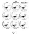
- The increase in detectable Treg cells after culture in the presence of IL-2 with three different staining protocols. PBMC were stained as described in Example 1, using Alexa-647-conjugated anti-Foxp3 (clone 259D, Biolegend) (top row), APC-conjugated anti-Foxp3 (clone 236/A/E7, eBioscience) (center row), or PE conjugated anti-Foxp3 (clone 236A/E7, eBioscience) for Foxp3 detection. Dot plots were gated on CD4+ lymphocytes.
- Fig. 9:
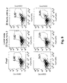
- Data showing that no increase in detectable Treg cells after culture in the presence of IL-2 is observed in lymph node cells. Lymph node cells from the same donor whose PBMC were analyzed in
Figure 8 were stained as inFig 8 , center and bottom rows. Dot plots were gated on CD4+ lymphocytes. - For the determination of Treg frequencies, the present invention as well as conventional state-of-the-art measurements use PBMC isolated from heparinized venous blood by centrifugation over a density gradient (lymphocyte separation medium Pancoll human, PAN-BIOTECH GmbH, Aidenbach, Germany) following the manufacturer's instructions.
- PBMCs were cultured in 48-well tissue culture plates (Greiner bio-one, Frickenhausen, Germany), in which 0,2, 0,5, or 1 ml of cells adjusted to 1 Mio/ml were cultured per well in the three types of wells mentioned, using enriched RPMI 1640 culture medium (GIBCO/Invitrogen, Long Island, NY, USA) supplemented with 10% autologous serum or commercially available pooled human AB serum (Sigma-Aldrich), with essentially the same results.
- The frequency of Treg cells was determined by 3-colour immunofluorescence and flow cytometry, using fluorochrome-conjugated mAb specific for CD4, CD25 and Foxp3. For the staining procedure, the cells were suspended at 1 x 10⁶/ml in staining buffer (PBS, 0,1 % BSA, 0,2 % NaN3), and the CD4 and CD25 specific mAb were added to 0,2 ml of this suspension. After 20 min. on ice, cells were washed with staining buffer by centrifugation (4°C, 1600 rpm). For intracellular staining of Foxp3, the cells were then suspended in Fix/Perm (eBioscience), and stained with the appropriate antibody diluted in Perm/Wash (eBioscience).
- The following fluorochrome-conjugated mAb were used: CD4 (clone RPA-T4, PECy5,1:300, BioLegend), CD25 (clone M-A251, PE,1:25,-BD), FoxP3 (clone 259D), Alexa647,1:50, BioLegend).
- After the final wash, PBMC were resuspended in 0,1 ml staining buffer and analyzed on a FACS Calibur flow cytometer (Becton Dickinson, Mountain View, CA). Data were then analyzed using FlowJo software (TreeStar). They were displayed as dot plots wherein each cell is represented by a dot, and logarithmic fluorescence intensity of two markers (e.g. CD4 and Foxp3) defines the position of each dot. Horizontal and vertical lines divide the dot plot into four quadrants, with lower left containing cells expressing neither marker, upper right both markers, and upper left and lower right only one of the two markers studied. Marker-positive cell populations can also be defined by a window encompassing a "cloud" of dots.
- Analysis of PBMC prepared from venous blood from a healthy donor is shown in
Figure 1 . Staining was performed directly after isolation of PBMC (Fig. 1A ), and after six (Fig. 1B ) or 20 hours (Fig. 1C ) of incubation in the absence or in the presence of 200 U/ml recombinant human 1 L-2 (Novartis). Numbers represent percentages in the respective fields. Numbers in brackets represent frequencies of Foxp3 positive cells among CD4 positive T-cells. - As is readily seen, the frequency of Foxp3 expressing and of Foxp3 and CD25 coexpressing cells within the CD4 T-cell subset, i.e. cells identified as Treg cells, almost doubled after overnight culture in the presence, but further decreased in the absence of IL-2. This positive effect became first
apparent alter 6 hours of culture. In addition to frequencies of cell populations, also the mean fluorescence intensities (MFI) observed for Foxp3 and CD25 in the positive populations are shown. These are proportional to the amount of the respective proteins expressed by the marker-positive cells. It is obvious that not only the frequencies, but also the expression levels of Foxp3 and CD25 expression per cell were increased as a result of IL-2 stimulation. - Compiled data from four randomly chosen healthy donors obtained in an identical fashion are summarized in Table 1. It can be seen that not only the frequency of Treg marker-positive cells, but also the level of marker expression were increased as a result of IL-2 treatment. The top group shows data for PBMC isolated directly from venous blood. The bottom group shows data for PBMC isolated from leukocyte reduction chambers obtained as a by-product of platelet concentration production. (A = CD25+/CD4+ cells [%], B = MFI of CD25 in CD4+ CD25+ cells, C = Foxp3+/CD4+ cells [%], D = MFI of Foxp3 in CD4+ Foxp3+ cells, E = Treg (CD25+Foxp3+ cells/CD4+ cells [%])
Table 1: Donor Time A B C D E [h] no no no no no IL-2 IL-2 IL-2 IL-2 IL-2 IL-2 IL-2 IL-2 IL-2 IL-2 D263 0 4,56 38,2 1,33 211 2,06 2 1,77 17,0 36,2 37,2 0,92 1,05 198 215 1,18 1,36 6 2,52 3,11 37,5 41,4 1,35 2,51 215 257 1,64 2,69 20 1,93 3,82 38,2 54,2 1,34 3,31 204 382 1,41 3,28 D264 0 5,39 39,2 2,02 211 2,19 2 2,81 2,83 38,2 38,2 1,40 1,99 213 219 1,28 1,70 6 2,91 3,73 38,5 41,0 1,99 3,32 217 264 1,73 2,78 20 2,20 5,71 38,2 63,2 2,63 4,04 221 355 1,54 3,90 D265 0 4,32 39,2 2,14 221 2,60 2 2,23 2,68 37,9 38,2 2,23 2,44 217 217 2,06 2,33 6 2,51 3,27 39,2 42,6 2,51 3,75 229 305 2,31 3,36 20 2,54 5,04 40,3 59,9 2,72 4,35 239 385 2,29 4,33 D266 0 8,33 36,8 1,38 213 1,80 2 3,25 2,47 37,5 37,2 2,24 2,33 207 213 1,79 1,61 6 3,50 4,62 37,9 40,7 2,56 3,48 221 271 1,96 2,85 20 2,86 5,36 40,7 56,2 1,73 4,21 231 346 1,51 3,79 D267 20 5,46 8,08 29,5 49,1 2,31 6,70 221 273 2,16 5,53 46 4,61 8,02 30,3 60,9 1,08 3,83 224 407 1,37 4,40 D254 0 5,87 37,2 0,58 198 3,33 24 6,26 8,94 39,6 55,2 0,47 3,14 175 172 2,52 7,72 D256 0 2,85 67,3 1,08 284 2,21 24 0,98 3,74 63,2 87,4 1,17 3,83 274 365 1,36 4,21 D257 0 4,57 70,4 3,02 237 3,35 24 3,64 8,01 66,1 96,5 4,09 7,23 239 340 3,10 6,79 - The experiment shown in
Fig. 2 asked the question whether the recovery of Foxp3 expression requires additional cell types such as monocytes contained within the PBMC preparation, and whether additional cytokines beyond IL-2, which activate the STAT5 pathway of signal transduction, would be able to promote recovery of Foxp3 expression.Fig. 2A shows results obtained with unseparated PBMC andFig. 2B results from purified CD4 positive cells. CD4 T-cells were purified by using a CD4 T-cell purification kit (CD4+ TCell Isolation Kit 11, Miltenyl Biotec) and were then cultured as given in previous experiments for 20 hours in the presence of 200 U/ml IL-2, 20 pg/ml IL-7, or 20 pg/m1 IL-15, before determining the frequency of Foxp3+ cells among CD4 T-cells. It is apparent that Foxp3 negative Treg cells contained within the CD4 T-cell population are able to respond to IL-2 with re-expression of Foxp3. Surprisingly, IL-7 and IL-15 are also highly efficient in providing this effect. - PBMC were cultured with titrated concentrations of IL-2 for 20 hours as in Example 1. Frequencies of Foxp3+CD25+ cells within the CD4 T-cell compartment were determined as in example 1, and are graphically displayed in
Fig. 3 . It is found that an increase in detectable Treg cells is apparent with as little as 6,25 U/ml of IL-2, followed by a dose-dependent further increase. - To determine the length of time required for an optimal effect, purified CD4 T-cells were incubated with or without 200 U/ml of IL-2 for various lengths of time (Table I). While in the absence of IL-2, the frequency of Foxp3 positive cells declined, it increased in its presence, reaching a plateau after 20 hours. Extending the culture period to 46 hours did not show any further increase over the overnight incubation time, as evident from Table I, above (donor D267).
- The method of carboxyfluorescein succinimidyl ester (CFSE) dye dilution (see e.g. Tabares, P. et al., Human regulatory T cells are selectively activated by low-dose application of the CD28 superagonist TGN1412/TAB08, Eur J Immunol 2014.44: 1225-:1236) was employed to determine the proliferation history of Foxp3+ cells recovered from IL-2 stimulated overnight cultures. In this method, which is well known to skilled immunologists, individual cell divisions are easily visualized by flow cytometry as a 50% reduction of fluorescence intensity when the label is distributed to the two daughter cells. In this experiment, which is shown in
Fig.4 , the frequency of Foxp3+ cells among CD4 T-cells was 2,4 fold higher in 20 hour cultures containing 200 U/ml IL-2 as compared to cultures without IL-2. Nevertheless, the cells display a single peak of CFSE Label of the same MFI under both conditions, indicating that no cell divisions had taken place during the culture period. - Thus, this experiment reveals no cell division at all in the increased population of Treg cells identifiable in IL-2 treated cultures, thereby establishing that the effect of IL-2, i.e. doubling in the frequency of Foxp3+ cells, is by up-regulation of Foxp3 expression in previously "invisible" Treg cells rather than by stimulation of Treg cell proliferation.
- For the results in
Fig. 4 the PBMC were stained for CD4 and Foxp3. The left hand CFSE diagrams are for Foxp3 negative and the right hand diagrams for Foxp3 positive CD4 T-cells. - To remove Treg cells which had transiently down-regulated Foxp3 from PBMC, cells expressing CD25 (which is expressed by all Treg cells and a few activated conventional CD4 T-cells) were removed by mAb-mediated magnetic depletion. As seen in
Figure 5 , this eliminated the capacity of IL-2 to increase the frequency of Foxp3+ cells, indicating that culture in the presence of IL-2, and, to a large extent, of IL-7 or IL-15 to increase the frequency of Foxp3+ CD25+ cells, indicating that culture in the presence of these STAT5-activating cytokines allows re-expression of Foxp3 in Treg cells which had transiently lost Foxp3 expression but still express sufficient CD25 to allow depletion with CD25-specific mAb. Culturing was carried out overnight with 200 U/ml IL-2, IL-7 or IL-15 before analysis as inFig. 1 . - PBMC were stored frozen in DMSO containing medium at -80°C, and thawed prior to analysis. This procedure is routinely applied by researchers working with PBMC, which are aliquoted and frozen for later analysis. As can be seen in
Fig. 6 , the detectable Treg frequency in these cells was also considerably enhanced by culture with IL-2. The experiments were performed as inFigure 1 , but with previously frozen PBMC. - Freshly drawn heparinized venous blood was either used for immediate PBMC preparation, or transport at 20°C was mimicked by gently rocking the heparinized blood with or without 400 U/ml of IL-2 for 22 h before PBMC preparation. As can be seen in
Figure 7 , this procedure also increased the frequency of detectable Treg cells. The dot plots show raw data and the bar graph summarized results. - PBMC were stained without pre-culture, or after 20 h culture in the presence or absence of 200 U/ml of IL-2. In addition to the Alexa647 conjugate of the anti-Foxp3 mAb clone 259B (Biolegend, top row in
Fig. 8 ) used so far, two different antibody-fluorochrome conjugates of an alternative clone were used for the detection of Foxp3: Clone 236A/E7 conjugated with phycoerythrin (PE, bottom row inFig. 8 ) or with allophycocyanin (APC, both from eBioscience, center row inFig. 8). Fig. 8 shows that while the latter two mAb yielded superior results in the detection of Foxp3, the phenomenon that culture with IL-2 increases the frequency of detectable Treg among CD4 T-cells, whereas culture without IL-2 leads to a further decrease was clearly visible with all three staining procedures. The dot plots were gated on CD4+ lymphocytes. - Lymph nodes were collected from the para-iliac region of renal transplant recipients at the Academic Medical Center, Amsterdam, The Netherlands. Paired peripheral blood samples were collected before the transplantation procedure. Cells were frozen in IMDM supplemented with 10% DMSO, 20% FCS, penicillin, streptomycin, and 0,00036% mercaptoethanol. The study was approved by the local ethics committee at the Academic Center at the University of Amsterdam.
- Cells shown in
Figure 9 were stained without pre-culture, or after 20 h culture in the presence or the absence of 200 U/ml of IL-2, using the anti-Foxp3 mAb clone 236A/E7 conjugated with phycoerythrin (EP) or with allophycocyanin (APC, both from eBioscience). The lymph node cells were from the same individual as the PBMC inFig. 8 . It is noted that in contrast to these, culture with IL-2 did not lead to a further increase in Treg frequency, whereas as predicted, culture without IL-2 led to their apparent reduction.Table I. Compiled data of samples analysed as in Figure 1. Top group: PBMC isolated directly from venous blood. Bottom group: PBMC isolated from leukocyte reduction chambers obtained as a by-product of platelet concentrate production. Donor Time (hours) % CD25 + cells / CD4+ cells MFI CD4+ CD25+ cells % FoxP3+ cells / CD4+ cells MFI CD4+ FoxP3+ cells % Tregs No IL-2 IL-2 No IL-2 IL-2 No IL-2 IL-2 No IL-2 IL-2 No IL-2 IL-2 D263 0 4.56 - 38.2 - 1.33 - 211 - 2.06 - 2 1.77 17.01 36.2 37.2 0.92 1.05 198 215 1.18 1.36 6 2.52 3.11 37.5 41.4 1.35 2.51 215 257 1.64 2.69 20 1.93 3.82 38.2 54.2 1.34 3.31 204 382 1.41 3.28 D264 0 5.39 - 39.20 - 2.02 - 211 - 2.19 - 2 2.81 2.83 38.20 38.20 1.40 1.99 213 219 1.28 1.70 6 2.91 3.73 38.50 41.00 1.99 3.32 217 264 1.73 2.78 20 2.20 5.71 38.20 63.20 2.63 4.04 221 355 1.54 3.90 D265 0 4.32 - 39.20 - 2.14 - 221 - 2.60 - 2 2.23 2.68 37.90 38.20 2.23 2.44 217 217 2.06 2.33 6 2.51 3.27 39.20 42.60 2.51 3.75 229 305 2.31 3.36 20 2.54 5.04 40.30 59.90 2.72 4.35 239 385 2.29 4.33 D266 0 8.33 - 36.80 - 1.38 - 213 - 1.80 - 2 3.25 2.47 37.50 37.20 2.24 2.33 207 213 1.79 1.61 6 3.50 4.62 37.90 40.70 2.56 3.48 221 271 1.96 2.85 20 2.86 5.36 40.70 56.20 1.73 4.21 231 346 1.51 3.79 D267 20 5.46 8.08 29.50 49.13 2.31 6.70 221 273 2.16 5.53 46 4.61 8.02 30.33 60.87 1.08 3.83 224 407 1.37 4.40 Donor Time (hours) % CD25 + cells / CD4+ cells MFI CD4+ CD25+ cells % FoxP3+ cells / CD4+ cells MFI CD4+ FoxP3+ cells % Tregs No IL-2 IL-2 No IL-2 IL-2 No IL-2 IL-2 No IL-2 IL- 2 No IL-2 IL-2 D254 0 5.87 - 37.20 - 0.58 - 198 - 3.3 - 24 6.26 8.94 39.60 55.20 0.47 3.14 175 172 2.52 7.72 D256 0 2.85 - 67.30 - 1.08 - 284 - 2.21 - 24 0.98 3.74 63.20 87.40 1.17 3.83 274 365 1.36 4.21 D257 0 4.57 - 70.40 - 3.02 - 237 - 3.35 - 24 3.64 8.01 66.10 96.50 4.09 7.23 239 340 3.10 6.79
Claims (12)
- Method for the in-vitro determination of regulatory T-cells (Treg cells) in a sample obtained from human blood with the following steps:A1) said sample is supplemented with a compound capable of activating the Signal Transducer and Activator of Transcription 5 (STAT5) and in an amount capable for such activation, and cultured for a predetermined cultivation duration under conditions which keep T-cells comprised in said sample viable, wherein the cultivation is carried out either during said supplementing or after said supplementing,A2) after cultivation of said sample an analytical compound specific for CD25 and/or Forkhead-Box-Protein P3 (Foxp3) is added to said sample and the cell frequency of CD25 and/or Foxp3 positive cells is determined in said sample, in particular by determining the ratio of the amount of CD25 and/or Foxp3 positive cells to the amount of CD4 positive cells.
- Method for preparing a composition, in particular a pharmaceutical composition, comprising a predetermined amount of viable Treg cells with the following steps:B1) a sample is obtained from human blood and subjected to an in-vitro cultivation under conditions which keep T-cells comprised in said sample viable, said cultivation being performed during or after supplementing said sample with a compound capable of activating the Signal Transducer and Activator of Transcription 5 (STAT5),B2) then an analytic compound specific for CD25 is added and the frequency of CD25 positive cells is determined, in particular by determining the ratio of the amount of CD25 positive cells to the amount of CD4 positive cells.B3) then a fraction of said sample is taken, wherein the fraction is calculated from the frequency of CD25 positive cells such that the fraction comprises the predetermined amount of viable Treg cells, and said fraction is optionally prepared for administration.
- Method for preparing a composition, in particular a pharmaceutical composition, comprising a predetermined amount of viable Treg cells with the following steps:C1) a sample is obtained from human blood and subjected to an in-vitro cultivation under conditions which keep T-cells comprised in said sample viable, said cultivation being performed during or after supplementing said sample with a compound capable of activating the Signal Transducer and Activator of Transcription 5 (STAT5),C2) then an analytic compound specific for Foxp3 is added to a first fraction of said sample and the frequency of Foxp3 positive cells is determined, in particular by determining the ratio of the amount of Foxp3 positive cells to the amount of CD4 positive cells.C3) then a second fraction of said sample is taken, wherein the amount of the second fraction is calculated from the frequency of Foxp3 positive cells in the first fraction such that the second fraction comprises the predetermined amount of viable Treg cells, and said second fraction is optionally prepared for administration.
- Method according to one of the claims 1 to 3, wherein said sample is untreated human blood or obtained from the human blood by isolation of peripheral blood mononuclear cells (PBMC) from the human blood.
- Method according to one of the claims 1 to 4, wherein the analytic compound specific for Forkhead-Box-Protein P3 (Foxp3) is a monoclonal antibody comprising a marker, preferably a fluorochrome.
- Method according to one of the claims 1 to 5, wherein additionally cells, which are CD25 and/or CD4 positive, are identified and/or isolated before, at the same time, or after step A2), B2), or C2),.
- Method according to one of the claims 1 to 6, wherein the compound capable of activating STAT5 is selected from the group consisting of IL-2, IL-7 and IL-15, in particular is IL-2.
- Method according to one of the claims 1 to 7, wherein CD25 positive, and optionally CD4 positive, Treg are separated from said sample.
- Method according to one of the claims 1 to 8, wherein the cultivation is carried out for at least 1 h, preferably at least 6 h, more preferably at least 16 h, and up to 48 hours or longer, most preferably for 16 to 24 hours.
- Method according to one of the claims 1 to 9, wherein the compound capable of activating STAT5 is added at a dose of at least 1 U/ml, preferably at least 5 U/ml, more preferably at least 10 U/ml, even more preferably at least 50 U/ml, most preferably at least 200 U/ml.
- Composition obtained with a method according to one of the claims 2 to 10.
- Composition according to claim 11 for the treatment of a condition induced by too low levels of Treg in an organism.
Priority Applications (3)
| Application Number | Priority Date | Filing Date | Title |
|---|---|---|---|
| EP14004388.6A EP3037816A1 (en) | 2014-12-23 | 2014-12-23 | Method for the identification of transiently Foxp3 negative regulatory T-cells from human peripheral blood |
| US15/539,313 US20170368100A1 (en) | 2014-12-23 | 2015-12-23 | Method for the identification of transiently foxp3 negative regulatory t-cells from human peripheral blood |
| PCT/EP2015/002610 WO2016102070A1 (en) | 2014-12-23 | 2015-12-23 | Method for the identification of transiently foxp3 negative regulatory t-cells from human peripheral blood |
Applications Claiming Priority (1)
| Application Number | Priority Date | Filing Date | Title |
|---|---|---|---|
| EP14004388.6A EP3037816A1 (en) | 2014-12-23 | 2014-12-23 | Method for the identification of transiently Foxp3 negative regulatory T-cells from human peripheral blood |
Publications (1)
| Publication Number | Publication Date |
|---|---|
| EP3037816A1 true EP3037816A1 (en) | 2016-06-29 |
Family
ID=52302036
Family Applications (1)
| Application Number | Title | Priority Date | Filing Date |
|---|---|---|---|
| EP14004388.6A Withdrawn EP3037816A1 (en) | 2014-12-23 | 2014-12-23 | Method for the identification of transiently Foxp3 negative regulatory T-cells from human peripheral blood |
Country Status (3)
| Country | Link |
|---|---|
| US (1) | US20170368100A1 (en) |
| EP (1) | EP3037816A1 (en) |
| WO (1) | WO2016102070A1 (en) |
Cited By (1)
| Publication number | Priority date | Publication date | Assignee | Title |
|---|---|---|---|---|
| EP3611504A4 (en) * | 2017-04-14 | 2020-11-11 | National Cancer Center Japan | METHOD FOR DETECTING REGULATORY T CELLS OF THE EFFECT TYPE, DEVICE FOR ANALYSIS OF REGULATORY T CELLS OF THE EFFECT TYPE, SYSTEM FOR ANALYSIS OF REGULATORY T CELLS OF THE EFFECT TYPE AND PROGRAM |
Families Citing this family (1)
| Publication number | Priority date | Publication date | Assignee | Title |
|---|---|---|---|---|
| CN116376828B (en) * | 2023-06-02 | 2023-08-11 | 成都云测医学生物技术有限公司 | Method for inducing CD4+ T cells to generate Treg cells and application |
Citations (2)
| Publication number | Priority date | Publication date | Assignee | Title |
|---|---|---|---|---|
| WO2006107850A2 (en) * | 2005-04-01 | 2006-10-12 | University Of Southern California | Preventing rejection of transplanted tissue using regulatory t cells |
| US20140072537A1 (en) * | 2011-11-30 | 2014-03-13 | Advanced Cell Technology, Inc. | Mesenchymal stromal cells and uses related thereto |
-
2014
- 2014-12-23 EP EP14004388.6A patent/EP3037816A1/en not_active Withdrawn
-
2015
- 2015-12-23 WO PCT/EP2015/002610 patent/WO2016102070A1/en not_active Ceased
- 2015-12-23 US US15/539,313 patent/US20170368100A1/en not_active Abandoned
Patent Citations (2)
| Publication number | Priority date | Publication date | Assignee | Title |
|---|---|---|---|---|
| WO2006107850A2 (en) * | 2005-04-01 | 2006-10-12 | University Of Southern California | Preventing rejection of transplanted tissue using regulatory t cells |
| US20140072537A1 (en) * | 2011-11-30 | 2014-03-13 | Advanced Cell Technology, Inc. | Mesenchymal stromal cells and uses related thereto |
Non-Patent Citations (30)
| Title |
|---|
| "FlowCellect(TM) Human FOXP3 Treg Characterization Kit 25 Tests", 1 October 2010 (2010-10-01), XP055193537, Retrieved from the Internet <URL:http://www.merckmillipore.com/DE/en/product/FlowCellect%E2%84%A2-Human-FOXP3-Treg-Characterization-Kit,MM_NF-FCIM025118#anchor_BRO> [retrieved on 20150603] * |
| DALLA LIBERA, D ET AL.: "T regulatory cells are markers of disease activity in multiple sclerosis patients", PLOS ONE, vol. 6, 2011, pages 21386 |
| DELACHER, M. ET AL.: "Transcriptional Control of Regulatory T cells", CURRTOP MICROBIOL IMMUNOL, vol. 381, 2014, pages 83 - 124 |
| DIETZ, A. B. ET AL.: "A novel source of viable peripheral blood mononuclear cells from leukoreduction system chambers", TRANSFUSION, vol. 46, 2006, pages 2083 - 2089 |
| EDINGER, M.; HOFFMANN, P.: "Regulatory T cells in stem cell transplantation: strategies and first clinical experiences", CURR OPIN IMMUNOL, vol. 23, 2011, pages 679 - 684, XP028309082, DOI: doi:10.1016/j.coi.2011.06.006 |
| FEDERICO SIMONETTA ET AL: "Increased CD127 expression on activated FOXP3+CD4+ regulatory T cells", EUROPEAN JOURNAL OF IMMUNOLOGY, vol. 40, no. 9, 20 September 2010 (2010-09-20), pages 2528 - 2538, XP055083583, ISSN: 0014-2980, DOI: 10.1002/eji.201040531 * |
| GUNNLAUGSDOTTIR B ET AL: "The Ex Vivo Induction of Human CD103(+) CD25(hi) Foxp3(+) CD4(+) and CD8(+) Tregs is IL-2 and TGF-beta 1 Dependent", vol. 77, no. 2, 1 February 2013 (2013-02-01), pages 125 - 134, XP002698695, ISSN: 0300-9475, Retrieved from the Internet <URL:http://onlinelibrary.wiley.com/doi/10.1111/sji.12009/pdf> [retrieved on 20130124], DOI: 10.1111/SJI.12009 * |
| J. KELSEN ET AL: "FoxP3+CD4+CD25+ T cells with regulatory properties can be cultured from colonic mucosa of patients with Crohn's disease", CLINICAL & EXPERIMENTAL IMMUNOLOGY, vol. 141, no. 3, 1 September 2005 (2005-09-01), pages 549 - 557, XP055193542, ISSN: 0009-9104, DOI: 10.1111/j.1365-2249.2005.02876.x * |
| JAMSHIDIAN, A. ET AL.: "Biased TreglTh17 balance away from regulatory toward inflammatory phenotype in relapsed multiple sclerosis and its correlation with severity of symptoms", J- NEUROIMMUNOT, vol. 262, 2013, pages 106 - 112, XP028709434, DOI: doi:10.1016/j.jneuroim.2013.06.007 |
| KAWASHIRI, S. Y. ET AL.: "CD4+CD25(high)CD127(low/-) Treg cell frequency from peripheral blood correlates with disease activity In patients with rheumatoid arthritis", J RHEUMATOL, vol. 38, 2011, pages 2517 - 2521 |
| L. PASSERINI ET AL: "STAT5-signaling cytokines regulate the expression of FOXP3 in CD4+CD25+ regulatory T cells and CD4+CD25- effector T cells", INTERNATIONAL IMMUNOLOGY, vol. 20, no. 3, 12 February 2008 (2008-02-12), pages 421 - 431, XP055193539, ISSN: 0953-8178, DOI: 10.1093/intimm/dxn002 * |
| LONG S A ET AL: "Combination of rapamycin and IL-2 increases de novo induction of human CD4<+>CD25<+>FOXP3<+> T cells", JOURNAL OF AUTOIMMUNITY, LONDON, GB, vol. 30, no. 4, 1 June 2008 (2008-06-01), pages 293 - 302, XP022620900, ISSN: 0896-8411, [retrieved on 20080303], DOI: 10.1016/J.JAUT.2007.12.012 * |
| MORAWSKI, P. A.: "Foxp3 protein stability is regulated by cyclin-dependent kinase 2", J BIOL CHEM, vol. 288, 2013, pages 24494 - 24502 |
| P. ZHANG ET AL: "Induced Regulatory T Cells Promote Tolerance When Stabilized by Rapamycin and IL-2 In Vivo", THE JOURNAL OF IMMUNOLOGY, vol. 191, no. 10, 11 October 2013 (2013-10-11), pages 5291 - 5303, XP055193043, ISSN: 0022-1767, DOI: 10.4049/jimmunol.1301181 * |
| PASSERINI, L: "STATS-signaling cytokines regulate the expression of FOXP3 in CD4+CD2S+ regulatory T cells and CD4+CD25-eIfector T cells", INT IMMUNOL, vol. 20, 2008, pages 421 - 431 |
| RUBTSOV, Y. P.: "Stability of the regulatory T cell lineage in vivo", SCIENCE, vol. 329, 2010, pages 1667 - 1671 |
| SAKAGUCHI, S ET AL.: "Naturally arising Foxp3-expressing CD25+CD4+ regulatory T cells in self-tolerance and autoimmune disease", CURR TOP MICROBIOLLMMUNOL, vol. 305, 2006, pages 57 - 66 |
| SAKAGUCHI, S. ET AL.: "FOXP3+ regulatory T cells in the human immune system", NAT REV (MMUNO, vol. 10, 2010, pages 490 - 500, XP002663981, DOI: doi:10.1038/nri2785 |
| SAKEGUCHI, S. ET AL.: "D. A., FOXP3+ regulatory T cells in the human immune system", NAT REV LMMUNOL, vol. 10, 2010, pages 490 - 600 |
| SEDDIKI, N. ET AL.: "Expression of interleukin (IL)-2 and IL-7 receptors discriminates between human regulatory and activated T cells", J EXP MED, vol. 203, 2006, pages 1693 - 1700, XP055034957, DOI: doi:10.1084/jem.20060468 |
| T. AKIMOVA ET AL: "Differing Effects of Rapamycin or Calcineurin Inhibitor on T-Regulatory Cells in Pediatric Liver and Kidney Transplant Recipients", AMERICAN JOURNAL OF TRANSPLANTATION, vol. 12, no. 12, 20 September 2012 (2012-09-20), pages 3449 - 3461, XP055193534, ISSN: 1600-6135, DOI: 10.1111/j.1600-6143.2012.04269.x * |
| TABARES, P. ET AL.: "Human regulatory T cells are selectively activated by low-dose application of the CD28 superagonist TGN1412/TA808", FUR J IMMUNOL, vol. 44, 2014, pages 1225 - 1236, XP055134574, DOI: doi:10.1002/eji.201343967 |
| TECHNICAL PROTOCOL, 1 June 2014 (2014-06-01), pages 1 - 4, XP055193535, Retrieved from the Internet <URL:http://www.bdbiosciences.com/documents/Measurement-Cell-Surface-Markers-FoxP3-Stat5-Protocol.pdf> [retrieved on 20150603] * |
| VAN DER VLIET, H.J.; NIEUWENHUIS, E. E.: "IPEX as a result of mutations in FOXP3", CLIN DEV IMMUNOL, 2007, pages 89017 |
| VAN LOOSDREGT. J. ET AL.: "Regulation of Treg functionality by acetylation-mediated Foxp3 protein stabilization", BLOOD, vol. 115, 2010, pages 965 - 974 |
| XUMING GUO ET AL: "In vitro-expanded CD4+CD25highFoxp3+ regulatory T cells controls corneal allograft rejection", HUMAN IMMUNOLOGY, vol. 73, no. 11, 1 November 2012 (2012-11-01), pages 1061 - 1067, XP055193540, ISSN: 0198-8859, DOI: 10.1016/j.humimm.2012.08.014 * |
| YANG J ET AL: "Amelioration of acute graft-versus-host disease by adoptive transfer of ex vivo expanded human cord blood CD4+CD25+forkhead box protein 3+regulatory T cells is associated with the polarization of Treg/Th17 balance in a mouse model", TRANSFUSION, AMERICAN ASSOCIATION OF BLOOD BANKS, BETHESDA, MD, US, vol. 52, 1 June 2012 (2012-06-01), pages 1333 - 1347, XP002698696, ISSN: 0041-1132 * |
| ZOM, E.: "IL-2 regulates FOXP3 expression in human CD4+CD25+ regulatory T cells through a STAT-dependent mechanism and induces the expansion of these cells in vivo", BLOOD, vol. 108, 2006, pages 1571 - 1579 |
| ZORN EMMANUEL ET AL: "IL-2 REGULATES FOXP3 EXPRESSION IN HUMAN CD4(+)CD25(+) REGULATORY T CELLS THROUGH A STAT-DEPENDENT MECHANISM AND INDUCES THE EXPANSION OF THESE CELLS IN VIVO", BLOOD, AMERICAN SOCIETY OF HEMATOLOGY, US, vol. 108, no. 5, 1 September 2006 (2006-09-01), pages 1571 - 1579, XP008079309, ISSN: 0006-4971, DOI: 10.1182/BLOOD-2006-02-004747 * |
| ZORN, E. ET AL.: "IL-2 regulates FOXP3 expression in human CD4+CD25+ regulatory T cells through a STAT-dependent mechanism and induces the expansion of these cells in vivo", BLOOD, vol. 108, 2006, pages 1571 - 1579, XP008079309, DOI: doi:10.1182/blood-2006-02-004747 |
Cited By (1)
| Publication number | Priority date | Publication date | Assignee | Title |
|---|---|---|---|---|
| EP3611504A4 (en) * | 2017-04-14 | 2020-11-11 | National Cancer Center Japan | METHOD FOR DETECTING REGULATORY T CELLS OF THE EFFECT TYPE, DEVICE FOR ANALYSIS OF REGULATORY T CELLS OF THE EFFECT TYPE, SYSTEM FOR ANALYSIS OF REGULATORY T CELLS OF THE EFFECT TYPE AND PROGRAM |
Also Published As
| Publication number | Publication date |
|---|---|
| WO2016102070A1 (en) | 2016-06-30 |
| US20170368100A1 (en) | 2017-12-28 |
Similar Documents
| Publication | Publication Date | Title |
|---|---|---|
| Cosmi et al. | Human interleukin 17–producing cells originate from a CD161+ CD4+ T cell precursor | |
| JP7254128B2 (en) | New Subpopulations of CD8+CD45RClow Tregs and Their Use | |
| Lerret et al. | Donor-specific CD8+ Foxp3+ T cells protect skin allografts and facilitate induction of conventional CD4+ Foxp3+ regulatory T cells | |
| Cardona et al. | Staphylococcal enterotoxin B inhibits regulatory T cells by inducing glucocorticoid-induced TNF receptor-related protein ligand on monocytes | |
| AU2016258845A1 (en) | Methods of isolating T cells and T cell receptors having antigenic specificity for a cancer-specific mutation from peripheral blood | |
| Kawai et al. | IL-33 drives the production of mouse regulatory T cells with enhanced in vivo suppressive activity in skin transplantation | |
| WO2018207900A1 (en) | Highly active nk cell and use thereof | |
| Krenzien et al. | Age-dependent metabolic and immunosuppressive effects of tacrolimus | |
| Maio et al. | Elevated glycolytic metabolism of monocytes limits the generation of HIF1A-driven migratory dendritic cells in tuberculosis | |
| KR20130065657A (en) | New methods for isolating tr1 cells | |
| Zeng et al. | B7-1 and B7-2 differentially control peripheral homeostasis of CD4+ CD25+ Foxp3+ regulatory T cells | |
| Calvo-Turrubiartes et al. | Quantitative analysis of regulatory T cells in kidney graft recipients: a relationship with calcineurin inhibitor level | |
| Amir et al. | Identification of a coordinated CD8 and CD4 T cell response directed against mismatched HLA Class I causing severe acute graft-versus-host disease | |
| EP3607052A1 (en) | Methods of isolating naïve regulatory t cells | |
| Rosado-Sánchez et al. | An in vitro system of autologous lymphocytes culture that allows the study of homeostatic proliferation mechanisms in human naive CD4 T-cells | |
| US20170368100A1 (en) | Method for the identification of transiently foxp3 negative regulatory t-cells from human peripheral blood | |
| Paul et al. | In vitro assessment of cord blood–derived proinsulin-specific regulatory T cells for cellular therapy in type 1 diabetes | |
| Helena et al. | Reconstitution of multifunctional CD56lowCD16low natural killer cell subset in children with acute leukemia given α/β T cell-depleted HLA-haploidentical haematopoietic stem cell transplantation | |
| US20220333072A1 (en) | The process for manufacturing of antigen-specific t lymphocytes | |
| WO2012018930A1 (en) | Methods of isolating and expanding human t regulatory cells and uses thereof for cellular therapy | |
| JP2019137696A (en) | Highly active nk cells and application thereof | |
| Heimann | Elongated T cell expansion by utilizing IL7 and IL15 leads to increased T cell yield while preserving Tcm/Tscm characteristics feasible for adoptive T cell therapy | |
| Schlickeiser et al. | Peripheral biomarkers for individualizing immunosuppression in transplantation-Regulatory T cells | |
| US20240182853A1 (en) | Method for large-scale production of human allospecific induced-regulatory t cells with functional stability in the presence of pro-inflammatory cytokines with therapeutic potential in transplantation | |
| Lee et al. | Proliferation of CD4+ CD25high+ Foxp3+ regulatory T lymphocytes in ex vivo expanded ascitic fluid from primary and recurrent ovarian carcinoma |
Legal Events
| Date | Code | Title | Description |
|---|---|---|---|
| PUAI | Public reference made under article 153(3) epc to a published international application that has entered the european phase |
Free format text: ORIGINAL CODE: 0009012 |
|
| AK | Designated contracting states |
Kind code of ref document: A1 Designated state(s): AL AT BE BG CH CY CZ DE DK EE ES FI FR GB GR HR HU IE IS IT LI LT LU LV MC MK MT NL NO PL PT RO RS SE SI SK SM TR |
|
| AX | Request for extension of the european patent |
Extension state: BA ME |
|
| STAA | Information on the status of an ep patent application or granted ep patent |
Free format text: STATUS: REQUEST FOR EXAMINATION WAS MADE |
|
| 17P | Request for examination filed |
Effective date: 20161229 |
|
| RBV | Designated contracting states (corrected) |
Designated state(s): AL AT BE BG CH CY CZ DE DK EE ES FI FR GB GR HR HU IE IS IT LI LT LU LV MC MK MT NL NO PL PT RO RS SE SI SK SM TR |
|
| STAA | Information on the status of an ep patent application or granted ep patent |
Free format text: STATUS: EXAMINATION IS IN PROGRESS |
|
| 17Q | First examination report despatched |
Effective date: 20170612 |
|
| STAA | Information on the status of an ep patent application or granted ep patent |
Free format text: STATUS: THE APPLICATION IS DEEMED TO BE WITHDRAWN |
|
| 18D | Application deemed to be withdrawn |
Effective date: 20171024 |
|
| 18RA | Request filed for re-establishment of rights before grant |
Effective date: 20180524 |
|
| STAA | Information on the status of an ep patent application or granted ep patent |
Free format text: STATUS: EXAMINATION IS IN PROGRESS |
|
| D18D | Application deemed to be withdrawn (deleted) | ||
| STAA | Information on the status of an ep patent application or granted ep patent |
Free format text: STATUS: THE APPLICATION IS DEEMED TO BE WITHDRAWN |
|
| R18D | Application deemed to be withdrawn (corrected) |
Effective date: 20200108 |