EP3020732A1 - Immunoglobulin fc conjugate maintaining fcrn binding strength - Google Patents
Immunoglobulin fc conjugate maintaining fcrn binding strength Download PDFInfo
- Publication number
- EP3020732A1 EP3020732A1 EP14822721.8A EP14822721A EP3020732A1 EP 3020732 A1 EP3020732 A1 EP 3020732A1 EP 14822721 A EP14822721 A EP 14822721A EP 3020732 A1 EP3020732 A1 EP 3020732A1
- Authority
- EP
- European Patent Office
- Prior art keywords
- immunoglobulin
- fragment
- conjugate
- fcrn
- binding
- Prior art date
- Legal status (The legal status is an assumption and is not a legal conclusion. Google has not performed a legal analysis and makes no representation as to the accuracy of the status listed.)
- Granted
Links
Images


Classifications
-
- A—HUMAN NECESSITIES
- A61—MEDICAL OR VETERINARY SCIENCE; HYGIENE
- A61K—PREPARATIONS FOR MEDICAL, DENTAL OR TOILETRY PURPOSES
- A61K38/00—Medicinal preparations containing peptides
-
- A—HUMAN NECESSITIES
- A61—MEDICAL OR VETERINARY SCIENCE; HYGIENE
- A61K—PREPARATIONS FOR MEDICAL, DENTAL OR TOILETRY PURPOSES
- A61K47/00—Medicinal preparations characterised by the non-active ingredients used, e.g. carriers or inert additives; Targeting or modifying agents chemically bound to the active ingredient
- A61K47/50—Medicinal preparations characterised by the non-active ingredients used, e.g. carriers or inert additives; Targeting or modifying agents chemically bound to the active ingredient the non-active ingredient being chemically bound to the active ingredient, e.g. polymer-drug conjugates
-
- C—CHEMISTRY; METALLURGY
- C07—ORGANIC CHEMISTRY
- C07K—PEPTIDES
- C07K14/00—Peptides having more than 20 amino acids; Gastrins; Somatostatins; Melanotropins; Derivatives thereof
- C07K14/435—Peptides having more than 20 amino acids; Gastrins; Somatostatins; Melanotropins; Derivatives thereof from animals; from humans
- C07K14/705—Receptors; Cell surface antigens; Cell surface determinants
- C07K14/72—Receptors; Cell surface antigens; Cell surface determinants for hormones
-
- C—CHEMISTRY; METALLURGY
- C07—ORGANIC CHEMISTRY
- C07K—PEPTIDES
- C07K16/00—Immunoglobulins [IG], e.g. monoclonal or polyclonal antibodies
- C07K16/46—Hybrid immunoglobulins
-
- C—CHEMISTRY; METALLURGY
- C07—ORGANIC CHEMISTRY
- C07K—PEPTIDES
- C07K19/00—Hybrid peptides, i.e. peptides covalently bound to nucleic acids, or non-covalently bound protein-protein complexes
-
- C—CHEMISTRY; METALLURGY
- C12—BIOCHEMISTRY; BEER; SPIRITS; WINE; VINEGAR; MICROBIOLOGY; ENZYMOLOGY; MUTATION OR GENETIC ENGINEERING
- C12N—MICROORGANISMS OR ENZYMES; COMPOSITIONS THEREOF; PROPAGATING, PRESERVING, OR MAINTAINING MICROORGANISMS; MUTATION OR GENETIC ENGINEERING; CULTURE MEDIA
- C12N15/00—Mutation or genetic engineering; DNA or RNA concerning genetic engineering, vectors, e.g. plasmids, or their isolation, preparation or purification; Use of hosts therefor
- C12N15/09—Recombinant DNA-technology
-
- C—CHEMISTRY; METALLURGY
- C07—ORGANIC CHEMISTRY
- C07K—PEPTIDES
- C07K2319/00—Fusion polypeptide
- C07K2319/30—Non-immunoglobulin-derived peptide or protein having an immunoglobulin constant or Fc region, or a fragment thereof, attached thereto
Definitions
- the present invention relates to a physiologically active polypeptide-immunoglobulin Fc fragment conjugate comprising a physiologically active polypeptide linked via a non-peptidyl linker to an immunoglobulin Fc fragment with an FcRn-binding region capable of maintaining the intrinsic binding affinity of the immunoglobulin Fc fragment, a method for preparing the conjugate, a method of maintaining the intrinsic binding affinity of the conjugate for FcRn, and a composition comprising the conjugate capable of maintaining the intrinsic binding affinity of the immunoglobulin Fc fragment for FcRn.
- protein conjugates or complexes that comprise a carrier, such as a polyethylene glycol polymer, albumin, fatty acid or antibody Fc (constant region), linked to a protein in order to increase the serum half-life of the protein.
- a carrier such as a polyethylene glycol polymer, albumin, fatty acid or antibody Fc (constant region)
- Studies known to date about such protein conjugates or complexes mostly aim to increase the serum half-life of a drug to shorten the interval of drug administration to thereby improve patient convenience.
- problems such as a decrease in the activity of a therapeutic protein due to, for example, a spatial hindrance caused by a non-specific binding between a therapeutic protein and a carrier protein.
- immunoglobulin fragments to increase the half-life of physiologically active substances including proteins.
- the CH2-CH3 region of immunoglobulin Fc includes an FcRn (protection receptor)-binding site that prolongs the half-life of an antibody.
- FcRn is an MHC class I-related protein expressed in vascular endothelial cells and binds to IgG and albumin. Characteristically, IgG and FcRn strongly bind to each other at a weak acidic pH and dissociate from each other at a neutral pH.
- IgG enters vascular endothelial cells from blood vessels by pinocytosis or endocytosis and enters lysosomes (pH 6.0), it is protected by FcRn without being degraded.
- IgG is fused with the cell membrane by recycling, it dissociates from FcRn at pH 7.4 and is released into blood vessels. Due to this procedure, the in vivo half-lives of IgG1, IgG2 and IgG4, which include the FcRn-binding site, are 3 weeks on average, being longer than those of other proteins.
- the present inventors have made extensive efforts to develop a conjugate that can maintain the intrinsic binding affinity of an immunoglobulin Fc fragment itself for FcRn without reducing the activity of a physiologically active polypeptide and can easily dissociate from FcRn at a neutral pH.
- a conjugate comprising a physiologically active polypeptide linked via a non-peptidyl linker to an immunoglobulin Fc fragment having an FcRn-binding region can maintain the intrinsic binding affinity of the immunoglobulin Fc fragment for FcRn and, at the same time, can easily dissociate from FcRn at a neutral pH of 7.4, thereby completing the present invention.
- Still another objective of the present invention is to provide a method for preparing a physiologically active polypeptide-immunoglobulin Fc fragment conjugate that maintains the intrinsic binding affinity of an immunoglobulin Fc fragment for FcRn, the method comprising: (a) linking a physiologically active polypeptide via a non-peptidyl linker to an immunoglobulin Fc fragment having an FcRn-binding region to prepare a mixture of physiologically active polypeptide-immunoglobulin Fc fragment conjugates; and (b) separating from the mixture a physiologically active polypeptide-immunoglobulin Fc fragment conjugate that shows a binding ratio within the range of ⁇ 6% of the binding ratio of the immunoglobulin Fc fragment, as determined by substituting the amount bound to FcRn at pH 6.0 and the amount bound to FcRn at pH 7.4 into equation 1.
- Still another objective of the present invention is to provide a method of maintaining the intrinsic binding affinity of a physiologically active polypeptide-immunoglobulin Fc fragment conjugate for FcRn, the method comprising linking a physiologically active polypeptide via a non-peptidyl linker to an immunoglobulin Fc fragment comprising an FcRn-binding region.
- Still another objective of the present invention is to provide a composition comprising the physiologically active polypeptide-immunoglobulin Fc fragment conjugate that maintains the intrinsic binding affinity of the immunoglobulin Fc fragment for FcRn.
- the conjugate of the present invention maintains the intrinsic binding affinity of the immunoglobulin Fc fragment for FcRn and, at the same time, easily dissociates from FcRn at a neutral pH such as a pH of 7.4.
- a neutral pH such as a pH of 7.4.
- the present invention provides a physiologically active polypeptide-immunoglobulin Fc fragment conjugate that comprises a physiologically active polypeptide linked via a non-peptidyl linker to an immunoglobulin Fc fragment comprising an FcRn-binding region, wherein the binding ratio of the amount of the conjugate bound to FcRn at pH 6.0 and the amount of the conjugate bound to FcRn at pH 7.4 is within the range of ⁇ 6% of a ratio determined by measuring the amounts of the immunoglobulin Fc fragment bound to FcRn under the same conditions as those used for the conjugate.
- the conjugate according to the present invention may be obtained by reacting the physiologically active polypeptide having the non-peptidyl linker linked thereto with the immunoglobulin Fc fragment at a pH of 4.0-9.0, thereby linking the physiologically active polypeptide via the non-peptidyl linker to a portion excluding the FcRn-binding region of the immunoglobulin Fc fragment.
- the non-peptidyl linker that is included in the conjugate according to the present invention may be selected from the group consisting of polyethylene glycol, polypropylene glycol, an ethylene glycol-propylene glycol copolymer, polyoxyethylated polyol, polyvinyl alcohol, polysaccharide, dextran, polyvinyl ethyl ether, a biodegradable polymer, a lipid polymer, chitin, hyaluronic acid, and combinations thereof
- non-peptidyl linker that is included in the conjugate according to the present invention may be a polyethylene glycol polymer represented by the following formula 1: wherein n ranges from 10 to 2400.
- the non-peptidyl linker that is included in the conjugate according to the present invention may have a reactive group selected from the group consisting of an aldehyde group, a propionaldehyde group, a butyraldehyde group, a maleimide group, and succinimide derivatives.
- the physiologically active polypeptide that is included in the conjugate according to the present invention may be selected from the group consisting of glucagon-like peptide-1 (GLP-1), granulocyte colony stimulating factor (G-CSF), human growth hormone (hGH), erythropoietin (EPO), glucagon, oxyntomodulin, insulin, growth hormone releasing hormone, growth hormone releasing peptide, interferons, interferon receptors, G-protein-coupled receptor, interleukins, interleukin receptors, enzymes, interleukin binding proteins, cytokine binding proteins, macrophage activating factor, macrophage peptide, B cell factor, T cell factor, protein A, allergy inhibitor, cell necrosis glycoproteins, immunotoxin, lymphotoxin, tumor necrosis factor, tumor suppressors, metastasis growth factor, alpha-1 antitrypsin, albumin, ⁇ -lactalbumin, apolipoprotein-E, highly glycos
- the immunoglobulin Fc fragment comprising the FcRn-binding region which is included in the conjugate according to the present invention, may comprise a CH2 domain, a CH3 domain, or both.
- the immunoglobulin Fc fragment that is included in the conjugate according to the present invention may be in a non-glycosylated form.
- the immunoglobulin Fc fragment that is included in the conjugate according to the present invention may comprise a hinge region.
- the immunoglobulin Fc fragment that is included in the conjugate according to the present invention may be selected from the group consisting of IgG, IgA, IgD, IgE, IgM, combinations thereof, and hybrids thereof
- the immunoglobulin Fc fragment that is included in the conjugate according to the present invention may be an IgG4 Fc fragment.
- the present invention provides a method for preparing a physiologically active polypeptide-immunoglobulin Fc fragment conjugate that maintains the intrinsic binding affinity of an immunoglobulin Fc fragment for FcRn, the method comprising: (a) linking a physiologically active polypeptide via a non-peptidyl linker to an immunoglobulin Fc fragment comprising an FcRn-binding region to a physiologically active polypeptide via a non-peptidyl linker to prepare a mixture of physiologically active polypeptide-immunoglobulin Fc fragment conjugates; and (b) separating from the mixture a physiologically active polypeptide-immunoglobulin Fc fragment conjugate that shows a binding ratio within the range of ⁇ 6% of the binding ratio of the immunoglobulin Fc fragment, as determined by substituting the amount of binding to FcRn at pH 6.0 and the amount of binding to FcRn at pH 7.4 into equation 1.
- the non-peptidyl linker that is used in the preparation method according to the present invention may be a polyethylene glycol polymer represented by the following formula 1: wherein n ranges from 10 to 2400.
- the conjugate separated in step (b) of the preparation method according to the present invention may have a structure in which the non-peptidyl linker is bound to the N-terminus of the immunoglobulin Fc fragment.
- the present invention provides a method of maintaining the intrinsic binding affinity of a physiologically active polypeptide-immunoglobulin Fc fragment conjugate for FcRn, the method comprising linking a physiologically active polypeptide via a non-peptidyl linker to an immunoglobulin Fc fragment comprising an FcRn-binding region.
- the maintaining of the intrinsic binding affinity may be effected in vivo.
- the present invention provides a composition comprising the physiologically active polypeptide-immunoglobulin Fc fragment conjugate, which maintains the intrinsic binding affinity of the immunoglobulin Fc fragment for FcRn.
- the present invention provides a physiologically active polypeptide-immunoglobulin Fc fragment conjugate that comprises a physiologically active polypeptide linked via a non-peptidyl linker to an immunoglobulin Fc fragment comprising an FcRn-binding region, wherein the ratio of the amount of the conjugate bound to FcRn at pH 6.0 and the amount of the conjugate bound to FcRn at pH 7.4 is within the range of ⁇ 6% of a ratio determined by measuring the amounts of the immunoglobulin Fc fragment bound to FcRn under the same conditions as those used for the conjugate.
- the expression "amount of binding of the physiologically active polypeptide-immunoglobulin Fc fragment conjugate” refers to the ratio of the concentration of the conjugate protein bound to FcRn relative to the total concentration of the conjugate protein bound or unbound to FcRn at the pH of interest.
- the ratio between the amounts of binding may be calculated from the absolute amount of binding measured at one pH and may also be determined by measuring other physical amounts, which are proportional to the amount of the bound conjugate and easy to measure.
- the ratio between the amounts of binding can be determined by measuring surface plasmon resonance (SPR) signals and calculating the ratio between the signals at pH 6.0 and pH 7.4.
- SPR surface plasmon resonance
- other methods may also be used to determine the ratio between the amounts of binding.
- the binding ratio can be obtained as a percentage (%) value by dividing the amount of the conjugate (or immunoglobulin Fc fragment) binding to FcRn at pH 7.4 by the amount of the conjugate (or immunoglobulin Fc fragment) binding to FcRn at pH 6.0 and multiplying the divided value by 100.
- the binding ratio can be calculated using the method proposed by Weirong Wang et al. (DRUG METABOLISM AND DISPOSITION 39:1469-1477, 2011 ) for predicting the half-life of a monoclonal antibody, but is not specifically limited thereto.
- a monoclonal antibody having a high binding ratio showed a short in vivo half life.
- the method for measuring the binding ratio may be performed, particularly using a surface plasmon resonance method, but is not specifically limited thereto.
- the method for measuring the binding ratio can be performed by immobilizing FcRn onto a biosensor chip (e.g., a Biacore CM5 biosensor chip) using an amine coupling kit or the like, injecting the FcRn-immobilized biosensor chip with an acidic buffer (e.g., pH 6.0 buffer) containing a material (e.g., the conjugate or the immunoglobulin Fc fragment) to be measured for the extent of binding to FcRn, and then injecting a neutral buffer (e.g., pH 7.4 buffer) into the biosensor chip.
- an acidic buffer e.g., pH 6.0 buffer
- a material e.g., the conjugate or the immunoglobulin Fc fragment
- the binding ratio can be determined by measuring the resonance unit (RU) in an equilibrium state before completion of the injection of the sample in the pH 6.0 buffer into the chip to determine the amount bound at pH 6.0, measuring the resonance unit after injection of the pH 7.4 buffer to determine the amount bound at pH 7.4, and then dividing the amount bound at pH 7.4 by the amount bound at pH 6.0.
- the binding ratio is to be expressed in percentage, the divided value can be multiplied by 100.
- the composition of the pH 6.0 buffer is not specifically limited, as long as it can induce a binding between the material to be measured for the extent of binding to FcRn and FcRn.
- the pH 6.0 buffer may be a buffer containing a salt such as phosphate.
- the concentration of the salt in the buffer may be 50-200 mM, but is not limited thereto.
- the buffer may be injected into the FcRn-immobilized biosensor chip at a temperature of about 25 °C, but the temperature of measurement may be changed within the temperature range in which the pH of the buffer does not change.
- the pH 6.0 buffer may contain the material to be measured for the extent of binding to FcRn.
- the concentration of the material to be measured for the degree of binding to FcRn in the pH 6.0 buffer is not specifically limited, as long as the extent of binding to FcRn can be measured.
- the concentration of the material to be measured for the degree of binding to FcRn in the pH 6.0 buffer may be between 100 nM and 12.5 nM.
- the composition of the pH 7.4 buffer is not specifically limited, as long as it can induce dissociation between the material to be measured for the extent of binding to FcRn and FcRn.
- the pH 7.4 buffer may be a buffer containing phosphate.
- the concentration of a salt in the buffer may be 50-200 mM, but is not limited thereto.
- the pH 7.4 buffer may be injected into the FcRn-immobilized biosensor chip at a temperature of 25 ⁇ 37 °C, but is not limited thereto.
- the ratio between the amounts of binding is measured at a temperature of 25 °C.
- the amount of binding between FcRn and the material to be measured for the extend of binding to FcRn at pH 6.0 may be a resonance unit value measured at 2-60 seconds before the completion of injection of the pH 6.0 sample. Binding between FcRn and the material to be measured for the extent of binding to FcRn is preferably in an equilibrium state at the time point of measurement.
- the amount of binding between FcRn and the material to be measured for the extent of binding to FcRn at pH 7.4 may be a resonance unit value measured at 10-20 seconds after injection of the pH 7.4 buffer.
- the time point of measurement is preferably between the time point at which the RU value changes rapidly and before the RU value reaches 0.
- the binding ratios are preferably values determined by the same experimental method under the same experimental conditions, but the compositions and temperatures of the buffers may vary depending on the kind of conjugate.
- the surface plasmon resonance method as described above is one of methods for determining the binding ratio.
- any method may be used in the present invention, as long as it is a method such as an enzyme-immunosorbent assay (ELISA), which can measure the amount of the conjugate bound to FcRn.
- ELISA enzyme-immunosorbent assay
- the unit of the amount of binding may vary depending on the method used.
- the physiologically active polypeptide-immunoglobulin Fc fragment conjugate that comprises a physiologically active polypeptide linked via a non-peptidyl linker to an immunoglobulin Fc fragment having an FcRn-binding region is within the range of ⁇ 6.0% of the binding ratio of the immunoglobulin Fc fragment itself, as measured by the above-described method, the physiologically active polypeptide-immunoglobulin Fc fragment conjugate has a long in vivo half-life.
- n in-[O-CH 2 -CH 2 ]n- is 10 or greater, particularly 10-2400, more particularly 10-480, and even more particularly 50-250, but is not limited thereto.
- n in -[O-CH 2 -CH 2 ]n- is particularly 10-2400, and more particularly 50-2400, the polyethylene glycol does not influence the physiologically activity and FcRn-binding activity of each of the physiologically active polypeptide and the immunoglobulin Fc fragment.
- the present invention is based on this characteristic.
- physiologically active polypeptide-immunoglobulin Fc fragment conjugate refers to a conjugate that comprises a physiologically active polypeptide covalently linked via a non-peptidyl linker to an immunoglobulin Fc fragment having an FcRn-binding region and shows a binding ratio within the range of ⁇ 6.0% of the binding ratio of the immunoglobulin Fc fragment, as determined by substituting the amount of binding to FcRn at pH 6.0 and the amount of binding to FcRn at pH 7.4 into equation 1.
- the non-peptidyl linker in the conjugate may be linked to amino acid residues away from the FcRn-binding region of the immunoglobulin Fc fragment, for example, a region corresponding to positions 252-257 and 307-311 of CH2 and positions 433-436 of CH2, numbered according to the Kabat numbering system.
- the non-peptidyl linker may preferably be linked to the N-terminus or C-terminus of the immunoglobulin Fc fragment, and more preferably linked to the N-terminus, but is not limited thereto.
- the non-peptidyl linker When the non-peptidyl linker is linked to the N-terminus or C-terminus of the immunoglobulin Fc fragment, it does not substantially reduce the binding affinity of the immunoglobulin Fc fragment for FcRn, and thus the intrinsic binding affinity of the immunoglobulin Fc fragment of the conjugate for FcRn can be maintained.
- non-peptidyl linker refers to a biocompatible polymer composed of two or more repeating units linked to each other, in which the repeating units are linked to each other by any non-peptide covalent bond. This non-peptidyl linker may have two or three ends.
- the non-peptidyl linker used in the present invention may be selected from the group consisting of biodegradable polymers such as polyethylene glycol, polypropylene glycol, a copolymer of ethylene glycol with propylene glycol, polyoxyethylated polyol, polyvinyl alcohol, polysaccharide, dextran, polyvinyl ethyl ether, biodegradable polymers such as polylactic acid (PLA) and polylactic-glycolic acid (PLGA), lipid polymers, chitins, hyaluronic acid, and combinations thereof, but is not limited thereto.
- biodegradable polymers such as polyethylene glycol, polypropylene glycol, a copolymer of ethylene glycol with propylene glycol, polyoxyethylated polyol, polyvinyl alcohol, polysaccharide, dextran, polyvinyl ethyl ether
- biodegradable polymers such as polylactic acid (PLA) and poly
- the non-peptidyl linker is polyethylene glycol, for example, a polyethylene glycol polymer represented by the following formula 1, but is not limited: wherein n ranges from 10 to 2400, preferably from 10 to 480, and more preferably from 50 to 250, but is not limited thereto.
- non-peptidyl linkers having a molecular weight corresponding to that of the polyethylene glycol of formula 1 also fall within the scope of the present invention.
- Polyethylene glycol that is used as the non-peptidyl linker in the present invention has an advantage in that it does not result in spatial hindrance between the physiologically active polypeptide and immunoglobulin Fc fragment bound to both ends thereof so that both the physiological activity of the physiologically active polypeptide and the binding affinity of the immunoglobulin Fc fragment for FcRn can be maintained.
- a peptidyl linker that is used in a fusion protein prepared by a conventional in-frame fusion method has a disadvantage in that it is easily cleaved by protease in vivo, and thus the expected effect of increasing the serum half-life of the active drug by a carrier cannot be obtained.
- the conjugate comprising the non-peptidyl linker according to the present invention dramatically overcomes this disadvantage.
- the non-peptidyl linker may be a polymer that has resistance to protease to maintain the serum half-life of the peptide, similar to that of a carrier. Therefore, any non-peptidyl linker may be used in the present invention without any limitation, as long as it is a polymer having the above-described function, that is, a polymer having resistance to protease in vivo.
- non-peptidyl linker that is linked to the immunoglobulin Fc fragment in the present invention may be made not only of one kind of polymer, but also of a combination of different kinds of polymers.
- the non-peptidyl linker that is used in the present invention has reactive groups capable of binding to the immunoglobulin Fc fragment and the physiologically active polypeptide.
- the reactive groups at both ends of the non-peptidyl polymer are preferably selected from the group consisting of a reactive aldehyde group, a propionaldehyde group, a butyraldehyde group, a maleimide group, and succinimide derivative.
- the succinimide derivative may be succinimidyl propionate, hydroxy succinimidyl, succinimidyl carboxymethyl, or succinimidyl carbonate.
- a physiologically active polypeptide and an immunoglobulin effectively bind to both ends of the non-peptidyl linker, respectively, while minimizing non-specific reactions.
- a final product generated by reductive alkylation via an aldehyde bond is much more stable than that linked via an amide bond.
- the aldehyde reactive group can bind selectively to the N-terminus at a low pH and can form a covalent bond with a lysine residue at a high pH, for example, at pH 9.0.
- the non-peptidyl linker may contain two or more aldehyde groups or have two or more alcohol groups substituted with functional groups including aldehyde.
- the reactive groups at both ends of the non-peptidyl linker may be the same or different.
- one end of the non-peptidyl linker may have a maleimide group, and the other end may have an aldehyde group, a propionaldehyde group, or an alkyl aldehyde group such as butyl aldehyde.
- the hydroxy groups may be activated into various reactive groups by a known chemical reaction.
- a commercially available polyethylene glycol having a modified reactive group may be used to prepare the conjugate of the present invention.
- physiologically active polypeptide collectively refers to polypeptides having any physiological activity in vivo, which commonly have a polypeptide structure and have various physiological activities.
- the physiologically active polypeptides include those that function to regulate genetic expression and physiological function and to correct an abnormal condition caused by the lack or excessive secretion of a substance that is involved in the regulation of functions in vivo.
- the physiologically active polypeptides may also include general protein therapeutic agents.
- physiologically active polypeptide is meant to include not only native polypeptides, but also derivatives thereof
- physiologically active polypeptide in the conjugate of the present invention are not specifically limited, as long as it is a physiologically active polypeptide can show an increase in the serum half-life by the conjugate structure of the present invention.
- conjugates are prepared using various physiologically active polypeptides, including insulin, interferon, human growth hormone and a GLP-1 agonist, which are representative examples of physiologically active polypeptides, and it was found that the intrinsic binding affinity of the immunoglobulin Fc fragment itself for FcRn can be maintained regardless of the kind and size of physiologically active polypeptide.
- the physiologically active polypeptide included in the conjugate according to the present invention may be selected from the group consisting of glucagon-like peptide-1 (GLP-1), granulocyte colony stimulating factor (G-CSF), human growth hormone (hGH), erythropoietin (EPO), glucagon, oxyntomodulin, insulin, growth hormone releasing hormone, growth hormone releasing peptide, interferons, interferon receptors, G-protein-coupled receptor, interleukins, interleukin receptors, enzymes, interleukin binding proteins, cytokine binding proteins, macrophage activating factor, macrophage peptide, B cell factor, T cell factor, protein A, allergy inhibitor, cell necrosis glycoproteins, immunotoxin, lymphotoxin, tumor necrosis factor, tumor suppressors, metastasis growth factor, alpha-1 antitrypsin, albumin, ⁇ -lactalbumin, apolipoprotein-E, highly glycosylated erythro
- physiologically active polypeptide is meant to include not only natural physiologically active polypeptides, but also agonists, precursors, derivatives, fragments or variants of each polypeptide.
- examples of oxyntomodulin derivatives include all those disclosed in Korean Patent Application Publication No. 10-2012-0137271
- examples of insulin-releasing peptide derivatives include those disclosed in Korean Patent Application Publication No. 10-2009-0008151 , but are not limited thereto.
- immunoglobulin Fc fragment refers to a protein that contains the heavy-chain constant region of an immunoglobulin, excluding the heavy-chain constant region 1 (CH1) and light-chain constant region 1 (CL1) of the immunoglobulin.
- the Fc fragment may include a hinge region in the heavy-chain constant region.
- the immunoglobulin Fc fragment preferably comprises a CH2 domain, a CH3 domain, or both, because the binding affinity of the immunoglobulin Fc fragment for FcRn should be maintained.
- the immunoglobulin Fc region in the present invention may be an extended Fc fragment that includes a portion or whole of the heavy-chain constant region 1 (CH1) and/or the light-chain constant region 1 (CL1), except for the heavy-chain and light-chain variable regions of the immunoglobulin, as long as it maintains its intrinsic binding affinity for FcRn, even when it is linked to the physiologically active polypeptide and the non-peptidyl linker.
- CH1 heavy-chain constant region 1
- CL1 light-chain constant region 1
- immunoglobulin Fc fragment is a biodegradable polypeptide metabolized in vivo, it is safe for use as a drug carrier. Also, because the immunoglobulin Fc fragment has a molecular weight lower than the entire immunoglobulin molecule, it is beneficial in terms of the preparation, purification and yield of the conjugate. In addition, because the Fab region, which shows high non-homogeneity due to the difference in amino acid sequence between antibodies, is removed, the Fc fragment has a considerably increased homogeneity and a low potential to induce serum antigenicity.
- the immunoglobulin Fc fragment includes not only a native amino acid sequence, but also a sequence mutant thereof
- amino acid sequence mutant refers to a sequence that is different from the native amino acid sequence due to a deletion, insertion, non-conservative or conservative substitution or combinations thereof of one or more amino acid residues.
- amino acid residues at positions 214 to 238, 297 to 299, 318 to 322 or 327 to 331, known to be important in binding may be used as a suitable target for modification.
- mutants are also possible, including mutants having a deletion of a region capable of forming a disulfide bond, a deletion of several amino acid residues at the N-terminus of a native Fc, or an addition of methionine residue to the N-terminus of a native Fc.
- a complement-binding site for example, a C1q-binding site, may be removed, and an antibody dependent cell mediated cytotoxicity (ADCC) site may also be removed.
- ADCC antibody dependent cell mediated cytotoxicity
- the immunoglobulin Fc fragment may also be modified by, for example, phosphorylation, sulfation, acrylation, glycosylation, methylation, farnesylation, acetylation or amidation.
- Fc mutants are mutants that show the same biological activity as that of the Fc fragment of the present invention, but have improved structural stability against heat, pH, or the like.
- these Fc fragments may be obtained from native forms isolated from humans and animals including cows, goats, pigs, mice, rabbits, hamsters, rats and guinea pigs, or may be recombinant forms or derivatives thereof, obtained from transformed animal cells or microorganisms.
- they may be obtained from a native immunoglobulin by isolating a whole immunoglobulin from the living body of humans or animals and treating it with protease.
- papain it is cleaved into Fab and Fc.
- pepsin it is cleaved into pF'c and F(ab) 2 .
- These fragments may be subjected, for example, to size-exclusion chromatography to isolate Fc or pF'c.
- it is a recombinant immunoglobulin Fc fragment obtained from a microorganism using a human Fc fragment.
- the immunoglobulin Fc fragment may be in the form of having native sugar chains, increased sugar chains compared to a native form or decreased sugar chains compared to the native form, or may be in a deglycosylated form.
- the increase, decrease or removal of the immunoglobulin Fc sugar chains may be performed using conventional methods, such as a chemical method, an enzymatic method and a genetic engineering method using a microorganism.
- the immunoglobulin Fc fragment obtained by removing sugar chains from an Fc shows a sharp decrease in binding affinity for the complement (c1q) and a decrease or loss in antibody-dependent cell-mediated cytotoxicity or complement-dependent cytotoxicity, and thus does not induce unnecessary immune responses in vivo.
- an immunoglobulin Fc fragment in a deglycosylated or aglycosylated form may be more suitable for use as a drug carrier.
- deglycosylation refers to an enzymatic removal of sugar moieties from an Fc fragment
- amino acid sequence means an unglycosylated Fc fragment produced in prokaryotes, preferably in E. coli.
- the immunoglobulin Fc fragment may originate from humans or animals such as cattle, goats, pigs, mice, rabbits, hamsters, rats or guinea pigs. Preferably, it is of human origin.
- the immunoglobulin Fc fragment may be an Fc fragment that is derived from IgG, IgA, IgD, IgE and IgM, combinations thereof, or hybrids thereof. Preferably, it is derived from IgG or IgM, which is among the most abundant proteins in the human blood, and most preferably it is derived from IgG known to enhance the half-life of ligand-binding proteins.
- a dimer or multimer may be formed from two or more fragments selected from the group consisting of IgG Fc, IgA Fc, IgM Fc, IgD Fc, and IgE Fc fragments.
- hybrid refers to the presence of two or more sequences corresponding to immunoglobulin Fc fragments of different origins in a single-chain immunoglobulin Fc fragment.
- domain hybrids may be composed of one to four domains selected from the group consisting of CH1, CH2, CH3 and CH4 of IgG Fc, IgM Fc, IgA Fc, IgE Fc and IgD Fc, and may include a hinge region.
- IgG can be divided into IgG1, IgG2, IgG3 and IgG4 subclasses, and combinations or hybrids thereof may be used in the present invention, preferably IgG2 and IgG4 subclasses, and most preferably the Fc fragment of IgG4 rarely having effector functions such as CDC (complement dependent cytotoxicity).
- the most preferable immunoglobulin Fc fragment for use as a drug carrier in the present invention is a human IgG4-derived unglycosylated Fc fragment.
- the human Fc fragment is more preferable than a non-human Fc fragment, which may act as an antigen in the human body and cause undesirable immune responses such as the production of a new antibody against the antigen.
- each of insulin, interferon, human growth hormone and a GLP-1 agonist is linked via a non-peptidyl linker to an immunoglobulin Fc fragment having the ability to bind to FcRn, thereby preparing conjugates that increase the intrinsic binding affinity of the immunoglobulin Fc fragment for FcRn and can easily dissociate from FcRn at a neutral pH and can also show a dissociation degree similar to that of the immunoglobulin Fc fragment (Examples and FIGS. 2 and 3 ).
- the conjugate of the present invention has a long in vivo duration of action compared to an in-frame conjugate ( FIG. 4 ).
- physiologically active polypeptide the immunoglobulin Fc fragment, the non-peptidyl linker, the conjugate and the determination of the binding ratio are as described above.
- Step (a) in the method of the present invention is a step of covalently linking a physiologically active polypeptide via a non-peptidyl linker to an immunoglobulin Fc fragment.
- Step (a) may comprise the steps of: (i) linking any one of the physiologically active polypeptide and the immunoglobulin Fc fragment to a reactive group at one end of the non-peptidyl linker; and (ii) linking the remaining one to a reactive group at the other end of the non-peptidyl linker.
- Step (a) may further comprise, between steps (i) and (ii), a step of separating the physiologically active polypeptide or immunoglobulin Fc fragment linked to one end of the non-peptidyl linker.
- Korean Patent No. 10-0725315 may be incorporated herein by reference.
- the physiologically active polypeptide having the non-peptidyl linker linked thereto may be reacted with the immunoglobulin Fc fragment at a pH of 4.0-9.0.
- conjugate When the conjugate is prepared by this process, byproducts such as a conjugate showing a decrease in the binding affinity of the immunoglobulin Fc fragment for FcRn can be generated in addition to a conjugate that maintains the intrinsic binding affinity of the immunoglobulin Fc fragment for FcRn. For this reason, after the reaction that links the physiologically active polypeptide via the non-peptidyl peptide to the immunoglobulin Fc fragment, a process is additionally required to separate from the conjugate mixture a physiologically active polypeptide-immunoglobulin Fc fragment that maintains the intrinsic binding affinity of the immunoglobulin Fc fragment for FcRn.
- the method of the present invention comprises step (b) of separating from the conjugate mixture a physiologically active polypeptide-immunoglobulin Fc fragment conjugate that shows a binding ratio within the range of ⁇ 6% of the binding ratio of the immunoglobulin Fc fragment, as determined by substituting the amount boundg to FcRn at pH 6.0 and the amount bound to FcRn at pH 7.4 into equation 1.
- Step (b) is preferably a process for separating a conjugate in which the non-peptidyl linker is linked to amino acid residues away from the FcRn-binding region of the immunoglobulin Fc fragment, for example, a region corresponding to positions 252-257 and 307-311 of CH2 and positions 433-436 of CH2, numbered according to the cobat numbering system.
- step (b) is a process for selectively separating only a conjugate in which the non-peptidyl linker is connected to the N-terminus of the immunoglobulin Fc fragment so that the intrinsic binding affinity of the Fc fragment for FcRn is maintained.
- Separation and purification conditions in step (b) may vary depending on the kinds of non-peptidyl linker, physiologically active polypeptide and the like used.
- the present invention provides a method of maintaining the intrinsic binding affinity of a physiologically active polypeptide-immunoglobulin Fc fragment conjugate for FcRn, the method comprising linking a physiologically active polypeptide via a non-peptidyl linker to an immunoglobulin Fc fragment comprising an FcRn-binding region.
- physiologically active polypeptide the immunoglobulin Fc fragment, the non-peptidyl linker and the conjugate are as described above.
- the present invention has advantages in that, because the physiologically active polypeptide is linked via the non-peptidyl linker to the immunoglobulin Fc fragment comprising the FcRn-binding region, the intrinsic binding affinity of the immunoglobulin Fc fragment for FcRn is maintained while the immunoglobulin Fc fragment can easily dissociate from FcRn at a neutral pH and can be easily recycled, suggesting that the in vivo half-life of the immunoglobulin Fc fragment can be effectively increased.
- the maintaining of the intrinsic binding affinity may be effected in vivo.
- the present invention provides a composition comprising the physiologically active polypeptide-immunoglobulin Fc fragment conjugate, which maintains the intrinsic binding affinity of the immunoglobulin Fc fragment for FcRn.
- physiologically active polypeptide the immunoglobulin Fc fragment and the conjugate are as described above.
- An immunoglobulin Fc fragment was prepared according to the method disclosed in Korean Patent Application No. 10-2006-0077377 (entitled “method for mass production of methionine residue-free immunoglobulin Fc region”) filed in the name of the present inventors.
- a reaction was performed to PEGylate a 3.4-kDa propion-ALD2 PEG (IDB, Korea) specifically at the N-terminus of the beta-chain of insulin.
- the reaction solution was purified using a cation-exchange column.
- To prepare an insulin-immunoglobulin Fc fragment conjugate the purified mono-PEGylated insulin was reacted with the immunoglobulin Fc.
- the reaction was performed at pH 6.0-8.2 in order to direct the insulin specifically to the N-terminus of the immunoglobulin Fc.
- the reaction solution was first purified using an anion-exchange column, and then purified using a hydrophobic column, thereby obtaining an insulin conjugate comprising insulin linked site-specifically to the immunoglobulin.
- a 3.4-kDa propion-ALD2 PEG (IDB, Korea) was added to and reacted with a buffer containing human interferon alpha-2b (hIFN ⁇ -2b; molecular weight: 19 kDa) dissolved therein.
- hIFN ⁇ -2b human interferon alpha-2b
- the reaction mixture was subjected to anion exchange column chromatography to purify a mono-PEGylated IFN ⁇ -2b.
- a reaction was performed at a pH of 5.5-6.5.
- the reaction mixture was passed through an anion-exchange column to obtain an interferon-immunoglobulin Fc fragment conjugate fraction.
- the conjugate fraction was additionally purified using a hydrophobic column, thereby obtaining an interferon conjugate comprising interferon linked site-specifically to the immunoglobulin Fc.
- a 3.4-kDa propion-ALD2 PEG (IDB, Korea) was added to and reacted with a buffer containing human growth hormone (hGH; molecular weight: 22 kDa) dissolved therein.
- hGH human growth hormone
- the reaction mixture was subjected to anion exchange column chromatography to purify a mono-PEGylated human growth hormone (hGH).
- hGH mono-PEGylated human growth hormone
- a reaction was performed at a pH of 5.5-6.5. After the linking reaction, the reaction mixture was purified using an anion-exchange column, thereby obtaining a human growth hormone conjugate fraction comprising hormone growth hormone linked site-specifically to the immunoglobulin Fc.
- a 3.4-kDa propion-ALD2 PEG (IDB, Korea) was reacted site-specifically with the lysine residue of imidazo-acetyl-exendin-4 (CA exendin-4, Bachem, Switzerland).
- CA exendin-4 imidazo-acetyl-exendin-4, Bachem, Switzerland.
- the reaction mixture was then subjected to cation-exchange column chromatography to purify mono-PEGylated exendin-4.
- a reaction was performed at a pH of 5.0-8.2. After the coupling reaction, a two-step purification process was performed using a hydrophobic column and an anion-exchange column, thereby obtaining a GLP-1R agonist-immunoglobulin Fc fragment conjugate comprising the GLP-1R agonist linked site-specifically to the immunoglobulin Fc.
- a fusion protein of a GLP-1R agonist with an immunoglobulin Fc fragment comprising an about 50 kDa FcRn site was prepared by a gene recombination method without using a linker.
- the conjugate comprising the GLP-1R agonist linked in-frame to the immunoglobulin Fc fragment (hereinafter also referred to as "in-frame GLP-1R agonist-immunoglobulin Fc fragment conjugate”) was purified from the culture using an affinity column.
- a protein having an FcRn-binding site binds to FcRn at an acidic pH, whereas a protein that does not bind to FcRn is removed by lysosomal degradation.
- the protein bound to FcRn dissociates from FcRn at a neutral pH, it is released to the cell surface, whereas the FcRn-bound protein that does not dissociate from FcRn undergoes lysosomal degradation. This mechanism is shown in FIG. 1 .
- the conjugate of the Example obtained by linking the physiologically active protein via the non-peptidyl linker to the immunoglobulin Fc comprising the FcRn binding site, can maintain the binding affinity of the immunoglobulin Fc alone for FcRn or whether it can easily dissociate from FcRn at a neutral pH and can be released into blood, the following experiment was performed.
- the binding affinity between FcRn and the conjugate of the Example or the Comparative Example was measured using SPR (surface Plasmon resonance, BIACORE 3000).
- FcRn FCGRT & B2M dimer receptor (Sino Biological Inc.) was used.
- FcRn was immobilized onto a CM5 chip using an amine coupling method, and then the conjugate was added thereto at a concentration between 100 nM to 12.5 nM to measure the binding affinity therebetween.
- the mechanism of the FcRn-mediated increase in the half-life depends on pH, the experiment was carried out at each of an acidic pH and a neutral pH.
- a phosphate buffer pH 6.0
- All the immunoglobulin fragment-protein conjugates were diluted in running buffer-1 to induce the binding, and dissociation of the conjugates was also performed using running buffer-1.
- Each of the immunoglobulin fragment-protein conjugates was brought contact with the FcRn-immobilized chip for 4 minutes to induce the binding thereof, and then subjected to a dissociation process for 6 minutes.
- pH 7.4 Hepes buffer was brought into contact with the immunoglobulin fragment-protein conjugates bound to FcRn for about 30 seconds.
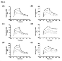
- the binding affinity of each of the immunoglobulin fragment-protein conjugates for FcRn at an acidic pH was analyzed using the 1:1 Langmuir binding with drifting baseline model in the BIAevaluation software, and the results of the analysis are shown in FIG. 2 .
- Hepes buffer pH 7.4 was used as running buffer-2.
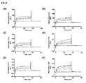
- the binding between FcRn and each of the immunoglobulin fragment-protein conjugates was induced for 4 minutes using the sample comprising each of the immunoglobulin fragment-protein conjugates, which diluted in running buffer-1, and then dissociation of each of the immunoglobulin fragment-protein conjugates from FcRn was induced for 1 minute using running buffer-2.
- Sensorgrams of the immunoglobulin fragment-protein conjugates are shown in FIG. 3 .
- the degree of dissociation of the immunoglobulin fragment-protein conjugate from FcRn was expressed as a binding ratio (%) according to the following equation 2.
- the resonance unit measured at 2 seconds before the completion of injection of the pH 6.0 sample was selected as a physical amount that is proportional to the amount bound at pH 6.0
- the resonance unit measured at 10 seconds after the start of dissociation at pH 7.4 was selected as a physical amount that is proportional to the amount bound at pH 7.4.
- binding ratio refers to the extent to which the immunoglobulin fragment-protein conjugate easily dissociates from FcRn. It can be seen that, as the value of the binding ratio becomes smaller, the dissociation of the conjugate at a neutral pH is better and the FcRn-mediated recycling of the conjugate more easily occurs, whereas, as the value of the binding ratio becomes larger, the dissociation of the conjugate at a neutral pH is insufficient, so that the conjugate is more likely to be removed by lysosomal degradation even when it is endocytosed by binding to FcRn.
- the binding affinity at an acidic pH or a neutral pH did not significantly differ between the immunoglobulin fragment alone (an independent Fc having no therapeutic protein linked thereto) and the conjugates of the present invention.
- the binding affinity of the immunoglobulin fragment for FcRn did not change even when the immunoglobulin did bind to the physiologically active protein, and particularly, the conjugates comprising various physiologically active proteins having different sizes showed similar results.
- the in-frame GLP-1R agonist-immunoglobulin Fc fragment conjugate of the Comparative Example showed a high binding ratio, and thus could not suitably dissociate from FcRn at a neutral pH, suggesting that it has a lower effect on prolonging the half-life of the physiologically active protein, compared to the conjugate of the present invention.
- each of the GLP-1R agonist-immunoglobulin Fc fragment conjugate (400 mcg/kg) and the in-frame GLP-1R agonist-immunoglobulin Fc fragment conjugate (400 mcg/kg) was diluted in physiological saline and administered subcutaneously to the animals at a dose of 2 mL/kg.
- blood was collected from the jugular vein of the rats, and serum was separated from the blood.
- the concentration of the drug in each of the serum samples was quantified by an enzyme-linked immunosorbent assay, and the results of the quantification are shown in FIG. 4 .
- the serum half-lives of the GLP-1R agonist-immunoglobulin Fc fragment conjugate and the in-frame GLP-1R agonist-immunoglobulin Fc fragment conjugate were 40.9 hours and 28 hours, respectively, and the maximum serum concentrations of the conjugates were 1758.6 ng/mL and 742.7 ng/mL, respectively.
- the GLP-1R agonist-immunoglobulin fragment conjugate of the present invention was excellent in terms of the in vivo absorption and half-life compared to the in-frame GLP-1R agonist-immunoglobulin Fc fragment conjugate ( FIG. 4 ).
Landscapes
- Health & Medical Sciences (AREA)
- Chemical & Material Sciences (AREA)
- Life Sciences & Earth Sciences (AREA)
- Organic Chemistry (AREA)
- General Health & Medical Sciences (AREA)
- Genetics & Genomics (AREA)
- Biochemistry (AREA)
- Medicinal Chemistry (AREA)
- Molecular Biology (AREA)
- Proteomics, Peptides & Aminoacids (AREA)
- Immunology (AREA)
- Biophysics (AREA)
- Engineering & Computer Science (AREA)
- Bioinformatics & Cheminformatics (AREA)
- Zoology (AREA)
- Public Health (AREA)
- General Engineering & Computer Science (AREA)
- Wood Science & Technology (AREA)
- Biotechnology (AREA)
- Veterinary Medicine (AREA)
- Biomedical Technology (AREA)
- Animal Behavior & Ethology (AREA)
- Epidemiology (AREA)
- Pharmacology & Pharmacy (AREA)
- Toxicology (AREA)
- Gastroenterology & Hepatology (AREA)
- Cell Biology (AREA)
- Endocrinology (AREA)
- Physics & Mathematics (AREA)
- Plant Pathology (AREA)
- Microbiology (AREA)
- Peptides Or Proteins (AREA)
- Medicinal Preparation (AREA)
- Medicines Containing Antibodies Or Antigens For Use As Internal Diagnostic Agents (AREA)
Abstract
Description
- The present invention relates to a physiologically active polypeptide-immunoglobulin Fc fragment conjugate comprising a physiologically active polypeptide linked via a non-peptidyl linker to an immunoglobulin Fc fragment with an FcRn-binding region capable of maintaining the intrinsic binding affinity of the immunoglobulin Fc fragment, a method for preparing the conjugate, a method of maintaining the intrinsic binding affinity of the conjugate for FcRn, and a composition comprising the conjugate capable of maintaining the intrinsic binding affinity of the immunoglobulin Fc fragment for FcRn.
- Various studies have been conducted on protein conjugates or complexes that comprise a carrier, such as a polyethylene glycol polymer, albumin, fatty acid or antibody Fc (constant region), linked to a protein in order to increase the serum half-life of the protein. Studies known to date about such protein conjugates or complexes mostly aim to increase the serum half-life of a drug to shorten the interval of drug administration to thereby improve patient convenience. However, many conventional technologies have problems such as a decrease in the activity of a therapeutic protein due to, for example, a spatial hindrance caused by a non-specific binding between a therapeutic protein and a carrier protein. In addition, in the case of fatty acid conjugates that reversibly bind to serum albumin to increase their serum half-life, there is a limit to significantly increase the serum half-life of a protein drug, because renal clearance, which accounts for the greatest loss of a protein drug, cannot be avoided due to the reversible binding between the protein and the fatty acid.
- Moreover, efforts have been made to use immunoglobulin fragments to increase the half-life of physiologically active substances including proteins. The CH2-CH3 region of immunoglobulin Fc includes an FcRn (protection receptor)-binding site that prolongs the half-life of an antibody. FcRn is an MHC class I-related protein expressed in vascular endothelial cells and binds to IgG and albumin. Characteristically, IgG and FcRn strongly bind to each other at a weak acidic pH and dissociate from each other at a neutral pH. Thus, when IgG enters vascular endothelial cells from blood vessels by pinocytosis or endocytosis and enters lysosomes (pH 6.0), it is protected by FcRn without being degraded. When IgG is fused with the cell membrane by recycling, it dissociates from FcRn at pH 7.4 and is released into blood vessels. Due to this procedure, the in vivo half-lives of IgG1, IgG2 and IgG4, which include the FcRn-binding site, are 3 weeks on average, being longer than those of other proteins.
- Thus, when an immunoglobulin Fc fragment is linked to a physiologically active substance, the half-life of the physiologically active substance can be increased by FcRn-mediated recycling. In this regard, there is a need for the development of a method capable of maintaining the binding affinity of an immunoglobulin Fc fragment for FcRn without reducing the activity of a physiologically active substance when the physiologically active substance and the immunoglobulin Fc fragment are linked together.
- The present inventors have made extensive efforts to develop a conjugate that can maintain the intrinsic binding affinity of an immunoglobulin Fc fragment itself for FcRn without reducing the activity of a physiologically active polypeptide and can easily dissociate from FcRn at a neutral pH. As a result, the present inventors have found that a conjugate comprising a physiologically active polypeptide linked via a non-peptidyl linker to an immunoglobulin Fc fragment having an FcRn-binding region can maintain the intrinsic binding affinity of the immunoglobulin Fc fragment for FcRn and, at the same time, can easily dissociate from FcRn at a neutral pH of 7.4, thereby completing the present invention.
- It is an objective of the present invention to provide a physiologically active polypeptide-immunoglobulin Fc fragment conjugate that comprises a physiologically active polypeptide linked via a non-peptidyl linker to an immunoglobulin Fc fragment comprising an FcRn-binding region, wherein the binding ratio of the amount of the conjugate bound to FcRn at pH 6.0 and the amount of the conjugate bound to FcRn at pH 7.4 is within the range of ± 6% of a ratio determined by measuring the amounts of binding of the immunoglobulin Fc fragment to FcRn under the same conditions as those used for the conjugate.
- Another objective of the present invention is to provide a physiologically active polypeptide-immunoglobulin Fc fragment conjugate that comprises a physiologically active polypeptide linked via a non-peptidyl linker to an immunoglobulin Fc fragment comprising an FcRn-binding region, wherein a binding ratio determined by substituting the amount of the conjugate bound to FcRn at pH 6.0 and the amount of the conjugate bound to FcRn at pH 7.4 into the following
equation 1 is within the range of ± 6% of a binding ratio determined by measuring the amounts the immunoglobulin Fc fragment bound to FcRn under the same conditions as those used for the conjugate:
- Still another objective of the present invention is to provide a method for preparing a physiologically active polypeptide-immunoglobulin Fc fragment conjugate that maintains the intrinsic binding affinity of an immunoglobulin Fc fragment for FcRn, the method comprising: (a) linking a physiologically active polypeptide via a non-peptidyl linker to an immunoglobulin Fc fragment having an FcRn-binding region to prepare a mixture of physiologically active polypeptide-immunoglobulin Fc fragment conjugates; and (b) separating from the mixture a physiologically active polypeptide-immunoglobulin Fc fragment conjugate that shows a binding ratio within the range of ± 6% of the binding ratio of the immunoglobulin Fc fragment, as determined by substituting the amount bound to FcRn at pH 6.0 and the amount bound to FcRn at pH 7.4 into
equation 1. - Still another objective of the present invention is to provide a method of maintaining the intrinsic binding affinity of a physiologically active polypeptide-immunoglobulin Fc fragment conjugate for FcRn, the method comprising linking a physiologically active polypeptide via a non-peptidyl linker to an immunoglobulin Fc fragment comprising an FcRn-binding region.
- Still another objective of the present invention is to provide a composition comprising the physiologically active polypeptide-immunoglobulin Fc fragment conjugate that maintains the intrinsic binding affinity of the immunoglobulin Fc fragment for FcRn.
- As described above, the conjugate of the present invention maintains the intrinsic binding affinity of the immunoglobulin Fc fragment for FcRn and, at the same time, easily dissociates from FcRn at a neutral pH such as a pH of 7.4. Thus, it can be advantageously used to increase the serum half-life of a physiologically active polypeptide.
-
-
FIG. 1 is a schematic view showing the process of recycling a protein bound to FcRn. -
FIG. 2 shows sensograms of the binding of immunoglobulin-protein conjugates to FcRn at an acidic pH. -
FIG. 3 shows sensograms of the binding of immunoglobulin-protein conjugates to FcRn at a neutral pH. -
FIG. 4 shows the results of a test for comparison of in vivo pharmacokinetics of GLP-1R agonist-immunoglobulin fragment conjugate - To achieve the above objects, in one aspect, the present invention provides a physiologically active polypeptide-immunoglobulin Fc fragment conjugate that comprises a physiologically active polypeptide linked via a non-peptidyl linker to an immunoglobulin Fc fragment comprising an FcRn-binding region, wherein the binding ratio of the amount of the conjugate bound to FcRn at pH 6.0 and the amount of the conjugate bound to FcRn at pH 7.4 is within the range of ± 6% of a ratio determined by measuring the amounts of the immunoglobulin Fc fragment bound to FcRn under the same conditions as those used for the conjugate.
- In one embodiment, the conjugate according to the present invention shows a binding ratio within the range of ± 6% of the binding ratio of the immunoglobulin Fc fragment, as determined using the following equation:

- In another embodiment, the conjugate according to the present invention may be obtained by reacting the physiologically active polypeptide having the non-peptidyl linker linked thereto with the immunoglobulin Fc fragment at a pH of 4.0-9.0, thereby linking the physiologically active polypeptide via the non-peptidyl linker to a portion excluding the FcRn-binding region of the immunoglobulin Fc fragment.
- In still another embodiment, the non-peptidyl linker that is included in the conjugate according to the present invention may be selected from the group consisting of polyethylene glycol, polypropylene glycol, an ethylene glycol-propylene glycol copolymer, polyoxyethylated polyol, polyvinyl alcohol, polysaccharide, dextran, polyvinyl ethyl ether, a biodegradable polymer, a lipid polymer, chitin, hyaluronic acid, and combinations thereof
- In still another embodiment, the non-peptidyl linker that is included in the conjugate according to the present invention may be a polyethylene glycol polymer represented by the following formula 1:

- In still another embodiment, the non-peptidyl linker that is included in the conjugate according to the present invention may have a reactive group selected from the group consisting of an aldehyde group, a propionaldehyde group, a butyraldehyde group, a maleimide group, and succinimide derivatives.
- In still another embodiment, the physiologically active polypeptide that is included in the conjugate according to the present invention may be selected from the group consisting of glucagon-like peptide-1 (GLP-1), granulocyte colony stimulating factor (G-CSF), human growth hormone (hGH), erythropoietin (EPO), glucagon, oxyntomodulin, insulin, growth hormone releasing hormone, growth hormone releasing peptide, interferons, interferon receptors, G-protein-coupled receptor, interleukins, interleukin receptors, enzymes, interleukin binding proteins, cytokine binding proteins, macrophage activating factor, macrophage peptide, B cell factor, T cell factor, protein A, allergy inhibitor, cell necrosis glycoproteins, immunotoxin, lymphotoxin, tumor necrosis factor, tumor suppressors, metastasis growth factor, alpha-1 antitrypsin, albumin, α-lactalbumin, apolipoprotein-E, highly glycosylated erythropoietin, angiopoietins, hemoglobin, thrombin, thrombin receptor activating peptide, thrombomodulin, blood factors VII, VIIa, VIII, IX and XIII, plasminogen activating factor, fibrin-binding peptide, urokinase, streptokinase, hirudin, protein C, C-reactive protein, renin inhibitor, collagenase inhibitor, superoxide dismutase, leptin, platelet-derived growth factor, epithelial growth factor, epidermal growth factor, angiostatin, angiotensin, bone growth factor, bone stimulating protein, calcitonin, atriopeptin, cartilage inducing factor, elcatonin, connective tissue activating factor, tissue factor pathway inhibitor, follicle stimulating hormone, luteinizing hormone, luteinizing hormone releasing hormone, nerve growth factors, parathyroid hormone, relaxin, secretin, somatomedin, insulin-like growth factor, adrenocortical hormone, cholecystokinin, pancreatic polypeptide, gastrin releasing peptide, corticotropin releasing factor, thyroid stimulating hormone, autotaxin, lactoferrin, myostatin, cell surface antigens, virus derived vaccine antigens, monoclonal antibodies, polyclonal antibodies, and antibody fragments.
- In still another embodiment, the immunoglobulin Fc fragment comprising the FcRn-binding region, which is included in the conjugate according to the present invention, may comprise a CH2 domain, a CH3 domain, or both.
- In still another embodiment, the immunoglobulin Fc fragment that is included in the conjugate according to the present invention may be in a non-glycosylated form.
- In still another embodiment, the immunoglobulin Fc fragment that is included in the conjugate according to the present invention may comprise a hinge region.
- In still another embodiment, the immunoglobulin Fc fragment that is included in the conjugate according to the present invention may be selected from the group consisting of IgG, IgA, IgD, IgE, IgM, combinations thereof, and hybrids thereof
- In still another embodiment, the immunoglobulin Fc fragment that is included in the conjugate according to the present invention may be an IgG4 Fc fragment.
- In another aspect, the present invention provides a method for preparing a physiologically active polypeptide-immunoglobulin Fc fragment conjugate that maintains the intrinsic binding affinity of an immunoglobulin Fc fragment for FcRn, the method comprising: (a) linking a physiologically active polypeptide via a non-peptidyl linker to an immunoglobulin Fc fragment comprising an FcRn-binding region to a physiologically active polypeptide via a non-peptidyl linker to prepare a mixture of physiologically active polypeptide-immunoglobulin Fc fragment conjugates; and (b) separating from the mixture a physiologically active polypeptide-immunoglobulin Fc fragment conjugate that shows a binding ratio within the range of ± 6% of the binding ratio of the immunoglobulin Fc fragment, as determined by substituting the amount of binding to FcRn at pH 6.0 and the amount of binding to FcRn at pH 7.4 into
equation 1. - In one embodiment, the non-peptidyl linker that is used in the preparation method according to the present invention may be a polyethylene glycol polymer represented by the following formula 1:

- In still another embodiment, the conjugate separated in step (b) of the preparation method according to the present invention may have a structure in which the non-peptidyl linker is bound to the N-terminus of the immunoglobulin Fc fragment.
- In still another aspect, the present invention provides a method of maintaining the intrinsic binding affinity of a physiologically active polypeptide-immunoglobulin Fc fragment conjugate for FcRn, the method comprising linking a physiologically active polypeptide via a non-peptidyl linker to an immunoglobulin Fc fragment comprising an FcRn-binding region.
- In one embodiment, the maintaining of the intrinsic binding affinity may be effected in vivo.
- In still another aspect, the present invention provides a composition comprising the physiologically active polypeptide-immunoglobulin Fc fragment conjugate, which maintains the intrinsic binding affinity of the immunoglobulin Fc fragment for FcRn.
- In one aspect, the present invention provides a physiologically active polypeptide-immunoglobulin Fc fragment conjugate that comprises a physiologically active polypeptide linked via a non-peptidyl linker to an immunoglobulin Fc fragment comprising an FcRn-binding region, wherein the ratio of the amount of the conjugate bound to FcRn at pH 6.0 and the amount of the conjugate bound to FcRn at pH 7.4 is within the range of ± 6% of a ratio determined by measuring the amounts of the immunoglobulin Fc fragment bound to FcRn under the same conditions as those used for the conjugate.
- As used herein, the expression "amount of binding of the physiologically active polypeptide-immunoglobulin Fc fragment conjugate" refers to the ratio of the concentration of the conjugate protein bound to FcRn relative to the total concentration of the conjugate protein bound or unbound to FcRn at the pH of interest. In the present invention, because this amount of binding is used to calculate the ratio between the amounts of binding at two pHs, the ratio between the amounts of binding may be calculated from the absolute amount of binding measured at one pH and may also be determined by measuring other physical amounts, which are proportional to the amount of the bound conjugate and easy to measure. For example, the ratio between the amounts of binding can be determined by measuring surface plasmon resonance (SPR) signals and calculating the ratio between the signals at pH 6.0 and pH 7.4. In addition, other methods may also be used to determine the ratio between the amounts of binding.
- Specifically, the present invention provides a physiologically active polypeptide-immunoglobulin Fc fragment conjugate that comprises a physiologically active polypeptide linked via a non-peptidyl linker to an immunoglobulin Fc fragment comprising an FcRn-binding region, wherein a binding ratio determined by substituting the amount of the conjugate bound to FcRn at pH 6.0 and the amount of the conjugate bound to FcRn at pH 7.4 into the following
equation 1 is less than the range of ± 6% of a binding ratio determined by measuring the amounts of the immunoglobulin Fc fragment bound to FcRn under the same conditions as those used for the conjugate:
- The binding ratio can be obtained as a percentage (%) value by dividing the amount of the conjugate (or immunoglobulin Fc fragment) binding to FcRn at pH 7.4 by the amount of the conjugate (or immunoglobulin Fc fragment) binding to FcRn at pH 6.0 and multiplying the divided value by 100.
- The binding ratio can be calculated using the method proposed by Weirong Wang et al. (DRUG METABOLISM AND DISPOSITION 39:1469-1477, 2011) for predicting the half-life of a monoclonal antibody, but is not specifically limited thereto. In the method described in the above literature, a monoclonal antibody having a high binding ratio showed a short in vivo half life.
- The method for measuring the binding ratio may be performed, particularly using a surface plasmon resonance method, but is not specifically limited thereto.
- For example, the method for measuring the binding ratio can be performed by immobilizing FcRn onto a biosensor chip (e.g., a Biacore CM5 biosensor chip) using an amine coupling kit or the like, injecting the FcRn-immobilized biosensor chip with an acidic buffer (e.g., pH 6.0 buffer) containing a material (e.g., the conjugate or the immunoglobulin Fc fragment) to be measured for the extent of binding to FcRn, and then injecting a neutral buffer (e.g., pH 7.4 buffer) into the biosensor chip. In this case, the binding ratio can be determined by measuring the resonance unit (RU) in an equilibrium state before completion of the injection of the sample in the pH 6.0 buffer into the chip to determine the amount bound at pH 6.0, measuring the resonance unit after injection of the pH 7.4 buffer to determine the amount bound at pH 7.4, and then dividing the amount bound at pH 7.4 by the amount bound at pH 6.0. When the binding ratio is to be expressed in percentage, the divided value can be multiplied by 100.
- In the above-described method, the composition of the pH 6.0 buffer is not specifically limited, as long as it can induce a binding between the material to be measured for the extent of binding to FcRn and FcRn. For example, the pH 6.0 buffer may be a buffer containing a salt such as phosphate. The concentration of the salt in the buffer may be 50-200 mM, but is not limited thereto. Also, the buffer may be injected into the FcRn-immobilized biosensor chip at a temperature of about 25 °C, but the temperature of measurement may be changed within the temperature range in which the pH of the buffer does not change.
- In addition, the pH 6.0 buffer may contain the material to be measured for the extent of binding to FcRn. The concentration of the material to be measured for the degree of binding to FcRn in the pH 6.0 buffer is not specifically limited, as long as the extent of binding to FcRn can be measured. For example, the concentration of the material to be measured for the degree of binding to FcRn in the pH 6.0 buffer may be between 100 nM and 12.5 nM.
- In the above-described method, the composition of the pH 7.4 buffer is not specifically limited, as long as it can induce dissociation between the material to be measured for the extent of binding to FcRn and FcRn. For example, the pH 7.4 buffer may be a buffer containing phosphate. The concentration of a salt in the buffer may be 50-200 mM, but is not limited thereto. Also, the pH 7.4 buffer may be injected into the FcRn-immobilized biosensor chip at a temperature of 25~37 °C, but is not limited thereto. In a specific embodiment, the ratio between the amounts of binding is measured at a temperature of 25 °C.
- Moreover, the amount of binding between FcRn and the material to be measured for the extend of binding to FcRn at pH 6.0 may be a resonance unit value measured at 2-60 seconds before the completion of injection of the pH 6.0 sample. Binding between FcRn and the material to be measured for the extent of binding to FcRn is preferably in an equilibrium state at the time point of measurement.
- Also, the amount of binding between FcRn and the material to be measured for the extent of binding to FcRn at pH 7.4 may be a resonance unit value measured at 10-20 seconds after injection of the pH 7.4 buffer. The time point of measurement is preferably between the time point at which the RU value changes rapidly and before the RU value reaches 0.
- In a comparison of the binding ratio between the immunoglobulin Fc fragment and the conjugate according to the present invention, the binding ratios are preferably values determined by the same experimental method under the same experimental conditions, but the compositions and temperatures of the buffers may vary depending on the kind of conjugate.
- The surface plasmon resonance method as described above is one of methods for determining the binding ratio. In addition to the surface plasmon resonance method, any method may be used in the present invention, as long as it is a method such as an enzyme-immunosorbent assay (ELISA), which can measure the amount of the conjugate bound to FcRn. In addition, the unit of the amount of binding may vary depending on the method used.
- When the binding ratio of a physiologically active polypeptide-immunoglobulin Fc fragment conjugate that comprises a physiologically active polypeptide linked via a non-peptidyl linker to an immunoglobulin Fc fragment having an FcRn-binding region is within the range of ± 6.0% of the binding ratio of the immunoglobulin Fc fragment itself, as measured by the above-described method, the physiologically active polypeptide-immunoglobulin Fc fragment conjugate has a long in vivo half-life.
- In the present invention, it was found that, even when a physiologically active polypeptide was covalently linked via a non-peptidyl linker to an immunoglobulin Fc fragment having an FcRn-binding region to form a conjugate, the intrinsic binding affinity of the immunoglobulin Fc fragment for FcRn could be maintained, suggesting that intracellular recycling of the immunoglobulin Fc fragment can easily occur to increase the in vivo half-life. Particularly, when the non-peptidyl linker binds to the portion excluding the FcRn-binding region of the immunoglobulin Fc fragment, it does not reduce the binding affinity of the immunoglobulin Fc fragment for FcRn. When the non-peptidyl linker is polyethylene glycol (-[O-CH2-CH2]n-), n in-[O-CH2-CH2]n- is 10 or greater, particularly 10-2400, more particularly 10-480, and even more particularly 50-250, but is not limited thereto.
- It was found that when n in -[O-CH2-CH2]n- is particularly 10-2400, and more particularly 50-2400, the polyethylene glycol does not influence the physiologically activity and FcRn-binding activity of each of the physiologically active polypeptide and the immunoglobulin Fc fragment. The present invention is based on this characteristic.
- As used herein, the term "physiologically active polypeptide-immunoglobulin Fc fragment conjugate" refers to a conjugate that comprises a physiologically active polypeptide covalently linked via a non-peptidyl linker to an immunoglobulin Fc fragment having an FcRn-binding region and shows a binding ratio within the range of ± 6.0% of the binding ratio of the immunoglobulin Fc fragment, as determined by substituting the amount of binding to FcRn at pH 6.0 and the amount of binding to FcRn at pH 7.4 into
equation 1. - The non-peptidyl linker in the conjugate may be linked to amino acid residues away from the FcRn-binding region of the immunoglobulin Fc fragment, for example, a region corresponding to positions 252-257 and 307-311 of CH2 and positions 433-436 of CH2, numbered according to the Kabat numbering system. The non-peptidyl linker may preferably be linked to the N-terminus or C-terminus of the immunoglobulin Fc fragment, and more preferably linked to the N-terminus, but is not limited thereto.
- When the non-peptidyl linker is linked to the N-terminus or C-terminus of the immunoglobulin Fc fragment, it does not substantially reduce the binding affinity of the immunoglobulin Fc fragment for FcRn, and thus the intrinsic binding affinity of the immunoglobulin Fc fragment of the conjugate for FcRn can be maintained.
- As used herein, the term "non-peptidyl linker" refers to a biocompatible polymer composed of two or more repeating units linked to each other, in which the repeating units are linked to each other by any non-peptide covalent bond. This non-peptidyl linker may have two or three ends.
- The non-peptidyl linker used in the present invention may be selected from the group consisting of biodegradable polymers such as polyethylene glycol, polypropylene glycol, a copolymer of ethylene glycol with propylene glycol, polyoxyethylated polyol, polyvinyl alcohol, polysaccharide, dextran, polyvinyl ethyl ether, biodegradable polymers such as polylactic acid (PLA) and polylactic-glycolic acid (PLGA), lipid polymers, chitins, hyaluronic acid, and combinations thereof, but is not limited thereto. Preferably, the non-peptidyl linker is polyethylene glycol, for example, a polyethylene glycol polymer represented by the following
formula 1, but is not limited:
- Meanwhile, other non-peptidyl linkers having a molecular weight corresponding to that of the polyethylene glycol of
formula 1 also fall within the scope of the present invention. - In addition, their derivatives known in the art and non-peptidyl linker derivatives that can be easily prepared in the state of the art also fall within the scope of the present invention.
- Polyethylene glycol that is used as the non-peptidyl linker in the present invention has an advantage in that it does not result in spatial hindrance between the physiologically active polypeptide and immunoglobulin Fc fragment bound to both ends thereof so that both the physiological activity of the physiologically active polypeptide and the binding affinity of the immunoglobulin Fc fragment for FcRn can be maintained.
- A peptidyl linker that is used in a fusion protein prepared by a conventional in-frame fusion method has a disadvantage in that it is easily cleaved by protease in vivo, and thus the expected effect of increasing the serum half-life of the active drug by a carrier cannot be obtained. However, the conjugate comprising the non-peptidyl linker according to the present invention dramatically overcomes this disadvantage. The non-peptidyl linker may be a polymer that has resistance to protease to maintain the serum half-life of the peptide, similar to that of a carrier. Therefore, any non-peptidyl linker may be used in the present invention without any limitation, as long as it is a polymer having the above-described function, that is, a polymer having resistance to protease in vivo.
- In addition, the non-peptidyl linker that is linked to the immunoglobulin Fc fragment in the present invention may be made not only of one kind of polymer, but also of a combination of different kinds of polymers.
- The non-peptidyl linker that is used in the present invention has reactive groups capable of binding to the immunoglobulin Fc fragment and the physiologically active polypeptide.
- The reactive groups at both ends of the non-peptidyl polymer are preferably selected from the group consisting of a reactive aldehyde group, a propionaldehyde group, a butyraldehyde group, a maleimide group, and succinimide derivative. Herein, the succinimide derivative may be succinimidyl propionate, hydroxy succinimidyl, succinimidyl carboxymethyl, or succinimidyl carbonate. In particular, when the non-peptidyl polymer has a reactive aldehyde group at both ends thereof, a physiologically active polypeptide and an immunoglobulin effectively bind to both ends of the non-peptidyl linker, respectively, while minimizing non-specific reactions. A final product generated by reductive alkylation via an aldehyde bond is much more stable than that linked via an amide bond. The aldehyde reactive group can bind selectively to the N-terminus at a low pH and can form a covalent bond with a lysine residue at a high pH, for example, at pH 9.0. Herein, the non-peptidyl linker may contain two or more aldehyde groups or have two or more alcohol groups substituted with functional groups including aldehyde.
- The reactive groups at both ends of the non-peptidyl linker may be the same or different. For example, one end of the non-peptidyl linker may have a maleimide group, and the other end may have an aldehyde group, a propionaldehyde group, or an alkyl aldehyde group such as butyl aldehyde. When a polyethylene glycol having hydroxyl reactive groups at both ends is used as the non-peptidyl linker, the hydroxy groups may be activated into various reactive groups by a known chemical reaction. Alternatively, a commercially available polyethylene glycol having a modified reactive group may be used to prepare the conjugate of the present invention.
- As used herein, the term "physiologically active polypeptide" collectively refers to polypeptides having any physiological activity in vivo, which commonly have a polypeptide structure and have various physiological activities. The physiologically active polypeptides include those that function to regulate genetic expression and physiological function and to correct an abnormal condition caused by the lack or excessive secretion of a substance that is involved in the regulation of functions in vivo. The physiologically active polypeptides may also include general protein therapeutic agents. In addition, the term "physiologically active polypeptide" is meant to include not only native polypeptides, but also derivatives thereof
- The kind and size of physiologically active polypeptide in the conjugate of the present invention are not specifically limited, as long as it is a physiologically active polypeptide can show an increase in the serum half-life by the conjugate structure of the present invention. In an embodiment of the present invention, conjugates are prepared using various physiologically active polypeptides, including insulin, interferon, human growth hormone and a GLP-1 agonist, which are representative examples of physiologically active polypeptides, and it was found that the intrinsic binding affinity of the immunoglobulin Fc fragment itself for FcRn can be maintained regardless of the kind and size of physiologically active polypeptide.
- The physiologically active polypeptide included in the conjugate according to the present invention may be selected from the group consisting of glucagon-like peptide-1 (GLP-1), granulocyte colony stimulating factor (G-CSF), human growth hormone (hGH), erythropoietin (EPO), glucagon, oxyntomodulin, insulin, growth hormone releasing hormone, growth hormone releasing peptide, interferons, interferon receptors, G-protein-coupled receptor, interleukins, interleukin receptors, enzymes, interleukin binding proteins, cytokine binding proteins, macrophage activating factor, macrophage peptide, B cell factor, T cell factor, protein A, allergy inhibitor, cell necrosis glycoproteins, immunotoxin, lymphotoxin, tumor necrosis factor, tumor suppressors, metastasis growth factor, alpha-1 antitrypsin, albumin, α-lactalbumin, apolipoprotein-E, highly glycosylated erythropoietin, angiopoietins, hemoglobin, thrombin, thrombin receptor activating peptide, thrombomodulin, blood factors VII, VIIa, VIII, IX and XIII, plasminogen activating factor, fibrin-binding peptide, urokinase, streptokinase, hirudin, protein C, C-reactive protein, renin inhibitor, collagenase inhibitor, superoxide dismutase, leptin, platelet-derived growth factor, epithelial growth factor, epidermal growth factor, angiostatin, angiotensin, bone growth factor, bone stimulating protein, calcitonin, atriopeptin, cartilage inducing factor, elcatonin, connective tissue activating factor, tissue factor pathway inhibitor, follicle stimulating hormone, luteinizing hormone, luteinizing hormone releasing hormone, nerve growth factors, parathyroid hormone, relaxin, secretin, somatomedin, insulin-like growth factor, adrenocortical hormone, cholecystokinin, pancreatic polypeptide, gastrin releasing peptide, corticotropin releasing factor, thyroid stimulating hormone, autotaxin, lactoferrin, myostatin, cell surface antigens, virus derived vaccine antigens, monoclonal antibodies, polyclonal antibodies, and antibody fragments, but is not limited thereto. In addition, the term "physiologically active polypeptide", as used herein, is meant to include not only natural physiologically active polypeptides, but also agonists, precursors, derivatives, fragments or variants of each polypeptide. Herein, examples of oxyntomodulin derivatives include all those disclosed in Korean Patent Application Publication No.
10-2012-0137271 10-2009-0008151 - As used herein, the term "immunoglobulin Fc fragment" refers to a protein that contains the heavy-chain constant region of an immunoglobulin, excluding the heavy-chain constant region 1 (CH1) and light-chain constant region 1 (CL1) of the immunoglobulin. The Fc fragment may include a hinge region in the heavy-chain constant region. In the present invention, the immunoglobulin Fc fragment preferably comprises a CH2 domain, a CH3 domain, or both, because the binding affinity of the immunoglobulin Fc fragment for FcRn should be maintained.
- Also, the immunoglobulin Fc region in the present invention may be an extended Fc fragment that includes a portion or whole of the heavy-chain constant region 1 (CH1) and/or the light-chain constant region 1 (CL1), except for the heavy-chain and light-chain variable regions of the immunoglobulin, as long as it maintains its intrinsic binding affinity for FcRn, even when it is linked to the physiologically active polypeptide and the non-peptidyl linker.
- Because immunoglobulin Fc fragment is a biodegradable polypeptide metabolized in vivo, it is safe for use as a drug carrier. Also, because the immunoglobulin Fc fragment has a molecular weight lower than the entire immunoglobulin molecule, it is beneficial in terms of the preparation, purification and yield of the conjugate. In addition, because the Fab region, which shows high non-homogeneity due to the difference in amino acid sequence between antibodies, is removed, the Fc fragment has a considerably increased homogeneity and a low potential to induce serum antigenicity.
- In the present invention, the immunoglobulin Fc fragment includes not only a native amino acid sequence, but also a sequence mutant thereof As used herein, the term "amino acid sequence mutant" refers to a sequence that is different from the native amino acid sequence due to a deletion, insertion, non-conservative or conservative substitution or combinations thereof of one or more amino acid residues. For example, in the case of IgG Fc, amino acid residues at positions 214 to 238, 297 to 299, 318 to 322 or 327 to 331, known to be important in binding, may be used as a suitable target for modification.
- In addition, various mutants are also possible, including mutants having a deletion of a region capable of forming a disulfide bond, a deletion of several amino acid residues at the N-terminus of a native Fc, or an addition of methionine residue to the N-terminus of a native Fc. Furthermore, to eliminate effector functions, a complement-binding site, for example, a C1q-binding site, may be removed, and an antibody dependent cell mediated cytotoxicity (ADCC) site may also be removed. Techniques of preparing such sequence derivatives of the immunoglobulin Fc fragment are disclosed in International Patent Publication Nos.
WO 97/34631 WO 96/32478 - Amino acid exchanges in proteins and peptides, which do not generally alter the activity of molecules, are known in the art (H. Neurath, R. L. Hill, The Proteins, Academic Press, New York,197 9). The most commonly occurring exchanges are Ala/Ser, Val/Ile, Asp/Glu, Thr/Ser, Ala/Gly, Ala/Thr, Ser/Asn, Ala/Val, Ser/Gly, Thy/Phe, Ala/Pro, Lys/Arg, Asp/Asn, Leu/Ile, Leu/Val, Ala/Glu, Asp/Gly, in both directions.
- In some cases, the immunoglobulin Fc fragment may also be modified by, for example, phosphorylation, sulfation, acrylation, glycosylation, methylation, farnesylation, acetylation or amidation.
- The above-described Fc mutants are mutants that show the same biological activity as that of the Fc fragment of the present invention, but have improved structural stability against heat, pH, or the like.
- In addition, these Fc fragments may be obtained from native forms isolated from humans and animals including cows, goats, pigs, mice, rabbits, hamsters, rats and guinea pigs, or may be recombinant forms or derivatives thereof, obtained from transformed animal cells or microorganisms. Herein, they may be obtained from a native immunoglobulin by isolating a whole immunoglobulin from the living body of humans or animals and treating it with protease. When the whole immunoglobulin is treated with papain, it is cleaved into Fab and Fc. Meanwhile, when it is treated with pepsin, it is cleaved into pF'c and F(ab)2. These fragments may be subjected, for example, to size-exclusion chromatography to isolate Fc or pF'c.
- Preferably, it is a recombinant immunoglobulin Fc fragment obtained from a microorganism using a human Fc fragment.
- In addition, the immunoglobulin Fc fragment may be in the form of having native sugar chains, increased sugar chains compared to a native form or decreased sugar chains compared to the native form, or may be in a deglycosylated form. The increase, decrease or removal of the immunoglobulin Fc sugar chains may be performed using conventional methods, such as a chemical method, an enzymatic method and a genetic engineering method using a microorganism. The immunoglobulin Fc fragment obtained by removing sugar chains from an Fc shows a sharp decrease in binding affinity for the complement (c1q) and a decrease or loss in antibody-dependent cell-mediated cytotoxicity or complement-dependent cytotoxicity, and thus does not induce unnecessary immune responses in vivo. In this regard, an immunoglobulin Fc fragment in a deglycosylated or aglycosylated form may be more suitable for use as a drug carrier.
- As used herein, the term "deglycosylation" refers to an enzymatic removal of sugar moieties from an Fc fragment, and the term "aglycosylation" means an unglycosylated Fc fragment produced in prokaryotes, preferably in E. coli.
- Meanwhile, the immunoglobulin Fc fragment may originate from humans or animals such as cattle, goats, pigs, mice, rabbits, hamsters, rats or guinea pigs. Preferably, it is of human origin. In addition, the immunoglobulin Fc fragment may be an Fc fragment that is derived from IgG, IgA, IgD, IgE and IgM, combinations thereof, or hybrids thereof. Preferably, it is derived from IgG or IgM, which is among the most abundant proteins in the human blood, and most preferably it is derived from IgG known to enhance the half-life of ligand-binding proteins.
- The term "combination", as used herein, refers to a formation of linkage between a polypeptide encoding single-chain immunoglobulin Fc fragments of the same origin and a single-chain polypeptide of a different origin to form a dimer or multimer. In other words, a dimer or multimer may be formed from two or more fragments selected from the group consisting of IgG Fc, IgA Fc, IgM Fc, IgD Fc, and IgE Fc fragments.
- As used herein, the term "hybrid" refers to the presence of two or more sequences corresponding to immunoglobulin Fc fragments of different origins in a single-chain immunoglobulin Fc fragment. In the present invention, various types of hybrids are possible. In other words, domain hybrids may be composed of one to four domains selected from the group consisting of CH1, CH2, CH3 and CH4 of IgG Fc, IgM Fc, IgA Fc, IgE Fc and IgD Fc, and may include a hinge region.
- On the other hand, IgG can be divided into IgG1, IgG2, IgG3 and IgG4 subclasses, and combinations or hybrids thereof may be used in the present invention, preferably IgG2 and IgG4 subclasses, and most preferably the Fc fragment of IgG4 rarely having effector functions such as CDC (complement dependent cytotoxicity). In other words, the most preferable immunoglobulin Fc fragment for use as a drug carrier in the present invention is a human IgG4-derived unglycosylated Fc fragment. The human Fc fragment is more preferable than a non-human Fc fragment, which may act as an antigen in the human body and cause undesirable immune responses such as the production of a new antibody against the antigen.
- In an embodiment of the present invention, each of insulin, interferon, human growth hormone and a GLP-1 agonist is linked via a non-peptidyl linker to an immunoglobulin Fc fragment having the ability to bind to FcRn, thereby preparing conjugates that increase the intrinsic binding affinity of the immunoglobulin Fc fragment for FcRn and can easily dissociate from FcRn at a neutral pH and can also show a dissociation degree similar to that of the immunoglobulin Fc fragment (Examples and
FIGS. 2 and3 ). In addition, it was found that the conjugate of the present invention has a long in vivo duration of action compared to an in-frame conjugate (FIG. 4 ). - In still another aspect, the present invention provides a method for preparing a physiologically active polypeptide-immunoglobulin Fc fragment conjugate that maintains the intrinsic binding affinity of an immunoglobulin Fc fragment for FcRn, the method comprising the steps of: (a) linking a physiologically active polypeptide via a non-peptidyl linker to an immunoglobulin Fc fragment having an FcRn-binding region to prepare a mixture of physiologically active polypeptide-immunoglobulin Fc fragment conjugates; and (b) separating from the mixture a physiologically active polypeptide-immunoglobulin Fc fragment conjugate that shows a binding ratio within the range of ± 6% of the binding ratio of the immunoglobulin Fc fragment, as determined by substituting the amount bound to FcRn at pH 6.0 and the amount bound to FcRn at pH 7.4 into the
following equation 1.
- Herein, the physiologically active polypeptide, the immunoglobulin Fc fragment, the non-peptidyl linker, the conjugate and the determination of the binding ratio are as described above.
- Step (a) in the method of the present invention is a step of covalently linking a physiologically active polypeptide via a non-peptidyl linker to an immunoglobulin Fc fragment. Step (a) may comprise the steps of: (i) linking any one of the physiologically active polypeptide and the immunoglobulin Fc fragment to a reactive group at one end of the non-peptidyl linker; and (ii) linking the remaining one to a reactive group at the other end of the non-peptidyl linker. Step (a) may further comprise, between steps (i) and (ii), a step of separating the physiologically active polypeptide or immunoglobulin Fc fragment linked to one end of the non-peptidyl linker. For preparation of this conjugate, the disclosure of Korean Patent No.
10-0725315 - In order to link the physiologically active polypeptide via the non-peptidyl linker to a portion excluding the FcRn-binding region of the immunoglobulin Fc fragment, the physiologically active polypeptide having the non-peptidyl linker linked thereto may be reacted with the immunoglobulin Fc fragment at a pH of 4.0-9.0.
- When the conjugate is prepared by this process, byproducts such as a conjugate showing a decrease in the binding affinity of the immunoglobulin Fc fragment for FcRn can be generated in addition to a conjugate that maintains the intrinsic binding affinity of the immunoglobulin Fc fragment for FcRn. For this reason, after the reaction that links the physiologically active polypeptide via the non-peptidyl peptide to the immunoglobulin Fc fragment, a process is additionally required to separate from the conjugate mixture a physiologically active polypeptide-immunoglobulin Fc fragment that maintains the intrinsic binding affinity of the immunoglobulin Fc fragment for FcRn.
- Thus, the method of the present invention comprises step (b) of separating from the conjugate mixture a physiologically active polypeptide-immunoglobulin Fc fragment conjugate that shows a binding ratio within the range of ± 6% of the binding ratio of the immunoglobulin Fc fragment, as determined by substituting the amount boundg to FcRn at pH 6.0 and the amount bound to FcRn at pH 7.4 into
equation 1. - Step (b) is preferably a process for separating a conjugate in which the non-peptidyl linker is linked to amino acid residues away from the FcRn-binding region of the immunoglobulin Fc fragment, for example, a region corresponding to positions 252-257 and 307-311 of CH2 and positions 433-436 of CH2, numbered according to the cobat numbering system. Specifically, step (b) is a process for selectively separating only a conjugate in which the non-peptidyl linker is connected to the N-terminus of the immunoglobulin Fc fragment so that the intrinsic binding affinity of the Fc fragment for FcRn is maintained.
- Separation and purification conditions in step (b) may vary depending on the kinds of non-peptidyl linker, physiologically active polypeptide and the like used.
- In still another aspect, the present invention provides a method of maintaining the intrinsic binding affinity of a physiologically active polypeptide-immunoglobulin Fc fragment conjugate for FcRn, the method comprising linking a physiologically active polypeptide via a non-peptidyl linker to an immunoglobulin Fc fragment comprising an FcRn-binding region.
- Herein, the physiologically active polypeptide, the immunoglobulin Fc fragment, the non-peptidyl linker and the conjugate are as described above.
- The present invention has advantages in that, because the physiologically active polypeptide is linked via the non-peptidyl linker to the immunoglobulin Fc fragment comprising the FcRn-binding region, the intrinsic binding affinity of the immunoglobulin Fc fragment for FcRn is maintained while the immunoglobulin Fc fragment can easily dissociate from FcRn at a neutral pH and can be easily recycled, suggesting that the in vivo half-life of the immunoglobulin Fc fragment can be effectively increased.
- Herein, the maintaining of the intrinsic binding affinity may be effected in vivo.
- In still another aspect, the present invention provides a composition comprising the physiologically active polypeptide-immunoglobulin Fc fragment conjugate, which maintains the intrinsic binding affinity of the immunoglobulin Fc fragment for FcRn.
- Herein, the physiologically active polypeptide, the immunoglobulin Fc fragment and the conjugate are as described above.
- Hereinafter, the present invention will be described in further detail with reference to examples. It is to be understood, however, that these examples are for illustrative purposes only and are not intended to limit the scope of the present invention.
- An immunoglobulin Fc fragment was prepared according to the method disclosed in Korean Patent Application No.
10-2006-0077377 - A reaction was performed to PEGylate a 3.4-kDa propion-ALD2 PEG (IDB, Korea) specifically at the N-terminus of the beta-chain of insulin. The reaction solution was purified using a cation-exchange column. To prepare an insulin-immunoglobulin Fc fragment conjugate, the purified mono-PEGylated insulin was reacted with the immunoglobulin Fc. At this time, the reaction was performed at pH 6.0-8.2 in order to direct the insulin specifically to the N-terminus of the immunoglobulin Fc. After completion of the reaction, the reaction solution was first purified using an anion-exchange column, and then purified using a hydrophobic column, thereby obtaining an insulin conjugate comprising insulin linked site-specifically to the immunoglobulin.
- A 3.4-kDa propion-ALD2 PEG (IDB, Korea) was added to and reacted with a buffer containing human interferon alpha-2b (hIFNα-2b; molecular weight: 19 kDa) dissolved therein. To obtain a conjugate in which PEG is linked specifically to the amino terminal end of interferon-alpha and PEG and interferon-alpha are linked to each other at a ratio of 1:1, the reaction mixture was subjected to anion exchange column chromatography to purify a mono-PEGylated IFNα-2b. To direct the purified mono-PEGylated interferon specifically to the N-terminus of the immunoglobulin Fc, a reaction was performed at a pH of 5.5-6.5. After the linking reaction, to purify the produced interferon-immunoglobulin Fc fragment conjugate, the reaction mixture was passed through an anion-exchange column to obtain an interferon-immunoglobulin Fc fragment conjugate fraction. The conjugate fraction was additionally purified using a hydrophobic column, thereby obtaining an interferon conjugate comprising interferon linked site-specifically to the immunoglobulin Fc.
- A 3.4-kDa propion-ALD2 PEG (IDB, Korea) was added to and reacted with a buffer containing human growth hormone (hGH; molecular weight: 22 kDa) dissolved therein. To obtain a conjugate in which PEG is linked specifically to the amino terminal end of human growth hormone and PEG and human growth hormone are linked to each other at a ratio of 1:1, the reaction mixture was subjected to anion exchange column chromatography to purify a mono-PEGylated human growth hormone (hGH). To direct and link the purified mono-PEGylated human growth hormone specifically to the N-terminus of the immunoglobulin Fc, a reaction was performed at a pH of 5.5-6.5. After the linking reaction, the reaction mixture was purified using an anion-exchange column, thereby obtaining a human growth hormone conjugate fraction comprising hormone growth hormone linked site-specifically to the immunoglobulin Fc.
- A 3.4-kDa propion-ALD2 PEG (IDB, Korea) was reacted site-specifically with the lysine residue of imidazo-acetyl-exendin-4 (CA exendin-4, Bachem, Switzerland). To obtain a conjugate in which PEG and the GLP-1R agonist are linked to each other at a ratio of 1:1, the reaction mixture was then subjected to cation-exchange column chromatography to purify mono-PEGylated exendin-4. To prepare a GLP-1R agonist-immunoglobulin Fc fragment conjugate comprising the mono-PEGylated exendin-4 linked specifically to the N-terminus of the immunoglobulin Fc, a reaction was performed at a pH of 5.0-8.2. After the coupling reaction, a two-step purification process was performed using a hydrophobic column and an anion-exchange column, thereby obtaining a GLP-1R agonist-immunoglobulin Fc fragment conjugate comprising the GLP-1R agonist linked site-specifically to the immunoglobulin Fc.
- For comparison with the conjugate of the present invention, a fusion protein of a GLP-1R agonist with an immunoglobulin Fc fragment comprising an about 50 kDa FcRn site was prepared by a gene recombination method without using a linker. The conjugate comprising the GLP-1R agonist linked in-frame to the immunoglobulin Fc fragment (hereinafter also referred to as "in-frame GLP-1R agonist-immunoglobulin Fc fragment conjugate") was purified from the culture using an affinity column.
- Among proteins introduced into cells by endocytosis, a protein having an FcRn-binding site binds to FcRn at an acidic pH, whereas a protein that does not bind to FcRn is removed by lysosomal degradation. When the protein bound to FcRn dissociates from FcRn at a neutral pH, it is released to the cell surface, whereas the FcRn-bound protein that does not dissociate from FcRn undergoes lysosomal degradation. This mechanism is shown in
FIG. 1 . - Thus, in order to examine whether the conjugate of the Example, obtained by linking the physiologically active protein via the non-peptidyl linker to the immunoglobulin Fc comprising the FcRn binding site, can maintain the binding affinity of the immunoglobulin Fc alone for FcRn or whether it can easily dissociate from FcRn at a neutral pH and can be released into blood, the following experiment was performed.
- Specifically, in order to examine whether the binding affinity of the immunoglobulin fragment for FcRn does not change even when the conjugate of the present invention is formed, the binding affinity between FcRn and the conjugate of the Example or the Comparative Example was measured using SPR (surface Plasmon resonance, BIACORE 3000). As FcRn, FCGRT & B2M dimer receptor (Sino Biological Inc.) was used. FcRn was immobilized onto a CM5 chip using an amine coupling method, and then the conjugate was added thereto at a concentration between 100 nM to 12.5 nM to measure the binding affinity therebetween. However, because the mechanism of the FcRn-mediated increase in the half-life depends on pH, the experiment was carried out at each of an acidic pH and a neutral pH.
- Because the binding of an endocytosed protein to FcRn occurs at an acidic pH, a phosphate buffer (pH 6.0) was used as running buffer-1 in order to reproduce this binding. All the immunoglobulin fragment-protein conjugates were diluted in running buffer-1 to induce the binding, and dissociation of the conjugates was also performed using running buffer-1. Each of the immunoglobulin fragment-protein conjugates was brought contact with the FcRn-immobilized chip for 4 minutes to induce the binding thereof, and then subjected to a dissociation process for 6 minutes. Next, in order to measure the values of the immunoglobulin fragment-protein conjugates at different concentrations, i.e., binds the immunoglobulin fragment-protein conjugates at different concentrations to the chip, pH 7.4 Hepes buffer was brought into contact with the immunoglobulin fragment-protein conjugates bound to FcRn for about 30 seconds. The binding affinity of each of the immunoglobulin fragment-protein conjugates for FcRn at an acidic pH was analyzed using the 1:1 Langmuir binding with drifting baseline model in the BIAevaluation software, and the results of the analysis are shown in
FIG. 2 . - Because the release of the conjugate from cells after binding to FcRn occurs when the pH changes from an acidic pH to a neutral pH, Hepes buffer (pH 7.4) was used as running buffer-2. However, the binding between FcRn and each of the immunoglobulin fragment-protein conjugates was induced for 4 minutes using the sample comprising each of the immunoglobulin fragment-protein conjugates, which diluted in running buffer-1, and then dissociation of each of the immunoglobulin fragment-protein conjugates from FcRn was induced for 1 minute using running buffer-2. Sensorgrams of the immunoglobulin fragment-protein conjugates are shown in
FIG. 3 . The degree of dissociation of the immunoglobulin fragment-protein conjugate from FcRn was expressed as a binding ratio (%) according to thefollowing equation 2. Herein, the resonance unit measured at 2 seconds before the completion of injection of the pH 6.0 sample was selected as a physical amount that is proportional to the amount bound at pH 6.0, and the resonance unit measured at 10 seconds after the start of dissociation at pH 7.4 was selected as a physical amount that is proportional to the amount bound at pH 7.4. Using the selected resonance units, the binding ratio was calculated according to the following equation 2:
- As used herein, the term "binding ratio" refers to the extent to which the immunoglobulin fragment-protein conjugate easily dissociates from FcRn. It can be seen that, as the value of the binding ratio becomes smaller, the dissociation of the conjugate at a neutral pH is better and the FcRn-mediated recycling of the conjugate more easily occurs, whereas, as the value of the binding ratio becomes larger, the dissociation of the conjugate at a neutral pH is insufficient, so that the conjugate is more likely to be removed by lysosomal degradation even when it is endocytosed by binding to FcRn.
- As a result, as shown in
FIGS. 2 and3 , the various immunoglobulin fragment-protein conjugates of the Example did bind to FcRn in a concentration-dependent manner. The results are also shown in Table 1 below, and the degrees of dissociation of the conjugates, calculated usingequation 2, are shown in Table 2 below.Table 1: Comparison of binding affinities of immunoglobulin fragment-protein conjugates for FcRn at pH 6.0 Test materials pH 6.0 ka (1/Ms, X105) kd (1/s, X10-3) KD (nM) Immunoglobulin fragment 4.8 10.5 22.0 ± 3.0 Insulin-immunoglobulin fragment conjugate 3.1 7.1 22.6 ± 3.9 Interferon-immunoglobulin fragment conjugate 3.0 9.3 30.0 ± 4.8 Human growth hormone-immunoglobulin fragment conjugate 1.2 3.6 26.8 ± 9.0 GLP-1R agonist-immunoglobulin fragment conjugate 3.8 9.5 24.7 ± 5.5 In-frame GLP-1R agonist-immunoglobulin fragment conjugate 3.2 5.3 17.0 ± 3.7 *ka: association rate constant, kd: dissociation rate constant, KD: affinity constant, binding ratio: a value obtained by dividing the amount of binding at pH 7.4 by the amount of binding at pH 6.0 and multiplying the divided value by 100. Table 2: Comparison of degrees of dissociation of immunoglobulin fragment-protein conjugates from FcRn Test materials Concentration (nM) Binding amount (RU) at pH6.0 Binding amount (RU) at pH7.4 Binding ratio (%) Average binding ratio (%) Immunoglobulin fragment 100 163.7 4.8 2.9 5.4 ± 2.0 50 137.4 6.5 4.7 25 110.9 7.4 6.6 12.5 88.0 6.5 7.3 Insulin- immunoglobulin fragment conjugate 100 214.1 4.8 2.2 5.0 ± 2.2 50 174.3 8.0 4.6 25 143.9 8.4 5.9 12.5 106.6 7.9 7.4 Interferon- immunoglobulin fragment conjugate 100 196.9 6.4 3.2 5.0 ± 1.6 50 157.2 6.7 4.3 25 120.3 6.7 5.6 12.5 91.3 6.4 7.0 Human growth hormone- immunoglobulin fragment conjugate 100 205.7 8.2 4.0 6.6 ± 2.6 50 153.0 7.7 5.1 25 111.9 8.3 7.4 12.5 82.9 8.1 9.8 GLP-1R agonist- immunoglobulin fragment conjugate 100 163.5 6.2 3.8 6.6 ± 2.6 50 135.5 8.1 5.9 25 107.7 7.2 6.7 12.5 86.2 8.6 10.0 In-frame GLP-1R agonist- immunoglobulin fragment conjugate 100 301.7 38.2 12.7 12.7 ± 0.3 50 249.5 31.2 12.5 25 197.6 25.9 13.1 12.5 148.7 18.6 12.5 - As can be seen in Tables 1 and 2 above, the binding affinity at an acidic pH or a neutral pH did not significantly differ between the immunoglobulin fragment alone (an independent Fc having no therapeutic protein linked thereto) and the conjugates of the present invention. In other words, it was found that, in the case of the immunoglobulin-protein conjugates produced according to the present invention, the binding affinity of the immunoglobulin fragment for FcRn did not change even when the immunoglobulin did bind to the physiologically active protein, and particularly, the conjugates comprising various physiologically active proteins having different sizes showed similar results. However, the in-frame GLP-1R agonist-immunoglobulin Fc fragment conjugate of the Comparative Example showed a high binding ratio, and thus could not suitably dissociate from FcRn at a neutral pH, suggesting that it has a lower effect on prolonging the half-life of the physiologically active protein, compared to the conjugate of the present invention.
- In order to compare in vivo pharmacokinetics between the GLP-1R agonist-immunoglobulin Fc fragment conjugate of Example (5) and the in-frame GLP-1R agonist-immunoglobulin Fc fragment conjugate, changes in the serum concentrations of the conjugates were analyzed using normal SD rats.
- Specifically, each of the GLP-1R agonist-immunoglobulin Fc fragment conjugate (400 mcg/kg) and the in-frame GLP-1R agonist-immunoglobulin Fc fragment conjugate (400 mcg/kg) was diluted in physiological saline and administered subcutaneously to the animals at a dose of 2 mL/kg. At 4, 8, 24, 48, 72, 96, 120, 144, 168, 192, 216, 240, 288, 312 and 336 hours after administration of the test materials, blood was collected from the jugular vein of the rats, and serum was separated from the blood. Next, the concentration of the drug in each of the serum samples was quantified by an enzyme-linked immunosorbent assay, and the results of the quantification are shown in
FIG. 4 . - As a result, the serum half-lives of the GLP-1R agonist-immunoglobulin Fc fragment conjugate and the in-frame GLP-1R agonist-immunoglobulin Fc fragment conjugate were 40.9 hours and 28 hours, respectively, and the maximum serum concentrations of the conjugates were 1758.6 ng/mL and 742.7 ng/mL, respectively. In other words, when the drugs were administered subcutaneously to the normal rats at the same dose, it was shown that the GLP-1R agonist-immunoglobulin fragment conjugate of the present invention was excellent in terms of the in vivo absorption and half-life compared to the in-frame GLP-1R agonist-immunoglobulin Fc fragment conjugate (
FIG. 4 ). - While the present invention has been described with reference to the particular illustrative embodiments, it will be understood by those skilled in the art to which the present invention pertains that the present invention may be embodied in other specific forms without departing from the technical spirit or essential characteristics of the present invention. Therefore, the embodiments described above are considered to be illustrative in all respects and not restrictive. Furthermore, the scope of the present invention is defined by the appended claims rather than the detailed description, and it should be understood that all modifications or variations derived from the meanings and scope of the present invention and equivalents thereof are included in the scope of the appended claims.
Claims (18)
- A physiologically active polypeptide-immunoglobulin Fc fragment conjugate that comprises a physiologically active polypeptide linked via a non-peptidyl linker to an immunoglobulin Fc fragment comprising an FcRn-binding region, wherein a binding ratio of the amount of the conjugate bound to FcRn at pH 6.0 and the amount of the conjugate bound to FcRn at pH 7.4 is within the range of ± 6% of a ratio determined by measuring the amounts of the immunoglobulin Fc fragment bound to FcRn under the same conditions as those used for the conjugate.
- The conjugate of claim 1, wherein the binding ratio of the physiologically active polypeptide-immunoglobulin Fc fragment conjugate is within the range of ± 6% of the binding ratio of the immunoglobulin Fc fragment, as determined using the following equation:

- The conjugate of claim 1 or 2, wherein the conjugate is obtained by reacting the physiologically active polypeptide having the non-peptidyl linker linked thereto with the immunoglobulin Fc fragment at a pH of 4.0-9.0, thereby linking the physiologically active polypeptide via the non-peptidyl linker to a portion excluding the FcRn-binding region of the immunoglobulin Fc fragment.
- The conjugate of claim 1 or 2, wherein the non-peptidyl linker is selected from the group consisting of polyethylene glycol, polypropylene glycol, an ethylene glycol-propylene glycol copolymer, polyoxyethylated polyol, polyvinyl alcohol, polysaccharide, dextran, polyvinyl ethyl ether, a biodegradable polymer, a lipid polymer, chitin, hyaluronic acid, and combinations thereof.
- The conjugate of claim 1 or 2, wherein the non-peptidyl linker is a polyethylene glycol polymer represented by the following formula 1:

- The conjugate of claim 1 or 2, wherein the non-peptidyl linker has a reactive group selected from the group consisting of an aldehyde group, a propionaldehyde group, a butyraldehyde group, a maleimide group, and succinimide derivatives.
- The conjugate of claim 1 or 2, wherein the physiologically active polypeptide is selected from the group consisting of glucagon-like peptide-1 (GLP-1), granulocyte colony stimulating factor (G-CSF), human growth hormone (hGH), erythropoietin (EPO), glucagon, oxyntomodulin, insulin, growth hormone releasing hormone, growth hormone releasing peptide, interferons, interferon receptors, G-protein-coupled receptor, interleukins, interleukin receptors, enzymes, interleukin binding proteins, cytokine binding proteins, macrophage activating factor, macrophage peptide, B cell factor, T cell factor, protein A, allergy inhibitor, cell necrosis glycoproteins, immunotoxin, lymphotoxin, tumor necrosis factor, tumor suppressors, metastasis growth factor, alpha-1 antitrypsin, albumin, α-lactalbumin, apolipoprotein-E, highly glycosylated erythropoietin, angiopoietins, hemoglobin, thrombin, thrombin receptor activating peptide, thrombomodulin, blood factors VII, VIIa, VIII, IX and XIII, plasminogen activating factor, fibrin-binding peptide, urokinase, streptokinase, hirudin, protein C, C-reactive protein, renin inhibitor, collagenase inhibitor, superoxide dismutase, leptin, platelet-derived growth factor, epithelial growth factor, epidermal growth factor, angiostatin, angiotensin, bone growth factor, bone stimulating protein, calcitonin, atriopeptin, cartilage inducing factor, elcatonin, connective tissue activating factor, tissue factor pathway inhibitor, follicle stimulating hormone, luteinizing hormone, luteinizing hormone releasing hormone, nerve growth factors, parathyroid hormone, relaxin, secretin, somatomedin, insulin-like growth factor, adrenocortical hormone, cholecystokinin, pancreatic polypeptide, gastrin releasing peptide, corticotropin releasing factor, thyroid stimulating hormone, autotaxin, lactoferrin, myostatin, cell surface antigens, virus derived vaccine antigens, monoclonal antibodies, polyclonal antibodies, and antibody fragments.
- The conjugate of claim 1 or 2, wherein the immunoglobulin Fc fragment comprising the FcRn-binding region comprises a CH2 domain, a CH3 domain, or both.
- The conjugate of claim 1 or 2, wherein the immunoglobulin Fc fragment is in a non-glycosylated form.
- The conjugate of claim 1 or 2, wherein the immunoglobulin Fc fragment further comprises a hinge region.
- The conjugate of claim 1 or 2, wherein the immunoglobulin Fc fragment is selected from the group consisting of IgG, IgA, IgD, IgE, IgM, combinations thereof, and hybrids thereof.
- The conjugate of claim 1 or 2, wherein the immunoglobulin Fc fragment is an IgG4 Fc fragment.
- A method for preparing a physiologically active polypeptide-immunoglobulin Fc fragment conjugate that maintains the intrinsic binding affinity of an immunoglobulin Fc fragment for FcRn, the method comprising:(a) linking a physiologically active polypeptide via a non-peptidyl linker to an immunoglobulin Fc fragment comprising an FcRn-binding region to prepare a mixture of physiologically active polypeptide-immunoglobulin Fc fragment conjugates; and(b) separating from the mixture a physiologically active polypeptide-immunoglobulin Fc fragment conjugate that shows a binding ratio within the range of ± 6% of the binding ratio of the immunoglobulin Fc fragment, as determined by substituting the amount of binding to FcRn at pH 6.0 and the amount of binding to FcRn at pH 7.4 into the following equation 1:

- The method of claim 13, wherein the non-peptidyl linker is a polyethylene glycol polymer represented by the following formula 1:

- The method of claim 13, wherein the conjugate separated in step (b) has a structure in which the non-peptidyl linker is bound to the N-terminus of the immunoglobulin Fc fragment.
- A method of maintaining the intrinsic binding affinity of a physiologically active polypeptide-immunoglobulin Fc fragment conjugate for FcRn, the method comprising linking a physiologically active polypeptide via a non-peptidyl linker to an immunoglobulin Fc fragment comprising an FcRn-binding region.
- The method of claim 16, wherein the maintaining of the intrinsic binding affinity is effected in vivo.
- A composition comprising the physiologically active polypeptide-immunoglobulin Fc fragment conjugate of claim 1, which maintains the intrinsic binding affinity of the immunoglobulin Fc fragment for FcRn.
Applications Claiming Priority (2)
| Application Number | Priority Date | Filing Date | Title |
|---|---|---|---|
| KR20130082509 | 2013-07-12 | ||
| PCT/KR2014/006328 WO2015005747A1 (en) | 2013-07-12 | 2014-07-14 | Immunoglobulin fc conjugate maintaining fcrn binding strength |
Publications (3)
| Publication Number | Publication Date |
|---|---|
| EP3020732A1 true EP3020732A1 (en) | 2016-05-18 |
| EP3020732A4 EP3020732A4 (en) | 2016-12-28 |
| EP3020732B1 EP3020732B1 (en) | 2020-03-18 |
Family
ID=52280325
Family Applications (1)
| Application Number | Title | Priority Date | Filing Date |
|---|---|---|---|
| EP14822721.8A Active EP3020732B1 (en) | 2013-07-12 | 2014-07-14 | An immunoglobulin fc conjugate which maintains binding affinity of immunoglobulin fc fragment to fcrn |
Country Status (21)
| Country | Link |
|---|---|
| US (2) | US20160152684A1 (en) |
| EP (1) | EP3020732B1 (en) |
| JP (2) | JP2016525099A (en) |
| KR (2) | KR101872559B1 (en) |
| CN (2) | CN116082528A (en) |
| AR (1) | AR096890A1 (en) |
| AU (1) | AU2014287879B2 (en) |
| CA (1) | CA2917838C (en) |
| DK (1) | DK3020732T3 (en) |
| EA (1) | EA034297B1 (en) |
| ES (1) | ES2799326T3 (en) |
| HU (1) | HU231424B1 (en) |
| IL (1) | IL243567B (en) |
| MX (1) | MX376370B (en) |
| MY (1) | MY180374A (en) |
| NO (1) | NO20160182A1 (en) |
| PH (1) | PH12016500111A1 (en) |
| PT (1) | PT3020732T (en) |
| TW (1) | TWI693236B (en) |
| UA (1) | UA119644C2 (en) |
| WO (1) | WO2015005747A1 (en) |
Families Citing this family (8)
| Publication number | Priority date | Publication date | Assignee | Title |
|---|---|---|---|---|
| KR20130049671A (en) | 2011-11-04 | 2013-05-14 | 한미사이언스 주식회사 | Method for preparation of biological active polypeptide conjugate |
| AR090281A1 (en) | 2012-03-08 | 2014-10-29 | Hanmi Science Co Ltd | IMPROVED PROCESS FOR THE PREPARATION OF A PHYSIOLOGICALLY ACTIVE POLYPEPTIDE COMPLEX |
| UA130573C2 (en) * | 2014-03-31 | 2026-03-25 | Ханмі Фарм. Ко., Лтд. | Method for improving solubility of protein and peptide by using immunoglobulin fc fragment linkage |
| AR107483A1 (en) * | 2016-01-29 | 2018-05-02 | Hanmi Pharm Ind Co Ltd | CONJUGATE OF THERAPEUTIC ENZYMES |
| KR20180071193A (en) * | 2016-12-19 | 2018-06-27 | 한미약품 주식회사 | Long-acting protein conjugates for brain targeting |
| KR102282240B1 (en) | 2018-12-06 | 2021-07-28 | 경상국립대학교산학협력단 | A Long-acting Exendin-4 and Use of There |
| WO2021010531A1 (en) * | 2019-07-18 | 2021-01-21 | 한미약품 주식회사 | Novel method for preparing protein conjugate |
| KR102382593B1 (en) * | 2020-01-29 | 2022-04-05 | 고려대학교 산학협력단 | Ph-sensitive fc variants |
Family Cites Families (25)
| Publication number | Priority date | Publication date | Assignee | Title |
|---|---|---|---|---|
| US6030613A (en) * | 1995-01-17 | 2000-02-29 | The Brigham And Women's Hospital, Inc. | Receptor specific transepithelial transport of therapeutics |
| US6096871A (en) | 1995-04-14 | 2000-08-01 | Genentech, Inc. | Polypeptides altered to contain an epitope from the Fc region of an IgG molecule for increased half-life |
| ATE279947T1 (en) | 1996-03-18 | 2004-11-15 | Univ Texas | IMMUNOGLOBULIN-LIKE DOMAIN WITH INCREASED HALF-LIFE TIMES |
| CN1859925A (en) * | 2002-12-26 | 2006-11-08 | 山景医药公司 | Polymer conjugates of cytokines, chemokines, growth factors, polypeptide hormones and antagonists thereof with preserved receptor-binding activity |
| US20050176108A1 (en) * | 2003-03-13 | 2005-08-11 | Young-Min Kim | Physiologically active polypeptide conjugate having prolonged in vivo half-life |
| WO2005047337A1 (en) * | 2003-11-13 | 2005-05-26 | Hanmi Pharmaceutical Co., Ltd. | A pharmaceutical composition comprising an immunoglobulin fc region as a carrier |
| KR100609434B1 (en) | 2004-12-30 | 2006-08-03 | 한국항공우주연구원 | Transversal Filter |
| KR100754667B1 (en) * | 2005-04-08 | 2007-09-03 | 한미약품 주식회사 | Immunoglobulin Fc Fragments Modified with Non-Peptide Polymers and Pharmaceutical Compositions Comprising the Same |
| US7625564B2 (en) * | 2006-01-27 | 2009-12-01 | Novagen Holding Corporation | Recombinant human EPO-Fc fusion proteins with prolonged half-life and enhanced erythropoietic activity in vivo |
| JP2008169195A (en) * | 2007-01-05 | 2008-07-24 | Hanmi Pharmaceutical Co Ltd | Insulin secretory peptide drug conjugates using carrier substances |
| KR20080098216A (en) * | 2007-05-04 | 2008-11-07 | 한미약품 주식회사 | Sodium excretion peptide drug conjugate using a carrier substance |
| KR100897938B1 (en) * | 2007-05-30 | 2009-05-18 | 주식회사 제넥신 | Immunoglobulin fusion proteins |
| JP2009019027A (en) | 2007-07-16 | 2009-01-29 | Hanmi Pharmaceutical Co Ltd | Insulin-secreting peptide derivatives with amino-terminal amino acid mutations |
| TWI388570B (en) * | 2008-07-23 | 2013-03-11 | 韓美科學有限公司 | Polypeptide complex comprising a non-peptidomimetic polymer having three functional ends |
| BR112012022917A2 (en) * | 2010-03-11 | 2017-01-10 | Pfizer | ph-dependent antigen binding antibodies |
| AR081066A1 (en) * | 2010-04-02 | 2012-06-06 | Hanmi Holdings Co Ltd | INSULIN CONJUGATE WHERE AN IMMUNOGLOBULIN FRAGMENT IS USED |
| KR101337797B1 (en) * | 2010-07-14 | 2013-12-06 | 한미사이언스 주식회사 | A liquid formulation of long acting human growth hormone conjugate |
| KR101333958B1 (en) * | 2010-10-20 | 2013-11-27 | 주식회사 한독 | Human Interleukin-1 receptor antagonist-hybrid Fc fusion protein |
| UA113626C2 (en) * | 2011-06-02 | 2017-02-27 | A COMPOSITION FOR THE TREATMENT OF DIABETES CONTAINING THE DURABLE INSULIN CON conjugate AND THE DUAL ACTION INSULINOTROPIC PIPIDE | |
| UA126465C2 (en) | 2011-06-10 | 2022-10-12 | Ханмі Сайенс Ко., Лтд. | A peptide having oxyntomodulin activity and a pharmaceutical composition for the treatment of obesity containing it |
| PL2721062T3 (en) * | 2011-06-17 | 2019-05-31 | Hanmi Science Co Ltd | A conjugate comprising oxyntomodulin and an immunoglobulin fragment, and use thereof |
| CN111053918A (en) * | 2011-09-05 | 2020-04-24 | 韩美科学株式会社 | A kind of anticancer pharmaceutical composition comprising interferon alpha-conjugate |
| KR20130049671A (en) * | 2011-11-04 | 2013-05-14 | 한미사이언스 주식회사 | Method for preparation of biological active polypeptide conjugate |
| KR102041412B1 (en) * | 2011-12-30 | 2019-11-11 | 한미사이언스 주식회사 | Derivatives of Immunglobulin Fc fragment |
| AR094821A1 (en) * | 2012-07-25 | 2015-09-02 | Hanmi Pharm Ind Co Ltd | LIQUID FORMULATION OF AN INSULINOTROPIC PEPTIDE CONJUGATE OF PROLONGED ACTION |
-
2014
- 2014-07-11 AR ARP140102575A patent/AR096890A1/en active IP Right Grant
- 2014-07-14 EP EP14822721.8A patent/EP3020732B1/en active Active
- 2014-07-14 HU HUP1600206A patent/HU231424B1/en unknown
- 2014-07-14 CA CA2917838A patent/CA2917838C/en active Active
- 2014-07-14 PT PT148227218T patent/PT3020732T/en unknown
- 2014-07-14 CN CN202310310247.2A patent/CN116082528A/en active Pending
- 2014-07-14 JP JP2016525294A patent/JP2016525099A/en active Pending
- 2014-07-14 EA EA201690114A patent/EA034297B1/en unknown
- 2014-07-14 WO PCT/KR2014/006328 patent/WO2015005747A1/en not_active Ceased
- 2014-07-14 UA UAA201600304A patent/UA119644C2/en unknown
- 2014-07-14 DK DK14822721.8T patent/DK3020732T3/en active
- 2014-07-14 KR KR1020140088530A patent/KR101872559B1/en active Active
- 2014-07-14 AU AU2014287879A patent/AU2014287879B2/en active Active
- 2014-07-14 TW TW103124093A patent/TWI693236B/en active
- 2014-07-14 MY MYPI2016000028A patent/MY180374A/en unknown
- 2014-07-14 MX MX2016000344A patent/MX376370B/en active IP Right Grant
- 2014-07-14 CN CN201480049342.0A patent/CN105593243A/en active Pending
- 2014-07-14 US US14/904,543 patent/US20160152684A1/en not_active Abandoned
- 2014-07-14 ES ES14822721T patent/ES2799326T3/en active Active
-
2016
- 2016-01-11 IL IL243567A patent/IL243567B/en active IP Right Grant
- 2016-01-12 PH PH12016500111A patent/PH12016500111A1/en unknown
- 2016-02-03 NO NO20160182A patent/NO20160182A1/en unknown
-
2018
- 2018-06-19 KR KR1020180070435A patent/KR102076718B1/en active Active
-
2020
- 2020-04-27 JP JP2020078005A patent/JP6880278B2/en active Active
-
2022
- 2022-08-03 US US17/880,358 patent/US20220380437A1/en active Pending
Also Published As
Similar Documents
| Publication | Publication Date | Title |
|---|---|---|
| US20220380437A1 (en) | Immunoglobulin fc conjugate which maintains binding affinity of immunoglobulin fc fragment to fcrn | |
| EP3020418B1 (en) | Conjugate of biologically active polypeptide monomer and immunoglobulin fc fragment with reduced receptor-mediated clearance, and the method for preparing the same | |
| HK1221957B (en) | An immunoglobulin fc conjugate which maintains binding affinity of immunoglobulin fc fragment to fcrn | |
| NZ716275B2 (en) | An Immunoglobulin Fc Conjugate which Maintains Binding Affinity of Immunoglobulin Fc Fragment to FcRn | |
| HK40016877A (en) | Conjugate of biologically active polypeptide monomer and immunoglobulin fc fragment with reduced receptor-mediated clearance, and the method for preparing the same | |
| HK1223017B (en) | Conjugate of biologically active polypeptide monomer and immunoglobulin fc fragment with reduced receptor-mediated clearance, and the method for preparing the same | |
| NZ716251B2 (en) | Conjugate of biologically active polypeptide monomer and immunoglobulin fc fragment with reduced receptor-mediated clearance, and the method for preparing the same |
Legal Events
| Date | Code | Title | Description |
|---|---|---|---|
| PUAI | Public reference made under article 153(3) epc to a published international application that has entered the european phase |
Free format text: ORIGINAL CODE: 0009012 |
|
| 17P | Request for examination filed |
Effective date: 20160107 |
|
| AK | Designated contracting states |
Kind code of ref document: A1 Designated state(s): AL AT BE BG CH CY CZ DE DK EE ES FI FR GB GR HR HU IE IS IT LI LT LU LV MC MK MT NL NO PL PT RO RS SE SI SK SM TR |
|
| AX | Request for extension of the european patent |
Extension state: BA ME |
|
| DAX | Request for extension of the european patent (deleted) | ||
| RIN1 | Information on inventor provided before grant (corrected) |
Inventor name: HWANG, SANG YOUN Inventor name: KWON, SE CHANG Inventor name: HONG, SUNG HEE Inventor name: LEE, JONG SOO Inventor name: JUNG, SUNG YOUB Inventor name: CHOI, IN YOUNG |
|
| A4 | Supplementary search report drawn up and despatched |
Effective date: 20161128 |
|
| RIC1 | Information provided on ipc code assigned before grant |
Ipc: C07K 16/46 20060101ALI20161122BHEP Ipc: C07K 19/00 20060101AFI20161122BHEP Ipc: C07K 14/72 20060101ALI20161122BHEP |
|
| REG | Reference to a national code |
Ref country code: HK Ref legal event code: DE Ref document number: 1221957 Country of ref document: HK |
|
| STAA | Information on the status of an ep patent application or granted ep patent |
Free format text: STATUS: REQUEST FOR EXAMINATION WAS MADE |
|
| STAA | Information on the status of an ep patent application or granted ep patent |
Free format text: STATUS: EXAMINATION IS IN PROGRESS |
|
| 17Q | First examination report despatched |
Effective date: 20180511 |
|
| GRAJ | Information related to disapproval of communication of intention to grant by the applicant or resumption of examination proceedings by the epo deleted |
Free format text: ORIGINAL CODE: EPIDOSDIGR1 |
|
| GRAP | Despatch of communication of intention to grant a patent |
Free format text: ORIGINAL CODE: EPIDOSNIGR1 |
|
| GRAP | Despatch of communication of intention to grant a patent |
Free format text: ORIGINAL CODE: EPIDOSNIGR1 |
|
| STAA | Information on the status of an ep patent application or granted ep patent |
Free format text: STATUS: GRANT OF PATENT IS INTENDED |
|
| INTG | Intention to grant announced |
Effective date: 20191008 |
|
| GRAS | Grant fee paid |
Free format text: ORIGINAL CODE: EPIDOSNIGR3 |
|
| GRAA | (expected) grant |
Free format text: ORIGINAL CODE: 0009210 |
|
| STAA | Information on the status of an ep patent application or granted ep patent |
Free format text: STATUS: THE PATENT HAS BEEN GRANTED |
|
| AK | Designated contracting states |
Kind code of ref document: B1 Designated state(s): AL AT BE BG CH CY CZ DE DK EE ES FI FR GB GR HR HU IE IS IT LI LT LU LV MC MK MT NL NO PL PT RO RS SE SI SK SM TR |
|
| REG | Reference to a national code |
Ref country code: GB Ref legal event code: FG4D |
|
| REG | Reference to a national code |
Ref country code: DE Ref legal event code: R096 Ref document number: 602014062599 Country of ref document: DE |
|
| REG | Reference to a national code |
Ref country code: AT Ref legal event code: REF Ref document number: 1245813 Country of ref document: AT Kind code of ref document: T Effective date: 20200415 Ref country code: IE Ref legal event code: FG4D |
|
| REG | Reference to a national code |
Ref country code: DK Ref legal event code: T3 Effective date: 20200611 |
|
| REG | Reference to a national code |
Ref country code: FI Ref legal event code: FGE |
|
| REG | Reference to a national code |
Ref country code: PT Ref legal event code: SC4A Ref document number: 3020732 Country of ref document: PT Date of ref document: 20200625 Kind code of ref document: T Free format text: AVAILABILITY OF NATIONAL TRANSLATION Effective date: 20200617 |
|
| REG | Reference to a national code |
Ref country code: CH Ref legal event code: NV Representative=s name: NOVAGRAAF INTERNATIONAL SA, CH |
|
| REG | Reference to a national code |
Ref country code: NL Ref legal event code: FP |
|
| REG | Reference to a national code |
Ref country code: SE Ref legal event code: TRGR |
|
| PG25 | Lapsed in a contracting state [announced via postgrant information from national office to epo] |
Ref country code: RS Free format text: LAPSE BECAUSE OF FAILURE TO SUBMIT A TRANSLATION OF THE DESCRIPTION OR TO PAY THE FEE WITHIN THE PRESCRIBED TIME-LIMIT Effective date: 20200318 Ref country code: NO Free format text: LAPSE BECAUSE OF FAILURE TO SUBMIT A TRANSLATION OF THE DESCRIPTION OR TO PAY THE FEE WITHIN THE PRESCRIBED TIME-LIMIT Effective date: 20200618 |
|
| PG25 | Lapsed in a contracting state [announced via postgrant information from national office to epo] |
Ref country code: LV Free format text: LAPSE BECAUSE OF FAILURE TO SUBMIT A TRANSLATION OF THE DESCRIPTION OR TO PAY THE FEE WITHIN THE PRESCRIBED TIME-LIMIT Effective date: 20200318 Ref country code: BG Free format text: LAPSE BECAUSE OF FAILURE TO SUBMIT A TRANSLATION OF THE DESCRIPTION OR TO PAY THE FEE WITHIN THE PRESCRIBED TIME-LIMIT Effective date: 20200618 Ref country code: HR Free format text: LAPSE BECAUSE OF FAILURE TO SUBMIT A TRANSLATION OF THE DESCRIPTION OR TO PAY THE FEE WITHIN THE PRESCRIBED TIME-LIMIT Effective date: 20200318 |
|
| REG | Reference to a national code |
Ref country code: LT Ref legal event code: MG4D |
|
| REG | Reference to a national code |
Ref country code: GR Ref legal event code: EP Ref document number: 20200401681 Country of ref document: GR Effective date: 20200916 |
|
| PG25 | Lapsed in a contracting state [announced via postgrant information from national office to epo] |
Ref country code: SM Free format text: LAPSE BECAUSE OF FAILURE TO SUBMIT A TRANSLATION OF THE DESCRIPTION OR TO PAY THE FEE WITHIN THE PRESCRIBED TIME-LIMIT Effective date: 20200318 Ref country code: IS Free format text: LAPSE BECAUSE OF FAILURE TO SUBMIT A TRANSLATION OF THE DESCRIPTION OR TO PAY THE FEE WITHIN THE PRESCRIBED TIME-LIMIT Effective date: 20200718 Ref country code: SK Free format text: LAPSE BECAUSE OF FAILURE TO SUBMIT A TRANSLATION OF THE DESCRIPTION OR TO PAY THE FEE WITHIN THE PRESCRIBED TIME-LIMIT Effective date: 20200318 Ref country code: LT Free format text: LAPSE BECAUSE OF FAILURE TO SUBMIT A TRANSLATION OF THE DESCRIPTION OR TO PAY THE FEE WITHIN THE PRESCRIBED TIME-LIMIT Effective date: 20200318 Ref country code: CZ Free format text: LAPSE BECAUSE OF FAILURE TO SUBMIT A TRANSLATION OF THE DESCRIPTION OR TO PAY THE FEE WITHIN THE PRESCRIBED TIME-LIMIT Effective date: 20200318 Ref country code: EE Free format text: LAPSE BECAUSE OF FAILURE TO SUBMIT A TRANSLATION OF THE DESCRIPTION OR TO PAY THE FEE WITHIN THE PRESCRIBED TIME-LIMIT Effective date: 20200318 Ref country code: RO Free format text: LAPSE BECAUSE OF FAILURE TO SUBMIT A TRANSLATION OF THE DESCRIPTION OR TO PAY THE FEE WITHIN THE PRESCRIBED TIME-LIMIT Effective date: 20200318 |
|
| REG | Reference to a national code |
Ref country code: ES Ref legal event code: FG2A Ref document number: 2799326 Country of ref document: ES Kind code of ref document: T3 Effective date: 20201216 |
|
| REG | Reference to a national code |
Ref country code: DE Ref legal event code: R097 Ref document number: 602014062599 Country of ref document: DE |
|
| PLBE | No opposition filed within time limit |
Free format text: ORIGINAL CODE: 0009261 |
|
| STAA | Information on the status of an ep patent application or granted ep patent |
Free format text: STATUS: NO OPPOSITION FILED WITHIN TIME LIMIT |
|
| 26N | No opposition filed |
Effective date: 20201221 |
|
| PG25 | Lapsed in a contracting state [announced via postgrant information from national office to epo] |
Ref country code: PL Free format text: LAPSE BECAUSE OF FAILURE TO SUBMIT A TRANSLATION OF THE DESCRIPTION OR TO PAY THE FEE WITHIN THE PRESCRIBED TIME-LIMIT Effective date: 20200318 |
|
| PG25 | Lapsed in a contracting state [announced via postgrant information from national office to epo] |
Ref country code: LU Free format text: LAPSE BECAUSE OF NON-PAYMENT OF DUE FEES Effective date: 20200714 |
|
| PG25 | Lapsed in a contracting state [announced via postgrant information from national office to epo] |
Ref country code: SI Free format text: LAPSE BECAUSE OF FAILURE TO SUBMIT A TRANSLATION OF THE DESCRIPTION OR TO PAY THE FEE WITHIN THE PRESCRIBED TIME-LIMIT Effective date: 20200318 |
|
| REG | Reference to a national code |
Ref country code: AT Ref legal event code: UEP Ref document number: 1245813 Country of ref document: AT Kind code of ref document: T Effective date: 20200318 |
|
| PG25 | Lapsed in a contracting state [announced via postgrant information from national office to epo] |
Ref country code: TR Free format text: LAPSE BECAUSE OF FAILURE TO SUBMIT A TRANSLATION OF THE DESCRIPTION OR TO PAY THE FEE WITHIN THE PRESCRIBED TIME-LIMIT Effective date: 20200318 Ref country code: MT Free format text: LAPSE BECAUSE OF FAILURE TO SUBMIT A TRANSLATION OF THE DESCRIPTION OR TO PAY THE FEE WITHIN THE PRESCRIBED TIME-LIMIT Effective date: 20200318 Ref country code: CY Free format text: LAPSE BECAUSE OF FAILURE TO SUBMIT A TRANSLATION OF THE DESCRIPTION OR TO PAY THE FEE WITHIN THE PRESCRIBED TIME-LIMIT Effective date: 20200318 |
|
| PG25 | Lapsed in a contracting state [announced via postgrant information from national office to epo] |
Ref country code: MK Free format text: LAPSE BECAUSE OF FAILURE TO SUBMIT A TRANSLATION OF THE DESCRIPTION OR TO PAY THE FEE WITHIN THE PRESCRIBED TIME-LIMIT Effective date: 20200318 Ref country code: AL Free format text: LAPSE BECAUSE OF FAILURE TO SUBMIT A TRANSLATION OF THE DESCRIPTION OR TO PAY THE FEE WITHIN THE PRESCRIBED TIME-LIMIT Effective date: 20200318 |
|
| P01 | Opt-out of the competence of the unified patent court (upc) registered |
Effective date: 20230602 |
|
| PGFP | Annual fee paid to national office [announced via postgrant information from national office to epo] |
Ref country code: MC Payment date: 20250625 Year of fee payment: 12 |
|
| PGFP | Annual fee paid to national office [announced via postgrant information from national office to epo] |
Ref country code: FI Payment date: 20250623 Year of fee payment: 12 |
|
| PGFP | Annual fee paid to national office [announced via postgrant information from national office to epo] |
Ref country code: DK Payment date: 20250624 Year of fee payment: 12 Ref country code: GB Payment date: 20250624 Year of fee payment: 12 |
|
| PGFP | Annual fee paid to national office [announced via postgrant information from national office to epo] |
Ref country code: NL Payment date: 20250623 Year of fee payment: 12 Ref country code: BE Payment date: 20250623 Year of fee payment: 12 |
|
| PGFP | Annual fee paid to national office [announced via postgrant information from national office to epo] |
Ref country code: PT Payment date: 20250623 Year of fee payment: 12 |
|
| PGFP | Annual fee paid to national office [announced via postgrant information from national office to epo] |
Ref country code: FR Payment date: 20250624 Year of fee payment: 12 |
|
| PGFP | Annual fee paid to national office [announced via postgrant information from national office to epo] |
Ref country code: GR Payment date: 20250623 Year of fee payment: 12 |
|
| PGFP | Annual fee paid to national office [announced via postgrant information from national office to epo] |
Ref country code: IE Payment date: 20250623 Year of fee payment: 12 |
|
| PGFP | Annual fee paid to national office [announced via postgrant information from national office to epo] |
Ref country code: SE Payment date: 20250624 Year of fee payment: 12 |
|
| PGFP | Annual fee paid to national office [announced via postgrant information from national office to epo] |
Ref country code: ES Payment date: 20250822 Year of fee payment: 12 |
|
| PGFP | Annual fee paid to national office [announced via postgrant information from national office to epo] |
Ref country code: DE Payment date: 20250624 Year of fee payment: 12 |
|
| PGFP | Annual fee paid to national office [announced via postgrant information from national office to epo] |
Ref country code: IT Payment date: 20250624 Year of fee payment: 12 |
|
| PGFP | Annual fee paid to national office [announced via postgrant information from national office to epo] |
Ref country code: AT Payment date: 20250623 Year of fee payment: 12 |
|
| PGFP | Annual fee paid to national office [announced via postgrant information from national office to epo] |
Ref country code: CH Payment date: 20250801 Year of fee payment: 12 |