EP3016983B1 - A new fusion protein to target and treat acute myloid leukemia cells - Google Patents
A new fusion protein to target and treat acute myloid leukemia cells Download PDFInfo
- Publication number
- EP3016983B1 EP3016983B1 EP14734837.9A EP14734837A EP3016983B1 EP 3016983 B1 EP3016983 B1 EP 3016983B1 EP 14734837 A EP14734837 A EP 14734837A EP 3016983 B1 EP3016983 B1 EP 3016983B1
- Authority
- EP
- European Patent Office
- Prior art keywords
- scfv
- cells
- antibody
- snap
- artificial
- Prior art date
- Legal status (The legal status is an assumption and is not a legal conclusion. Google has not performed a legal analysis and makes no representation as to the accuracy of the status listed.)
- Not-in-force
Links
Images


Classifications
-
- C—CHEMISTRY; METALLURGY
- C07—ORGANIC CHEMISTRY
- C07K—PEPTIDES
- C07K16/00—Immunoglobulins [IG], e.g. monoclonal or polyclonal antibodies
- C07K16/18—Immunoglobulins [IG], e.g. monoclonal or polyclonal antibodies against material from animals or humans
- C07K16/28—Immunoglobulins [IG], e.g. monoclonal or polyclonal antibodies against material from animals or humans against receptors, cell surface antigens or cell surface determinants
- C07K16/30—Immunoglobulins [IG], e.g. monoclonal or polyclonal antibodies against material from animals or humans against receptors, cell surface antigens or cell surface determinants from tumour cells
- C07K16/3061—Blood cells
-
- A—HUMAN NECESSITIES
- A61—MEDICAL OR VETERINARY SCIENCE; HYGIENE
- A61P—SPECIFIC THERAPEUTIC ACTIVITY OF CHEMICAL COMPOUNDS OR MEDICINAL PREPARATIONS
- A61P35/00—Antineoplastic agents
- A61P35/02—Antineoplastic agents specific for leukemia
-
- C—CHEMISTRY; METALLURGY
- C12—BIOCHEMISTRY; BEER; SPIRITS; WINE; VINEGAR; MICROBIOLOGY; ENZYMOLOGY; MUTATION OR GENETIC ENGINEERING
- C12N—MICROORGANISMS OR ENZYMES; COMPOSITIONS THEREOF; PROPAGATING, PRESERVING, OR MAINTAINING MICROORGANISMS; MUTATION OR GENETIC ENGINEERING; CULTURE MEDIA
- C12N9/00—Enzymes; Proenzymes; Compositions thereof; Processes for preparing, activating, inhibiting, separating or purifying enzymes
- C12N9/10—Transferases (2.)
- C12N9/1048—Glycosyltransferases (2.4)
- C12N9/1077—Pentosyltransferases (2.4.2)
-
- G—PHYSICS
- G01—MEASURING; TESTING
- G01N—INVESTIGATING OR ANALYSING MATERIALS BY DETERMINING THEIR CHEMICAL OR PHYSICAL PROPERTIES
- G01N33/00—Investigating or analysing materials by specific methods not covered by groups G01N1/00 - G01N31/00
- G01N33/48—Biological material, e.g. blood, urine; Haemocytometers
- G01N33/50—Chemical analysis of biological material, e.g. blood, urine; Testing involving biospecific ligand binding methods; Immunological testing
- G01N33/53—Immunoassay; Biospecific binding assay; Materials therefor
- G01N33/575—Immunoassay; Biospecific binding assay; Materials therefor for cancer
- G01N33/57505—Immunoassay; Biospecific binding assay; Materials therefor for cancer of the blood, e.g. leukaemia
-
- C—CHEMISTRY; METALLURGY
- C07—ORGANIC CHEMISTRY
- C07K—PEPTIDES
- C07K2317/00—Immunoglobulins specific features
- C07K2317/50—Immunoglobulins specific features characterized by immunoglobulin fragments
- C07K2317/56—Immunoglobulins specific features characterized by immunoglobulin fragments variable (Fv) region, i.e. VH and/or VL
-
- C—CHEMISTRY; METALLURGY
- C07—ORGANIC CHEMISTRY
- C07K—PEPTIDES
- C07K2317/00—Immunoglobulins specific features
- C07K2317/50—Immunoglobulins specific features characterized by immunoglobulin fragments
- C07K2317/56—Immunoglobulins specific features characterized by immunoglobulin fragments variable (Fv) region, i.e. VH and/or VL
- C07K2317/565—Complementarity determining region [CDR]
-
- C—CHEMISTRY; METALLURGY
- C07—ORGANIC CHEMISTRY
- C07K—PEPTIDES
- C07K2317/00—Immunoglobulins specific features
- C07K2317/60—Immunoglobulins specific features characterized by non-natural combinations of immunoglobulin fragments
- C07K2317/62—Immunoglobulins specific features characterized by non-natural combinations of immunoglobulin fragments comprising only variable region components
- C07K2317/622—Single chain antibody (scFv)
-
- C—CHEMISTRY; METALLURGY
- C07—ORGANIC CHEMISTRY
- C07K—PEPTIDES
- C07K2317/00—Immunoglobulins specific features
- C07K2317/70—Immunoglobulins specific features characterized by effect upon binding to a cell or to an antigen
- C07K2317/73—Inducing cell death, e.g. apoptosis, necrosis or inhibition of cell proliferation
-
- C—CHEMISTRY; METALLURGY
- C07—ORGANIC CHEMISTRY
- C07K—PEPTIDES
- C07K2317/00—Immunoglobulins specific features
- C07K2317/70—Immunoglobulins specific features characterized by effect upon binding to a cell or to an antigen
- C07K2317/77—Internalization into the cell
-
- C—CHEMISTRY; METALLURGY
- C07—ORGANIC CHEMISTRY
- C07K—PEPTIDES
- C07K2317/00—Immunoglobulins specific features
- C07K2317/90—Immunoglobulins specific features characterized by (pharmaco)kinetic aspects or by stability of the immunoglobulin
- C07K2317/92—Affinity (KD), association rate (Ka), dissociation rate (Kd) or EC50 value
-
- C—CHEMISTRY; METALLURGY
- C07—ORGANIC CHEMISTRY
- C07K—PEPTIDES
- C07K2319/00—Fusion polypeptide
- C07K2319/55—Fusion polypeptide containing a fusion with a toxin, e.g. diphteria toxin
-
- C—CHEMISTRY; METALLURGY
- C07—ORGANIC CHEMISTRY
- C07K—PEPTIDES
- C07K2319/00—Fusion polypeptide
- C07K2319/60—Fusion polypeptide containing spectroscopic/fluorescent detection, e.g. green fluorescent protein [GFP]
-
- C—CHEMISTRY; METALLURGY
- C12—BIOCHEMISTRY; BEER; SPIRITS; WINE; VINEGAR; MICROBIOLOGY; ENZYMOLOGY; MUTATION OR GENETIC ENGINEERING
- C12Y—ENZYMES
- C12Y204/00—Glycosyltransferases (2.4)
- C12Y204/02—Pentosyltransferases (2.4.2)
- C12Y204/02036—NAD(+)--diphthamide ADP-ribosyltransferase (2.4.2.36)
Definitions
- Subject matter of the present invention is a compound comprising the polypeptide of the invention, the pharmaceutical composition comprising the compound of the invention, a therapeutic and diagnostic use of the compound or polypeptide of the invention.
- AML Acute myeloid leukemias
- AML Acute myeloid leukemias
- the American Cancer Society was estimating 14,590 cases of newly diagnosed AML and 10,370 AML-related deaths in 2013 2 .
- the median age at diagnosis is 65 years, which implies a dramatic rise of AML incidences in the coming years due to increased life expectancy and the resulting demographic changes 3 .
- the current prognosis is poor with a five-year relative survival rate of only 24% 2 .
- AML acute myeloid leukemia
- One object of the present invention is therefore to provide a medicament which is able to achieve the above-mentioned aims and to avoid the drawbacks of the methods of treatment employed up to date.
- WO 2013/006490 A2 discloses antibodies specific for TIM3 that can be used to detect cancer cells, in particular, cancer stem cells.
- the antibodies can also be used in therapeutic compositions for treating cancer and reducing inflammation.
- WO 2013/003652 A2 discloses antibodies which bind to the ADP-ribosyl cyclase 2.
- Nucleic acid molecules encoding the antibodies, expression vectors, host cells and methods for expressing the antibodies are also provided.
- the antibodies may be used for the treatment of human cancers, including acute myeloid leukemia (AML), B-cell chronic lymphocytic leukemia, breast cancer, colorectal cancer, kidney cancer, head and neck cancer, lung cancer, ovarian cancer and pancreatic cancer and human inflammatory diseases, including asthma, gout, crohns, lupus, multiple sclerosis, rheumatoid arthritis, psoriasis, diabetes and atherosclerotic.
- AML acute myeloid leukemia
- B-cell chronic lymphocytic leukemia breast cancer
- colorectal cancer kidney cancer
- head and neck cancer lung cancer
- lung cancer ovarian cancer and pancreatic cancer
- human inflammatory diseases including asthma, gout,
- WO 2007/049044 A1 discloses agents capable of binding sialic acid-binding immunoglobulin-like lectin-9 (Siglec-9) and their use in the treatment of cell proliferation and differentiation disorders. Furthermore, the present invention provides associated pharmaceutical formulations and methods.
- WO 2007/014743 A2 discloses fusion proteins comprising an antibody fragment portion and a modified toxic portion, in particular comprising a scFV-fragment which binds specifically to CD33 and an engineered variant of Pseudomonas Exotoxin A (ETA) as the modified toxic protein.
- a modified toxic portion in particular comprising a scFV-fragment which binds specifically to CD33 and an engineered variant of Pseudomonas Exotoxin A (ETA) as the modified toxic protein.
- ETA Pseudomonas Exotoxin A
- the invention is based on the discovery that one scFv fused to a truncated form of Pseudomonas exotoxin A (ETA') results in a recombinant immunotoxin (IT) and effecting AML-selective inhibition of cell proliferation and induction of apoptosis.
- the present invention is defined according to the claims and pertains to a compound comprising a domain comprising the polypeptide of the invention and a cytotoxic domain, wherein the compound is a fusion polypeptide with the amino acid sequence of SEQ ID NO: 43.
- the cytotoxic domain of the compound of the invention comprises at least one structural element of a molecule which element exhibits a cytotoxic or cell killing activity.
- Subject matter of the present invention is also a pharmaceutical composition comprising the compound of the invention.
- the compound of the invention can be used in diagnosis and treatment of AML.
- intracellular uptake upon specific binding of the target cell structure.
- immunotoxin refers to chimeric molecules in which a cell-binding monoclonal antibody or fragments thereof are chemically coupled or genetically fused to toxins or their subunits.
- the toxin portion of the immunotoxin can be derived from various sources, such as bacteria, fungi, plants or animals. Toxins of human origin or synthetic toxins (drugs) can be used as well. Immunotoxins as well their construction have been reviewed above and are well known to the person skilled in the art.
- antibody refers to polyclonal antibodies, monoclonal antibodies, humanized antibodies, single-chain antibodies, and fragments thereof such as Fab, F(ab')2, Fv, and other fragments which retain the antigen binding function and specificity of the parent antibody.
- the term "monoclonal antibody” refers to an antibody composition having a homogeneous antibody population.
- the term is not limited regarding the species or source of the antibody, nor is limited by the manner in which it is made.
- the term encompasses whole immunoglobulins as well as fragments such as Fab, F(ab')2, Fv, and others, which retain the antigen binding function and specificity of the antibody.
- Monoclonal antibodies of any mammalian species can be used in this invention. In practice, however, the antibodies will typically be of rat or murine origin because of the availability of rat or murine cell lines for use in making the required hybrid cell lines or hybridomas to produce monoclonal antibodies.
- human antibodies means that the framework regions of an immunoglobulin are derived from human immunoglobulin sequences.
- single chain antibody fragments refers to antibodies prepared by determining the binding domains (both heavy and light chains) of a binding antibody, and supplying a linking moiety, which permits preservation of the binding function. This forms, in essence, a radically abbreviated antibody, having only that part of the variable domain necessary for binding to the antigen.
- cytotoxic domain or “cell-killing domain” may be any toxin known in the art; preferably it is chosen from the following group: ADP-ribosylating enzymes , such as Pseudomonas Exotoxin A, Diphtheria-, Cholera-, Pertussis-, Botulinum-toxin, etc. (see Carroll SF, Collier RJ, Meth. Enzymol. 235, 1994, 631-639 ), ribosome-inactivating proteins such as Dianthin, Saporin, Bryodin, Gelonin, Ricin, Abrin, Pokeweed Antiviral Protein (PAP), Restrictocin, etc. (see Barbieri L et al.
- ADP-ribosylating enzymes such as Pseudomonas Exotoxin A, Diphtheria-, Cholera-, Pertussis-, Botulinum-toxin, etc. (see Carroll SF, Collier RJ,
- Rnases such as Phosphodiesterases, including Bovine seminal RNase, Bovine RNase A, Bovine pancreatic RNase, Angiogenin, Eosinophil-derived Neurotoxin (EDN), Eosinophilic Cationic Protein (ECP), Onconase, Bullfrog Lectin, etc. (see Youle RJ et al. Crit. Rev. Therap.
- Prodrug-activating enzymes such as Calicheamicin, Glucose Oxidase, Carboxypeptidase, Alkaline Phosphatase, Cytosindeaminase, beta-Glucosidase, beta-Glucuronidase, beta-Lactamase, Nitroreductase, Thymidinkinase, Purin Nukleosid Phosphorylase, etc. (see also Deonarain MP, Epenetos AA, Br. J.
- a compound of the invention which is a fusion polypeptide having a cytotoxic domain comprising Pseudomonas exotoxin A of the amino acid sequence SEQ ID NO: 43.
- detectable label may be any structural element which can exhibit a measurable parameter for example intrinsically by emission of radiation (radioactivity or by interaction.
- Detectable labels are fluorescent dyes such as fluorescein, rhodamine, courmarine, and cyanine and derivatives hereof.
- Preferred fluorophores are emitting in the near infra red (NIR) range between 680 and 950 nm. This wavelength results in very low background fluorescence and excellent tissue penetration and is therefore ideally suited for fluorescence detection in vivo.
- NIR near infra red
- a tumor specific antibody or other ligand in fusion with the Snap-tag is labeled with a BG derivative of a NIR dye.
- the labeled antibody or ligand serves as an imaging tool that can be used to visualize tumor growth and/or treatment in vivo.
- a BG derivative of a NIR dye emitting at 782nm was coupled to a single chain antibody fragment SNAP-tag fusion protein targeting EGFR.
- the resulting in vivo imaging probe was used to detect EGFR expression in a pancreatic carcinoma xenograft model.
- fluorophore coupled complexes AB were used for flow cytometry and confocal microscopy applications.
- the detectable label can be gamma emitting radioisotopes as e.g. iodine- 131, lutetium-177, yttrium 90 or any other diagnostically relevant isotope usually combined with a complexing agent as DOTA or DTAP.
- the detectable label can be a quantum dot composed of heavy metals like CdSe or InGaP. Quantum dots are favourable optical imaging agents due to their high quantum yield and photostability.
- a fluorescent label represented by component C may be noble metal nanoclusters composed of a few (8-12) gold or silver atoms, or synthetic fluorophores captured in nanoparticles made from silicon dixode.
- detectable labels are superparamagnetic iron oxid particles for MRI based molecular imaging.
- Fluorescent proteins like GFP or dsRED or derivatives hereof can serve as detectable label coupled to the complexes AB. Fluorescent proteins today cover a wide range of the visible spectrum as well as the near infrared.
- detectable labels can be enzymes like alkaline phosphatase, peroxidases and galactosidases that are commonly applied in a variety of immunoassays.
- nonpolar amino acids designate well known properties of both essential and other amino acids.
- Table 1 Amino Acid 3-Letter 1-Letter Side-chain polarity Side-chain charge (pH 7.4) Alanine Ala A nonpolar neutral Arginine Arq R Basic polar positive Asparagine Asn N polar neutral Aspartic acid Asp D acidic polar negative Cysteine Cys C nonpolar neutral Glutamic acid Glu E acidic polar negative Glutamine Gln Q polar neutral Glycine Gly G nonpolar neutral Histidine His H Basic polar positive(10%) neutral(90%) Isoleucine Ile I nonpolar neutral Leucine Leu L nonpolar neutral Lysine Lys K Basic polar positive Methionine Met M nonpolar neutral Phenylalanine Phe F nonpolar neutral Proline Pro P nonpolar neutral Serine Ser S polar neutral Threonine
- Polypeptides show a peptide bond which is used to polymerise single amino acids to the biopolymer. Peptide bonds are subject to an enzymatical degradation by exo- or endopeptidases. In order to increase stability of polypeptides under natural conditions it is possible to block the N-terminal or C-terminal and/or to modify the polypeptide backbone for example by introducing peptide bonds formed by D-amino acids in particular as retro/inverso orientation.
- the human acute myeloid leukemia M2-derived cell line Kasumi-1 was purchased from the German Resource Centre for Biological Material (DSMZ, Braunschweig, Germany) and used as selection antigen. Cells were cultured in 80% (v/v) RPMI 1640 GlutaMAX-I medium (Invitrogen, Eggenstein, Germany) supplemented with 20% (v/v) fetal calf serum (FCS, Invitrogen) at 37°C and 5% CO 2 and splitted every 3-4 days in a ratio of 1:2.
- RPMI 1640 GlutaMAX-I medium Invitrogen, Eggenstein, Germany
- FCS fetal calf serum
- HEK293T cells were used for transfection and expression of scFv-SNAP-tag fusion proteins. To do so, cells were seeded into 24-well culture plates at a density of 6 x 10 4 cells/well and incubated with 1-2 ⁇ g plasmid DNA and 3 ⁇ l FuGene HD Transfection Reagent (Roche Diagnostics GmbH, Mannheim, Germany) for 48h. The expression of functional protein and the SNAP-tag activity was tested as previously described 10 . Successfully transfected cells were cultured under Zeocin selection pressure by adding 100 ⁇ g/ml Zeocin (InvivoGen, San Diego, CA, USA) to the standard medium. For the production of large quantities of protein, transfected cells were cultured in triple flasks (Nunc, Langenselbold, Germany) using 200 ml medium. Medium was renewed every 7-8 days.
- Soluble scFv SNAP-tag fusion protein analysis in ELISA and flow cytometry Soluble scFv SNAP-tag fusion protein analysis in ELISA and flow cytometry
- a 96-well microtiter plate was coated overnight at 4°C with 100 ⁇ l of a 1:100 dilution of Kasumi-1 and PBMC membrane fragments. Subsequently, the plate was washed three times with PBS and blocked for 2 h with 2% MPBS, 100 ⁇ l/well of the scFv containing cell supernatant was incubated for 1 h shaking at 400 rpm at RT.
- Unbound protein was washed away with 0.05% PBST and bound scFv were detected using a rabbit anti SNAP-tag polyclonal antibody (A00684, GenScript, Piscataway, NJ, USA) in a concentration of 0.2 ⁇ g/ml as primary antibody and a polyclonal goat anti rabbit HRP-labelled antibody (ab6721, Abcam, Cambridge, UK) in a dilution of 1:5,000 as secondary antibody. Finally, 100 ⁇ l of freshly prepared ABTS substrate was added to each well and positive binding was evaluated as described above.
- a rabbit anti SNAP-tag polyclonal antibody A00684, GenScript, Piscataway, NJ, USA
- a polyclonal goat anti rabbit HRP-labelled antibody ab6721, Abcam, Cambridge, UK
- the internalization behaviour was tested using the Alexa Fluor 647 labelled scFv SNAP-tag fusion proteins in a flow cytometry-based assay: 5 x 10 5 Kasumi-1 cells were washed three times with PBS, re-suspended in standard culturing medium and incubated with 1 ⁇ g of the scFv protein for 1 h at 37°C to allow internalization. In parallel, incubation was performed at 4°C in 1% NaN 3 as a reference for non-internalization. After an additional washing step, the surface-bound scFv SNAP proteins were stripped off by the addition of 0.5 ml 0.05% trypsin for 10 min at 37°C.
- the reaction was stopped by addition of 0.5 ml RPMI + 10% FCS and excess trypsin was washed away twice with PBS.
- the cell pellet was re-suspended in blocking buffer and the samples were analysed using FACSCalibur flow cytometer (BD Biosciences; Heidelberg, Germany). Each sample was normalized to the non-internalization control (0%) and the positive binding signal before trypsin treatment (100%).
- FACSCalibur flow cytometer BD Biosciences; Heidelberg, Germany
- Each sample was normalized to the non-internalization control (0%) and the positive binding signal before trypsin treatment (100%).
- To detect any residual scFv fusion protein on the cell surface we added 0.5 ⁇ l rabbit anti SNAP-tag polyclonal antibody as a primary antibody and 0.5 ⁇ l goat anti rabbit Alexa Fluor 488 polyclonal antibody (A-11009, Invitrogen, Darmstadt, Germany) as a secondary antibody.
- the cells were treated as described above and monitored with a TCS SP5 confocal microscope (LEICA Microsystem, Wetzlar, Germany).
- the Alexa Fluor 647 labelled proteins were excited using a helium-neon laser at 633 nm. Images of fluorophore and transmitted light were merged using LEICA software.
- the selected scFv-SNAP fusion proteins were introduced into the OPERA live cell imaging system. To do so, the Alexa Fluor 647 labelled scFv was incubated at 4°C with the target cell line Kasumi-1.
- Recombinant immunotoxins were constructed using the scFv genes from the ELISA-positive phagemid clones.
- the reverse-mutated scFv inserts were cloned into the pMT ETA' procaryotic expression vector 15 and transformed into E. coli Rosetta 2 (DE3) for high yield expression of scFv-ETA' fusion proteins.
- the expression of scFv-ETA' immunotoxin was performed under stress conditions in the presence of compatible solutes as described previously 16 .
- the bacteria cells were sedimented, re-suspended in buffer (75 mM Tris-HCl, 300 mM NaCl, 10% glycerol, 1 tablet protease inhibitor per 50 ml) and sonicated 9 x 1 min at 70% intensity.
- the periplasmic fraction was recovered after centrifugation at 30,000 x g for 30 min and recombinant immunotoxin was purified in batch procedure using Ni-NTA sepharose (QIAGEN, Hilden, Germany). The flow-through after initial incubation was collected an incubated again twice.
- the resin was washed once with buffer 1 (50 mM NaH 2 PO 4 , 300 mM NaCl, 20 mM imidazole), twice with buffer 2 (50 mM NaH 2 PO 4 , 300 mM NaCl, 40 mM imidazole) and bound protein was stripped with elution buffer (50 mM NaH 2 PO 4 , 300 mM NaCl, 250 mM imidazole).
- the eluted protein was finally purified using size exclusion chromatography as described 16 and analyzed as well as quantified using SDS-PAGE. Functional binding activity was confirmed in flow cytometry using the anti-ETA' monoclonal antibody TC-1 (kindly provided by Dr Galloway, Ohio, USA).
- apoptosis was detected using a caspase-3/7 activity detection kit (Apo-One Homogeneous Caspase-3/7 Assay, Promega, Mannheim, Germany) according to the manufacturer's instructions. To do so, 0.5 ml aliquots at a concentration of 5 x 10 5 Kasumi-1 cells were seeded out in 24-well microtiter plates and incubated with 1 ⁇ g immunotoxin for 24 h under standard culturing conditions. Samples were analyzed in duplicate and measured in an ELISA reader. Additionally, apoptosis was analyzed via Annexin A5 staining as described previously 19 .
- CDRs complementarity determining regions
- the cDNA and amino acid sequence was analyzed using the Vector NTI Advance 11 Sequence Analysis Software and Vector NTI AlignX algorithm (Invitrogen), respectively. Unique sequences were then analyzed with V-BASE (http://vbase.mrc-cpe.cam.ac.uk/) and IgBLAST (http://www.ncbi.nlm.nih.gov/blast/Blast.cgi) using the Kabat numbering system 20 to determine framework and CDR of the variable heavy (V H ) and variable light (V L ) chains.
- SWISS-MODEL http://swissmodel.expasy.org/) 21-23 was used to model V H and V L chain
- PyMOL v1.5 was used for computer graphic modeling. Homology of the selected scFv antibodies was modeled in a phylogenetic tree using the Neighbor-Joining algorithm.
- Quantitative analysis of soluble scFv proteins was carried out using the AIDA image analyzer 4.27 software (Raytest, Straubenhardt, Germany) after digital scanning of Coomassie stained SDS polyacrylamide gels. Fluorophore-labelled scFv were detected in the VersaDoc MP System (BIO-Rad, Offenbach, Germany) using the QuantityOne Basic 1-D Analysis software v4.2.1 (BIO-Rad). Data from flow cytometric analysis were evaluated using the CellQuest software (Becton Dickinson, Heidelberg, Germany) and the Windows Multiple Document Interface for Flow Cytometry version 2.8 (WinMDI, Joseph Trotter, USA).
- FIG. 1 A. Schematic diagram of the secondary structure of scFv-405. The orientation of the represented scFv is VH-linker-V L .
- the heavy (V H ) and light chain (V L ) are depicted in grey, the CDRs of V H are highlighted in warm colors (H1: red, H2: yellow, H3: orange), the CDRs of V L in cold colors (L1: dark blue, L2: green, L3: light blue).
- the CDRs are defined according to Kabat numbering system.
- the connecting (Gly 4 Ser) 3 linker is display in black.
- the reverse-mutated scFv inserts were cloned into the bicistronic pMS SNAP-tag eukaryotic expression vector to generate scFv-SNAP-tag fusion proteins of the selected binders ( Figure 2A ) after transfection into HEK293T cells. Effective transfection was realized by selection with Zeocin and enhanced green fluorescent (eGFP) protein activity in fluorescence microscopy.
- the scFv-SNAP-tag fusion proteins were secreted into the supernatant, purified via IMAC and analysed by SDS-PAGE and Western blot. The purified proteins were either used directly or after coupling to the fluorophores Vista Green or Alexa Fluor 647 using BG-SNAP substrates.
- one of the selected internalizing binders namely EMI404
- EMI404 revealed high binding activity to Kasumi-1 membrane fragments
- the remaining three namely EMI405, EMI406 and EMI407
- Figure 2B showed moderate binding activity
- the K D values were determined by incubating Kasumi-1 cells with up to 2000 nM of each binder to reach a saturation level.
- the increasing MFIs of cell-bound scFv were measured, normalized to background fluorescence and plotted against the applied scFv concentrations in a saturation-binding curve.
- the calculated K D values of each sample using non-linear regression ranged from 62.9 ⁇ 26,8 nM for clone EMI404 to 404.4 ⁇ 183.0 nM for clone EMI407 (Tab. I). All experiments were at least repeated three times.
- FIG. 2 Construction and binding analysis of purified scFv-SNAP-tag fusion proteins.
- A Schematic diagram of the bicistronic eukaryotic expression cassette for the recombinant SNAP-tag fusion proteins. Under the control of the cytomegalovirus promoter (CMV pro), the pMS scFv-SNAP vector encodes the antigen binding domain (scFv) joined in-frame to the SNAP-tag.
- the immunoglobulin kappa leader sequence (Ig ⁇ ) upstream of the scFv SNAP-tag fusion leads to the secretion of fusion proteins into the supernatant, and a TGA stop codon is placed immediately after the C-terminal His 6 -tag.
- IVS-IRES transcribed internal ribosome entry site
- eGFP enhanced fluorescent protein
- the Alexa Fluor 647 labelled scFv SNAP-tag fusion proteins were incubated with the target cells at 37°C for 1 h to allow internalization. After having confirmed functional binding activity by flow cytometry, surface-bound scFv proteins were stripped using trypsin elution and the fluorescent profile was checked once more. The clones EMI404, 405, 406 and 407 still showed a significant fluorescent signal in FL-4 generated by endocytosed scFv fusion proteins. In contrast, a signal was neither detected after trypsin elution when incubation was carried out at 4°C nor when the target cells were incubated with the unspecific 425(scFv)-SNAP fusion protein ( Figure 3A ).
- FIG. 3 Internalization of scFv-SNAP-tag fusion proteins.
- A Flow cytometry-based scFv-SNAP internalization assay. Alexa Fluor 647 dye-labelled scFv-SNAP-tag fusion proteins were incubated with Kasumi-1 cells and fluorescence signal was measured in the FL-4 channel (grey bars). After trypsin elution of surface bound proteins, maintained fluorescence signal was observed when incubation was performed at 37°C (black bars), indicating internalized scFv fusion protein. When incubated at 4°C no fluorescence signal could be detected after trypsin elution (white bars) B.
- the internalization pattern of clone EMI405 was monitored by confocal microscopy.
- the labelled scFv protein bound specifically to Kasumi-1 cells and was internalized under the same condition as described before, while unspecific 425(scFv)-SNAP showed no fluorescence signal.
- incubation at 4°C inhibited the internalization into the cytosol ( Figure 4A ).
- clones showing internalizing behavior in FACS analysis were checked for their internalization kinetics in the Opera live cell imager and cellular uptake within the first 15 min incubation at 37°C was observed ( Figure 4B ).
- Figure 4 Binding and internalization of scFv-SNAP-tag fusion proteins by confocal microscopy.
- the scFv insert was transferred to the pMT ETA' prokaryotic expression vector to generate scFv-ETA' fusion proteins ( Figure 5A ).
- the immunotoxin was successfully produced, purified to 90% and analyzed for functional binding activity in flow cytometry (data not shown). Its specific binding was confirmed by flow cytometry after incubating the target cell line Kasumi-1 with increasing amounts of the immunotoxin ( Figure 5B ).
- cytotoxic effect was evaluated based on a colorimetric XTT cell proliferation assay using the target cell line Kasumi-1 and KG-1 as negative control and compared to the effect of the unspecific 425(scFv)-ETA' on both cells.
- the cell viability of Kasumi-1 cells was neither affected by the unspecific 425(scFv)-ETA', nor by the non-internalizing scFv EMI408.
- scFv EMI405 had no impact on the cell viability of the negative control cells KG-1, demonstrating the exclusive toxicity to the target cells.
- FIG. 5 Construction and cell viability impact of EMI405(scFv)-ETA' fusion protein (SEQ ID NO: 43).
- Apoptosis was analyzed by labelling the cells with annexin A5-eGFP and propidium iodide. We could show a time dependent increase of bound annexin A5 exclusively on Kasumi-1 cells after 24 h exposure to the immunotoxin EMI405(scFv)-ETA'. The non-specific 425(scFv)-ETA' construct had no impact on Kasumi-1 ( Figure 6A ). Additionally, the caspase-3/7 activity after immunotoxin incubation was examined for 24, 48, 72 and 96 h. The signal of metabolized profluorescent caspase substrate in Kasumi-1 samples was measured and compared to the KG-1 negative control cells.
- FIG. 6 Apoptosis analysis of the recombinant EMI405(scFv)-ETA' immunotoxin fusion protein.
Landscapes
- Health & Medical Sciences (AREA)
- Life Sciences & Earth Sciences (AREA)
- Chemical & Material Sciences (AREA)
- Organic Chemistry (AREA)
- Immunology (AREA)
- Engineering & Computer Science (AREA)
- Genetics & Genomics (AREA)
- Medicinal Chemistry (AREA)
- General Health & Medical Sciences (AREA)
- Molecular Biology (AREA)
- Cell Biology (AREA)
- Hematology (AREA)
- Biochemistry (AREA)
- Zoology (AREA)
- Wood Science & Technology (AREA)
- Bioinformatics & Cheminformatics (AREA)
- Biomedical Technology (AREA)
- Microbiology (AREA)
- Biotechnology (AREA)
- Biophysics (AREA)
- Proteomics, Peptides & Aminoacids (AREA)
- General Engineering & Computer Science (AREA)
- Urology & Nephrology (AREA)
- Nuclear Medicine, Radiotherapy & Molecular Imaging (AREA)
- Physics & Mathematics (AREA)
- Chemical Kinetics & Catalysis (AREA)
- Pharmacology & Pharmacy (AREA)
- Animal Behavior & Ethology (AREA)
- Public Health (AREA)
- Veterinary Medicine (AREA)
- Oncology (AREA)
- Food Science & Technology (AREA)
- General Chemical & Material Sciences (AREA)
- Analytical Chemistry (AREA)
- General Physics & Mathematics (AREA)
- Pathology (AREA)
- Peptides Or Proteins (AREA)
- Medicines Containing Antibodies Or Antigens For Use As Internal Diagnostic Agents (AREA)
- Medicines That Contain Protein Lipid Enzymes And Other Medicines (AREA)
- Medicines Containing Material From Animals Or Micro-Organisms (AREA)
Description
- Subject matter of the present invention is a compound comprising the polypeptide of the invention, the pharmaceutical composition comprising the compound of the invention, a therapeutic and diagnostic use of the compound or polypeptide of the invention.
- Acute myeloid leukemias (AML) are a heterogeneous group of hematologic disorders characterized by an uncontrolled overproduction of hematopoietic precursor cells with an inefficient activation of the apoptotic program leading to their accumulation in peripheral blood and bone marrow 1 . The American Cancer Society was estimating 14,590 cases of newly diagnosed AML and 10,370 AML-related deaths in 2013 2 . The median age at diagnosis is 65 years, which implies a dramatic rise of AML incidences in the coming years due to increased life expectancy and the resulting demographic changes 3 . The current prognosis is poor with a five-year relative survival rate of only 24% 2 . The majority of patients still die of their disease because of both treatment-associated mortality and relapse caused by blast cells surviving complete remission 1 . To control this minimal residual disease, current research is analysing the lineage-specific antigen expression on AML cells which may be exploited for specific targeting using monoclonal antibodies 4 . Several specific binders were developed against leukemia-associated antigens, such as CD45 5 , a pan-leukocyte antigen, and CD66 6 , a glycoprotein found on mature myeloid cells. Since single agents revealed only marginal effects, they were coupled to radionuclides to create radioimmunoconjugates. Their use is currently under investigation in clinical phase I and II trials, where they are administered prior to allogeneic stem cell transplantation. Apart from these developments, most of the attention is still focussed on the calicheamicin-coupled CD33 targeting antibody Gemtuzumab (Mylotarg). The immunoconjugate was approved by the FDA in 2000 for the treatment of
patients 60 years and older with recurrent AML who were not considered candidates for standard chemotherapy 7 and withdrawn in 2010 due to a higher number of deaths in patients treated 8 . The main drawback of the mentioned target antigens is that they are not exclusively expressed on the surface of AML-cells but rather also on normal cells 9 , leading to an undesired toxicity profile with severe side effects. - Current standard treatment for acute myeloid leukemia (AML) is a high dose chemotherapy based on cytarabine and daunorubicine (7+3), which poorly discriminates between malignant and benign cells. Dose limiting off-target effects as well as intrinsical drug resistance result in inefficient eradication of leukemic blast cells and their survival beyond remission. This minimal residual disease is the major cause of relapse and finally results in a five-year survival rate of only 24%.
- Thus, there is a veritable need for highly specific and efficient eradication of malignant cells leaving healthy cells unaffected. One object of the present invention is therefore to provide a medicament which is able to achieve the above-mentioned aims and to avoid the drawbacks of the methods of treatment employed up to date.
-
WO 2013/006490 A2 discloses antibodies specific for TIM3 that can be used to detect cancer cells, in particular, cancer stem cells. The antibodies can also be used in therapeutic compositions for treating cancer and reducing inflammation. -
WO 2013/003652 A2 discloses antibodies which bind to the ADP-ribosyl cyclase 2. Nucleic acid molecules encoding the antibodies, expression vectors, host cells and methods for expressing the antibodies are also provided. The antibodies may be used for the treatment of human cancers, including acute myeloid leukemia (AML), B-cell chronic lymphocytic leukemia, breast cancer, colorectal cancer, kidney cancer, head and neck cancer, lung cancer, ovarian cancer and pancreatic cancer and human inflammatory diseases, including asthma, gout, crohns, lupus, multiple sclerosis, rheumatoid arthritis, psoriasis, diabetes and atherosclerotic. -
WO 2007/049044 A1 discloses agents capable of binding sialic acid-binding immunoglobulin-like lectin-9 (Siglec-9) and their use in the treatment of cell proliferation and differentiation disorders. Furthermore, the present invention provides associated pharmaceutical formulations and methods. -
WO 2007/014743 A2 discloses fusion proteins comprising an antibody fragment portion and a modified toxic portion, in particular comprising a scFV-fragment which binds specifically to CD33 and an engineered variant of Pseudomonas Exotoxin A (ETA) as the modified toxic protein. - In the publication Mehmet Kemal Tur et al, International Journal of Cancer, vol. 129, no.5, 2011, pages 1277-1282, anti-CD64 fusion proteins comprising a cytotoxic domain.
- Stein, et al., 2010 (British Journal of Haematology, vol. 148, no. 6, pages 879-889) describes anti-CD123 fusion proteins comprising a cytotoxic domain.
- The invention is based on the discovery that one scFv fused to a truncated form of Pseudomonas exotoxin A (ETA') results in a recombinant immunotoxin (IT) and effecting AML-selective inhibition of cell proliferation and induction of apoptosis.
- The present invention is defined according to the claims and pertains to a compound comprising a domain comprising the polypeptide of the invention and a cytotoxic domain, wherein the compound is a fusion polypeptide with the amino acid sequence of SEQ ID NO: 43.
- In particular the cytotoxic domain of the compound of the invention comprises at least one structural element of a molecule which element exhibits a cytotoxic or cell killing activity.
- Subject matter of the present invention is also a pharmaceutical composition comprising the compound of the invention. The compound of the invention can be used in diagnosis and treatment of AML.
-
- Figure 1:
- A. Schematic diagram of the secondary structure of scFv-405. B. Phylogenetic tree based on the neighbor-joining method for light (VL, left) and heavy (VH, right) chain of selected scFv antibody fragments.
- Figure 2:
- Construction and binding analysis of purified scFv-SNAP-tag fusion proteins.
- Figure 3:
- Internalization of scFv-SNAP-tag fusion proteins.
- Figure 4:
- Binding and internalization of scFv-SNAP-tag fusion proteins by confocal microscopy.
- Figure 5:
- Construction and cell viability impact of EMI405(scFv)-ETA' fusion proteins.
- Figure 6:
- Apoptosis analysis of the recombinant EMI405(scFv)-ETA' immunotoxin fusion protein.
- The term "internalise" refers to intracellular uptake upon specific binding of the target cell structure.
- As used herein, the term "immunotoxin" refers to chimeric molecules in which a cell-binding monoclonal antibody or fragments thereof are chemically coupled or genetically fused to toxins or their subunits. The toxin portion of the immunotoxin can be derived from various sources, such as bacteria, fungi, plants or animals. Toxins of human origin or synthetic toxins (drugs) can be used as well. Immunotoxins as well their construction have been reviewed above and are well known to the person skilled in the art.
- As used herein, the term "antibody" refers to polyclonal antibodies, monoclonal antibodies, humanized antibodies, single-chain antibodies, and fragments thereof such as Fab, F(ab')2, Fv, and other fragments which retain the antigen binding function and specificity of the parent antibody.
- As used herein, the term "monoclonal antibody" refers to an antibody composition having a homogeneous antibody population. The term is not limited regarding the species or source of the antibody, nor is limited by the manner in which it is made. The term encompasses whole immunoglobulins as well as fragments such as Fab, F(ab')2, Fv, and others, which retain the antigen binding function and specificity of the antibody. Monoclonal antibodies of any mammalian species can be used in this invention. In practice, however, the antibodies will typically be of rat or murine origin because of the availability of rat or murine cell lines for use in making the required hybrid cell lines or hybridomas to produce monoclonal antibodies.
- As used herein, the term "human antibodies" means that the framework regions of an immunoglobulin are derived from human immunoglobulin sequences.
- As used herein, the term "single chain antibody fragments" (scFv) refers to antibodies prepared by determining the binding domains (both heavy and light chains) of a binding antibody, and supplying a linking moiety, which permits preservation of the binding function. This forms, in essence, a radically abbreviated antibody, having only that part of the variable domain necessary for binding to the antigen.
- Determination and construction of single chain antibodies are described in
U.S. Pat. No. 4,946,778 by Ladner et al. - The term "cytotoxic domain" or "cell-killing domain" may be any toxin known in the art; preferably it is chosen from the following group: ADP-ribosylating enzymes , such as Pseudomonas Exotoxin A, Diphtheria-, Cholera-, Pertussis-, Botulinum-toxin, etc. (see Carroll SF, Collier RJ, Meth. Enzymol. 235, 1994, 631-639), ribosome-inactivating proteins such as Dianthin, Saporin, Bryodin, Gelonin, Ricin, Abrin, Pokeweed Antiviral Protein (PAP), Restrictocin, etc. (see Barbieri L et al. Biochem Biophys. Acta 1154, 1993, 237-282), Rnases, such as Phosphodiesterases, including Bovine seminal RNase, Bovine RNase A, Bovine pancreatic RNase, Angiogenin, Eosinophil-derived Neurotoxin (EDN), Eosinophilic Cationic Protein (ECP), Onconase, Bullfrog Lectin, etc. (see Youle RJ et al. Crit. Rev. Therap. ), Prodrug-activating enzymes such as Calicheamicin, Glucose Oxidase, Carboxypeptidase, Alkaline Phosphatase, Cytosindeaminase, beta-Glucosidase, beta-Glucuronidase, beta-Lactamase, Nitroreductase, Thymidinkinase, Purin Nukleosid Phosphorylase, etc. (see also Deonarain MP, Epenetos AA, ), members of the cathepsin protease family, of the calpains, granzymes, in particular granzym B und granzym M, tumor suppressor-like kinases in particular death-associated protein
kinases including DAPK 1 und 2, or microtubule-associated protein (MAP Tau), or any derivative of the above mentioned proteins, or a combination thereof. - Particularly useful is a compound of the invention which is a fusion polypeptide having a cytotoxic domain comprising Pseudomonas exotoxin A of the amino acid sequence SEQ ID NO: 43.
- The term "detectable label" may be any structural element which can exhibit a measurable parameter for example intrinsically by emission of radiation (radioactivity or by interaction. Detectable labels are fluorescent dyes such as fluorescein, rhodamine, courmarine, and cyanine and derivatives hereof. Preferred fluorophores are emitting in the near infra red (NIR) range between 680 and 950 nm. This wavelength results in very low background fluorescence and excellent tissue penetration and is therefore ideally suited for fluorescence detection in vivo. In a specific embodiment a tumor specific antibody or other ligand in fusion with the Snap-tag is labeled with a BG derivative of a NIR dye. The labeled antibody or ligand serves as an imaging tool that can be used to visualize tumor growth and/or treatment in vivo.
- In a specific example a BG derivative of a NIR dye emitting at 782nm was coupled to a single chain antibody fragment SNAP-tag fusion protein targeting EGFR. The resulting in vivo imaging probe was used to detect EGFR expression in a pancreatic carcinoma xenograft model. In other concrete examples several fluorophore coupled complexes AB were used for flow cytometry and confocal microscopy applications. Further the detectable label can be gamma emitting radioisotopes as e.g. iodine- 131, lutetium-177,
yttrium 90 or any other diagnostically relevant isotope usually combined with a complexing agent as DOTA or DTAP. - Further the detectable label can be a quantum dot composed of heavy metals like CdSe or InGaP. Quantum dots are favourable optical imaging agents due to their high quantum yield and photostability. Another possibility for a fluorescent label represented by component C may be noble metal nanoclusters composed of a few (8-12) gold or silver atoms, or synthetic fluorophores captured in nanoparticles made from silicon dixode.
- Further detectable labels are superparamagnetic iron oxid particles for MRI based molecular imaging.
- Fluorescent proteins like GFP or dsRED or derivatives hereof can serve as detectable label coupled to the complexes AB. Fluorescent proteins today cover a wide range of the visible spectrum as well as the near infrared.
- Further detectable labels can be enzymes like alkaline phosphatase, peroxidases and galactosidases that are commonly applied in a variety of immunoassays.
- The terms "nonpolar amino acids", "polar amino acids", "neutral amino acids", "positive amino acids"as well as "negative amino acids" designate well known properties of both essential and other amino acids. For proteinogenic amino acids the Table 1 summarises these properties:
Table 1 Amino Acid 3-Letter 1-Letter Side-chain polarity Side-chain charge (pH 7.4) Alanine Ala A nonpolar neutral Arginine Arq R Basic polar positive Asparagine Asn N polar neutral Aspartic acid Asp D acidic polar negative Cysteine Cys C nonpolar neutral Glutamic acid Glu E acidic polar negative Glutamine Gln Q polar neutral Glycine Gly G nonpolar neutral Histidine His H Basic polar positive(10%) neutral(90%) Isoleucine Ile I nonpolar neutral Leucine Leu L nonpolar neutral Lysine Lys K Basic polar positive Methionine Met M nonpolar neutral Phenylalanine Phe F nonpolar neutral Proline Pro P nonpolar neutral Serine Ser S polar neutral Threonine Thr T polar neutral Tryptophan Trp W nonpolar neutral Tyrosine Tyr Y polar neutral Valine Val V nonpolar neutral - Polypeptides show a peptide bond which is used to polymerise single amino acids to the biopolymer. Peptide bonds are subject to an enzymatical degradation by exo- or endopeptidases. In order to increase stability of polypeptides under natural conditions it is possible to block the N-terminal or C-terminal and/or to modify the polypeptide backbone for example by introducing peptide bonds formed by D-amino acids in particular as retro/inverso orientation.
- The human acute myeloid leukemia M2-derived cell line Kasumi-1 was purchased from the German Resource Centre for Biological Material (DSMZ, Braunschweig, Germany) and used as selection antigen. Cells were cultured in 80% (v/v) RPMI 1640 GlutaMAX-I medium (Invitrogen, Eggenstein, Germany) supplemented with 20% (v/v) fetal calf serum (FCS, Invitrogen) at 37°C and 5% CO2 and splitted every 3-4 days in a ratio of 1:2. Beside freshly isolated human PBMCs from heparinised full blood using Ficoll reagent (GE Healthcare, München, Germany), the human embryonic kidney cell line HEK293T obtained from the American Type Culture Collection (ATCC, Wesel, Germany) and the KG-1 cell line (DSMZ) were used as negative controls. Cells were grown in 90% (v/v) RPMI 1640 GlutaMAX-I medium containing 10% (v/v) FCS and 1% (v/v) Penicillin/Streptomycin (stock solution of 10,000 units penicillin and 10,000 µg streptomycin/ml, Invitrogen, Germany) using the same conditions as above. Additionally, HEK293T cells were used for transfection and expression of scFv-SNAP-tag fusion proteins. To do so, cells were seeded into 24-well culture plates at a density of 6 x 10⁴ cells/well and incubated with 1-2 µg plasmid DNA and 3 µl FuGene HD Transfection Reagent (Roche Diagnostics GmbH, Mannheim, Germany) for 48h. The expression of functional protein and the SNAP-tag activity was tested as previously described 10 . Successfully transfected cells were cultured under Zeocin selection pressure by adding 100 µg/ml Zeocin (InvivoGen, San Diego, CA, USA) to the standard medium. For the production of large quantities of protein, transfected cells were cultured in triple flasks (Nunc, Langenselbold, Germany) using 200 ml medium. Medium was renewed every 7-8 days.
- Successfully mutated scFv were cloned into the pMS expression vector for eukaryotic production of soluble SNAP-tag fusion proteins 13 . The cloning procedure, transfection and culturing of HEK293T cells as well as expression, purification and coupling of eukaryotic scFv-SNAP-tag protein to benzyl-guanine (BG)-modified fluorophores were performed as described previously 10 . The functionality of the scFv-SNAP fusion protein was demonstrated by using the crude cell culture supernatant as well as purified protein in an ELISA experiment. To do so, a 96-well microtiter plate was coated overnight at 4°C with 100 µl of a 1:100 dilution of Kasumi-1 and PBMC membrane fragments. Subsequently, the plate was washed three times with PBS and blocked for 2 h with 2% MPBS, 100 µl/well of the scFv containing cell supernatant was incubated for 1 h shaking at 400 rpm at RT. Unbound protein was washed away with 0.05% PBST and bound scFv were detected using a rabbit anti SNAP-tag polyclonal antibody (A00684, GenScript, Piscataway, NJ, USA) in a concentration of 0.2 µg/ml as primary antibody and a polyclonal goat anti rabbit HRP-labelled antibody (ab6721, Abcam, Cambridge, UK) in a dilution of 1:5,000 as secondary antibody. Finally, 100 µl of freshly prepared ABTS substrate was added to each well and positive binding was evaluated as described above. For quantitative comparison of the binding strength of the scFv-SNAP-tag fusion proteins, 1 µg of IMAC purified protein pre-blocked in 2% MPBS to a total volume of 100 µl was applied in each microtiter plate well and the ELISA procedure was performed as described above. Bound scFv SNAP-tag fusion proteins were detected via their SNAP-tag as described above. For qualitative testing of binding activity of directly labelled scFv clones on whole cells, 1 µg of the eluted scFv protein was incubated for 1 h on ice and under protection from light with 5 x 10⁵ freshly harvested PBMCs or Kasumi-1 cells that had been washed three times in blocking buffer (PBS containing 0.5% bovine serum albumin, BSA). After two washing steps with PBS in a cell washer, cells were re-suspended in 300 µl blocking buffer and directly used for binding analysis in flow cytometry and internalization analysis. Gating was performed according to physical characteristics in forward and sideward scatter or trypan blue staining in FL-3 to exclude dead cells and debris.
- A modification of the method described by Benedict et al.14 was used to determine the affinity constants of each selected scFv antibody. The incubation of Kasumi-1 cells with various PBS-dilutions of each Vista Green-labelled scFv-SNAP protein was performed as described above. Concentrations ranged from 0.5 nM-2000 nM to reach a saturation level with increasing scFv SNAP-tag amount. After subtraction of the background fluorescence signal produced by intrinsic cell fluorescence and unspecific binding of scFv SNAP-tag proteins, the geometric mean of the fluorescence intensity for each scFv and applied concentration was calculated. The functional affinity to PBMCs was tested in parallel to proof the specificity.
- The internalization behaviour was tested using the
Alexa Fluor 647 labelled scFv SNAP-tag fusion proteins in a flow cytometry-based assay: 5 x 10⁵ Kasumi-1 cells were washed three times with PBS, re-suspended in standard culturing medium and incubated with 1 µg of the scFv protein for 1 h at 37°C to allow internalization. In parallel, incubation was performed at 4°C in 1% NaN3 as a reference for non-internalization. After an additional washing step, the surface-bound scFv SNAP proteins were stripped off by the addition of 0.5 ml 0.05% trypsin for 10 min at 37°C. The reaction was stopped by addition of 0.5 ml RPMI + 10% FCS and excess trypsin was washed away twice with PBS. The cell pellet was re-suspended in blocking buffer and the samples were analysed using FACSCalibur flow cytometer (BD Biosciences; Heidelberg, Germany). Each sample was normalized to the non-internalization control (0%) and the positive binding signal before trypsin treatment (100%). To detect any residual scFv fusion protein on the cell surface, we added 0.5 µl rabbit anti SNAP-tag polyclonal antibody as a primary antibody and 0.5 µl goat anti rabbit Alexa Fluor 488 polyclonal antibody (A-11009, Invitrogen, Darmstadt, Germany) as a secondary antibody. - In order to confirm the internalization behaviour of the selected scFv by confocal imaging, the cells were treated as described above and monitored with a TCS SP5 confocal microscope (LEICA Microsystem, Wetzlar, Germany). The
Alexa Fluor 647 labelled proteins were excited using a helium-neon laser at 633 nm. Images of fluorophore and transmitted light were merged using LEICA software. For evaluation of cell internalization kinetics, the selected scFv-SNAP fusion proteins were introduced into the OPERA live cell imaging system. To do so, theAlexa Fluor 647 labelled scFv was incubated at 4°C with the target cell line Kasumi-1. For optimal cell density, 50-100 µl per well of each sample were pipetted in a black 96-well microtiter plate with transparent bottom (µClear, Greiner bio-one, Frickenhausen, Germany) and cells were allowed to settle at 4°C. A kinetic measurement was performed over the course of 12 h in an Opera® microtiter plate imaging reader (Evotec Technologies, Hamburg, Germany). - Recombinant immunotoxins were constructed using the scFv genes from the ELISA-positive phagemid clones. The reverse-mutated scFv inserts were cloned into the pMT ETA' procaryotic expression vector 15 and transformed into E. coli Rosetta 2 (DE3) for high yield expression of scFv-ETA' fusion proteins. The expression of scFv-ETA' immunotoxin was performed under stress conditions in the presence of compatible solutes as described previously 16 . For recovery of the periplasmic fraction, the bacteria cells were sedimented, re-suspended in buffer (75 mM Tris-HCl, 300 mM NaCl, 10% glycerol, 1 tablet protease inhibitor per 50 ml) and sonicated 9 x 1 min at 70% intensity. The periplasmic fraction was recovered after centrifugation at 30,000 x g for 30 min and recombinant immunotoxin was purified in batch procedure using Ni-NTA sepharose (QIAGEN, Hilden, Germany). The flow-through after initial incubation was collected an incubated again twice. The resin was washed once with buffer 1 (50 mM NaH2PO4, 300 mM NaCl, 20 mM imidazole), twice with buffer 2 (50 mM NaH2PO4, 300 mM NaCl, 40 mM imidazole) and bound protein was stripped with elution buffer (50 mM NaH2PO4, 300 mM NaCl, 250 mM imidazole). The eluted protein was finally purified using size exclusion chromatography as described 16 and analyzed as well as quantified using SDS-PAGE. Functional binding activity was confirmed in flow cytometry using the anti-ETA' monoclonal antibody TC-1 (kindly provided by Dr Galloway, Ohio, USA).
- Inhibition of cell proliferation was performed as previously described 13, 17, 18 A serial dilution of the recombinant immunotoxin was distributed in a 96-well microtiter plate starting from 30 ng/µl. After adding 4 x 10⁴ Kasumi-1 cells in complete medium, the plates were incubated for 72 h under standard culturing conditions. Cell proliferation was assayed as the metabolization of the tetrazolium salt (XTT) to an orange formazan dye, the absorbance of which was measured at 450 nm in an ELISA reader. All measurements were done in triplicate. Each sample was normalized to DMSO-treated positive controls (0%) and untreated cells (100%) to determine IC50.
- Induction of apoptosis was detected using a caspase-3/7 activity detection kit (Apo-One Homogeneous Caspase-3/7 Assay, Promega, Mannheim, Germany) according to the manufacturer's instructions. To do so, 0.5 ml aliquots at a concentration of 5 x 10⁵ Kasumi-1 cells were seeded out in 24-well microtiter plates and incubated with 1 µg immunotoxin for 24 h under standard culturing conditions. Samples were analyzed in duplicate and measured in an ELISA reader. Additionally, apoptosis was analyzed via Annexin A5 staining as described previously 19 . Cells were treated as described above and apoptotic cells were stained with annexin A5 eGFP fusion protein-containing HEK293T cell supernatant 19 . After coun-terstaining of necrotic cells with propidium iodide, samples were measured in flow cytometry using FACSCalibur.
- The identification and elucidation of complementarity determining regions (CDRs) in the anti-AML scFv antibodies is demonstrated using commonly available Bioinformatics tools and techniques. First, positive binders in monoclonal phage ELISA were prepared for sequencing by extracting the plasmid vectors using the Nucle-oSpin Plasmid kit (Macherey-Nagel, Düren, Germany) and subsequent insert PCR using the primer LMB3 (5'-CAG GAA ACA GCT ATG AC-3') and fdSEQ1 (5'-GAA TTT TCT GTA TGA GG-3') as described 11 . The cDNA and amino acid sequence was analyzed using the Vector NTI Advance 11 Sequence Analysis Software and Vector NTI AlignX algorithm (Invitrogen), respectively. Unique sequences were then analyzed with V-BASE (http://vbase.mrc-cpe.cam.ac.uk/) and IgBLAST (http://www.ncbi.nlm.nih.gov/blast/Blast.cgi) using the Kabat numbering system 20 to determine framework and CDR of the variable heavy (VH) and variable light (VL) chains. SWISS-MODEL (http://swissmodel.expasy.org/) 21-23 was used to model VH and VL chain, PyMOL v1.5 was used for computer graphic modeling. Homology of the selected scFv antibodies was modeled in a phylogenetic tree using the Neighbor-Joining algorithm.
- Quantitative analysis of soluble scFv proteins was carried out using the AIDA image analyzer 4.27 software (Raytest, Straubenhardt, Germany) after digital scanning of Coomassie stained SDS polyacrylamide gels. Fluorophore-labelled scFv were detected in the VersaDoc MP System (BIO-Rad, Offenbach, Germany) using the QuantityOne Basic 1-D Analysis software v4.2.1 (BIO-Rad). Data from flow cytometric analysis were evaluated using the CellQuest software (Becton Dickinson, Heidelberg, Germany) and the Windows Multiple Document Interface for Flow Cytometry version 2.8 (WinMDI, Joseph Trotter, USA). Statistical analysis was carried out with GraphPad Prism software (GraphPad, La Jolla, CA, USA). Data were quoted as mean ± standard deviation (SD). A two-tailed t-test was used to determine the significance of independent experiments. The criterion p < 0.05 was considered significant (*), p < 0.01 very significant (**) and p < 0.001 highly significant (***). Saturation binding curves were generated by non-linear regression using the Levenberg-Marquardt algorithm. IC50 of immunotoxin was calculated by non-linear regression using a sigmoidal dose response equation.
- Binding of selected AML-specific antibody fragments on intact cells
scFv phage scFv SNAP fusion Clone incidence binder binder Affinity Internalization Seq. ID NO ELISA FACS ELISA FACS KD± SD in nM FACS Confocal 37 1 x ++ ++ ++ ++ 62.9 ± 6.8 ++ + 38 8 x + ++ + ++ 63.9 ± 8.3 ++ ++ 39 13 x + ++ + ++ 159.4 ± 87.5 ++ ++ 40 3 x + + + ++ 404.4 ± 183.0 + ++ Selected binders are categorized as moderate (+) or strong (++) based on the ELISA absorption value (+ < 5x, ++ > 5x higher than background), the percentage of shifted cells identified by FACS (+ < 60%, ++ > 60%) and the fluorescence intensity under confocal microscopy (+ high, ++ very high). Experiments were carried out at least three times. -
Figure 1 : A. Schematic diagram of the secondary structure of scFv-405. The orientation of the represented scFv is VH-linker-VL. The heavy (VH) and light chain (VL) are depicted in grey, the CDRs of VH are highlighted in warm colors (H1: red, H2: yellow, H3: orange), the CDRs of VL in cold colors (L1: dark blue, L2: green, L3: light blue). The CDRs are defined according to Kabat numbering system. The connecting (Gly4Ser)3 linker is display in black. B. Phylogenetic tree based on the neighbor-joining method for light (VL, left) and heavy (VH, right) chain of selected scFv antibody fragments. The lateral distance (scale bar) is proportional to their sequence homology. - The reverse-mutated scFv inserts were cloned into the bicistronic pMS SNAP-tag eukaryotic expression vector to generate scFv-SNAP-tag fusion proteins of the selected binders (
Figure 2A ) after transfection into HEK293T cells. Effective transfection was realized by selection with Zeocin and enhanced green fluorescent (eGFP) protein activity in fluorescence microscopy. The scFv-SNAP-tag fusion proteins were secreted into the supernatant, purified via IMAC and analysed by SDS-PAGE and Western blot. The purified proteins were either used directly or after coupling to the fluorophores Vista Green orAlexa Fluor 647 using BG-SNAP substrates. First, classification of binding strength was carried out based on the measured absorption values in monoclonal scFv-ELISA. To do so, 1 µg of each purified scFv protein was incubated with immobilized membrane fragments of Kasumi-1 and PBMC as negative control. Positive binding was detected using a rabbit anti-SNAP-tag primary antibody and a HRP-labelled goat anti rabbit secondary antibody and visualized after the addition of ABTS. Selected clones with an absorption value at least 2.5fold higher than the negative controls were classified as moderately binding, while clones with an absorption value more than 5fold higher were declared highly binding. According to this classification, one of the selected internalizing binders, namely EMI404, revealed high binding activity to Kasumi-1 membrane fragments, the remaining three, namely EMI405, EMI406 and EMI407, showed moderate binding activity (Figure 2B ). Additionally, we assessed the binding strength to viable target cells based on flow cytometric analysis and found a minimum of 67.3% for clone EMI404 and a maximum of 80.2 shifted Kasumi-1 cells in FL-1 for clone EMI406 when incubated with 1 µg Vista Green labelled protein (Figure 2C ). No unspecific binding to PBMC or other negative control cells such as HEK293T was observed at any time. The KD values were determined by incubating Kasumi-1 cells with up to 2000 nM of each binder to reach a saturation level. The increasing MFIs of cell-bound scFv were measured, normalized to background fluorescence and plotted against the applied scFv concentrations in a saturation-binding curve. The calculated KD values of each sample using non-linear regression ranged from 62.9 ± 26,8 nM for clone EMI404 to 404.4 ± 183.0 nM for clone EMI407 (Tab. I). All experiments were at least repeated three times. -
Figure 2 . Construction and binding analysis of purified scFv-SNAP-tag fusion proteins. A. Schematic diagram of the bicistronic eukaryotic expression cassette for the recombinant SNAP-tag fusion proteins. Under the control of the cytomegalovirus promoter (CMV pro), the pMS scFv-SNAP vector encodes the antigen binding domain (scFv) joined in-frame to the SNAP-tag. The immunoglobulin kappa leader sequence (Ig κ) upstream of the scFv SNAP-tag fusion leads to the secretion of fusion proteins into the supernatant, and a TGA stop codon is placed immediately after the C-terminal His6-tag. The additionally transcribed internal ribosome entry site (IVS-IRES) mediates the cotranslational expression of enhanced fluorescent protein (eGFP). B. Binding analysis of scFv SNAP-tag fusion proteins in monoclonal scFv ELISA on functional membrane fragments. Tumor binding activity to AML-derived Kasumi-1 cells (grey bars) was compared to healthy peripheral blood mononuclear cells (PBMC, white bars). C. Flow cytometric binding analysis of fluoro-phor-labelled scFv-SNAP-tag proteins on Kasumi-1 (grey bars) and PBMC (white bars) cells. After subtraction of background fluorescence, the percentage of fusion protein binding was detected in the FL-1 channel (488 nm). NC: negative controls. The stars indicate a significant difference relative to the PBMC controls. Internalization behavior - The
Alexa Fluor 647 labelled scFv SNAP-tag fusion proteins were incubated with the target cells at 37°C for 1 h to allow internalization. After having confirmed functional binding activity by flow cytometry, surface-bound scFv proteins were stripped using trypsin elution and the fluorescent profile was checked once more. The clones EMI404, 405, 406 and 407 still showed a significant fluorescent signal in FL-4 generated by endocytosed scFv fusion proteins. In contrast, a signal was neither detected after trypsin elution when incubation was carried out at 4°C nor when the target cells were incubated with the unspecific 425(scFv)-SNAP fusion protein (Figure 3A ). Normalizing the signal after trypsin treatment to the signal before surface bound scFv were eluted, 68.7 ± 3.5% of clone EMI404, 66.2 ± 9.9% of clone EMI405, 65.5 ± 20.4% of clone EMI406 and 11.8 ± 6.3% of clone EMI407 were internalized after 1 h incubation at 37°C (Figure 3B ). Analysis was repeated three times. No surface bound scFv SNAP-tag protein could be detected via secondary anti SNAP-tag antibody after trypsin elution. Using this approach, we confirmed that surface-bound scFv was efficiently eluted and that the detected fluorescence signal must be due to internalized scFv protein. -
Figure 3 . Internalization of scFv-SNAP-tag fusion proteins. A. Flow cytometry-based scFv-SNAP internalization assay.Alexa Fluor 647 dye-labelled scFv-SNAP-tag fusion proteins were incubated with Kasumi-1 cells and fluorescence signal was measured in the FL-4 channel (grey bars). After trypsin elution of surface bound proteins, maintained fluorescence signal was observed when incubation was performed at 37°C (black bars), indicating internalized scFv fusion protein. When incubated at 4°C no fluorescence signal could be detected after trypsin elution (white bars) B. Relative amount of internalized scFv fusion protein after 60 min incubation at 37°C. Flourescence signal after elution of surface bound scFv constructs was related to the signal before trypsin elution. The stars indicate a significant difference relative to the incubation at 4°C. - As an example, the internalization pattern of clone EMI405 was monitored by confocal microscopy. The labelled scFv protein bound specifically to Kasumi-1 cells and was internalized under the same condition as described before, while unspecific 425(scFv)-SNAP showed no fluorescence signal. However, incubation at 4°C inhibited the internalization into the cytosol (
Figure 4A ). Additionally, clones showing internalizing behavior in FACS analysis were checked for their internalization kinetics in the Opera live cell imager and cellular uptake within the first 15 min incubation at 37°C was observed (Figure 4B ). -
Figure 4 . Binding and internalization of scFv-SNAP-tag fusion proteins by confocal microscopy. - A. Confocal images were obtained for the Kasumi-1 cell line. Cells were incubated with 1 µg EMI405(scFv)-SNAP-647 for 60 min at 37°C (a+b) or at 4°C (c). Surface bound proteins were eluted with trypsin to detect internalized scFv protein. As a negative control, the Kasumi-1 cells were incubated with the irrelevant 425(scFv)-SNAP at 37°C (d). Magnification is indicated by the white scale bars.
- B. The internalization kinetics were observed in a live cell imager. Kasumi-1 cells revealed a corona-shaped fluorescent signal when incubated with EMI405, EMI406 and EMI407 at
time point 0 min (a). The cellular uptake was observed after 15 min incubation (b.) under physiologic conditions indicated by intracellular fluorescent vesikels (white arrows). - After verification of internalizing behavior of scFv EMI405 SNAP fusion protein in flow cytometry and confocal microscopy, the scFv insert was transferred to the pMT ETA' prokaryotic expression vector to generate scFv-ETA' fusion proteins (
Figure 5A ). The immunotoxin was successfully produced, purified to 90% and analyzed for functional binding activity in flow cytometry (data not shown). Its specific binding was confirmed by flow cytometry after incubating the target cell line Kasumi-1 with increasing amounts of the immunotoxin (Figure 5B ). Its cytotoxic effect was evaluated based on a colorimetric XTT cell proliferation assay using the target cell line Kasumi-1 and KG-1 as negative control and compared to the effect of the unspecific 425(scFv)-ETA' on both cells. We observed a dose dependent inhibition of cell growth with an IC50 value of 265.2 ± 0.2 nM for EMI405(scFv)-ETA' (Figure 5C ). By contrast, the cell viability of Kasumi-1 cells was neither affected by the unspecific 425(scFv)-ETA', nor by the non-internalizing scFv EMI408. Moreover, scFv EMI405 had no impact on the cell viability of the negative control cells KG-1, demonstrating the exclusive toxicity to the target cells. -
Figure 5 . Construction and cell viability impact of EMI405(scFv)-ETA' fusion protein (SEQ ID NO: 43). - A. Schematic diagram of the pET-derived expression cassette. Under the transcriptional control of the T7 promotor (T7p) and T7 terminator sequence (T7t), the pMT-scFv-vector encodes the phage display-selected scFv, joined in-frame to a truncated variant of Pseudomonas exotoxin A (ETA'). The fusion protein is expressed with a N-terminal deca-histidine tag (His10) and translocated into the periplasmic space of E. coli by the pectate lyase B (pelB) signal peptide. The stop codon TAA is generated immediately after the C-terminal ETA' domain.
- B. Flow cytometry-based determination of dose-dependend binding activity. Kasumi-1 cells were incubated with increasing amounts (14 fM - 420 nM) of EMI405(scFv)-ETA' fusion protein. Surface bound molecules were detected (FL-1) via the anti-ETA' monoclonal antibody TC-1 and their mean fluorescence intensity (MFI) was measured in the FL-1 channel in three independent experiments.
- C. XTT cell proliferation assays were carried out using the EMI405(scFv)-ETA'. The cytotoxicity of the fusion protein EMI405(scFv)-ETA' against Kasumi-1 cells was determined (■). The non bound KG-1 cell line was used as a negative control (°). The percentage of viable cells was calculated after incubation with PBS (100% viability) and DMSO (0% viability). The half maximal inhibitory concentration (IC50) was determined by non-linear regression analysis (sigmoidal dose response).
- Apoptosis was analyzed by labelling the cells with annexin A5-eGFP and propidium iodide. We could show a time dependent increase of bound annexin A5 exclusively on Kasumi-1 cells after 24 h exposure to the immunotoxin EMI405(scFv)-ETA'. The non-specific 425(scFv)-ETA' construct had no impact on Kasumi-1 (
Figure 6A ). Additionally, the caspase-3/7 activity after immunotoxin incubation was examined for 24, 48, 72 and 96 h. The signal of metabolized profluorescent caspase substrate in Kasumi-1 samples was measured and compared to the KG-1 negative control cells. After 96 h, it has been found 33.0 ± 1.4% Kasumi-1 cells but only 0.6 ± 0.1% of the negative cell line KG-1 to be apoptotic (p < 0.0001). The percentage of apoptotic Kasumi-1 cells was 15.9 ± 0.6% after 72 h and 4.2 ± 2.1% after 48 h indicating a time dependent increase when incubated with the same amount of immunotoxin (Figure 6B ). The addition of PBS or the unspecific 425(scFv)-ETA had no impact on cell viability (data not shown). Experiments were repeated twice. -
Figure 6 . Apoptosis analysis of the recombinant EMI405(scFv)-ETA' immunotoxin fusion protein. - (A) Flow cytometry-based detection of apoptotic cells using Annexin V-FITC/PI double-staining. Kasumi-1 cells were treated with 1 µg EMI405(scFv)-ETA' (a.), non binding 425(scFv)-ETA' (b.) and PBS (c.) and analyzed after 24 h of incubation. Viable cells are shown in the lower left quadrant, early apoptotic cells in the lower right quadrant and late apoptotic/necrotic cells in the upper right quadrant.
- (B) Bar diagram shows percent apoptosis based on activation of caspases-3/7 in Kasumi-1 target (grey bars) and KG-1 control (white bars) cells. Cells were treated with 1 µg EMI405(scFv)-ETA' for up to 96 h. Results are displayed after subtraction of background level when incubated with PBS. ***: highly significant
-
- (1) Abutalib, S. A., and Tallman, M. S. (2006) Monoclonal antibodies for the treatment of acute myeloid leukemia. Curr Pharm Biotechnol 7, 343-69 .
- (2) American Cancer Society Cancer Facts & Figures 2013. http://www.cancer. org/acs/groups/content/@epidemiologysurveilance/documents/document/acspc-036845.pdf. Accession date: 2013
- (3) Stone, R. M. (2002) The difficult problem of acute myeloid leukemia in the older adult. CA Cancer J Clin 52, 363-71 .
- (4) Mulford, D. (2008) Antibody therapy for acute myeloid leukemia. Semin Hematol 45, 104-9 .
- (5) Pagel, J. M., Gooley, T. A., Rajendran, J., Fisher, D. R., Wilson, W. A., Sand-maier, B. M., Matthews, D. C., Deeg, H. J., Gopal, A. K., Martin, P. J., Storb, R. F., Press, O. W., and Appelbaum, F. R. (2009) Allogeneic hematopoietic cell transplantation after conditioning with 131I-anti-CD45 antibody plus fludarabine and low-dose total body irradiation for elderly patients with advanced acute myeloid leukemia or high-risk myelodysplastic syndrome. Blood 114, 5444-53 .
- (6) Bunjes, D., Buchmann, I., Duncker, C., Seitz, U., Kotzerke, J., Wiesneth, M., Dohr, D., Stefanic, M., Buck, A., Harsdorf, S. V., Glatting, G., Grimminger, W., Karakas, T., Munzert, G., Dohner, H., Bergmann, L., and Reske, S. N. (2001) Rhenium 188-labeled anti-CD66 (a, b, c, e) monoclonal antibody to intensify the conditioning regimen prior to stem cell transplantation for patients with high-risk acute myeloid leukemia or myelodysplastic syndrome: results of a phase I-II study. Blood 98, 565-72 .
- (7) ten Cate, B., Bremer, E., de Bruyn, M., Bijma, T., Samplonius, D., Schwemmlein, M., Huls, G., Fey, G., and Helfrich, W. (2009) A novel AML-selective TRAIL fusion protein that is superior to Gemtuzumab Ozogamicin in terms of in vitro selectivity, activity and stability. Leukemia 23, 1389-97 .
- (8) Pfizer Voluntarily Withdraws Cancer Treatment Mylotarg from U.S. Market. http://www.fda.gov/NewsEvents/Newsroom/PressAnnounce-ments/ucm216448.htm. Accession date: 11.10.2011
- (9) Frankel, A. E., Sievers, E. L., and Scheinberg, D. A. (2000) Cell surface receptor-targeted therapy of acute myeloid leukemia: a review. .
- (10) Kampmeier, F., Ribbert, M., Nachreiner, T., Dembski, S., Beaufils, F., Brecht, A., and Barth, S. (2009) Site-Specific, Covalent Labeling of Recombinant Antibody Fragments via Fusion to an Engineered Version of 6-O-Alkylguanine DNA Alkyltransferase. .
- (11) Fitting, J., Killian, D., Junghanss, C., Willenbrock, S., Murua Escobar, H., Lange, S., Nolte, I., Barth, S., and Tur, M. K. (2011) Generation of recombinant antibody fragments that target canine dendritic cells by phage display technology. Vet Comp Oncol 9, 183-95 .
- (12) Tur, M. K., Rothe, A., Huhn, M., Goerres, U., Klimka, A., Stocker, M., Engert, A., Fischer, R., Finner, R., and Barth, S. (2003) A novel approach for immunization, screening and characterization of selected scFv libraries using membrane fractions of tumor cells. Int J Mol Med 11, 523-7 .
- (13) Stocker, M., Tur, M. K., Sasse, S., Krussmann, A., Barth, S., and Engert, A. (2003) Secretion of functional anti-CD30-angiogenin immunotoxins into the supernatant of transfected 293T-cells. Protein Expr Purif 28, 211-9 .
- (14) Benedict, C. A., MacKrell, A. J., and Anderson, W. F. (1997) Determination of the binding affinity of an anti-CD34 single-chain antibody using a novel, flow cytometry based assay. J Immunol Methods 201, 223-31 .
- (15) Tur, M. K., Huhn, M., Thepen, T., Stocker, M., Krohn, R., Vogel, S., Jost, E., Osieka, R., van de Winkel, J. G., Fischer, R., Finnern, R., and Barth, S. (2003) Recombinant CD64-specific single chain immunotoxin exhibits specific cytotoxicity against acute myeloid leukemia cells. Cancer Res 63, 8414-9 .
- (16) Tur, M. K., Huhn, M., Jost, E., Thepen, T., Brummendorf, T. H., and Barth, S. (2011) In vivo efficacy of the recombinant anti-CD64 immunotoxin H22(scFv)-ETA' in a human acute myeloid leukemia xenograft tumor model. Int J Cancer 129, 1277-82 .
- (17) Barth, S., Huhn, M., Matthey, B., Klimka, A., Galinski, E. A., and Engert, A. (2000) Compatible-solute-supported periplasmic expression of functional recombinant proteins under stress conditions. Appl Environ Microbiol 66, 1572-9 .
- (18) Barth, S., Huhn, M., Matthey, B., Tawadros, S., Schnell, R., Schinkothe, T., Diehl, V., and Engert, A. (2000) Ki-4(scFv)-ETA', a new recombinant anti-CD30 immunotoxin with highly specific cytotoxic activity against disseminated Hodgkin tumors in SCID mice. Blood 95, 3909-14 .
- (19) Tur, M. K., Neef, I., Jager, G., Teubner, A., Stocker, M., Melmer, G., and Barth, S. (2009) Immunokinases, a novel class of immunotherapeutics for targeted cancer therapy. .
- (20) Kabat, E. A., and Wu, T. T. (1991) Identical V region amino acid sequences and segments of sequences in antibodies of different specificities. Relative contributions of VH and VL genes, minigenes, and complementarity-determining regions to binding of antibody-combining sites. J Immunol 147, 1709-19 .
- (21) Arnold, K., Bordoli, L., Kopp, J., and Schwede, T. (2006) The SWISS-MODEL workspace: a web-based environment for protein structure homology modelling. Bioinformatics 22, 195-201 .
- (22) Schwede, T., Kopp, J., Guex, N., and Peitsch, M. C. (2003) SWISS-MODEL: An automated protein homology-modeling server. Nucleic Acids Res 31, 3381-5 .
- (23) Guex, N., and Peitsch, M. C. (1997) SWISS-MODEL and the Swiss-Pdb-Viewer: an environment for comparative protein modeling. Electrophoresis 18, 2714-23 .
- (24) Chothia, C., and Lesk, A. M. (1987) Canonical structures for the hypervariable regions of immunoglobulins. J Mol Biol 196, 901-17 .
- (25) Chothia, C., Lesk, A. M., Tramontano, A., Levitt, M., Smith-Gill, S. J., Air, G., Sheriff, S., Padlan, E. A., Davies, D., Tulip, W. R., and et al. (1989) Conformations of immunoglobulin hypervariable regions. Nature 342, 877-83 .
- (26) Stone, R. M., O'Donnell, M. R., and Sekeres, M. A. (2004) Acute myeloid leukemia. Hematology Am Soc Hematol Educ Program, 98-117 .
- (27) Tallman, M. S. (2005) New strategies for the treatment of acute myeloid leukemia including antibodies and other novel agents. Hematology Am Soc Hematol Educ Program, 143-50 .
- (28) Khandare, J. J., and Minko, T. (2006) Antibodies and peptides in cancer therapy. Crit Rev Ther Drug Carrier Syst 23, 401-35 .
- (29) Becerril, B., Poul, M. A., and Marks, J. D. (1999) Toward selection of internalizing antibodies from phage libraries. Biochem Biophys Res Commun 255, 386-93.
- (30) Barth, S., Winkler, U., Diehl, V., and Engert, A. (1997) [Immunotoxins. Mechanism of action and applications in malignant diseases]. Internist (Berl) 38, 1063-9 .
- (31) Hetzel, C., Bachran, C., Tur, M. K., Fuchs, H., and Stocker, M. (2009) Improved immunotoxins with novel functional elements. .
- (32) Stish, B. J., Chen, H., Shu, Y., Panoskaltsis-Mortari, A., and Vallera, D. A. (2007) Increasing anticarcinoma activity of an anti-erbB2 recombinant immunotoxin by the addition of an anti-EpCAM sFv. Clin Cancer Res 13, 3058-67 .
- (33) Schmidt, M. M., Thurber, G. M., and Wittrup, K. D. (2008) Kinetics of anti-carcinoembryonic antigen antibody internalization: effects of affinity, bivalency, and stability. Cancer Immunol Immunother 57, 1879-90 .
- (34) Hetzel, C., Bachran, C., Fischer, R., Fuchs, H., Barth, S., and Stocker, M. (2008) Small cleavable adapters enhance the specific cytotoxicity of a humanized immunotoxin directed against CD64-positive cells. J Immunother 31, 370-6 .
- (35) Stahnke, B., Thepen, T., Stocker, M., Rosinke, R., Jost, E., Fischer, R., Tur, M. K., and Barth, S. (2008) Granzyme B-H22(scFv), a human immunotoxin targeting CD64 in acute myeloid leukemia of monocytic subtypes. Mol Cancer Ther 7, 2924-32 .
-
- <110> Fraunhofer-Gesellschaft zur Förderung der angewandten Forschung e.V.
- <120> A new fusion protein to target and treat acute myloid leukemia cells
- <130> 140225wo
- <150>
EP13175180
<151> 2013-07-04 - <160> 43
- <170> PatentIn version 3.3
- <210> 1
<211> 5
<212> PRT
<213> artificial - <220>
<223> CDR1 of heavy chain of antibody structure - <400> 1

- <210> 2
<211> 17
<212> PRT
<213> artificial - <220>
<223> CDR2 of heavy chain of antibody structur - <400> 2

- <210> 3
<211> 17
<212> PRT
<213> artificial - <220>
<223> CDR2 of heavy chain of antibody structure - <400> 3

- <210> 4
<211> 17
<212> PRT
<213> artificial - <220>
<223> CDR2 of heavy chain of antibody structure - <400> 4

- <210> 5
<211> 17
<212> PRT
<213> artificial - <220>
<223> CDR2 of heavy chain of antibody structure - <400> 5

- <210> 6
<211> 17
<212> PRT
<213> artificial - <220>
<223> CDR2 of heavy chain of antibody structure - <400> 6

- <210> 7
<211> 17
<212> PRT
<213> artificial - <220>
<223> CDR2 of heavy chain of antibody structure - <400> 7

- <210> 8
<211> 7
<212> PRT
<213> artificial - <220>
<223> CDR3 of heavy chain of antibody structure - <400> 8

- <210> 9
<211> 7
<212> PRT
<213> artificial - <220>
<223> CDR3 of heavy chain of antibody structure - <400> 9

- <210> 10
<211> 7
<212> PRT
<213> artificial - <220>
<223> CDR3 of heavy chain of antibody structure - <400> 10

- <210> 11
<211> 7
<212> PRT
<213> artificial - <220>
<223> CDR3 of heavy chain of antibody structure - <400> 11

- <210> 12
<211> 7
<212> PRT
<213> artificial - <220>
<223> CDR3 of heavy chain of antibody structure - <400> 12

- <210> 13
<211> 11
<212> PRT
<213> artificial - <220>
<223> CDR1 of light chain of antibody structure - <400> 13

- <210> 14
<211> 7
<212> PRT
<213> artificial - <220>
<223> CDR2 of light chain of antibody structure - <400> 14

- <210> 15
<211> 7
<212> PRT
<213> artificial - <220>
<223> CDR2 of light chain of antibody structure - <400> 15

- <210> 16
<211> 7
<212> PRT
<213> artificial - <220>
<223> CDR2 of light chain of antibody structure - <400> 16

- <210> 17
<211> 7
<212> PRT
<213> artificial - <220>
<223> CDR2 of light chain of antibody structure - <400> 17

- <210> 18
<211> 7
<212> PRT
<213> artificial - <220>
<223> CDR2 of light chain of antibody structure - <400> 18

- <210> 19
<211> 7
<212> PRT
<213> artificial - <220>
<223> CDR2 of light chain of antibody structure - <400> 19

- <210> 20
<211> 9
<212> PRT
<213> artificial - <220>
<223> CDR3 of light chain of antibody structure - <400> 20

- <210> 21
<211> 9
<212> PRT
<213> artificial - <220>
<223> CDR3 of light chain of antibody structure - <400> 21

- <210> 22
<211> 9
<212> PRT
<213> artificial - <220>
<223> CDR3 of light chain of antibody structure - <400> 22

- <210> 23
<211> 9
<212> PRT
<213> artificial - <220>
<223> CDR3 of light chain of antibody structure - <400> 23

- <210> 24
<211> 9
<212> PRT
<213> artificial - <220>
<223> CDR3 of light chain of antibody structure - <400> 24

- <210> 25
<211> 9
<212> PRT
<213> artificial - <220>
<223> CDR3 of light chain of antibody structure - <400> 25

- <210> 26
<211> 9
<212> PRT
<213> artificial - <220>
<223> CDR3 of light chain of antibody structure - <400> 26

- <210> 27
<211> 117
<212> PRT
<213> artificial - <220>
<223> heavy chain of antibody - <400> 27


- <210> 28
<211> 117
<212> PRT
<213> artificial - <220>
<223> heavy chain of antibody - <400> 28

- <210> 29
<211> 117
<212> PRT
<213> artificial - <220>
<223> heavy chain of antibody - <400> 29


- <210> 30
<211> 117
<212> PRT
<213> artificial - <220>
<223> heavy chain of antibody - <400> 30


- <210> 31
<211> 117
<212> PRT
<213> artificial - <220>
<223> heavy chain of antibody - <400> 31

- <210> 32
<211> 117
<212> PRT
<213> artificial - <220>
<223> heavy chain of antibody - <400> 32


- <210> 33
<211> 117
<212> PRT
<213> artificial - <220>
<223> heavy chain of antibody - <400> 33


- <210> 34
<211> 109
<212> PRT
<213> artificial - <220>
<223> light chain of antibody - <400> 34

- <210> 35
<211> 109
<212> PRT
<213> artificial - <220>
<223> light chain of antibody - <400> 35


- <210> 36
<211> 109
<212> PRT
<213> artificial - <220>
<223> light chain of antibody - <400> 36

- <210> 37
<211> 109
<212> PRT
<213> artificial - <220>
<223> light chain of antibody - <400> 37

- <210> 38
<211> 109
<212> PRT
<213> artificial - <220>
<223> light chain of antibody - <400> 38


- <210> 39
<211> 109
<212> PRT
<213> artificial - <220>
<223> light chain of antibody - <400> 39

- <210> 40
<211> 109
<212> PRT
<213> artificial - <220>
<223> light chain of antibody - <400> 40
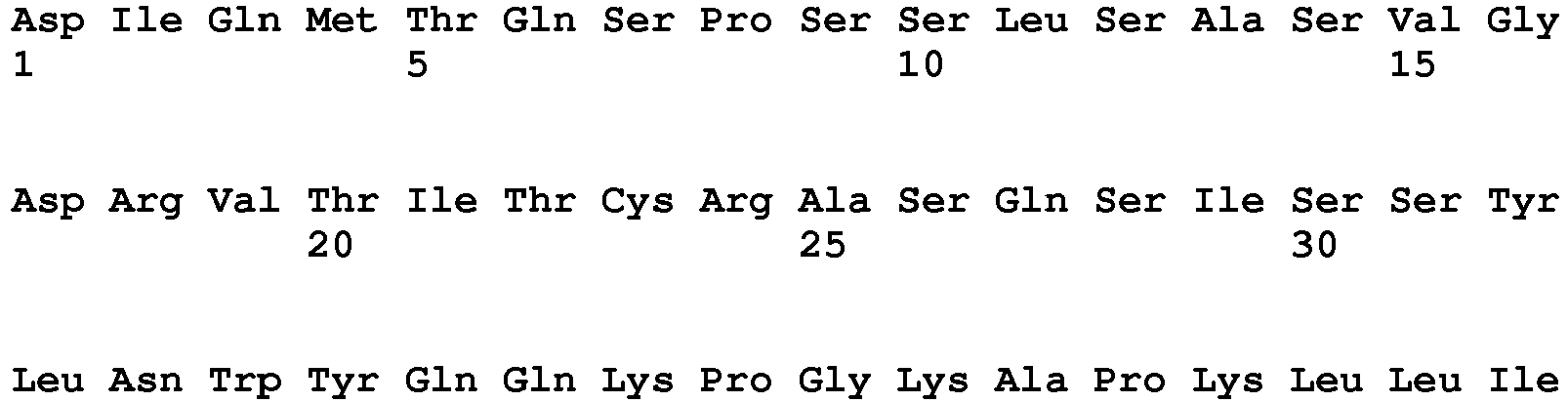

- <210> 41
<211> 17
<212> DNA
<213> artificial - <220>
<223> Primer LMB3 - <400> 41
caggaaacag ctatgac 17 - <210> 42
<211> 17
<212> DNA
<213> artificial - <220>
<223> Primer fdSEQ1 - <400> 42
gaattttctg tatgagg 17 - <210> 43
<211> 614
<212> PRT
<213> artificial - <220>
<223> fusion polypeptide comprising ETA - <400> 43


Claims (3)
- A compound comprising(i) a domain comprising a polypeptide which binds to the surface of AML blast cells and is internalised upon binding to the AML blast cells, and(ii) a cytotoxic domain,wherein the compound is a fusion polypeptide with the amino acid sequence of SEQ ID NO: 43.
- A pharmaceutical composition comprising the compound of claim 1.
- The compound of claim 1 for use in diagnosis and treatment of AML.
Priority Applications (1)
| Application Number | Priority Date | Filing Date | Title |
|---|---|---|---|
| EP14734837.9A EP3016983B1 (en) | 2013-07-04 | 2014-07-04 | A new fusion protein to target and treat acute myloid leukemia cells |
Applications Claiming Priority (3)
| Application Number | Priority Date | Filing Date | Title |
|---|---|---|---|
| EP13175180 | 2013-07-04 | ||
| EP14734837.9A EP3016983B1 (en) | 2013-07-04 | 2014-07-04 | A new fusion protein to target and treat acute myloid leukemia cells |
| PCT/EP2014/064304 WO2015001078A1 (en) | 2013-07-04 | 2014-07-04 | A new fusion protein to target and treat acute myloid leukemia cells |
Publications (2)
| Publication Number | Publication Date |
|---|---|
| EP3016983A1 EP3016983A1 (en) | 2016-05-11 |
| EP3016983B1 true EP3016983B1 (en) | 2018-05-16 |
Family
ID=48703357
Family Applications (1)
| Application Number | Title | Priority Date | Filing Date |
|---|---|---|---|
| EP14734837.9A Not-in-force EP3016983B1 (en) | 2013-07-04 | 2014-07-04 | A new fusion protein to target and treat acute myloid leukemia cells |
Country Status (6)
| Country | Link |
|---|---|
| US (1) | US10072093B2 (en) |
| EP (1) | EP3016983B1 (en) |
| JP (1) | JP2016534051A (en) |
| KR (1) | KR20160029027A (en) |
| CA (1) | CA2907429A1 (en) |
| WO (1) | WO2015001078A1 (en) |
Families Citing this family (6)
| Publication number | Priority date | Publication date | Assignee | Title |
|---|---|---|---|---|
| JP2016534051A (en) | 2013-07-04 | 2016-11-04 | フラウンホーファ−ゲゼルシャフト ツァー フォルデルング デア アンゲバンデン フォルシュンク エー. ファオ.Fraunhofer−Gesellschaft Zur Forderung Der Angewandten Forschung E. V. | A novel fusion protein for targeting and treating acute myeloid leukemia cells |
| KR102543530B1 (en) | 2016-03-10 | 2023-06-15 | 현대모비스 주식회사 | Vehicle door system and operating method thereof |
| US11633363B2 (en) | 2017-09-27 | 2023-04-25 | Emory University | Fusion proteins having a toxin and cancer marker, nanoparticles, and uses related thereto |
| GB201720358D0 (en) | 2017-12-06 | 2018-01-17 | Univ Cape Town | Fusion proteins for the detection of apoptosis |
| US20210401821A1 (en) * | 2018-11-15 | 2021-12-30 | Kyushu University, National University Corporation | Prophylactic or therapeutic agent and medicinal composition for il-31-mediated disease |
| CN116157513A (en) | 2020-08-18 | 2023-05-23 | 昂希莱斯制药公司 | Modified porcine pancreatic elastase protein |
Family Cites Families (12)
| Publication number | Priority date | Publication date | Assignee | Title |
|---|---|---|---|---|
| US4946778A (en) | 1987-09-21 | 1990-08-07 | Genex Corporation | Single polypeptide chain binding molecules |
| US6599505B1 (en) * | 1992-04-10 | 2003-07-29 | Research Development Foundation | Immunotoxins directed against CD33 related surface antigens |
| TW200726776A (en) | 2005-07-29 | 2007-07-16 | Friedrich Alexander University Of Erlangen Nuremberg | CD33-specific single-chain immunotoxin and methods of use |
| GB0521991D0 (en) | 2005-10-28 | 2005-12-07 | Univ Dundee | Siglec-9 binding agents |
| WO2009013359A2 (en) | 2007-07-25 | 2009-01-29 | Fraunhofer Gesellschaft Zur Förderung Der Angewandten Forschung E. V. | Self coupling recombinant antibody fusion proteins |
| WO2011076922A1 (en) * | 2009-12-23 | 2011-06-30 | Synimmune Gmbh | Anti-flt3 antibodies and methods of using the same |
| SA112330278B1 (en) * | 2011-02-18 | 2015-10-09 | ستيم سينتركس، انك. | Novel modulators and methods of use |
| CN103649121B (en) | 2011-06-28 | 2016-10-19 | 牛津生物疗法有限公司 | Antibody against ADP-ribosyl cyclase 2 |
| WO2013003647A2 (en) | 2011-06-28 | 2013-01-03 | Sea Lane Biotechnologies, Llc | Multispecific stacked variable domain binding proteins |
| WO2013006490A2 (en) | 2011-07-01 | 2013-01-10 | Cellerant Therapeutics, Inc. | Antibodies that specifically bind to tim3 |
| PL2900694T3 (en) * | 2012-09-27 | 2018-12-31 | Merus N.V. | Bispecific igg antibodies as t cell engagers |
| JP2016534051A (en) | 2013-07-04 | 2016-11-04 | フラウンホーファ−ゲゼルシャフト ツァー フォルデルング デア アンゲバンデン フォルシュンク エー. ファオ.Fraunhofer−Gesellschaft Zur Forderung Der Angewandten Forschung E. V. | A novel fusion protein for targeting and treating acute myeloid leukemia cells |
-
2014
- 2014-07-04 JP JP2016522640A patent/JP2016534051A/en active Pending
- 2014-07-04 CA CA2907429A patent/CA2907429A1/en not_active Abandoned
- 2014-07-04 US US14/901,016 patent/US10072093B2/en not_active Expired - Fee Related
- 2014-07-04 KR KR1020157036239A patent/KR20160029027A/en not_active Withdrawn
- 2014-07-04 WO PCT/EP2014/064304 patent/WO2015001078A1/en not_active Ceased
- 2014-07-04 EP EP14734837.9A patent/EP3016983B1/en not_active Not-in-force
Non-Patent Citations (1)
| Title |
|---|
| None * |
Also Published As
| Publication number | Publication date |
|---|---|
| KR20160029027A (en) | 2016-03-14 |
| US10072093B2 (en) | 2018-09-11 |
| CA2907429A1 (en) | 2015-01-08 |
| WO2015001078A1 (en) | 2015-01-08 |
| US20160152724A1 (en) | 2016-06-02 |
| JP2016534051A (en) | 2016-11-04 |
| EP3016983A1 (en) | 2016-05-11 |
Similar Documents
| Publication | Publication Date | Title |
|---|---|---|
| US7355012B2 (en) | Mutated anti-CD22 antibodies with increased affinity to CD22-expressing leukemia cells | |
| US10072093B2 (en) | Fusion protein to target and treat acute myeloid leukemia cells | |
| JP7027530B2 (en) | Antibodies to human DLK1 and their uses | |
| JP6990652B2 (en) | New polypeptide | |
| JP2016020346A (en) | Use of monoclonal antibody 8H9 | |
| WO2008105560A1 (en) | Pharmaceutical composition comprising anti-grp78 antibody as active ingredient | |
| CA2996205A1 (en) | Monoclonal antibodies specific for fibroblast growth factor receptor 4 (fgfr4) and methods of their use | |
| CN108290931A (en) | There is the new polypeptide of affinity to PD-L1 | |
| WO2016022939A1 (en) | Human monoclonal antibodies specific for 5t4 and methods of their use | |
| JP2022525435A (en) | CD19 antibody and method of using it | |
| US20230365676A1 (en) | Cd33 antibodies | |
| CN117957258A (en) | Protease-mediated target-specific cytokine delivery using fusion peptides | |
| JP2021527408A (en) | Anti-BCMA CAR antibody, conjugate, and usage | |
| JP2025502247A (en) | Antibody sequences of three antibody candidates targeting human lymphocyte antigen 6 family member K (LY6K) | |
| Arndt et al. | Generation of a highly stable, internalizing anti‐CD22 single‐chain Fv fragment for targeting non‐Hodgkin's lymphoma | |
| CN116981693A (en) | Anti-FGFR3 antibodies and their uses | |
| US11643471B2 (en) | Therapeutic antibodies | |
| US10668147B2 (en) | Anti-CD89 cytotoxic complex | |
| RU2803097C2 (en) | Therapeutic antibodies binding to lewis b and lewis y biantenna antigens | |
| US20160304618A1 (en) | Antibodies for diagnosis of acute myeloid leukemia | |
| AU2002327053C1 (en) | Mutated Anti-CD22 antibodies with increased affinity to CD22-expressing leukemia cells | |
| AU2002327053A1 (en) | Mutated Anti-CD22 antibodies with increased affinity to CD22-expressing leukemia cells |
Legal Events
| Date | Code | Title | Description |
|---|---|---|---|
| PUAI | Public reference made under article 153(3) epc to a published international application that has entered the european phase |
Free format text: ORIGINAL CODE: 0009012 |
|
| 17P | Request for examination filed |
Effective date: 20160203 |
|
| AK | Designated contracting states |
Kind code of ref document: A1 Designated state(s): AL AT BE BG CH CY CZ DE DK EE ES FI FR GB GR HR HU IE IS IT LI LT LU LV MC MK MT NL NO PL PT RO RS SE SI SK SM TR |
|
| AX | Request for extension of the european patent |
Extension state: BA ME |
|
| DAX | Request for extension of the european patent (deleted) | ||
| RIN1 | Information on inventor provided before grant (corrected) |
Inventor name: FITTING, JENNY Inventor name: BARTH, STEFAN Inventor name: TUR, MEHMET KEMAL |
|
| STAA | Information on the status of an ep patent application or granted ep patent |
Free format text: STATUS: EXAMINATION IS IN PROGRESS |
|
| 17Q | First examination report despatched |
Effective date: 20170207 |
|
| REG | Reference to a national code |
Ref country code: DE Ref legal event code: R079 Ref document number: 602014025562 Country of ref document: DE Free format text: PREVIOUS MAIN CLASS: C07K0016300000 Ipc: G01N0033574000 |
|
| GRAP | Despatch of communication of intention to grant a patent |
Free format text: ORIGINAL CODE: EPIDOSNIGR1 |
|
| STAA | Information on the status of an ep patent application or granted ep patent |
Free format text: STATUS: GRANT OF PATENT IS INTENDED |
|
| RIC1 | Information provided on ipc code assigned before grant |
Ipc: C07K 16/30 20060101ALI20171117BHEP Ipc: C12N 9/10 20060101ALI20171117BHEP Ipc: G01N 33/574 20060101AFI20171117BHEP |
|
| INTG | Intention to grant announced |
Effective date: 20171208 |
|
| GRAS | Grant fee paid |
Free format text: ORIGINAL CODE: EPIDOSNIGR3 |
|
| GRAA | (expected) grant |
Free format text: ORIGINAL CODE: 0009210 |
|
| STAA | Information on the status of an ep patent application or granted ep patent |
Free format text: STATUS: THE PATENT HAS BEEN GRANTED |
|
| AK | Designated contracting states |
Kind code of ref document: B1 Designated state(s): AL AT BE BG CH CY CZ DE DK EE ES FI FR GB GR HR HU IE IS IT LI LT LU LV MC MK MT NL NO PL PT RO RS SE SI SK SM TR |
|
| REG | Reference to a national code |
Ref country code: GB Ref legal event code: FG4D |
|
| REG | Reference to a national code |
Ref country code: CH Ref legal event code: EP |
|
| REG | Reference to a national code |
Ref country code: IE Ref legal event code: FG4D |
|
| REG | Reference to a national code |
Ref country code: DE Ref legal event code: R096 Ref document number: 602014025562 Country of ref document: DE |
|
| REG | Reference to a national code |
Ref country code: AT Ref legal event code: REF Ref document number: 1000051 Country of ref document: AT Kind code of ref document: T Effective date: 20180615 |
|
| REG | Reference to a national code |
Ref country code: NL Ref legal event code: FP |
|
| REG | Reference to a national code |
Ref country code: FR Ref legal event code: PLFP Year of fee payment: 5 |
|
| REG | Reference to a national code |
Ref country code: CH Ref legal event code: NV Representative=s name: RENTSCH PARTNER AG, CH |
|
| REG | Reference to a national code |
Ref country code: LT Ref legal event code: MG4D |
|
| PG25 | Lapsed in a contracting state [announced via postgrant information from national office to epo] |
Ref country code: NO Free format text: LAPSE BECAUSE OF FAILURE TO SUBMIT A TRANSLATION OF THE DESCRIPTION OR TO PAY THE FEE WITHIN THE PRESCRIBED TIME-LIMIT Effective date: 20180816 Ref country code: BG Free format text: LAPSE BECAUSE OF FAILURE TO SUBMIT A TRANSLATION OF THE DESCRIPTION OR TO PAY THE FEE WITHIN THE PRESCRIBED TIME-LIMIT Effective date: 20180816 Ref country code: FI Free format text: LAPSE BECAUSE OF FAILURE TO SUBMIT A TRANSLATION OF THE DESCRIPTION OR TO PAY THE FEE WITHIN THE PRESCRIBED TIME-LIMIT Effective date: 20180516 Ref country code: SE Free format text: LAPSE BECAUSE OF FAILURE TO SUBMIT A TRANSLATION OF THE DESCRIPTION OR TO PAY THE FEE WITHIN THE PRESCRIBED TIME-LIMIT Effective date: 20180516 Ref country code: LT Free format text: LAPSE BECAUSE OF FAILURE TO SUBMIT A TRANSLATION OF THE DESCRIPTION OR TO PAY THE FEE WITHIN THE PRESCRIBED TIME-LIMIT Effective date: 20180516 Ref country code: ES Free format text: LAPSE BECAUSE OF FAILURE TO SUBMIT A TRANSLATION OF THE DESCRIPTION OR TO PAY THE FEE WITHIN THE PRESCRIBED TIME-LIMIT Effective date: 20180516 |
|
| PGFP | Annual fee paid to national office [announced via postgrant information from national office to epo] |
Ref country code: NL Payment date: 20180723 Year of fee payment: 5 Ref country code: FR Payment date: 20180723 Year of fee payment: 5 Ref country code: DE Payment date: 20180725 Year of fee payment: 5 Ref country code: IT Payment date: 20180829 Year of fee payment: 5 |
|
| PG25 | Lapsed in a contracting state [announced via postgrant information from national office to epo] |
Ref country code: RS Free format text: LAPSE BECAUSE OF FAILURE TO SUBMIT A TRANSLATION OF THE DESCRIPTION OR TO PAY THE FEE WITHIN THE PRESCRIBED TIME-LIMIT Effective date: 20180516 Ref country code: HR Free format text: LAPSE BECAUSE OF FAILURE TO SUBMIT A TRANSLATION OF THE DESCRIPTION OR TO PAY THE FEE WITHIN THE PRESCRIBED TIME-LIMIT Effective date: 20180516 Ref country code: LV Free format text: LAPSE BECAUSE OF FAILURE TO SUBMIT A TRANSLATION OF THE DESCRIPTION OR TO PAY THE FEE WITHIN THE PRESCRIBED TIME-LIMIT Effective date: 20180516 Ref country code: GR Free format text: LAPSE BECAUSE OF FAILURE TO SUBMIT A TRANSLATION OF THE DESCRIPTION OR TO PAY THE FEE WITHIN THE PRESCRIBED TIME-LIMIT Effective date: 20180817 |
|
| PGFP | Annual fee paid to national office [announced via postgrant information from national office to epo] |
Ref country code: CH Payment date: 20180725 Year of fee payment: 5 Ref country code: GB Payment date: 20180725 Year of fee payment: 5 |
|
| PG25 | Lapsed in a contracting state [announced via postgrant information from national office to epo] |
Ref country code: DK Free format text: LAPSE BECAUSE OF FAILURE TO SUBMIT A TRANSLATION OF THE DESCRIPTION OR TO PAY THE FEE WITHIN THE PRESCRIBED TIME-LIMIT Effective date: 20180516 Ref country code: CZ Free format text: LAPSE BECAUSE OF FAILURE TO SUBMIT A TRANSLATION OF THE DESCRIPTION OR TO PAY THE FEE WITHIN THE PRESCRIBED TIME-LIMIT Effective date: 20180516 Ref country code: RO Free format text: LAPSE BECAUSE OF FAILURE TO SUBMIT A TRANSLATION OF THE DESCRIPTION OR TO PAY THE FEE WITHIN THE PRESCRIBED TIME-LIMIT Effective date: 20180516 Ref country code: EE Free format text: LAPSE BECAUSE OF FAILURE TO SUBMIT A TRANSLATION OF THE DESCRIPTION OR TO PAY THE FEE WITHIN THE PRESCRIBED TIME-LIMIT Effective date: 20180516 Ref country code: PL Free format text: LAPSE BECAUSE OF FAILURE TO SUBMIT A TRANSLATION OF THE DESCRIPTION OR TO PAY THE FEE WITHIN THE PRESCRIBED TIME-LIMIT Effective date: 20180516 Ref country code: SK Free format text: LAPSE BECAUSE OF FAILURE TO SUBMIT A TRANSLATION OF THE DESCRIPTION OR TO PAY THE FEE WITHIN THE PRESCRIBED TIME-LIMIT Effective date: 20180516 |
|
| REG | Reference to a national code |
Ref country code: DE Ref legal event code: R097 Ref document number: 602014025562 Country of ref document: DE |
|
| PG25 | Lapsed in a contracting state [announced via postgrant information from national office to epo] |
Ref country code: SM Free format text: LAPSE BECAUSE OF FAILURE TO SUBMIT A TRANSLATION OF THE DESCRIPTION OR TO PAY THE FEE WITHIN THE PRESCRIBED TIME-LIMIT Effective date: 20180516 |
|
| PLBE | No opposition filed within time limit |
Free format text: ORIGINAL CODE: 0009261 |
|
| STAA | Information on the status of an ep patent application or granted ep patent |
Free format text: STATUS: NO OPPOSITION FILED WITHIN TIME LIMIT |
|
| PG25 | Lapsed in a contracting state [announced via postgrant information from national office to epo] |
Ref country code: LU Free format text: LAPSE BECAUSE OF NON-PAYMENT OF DUE FEES Effective date: 20180704 Ref country code: MC Free format text: LAPSE BECAUSE OF FAILURE TO SUBMIT A TRANSLATION OF THE DESCRIPTION OR TO PAY THE FEE WITHIN THE PRESCRIBED TIME-LIMIT Effective date: 20180516 |
|
| REG | Reference to a national code |
Ref country code: BE Ref legal event code: MM Effective date: 20180731 |
|
| REG | Reference to a national code |
Ref country code: IE Ref legal event code: MM4A |
|
| 26N | No opposition filed |
Effective date: 20190219 |
|
| PG25 | Lapsed in a contracting state [announced via postgrant information from national office to epo] |
Ref country code: IE Free format text: LAPSE BECAUSE OF NON-PAYMENT OF DUE FEES Effective date: 20180704 |
|
| PG25 | Lapsed in a contracting state [announced via postgrant information from national office to epo] |
Ref country code: BE Free format text: LAPSE BECAUSE OF NON-PAYMENT OF DUE FEES Effective date: 20180731 Ref country code: SI Free format text: LAPSE BECAUSE OF FAILURE TO SUBMIT A TRANSLATION OF THE DESCRIPTION OR TO PAY THE FEE WITHIN THE PRESCRIBED TIME-LIMIT Effective date: 20180516 |
|
| PG25 | Lapsed in a contracting state [announced via postgrant information from national office to epo] |
Ref country code: AL Free format text: LAPSE BECAUSE OF FAILURE TO SUBMIT A TRANSLATION OF THE DESCRIPTION OR TO PAY THE FEE WITHIN THE PRESCRIBED TIME-LIMIT Effective date: 20180516 |
|
| PG25 | Lapsed in a contracting state [announced via postgrant information from national office to epo] |
Ref country code: MT Free format text: LAPSE BECAUSE OF NON-PAYMENT OF DUE FEES Effective date: 20180704 |
|
| REG | Reference to a national code |
Ref country code: DE Ref legal event code: R119 Ref document number: 602014025562 Country of ref document: DE |
|
| REG | Reference to a national code |
Ref country code: AT Ref legal event code: UEP Ref document number: 1000051 Country of ref document: AT Kind code of ref document: T Effective date: 20180516 |
|
| REG | Reference to a national code |
Ref country code: CH Ref legal event code: PL |
|
| GBPC | Gb: european patent ceased through non-payment of renewal fee |
Effective date: 20190704 |
|
| PG25 | Lapsed in a contracting state [announced via postgrant information from national office to epo] |
Ref country code: TR Free format text: LAPSE BECAUSE OF FAILURE TO SUBMIT A TRANSLATION OF THE DESCRIPTION OR TO PAY THE FEE WITHIN THE PRESCRIBED TIME-LIMIT Effective date: 20180516 |
|
| PG25 | Lapsed in a contracting state [announced via postgrant information from national office to epo] |
Ref country code: GB Free format text: LAPSE BECAUSE OF NON-PAYMENT OF DUE FEES Effective date: 20190704 Ref country code: NL Free format text: LAPSE BECAUSE OF NON-PAYMENT OF DUE FEES Effective date: 20190801 Ref country code: DE Free format text: LAPSE BECAUSE OF NON-PAYMENT OF DUE FEES Effective date: 20200201 |
|
| REG | Reference to a national code |
Ref country code: NL Ref legal event code: MM Effective date: 20190801 |
|
| PG25 | Lapsed in a contracting state [announced via postgrant information from national office to epo] |
Ref country code: PT Free format text: LAPSE BECAUSE OF FAILURE TO SUBMIT A TRANSLATION OF THE DESCRIPTION OR TO PAY THE FEE WITHIN THE PRESCRIBED TIME-LIMIT Effective date: 20180516 Ref country code: LI Free format text: LAPSE BECAUSE OF NON-PAYMENT OF DUE FEES Effective date: 20190731 Ref country code: CH Free format text: LAPSE BECAUSE OF NON-PAYMENT OF DUE FEES Effective date: 20190731 |
|
| PG25 | Lapsed in a contracting state [announced via postgrant information from national office to epo] |
Ref country code: MK Free format text: LAPSE BECAUSE OF NON-PAYMENT OF DUE FEES Effective date: 20180516 Ref country code: CY Free format text: LAPSE BECAUSE OF FAILURE TO SUBMIT A TRANSLATION OF THE DESCRIPTION OR TO PAY THE FEE WITHIN THE PRESCRIBED TIME-LIMIT Effective date: 20180516 Ref country code: FR Free format text: LAPSE BECAUSE OF NON-PAYMENT OF DUE FEES Effective date: 20190731 Ref country code: HU Free format text: LAPSE BECAUSE OF FAILURE TO SUBMIT A TRANSLATION OF THE DESCRIPTION OR TO PAY THE FEE WITHIN THE PRESCRIBED TIME-LIMIT; INVALID AB INITIO Effective date: 20140704 |
|
| PG25 | Lapsed in a contracting state [announced via postgrant information from national office to epo] |
Ref country code: IS Free format text: LAPSE BECAUSE OF FAILURE TO SUBMIT A TRANSLATION OF THE DESCRIPTION OR TO PAY THE FEE WITHIN THE PRESCRIBED TIME-LIMIT Effective date: 20180916 |
|
| PG25 | Lapsed in a contracting state [announced via postgrant information from national office to epo] |
Ref country code: IT Free format text: LAPSE BECAUSE OF NON-PAYMENT OF DUE FEES Effective date: 20190704 |
|
| REG | Reference to a national code |
Ref country code: AT Ref legal event code: MM01 Ref document number: 1000051 Country of ref document: AT Kind code of ref document: T Effective date: 20190704 |
|
| PG25 | Lapsed in a contracting state [announced via postgrant information from national office to epo] |
Ref country code: AT Free format text: LAPSE BECAUSE OF NON-PAYMENT OF DUE FEES Effective date: 20190704 |