EP3007731B1 - Production of magnetic nanoparticles in recombinant host cells - Google Patents
Production of magnetic nanoparticles in recombinant host cells Download PDFInfo
- Publication number
- EP3007731B1 EP3007731B1 EP14733554.1A EP14733554A EP3007731B1 EP 3007731 B1 EP3007731 B1 EP 3007731B1 EP 14733554 A EP14733554 A EP 14733554A EP 3007731 B1 EP3007731 B1 EP 3007731B1
- Authority
- EP
- European Patent Office
- Prior art keywords
- host cell
- gene expression
- recombinant host
- rubrum
- operon
- Prior art date
- Legal status (The legal status is an assumption and is not a legal conclusion. Google has not performed a legal analysis and makes no representation as to the accuracy of the status listed.)
- Not-in-force
Links
- 239000002122 magnetic nanoparticle Substances 0.000 title claims description 38
- 238000004519 manufacturing process Methods 0.000 title claims description 17
- 210000004027 cell Anatomy 0.000 claims description 214
- 210000002244 magnetosome Anatomy 0.000 claims description 147
- 230000014509 gene expression Effects 0.000 claims description 138
- 101150044311 mms6 gene Proteins 0.000 claims description 94
- 241000543670 Magnetospirillum gryphiswaldense Species 0.000 claims description 86
- 241000190984 Rhodospirillum rubrum Species 0.000 claims description 44
- 150000007523 nucleic acids Chemical group 0.000 claims description 38
- SZVJSHCCFOBDDC-UHFFFAOYSA-N iron(II,III) oxide Inorganic materials O=[Fe]O[Fe]O[Fe]=O SZVJSHCCFOBDDC-UHFFFAOYSA-N 0.000 claims description 33
- 238000012546 transfer Methods 0.000 claims description 28
- 108091028043 Nucleic acid sequence Proteins 0.000 claims description 27
- 230000002068 genetic effect Effects 0.000 claims description 17
- 238000000034 method Methods 0.000 claims description 14
- 230000001105 regulatory effect Effects 0.000 claims description 12
- 230000017105 transposition Effects 0.000 claims description 9
- 241000721720 Magnetospirillum Species 0.000 claims description 6
- 238000003384 imaging method Methods 0.000 claims description 5
- 210000001236 prokaryotic cell Anatomy 0.000 claims description 4
- 108090000623 proteins and genes Proteins 0.000 description 91
- XEEYBQQBJWHFJM-UHFFFAOYSA-N iron Substances [Fe] XEEYBQQBJWHFJM-UHFFFAOYSA-N 0.000 description 70
- 239000013078 crystal Substances 0.000 description 68
- 239000012528 membrane Substances 0.000 description 46
- 239000002245 particle Substances 0.000 description 43
- 230000005291 magnetic effect Effects 0.000 description 40
- 102000004169 proteins and genes Human genes 0.000 description 40
- 230000015572 biosynthetic process Effects 0.000 description 36
- 229910052742 iron Inorganic materials 0.000 description 35
- 230000001965 increasing effect Effects 0.000 description 27
- 230000033558 biomineral tissue development Effects 0.000 description 25
- 238000003780 insertion Methods 0.000 description 23
- 230000037431 insertion Effects 0.000 description 23
- 238000012217 deletion Methods 0.000 description 20
- 230000037430 deletion Effects 0.000 description 20
- 239000013612 plasmid Substances 0.000 description 20
- 102000001218 Rec A Recombinases Human genes 0.000 description 19
- 108010055016 Rec A Recombinases Proteins 0.000 description 19
- 239000006249 magnetic particle Substances 0.000 description 18
- 239000013598 vector Substances 0.000 description 18
- 238000003786 synthesis reaction Methods 0.000 description 17
- 101100174478 Magnetospirillum gryphiswaldense (strain DSM 6361 / JCM 21280 / NBRC 15271 / MSR-1) ftsZ-like gene Proteins 0.000 description 16
- 230000002018 overexpression Effects 0.000 description 16
- 238000005215 recombination Methods 0.000 description 14
- 230000004044 response Effects 0.000 description 14
- 241000894006 Bacteria Species 0.000 description 13
- 230000006870 function Effects 0.000 description 13
- 230000012010 growth Effects 0.000 description 12
- 230000003834 intracellular effect Effects 0.000 description 12
- 238000004458 analytical method Methods 0.000 description 11
- 230000001580 bacterial effect Effects 0.000 description 11
- 101150086471 mamJ gene Proteins 0.000 description 11
- 238000004627 transmission electron microscopy Methods 0.000 description 11
- 241000588724 Escherichia coli Species 0.000 description 10
- XMQFTWRPUQYINF-UHFFFAOYSA-N bensulfuron-methyl Chemical compound COC(=O)C1=CC=CC=C1CS(=O)(=O)NC(=O)NC1=NC(OC)=CC(OC)=N1 XMQFTWRPUQYINF-UHFFFAOYSA-N 0.000 description 10
- 230000004927 fusion Effects 0.000 description 9
- 241000023503 Magnetospirillum gryphiswaldense MSR-1 Species 0.000 description 8
- 241000190967 Rhodospirillum Species 0.000 description 8
- 238000003917 TEM image Methods 0.000 description 8
- 230000002759 chromosomal effect Effects 0.000 description 8
- 108010020764 Transposases Proteins 0.000 description 7
- COQLPRJCUIATTQ-UHFFFAOYSA-N Uranyl acetate Chemical compound O.O.O=[U]=O.CC(O)=O.CC(O)=O COQLPRJCUIATTQ-UHFFFAOYSA-N 0.000 description 7
- 238000002425 crystallisation Methods 0.000 description 7
- 230000008025 crystallization Effects 0.000 description 7
- 230000001939 inductive effect Effects 0.000 description 7
- 239000002159 nanocrystal Substances 0.000 description 7
- 238000003325 tomography Methods 0.000 description 7
- 230000003115 biocidal effect Effects 0.000 description 6
- 238000010367 cloning Methods 0.000 description 6
- 108091008053 gene clusters Proteins 0.000 description 6
- 238000002173 high-resolution transmission electron microscopy Methods 0.000 description 6
- 239000002773 nucleotide Substances 0.000 description 6
- 125000003729 nucleotide group Chemical group 0.000 description 6
- 239000011368 organic material Substances 0.000 description 6
- 230000037361 pathway Effects 0.000 description 6
- 230000010076 replication Effects 0.000 description 6
- 230000032258 transport Effects 0.000 description 6
- CSCPPACGZOOCGX-UHFFFAOYSA-N Acetone Chemical compound CC(C)=O CSCPPACGZOOCGX-UHFFFAOYSA-N 0.000 description 5
- 108020004414 DNA Proteins 0.000 description 5
- 229930182566 Gentamicin Natural products 0.000 description 5
- CEAZRRDELHUEMR-URQXQFDESA-N Gentamicin Chemical compound O1[C@H](C(C)NC)CC[C@@H](N)[C@H]1O[C@H]1[C@H](O)[C@@H](O[C@@H]2[C@@H]([C@@H](NC)[C@@](C)(O)CO2)O)[C@H](N)C[C@@H]1N CEAZRRDELHUEMR-URQXQFDESA-N 0.000 description 5
- 239000004098 Tetracycline Substances 0.000 description 5
- 210000004899 c-terminal region Anatomy 0.000 description 5
- 230000032823 cell division Effects 0.000 description 5
- 210000003764 chromatophore Anatomy 0.000 description 5
- 210000000349 chromosome Anatomy 0.000 description 5
- 230000021615 conjugation Effects 0.000 description 5
- 238000010276 construction Methods 0.000 description 5
- 238000009826 distribution Methods 0.000 description 5
- 230000000694 effects Effects 0.000 description 5
- 239000012634 fragment Substances 0.000 description 5
- 229960002518 gentamicin Drugs 0.000 description 5
- 229930027917 kanamycin Natural products 0.000 description 5
- 229960000318 kanamycin Drugs 0.000 description 5
- SBUJHOSQTJFQJX-NOAMYHISSA-N kanamycin Chemical compound O[C@@H]1[C@@H](O)[C@H](O)[C@@H](CN)O[C@@H]1O[C@H]1[C@H](O)[C@@H](O[C@@H]2[C@@H]([C@@H](N)[C@H](O)[C@@H](CO)O2)O)[C@H](N)C[C@@H]1N SBUJHOSQTJFQJX-NOAMYHISSA-N 0.000 description 5
- 229930182823 kanamycin A Natural products 0.000 description 5
- 238000007885 magnetic separation Methods 0.000 description 5
- IYDGMDWEHDFVQI-UHFFFAOYSA-N phosphoric acid;trioxotungsten Chemical compound O=[W](=O)=O.O=[W](=O)=O.O=[W](=O)=O.O=[W](=O)=O.O=[W](=O)=O.O=[W](=O)=O.O=[W](=O)=O.O=[W](=O)=O.O=[W](=O)=O.O=[W](=O)=O.O=[W](=O)=O.O=[W](=O)=O.OP(O)(O)=O IYDGMDWEHDFVQI-UHFFFAOYSA-N 0.000 description 5
- 230000006798 recombination Effects 0.000 description 5
- 229960002180 tetracycline Drugs 0.000 description 5
- 229930101283 tetracycline Natural products 0.000 description 5
- 235000019364 tetracycline Nutrition 0.000 description 5
- 150000003522 tetracyclines Chemical class 0.000 description 5
- 229920001817 Agar Polymers 0.000 description 4
- RYGMFSIKBFXOCR-UHFFFAOYSA-N Copper Chemical compound [Cu] RYGMFSIKBFXOCR-UHFFFAOYSA-N 0.000 description 4
- CWYNVVGOOAEACU-UHFFFAOYSA-N Fe2+ Chemical compound [Fe+2] CWYNVVGOOAEACU-UHFFFAOYSA-N 0.000 description 4
- 241000134686 Rhodospirillum rubrum ATCC 11170 Species 0.000 description 4
- 102000008579 Transposases Human genes 0.000 description 4
- 239000008272 agar Substances 0.000 description 4
- 229960000723 ampicillin Drugs 0.000 description 4
- AVKUERGKIZMTKX-NJBDSQKTSA-N ampicillin Chemical compound C1([C@@H](N)C(=O)N[C@H]2[C@H]3SC([C@@H](N3C2=O)C(O)=O)(C)C)=CC=CC=C1 AVKUERGKIZMTKX-NJBDSQKTSA-N 0.000 description 4
- 230000008436 biogenesis Effects 0.000 description 4
- 230000001413 cellular effect Effects 0.000 description 4
- 229910052802 copper Inorganic materials 0.000 description 4
- 239000010949 copper Substances 0.000 description 4
- 230000003436 cytoskeletal effect Effects 0.000 description 4
- 230000008014 freezing Effects 0.000 description 4
- 238000007710 freezing Methods 0.000 description 4
- LIKBJVNGSGBSGK-UHFFFAOYSA-N iron(3+);oxygen(2-) Chemical compound [O-2].[O-2].[O-2].[Fe+3].[Fe+3] LIKBJVNGSGBSGK-UHFFFAOYSA-N 0.000 description 4
- 101150109249 lacI gene Proteins 0.000 description 4
- 101150080940 mamH gene Proteins 0.000 description 4
- 101150024206 mamI gene Proteins 0.000 description 4
- 239000000463 material Substances 0.000 description 4
- 102000004196 processed proteins & peptides Human genes 0.000 description 4
- 108090000765 processed proteins & peptides Proteins 0.000 description 4
- 241000894007 species Species 0.000 description 4
- 238000001228 spectrum Methods 0.000 description 4
- 108020004465 16S ribosomal RNA Proteins 0.000 description 3
- WEVYAHXRMPXWCK-UHFFFAOYSA-N Acetonitrile Chemical compound CC#N WEVYAHXRMPXWCK-UHFFFAOYSA-N 0.000 description 3
- 241001657388 Magnetospirillum magneticum Species 0.000 description 3
- 241001446467 Mama Species 0.000 description 3
- OKKJLVBELUTLKV-UHFFFAOYSA-N Methanol Chemical compound OC OKKJLVBELUTLKV-UHFFFAOYSA-N 0.000 description 3
- 238000000862 absorption spectrum Methods 0.000 description 3
- 238000005054 agglomeration Methods 0.000 description 3
- 230000002776 aggregation Effects 0.000 description 3
- 150000001413 amino acids Chemical group 0.000 description 3
- 239000003242 anti bacterial agent Substances 0.000 description 3
- 238000003556 assay Methods 0.000 description 3
- 210000000170 cell membrane Anatomy 0.000 description 3
- 239000006285 cell suspension Substances 0.000 description 3
- 229960005091 chloramphenicol Drugs 0.000 description 3
- WIIZWVCIJKGZOK-RKDXNWHRSA-N chloramphenicol Chemical compound ClC(Cl)C(=O)N[C@H](CO)[C@H](O)C1=CC=C([N+]([O-])=O)C=C1 WIIZWVCIJKGZOK-RKDXNWHRSA-N 0.000 description 3
- 230000001276 controlling effect Effects 0.000 description 3
- 108010048367 enhanced green fluorescent protein Proteins 0.000 description 3
- 238000002474 experimental method Methods 0.000 description 3
- 239000000284 extract Substances 0.000 description 3
- 239000000834 fixative Substances 0.000 description 3
- 239000000499 gel Substances 0.000 description 3
- 230000006801 homologous recombination Effects 0.000 description 3
- 238000002744 homologous recombination Methods 0.000 description 3
- 238000001727 in vivo Methods 0.000 description 3
- 230000000977 initiatory effect Effects 0.000 description 3
- 230000010354 integration Effects 0.000 description 3
- 238000002955 isolation Methods 0.000 description 3
- 230000004807 localization Effects 0.000 description 3
- 230000004048 modification Effects 0.000 description 3
- 238000012986 modification Methods 0.000 description 3
- 230000035772 mutation Effects 0.000 description 3
- PXHVJJICTQNCMI-UHFFFAOYSA-N nickel Substances [Ni] PXHVJJICTQNCMI-UHFFFAOYSA-N 0.000 description 3
- 230000003287 optical effect Effects 0.000 description 3
- 230000000243 photosynthetic effect Effects 0.000 description 3
- 238000003752 polymerase chain reaction Methods 0.000 description 3
- 229920001184 polypeptide Polymers 0.000 description 3
- 238000002415 sodium dodecyl sulfate polyacrylamide gel electrophoresis Methods 0.000 description 3
- 229960000268 spectinomycin Drugs 0.000 description 3
- UNFWWIHTNXNPBV-WXKVUWSESA-N spectinomycin Chemical compound O([C@@H]1[C@@H](NC)[C@@H](O)[C@H]([C@@H]([C@H]1O1)O)NC)[C@]2(O)[C@H]1O[C@H](C)CC2=O UNFWWIHTNXNPBV-WXKVUWSESA-N 0.000 description 3
- 238000004885 tandem mass spectrometry Methods 0.000 description 3
- 238000013518 transcription Methods 0.000 description 3
- 230000035897 transcription Effects 0.000 description 3
- 238000010246 ultrastructural analysis Methods 0.000 description 3
- 238000001262 western blot Methods 0.000 description 3
- 241001135756 Alphaproteobacteria Species 0.000 description 2
- IJGRMHOSHXDMSA-UHFFFAOYSA-N Atomic nitrogen Chemical compound N#N IJGRMHOSHXDMSA-UHFFFAOYSA-N 0.000 description 2
- OKTJSMMVPCPJKN-UHFFFAOYSA-N Carbon Chemical compound [C] OKTJSMMVPCPJKN-UHFFFAOYSA-N 0.000 description 2
- 241000632511 Daviesia arborea Species 0.000 description 2
- UQSXHKLRYXJYBZ-UHFFFAOYSA-N Iron oxide Chemical compound [Fe]=O UQSXHKLRYXJYBZ-UHFFFAOYSA-N 0.000 description 2
- SRBFZHDQGSBBOR-HWQSCIPKSA-N L-arabinopyranose Chemical compound O[C@H]1COC(O)[C@H](O)[C@H]1O SRBFZHDQGSBBOR-HWQSCIPKSA-N 0.000 description 2
- 239000000232 Lipid Bilayer Substances 0.000 description 2
- TWRXJAOTZQYOKJ-UHFFFAOYSA-L Magnesium chloride Chemical compound [Mg+2].[Cl-].[Cl-] TWRXJAOTZQYOKJ-UHFFFAOYSA-L 0.000 description 2
- 241001182995 Magnetococcales Species 0.000 description 2
- 241000589945 Magnetospirillum magnetotacticum Species 0.000 description 2
- 108091034117 Oligonucleotide Proteins 0.000 description 2
- 102000018120 Recombinases Human genes 0.000 description 2
- 108010091086 Recombinases Proteins 0.000 description 2
- 241001185316 Rhodospirillales Species 0.000 description 2
- 229920004890 Triton X-100 Polymers 0.000 description 2
- 239000013504 Triton X-100 Substances 0.000 description 2
- JLCPHMBAVCMARE-UHFFFAOYSA-N [3-[[3-[[3-[[3-[[3-[[3-[[3-[[3-[[3-[[3-[[3-[[5-(2-amino-6-oxo-1H-purin-9-yl)-3-[[3-[[3-[[3-[[3-[[3-[[5-(2-amino-6-oxo-1H-purin-9-yl)-3-[[5-(2-amino-6-oxo-1H-purin-9-yl)-3-hydroxyoxolan-2-yl]methoxy-hydroxyphosphoryl]oxyoxolan-2-yl]methoxy-hydroxyphosphoryl]oxy-5-(5-methyl-2,4-dioxopyrimidin-1-yl)oxolan-2-yl]methoxy-hydroxyphosphoryl]oxy-5-(6-aminopurin-9-yl)oxolan-2-yl]methoxy-hydroxyphosphoryl]oxy-5-(6-aminopurin-9-yl)oxolan-2-yl]methoxy-hydroxyphosphoryl]oxy-5-(6-aminopurin-9-yl)oxolan-2-yl]methoxy-hydroxyphosphoryl]oxy-5-(6-aminopurin-9-yl)oxolan-2-yl]methoxy-hydroxyphosphoryl]oxyoxolan-2-yl]methoxy-hydroxyphosphoryl]oxy-5-(5-methyl-2,4-dioxopyrimidin-1-yl)oxolan-2-yl]methoxy-hydroxyphosphoryl]oxy-5-(4-amino-2-oxopyrimidin-1-yl)oxolan-2-yl]methoxy-hydroxyphosphoryl]oxy-5-(5-methyl-2,4-dioxopyrimidin-1-yl)oxolan-2-yl]methoxy-hydroxyphosphoryl]oxy-5-(5-methyl-2,4-dioxopyrimidin-1-yl)oxolan-2-yl]methoxy-hydroxyphosphoryl]oxy-5-(6-aminopurin-9-yl)oxolan-2-yl]methoxy-hydroxyphosphoryl]oxy-5-(6-aminopurin-9-yl)oxolan-2-yl]methoxy-hydroxyphosphoryl]oxy-5-(4-amino-2-oxopyrimidin-1-yl)oxolan-2-yl]methoxy-hydroxyphosphoryl]oxy-5-(4-amino-2-oxopyrimidin-1-yl)oxolan-2-yl]methoxy-hydroxyphosphoryl]oxy-5-(4-amino-2-oxopyrimidin-1-yl)oxolan-2-yl]methoxy-hydroxyphosphoryl]oxy-5-(6-aminopurin-9-yl)oxolan-2-yl]methoxy-hydroxyphosphoryl]oxy-5-(4-amino-2-oxopyrimidin-1-yl)oxolan-2-yl]methyl [5-(6-aminopurin-9-yl)-2-(hydroxymethyl)oxolan-3-yl] hydrogen phosphate Polymers Cc1cn(C2CC(OP(O)(=O)OCC3OC(CC3OP(O)(=O)OCC3OC(CC3O)n3cnc4c3nc(N)[nH]c4=O)n3cnc4c3nc(N)[nH]c4=O)C(COP(O)(=O)OC3CC(OC3COP(O)(=O)OC3CC(OC3COP(O)(=O)OC3CC(OC3COP(O)(=O)OC3CC(OC3COP(O)(=O)OC3CC(OC3COP(O)(=O)OC3CC(OC3COP(O)(=O)OC3CC(OC3COP(O)(=O)OC3CC(OC3COP(O)(=O)OC3CC(OC3COP(O)(=O)OC3CC(OC3COP(O)(=O)OC3CC(OC3COP(O)(=O)OC3CC(OC3COP(O)(=O)OC3CC(OC3COP(O)(=O)OC3CC(OC3COP(O)(=O)OC3CC(OC3COP(O)(=O)OC3CC(OC3COP(O)(=O)OC3CC(OC3CO)n3cnc4c(N)ncnc34)n3ccc(N)nc3=O)n3cnc4c(N)ncnc34)n3ccc(N)nc3=O)n3ccc(N)nc3=O)n3ccc(N)nc3=O)n3cnc4c(N)ncnc34)n3cnc4c(N)ncnc34)n3cc(C)c(=O)[nH]c3=O)n3cc(C)c(=O)[nH]c3=O)n3ccc(N)nc3=O)n3cc(C)c(=O)[nH]c3=O)n3cnc4c3nc(N)[nH]c4=O)n3cnc4c(N)ncnc34)n3cnc4c(N)ncnc34)n3cnc4c(N)ncnc34)n3cnc4c(N)ncnc34)O2)c(=O)[nH]c1=O JLCPHMBAVCMARE-UHFFFAOYSA-N 0.000 description 2
- 230000001594 aberrant effect Effects 0.000 description 2
- 238000010521 absorption reaction Methods 0.000 description 2
- 239000002253 acid Substances 0.000 description 2
- 150000007513 acids Chemical class 0.000 description 2
- 229940088710 antibiotic agent Drugs 0.000 description 2
- 238000013459 approach Methods 0.000 description 2
- SRBFZHDQGSBBOR-UHFFFAOYSA-N beta-D-Pyranose-Lyxose Natural products OC1COC(O)C(O)C1O SRBFZHDQGSBBOR-UHFFFAOYSA-N 0.000 description 2
- 230000000035 biogenic effect Effects 0.000 description 2
- 230000005540 biological transmission Effects 0.000 description 2
- 230000006696 biosynthetic metabolic pathway Effects 0.000 description 2
- 238000013452 biotechnological production Methods 0.000 description 2
- 229910052799 carbon Inorganic materials 0.000 description 2
- 238000005119 centrifugation Methods 0.000 description 2
- 230000000295 complement effect Effects 0.000 description 2
- 238000001514 detection method Methods 0.000 description 2
- 238000001962 electrophoresis Methods 0.000 description 2
- 238000005516 engineering process Methods 0.000 description 2
- 230000001747 exhibiting effect Effects 0.000 description 2
- 229960002413 ferric citrate Drugs 0.000 description 2
- 238000000799 fluorescence microscopy Methods 0.000 description 2
- 239000007789 gas Substances 0.000 description 2
- 238000001476 gene delivery Methods 0.000 description 2
- 229910052595 hematite Inorganic materials 0.000 description 2
- 239000011019 hematite Substances 0.000 description 2
- 229910052500 inorganic mineral Inorganic materials 0.000 description 2
- NPFOYSMITVOQOS-UHFFFAOYSA-K iron(III) citrate Chemical compound [Fe+3].[O-]C(=O)CC(O)(CC([O-])=O)C([O-])=O NPFOYSMITVOQOS-UHFFFAOYSA-K 0.000 description 2
- 238000011031 large-scale manufacturing process Methods 0.000 description 2
- 239000010410 layer Substances 0.000 description 2
- 238000001294 liquid chromatography-tandem mass spectrometry Methods 0.000 description 2
- 238000002595 magnetic resonance imaging Methods 0.000 description 2
- 210000004962 mammalian cell Anatomy 0.000 description 2
- 238000004949 mass spectrometry Methods 0.000 description 2
- 230000007246 mechanism Effects 0.000 description 2
- 229910044991 metal oxide Inorganic materials 0.000 description 2
- 150000004706 metal oxides Chemical class 0.000 description 2
- 229910052976 metal sulfide Inorganic materials 0.000 description 2
- 238000001000 micrograph Methods 0.000 description 2
- 239000011707 mineral Substances 0.000 description 2
- 235000010755 mineral Nutrition 0.000 description 2
- 239000000203 mixture Substances 0.000 description 2
- 238000010369 molecular cloning Methods 0.000 description 2
- 229910052759 nickel Inorganic materials 0.000 description 2
- 108020004707 nucleic acids Proteins 0.000 description 2
- 102000039446 nucleic acids Human genes 0.000 description 2
- 210000003463 organelle Anatomy 0.000 description 2
- 239000012044 organic layer Substances 0.000 description 2
- 229910000489 osmium tetroxide Inorganic materials 0.000 description 2
- 239000012285 osmium tetroxide Substances 0.000 description 2
- 230000000737 periodic effect Effects 0.000 description 2
- 230000009564 phototrophic growth Effects 0.000 description 2
- 229920002401 polyacrylamide Polymers 0.000 description 2
- 238000000575 proteomic method Methods 0.000 description 2
- 230000003362 replicative effect Effects 0.000 description 2
- 101150098466 rpsL gene Proteins 0.000 description 2
- 238000012163 sequencing technique Methods 0.000 description 2
- 238000003860 storage Methods 0.000 description 2
- 230000004960 subcellular localization Effects 0.000 description 2
- 239000000126 substance Substances 0.000 description 2
- 238000006467 substitution reaction Methods 0.000 description 2
- 238000012360 testing method Methods 0.000 description 2
- 230000002103 transcriptional effect Effects 0.000 description 2
- 238000013519 translation Methods 0.000 description 2
- 238000001521 two-tailed test Methods 0.000 description 2
- 238000011144 upstream manufacturing Methods 0.000 description 2
- XLYOFNOQVPJJNP-UHFFFAOYSA-N water Substances O XLYOFNOQVPJJNP-UHFFFAOYSA-N 0.000 description 2
- CXNPLSGKWMLZPZ-GIFSMMMISA-N (2r,3r,6s)-3-[[(3s)-3-amino-5-[carbamimidoyl(methyl)amino]pentanoyl]amino]-6-(4-amino-2-oxopyrimidin-1-yl)-3,6-dihydro-2h-pyran-2-carboxylic acid Chemical group O1[C@@H](C(O)=O)[C@H](NC(=O)C[C@@H](N)CCN(C)C(N)=N)C=C[C@H]1N1C(=O)N=C(N)C=C1 CXNPLSGKWMLZPZ-GIFSMMMISA-N 0.000 description 1
- 108091032973 (ribonucleotides)n+m Proteins 0.000 description 1
- FVFVNNKYKYZTJU-UHFFFAOYSA-N 6-chloro-1,3,5-triazine-2,4-diamine Chemical group NC1=NC(N)=NC(Cl)=N1 FVFVNNKYKYZTJU-UHFFFAOYSA-N 0.000 description 1
- 108050006855 Acriflavin resistance proteins Proteins 0.000 description 1
- 241000589155 Agrobacterium tumefaciens Species 0.000 description 1
- 108010077805 Bacterial Proteins Proteins 0.000 description 1
- 108010070075 Bacteriochlorophyll A Proteins 0.000 description 1
- 108010003118 Bacteriochlorophylls Proteins 0.000 description 1
- 241000010804 Caulobacter vibrioides Species 0.000 description 1
- 241001185306 Caulobacterales Species 0.000 description 1
- 241000282693 Cercopithecidae Species 0.000 description 1
- VYZAMTAEIAYCRO-UHFFFAOYSA-N Chromium Chemical compound [Cr] VYZAMTAEIAYCRO-UHFFFAOYSA-N 0.000 description 1
- 208000036086 Chromosome Duplication Diseases 0.000 description 1
- 241000669072 Chrysomphalus dictyospermi Species 0.000 description 1
- KRKNYBCHXYNGOX-UHFFFAOYSA-K Citrate Chemical compound [O-]C(=O)CC(O)(CC([O-])=O)C([O-])=O KRKNYBCHXYNGOX-UHFFFAOYSA-K 0.000 description 1
- 241000699800 Cricetinae Species 0.000 description 1
- 102000018832 Cytochromes Human genes 0.000 description 1
- 108010052832 Cytochromes Proteins 0.000 description 1
- 101710088194 Dehydrogenase Proteins 0.000 description 1
- 101100316841 Escherichia phage lambda bet gene Proteins 0.000 description 1
- 101100226347 Escherichia phage lambda exo gene Proteins 0.000 description 1
- OTMSDBZUPAUEDD-UHFFFAOYSA-N Ethane Chemical compound CC OTMSDBZUPAUEDD-UHFFFAOYSA-N 0.000 description 1
- 241000206602 Eukaryota Species 0.000 description 1
- VTLYFUHAOXGGBS-UHFFFAOYSA-N Fe3+ Chemical compound [Fe+3] VTLYFUHAOXGGBS-UHFFFAOYSA-N 0.000 description 1
- 241000282326 Felis catus Species 0.000 description 1
- MBMLMWLHJBBADN-UHFFFAOYSA-N Ferrous sulfide Chemical compound [Fe]=S MBMLMWLHJBBADN-UHFFFAOYSA-N 0.000 description 1
- 206010064571 Gene mutation Diseases 0.000 description 1
- 108700039691 Genetic Promoter Regions Proteins 0.000 description 1
- SXRSQZLOMIGNAQ-UHFFFAOYSA-N Glutaraldehyde Chemical compound O=CCCCC=O SXRSQZLOMIGNAQ-UHFFFAOYSA-N 0.000 description 1
- 101000848922 Homo sapiens Protein FAM72A Proteins 0.000 description 1
- 102000004310 Ion Channels Human genes 0.000 description 1
- 108091006975 Iron transporters Proteins 0.000 description 1
- 241000341320 Kiloniellales Species 0.000 description 1
- 241000235058 Komagataella pastoris Species 0.000 description 1
- 101100455989 Magnetospirillum gryphiswaldense (strain DSM 6361 / JCM 21280 / NBRC 15271 / MSR-1) mamA gene Proteins 0.000 description 1
- 101100455992 Magnetospirillum gryphiswaldense (strain DSM 6361 / JCM 21280 / NBRC 15271 / MSR-1) mamB gene Proteins 0.000 description 1
- 101100344396 Magnetospirillum gryphiswaldense (strain DSM 6361 / JCM 21280 / NBRC 15271 / MSR-1) mamC gene Proteins 0.000 description 1
- 101100344401 Magnetospirillum gryphiswaldense (strain DSM 6361 / JCM 21280 / NBRC 15271 / MSR-1) mamD gene Proteins 0.000 description 1
- 101100344403 Magnetospirillum gryphiswaldense (strain DSM 6361 / JCM 21280 / NBRC 15271 / MSR-1) mamE gene Proteins 0.000 description 1
- 101100344405 Magnetospirillum gryphiswaldense (strain DSM 6361 / JCM 21280 / NBRC 15271 / MSR-1) mamF gene Proteins 0.000 description 1
- 101100344413 Magnetospirillum gryphiswaldense (strain DSM 6361 / JCM 21280 / NBRC 15271 / MSR-1) mamJ gene Proteins 0.000 description 1
- 101100400396 Magnetospirillum gryphiswaldense (strain DSM 6361 / JCM 21280 / NBRC 15271 / MSR-1) mamK gene Proteins 0.000 description 1
- 101100400405 Magnetospirillum gryphiswaldense (strain DSM 6361 / JCM 21280 / NBRC 15271 / MSR-1) mamM gene Proteins 0.000 description 1
- 101100400409 Magnetospirillum gryphiswaldense (strain DSM 6361 / JCM 21280 / NBRC 15271 / MSR-1) mamO gene Proteins 0.000 description 1
- 101100129306 Magnetospirillum gryphiswaldense (strain DSM 6361 / JCM 21280 / NBRC 15271 / MSR-1) mamY gene Proteins 0.000 description 1
- 101100345749 Magnetospirillum gryphiswaldense (strain DSM 6361 / JCM 21280 / NBRC 15271 / MSR-1) mmsF gene Proteins 0.000 description 1
- 108010052285 Membrane Proteins Proteins 0.000 description 1
- 102000018697 Membrane Proteins Human genes 0.000 description 1
- 241000867492 Mint virus 1 Species 0.000 description 1
- 101100301239 Myxococcus xanthus recA1 gene Proteins 0.000 description 1
- 206010028980 Neoplasm Diseases 0.000 description 1
- GRYLNZFGIOXLOG-UHFFFAOYSA-N Nitric acid Chemical compound O[N+]([O-])=O GRYLNZFGIOXLOG-UHFFFAOYSA-N 0.000 description 1
- 241000283973 Oryctolagus cuniculus Species 0.000 description 1
- 229910019142 PO4 Inorganic materials 0.000 description 1
- 241001377014 Parvularculales Species 0.000 description 1
- 101100390602 Porphyromonas gingivalis (strain ATCC BAA-308 / W83) feoB1 gene Proteins 0.000 description 1
- 102100034514 Protein FAM72A Human genes 0.000 description 1
- 241000191023 Rhodobacter capsulatus Species 0.000 description 1
- 241000191043 Rhodobacter sphaeroides Species 0.000 description 1
- 241001185307 Rhodobacterales Species 0.000 description 1
- 241000131970 Rhodospirillaceae Species 0.000 description 1
- 241000606651 Rickettsiales Species 0.000 description 1
- 240000004808 Saccharomyces cerevisiae Species 0.000 description 1
- 241000293869 Salmonella enterica subsp. enterica serovar Typhimurium Species 0.000 description 1
- 241000656192 Sneathiellales Species 0.000 description 1
- 241001185305 Sphingomonadales Species 0.000 description 1
- QAOWNCQODCNURD-UHFFFAOYSA-L Sulfate Chemical compound [O-]S([O-])(=O)=O QAOWNCQODCNURD-UHFFFAOYSA-L 0.000 description 1
- 108700026226 TATA Box Proteins 0.000 description 1
- 108010012306 Tn5 transposase Proteins 0.000 description 1
- 230000004913 activation Effects 0.000 description 1
- 229910052782 aluminium Inorganic materials 0.000 description 1
- XAGFODPZIPBFFR-UHFFFAOYSA-N aluminium Chemical compound [Al] XAGFODPZIPBFFR-UHFFFAOYSA-N 0.000 description 1
- 230000003321 amplification Effects 0.000 description 1
- 239000007864 aqueous solution Substances 0.000 description 1
- 238000000149 argon plasma sintering Methods 0.000 description 1
- 210000004507 artificial chromosome Anatomy 0.000 description 1
- ZSERVQBSOBTXFV-DHHJBRQQSA-M bacteriochlorophyll a Chemical compound C1([C@H](C(=O)OC)C(=O)C2=C3C)=C2N2C3=CC([C@@H](CC)[C@@H]3C)=[N+]4C3=CC3=C(C(C)=O)C(C)=C5N3[Mg]42[N+]2=C1[C@@H](CCC(=O)OC\C=C(/C)CCC[C@H](C)CCC[C@H](C)CCCC(C)C)[C@H](C)C2=C5 ZSERVQBSOBTXFV-DHHJBRQQSA-M 0.000 description 1
- XZSVAMUZTKNGDN-JBRJOJLESA-L bacteriochlorophylls Chemical compound [Mg+2].[N-]1C2=C(C=3C(C(C)C(=CC=4C(=C(C(C)=O)C(=C5)N=4)C)N=3)CCC(=O)OC\C=C(/C)CCCC(C)CCCC(C)CCCC(C)C)C(C(=O)OC)C([O-])=C2C(C)=C1C=C1C(CC)C(C)C5=N1 XZSVAMUZTKNGDN-JBRJOJLESA-L 0.000 description 1
- 230000033228 biological regulation Effects 0.000 description 1
- HOQPTLCRWVZIQZ-UHFFFAOYSA-H bis[[2-(5-hydroxy-4,7-dioxo-1,3,2$l^{2}-dioxaplumbepan-5-yl)acetyl]oxy]lead Chemical compound [Pb+2].[Pb+2].[Pb+2].[O-]C(=O)CC(O)(CC([O-])=O)C([O-])=O.[O-]C(=O)CC(O)(CC([O-])=O)C([O-])=O HOQPTLCRWVZIQZ-UHFFFAOYSA-H 0.000 description 1
- CXNPLSGKWMLZPZ-UHFFFAOYSA-N blasticidin-S Natural products O1C(C(O)=O)C(NC(=O)CC(N)CCN(C)C(N)=N)C=CC1N1C(=O)N=C(N)C=C1 CXNPLSGKWMLZPZ-UHFFFAOYSA-N 0.000 description 1
- 239000003054 catalyst Substances 0.000 description 1
- 230000003197 catalytic effect Effects 0.000 description 1
- 230000030570 cellular localization Effects 0.000 description 1
- 238000012512 characterization method Methods 0.000 description 1
- 230000009563 chemotrophic growth Effects 0.000 description 1
- 229910052804 chromium Inorganic materials 0.000 description 1
- 239000011651 chromium Substances 0.000 description 1
- MYSWGUAQZAJSOK-UHFFFAOYSA-N ciprofloxacin Chemical compound C12=CC(N3CCNCC3)=C(F)C=C2C(=O)C(C(=O)O)=CN1C1CC1 MYSWGUAQZAJSOK-UHFFFAOYSA-N 0.000 description 1
- 230000008045 co-localization Effects 0.000 description 1
- 229910017052 cobalt Inorganic materials 0.000 description 1
- 239000010941 cobalt Substances 0.000 description 1
- GUTLYIVDDKVIGB-UHFFFAOYSA-N cobalt atom Chemical compound [Co] GUTLYIVDDKVIGB-UHFFFAOYSA-N 0.000 description 1
- 239000003086 colorant Substances 0.000 description 1
- 239000000470 constituent Substances 0.000 description 1
- NKLPQNGYXWVELD-UHFFFAOYSA-M coomassie brilliant blue Chemical compound [Na+].C1=CC(OCC)=CC=C1NC1=CC=C(C(=C2C=CC(C=C2)=[N+](CC)CC=2C=C(C=CC=2)S([O-])(=O)=O)C=2C=CC(=CC=2)N(CC)CC=2C=C(C=CC=2)S([O-])(=O)=O)C=C1 NKLPQNGYXWVELD-UHFFFAOYSA-M 0.000 description 1
- 238000013480 data collection Methods 0.000 description 1
- 230000003247 decreasing effect Effects 0.000 description 1
- 230000002950 deficient Effects 0.000 description 1
- 230000018044 dehydration Effects 0.000 description 1
- 238000006297 dehydration reaction Methods 0.000 description 1
- 230000003111 delayed effect Effects 0.000 description 1
- 230000001419 dependent effect Effects 0.000 description 1
- 239000003599 detergent Substances 0.000 description 1
- 229910003460 diamond Inorganic materials 0.000 description 1
- 239000010432 diamond Substances 0.000 description 1
- 238000012377 drug delivery Methods 0.000 description 1
- 230000002900 effect on cell Effects 0.000 description 1
- 238000002003 electron diffraction Methods 0.000 description 1
- 238000000619 electron energy-loss spectrum Methods 0.000 description 1
- 239000003623 enhancer Substances 0.000 description 1
- 210000003527 eukaryotic cell Anatomy 0.000 description 1
- 101150073057 feoA gene Proteins 0.000 description 1
- 101150016604 feoB gene Proteins 0.000 description 1
- 238000000855 fermentation Methods 0.000 description 1
- 230000004151 fermentation Effects 0.000 description 1
- 230000005294 ferromagnetic effect Effects 0.000 description 1
- 239000011790 ferrous sulphate Substances 0.000 description 1
- 235000003891 ferrous sulphate Nutrition 0.000 description 1
- 238000010230 functional analysis Methods 0.000 description 1
- 108020001507 fusion proteins Proteins 0.000 description 1
- 102000037865 fusion proteins Human genes 0.000 description 1
- 101150045500 galK gene Proteins 0.000 description 1
- 101150041954 galU gene Proteins 0.000 description 1
- 238000012224 gene deletion Methods 0.000 description 1
- 238000010353 genetic engineering Methods 0.000 description 1
- PCHJSUWPFVWCPO-UHFFFAOYSA-N gold Chemical class [Au] PCHJSUWPFVWCPO-UHFFFAOYSA-N 0.000 description 1
- UHUWQCGPGPPDDT-UHFFFAOYSA-N greigite Chemical compound [S-2].[S-2].[S-2].[S-2].[Fe+2].[Fe+3].[Fe+3] UHUWQCGPGPPDDT-UHFFFAOYSA-N 0.000 description 1
- 101150096208 gtaB gene Proteins 0.000 description 1
- 101150023479 hsdS gene Proteins 0.000 description 1
- 210000005260 human cell Anatomy 0.000 description 1
- 230000000984 immunochemical effect Effects 0.000 description 1
- 230000001771 impaired effect Effects 0.000 description 1
- 238000011534 incubation Methods 0.000 description 1
- 230000003993 interaction Effects 0.000 description 1
- BAUYGSIQEAFULO-UHFFFAOYSA-L iron(2+) sulfate (anhydrous) Chemical compound [Fe+2].[O-]S([O-])(=O)=O BAUYGSIQEAFULO-UHFFFAOYSA-L 0.000 description 1
- 229910000359 iron(II) sulfate Inorganic materials 0.000 description 1
- 239000007788 liquid Substances 0.000 description 1
- 238000009630 liquid culture Methods 0.000 description 1
- 238000000464 low-speed centrifugation Methods 0.000 description 1
- 229910001629 magnesium chloride Inorganic materials 0.000 description 1
- 230000005381 magnetic domain Effects 0.000 description 1
- 238000012423 maintenance Methods 0.000 description 1
- 101150029630 mamK gene Proteins 0.000 description 1
- 101150005523 mamX gene Proteins 0.000 description 1
- 101150067920 mamY gene Proteins 0.000 description 1
- 101150011266 mamZ gene Proteins 0.000 description 1
- 229910052748 manganese Inorganic materials 0.000 description 1
- 239000011572 manganese Substances 0.000 description 1
- WPBNNNQJVZRUHP-UHFFFAOYSA-L manganese(2+);methyl n-[[2-(methoxycarbonylcarbamothioylamino)phenyl]carbamothioyl]carbamate;n-[2-(sulfidocarbothioylamino)ethyl]carbamodithioate Chemical compound [Mn+2].[S-]C(=S)NCCNC([S-])=S.COC(=O)NC(=S)NC1=CC=CC=C1NC(=S)NC(=O)OC WPBNNNQJVZRUHP-UHFFFAOYSA-L 0.000 description 1
- 239000003550 marker Substances 0.000 description 1
- 238000005259 measurement Methods 0.000 description 1
- 108020004999 messenger RNA Proteins 0.000 description 1
- 230000037353 metabolic pathway Effects 0.000 description 1
- 230000006609 metabolic stress Effects 0.000 description 1
- 238000000386 microscopy Methods 0.000 description 1
- 102000035118 modified proteins Human genes 0.000 description 1
- 108091005573 modified proteins Proteins 0.000 description 1
- 230000008722 morphological abnormality Effects 0.000 description 1
- 230000004899 motility Effects 0.000 description 1
- 238000007481 next generation sequencing Methods 0.000 description 1
- 229910017604 nitric acid Inorganic materials 0.000 description 1
- 229910052757 nitrogen Inorganic materials 0.000 description 1
- 230000006911 nucleation Effects 0.000 description 1
- 238000010899 nucleation Methods 0.000 description 1
- 238000003199 nucleic acid amplification method Methods 0.000 description 1
- 101150012154 nupG gene Proteins 0.000 description 1
- 230000008520 organization Effects 0.000 description 1
- 238000012261 overproduction Methods 0.000 description 1
- 230000036542 oxidative stress Effects 0.000 description 1
- 239000008188 pellet Substances 0.000 description 1
- 239000010452 phosphate Substances 0.000 description 1
- NBIIXXVUZAFLBC-UHFFFAOYSA-K phosphate Chemical compound [O-]P([O-])([O-])=O NBIIXXVUZAFLBC-UHFFFAOYSA-K 0.000 description 1
- 150000003904 phospholipids Chemical class 0.000 description 1
- 230000008488 polyadenylation Effects 0.000 description 1
- 238000001556 precipitation Methods 0.000 description 1
- 238000002360 preparation method Methods 0.000 description 1
- 230000008569 process Effects 0.000 description 1
- 238000012545 processing Methods 0.000 description 1
- 230000004853 protein function Effects 0.000 description 1
- 238000011002 quantification Methods 0.000 description 1
- 230000008707 rearrangement Effects 0.000 description 1
- 239000011347 resin Substances 0.000 description 1
- 229920005989 resin Polymers 0.000 description 1
- 230000000717 retained effect Effects 0.000 description 1
- 238000012552 review Methods 0.000 description 1
- 239000012723 sample buffer Substances 0.000 description 1
- 229930000044 secondary metabolite Natural products 0.000 description 1
- 239000004065 semiconductor Substances 0.000 description 1
- 238000000926 separation method Methods 0.000 description 1
- 229910052709 silver Inorganic materials 0.000 description 1
- 239000004332 silver Substances 0.000 description 1
- 238000005549 size reduction Methods 0.000 description 1
- IHQKEDIOMGYHEB-UHFFFAOYSA-M sodium dimethylarsinate Chemical compound [Na+].C[As](C)([O-])=O IHQKEDIOMGYHEB-UHFFFAOYSA-M 0.000 description 1
- 239000007787 solid Substances 0.000 description 1
- 230000002269 spontaneous effect Effects 0.000 description 1
- 238000010186 staining Methods 0.000 description 1
- 238000001370 static light scattering Methods 0.000 description 1
- 230000000638 stimulation Effects 0.000 description 1
- 150000004763 sulfides Chemical class 0.000 description 1
- 239000000725 suspension Substances 0.000 description 1
- 230000008685 targeting Effects 0.000 description 1
- 238000010361 transduction Methods 0.000 description 1
- 230000026683 transduction Effects 0.000 description 1
- 238000001890 transfection Methods 0.000 description 1
- 230000009466 transformation Effects 0.000 description 1
- 230000007332 vesicle formation Effects 0.000 description 1
- 238000005406 washing Methods 0.000 description 1
- 238000012070 whole genome sequencing analysis Methods 0.000 description 1
- 230000009614 wildtype growth Effects 0.000 description 1
Images








Classifications
-
- A—HUMAN NECESSITIES
- A61—MEDICAL OR VETERINARY SCIENCE; HYGIENE
- A61K—PREPARATIONS FOR MEDICAL, DENTAL OR TOILETRY PURPOSES
- A61K49/00—Preparations for testing in vivo
- A61K49/06—Nuclear magnetic resonance [NMR] contrast preparations; Magnetic resonance imaging [MRI] contrast preparations
- A61K49/18—Nuclear magnetic resonance [NMR] contrast preparations; Magnetic resonance imaging [MRI] contrast preparations characterised by a special physical form, e.g. emulsions, microcapsules, liposomes
- A61K49/1896—Nuclear magnetic resonance [NMR] contrast preparations; Magnetic resonance imaging [MRI] contrast preparations characterised by a special physical form, e.g. emulsions, microcapsules, liposomes not provided for elsewhere, e.g. cells, viruses, ghosts, red blood cells, virus capsides
-
- C—CHEMISTRY; METALLURGY
- C07—ORGANIC CHEMISTRY
- C07K—PEPTIDES
- C07K14/00—Peptides having more than 20 amino acids; Gastrins; Somatostatins; Melanotropins; Derivatives thereof
- C07K14/195—Peptides having more than 20 amino acids; Gastrins; Somatostatins; Melanotropins; Derivatives thereof from bacteria
-
- C—CHEMISTRY; METALLURGY
- C12—BIOCHEMISTRY; BEER; SPIRITS; WINE; VINEGAR; MICROBIOLOGY; ENZYMOLOGY; MUTATION OR GENETIC ENGINEERING
- C12N—MICROORGANISMS OR ENZYMES; COMPOSITIONS THEREOF; PROPAGATING, PRESERVING, OR MAINTAINING MICROORGANISMS; MUTATION OR GENETIC ENGINEERING; CULTURE MEDIA
- C12N15/00—Mutation or genetic engineering; DNA or RNA concerning genetic engineering, vectors, e.g. plasmids, or their isolation, preparation or purification; Use of hosts therefor
- C12N15/09—Recombinant DNA-technology
- C12N15/63—Introduction of foreign genetic material using vectors; Vectors; Use of hosts therefor; Regulation of expression
- C12N15/74—Vectors or expression systems specially adapted for prokaryotic hosts other than E. coli, e.g. Lactobacillus, Micromonospora
-
- C—CHEMISTRY; METALLURGY
- C12—BIOCHEMISTRY; BEER; SPIRITS; WINE; VINEGAR; MICROBIOLOGY; ENZYMOLOGY; MUTATION OR GENETIC ENGINEERING
- C12P—FERMENTATION OR ENZYME-USING PROCESSES TO SYNTHESISE A DESIRED CHEMICAL COMPOUND OR COMPOSITION OR TO SEPARATE OPTICAL ISOMERS FROM A RACEMIC MIXTURE
- C12P3/00—Preparation of elements or inorganic compounds except carbon dioxide
Definitions
- the present invention relates to recombinant host cells being genetically engineered to allow for the heterologous high-yield production of magnetic nanoparticles, preferably of magnetosomes derived from magnetotactic bacteria. More specifically, the recombinant host cells comprise in their genome the entire genetic information sufficient to express the biosynthetic pathway for the production of magnetic nanoparticles.
- Magnetic nanoparticles typically comprise nanocrystals made of oxides (or to a lesser extent of sulfides) of the elements in the forth row of the periodic table (i.e. Cr, Mn, Fe, Co, and Ni).
- the ability to produce such magnetic nanoparticles is inevitable not only for the general understanding of magnetic properties in a nanometer regime, but also for manifold technical applications ranging from magnetic resonance imaging, drug delivery, catalysts, and biosensing to nanoelectronics, semiconductor materials, and magnetic storage media (reviewed in Lu, A. et al. (2007) Angew. Chem. Int. Ed. 46, 1222-1224 ).
- Magnetic nanoparticles can be synthesized chemically through precipitation of the crystals from basic aqueous solutions.
- biogenic magnetic nanoparticles can be employed that are produced by magnetotactic organisms, predominantly magnetotactic bacteria.
- magnetotactic bacteria The ability of magnetotactic bacteria to orient in the Earth's magnetic field is based on the presence of specific organelles, the magnetosomes, which are membrane-enveloped monocrystalline crystals of a magnetic mineral that are arranged in chain-like structures within the cell. Magnetosomes display a variety of species-specific shapes within the single magnetic domain size range (reviewed in Bazylinski, D.A. and Frankel, R.B. (2004) Nature Rev. Microbiol. 2, 217-230 ).
- MM magnetosome membrane
- the biotechnological usability is hampered by the fact that they can only be produced in comparably small amounts due to the methodological difficulties to cultivate magnetotactic bacteria due to fastidious growth requirements.
- the cell yields obtained are only about 1 g (fresh weight) per liter of culture, not enough for the large scale production of magnetosomes ( Heyen, U. and scaffold, D. (2003) Appl. Microbiol. Biotechnol. 61, 536-544 ).
- genetic interference in or manipulation of the genetic pathway for magnetosome production is cumbersome due to the limited availability of molecular tools for the recalcitrant native host cells.
- the present invention relates to a recombinant host cell, comprising in its genome: (i) a gene expression cassette capable of being expressed in the host cell, the gene expression cassette encoding a nucleic acid sequence encompassing the full-length sequence of the mamAB operon of a magnetotactic alpha-proteobacterium; (ii) a gene expression cassette capable of being expressed in the host cell, the gene expression cassette encoding a nucleic acid sequence encompassing the full-length sequence of the mamGDFC operon of a magnetotactic alpha-proteobacterium; and (iii) a gene expression cassette capable of being expressed in the host cell, the gene expression cassette encoding a nucleic acid sequence encompassing the full-length sequence of the mms6 operon of a magnetotactic alpha-proteobacterium; (iv) a gene expression cassette capable of being expressed in the host cell, the gene expression cassette encoding a nucleic acid sequence encompassing the full-
- the recombinant host cell further comprises: (v) a gene expression cassette capable of being expressed in the host cell, the gene expression cassette encoding a nucleic acid sequence encompassing the full-length sequence of the feoAB1 operon of a magnetotactic alpha-proteobacterium.
- the recombinant host cell comprises the gene expression cassettes (i) to (v), wherein the gene expression cassettes all encompass the full-length sequences of the respective operons.
- the total length of the gene expression cassettes introduced in the recombinant host cell is less than 35 kb.
- the recombinant host cell allows for heterologous expression of the gene expression cassettes in their entirety.
- the magnetotactic alpha-proteobacterium from which the respective operon sequences are derived is Magnetospirillum spec., and in particular Magnetospirillum gryphiswaldense.
- the gene expression cassettes are stably integrated into the host cell's genome.
- the magnetic nanoparticles are magnetosomes, and in particular magnetosomes consisting of magnetite.
- any one or more of the gene expression cassettes are under the control of their respective endogenous regulatory sequences.
- the recombinant host cell comprises two or more copies of any one or more of the gene expression cassettes.
- any one or more of the gene expression cassettes represent heterologous nucleic acid sequences.
- the recombinant host cell is a prokaryotic cell, and particularly derived from an alpha-proteobacterium. Particularly preferably, the recombinant host cell is derived from Rhodospirillum rubrum.
- the present invention relates to a method for the production of a recombinant host cell as defined herein, the method comprising the transfer of the gene expression cassettes into the host cell by means of genetic transposition, and in particular comprising a modular transfer of the gene expression cassettes.
- the present invention relates to the use of a recombinant host cell as defined herein for the biotechnological production of magnetic nanoparticles.
- the present invention relates to the use of a recombinant host cell as defined herein for the in vivo synthesis of magnetic nanoparticles for application in magnetogenetics or biomedical imaging.
- the present invention is related to the unexpected finding that gene expression cassettes encoding all relevant genes being sufficient for the biomineralization of bacterial magnetic nanoparticles could be successfully transferred, in their entirety, either to a homologous (i.e., magnetotactic) to a heterologous (i.e., non-magnetotactic) host cell and, upon expression (or overexpression), resulted in the synthesis of functional magnetic nanoparticles.
- the present invention relates to a recombinant host cell, comprising in its genome:
- the recombinant host cells do not have endogenous copies of any one or more of the mamAB, mamGDFC, mms6, mamXY, and feoAB1 operons (that is, any one, any two, any three, any four or all five).
- any one or more of gene expression cassettes as defined herein represent heterologous nucleic acid sequences with respect to the host cells employed (i.e., the host cells allow for heterologous gene expression).
- the recombinant host cells to be employed are prokaryotic cells, such as bacterial or archeal cells, with bacterial cells being preferred.
- suitable prokaryotic cells include inter alia E. coli, and Salmonella typhimurium.
- the bacterial host cells are derived from an alpha-proteobacterium, that is, any species belonging to one of the orders Magnetococcales, Rhodobacterales, Rhodospirillales, Rickettsiales, Sphingomonadales, Caulobacterales, Kordiimondales, Parvularculales, Kiloniellales, and Sneathiellales ( Williams, K.P. et al. (2007) J. Bacteriol.
- the bacterial host cells are derived from the order Rhodospirillales, more preferably from the genus Rhodospirillum, and particularly preferably from the species Rhodospirillum rubrum ( R. rubrum ).
- the recombinant host cell having stably integrated in its genome the expression cassettes (i) to (v) as described above relates to the host cell being deposited on November 14, 2013 under the Budapest Treaty at the Deutsche Sammlung von Mikroorganismen und Zellkulturen (Braunschweig, Germany) having the accession number DSM 28038.
- the deposited recombinant host cell has the taxonomic designation Rhodospirillum rubrum ATCC 11170 (which is herein also referred to as Rhodospirillum rubrum_ABG6X_feo ) .
- the bacterial host cells are derived from the order Magnetococcales (herein also referred to as "magnetotactic alpha-proteobacteria") more preferably from the genus Magnetospirillum, and particularly preferably from the species Magnetospirillum gryphiswaldense ( M. gryphiswaldense ).
- suitable host cells include M. magneticum, M. magnetotacticum, MV-1, and MC-1.
- Such host cells typically comprise endogenous copies of any one or more of the mamAB, mamGDFC, mms6, mamXY, and feoAB1 operons (i.e., the host cells allow for heterologous gene expression, particularly overexpression of the relevant genetic elements).
- the recombinant host cells to be employed are eukaryotic cells, such as yeast (e.g., Saccharomyes spec .), Pichia pastoris, and mammalian cells, with the latter ones being preferred.
- yeast e.g., Saccharomyes spec .
- Pichia pastoris examples include mouse, rat, hamster, rabbit, cat, dog, pig, cow, horse, monkey, and particularly preferably human cells.
- mammalian cells include mouse, rat, hamster, rabbit, cat, dog, pig, cow, horse, monkey, and particularly preferably human cells.
- the term "magnetic nanoparticles", as used herein, denotes any particles having a size in the nanometer scale that exhibits magnetic properties (i.e. that orients in a magnetic field along the magnetic field lines), either being ferromagnetic or being superparamagnetic or exhibiting an intermediate characteristic.
- magnétique scale refers to a particle diameter of less than 1000 nm (i.e. 1 ⁇ m), preferably of less than 500 nm, and particular preferably of less than 100 nm.
- the “magnetic nanoparticles” may comprise one or more magnetic crystals but are preferably "mono-crystalline". In preferred embodiments, the magnetic nanoparticles further comprise an outer lipid bilayer membrane surrounding the one or more crystals ( Grünberg, K. et al. (2004) Appl. Environ. Microbiol. 70, 1040-1050 ): Particles comprising a lipid bilayer membrane are also referred to as "magnetosomes”.
- the magnetic nanoparticles/magnetosomes being produced by the recombinant host cells employed herein may consist of one or more metal oxides and/or metal sulfides, preferably of the elements in the fourth row of the periodic table (i.e., chrome, manganese, iron, cobalt, and nickel).
- the magnetic nanocrystals are made of a single metal oxide or a single metal sulfide, preferably of an iron oxide such as hematite (Fe 2 O 3 ) and magnetite (Fe 3 O 4 ) or an iron sulfide such as greigite (Fe 3 S 4 ).
- the magnetic nanoparticles produced according to the present invention consist of magnetite.
- the term “capable of producing magnetic nanoparticles”, as used herein, denotes the capacity of the recombinant host cells employed to synthesis, upon concomitant expression of the respective "mamAB, mamGDFC, mms6 gene expression cassettes” and also the “mamXY “and/or” feoAB1 gene expression cassettes", functional magnetic nanoparticles as described above.
- An operable linkage is a linkage in which the regulatory sequence elements and the sequence to be expressed (and/or the sequences to be expressed among each other) are connected in a way that enables gene expression.
- the precise nature of the regulatory regions necessary for gene expression may vary among species, but in general these regions comprise a promoter which, in prokaryotes, contains both the promoter per se, i.e. DNA elements directing the initiation of transcription, as well as DNA elements which, when transcribed into RNA, will signal the initiation of translation.
- Such promoter regions normally include 5' non-coding sequences involved in initiation of transcription and translation, such as the -35/-10 boxes and the Shine-Dalgarno element in prokaryotes or the TATA box, CAAT sequences, and 5'-capping elements in eukaryotes. These regions can also include enhancer or repressor elements as well as translated signal and leader sequences for targeting the native polypeptide to a specific compartment of a host cell.
- the 3' non-coding sequences may contain regulatory elements involved in transcriptional termination, polyadenylation or the like. If, however, these termination sequences are not satisfactory functional in a particular host cell, then they may be substituted with signals functional in that cell.
- the term "operon”, as used herein, refers to a functional genetic unit of two or more genes, which are transcribed together into a single mRNA and are typically under the control of common regulatory sequence elements (i.e. promoters, terminators, and the like).
- the mamAB, mamGDFC, mamXY, mms6 and feoAB1 operons encoded by the respective gene expression cassettes as described herein are derived from a "magnetotactic alpha-proteobacterium" (cf. the definition herein above), more preferably from the genus Magnetospirillum, and particularly preferably from the species M . gryphiswaldense.
- the mamAB, mamGDFC, mamXY, mms6 and feoAB1 operons encoded by the respective gene expression cassettes present in a particular host cell are all derived from the same magnetotactic alpha-proteobacterium, for example from M. gryphiswaldense.
- the operons encoded by the respective gene expression cassettes present in a particular host cell are derived from two or more different magnetotactic alpha-proteobacteria, for example mamAB and mamGDFC from M. gryphiswaldense, and the remaining operons from M . magnetotacticum.
- Each of the gene expression cassettes (that is, independently from each other) introduced into the genome of the recombinant host cell encode nucleic acid sequences encompassing the full-length sequence of the respective operon sequences.
- Sequence identity is calculated over the entire length of the full-length sequence of the respective operon, that is, the sequence present in each of the gene expression cassettes may be terminally truncated (either 5'-terminally or 3'-terminally or 5'- and 3'-terminally) and/or may include one or more deletions or substitutions of internal sequences (e.g., the deletion and/or replacement of one or more genes comprised in a given operon).
- one or more of the gene expression cassettes encode nucleic acid sequences encompassing the full-length sequence of the respective operon sequences.
- the one or more of the gene expression cassettes encode nucleic acid sequences encompassing the full-length sequence of the respective operon sequences.
- one or more of the gene expression cassettes encode nucleic acid sequences encompassing the full-length sequence of the respective operon sequences. Sequence identity may be determined based on the nucleotide sequence or on the amino acid sequence. Various software tools and algorithms for determining nucleotide sequence and/or amino acid sequence identity are well established in the art (e.g., BLAST, FASTA, ClustalW, UGENE).
- the present invention relates to a recombinant host cell, comprising in its genome: a gene expression cassette capable of being expressed in the host cell, the gene expression cassette encoding a nucleic acid sequence encompassing the full-length sequence of the mamAB operon of M .
- the present invention relates to a recombinant host cell, comprising in its genome: a gene expression cassette capable of being expressed in the host cell, the gene expression cassette encoding a nucleic acid sequence encompassing the full-length sequence of the mamAB operon of M .
- gryphiswaldense a gene expression cassette capable of being expressed in the host cell, the gene expression cassette encoding a nucleic acid sequence encompassing the full-length sequence of the mamGDFC operon of M . gryphiswaldense ; and a gene expression cassette capable of being expressed in the host cell, the gene expression cassette encoding a nucleic acid sequence encompassing the full-length sequence of the mms6 operon of M . gryphiswaldense.
- the gene expression cassettes (i) to (v) all encode nucleic acid sequences encompassing the full-length sequences of the respective operons.
- the combination of the gene expression cassettes (i), (ii), and (iii) has a nucleic acid sequence as shown in SEQ ID NO. 35.
- gene expression cassette (iv) has a nucleic acid sequence as shown in SEQ ID NO. 36 and/or gene expression cassette (v) has a nucleic acid sequence as shown in SEQ ID NO. 37.
- the total length of the gene expression cassettes (i) to (iii), and optionally also (iv) and/or (v) comprised in the genome of the recombinant host cell (each being present in single copy) is less than 100 kb, preferably less than 80 kb or less than 60 kb, and particularly preferably less than 40 kb or less than 35 kb.
- gene expression cassette capable of being expressed in the host cell denotes that the operon encoded by the gene expression cassette is under the control of regulatory elements being functional in the host cell employed.
- gene expression of the respective operons is under the control of the endogenous regulatory elements, that is, the homologous regulatory elements naturally occurring in the particular organism from which the respective operon(s) is/are derived. For example, if the respective operons are derived from M . gryphiswaldense, the corresponding regulatory elements derived from the same organism may be employed.
- the gene expression cassettes may then comprise a nucleic acid sequence encompassing 100-800, preferably 200-600 nucleotides upstream (i.e., 5') of the respective operon and 100-800, preferably 200-600 nucleotides downstream (i.e., 3') of the respective operon.
- the regulatory elements comprise non-endogenous sequences, that is exogenous inducible or constitutive promoters (e.g., tet, lac, CMV promoters) and terminators.
- the recombinant host cell comprises two or more copies of any one or more of the gene expression cassettes.
- the copy numbers of all gene expression cassettes being present in a recombinant host cell are identical, for example two copies. In other embodiments, the copy numbers of the respective gene expression cassettes are different.
- the present invention relates to a method for the production of a recombinant host cell as defined herein, the method comprising the transfer of the gene expression cassettes into the host cell by means of genetic transposition, and in particular comprising a modular transfer of the gene expression cassettes.
- the gene expression cassettes (i) to (v) as described above may be introduced in to the recombinant host cells independently or any two, any three, any four or all five gene expression cassettes may be introduced concomitantly on a common genetic vector or transposon.
- the present invention relates to the use of a recombinant host cell as defined herein for the biotechnological production of magnetic nanoparticles.
- the present invention relates to the use of a recombinant host cell as defined herein for the in vivo synthesis of magnetic nanoparticles for application in magnetogenetics or biomedical imaging.
- E. coli strains were cultivated as previously described ( Sambrook, J. & Russell, D. (2001) Molecular cloning: a laboratory manual. Vol. 3, Cold Spring Harbor Laboratory Press ). 1 mM DL- ⁇ , ⁇ -diaminopimelic was added for growth of BW29427 and WM3064.
- Rhodospirillum rubrum strains were cultivated chemoheterotrophically (ATCC medium 112) with free gas exchange in the dark, microaerobically in M2SF medium ( Ghosh, R. et al. (1994) Appl. Environ. Microbiol. 60, 1698-1700 ), or photoheterotrophically in Sistrom A medium ( Sistrom, W. R. (1962) J. Gen. Microbiol. 28, 607-616 ) supplemented with Fe(III) citrate or Fe(II) sulfate at 18°C, 23°C or 30°C (10, 400, 1000, 9000 lux).
- the 16S rRNA similarity to M. gryphiswaldense is shown in Table 2.
- Plasmids and oligonucleotides used are shown in Table 4 and Table 5 , respectively. Red/ET recombination was performed as previously described ( Wang, J. et al. (2006) Mol. Biotechnol. 32, 43-53 (2006 ); Fu, J. et al. (2008) Nucl. Acids Re.s 36, e113 ) in order to construct expression cassettes harboring a mariner transposon vector backbone and magnetosome operons obtained from a BAC sequence pSSK18 ( Schübbe, S. et al. (2003) J. Bacteriol. 185, 5779-5790 ) ( mamAB ) and M.
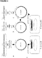
- gryphiswaldense genomic DNA ( mamGFDC, mms6, mamXY). Briefly, a cloning cassette was amplified by polymerase chain reaction (PCR) and transferred into electro-competent E. coli cells (DH10b) expressing phage-derived recombinases from a circular plasmid (pSC101-PBad-gbaA). After transfer of the cassette, recombination between homologous regions on the linear fragment and the plasmid occurred. For introducing of the magnetosome gene operons (cf. FIG. 4 ), triple recombination was used with cotransforming of two linear fragments, which recombined with a circular plasmid ( Fu; j. et al. (2008) Nucleic Acids Res. 36, e113 ). Recombinants harboring the correct plasmids were selected by restriction analysis.
- the mamDC promoter ( XbaI, BamHI restriction sites added), mamGFDC operon or mamJ gene ( NdeI, KpnI ) and egfp ( KpnI, EcoRI ) were cloned into a pBAM1 plasmid ( Martinez-Garcia, E. et al. (2011) BMC Microbiol. 11, 38 ) with a tetracycline resistance cassette (exchange of km R against tc R with SanDI and AatII ).
- the replicative plasmid pFM211 (obtained from Dr.
- pBAM_feoAB1 a fragment with P mamH and feoAB1 was amplified with PCR from pRU1feoAB ( XbaI, EcoRI ) and cloned into Tet-pBAM1. Conjugation experiments of M. gryphiswaldense deletion mutants ( Lohsse, A. et al. (2011) PLoS One 6, e25561 ) were performed as described (Kolinko, I. et al. (2011) supra).
- Optical density of M. gryphiswaldense cultures was measured at 565 nm (human, D. et al. (1995) FEMS Microbiol. Ecol. 132, 139-145 ).
- Optical density of R. rubrum cultures was measured at 660 nm (minimal absorption of bacteriochlorophyll a (Bchl a)) and 880 nm (maximal Bchl a absorption) (Ghosh, R. et al. (1994) supra). The ratio of 880/660 nm was used to determine yields of chromatophores within intact cells.
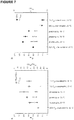
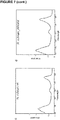
- Bchl a was extracted from cultures with methanol and absorption spectra were measured in an Ultrospec 3000 photometer (GE Healthcare). The spectra of photoheterotrophically cultivated R . rubrum_ABG6X cells were indistinguishable from those of the wild-type ( FIG. 7 ).
- Genomic sequencing of strain R. rubrum_ABG6X was performed via Next-Generation Sequencing by using the SOLID EZ Bead System (Life Technologies GmbH, Darmstadt, Germany) according to the manufacturer's protocol. No mutations were detected, except for a deletion (aa 169-247) within the hypervariable non-essential CAR domain of mamJ.
- TEM transmission electron microscopy
- PTA phosphotungstic acid
- UrAc Uranyl acetate
- Dehydration was performed with a graded acetone series. Samples were then in-filtrated and embedded in Spurr's low-viscosity resin. For high-pressure freezing, aluminum platlets were filled with concentrated cell suspensions and the cells immobilized by high-pressure freezing (Leica; HPM100). Freeze substitution was performed in acetone with 2% osmium tetroxide and 0.2% uranyl acetate, including 5% water. After embedding the samples in Epon, ultrathin sections were cut with a diamond knife and mounted onto uncoated copper grids. The sections were post-stained with aqueous lead citrate (100 mM, pH 13.0. Micrographs were taken with an EM 912 electron microscope (Zeiss) equipped with an integrated OMEGA energy filter operated at 80 kV in the zero loss mode). Vesicle sizes were determined using ImageJ software.
- High-resolution TEM HRTEM was performed using a JEOL 3010 microscope, operated at 297 kV and equipped with a Gatan Imaging Filter (GIF) for the acquisition of electron energy-loss spectra and energy-filtered compositional maps.
- GIF Gatan Imaging Filter
- a droplet of culture was added onto the prepared grid, and after blotting embedded in vitreous ice by plunge freezing into liquid ethane (temperature -170°C).
- the specimen was tilted typically about one axis with 1.5° increments over a total angular range of +/-65° to minimize the electron dose.
- the total dose accumulated during the tilt series was kept below 200 e/ ⁇ 2 .
- the exposure time was multiplied by a factor of 1/cos ⁇ .
- the pixel size in unbinned images was 0.805 at a magnification of 27500 and 1.10 at a magnification of 41000.
- Fluorescence microscopy was performed with an Olympus BX81 microscope equipped with a Hamamatsu Orca-ER camera. Exposure time of 0.12-0.25 s was used. Image scaling and cropping was performed with Photoshop 9.0 software.
- Crystals were isolated from sonification-disrupted R. rubrum_ABG6X_feo cells using magnetic separation ( Uebe, R. et al. (2011) Mol. Microbio.l 82, 818-835 ). For TEM analysis, whole cells were directly absorbed on carbon-coated copper grids. Alternatively, cells were chemically fixed or high pressure frozen and thin sectioned prior to ultrastructural analysis ( Jogler, C. et al. (2009) Environ. Microbiol. 11, 1267-1277 ). Electron dense particles of R. rubrum WT, R. rubrum_ABG, R. rubrum_ABG6 and R. rubrum_ABG6X were analyzed by HRTEM ( Uebe, R. et al. (2011) Mol. Microbiol.
- Polyacrylamide gels were prepared according to the procedure of Laemmli ( Laemmli, U. K. (1970) Nature 227, 680-685 ). Protein samples from different cellular fractions were resuspended in electrophoresis sample buffer and denatured at 98°C for 5 min (Uebe, R. et al. (2011) supra ). 10 ⁇ g of protein extracts were separated on a 15% SDS-polyacrylamide gel. Protein bands were visualized by Coomassie brilliant blue staining. Western blot analysis for detection of MamC was performed as previously described (Lang, C. and Sci, D. (2008) supra ).
- the smaller mamGFDC (2.1 kb), mms6 (3.6 kb) and mamXY operons (5.1 kb) have accessory functions in biomineralization of properly sized and shaped crystals
- the large mamAB operon (16.4 kb) encodes proteins essential for iron transport, magnetosome membrane assembly/formation, and crystallization of magnetosome particles as well as their assembly and intracellular positioning.
- transposition cassettes comprising the MycoMar transposase gene ( tps ) or Tn5 transposase gene, two corresponding inverted repeats, the origin of transfer oriT, and an antibiotic resistance gene were designed to enable transfer and chromosomal integration of the gene clusters in single copy. Chromosomal re-integration of all cassettes into different non-magnetic, single-gene as well as large operon deletion strains of M . gryphiswaldense resulted in stable and complete restoration of magnetosome biomineralization ( FIG. 5 ).
- Rhodospirillum rubrum was chosen as potential expression host due to the phylogenetic relationship between the Rhodospirillaceae and magnetotactic bacteria, which except magnetosome and photosynthetic gene functions, shares a high proportion of conserved genes ( Richter, M. et al. (2007) J Bacteriol 189, 4899-4910 ) ( FIG. 1 ).
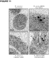
- FIG. 2b TEM revealed the presence of cuboidal electron dense particles with an average size of 25 nm, which were aligned in short, fragmented chains loosely dispersed within the cells ( FIG. 2a iii ). Electron diffraction identified the electron dense particles as single or twinned crystals of magnetite (Fe 3 O 4 ) ( FIG. 11 ).
- Phototrophic growth with moderate light intensity (1000 lux) at 23°C was found to support highest magnetic responses and reasonable/robust growth compared to low light conditions or high light intensity ( ⁇ 400 lux, 9000 lux). Therefore, cells were grown under these conditions in all subsequent experiments.
- MamJ is assumed to connect magnetosome particles to the cytoskeletal magnetosome filament formed by the actin-like MamK ( Scheffel, A. et al. (2006) Nature 440, 110-114 ).
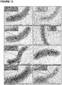
- M. gryphiswaldense deletion of mamJ caused agglomeration of magnetosome crystals in about 65% of R. rubrum_ABG6X-dJ cells ( FIGs. 2a v and 13 , TABLE 1 ).
- M magnetosome membrane
- ICMs intracytoplasmic membranes
- rubrum a filamentous/chain-like fluorescent signal became apparent in the R. rubrum_ABG6X background in which the full complement of magnetosome genes is present, reminiscent to the magnetosome-chain localization of these proteins in M . gryphiswaldensis ( Scheffel, A. et al. (2006) Nature 440, 110-114 ; Lang, C. and scaffold, D. (2008) supra) ( FIG. 8 ).
- R. rubrum_ABG6X-dl failed to express magnetic particles as revealed by C mag and TEM, which phenotypicxally paralleled the deletion of mamI in M . magneticum ( Murat, D. et al. (2010) Proc. Natl. Acad. Sci. USA 107, 5593-5598 ).
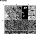
- Another example is MamJ, which is assumed to connect magnetosome particles to the actin-like cytoskeletal magnetosome filament formed by MamK. Deletion of mamJ did not affect biomineralization, but caused agglomeration of magnetic particles in R. rubrum_ABG6X-dJ ( FIG. 3 ), much as observed in M. gryphiswaldense (Scheffel, A. et al. (2006) supra), indicating that also the chain-like assembly of magnetosome crystals is genetically controlled in R. rubrum_ABG6X ( FIG. 13 ).
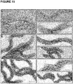
- Magnetosome particles could be purified from disrupted cells by magnetic separation and centrifugation and formed stable suspensions ( FIG. 3 ). Isolated crystals were clearly enclosed by a layer of organic material resembling the MM attached to magnetosomes of M. gryphiswaldense. Smaller, immature crystals were surrounded by partially empty vesicles ( FIG. 3c inset ) that were also seen in thin-sectioned cells ( FIGs. 3a and 11 ) and on average were smaller (66 ⁇ 6 nm) than the abundant photosynthetic intracytoplasmic membranes (ICM) (93 ⁇ 34 nm ( FIG. 11 ). Organic material of the putative MM could be solubilized from isolated magnetite crystals of R.
- ICM intracytoplasmic membranes
- rubrum_ABG6X by various detergents ( FIG. 3d ), similar as reported for MM of M. gryphiswaldense (Grünberg et al. (2004) supra ).
- Proteomic analysis of the SDS-solubilized MM revealed a complex composition ( FIG. 9 ), and several genuine magnetosome proteins (MamKCJAFDMBYOE, Mms6, MmsF) were detected among the most abundant polypeptides ( TABLE 3 ).
- An antibody against MamC the most abundant protein in the MM of M. gryphiswaldense, recognized a prominent band with the expected mass (12.4 kDa) also in the MM of R. rubrum_ABG6X ( FIG. 9 ).
- the subcellular localization of selected magnetosome proteins in R. rubrum depended on the presence of further determinants encoded by the transferred genes.
- GFP fusions of MamC displayed a punctuate pattern in the R. rubrum wild-type background.
- a filamentous fluorescent signal became apparent in the majority of cells (79%) of the R. rubrum_ABG6X background in which the full complement of magnetosome genes is present ( FIG. 10 ), reminiscent of the magnetosome-chain localization of these proteins in M. gryphiswaldense.
- the heterologous production of magnetic nanoparticles in R. rubrum should result in a significant increase in yield as compared to M . gryphiswaldense. From largecultures of M. gryphiswaldense, a maximal cell yield of 1 g fresh weight per liter of culture can be typically obtained, resulting in a final yield of 10 mg magnetic nanoparticles (i.e. magnetite) per liter of culture. With R. rubrum, a cell yield of up to 60 mg per liter can be accomplished (by means of high cell-density fermentation). However, in R. rubrum, the relative amount of magnetite per cell mass can be estimated to be only about 20% of that in M. gryphiswaldense. Nevertheless, due to the much higher cell density of R. rubrum, a final yield of 100-120 mg magnetic nanoparticles (i.e. magnetite) per liter of culture can be calculated, that is, an increase by a factor of 10-12 as compared to M . gryphiswaldense.
- a recombinant M. gryphiswaldense strain harboring, in addition to its endogenous copies of the mamAB, mamGFDC, mms6 ,and mamXY operons, additional (i.e. genetically introduced) copies of the respective four expression cassettes, produced about two times the amount of magnetic nanoparticles than the corresponding wild-type strain (data not shown), thus suggesting the "gene copy number" as a further important determinant for improving the overall yield of magnetic nanoparticles produced.
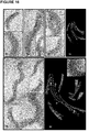
- M. gryphiswaldense MSR was engineered by mariner- or Tn5 transposon-driven random chromosomal insertion ( FIG. 14 ).
- Transconjugants were obtained at frequencies between 2-5 x 10 -7 , and chromosomal insertions were stably inherited as indicated by the ability of transformed strains to grow in the presence of kanamycin after 120 generations without antibiotic selection. All insertants essentially displayed wild type-like growth.
- ⁇ RecA + mms6 3x carrying four copies of mms6 operon produced 58 magnetite crystals per cell with a diameter of 44 nm and an intracellular iron content increased from 2.68% to 3.73% iron per dry weight, which represents only a slight further increase compared to ⁇ recA + mms6 2x (increase by 38.8 ⁇ 2.5% compared to ⁇ RecA ( FIG. 14 , TABLE 6 ). Cultivation under anaerobic conditions with 50 ⁇ M or 500 ⁇ M iron did not significantly increase iron uptake of ⁇ RecA + mms6 3x compared to cultivation under microaerobic conditions with 50 ⁇ M iron.
- Shorter cells ( ⁇ 10 ⁇ m) of ⁇ RecA+ mms6 2x contained fewer particles with smaller magnetite crystals (43 nm), whereas highly elongated cells (>10 ⁇ m) had significantly more (53-138 particles, mean: 104; n 572, equivalent to a 206% increase) and larger magnetite crystals (49 nm) with a maximum size of 80 nm ( FIG. 14 ).
- the magnetosome chains were persistently located at the mid of the cells and split into two sub-chains during cell division, similar as in the wild type ( Katzmann, E. et al. (2011) Mol. Microbiol.
- TEM transmission electron microscopy
- Mutant strain ⁇ RecA + mamAB 2x was constructed by transfer of pTps-mamAB-Gm into insertion mutant ⁇ RecA + mamAB 1x.
- the mutant showed a similar phenotype as ⁇ RecA + mamAB 1x, and the number of magnetosomes did not further increase (68), despite of a slightly increased intracellular iron content (by 9.4 ⁇ 0.5%).
- the magnetosome membrane of magnetosome particles isolated from strain ⁇ RecA + mamAB 1x had the same appearance and thickness of 5.4 ⁇ 1.8 nm as compared to ⁇ RecA 5.2 ⁇ 1.9 nm.
- Coomassie-stained SDS-PAGE profiles of magnetosome membranes from strain ⁇ RecA + mamAB 1x revealed similar patterns compared to ⁇ RecA. However, in strain ⁇ RecA + mamAB 1x several bands including magnetosome proteins MamA and MamM, showed higher intensities between 51% and 145%.
- the mamAB, mms6, and mamGFDC operons were simultaneously integrated into the genome of ⁇ RecA that was further modified by insertion of the mamXY operon using mariner transposon based gene delivery, resulting in strain RecA + AB6GX.
- the intracellular iron content of the resulting mutant strain was drastically increased to 3.77% iron per dry weight, which is an increase of 140.7% ⁇ 2.4% as compared to ⁇ RecA.
- Mutant cells of strain RecA + ABG6X exhibited delayed growth at 30°C, but wild type growth at 23 °C. Additionally, mutant cells were longer ( FIG. 16 ), and in dividing cells tubular extensions during or after cell division were observed. In contrast to the giant magnetosome membrane vesicles observed by cryo-electron tomography (CET) in strains RecA + mms6 2x and RecA + mms6 3x, magnetosome membrane vesicles in strain RecA + ABG6X were not significantly enlarged in relation to the crystals ( FIG. 16E and 16F ).
- R. rubrum_ABG6X_dI - 0.09 ⁇ 0.07 (n 3) - - R.
- Organisms were selected based on their close phylogenetic affiliation, availability of a genetic system or predominant role in heterologous protein expression.
- Organism Phylogenetic class 16S rRNA gene identity (%) NCBI Accession number Rhodospirillum rubrum Alphaproteobacteria 90 NR_074249 Caulobacter crescentus Alphaproteobacteria 88 AE005673 Rhodobacter sphaeroides
- Alphaproteobacteria 86 NR_102927 Agrobacterium tumefaciens
- Alphaproteobacteria 88 AJ012209 Escherichia coli
- rubrum_ABG6X by nano-electrospray ionization-LC tandem MS (ESI-LC-MS/MS). Spectra were analyzed via MascotTM software using the NCBI nr Protein Database and a database from M. gryphiswaldense (asterisks; Richter, M. et al. (2007) J. Bacteriol. 189, 4899-4910 ). Proteins are listed in the order of their exponentially modified protein abundance index (emPAI). The data have been deposited to ProteomeXchange with identifier PXD000348 (DOI 10.6019/PXD000348). Protein Acc. No. Coverage (%) No. of spectrum matches No. of seq.
- Km R kanamycin resistance
- Tc R tetracycline resistance
- Ap R ampicillin resistance
- BSD R blasticidin S resistance
- Cm R chloramphenicol resistance
- Gm R gentamicin resistance
- Spec R spectinomycin resistance.
- Strain or plasmid Characteristics Reference or source M. gryphiswaldense MSR-1 Wild-type (wt) DSM-6361; Schleifer, K. et al. (1991) Syst. M. gryphiswaldense MSR-1B spontaneous unmagnetic mutant lacking parts of the MAI Schübbe, S. et al. (2993) J. Bacteriol. 185, 5779-5790 M.
- gryphiswaldense ⁇ mamAB mamAB deletion mutant Lohsse, A. et al. (2011) PLoS One 6, e25561
- M. gryphiswaldense ⁇ mamAB _AB Km R transposon mutant with inserted mamAB operon
- M. gryphiswaldense MSR-1 B_ AB Km R transposon mutant with inserted mamAB operon
- M. gryphiswaldense MSR-1B_ ABG Km R , Spec R transposon mutant with inserted mamAB and mamGFDC operon
- rubrum_ABG6X Km R , Cm R , Gm R transposon mutant with inserted mamAB, mamGFDC and mms6 operon This study R. rubrum_ABG6X Km R , Cm R , Gm R transposon mutant with inserted mamAB, mamGFDC, mms6 and mamXYoperon (without ftsZm )
- rubrum_J-EGFP Tc R transposon mutant with inserted mamGFDC-EGFP
Landscapes
- Health & Medical Sciences (AREA)
- Life Sciences & Earth Sciences (AREA)
- Chemical & Material Sciences (AREA)
- Organic Chemistry (AREA)
- Genetics & Genomics (AREA)
- Engineering & Computer Science (AREA)
- General Health & Medical Sciences (AREA)
- Zoology (AREA)
- Wood Science & Technology (AREA)
- Biochemistry (AREA)
- General Engineering & Computer Science (AREA)
- Bioinformatics & Cheminformatics (AREA)
- Biotechnology (AREA)
- Molecular Biology (AREA)
- Biophysics (AREA)
- Microbiology (AREA)
- Biomedical Technology (AREA)
- Radiology & Medical Imaging (AREA)
- Veterinary Medicine (AREA)
- Epidemiology (AREA)
- Hematology (AREA)
- Virology (AREA)
- Public Health (AREA)
- Animal Behavior & Ethology (AREA)
- Nuclear Medicine, Radiotherapy & Molecular Imaging (AREA)
- Medicinal Chemistry (AREA)
- Proteomics, Peptides & Aminoacids (AREA)
- Gastroenterology & Hepatology (AREA)
- Plant Pathology (AREA)
- Physics & Mathematics (AREA)
- Chemical Kinetics & Catalysis (AREA)
- General Chemical & Material Sciences (AREA)
- Micro-Organisms Or Cultivation Processes Thereof (AREA)
Description
- The present invention relates to recombinant host cells being genetically engineered to allow for the heterologous high-yield production of magnetic nanoparticles, preferably of magnetosomes derived from magnetotactic bacteria. More specifically, the recombinant host cells comprise in their genome the entire genetic information sufficient to express the biosynthetic pathway for the production of magnetic nanoparticles.
- Magnetic nanoparticles typically comprise nanocrystals made of oxides (or to a lesser extent of sulfides) of the elements in the forth row of the periodic table (i.e. Cr, Mn, Fe, Co, and Ni). The ability to produce such magnetic nanoparticles is inevitable not only for the general understanding of magnetic properties in a nanometer regime, but also for manifold technical applications ranging from magnetic resonance imaging, drug delivery, catalysts, and biosensing to nanoelectronics, semiconductor materials, and magnetic storage media (reviewed in Lu, A. et al. (2007) Angew. Chem. Int. Ed. 46, 1222-1224).
Magnetic nanoparticles can be synthesized chemically through precipitation of the crystals from basic aqueous solutions. However, the production of particularly dimensioned ("tailored") nanocrystals via these synthesis routes is significantly hampered by the broad size distribution of the crystal populations obtained. More recently, the synthesis of nanocrystals has been directed to non-aqueous approaches generally resulting in the formation of crystals having not only an improved overall quality but also a narrower size distribution. Nevertheless, in most chemical syntheses reported so far only sub-gram to low gram quantities of monodisperse nanocrystals were obtained, not sufficient for many applications. Furthermore, only a fraction of such synthetic particles constitutes monocrystalline particles having defined magnetic properties. Typically, however, the resulting particles were too small for clinically relevant applications such as magneto-hyperthermic treatment of tumors (Park, J. et al. (2004) Nature Materials 3, 891-895). - Alternatively, biogenic magnetic nanoparticles can be employed that are produced by magnetotactic organisms, predominantly magnetotactic bacteria. The ability of magnetotactic bacteria to orient in the Earth's magnetic field is based on the presence of specific organelles, the magnetosomes, which are membrane-enveloped monocrystalline crystals of a magnetic mineral that are arranged in chain-like structures within the cell. Magnetosomes display a variety of species-specific shapes within the single magnetic domain size range (reviewed in Bazylinski, D.A. and Frankel, R.B. (2004) Nature Rev. Microbiol. 2, 217-230). In the prototypical Magnetospirillum, cubo-octahedral nanocrystals of the mineral magnetite (Fe3O4) having a maximal diameter of 50 nm are synthesized within magnetosome membrane (MM) vesicles. The MM is a phospholipid bilayer of a distinctive biochemical composition. Their specific structural and magnetic properties make these bacterial magnetosomes highly attractive for various nanotechnological and biomedical applications.
- However, the biotechnological usability is hampered by the fact that they can only be produced in comparably small amounts due to the methodological difficulties to cultivate magnetotactic bacteria due to fastidious growth requirements. Typically, the cell yields obtained are only about 1 g (fresh weight) per liter of culture, not enough for the large scale production of magnetosomes (Heyen, U. and Schüler, D. (2003) Appl. Microbiol. Biotechnol. 61, 536-544). In addition, genetic interference in or manipulation of the genetic pathway for magnetosome production is cumbersome due to the limited availability of molecular tools for the recalcitrant native host cells.
- The biogenesis of functional magnetosomes is highly complex and involves the invagination of magnetosome vesicles from the cytoplasmic membrane, the magnetosomal uptake of iron and the crystallization of magnetite particles, as well as their assembly into chains along a dedicated cytoskeletal structure (Komeili, A. et al. (2006) Science 311, 242-245; Katzmann, E. et al. (2010) Mol. Microbiol. 77, 208-224). Recently, in M. gryphiswaldense, genes controlling magnetosome synthesis within several clusters of a larger (115 kb) genomic magnetosome island (MAI) were discovered, which are interspersed by transposases and genes of unknown function (Jogler, C. et al. (2009) Environ. Microbiol. 11, 1267-1277; Jogler, C. et al. (2011) Proc. Natl. Acad. Sci. USA 108, 1134-1139). Whereas the smaller mamGFDC (2.1 kb), mms6 (3.6 kb) and mamXY operons (5.1 kb) have accessory functions in biomineralization of properly sized and shaped crystals, the large mamAB operon (16.4 kb) encodes proteins essential for iron transport, magnetosome membrane formation, and crystallization of magnetosome particles as well as their assembly and intracellular positioning. However, due to the structural complexity of the magnetosome organelle, current knowledge about particular gene functions is still quite limited and the presumably complex interplay between the numerous factors involved in the production of magnetosomes needs still to be unraveled in closer detail which is further complicated by the limited methodology for genetic manipulation of magnetotactic bacteria that is presently available. Lohsse et al. (2011) PLOS One 6, e25561 discloses a functional analysis of the magnetosome island in Magnetospirillum gryphiswaldense and suggests that the mamAB operon is sufficient for magnetite biomineralization. Schuler (2008) FEMS Microbiol. Rev. 32, 654-672 is a review on the genetics and cell biology of magnetosome formation in magnetotactic bacteria. Murat et al. (2012) Mol. Microbiol. 85, 684-699 discusses the role of magnetosome membrane protein MmsF in magnetite biomineralization.
WO 2009/047301 A1 discloses a method wherein the expression of one or more genes of the MamGFDC operon is modified in magnetotactic bacterial cells. Scheffel et al. (2008) J. Bacteriol. 190, 377-386 looks at the magnetite biomineralization in Magnetospirillum gryphiswaldense and comes to the conclusion that the magnetosome proteins of MamGFDC are not essential but regulate the size of magnetosome crystals. - Thus, there is a need for molecular tools as well as corresponding methods in order to improve the yield of biogenic magnetic nanoparticles that can be obtained from bacterial cultures - both in homologous and heterologous genetic backgrounds (i.e. settings). In particular, there remains an ongoing need for techniques that allow for the large-scale production of magnetic nanoparticles in an easy-to-do and cost-efficient manner.
Accordingly, it is an object of the present invention to provide such molecular tools and corresponding methods. - In one aspect, the present invention relates to a recombinant host cell, comprising in its genome: (i) a gene expression cassette capable of being expressed in the host cell, the gene expression cassette encoding a nucleic acid sequence encompassing the full-length sequence of the mamAB operon of a magnetotactic alpha-proteobacterium; (ii) a gene expression cassette capable of being expressed in the host cell, the gene expression cassette encoding a nucleic acid sequence encompassing the full-length sequence of the mamGDFC operon of a magnetotactic alpha-proteobacterium; and (iii) a gene expression cassette capable of being expressed in the host cell, the gene expression cassette encoding a nucleic acid sequence encompassing the full-length sequence of the mms6 operon of a magnetotactic alpha-proteobacterium; (iv) a gene expression cassette capable of being expressed in the host cell, the gene expression cassette encoding a nucleic acid sequence encompassing the full-length sequence of the mamXY operon of a magnetotactic alpha-proteobacterium;
wherein the recombinant host cell allows for the heterologous expression of the gene expression cassettes in their entirety, wherein the gene expression cassettes represent heterologous nucleic acid sequences, and wherein the recombinant host cell, upon expression of the gene expression cassettes in their entirety, is capable of producing magnetic nanoparticles. - In preferred embodiments, the recombinant host cell further comprises: (v) a gene expression cassette capable of being expressed in the host cell, the gene expression cassette encoding a nucleic acid sequence encompassing the full-length sequence of the feoAB1 operon of a magnetotactic alpha-proteobacterium.
In specific embodiments, the recombinant host cell comprises the gene expression cassettes (i) to (v), wherein the gene expression cassettes all encompass the full-length sequences of the respective operons. Particularly, the total length of the gene expression cassettes introduced in the recombinant host cell is less than 35 kb. - In other specific embodiments, the recombinant host cell allows for heterologous expression of the gene expression cassettes in their entirety. In further preferred embodiments, the magnetotactic alpha-proteobacterium from which the respective operon sequences are derived is Magnetospirillum spec., and in particular Magnetospirillum gryphiswaldense.
Preferably, the gene expression cassettes are stably integrated into the host cell's genome. In specific embodiments, the magnetic nanoparticles are magnetosomes, and in particular magnetosomes consisting of magnetite.
In further specific embodiments, any one or more of the gene expression cassettes are under the control of their respective endogenous regulatory sequences.
In yet further specific embodiments, the recombinant host cell comprises two or more copies of any one or more of the gene expression cassettes. - Preferably, with respect to the recombinant host cell, any one or more of the gene expression cassettes represent heterologous nucleic acid sequences.
- In further preferred embodiments, the recombinant host cell is a prokaryotic cell, and particularly derived from an alpha-proteobacterium. Particularly preferably, the recombinant host cell is derived from Rhodospirillum rubrum.
- In a further aspect, the present invention relates to a method for the production of a recombinant host cell as defined herein, the method comprising the transfer of the gene expression cassettes into the host cell by means of genetic transposition, and in particular comprising a modular transfer of the gene expression cassettes.
- In yet another aspect, the present invention relates to the use of a recombinant host cell as defined herein for the biotechnological production of magnetic nanoparticles.
- In yet another aspect, the present invention relates to the use of a recombinant host cell as defined herein for the in vivo synthesis of magnetic nanoparticles for application in magnetogenetics or biomedical imaging.
-
-
FIGURE 1 : Schematic representation of Magnetospirillum gryphiswaldense gene clusters that were transferred to the chromosome of Rhodospirillum rubrum. Different colors indicate single genes or operons inserted into the chromosome (oval shape). Dark blue: mamAB operon, green: mamGFDC operon, brown: mms6 operon, pale blue: mamXY operon, red: feoAB1 operon, grey: lacI repressor gene, yellow: antibiotic resistance genes (kmR : kanamycin resistance, tcR : tetracycline resistance, apR : ampicillin resistance, gmR : gentamicin resistance). Red arrows indicate different promoters (P) controlling expression of antibiotic resistance genes (P km, P gm, P tc ), repressor genes (P lacI ) or magnetosome genes (P mms , P mamDC, P mamH, PmamXY, Plac ). Crossed lines indicate gene deletions by homologous recombination, which were introduced into the recipient genome to prove equivalent functions as in the donor organism. IR=inverted repeat defining the boundaries of the sequence inserted by the transposase. -
FIGURE 2 : Phenotypes of Rhodospirillum rubrum strains expressing different magnetosome gene clusters and auxiliary genes.- (2a) Transmission electron micrographs. R. rubrum wt (i) harbors larger phosphate inclusion (P) and some small, non-crystalline electron-dense particles. R. rubrum_ABG6 (ii), R. rubrum_ABG6X (iii), R. rubrum_ABG6X_ftsZm (iv), R. rubrum_ABG6X_dJ (v), R. rubrum_ ABG6X_feo (vi). Insets show magnifications of non-crystalline electron dense particles or heterologously expressed nanocrystals from (i)-(vi). (ii) R. rubrum_ABG6 (mamAB, mamGFDC, and mms6 operons inserted) showed small crystalline particles aligned in a chain-like pattern. (iii) Additional insertion of the mamXY operon lacking ftsZm resulted in formation of larger magnetic particles with an average size of 25 nm (R. rubrum_ABG6X). (iv) Additional transposition of ftsZm under control of an inducible lac promoter did not further increase crystal number and size. (v) Deletion of mamJ in R. rubrum_ABG6X caused agglomeration of the crystals into clusters. (vi) Additional insertion of feoAB1 into R. rubrum_ABG6X resulted in increased magnetosome biomineralization (
average crystal diameter 35 nm, formation of longer magnetosome chains of 440 nm). Scale bar: 0.2 µm. For further TEM micrographs seeFIG. 13 . (2b) Unlike the untransformed wild-type, cells of R. rubrum_ABG6X accumulated as a visible red spot near the pole of a permanent magnet at the edge of a culture flask. (2c) TEM micrograph of a mixed culture of the donor M. gryphiswaldense and the recipient R. rubrum_ABG6X_feo, illustrating characteristic cell properties and magnetosome organization. Insets show magnifications of magnetosomes from M. gryphiswaldense and R. rubrum_ABG6X_feo. (2d) HRTEM lattice image of a twinned crystal from R. rubrum_ABG6X, with the Fourier transforms (i) and (ii) that show intensity maxima consistent with the structures of magnetite, respectively.
- (2a) Transmission electron micrographs. R. rubrum wt (i) harbors larger phosphate inclusion (P) and some small, non-crystalline electron-dense particles. R. rubrum_ABG6 (ii), R. rubrum_ABG6X (iii), R. rubrum_ABG6X_ftsZm (iv), R. rubrum_ABG6X_dJ (v), R. rubrum_ ABG6X_feo (vi). Insets show magnifications of non-crystalline electron dense particles or heterologously expressed nanocrystals from (i)-(vi). (ii) R. rubrum_ABG6 (mamAB, mamGFDC, and mms6 operons inserted) showed small crystalline particles aligned in a chain-like pattern. (iii) Additional insertion of the mamXY operon lacking ftsZm resulted in formation of larger magnetic particles with an average size of 25 nm (R. rubrum_ABG6X). (iv) Additional transposition of ftsZm under control of an inducible lac promoter did not further increase crystal number and size. (v) Deletion of mamJ in R. rubrum_ABG6X caused agglomeration of the crystals into clusters. (vi) Additional insertion of feoAB1 into R. rubrum_ABG6X resulted in increased magnetosome biomineralization (
-
FIGURE 3 : Ultrastructural analysis of Rhodospirillum rubrum_ABG6X and isolated crystals.- (3a) Cryo-fixed, thin-sectioned R. rubrum_ABG6X harbored intracytoplasmic membranes (ICMs) (93 ± 34 nm, n = 95), magnetic particles (MP) and a vesicle (V) enclosing an immature crystal (66 ± 6 nm, ± = s.d., n = 7.). The inset shows a magnification of the magnetite crystals. Scale bar: 100 nm. (3b) Cryo-electron tomography (CET) of isolated magnetic particles of R. rubrum_ABG6X: X-Y slice of a reconstructed tomogram (I) and surface-rendered 3D representation (II). A membrane-like structure (thickness 3.4 ± 1.0 nm, n = 6) surrounds magnetic particles (red). Yellow: membrane like structure, blue: empty vesicle. Scale bar: 100 nm. (3c and 3d) Transmission electron micrographs of isolated magnetosomes from R. rubrum_ABG6X (3c and 3d (ii), (iii), (iv)) and M. gryphiswaldense (3d (i)) negatively stained by (3c) uranyl acetate or (3d) phosphotungstic acid. Insets show magnifications of respective magnetic particles. Arrows indicate the magnetosome membrane, which encloses magnetic crystals of M. gryphiswaldense (3.2 ± 1.0 nm, n = 103) and R. rubrum_ABG6X (3.6 ± 1.2 nm, n = 100). Organic material could be solubilized from magnetite crystals of R. rubrum_ABG6X with SDS (iv) and less effective also by Triton X-100 (III). Scale bar: 100 nm.
-
FIGURE 4 : Construction scheme of insertion cassettes for modular expression of the mam and mms operons.- (4a) Recombinant engineering of a BAC containing the mamAB operon (blue) and a vector backbone (Km-p15A-Tps-oriT-Km, orange) harboring a MycoMar transposase gene (tps), inverted repeats (IR), origin of transfer (oriT), p15A origin of replication (p15A) and a kanamycinR cassette (kmR, orange). (4b) Insertion of a spectinomycinR cassette (specR, pink) and the mamGFDC operon (green) into pTps_AB by triple recombination. (4c and 4d) Stitching of pTps_ABG by insertion of the mms6 operon and a chloramphenicolR cassette. (4e) pTps_XY consisting of a Tps vector backbone (orange), the mamXY operon (pale blue) and a gentamicinR gene (gmR, purple) was constructed. (4f) Plasmids were transferred by conjugation into R. rubrum. Transposition of the DNA fragments within the IR sequences occurred randomly at TA dinucleotide insertion sites by a "cut and paste" mechanism (Rubin, E.J. et al. (1990) Proc. Natl. Adac. Sci. USA 96, 1645-1650). (4g) Chromosomal insertion sites of the transposed constructs in R. rubrum_ABG6X are shown with adjacent genes (red) as revealed by whole genome sequencing performed with a MiSeq sequencer (Illumina) (accession number of R. rubrum ATCC 11170: NC_007643). pTps_ABG6 inserted within a gene encoding a putative aldehyd dehydrogenase (YP_426002), and pTps_XYZ inserted within rru_A2927, encoding an acriflavin resistance protein (protein accession number YP_428011). Sequences of inserted magnetosome operons matched those of the donor (M. gryphiswaldense) with no detectable mutations, except for a deletion (aa 169-247) within the hypervariable non-essential CAR domain of mamJ, which was shown to be irrelevant for protein function (Scheffel, A. and Schüler, D. (2007) J. Bacteriol. 189, 6437-6446).
-
FIGURE 5 : Transmission electron micrographs of M. gryphiswaldense MSR-1 mutants expressing various insertional transposon constructs.
The plasmids pTps_AB, pTps_ABG and pTps_ABG6 were transferred into the non-magnetic M. gryphiswaldense MSR-1 mutants ΔmamAB and MSR-1B, the latter lacking most of the magnetosome genes except of the mamXY operon (Lohsse, A. et al. (2011) PLoS One 6, e25561). After transfer of pTps_AB, a wt-like phenotype was restored in ΔmamAB_AB as revealed by Cmag (1.2 ± 0.2) and measured crystal sizes (37 ± 10 nm) in comparison with M. gryphiswaldense wild-type (36 ± 9 nm, Cmag = 1.4 ± 0.2) (see also Table 1). Mutant MSR-1B was only partly complemented after insertion of pTps_AB and pTps_ABG, that is, Cmag and crystal sizes were still lower than in the wild-type (wt) (see also Table 1). Transfer of pTps_ABG6 restored nearly wild-type like magnetosome formation in MSR-1B (35 ± 8 nm, Cmag= 0.9 ± 0.1). ± = s.d. Scale bar: 1 µm, insets: 0.2 µm. -
FIGURE 6 : HRTEM lattice image of a crystal from Rhodospirillum rubrum_ABG6 with the corresponding Fourier transform (i) that shows intensity maxima consistent with the structures of hematite. -
FIGURE 7 : Growth and magnetic response of Rhodospirillum rubrum_AGB6X cultivated under various growth conditions.- (7a and 7b) Cells were grown in ATCC 112 (chemotrophic, 20% O2), Sistrom A (phototrophic, anoxic) and M2SF (chemotrophic, 1% O2) medium for 3 (30°C), 4 (23°C) or 10 (18°C) days. Optical density at 660 and 880 nm and magnetic response were measured. The ratio OD880/OD660 correlates with the amount of chromatophores produced in the cells (median values n = 3, error bars indicate s.d.). No Cmag was detectable under aerobic and microaerobic conditions at 30°C. Anoxic and microaerobic conditions (23°C) supported particle formation. Furthermore, absorption spectra of extracted bacteriochlorophylls of R. rubrum WT (7c) and R. rubrum_ABG6X (7d) were directly measured, respectively (phototrophic growth, 30°C). No differences in absorption spectra were detectable.
-
FIGURE 8 : Growth of Magnetospirillum gryphiswaldense (OD565), Rhodospirillum rubrum wt and Rhodospirillum rubrum_ABG6X (OD660 ).
Cells of M. gryphiswaldense (FSM medium) or R. rubrum (Sistrom A medium, 1000 lux) were incubated for 3 days at 23°C under anaerobic conditions. Growth of the mutant strain ABG6X (median values n=3, error bars indicate s.d.) was indistinguishable from that of the untransformed wild-type (n=3). -
FIGURE 9 : Proteomic analysis of magnetosomes from Rhodospirillum rubrum_ABG6X.- (9a) 1D-SDS-PAGE (Comassie blue stained) of proteins solubilized from isolated magnetosome particles of M. gryphiswaldense and R. rubrum_ABG6X. Bands (∼ 90, 70, 60, 40, 30, 25, 20, 15, 12.4 kDa) of the same size are indicated (arrowheads). (9b) Immunodetection of MamC (12.4 kDa) in blotted fractions of M. gryphiswaldense and R. rubrum_ABG6X using an anti-MamC antibody (Lang, C. and Schüler, D. (2008) Appl. Environ. Microbiol. 74, 4944-4953). A signal for MamC was detectable in the magnetic membrane fraction of R. rubrum_ABG6X (6), which was absent from the soluble fraction, but faintly present also in the non-magnetic membrane fraction (5), possibly originating from empty membrane vesicles or incomplete magnetic separation during isolation. Protein extracts from M. gryphiswaldense: 1. soluble fraction, 2. non-magnetic membrane fraction, 3. magnetosome membrane. Protein extracts from R. rubrum_ABG6X: 4. soluble fraction, 5. non-magnetic membrane fraction, 6. magnetic ("magnetosome") membrane fraction. M: marker.
-
FIGURE 10 : Fluorescence microscopy of Rhodospirillum rubrum WT and Rhodospirillum rubrum_ABG6X expressing different EGFP-tagged magnetosome proteins.
For localization studies of fluorescently labeled magnetosome proteins, strains were cultivated in ATCC medium overnight at 30 °C with appropriate antibiotics (Table S3). (10a and 10b) MamGFDC with a C-terminal MamC-EGFP fusion expressed in R. rubrum wt (n = 151) (10a), and R. rubrum_ABG6X (n = 112) (10b). In the transformed strain, a filamentous structure is visible for 79% of the cells (n = 89). (10c and 10d) MamJ-EGFP expressed in R. rubrum wt (n = 109) (10c), and in R. rubrum_ABG6X (n = 89) displaying a chain-like fluorescence signal in 63% of the cells (n = 56) (10d). Scale bar: 2 µm. -
FIGURE 11 : TEM of cryo-fixed or chemically fixed, thin sectioned Rhodospirillum rubrum strains.
Cells were cultivated under photoheterotrophic conditions prior to ultrastructural analysis. ICM sizes of cryo-fixed R. rubrum wt (93 ± 34 nm, n = 95) and vesicles surrounding immature magnetosomes of cryo-fixed R. rubrum_ABG6X (66 ± 6 nm, n = 6) were measured. -
FIGURE 12 : Size distribution of magnetosome crystals in Magnetospirillum gryphiswaldense and different Rhodospirillum rubrum strains.
Whereas crystals of R. rubrum_ABG6 (n = 303) and R. rubrum_ABG6X (n = 306) were smaller than those of the donor M. gryphiswaldense (n = 310), crystal sizes of R. rubrum_ABG6X_feo (n = 301) were significantly larger, approaching those of the donor strain (see also Table 1). -
FIGURE 13 : Transmission electron micrographs of whole cells of different Rhodospirillum rubrum strains expressing magnetosome gene clusters. Scale bar: 1 µm, inset: 0.2 µm. -
FIGURE 14 : Overexpression of magnetosome operons in Magnetospirillum gryphiswaldense.
Strategy for construction of overexpression strains by amplification of different magnetosome operons (14A). Insertional plasmids were constructed based on genomic DNA from M. gryphiswaldense MSR-1. Plasmids contain the magnetosome operons mamAB (blue, AB), mamGFDC (green, GFDC), mms6 (brown) and the mamXY operon lacking ftsZm (pale blue, XYZ). The vector backbone (genes are indicated in red) contains transposase gene (tps), inverted repeats (IR), origin of transfer (oriT), an R6K or p15A origin of replication (ori), and antibiotic resistance cassette (abR ). After conjugative transfer of the plasmids, the transposase recognizes IR sequences and catalyzes chromosomal insertion of the target sequence. Additional copies of respective magnetosome operons in the chromosome (oval shape) are marked with asterisks. (14B) Transmission electron microscopy (TEM) analysis of overexpressing strains. Arrows labeled with 1 and 2 illustrate different magnetosome morphologies identified in strain ΔRecA +mamAB 1x. -
FIGURE15 : Enhanced magnetosome formation by overexpression of the mms6 operon in M. gryphiswaldense MSR-1.
Representative TEM micrographs of ΔRecA (15A) and overexpressing strainsΔRecA + mms6 2x (15B) (two additional copies of mms6 operon; integrated in genome) and ΔRecA + mms6 3x (15C) (three additional copies of mms6 operon; integrated in genome). -
FIGURE 16 : Characterization of the mutant M. gryphiswaldense RecA+ABG6X (mms6, mamGFDC, mamAB and mamXY operons integrated into the genome of ΔRecA.- (16A-D) Representative TEM micrographs of mutant M. gryphiswaldense RecA+AB6GX exhibiting enhanced formation of magnetosomes. Mutant cells show various organizations of magnetosomes. (16E-F) Cryo-electron tomography (CET) of mutant cells. Cell membrane (blue); electron dense particles (red); magnetosome vesicles (yellow).
- The present invention is related to the unexpected finding that gene expression cassettes encoding all relevant genes being sufficient for the biomineralization of bacterial magnetic nanoparticles could be successfully transferred, in their entirety, either to a homologous (i.e., magnetotactic) to a heterologous (i.e., non-magnetotactic) host cell and, upon expression (or overexpression), resulted in the synthesis of functional magnetic nanoparticles.
- The present invention will be described in the following with respect to particular embodiments and with reference to certain drawings but the invention is to be understood as not limited thereto but only by the appended claims. The drawings described are only schematic and are to be considered non-limiting.
- Where the term "comprising" is used in the present description and claims, it does not exclude other elements or steps. For the purposes of the present invention, the term "consisting of" is considered to be a preferred embodiment of the term "comprising". If hereinafter a group is defined to comprise at least a certain number of embodiments, this is also to be understood to disclose a group, which preferably consists only of these embodiments.
- Where an indefinite or definite article is used when referring to a singular noun e.g. "a", "an" or "the", this includes a plural of that noun unless specifically stated otherwise.
In case, numerical values are indicated in the context of the present invention the skilled person will understand that the technical effect of the feature in question is ensured within an interval of accuracy, which typically encompasses a deviation of the numerical value given of ± 10%, and preferably of ± 5%.
Furthermore, the terms first, second, third, (a), (b), (c), and the like in the description and in the claims, are used for distinguishing between similar elements and not necessarily for describing a sequential or chronological order. It is to be understood that the terms so used are interchangeable under appropriate circumstances and that the embodiments of the invention described herein are capable of operation in other sequences than described or illustrated herein.
Further definitions of term will be given in the following in the context of which the terms are used. The following terms or definitions are provided solely to aid in the understanding of the invention. These definitions should not be construed to have a scope less than understood by a person of ordinary skill in the art.
In one aspect, the present invention relates to a recombinant host cell, comprising in its genome: - (i) a gene expression cassette capable of being expressed in the host cell, the gene expression cassette encoding a nucleic acid sequence encompassing the full-length sequence of the mamAB operon of a magnetotactic alpha-proteobacterium;
- (ii) a gene expression cassette capable of being expressed in the host cell, the gene expression cassette encoding a nucleic acid sequence encompassing of the full-length sequence of the mamGDFC operon of a magnetotactic alpha-proteobacterium; and
- (iii) a gene expression cassette capable of being expressed in the host cell, the gene expression cassette encoding a nucleic acid sequence encompassing of the full-length sequence of the mms6 operon of a magnetotactic alpha-proteobacterium;
- (iv) a gene expression cassette capable of being expressed in the host cell, the gene expression cassette encoding a nucleic acid sequence encompassing the full-length sequence of the mamXY operon of a magnetotactic alpha-proteobacterium;
wherein the recombinant host cell allows for the heterologous expression of the gene expression cassettes in their entirety, wherein the gene expression cassettes represent heterologous nucleic acid sequences, and
wherein the recombinant host cell, upon expression of the gene expression cassettes in their entirety, is capable of producing magnetic nanoparticles. In preferred embodiments, the recombinant host cell as defined herein further comprises: - (v) a gene expression cassette capable of being expressed in the host cell, the gene expression cassette encoding a nucleic acid sequence encompassing the full-length sequence of the feoAB1 operon of a magnetotactic alpha-proteobacterium.
- In preferred embodiments, the recombinant host cells do not have endogenous copies of any one or more of the mamAB, mamGDFC, mms6, mamXY, and feoAB1 operons (that is, any one, any two, any three, any four or all five). In other words, any one or more of gene expression cassettes as defined herein represent heterologous nucleic acid sequences with respect to the host cells employed (i.e., the host cells allow for heterologous gene expression).
- In specific embodiments, the recombinant host cells to be employed are prokaryotic cells, such as bacterial or archeal cells, with bacterial cells being preferred. Examples of suitable prokaryotic cells include inter alia E. coli, and Salmonella typhimurium. In particular embodiments, the bacterial host cells are derived from an alpha-proteobacterium, that is, any species belonging to one of the orders Magnetococcales, Rhodobacterales, Rhodospirillales, Rickettsiales, Sphingomonadales, Caulobacterales, Kordiimondales, Parvularculales, Kiloniellales, and Sneathiellales (Williams, K.P. et al. (2007) J. Bacteriol. 189, 4578-4586). In preferred embodiments, the bacterial host cells are derived from the order Rhodospirillales, more preferably from the genus Rhodospirillum, and particularly preferably from the species Rhodospirillum rubrum (R. rubrum).
- In a particularly preferred embodiment, the recombinant host cell having stably integrated in its genome the expression cassettes (i) to (v) as described above relates to the host cell being deposited on November 14, 2013 under the Budapest Treaty at the Deutsche Sammlung von Mikroorganismen und Zellkulturen (Braunschweig, Germany) having the accession number DSM 28038. The deposited recombinant host cell has the taxonomic designation Rhodospirillum rubrum ATCC 11170 (which is herein also referred to as Rhodospirillum rubrum_ABG6X_feo).
- In further preferred embodiments, the bacterial host cells are derived from the order Magnetococcales (herein also referred to as "magnetotactic alpha-proteobacteria") more preferably from the genus Magnetospirillum, and particularly preferably from the species Magnetospirillum gryphiswaldense (M. gryphiswaldense). Further suitable host cells include M. magneticum, M. magnetotacticum, MV-1, and MC-1. Such host cells typically comprise endogenous copies of any one or more of the mamAB, mamGDFC, mms6, mamXY, and feoAB1 operons (i.e., the host cells allow for heterologous gene expression, particularly overexpression of the relevant genetic elements).
In specific embodiments, the recombinant host cells to be employed are eukaryotic cells, such as yeast (e.g., Saccharomyes spec.), Pichia pastoris, and mammalian cells, with the latter ones being preferred. Examples of mammalian cells include mouse, rat, hamster, rabbit, cat, dog, pig, cow, horse, monkey, and particularly preferably human cells.
The term "magnetic nanoparticles", as used herein, denotes any particles having a size in the nanometer scale that exhibits magnetic properties (i.e. that orients in a magnetic field along the magnetic field lines), either being ferromagnetic or being superparamagnetic or exhibiting an intermediate characteristic. The term "nanometer scale", as used herein, refers to a particle diameter of less than 1000 nm (i.e. 1 µm), preferably of less than 500 nm, and particular preferably of less than 100 nm. The "magnetic nanoparticles" may comprise one or more magnetic crystals but are preferably "mono-crystalline". In preferred embodiments, the magnetic nanoparticles further comprise an outer lipid bilayer membrane surrounding the one or more crystals (Grünberg, K. et al. (2004) Appl. Environ. Microbiol. 70, 1040-1050): Particles comprising a lipid bilayer membrane are also referred to as "magnetosomes".
The magnetic nanoparticles/magnetosomes being produced by the recombinant host cells employed herein may consist of one or more metal oxides and/or metal sulfides, preferably of the elements in the fourth row of the periodic table (i.e., chrome, manganese, iron, cobalt, and nickel). In particular embodiments, the magnetic nanocrystals are made of a single metal oxide or a single metal sulfide, preferably of an iron oxide such as hematite (Fe2O3) and magnetite (Fe3O4) or an iron sulfide such as greigite (Fe3S4). In particularly preferred embodiments, the magnetic nanoparticles produced according to the present invention consist of magnetite.
The term "capable of producing magnetic nanoparticles", as used herein, denotes the capacity of the recombinant host cells employed to synthesis, upon concomitant expression of the respective "mamAB, mamGDFC, mms6 gene expression cassettes" and also the "mamXY "and/or" feoAB1 gene expression cassettes", functional magnetic nanoparticles as described above.
The term "gene expression cassette", as used herein, typically denotes a nucleic acid sequence comprising sequence elements which contain information regarding to transcriptional and/or translational regulation, such regulatory sequences being "operably linked" to the nucleotide sequence encoding one or more polypeptides to be expressed (i.e. to be synthesized starting from the gene sequence). An operable linkage is a linkage in which the regulatory sequence elements and the sequence to be expressed (and/or the sequences to be expressed among each other) are connected in a way that enables gene expression. The precise nature of the regulatory regions necessary for gene expression may vary among species, but in general these regions comprise a promoter which, in prokaryotes, contains both the promoter per se, i.e. DNA elements directing the initiation of transcription, as well as DNA elements which, when transcribed into RNA, will signal the initiation of translation. Such promoter regions normally include 5' non-coding sequences involved in initiation of transcription and translation, such as the -35/-10 boxes and the Shine-Dalgarno element in prokaryotes or the TATA box, CAAT sequences, and 5'-capping elements in eukaryotes. These regions can also include enhancer or repressor elements as well as translated signal and leader sequences for targeting the native polypeptide to a specific compartment of a host cell. In addition, the 3' non-coding sequences may contain regulatory elements involved in transcriptional termination, polyadenylation or the like. If, however, these termination sequences are not satisfactory functional in a particular host cell, then they may be substituted with signals functional in that cell.
The term "operon", as used herein, refers to a functional genetic unit of two or more genes, which are transcribed together into a single mRNA and are typically under the control of common regulatory sequence elements (i.e. promoters, terminators, and the like).
The mamAB, mamGDFC, mamXY, mms6 and feoAB1 operons encoded by the respective gene expression cassettes as described herein are derived from a "magnetotactic alpha-proteobacterium" (cf. the definition herein above), more preferably from the genus Magnetospirillum, and particularly preferably from the species M. gryphiswaldense. The exact nucleotide sequences of all these operons are known in the art and can be accessed under GenBank accession number AM085146.1 (relating to the complete 130 kb sequence of the magnetosome island of M. gryphiswaldense MSR-1; cf. also Ullrich, S. et al. (2005) J. Bacteriol. 187, 7176-7184). In specific embodiments, the mamAB, mamGDFC, mamXY, mms6 and feoAB1 operons encoded by the respective gene expression cassettes present in a particular host cell are all derived from the same magnetotactic alpha-proteobacterium, for example from M. gryphiswaldense. In other specific embodiments, the operons encoded by the respective gene expression cassettes present in a particular host cell are derived from two or more different magnetotactic alpha-proteobacteria, for example mamAB and mamGDFC from M. gryphiswaldense, and the remaining operons from M. magnetotacticum. Each of the gene expression cassettes (that is, independently from each other) introduced into the genome of the recombinant host cell encode nucleic acid sequences encompassing the full-length sequence of the respective operon sequences. Sequence identity is calculated over the entire length of the full-length sequence of the respective operon, that is, the sequence present in each of the gene expression cassettes may be terminally truncated (either 5'-terminally or 3'-terminally or 5'- and 3'-terminally) and/or may include one or more deletions or substitutions of internal sequences (e.g., the deletion and/or replacement of one or more genes comprised in a given operon).
In preferred embodiments, one or more of the gene expression cassettes encode nucleic acid sequences encompassing the full-length sequence of the respective operon sequences. In particular embodiments, the one or more of the gene expression cassettes encode nucleic acid sequences encompassing the full-length sequence of the respective operon sequences. In specific embodiments, one or more of the gene expression cassettes encode nucleic acid sequences encompassing the full-length sequence of the respective operon sequences. Sequence identity may be determined based on the nucleotide sequence or on the amino acid sequence. Various software tools and algorithms for determining nucleotide sequence and/or amino acid sequence identity are well established in the art (e.g., BLAST, FASTA, ClustalW, UGENE).
For example, in one embodiment, the present invention relates to a recombinant host cell, comprising in its genome: a gene expression cassette capable of being expressed in the host cell, the gene expression cassette encoding a nucleic acid sequence encompassing the full-length sequence of the mamAB operon of M. gryphiswaldense; a gene expression cassette capable of being expressed in the host cell, the gene expression cassette encoding a nucleic acid sequence encompassing the full-length sequence of the mamGDFC operon of M. gryphiswaldense; and a gene expression cassette capable of being expressed in the host cell, the gene expression cassette encoding a nucleic acid sequence encompassing the full-length sequence of the mms6 operon of M. gryphiswaldense. In another, embodiment, the present invention relates to a recombinant host cell, comprising in its genome: a gene expression cassette capable of being expressed in the host cell, the gene expression cassette encoding a nucleic acid sequence encompassing the full-length sequence of the mamAB operon of M. gryphiswaldense; a gene expression cassette capable of being expressed in the host cell, the gene expression cassette encoding a nucleic acid sequence encompassing the full-length sequence of the mamGDFC operon of M. gryphiswaldense; and a gene expression cassette capable of being expressed in the host cell, the gene expression cassette encoding a nucleic acid sequence encompassing the full-length sequence of the mms6 operon of M. gryphiswaldense. - In a particularly preferred embodiment, the gene expression cassettes (i) to (v) all encode nucleic acid sequences encompassing the full-length sequences of the respective operons.
- Preferably, the combination of the gene expression cassettes (i), (ii), and (iii) has a nucleic acid sequence as shown in SEQ ID NO. 35. Also preferably, gene expression cassette (iv) has a nucleic acid sequence as shown in SEQ ID NO. 36 and/or gene expression cassette (v) has a nucleic acid sequence as shown in SEQ ID NO. 37.
- The total length of the gene expression cassettes (i) to (iii), and optionally also (iv) and/or (v) comprised in the genome of the recombinant host cell (each being present in single copy) is less than 100 kb, preferably less than 80 kb or less than 60 kb, and particularly preferably less than 40 kb or less than 35 kb.
- The term "gene expression cassette capable of being expressed in the host cell", as used herein, denotes that the operon encoded by the gene expression cassette is under the control of regulatory elements being functional in the host cell employed. In preferred embodiments, gene expression of the respective operons is under the control of the endogenous regulatory elements, that is, the homologous regulatory elements naturally occurring in the particular organism from which the respective operon(s) is/are derived. For example, if the respective operons are derived from M. gryphiswaldense, the corresponding regulatory elements derived from the same organism may be employed. Typically, the gene expression cassettes may then comprise a nucleic acid sequence encompassing 100-800, preferably 200-600 nucleotides upstream (i.e., 5') of the respective operon and 100-800, preferably 200-600 nucleotides downstream (i.e., 3') of the respective operon. In other embodiments, the regulatory elements comprise non-endogenous sequences, that is exogenous inducible or constitutive promoters (e.g., tet, lac, CMV promoters) and terminators.
- In further specific embodiments, the recombinant host cell comprises two or more copies of any one or more of the gene expression cassettes. Preferably, the copy numbers of all gene expression cassettes being present in a recombinant host cell are identical, for example two copies. In other embodiments, the copy numbers of the respective gene expression cassettes are different.
- In a further aspect, the present invention relates to a method for the production of a recombinant host cell as defined herein, the method comprising the transfer of the gene expression cassettes into the host cell by means of genetic transposition, and in particular comprising a modular transfer of the gene expression cassettes.
- The gene expression cassettes (i) to (v) as described above may be introduced in to the recombinant host cells independently or any two, any three, any four or all five gene expression cassettes may be introduced concomitantly on a common genetic vector or transposon.
- In yet another aspect, the present invention relates to the use of a recombinant host cell as defined herein for the biotechnological production of magnetic nanoparticles.
- In yet another aspect, the present invention relates to the use of a recombinant host cell as defined herein for the in vivo synthesis of magnetic nanoparticles for application in magnetogenetics or biomedical imaging.
- The invention is further described by the figures and the following examples, which are solely for the purpose of illustrating specific embodiments of this invention, and are not to be construed as limiting the claimed subject matter in any way.
- Bacterial strains are shown in Table 4. E. coli strains were cultivated as previously described (Sambrook, J. & Russell, D. (2001) Molecular cloning: a laboratory manual. Vol. 3, Cold Spring Harbor Laboratory Press). 1 mM DL-α,ε-diaminopimelic was added for growth of BW29427 and WM3064.
- Liquid cultures and single colonies of Magnetospirillum gryphiswaldense were cultivated in FSM medium or on agar plates (1.5% agar) incubated at 30°C under microaerobic (1% O2) conditions (Heyen, U. and Schüler, D. (2003) Appl. Microbiol. Biotechnol. 61, 536-544; Kolinko, I. et al. (2011) J. Bacteriol. 193, 5328-5334)
- Cultures of Rhodospirillum rubrum strains were cultivated chemoheterotrophically (ATCC medium 112) with free gas exchange in the dark, microaerobically in M2SF medium (Ghosh, R. et al. (1994) Appl. Environ. Microbiol. 60, 1698-1700), or photoheterotrophically in Sistrom A medium (Sistrom, W. R. (1962) J. Gen. Microbiol. 28, 607-616) supplemented with Fe(III) citrate or Fe(II) sulfate at 18°C, 23°C or 30°C (10, 400, 1000, 9000 lux). The 16S rRNA similarity to M. gryphiswaldense is shown in Table 2.
- Plasmids and oligonucleotides used are shown in Table 4 and Table 5, respectively. Red/ET recombination was performed as previously described (Wang, J. et al. (2006) Mol. Biotechnol. 32, 43-53 (2006); Fu, J. et al. (2008) Nucl. Acids Re.s 36, e113) in order to construct expression cassettes harboring a mariner transposon vector backbone and magnetosome operons obtained from a BAC sequence pSSK18 (Schübbe, S. et al. (2003) J. Bacteriol. 185, 5779-5790) (mamAB) and M. gryphiswaldense genomic DNA (mamGFDC, mms6, mamXY). Briefly, a cloning cassette was amplified by polymerase chain reaction (PCR) and transferred into electro-competent E. coli cells (DH10b) expressing phage-derived recombinases from a circular plasmid (pSC101-PBad-gbaA). After transfer of the cassette, recombination between homologous regions on the linear fragment and the plasmid occurred. For introducing of the magnetosome gene operons (cf.
FIG. 4 ), triple recombination was used with cotransforming of two linear fragments, which recombined with a circular plasmid (Fu; j. et al. (2008) Nucleic Acids Res. 36, e113). Recombinants harboring the correct plasmids were selected by restriction analysis. - For construction of plasmids harboring fusion proteins, the mamDC promoter (XbaI, BamHI restriction sites added), mamGFDC operon or mamJ gene (NdeI, KpnI) and egfp (KpnI, EcoRI) were cloned into a pBAM1 plasmid (Martinez-Garcia, E. et al. (2011) BMC Microbiol. 11, 38) with a tetracycline resistance cassette (exchange of kmR against tcR with SanDI and AatII). The replicative plasmid pFM211 (obtained from Dr. Frank Muller, Ludwig-Maximilians-Universität München, Germany, unpublished) harboring ftsZm with a mCherry fusion under control of an inducible lac promoter was recombined with pBAM1 to construct pBAM-ftsZm_mcherry. The resident kmR exchanged by tcR using ET-recombination. For construction of pBAM_feoAB1, a fragment with PmamH and feoAB1 was amplified with PCR from pRU1feoAB (XbaI, EcoRI) and cloned into Tet-pBAM1. Conjugation experiments of M. gryphiswaldense deletion mutants (Lohsse, A. et al. (2011) PLoS One 6, e25561) were performed as described (Kolinko, I. et al. (2011) supra).
- For conjugation of R. rubrum, cultures were incubated in ATCC 112 medium. Approximately 2*109 cells were mixed with 1*109 E. coli cells, spotted on ATCC agar medium and incubated for 15h. Cells were flushed from the plates and incubated on ATCC 112 agar medium supplemented with appropriate antibiotics for 7-10 days (Tc (tetracycline): 10 µg/ml, Km (kanamycin: 20 µg/ml, Gm (gentamicin): 10 µg/ml). Sequential transfer of the plasmids resulted in 10-6-10-8 antibiotic-resistant transconjugants per recipient, respectively. Two clones from each conjugation experiments were chosen for further analyses.
- Transposition of the cassettes resulted in single-copy integration into the genome and stable maintenance for at least 40 generations in the absence of selection characterized transconjugants were indistinguishable from WT with respect to motility, cell morphology or growth during photoheterotrophic cultivation (
FIG. 8 ). - Optical density of M. gryphiswaldense cultures was measured at 565 nm (Schüler, D. et al. (1995) FEMS Microbiol. Ecol. 132, 139-145). Optical density of R. rubrum cultures was measured at 660 nm (minimal absorption of bacteriochlorophyll a (Bchl a)) and 880 nm (maximal Bchl a absorption) (Ghosh, R. et al. (1994) supra). The ratio of 880/660 nm was used to determine yields of chromatophores within intact cells. Furthermore, Bchl a was extracted from cultures with methanol and absorption spectra were measured in an Ultrospec 3000 photometer (GE Healthcare). The spectra of photoheterotrophically cultivated R. rubrum_ABG6X cells were indistinguishable from those of the wild-type (
FIG. 7 ). - Intracellular iron concentrations were determined after incubation under anaerobic conditions using a modified version of the ferrozine assay (Viollier, E. et al. (2000) Appl. ). In brief, 4 ml cultures were centrifuged for 1 min at 11.000 rpm and resuspended in 90 µl HNO3 (65%) for 3 h at 99 C. The average magnetic orientation of cell suspensions (magnetic response) was assayed with a light scattering assay as previously described (Schüler, D. et al. (1995) supra). Briefly, cells were aligned at different angles to a light beam by application of an external magnetic field. Crystal size (n = 120) and vesicle measurements (n = 100) were performed with ImageJ software.
- Genomic sequencing of strain R. rubrum_ABG6X was performed via Next-Generation Sequencing by using the SOLID EZ Bead System (Life Technologies GmbH, Darmstadt, Germany) according to the manufacturer's protocol. No mutations were detected, except for a deletion (aa 169-247) within the hypervariable non-essential CAR domain of mamJ.
- For transmission electron microscopy (TEM), cells and isolated magnetosomes were absorbed on carbon-coated copper grids. Purified magnetosomes were stained with 1% phosphotungstic acid (PTA) or 2% Uranyl acetate (UrAc). Samples were viewed and recorded with a Morgagni 268 microscope. Crystal sizes and Vesicle sizes were determined using ImageJ software.
- Chemical fixation, high pressure freezing and thin sectioning of cells was performed as previously described (Jogler, C. et al. (2011) Proc. Natl. Acad. Sci. USA 108, 1134-1139). In brief, for chemical fixation, cells were incubated in 2.5% glutardialdehyde containing fixative buffer (75 mM sodium cacodylate, 2 mM MgCl2, pH 7.0), for 1 h at room temperature. Afterward, samples were rinsed several times in fixative buffer and post-fixed at room temperature for 1 h with 1% osmium tetroxide in fixative buffer. After two washing steps in water, the cells were stained enbloc for 30 min with 1% uranyl acetate in 20% acetone. Dehydration was performed with a graded acetone series. Samples were then in-filtrated and embedded in Spurr's low-viscosity resin. For high-pressure freezing, aluminum platlets were filled with concentrated cell suspensions and the cells immobilized by high-pressure freezing (Leica; HPM100). Freeze substitution was performed in acetone with 2% osmium tetroxide and 0.2% uranyl acetate, including 5% water. After embedding the samples in Epon, ultrathin sections were cut with a diamond knife and mounted onto uncoated copper grids. The sections were post-stained with aqueous lead citrate (100 mM, pH 13.0. Micrographs were taken with an EM 912 electron microscope (Zeiss) equipped with an integrated OMEGA energy filter operated at 80 kV in the zero loss mode). Vesicle sizes were determined using ImageJ software.
- High-resolution TEM (HRTEM) was performed using a JEOL 3010 microscope, operated at 297 kV and equipped with a Gatan Imaging Filter (GIF) for the acquisition of electron energy-loss spectra and energy-filtered compositional maps. For TEM data processing and interpretation the DigitalMicrograph and SingleCrystal software were used (Uebe, R. et al. (2011) Mol. Microbiol. 82, 818-835).
- Cryo-electron-tomography was performed as previously described (Katzmann, E. et al. (2011) Mol. Microbiol. 82, 1316-1329). In brief, a FEI Tecnai F30 Polara transmission electron microscope was used. All data collection was performed at 300 kV, with the energy filter operated in the zero-loss mode (slit width of 20 eV). Tilt series were acquired using Serial EM and FEI software. Quantifoil copper grids (Quantifoil Micro Tools GmbH, Jena) were prepared by placing a droplet of 10-15 nm colloidal gold clusters (Sigma) on each grid for subsequent alignment purposes. Additionally a droplet of culture was added onto the prepared grid, and after blotting embedded in vitreous ice by plunge freezing into liquid ethane (temperature -170°C). The specimen was tilted typically about one axis with 1.5° increments over a total angular range of +/-65° to minimize the electron dose. The total dose accumulated during the tilt series was kept below 200 e/Å2. To account for the increased specimen thickness at high tilt angles, the exposure time was multiplied by a factor of 1/cos α. The pixel size in unbinned images was 0.805 at a magnification of 27500 and 1.10 at a magnification of 41000.
- Fluorescence microscopy was performed with an Olympus BX81 microscope equipped with a Hamamatsu Orca-ER camera. Exposure time of 0.12-0.25 s was used. Image scaling and cropping was performed with Photoshop 9.0 software.
- Unless described otherwise, expression of magnetic particles in R. rubrum was performed under photoheterotrophic conditions at 23°C and a light intensity of 1000 lux. Cells were harvested, washed and resuspended. Cell suspensions were lysed by sonification and cellular debris was removed by low-speed centrifugation.
- Crystals were isolated from sonification-disrupted R. rubrum_ABG6X_feo cells using magnetic separation (Uebe, R. et al. (2011) Mol. Microbio.l 82, 818-835). For TEM analysis, whole cells were directly absorbed on carbon-coated copper grids. Alternatively, cells were chemically fixed or high pressure frozen and thin sectioned prior to ultrastructural analysis (Jogler, C. et al. (2009) Environ. Microbiol. 11, 1267-1277). Electron dense particles of R. rubrum WT, R. rubrum_ABG, R. rubrum_ABG6 and R. rubrum_ABG6X were analyzed by HRTEM (Uebe, R. et al. (2011) Mol. Microbiol. 82, 818-835). Cryo-electron tomography was performed with cultures of R. rubrum_ABG6X or isolated magnetic particles from R. rubrum_ABG6X_feo as previously described (Katzmann, E. et al. (2011) supra). Magnetic particles were stained with 1% phosphotungstic acid (PTA) or 2% uranyl acetate (UrAc) to visualize organic material surrounding magnetic particles. Proteins of the organic layer were solubilized as previously described (Grünberg, K. et al. (2004) Appl. Environ. Microbiol. 70, 1040-1050; Lang, C. and Schüler, D. (2008) Appl. Environ. Microbio.l 74, 4944-4953) and analyzed by Western blotting and LC tandem MS.
- Polyacrylamide gels were prepared according to the procedure of Laemmli (Laemmli, U. K. (1970) Nature 227, 680-685). Protein samples from different cellular fractions were resuspended in electrophoresis sample buffer and denatured at 98°C for 5 min (Uebe, R. et al. (2011) supra). 10 µg of protein extracts were separated on a 15% SDS-polyacrylamide gel. Protein bands were visualized by Coomassie brilliant blue staining. Western blot analysis for detection of MamC was performed as previously described (Lang, C. and Schüler, D. (2008) supra).
- For MS analyses, 25 µg solubilized proteins were tryptically in-gel digested as described previously (Klein, A. et al. (2012) J. Cell. Biol. 199, 599-611). For protein identification and quantification, the tryptic fragments were separated on a C18 reversed-phase column with a linear acetonitrile gradient and analyzed by nano-electrospray ionization-LC tandem MS (ESI-LC-MS/MS), recorded on an Orbitrap mass spectrometer. Resulting spectra were analyzed via Mascot™ software using the National Center for Biotechnology Information (NCBI) nr Protein Database and a database from M. gryphiswaldense ( Richter, M. et al. (2007) J. Bacteriol. 189, 4899-4910).
- In the alpha-proteobacterium Magnetospirillum gryphiswaldense and related magnetotactic bacteria, biogenesis of functional magnetosomes is highly complex and involves the invagination of magnetosome vesicles from the cytoplasmic membrane, the magnetosomal uptake of iron and the crystallization of magnetite particles, as well as their assembly into chains along a dedicated cytoskeletal structure (Komeili, A. et al. (2006) Science 311, 242-245; Katzmann, E. et al. (2010) Mol. Microbiol. 77, 208-224). Recently, genes controlling magnetosome synthesis within several clusters of a larger (115 kb) genomic magnetosome island (MAI) were discovered, which are interspersed by transposases and genes of unknown function (Jogler, C. et al. (2009) supra; Jogler, C. et al. (2011), supra; Lefevre, C.T. et al. (2013) Environ. Microbiol., April 1, doi: 10.1111/1462-2920.12128). Whereas the smaller mamGFDC (2.1 kb), mms6 (3.6 kb) and mamXY operons (5.1 kb) have accessory functions in biomineralization of properly sized and shaped crystals, the large mamAB operon (16.4 kb) encodes proteins essential for iron transport, magnetosome membrane assembly/formation, and crystallization of magnetosome particles as well as their assembly and intracellular positioning.
- Using recombinant engineering based on phage driven homologous recombination (Zhang, Y. et al. (2000); ; Fu, J. et al. (2008) Nucl. Acids Res. 36, e113), several compact modular expression cassettes were put together comprising all 29 genes (about 26 kb in total) of the four major/relevant operons in various combinations, but lacking the tubulin-like ftsZm. This gene was omitted from its native mamXY operon because of its observed interference with cell division during cloning. Regions 200-400 bp upstream of all operons were retained to ensure transcription from their native promoters (Schübbe, S. et al. (2006) ) (
FIG. 4 ). - Since in initial attempts conjugational transfer of multicopy plasmids harboring large parts of the mamAB operon resulted in lack of functional expression and genetic instability due to extensive rearrangements and deletions (data now shown), transposition cassettes comprising the MycoMar transposase gene (tps) or Tn5 transposase gene, two corresponding inverted repeats, the origin of transfer oriT, and an antibiotic resistance gene were designed to enable transfer and chromosomal integration of the gene clusters in single copy. Chromosomal re-integration of all cassettes into different non-magnetic, single-gene as well as large operon deletion strains of M. gryphiswaldense resulted in stable and complete restoration of magnetosome biomineralization (
FIG. 5 ). - Chromosomal re-integration of all cassettes into different non-magnetic single-gene and operon deletion strains of M. gryphiswaldense resulted in stable wild-type like restoration of magnetosome biomineralization, indicating that transferred operons maintained functionality upon cloning and transfer (
FIG. 5 ). - Next, the transfer of expression cassettes to a foreign non-magnetic organism was analyzed. The photosynthetic alpha-proteobacterium Rhodospirillum rubrum was chosen as potential expression host due to the phylogenetic relationship between the Rhodospirillaceae and magnetotactic bacteria, which except magnetosome and photosynthetic gene functions, shares a high proportion of conserved genes (Richter, M. et al. (2007) J Bacteriol 189, 4899-4910) (
FIG. 1 ). - Neither conjugational transfer of the mamAB operon alone (pTps_AB), which was reported to be sufficient for weak magnetosome biomineralization in its native host, nor in combination with the accessory mamGFDC genes (pTps_ABG) had any detectable phenotypic effect on cells with respect to iron content and magnetic response Cmag (cf. TABLE 1). However, although no magnetic response after insertion of pTps_ABG6 (mamAB+mamGFDC+mms6 operons) could be detected, the cellular iron content of R. rubrum_ABG6 was increased 2.4-fold compared to the untransformed (i.e., wild-type) R. rubrum. Transmission electron micrography revealed the presence of irregularly shaped, flake-like electron dense particles (∼12 nm) traversing the cell in a chain-like structure (
FIG. 2a ii), which were identified as poorly crystalline hematite (Fe2O3) by analysis of lattice spacings in high-resolution TEM images (cf.FIG. 6 ), much as in the hematite particles previously identified in M. gryphiswaldense mutants affected in crystal formation (Uebe, R. et al. (2011) Mol. Microbiol. 82, 818-835) - Next, in order to further enhance biomineralization pTps_XY was transferred into R. rubrum_ABG6, resulting in strain R. rubrum_ABG6X harboring, in addition to mamAB, mamGFDC, and mms6, also mamXYZ, thus encompassing all 29 relevant MAI (magnetosome island) genes, except for frftsZm. Intriguingly, cells of this strain exhibited a magnetic response in the classical light scattering assay for Magnetospirillum (Cmag ≈ 0.3) (Schüler, D. et al. (1995) supra) and were "magnetotactic" (TABLE 1), i.e., within several hours accumulated as a visible pellet near the pole of a permanent magnet at the edge of a culture flask (
FIG. 2b ). TEM revealed the presence of cuboidal electron dense particles with an average size of 25 nm, which were aligned in short, fragmented chains loosely dispersed within the cells (FIG. 2a iii). Electron diffraction identified the electron dense particles as single or twinned crystals of magnetite (Fe3O4) (FIG. 11 ). Despite their smaller sizes (average: 24 nm) the particles strongly resembled the magnetosomes of the donor strain with respect to their projected outlines and thickness contrast, suggestive of cubooctahedral or octahedral crystal morphologies (FIG. 2d ). - Separate insertion of the ftsZm gene, which was first omitted from pTps_XY because of its observed interference with cell division during cloning, under control of the inducible lac promoter did not further increase the intracellular iron content and number and size of the crystals compared to strains R. rubrum_ABG6X (
FIG. 1a iv; TABLE 1), despite of an increased Cmag, possibly due to subtle effects on cell morphology. Although it was implicated in biomineralization in M. gryphiswaldense (Ding, Y. et al. (2010) J. Bacteriol. 192, 1097-1105), the tubulin-like FtsZm does not appear to have a crucial role for biomineralization in R. rubrum. - Subsequently, the relationship between magnetite formation and growth conditions of the metabolically versatile R. rubrum was investigated. During both photoheterotrophic and microoxic chemotrophic growth in the dark a magnetic response of strain R. rubrum_ABG6X was detectable. No magnetic response was detectable under aerobic conditions (
FIGs. 7 and8 ). In contrast to extracellular iron speciation (ferric vs. ferrous iron at various concentrations), which did not affect particles formation, biomineralization was completely inhibited by higher temperatures (>30°C) under microoxic conditions, which was also observed after serial passages under phototrophic conditions. Phototrophic growth with moderate light intensity (1000 lux) at 23°C was found to support highest magnetic responses and reasonable/robust growth compared to low light conditions or high light intensity (<400 lux, 9000 lux). Therefore, cells were grown under these conditions in all subsequent experiments. - To test whether known mutation phenotypes from M. gryphiswaldense could be replicated in R rubrum, variants of expression cassettes were constructed, in which single genes were omitted from the mamAB operon by deletion within the cloning host E. coli. The small (77 amino acids) MamI protein was previously implicated in MM vesicle formation and found to be essential for magnetosome synthesis (Murat, D. et al. (2010) Proc. Natl. Acad. Sci. USA 107, 5593-5598). R. rubrum_ABG6X-dl failed to express magnetosome particles (
FIG. 13 ), which resembles the phenotype of a mamI deletion in the related M. magneticum. Another tested example was MamJ, which is assumed to connect magnetosome particles to the cytoskeletal magnetosome filament formed by the actin-like MamK (Scheffel, A. et al. (2006) Nature 440, 110-114). As in M. gryphiswaldense, deletion of mamJ caused agglomeration of magnetosome crystals in about 65% of R. rubrum_ABG6X-dJ cells (FIGs. 2a v and13 , TABLE 1). Altogether, these observations indicate that magnetosome biogenesis and assembly within the foreign host are governed by very similar mechanisms and structures as in the donor, which are conferred by the transferred genes. - In M. gryphiswaldense and other magnetotactic bacteria, magnetite synthesis occurs within intracellular vesicles surrounded by the magnetosome membrane (MM). In cells of R. rubrum_ABG6X vesicular structures surrounding small, immature magnetite crystals were occasionally detected in thin-sectioned cells or by cryo-electron tomography (CET) that, however, were diffcult to be distinguished from the highly abundant intracytoplasmic membranes (ICMs) having diameters of 93 ± 34 nm (
FIG. 11 ). Particles that were isolated from disrupted cells by magnetic separation and centrifugation were clearly surrounded by a layer of organic material (3.6 ± 1.2 nm), resembling the magnetosome membrane attached to isolated magnetosomes of M. gryphiswaldense (3.2 ± 1.0 nm). This organic layer could also be seen by means of CET (3.4 ± 0.9 nm). Smaller, apparently immature crystals (19 ± 6 nm) were sometimes surrounded by partially empty vesicles (66 ± 6 nm) visible both in micrographs of negatively stained samples and cryo-electron tomograms (FIG. 11 ). Although these vesicular structures overlapped with the reported size range of R. rubrum chromatophores (50-150 nm) (Cohen-Bazire, G. and Kunisawa, R. (1963) J Cell Biol 16, 401-419), no esicles larger than 100 nm surrounding the magnetic particles were detected, whereas 28% of ICM vesicles had a size of 100-170 nm. Biomineralization was severely impaired in the absence of ICM, as transfer of the entire gene set into an ICM deficient mutant of R. rubrum (strain GN21) resulted in a very weak magnetic response (Cmag = 0.02), with only a minor portion of cells (-10 %) harboring magnetic particles of 27 nm, if grown under microoxic conditions in the dark. Occasionally empty vesicles were observed in GN21_ABG6X (FIG. 11 ), which were absent from untransformed cells. These observations seems to point towards a scenario in which the ability to synthesize magnetosome membrane-like structures is conferred by the transformed magnetosome operons, rather than adopting pre-existing chromatophore vesicles synthesis of magnetic particles. - Organic material potentially identical with a MM could be solubilized from isolated magnetite crystals of R. rubrum_ABG6X by 1% SDS, and less effective also by Triton X-100 treatment (
FIGs. 3 and9 ), similar as reported for MM of M. gryphiswaldense (Grünberg, K. et al. (2004) supra). 1D SDS-PAGE of proteins that co-purified with magnetic particles resolved a complex pattern that displayed several bands of identical size also present in MM preparations from M. gryphiswaldense (FIG. 9 , arrows). An antibody against MamC, the most abundant protein in the MM of M. gryphiswaldense (Grünberg, K. et al. (2004) supra), revealed a band of 12.4 kDa, which was absent from the soluble fraction, but also yielded a faint signal in the non-magnetic membrane fraction, possibly originating from incomplete magnetic separation of the particles during the isolation process or colocalization in empty membrane vesicles (FIG. 9 ). - In addition to several proteins characteristic for the specialized ICM systems of R. rubrum (LH complex, reaction center, cytochrome C, ATP synthases) (Smith, L. (1954) Bacteriol. Rev. 18, 106-130; Pace, G. W. et al. (1979) European J. Appl. Microbiol. Biotechnol. 6, 271-278 (1979); Wang, Z. Y. et al. (2005). J. Mol. Biol 347, 465-477), mass spectrometry identified several magnetosome proteins from M. gryphiswaldense (MamJ, MamK, MamC, MamD, MamE, MamO, MamH, MamY, Mms6, MamA, MmsF, MamF, MamM) among most abundant proteins in the putative MM). These findings might indicate a physical connection between magnetosomes and ICMs, or alternatively presence of highly abundant chromatophore proteins might simply result from cross-contaminantion during cell disruption. The subcellular localization of selected single magnetosome proteins in R. rubrum dependent on the presence of further determinants encoded by the transferred genes. Whereas GFP fusions to either MamJ, or MamC in displayed a dispersed pattern in the untransformed R. rubrum, a filamentous/chain-like fluorescent signal became apparent in the R. rubrum_ABG6X background in which the full complement of magnetosome genes is present, reminiscent to the magnetosome-chain localization of these proteins in M. gryphiswaldensis (Scheffel, A. et al. (2006) Nature 440, 110-114; Lang, C. and Schüler, D. (2008) supra) (
FIG. 8 ). - In order to further verify whether phenotypic effects known from magnetosome gene mutations in M. gryphiswaldense could be replicated in R. rubrum, variants of the expression cassettes were constructed, in which single genes were deleted from the mamAB operon.
- For example, R. rubrum_ABG6X-dl failed to express magnetic particles as revealed by Cmag and TEM, which phenotypicxally paralleled the deletion of mamI in M. magneticum (Murat, D. et al. (2010) Proc. Natl. Acad. Sci. USA 107, 5593-5598). Another example is MamJ, which is assumed to connect magnetosome particles to the actin-like cytoskeletal magnetosome filament formed by MamK. Deletion of mamJ did not affect biomineralization, but caused agglomeration of magnetic particles in R. rubrum_ABG6X-dJ (
FIG. 3 ), much as observed in M. gryphiswaldense (Scheffel, A. et al. (2006) supra), indicating that also the chain-like assembly of magnetosome crystals is genetically controlled in R. rubrum_ABG6X (FIG. 13 ). - The size of the magnetic particles in R. rubrum_ABG6Xwas still smaller compared to those formed in the donor organism M. gryphiswaldense. This raised the question whether full expression of biomineralization may depend on the presence of auxiliary gene functions, which were recently identified also outside the transferred gene clusters (refs nap, feo). Thus, we genetically introduced a ferrous iron transporter (FeoAB1), which has been found to exhibit accessory functions for magnetosome biomineralization (Rong, C. et al. (2008) Res. Microbiol. 159, 530-536). Genomic insertion of feoAB1, in addition to the mamAB, mamGFDC, mms6 ,and mamXY operons, into strain R. rubrum_ABG6X resulted in strain R. rubrum_ABG6X_feo, which displayed a further increased iron content size of the particles increased significantly, approaching the crystal size of M. gryphiswaldense. (
FIG. 3 ). - As magnetosomes in R. rubrum_ABG6X were still smaller than those of M. gryphiswaldense, it was asked whether full expression of biomineralization may depend on the presence of further auxiliary functions possibly encoded outside the canonical magnetosome operons. For instance, deletion of feoB1 encoding a constituent of a MTB-specific ferrous iron transport system caused fewer and smaller magnetosomes in M. gryphiswaldense (Rong et al. (2008) supra). Strikingly, insertion of feoAB1 into R. rubrum strain ABG6X resulted in even larger, single-crystalline and twinned magnetosomes and longer chains (440 nm) (
FIG. 2a vi, TABLE 1). The size (37 nm) of the crystals approached that of the donor and cellular iron content was substantially increased (0.28% of dry weight) compared to R. rubrum_ABG6X (0.18%), although still lower than in M. gryphiswaldense (3.5%), partly owing to the considerably larger volume of R. rubrum cells (FIG. 2c ). - Magnetosome particles could be purified from disrupted cells by magnetic separation and centrifugation and formed stable suspensions (
FIG. 3 ). Isolated crystals were clearly enclosed by a layer of organic material resembling the MM attached to magnetosomes of M. gryphiswaldense. Smaller, immature crystals were surrounded by partially empty vesicles (FIG. 3c inset) that were also seen in thin-sectioned cells (FIGs. 3a and11 ) and on average were smaller (66 ± 6 nm) than the abundant photosynthetic intracytoplasmic membranes (ICM) (93 ± 34 nm (FIG. 11 ). Organic material of the putative MM could be solubilized from isolated magnetite crystals of R. rubrum_ABG6X by various detergents (FIG. 3d ), similar as reported for MM of M. gryphiswaldense (Grünberg et al. (2004) supra). Proteomic analysis of the SDS-solubilized MM revealed a complex composition (FIG. 9 ), and several genuine magnetosome proteins (MamKCJAFDMBYOE, Mms6, MmsF) were detected among the most abundant polypeptides (TABLE 3). An antibody against MamC, the most abundant protein in the MM of M. gryphiswaldense, recognized a prominent band with the expected mass (12.4 kDa) also in the MM of R. rubrum_ABG6X (FIG. 9 ). - The subcellular localization of selected magnetosome proteins in R. rubrum depended on the presence of further determinants encoded by the transferred genes. For example, GFP fusions of MamC displayed a punctuate pattern in the R. rubrum wild-type background. In contrast, a filamentous fluorescent signal became apparent in the majority of cells (79%) of the R. rubrum_ABG6X background in which the full complement of magnetosome genes is present (
FIG. 10 ), reminiscent of the magnetosome-chain localization of these proteins in M. gryphiswaldense. - The heterologous production of magnetic nanoparticles in R. rubrum should result in a significant increase in yield as compared to M. gryphiswaldense. From largecultures of M. gryphiswaldense, a maximal cell yield of 1 g fresh weight per liter of culture can be typically obtained, resulting in a final yield of 10 mg magnetic nanoparticles (i.e. magnetite) per liter of culture. With R. rubrum, a cell yield of up to 60 mg per liter can be accomplished (by means of high cell-density fermentation). However, in R. rubrum, the relative amount of magnetite per cell mass can be estimated to be only about 20% of that in M. gryphiswaldense. Nevertheless, due to the much higher cell density of R. rubrum, a final yield of 100-120 mg magnetic nanoparticles (i.e. magnetite) per liter of culture can be calculated, that is, an increase by a factor of 10-12 as compared to M. gryphiswaldense.
- In a further analysis, a recombinant M. gryphiswaldense strain harboring, in addition to its endogenous copies of the mamAB, mamGFDC, mms6 ,and mamXY operons, additional (i.e. genetically introduced) copies of the respective four expression cassettes, produced about two times the amount of magnetic nanoparticles than the corresponding wild-type strain (data not shown), thus suggesting the "gene copy number" as a further important determinant for improving the overall yield of magnetic nanoparticles produced.
- In order to controll overexpression of different magnetosome operons, M. gryphiswaldense MSR was engineered by mariner- or Tn5 transposon-driven random chromosomal insertion (
FIG. 14 ). Transconjugants were obtained at frequencies between 2-5x 10⁻⁷, and chromosomal insertions were stably inherited as indicated by the ability of transformed strains to grow in the presence of kanamycin after 120 generations without antibiotic selection. All insertants essentially displayed wild type-like growth. - Chromosomal duplication of the mms6 operon resulted in
strain ΔRecA+mms6 1x that possesses one native and one inserted mms6 operon, and remarkably increased magnetosome biomineralization. The mutant strain synthesized 36% more crystals per cell (47 compared to 34 magnetosomes per cell within ΔRecA (TABLE 6)) with an increased size of 46 nm (ΔrecA = 36 nm) and formed a high proportion of multiple chains that were less frequently observed in ΔRecA. Intracellular iron content ofΔRecA + mms6 1x was increased by 14.9 ± 2.9%. Insertion of two further mms6 operons (one native and two inserted mms6 operons) in strainsΔrecA + mms6 2x lead in average (n = 1183) to 54 magnetite particles per cell with a size of 48 nm that corresponds to an increase in number by 58%, size by 35%, and intracellular iron content of 34.8 ± 2.5% compared to ΔRecA (FIG. 14 , TABLE 6).ΔRecA + mms6 3x carrying four copies of mms6 operon produced 58 magnetite crystals per cell with a diameter of 44 nm and an intracellular iron content increased from 2.68% to 3.73% iron per dry weight, which represents only a slight further increase compared toΔrecA + mms6 2x (increase by 38.8 ± 2.5% compared to ΔRecA (FIG. 14 , TABLE 6). Cultivation under anaerobic conditions with 50 µM or 500 µM iron did not significantly increase iron uptake ofΔRecA + mms6 3x compared to cultivation under microaerobic conditions with 50 µM iron. Insertion of four additional copies of the mms6 operon in ΔRecA + mms6 4x (5 mms6 operons in total) did not further increase biomineralization, but caused a size reduction of 13% and 6% compared toΔRecA + mms6 2x and ΔRecA + mms6 3x, respectively. Cultivation of overproducingstrains ΔRecA+ mms6 2x at higher iron concentrations (250 µM iron) did not further increase magnetosome numbers, although size distributions were slightly shifted towards to larger crystals with maximum sizes up to 85 nm. - In anarobically grown cells of
ΔRecA + mms6 2x and ΔRecA + mms6 3x a variable proportion of enlarged vesicle with were visible in cryo-electron tomograms (FIG. 14 ). These vesicles appeared as regularly shaped as in the wild type, but their size was increased up to 119 nm (ΔRecA + mms6 2x), whereas wild type (WT) vesicles had a maximum size of 54 nm. However, the ratio between the size of the vesicle and the particle sizes measured by means of tomography was similar and not significantly increased (ΔRecA + mms6 2x: 2.8 ± 0.8;ΔRecA + mms6 3x: 2.3 ± 0.9; WT: 2.1 ± 1.4). - As the parental strain ΔRecA, all overexpression strains had a variable proportion of small vibrioid and elongated cells. On average, cells became more elongated with increasing copy number of inserted mms6 operons (4.53 ± 1.59 µm; 4.56 ± 1.46 µm; 5.10 ± 1.95 µm; and 5.3 ± 1.7 µm for ΔRecA + mms6 1x, 2x, 3x, and 4x, respectively, as compared to 4.44 ± 1.26 µm for ΔRecA (
FIG. 15A ). Shorter cells (< 10 µm) ofΔRecA+ mms6 2x contained fewer particles with smaller magnetite crystals (43 nm), whereas highly elongated cells (>10 µm) had significantly more (53-138 particles, mean: 104; n = 572, equivalent to a 206% increase) and larger magnetite crystals (49 nm) with a maximum size of 80 nm (FIG. 14 ). As observed via TEM, inΔRecA+mms6 ΔRecA+mms6 FIG. 15B ). - Next, overexpression of the mamGFDC operon, which is adjacent to the mms6 operon, was investigated. While duplication of mamGFDC alone had only a weak effect on crystal number per cell (mean: 36; n = 419), the duplication of both the mms6 and mamGFDC operons (RecA + mms6/GFDC) caused the synthesis of 32% more crystals per cell (mean: 45; n = 483). Intracellular iron content of RecA + mamGFDC was increased by 7.4% ± 1.1% and even further by 14.1% ± 1.9% in RecA + mms6/GFDC. RecA + mamGFDC and RecA + mms6/GFDC produced 26%, and 27% larger crystals, respectively, as compared to ΔRecA (TABLE 6).
- In summary, the genomic insertion of up to additional three mms6 operons resulted in enhanced biosynthesis of magnetosomes with both increased particle sizes and numbers. However, the introduction of either additional mms6 operon copies or the combined overexpression of mamGFDC did not further increase biomineralization, thus suggesting that magnetosome synthesis was limited by factors encoded elsewhere, which control growth of magnetite particles other than by vesicle sizes, such as iron transport, activation and nucleation of crystals. Hence, it was attempted to overexpress the large mamAB operon that encodes most magnetosome proteins being essential for magnetosome formation.
- Transfer and single-copy chromosomal insertion of the mamAB operon was achieved by mariner transposon based gene delivery into random sites with a conjugational efficiency of 10-7-10-8. As with the smaller mms6 and mamGFDC operons, the mamAB operon was also stable for 40 generations, after repeated passaging under metabolic stress (cold storage, oxidative stress).
ΔRecA + mamAB 1x (encompassing one endogenous and one inserted mamAB operon) showed a similar magnetic response like the parent strain (Cmag = 0.8 ± 0.2) and the iron content was not significantly increased (by 0.4 ± 0.5%; TABLE 6). Cells were slightly elongated (4.81 ± 1.82 µm; as compared to ΔRecA: 4.44 ± 1.26 µm;FIG. 15A ) and displayed no obvious morphological abnormalities. However, transmission electron microscopy (TEM) analyses revealed phenotypic heterogeneity with respect to magnetosome formation with two distinct "morphotypes" being present in variable proportions, comprising (i) about 47% cells in which the number of regular-sized magnetosomes was increased to 77, (ii) 42% cells in which the number of magnetosomes increased (68) with aberrant crystal sizes and intracellular localization, and (iii) 10% wild type (WT)-like cells (FIG. 16 ). - Mutant strain ΔRecA +
mamAB 2x was constructed by transfer of pTps-mamAB-Gm into insertion mutant ΔRecA +mamAB 1x. The mutant showed a similar phenotype as ΔRecA +mamAB 1x, and the number of magnetosomes did not further increase (68), despite of a slightly increased intracellular iron content (by 9.4 ± 0.5%). The Cmag value of ΔRecA +mamAB 2x was even lower than that of the parent strain (Cmag = 0.5 ± 0.2), presumably caused by altered cell dimensions (5.98 ± 2.58 µm, as compared to 4.44 ± 1.26 µm in ΔRecA (TABLE 6,FIG. 15A ). - The magnetosome membrane of magnetosome particles isolated from strain ΔRecA +
mamAB 1x had the same appearance and thickness of 5.4 ± 1.8 nm as compared to ΔRecA 5.2 ± 1.9 nm. Coomassie-stained SDS-PAGE profiles of magnetosome membranes from strain ΔRecA +mamAB 1x revealed similar patterns compared to ΔRecA. However, in strain ΔRecA +mamAB 1x several bands including magnetosome proteins MamA and MamM, showed higher intensities between 51% and 145%. Western Blot analysis of selected proteins confirmed that MamM and MamA were more abundant in strain ΔRecA +mamAB 1x (by 128% and 145%, respectively), whereas the abundance of MamC was not significantly increased (by 9%), although the Coomassie-stained MamC band appeared more intense in ΔRecA. - In summary, the overexpression of the mamAB operon alone did not consistently enhance magnetosome formation and therefore it appears as if further regulators for biogenesis are necessary to increase magnetosome yield.
- In order to generate a mutant strain overexpressing all four operons, the mamAB, mms6, and mamGFDC operons were simultaneously integrated into the genome of ΔRecA that was further modified by insertion of the mamXY operon using mariner transposon based gene delivery, resulting in strain RecA + AB6GX.
- The intracellular iron content of the resulting mutant strain was drastically increased to 3.77% iron per dry weight, which is an increase of 140.7% ± 2.4% as compared to ΔRecA. TEM revealed that the number of magnetosomes per cell was increased by 117% as compared to the parental strain (TABLE 6,
FIG. 16A ). 28% of the cells contained more than 100 magnetosomes, whereas ΔRecA did not produce more than 58 particles per cell. Most cells formed multiple magnetosome chains (2-4), whereas the wild type exhibited not more than two chains per cell. Aside from cells with proper (WT-like) magnetosome chains localized at the inner convex curvature of the cell, cells with one chain located at the inner convex cell curvature and up to three magnetosome chains at the concave curvature were also frequently observed (FIG. 16B ). Additionally, the particles in some cells lacked a clearly ordered chain-like alignment, but were "stuffed" into compact bundles or clusters (FIG. 16C ). Notably, the mean size of crystals was only slightly increased to 39 nm. - While strain RecA +
mamAB 1x showed two distinct magnetosome "morphotypes", in strain RecA + ABG6X only 12% cells had scattered magnetosomes and aberrant crystal sizes (RecA +mamAB 1x: 42%). The magnetic response of RecA+AB6GX was not affected by the altered biomineralization and consistent with the Cmag of parent strain ΔRecA (i.e. Cmag = 0.7). - Mutant cells of strain RecA + ABG6X exhibited delayed growth at 30°C, but wild type growth at 23 °C. Additionally, mutant cells were longer (
FIG. 16 ), and in dividing cells tubular extensions during or after cell division were observed. In contrast to the giant magnetosome membrane vesicles observed by cryo-electron tomography (CET) in strains RecA +mms6 2x and RecA +mms6 3x, magnetosome membrane vesicles in strain RecA + ABG6X were not significantly enlarged in relation to the crystals (FIG. 16E and 16F ). - The expression of the FeoAB proteins increased particle size in a heterologous host, suggesting that overexpression of these proteins also enhances particle synthesis. Therefore, the genes feoA and feoB were inserted into RecA + AB6GX by Tn5 transposition. However, crystal sizes were only slightly increased in strain ΔRecA+ABG6X+feo (41 nm as compared to 39 nm in RecA + AB6GX), while crystal numbers per cell even slightly decreased (69 particles as compared to 74 in strain RecA+AB6GX).
- So far, significant efforts have been made to genetically or metabolically magnetize prokaryotic and eukaryotic organisms/cells (Vainshtein, M. et al. (2002). Biol Cell 94, 29-35; Nishida, K. and Silver, P.A. (2012) ). However, engineering resulted in only few undefined, delocalized and poorly crystalline Fe deposits.
- This has prompted ideas to transfer genetic parts/the entire pathway for producing magnetic nanoparticle from magnetotactic bacteria in other organisms. Successful transfer and heterologous expression has been demonstrated for other bacterial gene clusters encoding mostly soluble metabolic pathways such as nitrogen fixation (Temme, K., et al. (2012). Proc. Natl. Acad. Sci. USA 109, 7085-7090) or synthesis of secondary metabolites (Fischbach, M. and Voigt, C.A. (2010) Biotechnol. J. 5, 1277-1290). Furthermore, the ability to synthesize structures, such as microcompartments and gas vesicles by the transfer of few genes has also been shown in foreign hosts (Li, N. and Cannon, M. C. (1998) J. Bacteriol 180, 2450-2458 (1998) Bonacci, W. et al. (2012) Proc Natl Acad Sci U S A 109, 478-483).
- However, transfer and reconstitution of a structurally and genetically similarly complex gene pathways or organelles, which requires the balanced expression of a multitude of structural and catalytic membrane-bound factors has not yet been achieved. The present findings are important for a number of reasons. First, they demonstrate that the 30 genes derived from the mamAB, mamXY, mamGFDC and mms6 operons constitute a minimal gene set which supports the biomineralization and assembly of magnetosome chains in heterologous host cells, but auxilliary functions encoded outside the genomic MAI are required for magnetosome formation as well, although the mamAB operon alone has been shown to be sufficient for some rudimentary biomineralization in M. gryphiswaldense (Lohsse, A. et al. (2011) supra).
- The distribution of similar MAI-like gene clusters in all the phylogenetically diverse magnetotactic bacteria analyzed stimulated two different hypotheses: horizontal gene transfer vs. vertical inheritance from a magnetic ancestor. However, the inheritance of magnetosome genes has remained unsolved so far. The present results provide first experimental evidence that the gene pathway for the production of magnetosomes can be transferred horizontally to a phototrophic organism and thus pointing towards a possible evolutionary link between magnetosome formation and phototrophy. In addition, the data demonstrate that genetic multiplication of the entire biosynthesis pathway results in significant overproduction of magnetic nanoparticles in homologous host, also indicates a feasible and promising strategy for high-yield production in heterologous hosts
- Functional reconstitution of the magnetosome pathway will provide for an analysis of gene functions by "surrogate genetics" in genetically more amenable host cells, thus resulting in an improved/and sustainable production of magnetic nanoparticles as compared to the homologous system. Furthermore, the present findings might also enable synthetic biology approaches for production of tailored magnetic nanoparticles in both prokaryotic and eukaryotic organisms, for example, for the in vivo synthesis of magnetic nanoparticles for applications in "magnetogenetics" such as magneto-thermal and magneto-mechanical stimulation of ion channels, or for the endogenous expression of a magnetic reporter for biomedical imaging (e.g., MRI).
- The present invention illustratively described herein may suitably be practiced in the absence of any element or elements, limitation or limitations, not specifically disclosed herein. Thus, for example, the terms "comprising", "including", "containing", etc. shall be read expansively and without limitation. Additionally, the terms and expressions employed herein have been used as terms of description and not of limitation, and there is no intention in the use of such terms and expressions of excluding any equivalents of the features shown and described or portions thereof, but it is recognized that various modifications are possible within the scope of the invention claimed. Thus, it should be understood that although the present invention has been specifically disclosed by embodiments and optional features, modifications and variations of the inventions embodied therein may be resorted to by those skilled in the art, and that such modifications and variations are considered to be within the scope of this invention.
- The invention has been described broadly and generically herein. Each of the narrower species and sub-generic groupings falling within the generic disclosure also form part of the invention. This includes the generic description of the invention with a proviso or negative limitation removing any subject matter from the genus, regardless of whether or not the excised material is specifically recited herein.
- Other embodiments are within the following claims. In addition, where features or aspects of the invention are described in terms of Markush groups, those skilled in the art will recognize that the invention is also thereby described in terms of any individual member or subgroup of members of the Markush group.
TABLE 1: Summary of magnetic responses (Cmag), intracellular iron content and crystal size and number of various strains (median values, ± = standard deviation). If not indicated otherwise, cells were grown in the presence of 50 µM ferric citrate. Magnetic response and total iron content were measured with (n) biological replicates under identical conditions. For determination of crystal size and number per cell, (n) cells of one clone were analyzed by TEM. The Mann-Whitney test (http://elegans.som.vcu.edu/∼leon/stats/utest.html) was performed for crystal size comparison of R. rubrum_ABG6X and R. rubrum_ABG6X_feo: the difference was highly significant (p < 0.001, two tailed test). Crystal size comparison of R. rubrum_ABG6X_feo and M. gryphiswaldense revealed no significant difference (p ≥ 0.05, two tailed test). Strain (Cmag) Iron content (% dry weight) Crystal size (nm) Crystal number per cell M. gryphiswaldense MSR-1 1.4 ± 0.2 (n = 3) 3.5 (n = 3) 36 ± 9 (n = 310) 24 ± 8 (n = 52) M. gryphiswaldense ΔmamAB_AB 1.2 ± 0.2 (n = 3) n.d. 37 ± 10 (n = 112) 23 ± 7 (n = 24) M. gryphiswaldense MSR-1B_AB 0.2 (n = 3) n.d. 17 ± 6 (n = 112) 16 ± 6 (n = 20) M. gryphiswaldense MSR-1B_ABG 0.6 ± 0.1 (n = 3) n.d. 25 ± 6 (n = 104) 13 ± 6 (n = 20) M. gryphiswaldense MSR-1B_ABG6 0.9 ± 0.2 (n = 3) n.d. 35 ± 8 (n = 103) 18 ± 8 (n = 22) R. rubrum ATCC 11170 - 0.07 ± 0.04 (n = 3) - - R. rubrum_AB - 0.08 (n = 3) - - R. rubrum_ABG - 0.10 ± 0.01 (n = 3) - - R. rubrum_ABG6 - 0.17 (n = 4) 12 ± 6 (n = 304) 26 ± 10 (n = 50) R. rubrum_ABG6X 0.3 ± 0.2 (n = 3) 0.17 ± 0.02 (n = 4) 24 ± 7 (n = 307) 10 ± 4 (n = 50) R. rubrum_ABG6X 500 µM ferric citrate 0.3 (n = 4) n. d. 25 ± 7 (n = 301) 11 ± 5 (n = 51) R. rubrum_ABG6X 100 µM ferrous sulfate 0.2 (n = 4) n.d. 24 ± 8 (n = 312) 10 ± 5 (n = 52) R. rubrum_ABG6X_ftsZm 0.6 ± 0.1* (n = 3) 0.18 ± 0.03 (n = 3) 26 ± 9 (n = 300) 11 ± 4 (n = 51) R. rubrum_ABG6X_dJ 0.2 (n = 3) 0.18 ± 0.01 (n = 3) 27 ± 9 (n=300) 9 ± 4 (n = 50)** R. rubrum_ABG6X_dI - 0.09 ± 0.07 (n = 3) - - R. rubrum_ABG6X_feo 0.8 ± 0.1 (n = 3) 0.28 ± 0.07 (n = 3) 37 ± 10 (n = 300) 10 ± 4 (n = 52) *The slightly increased Cmag is likely due to effects of the genuine cell division protein FtsZm on cell morphology, as no difference in iron content and crystal size or number per cell was detectable.
**64% of mutant cells (n = 32) harbored clustered magnetosomes, whereas 36% still showed a chain-like alignment of magnetosomes (n = 18).TABLE 2: Results of BLAST analysis of 16S rRNA gene of M. gryphiswaldense against R. rubrum and potential production strains. Organisms were selected based on their close phylogenetic affiliation, availability of a genetic system or predominant role in heterologous protein expression. Organism Phylogenetic class 16S rRNA gene identity (%) NCBI Accession number Rhodospirillum rubrum Alphaproteobacteria 90 NR_074249 Caulobacter crescentus Alphaproteobacteria 88 AE005673 Rhodobacter sphaeroides Alphaproteobacteria 87 CP000144 Rhodobacter capsulatus Alphaproteobacteria 86 NR_102927 Agrobacterium tumefaciens Alphaproteobacteria 88 AJ012209 Escherichia coli Gammaproteobacteria 83 U00096 TABLE 3: Magnetosome proteins identified in the MM of strain R. rubrum_ABG6X by nano-electrospray ionization-LC tandem MS (ESI-LC-MS/MS). Spectra were analyzed via Mascot™ software using the NCBI nr Protein Database and a database from M. gryphiswaldense (asterisks; Richter, M. et al. (2007) J. Bacteriol. 189, 4899-4910). Proteins are listed in the order of their exponentially modified protein abundance index (emPAI). The data have been deposited to ProteomeXchange with identifier PXD000348 (DOI 10.6019/PXD000348). Protein Acc. No. Coverage (%) No. of spectrum matches No. of seq. peptides MW (kDa) emPAI Putative function MamK MGR_4093 57 9 9 39.6 1.51 Magnetosome chain assembly/positioning MamC MGR_4078 32 4 3 12.4 1.01 Crystal size and shape control MamJ MGR_4092 32 10 6 48.6 0.76 Magnetosome chain assembly MamA MGR_4099 37 1 1 23.9 0.65 TPR-like protein assoc. with the MM MamF MGR_4076 17 1 1 12.4 0.60 Magnetosome size and shape control Mms6 MGR_4073 19 1 1 12.7 0.58 Magnetosome crystallization MamD MGR_4077 20 3 3 30.2 0.49 Crystal size and shape control MamM* MGR_4095 15 3 3 34.7 0.42 Iron transport/MM assembly MmsF* MGR_4072 8 2 1 13.9 0.23 Crystal size and shape control MamB* MGR_4102 7 1 1 32.1 0.21 Iron transport/MM assembly MamY* MGR_4150 18 2 2 40.9 0.16 Tubulation and MM formation MamO* MGR_4097 6 3 3 65.3 0.15 Magnetosome crystallization MamE MGR_4091 4 2 2 78.3 0.08 Magnetosome crystallization TABLE 4: Strains and plasmids used in this study. KmR = kanamycin resistance, TcR = tetracycline resistance, ApR = ampicillin resistance, BSDR = blasticidin S resistance, CmR = chloramphenicol resistance, GmR = gentamicin resistance, SpecR = spectinomycin resistance. Strain or plasmid Characteristics Reference or source M. gryphiswaldense MSR-1 Wild-type (wt) DSM-6361; Schleifer, K. et al. (1991) Syst. M. gryphiswaldense MSR-1B spontaneous unmagnetic mutant lacking parts of the MAI Schübbe, S. et al. (2993) J. Bacteriol. 185, 5779-5790 M. gryphiswaldense ΔmamAB mamAB deletion mutant Lohsse, A. et al. (2011) PLoS One 6, e25561 M. gryphiswaldense ΔmamAB _AB KmR, transposon mutant with inserted mamAB operon This study M. gryphiswaldense MSR-1 B_AB KmR, transposon mutant with inserted mamAB operon This study M. gryphiswaldense MSR-1B_ ABG KmR, SpecR, transposon mutant with inserted mamAB and mamGFDC operon This study M. gryphiswaldense MSR-1B_ ABG6 KmR, CmR, transposon mutant with inserted mamAB, mamGFDC and mms6 operon This study R. rubrum ATCC 11170 Wild-type (wt) Pfenning, N. & Trüper H.G. (1971) Int.J. Syst. Bacteriol. 21, 19-24 R. rubrum_AB KmR, transposon mutant with inserted mamAB operon This study R. rubrum_ABG KmR, SpecR, transposon mutant with inserted mamAB and mamGFDC operon This study R. rubrum_ABG6 KmR, CmR, transposon mutant with inserted mamAB, mamGFDC and mms6 operon This study R. rubrum_ABG6X KmR, CmR, GmR transposon mutant with inserted mamAB, mamGFDC, mms6 and mamXYoperon (without ftsZm) This study R. rubrum_ABG6X_dJ KmR, CmR, GmR, ApR transposon mutant with inserted mamAB (mamJ deletion), mamGFDC, mms6 and mamXY operon (without ftsZm) This study R. rubrum_ABG6X_dl KmR, CmR, GmR, ApR transposon mutant with inserted mamAB (mamI deletion), mamGFDC, mms6 and mamXY operon (without ftsZm) This study R. rubrum_ABG6X_ftsZm KmR, CmR, GmR, TcR transposon mutant with inserted mamAB, mamGFDC, mms6 and mamXY operon (without ftsZm) and ftsZm under control of an inducible lac promoter This study R. rubrum_ABG6X_feo KmR, CmR, GmR, TcR transposon mutant with inserted with inserted mamAB, mamGFDC, mms6, mamXY and feoAB1 operon This study R.rubrum_GFDC-EGFP TcR transposon mutant with inserted mamGFDC-EGFP This study R.rubrum_ABG6X_GFDC-EGFP KmR, CmR, GmR, TcR transposon mutant with inserted mamAB, mamGFDC, mms6 and mamXYoperon (without ftsZm) and mamGFDC-EGFP This study R. rubrum_J-EGFP TcR transposon mutant with inserted mamGFDC-EGFP This study R. rubrum_ABG6X_J-EGFP KmR, CmR, GmR, TcR transposon mutant with inserted mamAB, mamGFDC, mms6 and mamXYoperon (without ftsZm) and mamJ-EGFP This study E.coli DH10b F-mcrA Δ(mrr-hsdRMS-mcrBC) Φ80lacZΔM15 ΔlacX74 recA1 endA1 araD139 Δ(ara leu) 7697 galU galK rpsL nupG λ- Invitrogen E.coli BW29427 dap auxotroph derivative of E. coli strain B2155 K. Datsenko & B. L. Wanner, unpublished E.coli WM3064 thrB1004 pro thi rpsL hsdS lacZΔM15 RP4-1360 Δ(araBAD)567 ΔdapA1341::[erm pir] W. Metcalf, kindly provided by J. Gescher, KIT Karlsruhe pSC101-BAD-gbaA TC R, replicative plasmid containing redα/redβ recombinases under the control of a L-Arabinose inducible promoter, temperature sensitive origin of replication Wang, J. et al. (2006) Mol. Biotechnol. 32, 43-53 p15A-Tps-oriT-Km KmR, BSDR, oriT, p15A origin of replication, mariner tps, cloning cassette Fu, J. et al. (2008) Nucleic Acids Res. 36, e113 pSSK18 (BAC_mamAB) BAC containing the mamAB operon from M. gryphiswaldense Schübbe, S. et al. (2993) J. Bacteriol. 185, 5779-5790 pTps_AB KmR, BSDR, mariner tps vector containing mamAB operon This study pTps_ABG SpecR, KmR, BSDR, mariner tps vector with mamAB and mamGFDC operon This study pTps_ABG6 CmR, KmR, BSDR, mariner tps vector with mamAB, mamGFDC, and mms6 operon This study pTps_XYZ GmR, BSDR, mariner Tps vector with mamY, mamX and mamZ This study pTps_ABG6_dJ CmR, KmR, BSDR, ApR, mariner tps vector with mamAB, mamGFDC, and mms6 operon, (mamJ deletion) This study pTps_ABG6_dl CmR, KmR, BSDR, ApR, mariner tps vector with mamAB, mamGFDC, and mms6 operon, (maml deletion) This study pBAM1 KmR, ApR, γR6K origin of replication, oriT, Tn5 vector Martinez-Garcia, E. et al. (2011) BMC Microbiol. 11, 38 Tet-pBAM1 TcR, ApR, γR6K origin of replication, oriT, Tn5 vector This study pBam_mamGFDC-EGFP TcR, ApR, mamGFDC operon under control of PmamDC with a C-terminal EGFP fusion, Tn5 vector This study pBam_MamJ-EGFP TcR, ApR, mamJ under control of P mamDC with a C-terminal EGFP fusion, Tn5 vector This study pRU-1feoAB KmR, broad host range pBBRMCS2, feoAB1 operon under the control of P mamH R. Uebe, unpublished pBam-feoAB1 TcR, ApR, feoAB1 operon under the control of P mamH, Tn5 vector This study pBam-ftsZm_mCherry TcR, ApR, ftsZm, lacI with a C-terminal mCherry fusion under control of inducible Plac , Tn5 vector This study pFM211 KmR, broad host range pBBRMCS2, lacI, ftsZm with C-terminal mCherry fusion, mamK with N-terminal EGFP fusion F. Müller, unpublished TABLE 5: Oligonucleotides used in this study. SEQ ID NO: Primer Nucleotide sequence (5'-3') a Product 1 Mam-tps5 
p15A-Tps-oriT-Km, ET-recombination BAC_mamAB, pTps_AB 2 Mam-tps3 
3 mamGFC3 
mamGFDC operon, ET-recombination with pTps_AB 4 mamGFC5 CGCTAGCTGCGGGTTATTCGCATTTGC 5 spectMam3 
Spectinomycin resistance cassette, ET-recombination with pTps_AB 6 spectMam5 
7 mms6cm5 
mms6 operon, ET-recombination pTps_ABG 8 mms6mam3 GTGCTTCGCTGTGTCCACAAGAACC 9 cm-mms6-3 
Chloramphenicol resistance cassette, ET-recombination with pTps_ABG 10 cm-mms6-5 
11 IK097 
P mamDC in pBAM1 12 IK098 
13 IK107 CATATGATCAAGGGCATCGCGGG mamGFDC operon in pBAM1 14 IK101 GGTACCGGCCAATTCTTCCCTCAGAA 15 IK102 GGTACCGGAGGCGGAGGCGGT egfp in pBAM1 16 IK103 GAATTCTTACTTGTACAGCTCGTCCATG 17 IK163 GAATTCTTAGCCGATTCGCAG mamXY-operon (without ftsZm), ET- 18 IK164 GAGCTCGGCAGCCTCATTTAAA recombination with p15A-Tps-oriT-Gm 19 IK173 
Gentamicin resistance cassette, ET-Recombination p15A-Tps-oriT-Km 20 IK174 
21 IK208 
p15A-Tps-oriT-Gm, ET-recombination with mamXY 22 IK209 
23 IK213 GACGTCGAGCCACGGCGG Tetracycline resistance cassette in pBAM1 24 IK214 GGGTCCCTCAGGTCGAGGTGGC 25 IK215 TCTAGACTACAAGAATGTCCCGC feoAB1 operon+P mamH in pBAM1 26 IK216 GAATTCGGCATCCTGATCGGT 27 IK217 CATATGATGGCAAAAAACCGG mamJ in pBAM1 28 IK218 GGCGGTACCTTTATTCTTATCTTCAGCATCAC 29 IK235 
Ampicillin resistance cassette insertion into mamJ of pTps_ABG6 30 IK236 
31 IK239 
Ampicillin resistance cassette Insertion into mamI of pTps_ABG6 32 IK240 
33 IK251 
Tet-pBam1, ET-recombination pFM211 34 IK252 
TABLE 6: Characteristics of overexpressing M. gryphiswaldense strains. Strain Geno-type Crystal size [%] size increase as to WT Crystal number per cell [%] increase in no. as to WT [%] Cells w. > 100 crystals [%] iron content as to ΔRecA Cell length [µm] WT 1 x MAI 35.6 ± 13.0 - 34.3 ± 8.4 - 0 - - ΔRecA 1 x MAI 36.2 ± 11.0 1.7 33.9 ± 10.3 - 1.2 0 100 4.44 ± 1.26 ΔRecA+ mamGFDC 2 x mamGFDC 44.9 ± 13.5 26.1 36.3 ± 12.4 5.8 0 7.4 ± 1.1 ΔRecA+ mms6 1 x 2 x mms6 45.7 ± 14.2 28.4 46.5 ± 14.3 35.6 0 14.9 ± 2.9 4.53 ± 1.59 ΔRecA+ mamGFDC /mms6 2 x mms6 2 x mamGFDC 45.1 ± 12.2 26.7 45.1 ± 14.3 31.5 0 14.1 ± 1.9 ΔRecA+ mms6 2x 3 x mms6 47.9 ± 12.8 34.6 54.3 ± 29.9 58.3 9.3 34.8 ± 2.5 4.56 ± 1.46 ΔRecA+ mms6 3x 4 x mms6 44.4 ± 13.2 24.7 57.8 ± 26.9 68.5 7.8 38.8 ± 2.5 5.10 ± 1.95 ΔRecA+ mms6 4x 5 x mms6 41.9 ± 12.0 17.7 46.0 ± 14.8 34.1 0 5.30 ± 1.70 ΔRecA+ mamAB 1x 2 x mamAB 34.0 ± 17.6 -4.5 73.4 ± 43.1 114.0 21.9 0.4 ± 0.5 4.81 ± 1.82 ΔRecA+ mamAB 2x 3 x mamAB 9.4 ± 0.5 5.98 ± 2.58 ΔRecA+ ABG6X 2 x mamGFDC 2 x mms6 2 x mamAB 2 x mamXY 38.7 ± 11.9 8.7 74.5 ± 34.9 117.2 28.8 140.7 ± 2.4
Claims (14)
- Recombinant host cell, comprising in its genome:(i) a gene expression cassette capable of being expressed in the host cell, the gene expression cassette encoding a nucleic acid sequence encompassing the full-length sequence of the mamAB operon of a magnetotactic alpha-proteobacterium;(ii) a gene expression cassette capable of being expressed in the host cell, the gene expression cassette encoding a nucleic acid sequence encompassing of the full-length sequence of the mamGDFC operon of a magnetotactic alpha-proteobacterium; and(iii) a gene expression cassette capable of being expressed in the host cell, the gene expression cassette encoding a nucleic acid sequence encompassing of the full-length sequence of the mms6 operon of a magnetotactic alpha-proteobacterium;(iv) a gene expression cassette capable of being expressed in the host cell, the gene expression cassette encoding a nucleic acid sequence encompassing the full-length sequence of the mamXY operon of a magnetotactic alpha-proteobacterium;wherein the recombinant host cell allows for the heterologous expression of the gene expression cassettes in their entirety, wherein the gene expression cassettes represent heterologous nucleic acid sequences, and
wherein the recombinant host cell, upon expression of the gene expression cassettes in their entirety, is capable of producing magnetic nanoparticles. - The recombinant host cell of claim 1, further comprising:(v) a gene expression cassette capable of being expressed in the host cell, the gene expression cassette encoding a nucleic acid sequence encompassing of the full-length sequence of the feoAB1 operon of a magnetotactic alpha-proteobacterium.
- The recombinant host cell of claim 2, comprising gene expression cassettes (i) to (v), wherein the gene expression cassettes all encompass the full-length sequences of the respective operons; and in particular wherein the total length of the gene expression cassettes comprised in the genome is less than 35 kb.
- The recombinant host cell of any one of claims 1 to 3, wherein the magnetotactic alpha-proteobacterium is Magnetospirillum spec., and in particular Magnetospirillum gryphiswaldense.
- The recombinant host cell of any one of claims 1 to 4, wherein the gene expression cassettes are stably integrated into the host cell's genome.
- The recombinant host cell of any one of claims 1 to 5, wherein the magnetic nanoparticles are magnetosomes, and in particular magnetosomes consisting of magnetite.
- The recombinant host cell of any one of claims 1 to 6, wherein any one or more of the gene expression cassettes are under the control of their respective endogenous regulatory sequences.
- The recombinant host cell of any one of claims 1 to 7, wherein the host cell comprises two or more copies of any one or more of the gene expression cassettes.
- The recombinant host cell of any one of claims 1 to 8, wherein the host cell is a prokaryotic cell, and particularly derived from an alphaproteobacterium.
- The recombinant host cell of claim 9, wherein the host cell is derived from Rhodospirillum rubrum.
- The recombinant host cell of claim 10 having the accession number ATCC 11170.
- In-vitro-method for the production of a recombinant host cell as defined in any one of claims 1 to 11, the method comprising the transfer of the gene expression cassettes into the host cell by means of genetic transposition, and in particular comprising a modular transfer of the gene expression cassettes.
- Use of a recombinant host cell as defined in any one of claims 1 to 10 for the biotechnological in-vitro-production of magnetic nanoparticles.
- The recombinant host cell as defined in any one of claims 1 to 10 for use in non-invasive magnetogenetics or biomedical imaging.
Priority Applications (1)
| Application Number | Priority Date | Filing Date | Title |
|---|---|---|---|
| EP14733554.1A EP3007731B1 (en) | 2013-06-10 | 2014-06-10 | Production of magnetic nanoparticles in recombinant host cells |
Applications Claiming Priority (4)
| Application Number | Priority Date | Filing Date | Title |
|---|---|---|---|
| EP13171319 | 2013-06-10 | ||
| EP20130193478 EP2873425A1 (en) | 2013-11-19 | 2013-11-19 | Production of magnetic nanoparticles in recombinant host cells |
| PCT/EP2014/061987 WO2014198701A1 (en) | 2013-06-10 | 2014-06-10 | Production of magnetic nanoparticles in recombinant host cells |
| EP14733554.1A EP3007731B1 (en) | 2013-06-10 | 2014-06-10 | Production of magnetic nanoparticles in recombinant host cells |
Publications (2)
| Publication Number | Publication Date |
|---|---|
| EP3007731A1 EP3007731A1 (en) | 2016-04-20 |
| EP3007731B1 true EP3007731B1 (en) | 2018-03-07 |
Family
ID=51022819
Family Applications (1)
| Application Number | Title | Priority Date | Filing Date |
|---|---|---|---|
| EP14733554.1A Not-in-force EP3007731B1 (en) | 2013-06-10 | 2014-06-10 | Production of magnetic nanoparticles in recombinant host cells |
Country Status (3)
| Country | Link |
|---|---|
| US (1) | US9913918B2 (en) |
| EP (1) | EP3007731B1 (en) |
| WO (1) | WO2014198701A1 (en) |
Families Citing this family (1)
| Publication number | Priority date | Publication date | Assignee | Title |
|---|---|---|---|---|
| AU2024373990A1 (en) * | 2023-10-31 | 2026-05-14 | King's College London | Nanostructure |
Family Cites Families (2)
| Publication number | Priority date | Publication date | Assignee | Title |
|---|---|---|---|---|
| WO2009047301A1 (en) * | 2007-10-11 | 2009-04-16 | Ludwig-Maximilians-Universität | Method for the recombinant production of magnetic nanoparticles |
| US8956873B2 (en) * | 2012-01-13 | 2015-02-17 | Bell Biosystems, Inc. | Host cells with artificial endosymbionts |
-
2014
- 2014-06-10 US US14/897,237 patent/US9913918B2/en not_active Expired - Fee Related
- 2014-06-10 EP EP14733554.1A patent/EP3007731B1/en not_active Not-in-force
- 2014-06-10 WO PCT/EP2014/061987 patent/WO2014198701A1/en not_active Ceased
Also Published As
| Publication number | Publication date |
|---|---|
| US20160296643A1 (en) | 2016-10-13 |
| US9913918B2 (en) | 2018-03-13 |
| EP3007731A1 (en) | 2016-04-20 |
| WO2014198701A1 (en) | 2014-12-18 |
Similar Documents
| Publication | Publication Date | Title |
|---|---|---|
| US8623614B2 (en) | Method for the recombinant production of magnetic nanoparticles | |
| Stoffler et al. | The nuclear pore complex: from molecular architecture to functional dynamics | |
| Koebnik et al. | Insertion derivatives containing segments of up to 16 amino acids identify surface-and periplasm-exposed regions of the FhuA outer membrane receptor of Escherichia coli K-12 | |
| Kindle | Nuclear transformation: technology and applications | |
| CN109609427B (en) | Hishewanella loevansis genetic engineering bacterium capable of producing heme at high yield and construction method thereof | |
| AU2001297770A1 (en) | Use of CLyA hemolysin for excretion of fusion proteins | |
| WO2002083890A2 (en) | Use of clya hemolysin for excretion of fusion proteins | |
| EP2873425A1 (en) | Production of magnetic nanoparticles in recombinant host cells | |
| WO2000073433A1 (en) | Screening method for peptides | |
| US9913918B2 (en) | Production of magnetic nanoparticles in recombinant host cells | |
| WO1997036613A1 (en) | Method for increasing viability and transformation efficiency of bacteria during storage at low temperatures | |
| JPH069697A (en) | Bacteriorhodopsin dual mutant | |
| CN118374547A (en) | A kind of non-antibiotic microplasmid and its preparation method and application | |
| ES2529346T3 (en) | Selection system containing a selection marker that is not antibiotic resistant | |
| AU699919B2 (en) | Method for increasing plasmid yield | |
| KR101590606B1 (en) | Proton-transport Vesicles Reconstituted with Two Different Light-sensitive Proteins from Different Biological Species and a Method for Preparing Thereof | |
| CN114008070A (en) | Genome-wide rationally engineered mutations leading to increased lysine production in Escherichia coli | |
| EP3744848A1 (en) | Recombinant vector carrying disulfide bond isomerase signal peptide and use thereof | |
| Kinclova-Zimmermannova et al. | Rice Na+/H+-antiporter Nhx1 partially complements the alkali-metal-cation sensitivity of yeast strains lacking three sodium transporters | |
| KR100701319B1 (en) | Escherichia coli with improved lycopene production and lycopene production method using the same | |
| Müller et al. | Biosynthesis of magnetic nanostructures in a foreign organism by transfer of bacterial magnetosome gene clusters | |
| Arakaki et al. | Protein-mediated morphological regulation of magnetite crystal in magnetotactic bacteria | |
| EP4686755A1 (en) | Proteinaceous nanocapsules with oxide-dependent open-close capability | |
| Goel et al. | Genetic Programming of Bacterial Microcompartments: Operon Order as a Tool for Nanoscale Morphogenesis | |
| Scheffel et al. | Size of magnetite crystals is controlled by the cumulative action of MamGFDC proteins in Magnetospirillum gryphiswaldense |
Legal Events
| Date | Code | Title | Description |
|---|---|---|---|
| PUAI | Public reference made under article 153(3) epc to a published international application that has entered the european phase |
Free format text: ORIGINAL CODE: 0009012 |
|
| 17P | Request for examination filed |
Effective date: 20151208 |
|
| AK | Designated contracting states |
Kind code of ref document: A1 Designated state(s): AL AT BE BG CH CY CZ DE DK EE ES FI FR GB GR HR HU IE IS IT LI LT LU LV MC MK MT NL NO PL PT RO RS SE SI SK SM TR |
|
| AX | Request for extension of the european patent |
Extension state: BA ME |
|
| RAP1 | Party data changed (applicant data changed or rights of an application transferred) |
Owner name: UNIVERSITAET BAYREUTH |
|
| DAX | Request for extension of the european patent (deleted) | ||
| 17Q | First examination report despatched |
Effective date: 20170123 |
|
| REG | Reference to a national code |
Ref country code: DE Ref legal event code: R079 Ref document number: 602014022014 Country of ref document: DE Free format text: PREVIOUS MAIN CLASS: A61K0049180000 Ipc: C12N0015740000 |
|
| GRAP | Despatch of communication of intention to grant a patent |
Free format text: ORIGINAL CODE: EPIDOSNIGR1 |
|
| RIC1 | Information provided on ipc code assigned before grant |
Ipc: C12P 3/00 20060101ALI20170829BHEP Ipc: C12N 15/74 20060101AFI20170829BHEP Ipc: C07K 14/195 20060101ALI20170829BHEP Ipc: A61K 49/18 20060101ALI20170829BHEP |
|
| INTG | Intention to grant announced |
Effective date: 20170928 |
|
| GRAS | Grant fee paid |
Free format text: ORIGINAL CODE: EPIDOSNIGR3 |
|
| GRAA | (expected) grant |
Free format text: ORIGINAL CODE: 0009210 |
|
| RIN1 | Information on inventor provided before grant (corrected) |
Inventor name: KOLINKO, ISABEL Inventor name: SCHUELER, DIRK |
|
| AK | Designated contracting states |
Kind code of ref document: B1 Designated state(s): AL AT BE BG CH CY CZ DE DK EE ES FI FR GB GR HR HU IE IS IT LI LT LU LV MC MK MT NL NO PL PT RO RS SE SI SK SM TR |
|
| REG | Reference to a national code |
Ref country code: GB Ref legal event code: FG4D |
|
| REG | Reference to a national code |
Ref country code: CH Ref legal event code: EP Ref country code: AT Ref legal event code: REF Ref document number: 976603 Country of ref document: AT Kind code of ref document: T Effective date: 20180315 |
|
| REG | Reference to a national code |
Ref country code: IE Ref legal event code: FG4D |
|
| REG | Reference to a national code |
Ref country code: DE Ref legal event code: R096 Ref document number: 602014022014 Country of ref document: DE |
|
| REG | Reference to a national code |
Ref country code: NL Ref legal event code: MP Effective date: 20180307 |
|
| REG | Reference to a national code |
Ref country code: LT Ref legal event code: MG4D |
|
| PG25 | Lapsed in a contracting state [announced via postgrant information from national office to epo] |
Ref country code: NO Free format text: LAPSE BECAUSE OF FAILURE TO SUBMIT A TRANSLATION OF THE DESCRIPTION OR TO PAY THE FEE WITHIN THE PRESCRIBED TIME-LIMIT Effective date: 20180607 Ref country code: FI Free format text: LAPSE BECAUSE OF FAILURE TO SUBMIT A TRANSLATION OF THE DESCRIPTION OR TO PAY THE FEE WITHIN THE PRESCRIBED TIME-LIMIT Effective date: 20180307 Ref country code: LT Free format text: LAPSE BECAUSE OF FAILURE TO SUBMIT A TRANSLATION OF THE DESCRIPTION OR TO PAY THE FEE WITHIN THE PRESCRIBED TIME-LIMIT Effective date: 20180307 Ref country code: CY Free format text: LAPSE BECAUSE OF FAILURE TO SUBMIT A TRANSLATION OF THE DESCRIPTION OR TO PAY THE FEE WITHIN THE PRESCRIBED TIME-LIMIT Effective date: 20180307 Ref country code: ES Free format text: LAPSE BECAUSE OF FAILURE TO SUBMIT A TRANSLATION OF THE DESCRIPTION OR TO PAY THE FEE WITHIN THE PRESCRIBED TIME-LIMIT Effective date: 20180307 Ref country code: HR Free format text: LAPSE BECAUSE OF FAILURE TO SUBMIT A TRANSLATION OF THE DESCRIPTION OR TO PAY THE FEE WITHIN THE PRESCRIBED TIME-LIMIT Effective date: 20180307 |
|
| REG | Reference to a national code |
Ref country code: AT Ref legal event code: MK05 Ref document number: 976603 Country of ref document: AT Kind code of ref document: T Effective date: 20180307 |
|
| PG25 | Lapsed in a contracting state [announced via postgrant information from national office to epo] |
Ref country code: RS Free format text: LAPSE BECAUSE OF FAILURE TO SUBMIT A TRANSLATION OF THE DESCRIPTION OR TO PAY THE FEE WITHIN THE PRESCRIBED TIME-LIMIT Effective date: 20180307 Ref country code: BG Free format text: LAPSE BECAUSE OF FAILURE TO SUBMIT A TRANSLATION OF THE DESCRIPTION OR TO PAY THE FEE WITHIN THE PRESCRIBED TIME-LIMIT Effective date: 20180607 Ref country code: LV Free format text: LAPSE BECAUSE OF FAILURE TO SUBMIT A TRANSLATION OF THE DESCRIPTION OR TO PAY THE FEE WITHIN THE PRESCRIBED TIME-LIMIT Effective date: 20180307 Ref country code: SE Free format text: LAPSE BECAUSE OF FAILURE TO SUBMIT A TRANSLATION OF THE DESCRIPTION OR TO PAY THE FEE WITHIN THE PRESCRIBED TIME-LIMIT Effective date: 20180307 Ref country code: GR Free format text: LAPSE BECAUSE OF FAILURE TO SUBMIT A TRANSLATION OF THE DESCRIPTION OR TO PAY THE FEE WITHIN THE PRESCRIBED TIME-LIMIT Effective date: 20180608 |
|
| PG25 | Lapsed in a contracting state [announced via postgrant information from national office to epo] |
Ref country code: PL Free format text: LAPSE BECAUSE OF FAILURE TO SUBMIT A TRANSLATION OF THE DESCRIPTION OR TO PAY THE FEE WITHIN THE PRESCRIBED TIME-LIMIT Effective date: 20180307 Ref country code: NL Free format text: LAPSE BECAUSE OF FAILURE TO SUBMIT A TRANSLATION OF THE DESCRIPTION OR TO PAY THE FEE WITHIN THE PRESCRIBED TIME-LIMIT Effective date: 20180307 Ref country code: IT Free format text: LAPSE BECAUSE OF FAILURE TO SUBMIT A TRANSLATION OF THE DESCRIPTION OR TO PAY THE FEE WITHIN THE PRESCRIBED TIME-LIMIT Effective date: 20180307 Ref country code: RO Free format text: LAPSE BECAUSE OF FAILURE TO SUBMIT A TRANSLATION OF THE DESCRIPTION OR TO PAY THE FEE WITHIN THE PRESCRIBED TIME-LIMIT Effective date: 20180307 Ref country code: EE Free format text: LAPSE BECAUSE OF FAILURE TO SUBMIT A TRANSLATION OF THE DESCRIPTION OR TO PAY THE FEE WITHIN THE PRESCRIBED TIME-LIMIT Effective date: 20180307 Ref country code: AL Free format text: LAPSE BECAUSE OF FAILURE TO SUBMIT A TRANSLATION OF THE DESCRIPTION OR TO PAY THE FEE WITHIN THE PRESCRIBED TIME-LIMIT Effective date: 20180307 |
|
| PG25 | Lapsed in a contracting state [announced via postgrant information from national office to epo] |
Ref country code: SK Free format text: LAPSE BECAUSE OF FAILURE TO SUBMIT A TRANSLATION OF THE DESCRIPTION OR TO PAY THE FEE WITHIN THE PRESCRIBED TIME-LIMIT Effective date: 20180307 Ref country code: CZ Free format text: LAPSE BECAUSE OF FAILURE TO SUBMIT A TRANSLATION OF THE DESCRIPTION OR TO PAY THE FEE WITHIN THE PRESCRIBED TIME-LIMIT Effective date: 20180307 Ref country code: SM Free format text: LAPSE BECAUSE OF FAILURE TO SUBMIT A TRANSLATION OF THE DESCRIPTION OR TO PAY THE FEE WITHIN THE PRESCRIBED TIME-LIMIT Effective date: 20180307 Ref country code: AT Free format text: LAPSE BECAUSE OF FAILURE TO SUBMIT A TRANSLATION OF THE DESCRIPTION OR TO PAY THE FEE WITHIN THE PRESCRIBED TIME-LIMIT Effective date: 20180307 |
|
| REG | Reference to a national code |
Ref country code: DE Ref legal event code: R097 Ref document number: 602014022014 Country of ref document: DE |
|
| PG25 | Lapsed in a contracting state [announced via postgrant information from national office to epo] |
Ref country code: PT Free format text: LAPSE BECAUSE OF FAILURE TO SUBMIT A TRANSLATION OF THE DESCRIPTION OR TO PAY THE FEE WITHIN THE PRESCRIBED TIME-LIMIT Effective date: 20180709 |
|
| REG | Reference to a national code |
Ref country code: DE Ref legal event code: R119 Ref document number: 602014022014 Country of ref document: DE |
|
| PLBE | No opposition filed within time limit |
Free format text: ORIGINAL CODE: 0009261 |
|
| STAA | Information on the status of an ep patent application or granted ep patent |
Free format text: STATUS: NO OPPOSITION FILED WITHIN TIME LIMIT |
|
| PG25 | Lapsed in a contracting state [announced via postgrant information from national office to epo] |
Ref country code: DK Free format text: LAPSE BECAUSE OF FAILURE TO SUBMIT A TRANSLATION OF THE DESCRIPTION OR TO PAY THE FEE WITHIN THE PRESCRIBED TIME-LIMIT Effective date: 20180307 |
|
| REG | Reference to a national code |
Ref country code: CH Ref legal event code: PL |
|
| 26N | No opposition filed |
Effective date: 20181210 |
|
| GBPC | Gb: european patent ceased through non-payment of renewal fee |
Effective date: 20180610 |
|
| PG25 | Lapsed in a contracting state [announced via postgrant information from national office to epo] |
Ref country code: SI Free format text: LAPSE BECAUSE OF FAILURE TO SUBMIT A TRANSLATION OF THE DESCRIPTION OR TO PAY THE FEE WITHIN THE PRESCRIBED TIME-LIMIT Effective date: 20180307 |
|
| REG | Reference to a national code |
Ref country code: BE Ref legal event code: MM Effective date: 20180630 |
|
| REG | Reference to a national code |
Ref country code: IE Ref legal event code: MM4A |
|
| PG25 | Lapsed in a contracting state [announced via postgrant information from national office to epo] |
Ref country code: MC Free format text: LAPSE BECAUSE OF FAILURE TO SUBMIT A TRANSLATION OF THE DESCRIPTION OR TO PAY THE FEE WITHIN THE PRESCRIBED TIME-LIMIT Effective date: 20180307 Ref country code: LU Free format text: LAPSE BECAUSE OF NON-PAYMENT OF DUE FEES Effective date: 20180610 |
|
| PG25 | Lapsed in a contracting state [announced via postgrant information from national office to epo] |
Ref country code: FR Free format text: LAPSE BECAUSE OF NON-PAYMENT OF DUE FEES Effective date: 20180630 Ref country code: DE Free format text: LAPSE BECAUSE OF NON-PAYMENT OF DUE FEES Effective date: 20190101 Ref country code: IE Free format text: LAPSE BECAUSE OF NON-PAYMENT OF DUE FEES Effective date: 20180610 Ref country code: GB Free format text: LAPSE BECAUSE OF NON-PAYMENT OF DUE FEES Effective date: 20180610 Ref country code: LI Free format text: LAPSE BECAUSE OF NON-PAYMENT OF DUE FEES Effective date: 20180630 Ref country code: CH Free format text: LAPSE BECAUSE OF NON-PAYMENT OF DUE FEES Effective date: 20180630 |
|
| PG25 | Lapsed in a contracting state [announced via postgrant information from national office to epo] |
Ref country code: BE Free format text: LAPSE BECAUSE OF NON-PAYMENT OF DUE FEES Effective date: 20180630 |
|
| PG25 | Lapsed in a contracting state [announced via postgrant information from national office to epo] |
Ref country code: MT Free format text: LAPSE BECAUSE OF NON-PAYMENT OF DUE FEES Effective date: 20180610 |
|
| PG25 | Lapsed in a contracting state [announced via postgrant information from national office to epo] |
Ref country code: TR Free format text: LAPSE BECAUSE OF FAILURE TO SUBMIT A TRANSLATION OF THE DESCRIPTION OR TO PAY THE FEE WITHIN THE PRESCRIBED TIME-LIMIT Effective date: 20180307 |
|
| PG25 | Lapsed in a contracting state [announced via postgrant information from national office to epo] |
Ref country code: HU Free format text: LAPSE BECAUSE OF FAILURE TO SUBMIT A TRANSLATION OF THE DESCRIPTION OR TO PAY THE FEE WITHIN THE PRESCRIBED TIME-LIMIT; INVALID AB INITIO Effective date: 20140610 Ref country code: MK Free format text: LAPSE BECAUSE OF NON-PAYMENT OF DUE FEES Effective date: 20180307 |
|
| PG25 | Lapsed in a contracting state [announced via postgrant information from national office to epo] |
Ref country code: IS Free format text: LAPSE BECAUSE OF FAILURE TO SUBMIT A TRANSLATION OF THE DESCRIPTION OR TO PAY THE FEE WITHIN THE PRESCRIBED TIME-LIMIT Effective date: 20180707 |