EP2996085B1 - Method and system for analyzing image data - Google Patents
Method and system for analyzing image data Download PDFInfo
- Publication number
- EP2996085B1 EP2996085B1 EP14184117.1A EP14184117A EP2996085B1 EP 2996085 B1 EP2996085 B1 EP 2996085B1 EP 14184117 A EP14184117 A EP 14184117A EP 2996085 B1 EP2996085 B1 EP 2996085B1
- Authority
- EP
- European Patent Office
- Prior art keywords
- image
- segmentation
- outliers
- elements
- processor
- Prior art date
- Legal status (The legal status is an assumption and is not a legal conclusion. Google has not performed a legal analysis and makes no representation as to the accuracy of the status listed.)
- Active
Links
Images


Classifications
-
- G—PHYSICS
- G06—COMPUTING OR CALCULATING; COUNTING
- G06T—IMAGE DATA PROCESSING OR GENERATION, IN GENERAL
- G06T7/00—Image analysis
- G06T7/10—Segmentation; Edge detection
- G06T7/11—Region-based segmentation
-
- G—PHYSICS
- G06—COMPUTING OR CALCULATING; COUNTING
- G06F—ELECTRIC DIGITAL DATA PROCESSING
- G06F18/00—Pattern recognition
- G06F18/20—Analysing
- G06F18/24—Classification techniques
- G06F18/243—Classification techniques relating to the number of classes
- G06F18/2431—Multiple classes
-
- G—PHYSICS
- G06—COMPUTING OR CALCULATING; COUNTING
- G06T—IMAGE DATA PROCESSING OR GENERATION, IN GENERAL
- G06T12/00—Tomographic reconstruction from projections
- G06T12/30—Image post-processing, e.g. metal artefact correction
-
- G—PHYSICS
- G06—COMPUTING OR CALCULATING; COUNTING
- G06T—IMAGE DATA PROCESSING OR GENERATION, IN GENERAL
- G06T3/00—Geometric image transformations in the plane of the image
- G06T3/14—Transformations for image registration, e.g. adjusting or mapping for alignment of images
-
- G—PHYSICS
- G06—COMPUTING OR CALCULATING; COUNTING
- G06T—IMAGE DATA PROCESSING OR GENERATION, IN GENERAL
- G06T7/00—Image analysis
- G06T7/0002—Inspection of images, e.g. flaw detection
- G06T7/0012—Biomedical image inspection
- G06T7/0014—Biomedical image inspection using an image reference approach
-
- G—PHYSICS
- G06—COMPUTING OR CALCULATING; COUNTING
- G06T—IMAGE DATA PROCESSING OR GENERATION, IN GENERAL
- G06T7/00—Image analysis
- G06T7/10—Segmentation; Edge detection
- G06T7/143—Segmentation; Edge detection involving probabilistic approaches, e.g. Markov random field [MRF] modelling
-
- G—PHYSICS
- G06—COMPUTING OR CALCULATING; COUNTING
- G06T—IMAGE DATA PROCESSING OR GENERATION, IN GENERAL
- G06T7/00—Image analysis
- G06T7/10—Segmentation; Edge detection
- G06T7/174—Segmentation; Edge detection involving the use of two or more images
-
- G—PHYSICS
- G06—COMPUTING OR CALCULATING; COUNTING
- G06T—IMAGE DATA PROCESSING OR GENERATION, IN GENERAL
- G06T7/00—Image analysis
- G06T7/60—Analysis of geometric attributes
- G06T7/62—Analysis of geometric attributes of area, perimeter, diameter or volume
-
- A—HUMAN NECESSITIES
- A61—MEDICAL OR VETERINARY SCIENCE; HYGIENE
- A61B—DIAGNOSIS; SURGERY; IDENTIFICATION
- A61B2576/00—Medical imaging apparatus involving image processing or analysis
- A61B2576/02—Medical imaging apparatus involving image processing or analysis specially adapted for a particular organ or body part
- A61B2576/026—Medical imaging apparatus involving image processing or analysis specially adapted for a particular organ or body part for the brain
-
- G—PHYSICS
- G06—COMPUTING OR CALCULATING; COUNTING
- G06T—IMAGE DATA PROCESSING OR GENERATION, IN GENERAL
- G06T2207/00—Indexing scheme for image analysis or image enhancement
- G06T2207/10—Image acquisition modality
- G06T2207/10072—Tomographic images
- G06T2207/10084—Hybrid tomography; Concurrent acquisition with multiple different tomographic modalities
-
- G—PHYSICS
- G06—COMPUTING OR CALCULATING; COUNTING
- G06T—IMAGE DATA PROCESSING OR GENERATION, IN GENERAL
- G06T2207/00—Indexing scheme for image analysis or image enhancement
- G06T2207/10—Image acquisition modality
- G06T2207/10072—Tomographic images
- G06T2207/10088—Magnetic resonance imaging [MRI]
-
- G—PHYSICS
- G06—COMPUTING OR CALCULATING; COUNTING
- G06T—IMAGE DATA PROCESSING OR GENERATION, IN GENERAL
- G06T2207/00—Indexing scheme for image analysis or image enhancement
- G06T2207/20—Special algorithmic details
- G06T2207/20112—Image segmentation details
- G06T2207/20128—Atlas-based segmentation
-
- G—PHYSICS
- G06—COMPUTING OR CALCULATING; COUNTING
- G06T—IMAGE DATA PROCESSING OR GENERATION, IN GENERAL
- G06T2207/00—Indexing scheme for image analysis or image enhancement
- G06T2207/30—Subject of image; Context of image processing
- G06T2207/30004—Biomedical image processing
- G06T2207/30016—Brain
-
- G—PHYSICS
- G06—COMPUTING OR CALCULATING; COUNTING
- G06T—IMAGE DATA PROCESSING OR GENERATION, IN GENERAL
- G06T2207/00—Indexing scheme for image analysis or image enhancement
- G06T2207/30—Subject of image; Context of image processing
- G06T2207/30004—Biomedical image processing
- G06T2207/30096—Tumor; Lesion
Definitions
- the present invention pertains to the field of digital image analysis, in particular to the analysis of medical images.
- MR magnetic resonance
- CT computerized tomography
- the (estimated) volume of white matter lesions is used as a biomarker for some neurological diseases, in particular multiple sclerosis (MS).
- MS multiple sclerosis
- K. VAN LEEMPUT et al. present an algorithm for segmentation of multiple sclerosis lesions from multispectral magnetic resonance images.
- MS lesions are detected as outliers with respect to a statistical model for normal brain tissue intensities in MR images.
- the T1 image is first segmented into white matter (WM), gray matter (GM) and cerebro-spinal fluid (CSF).
- WM white matter
- GM gray matter
- CSF cerebro-spinal fluid
- the result of this segmentation is transferred into the FLAIR image to detect outliers by thresholding.
- the invention is based inter alia on the insight of the inventors that different types of images may be more suitable for segmentation and outlier detection, respectively.
- the invention is further based on an insight of the inventors that different respective types of information obtained from both types of images may advantageously be combined if a mapping between two images is available, thus resulting in better segmentation and better outlier detection.
- the invention is further based on an insight of the inventors that this combining of information may be performed iteratively to obtain any desired balance between time consumption and accuracy of the results.
- the first image and the second image may be two-dimensional or three-dimensional images, whereby the elements of the images are pixels or voxels, respectively.
- the object of the images may comprise biological tissues or organs, in particular tissues of the central nervous system, more in particular brain tissue.
- the images may have been acquired by a medical imaging technique such as magnetic resonance imaging.
- the outliers are detected by segmenting the first image starting from the segmentation of the second image with an additional outlier class.
- the method according to the present invention further comprises initiating the segmenting by transferring a segmentation of an example to the second image.
- the segmenting step can be carried out in a shorter time by using an exemplary segmentation, e.g. an atlas.
- the obtaining of the mapping comprises performing a registration of the obtained first image to the obtained second image.
- mapping In cases where the mapping is not available in advance, it may be obtained as part of a method according to the present invention by means of a registration step.
- the object comprises at least a part of a brain
- the first image is a FLAIR image
- the second image is a T1 image
- the segmentation comprises a classification of elements of the T1 image as gray matter, white matter, or cerebro-spinal fluid
- the outliers are detected among elements of the first image that are classified as white matter.
- This embodiment applies the principles of the invention to neurological MRI images, in order to obtain diagnostically or therapeutically relevant segmentation results.
- the time required for image analysis with the automated method according to the present invention is much shorter than the time that would be required for manual analysis by human experts.
- the method of the present invention is particularly suited for processing the large numbers of images involved in clinical trials.
- the replacement values are based on average values of elements classified as white matter in non-outlier elements in proximity of said respective outliers.
- the replacement values may be the actual average values of elements classified as white matter in non-outlier elements in proximity of said respective outliers, or values derived from these averages, for example by the addition of a noise term.
- the inventors have found that using average values, preferably with added noise, as replacement values produces very good results.
- the method according to the present invention further comprises removing elements representing non-brain tissue from the second image.
- the segmentation is used to calculate respective volumes or areas of gray matter, white matter, and cerebro-spinal fluid.
- volume or area calculations are useful to detect anomalies or changes in the distribution of these types of matter in the brain.
- the outlier detection is used to calculate volumes or areas of the outliers.
- This volume or area calculation is useful to detect suspected anomalies such as lesions.
- the method according to the present invention further comprises the calculation of volumes or areas in different anatomical regions.
- the different anatomical regions may be defined according to divisions that are in general use in the field of neurology, such as for example the division of the brain into left and right hemispheres, and into a frontal lobe, a parietal lobe, a temporal lobe, and an occipital lobe.
- the different anatomical regions may be defined according to a division that it is preferentially used in the diagnosis and follow-up of a specific disorder, such as multiple sclerosis, for which an exemplary division may consist of a juxtacortical region, a periventricular region, and a posterior fossa.
- a specific disorder such as multiple sclerosis
- an exemplary division may consist of a juxtacortical region, a periventricular region, and a posterior fossa.
- a division may consist of a juxtacortical region, a periventricular region, and a posterior fossa.
- a computer program product comprising code means configured to cause a processor to carry out the method described above.
- an image processing system comprising: an input interface adapted to receive a first image of a first part of an object and a second image of a second part of the object, the second part having substantial overlap with the first part; a processor, operatively connected to the input interface; and an output interface, operatively connected to the processor and adapted to output results produced by the processor; and a memory, operatively connected to the processor and arranged to comprise code to be executed by the processor, the memory comprising code configured to cause the processor to carry out the method of any of the preceding claims using the first image and the second image as inputs.
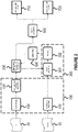
- Figure 1 provides a flow chart of a method of analyzing image data according to a general embodiment of the present invention.
- the illustrated method comprises obtaining 1 a first image 10 of a first part of an object and obtaining 2 a second image 20 of a second part of the object, the second part having substantial overlap with the first part.
- the images may be obtained from storage, or directly from an imaging device.
- the method further comprises obtaining a mapping 110 between the first image 10 and the second image 20.
- the mapping may be available from storage, or may be produced on the fly by applying a registration algorithm.
- the second image 20 is segmented 200 to obtain a segmentation, i.e. different elements (pixels or voxels) of the second image 20 are classified into various predefined categories.
- the segmentation information is transferred to the first image 10 on the basis of the aforementioned mapping.
- outliers are detected 300 on the basis of the segmentation; this step thus consists of identifying extreme intensity values of elements within one or more classes of elements.
- elements that correspond, according to the mapping, to at least some of the detected outliers of the first image 10 are replaced 500 with replacement values, so as to obtain a corrected second image.
- the replacement values are preferably average values of elements classified within the same class in non-outlier elements in proximity of the respective outliers.
- the segmentation is updated by performing the segmenting 200 on the corrected second image.
- the detecting 300, replacing 500 and the updating 200 are performed iteratively until a predetermined convergence criterion 550 is met.
- the convergence criterion is advantageously defined so as to stop the iteration at a point at which there no longer is a significant change in the tissue and the lesion segmentation relative to the previous iteration. Once this criterion is met, the final segmentation information is sent to a desired output.
- the present application more particularly discloses a method that measures the volumes of white matter, grey matter and cerebrospinal fluid in presence of white matter lesions based on two brain MR scans (a T1 weighted MR image and a FLAIR MR image). Moreover, the volumes of the present lesions and the parenchymal brain tissue are measured.
- the lesions may be due to a brain disease such as, for example, Multiple Sclerosis (MS).
- MS Multiple Sclerosis
- the volumes of white and grey matter are important biomarkers for neurodegenerative diseases, the lesion volume for neurological diseases involving focal lesions.
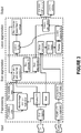
- the first step 100 will be referred to as "preprocessing", and consists of three stages:
- the algorithm models the intensities of each tissue class as a normal distribution, it assumes a Gaussian distributed bias field for the correction of intensity nonuniformities and it contains a spatial consistency model based on Markov Random Field (MRF).
- MRF Markov Random Field
- the algorithm iteratively estimates the parameters of each tissue class, as well as the bias field parameters, and maintains the spatial consistency until convergence.
- the T1 image 20 is bias corrected and segmented into the three tissue classes, i.e., GM, WM and CSF.
- the three tissue class segmentations from the previous step 200, together with the co-registered FLAIR image 10 of the same patient, are used to estimate the outliers in the FLAIR image 10.
- This is performed using the same expectation maximization (EM) algorithm (Cardoso et al., 2011), as described in the second step 200, but now an outlier map is included and the segmentations of the T1 image 20 are used as prior information.
- EM expectation maximization
- the algorithm iteratively estimates the outlier belief map and, after convergence, an outlier belief image is produced.
- This outlier belief image is an initialization for the MS lesions segmentation.
- the lesions are estimated from the outlier map as not every outlier is a lesion (e.g. the outlier map might include partial volume effects, artifacts, etc.).
- the outlier map is masked.
- the mask is a subset of the brain mask where only intensities that are hyper intense towards GM are included.
- the outliers need to be within WM (a mask is used to exclude GM and CSF outliers as well as regions that commonly show artifacts).
- each outlier needs to have a minimum number of voxels.
- this lesion segmentation is then used to fill in the lesions in the bias corrected T1 image 20 with their neighborhood WM intensities.
- steps 2 ( 200 ), 3 ( 301 ), 4 ( 302 ), and 5 ( 500 ) are repeated until there is no significant change in the tissue and the lesion segmentation.
- the idea of repeating the second and the third step is that the lesions are primarily WM, therefore, the T1 lesion filling will result in better brain tissues segmentation, which in turn results in better segmentation of lesions.
- lesions are recovered from the GM in case the outlier belief is high. These lesions are added to the previously found lesions and then these final segmented lesions from the FLAIR 10 are dilated to better approach the volume of the lesions. Subsequently the T1 weighted image 20 is filled one more time and segmented, providing again the final segmentations of WM, GM and CSF.
- the volumes and count of the lesions is determined within different anatomical regions 600, for example by transforming the region labels from an anatomical atlas. This leads to final estimated values of WM, GM, and CSF volume 710, and a lesion count and volume 720.
- the present invention also pertains to a system comprising an image processor configured to carry out the methods described above.
- the image processor may be implemented in dedicated hardware (e.g., ASIC), configurable hardware (e.g., FPGA), programmable components (e.g., a DSP or general purpose processor with appropriate software), or any combination thereof.
- the same component(s) may also include other functions.
- the system 1000 comprises an input interface 1030 adapted to receive the input images to be used from an image acquisition apparatus or a storage element. Without loss of generality, two storage elements 1010, 1020 are illustrated, representing respective storage devices for the FLAIR image 10 and the T1 image 20.
- the system 1000 further comprises a processor 1040, operatively connected to the input interface 1030 and an output interface 1060.
- the processor 1040 is capable of running a program stored in memory 1050, which is configured to cause the processor 1040 to carry out the method according to the present invention.
- the term "interface" is used to designate the combination of hardware and software or firmware required to allow an exchange of data between the processor 1040 and the components providing or receiving the corresponding data.
- the input interface 1030 and the output interface 1060 may share common hardware.
- the interface may be a local area network (LAN) interface, such as an interface according to the IEEE 802.3 "Ethernet" standard, on which appropriate network and transport protocols are implemented, such as a TCP/IP stack.
- the interfaces may provide access to a storage area network (SAN) or network attached storage (NAS), which is intended to store the input images to be used and/or the result of the analysis.
- SAN storage area network
- NAS network attached storage
- the interfaces may provide access to a wide area network (WAN) such as the Internet, which includes other computers that provide the input images to be used and/or retrieve the result of the analysis.
- WAN wide area network
- the present invention also pertains to a computer program product comprising code means configured to cause a processor configured to carry out the methods described above.
- the computer program product may comprise a computer readable medium, such as a magnetic tape, a magnetic disc, an optical disc, a semiconductor memory, or the like, having the code means stored thereon.
Landscapes
- Engineering & Computer Science (AREA)
- Physics & Mathematics (AREA)
- Theoretical Computer Science (AREA)
- General Physics & Mathematics (AREA)
- Computer Vision & Pattern Recognition (AREA)
- Software Systems (AREA)
- Probability & Statistics with Applications (AREA)
- Data Mining & Analysis (AREA)
- Radiology & Medical Imaging (AREA)
- Quality & Reliability (AREA)
- Health & Medical Sciences (AREA)
- Nuclear Medicine, Radiotherapy & Molecular Imaging (AREA)
- Geometry (AREA)
- General Health & Medical Sciences (AREA)
- Medical Informatics (AREA)
- Bioinformatics & Computational Biology (AREA)
- Evolutionary Biology (AREA)
- Evolutionary Computation (AREA)
- General Engineering & Computer Science (AREA)
- Bioinformatics & Cheminformatics (AREA)
- Artificial Intelligence (AREA)
- Life Sciences & Earth Sciences (AREA)
- Magnetic Resonance Imaging Apparatus (AREA)
Description
- The present invention pertains to the field of digital image analysis, in particular to the analysis of medical images.
- In many medical disciplines, diagnosis and follow-up of diseases relies to an ever increasing degree on the interpretation of data obtained by medical imaging techniques. In the field of neurology, magnetic resonance (MR) imaging has become particularly important, as it has specific advantages over X-ray based computerized tomography (CT) in certain situations.
- In particular, the (estimated) volume of white matter lesions is used as a biomarker for some neurological diseases, in particular multiple sclerosis (MS). In past years, it has been attempted to automate the detection of such lesions on the basis of MR images.
- In the article Automated Segmentation of Multiple Sclerosis Lesions by Model Outlier Detection (IEEE TRANSACTIONS ON MEDICAL IMAGING, VOL. 20, NO. 8, AUGUST 2001), K. VAN LEEMPUT et al. present an algorithm for segmentation of multiple sclerosis lesions from multispectral magnetic resonance images. In the method described in that article, MS lesions are detected as outliers with respect to a statistical model for normal brain tissue intensities in MR images.
- In the article An Automatic Segmentation of T2-FLAIR Multiple Sclerosis Lesions (The MIDAS Journal - MS Lesion Segmentation (MICCAI 2008 Workshop)), JC. SOUPLET et al. present a method designed to detect a hyperintense signal area on a T2-FLAIR sequence. The disclosed algorithm uses three conventional MRI sequences: T1, T2 and T2-FLAIR. First, images are cropped, spatially unbiased and skull-stripped. A segmentation of the brain into its different compartments is performed on the T1 and the T2 sequences. From these segmentations, a threshold for the T2-FLAIR sequence is automatically computed. Then post-processing operations select the most plausible lesions in the obtained hyperintense signals.
In the article Evaluating and Reducing the Impact of White Matter Lesions on Brain Volume Measurements (Human Brain Mapping Volume 33, Issue 9, 2011), M. BATTAGLINI et al. describe how the presence of white matter lesions affects certain segmentation-based brain volume measurements. The article indicates that refilling the lesions with intensities matching the surrounding normal-appearing white matter ensured accurate tissue-class measurements and thus represents a promising approach for accurate tissue classification and brain volume measurements.
The article "White matter lesion extension to automatic brain tissue segmentation on MRI", DE BOER R ET AL., 2009, discloses a method for segmenting brain lesion based on co-registered T1 and FLAIR images. The T1 image is first segmented into white matter (WM), gray matter (GM) and cerebro-spinal fluid (CSF). The result of this segmentation is transferred into the FLAIR image to detect outliers by thresholding.
There is a need for an automated way to more accurately and reliably estimate the respective volumes of white matter, gray matter, and/or cerebrospinal fluid (CSF), in the presence of lesions, in particular white matter lesions. - According to an aspect of the present invention, there is provided a method of analyzing image data, in accordance with
claim 1. - It is an advantage of the method according to the present invention that it judiciously combines information from different types of images to extract additional information that would be extremely difficult to discern for a human observer. The invention is based inter alia on the insight of the inventors that different types of images may be more suitable for segmentation and outlier detection, respectively. The invention is further based on an insight of the inventors that different respective types of information obtained from both types of images may advantageously be combined if a mapping between two images is available, thus resulting in better segmentation and better outlier detection. The invention is further based on an insight of the inventors that this combining of information may be performed iteratively to obtain any desired balance between time consumption and accuracy of the results.
The first image and the second image may be two-dimensional or three-dimensional images, whereby the elements of the images are pixels or voxels, respectively. The object of the images may comprise biological tissues or organs, in particular tissues of the central nervous system, more in particular brain tissue. The images may have been acquired by a medical imaging technique such as magnetic resonance imaging. - In an embodiment of the method according to the present invention, the outliers are detected by segmenting the first image starting from the segmentation of the second image with an additional outlier class.
- It is an advantage of this embodiment that the outliers are efficiently detected by the same general process that provides segmentation.
- In an embodiment, the method according to the present invention further comprises initiating the segmenting by transferring a segmentation of an example to the second image.
- It is an advantage of this embodiment that the segmenting step can be carried out in a shorter time by using an exemplary segmentation, e.g. an atlas.
- In a particular embodiment, the obtaining of the mapping comprises performing a registration of the obtained first image to the obtained second image.
- In cases where the mapping is not available in advance, it may be obtained as part of a method according to the present invention by means of a registration step.
- In an embodiment of the method according to the present invention, the object comprises at least a part of a brain, the first image is a FLAIR image, the second image is a T1 image; the segmentation comprises a classification of elements of the T1 image as gray matter, white matter, or cerebro-spinal fluid; and the outliers are detected among elements of the first image that are classified as white matter.
- This embodiment applies the principles of the invention to neurological MRI images, in order to obtain diagnostically or therapeutically relevant segmentation results.
- It is a further advantage of this embodiment that the time required for image analysis with the automated method according to the present invention is much shorter than the time that would be required for manual analysis by human experts. Hence, the method of the present invention is particularly suited for processing the large numbers of images involved in clinical trials.
- In a particular embodiment, the replacement values are based on average values of elements classified as white matter in non-outlier elements in proximity of said respective outliers.
- The replacement values may be the actual average values of elements classified as white matter in non-outlier elements in proximity of said respective outliers, or values derived from these averages, for example by the addition of a noise term. The inventors have found that using average values, preferably with added noise, as replacement values produces very good results.
- In an embodiment, the method according to the present invention further comprises removing elements representing non-brain tissue from the second image.
- It is an advantage of this embodiment that the further stages of the image processing proceed in a more efficient way if the images don't contain any elements that are not relevant to the desired end result.
- In an embodiment of the method according to the present invention, the segmentation is used to calculate respective volumes or areas of gray matter, white matter, and cerebro-spinal fluid.
- These volume or area calculations are useful to detect anomalies or changes in the distribution of these types of matter in the brain.
- In an embodiment of the method according to the present invention, the outlier detection is used to calculate volumes or areas of the outliers.
- This volume or area calculation is useful to detect suspected anomalies such as lesions.
- In an embodiment, the method according to the present invention further comprises the calculation of volumes or areas in different anatomical regions.
- It is an advantage of this embodiment that medically relevant volume or area estimates can be obtained for different anatomical regions, anomalies of which may correspond to different medical conditions. The different anatomical regions may be defined according to divisions that are in general use in the field of neurology, such as for example the division of the brain into left and right hemispheres, and into a frontal lobe, a parietal lobe, a temporal lobe, and an occipital lobe. The different anatomical regions may be defined according to a division that it is preferentially used in the diagnosis and follow-up of a specific disorder, such as multiple sclerosis, for which an exemplary division may consist of a juxtacortical region, a periventricular region, and a posterior fossa. The skilled person will appreciate that other divisions into anatomical regions may be used according to the desired type of information.
- According to an aspect of the present invention, there is provided a computer program product comprising code means configured to cause a processor to carry out the method described above.
- According to an aspect of the present invention, there is provided an image processing system comprising: an input interface adapted to receive a first image of a first part of an object and a second image of a second part of the object, the second part having substantial overlap with the first part; a processor, operatively connected to the input interface; and an output interface, operatively connected to the processor and adapted to output results produced by the processor; and a memory, operatively connected to the processor and arranged to comprise code to be executed by the processor, the memory comprising code configured to cause the processor to carry out the method of any of the preceding claims using the first image and the second image as inputs.
- The technical effects and advantages of embodiments of the computer program product and the system according to the present invention correspond mutatis mutandis to those of the corresponding embodiments of the method according to the present invention.
- These and other technical aspects and advantages of embodiments of the present invention will now be described in more detail with reference to the accompanying drawings, in which:
-
Figure 1 provides a flow chart of a method according to an embodiment of the present invention; -
Figure 2 provides a flow chart of a method according to another embodiment of the present invention; -
Figure 3 provides a more detailed flow chart of the method represented inFigure 2 ; and -
Figure 4 schematically illustrates a system according to an embodiment of the present invention. -
Figure 1 provides a flow chart of a method of analyzing image data according to a general embodiment of the present invention. - The illustrated method comprises obtaining 1 a first image 10 of a first part of an object and obtaining 2 a second image 20 of a second part of the object, the second part having substantial overlap with the first part. The images may be obtained from storage, or directly from an imaging device. The method further comprises obtaining a
mapping 110 between the first image 10 and the second image 20. The mapping may be available from storage, or may be produced on the fly by applying a registration algorithm. - The second image 20 is segmented 200 to obtain a segmentation, i.e. different elements (pixels or voxels) of the second image 20 are classified into various predefined categories. The segmentation information is transferred to the first image 10 on the basis of the aforementioned mapping. In the first image 10, outliers are detected 300 on the basis of the segmentation; this step thus consists of identifying extreme intensity values of elements within one or more classes of elements. In the second image 20, elements that correspond, according to the mapping, to at least some of the detected outliers of the first image 10, are replaced 500 with replacement values, so as to obtain a corrected second image. The replacement values are preferably average values of elements classified within the same class in non-outlier elements in proximity of the respective outliers. The segmentation is updated by performing the segmenting 200 on the corrected second image. The detecting 300, replacing 500 and the updating 200 are performed iteratively until a
predetermined convergence criterion 550 is met. The convergence criterion is advantageously defined so as to stop the iteration at a point at which there no longer is a significant change in the tissue and the lesion segmentation relative to the previous iteration. Once this criterion is met, the final segmentation information is sent to a desired output. - The present application more particularly discloses a method that measures the volumes of white matter, grey matter and cerebrospinal fluid in presence of white matter lesions based on two brain MR scans (a T1 weighted MR image and a FLAIR MR image). Moreover, the volumes of the present lesions and the parenchymal brain tissue are measured.
- The lesions may be due to a brain disease such as, for example, Multiple Sclerosis (MS). The volumes of white and grey matter are important biomarkers for neurodegenerative diseases, the lesion volume for neurological diseases involving focal lesions.
A more particular embodiment of the method according to the present invention will now be described with reference toFigure 2 (general overview) andFigure 3 (detailed flow diagram). The interdependence of the various steps of the method is indicated by the arrows in the diagrams. The order in which the various steps of the method are described below is not intended to limit the invention, unless a particular temporal dependence is implicitly or explicitly given by the causal relationship between the steps. Certain steps will be described hereinafter from a functional point of view. The skilled person will be able to carry out these steps by turning to the relevant published literature, without having to resort to undue experimentation. The relevant literature includes in particular the following publications: - S. OURSELIN et al., Robust Registration of Multi-modal Images: Towards Real-Time Clinical Applications, Medical Image Computing and Computer-Assisted Intervention (MICCAI'02);
- M. MODAT et al., Fast free-form deformation using graphics processing units, Computer Methods and Programs in Biomedicine, Volume 98, Issue 3, Pages 278-284, June 2010;
- M. MODAT, Efficient dense non-rigid registration using the free-form deformation framework, Doctoral Thesis University College London, 2012;
- M. CARDOSO et al., Adaptive neonate segmentation, 2011 Medical Image Computing and Computer-Assisted Intervention (MICCAI' 11).
- The
first step 100 will be referred to as "preprocessing", and consists of three stages: - In the
first stage 110, the input FLAIR image 10 of the patient is rigidly co-registered with the input T1-weighted image 20 (Ourselin et al., 2002). - In the
second stage 120, the T1-weighted input image 20 is skull stripped classifying each voxel either as a brain region or a non-brain region based on the affine registration of a brain mask available from an atlas (MNI) using NiftyReg (Modat et al., 2010). The registration is performed using a multi resolution affine transformation, based on the Trimmed Least Square scheme and a block-matching approach (Ourselin et al., 2002), followed by a multi resolution non-rigid registration based on the Free-Form deformation (Modat et al., 2012). - In the
third stage 130, the probabilistic anatomical priors for gray matter (GM), white matter (WM) and cerebrospinal fluid (CSF), which are also available from the MNI brain atlas, are transferred to the T1-weighted image space using an affine registration (Ourselin et al., 2002) followed by a non-rigid registration (Modat et al., 2010). - In the
second step 200, the three probabilistic tissue priors mentioned above, together with the skull stripped T1 image 20, act as a prior knowledge to an expectation maximization (EM) algorithm (Cardoso et al., 2011). The algorithm models the intensities of each tissue class as a normal distribution, it assumes a Gaussian distributed bias field for the correction of intensity nonuniformities and it contains a spatial consistency model based on Markov Random Field (MRF). The algorithm iteratively estimates the parameters of each tissue class, as well as the bias field parameters, and maintains the spatial consistency until convergence. After the convergence of the EM algorithm, the T1 image 20 is bias corrected and segmented into the three tissue classes, i.e., GM, WM and CSF. - In the third step 301 (corresponding to a first part of
step 300 ofFigure 1 ), the three tissue class segmentations from theprevious step 200, together with the co-registered FLAIR image 10 of the same patient, are used to estimate the outliers in the FLAIR image 10. This is performed using the same expectation maximization (EM) algorithm (Cardoso et al., 2011), as described in thesecond step 200, but now an outlier map is included and the segmentations of the T1 image 20 are used as prior information. In other words, while modelling the intensities of each tissue class as a normal distribution, the deviation of each intensity from these normal distributions is also estimated and modelled as an outlier belief map. The algorithm iteratively estimates the outlier belief map and, after convergence, an outlier belief image is produced. This outlier belief image is an initialization for the MS lesions segmentation. - In the fourth step 302 (corresponding to a second part of
step 300 ofFigure 1 ), the lesions are estimated from the outlier map as not every outlier is a lesion (e.g. the outlier map might include partial volume effects, artifacts, etc.). In order to differentiate the lesions from such non-lesion outliers, some extra a priori information about the location and the appearance of the lesions needs to be incorporated. In particular, the outlier map is masked. The mask is a subset of the brain mask where only intensities that are hyper intense towards GM are included. Furthermore, the outliers need to be within WM (a mask is used to exclude GM and CSF outliers as well as regions that commonly show artifacts). Finally, each outlier needs to have a minimum number of voxels. - In a
fifth step 500, this lesion segmentation is then used to fill in the lesions in the bias corrected T1 image 20 with their neighborhood WM intensities. - Subsequently steps 2 (200), 3 (301), 4 (302), and 5 (500) are repeated until there is no significant change in the tissue and the lesion segmentation. The idea of repeating the second and the third step is that the lesions are primarily WM, therefore, the T1 lesion filling will result in better brain tissues segmentation, which in turn results in better segmentation of lesions.
- Optionally, after the last iteration, lesions are recovered from the GM in case the outlier belief is high. These lesions are added to the previously found lesions and then these final segmented lesions from the FLAIR 10 are dilated to better approach the volume of the lesions. Subsequently the T1 weighted image 20 is filled one more time and segmented, providing again the final segmentations of WM, GM and CSF.
- Optionally, the volumes and count of the lesions is determined within different
anatomical regions 600, for example by transforming the region labels from an anatomical atlas. This leads to final estimated values of WM, GM, andCSF volume 710, and a lesion count andvolume 720. - The present invention also pertains to a system comprising an image processor configured to carry out the methods described above. The image processor may be implemented in dedicated hardware (e.g., ASIC), configurable hardware (e.g., FPGA), programmable components (e.g., a DSP or general purpose processor with appropriate software), or any combination thereof. The same component(s) may also include other functions.
- An exemplary design of such a system is schematically illustrated in
Figure 4 . Thesystem 1000 comprises aninput interface 1030 adapted to receive the input images to be used from an image acquisition apparatus or a storage element. Without loss of generality, twostorage elements system 1000 further comprises aprocessor 1040, operatively connected to theinput interface 1030 and anoutput interface 1060. Theprocessor 1040 is capable of running a program stored inmemory 1050, which is configured to cause theprocessor 1040 to carry out the method according to the present invention.
The term "interface" is used to designate the combination of hardware and software or firmware required to allow an exchange of data between theprocessor 1040 and the components providing or receiving the corresponding data. Theinput interface 1030 and theoutput interface 1060 may share common hardware. In particular, the interface may be a local area network (LAN) interface, such as an interface according to the IEEE 802.3 "Ethernet" standard, on which appropriate network and transport protocols are implemented, such as a TCP/IP stack. The interfaces may provide access to a storage area network (SAN) or network attached storage (NAS), which is intended to store the input images to be used and/or the result of the analysis. The interfaces may provide access to a wide area network (WAN) such as the Internet, which includes other computers that provide the input images to be used and/or retrieve the result of the analysis.
The present invention also pertains to a computer program product comprising code means configured to cause a processor configured to carry out the methods described above. The computer program product may comprise a computer readable medium, such as a magnetic tape, a magnetic disc, an optical disc, a semiconductor memory, or the like, having the code means stored thereon.
While the invention has been described hereinabove with reference to specific embodiments, this was done to clarify and not to limit the invention. The skilled person will appreciate that various modifications and different combinations of disclosed features are possible without departing from the scope of the invention as defined by the claims.
Claims (12)
- A method of analyzing image data, the method comprising:- obtaining (1) a first image (10) of a first part of an object;- obtaining (2) a second image (20) of a second part of the object, said second part having substantial overlap with said first part;- obtaining a mapping (110) between said first image (10) and said second image (20);- segmenting (200) said second image (20) to obtain a tissue segmentation;- detecting outliers (300) in said first image (10) by identifying extreme intensity values of elements within one or more classes of elements on the basis of said segmentation;- replacing (500) elements of said second image (20) that correspond, according to said mapping, to at least some of said detected outliers of said first image (10), with replacement values, so as to obtain a corrected second image, the at least some of said detected outliers of said first image being a lesion segmentation; and- updating said segmentation by performing said segmenting (200) on said corrected second image;wherein said detecting outliers (300), said replacing (500), and said updating (200) are performed iteratively until a predetermined convergence criterion (550) is met, said predetermined convergence criterion representing a point at which there is no significant change in the tissue and the lesion segmentation.
- The method according to claim 1, wherein said outliers are detected (300) by segmenting said first image (10) starting from said segmentation (200) of said second image (20) with an additional outlier class.
- The method according to any of the preceding claims, further comprising initiating said segmenting (200) by transferring (130) a segmentation of an example to said second image (20).
- The method according to any of the preceding claims, wherein said obtaining of said mapping (110) comprises performing a registration of said obtained first image (10) to said obtained second image (20).
- The method according to any of the preceding claims,
wherein said object comprises at least a part of a brain, said first image (10) is a FLAIR image, said second image (20) is a T1 image;
wherein said segmentation comprises a classification of elements of said T1 image (20) as gray matter, white matter, or cerebro-spinal fluid; and
wherein said outliers are detected (300) among elements of said first image (10) that are classified as white matter. - The method according to claim 5, wherein said replacement values are based on average values of elements classified as white matter in non-outlier elements in proximity of said respective outliers.
- The method according to any of claims 5 to 6, further comprising removing (120) elements representing non-brain tissue from said second image (20).
- The method according to any of claims 5 to 7, wherein said segmentation (200) is used to calculate (600) respective volumes or areas of gray matter, white matter, and cerebro-spinal fluid (710).
- The method according to any of claims 5 to 8, wherein said outlier detection (300) is used to calculate (600) volumes or areas of said outliers (720).
- The method according to any of claims 8 to 9, further comprising the calculation (600) of volumes or areas in different anatomical regions.
- A computer program product comprising code means configured to cause a processor to carry out the method according to any of the preceding claims.
- An image processing system (1000) comprising:- an input interface (1030) adapted to receive a first image (10) of a first part of an object and a second image (20) of a second part of said object, said second part having substantial overlap with said first part ;- a processor (1040), operatively connected to said input interface (1030); and- an output interface (1060), operatively connected to said processor (1040) and adapted to output results produced by said processor (1040); and- a memory (1050), operatively connected to said processor (1040) and arranged to comprise code to be executed by said processor (1040), said memory (1050) comprising code configured to cause said processor (1040) to carry out the method of any of claims 1-10 using said first image (10) and said second image (20) as inputs.
Priority Applications (4)
| Application Number | Priority Date | Filing Date | Title |
|---|---|---|---|
| ES14184117.1T ES2685977T3 (en) | 2014-09-09 | 2014-09-09 | Method and system to analyze image data |
| EP14184117.1A EP2996085B1 (en) | 2014-09-09 | 2014-09-09 | Method and system for analyzing image data |
| US15/510,151 US10198815B2 (en) | 2014-09-09 | 2015-09-09 | Method and system for analyzing image data |
| PCT/EP2015/070663 WO2016038118A1 (en) | 2014-09-09 | 2015-09-09 | Method and system for analyzing image data |
Applications Claiming Priority (1)
| Application Number | Priority Date | Filing Date | Title |
|---|---|---|---|
| EP14184117.1A EP2996085B1 (en) | 2014-09-09 | 2014-09-09 | Method and system for analyzing image data |
Publications (2)
| Publication Number | Publication Date |
|---|---|
| EP2996085A1 EP2996085A1 (en) | 2016-03-16 |
| EP2996085B1 true EP2996085B1 (en) | 2018-05-23 |
Family
ID=51542156
Family Applications (1)
| Application Number | Title | Priority Date | Filing Date |
|---|---|---|---|
| EP14184117.1A Active EP2996085B1 (en) | 2014-09-09 | 2014-09-09 | Method and system for analyzing image data |
Country Status (4)
| Country | Link |
|---|---|
| US (1) | US10198815B2 (en) |
| EP (1) | EP2996085B1 (en) |
| ES (1) | ES2685977T3 (en) |
| WO (1) | WO2016038118A1 (en) |
Families Citing this family (20)
| Publication number | Priority date | Publication date | Assignee | Title |
|---|---|---|---|---|
| US10635894B1 (en) * | 2016-10-13 | 2020-04-28 | T Stamp Inc. | Systems and methods for passive-subject liveness verification in digital media |
| US11373449B1 (en) * | 2016-10-13 | 2022-06-28 | T Stamp Inc. | Systems and methods for passive-subject liveness verification in digital media |
| CN107025457B (en) * | 2017-03-29 | 2022-03-08 | 腾讯科技(深圳)有限公司 | Image processing method and device |
| US10474667B2 (en) * | 2017-07-29 | 2019-11-12 | Vmware, Inc | Methods and systems to detect and correct outliers in a dataset stored in a data-storage device |
| US11885861B2 (en) * | 2018-02-26 | 2024-01-30 | The Board Of Regents Of The University Of Texas System | 3-dimensional representations of post-contrast enhanced brain lesions |
| US11093771B1 (en) | 2018-05-04 | 2021-08-17 | T Stamp Inc. | Systems and methods for liveness-verified, biometric-based encryption |
| US11496315B1 (en) | 2018-05-08 | 2022-11-08 | T Stamp Inc. | Systems and methods for enhanced hash transforms |
| US11301586B1 (en) | 2019-04-05 | 2022-04-12 | T Stamp Inc. | Systems and processes for lossy biometric representations |
| CN110047046B (en) * | 2019-04-15 | 2022-09-06 | 南京邮电大学 | Image segmentation and offset field correction method, readable storage medium and terminal |
| AU2020355094A1 (en) * | 2019-09-24 | 2022-04-14 | The Board Of Regents Of The University Of Texas System | Methods and systems for analyzing brain lesions with longitudinal 3D MRI data |
| US12513160B1 (en) | 2020-04-14 | 2025-12-30 | T Stamp Inc. | Systems and processes for multifactor authentication and identification |
| US11967173B1 (en) | 2020-05-19 | 2024-04-23 | T Stamp Inc. | Face cover-compatible biometrics and processes for generating and using same |
| CN112308853A (en) * | 2020-10-20 | 2021-02-02 | 平安科技(深圳)有限公司 | Electronic equipment, medical image index generation method and device and storage medium |
| US11610306B2 (en) | 2020-12-16 | 2023-03-21 | Industrial Technology Research Institute | Medical image analysis method and device |
| JP7338902B2 (en) * | 2020-12-30 | 2023-09-05 | ニューロフェット インコーポレイテッド | : Method for providing auxiliary diagnostic information and apparatus for executing it |
| US12079371B1 (en) | 2021-04-13 | 2024-09-03 | T Stamp Inc. | Personal identifiable information encoder |
| US12315294B1 (en) | 2021-04-21 | 2025-05-27 | T Stamp Inc. | Interoperable biometric representation |
| CN113256654B (en) * | 2021-05-26 | 2023-09-12 | 西安交通大学 | A method and system for white matter hyperintensity extraction based on lesion diffusion algorithm |
| US12353530B1 (en) | 2021-12-08 | 2025-07-08 | T Stamp Inc. | Shape overlay for proof of liveness |
| CN120654988B (en) * | 2025-05-15 | 2025-12-09 | 中国矿业大学(北京) | Energy demand analysis method and system |
Family Cites Families (2)
| Publication number | Priority date | Publication date | Assignee | Title |
|---|---|---|---|---|
| US5983251A (en) * | 1993-09-08 | 1999-11-09 | Idt, Inc. | Method and apparatus for data analysis |
| US20160034638A1 (en) * | 2013-03-14 | 2016-02-04 | University Of Rochester | System and Method for Detecting Population Variation from Nucleic Acid Sequencing Data |
-
2014
- 2014-09-09 EP EP14184117.1A patent/EP2996085B1/en active Active
- 2014-09-09 ES ES14184117.1T patent/ES2685977T3/en active Active
-
2015
- 2015-09-09 WO PCT/EP2015/070663 patent/WO2016038118A1/en not_active Ceased
- 2015-09-09 US US15/510,151 patent/US10198815B2/en active Active
Non-Patent Citations (1)
| Title |
|---|
| None * |
Also Published As
| Publication number | Publication date |
|---|---|
| US10198815B2 (en) | 2019-02-05 |
| US20170256056A1 (en) | 2017-09-07 |
| ES2685977T3 (en) | 2018-10-15 |
| EP2996085A1 (en) | 2016-03-16 |
| WO2016038118A1 (en) | 2016-03-17 |
Similar Documents
| Publication | Publication Date | Title |
|---|---|---|
| US10198815B2 (en) | Method and system for analyzing image data | |
| US11379985B2 (en) | System and computer-implemented method for segmenting an image | |
| Nabizadeh et al. | Histogram-based gravitational optimization algorithm on single MR modality for automatic brain lesion detection and segmentation | |
| EP2545527B1 (en) | Probabilistic refinement of model-based segmentation | |
| CN109886944B (en) | A method for detecting and localizing white matter hyperintensities based on multi-atlas | |
| Saritha et al. | A comprehensive review: Segmentation of MRI images—brain tumor | |
| JP2016007270A (en) | Medical image processor | |
| WO2016134125A1 (en) | Image segmentation via multi-atlas fusion with context learning | |
| EP3289563A1 (en) | Brain tissue classification | |
| Pedoia et al. | Glial brain tumor detection by using symmetry analysis | |
| Verma et al. | Superpixels in brain MR image analysis | |
| Bhatia et al. | Similarity metrics for groupwise non-rigid registration | |
| Hameeteman et al. | Carotid wall volume quantification from magnetic resonance images using deformable model fitting and learning-based correction of systematic errors | |
| Mori et al. | Medical Image Computing and Computer-Assisted Intervention--MICCAI 2013: 16th International Conference, Nagoya, Japan, September 22-26, 2013, Proceedings, Part III | |
| Srhoj-Egekher et al. | Automatic neonatal brain tissue segmentation with MRI | |
| Asman et al. | Robust GM/WM segmentation of the spinal cord with iterative non-local statistical fusion | |
| CA2650075A1 (en) | A method, a system, a computer program product and a user interface for segmenting image sets | |
| Caldairou et al. | Segmentation of the cortex in fetal MRI using a topological model | |
| Myint et al. | Effective kidney segmentation using gradient based approach in abdominal CT images | |
| del Toro et al. | Hierarchical multi–structure segmentation guided by anatomical correlations | |
| Gan | Automated segmentation of the lateral ventricle in MR images of human brain | |
| Sarikaya et al. | Multi-atlas brain MRI segmentation with multiway cut | |
| Licandro et al. | A longitudinal diffeomorphic atlas-based tissue labeling framework for fetal brains using geodesic regression | |
| Öziç et al. | Atlas-Based Segmentation Pipelines on 3D Brain MR Images: A Preliminary Study | |
| Xue et al. | CLASSIC: consistent longitudinal alignment and segmentation for serial image computing |
Legal Events
| Date | Code | Title | Description |
|---|---|---|---|
| PUAI | Public reference made under article 153(3) epc to a published international application that has entered the european phase |
Free format text: ORIGINAL CODE: 0009012 |
|
| AK | Designated contracting states |
Kind code of ref document: A1 Designated state(s): AL AT BE BG CH CY CZ DE DK EE ES FI FR GB GR HR HU IE IS IT LI LT LU LV MC MK MT NL NO PL PT RO RS SE SI SK SM TR |
|
| AX | Request for extension of the european patent |
Extension state: BA ME |
|
| 17P | Request for examination filed |
Effective date: 20160901 |
|
| RBV | Designated contracting states (corrected) |
Designated state(s): AL AT BE BG CH CY CZ DE DK EE ES FI FR GB GR HR HU IE IS IT LI LT LU LV MC MK MT NL NO PL PT RO RS SE SI SK SM TR |
|
| REG | Reference to a national code |
Ref country code: DE Ref legal event code: R079 Ref document number: 602014025786 Country of ref document: DE Free format text: PREVIOUS MAIN CLASS: G06T0007000000 Ipc: G06T0007110000 |
|
| GRAP | Despatch of communication of intention to grant a patent |
Free format text: ORIGINAL CODE: EPIDOSNIGR1 |
|
| STAA | Information on the status of an ep patent application or granted ep patent |
Free format text: STATUS: GRANT OF PATENT IS INTENDED |
|
| RIC1 | Information provided on ipc code assigned before grant |
Ipc: G06T 7/143 20170101ALI20170203BHEP Ipc: G06T 7/174 20170101ALI20170203BHEP Ipc: G06T 7/11 20170101AFI20170203BHEP |
|
| INTG | Intention to grant announced |
Effective date: 20170223 |
|
| GRAJ | Information related to disapproval of communication of intention to grant by the applicant or resumption of examination proceedings by the epo deleted |
Free format text: ORIGINAL CODE: EPIDOSDIGR1 |
|
| GRAJ | Information related to disapproval of communication of intention to grant by the applicant or resumption of examination proceedings by the epo deleted |
Free format text: ORIGINAL CODE: EPIDOSDIGR1 |
|
| STAA | Information on the status of an ep patent application or granted ep patent |
Free format text: STATUS: REQUEST FOR EXAMINATION WAS MADE |
|
| INTG | Intention to grant announced |
Effective date: 20170223 |
|
| GRAP | Despatch of communication of intention to grant a patent |
Free format text: ORIGINAL CODE: EPIDOSNIGR1 |
|
| STAA | Information on the status of an ep patent application or granted ep patent |
Free format text: STATUS: GRANT OF PATENT IS INTENDED |
|
| INTC | Intention to grant announced (deleted) | ||
| INTG | Intention to grant announced |
Effective date: 20171201 |
|
| RAP1 | Party data changed (applicant data changed or rights of an application transferred) |
Owner name: ICOMETRIX NV |
|
| GRAS | Grant fee paid |
Free format text: ORIGINAL CODE: EPIDOSNIGR3 |
|
| GRAA | (expected) grant |
Free format text: ORIGINAL CODE: 0009210 |
|
| STAA | Information on the status of an ep patent application or granted ep patent |
Free format text: STATUS: THE PATENT HAS BEEN GRANTED |
|
| AK | Designated contracting states |
Kind code of ref document: B1 Designated state(s): AL AT BE BG CH CY CZ DE DK EE ES FI FR GB GR HR HU IE IS IT LI LT LU LV MC MK MT NL NO PL PT RO RS SE SI SK SM TR |
|
| REG | Reference to a national code |
Ref country code: GB Ref legal event code: FG4D |
|
| REG | Reference to a national code |
Ref country code: CH Ref legal event code: EP |
|
| REG | Reference to a national code |
Ref country code: IE Ref legal event code: FG4D |
|
| REG | Reference to a national code |
Ref country code: AT Ref legal event code: REF Ref document number: 1002110 Country of ref document: AT Kind code of ref document: T Effective date: 20180615 |
|
| REG | Reference to a national code |
Ref country code: DE Ref legal event code: R096 Ref document number: 602014025786 Country of ref document: DE |
|
| REG | Reference to a national code |
Ref country code: NL Ref legal event code: FP |
|
| REG | Reference to a national code |
Ref country code: SE Ref legal event code: TRGR |
|
| REG | Reference to a national code |
Ref country code: FR Ref legal event code: PLFP Year of fee payment: 5 |
|
| REG | Reference to a national code |
Ref country code: LT Ref legal event code: MG4D |
|
| REG | Reference to a national code |
Ref country code: ES Ref legal event code: FG2A Ref document number: 2685977 Country of ref document: ES Kind code of ref document: T3 Effective date: 20181015 |
|
| PG25 | Lapsed in a contracting state [announced via postgrant information from national office to epo] |
Ref country code: NO Free format text: LAPSE BECAUSE OF FAILURE TO SUBMIT A TRANSLATION OF THE DESCRIPTION OR TO PAY THE FEE WITHIN THE PRESCRIBED TIME-LIMIT Effective date: 20180823 Ref country code: FI Free format text: LAPSE BECAUSE OF FAILURE TO SUBMIT A TRANSLATION OF THE DESCRIPTION OR TO PAY THE FEE WITHIN THE PRESCRIBED TIME-LIMIT Effective date: 20180523 Ref country code: BG Free format text: LAPSE BECAUSE OF FAILURE TO SUBMIT A TRANSLATION OF THE DESCRIPTION OR TO PAY THE FEE WITHIN THE PRESCRIBED TIME-LIMIT Effective date: 20180823 Ref country code: LT Free format text: LAPSE BECAUSE OF FAILURE TO SUBMIT A TRANSLATION OF THE DESCRIPTION OR TO PAY THE FEE WITHIN THE PRESCRIBED TIME-LIMIT Effective date: 20180523 |
|
| PG25 | Lapsed in a contracting state [announced via postgrant information from national office to epo] |
Ref country code: LV Free format text: LAPSE BECAUSE OF FAILURE TO SUBMIT A TRANSLATION OF THE DESCRIPTION OR TO PAY THE FEE WITHIN THE PRESCRIBED TIME-LIMIT Effective date: 20180523 Ref country code: GR Free format text: LAPSE BECAUSE OF FAILURE TO SUBMIT A TRANSLATION OF THE DESCRIPTION OR TO PAY THE FEE WITHIN THE PRESCRIBED TIME-LIMIT Effective date: 20180824 Ref country code: HR Free format text: LAPSE BECAUSE OF FAILURE TO SUBMIT A TRANSLATION OF THE DESCRIPTION OR TO PAY THE FEE WITHIN THE PRESCRIBED TIME-LIMIT Effective date: 20180523 Ref country code: RS Free format text: LAPSE BECAUSE OF FAILURE TO SUBMIT A TRANSLATION OF THE DESCRIPTION OR TO PAY THE FEE WITHIN THE PRESCRIBED TIME-LIMIT Effective date: 20180523 |
|
| REG | Reference to a national code |
Ref country code: CH Ref legal event code: PK Free format text: BERICHTIGUNGEN |
|
| REG | Reference to a national code |
Ref country code: AT Ref legal event code: MK05 Ref document number: 1002110 Country of ref document: AT Kind code of ref document: T Effective date: 20180523 |
|
| RIC2 | Information provided on ipc code assigned after grant |
Ipc: G06T 7/11 20170101AFI20170203BHEP Ipc: G06T 7/174 20170101ALI20170203BHEP Ipc: G06T 7/143 20170101ALI20170203BHEP |
|
| PG25 | Lapsed in a contracting state [announced via postgrant information from national office to epo] |
Ref country code: DK Free format text: LAPSE BECAUSE OF FAILURE TO SUBMIT A TRANSLATION OF THE DESCRIPTION OR TO PAY THE FEE WITHIN THE PRESCRIBED TIME-LIMIT Effective date: 20180523 Ref country code: PL Free format text: LAPSE BECAUSE OF FAILURE TO SUBMIT A TRANSLATION OF THE DESCRIPTION OR TO PAY THE FEE WITHIN THE PRESCRIBED TIME-LIMIT Effective date: 20180523 Ref country code: AT Free format text: LAPSE BECAUSE OF FAILURE TO SUBMIT A TRANSLATION OF THE DESCRIPTION OR TO PAY THE FEE WITHIN THE PRESCRIBED TIME-LIMIT Effective date: 20180523 Ref country code: EE Free format text: LAPSE BECAUSE OF FAILURE TO SUBMIT A TRANSLATION OF THE DESCRIPTION OR TO PAY THE FEE WITHIN THE PRESCRIBED TIME-LIMIT Effective date: 20180523 Ref country code: RO Free format text: LAPSE BECAUSE OF FAILURE TO SUBMIT A TRANSLATION OF THE DESCRIPTION OR TO PAY THE FEE WITHIN THE PRESCRIBED TIME-LIMIT Effective date: 20180523 Ref country code: CZ Free format text: LAPSE BECAUSE OF FAILURE TO SUBMIT A TRANSLATION OF THE DESCRIPTION OR TO PAY THE FEE WITHIN THE PRESCRIBED TIME-LIMIT Effective date: 20180523 Ref country code: SK Free format text: LAPSE BECAUSE OF FAILURE TO SUBMIT A TRANSLATION OF THE DESCRIPTION OR TO PAY THE FEE WITHIN THE PRESCRIBED TIME-LIMIT Effective date: 20180523 |
|
| REG | Reference to a national code |
Ref country code: CH Ref legal event code: PK Free format text: BERICHTIGUNGEN |
|
| RIC2 | Information provided on ipc code assigned after grant |
Ipc: G06T 7/174 20170101ALI20170203BHEP Ipc: G06T 7/11 20170101AFI20170203BHEP Ipc: G06T 7/143 20170101ALI20170203BHEP |
|
| REG | Reference to a national code |
Ref country code: DE Ref legal event code: R097 Ref document number: 602014025786 Country of ref document: DE |
|
| PG25 | Lapsed in a contracting state [announced via postgrant information from national office to epo] |
Ref country code: SM Free format text: LAPSE BECAUSE OF FAILURE TO SUBMIT A TRANSLATION OF THE DESCRIPTION OR TO PAY THE FEE WITHIN THE PRESCRIBED TIME-LIMIT Effective date: 20180523 Ref country code: IT Free format text: LAPSE BECAUSE OF FAILURE TO SUBMIT A TRANSLATION OF THE DESCRIPTION OR TO PAY THE FEE WITHIN THE PRESCRIBED TIME-LIMIT Effective date: 20180523 |
|
| PLBE | No opposition filed within time limit |
Free format text: ORIGINAL CODE: 0009261 |
|
| STAA | Information on the status of an ep patent application or granted ep patent |
Free format text: STATUS: NO OPPOSITION FILED WITHIN TIME LIMIT |
|
| PG25 | Lapsed in a contracting state [announced via postgrant information from national office to epo] |
Ref country code: MC Free format text: LAPSE BECAUSE OF FAILURE TO SUBMIT A TRANSLATION OF THE DESCRIPTION OR TO PAY THE FEE WITHIN THE PRESCRIBED TIME-LIMIT Effective date: 20180523 |
|
| 26N | No opposition filed |
Effective date: 20190226 |
|
| PG25 | Lapsed in a contracting state [announced via postgrant information from national office to epo] |
Ref country code: SI Free format text: LAPSE BECAUSE OF FAILURE TO SUBMIT A TRANSLATION OF THE DESCRIPTION OR TO PAY THE FEE WITHIN THE PRESCRIBED TIME-LIMIT Effective date: 20180523 |
|
| PG25 | Lapsed in a contracting state [announced via postgrant information from national office to epo] |
Ref country code: LU Free format text: LAPSE BECAUSE OF NON-PAYMENT OF DUE FEES Effective date: 20180909 |
|
| PG25 | Lapsed in a contracting state [announced via postgrant information from national office to epo] |
Ref country code: AL Free format text: LAPSE BECAUSE OF FAILURE TO SUBMIT A TRANSLATION OF THE DESCRIPTION OR TO PAY THE FEE WITHIN THE PRESCRIBED TIME-LIMIT Effective date: 20180523 |
|
| PG25 | Lapsed in a contracting state [announced via postgrant information from national office to epo] |
Ref country code: MT Free format text: LAPSE BECAUSE OF NON-PAYMENT OF DUE FEES Effective date: 20180909 |
|
| PG25 | Lapsed in a contracting state [announced via postgrant information from national office to epo] |
Ref country code: TR Free format text: LAPSE BECAUSE OF FAILURE TO SUBMIT A TRANSLATION OF THE DESCRIPTION OR TO PAY THE FEE WITHIN THE PRESCRIBED TIME-LIMIT Effective date: 20180523 |
|
| PG25 | Lapsed in a contracting state [announced via postgrant information from national office to epo] |
Ref country code: PT Free format text: LAPSE BECAUSE OF FAILURE TO SUBMIT A TRANSLATION OF THE DESCRIPTION OR TO PAY THE FEE WITHIN THE PRESCRIBED TIME-LIMIT Effective date: 20180523 |
|
| PG25 | Lapsed in a contracting state [announced via postgrant information from national office to epo] |
Ref country code: MK Free format text: LAPSE BECAUSE OF NON-PAYMENT OF DUE FEES Effective date: 20180523 Ref country code: CY Free format text: LAPSE BECAUSE OF FAILURE TO SUBMIT A TRANSLATION OF THE DESCRIPTION OR TO PAY THE FEE WITHIN THE PRESCRIBED TIME-LIMIT Effective date: 20180523 Ref country code: HU Free format text: LAPSE BECAUSE OF FAILURE TO SUBMIT A TRANSLATION OF THE DESCRIPTION OR TO PAY THE FEE WITHIN THE PRESCRIBED TIME-LIMIT; INVALID AB INITIO Effective date: 20140909 |
|
| PG25 | Lapsed in a contracting state [announced via postgrant information from national office to epo] |
Ref country code: IS Free format text: LAPSE BECAUSE OF FAILURE TO SUBMIT A TRANSLATION OF THE DESCRIPTION OR TO PAY THE FEE WITHIN THE PRESCRIBED TIME-LIMIT Effective date: 20180923 |
|
| PGFP | Annual fee paid to national office [announced via postgrant information from national office to epo] |
Ref country code: NL Payment date: 20250827 Year of fee payment: 12 |
|
| REG | Reference to a national code |
Ref country code: CH Ref legal event code: U11 Free format text: ST27 STATUS EVENT CODE: U-0-0-U10-U11 (AS PROVIDED BY THE NATIONAL OFFICE) Effective date: 20251001 |
|
| PGFP | Annual fee paid to national office [announced via postgrant information from national office to epo] |
Ref country code: DE Payment date: 20250919 Year of fee payment: 12 |
|
| PGFP | Annual fee paid to national office [announced via postgrant information from national office to epo] |
Ref country code: GB Payment date: 20250919 Year of fee payment: 12 Ref country code: BE Payment date: 20250827 Year of fee payment: 12 |
|
| PGFP | Annual fee paid to national office [announced via postgrant information from national office to epo] |
Ref country code: FR Payment date: 20250922 Year of fee payment: 12 |
|
| PGFP | Annual fee paid to national office [announced via postgrant information from national office to epo] |
Ref country code: SE Payment date: 20250918 Year of fee payment: 12 |
|
| PGFP | Annual fee paid to national office [announced via postgrant information from national office to epo] |
Ref country code: IE Payment date: 20250918 Year of fee payment: 12 |
|
| PGFP | Annual fee paid to national office [announced via postgrant information from national office to epo] |
Ref country code: CH Payment date: 20251001 Year of fee payment: 12 |
|
| PGFP | Annual fee paid to national office [announced via postgrant information from national office to epo] |
Ref country code: ES Payment date: 20251028 Year of fee payment: 12 |