EP2994776B1 - Apparatus and method for the evaluation of gamma radiation events - Google Patents
Apparatus and method for the evaluation of gamma radiation events Download PDFInfo
- Publication number
- EP2994776B1 EP2994776B1 EP14720163.6A EP14720163A EP2994776B1 EP 2994776 B1 EP2994776 B1 EP 2994776B1 EP 14720163 A EP14720163 A EP 14720163A EP 2994776 B1 EP2994776 B1 EP 2994776B1
- Authority
- EP
- European Patent Office
- Prior art keywords
- events
- gamma radiation
- scintillator
- gamma
- photo conversion
- Prior art date
- Legal status (The legal status is an assumption and is not a legal conclusion. Google has not performed a legal analysis and makes no representation as to the accuracy of the status listed.)
- Active
Links
- 230000005855 radiation Effects 0.000 title claims description 95
- 238000000034 method Methods 0.000 title claims description 47
- 238000011156 evaluation Methods 0.000 title claims description 28
- 238000009826 distribution Methods 0.000 claims description 92
- 238000006243 chemical reaction Methods 0.000 claims description 55
- 239000013078 crystal Substances 0.000 claims description 51
- 238000012633 nuclear imaging Methods 0.000 claims description 28
- 238000007476 Maximum Likelihood Methods 0.000 claims description 25
- 230000004044 response Effects 0.000 claims description 22
- 238000000342 Monte Carlo simulation Methods 0.000 claims description 9
- 238000004590 computer program Methods 0.000 claims description 7
- 230000005251 gamma ray Effects 0.000 description 33
- 238000001228 spectrum Methods 0.000 description 17
- 230000008569 process Effects 0.000 description 9
- 238000002600 positron emission tomography Methods 0.000 description 8
- 238000010586 diagram Methods 0.000 description 7
- 238000011065 in-situ storage Methods 0.000 description 7
- 238000005259 measurement Methods 0.000 description 7
- 238000003491 array Methods 0.000 description 6
- 230000008878 coupling Effects 0.000 description 6
- 238000010168 coupling process Methods 0.000 description 6
- 238000005859 coupling reaction Methods 0.000 description 6
- 230000000694 effects Effects 0.000 description 6
- 238000001514 detection method Methods 0.000 description 5
- 238000002603 single-photon emission computed tomography Methods 0.000 description 5
- 238000010521 absorption reaction Methods 0.000 description 4
- 238000013459 approach Methods 0.000 description 4
- 238000002591 computed tomography Methods 0.000 description 4
- 230000008901 benefit Effects 0.000 description 3
- 230000001419 dependent effect Effects 0.000 description 3
- 238000009795 derivation Methods 0.000 description 3
- 230000005484 gravity Effects 0.000 description 3
- 238000003384 imaging method Methods 0.000 description 3
- 239000011159 matrix material Substances 0.000 description 3
- 238000012545 processing Methods 0.000 description 3
- AOYNUTHNTBLRMT-SLPGGIOYSA-N 2-deoxy-2-fluoro-aldehydo-D-glucose Chemical compound OC[C@@H](O)[C@@H](O)[C@H](O)[C@@H](F)C=O AOYNUTHNTBLRMT-SLPGGIOYSA-N 0.000 description 2
- XUIMIQQOPSSXEZ-UHFFFAOYSA-N Silicon Chemical compound [Si] XUIMIQQOPSSXEZ-UHFFFAOYSA-N 0.000 description 2
- 230000032683 aging Effects 0.000 description 2
- 238000004458 analytical method Methods 0.000 description 2
- 238000004364 calculation method Methods 0.000 description 2
- 230000000052 comparative effect Effects 0.000 description 2
- 238000002059 diagnostic imaging Methods 0.000 description 2
- 238000001914 filtration Methods 0.000 description 2
- 230000001678 irradiating effect Effects 0.000 description 2
- 230000004807 localization Effects 0.000 description 2
- 239000000463 material Substances 0.000 description 2
- 238000013421 nuclear magnetic resonance imaging Methods 0.000 description 2
- 231100000289 photo-effect Toxicity 0.000 description 2
- 230000002285 radioactive effect Effects 0.000 description 2
- 238000005070 sampling Methods 0.000 description 2
- 229910052710 silicon Inorganic materials 0.000 description 2
- 239000010703 silicon Substances 0.000 description 2
- 238000004088 simulation Methods 0.000 description 2
- 241001637516 Polygonia c-album Species 0.000 description 1
- 230000005266 beta plus decay Effects 0.000 description 1
- JJWKPURADFRFRB-UHFFFAOYSA-N carbonyl sulfide Chemical compound O=C=S JJWKPURADFRFRB-UHFFFAOYSA-N 0.000 description 1
- 230000015556 catabolic process Effects 0.000 description 1
- 238000012937 correction Methods 0.000 description 1
- 230000003247 decreasing effect Effects 0.000 description 1
- 238000006731 degradation reaction Methods 0.000 description 1
- 230000004069 differentiation Effects 0.000 description 1
- 238000000605 extraction Methods 0.000 description 1
- 230000006872 improvement Effects 0.000 description 1
- 238000002347 injection Methods 0.000 description 1
- 239000007924 injection Substances 0.000 description 1
- 230000010354 integration Effects 0.000 description 1
- 230000003993 interaction Effects 0.000 description 1
- 238000011835 investigation Methods 0.000 description 1
- RCVOTEPLGFCFIG-UHFFFAOYSA-N lutetium(3+);yttrium(3+);silicate Chemical compound [Y+3].[Lu+3].[O-][Si]([O-])([O-])[O-] RCVOTEPLGFCFIG-UHFFFAOYSA-N 0.000 description 1
- 238000002595 magnetic resonance imaging Methods 0.000 description 1
- 238000004519 manufacturing process Methods 0.000 description 1
- 230000003287 optical effect Effects 0.000 description 1
- 239000000941 radioactive substance Substances 0.000 description 1
- 230000009467 reduction Effects 0.000 description 1
- 230000000630 rising effect Effects 0.000 description 1
- 239000000523 sample Substances 0.000 description 1
- 238000013179 statistical model Methods 0.000 description 1
- 238000003860 storage Methods 0.000 description 1
- 230000002123 temporal effect Effects 0.000 description 1
- 238000012360 testing method Methods 0.000 description 1
Images






Classifications
-
- G—PHYSICS
- G01—MEASURING; TESTING
- G01T—MEASUREMENT OF NUCLEAR OR X-RADIATION
- G01T7/00—Details of radiation-measuring instruments
- G01T7/005—Details of radiation-measuring instruments calibration techniques
-
- G—PHYSICS
- G01—MEASURING; TESTING
- G01T—MEASUREMENT OF NUCLEAR OR X-RADIATION
- G01T1/00—Measuring X-radiation, gamma radiation, corpuscular radiation, or cosmic radiation
- G01T1/16—Measuring radiation intensity
- G01T1/161—Applications in the field of nuclear medicine, e.g. in vivo counting
- G01T1/164—Scintigraphy
- G01T1/1641—Static instruments for imaging the distribution of radioactivity in one or two dimensions using one or several scintillating elements; Radio-isotope cameras
- G01T1/1642—Static instruments for imaging the distribution of radioactivity in one or two dimensions using one or several scintillating elements; Radio-isotope cameras using a scintillation crystal and position sensing photodetector arrays, e.g. ANGER cameras
-
- G—PHYSICS
- G01—MEASURING; TESTING
- G01T—MEASUREMENT OF NUCLEAR OR X-RADIATION
- G01T1/00—Measuring X-radiation, gamma radiation, corpuscular radiation, or cosmic radiation
- G01T1/16—Measuring radiation intensity
- G01T1/20—Measuring radiation intensity with scintillation detectors
- G01T1/2018—Scintillation-photodiode combinations
Definitions
- the present invention relates to an evaluation apparatus, a nuclear imaging device and corresponding methods for the use in nuclear imaging, in particular for the use with PET and SPECT cameras.
- Gamma ray detection with gamma cameras for nuclear imaging is a multiple stage process.
- the incident gamma photon is converted to a scintillation light flash (i.e. a scintillation photon).
- each of the visible light photons from this flash (sometimes ultraviolet or other photons may be generated) is captured with a photodetector and a spatial signal distribution is obtained, which is then used to estimate the energy of the incident gamma photon and the two- or three-dimensional position of the gamma ray photo conversion.
- This determined position forms the basis for the estimation of a line of response (LOR) and the image reconstruction from a nuclear decay process.
- LOR line of response
- the photo conversion position i.e. the position where the gamma ray interacts with the scintillator and the light flash is created
- the photo conversion position may be extracted from the spatial distribution of the visible light photons.
- One possibility is to couple each element of the scintillator to a single photodetector element. All the light is collected by this single photodetector element and the position of the gamma ray impact is given by the position of the photodetector. This type of readout is generally called one-to-one coupling.
- One approach is to analyze the spatial position of the centroid of the detected spatial signal distribution, the rising edge of the temporal charge pulse (pulse shape discrimination) or the energy of the event.
- a location-dependent probability function is formed based on a comparison of the outputs of the photomultipliers for a scintillation event of unknown location with the comparative signal sets, and the location of the scintillation event of unknown location is defined as the location corresponding to the maximum of the probability function. Further, the speed with which the localization is accomplished is increased by initially defining a portion of the total gamma camera surface in which there is a high probability that the location of the scintillation event of unknown origin lies, and limiting the investigation for the location of the scintillation event of unknown location to that portion of the camera surface, with increasing precision.
- an evaluation apparatus for evaluating gamma radiation events detected by a gamma camera and discriminating valid gamma radiation events from undesired pile-up and inter-crystal Compton scatter events, said gamma camera including a scintillator for emitting scintillation photons at photo conversion positions in the scintillator in response to incident gamma rays and resulting gamma radiation events and a position-sensitive photodetector for detecting emitted scintillation photons and obtaining therefrom a spatial signal distribution.
- the evaluation apparatus comprises a comparator for comparing an obtained spatial signal distribution with predetermined model distributions for different possible photo conversion positions in the scintillator and for determining therefrom the likelihood function for said different possible photo conversion positions.
- the evaluation apparatus further comprises a selector for selecting the most likely photo conversion position based on the maximum value of said likelihood function for said different possible photo conversion positions and for determining whether said maximum value of the likelihood function fulfills a predetermined selection criterion indicating whether the evaluated gamma radiation event is a valid gamma radiation event, said predetermined selection criterion being a comparison with a threshold value that is defined as a quantile of a distribution of maximum likelihood values of different valid gamma radiation events in a calibration data set and is effective to discriminate between valid gamma radiation events and invalid gamma radiation events comprising pile-up events and inter-crystal Compton scatter events.
- the most likely photo conversion position in the scintillator is determined.
- the determination of the likelihood function i.e. the determination of the likelihood values for the different possible photo conversion position
- a spatial signal distribution i.e. a distribution of the visible light photos generated in the scintillator
- charge pulses may be digitized by means of an analog-digital converter
- a digital photodetector may be used to directly obtain a digital signal.
- the obtained spatial signal distribution is compared with predetermined model distributions, referring to resulting spatial signal distributions of valid gamma radiation events that are used as a reference.
- the most likely corresponding photo conversion position and gamma radiation event (in particular from a single gamma ray impact) causing this obtained spatial signal distribution is determined.
- the function value of the likelihood function i.e. the maximum likelihood value, is further used for determining whether this current gamma radiation event is sufficiently likely to be interpreted as a valid gamma radiation event.
- the photo conversion position in the scintillator and the determined validity-indicator are returned.
- a valid gamma radiation event results from a single gamma ray impact and a single photo conversion position.
- Other gamma radiation events can, e.g., result from pile-up events or inter-crystal Compton scatter events where it may not be possible to clearly identify the original photo conversion position from the incident gamma ray or where multiple original photo conversion positions exist (e.g. due to multiple simultaneous impacts or scatter).
- Gamma radiation events can result at least from incident gamma rays, pile-up events and inter-crystal Compton scatter events. All different types of gamma radiation events lead to a spatial signal distribution, which is obtained by the photodetector.
- the determined photo conversion position in the scintillator which is the basis for the determination of a line of response and for the image reconstruction in nuclear imaging devices, can lead to unclear or incorrect results if the same processing is applied for pile-up events and inter-crystal Compton scatter events.
- One approach can be the evaluation of the spatial signal distribution or other parameters for determining whether the current gamma radiation event is valid, i.e. results directly from a single incident gamma ray from the decay process that is to be measured/imaged or invalid, i.e., results from a pile-up event or an inter-crystal Compton scatter event.
- the evaluation of the detected spatial signal distributions based on the value of the likelihood function is used to differentiate valid and invalid gamma radiation events.
- Invalid gamma radiation events thereby especially include but are not necessarily limited to gamma radiation events resulting from pile-up events and inter-crystal Compton scatter events.
- Valid gamma radiation events thereby especially include but are not necessarily limited to gamma radiation events resulting directly from single gamma ray impacts.
- One possibility for the predetermined selection criterion that is used in the selector is to make use of a comparison with a threshold value.
- This threshold value can, e.g., be determined prior to the use of the evaluation apparatus.
- One possible threshold may be determined based on the number of detected events, i.e. the number of gamma radiation events caused by the nuclear decay processes in a subject (e.g. a patient, a material probe, etc.) being observed with the gamma camera.
- Another possibility is to determine the threshold based on the intensity of the observed decay process (i.e. the used isotope). The choice of a higher threshold leads to a lower percentage of events classified as valid with reference to all detected events. If a lower threshold is selected, a higher number of events are classified as valid.
- other criteria e.g. criteria based on the signal distribution like shape analysis, spot size, standard deviation or combinations, may also be considered.
- this threshold value is defined as a quantile of the distribution of the maximum likelihood values of different valid gamma radiation events in a calibration data set.
- the calibration data set can be obtained from an experimental study, wherein a number of different subjects and/or calibration phantoms are examined.
- the calibration data set can be obtained from Monte Carlo simulations, wherein a number of different subjects and/or calibration phantoms are simulated.
- this calibration data set can be obtained from analytical modeling, wherein at least one subject and/or calibration phantom is modeled.
- the spatial signal distributions resulting from single gamma ray impacts and the corresponding maximum likelihood values are used to establish a reference distribution from which the threshold value is derived.
- a quantile of the distribution of the maximum likelihood values of different valid gamma radiation events the percentage of events that are classified as invalid can directly be defined. If, e.g., the 75 % quantile is chosen, then, given that the calibration data set used to determine the distribution of the maximum likelihood values is adequate, it can be assured that those 25 % of the events which have the lowest similarity to valid gamma radiation events are rejected. Again, the percentage can be adjusted depending on the total number of detected events as explained above.
- the predetermined model distributions are obtained from an experimental calibration procedure, Monte Carlo simulations, and/or analytical modeling.
- the resulting spatial signal distributions as obtained by the position-sensitive photodetector are compared to reference or model distributions of possible valid gamma radiation events. This means that for each valid gamma radiation event the corresponding spatial signal distribution is determined by means of experimental calibration, simulation or modeling and used as a model distribution, i.e., a predetermined model distribution.
- This predetermined model distribution serves as a basis for the comparison.
- a detected valid gamma radiation event results in the very same spatial signal distribution for the same photo conversion position in the scintillator.
- deviations occur due to slight variations of the photo conversion positions in the scintillator or effects in the real measurement like scatter or noise or others.
- Monte Carlo simulations refer to repeated random sampling in order to obtain a model of the underlying distribution. If a statistical model is available with known dependencies and distributions, then Monte Carlo simulations can be used to determine a resulting distribution even if an analytical model would be too difficult to calculate.
- Analytical modeling refers to using a model and to determining the resulting distributions based on calculations based on said model. Both, Monte Carlo simulations and analytical modeling are especially useful if it is not possible to carry out an experimental calibration procedure.
- Another essential aspect for obtaining reasonable results from a position-sensitive photodetector including the use in an evaluation apparatus as defined above is the calibration of the photodetector.
- Light-sharing usually renders in-situ calibration by locating the full absorption peak of the gamma rays energy spectrum impossible and dismounting the arrays for gain calibration is usually not feasible for commercial products, since it generally has to be repeated periodically.
- a nuclear imaging device as defined in claims 4 to 7, said device comprising a gamma camera including a scintillator for emitting scintillation photons at photo conversion positions in the scintillator in response to incident gamma rays and resulting gamma radiation events and a position-sensitive photodetector for detecting emitted scintillation photons and obtaining therefrom a spatial signal distribution.
- This nuclear imaging device further comprises an evaluation apparatus as defined above and a calibration apparatus as defined above.
- Nuclear imaging devices particularly include Positron Emission Tomography (PET) devices and Single Photon Emission Computed Tomography (SPECT) devices being often used in medical tomographic imaging.
- PET Positron Emission Tomography
- SPECT Single Photon Emission Computed Tomography
- Gamma rays are emitted by a radioactive substance being delivered into a patient (a subject), e.g. through injection into the bloodstream or through oral intake.
- PET Positron Emission Tomography
- SPECT Single Photon Emission Computed Tomography
- Gamma rays are emitted by a radioactive substance being delivered into a patient (a subject), e.g. through injection into the bloodstream or through oral intake.
- These gamma rays are captured by means of a gamma camera, and images can be generated based on the reconstruction of so-called lines of response. Lines of response are straight lines between two coinstantaneous gamma ray impacts at opposite positions on each side of the subject.
- this nuclear imaging device is a nuclear imaging device wherein the scintillator comprises a monolithic scintillation crystal or an array of scintillation crystals, in particular a Lutetium-Yttrium Orthosilicate (LYSO) crystal array.
- LYSO Lutetium-Yttrium Orthosilicate
- the position-sensitive photodetector comprises an array of photosensitive elements, in particular a Silicon photomultiplier (SIPM) array.
- SIPM Silicon photomultiplier
- Such arrays of photosensitive elements are particularly prone to pile-up events and inter-crystal Compton scatter events, which usually result in a reduction of the image quality.
- a possible countermeasure is the use of a higher dose of the radioactive isotope in the subject and the filtering of the detected gamma radiation events, e.g. with an evaluation apparatus as defined above.
- Still another embodiment of the nuclear imaging device comprises a scintillator and a position-sensitive photodetector that are mounted in light-sharing configuration with a light-guide interposed in between the scintillator and the photosensitive photodetector.
- the evaluation apparatus according to the present invention is particularly useful if the photodetector and the scintillator are mounted in light-sharing configuration.
- Light-sharing configuration means that the scintillation photons emitted by the scintillator are detected by multiple elements of the photodetector so that a high number of pile-up events may occur.
- a method for evaluating gamma radiation events detected by a gamma camera and identifying valid gamma radiation events, a method for in-situ calibrating a position-sensitive photodetector and a method for nuclear imaging.
- the methods may particularly make use of an evaluation apparatus, a calibration apparatus or a nuclear imaging device as defined above.
- a computer program as defined in independent claim 10, which comprises program code means for causing a computer to perform the steps of the methods as outlined above when said computer program is carried out on a computer as well as a non-transitory computer-readable recording medium that stores therein a computer program product, which, when executed by a processor, causes the methods disclosed herein to be performed.
- the gamma camera generally comprises a scintillator where incident gamma rays are converted to scintillation photons and a position-sensitive photodetector e.g. a position-sensitive photomultiplier (PSPMT), an array of photomultiplier, arrays of avalanche photodiodes (APDs) or position-sensitive APDs or arrays of silicon photomultipliers (SiPMs, also digital SiPMs, dSiPMs).
- a position-sensitive photodetector allows determining a position of incident photons.
- Incident photons are converted to a spatial signal distribution or charge pattern which can then be evaluated in order to locate the position of the incident photon.
- the scintillation position i.e. the photo conversion position in the scintillator, is usually determined from the emitted scintillation photons. These scintillation photons are captured with the position-sensitive photodetector. Then, the position in the scintillator is determined based on the obtained spatial signal distribution. Based on this position, the lines of response (LOR) are reconstructed which are then the basis for the image reconstruction.
- LOR lines of response
- each scintillator element is coupled to a single photodetector.
- the determination of the photo conversion position in the scintillator can then be directly based on the corresponding photodetector, in particular the corresponding photosensitive element of the photodetector array.
- This requires a large number of digitization channels (one for each scintillation element of the scintillator).
- An alternative is the use of light-sharing coupling where a photo conversion in one scintillation element of the scintillator is recorded by different photodetectors or elements in a photodetector array.
- a light-guide is interposed in between the scintillator and the photodetector. In this way, the scintillation light from a single photo conversion is distributed over several photodetectors and the number of required digitization channels is reduced considerably.
- the readout from the photodetector is then a spatial signal distribution or charge pattern.
- centroid computation also called Anger method or Centre of Gravity (CoG)
- maximum likelihood estimation can be used.
- q ⁇ j q j log c i , j E ⁇ c i , j E , where E is the energy of the incident gamma ray and c i,j refers to the elements of the spatial signal distribution matrix that gives the expected charge for every crystal i and every photodetector j.
- E n + 1 ⁇ j q j ⁇ j c i n , j .
- occurring gamma radiation events are not limited to single gamma ray impacts at photo conversion positions. Also, pile-up events, inter-crystal Compton scatter and other inter-crystal scatter events occur and can lead to ambiguous spatial signal distributions.
- Fig. 1 shows illustrations of two typical spatial signal distributions registered by a photodetector in a gamma camera, comprising a 30x30 LYSO pixel array representing the scintillator coupled to an 8x8 digital SiPM (dSIPM) array representing the photodetector.
- Fig. 1a shows a spatial signal distribution from a gamma radiation event resulting from a single gamma ray impact.
- Fig. 1b shows a spatial signal distribution for a gamma radiation event resulting from a pile-up event.
- CoG positioning and ML based positioning work fine, as long as there are only gamma radiation events resulting from single gamma ray impacts.
- the detected spatial signal distribution has more than one maximum as shown in Fig. 1b and the gamma ray events are wrongly positioned when using CoG or ML positioning alone.
- Fig. 2a shows a cross-sectional view of an embodiment of a gamma camera 10, i.e. a scintillation detector.
- the gamma camera 10 comprises a scintillator 12, a photodetector 14 and a light-guide 16 in between.
- a gamma radiation event resulting from a pile-up event 18, which is caused by two coinstantaneous but independent gamma rays 20.
- the independent gamma rays 20 impact at the same time.
- FIG. 2b shows an inter-crystal Compton scatter event.
- An incident gamma ray 22 first interacts with the scintillator 12 in the gamma camera 10 via Compton Effect. Then, the deflected gamma ray travels through the scintillator 12 and interacts via photo effect. The distance between both interactions can be several pixels.
- the obtained spatial signal distribution on the photodetector 24 of the gamma radiation event resulting from an inter-crystal Compton scatter event again does not allow accurately determining the position of the incident gamma ray 22. Especially for high resolution systems this effect occurs frequently and may lead to a substantial number of position errors.
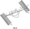
- Fig. 3 illustrates the consequences of erroneously determined photo conversion positions 30 for PET image reconstruction.
- the ⁇ + decay 26 generates two gamma photons that fly away in opposite directions 28 and that are detected by means of two gamma cameras 10a, 10b.
- Fig. 4 shows an embodiment of an evaluation apparatus 50 according to the present invention.
- Gamma radiation events are detected by a gamma camera 10 comprising a scintillator 12 and a photodetector 14. These gamma radiation events result from all events that cause the scintillator 12 to emit scintillation photons 42 at photo conversion positions 44 in the scintillator 12.
- the impact of a single gamma ray 46 at a single photo conversion position 44 results in a gamma radiation event.
- nuclear imaging it is the goal to correctly determine the position of the impact 44 of the gamma ray 46, i.e. the photo conversion position 44 of a single gamma ray.
- a line of response can be determined. Based on a plurality of such lines of response, an image can be reconstructed.
- invalid gamma radiation events refer to events that lead to ambiguous spatial signal distributions and to errors when reconstructing an image from the determined lines of response.
- a valid gamma radiation event results particularly from a single gamma ray impact.
- the obtained spatial signal distribution 24 of the gamma radiation event is evaluated in an evaluation apparatus 50.
- This evaluation apparatus 50 comprises a comparator 52 for comparing the obtained spatial signal distribution 24 with predetermined model distributions for different possible photo conversion positions in the scintillator 12 and for determining therefrom the likelihood function for said different possible photo conversion positions.
- the evaluation apparatus 50 comprises a selector 54 for selecting the most likely photo conversion position based on the maximum value of said likelihood function for said different possible photo conversion positions and for determining whether said maximum value of the likelihood function fulfills a predetermined selection criterion indicating whether the evaluated gamma radiation event is a valid gamma radiation event.
- the invention is based on the insight that the likelihood value of the spatial signal distribution is very well suited to identify pile-up events and inter-crystal Compton scatter events.
- the approach is particularly designed for gamma cameras or scintillation detectors comprising a scintillator including an array of small scintillation crystals or a single monolithic scintillator, a photodetector including an array of photosensitive elements or a position-sensitive photodetector and a readout electronics that is able to read out all photosensitive elements at the same time in order to obtain the spatial signal distribution of a gamma radiation event.
- predetermined model distributions can be obtained from measurements, i.e. an experimental calibration procedure, Monte Carlo simulations, i.e. discrete simulations of the events in order to determine a distribution or analytic modeling.
- a list that contains likelihood values for a typical data set is used for determining a statistical distribution and corresponding quantiles. This list can be obtained from a calibration data set. For each event of this calibration data set, the likelihood value is recorded and stored. The quantiles are determined in order to relate the likelihood values to fractions of events that are to be filtered out. This allows establishing the likelihood threshold for any filter fraction that may be applied.
- the likelihood value of the event is determined and compared to the likelihood threshold.
- the event is discarded (invalid event), if it is smaller than the threshold value.
- the presented apparatus and method can be used with any scintillation detector or gamma camera that comprises a pixelated or monolithic scintillation crystal and a position-sensitive photodetector, e.g. an array of photosensitive elements. If the pitch of the photosensitive elements is a multiple of the pitch of the scintillation crystal elements, then a light-guide is placed in between the scintillator and the position-sensitive photodetector. In case the pitch of the photosensitive elements matches the pitch of the crystal elements, each crystal will be read out by a single photosensitive element of a photodetector array (one-to-one coupling). In this case, pile-up events become unlikely but inter-crystal Compton scatter is still present.
- the readout electronic may then read out every photosensitive element in a reasonably short time in order to obtain complete spatial signal distributions. From these spatial signal distributions, the likelihood value can be computed as follows: MLE : L i i , E
- q 1 M ′ ⁇ j ′ ⁇ c i , j ′ E + q j ′ log c i , j ′ E ⁇ log q j ′ !
- LS L i i , E
- q 1 M ′ ⁇ j ′ ⁇ 1 2 q j ′ ⁇ c i , j ′ E ⁇ j ′ 2 ⁇ log 2 ⁇ ⁇ ⁇ j ′ n !
- MLE means Maximum likelihood estimation
- LS means least square estimation
- i is the index of the crystal pixel
- j is the index of the photosensitive element of the photodetector element
- E is the energy of the gamma radiation event
- q j is the detected charge at photosensitive element (pixel) j
- j is the measurement error for q j
- c i,j is the predetermined model distribution for comparison.
- the matrix elements c i,j may be obtained by measurement and states how much light is expected in photosensitive element j if the photo conversion took place in photosensitive element (pixel) i . Since the logarithm of the factorial presents a problem for numerical computations, it is approximated using the Stirling approximation (last two equations).
- the maximum likelihood values are computed for each event. From a calibration procedure, threshold values for the maximum likelihood values may be derived. For this, the coincidences of a measured data set can be determined and the likelihood values for each coincident photo conversion position corresponding to a single gamma ray impact can be computed. From the likelihood values of the test data set the quantiles are computed. This allows establishing the likelihood threshold as a function of the total number of recorded events and the desired differentiation. During normal measurements, a reasonable threshold is chosen. For instance 10 % of all detected gamma radiation events are filtered out before reconstruction.
- FIG. 5 shows an embodiment of a calibration apparatus 56 according to the present invention.
- the obtained spatial signal distributions 24 are usually difficult to interpret if the photodetector 14 is not adequately calibrated.
- the measurement of the scintillation light amount with each photosensitive element of a photodetector array is complicated by the fact that there is a large spread of the gain due to fabrication. For example, gain variations of up to factors of two on a single PSPMT or SiPM array are easily observed. If the gain differences between the photosensitive elements of a photodetector array are known the data processing and image reconstruction may be improved.
- the calibration apparatus 56 as shown in Fig. 5 comprises a histogram module 58, a background value module 60 and a calibration value module 62.
- the histogram module 58 an inverse, logarithmic and normalized histogram of the energy spectrum of each photosensitive element resulting from a number of gamma radiation events is determined.
- the background value module 60 a background value for each photosensitive element is determined based on the maximum of the derivation of said histogram.
- a gain calibration value each photosensitive element is determined from the intersection point of the histogram and a vertical line calculated from a predetermined calibration value and the background value.
- Fig. 6 shows a schematic diagram of an embodiment of a nuclear imaging apparatus 64 according to the present invention.
- a nuclear decay process in a subject 66 e.g. a patient or a calibration phantom, is observed with a gamma camera 10 being part of the nuclear imaging apparatus 64.
- the nuclear imaging apparatus 64 includes an evaluation apparatus 50 and/or a calibration apparatus 56 as described above.
- Fig. 7 shows a diagram illustrating a method for evaluating gamma radiation events represented by an obtained spatial signal distribution according to the present invention.
- Incident gamma rays induce the emission of scintillation photons S10 in the scintillator. These scintillation photons are detected S12 in a photodetector and a spatial signal distribution is obtained S14.
- the obtained spatial signal distribution is compared S16 to predetermined model distributions 76.
- the likelihood of the obtained spatial signal distribution for a given possible photo conversion position corresponding to a single gamma ray impact is determined S18, i.e. the likelihood function is calculated. Then, the most likely position is selected S20 and it is checked S22 whether the value fulfills a predetermined selection criterion 84.
- this predetermined selection criteria 84 is a comparison to a threshold value. If the determined maximum likelihood value is above the threshold the evaluated gamma radiation event is valid, if not it is invalid.
- One advantage of the present invention may thus be the provision of reliable identification of gamma radiation events representing pile-up events and inter-crystal Compton and/or the provision of reliable identification of valid gamma radiation events that allow correctly determining the photo conversion position of the incident single gamma ray. Therefore, the obtained spatial signal distribution is analyzed directly and not a derived parameter such as the centroid or the total energy which makes the identification more reliable and also robust against failing photodetectors. Failing photodetectors can occur due to aging or due to dead time. In the first case, one or more photosensitive elements of a photodetector array stop working. The charge that corresponds to the position of the failing element then misses in the obtained spatial signal distribution, leading to incomplete data and erroneous centroid computation.
- MLE based positioning is robust against missing data and so is the identification of pile-up event based on Likelihood computation.
- a second reason for failing photodetectors is their dead time. The corresponding photosensitive element of the photodetector may still be busy handling the charge from a previous event giving also rise to incomplete data but only for this event. Consequences for CoG positioning and MLE positioning are the same as in the case of aging.
- the present invention may have the advantage to provide an adjustable threshold for the rejection of pile-up events and inter-crystal Compton scatter events. This allows to adapt the filtering to the amount of data that is acquired and also to the isotope that is used (e.g. 22 Na has an additional 1275 keV gamma photon that is emitted simultaneously and leads to additional pile-up events).
- the present invention may allow an improvement of the spatial resolution of reconstructed images based on data of a gamma camera. Because lines of response that are reconstructed based on gamma radiation events resulting from pile-up events or inter-crystal Compton scatter are filtered out, the data set used for image reconstruction is cleaned from erroneous lines of response. Additionally by removing erroneous lines of response, the signal-to-noise ratio (SNR) of the reconstructed images can be improved.
- SNR signal-to-noise ratio
- the present invention may allow to improve the energy resolution and to create more precise standard uptake values.
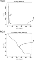
- Fig. 8 shows an energy spectrum 85 captured from a photosensitive element (i.e. a single pixel) of a SiPM (or dSiPM) array obtained by flooding the detector radiation with 511 keV gamma rays from Fluorodeoxyglucose (FDG). Characteristic points as the 511 keV full absorption peak or the Compton edge cannot be recognized because the light from each crystal is distributed over several pixels.
- the maximum around channel fifty 86 is caused by the energy threshold for this channel. This maximum cannot be used for gain estimation because it mainly depends on the threshold value.
- SiPMs i.e. pixels of a SiPM array or SiPM pixels
- a full absorption peak could be observed.
- several gain values are mixed into one single value.
- the single SiPM pixel's spectrum is transformed into a form that allows the extraction of a characteristic end point of the energy spectrum (cannot be recognized in Fig. 8 ). This end point is a good estimator for the relative gain of the SiPM pixel compared to the surrounding SiPM pixels.
- the measured energy spectra of each photosensitive element e.g. SiPM pixel
- SiPM pixel The measured energy spectra of each photosensitive element (e.g. SiPM pixel) are mathematically transformed to allow extracting the endpoint of the spectrum.
- An advantage of the method is that no iterative least square fitting is required. Outcomes of least square fitting depend on the starting parameters and are not guaranteed to converge.
- j and i are the energy channels of the histograms f ( j ) and h(i) and N ADC - channel is the total number of available energy channels in the histograms.
- Fig. 9 shows an inverse, logarithmic and normalized histogram 88 of the energy spectrum.
- This graph is monotonically decreasing and normalized to the range [0,1] on the y- axis.
- a background is observed (very few scintillation events from natural background or cosmic radiation). This background may be taken into account, since it may be different for different measurements/SiPMs and can be extracted as follows: First, the inverse function f 1 is built. Fig. 10 shows the inverse 89 of the modified energy histogram. Then the derivative 90 of this inverse function is computed as shown in Fig. 11 . For lower values of x , a maximum 91 is observed in this new graph. The position of this maximum gives the background value b g 94 that is then used when calculating the intersection of the horizontal line with the inverse, logarithmic and normalized histogram f ( j ). The relative gain of the current SiPM pixel may then be determined as the energy channel (x-axis value) at which the histogram falls below the sum of y 0 and b g .
- Fig. 12 shows the modified histogram 88 (inverse, logarithmic and normalized histogram).
- the predetermined calibration value 92 is added to the determined background value 94, which was determined based on the maximum of the derivation of the histogram 91 as described above.
- the gain calibration value 96 is then represented by the ADC channel, i.e. the energy value, at which the histogram 88 falls below the limit value 100 (i.e. a threshold) determined from the sum of the background value b g 94 and the calibration value y 0 92.
- this outlined method allows fast in-situ calibration of PET block detectors using highly granulated scintillator pixels and light-sharing where no full absorption peak can be observed on a single SiPM.
- Fig. 13 illustrates a calibration method according to the present invention.
- a gamma camera By means of a gamma camera, different spatial signal distributions are recorded. For this, incident gamma rays, which induce the emission of scintillation photons S10 in the scintillator, are observed. These scintillation photons are detected S12 in a photodetector and a spatial signal distribution is obtained S14.
- an energy spectrum is recorded for each photosensitive element of the photodetector array and - based on this spectrum - an inverse, logarithmic and normalized histogram is determined S24.
- a background value representing the background or background effect is determined S26 as described above. From the inverse, logarithmic and normalized histogram and the determined background value, finally a gain calibration value is determined S28 taking into account a predetermined calibration value 92.
- CT computed tomography
- SPECT/CT SPECT/CT
- PET/CT PET/CT
- Magnetic Resonance Imaging MRI
- NMRI nuclear magnetic resonance imaging
- a computer program may be stored/distributed on a suitable medium, such as an optical storage medium or a solid-state medium supplied together with or as part of other hardware, but may also be distributed in other forms, such as via the Internet or other wired or wireless telecommunication systems.
- a suitable medium such as an optical storage medium or a solid-state medium supplied together with or as part of other hardware, but may also be distributed in other forms, such as via the Internet or other wired or wireless telecommunication systems.
Landscapes
- Health & Medical Sciences (AREA)
- Physics & Mathematics (AREA)
- Life Sciences & Earth Sciences (AREA)
- Spectroscopy & Molecular Physics (AREA)
- Molecular Biology (AREA)
- High Energy & Nuclear Physics (AREA)
- General Physics & Mathematics (AREA)
- Biomedical Technology (AREA)
- Optics & Photonics (AREA)
- Nuclear Medicine, Radiotherapy & Molecular Imaging (AREA)
- Medical Informatics (AREA)
- General Health & Medical Sciences (AREA)
- Engineering & Computer Science (AREA)
- Measurement Of Radiation (AREA)
- Nuclear Medicine (AREA)
Description
- The present invention relates to an evaluation apparatus, a nuclear imaging device and corresponding methods for the use in nuclear imaging, in particular for the use with PET and SPECT cameras.
- Gamma ray detection with gamma cameras for nuclear imaging is a multiple stage process. First, the incident gamma photon is converted to a scintillation light flash (i.e. a scintillation photon). Then, each of the visible light photons from this flash (sometimes ultraviolet or other photons may be generated) is captured with a photodetector and a spatial signal distribution is obtained, which is then used to estimate the energy of the incident gamma photon and the two- or three-dimensional position of the gamma ray photo conversion. This determined position then forms the basis for the estimation of a line of response (LOR) and the image reconstruction from a nuclear decay process.
- While the energy of the gamma photon is proportional to the number of detected visible light photons, the photo conversion position (i.e. the position where the gamma ray interacts with the scintillator and the light flash is created) may be extracted from the spatial distribution of the visible light photons. One possibility is to couple each element of the scintillator to a single photodetector element. All the light is collected by this single photodetector element and the position of the gamma ray impact is given by the position of the photodetector. This type of readout is generally called one-to-one coupling.
- Because one-to-one coupling usually requires a large number of digitization channels, light-sharing is often used, which refers to a light-guide interposed in between the scintillator and the photodetector. In this way, the scintillation light from the photo conversion is distributed over several photodetector elements and the number of required digitization channels is reduced considerably. After digitization of the charge pattern, i.e. based on the spatial signal distribution, energy and position are computed from the values. The most widely used method is centroid computation (also called anger method or center of gravity (COG)). Alternatively, maximum likelihood (MLE) estimation can be used (Lerche, gravity (COG)). Alternatively, maximum likelihood (MLE) estimation can be used (Lerche, C.W. et al.; "Maximum likelihood based positioning and energy correction for pixelated solid-state PET detectors"; Nuclear Science Symposium and Medical Imaging Conference (NSS/MIC), 2011 IEEE). This method is based on the calculation of the likelihood of a photo conversion position in the scintillator for a given observed charge pattern at the photodetector. The position with the highest likelihood value is chosen.
- Both, COG positioning and MLE positioning work fine and result in reasonable position estimates, as long as the observed gamma radiation events all result from single gamma ray impacts. However, if Gamma radiation events result from the impact of multiple gamma rays at the same time, i.e. within one sampling interval in a digital photodetector or within the integration time in an analog charge converter, (pile-up events) or from inter-crystal Compton scatter (inter-crystal Compton scatter events), the detected position might be erroneous.
- Further, the recognition of gamma radiation events resulting from pile-up events or from inter-crystal Compton scatter events is difficult during normal data acquisition. One approach is to analyze the spatial position of the centroid of the detected spatial signal distribution, the rising edge of the temporal charge pulse (pulse shape discrimination) or the energy of the event.
- In
US 5,293,044 (A ) a method for rapid localization of a scintillation event in a gamma camera using a maximum likelihood estimator is disclosed. The method is applied in a process for localizing a scintillation event in a gamma camera having a plurality of photomultipliers forming a camera surface. Each photomultiplier generates an output signal in response to the scintillation event. According to the disclosed method, a plurality of comparative signal sets are generated from output signals of the photomultiplier corresponding to respective scintillation events of known location. Then, a location-dependent probability function is formed based on a comparison of the outputs of the photomultipliers for a scintillation event of unknown location with the comparative signal sets, and the location of the scintillation event of unknown location is defined as the location corresponding to the maximum of the probability function. Further, the speed with which the localization is accomplished is increased by initially defining a portion of the total gamma camera surface in which there is a high probability that the location of the scintillation event of unknown origin lies, and limiting the investigation for the location of the scintillation event of unknown location to that portion of the camera surface, with increasing precision. - In McKinsey et al., Neutrino detection with CLEAN, Astroparticle Physics 22 (2005) 355-368, an approach to the detection of low-energy solar neutrinos and neutrinos released from supernova is described.
- It is an object of the present invention to enable a reliable identification of valid gamma radiation events, in particular gamma radiation events that do not result from pile-up events or inter-crystal Compton scatter, from an analysis of the obtained spatial signal distribution. Further goals are to make the identification more reliable and more robust against failing photodetectors and to obtain reconstructed images with an improved spatial resolution.
- It is further an object of the present invention to enable in-situ calibration of photodetector arrays in gamma cameras, respectively in nuclear imaging systems with differences in the gain values of the photosensitive elements in the array.
- According to a first aspect of the present invention there is provided an evaluation apparatus, as defined in independent claim 1, for evaluating gamma radiation events detected by a gamma camera and discriminating valid gamma radiation events from undesired pile-up and inter-crystal Compton scatter events, said gamma camera including a scintillator for emitting scintillation photons at photo conversion positions in the scintillator in response to incident gamma rays and resulting gamma radiation events and a position-sensitive photodetector for detecting emitted scintillation photons and obtaining therefrom a spatial signal distribution. The evaluation apparatus comprises a comparator for comparing an obtained spatial signal distribution with predetermined model distributions for different possible photo conversion positions in the scintillator and for determining therefrom the likelihood function for said different possible photo conversion positions. The evaluation apparatus further comprises a selector for selecting the most likely photo conversion position based on the maximum value of said likelihood function for said different possible photo conversion positions and for determining whether said maximum value of the likelihood function fulfills a predetermined selection criterion indicating whether the evaluated gamma radiation event is a valid gamma radiation event, said predetermined selection criterion being a comparison with a threshold value that is defined as a quantile of a distribution of maximum likelihood values of different valid gamma radiation events in a calibration data set and is effective to discriminate between valid gamma radiation events and invalid gamma radiation events comprising pile-up events and inter-crystal Compton scatter events.
- Thus, the most likely photo conversion position in the scintillator is determined. The determination of the likelihood function (i.e. the determination of the likelihood values for the different possible photo conversion position) can, e.g., be based on maximum likelihood estimation or least square estimation. In order to obtain a spatial signal distribution, i.e. a distribution of the visible light photos generated in the scintillator, either an analog photodetector may be used and charge pulses may be digitized by means of an analog-digital converter, or a digital photodetector may be used to directly obtain a digital signal. The obtained spatial signal distribution is compared with predetermined model distributions, referring to resulting spatial signal distributions of valid gamma radiation events that are used as a reference. From this comparison, the most likely corresponding photo conversion position and gamma radiation event (in particular from a single gamma ray impact) causing this obtained spatial signal distribution is determined. Then, the function value of the likelihood function, i.e. the maximum likelihood value, is further used for determining whether this current gamma radiation event is sufficiently likely to be interpreted as a valid gamma radiation event. The photo conversion position in the scintillator and the determined validity-indicator are returned.
- Particularly, a valid gamma radiation event results from a single gamma ray impact and a single photo conversion position. Other gamma radiation events can, e.g., result from pile-up events or inter-crystal Compton scatter events where it may not be possible to clearly identify the original photo conversion position from the incident gamma ray or where multiple original photo conversion positions exist (e.g. due to multiple simultaneous impacts or scatter). Gamma radiation events can result at least from incident gamma rays, pile-up events and inter-crystal Compton scatter events. All different types of gamma radiation events lead to a spatial signal distribution, which is obtained by the photodetector. However, the determined photo conversion position in the scintillator, which is the basis for the determination of a line of response and for the image reconstruction in nuclear imaging devices, can lead to unclear or incorrect results if the same processing is applied for pile-up events and inter-crystal Compton scatter events. One approach can be the evaluation of the spatial signal distribution or other parameters for determining whether the current gamma radiation event is valid, i.e. results directly from a single incident gamma ray from the decay process that is to be measured/imaged or invalid, i.e., results from a pile-up event or an inter-crystal Compton scatter event. According to the present invention, the evaluation of the detected spatial signal distributions based on the value of the likelihood function is used to differentiate valid and invalid gamma radiation events. Invalid gamma radiation events thereby especially include but are not necessarily limited to gamma radiation events resulting from pile-up events and inter-crystal Compton scatter events. Valid gamma radiation events thereby especially include but are not necessarily limited to gamma radiation events resulting directly from single gamma ray impacts.
- One possibility for the predetermined selection criterion that is used in the selector is to make use of a comparison with a threshold value. This threshold value can, e.g., be determined prior to the use of the evaluation apparatus. One possible threshold may be determined based on the number of detected events, i.e. the number of gamma radiation events caused by the nuclear decay processes in a subject (e.g. a patient, a material probe, etc.) being observed with the gamma camera. Another possibility is to determine the threshold based on the intensity of the observed decay process (i.e. the used isotope). The choice of a higher threshold leads to a lower percentage of events classified as valid with reference to all detected events. If a lower threshold is selected, a higher number of events are classified as valid. Additionally, other criteria, e.g. criteria based on the signal distribution like shape analysis, spot size, standard deviation or combinations, may also be considered.
- If, e.g., due to a higher intensity of the observed nuclear decay process or a longer observation time, the number of detected events is high, the choice of a higher threshold might result in a good discrimination of undesired pile-up and inter-crystal Compton scatter events. On the other hand, a higher percentage of events might be rejected that could have been used. If the total number of detected events is low then it might not be possible to reject a high percentage of the events because too few positions in the scintillator used in the estimation of lines of response might result in worse images.
- According to another embodiment of the present invention this threshold value is defined as a quantile of the distribution of the maximum likelihood values of different valid gamma radiation events in a calibration data set. The calibration data set can be obtained from an experimental study, wherein a number of different subjects and/or calibration phantoms are examined. Alternatively, the calibration data set can be obtained from Monte Carlo simulations, wherein a number of different subjects and/or calibration phantoms are simulated. Further alternatively, this calibration data set can be obtained from analytical modeling, wherein at least one subject and/or calibration phantom is modeled.
- This means that the spatial signal distributions resulting from single gamma ray impacts and the corresponding maximum likelihood values are used to establish a reference distribution from which the threshold value is derived. By selecting a quantile of the distribution of the maximum likelihood values of different valid gamma radiation events, the percentage of events that are classified as invalid can directly be defined. If, e.g., the 75 % quantile is chosen, then, given that the calibration data set used to determine the distribution of the maximum likelihood values is adequate, it can be assured that those 25 % of the events which have the lowest similarity to valid gamma radiation events are rejected. Again, the percentage can be adjusted depending on the total number of detected events as explained above.
- According to yet another embodiment of the present invention the predetermined model distributions are obtained from an experimental calibration procedure, Monte Carlo simulations, and/or analytical modeling. In order to determine the likelihood function for different possible photo conversion positions resulting from valid gamma radiation events the resulting spatial signal distributions as obtained by the position-sensitive photodetector are compared to reference or model distributions of possible valid gamma radiation events. This means that for each valid gamma radiation event the corresponding spatial signal distribution is determined by means of experimental calibration, simulation or modeling and used as a model distribution, i.e., a predetermined model distribution. This predetermined model distribution serves as a basis for the comparison. Ideally, a detected valid gamma radiation event results in the very same spatial signal distribution for the same photo conversion position in the scintillator. However, deviations occur due to slight variations of the photo conversion positions in the scintillator or effects in the real measurement like scatter or noise or others.
- Depending on the set-up it can be too difficult or too expensive to obtain the predetermined model distribution from an experimental calibration procedure. If this is the case an alternative way is to make use of Monte Carlo simulations or analytical modeling. Monte Carlo simulations refer to repeated random sampling in order to obtain a model of the underlying distribution. If a statistical model is available with known dependencies and distributions, then Monte Carlo simulations can be used to determine a resulting distribution even if an analytical model would be too difficult to calculate. Analytical modeling refers to using a model and to determining the resulting distributions based on calculations based on said model. Both, Monte Carlo simulations and analytical modeling are especially useful if it is not possible to carry out an experimental calibration procedure.
- Another essential aspect for obtaining reasonable results from a position-sensitive photodetector including the use in an evaluation apparatus as defined above is the calibration of the photodetector. Light-sharing usually renders in-situ calibration by locating the full absorption peak of the gamma rays energy spectrum impossible and dismounting the arrays for gain calibration is usually not feasible for commercial products, since it generally has to be repeated periodically.
- According to another aspect of the present invention there is provided a nuclear imaging device as defined in
claims 4 to 7, said device comprising a gamma camera including a scintillator for emitting scintillation photons at photo conversion positions in the scintillator in response to incident gamma rays and resulting gamma radiation events and a position-sensitive photodetector for detecting emitted scintillation photons and obtaining therefrom a spatial signal distribution. This nuclear imaging device further comprises an evaluation apparatus as defined above and a calibration apparatus as defined above. - Nuclear imaging devices particularly include Positron Emission Tomography (PET) devices and Single Photon Emission Computed Tomography (SPECT) devices being often used in medical tomographic imaging. Gamma rays are emitted by a radioactive substance being delivered into a patient (a subject), e.g. through injection into the bloodstream or through oral intake. These gamma rays are captured by means of a gamma camera, and images can be generated based on the reconstruction of so-called lines of response. Lines of response are straight lines between two coinstantaneous gamma ray impacts at opposite positions on each side of the subject. As outlined above, the used gamma camera usually comprises a scintillator and a photodetector.
- One specific embodiment of this nuclear imaging device according to the present invention is a nuclear imaging device wherein the scintillator comprises a monolithic scintillation crystal or an array of scintillation crystals, in particular a Lutetium-Yttrium Orthosilicate (LYSO) crystal array. Both types of scintillators, monolithic scintillation crystals or scintillation crystal arrays, allow the application of the evaluation apparatus and/or the application of the calibration apparatus as defined above.
- According to a further embodiment of the nuclear imaging device, the position-sensitive photodetector comprises an array of photosensitive elements, in particular a Silicon photomultiplier (SIPM) array. Such arrays of photosensitive elements are particularly prone to pile-up events and inter-crystal Compton scatter events, which usually result in a reduction of the image quality. A possible countermeasure is the use of a higher dose of the radioactive isotope in the subject and the filtering of the detected gamma radiation events, e.g. with an evaluation apparatus as defined above.
- Still another embodiment of the nuclear imaging device comprises a scintillator and a position-sensitive photodetector that are mounted in light-sharing configuration with a light-guide interposed in between the scintillator and the photosensitive photodetector. The evaluation apparatus according to the present invention is particularly useful if the photodetector and the scintillator are mounted in light-sharing configuration. Light-sharing configuration means that the scintillation photons emitted by the scintillator are detected by multiple elements of the photodetector so that a high number of pile-up events may occur.
- According to further aspects of the present invention, there are provided a method, as defined in
independent claim 8, for evaluating gamma radiation events detected by a gamma camera and identifying valid gamma radiation events, a method for in-situ calibrating a position-sensitive photodetector and a method for nuclear imaging. - The methods may particularly make use of an evaluation apparatus, a calibration apparatus or a nuclear imaging device as defined above.
- In yet further aspects of the present invention, there are provided a computer program, as defined in
independent claim 10, which comprises program code means for causing a computer to perform the steps of the methods as outlined above when said computer program is carried out on a computer as well as a non-transitory computer-readable recording medium that stores therein a computer program product, which, when executed by a processor, causes the methods disclosed herein to be performed. - Preferred embodiments of the invention are defined in the dependent claims. It shall be understood that the claimed methods, computer program and recording medium have similar and/or identical preferred embodiments as the claimed apparatus and as defined in the dependent claims.
- These and other aspects of the invention will be apparent from and elucidated with reference to the embodiment(s) described hereinafter. In the following drawings
-
Fig. 1 shows a diagram illustrating two obtained spatial signal distributions of a 8x8 photodetector array resulting from a single gamma ray impact (Fig. 1a ) and from a pile-up event (Fig. 1b ); -
Fig. 2 illustrates pile-up events (Fig. 2a ) and inter-crystal Compton scatter events (Fig. 2b ) by means of a section view of the scintillator, the light-guide and the position-sensitive photodetector comprised in a gamma camera; -
Fig. 3 illustrates the problems resulting from lines of response constructed on the basis of inaccurately determined photo conversion positions; -
Fig. 4 shows a diagram of an embodiment of an evaluation apparatus according to the present invention and a gamma camera; -
Fig. 5 shows a schematic diagram of an embodiment of a calibration apparatus according to the present invention and a gamma camera; -
Fig. 6 shows a schematic diagram of an embodiment of a nuclear imaging device according to the present invention; -
Fig. 7 shows a diagram illustrating a method for evaluating gamma radiation events according to the present invention; -
Fig. 8 shows an energy spectrum recorded from a photosensitive element of a photodetector; -
Fig. 9 shows a normalized histogram based on an energy spectrum; -
Fig. 10 shows a normalized, inverse and logarithmic histogram based on an energy spectrum; -
Fig. 11 shows the derivation of a normalized, inverse and logarithmic histogram; -
Fig. 12 illustrates the determination of a gain calibration value for a photosensitive element of a photodetector from a normalized, inverse and logarithmic histogram; and -
Fig. 13 illustrates a method for in-situ calibrating a position-sensitive photodetector according to the present invention. - The detection of gamma radiation events resulting from nuclear decay process is often used to reconstruct images, e.g. for medical purposes. For this detection the gamma camera generally comprises a scintillator where incident gamma rays are converted to scintillation photons and a position-sensitive photodetector e.g. a position-sensitive photomultiplier (PSPMT), an array of photomultiplier, arrays of avalanche photodiodes (APDs) or position-sensitive APDs or arrays of silicon photomultipliers (SiPMs, also digital SiPMs, dSiPMs). Generally, a position-sensitive photodetector allows determining a position of incident photons. Incident photons are converted to a spatial signal distribution or charge pattern which can then be evaluated in order to locate the position of the incident photon. In the present application the scintillation position, i.e. the photo conversion position in the scintillator, is usually determined from the emitted scintillation photons. These scintillation photons are captured with the position-sensitive photodetector. Then, the position in the scintillator is determined based on the obtained spatial signal distribution. Based on this position, the lines of response (LOR) are reconstructed which are then the basis for the image reconstruction.
- In one-to-one coupling configuration, each scintillator element is coupled to a single photodetector. The determination of the photo conversion position in the scintillator can then be directly based on the corresponding photodetector, in particular the corresponding photosensitive element of the photodetector array. This, however, requires a large number of digitization channels (one for each scintillation element of the scintillator). An alternative is the use of light-sharing coupling where a photo conversion in one scintillation element of the scintillator is recorded by different photodetectors or elements in a photodetector array. Usually, a light-guide is interposed in between the scintillator and the photodetector. In this way, the scintillation light from a single photo conversion is distributed over several photodetectors and the number of required digitization channels is reduced considerably. The readout from the photodetector is then a spatial signal distribution or charge pattern.
- After digitization of this spatial signal distribution, the position of the photo conversion (i.e. the scintillation) and the energy of the impact can be computed. The most widely used method is centroid computation (also called Anger method or Centre of Gravity (CoG)). Alternatively, maximum likelihood estimation can be used. For this alternative method the likelihood for every scintillation crystal i is computed from the digitized charges q= {qj} that were observed at photodetector j:


- With the knowledge of i (ML) and the matrix element ci,j, the energy of the gamma ray event can be predicted:

- However, occurring gamma radiation events are not limited to single gamma ray impacts at photo conversion positions. Also, pile-up events, inter-crystal Compton scatter and other inter-crystal scatter events occur and can lead to ambiguous spatial signal distributions.
-
Fig. 1 shows illustrations of two typical spatial signal distributions registered by a photodetector in a gamma camera, comprising a 30x30 LYSO pixel array representing the scintillator coupled to an 8x8 digital SiPM (dSIPM) array representing the photodetector.Fig. 1a shows a spatial signal distribution from a gamma radiation event resulting from a single gamma ray impact.Fig. 1b shows a spatial signal distribution for a gamma radiation event resulting from a pile-up event. CoG positioning and ML based positioning work fine, as long as there are only gamma radiation events resulting from single gamma ray impacts. If more than one gamma ray impact occurs at the same time (pile-up event), the detected spatial signal distribution has more than one maximum as shown inFig. 1b and the gamma ray events are wrongly positioned when using CoG or ML positioning alone. -
Fig. 2a shows a cross-sectional view of an embodiment of agamma camera 10, i.e. a scintillation detector. Thegamma camera 10 comprises ascintillator 12, aphotodetector 14 and a light-guide 16 in between. There is visualized the spatial signal distribution of a gamma radiation event resulting from a pile-upevent 18, which is caused by two coinstantaneous butindependent gamma rays 20. Theindependent gamma rays 20 impact at the same time. If both interact via photo effect, such events can be discriminated by means of an energy filter, but if they interact via Compton effect and the secondary gamma ray escapes from the scintillator, the sum of their energy deposits may lay inside the energy window around 511 keV. Thus, it might not be possible to detect such events by means of an energy filter and wrong positions will be computed with the CoG or ML method. - Apart from pile-up events and resulting gamma radiation events, another source for an ambiguous spatial signal distribution can be inter-crystal Compton scatter.
Fig. 2b shows an inter-crystal Compton scatter event. Anincident gamma ray 22 first interacts with thescintillator 12 in thegamma camera 10 via Compton Effect. Then, the deflected gamma ray travels through thescintillator 12 and interacts via photo effect. The distance between both interactions can be several pixels. The obtained spatial signal distribution on thephotodetector 24 of the gamma radiation event resulting from an inter-crystal Compton scatter event again does not allow accurately determining the position of theincident gamma ray 22. Especially for high resolution systems this effect occurs frequently and may lead to a substantial number of position errors. - The fraction of all gamma radiation events representing pile-up events or inter-crystal Compton scatter events strongly depends on the activity inside the gamma camera's 10 field of view (FOV), the used radioactive isotope and the geometry of the scintillator, photodetector, and gantry. Both PET and SPECT imaging are affected by this problem.
Fig. 3 illustrates the consequences of erroneously determined photo conversion positions 30 for PET image reconstruction. The β+decay 26 generates two gamma photons that fly away inopposite directions 28 and that are detected by means of twogamma cameras 10a, 10b. One photon is scattered 31 resulting in a spatial signal distribution that again results in an erroneously determinedphoto conversion position 30 in the scintillator. As a consequence, even if the photo conversion position in the scintillator on theother side 34 is determined correctly, an erroneous line ofresponse 36 is determined in contrast to thecorrect LOR 38. Thus, in the final reconstructed image, gamma radiation events resulting from pile-up events and inter-crystal Compton scatter result in a degradation of the spatial resolution and in a decrease of the signal-to-noise ratio because erroneous lines ofresponse 36 will be used during image reconstruction. -
Fig. 4 shows an embodiment of anevaluation apparatus 50 according to the present invention. Gamma radiation events are detected by agamma camera 10 comprising ascintillator 12 and aphotodetector 14. These gamma radiation events result from all events that cause thescintillator 12 to emitscintillation photons 42 at photo conversion positions 44 in thescintillator 12. In the example as illustrated inFig. 4 , the impact of asingle gamma ray 46 at a singlephoto conversion position 44 results in a gamma radiation event. In nuclear imaging, it is the goal to correctly determine the position of theimpact 44 of thegamma ray 46, i.e. thephoto conversion position 44 of a single gamma ray. Based on two coinstantaneous impacts and photo conversion positions from a single gamma ray impacts detected with gammas camera on both sides of a subject, a line of response can be determined. Based on a plurality of such lines of response, an image can be reconstructed. However, apart from impacts of single gamma rays (valid gamma radiation events), other possible gamma radiation events can also result from pile-up events, inter-crystal Compton scatter events and others (invalid gamma radiation events). In this context, invalid gamma radiation events refer to events that lead to ambiguous spatial signal distributions and to errors when reconstructing an image from the determined lines of response. A valid gamma radiation event results particularly from a single gamma ray impact. - As shown in
Fig. 4 the obtainedspatial signal distribution 24 of the gamma radiation event is evaluated in anevaluation apparatus 50. Thisevaluation apparatus 50 comprises acomparator 52 for comparing the obtainedspatial signal distribution 24 with predetermined model distributions for different possible photo conversion positions in thescintillator 12 and for determining therefrom the likelihood function for said different possible photo conversion positions. Further, theevaluation apparatus 50 comprises aselector 54 for selecting the most likely photo conversion position based on the maximum value of said likelihood function for said different possible photo conversion positions and for determining whether said maximum value of the likelihood function fulfills a predetermined selection criterion indicating whether the evaluated gamma radiation event is a valid gamma radiation event. - The invention is based on the insight that the likelihood value of the spatial signal distribution is very well suited to identify pile-up events and inter-crystal Compton scatter events. The approach is particularly designed for gamma cameras or scintillation detectors comprising a scintillator including an array of small scintillation crystals or a single monolithic scintillator, a photodetector including an array of photosensitive elements or a position-sensitive photodetector and a readout electronics that is able to read out all photosensitive elements at the same time in order to obtain the spatial signal distribution of a gamma radiation event. Models for the spatial signal distributions to which the measured charge pattern or spatial signal distribution is to be compared, i.e. predetermined model distributions, can be obtained from measurements, i.e. an experimental calibration procedure, Monte Carlo simulations, i.e. discrete simulations of the events in order to determine a distribution or analytic modeling. In a preferred embodiment a list that contains likelihood values for a typical data set is used for determining a statistical distribution and corresponding quantiles. This list can be obtained from a calibration data set. For each event of this calibration data set, the likelihood value is recorded and stored. The quantiles are determined in order to relate the likelihood values to fractions of events that are to be filtered out. This allows establishing the likelihood threshold for any filter fraction that may be applied.
- During normal operation of a nuclear imaging device (e.g. a PET or a SPECT apparatus), the likelihood value of the event is determined and compared to the likelihood threshold. The event is discarded (invalid event), if it is smaller than the threshold value.
- The presented apparatus and method can be used with any scintillation detector or gamma camera that comprises a pixelated or monolithic scintillation crystal and a position-sensitive photodetector, e.g. an array of photosensitive elements. If the pitch of the photosensitive elements is a multiple of the pitch of the scintillation crystal elements, then a light-guide is placed in between the scintillator and the position-sensitive photodetector. In case the pitch of the photosensitive elements matches the pitch of the crystal elements, each crystal will be read out by a single photosensitive element of a photodetector array (one-to-one coupling). In this case, pile-up events become unlikely but inter-crystal Compton scatter is still present. The readout electronic may then read out every photosensitive element in a reasonably short time in order to obtain complete spatial signal distributions. From these spatial signal distributions, the likelihood value can be computed as follows:


instance 10 % of all detected gamma radiation events are filtered out before reconstruction. - Another aspect of the present invention is the appropriate calibration of the photodetector which may be used with the present evaluation apparatus or nuclear imaging device.
Fig. 5 shows an embodiment of acalibration apparatus 56 according to the present invention. The obtainedspatial signal distributions 24 are usually difficult to interpret if thephotodetector 14 is not adequately calibrated. The measurement of the scintillation light amount with each photosensitive element of a photodetector array is complicated by the fact that there is a large spread of the gain due to fabrication. For example, gain variations of up to factors of two on a single PSPMT or SiPM array are easily observed. If the gain differences between the photosensitive elements of a photodetector array are known the data processing and image reconstruction may be improved. In the case of one-to-one coupling ofscintillator 12 andphotodetector 14, it is generally useful for correct system wide energy discrimination. In case of light-sharing, inter-pixel (photodetector pixel, i.e. photosensitive element) gain differences may even be more important since these gain differences introduce important errors in the estimation of the light distribution (i.e. the spatial signal distribution) which is used for the positioning. During calibration, gain differences lead to wrong likelihood values for the use in the evaluation of gamma radiation events. The gain differences of most devices can be measured by irradiating them with a controlled amount of light and measuring the amplitudes of the generated signals. Obviously, this cannot be done in-situ, since the scintillator crystal array generally has to be removed. Another possibility consists in irradiating the mounted detector block with gamma radiation and estimate the gain for each crystal pixel from the position of characteristic peaks in the energy spectra. - The
calibration apparatus 56 as shown inFig. 5 comprises ahistogram module 58, abackground value module 60 and acalibration value module 62. In thehistogram module 58, an inverse, logarithmic and normalized histogram of the energy spectrum of each photosensitive element resulting from a number of gamma radiation events is determined. In thebackground value module 60, a background value for each photosensitive element is determined based on the maximum of the derivation of said histogram. In thecalibration value module 62, a gain calibration value each photosensitive element is determined from the intersection point of the histogram and a vertical line calculated from a predetermined calibration value and the background value. -
Fig. 6 shows a schematic diagram of an embodiment of anuclear imaging apparatus 64 according to the present invention. A nuclear decay process in a subject 66, e.g. a patient or a calibration phantom, is observed with agamma camera 10 being part of thenuclear imaging apparatus 64. Apart from thegamma camera 10 thenuclear imaging apparatus 64 includes anevaluation apparatus 50 and/or acalibration apparatus 56 as described above. -
Fig. 7 shows a diagram illustrating a method for evaluating gamma radiation events represented by an obtained spatial signal distribution according to the present invention. Incident gamma rays induce the emission of scintillation photons S10 in the scintillator. These scintillation photons are detected S12 in a photodetector and a spatial signal distribution is obtained S14. In order to determine whether the evaluated gamma radiation event, respectively the obtained spatial signal distribution is a valid gamma radiation event, i.e. corresponds to a single gamma ray impact, the obtained spatial signal distribution is compared S16 topredetermined model distributions 76. Based on this comparison, the likelihood of the obtained spatial signal distribution for a given possible photo conversion position corresponding to a single gamma ray impact is determined S18, i.e. the likelihood function is calculated. Then, the most likely position is selected S20 and it is checked S22 whether the value fulfills apredetermined selection criterion 84. In the present embodiment thispredetermined selection criteria 84 is a comparison to a threshold value. If the determined maximum likelihood value is above the threshold the evaluated gamma radiation event is valid, if not it is invalid. - One advantage of the present invention may thus be the provision of reliable identification of gamma radiation events representing pile-up events and inter-crystal Compton and/or the provision of reliable identification of valid gamma radiation events that allow correctly determining the photo conversion position of the incident single gamma ray. Therefore, the obtained spatial signal distribution is analyzed directly and not a derived parameter such as the centroid or the total energy which makes the identification more reliable and also robust against failing photodetectors. Failing photodetectors can occur due to aging or due to dead time. In the first case, one or more photosensitive elements of a photodetector array stop working. The charge that corresponds to the position of the failing element then misses in the obtained spatial signal distribution, leading to incomplete data and erroneous centroid computation. However, MLE based positioning is robust against missing data and so is the identification of pile-up event based on Likelihood computation. A second reason for failing photodetectors is their dead time. The corresponding photosensitive element of the photodetector may still be busy handling the charge from a previous event giving also rise to incomplete data but only for this event. Consequences for CoG positioning and MLE positioning are the same as in the case of aging.
- Further, the present invention may have the advantage to provide an adjustable threshold for the rejection of pile-up events and inter-crystal Compton scatter events. This allows to adapt the filtering to the amount of data that is acquired and also to the isotope that is used (e.g. 22 Na has an additional 1275 keV gamma photon that is emitted simultaneously and leads to additional pile-up events).
- Still further, the present invention may allow an improvement of the spatial resolution of reconstructed images based on data of a gamma camera. Because lines of response that are reconstructed based on gamma radiation events resulting from pile-up events or inter-crystal Compton scatter are filtered out, the data set used for image reconstruction is cleaned from erroneous lines of response. Additionally by removing erroneous lines of response, the signal-to-noise ratio (SNR) of the reconstructed images can be improved.
- Still further, the present invention may allow to improve the energy resolution and to create more precise standard uptake values.
- In case of gamma cameras that use light-sharing for positioning, in-situ gain calibration using characteristic peaks of energy histograms is not possible because there are no characteristic peaks.
Fig. 8 shows anenergy spectrum 85 captured from a photosensitive element (i.e. a single pixel) of a SiPM (or dSiPM) array obtained by flooding the detector radiation with 511 keV gamma rays from Fluorodeoxyglucose (FDG). Characteristic points as the 511 keV full absorption peak or the Compton edge cannot be recognized because the light from each crystal is distributed over several pixels. The maximum around channel fifty 86 is caused by the energy threshold for this channel. This maximum cannot be used for gain estimation because it mainly depends on the threshold value. If the sum of all signal values from those SiPMs (i.e. pixels of a SiPM array or SiPM pixels) that are illuminated by the scintillation light pulse, then a full absorption peak could be observed. However due to the light-sharing, several gain values are mixed into one single value. The single SiPM pixel's spectrum is transformed into a form that allows the extraction of a characteristic end point of the energy spectrum (cannot be recognized inFig. 8 ). This end point is a good estimator for the relative gain of the SiPM pixel compared to the surrounding SiPM pixels. - The measured energy spectra of each photosensitive element (e.g. SiPM pixel) are mathematically transformed to allow extracting the endpoint of the spectrum. An advantage of the method is that no iterative least square fitting is required. Outcomes of least square fitting depend on the starting parameters and are not guaranteed to converge.
- According to one embodiment, the spectrum is transformed by using the following equation in order to obtain an inverse, logarithmic and normalized histogram of the energy spectrum:

- Herein j and i are the energy channels of the histograms f(j) and h(i) and N ADC-channel is the total number of available energy channels in the histograms. After this processing of the histogram, the endpoint of the energy histogram becomes visible and can be extracted.
-
Fig. 9 shows an inverse, logarithmic and normalizedhistogram 88 of the energy spectrum. This graph is monotonically decreasing and normalized to the range [0,1] on the y- axis. The relative gain of the SiPM pixel can now be extracted by finding the intersection of this graph with a horizontal line at y = y0, where y0 is a free calibration parameter between 0 and 1 that may be predetermined and set by an operator of the system. If this parameter is set to low values (e.g. ∼ 0.1), then the intersection of the line representing this calibration parameter with the modified histogram is in the range of the upper end point of the energy spectrum measured with this SiPM pixel. - Towards higher channels, usually a background is observed (very few scintillation events from natural background or cosmic radiation). This background may be taken into account, since it may be different for different measurements/SiPMs and can be extracted as follows: First, the inverse function f1 is built.
Fig. 10 shows theinverse 89 of the modified energy histogram. Then the derivative 90 of this inverse function is computed as shown inFig. 11 . For lower values of x, a maximum 91 is observed in this new graph. The position of this maximum gives thebackground value b g 94 that is then used when calculating the intersection of the horizontal line with the inverse, logarithmic and normalized histogram f(j). The relative gain of the current SiPM pixel may then be determined as the energy channel (x-axis value) at which the histogram falls below the sum of y0 and bg. -
Fig. 12 shows the modified histogram 88 (inverse, logarithmic and normalized histogram). In this example, thepredetermined calibration value 92 is added to thedetermined background value 94, which was determined based on the maximum of the derivation of thehistogram 91 as described above. Then, thegain calibration value 96 is determined from theintersection point 98 at which the histogram falls below thehorizontal line 100 representing the limit value y=y0 +bg. In this embodiment, thegain calibration value 96 is then represented by the ADC channel, i.e. the energy value, at which thehistogram 88 falls below the limit value 100 (i.e. a threshold) determined from the sum of thebackground value b g 94 and thecalibration value y 0 92. - In summary, this outlined method allows fast in-situ calibration of PET block detectors using highly granulated scintillator pixels and light-sharing where no full absorption peak can be observed on a single SiPM.
-
Fig. 13 illustrates a calibration method according to the present invention. By means of a gamma camera, different spatial signal distributions are recorded. For this, incident gamma rays, which induce the emission of scintillation photons S10 in the scintillator, are observed. These scintillation photons are detected S12 in a photodetector and a spatial signal distribution is obtained S14. Based on a plurality of recorded spatial signal distributions, an energy spectrum is recorded for each photosensitive element of the photodetector array and - based on this spectrum - an inverse, logarithmic and normalized histogram is determined S24. Then, a background value representing the background or background effect is determined S26 as described above. From the inverse, logarithmic and normalized histogram and the determined background value, finally a gain calibration value is determined S28 taking into account apredetermined calibration value 92. - Further possible embodiments of the invention may also include other apparatus and methods to be used with nuclear imaging devices that are not intended for the use in medical imaging, but for material sciences or other application areas. Additionally, the present invention is not limited to nuclear imaging alone. The herein presented concepts, apparatus and methods might very well be applied to other imaging systems for the use in various application areas, e.g. computed tomography (CT), SPECT/CT, PET/CT, Magnetic Resonance Imaging (MRI), nuclear magnetic resonance imaging (NMRI) or others.
- While the invention has been illustrated and described in detail in the drawings and foregoing description, such illustration and description are to be considered illustrative or exemplary and not restrictive; the invention is not limited to the disclosed embodiments. Other variations to the disclosed embodiments can be understood and effected by those skilled in the art in practicing the claimed invention, from a study of the drawings, the disclosure, and the appended claims.
- A computer program may be stored/distributed on a suitable medium, such as an optical storage medium or a solid-state medium supplied together with or as part of other hardware, but may also be distributed in other forms, such as via the Internet or other wired or wireless telecommunication systems.
Claims (10)
- Evaluation apparatus (50) for evaluating gamma radiation events detected by a gamma camera (10) and discriminating valid gamma radiation events from undesired pile-up and inter-crystal Compton scatter events, said gamma camera (10) including a scintillator (12) for emitting scintillation photons (42) at photo conversion positions (44) in the scintillator (12) in response to incident gamma rays (22) and resulting gamma radiation events and a position-sensitive photodetector (14) for detecting emitted scintillation photons (42) and obtaining therefrom a spatial signal distribution (24), said evaluation apparatus (50) comprising- a comparator (52) for comparing an obtained spatial signal distribution (24) with predetermined model distributions (76) for different possible photo conversion positions in the scintillator (12) and for determining (S18) therefrom the likelihood function for said different possible photo conversion positions; and- a selector (54) for selecting the most likely photo conversion position based on the maximum value of said likelihood function for said different possible photo conversion positions and for determining whether said maximum value of the likelihood function fulfills a predetermined selection criterion (84) indicating whether the evaluated gamma radiation event is a valid gamma radiation event, said predetermined selection criterion (84) being a comparison with a threshold value that is defined as a quantile of a distribution of maximum likelihood values of different valid gamma radiation events in a calibration data set and is effective to discriminate between valid gamma radiation events and invalid gamma radiation events comprising pile-up events and inter-crystal Compton scatter events.
- Evaluation apparatus according to claim 1, wherein the calibration data set is obtained from- an experimental calibration study, wherein a number of different subjects (66) and/or calibration phantoms are examined;- Monte Carlo simulations, wherein a number of different subjects (66) and/or calibration phantoms are simulated; and/or- analytical modeling, wherein at least one subject (66) and/or calibration phantom is modeled.
- Evaluation apparatus according to claim 1, wherein the predetermined model distributions are obtained from- an experimental calibration procedure;- Monte Carlo simulations; and/or- analytical modeling.
- Nuclear imaging device (64) comprising- a gamma camera (10) including a scintillator (12) for emitting scintillation photons (42) at photo conversion positions (44) in the scintillator (12) in response to incident gamma rays (22) and resulting gamma radiation events and a position-sensitive photodetector (14) for detecting emitted scintillation photons (42) and obtaining therefrom a spatial signal distribution (24); and- an evaluation apparatus (50) according to claim 1.
- Nuclear imaging device (64) according to claim 4, wherein the scintillator (12) comprises a monolithic scintillation crystal or an array of scintillation crystals, in particular a LYSO crystal array.
- Nuclear imaging device (64) according to claim 4, wherein the position-sensitive photodetector (14) comprises an array of photosensitive elements, in particular a SiPM or dSiPM array.
- Nuclear imaging device (64) according to claim 4, wherein the scintillator (12) and the position-sensitive photodetector (14) are mounted in light-sharing configuration with a light-guide (16) interposed in between the scintillator (12) and the position-sensitive photodetector (14).
- Method for evaluating gamma radiation events detected by a gamma camera (10) and discriminating valid gamma radiation events from undesired pile-up and inter-crystal Compton scatter events, said gamma camera (10) including a scintillator (12) for emitting scintillation photons (42) at photo conversion positions (44) in the scintillator (12) in response to incident gamma rays (22) and resulting gamma radiation events and a position-sensitive photodetector (14) for detecting emitted scintillation photons (42) and obtaining therefrom a spatial signal distribution (24), said method comprising- comparing (S16) an obtained spatial signal distribution (24) with predetermined model distributions (76) for different possible photo conversion positions in the scintillator (12) and determining (S18) therefrom the likelihood function for said different possible photo conversion positions; and- selecting (S20) the most likely photo conversion position based on the maximum value of said likelihood function for said different possible photo conversion positions and determining (S22) whether said maximum value of the likelihood function fulfills a predetermined selection criterion (84) indicating whether the evaluated gamma radiation event is a valid gamma radiation event, said predetermined selection criterion (84) being a comparison with a threshold value that is defined as a quantile of a distribution of maximum likelihood values of different valid gamma radiation events in a calibration data set and is effective to discriminate between valid gamma radiation events and invalid gamma radiation events comprising pile-up events and inter-crystal Compton scatter events.
- Method for nuclear imaging comprising- emitting (S10) scintillation photons (42) at photo conversion positions (44) in the scintillator (12) in response to incident gamma rays (46) and resulting gamma radiation events;- detecting (S12) emitted scintillation photons (42) and obtaining therefrom a spatial signal distribution (24); and- an evaluation method according to claim 8
- Computer program comprising program code means for causing a computer to carry out the steps of the method as claimed in claim 8 when said computer program is carried out on the computer.
Priority Applications (1)
| Application Number | Priority Date | Filing Date | Title |
|---|---|---|---|
| EP14720163.6A EP2994776B1 (en) | 2013-05-08 | 2014-04-30 | Apparatus and method for the evaluation of gamma radiation events |
Applications Claiming Priority (3)
| Application Number | Priority Date | Filing Date | Title |
|---|---|---|---|
| EP13167050 | 2013-05-08 | ||
| PCT/EP2014/058865 WO2014180734A2 (en) | 2013-05-08 | 2014-04-30 | Apparatus and method for the evaluation of gamma radiation events |
| EP14720163.6A EP2994776B1 (en) | 2013-05-08 | 2014-04-30 | Apparatus and method for the evaluation of gamma radiation events |
Publications (2)
| Publication Number | Publication Date |
|---|---|
| EP2994776A2 EP2994776A2 (en) | 2016-03-16 |
| EP2994776B1 true EP2994776B1 (en) | 2019-11-13 |
Family
ID=48444081
Family Applications (1)
| Application Number | Title | Priority Date | Filing Date |
|---|---|---|---|
| EP14720163.6A Active EP2994776B1 (en) | 2013-05-08 | 2014-04-30 | Apparatus and method for the evaluation of gamma radiation events |
Country Status (4)
| Country | Link |
|---|---|
| US (1) | US10061043B2 (en) |
| EP (1) | EP2994776B1 (en) |
| CN (1) | CN105190357B (en) |
| WO (1) | WO2014180734A2 (en) |
Families Citing this family (26)
| Publication number | Priority date | Publication date | Assignee | Title |
|---|---|---|---|---|
| EP3271752B1 (en) * | 2015-03-17 | 2018-10-24 | Koninklijke Philips N.V. | Scintillation event position determination in a radiation particle detector |
| WO2016201131A1 (en) * | 2015-06-09 | 2016-12-15 | University Of Washington | Gamma camera scintillation event positioning |
| CN106405609A (en) * | 2015-11-19 | 2017-02-15 | 南京瑞派宁信息科技有限公司 | Multiparticle event capturing method and device |
| CN106405608A (en) * | 2015-11-19 | 2017-02-15 | 南京瑞派宁信息科技有限公司 | Gamma light and visible light event level physical alignment method and device |
| CN106405619A (en) * | 2015-11-19 | 2017-02-15 | 南京瑞派宁信息科技有限公司 | Detector collimation method and device |
| CN107422364B (en) * | 2017-06-26 | 2019-06-25 | 东软医疗系统股份有限公司 | A time timing correction method and device for a detector |
| US10481284B2 (en) * | 2017-10-19 | 2019-11-19 | Kromek Group, PLC | Modular gamma imaging device |
| CN108523916B (en) * | 2018-03-20 | 2020-07-28 | 清华大学 | Multi-gamma photon coincidence imaging system and method |
| NO346536B1 (en) * | 2019-04-10 | 2022-09-26 | Bergen Teknologioverfoering As | System for charged particle therapy verification |
| WO2020242442A1 (en) * | 2019-05-24 | 2020-12-03 | Halliburton Energy Services, Inc. | Probabilistically distinguishing between true and false scintillator pulses |
| CN110613470B (en) * | 2019-09-26 | 2025-03-11 | 南昌华亮光电有限责任公司 | Fully simulated nuclide distribution imaging method and system |
| DE102019007136B3 (en) * | 2019-10-15 | 2020-10-08 | Forschungszentrum Jülich GmbH | Method for position and energy determination in scintillation detectors |
| CN115280360A (en) * | 2020-03-25 | 2022-11-01 | 华为技术有限公司 | Determination of illumination portion |
| EP3896494A1 (en) * | 2020-04-17 | 2021-10-20 | Terapet SA | Gamma ray detection system and calibration method thereof |
| CN111896996B (en) * | 2020-07-27 | 2025-02-11 | 核工业航测遥感中心 | Small-scale source model calibration method and device based on experimental comparison method |
| FR3113427B1 (en) * | 2020-08-17 | 2023-08-25 | Agence Nat Pour La Gestion Des Dechets Radioactifs | Method for calibrating an ionizing radiation measuring device, method for measuring ionizing radiation and device for measuring ionizing radiation |
| CN112014872B (en) * | 2020-09-03 | 2021-08-24 | 北京卫星环境工程研究所 | A method for determining the type or action position of incident particles based on pulse waveform discrimination |
| CN112596096B (en) * | 2020-11-24 | 2023-04-11 | 中国科学院上海光学精密机械研究所 | Ultrafast gamma ray real-time detection device based on SiPM |
| CN113009548B (en) * | 2020-12-15 | 2024-04-12 | 深圳湾实验室 | Detection equipment and radiation azimuth measurement method |
| CN113359178B (en) * | 2021-03-29 | 2023-05-16 | 山西中辐核仪器有限责任公司 | Radiometer based on self-adaptive dead time compensation |
| CN113628521B (en) * | 2021-07-12 | 2023-03-10 | 成都理工大学 | Nuclear detection simulation device and nuclear signal inversion technology based on nanosecond light source |
| US11933935B2 (en) | 2021-11-16 | 2024-03-19 | Saudi Arabian Oil Company | Method and system for determining gamma-ray measurements using a sensitivity map and controlled sampling motion |
| CN114757105B (en) * | 2022-04-29 | 2024-02-23 | 西安交通大学 | Three-energy band gamma imaging system and system design method based on energy window modulation |
| CN115453606B (en) * | 2022-09-23 | 2024-08-16 | 西北核技术研究所 | A real-time measurement method and prediction method for scintillator radiation resistance performance |
| CN118013398B (en) * | 2024-02-20 | 2025-04-11 | 成都理工大学 | A Bayesian characteristic peak identification method for gamma spectroscopy data |
| CN118962767B (en) * | 2024-10-17 | 2025-02-07 | 北京中科核安科技有限公司 | Rapid positioning method and device for 4 pi visual angle radioactive source and storage medium |
Family Cites Families (25)
| Publication number | Priority date | Publication date | Assignee | Title |
|---|---|---|---|---|
| US3878373A (en) * | 1971-06-30 | 1975-04-15 | Alvin Blum | Radiation detection device and a radiation detection method |
| US4507733A (en) * | 1980-07-31 | 1985-03-26 | Blum Alvin S | Photon emission imaging apparatus and method |
| US4437160A (en) * | 1980-07-31 | 1984-03-13 | Blum Alvin S | Photon emission imaging apparatus and method |
| JPH05203749A (en) * | 1991-08-28 | 1993-08-10 | Siemens Ag | Position checking method of multiple scintillation event |
| DE59102128D1 (en) * | 1991-09-11 | 1994-08-11 | Siemens Ag | Procedure for fast location using the maximum likelihood estimator for a gamma camera. |
| US6452164B1 (en) | 2000-03-10 | 2002-09-17 | General Electric Company | Apparatus and method for calibrating gamma cameras |
| US6993098B2 (en) * | 2001-07-12 | 2006-01-31 | Koninklijke Philips Electronics N.V. | Method and apparatus for efficient calculating distance metric |
| US6754298B2 (en) * | 2002-02-20 | 2004-06-22 | The Regents Of The University Of Michigan | Method for statistically reconstructing images from a plurality of transmission measurements having energy diversity and image reconstructor apparatus utilizing the method |
| US6835935B2 (en) | 2002-08-21 | 2004-12-28 | Siemens Medical Solutions Usa, Inc. | System and method for calibrating and tuning a gamma camera |
| AU2003303585A1 (en) | 2002-12-20 | 2004-07-29 | Board Of Regents, The University Of Texas System | Methods and apparatus for tuning scintillation detectors |
| EP1654563A2 (en) * | 2003-06-27 | 2006-05-10 | Koninklijke Philips Electronics N.V. | Pmt signal correlation filter |
| AU2005216070B2 (en) * | 2004-02-20 | 2012-01-19 | University Of Florida Research Foundation, Inc. | System for delivering conformal radiation therapy while simultaneously imaging soft tissue |
| US7554088B2 (en) * | 2005-10-13 | 2009-06-30 | Chul Hi Park | Method for positron emission imaging |
| US20080083870A1 (en) | 2006-09-28 | 2008-04-10 | Siemens Medical Solutions Usa, Inc. | Self-Adaptive Tuning of Gamma Camera |
| US8734313B2 (en) * | 2006-10-04 | 2014-05-27 | Hampton University | Accelerated partial breast irradiation with shielded brachytherapy applicator system and method of use |
| GB2459870B (en) * | 2008-05-07 | 2012-06-20 | Petrra Ltd | Positron emission detection and imaging |
| US20100044571A1 (en) * | 2008-08-19 | 2010-02-25 | University Of Washington | Method for determining the three-dimensional position of a scintillation event |
| JP5371086B2 (en) * | 2008-10-29 | 2013-12-18 | 独立行政法人理化学研究所 | Semiconductor radiation detector |
| CN101449980B (en) * | 2008-12-30 | 2010-11-03 | 北京海思威科技有限公司 | Single-photon fault imaging method and system |
| CN102227648B (en) | 2009-01-23 | 2013-09-04 | 株式会社岛津制作所 | Method for collecting calibration data in radiation tomographic apparatus |
| US8335363B2 (en) | 2009-06-16 | 2012-12-18 | Jefferson Science Associates, Llc | Method for image reconstruction of moving radionuclide source distribution |
| US8299438B2 (en) | 2009-07-16 | 2012-10-30 | Siemens Medical Solutions Usa, Inc. | Model based estimation of a complete or partial positron emission tomography attenuation map using maximum likelihood expectation maximization |
| US9513387B2 (en) * | 2010-02-01 | 2016-12-06 | Siemens Aktiengesellschaft | System and method for providing depth of interaction detection using positron emission tomography |
| US8849002B2 (en) | 2010-07-07 | 2014-09-30 | The Board Of Trustees Of The Leland Stanford Junior University | Noise robust decoder for multiplexing readout channels on an imaging sensor array |
| CN102707310B (en) * | 2012-06-21 | 2014-06-11 | 苏州瑞派宁科技有限公司 | Positron Emission Tomography Detectors Based on Multilayer Scintillation Crystals |
-
2014
- 2014-04-30 EP EP14720163.6A patent/EP2994776B1/en active Active
- 2014-04-30 WO PCT/EP2014/058865 patent/WO2014180734A2/en not_active Ceased
- 2014-04-30 US US14/888,567 patent/US10061043B2/en not_active Expired - Fee Related
- 2014-04-30 CN CN201480025944.2A patent/CN105190357B/en not_active Expired - Fee Related
Non-Patent Citations (1)
| Title |
|---|
| None * |
Also Published As
| Publication number | Publication date |
|---|---|
| WO2014180734A2 (en) | 2014-11-13 |
| US10061043B2 (en) | 2018-08-28 |
| US20160084974A1 (en) | 2016-03-24 |
| CN105190357A (en) | 2015-12-23 |
| CN105190357B (en) | 2019-09-10 |
| EP2994776A2 (en) | 2016-03-16 |
| WO2014180734A3 (en) | 2015-01-22 |
Similar Documents
| Publication | Publication Date | Title |
|---|---|---|
| US10061043B2 (en) | Apparatus and method for the evaluation of gamma radiation events | |
| EP3033636B1 (en) | Pixel identification for small pitch scintillation crystal arrays | |
| EP3218876B1 (en) | X-ray imaging based on image data from a photon-counting multi bin x-ray detector | |
| US9747701B2 (en) | Systems and methods for emission tomography quantitation | |
| CN105556340B (en) | Energy-Calibrated Scattering Rejection Method | |
| CN101983331A (en) | Radiation imaging method with individual signal resolution | |
| JP7317586B2 (en) | MEDICAL IMAGE PROCESSING APPARATUS, METHOD AND PROGRAM | |
| US11543545B2 (en) | Method and apparatus to use a broad-spectrum energy source to correct a nonlinear energy response of a gamma-ray detector | |
| US20110142367A1 (en) | Methods and systems for correcting image scatter | |
| US12042326B2 (en) | Identifying arrangement errors of detector elements within a gamma-ray detector system | |
| US7482593B2 (en) | Method to determine the depth-of-interaction function for PET detectors | |
| US9804279B2 (en) | Method for determining parameters of a reaction of a gamma quantum within a scintillator of a PET scanner | |
| US20060151707A1 (en) | Pmt signal correlation filter | |
| JP2007536549A (en) | Two-parameter spectrum correction method | |
| JP7001176B2 (en) | Data processing methods, programs, data processing equipment and positron emission tomographic imaging equipment | |
| JP7619864B2 (en) | Nuclear medicine diagnosis device, medical image processing device, nuclear medicine diagnosis method and program | |
| Karakatsanis et al. | Investigation of the minimum detectable activity level of a preclinical LSO PET scanner | |
| CN111736207B (en) | PET time calibration method | |
| CN113253330B (en) | Gamma-ray radiation imaging device and energy calibration method | |
| JP2024052403A (en) | Nuclear medicine diagnostic device, data processing method and program | |
| Rad et al. | A GATE Monte Carlo model for a newly developed small animal PET scanner: the IRI-microPET | |
| WO2015198207A1 (en) | Method for dead time determination in a gamma camera and a system for accomplishing the same |
Legal Events
| Date | Code | Title | Description |
|---|---|---|---|
| PUAI | Public reference made under article 153(3) epc to a published international application that has entered the european phase |
Free format text: ORIGINAL CODE: 0009012 |
|
| 17P | Request for examination filed |
Effective date: 20151208 |
|
| AK | Designated contracting states |
Kind code of ref document: A2 Designated state(s): AL AT BE BG CH CY CZ DE DK EE ES FI FR GB GR HR HU IE IS IT LI LT LU LV MC MK MT NL NO PL PT RO RS SE SI SK SM TR |
|
| AX | Request for extension of the european patent |
Extension state: BA ME |
|
| DAX | Request for extension of the european patent (deleted) | ||
| STAA | Information on the status of an ep patent application or granted ep patent |
Free format text: STATUS: EXAMINATION IS IN PROGRESS |
|
| 17Q | First examination report despatched |
Effective date: 20181212 |
|
| GRAP | Despatch of communication of intention to grant a patent |
Free format text: ORIGINAL CODE: EPIDOSNIGR1 |
|
| STAA | Information on the status of an ep patent application or granted ep patent |
Free format text: STATUS: GRANT OF PATENT IS INTENDED |
|
| INTG | Intention to grant announced |
Effective date: 20190528 |
|
| RIN1 | Information on inventor provided before grant (corrected) |
Inventor name: LODOMEZ, SARAH Inventor name: WEISSLER, BJOERN Inventor name: SCHULZ, VOLKMAR Inventor name: LERCHE, CHRISTOPH WERNER |
|
| GRAS | Grant fee paid |
Free format text: ORIGINAL CODE: EPIDOSNIGR3 |
|
| GRAA | (expected) grant |
Free format text: ORIGINAL CODE: 0009210 |
|
| STAA | Information on the status of an ep patent application or granted ep patent |
Free format text: STATUS: THE PATENT HAS BEEN GRANTED |
|
| AK | Designated contracting states |
Kind code of ref document: B1 Designated state(s): AL AT BE BG CH CY CZ DE DK EE ES FI FR GB GR HR HU IE IS IT LI LT LU LV MC MK MT NL NO PL PT RO RS SE SI SK SM TR |
|
| REG | Reference to a national code |
Ref country code: CH Ref legal event code: EP Ref country code: AT Ref legal event code: REF Ref document number: 1202265 Country of ref document: AT Kind code of ref document: T Effective date: 20191115 |
|
| REG | Reference to a national code |
Ref country code: DE Ref legal event code: R096 Ref document number: 602014056723 Country of ref document: DE |
|
| REG | Reference to a national code |
Ref country code: IE Ref legal event code: FG4D |
|
| RAP2 | Party data changed (patent owner data changed or rights of a patent transferred) |
Owner name: PHILIPS GMBH Owner name: KONINKLIJKE PHILIPS N.V. |
|
| REG | Reference to a national code |
Ref country code: NL Ref legal event code: MP Effective date: 20191113 |
|
| REG | Reference to a national code |
Ref country code: LT Ref legal event code: MG4D |
|
| PG25 | Lapsed in a contracting state [announced via postgrant information from national office to epo] |
Ref country code: NL Free format text: LAPSE BECAUSE OF FAILURE TO SUBMIT A TRANSLATION OF THE DESCRIPTION OR TO PAY THE FEE WITHIN THE PRESCRIBED TIME-LIMIT Effective date: 20191113 Ref country code: PL Free format text: LAPSE BECAUSE OF FAILURE TO SUBMIT A TRANSLATION OF THE DESCRIPTION OR TO PAY THE FEE WITHIN THE PRESCRIBED TIME-LIMIT Effective date: 20191113 Ref country code: LT Free format text: LAPSE BECAUSE OF FAILURE TO SUBMIT A TRANSLATION OF THE DESCRIPTION OR TO PAY THE FEE WITHIN THE PRESCRIBED TIME-LIMIT Effective date: 20191113 Ref country code: BG Free format text: LAPSE BECAUSE OF FAILURE TO SUBMIT A TRANSLATION OF THE DESCRIPTION OR TO PAY THE FEE WITHIN THE PRESCRIBED TIME-LIMIT Effective date: 20200213 Ref country code: GR Free format text: LAPSE BECAUSE OF FAILURE TO SUBMIT A TRANSLATION OF THE DESCRIPTION OR TO PAY THE FEE WITHIN THE PRESCRIBED TIME-LIMIT Effective date: 20200214 Ref country code: FI Free format text: LAPSE BECAUSE OF FAILURE TO SUBMIT A TRANSLATION OF THE DESCRIPTION OR TO PAY THE FEE WITHIN THE PRESCRIBED TIME-LIMIT Effective date: 20191113 Ref country code: PT Free format text: LAPSE BECAUSE OF FAILURE TO SUBMIT A TRANSLATION OF THE DESCRIPTION OR TO PAY THE FEE WITHIN THE PRESCRIBED TIME-LIMIT Effective date: 20200313 Ref country code: NO Free format text: LAPSE BECAUSE OF FAILURE TO SUBMIT A TRANSLATION OF THE DESCRIPTION OR TO PAY THE FEE WITHIN THE PRESCRIBED TIME-LIMIT Effective date: 20200213 Ref country code: SE Free format text: LAPSE BECAUSE OF FAILURE TO SUBMIT A TRANSLATION OF THE DESCRIPTION OR TO PAY THE FEE WITHIN THE PRESCRIBED TIME-LIMIT Effective date: 20191113 Ref country code: LV Free format text: LAPSE BECAUSE OF FAILURE TO SUBMIT A TRANSLATION OF THE DESCRIPTION OR TO PAY THE FEE WITHIN THE PRESCRIBED TIME-LIMIT Effective date: 20191113 |
|
| PG25 | Lapsed in a contracting state [announced via postgrant information from national office to epo] |
Ref country code: HR Free format text: LAPSE BECAUSE OF FAILURE TO SUBMIT A TRANSLATION OF THE DESCRIPTION OR TO PAY THE FEE WITHIN THE PRESCRIBED TIME-LIMIT Effective date: 20191113 Ref country code: IS Free format text: LAPSE BECAUSE OF FAILURE TO SUBMIT A TRANSLATION OF THE DESCRIPTION OR TO PAY THE FEE WITHIN THE PRESCRIBED TIME-LIMIT Effective date: 20200313 Ref country code: RS Free format text: LAPSE BECAUSE OF FAILURE TO SUBMIT A TRANSLATION OF THE DESCRIPTION OR TO PAY THE FEE WITHIN THE PRESCRIBED TIME-LIMIT Effective date: 20191113 |
|
| PG25 | Lapsed in a contracting state [announced via postgrant information from national office to epo] |
Ref country code: AL Free format text: LAPSE BECAUSE OF FAILURE TO SUBMIT A TRANSLATION OF THE DESCRIPTION OR TO PAY THE FEE WITHIN THE PRESCRIBED TIME-LIMIT Effective date: 20191113 |
|
| PG25 | Lapsed in a contracting state [announced via postgrant information from national office to epo] |
Ref country code: RO Free format text: LAPSE BECAUSE OF FAILURE TO SUBMIT A TRANSLATION OF THE DESCRIPTION OR TO PAY THE FEE WITHIN THE PRESCRIBED TIME-LIMIT Effective date: 20191113 Ref country code: CZ Free format text: LAPSE BECAUSE OF FAILURE TO SUBMIT A TRANSLATION OF THE DESCRIPTION OR TO PAY THE FEE WITHIN THE PRESCRIBED TIME-LIMIT Effective date: 20191113 Ref country code: EE Free format text: LAPSE BECAUSE OF FAILURE TO SUBMIT A TRANSLATION OF THE DESCRIPTION OR TO PAY THE FEE WITHIN THE PRESCRIBED TIME-LIMIT Effective date: 20191113 Ref country code: DK Free format text: LAPSE BECAUSE OF FAILURE TO SUBMIT A TRANSLATION OF THE DESCRIPTION OR TO PAY THE FEE WITHIN THE PRESCRIBED TIME-LIMIT Effective date: 20191113 Ref country code: ES Free format text: LAPSE BECAUSE OF FAILURE TO SUBMIT A TRANSLATION OF THE DESCRIPTION OR TO PAY THE FEE WITHIN THE PRESCRIBED TIME-LIMIT Effective date: 20191113 |
|
| REG | Reference to a national code |
Ref country code: DE Ref legal event code: R097 Ref document number: 602014056723 Country of ref document: DE |
|
| REG | Reference to a national code |
Ref country code: AT Ref legal event code: MK05 Ref document number: 1202265 Country of ref document: AT Kind code of ref document: T Effective date: 20191113 |
|
| PG25 | Lapsed in a contracting state [announced via postgrant information from national office to epo] |
Ref country code: SK Free format text: LAPSE BECAUSE OF FAILURE TO SUBMIT A TRANSLATION OF THE DESCRIPTION OR TO PAY THE FEE WITHIN THE PRESCRIBED TIME-LIMIT Effective date: 20191113 Ref country code: SM Free format text: LAPSE BECAUSE OF FAILURE TO SUBMIT A TRANSLATION OF THE DESCRIPTION OR TO PAY THE FEE WITHIN THE PRESCRIBED TIME-LIMIT Effective date: 20191113 |
|
| PLBE | No opposition filed within time limit |
Free format text: ORIGINAL CODE: 0009261 |
|
| STAA | Information on the status of an ep patent application or granted ep patent |
Free format text: STATUS: NO OPPOSITION FILED WITHIN TIME LIMIT |
|
| 26N | No opposition filed |
Effective date: 20200814 |
|
| REG | Reference to a national code |
Ref country code: DE Ref legal event code: R119 Ref document number: 602014056723 Country of ref document: DE |
|
| PG25 | Lapsed in a contracting state [announced via postgrant information from national office to epo] |
Ref country code: MC Free format text: LAPSE BECAUSE OF FAILURE TO SUBMIT A TRANSLATION OF THE DESCRIPTION OR TO PAY THE FEE WITHIN THE PRESCRIBED TIME-LIMIT Effective date: 20191113 Ref country code: AT Free format text: LAPSE BECAUSE OF FAILURE TO SUBMIT A TRANSLATION OF THE DESCRIPTION OR TO PAY THE FEE WITHIN THE PRESCRIBED TIME-LIMIT Effective date: 20191113 Ref country code: SI Free format text: LAPSE BECAUSE OF FAILURE TO SUBMIT A TRANSLATION OF THE DESCRIPTION OR TO PAY THE FEE WITHIN THE PRESCRIBED TIME-LIMIT Effective date: 20191113 |
|
| REG | Reference to a national code |
Ref country code: CH Ref legal event code: PL |
|
| PG25 | Lapsed in a contracting state [announced via postgrant information from national office to epo] |
Ref country code: LI Free format text: LAPSE BECAUSE OF NON-PAYMENT OF DUE FEES Effective date: 20200430 Ref country code: IT Free format text: LAPSE BECAUSE OF FAILURE TO SUBMIT A TRANSLATION OF THE DESCRIPTION OR TO PAY THE FEE WITHIN THE PRESCRIBED TIME-LIMIT Effective date: 20191113 Ref country code: CH Free format text: LAPSE BECAUSE OF NON-PAYMENT OF DUE FEES Effective date: 20200430 Ref country code: LU Free format text: LAPSE BECAUSE OF NON-PAYMENT OF DUE FEES Effective date: 20200430 Ref country code: FR Free format text: LAPSE BECAUSE OF NON-PAYMENT OF DUE FEES Effective date: 20200430 Ref country code: DE Free format text: LAPSE BECAUSE OF NON-PAYMENT OF DUE FEES Effective date: 20201103 |
|
| REG | Reference to a national code |
Ref country code: BE Ref legal event code: MM Effective date: 20200430 |
|
| PG25 | Lapsed in a contracting state [announced via postgrant information from national office to epo] |
Ref country code: BE Free format text: LAPSE BECAUSE OF NON-PAYMENT OF DUE FEES Effective date: 20200430 |
|
| GBPC | Gb: european patent ceased through non-payment of renewal fee |
Effective date: 20200430 |
|
| PG25 | Lapsed in a contracting state [announced via postgrant information from national office to epo] |
Ref country code: GB Free format text: LAPSE BECAUSE OF NON-PAYMENT OF DUE FEES Effective date: 20200430 Ref country code: IE Free format text: LAPSE BECAUSE OF NON-PAYMENT OF DUE FEES Effective date: 20200430 |
|
| PG25 | Lapsed in a contracting state [announced via postgrant information from national office to epo] |
Ref country code: TR Free format text: LAPSE BECAUSE OF FAILURE TO SUBMIT A TRANSLATION OF THE DESCRIPTION OR TO PAY THE FEE WITHIN THE PRESCRIBED TIME-LIMIT Effective date: 20191113 Ref country code: MT Free format text: LAPSE BECAUSE OF FAILURE TO SUBMIT A TRANSLATION OF THE DESCRIPTION OR TO PAY THE FEE WITHIN THE PRESCRIBED TIME-LIMIT Effective date: 20191113 Ref country code: CY Free format text: LAPSE BECAUSE OF FAILURE TO SUBMIT A TRANSLATION OF THE DESCRIPTION OR TO PAY THE FEE WITHIN THE PRESCRIBED TIME-LIMIT Effective date: 20191113 |
|
| PG25 | Lapsed in a contracting state [announced via postgrant information from national office to epo] |
Ref country code: MK Free format text: LAPSE BECAUSE OF FAILURE TO SUBMIT A TRANSLATION OF THE DESCRIPTION OR TO PAY THE FEE WITHIN THE PRESCRIBED TIME-LIMIT Effective date: 20191113 |