EP2991692B1 - Skin substitutes and methods for hair follicle neogenesis - Google Patents
Skin substitutes and methods for hair follicle neogenesis Download PDFInfo
- Publication number
- EP2991692B1 EP2991692B1 EP14728754.4A EP14728754A EP2991692B1 EP 2991692 B1 EP2991692 B1 EP 2991692B1 EP 14728754 A EP14728754 A EP 14728754A EP 2991692 B1 EP2991692 B1 EP 2991692B1
- Authority
- EP
- European Patent Office
- Prior art keywords
- cells
- skin
- hair follicle
- hair
- dermal
- Prior art date
- Legal status (The legal status is an assumption and is not a legal conclusion. Google has not performed a legal analysis and makes no representation as to the accuracy of the status listed.)
- Active
Links
Images


Classifications
-
- A—HUMAN NECESSITIES
- A61—MEDICAL OR VETERINARY SCIENCE; HYGIENE
- A61L—METHODS OR APPARATUS FOR STERILISING MATERIALS OR OBJECTS IN GENERAL; DISINFECTION, STERILISATION OR DEODORISATION OF AIR; CHEMICAL ASPECTS OF BANDAGES, DRESSINGS, ABSORBENT PADS OR SURGICAL ARTICLES; MATERIALS FOR BANDAGES, DRESSINGS, ABSORBENT PADS OR SURGICAL ARTICLES
- A61L27/00—Materials for grafts or prostheses or for coating grafts or prostheses
- A61L27/36—Materials for grafts or prostheses or for coating grafts or prostheses containing ingredients of undetermined constitution or reaction products thereof, e.g. transplant tissue, natural bone, extracellular matrix
- A61L27/38—Materials for grafts or prostheses or for coating grafts or prostheses containing ingredients of undetermined constitution or reaction products thereof, e.g. transplant tissue, natural bone, extracellular matrix containing added animal cells
- A61L27/3886—Materials for grafts or prostheses or for coating grafts or prostheses containing ingredients of undetermined constitution or reaction products thereof, e.g. transplant tissue, natural bone, extracellular matrix containing added animal cells comprising two or more cell types
-
- A—HUMAN NECESSITIES
- A61—MEDICAL OR VETERINARY SCIENCE; HYGIENE
- A61L—METHODS OR APPARATUS FOR STERILISING MATERIALS OR OBJECTS IN GENERAL; DISINFECTION, STERILISATION OR DEODORISATION OF AIR; CHEMICAL ASPECTS OF BANDAGES, DRESSINGS, ABSORBENT PADS OR SURGICAL ARTICLES; MATERIALS FOR BANDAGES, DRESSINGS, ABSORBENT PADS OR SURGICAL ARTICLES
- A61L27/00—Materials for grafts or prostheses or for coating grafts or prostheses
- A61L27/14—Macromolecular materials
- A61L27/22—Polypeptides or derivatives thereof, e.g. degradation products
- A61L27/24—Collagen
-
- A—HUMAN NECESSITIES
- A61—MEDICAL OR VETERINARY SCIENCE; HYGIENE
- A61L—METHODS OR APPARATUS FOR STERILISING MATERIALS OR OBJECTS IN GENERAL; DISINFECTION, STERILISATION OR DEODORISATION OF AIR; CHEMICAL ASPECTS OF BANDAGES, DRESSINGS, ABSORBENT PADS OR SURGICAL ARTICLES; MATERIALS FOR BANDAGES, DRESSINGS, ABSORBENT PADS OR SURGICAL ARTICLES
- A61L27/00—Materials for grafts or prostheses or for coating grafts or prostheses
- A61L27/36—Materials for grafts or prostheses or for coating grafts or prostheses containing ingredients of undetermined constitution or reaction products thereof, e.g. transplant tissue, natural bone, extracellular matrix
- A61L27/38—Materials for grafts or prostheses or for coating grafts or prostheses containing ingredients of undetermined constitution or reaction products thereof, e.g. transplant tissue, natural bone, extracellular matrix containing added animal cells
- A61L27/3804—Materials for grafts or prostheses or for coating grafts or prostheses containing ingredients of undetermined constitution or reaction products thereof, e.g. transplant tissue, natural bone, extracellular matrix containing added animal cells characterised by specific cells or progenitors thereof, e.g. fibroblasts, connective tissue cells, kidney cells
- A61L27/3813—Epithelial cells, e.g. keratinocytes, urothelial cells
-
- A—HUMAN NECESSITIES
- A61—MEDICAL OR VETERINARY SCIENCE; HYGIENE
- A61L—METHODS OR APPARATUS FOR STERILISING MATERIALS OR OBJECTS IN GENERAL; DISINFECTION, STERILISATION OR DEODORISATION OF AIR; CHEMICAL ASPECTS OF BANDAGES, DRESSINGS, ABSORBENT PADS OR SURGICAL ARTICLES; MATERIALS FOR BANDAGES, DRESSINGS, ABSORBENT PADS OR SURGICAL ARTICLES
- A61L27/00—Materials for grafts or prostheses or for coating grafts or prostheses
- A61L27/50—Materials characterised by their function or physical properties, e.g. injectable or lubricating compositions, shape-memory materials, surface modified materials
- A61L27/60—Materials for use in artificial skin
-
- A—HUMAN NECESSITIES
- A61—MEDICAL OR VETERINARY SCIENCE; HYGIENE
- A61P—SPECIFIC THERAPEUTIC ACTIVITY OF CHEMICAL COMPOUNDS OR MEDICINAL PREPARATIONS
- A61P17/00—Drugs for dermatological disorders
-
- A—HUMAN NECESSITIES
- A61—MEDICAL OR VETERINARY SCIENCE; HYGIENE
- A61P—SPECIFIC THERAPEUTIC ACTIVITY OF CHEMICAL COMPOUNDS OR MEDICINAL PREPARATIONS
- A61P17/00—Drugs for dermatological disorders
- A61P17/02—Drugs for dermatological disorders for treating wounds, ulcers, burns, scars, keloids, or the like
-
- A—HUMAN NECESSITIES
- A61—MEDICAL OR VETERINARY SCIENCE; HYGIENE
- A61P—SPECIFIC THERAPEUTIC ACTIVITY OF CHEMICAL COMPOUNDS OR MEDICINAL PREPARATIONS
- A61P17/00—Drugs for dermatological disorders
- A61P17/14—Drugs for dermatological disorders for baldness or alopecia
-
- A—HUMAN NECESSITIES
- A61—MEDICAL OR VETERINARY SCIENCE; HYGIENE
- A61L—METHODS OR APPARATUS FOR STERILISING MATERIALS OR OBJECTS IN GENERAL; DISINFECTION, STERILISATION OR DEODORISATION OF AIR; CHEMICAL ASPECTS OF BANDAGES, DRESSINGS, ABSORBENT PADS OR SURGICAL ARTICLES; MATERIALS FOR BANDAGES, DRESSINGS, ABSORBENT PADS OR SURGICAL ARTICLES
- A61L2430/00—Materials or treatment for tissue regeneration
- A61L2430/18—Materials or treatment for tissue regeneration for hair reconstruction
Definitions
- the present invention relates to compositions in the form of skin substitutes and microspheres comprising neural crest-derived cells, such as hair follicle dermal cells, e.g., dermal papilla ("DP") cells, or dermal sheath cells, that are capable of inducing hair follicles ("HFs").
- the present invention also relates to methods, uses, and compositions for inducing HF growth and neogenesis.
- the present invention can be used for the treatment of full- or partial-thickness skin loss, wounds, burns, scars, and full- or partial-hair loss.
- APLIGRAF® Organogenesis, Inc., Canton, MA
- APLIGRAF® which is reported to be the most clinically successful composite skin substitute currently available, is composed of allogeneic neonatal fibroblasts in bovine type I collagen overlaid with allogeneic neonatal keratinocytes.
- HF hair follicle
- HFs and their associated sebaceous glands are important for appearance, skin hydration, barrier formation, and protection against pathogens.
- HFs store epidermal stem cells that may be called upon during wound healing.
- skin with HFs heals more rapidly than skin without HFs.
- any stem cells that might exist in skin lacking HFs are located in superficial layers of the epidermis, making the cells susceptible to loss through minor trauma and damage through ultraviolet light.
- treatments that involve neogenesis of normal HFs would find much wider application for restoring normal skin function and appearance.
- DP cells During embryogenesis, mesenchymal cells signal the overlying epithelium to induce HF formation, and in adults a specialized group of mesenchymal cells, the dermal papilla (DP) cells, have been shown to retain the capacity to induce HF regeneration (Hardy 1992, Reddy et al., 2001, Gharzi et al., 2003). DP cells from rodents induce HFs in a variety of assays (reviewed in Ohyama et al., 2010), but it has been difficult to grow human DP cells that maintain inductive capacity in culture (Ohyama et al., 2012). This is a significant problem since DP cells must be enriched in culture to expand the cells needed for successful clinical use.
- WO 2011/160055 A2 discloses a skin substitute comprising epithelial cells and modified mesenchymal cells, wherein the modified mesenchymal cells have decreased TSC1/TSC2 function, increased mTORC1 function, and/or decreased mTORC2 function compared to wild type mesenchymal cells.
- the skin substitute is useful for inducing neogenesis of human hair follicles.
- US 2005/0089512 A1 discloses a skin/hair equivalent comprising reconstructed papillae implanted into a reconstructed dermis, wherein the reconstructed papillae comprise dermal papilla cells isolated from the temporal or occipital region of a scalp.
- chimeric HFs are highly valuable as investigative tools, they lack clinical utility because the HFs produced by these methods are not fully human constructs (but instead are chimeric rodent/human constructs), are not completely developed, contain hair shafts in the wrong anatomical location, do not exhibit long-term graft survival and normal HF cycling, and/or do not form HFs that contain sebaceous glands.
- HFs produced by such methods tend to grow in variable and uncontrollable directions, resulting in unnatural looking hair.
- the follicles produced by such methods are not useful for human HF neogenesis in skin lacking hair follicles.
- the present invention fills these needs by providing cellular compositions capable of hair growth, neogenesis, and regeneration.
- compositions of the present invention for use in a method for treatment of the human (or animal) body by therapy.
- the present invention provides compositions in the form of skin substitutes and microspheres comprising neural crest-derived cells, wherein the skin substitute is capable of inducing hair follicles that are morphologically-correct and are useful in any application requiring hair follicle formation/neogenesis, or in any condition where hair follicle formation/neogenesis is desired.
- the skin substitute or microsphere comprises neural crest-derived hair follicle dermal cells.
- the skin substitute or microsphere further comprises epithelial cells, optionally with collagen.
- the skin substitute or microsphere comprises epithelial cells and hair follicle dermal cells, wherein the hair follicle dermal cells are neural crest-derived cells, optionally with collagen.
- the skin substitute or microsphere comprises epithelial cells and hair follicle dermal cells, optionally with collagen.
- the hair follicle dermal cells are derived from a temporal region of the scalp.
- the epithelial cells are keratinocytes.
- the skin substitute comprises cells of human origin only.
- a skin substitute comprising isolated neural crest-derived hair follicle dermal cells and epithelial cells.
- the epithelial cells are keratinocytes
- the hair follicle dermal cells are hair follicle dermal cells derived from the temporal region of the scalp.
- the skin substitutes described above contain epithelial cells and neural crest-derived hair follicle dermal cells from a human.
- the skin substitutes further comprise collagen.
- the keratinocytes or keratinocyte-like cells are induced pluripotent stem (iPS) cells differentiated into keratinocytes or keratinocyte-like cells.
- iPS induced pluripotent stem
- the hair follicle dermal cells and epithelial cells are taken from the same donor and/or the same body regions of a donor (e.g., the hair follicle dermal cells and epithelial cells are taken from tissue in the same donor region, e.g., keratinocytes and hair follicle dermal cells from the temporal region of the scalp).
- the hair follicle dermal cells and epithelial cells are taken from different donors and/or different body regions of a donor (e.g., the hair follicle dermal cells and epithelial cells are not taken from tissue in the same donor region, e.g., keratinocytes and mesenchymal cells from the temporal region of the scalp).
- a skin substitute comprising epithelial cells and hair follicle dermal cells.
- the epithelial cells are keratinocytes.
- the hair follicle dermal cells are derived from the temporal region of the scalp.
- the skin substitutes described above contain epithelial cells and neural crest-derived hair follicle dermal cells from a human.
- the skin substitutes further comprise collagen.
- the skin substitutes are combined in therapeutically effective concentrations (e.g., concentrations not naturally found in combination in host tissue) and/or stored in a non-naturally occurring culture medium (e.g., Hanks media, keratinocyte-conditioned medium, or other cell culture media).
- the skin substitutes described above comprise hair follicle dermal cells provided within a matrix, e.g., a ground substance matrix or a collagen matrix such as a collagen type I matrix.
- a matrix e.g., a ground substance matrix or a collagen matrix such as a collagen type I matrix.
- the skin substitutes described above are provided in a suspension such as a microsphere.
- the skin substitutes described above comprise keratinocytes that are from one or more of neonatal foreskin keratinocytes, adult keratinocytes, or keratinocyte-like cells derived from pluripotential stem cells or from epithelial cells.
- the epithelial cells are primary cells or early passage cells (e.g., first through fourth passage, more preferably first or second passage).
- the primary or early passage epithelial cells are hair follicle dermal cells.
- the cells are the cells are passaged in keratinocyte-conditioned medium.
- the epithelial cells and hair follicle dermal cells can be autologous. In some embodiments, the cells are allogenic.
- the hair follicle dermal cells including dermal papilla cells and dermal sheath cells, are isolated from a temporal region of the scalp.
- the skin substitutes disclosed may be used in methods for transplanting cells capable of inducing human hair follicles, comprising delivering to a human subject any one of the skin substitutes discussed above.
- the epithelial cells are transplanted in combination with the hair follicle dermal cells and in combination with hair follicle dermal cells at therapeutically effective concentrations (e.g., concentrations not naturally found in combination in host tissue). Once the transplanted the cells can be used to induce hair follicle growth or hair follicle neogenesis.
- the skin substitutes disclosed may be suitable for transplanting, for example where, the subject to be treated with any of the compositions described above has a partial-thickness skin loss, full-thickness skin loss, a wound, a burn, a scar, or hair loss.
- transplanting the skin substitute induces eccrine glands and/or sebaceous glands.
- the epithelial cells and hair follicle dermal cells are for use in the methods described above, or are formulated for use in the methods, or are used in the preparation of a medicament for use in the methods described above.
- microspheres comprising neural crest-derived mesenchymal cells and epithelial cells.
- the microspheres comprise both hair follicle dermal cells and epithelial cells (e.g., keratinocytes).
- the hair follicle dermal cells such as dermal papilla cells, dermal sheath cells, or hair follicle dermal cells are derived from a temporal region of the scalp.
- the scalp derived hair follicle dermal cells are from a temporal, region of the scalp, but not derived from an occipital or nape region of the scalp.
- the epithelial cells and neural crest-derived hair follicle dermal cells in the microspheres are human cells, and may further comprise collagen.
- the microsphere comprises scalp- derived hair follicle dermal cells (e.g., human cells), wherein the hair follicle dermal cells are not derived from an occipital or nape region of the scalp.
- the microspheres described above can further comprise a matrix, e.g., a ground substance matrix or a collagen matrix such as a collagen type I matrix to contain the dermal.
- the keratinocytes in the microspheres are from one or more of neonatal foreskin keratinocytes, adult keratinocytes, or keratinocyte-like cells derived from pluripotential stem cells or from epithelial cells.
- the epithelial cells in microspheres are primary cells or early passage cells (e.g., first through fourth passage, more preferably first or second passage).
- the primary or early passage epithelial cells are hair follicle dermal cells.
- the cells are the cells are passaged in keratinocyte-conditioned medium.
- the microspheres described above comprise autologous mesenchymal cells and/or epithelial cells. In some embodiments, the mesenchymal cells and/or epithelial cells are allogenic.
- the microspheres disclosed can be used in methods for transplanting cells capable of inducing human hair follicles to a subject, comprising delivering to a human subject any of the microspheres described above.
- the microsphere can be subdermally or intradermally delivered to a subject.
- the microspheres disclosed can be used in a method for inducing hair follicle growth or hair follicle neogenesis, comprising delivering to a human subject any of the microspheres described above.
- the microsphere can be subdermally or intradermally delivered to a subject with partial-thickness skin loss, full-thickness skin loss, a wound, a burn, a scar, or hair loss.
- a composition for use in inducing hair follicle growth or hair follicle neogenesis in a subject, comprising any of the microspheres described above. Also disclosed herein are the microspheres described above for use in the manufacture of a medicament for inducing hair follicle formation or for inducing hair follicle neogenesis in a subject. In various embodiments, the subject has partial-thickness skin loss, full-thickness skin loss, a wound, a burn, a scar, or hair loss.
- Also disclosed herein are methods for making a skin substitute comprising (a) mixing a culture of primary or early-passage neural crest-derived hair follicle dermal cells with a matrix; and (b) overlaying a culture of primary or early-passage epithelial cells onto the mixture of (a).
- the method can comprise (a) mixing a culture of scalp -derived hair follicle dermal cells with a matrix; and (b) overlaying a culture of primary or early-passage epithelial cells onto the mixture of (a), wherein the hair follicle dermal cells are not derived from an occipital region of the scalp.
- the method can comprise (a) mixing a culture of hair follicle dermal cells with a matrix; and (b) overlaying a culture of primary or early-passage epithelial cells onto the mixture of (a), wherein the hair follicle dermal cells are not derived from an occipital region of the scalp.
- the matrix is a collagen matrix (e.g., a collagen type I matrix).
- the cells are cultured in keratinocyte-conditioned medium.
- compositions described and exemplified herein induce hair follicle formation when provided to human subjects, wherein the hair follicle that is formed is fully human and therefore does not elicit a host immune response.
- the invention provides a skin substitute comprising, in a suspension, epithelial cells and hair follicle dermal cells derived from a temporal region of the scalp.
- the hair follicle dermal cells include, for example, dermal papilla cells and dermal sheath cells.
- the neural crest-derived hair follicle dermal cells may be derived from the scalp.
- the neural crest-derived hair follicle dermal cells are derived from a temporal, region of the scalp.
- the neural crest-derived hair follicle dermal cells are not derived from an occipital or nape region of the scalp.
- the epithelial cells and neural crest-derived hair follicle dermal cells are human.
- the epithelial cells are keratinocytes or keratinocyte-like cells.
- the skin substitute may further comprise collagen.
- a skin substitute comprising epithelial cells and scalp- derived hair follicle dermal cells, wherein the hair follicle dermal cells are not derived from an occipital or nape region of the scalp is also encompassed.
- the hair follicle dermal cells may be dermal papilla cells or dermal sheath cells.
- epithelial cells may be keratinocytes or keratinocyte-like cells.
- the dermal papilla cells are derived from a temporal region of the scalp.
- the scalp- derived hair follicle dermal cells and the epithelial cells are human.
- the skin substitute may further comprise collagen.
- a skin substitute comprising epithelial cells and hair follicle dermal cells, wherein the dermal cells are not derived from an occipital or nape region of the scalp is encompassed.
- the epithelial cells are keratinocytes or keratinocyte-like cells.
- the hair follicle dermal cells are dermal papilla cells or dermal sheath cells.
- the cells of the skin substitute are human. According to the present invention the hair follicle dermal cells are derived from a temporal region of the scalp.
- the skin substitute may further comprise collagen.
- the skin substitutes described herein are provided with a matrix.
- the matrix may be a collagen matrix or a ground substance matrix.
- the matrix is a type I collagen matrix.
- the epithelial cells of the skin substitute comprise keratinocytes.
- the keratinocytes are neonatal foreskin keratinocytes (NFK).
- the skin substitutes comprising epithelial cells may comprise primary cells or early-passage epithelial cells, wherein early-passage cells are from a first, second, or third passage.
- the epithelial cells are keratinocytes and the keratinocytes are primary cells or early-passage keratinocyte cells, wherein early-passage cells are from a first, second, or third passage.
- the skin substitute comprising hair follicle dermal cells may comprise primary cells or early-passage cells, wherein early-passage cells are from a first, second, third, or fourth passage.
- the skin substitute comprises primary or early-passage dermal papilla cells, wherein early-passage dermal papilla cells are from a first, second, third, or fourth passage.
- the epithelial cells and hair follicle dermal cells may be derived from a same or different donor.
- the keratinocytes and dermal papilla cells may be derived from a same or different donor.
- the cells of the skin substitute are passaged in keratinocyte-conditioned medium.
- the epithelial cells and hair follicle dermal cells are autologous. In some aspects the epithelial cells and hair follicle dermal cells are allogenic.
- compositions for use in inducing hair follicle growth or hair follicle neogenesis comprising the skin substitute of any one of the claims are fully encompassed.
- a subject in need of inducing hair follicle growth has partial-thickness skin loss, full-thickness skin loss, a wound, a burn, a scar, or hair loss.
- the skin substitute of the invention induces eccrine glands. In some embodiments, the skin substitute of the invention induces sebaceous glands.
- Methods of making skin substitutes comprising (a) mixing a culture of primary or early-passage neural crest-derived hair follicle dermal cells with a matrix; and (b) overlaying a culture of primary or early-passage epithelial cells onto the mixture of (a) are encompassed, as are methods of making skin substitutes, comprising (a) mixing a culture of scalp-derived hair follicle dermal cells with a matrix; and (b) overlaying a culture of primary or early-passage epithelial cells onto the mixture of (a), wherein the hair follicle dermal cells are not derived from an occipital region of the scalp.
- methods of making skin substitutes comprising (a) mixing a culture of hair follicle dermal cells, such as dermal papilla cells or dermal sheath cells, with a matrix; and (b) overlaying a culture of primary or early-passage epithelial cells onto the mixture of (a), wherein the dermal papilla cells are not derived from an occipital region of the scalp are encompassed.
- a culture of hair follicle dermal cells such as dermal papilla cells or dermal sheath cells
- compositions comprising human dermal papilla cells for inducing hair follicle neogenesis are encompassed.
- the human dermal papilla cells are isolated from the temporal region of human scalp.
- the human dermal papilla cells are not isolated from the occipital or nape region of human scalp.
- the composition may comprise human dermal papilla cells and human keratinocytes.
- the composition may comprise human dermal papilla cells, human keratinocytes, and collagen.
- compositions disclosed can be used in a method for inducing human hair follicle growth in humans.
- the method can comprise delivering to a human subject a composition comprising human dermal papilla cells and human keratinocytes, optionally in combination with collagen.
- the composition is delivered subdermally or intradermally.
- compositions disclosed can be used in a method comprising delivering to a human subject a composition comprising human dermal papilla cells that are derived from the temporal region of the human scalp together with human keratinocytes, optionally in combination with collagen.
- the composition is delivered subdermally or intradermally.
- the invention also provides uses for a composition comprising human dermal papilla cells and human keratinocytes, optionally in combination with collagen, in the manufacture of a medicament for inducing hair follicle formation or for inducing hair follicle neogenesis.
- the composition is delivered subdermally or intradermally.
- the invention also provides a pharmaceutical composition comprising human dermal papilla cells and human keratinocytes, optionally in combination with collagen, for use in treating a subject who is at risk for, diagnosed with, or who has hair loss or is in need of hair follicle neogenesis.
- the skin substitutes disclosed can be used in methods and uses comprising grafting to a human subject a composition or skin substitute of the invention.
- the human subject is in need of hair growth.
- compositions or skin substitutes disclosed can be used in methods comprising delivering to a human subject a composition or skin substitute of the invention, wherein the subject has partial-thickness skin loss, full-thickness skin loss, a wound, a burn, a scar, or hair loss.
- the method and use induces formation of eccrine glands.
- the method and use induces formation of sebaceous glands.
- the neural crest-derived hair follicle dermal cells such as, for example, dermal papilla cells
- the microsphere further comprises epithelial cells.
- the microspheres are formed by mixing human dermal papilla cells and keratinocytes (e.g., neonatal foreskin keratinocytes) in a 1:1 mixture of dermal papilla medium and keratinocyte serum free medium, and incubating the clusters for about four weeks.
- the neural crest-derived mesenchymal cells or hair follicle dermal cells are provided with a matrix.
- the matrix is a collagen matrix or a ground substance matrix.
- the matrix is a type I collagen matrix.
- the matrix is a rat type I collagen matrix, a bovine type I collagen matrix, or a human type I collagen matrix.
- the epithelial cells comprise one or more epithelial cells from different sources.
- the epithelial cells are keratinocytes or keratinocyte-like cells.
- the keratinocytes are neonatal foreskin keratinocytes (NFKs).
- the keratinocytes or keratinocyte-like cells are induced pluripotent stem (iPS) cells differentiated into keratinocytes.
- iPS induced pluripotent stem
- the mesenchymal cells and/or epithelial cells are derived from the same donor.
- the donor is the patient.
- the mesenchymal cells and/or epithelial cells are derived from different donors.
- the donor of either the mesenchymal or epithelial cells is the patient.
- the terms “about” and “approximately” mean within an acceptable error range for the particular value as determined by one of ordinary skill in the art, which will depend in part on how the value is measured or determined, i.e., the limitations of the measurement system.
- “about” can mean from 1 to 1.5 standard deviation(s) or from 1 to 2 standard deviations, per the practice in the art.
- “about” can mean a range of up to and including 20%, 10%, 5%, or 1% of a given value.
- the term can mean up to and including an order of magnitude, up to and including 5-fold, and up to and including 2-fold, of a value.
- apocrine gland refers to glands in the skin that have a coiled, tubular excretory portion with widely dilated lumen, lined by cuboidal epithelial cells with eosinophilic cytoplasm and apical snouts, and an outer discontinuous layer of myoepithelial cells resting on a prominent basement membrane.
- composition refers to a mixture that contains a therapeutically active component(s) and a carrier, such as a pharmaceutically acceptable carrier or excipient that is conventional in the art and which is suitable for administration to a subject for therapeutic purposes.
- the therapeutically active component may include the mesenchymal cells of the invention.
- composition refers to the skin substitutes of the invention, which are described in further detail below.
- the compositions of the invention may further comprise a matrix, which is defined below.
- the term "dermal papilla” refers to the follicular dermal papilla, i.e., the mesenchymal cell condensation at the base of the hair follicle.
- the term "dermal sheath” refers to the region of connective tissue that envelops the hair follicle.
- Eccrine glands refers to sweat glands in the skin. Eccrine glands consist of two anatomical portions: (1) the secretory coil, located in the deep dermis at the junction with the subcutaneous tissue and composed of clear pyramidal cells and dark-stained cells, surrounded by a single outer discontinuous layer of myoepithelial cells resting on a well-defined basement membrane; and (2) the excretory part composed of a straight intradermal portion and an intraepidermal spiral portion (acrosyringium), and a double layer of small cuboidal cells with no underlying myoepithelial layer.
- endothelial cell refers to the specialized cells that line the inner walls of blood vessels.
- epidermal cell refers to cells derived from the epidermis of the skin.
- Epidermal cells are one type of epithelial cells. Examples of epidermal cells include, but are not limited to keratinocytes, melanocytes, Langerhans cells, and Merkel cells.
- epithelial cell refers to cells that line the outside (skin), mucous membranes, and the inside cavities and lumina of the body.
- epithelial cell refers to stratified squamous epithelial cells. Most epithelial cells exhibit an apical-basal polarization of cellular components. Epithelial cells are typically classified by shape and by their specialization. For example, squamous epithelial cells are thin and have an irregular flattened shape mainly defined by the nucleus. Squamous cells typically line surfaces of body cavities, such as the esophagus.
- Cuboidal epithelial cells are cube-shaped and usually have their nucleus in the center. Cuboidal epithelial cells are typically found in secretive or absorptive tissue, e.g., kidney tubules, glandular ducts, and the pancreatic exocrine gland. Columnar epithelial cells are longer than they are wide and the elongated nucleus is usually near the base of the cell. These cells also have tiny projections, called microvilli, which increase the surface area of the cells. Columnar epithelial cells typically form the lining of the stomach and intestines, as well as sensory organs.
- hair follicle or "HF” refers to a tubular infolding of the epidermis from which a hair may grow.
- a hair follicle may contain a hair shaft in the correct anatomical location, exhibit long-term graft survival, normal hair follicle cycling, and sebaceous glands.
- hair follicle dermal cell refers to mesenchymal cells in the dermis, including dermal papilla cells and dermal sheath cells.
- hair regeneration refers to the stimulation of existing quiescent hair follicles to enter the anagen phase of hair growth.
- the term also refers to stimulation of hair formation from hair follicle remnants or components of hair follicles (e.g., implantation of microdissected dermal papilla with or without follicular epithelium, or hair growth after plucking), rather than starting with intact quiescent hair follicles.
- hair neogenesis refers to the stimulation of de novo hair follicle growth where no hair follicle previously existed in skin with no preexisting hair follicles, or in skin with fewer than the desired number of hair follicles.
- keratinocyte refers to epithelial cells in the epidermis of the skin (including cells in the follicular epithelium) that undergo cell division and stratification from basal cells in contact with the epidermal basement membrane into squamous cells. Keratinocytes express keratin. In some embodiments, keratinocytes can be derived from iPS cells.
- keratinocyte-like cell refers to cells that express keratin and have the ability to form a stratified squamous epithelium or follicular epithelium. Keratinocyte-like cells may be derived from skin cells or other organs such as bone marrow or trachea, or from cells with stem-cell features (including embryonic stem cells) or that induce pluripotent stem cells. In some embodiments, keratinocyte-like cells can be derived from iPS cells.
- matrix and ground substance refer to any natural or synthetic extracellular matrix-like composition capable of forming a hydrated gel-like cellular support.
- Cells may be deposited within or on matrices and ground substances.
- Matrices and ground substances may comprise one or more fibrous proteins having both structural and adhesive functions. Such proteins include, but are not limited to elastin, fibronectin, laminin, and collagens I, II, III, IV, V, VI, VII, VIII, IX X, XI, and XII.
- matrices and ground substances may comprise proteoglycan molecules comprising polysaccharide chains covalently linked to proteins.
- proteoglycans include, but are not limited to, hyaluronan-, heparin sulfate-, chondroitin-, keratin sulfate-, and dermatin sulfate-linked proteins.
- meenchymal cell refers to multipotent cells with the capacity or potential capacity to induce hair follicle formation similar to cells of the dermal papilla and connective tissue sheath from hair follicles.
- Mesenchymal cells are usually considered mesodermal connective tissue cells that express vimentin, but cells with the desired attributes may also be neural crest derived.
- Mesenchymal cells may be isolated from one or more of the following sources: patient skin or mucosa for autologous cells; donor skin or mucosa for allogeneic cells; normal skin or mucosa; skin with an adnexal tumor; and other tissues (e.g. fat, bone marrow).
- Mesenchymal cells include, but are not limited to, fibroblasts, dermal papilla cells, dermal sheath cells, onychofibroblasts (fibroblasts from nail unit), dental pulp cells, periodontal ligament cells, neural crest cells, adnexal tumor cells, induced pluripotent stem cells, and mesenchymal stem cells from bone marrow, umbilical cord blood, umbilical cord, fat, and other organs.
- the terms “morphologically correct” and “fully developed” refers to hair follicles that have a normal configuration with an epithelial filament coming out of the distal end of the follicle and dermal papilla sitting at the base of the follicle.
- the follicles also have cells proliferating at the base of the follicle, and have concentric layers of outer and inner root sheath, cuticle and cortex.
- the follicles exhibit normal differentiation of the outer root sheath, and have hair shafts and sebaceous glands. The hairs go through normal cycles, and contain an epithelial stem cell component.
- pharmaceutically acceptable carrier refers to a non-toxic solid, semisolid, or liquid filler, diluents, encapsulating material, formulation auxiliary, or excipient of any conventional type.
- a pharmaceutically acceptable carrier is non-toxic to recipients at the dosages and concentrations employed, and is compatible with other ingredients of the formulation.
- neural crest-derived mesenchymal cells refers to cells having origins in the neural crest (i.e ., a transient embryonic structure in vertebrates) that have the capacity to self-renew and display developmental potential.
- Neural crest cells originate in the ectoderm at the margins of the neural tube and, after a phase of epithelial-mesenchymal transition and extensive migration, settle down in different parts of the body to contribute to the formation of a variety of different tissues and organs. See Shakhova, "Neural crest-derived stem cells," http://www.stembook.org/node/696 (incorporated herein by reference).
- Neural crest derivatives originate from four major segments of the neuraxis: cranial, cardiac, vagal, and trunk neural crest. Id. Neural crest cells from the trunk are able to produce mesenchymal derivatives. Id.
- primary cells refer to cells harvested and cultured directly from a donor source without further passage.
- the term "early-passage cells” refer to cells harvested and cultured from a donor and passaged fewer than three times (in the case of epithelial cells), or fewer than five times (in the case of mesenchymal or hair follicle dermal cells).
- sebaceous gland refers to hair follicle-dependent glands that originate as a budding of sebaceous glands primordium. Sebaceous glands consist of multiple lobules of rounded cells (sebocytes), filled with lipid-containing vacuoles, and rimmed by a single layer of small, dark germinative cells. The lobules converge on a short duct, which empties the lipid content of degenerated sebocytes into the hair follicle.
- skin substitute As used herein, the terms “skin substitute,” “skin equivalent,” “dermal-epidermal composite,” and “skin graft” refer to any product used for the purpose of damaged skin replacement, fully or partially, temporarily or permanently, and possessing some similarities with human skin, both anatomically or functionally.
- Skin substitutes include, but are not limited to, bioengineered skin equivalents, tissue-engineered skin, tissue-engineered skin constructs, biological skin substitutes, bioengineered skin substitutes, skin substitute bioconstructs, living skin replacements, dermal-epidermal composites and bioengineered alternative tissue.
- microsphere includes but is not limited to cell clusters and cell aggregates optionally comprising a biodegradable microsphere.
- treatment refers to any administration or application of remedies for a condition in a mammal, including a human, to obtain a desired pharmacological and/or physiological effect. Treatments include inhibiting the condition, arresting its development, or relieving the condition, for example, by restoring or repairing a lost, missing, or defective function, or stimulating an inefficient process, or improving symptoms of the condition.
- the term “trichogenic” refers to the ability of a cell to induce a hair follicle and/or to promote hair follicle morphogenesis, i.e., folliculogenesis.
- neural crest-derived mesenchymal cells are trichogenic.
- mesenchymal cells derived from a region of the face or scalp other than the occipital or nape regions are capable of inducing hair follicles.
- Such follicles are complete according to the criteria proposed by Chuong et al., "Defining hair follicles in the age of stem cell bioengineering," J. Invest. Dermatol., 127:2098-100 (2007 ).
- the follicles have a normal configuration with an epithelial filament coming out of the distal end of the follicle and dermal papilla sitting at the base of the follicle.
- the follicles have cells proliferating at the base of the follicle, and have concentric layers of outer and inner root sheath, cuticle and cortex.
- the follicles exhibit normal differentiation of the outer root sheath, and have hair shafts and sebaceous glands. The hairs go through normal cycles, and contain an epithelial stem cell component.
- the invention provides cellular compositions capable of hair neogenesis.
- the trichogenic cells are not derived from an occipital or nape region of the scalp.
- the trichogenic cells are hair follicle dermal cells.
- Mammalian skin contains two primary layers: an outer layer called the epidermis and an inner layer called the dermis.
- the epidermis primarily contains keratinocytes that are formed in the deeper layers of the epidermis by mitosis and then migrate up to the surface, where they are eventually shed.
- the dermis contains a variety of structures including hair follicles, sebaceous glands, sweat glands, apocrine glands, nerves, lymphatic vessels, and blood vessels.
- Hair follicle morphogenesis takes place mostly in utero during embryogenesis. Hair follicle formation begins with the appearance of epidermal placodes, which mark the location of the new hair follicle. Mesenchymal cells (i.e., inductive multipotent cells) then begin to aggregate in the dermis below the epidermal placodes. The mesenchymal aggregates signal to the keratinocytes in the overlaying placodes, which then begin growing downward into the dermis. When the epidermal keratinocytes reach the mesenchymal aggregates, the cells undergo a series of differentiation and proliferation processes, eventually forming a mature hair follicle.
- epidermal placodes mark the location of the new hair follicle.
- Mesenchymal cells i.e., inductive multipotent cells
- the mesenchymal aggregates signal to the keratinocytes in the overlaying placodes, which then begin growing downward into the dermis.
- Mature hair follicles contain four main parts: the dermal papilla (DP), dermal sheath (DS), follicular epithelium, and hair shaft ( Figure 1D ).
- the DP is located at the base, or bulb, of the hair follicle adjacent to the hair matrix that produces the hair shaft.
- the DS is made up of connective tissue and envelops the hair follicle.
- the follicular epithelium includes the outer root sheath and the inner root sheath.
- the hair shaft is a proteinaceous structure that extends from the base of the follicle through the epidermis to the exterior of the skin.
- the hair follicle is a dynamic miniorgan that repeatedly cycles through periods of growth (anagen), regression (catagen), and quiescence (telogen).
- the lower portion of the hair follicle regresses or regrows, regenerating in each cycle through complicated interactions between the dermal mesenchymal cells and epidermal cells.
- the permanent portion of the lower hair follicle above the continuously remodeled part is referred to as the "bulge" because it protrudes slightly from the follicle.
- the bulge contains multipotent cells capable of forming the follicle, sebaceous gland, and epidermis.
- the anagen and catagen phases of the hair follicle cycle become shorter, and hair follicles experience a more rapid shift to the telogen phase.
- normal hairs are gradually replaced by finer vellus hairs, and in some individuals, the cells may lose their trichogenic properties entirely.
- the skin substitutes of the invention comprise hair follicle dermal cells and epithelial cells.
- the hair follicle dermal and epithelial cells supplied in the skin substitute interact, with or without the patient's epithelial cells, to produce a hair follicle.
- Mesenchymal cells are usually considered mesodermal connective tissue cells that express vimentin, but vimentin-expressing cells with these attributes may also be neural crest derived.
- Sources of cells include the inductive multipotent cells of the dermal papilla and connective tissue sheath from hair follicles. It has been surprisingly found that mesenchymal cells (i.e., inductive multipotent cells) isolated from certain sources are trichogenic. For example, the inventors have found that neural crest-derived mesenchymal cells are trichogenic. The inventors have also found that mesenchymal cells, such as hair follicle dermal cells, harvested from the face or scalp at regions other than the occipital region or nape region, are trichogenic.
- any source of epithelial cells or cell line that can stratify into squamous epithelia are useful in the present invention. Accordingly, the present invention is not limited to the use of any particular source of cells that are capable of differentiating into squamous epithelia. Indeed, the present invention contemplates the use of a variety of cell lines and sources that can differentiate into stratified squamous epithelia. Sources of cells include primary and immortalized keratinocytes, keratinocyte-like cells, and cells with the capacity to be differentiated into keratinocyte-like cells, obtained from humans and cavaderic donors ( Auger et al., In Vitro Cell. Dev.
- Epithelial cells may also be obtained from: patient skin or mucosa (autologous), donor skin or mucosa (allogeneic), epidermal cell lines, epidermal cells derived from stem cells, primary or passaged epidermal cells, trachea, and cells derived from blood mononuclear cells or circulating stem cells. Subpopulations of epithelial cells from these sources may also be used, for example by enriching the number of cells with stem-cell properties. Epithelial cells express keratin or can be induced to express keratin, and have the capacity of forming a stratified squamous epithelium and/or follicular epithelium.
- the epithelial cells are from two different sources.
- the invention may be practiced using immortalized keratinocytes together with autologous keratinocytes.
- the relative proportion of autologous cells to immortalized cells may be 1:99, 5:95, 10:90, 20:80, 30:70, 40:60, 50:50, 60:40, 70:30, 80:20, or 90:10. In this way, the number of autologous keratinocytes may be reduced.
- the immortalized keratinocytes may be enhanced to promote skin healing, for example by genetically modifying the cells to express growth factors or angiogenic factors.
- the immortalized keratinocytes may be modified so that they can be targeted for elimination at any point following engraftment.
- so called "suicide genes” may be used and the cells can be genetically modified so that they die in response to a drug treatment.
- suicide genes See Vogler et al., An Improved Bicistronic CD20/tCD34 Vector for Efficient Purification and In Vivo Depletion of Gene-Modified T Cells for Adoptive Immunotherapy., Mol Ther. doi:10.1038 (May 11, 2010 ) (advanced online epublication); and Scaife et al., Novel Application of Lentiviral Vectors Towards Treatment of Graft-Versus-Host Disease, Expert Opin Biol Ther. 2009 Jun;9(6):749-61 .)
- Mesenchymal and epithelial cells may be isolated using any suitable techniques. For example, mesenchymal cells may be isolated by migration of cells from tissue explants. Alternatively, cells may be dissociated from skin or mucosa samples or skin tumors to isolate mesenchymal and epithelial cells. In addition, epithelial cells may be isolated by inducing multipotent stem cells to differentiate into epithelial cells. Exemplary methods for isolating cells are described in the Examples.
- Isolated cells may be grown in any suitable medium known to those skilled in the art. Exemplary media are discussed in the Examples.
- the samples may be enriched for hair inductive cells based on any technique known to those skilled in the art. For example, cells may be selected based on the presence of suitable cell markers, such as CD133, CD10, or nestin. Alternatively, growth factors such as BMP2, 4, 5, or 6, Wnt-3a, Wnt-10b, insulin, FGF2, KGF, etc. may be added to maintain and enrich the hair inductive cells, including dermal papilla cells. Cells may also be enriched for their ability to differentiate into hair follicles using the cell adhesion and cell sorting methods.
- the invention relates to skin substitutes and microsphere preparations for injection.
- the skin substitutes of the invention contain different cell types than prior art skin substitutes, yet may be prepared by using similar methods to those known in the art.
- Greenberg S et al. "In vivo transplantation of engineered human skin,” Methods Mol Biol., 289:425-30 (2005 ) discloses methods for creating in vitro skin substitutes.
- Shevchenko RV et al. "A review of tissue-engineered skin bioconstructs available for skin reconstruction," J R Soc Interface, 7(43):229-58 (2010 ) provides a review of various approaches that may be used for preparing skin substitutes. Exemplary methods are also provided in the Examples.
- the compositions comprising the trichogenic cells described herein are provided in the form of a skin substitute.
- the skin substitutes are formed by combining the trichogenic cells (or trichogenic cells with fibroblasts, endothelial cells, and/or other supportive mesenchymal cells) with a ground substance or matrix, and then overlaying the construct with epithelial cells.
- the epithelial cells Prior to grafting, the epithelial cells may be induced to partially or fully form a stratified squamous epithelium and cornified layer by exposing the surface of the substitute to air.
- the trichogenic cells may be cultured before combining with a matrix.
- the cell-matrix mixture is cultured before combining with the epithelial cells.
- the trichogenic cells are grown on or below, rather than being incorporated into, the ground substance or matrix, and this is overlaid with epithelial cells.
- the trichogenic cells are first made into microspheres before being incorporated or inserted into, or laid on, the ground substance/matrix/scaffold, and this is overlaid with epithelial cells.
- the microspheres may be composed of trichogenic cells with or without epithelial cells and with or without matrix. If the microsphere has a matrix, it may be the same or different in composition from that of dermal scaffold.
- the ground substance/matrix/scaffold into which the microspheres are placed may be with or without added fibroblasts, endothelial cells, and/or other supportive mesenchymal cells.
- the spacing of the microspheres may be random or at intervals replicating the spacing of hair follicles in normal human skin.
- the trichogenic cells are used in a dermal construct that is made separately from the epidermal construct, and the two are grafted sequentially to the patient.
- the epithelial cells may be sprayed onto the grafted dermal construct, using an aerosol of cells in media or in fibrin glue.
- Compounds that may be used for the ground substance/matrix/scaffold include collagens, elastin, laminin, fibrin, hyaluronan or hyaluronic acid, fibronectin, chitosan, cellulose, silk fibroin, and alginates. These compounds may be human, rat, porcine, or bovine; from crustaceons or fungi (chitosan) or plants or algae (cellulose); or proteins expressed as recombinant forms in bacteria or other organisms.
- These compounds may also be modified or combined, such as hair keratin-collagen sponge, hyaluronan coupled with fibronectin functional domains, poly(lactic-co-glycolic acid)/chitosan hybrid nanofibrous membrane, polycaprolactone (PCL) collagen nanofibrous membrane, silk fibroin and alginate, polyvinyl alcohol/chitosan/fibroin blended sponge, tegaderm-nanofibre construct, bacterial cellulose, ICX-SKN skin graft replacement (InterCytex, Cambridge, England), collagen-glycosaminoglycan-chitosan, composite nano-titanium oxide-chitosan, Collatamp® (EUSAPharma, Langhorne, PA), deacetylated chitin or plant cellulose transfer membranes.
- hair keratin-collagen sponge hyaluronan coupled with fibronectin functional domains
- the scaffold may also be human, porcine, or bovine acellular dermis, tendon, or submucosa, that can be lyophilized, cross-linked, meshed, or combined with any of the above compounds. It may be complex mixtures such as MatrigelTM (BD Biosciences) or extracellular matrix derived from fibroblasts or other cells.
- the matrix, ground substance, or scaffold may also consist of or incorporate synthetic materials, including silicone, polysiloxane, polyglycolic acid, polylactic acid, nylon, PolyActiveTM matrix (OctoPlus, Cambridge, MA) (polyethylene oxide terephthalate and polybutylene terephthalate), and biodegradable polyurethane microfibers
- the skin substitute may be supplied sealed in a heavy gauge polyethylene bag with a 10% CO 2 /air atmosphere and agarose nutrient medium, ready for single use.
- the skin substitute may be kept in the sealed bag at 68°F-73°F (20°C-23°C) until use.
- the skin substitute may be supplied as a circular disk, for example, approximately 75 mm in diameter and 0.75 mm thick.
- the agarose shipping medium may contain agarose, L-glutamine, hydrocortisone, human recombinant insulin, ethanolamine, O-phosphorylethanolamine, adenine, selenious acid, DMEM powder, HAM's F-12 powder, sodium bicarbonate, calcium chloride, and water for injection.
- the skin substitute may optionally be stored on a plastic tray or in a cell culture dish within the bag.
- the skin substitute may be packaged with an epidermal (dull, matte finish) layer facing up and a dermal (glossy) layer facing down, resting on a polycarbonate membrane.
- the invention includes microsphere preparations for injection or implantation. These preparations may be prepared by any methods known to those in the art. Exemplary methods are provided in the Examples.
- the trichogenic cells are presented in a buffer suitable for injection, such as a sterile saline solution, phosphate buffered saline, Dulbecco's modified Eagle's medium (DMEM), Hank's balanced salt solution, Plasmalyte A, or RPMI.
- the trichogenic cells are provided with a matrix or ground substance.
- the matrix may be natural polymers such as methylcellulose, collagen, chitosan, hyaluronic acid, gelatin, alginate, fibrin, fibronectin, or agarose.
- the matrix may be complex mixtures such as MatrigelTM or synthetic polymers.
- the trichogenic cells are combined with epithelial cells with or without matrix or ground substance before injection or implantation.
- compositions comprising the trichogenic cells described herein may be subdermally or intradermally injected or implanted at a site where hair growth is desired without further culture.
- Cells prepared by dissociation methods may be resuspended in buffer and injected directly or first combined with biodegradable microspheres prior to injection or implantation.
- the cells in culture medium can be stored on ice for 24 or more hours or frozen in liquid nitrogen for long-term storage. For cryopreservation, cells are placed in a solution of 10% DMSO, 70% DMEM and 20% fetal bovine serum.
- Cells are placed in cryovials at a concentration of 0.1-10 million cells per ml and frozen in a control-rate freezer and stored at -180°C until the day of injection or implantation. Viability of all thawed cells may be verified to be more than 85% before use.
- compositions comprising trichogenic cells may be injected or implanted into recipient skin or wound. Compositions may also be injected or implanted into grafts (split-thickness grafts or skin substitutes including dermal-epidermal composites and dermal constructs combined with epidermal constructs or cell spraying) before application to the patient or following grafting. In another embodiment, the compositions comprising trichogenic cells may be cultured before injection or implantation.
- the invention provides methods for transplanting cells to a patient that are capable of inducing human hair follicles in the patient.
- the skin substitutes of the invention may be grafted onto a patient, and the microspheres of the invention may be injected into a patient.
- the skin substitutes and microspheres of the invention are useful for treating patients with full-thickness or partial-thickness skin loss, devitalized skin, wounds, ulcers, chemical or thermal burns, scars, and full or partial losses or abnormalities of hair, sebaceous glands, or eccrine glands that may be congenital or acquired.
- Skin injuries are grouped into three categories: epidermal, partial-thickness, and full-thickness. Epidermal injuries do not require specific surgical treatment, as only the epidermis is affected and this regenerates rapidly without scarring. Partial-thickness wounds affect the epidermis and the dermis.
- Such wounds generally heal by epithelialization from the margins of the wound, where basal keratinocytes from the wound edge, hair follicle, or sweat glands migrate to cover the damaged area.
- Full-thickness injuries are characterized by the complete destruction of epithelial-regenerative elements. This type of injury heals by contraction, with epithelialization from only the edge of the wound. Partial-thickness injuries and full-thickness injuries often require skin grafting.
- the skin substitutes and microspheres of the invention may also be used to treat surgical wounds.
- surgical wounds For example, the removal of large skin lesions, such as giant nevi (moles), leaves wounds that cannot heal on their own, and are too large for autologous split-thickness skin grafts.
- the compositions of the invention will be useful for treating such lesions.
- hair loss is a progressive hair thinning condition called androgenic alopecia.
- Hair loss can occur on any part of the body and can arise from any number of factors. For example, traction alopecia is most commonly found in people who pull on their hair with excessive force into ponytails or cornrows.
- Alopecia areata is an autoimmune disorder that can result in hair loss in just one location (alopecia areata monolocularis), or can result in the loss of every hair on the entire body (alopecia areata universalis).
- Hypothyroidism, tumors, and skin outgrowths (such as cysts) also induce localized baldness. Hair loss can also be caused by chemotherapy, radiation therapy, childbirth, major surgery, poisoning, mycotic infections, and severe stress.
- iron deficiency is a common cause of hair thinning.
- the hair follicles have stopped cycling and have entered a quiescent stage.
- the hair follicles are lost completely, or never formed in the first place.
- compositions and methods of the invention are useful for treating any condition requiring growth of hair follicles.
- the method also induces eccrine glands.
- the method further induces sebaceous glands.
- the method comprises grafting to a patient the skin substitute of the invention.
- the skin substitutes of the invention may be administered by any suitable technique known to those skilled in the art.
- the graft site may be prepared by any technique known to those skilled in the art.
- the graft site may be injured skin (for example, partial- or full-thickness chemical or thermal burns, denuded skin, or devitalized skin), a wound bed with partial or complete absence of skin (for example, a site where the skin was avulsed or ulcerated), a surgical wound (for example, following excision of benign or malignant skin growths), or skin with any congenital (for example, aplasia cutis congenita) or acquired (for example, skin scarred by any cause) reduction, abnormality, or absence of hair follicles, sebaceous glands, and/or eccrine glands.
- skin for example, partial- or full-thickness chemical or thermal burns, denuded skin, or devitalized skin
- a wound bed with partial or complete absence of skin for example, a site where the skin was avulsed or ulcerated
- a surgical wound for example, following excision
- the graft site is washed with water, an antibiotic wash, or an alcohol solution (such as an alcohol swab).
- an alcohol solution such as an alcohol swab
- a desired pattern of hair is drawn on the graft site with a surgical marker.
- a local anesthetic is administered to the patient.
- a gaseous, intravenous, or nerve block anesthetic may be administered to the patient.
- the existing skin tissue, devitalized tissue, eschar, wound or ulcer edges, or scar tissue is removed using standard techniques in the art.
- any skin infections or deteriorating conditions should be resolved prior to application of the graft.
- Antimicrobial, antifungal, and antiviral agents, administered topically or systemically may be used during a period of time (such as a week) prior to and following administration of the skin substitute to reduce the risk of infection.
- Skin substitutes may be applied to a clean, debrided skin surface after thoroughly irrigating the wound with a non-cytotoxic solution.
- Debridement may extend to healthy, viable, bleeding tissue. Prior to application, hemostasis may be achieved.
- the wound Prior to debridement the wound may be thoroughly cleansed with sterile saline to remove loose debris and necrotic tissue. Using tissue nippers, a surgical blade, or curette, hyperkeratotic and/or necrotic tissue and debris may be removed from the wound surface. Ulcer margins may be debrided to have a saucer effect. After debridement, the wound may be cleansed thoroughly with sterile saline solution and gently dried with gauze.
- Oozing or bleeding resulting from debridement or revision of wound edges may be stopped through the use of gentle pressure, or if necessary ligation of vessels, electrocautery, chemical cautery, or laser.
- Heavy exudation may displace a skin substitute and reduce adherence. Exudation may be minimized by appropriate clinical treatment. For example, sterile air at room temperature or up to 42°C may be blown over the wound until the wound is sticky. If exudation persists, the skin substitute may be made permeable to exudate by perforating the skin substitute to allow for drainage.
- Skin substitutes may be applied in the outpatient clinic or in a surgical suite depending on the size of the defect being repaired, pain level, and the need for general anesthesia. Before applying the skin substitute, the practitioner can review the expiration date of the skin substitute, check the pH, and visually observe and smell the skin substitute to ensure that there are no contaminants, such as bacterial contaminants or particulate matter.
- the skin substitute may be stored in a polyethylene bag at controlled temperature 68°F-73°F (20°C-23°C) until immediately prior to use.
- the practitioner may cut open the sealed polyethylene bag, and if the skin substitute is provided in a cell culture dish or plastic tray, it may be transferred to the sterile field with aseptic technique. If present, a tray or cell culture dish lid may be lifted off, and the practitioner may note the epidermal and dermal layer orientation of the skin substitute. Using a sterile atraumatic instrument, a practitioner may gently dislodge approximately 0.5 inch (1.27 cm) of the skin substitute away from the wall of the tray or cell culture dish. When lifting the skin substitute, a practitioner may be careful not to perforate or lift any membrane beneath the skin substitute, which, if present, should remain in the tray.
- a practitioner may insert one index finger under the released section of the skin substitute and use the other index finger to grasp the skin substitute in a second spot along the edge of the device. Holding the skin substitute in two places, the practitioner may lift the entire skin substitute out of the tray or cell culture dish using a smooth, even motion. If excessive folding occurs, the skin substitute can be floated (epidermal surface up) onto warm sterile saline solution in a sterile tray.
- the skin substitute may be placed so that the dermal layer (the glossy layer closest to the medium) is in direct contact with the site for the skin substitute.
- the practitioner may smooth the skin substitute onto the site so there are no air bubbles or wrinkled edges. If the skin substitute is larger than the site for application, the excess skin substitute may be trimmed away to prevent it from adhering to the dressing. If the skin substitute is smaller than the site for application, multiple skin substitutes may be applied adjacent to each other until the defect is filled.
- the skin substitute may be secured with any appropriate clinical dressing. Sutures or samples are not required but may be used in some instances to anchor the graft to the graft bed. Dressings may be used to assure contact of the skin substitute to the site for application and to prevent movement. Therapeutic compression may be applied to the graft site. In some cases it may be necessary to immobilize the grafted limb to minimize shearing forces between the skin substitute and the application site. Dressings may be changed once a week or more frequently if necessary.
- Additional applications of skin substitutes may be necessary in certain instances. Prior to additional applications, non-adherent remnants of a prior skin graft or skin substitute should be gently removed. Healing tissue or adherent skin substitutes may be left in place. The site may be cleansed with a non-cytotoxic solution prior to additional applications of skin substitute. In one embodiment, an additional skin substitute may be applied to the areas where the prior skin substitute is not adherent.
- the trichogenic cells of the invention may be injected by any suitable method known to those skilled in the art.
- the method comprises subdermally or intradermally delivering to a patient trichogenic cells.
- the method further comprises delivering epithelial cells to the patient.
- Cells may be delivered as a suspension, cluster, aggregate, or in combination with biodegradable microspheres.
- each injection site may deliver 50-2,000 cells.
- each injection site may deliver one or more such combination.
- the graft site may be washed with water, an antibiotic wash, or an alcohol solution (such as an alcohol swab).
- a desired pattern of hair may be drawn on the graft site with a surgical marker, either in an outline fashion or a pixilated fashion showing each injection site.
- Paper templates or templates of other material may also be applied to the injection site showing the pattern for injection, or injections may be delivered at the correct spacing by using robotics or a device with multiple injection ports in a grid.
- a local anesthetic may be administered to the patient.
- a gaseous, intravenous, or nerve block anesthetic may be administered to the patient.
- the injections may be administered according to techniques known in the art for subdermal or intradermal injections.
- a concentration of 1,000 to 20,000 cells/ml may be used in the injection.
- a volume of 0.05 to 0.1 ml may be injected at each injection site using a 1-3 ml syringe with a 14-30 gauge needle.
- the skin is pulled taut, and the needle is inserted bevel up at a 5° to 30° angle with the skin.
- the cells are then injected slowly with gentle pressure, the needle is removed, and gentle pressure is applied to prevent leakage and promote absorption.
- Injections may be repeated over a period of time, either for patient comfort or because additional hair follicles may be produced after repeated administration.
- the administrations may be spaced a week apart, two weeks, three weeks, a month, two months, three months, or six months apart.
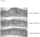
- Epithelial compartments of the HFs were intact with concentric layers of inner and outer root sheaths, hair shaft and sebaceous glands ( Figure 2 ).
- An antibody reactive with human but not mouse COX IV stained follicular epithelium and dermal fibroblasts of grafts Figure 4 ).
- HFs in anagen phase HFs had more concentrated immunoreactivity to Ki-67 in the region of the hair matrix relative to the overlying epidermis ( Figure 7 ).
- the companion layer as identified by keratin 75 staining was present between the inner and outer root sheaths ( Figure 8B ).
- the basal layer of the outer root sheath was immunoreactive for Keratin 15, a marker of HF stem cells located in the bulge region ( Figure 8A ).
- Skin Substitutes Three-dimensional in vitro constructs were prepared for grafting using established methods modified as described herein. Briefly, human dermal papilla cells were mixed with 1 mg/ml type I collagen (rat) (in other embodiments, the collagen could be bovine) in 10% FBS/DMEM, and added to 6 well transwell plates (Corning Incorporated, Corning, NY) at a density of 1.5 X 10 5 cells per cm 2 . The dermal equivalents were cultured in 10% FBS/DMEM for 4 days before aliquoting 1 X 10 6 keratinocytes on top.
- human dermal papilla cells were mixed with 1 mg/ml type I collagen (rat) (in other embodiments, the collagen could be bovine) in 10% FBS/DMEM, and added to 6 well transwell plates (Corning Incorporated, Corning, NY) at a density of 1.5 X 10 5 cells per cm 2 .
- the dermal equivalents were cultured in 10% FBS/DMEM for 4 days before ali
- the constructs were cultured submerged for 2 days in a mixture of DMEM and Ham's F12 (3:1) (GIBCO/Invitrogen, Grand Island, NY) containing 0.1% FBS, after which the keratinocytes were brought to the air-liquid interface and cultured in a mixture of DMEM and Ham's F12 (3:1) containing 1% FBS for another 2 days before grafting.
- Cell Clusters Cell aggregates for injection were formed using the hanging droplet method.
- Qiao J. et al. "Hair follicle neogenesis induced by cultured human scalp dermal papilla cells," Regen Med 4(5): 667-76 (2009 ).
- a mixture of human mesenchymal cells and keratinocytes (10:1, 5:1, 1:1, 1:5 or 1:10) was suspended in Dermal Papilla Medium (Promocell).
- the cells were applied in 10- ⁇ l droplets (each droplet contains either 1 x 10 4 or 0.5 x 10 4 cells) in the lid of a 100-mm petri dish oriented so that droplets were hanging upside down.
- aggregates were transferred individually to wells of a 96-well round-bottom assay plate containing 150 ⁇ l Chang medium.
- the wells were precoated with 0.24% methylcellulose medium to prevent adherence of proto-hairs.
- the culture medium was changed every 2-3 days.
- the microspheres may be collected by centrifugation, washed three times with distilled water, and strained to a size of 50-200 ⁇ m in diameter.
- the microspheres may be lyophilized and sterilized with ultraviolet light for 6 hours.
- Human mesenchymal cells (2.5 ⁇ 10 7 cells) and keratinocytes (6 ⁇ 10 6 cells) may be placed with PLGA microspheres (1 ⁇ g microspheres/10 5 cells) in a spinner flask (Bellco Glass Inc., Vineland, NJ) containing 30 ml of serum-free KGM containing 10 ng/ml of EGF for keratinocytes, or DMEM/F12 containing 10% (v/v) FBS for mesenchymal cells, and cultured at 50 rpm for 2 weeks.
- the medium may be exchanged every other day.
- Cell aggregates may be allowed to settle down, 16 ml of the culture supernatant may be collected and centrifuged, 15 ml of the supernatant may be removed, and 15 ml of fresh medium may be added to the centrifuged cells in 1 ml of remaining supernatant.
- the cells in fresh medium may be transferred to the spinner flasks.
- clusters of cells may be formed by suspending the cells in sodium alginate and then forming spherical droplets using a high-voltage electric droplet generator as described in Lin C.M. et al., "Microencapsulated human hair dermal papilla cells: a substitute for dermal papilla?," Arch Dermatol Res. 300(9):531-5 (2008 ).
- mice were grafted in a horizontal laminar flow hood using 6-8 week old female Cr:NIH(S)-nu/nu mice (FCRDC, Frederick, MD) anesthetized using inhalant anesthesia with a mixture of O 2 and isoflurane (2-4%).
- the grafting area on back of the mouse was carefully estimated, and skin was removed using curved scissors after washing with povidine and 70% ethanol.
- Skin substitutes were placed on the graft bed in correct anatomical orientation, covered with sterile petroleum jelly gauze, and secured with bandages.
- the mice were transferred back to the sterile cages after reawakening.
- the bandages were changed at 2 weeks and removed after 4 weeks. Mice are sacrificed 4-18 weeks after grafting.
- the injection was performed by first piercing the skin, then directing the needle back upward toward the surface and injecting the cells as superficially as possible. This led to the formation of a well-demarcated papule in the center of the injected area. Positive controls received 1 million dermal cells isolated from the same C57BI/6 mice skin and 50,000 epidermal aggregates. 2 to 4 weeks post implantation HF formation was analyzed on the injection site by dissecting and viewing ventral side of the patch skin microscopically.
- Implantation of Cells After anesthetizing, small incisions approximately 0.5-1.0 mm in width and length may be made using a 27-gauge needle. A single cultured aggregate (proto-hair) may be inserted at a shallow position within each incision. Following insertion, incisions may be left to heal.
- full-thickness skin wounds may be created on the transplantation area.
- the skin at the wound margins may be burned using a cautery and fixed to adjacent muscle layers with nonresorbable 5-0 nylon sutures (AILEE Co., Pusan, Korea).
- Mesenchymal cells approximately 10 8 cells/wound
- keratinocytes approximately 7.5 ⁇ 10 6 cells/wound
- PLGA microspheres may be transplanted to the wounds using a 1-mL syringe without a needle.
- the wounds may be dressed with dressing materials, Tegaderm (3M Health Care, St. Paul, MN) and sterile cotton gauze, and firmly fixed using Coban, a self-adhesive wrap (3M Health Care).
- An antibiotic (Cefazolin, 0.1 mg/mouse, Yuhan Co., Seoul, Korea) and an analgesic (Buprenorphine, 0.1 mg/kg, Hanlim Pharm Co., Seoul, Korea) may be administered intramuscularly and subcutaneously, respectively, for 5 days after transplantation.
- the mice may be housed singly after surgery and receive humane care in compliance with the guidelines for the care and use of laboratory animals of NIH.
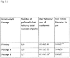
- Table 2 shows the results of the grafting experiments comparing human dermal papilla cells isolated from the temporal versus occipital scalp.
- the second column in Table 2 shows the number of grafts in which the epidermis showed a typical human epidermal morphology by H&E staining.
- Dermal papilla cells from the temporal scalp supported the development of the human keratinocytes into a stratified squamous epithelium much better than those from the occipital scalp.
- the mouse keratinocytes from the borders of the graft migrated in and replaced the human keratinocytes.
- the third column in Table 2 indicates the number of grafts with hair follicles.
- Human hair follicles formed using dermal papilla cells from 3/6 donors from the temporal scalp, but none from the occipital scalp. Hair follicles were observed in at least 80% of grafts using dermal papilla cells from 3 patients, including 19 out of 22 grafts from the trichogenic samples. No hair follicles were observed in grafts using dermal papilla cells from the occipital scalp. The data in parentheses indicates that there was no evidence for the formation of chimeric hair follicles (no induction of hair follicles by the dermal papilla cells in mouse keratinocytes overlying the graft).
- Fig. 13 provides H&E stain showing a hair shaft emerging from the infundibulum of a human hair follicle in a construct 8 weeks after grafting.
- Fig. 14 is a dermal-epidermal composite photographed under a light microscope after 10 weeks. Hair shafts can be seen emerging from the grafted region.
- Fig. 15A provides a representative graft with hair shafts visible 12 weeks after grafting, while Fig. 15B provides a magnified view showing the presence of pigmented hair shafts.
- Figs. 16A-C show that different hair follicle stages could be detected in grafts after 15 weeks.
- Telogen hair follicles showed no Ki-67 positive cells, consistent with telogen stage of hair follicle ( Fig. 17A , arrow).
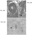
- FIG. 18A shows a representative H&E stained section of a graft that was harvested after 8 weeks, with hair follicle inner and outer root sheath and sebaceous gland visible.
- Fig. 18B shows that the sebaceous gland was highly immunoreactive to an antibody for cathelicidin, an antimicrobial peptide.
- keratinocytes used in the dermal-epidermal composite, which were constructed from neural crest derived human dermal papilla and neonatal foreskin epidermal cells, and grafted into nude mice.
- the effects of primary, first passage, and third passage human keratinocyte cultures are shown in Table 3.
Description
- The present invention relates to compositions in the form of skin substitutes and microspheres comprising neural crest-derived cells, such as hair follicle dermal cells, e.g., dermal papilla ("DP") cells, or dermal sheath cells, that are capable of inducing hair follicles ("HFs"). The present invention also relates to methods, uses, and compositions for inducing HF growth and neogenesis. In some embodiments, the present invention can be used for the treatment of full- or partial-thickness skin loss, wounds, burns, scars, and full- or partial-hair loss.
- Studies of hair and skin continue to be at the forefront of regenerative medicine. Skin substitutes were among the earliest products to be developed using principles of tissue engineering, and the success of these ventures is evident in the clinical use of several commercially available products. In addition, hair restoration is one of the fastest growing areas of cosmetic therapies for both men and women.
- The current clinical "gold standard" for treating major skin injuries involves the use of split-thickness skin autografts, which involves transplanting the epidermis with a portion of the dermis from one location on a patient to another. In cases where there is insufficient donor skin to cover the wounds, however, skin substitutes may be used. Skin substitutes available today have varied compositions, but generally comprise a nonliving collagen matrix and different combinations of keratinocytes and fibroblasts. For example, APLIGRAF® (Organogenesis, Inc., Canton, MA), which is reported to be the most clinically successful composite skin substitute currently available, is composed of allogeneic neonatal fibroblasts in bovine type I collagen overlaid with allogeneic neonatal keratinocytes.
- However, currently available skin substitutes cannot perform all the functions of normal skin. For example, hair follicle (HF) neogenesis is not observed using any currently available skin substitute, which limits their use in patients. HFs and their associated sebaceous glands are important for appearance, skin hydration, barrier formation, and protection against pathogens. In addition, HFs store epidermal stem cells that may be called upon during wound healing. Thus, skin with HFs heals more rapidly than skin without HFs. In addition, any stem cells that might exist in skin lacking HFs are located in superficial layers of the epidermis, making the cells susceptible to loss through minor trauma and damage through ultraviolet light. Thus, treatments that involve neogenesis of normal HFs would find much wider application for restoring normal skin function and appearance.
- During embryogenesis, mesenchymal cells signal the overlying epithelium to induce HF formation, and in adults a specialized group of mesenchymal cells, the dermal papilla (DP) cells, have been shown to retain the capacity to induce HF regeneration (Hardy 1992, Reddy et al., 2001, Gharzi et al., 2003). DP cells from rodents induce HFs in a variety of assays (reviewed in Ohyama et al., 2010), but it has been difficult to grow human DP cells that maintain inductive capacity in culture (Ohyama et al., 2012). This is a significant problem since DP cells must be enriched in culture to expand the cells needed for successful clinical use. Recent technological advances have enabled the use of human cells to form chimeric HFs, for example by combining human keratinocytes and rodent mesenchymal cells in chamber assays (Ehama et al., 2006), by combining human scalp dermal papilla cells and mouse epidermal keratinocytes in flap grafts (Qiao et al., 2009), or by injecting human DP cells, grown as spheroids, together with mouse epidermal cells in reconstitution or "patch" assays (Kang et al., 2012).
-
WO 2011/160055 A2 discloses a skin substitute comprising epithelial cells and modified mesenchymal cells, wherein the modified mesenchymal cells have decreased TSC1/TSC2 function, increased mTORC1 function, and/or decreased mTORC2 function compared to wild type mesenchymal cells. The skin substitute is useful for inducing neogenesis of human hair follicles. -
US 2005/0089512 A1 discloses a skin/hair equivalent comprising reconstructed papillae implanted into a reconstructed dermis, wherein the reconstructed papillae comprise dermal papilla cells isolated from the temporal or occipital region of a scalp. - However, while chimeric HFs are highly valuable as investigative tools, they lack clinical utility because the HFs produced by these methods are not fully human constructs (but instead are chimeric rodent/human constructs), are not completely developed, contain hair shafts in the wrong anatomical location, do not exhibit long-term graft survival and normal HF cycling, and/or do not form HFs that contain sebaceous glands. In addition, HFs produced by such methods tend to grow in variable and uncontrollable directions, resulting in unnatural looking hair. Thus, the follicles produced by such methods are not useful for human HF neogenesis in skin lacking hair follicles.
- Thus, a need exists for methods and compositions capable of generating morphologically-correct, fully-developed, non-immunogenic human hair follicles. Such methods and compositions would be useful for treating conditions such as full- or partial-thickness skin loss, wounds, burns, scars, and hair loss. The present invention fills these needs by providing cellular compositions capable of hair growth, neogenesis, and regeneration.
- The scope of this invention is defined by the claims. Any references in the description to methods of treatment refer to the compositions of the present invention for use in a method for treatment of the human (or animal) body by therapy. The present invention provides compositions in the form of skin substitutes and microspheres comprising neural crest-derived cells, wherein the skin substitute is capable of inducing hair follicles that are morphologically-correct and are useful in any application requiring hair follicle formation/neogenesis, or in any condition where hair follicle formation/neogenesis is desired.
- In one embodiment, the skin substitute or microsphere comprises neural crest-derived hair follicle dermal cells. The skin substitute or microsphere further comprises epithelial cells, optionally with collagen. In another embodiment, the skin substitute or microsphere comprises epithelial cells and hair follicle dermal cells, wherein the hair follicle dermal cells are neural crest-derived cells, optionally with collagen. In yet another embodiment, the skin substitute or microsphere comprises epithelial cells and hair follicle dermal cells, optionally with collagen. The hair follicle dermal cells are derived from a temporal region of the scalp. In some aspects the epithelial cells are keratinocytes. In some aspects the skin substitute comprises cells of human origin only.
- In various embodiments, a skin substitute is provided, comprising isolated neural crest-derived hair follicle dermal cells and epithelial cells. In some embodiments, the epithelial cells are keratinocytes, and the hair follicle dermal cells are hair follicle dermal cells derived from the temporal region of the scalp. In various embodiments, the skin substitutes described above contain epithelial cells and neural crest-derived hair follicle dermal cells from a human. In some embodiments, the skin substitutes further comprise collagen. In some embodiments, the keratinocytes or keratinocyte-like cells are induced pluripotent stem (iPS) cells differentiated into keratinocytes or keratinocyte-like cells. In some embodiments, the hair follicle dermal cells and epithelial cells are taken from the same donor and/or the same body regions of a donor (e.g., the hair follicle dermal cells and epithelial cells are taken from tissue in the same donor region, e.g., keratinocytes and hair follicle dermal cells from the temporal region of the scalp). In some embodiments, the hair follicle dermal cells and epithelial cells are taken from different donors and/or different body regions of a donor (e.g., the hair follicle dermal cells and epithelial cells are not taken from tissue in the same donor region, e.g., keratinocytes and mesenchymal cells from the temporal region of the scalp).
- In various embodiments, a skin substitute is provided, comprising epithelial cells and hair follicle dermal cells. In some embodiments, the epithelial cells are keratinocytes. The hair follicle dermal cells are derived from the temporal region of the scalp In various embodiments, the skin substitutes described above contain epithelial cells and neural crest-derived hair follicle dermal cells from a human. In some embodiments, the skin substitutes further comprise collagen. In some embodiments, the skin substitutes are combined in therapeutically effective concentrations (e.g., concentrations not naturally found in combination in host tissue) and/or stored in a non-naturally occurring culture medium (e.g., Hanks media, keratinocyte-conditioned medium, or other cell culture media).
- In various embodiments, the skin substitutes described above comprise hair follicle dermal cells provided within a matrix, e.g., a ground substance matrix or a collagen matrix such as a collagen type I matrix. In various embodiments, the skin substitutes described above are provided in a suspension such as a microsphere.
- In various embodiments, the skin substitutes described above comprise keratinocytes that are from one or more of neonatal foreskin keratinocytes, adult keratinocytes, or keratinocyte-like cells derived from pluripotential stem cells or from epithelial cells. In some embodiments, the epithelial cells are primary cells or early passage cells (e.g., first through fourth passage, more preferably first or second passage).. In some embodiments, the primary or early passage epithelial cells are hair follicle dermal cells. In some embodiments, the cells are the cells are passaged in keratinocyte-conditioned medium.
- In various embodiments, the epithelial cells and hair follicle dermal cells can be autologous. In some embodiments, the cells are allogenic.
- According to this invention, the hair follicle dermal cells, including dermal papilla cells and dermal sheath cells, are isolated from a temporal region of the scalp.
- The skin substitutes disclosed may be used in methods for transplanting cells capable of inducing human hair follicles, comprising delivering to a human subject any one of the skin substitutes discussed above. The epithelial cells are transplanted in combination with the hair follicle dermal cells and in combination with hair follicle dermal cells at therapeutically effective concentrations (e.g., concentrations not naturally found in combination in host tissue). Once the transplanted the cells can be used to induce hair follicle growth or hair follicle neogenesis.
- The skin substitutes disclosed may be suitable for transplanting, for example where, the subject to be treated with any of the compositions described above has a partial-thickness skin loss, full-thickness skin loss, a wound, a burn, a scar, or hair loss. In some embodiments, transplanting the skin substitute induces eccrine glands and/or sebaceous glands.
- In some embodiments, the epithelial cells and hair follicle dermal cells are for use in the methods described above, or are formulated for use in the methods, or are used in the preparation of a medicament for use in the methods described above.
- Also disclosed herein are microspheres comprising neural crest-derived mesenchymal cells and epithelial cells. According to the present invention, the microspheres comprise both hair follicle dermal cells and epithelial cells (e.g., keratinocytes). The hair follicle dermal cells, such as dermal papilla cells, dermal sheath cells, or hair follicle dermal cells are derived from a temporal region of the scalp. The scalp derived hair follicle dermal cells are from a temporal, region of the scalp, but not derived from an occipital or nape region of the scalp. In some embodiments, the epithelial cells and neural crest-derived hair follicle dermal cells in the microspheres are human cells, and may further comprise collagen. For example, in certain embodiments, the microsphere comprises scalp- derived hair follicle dermal cells (e.g., human cells), wherein the hair follicle dermal cells are not derived from an occipital or nape region of the scalp.
- In some embodiments, the microspheres described above can further comprise a matrix, e.g., a ground substance matrix or a collagen matrix such as a collagen type I matrix to contain the dermal. In some embodiments, the keratinocytes in the microspheres are from one or more of neonatal foreskin keratinocytes, adult keratinocytes, or keratinocyte-like cells derived from pluripotential stem cells or from epithelial cells. In some embodiments, the epithelial cells in microspheres are primary cells or early passage cells (e.g., first through fourth passage, more preferably first or second passage). In some embodiments, the primary or early passage epithelial cells are hair follicle dermal cells. In some embodiments, the cells are the cells are passaged in keratinocyte-conditioned medium.
- In various embodiments, the microspheres described above comprise autologous mesenchymal cells and/or epithelial cells. In some embodiments, the mesenchymal cells and/or epithelial cells are allogenic.
- The microspheres disclosed can be used in methods for transplanting cells capable of inducing human hair follicles to a subject, comprising delivering to a human subject any of the microspheres described above. The microsphere can be subdermally or intradermally delivered to a subject. The microspheres disclosed can be used in a method for inducing hair follicle growth or hair follicle neogenesis, comprising delivering to a human subject any of the microspheres described above. The microsphere can be subdermally or intradermally delivered to a subject with partial-thickness skin loss, full-thickness skin loss, a wound, a burn, a scar, or hair loss.
- In various embodiments, a composition is provided herein for use in inducing hair follicle growth or hair follicle neogenesis in a subject, comprising any of the microspheres described above. Also disclosed herein are the microspheres described above for use in the manufacture of a medicament for inducing hair follicle formation or for inducing hair follicle neogenesis in a subject. In various embodiments, the subject has partial-thickness skin loss, full-thickness skin loss, a wound, a burn, a scar, or hair loss.
- Also disclosed herein are methods for making a skin substitute, comprising (a) mixing a culture of primary or early-passage neural crest-derived hair follicle dermal cells with a matrix; and (b) overlaying a culture of primary or early-passage epithelial cells onto the mixture of (a). For example, the method can comprise (a) mixing a culture of scalp -derived hair follicle dermal cells with a matrix; and (b) overlaying a culture of primary or early-passage epithelial cells onto the mixture of (a), wherein the hair follicle dermal cells are not derived from an occipital region of the scalp. As another example, the method can comprise (a) mixing a culture of hair follicle dermal cells with a matrix; and (b) overlaying a culture of primary or early-passage epithelial cells onto the mixture of (a), wherein the hair follicle dermal cells are not derived from an occipital region of the scalp. In various embodiments, the matrix is a collagen matrix (e.g., a collagen type I matrix). In various embodiments, the cells are cultured in keratinocyte-conditioned medium.
- In certain embodiments, the compositions described and exemplified herein induce hair follicle formation when provided to human subjects, wherein the hair follicle that is formed is fully human and therefore does not elicit a host immune response.
- The accompanying drawings, which are incorporated in and constitute a part of this specification, illustrate several non-limiting embodiments of the invention and together with the description, serve to explain the principles of the invention.
-
Fig. 1 shows pilosebaceous units, including hair follicles, formed by grafting a skin substitute comprising human neonatal foreskin keratinocytes, human dermal papilla cells, and type I collagen. Sections were obtained 8 weeks after grafting onto nude mice. -
Fig. 2 shows that pilosebaceous units formed by grafting a skin substitute comprising human neonatal foreskin keratinocytes, human dermal papilla cells, and type I collagen are complete with dermal papillae, hair shafts, inner root sheaths (IRS), outer root sheaths (ORS), and sebaceous glands. -
Fig. 3A shows the hybridization of a fluorescein-labeled oligonucleotide probe for human Alu DNA to grafts containing hair follicles formed by grafting a skin substitute comprising human neonatal foreskin keratinocytes, human dermal papilla cells, and type I collagen. Positive signal (green) in the hair follicles indicates that the cells are of human origin.Fig. 3B shows a dermal papilla formed by grafting a skin substitute comprising human neonatal foreskin keratinocytes, human dermal papilla cells, and type I collagen, hybridized with the fluorescein-labeled oligonucleotide probe for human Alu DNA. Positive signal indicates that the dermal papilla is composed of cells of human origin. -
Fig. 4 shows hair follicles formed by grafting a skin substitute comprising human neonatal foreskin keratinocytes, human dermal papilla cells, and type I collagen, stained with an antibody that recognizes human COX IV. Positive staining (red) in both epithelial and mesenchymal cells (left) indicates that both are derived from the human cells contained within the graft. No staining is seen in mouse follicles (right). -
Figs. 5A and 5B show that the dermal papilla niche in the graft formed by a skin substitute comprising human neonatal foreskin keratinocytes, human dermal papilla cells, and type I collagen is formed from the dermal papilla cells.Fig. 5A shows positive signal in fluorescent in situ hybridization analysis using a human-specific pan-centromeric probe (green) shows that both the hair follicle and dermal papilla are derived from human cells.Fig. 5B uses a human Y chromosome-specific probe (red) to mark the hair follicle. The probe bound to foreskin keratinocytes (male origin), and not the dermal papilla cells (female origin), indicating that the newly formed dermal papilla was of female origin. -
Figs. 6A, 6B, and 6C show that grafts formed by a skin substitute comprising human neonatal foreskin keratinocytes, human dermal papilla cells, and type I collagen stain positively for markers of fully developed hair follicles.Fig. 6A : alkaline phosphatase activity (blue stain) is observed in the dermal papilla and lower dermal sheath.Fig. 6B : a human-specific Nestin antibody stains cells in the dermal papilla and lower dermal sheath.Fig. 6C : a human-specific Versican antibody stains mesenchymal cells in the dermal sheath region. -
Fig. 7 shows that proliferating cells in the graft formed by a skin substitute comprising human neonatal foreskin keratinocytes, human dermal papilla cells, and type I collagen are scattered in the basal layer of the epidermis and dense in the hair follicle matrix. Red staining shows immunoreactivity against Ki-67, a marker of cell proliferation. -
Figs. 8A and 8B show that hair follicles in grafts formed by a skin substitute comprising human neonatal foreskin keratinocytes, human dermal papilla cells, and type I collagen display normal expression ofKeratin 15 andKeratin 75.Fig. 8A :Keratin 15 is immunoreactive in the basal layer of the outer root sheath.Fig. 8B :Keratin 75 is immunoreactive in the hair follicle companion layer. -
Fig. 9 shows a schematic of certain regions of the scalp. -
Fig. 10 shows hair follicles in the grafts made with HDP47 and three different passages of keratinocytes (p0 (top), p1 (middle), and p3 (bottom)). Primary or P1 keratinocytes form more hair follicles than P3 keratinocytes. -
Fig. 11 shows the tabulated results of the experiment shown inFig. 10 . The optimal passage of human keratinocytes for hair follicle regeneration is the early passage keratinocytes. The early passage grafts had more and larger hair follicles with widest hair shafts. * Significantly less than primary keratinocytes, p<0.05, ** Significantly greater than passaged keratinocytes, p<0.02 -
Fig. 12 shows that dermal papilla microspheres augment HF formation in both reconstitution assays and in skin grafts. The bottom left two images show human DP spheroids induce chimeric HFs in reconstitution assay. Hair fibers are seen in the hypodermis 4 weeks following injection of a mixture of human DP spheroids and mouse epidermal cells (left-most image). Human DP 2D cultures do not induce HF. H&E stained horizontal section of injection cyst forming HFs and sebaceous glands (second image from left). The bottom right two images show that human DP spheroids induce human HFs in a reconstitution assay. Appearance of graft, 8 weeks after grafting on the back of a nude mouse, is shown in the first of the two images. A representative H&E stained graft section showing hair follicle with hair shaft is provided in the right-most image. -
Fig. 13 is an H&E stain showing a hair shaft emerging from the infundibulum of a human hair follicle in a dermal-epidermal composite construct 8 weeks after grafting. -
Fig. 14 is a dermal-epidermal composite (dermal papilla cells from temporal scalp dermis and neonatal foreskin keratinocytes) photographed under a dissection microscope after 10 weeks. -
Fig. 15A shows a representative dermal-epidermal composite graft with hair shafts visible 12 weeks after grafting, whileFig. 15B provides a magnified view showing the presence of pigmented hair shafts. -
Figs. 16A, 16B, and 16C show H&E and toluidine blue staining in dermal-epidermal composite grafts after 15 weeks.Figs. 16A-C show telogen hair follicles, confirmed by club-like appearance and spiky keratin fibers (Fig. 16A ), secondary hair germ with adjacent hair papilla (Fig. 16B ), and presence of a cornified club (Fig. 16C , toluidine blue staining). -
Figs. 17A and 17B show Ki-67 staining of dermal-epidermal composite grafts.Fig. 17A shows that telogen hair follicles do not contain Ki-67-positive cells, consistent with the telogen stage of the hair follicle (arrow).Fig. 17B shows an anagen hair follicle with dermal papilla (arrow), including dense Ki-67 reactivity. -
Figs. 18A and 18B. Fig. 18A shows a representative H&E stained section of a dermal-epidermal composite graft, showing hair follicle inner and outer root sheath, and sebaceous gland.Fig. 18B is antibody staining for cathelicidin in the dermal-epidermal composite graft, showing that sebaceous gland was highly immunoreactive to an antibody for cathelicidin, an antimicrobial peptide. - The invention provides a skin substitute comprising, in a suspension, epithelial cells and hair follicle dermal cells derived from a temporal region of the scalp. In one embodiment, the hair follicle dermal cells, include, for example, dermal papilla cells and dermal sheath cells. The neural crest-derived hair follicle dermal cells may be derived from the scalp. In one embodiment, the neural crest-derived hair follicle dermal cells are derived from a temporal, region of the scalp. In some aspects, the neural crest-derived hair follicle dermal cells are not derived from an occipital or nape region of the scalp. In some aspects, the epithelial cells and neural crest-derived hair follicle dermal cells are human. In some aspects, the epithelial cells are keratinocytes or keratinocyte-like cells. The skin substitute may further comprise collagen.
- A skin substitute comprising epithelial cells and scalp- derived hair follicle dermal cells, wherein the hair follicle dermal cells are not derived from an occipital or nape region of the scalp is also encompassed. The hair follicle dermal cells may be dermal papilla cells or dermal sheath cells. In some aspects that epithelial cells may be keratinocytes or keratinocyte-like cells. According to the present invention the dermal papilla cells are derived from a temporal region of the scalp. In some aspects the scalp- derived hair follicle dermal cells and the epithelial cells are human. The skin substitute may further comprise collagen.
- In another embodiment, a skin substitute comprising epithelial cells and hair follicle dermal cells, wherein the dermal cells are not derived from an occipital or nape region of the scalp is encompassed. In some embodiments the epithelial cells are keratinocytes or keratinocyte-like cells. In some embodiments, the hair follicle dermal cells are dermal papilla cells or dermal sheath cells. In some aspects the cells of the skin substitute are human. According to the present invention the hair follicle dermal cells are derived from a temporal region of the scalp. The skin substitute may further comprise collagen.
- In some embodiments, the skin substitutes described herein are provided with a matrix. The matrix may be a collagen matrix or a ground substance matrix. In some embodiments, the matrix is a type I collagen matrix.
- In certain embodiments, the epithelial cells of the skin substitute comprise keratinocytes. In some aspects the keratinocytes are neonatal foreskin keratinocytes (NFK).
- The skin substitutes comprising epithelial cells may comprise primary cells or early-passage epithelial cells, wherein early-passage cells are from a first, second, or third passage. In some aspects, the epithelial cells are keratinocytes and the keratinocytes are primary cells or early-passage keratinocyte cells, wherein early-passage cells are from a first, second, or third passage.
- The skin substitute comprising hair follicle dermal cells may comprise primary cells or early-passage cells, wherein early-passage cells are from a first, second, third, or fourth passage. In some embodiments, the skin substitute comprises primary or early-passage dermal papilla cells, wherein early-passage dermal papilla cells are from a first, second, third, or fourth passage.
- The epithelial cells and hair follicle dermal cells may be derived from a same or different donor. The keratinocytes and dermal papilla cells may be derived from a same or different donor. In some aspects, the cells of the skin substitute are passaged in keratinocyte-conditioned medium. In some embodiments, the epithelial cells and hair follicle dermal cells are autologous. In some aspects the epithelial cells and hair follicle dermal cells are allogenic.
- Compositions for use in inducing hair follicle growth or hair follicle neogenesis comprising the skin substitute of any one of the claims are fully encompassed.
- Uses of a skin substitute of any one of the claims in the manufacture of a medicament for inducing hair follicle formation or for inducing hair follicle neogenesis are encompassed.
- In some embodiments, a subject in need of inducing hair follicle growth has partial-thickness skin loss, full-thickness skin loss, a wound, a burn, a scar, or hair loss.
- In some embodiments, the skin substitute of the invention induces eccrine glands. In some embodiments, the skin substitute of the invention induces sebaceous glands.
- Methods of making skin substitutes, comprising (a) mixing a culture of primary or early-passage neural crest-derived hair follicle dermal cells with a matrix; and (b) overlaying a culture of primary or early-passage epithelial cells onto the mixture of (a) are encompassed, as are methods of making skin substitutes, comprising (a) mixing a culture of scalp-derived hair follicle dermal cells with a matrix; and (b) overlaying a culture of primary or early-passage epithelial cells onto the mixture of (a), wherein the hair follicle dermal cells are not derived from an occipital region of the scalp. In some embodiments, methods of making skin substitutes, comprising (a) mixing a culture of hair follicle dermal cells, such as dermal papilla cells or dermal sheath cells, with a matrix; and (b) overlaying a culture of primary or early-passage epithelial cells onto the mixture of (a), wherein the dermal papilla cells are not derived from an occipital region of the scalp are encompassed.
- In some embodiments, compositions comprising human dermal papilla cells for inducing hair follicle neogenesis are encompassed. According to the present invention the human dermal papilla cells are isolated from the temporal region of human scalp. The human dermal papilla cells are not isolated from the occipital or nape region of human scalp. The composition may comprise human dermal papilla cells and human keratinocytes. The composition may comprise human dermal papilla cells, human keratinocytes, and collagen.
- The compositions disclosed can be used in a method for inducing human hair follicle growth in humans. The method can comprise delivering to a human subject a composition comprising human dermal papilla cells and human keratinocytes, optionally in combination with collagen. In some aspects the composition is delivered subdermally or intradermally.
- The compositions disclosed can be used in a method comprising delivering to a human subject a composition comprising human dermal papilla cells that are derived from the temporal region of the human scalp together with human keratinocytes, optionally in combination with collagen. In some aspects the composition is delivered subdermally or intradermally.
- The invention also provides uses for a composition comprising human dermal papilla cells and human keratinocytes, optionally in combination with collagen, in the manufacture of a medicament for inducing hair follicle formation or for inducing hair follicle neogenesis. In some aspects the composition is delivered subdermally or intradermally.
- The invention also provides a pharmaceutical composition comprising human dermal papilla cells and human keratinocytes, optionally in combination with collagen, for use in treating a subject who is at risk for, diagnosed with, or who has hair loss or is in need of hair follicle neogenesis.
- The skin substitutes disclosed can be used in methods and uses comprising grafting to a human subject a composition or skin substitute of the invention. In some aspect the human subject is in need of hair growth.
- The compositions or skin substitutes disclosed can be used in methods comprising delivering to a human subject a composition or skin substitute of the invention, wherein the subject has partial-thickness skin loss, full-thickness skin loss, a wound, a burn, a scar, or hair loss. The method and use induces formation of eccrine glands. The method and use induces formation of sebaceous glands.
- In one embodiment, the neural crest-derived hair follicle dermal cells, such as, for example, dermal papilla cells, are incorporated into a microsphere. In another embodiment, the microsphere further comprises epithelial cells. In another embodiment, the microspheres are formed by mixing human dermal papilla cells and keratinocytes (e.g., neonatal foreskin keratinocytes) in a 1:1 mixture of dermal papilla medium and keratinocyte serum free medium, and incubating the clusters for about four weeks. In another embodiment, the neural crest-derived mesenchymal cells or hair follicle dermal cells are provided with a matrix. In yet another embodiment, the matrix is a collagen matrix or a ground substance matrix. In yet another embodiment, the matrix is a type I collagen matrix. In yet another embodiment, the matrix is a rat type I collagen matrix, a bovine type I collagen matrix, or a human type I collagen matrix.
- In one embodiment, the epithelial cells comprise one or more epithelial cells from different sources. In another embodiment, the epithelial cells are keratinocytes or keratinocyte-like cells. In yet another embodiment, the keratinocytes are neonatal foreskin keratinocytes (NFKs). In another embodiment, the keratinocytes or keratinocyte-like cells are induced pluripotent stem (iPS) cells differentiated into keratinocytes.
- In one embodiment, the mesenchymal cells and/or epithelial cells are derived from the same donor. In another embodiment, the donor is the patient. In yet another embodiment, the mesenchymal cells and/or epithelial cells are derived from different donors. In yet another embodiment, the donor of either the mesenchymal or epithelial cells is the patient.
- As used herein, the singular forms "a" "an" and "the" are intended to include the plural forms as well, unless the context clearly indicates otherwise. Furthermore, to the extent that the terms "including," "includes," "having," "has," "with," or variants thereof are used in either the detailed description and/or the claims, such terms are intended to be inclusive in a manner similar to the term "comprising."
- As used herein, the terms "about" and "approximately" mean within an acceptable error range for the particular value as determined by one of ordinary skill in the art, which will depend in part on how the value is measured or determined, i.e., the limitations of the measurement system. For example, "about" can mean from 1 to 1.5 standard deviation(s) or from 1 to 2 standard deviations, per the practice in the art. Alternatively, "about" can mean a range of up to and including 20%, 10%, 5%, or 1% of a given value. Alternatively, particularly with respect to biological systems or processes, the term can mean up to and including an order of magnitude, up to and including 5-fold, and up to and including 2-fold, of a value. Where particular values are described in the application and claims, unless otherwise stated the term "about" meaning within an acceptable error range for the particular value should be assumed.
- As used herein, the term "apocrine gland" refers to glands in the skin that have a coiled, tubular excretory portion with widely dilated lumen, lined by cuboidal epithelial cells with eosinophilic cytoplasm and apical snouts, and an outer discontinuous layer of myoepithelial cells resting on a prominent basement membrane.
- As used herein, the term "composition" refers to a mixture that contains a therapeutically active component(s) and a carrier, such as a pharmaceutically acceptable carrier or excipient that is conventional in the art and which is suitable for administration to a subject for therapeutic purposes. The therapeutically active component may include the mesenchymal cells of the invention. In other embodiments, term "composition" refers to the skin substitutes of the invention, which are described in further detail below. The compositions of the invention may further comprise a matrix, which is defined below.
- As used herein, the term "dermal papilla" refers to the follicular dermal papilla, i.e., the mesenchymal cell condensation at the base of the hair follicle.
- As used herein, the term "dermal sheath" refers to the region of connective tissue that envelops the hair follicle.
- As used herein, the term "eccrine glands" refers to sweat glands in the skin. Eccrine glands consist of two anatomical portions: (1) the secretory coil, located in the deep dermis at the junction with the subcutaneous tissue and composed of clear pyramidal cells and dark-stained cells, surrounded by a single outer discontinuous layer of myoepithelial cells resting on a well-defined basement membrane; and (2) the excretory part composed of a straight intradermal portion and an intraepidermal spiral portion (acrosyringium), and a double layer of small cuboidal cells with no underlying myoepithelial layer.
- As used herein, the term "endothelial cell" refers to the specialized cells that line the inner walls of blood vessels.
- As used herein, the term "epidermal cell" refers to cells derived from the epidermis of the skin. Epidermal cells are one type of epithelial cells. Examples of epidermal cells include, but are not limited to keratinocytes, melanocytes, Langerhans cells, and Merkel cells.
- As used herein, the term "epithelial cell" refers to cells that line the outside (skin), mucous membranes, and the inside cavities and lumina of the body. In particular embodiments, the term "epithelial cell" refers to stratified squamous epithelial cells. Most epithelial cells exhibit an apical-basal polarization of cellular components. Epithelial cells are typically classified by shape and by their specialization. For example, squamous epithelial cells are thin and have an irregular flattened shape mainly defined by the nucleus. Squamous cells typically line surfaces of body cavities, such as the esophagus. Specialized squamous epithelia line blood vessels (endothelial cells) and the heart (mesothelial cells). Cuboidal epithelial cells are cube-shaped and usually have their nucleus in the center. Cuboidal epithelial cells are typically found in secretive or absorptive tissue, e.g., kidney tubules, glandular ducts, and the pancreatic exocrine gland. Columnar epithelial cells are longer than they are wide and the elongated nucleus is usually near the base of the cell. These cells also have tiny projections, called microvilli, which increase the surface area of the cells. Columnar epithelial cells typically form the lining of the stomach and intestines, as well as sensory organs.
- As used herein, the term "hair follicle" or "HF" refers to a tubular infolding of the epidermis from which a hair may grow. A hair follicle may contain a hair shaft in the correct anatomical location, exhibit long-term graft survival, normal hair follicle cycling, and sebaceous glands.
- As used herein, the term "hair follicle dermal cell" refers to mesenchymal cells in the dermis, including dermal papilla cells and dermal sheath cells.
- As used herein, the term "hair regeneration" refers to the stimulation of existing quiescent hair follicles to enter the anagen phase of hair growth. The term also refers to stimulation of hair formation from hair follicle remnants or components of hair follicles (e.g., implantation of microdissected dermal papilla with or without follicular epithelium, or hair growth after plucking), rather than starting with intact quiescent hair follicles.
- As used herein, the term "hair neogenesis" refers to the stimulation of de novo hair follicle growth where no hair follicle previously existed in skin with no preexisting hair follicles, or in skin with fewer than the desired number of hair follicles.
- As used herein, the term "keratinocyte" refers to epithelial cells in the epidermis of the skin (including cells in the follicular epithelium) that undergo cell division and stratification from basal cells in contact with the epidermal basement membrane into squamous cells. Keratinocytes express keratin. In some embodiments, keratinocytes can be derived from iPS cells.
- As used herein, the term "keratinocyte-like cell" refers to cells that express keratin and have the ability to form a stratified squamous epithelium or follicular epithelium. Keratinocyte-like cells may be derived from skin cells or other organs such as bone marrow or trachea, or from cells with stem-cell features (including embryonic stem cells) or that induce pluripotent stem cells. In some embodiments, keratinocyte-like cells can be derived from iPS cells.
- As used herein, the terms "matrix" and "ground substance" refer to any natural or synthetic extracellular matrix-like composition capable of forming a hydrated gel-like cellular support. Cells may be deposited within or on matrices and ground substances. Matrices and ground substances may comprise one or more fibrous proteins having both structural and adhesive functions. Such proteins include, but are not limited to elastin, fibronectin, laminin, and collagens I, II, III, IV, V, VI, VII, VIII, IX X, XI, and XII. Alternatively, or in addition, matrices and ground substances may comprise proteoglycan molecules comprising polysaccharide chains covalently linked to proteins. Such proteoglycans include, but are not limited to, hyaluronan-, heparin sulfate-, chondroitin-, keratin sulfate-, and dermatin sulfate-linked proteins.
- As used herein, the term "mesenchymal cell" refers to multipotent cells with the capacity or potential capacity to induce hair follicle formation similar to cells of the dermal papilla and connective tissue sheath from hair follicles. Mesenchymal cells are usually considered mesodermal connective tissue cells that express vimentin, but cells with the desired attributes may also be neural crest derived. Mesenchymal cells may be isolated from one or more of the following sources: patient skin or mucosa for autologous cells; donor skin or mucosa for allogeneic cells; normal skin or mucosa; skin with an adnexal tumor; and other tissues (e.g. fat, bone marrow). Mesenchymal cells include, but are not limited to, fibroblasts, dermal papilla cells, dermal sheath cells, onychofibroblasts (fibroblasts from nail unit), dental pulp cells, periodontal ligament cells, neural crest cells, adnexal tumor cells, induced pluripotent stem cells, and mesenchymal stem cells from bone marrow, umbilical cord blood, umbilical cord, fat, and other organs.
- As used herein, the terms "morphologically correct" and "fully developed" refers to hair follicles that have a normal configuration with an epithelial filament coming out of the distal end of the follicle and dermal papilla sitting at the base of the follicle. The follicles also have cells proliferating at the base of the follicle, and have concentric layers of outer and inner root sheath, cuticle and cortex. The follicles exhibit normal differentiation of the outer root sheath, and have hair shafts and sebaceous glands. The hairs go through normal cycles, and contain an epithelial stem cell component.
- As used herein, the term "pharmaceutically acceptable carrier" refers to a non-toxic solid, semisolid, or liquid filler, diluents, encapsulating material, formulation auxiliary, or excipient of any conventional type. A pharmaceutically acceptable carrier is non-toxic to recipients at the dosages and concentrations employed, and is compatible with other ingredients of the formulation.
- As used herein, the term "neural crest-derived mesenchymal cells" refers to cells having origins in the neural crest (i.e., a transient embryonic structure in vertebrates) that have the capacity to self-renew and display developmental potential. Neural crest cells originate in the ectoderm at the margins of the neural tube and, after a phase of epithelial-mesenchymal transition and extensive migration, settle down in different parts of the body to contribute to the formation of a variety of different tissues and organs. See Shakhova, "Neural crest-derived stem cells," http://www.stembook.org/node/696 (incorporated herein by reference). Neural crest derivatives originate from four major segments of the neuraxis: cranial, cardiac, vagal, and trunk neural crest. Id. Neural crest cells from the trunk are able to produce mesenchymal derivatives. Id.
- As used herein, the term "primary cells" refer to cells harvested and cultured directly from a donor source without further passage.
- As used herein, the term "early-passage cells" refer to cells harvested and cultured from a donor and passaged fewer than three times (in the case of epithelial cells), or fewer than five times (in the case of mesenchymal or hair follicle dermal cells).
- As used herein, the term "sebaceous gland" refers to hair follicle-dependent glands that originate as a budding of sebaceous glands primordium. Sebaceous glands consist of multiple lobules of rounded cells (sebocytes), filled with lipid-containing vacuoles, and rimmed by a single layer of small, dark germinative cells. The lobules converge on a short duct, which empties the lipid content of degenerated sebocytes into the hair follicle.
- As used herein, the terms "skin substitute," "skin equivalent," "dermal-epidermal composite," and "skin graft" refer to any product used for the purpose of damaged skin replacement, fully or partially, temporarily or permanently, and possessing some similarities with human skin, both anatomically or functionally. Skin substitutes include, but are not limited to, bioengineered skin equivalents, tissue-engineered skin, tissue-engineered skin constructs, biological skin substitutes, bioengineered skin substitutes, skin substitute bioconstructs, living skin replacements, dermal-epidermal composites and bioengineered alternative tissue.
- As used herein, the term "microsphere" includes but is not limited to cell clusters and cell aggregates optionally comprising a biodegradable microsphere.
- As used herein, the term "treatment," refers to any administration or application of remedies for a condition in a mammal, including a human, to obtain a desired pharmacological and/or physiological effect. Treatments include inhibiting the condition, arresting its development, or relieving the condition, for example, by restoring or repairing a lost, missing, or defective function, or stimulating an inefficient process, or improving symptoms of the condition.
- As used herein, the term "trichogenic" refers to the ability of a cell to induce a hair follicle and/or to promote hair follicle morphogenesis, i.e., folliculogenesis.
- It has been surprisingly found that neural crest-derived mesenchymal cells are trichogenic. Specifically, mesenchymal cells derived from a region of the face or scalp other than the occipital or nape regions are capable of inducing hair follicles. Such follicles are complete according to the criteria proposed by Chuong et al., "Defining hair follicles in the age of stem cell bioengineering," J. Invest. Dermatol., 127:2098-100 (2007). The follicles have a normal configuration with an epithelial filament coming out of the distal end of the follicle and dermal papilla sitting at the base of the follicle. The follicles have cells proliferating at the base of the follicle, and have concentric layers of outer and inner root sheath, cuticle and cortex. The follicles exhibit normal differentiation of the outer root sheath, and have hair shafts and sebaceous glands. The hairs go through normal cycles, and contain an epithelial stem cell component.
- Accordingly, the invention provides cellular compositions capable of hair neogenesis.
- According to the invention, the trichogenic cells are not derived from an occipital or nape region of the scalp. In another embodiment, the trichogenic cells are hair follicle dermal cells.
- Mammalian skin contains two primary layers: an outer layer called the epidermis and an inner layer called the dermis. The epidermis primarily contains keratinocytes that are formed in the deeper layers of the epidermis by mitosis and then migrate up to the surface, where they are eventually shed. The dermis contains a variety of structures including hair follicles, sebaceous glands, sweat glands, apocrine glands, nerves, lymphatic vessels, and blood vessels.
- Hair follicle morphogenesis takes place mostly in utero during embryogenesis. Hair follicle formation begins with the appearance of epidermal placodes, which mark the location of the new hair follicle. Mesenchymal cells (i.e., inductive multipotent cells) then begin to aggregate in the dermis below the epidermal placodes. The mesenchymal aggregates signal to the keratinocytes in the overlaying placodes, which then begin growing downward into the dermis. When the epidermal keratinocytes reach the mesenchymal aggregates, the cells undergo a series of differentiation and proliferation processes, eventually forming a mature hair follicle.
- Mature hair follicles contain four main parts: the dermal papilla (DP), dermal sheath (DS), follicular epithelium, and hair shaft (
Figure 1D ). The DP is located at the base, or bulb, of the hair follicle adjacent to the hair matrix that produces the hair shaft. The DS is made up of connective tissue and envelops the hair follicle. The follicular epithelium includes the outer root sheath and the inner root sheath. The hair shaft is a proteinaceous structure that extends from the base of the follicle through the epidermis to the exterior of the skin. - The hair follicle is a dynamic miniorgan that repeatedly cycles through periods of growth (anagen), regression (catagen), and quiescence (telogen). The lower portion of the hair follicle regresses or regrows, regenerating in each cycle through complicated interactions between the dermal mesenchymal cells and epidermal cells. The permanent portion of the lower hair follicle above the continuously remodeled part is referred to as the "bulge" because it protrudes slightly from the follicle. The bulge contains multipotent cells capable of forming the follicle, sebaceous gland, and epidermis. As individuals age, the anagen and catagen phases of the hair follicle cycle become shorter, and hair follicles experience a more rapid shift to the telogen phase. As a result, normal hairs are gradually replaced by finer vellus hairs, and in some individuals, the cells may lose their trichogenic properties entirely.
- The skin substitutes of the invention comprise hair follicle dermal cells and epithelial cells. The hair follicle dermal and epithelial cells supplied in the skin substitute interact, with or without the patient's epithelial cells, to produce a hair follicle.
- Mesenchymal cells are usually considered mesodermal connective tissue cells that express vimentin, but vimentin-expressing cells with these attributes may also be neural crest derived. Sources of cells include the inductive multipotent cells of the dermal papilla and connective tissue sheath from hair follicles. It has been surprisingly found that mesenchymal cells (i.e., inductive multipotent cells) isolated from certain sources are trichogenic. For example, the inventors have found that neural crest-derived mesenchymal cells are trichogenic. The inventors have also found that mesenchymal cells, such as hair follicle dermal cells, harvested from the face or scalp at regions other than the occipital region or nape region, are trichogenic.
- Generally, any source of epithelial cells or cell line that can stratify into squamous epithelia are useful in the present invention. Accordingly, the present invention is not limited to the use of any particular source of cells that are capable of differentiating into squamous epithelia. Indeed, the present invention contemplates the use of a variety of cell lines and sources that can differentiate into stratified squamous epithelia. Sources of cells include primary and immortalized keratinocytes, keratinocyte-like cells, and cells with the capacity to be differentiated into keratinocyte-like cells, obtained from humans and cavaderic donors (Auger et al., In Vitro Cell. Dev. Biol.--Animal 36:96-103; and
U.S. Pat. Nos. 5,968,546 and5,693,332 ), neonatal foreskin (Asbill et al., Pharm. Research 17(9):1092-97 (2000); Meana et al., Burns 24:621-30 (1998); andU.S. Pat. Nos. 4,485,096 ;6,039,760 ; and5,536,656 ), and immortalized keratinocytes cell lines such as NM1 cells (Baden, In Vitro Cell. Dev. Biol. 23(3):205-213 (1987)), HaCaT cells (Boucamp et al., J. cell. Boil. 106:761-771 (1988)); and NIKS cells (Cell line BC-1-Ep/SL;U.S. Pat. No. 5,989,837 ; ATCC CRL-12191). - Epithelial cells may also be obtained from: patient skin or mucosa (autologous), donor skin or mucosa (allogeneic), epidermal cell lines, epidermal cells derived from stem cells, primary or passaged epidermal cells, trachea, and cells derived from blood mononuclear cells or circulating stem cells. Subpopulations of epithelial cells from these sources may also be used, for example by enriching the number of cells with stem-cell properties. Epithelial cells express keratin or can be induced to express keratin, and have the capacity of forming a stratified squamous epithelium and/or follicular epithelium.
- In some embodiments, the epithelial cells are from two different sources. For example, the invention may be practiced using immortalized keratinocytes together with autologous keratinocytes. The relative proportion of autologous cells to immortalized cells may be 1:99, 5:95, 10:90, 20:80, 30:70, 40:60, 50:50, 60:40, 70:30, 80:20, or 90:10. In this way, the number of autologous keratinocytes may be reduced. The immortalized keratinocytes may be enhanced to promote skin healing, for example by genetically modifying the cells to express growth factors or angiogenic factors. The immortalized keratinocytes may be modified so that they can be targeted for elimination at any point following engraftment. Specifically, in one embodiment, so called "suicide genes" may be used and the cells can be genetically modified so that they die in response to a drug treatment. (See Vogler et al., An Improved Bicistronic CD20/tCD34 Vector for Efficient Purification and In Vivo Depletion of Gene-Modified T Cells for Adoptive Immunotherapy., Mol Ther. doi:10.1038 (May 11, 2010) (advanced online epublication); and Scaife et al., Novel Application of Lentiviral Vectors Towards Treatment of Graft-Versus-Host Disease, Expert Opin Biol Ther. 2009 Jun;9(6):749-61.)
- Mesenchymal and epithelial cells may be isolated using any suitable techniques. For example, mesenchymal cells may be isolated by migration of cells from tissue explants. Alternatively, cells may be dissociated from skin or mucosa samples or skin tumors to isolate mesenchymal and epithelial cells. In addition, epithelial cells may be isolated by inducing multipotent stem cells to differentiate into epithelial cells. Exemplary methods for isolating cells are described in the Examples.
- Isolated cells may be grown in any suitable medium known to those skilled in the art. Exemplary media are discussed in the Examples. The samples may be enriched for hair inductive cells based on any technique known to those skilled in the art. For example, cells may be selected based on the presence of suitable cell markers, such as CD133, CD10, or nestin. Alternatively, growth factors such as BMP2, 4, 5, or 6, Wnt-3a, Wnt-10b, insulin, FGF2, KGF, etc. may be added to maintain and enrich the hair inductive cells, including dermal papilla cells. Cells may also be enriched for their ability to differentiate into hair follicles using the cell adhesion and cell sorting methods.
- The invention relates to skin substitutes and microsphere preparations for injection.
- The skin substitutes of the invention contain different cell types than prior art skin substitutes, yet may be prepared by using similar methods to those known in the art. For example, Greenberg S et al., "In vivo transplantation of engineered human skin," Methods Mol Biol., 289:425-30 (2005) discloses methods for creating in vitro skin substitutes. In addition, Shevchenko RV et al., "A review of tissue-engineered skin bioconstructs available for skin reconstruction," J R Soc Interface, 7(43):229-58 (2010) provides a review of various approaches that may be used for preparing skin substitutes. Exemplary methods are also provided in the Examples.
- In one embodiment, the compositions comprising the trichogenic cells described herein are provided in the form of a skin substitute. In some embodiments, the skin substitutes are formed by combining the trichogenic cells (or trichogenic cells with fibroblasts, endothelial cells, and/or other supportive mesenchymal cells) with a ground substance or matrix, and then overlaying the construct with epithelial cells. Prior to grafting, the epithelial cells may be induced to partially or fully form a stratified squamous epithelium and cornified layer by exposing the surface of the substitute to air.
- In another embodiment, the trichogenic cells may be cultured before combining with a matrix. In another embodiment, the cell-matrix mixture is cultured before combining with the epithelial cells. In another embodiment, the trichogenic cells are grown on or below, rather than being incorporated into, the ground substance or matrix, and this is overlaid with epithelial cells.
- In another embodiment, the trichogenic cells are first made into microspheres before being incorporated or inserted into, or laid on, the ground substance/matrix/scaffold, and this is overlaid with epithelial cells. The microspheres may be composed of trichogenic cells with or without epithelial cells and with or without matrix. If the microsphere has a matrix, it may be the same or different in composition from that of dermal scaffold. The ground substance/matrix/scaffold into which the microspheres are placed may be with or without added fibroblasts, endothelial cells, and/or other supportive mesenchymal cells. The spacing of the microspheres may be random or at intervals replicating the spacing of hair follicles in normal human skin.
- In another embodiment, the trichogenic cells (or trichogenic cells with fibroblasts or other supportive mesenchymal cells) are used in a dermal construct that is made separately from the epidermal construct, and the two are grafted sequentially to the patient. As an alternative to using an epidermal construct, the epithelial cells may be sprayed onto the grafted dermal construct, using an aerosol of cells in media or in fibrin glue.
- Compounds that may be used for the ground substance/matrix/scaffold include collagens, elastin, laminin, fibrin, hyaluronan or hyaluronic acid, fibronectin, chitosan, cellulose, silk fibroin, and alginates. These compounds may be human, rat, porcine, or bovine; from crustaceons or fungi (chitosan) or plants or algae (cellulose); or proteins expressed as recombinant forms in bacteria or other organisms. These compounds may also be modified or combined, such as hair keratin-collagen sponge, hyaluronan coupled with fibronectin functional domains, poly(lactic-co-glycolic acid)/chitosan hybrid nanofibrous membrane, polycaprolactone (PCL) collagen nanofibrous membrane, silk fibroin and alginate, polyvinyl alcohol/chitosan/fibroin blended sponge, tegaderm-nanofibre construct, bacterial cellulose, ICX-SKN skin graft replacement (InterCytex, Cambridge, England), collagen-glycosaminoglycan-chitosan, composite nano-titanium oxide-chitosan, Collatamp® (EUSAPharma, Langhorne, PA), deacetylated chitin or plant cellulose transfer membranes. The scaffold may also be human, porcine, or bovine acellular dermis, tendon, or submucosa, that can be lyophilized, cross-linked, meshed, or combined with any of the above compounds. It may be complex mixtures such as Matrigel™ (BD Biosciences) or extracellular matrix derived from fibroblasts or other cells. The matrix, ground substance, or scaffold may also consist of or incorporate synthetic materials, including silicone, polysiloxane, polyglycolic acid, polylactic acid, nylon, PolyActive™ matrix (OctoPlus, Cambridge, MA) (polyethylene oxide terephthalate and polybutylene terephthalate), and biodegradable polyurethane microfibers
- The skin substitute may be supplied sealed in a heavy gauge polyethylene bag with a 10% CO2/air atmosphere and agarose nutrient medium, ready for single use. The skin substitute may be kept in the sealed bag at 68°F-73°F (20°C-23°C) until use. The skin substitute may be supplied as a circular disk, for example, approximately 75 mm in diameter and 0.75 mm thick. The agarose shipping medium may contain agarose, L-glutamine, hydrocortisone, human recombinant insulin, ethanolamine, O-phosphorylethanolamine, adenine, selenious acid, DMEM powder, HAM's F-12 powder, sodium bicarbonate, calcium chloride, and water for injection. The skin substitute may optionally be stored on a plastic tray or in a cell culture dish within the bag. The skin substitute may be packaged with an epidermal (dull, matte finish) layer facing up and a dermal (glossy) layer facing down, resting on a polycarbonate membrane.
- The invention includes microsphere preparations for injection or implantation. These preparations may be prepared by any methods known to those in the art. Exemplary methods are provided in the Examples. In one embodiment, the trichogenic cells are presented in a buffer suitable for injection, such as a sterile saline solution, phosphate buffered saline, Dulbecco's modified Eagle's medium (DMEM), Hank's balanced salt solution, Plasmalyte A, or RPMI. In one embodiment, the trichogenic cells are provided with a matrix or ground substance. The matrix may be natural polymers such as methylcellulose, collagen, chitosan, hyaluronic acid, gelatin, alginate, fibrin, fibronectin, or agarose. The matrix may be complex mixtures such as Matrigel™ or synthetic polymers. In another embodiment, the trichogenic cells are combined with epithelial cells with or without matrix or ground substance before injection or implantation.
- In one embodiment, the compositions comprising the trichogenic cells described herein may be subdermally or intradermally injected or implanted at a site where hair growth is desired without further culture. Cells prepared by dissociation methods may be resuspended in buffer and injected directly or first combined with biodegradable microspheres prior to injection or implantation. The cells in culture medium can be stored on ice for 24 or more hours or frozen in liquid nitrogen for long-term storage. For cryopreservation, cells are placed in a solution of 10% DMSO, 70% DMEM and 20% fetal bovine serum. Cells are placed in cryovials at a concentration of 0.1-10 million cells per ml and frozen in a control-rate freezer and stored at -180°C until the day of injection or implantation. Viability of all thawed cells may be verified to be more than 85% before use.
- Compositions comprising trichogenic cells may be injected or implanted into recipient skin or wound. Compositions may also be injected or implanted into grafts (split-thickness grafts or skin substitutes including dermal-epidermal composites and dermal constructs combined with epidermal constructs or cell spraying) before application to the patient or following grafting. In another embodiment, the compositions comprising trichogenic cells may be cultured before injection or implantation.
- The invention provides methods for transplanting cells to a patient that are capable of inducing human hair follicles in the patient. For example, the skin substitutes of the invention may be grafted onto a patient, and the microspheres of the invention may be injected into a patient.
- The skin substitutes and microspheres of the invention are useful for treating patients with full-thickness or partial-thickness skin loss, devitalized skin, wounds, ulcers, chemical or thermal burns, scars, and full or partial losses or abnormalities of hair, sebaceous glands, or eccrine glands that may be congenital or acquired. Skin injuries are grouped into three categories: epidermal, partial-thickness, and full-thickness. Epidermal injuries do not require specific surgical treatment, as only the epidermis is affected and this regenerates rapidly without scarring. Partial-thickness wounds affect the epidermis and the dermis. Such wounds generally heal by epithelialization from the margins of the wound, where basal keratinocytes from the wound edge, hair follicle, or sweat glands migrate to cover the damaged area. Full-thickness injuries are characterized by the complete destruction of epithelial-regenerative elements. This type of injury heals by contraction, with epithelialization from only the edge of the wound. Partial-thickness injuries and full-thickness injuries often require skin grafting.
- The skin substitutes and microspheres of the invention may also be used to treat surgical wounds. For example, the removal of large skin lesions, such as giant nevi (moles), leaves wounds that cannot heal on their own, and are too large for autologous split-thickness skin grafts. The compositions of the invention will be useful for treating such lesions.
- The most common form of hair loss is a progressive hair thinning condition called androgenic alopecia. Hair loss can occur on any part of the body and can arise from any number of factors. For example, traction alopecia is most commonly found in people who pull on their hair with excessive force into ponytails or cornrows. Alopecia areata is an autoimmune disorder that can result in hair loss in just one location (alopecia areata monolocularis), or can result in the loss of every hair on the entire body (alopecia areata universalis). Hypothyroidism, tumors, and skin outgrowths (such as cysts) also induce localized baldness. Hair loss can also be caused by chemotherapy, radiation therapy, childbirth, major surgery, poisoning, mycotic infections, and severe stress. In addition, iron deficiency is a common cause of hair thinning. In many cases of hair loss, the hair follicles have stopped cycling and have entered a quiescent stage. In other cases, the hair follicles are lost completely, or never formed in the first place.
- The compositions and methods of the invention are useful for treating any condition requiring growth of hair follicles. In one embodiment, the method also induces eccrine glands. In another embodiment, the method further induces sebaceous glands.
- In one embodiment, the method comprises grafting to a patient the skin substitute of the invention. The skin substitutes of the invention may be administered by any suitable technique known to those skilled in the art.
- The graft site may be prepared by any technique known to those skilled in the art. The graft site may be injured skin (for example, partial- or full-thickness chemical or thermal burns, denuded skin, or devitalized skin), a wound bed with partial or complete absence of skin (for example, a site where the skin was avulsed or ulcerated), a surgical wound (for example, following excision of benign or malignant skin growths), or skin with any congenital (for example, aplasia cutis congenita) or acquired (for example, skin scarred by any cause) reduction, abnormality, or absence of hair follicles, sebaceous glands, and/or eccrine glands. In some embodiments, the graft site is washed with water, an antibiotic wash, or an alcohol solution (such as an alcohol swab). In another embodiment, a desired pattern of hair is drawn on the graft site with a surgical marker. In other embodiments, a local anesthetic is administered to the patient. In cases requiring further anesthetics, a gaseous, intravenous, or nerve block anesthetic may be administered to the patient.
- In yet further embodiments, the existing skin tissue, devitalized tissue, eschar, wound or ulcer edges, or scar tissue is removed using standard techniques in the art. When possible, any skin infections or deteriorating conditions should be resolved prior to application of the graft. Antimicrobial, antifungal, and antiviral agents, administered topically or systemically, may be used during a period of time (such as a week) prior to and following administration of the skin substitute to reduce the risk of infection.
- Skin substitutes may be applied to a clean, debrided skin surface after thoroughly irrigating the wound with a non-cytotoxic solution. Debridement may extend to healthy, viable, bleeding tissue. Prior to application, hemostasis may be achieved. Prior to debridement the wound may be thoroughly cleansed with sterile saline to remove loose debris and necrotic tissue. Using tissue nippers, a surgical blade, or curette, hyperkeratotic and/or necrotic tissue and debris may be removed from the wound surface. Ulcer margins may be debrided to have a saucer effect. After debridement, the wound may be cleansed thoroughly with sterile saline solution and gently dried with gauze. Oozing or bleeding resulting from debridement or revision of wound edges may be stopped through the use of gentle pressure, or if necessary ligation of vessels, electrocautery, chemical cautery, or laser. Heavy exudation may displace a skin substitute and reduce adherence. Exudation may be minimized by appropriate clinical treatment. For example, sterile air at room temperature or up to 42°C may be blown over the wound until the wound is sticky. If exudation persists, the skin substitute may be made permeable to exudate by perforating the skin substitute to allow for drainage.
- A variety of clinical techniques may be used for applying the skin substitute to the patient. Skin substitutes may be applied in the outpatient clinic or in a surgical suite depending on the size of the defect being repaired, pain level, and the need for general anesthesia. Before applying the skin substitute, the practitioner can review the expiration date of the skin substitute, check the pH, and visually observe and smell the skin substitute to ensure that there are no contaminants, such as bacterial contaminants or particulate matter. The skin substitute may be stored in a polyethylene bag at controlled temperature 68°F-73°F (20°C-23°C) until immediately prior to use.
- The practitioner may cut open the sealed polyethylene bag, and if the skin substitute is provided in a cell culture dish or plastic tray, it may be transferred to the sterile field with aseptic technique. If present, a tray or cell culture dish lid may be lifted off, and the practitioner may note the epidermal and dermal layer orientation of the skin substitute. Using a sterile atraumatic instrument, a practitioner may gently dislodge approximately 0.5 inch (1.27 cm) of the skin substitute away from the wall of the tray or cell culture dish. When lifting the skin substitute, a practitioner may be careful not to perforate or lift any membrane beneath the skin substitute, which, if present, should remain in the tray.
- With sterile gloved hands, a practitioner may insert one index finger under the released section of the skin substitute and use the other index finger to grasp the skin substitute in a second spot along the edge of the device. Holding the skin substitute in two places, the practitioner may lift the entire skin substitute out of the tray or cell culture dish using a smooth, even motion. If excessive folding occurs, the skin substitute can be floated (epidermal surface up) onto warm sterile saline solution in a sterile tray.
- The skin substitute may be placed so that the dermal layer (the glossy layer closest to the medium) is in direct contact with the site for the skin substitute.
- Using a saline moistened cotton applicator, the practitioner may smooth the skin substitute onto the site so there are no air bubbles or wrinkled edges. If the skin substitute is larger than the site for application, the excess skin substitute may be trimmed away to prevent it from adhering to the dressing. If the skin substitute is smaller than the site for application, multiple skin substitutes may be applied adjacent to each other until the defect is filled.
- The skin substitute may be secured with any appropriate clinical dressing. Sutures or samples are not required but may be used in some instances to anchor the graft to the graft bed. Dressings may be used to assure contact of the skin substitute to the site for application and to prevent movement. Therapeutic compression may be applied to the graft site. In some cases it may be necessary to immobilize the grafted limb to minimize shearing forces between the skin substitute and the application site. Dressings may be changed once a week or more frequently if necessary.
- Additional applications of skin substitutes may be necessary in certain instances. Prior to additional applications, non-adherent remnants of a prior skin graft or skin substitute should be gently removed. Healing tissue or adherent skin substitutes may be left in place. The site may be cleansed with a non-cytotoxic solution prior to additional applications of skin substitute. In one embodiment, an additional skin substitute may be applied to the areas where the prior skin substitute is not adherent.
- The trichogenic cells of the invention may be injected by any suitable method known to those skilled in the art. In one embodiment, the method comprises subdermally or intradermally delivering to a patient trichogenic cells. The method further comprises delivering epithelial cells to the patient. Cells may be delivered as a suspension, cluster, aggregate, or in combination with biodegradable microspheres. When injecting a suspension, each injection site may deliver 50-2,000 cells. When injecting cells in combination with biodegradable microspheres, each injection site may deliver one or more such combination.
- The graft site may be washed with water, an antibiotic wash, or an alcohol solution (such as an alcohol swab). In another embodiment, a desired pattern of hair may be drawn on the graft site with a surgical marker, either in an outline fashion or a pixilated fashion showing each injection site. Paper templates or templates of other material may also be applied to the injection site showing the pattern for injection, or injections may be delivered at the correct spacing by using robotics or a device with multiple injection ports in a grid. In other embodiments, a local anesthetic may be administered to the patient. In cases requiring further anesthetics, a gaseous, intravenous, or nerve block anesthetic may be administered to the patient.
- The injections may be administered according to techniques known in the art for subdermal or intradermal injections. A concentration of 1,000 to 20,000 cells/ml may be used in the injection. A volume of 0.05 to 0.1 ml may be injected at each injection site using a 1-3 ml syringe with a 14-30 gauge needle. In such embodiments, the skin is pulled taut, and the needle is inserted bevel up at a 5° to 30° angle with the skin. The cells are then injected slowly with gentle pressure, the needle is removed, and gentle pressure is applied to prevent leakage and promote absorption.
- Injections may be repeated over a period of time, either for patient comfort or because additional hair follicles may be produced after repeated administration. In such a case, the administrations may be spaced a week apart, two weeks, three weeks, a month, two months, three months, or six months apart.
- Several of the foregoing embodiments are illustrated in the non-limiting examples set forth below. However, other embodiments of the invention will be apparent to those skilled in the art from consideration of the specification and practice of the invention disclosed herein. It is intended that the specification and examples be considered as exemplary only and are not restrictive of the invention, as claimed.
- Human DP cells isolated from temporal scalp dermis (Promocell, Heidelberg, Germany) from one male and five female donors were propagated in vitro according to methods described below. Alkaline phosphatase activity, a DP marker which correlates with hair-inducing capacity (Ohyama et al., 2012), was measured in vitro using the BCIP/NBT substrate (Sigma-Aldrich, St. Louis, MO) on passage 5 DP cells. To screen for trichogenicity, the reconstitution described in Zheng et al., 2005, and Kang et al., 2012 was used with minor modifications.
- Human DP cells induced HFs when grown as spheroids, but not as monolayers, when coinjected with mouse epidermal aggregates in a reconstitution assay (Table 1), similar to the results of others (Kang et al, 2012). Skin substitutes (of which DECs are one type) constructed with human DP cells and human neonatal foreskin keratinocytes (NFKs) were grafted onto female nude mice. Eight weeks after grafting, HFs were observed in mice grafted with human DP cells that had high but not low alkaline phosphatase activity (Table 1,
Figure 1A ). HFs had a bulb, dermal sheath, hair matrix and cortex (Figure 2 ). Epithelial compartments of the HFs were intact with concentric layers of inner and outer root sheaths, hair shaft and sebaceous glands (Figure 2 ). Fluorescence in situ hybridization (FISH) using a probe for human-specific Alu repetitive sequences (green) hybridized to the nuclei of both epithelial and dermal components of the HF confirming their human origin (Figure 3A-3B ). An antibody reactive with human but not mouse COX IV stained follicular epithelium and dermal fibroblasts of grafts (Figure 4 ). FISH using a human-specific, pan-centromeric probe (green) hybridized to the nuclei of both epithelial and dermal components (Figure 5A ), whereas a human-specific Y-chromosome probe (red) hybridized to nuclei in the epidermis and the follicular epithelium, consistent with the origin of dermal and epidermal cells from female and male donors, respectively (Figure 5B ). HFs also stained for markers of specific compartments of a fully developed human HF. Cells in the region of the DP and lower DS displayed alkaline phosphatase activity (Figure 6A ), normal reactivity with specific antibodies to human nestin (Figure 6B ) and versican (Figure 6C ). As expected, HFs in anagen phase HFs had more concentrated immunoreactivity to Ki-67 in the region of the hair matrix relative to the overlying epidermis (Figure 7 ). The companion layer as identified bykeratin 75 staining was present between the inner and outer root sheaths (Figure 8B ). The basal layer of the outer root sheath was immunoreactive forKeratin 15, a marker of HF stem cells located in the bulge region (Figure 8A ). - These experiments show that human mesenchymal cells such as DP cells can induce complete pilosebaceous units (i.e., complete hair follicle growth or complete hair follicle neogenesis) in vivo.
Table 1. Evaluation of trichogenicity of human dermal papilla (DP) cells HDP47 HDP44 HDP41 HDP43 HDP60 HDP52 Hair follicle formation in reconstitution assay 5/5 2/2 2/2 2/2 2/2 2/3 Number of grafts with hair follicles / total number of grafts in dermal-epidermal composites 9/11 6/6 4/5 0/6 0/4 0/5 Percent cells positive for alkaline phosphatase activity (n=6, Mean±SD) 75±3 67±3 52±4 35±1 29±4 17±4 - Skin Substitutes: Three-dimensional in vitro constructs were prepared for grafting using established methods modified as described herein. Briefly, human dermal papilla cells were mixed with 1 mg/ml type I collagen (rat) (in other embodiments, the collagen could be bovine) in 10% FBS/DMEM, and added to 6 well transwell plates (Corning Incorporated, Corning, NY) at a density of 1.5 X 10⁵ cells per cm2. The dermal equivalents were cultured in 10% FBS/DMEM for 4 days before aliquoting 1 X 10⁶ keratinocytes on top. The constructs were cultured submerged for 2 days in a mixture of DMEM and Ham's F12 (3:1) (GIBCO/Invitrogen, Grand Island, NY) containing 0.1% FBS, after which the keratinocytes were brought to the air-liquid interface and cultured in a mixture of DMEM and Ham's F12 (3:1) containing 1% FBS for another 2 days before grafting.
- Cell Clusters: Cell aggregates for injection were formed using the hanging droplet method. (Qiao J. et al., "Hair follicle neogenesis induced by cultured human scalp dermal papilla cells," Regen Med 4(5): 667-76 (2009).) Briefly, a mixture of human mesenchymal cells and keratinocytes (10:1, 5:1, 1:1, 1:5 or 1:10) was suspended in Dermal Papilla Medium (Promocell). The cells were applied in 10-µl droplets (each droplet contains either 1 x 10⁴ or 0.5 x 10⁴ cells) in the lid of a 100-mm petri dish oriented so that droplets were hanging upside down. 10 ml of phosphate buffered saline was placed in the bottom of the petri dish. The suspended droplets were incubated in a 37°C, 5% CO2 incubator. Aggregate formation was completed within 18-20 h, and the reconstitution assay was performed 48 hours after seeding. An alternative approach is to use Chang medium containing 0.24% methylcellulose. The cells were applied in 20-µl droplets (each droplet contains 4 x 10⁴ cells) in the bottom of a 100-mm petri dish. The petri dish was inverted such that the droplets were hanging upside down. The suspended droplets were incubated in a 37°C, 5% CO2 incubator. Aggregate formation was completed within 18-20 h. Upon formation, aggregates were transferred individually to wells of a 96-well round-bottom assay plate containing 150 µl Chang medium. The wells were precoated with 0.24% methylcellulose medium to prevent adherence of proto-hairs. The culture medium was changed every 2-3 days.
- Optional Addition of Biodegradable Microspheres: Biodegradable microspheres for injection may be fabricated from 75:25 PLGA (molecular weight = 100,000 Da, Birmingham Polymers, Birmingham, AL) using a conventional oil/water emulsion and solvent evaporation/extraction method. For example, 600 mg PLGA may be dissolved in 12 ml of methylene chloride, added to 400 ml aqueous solution of 0.5% (w/v) polyvinyl alcohol (molecular weight = 30,000-70,000 Da, Sigma), and stirred vigorously at room temperature overnight. The microspheres may be collected by centrifugation, washed three times with distilled water, and strained to a size of 50-200 µm in diameter. The microspheres may be lyophilized and sterilized with ultraviolet light for 6 hours. Human mesenchymal cells (2.5 × 10⁷ cells) and keratinocytes (6 × 10⁶ cells) may be placed with PLGA microspheres (1 µg microspheres/105 cells) in a spinner flask (Bellco Glass Inc., Vineland, NJ) containing 30 ml of serum-free KGM containing 10 ng/ml of EGF for keratinocytes, or DMEM/F12 containing 10% (v/v) FBS for mesenchymal cells, and cultured at 50 rpm for 2 weeks. The medium may be exchanged every other day. Cell aggregates may be allowed to settle down, 16 ml of the culture supernatant may be collected and centrifuged, 15 ml of the supernatant may be removed, and 15 ml of fresh medium may be added to the centrifuged cells in 1 ml of remaining supernatant. The cells in fresh medium may be transferred to the spinner flasks. Alternatively, clusters of cells may be formed by suspending the cells in sodium alginate and then forming spherical droplets using a high-voltage electric droplet generator as described in Lin C.M. et al., "Microencapsulated human hair dermal papilla cells: a substitute for dermal papilla?," Arch Dermatol Res. 300(9):531-5 (2008).
- Placement of Composite: Mice were grafted in a horizontal laminar flow hood using 6-8 week old female Cr:NIH(S)-nu/nu mice (FCRDC, Frederick, MD) anesthetized using inhalant anesthesia with a mixture of O2 and isoflurane (2-4%). The grafting area on back of the mouse was carefully estimated, and skin was removed using curved scissors after washing with povidine and 70% ethanol. Skin substitutes were placed on the graft bed in correct anatomical orientation, covered with sterile petroleum jelly gauze, and secured with bandages. The mice were transferred back to the sterile cages after reawakening. The bandages were changed at 2 weeks and removed after 4 weeks. Mice are sacrificed 4-18 weeks after grafting.
- Injection of Cells: Cells were directly injected into mouse skin using a technique similar to that described in Ortiz-Urda et al. For injection of human mesenchymal cells into mouse skin, 6-8 week old female Cr:NIH(S)-nu/nu mice (FCRDC, MD) were injected hypodermally with a mixture of DP cells (1 million cells/patch; either dissociated DP cells from monolayer cultures or DP spheroids), and 100,000 epidermal aggregates (1 million cells) isolated from 0-2 day old C57BI/6 neonatal mice skin (Charles River, NC), using a 25-gauge needle. Injected cells were in a medium consisting of 1:1 mixture of DMEM and F12. The injection was performed by first piercing the skin, then directing the needle back upward toward the surface and injecting the cells as superficially as possible. This led to the formation of a well-demarcated papule in the center of the injected area. Positive controls received 1 million dermal cells isolated from the same C57BI/6 mice skin and 50,000 epidermal aggregates. 2 to 4 weeks post implantation HF formation was analyzed on the injection site by dissecting and viewing ventral side of the patch skin microscopically.
- Implantation of Cells: After anesthetizing, small incisions approximately 0.5-1.0 mm in width and length may be made using a 27-gauge needle. A single cultured aggregate (proto-hair) may be inserted at a shallow position within each incision. Following insertion, incisions may be left to heal.
- After the animal is anesthetized, full-thickness skin wounds (1.5 × 1.5 cm2 rectangular shape) may be created on the transplantation area. To minimize the migration of host skin cells from the wound margins and spontaneous wound contraction, the skin at the wound margins may be burned using a cautery and fixed to adjacent muscle layers with nonresorbable 5-0 nylon sutures (AILEE Co., Pusan, Korea). Mesenchymal cells (approximately 108 cells/wound) and keratinocytes (approximately 7.5 × 10⁶ cells/wound) cultured on PLGA microspheres may be transplanted to the wounds using a 1-mL syringe without a needle. After transplantation, the wounds may be dressed with dressing materials, Tegaderm (3M Health Care, St. Paul, MN) and sterile cotton gauze, and firmly fixed using Coban, a self-adhesive wrap (3M Health Care). An antibiotic (Cefazolin, 0.1 mg/mouse, Yuhan Co., Seoul, Korea) and an analgesic (Buprenorphine, 0.1 mg/kg, Hanlim Pharm Co., Seoul, Korea) may be administered intramuscularly and subcutaneously, respectively, for 5 days after transplantation. The mice may be housed singly after surgery and receive humane care in compliance with the guidelines for the care and use of laboratory animals of NIH.
- Studies were done using human dermal papilla cells grown from the temporal scalp of 6 individuals (HDP47, HDP44, HDP41, HDP43, HDP60, and HDP52) and human dermal papilla cells grown from the occipital scalp from 7 individuals (1-7). All cells were combined with human foreskin keratinocytes and rat collagen type I as described as in Example 1. The alkaline phosphatase activity of cells was measured in monolayer cultures before grafting.
- Table 2 shows the results of the grafting experiments comparing human dermal papilla cells isolated from the temporal versus occipital scalp.
- The second column in Table 2 shows the number of grafts in which the epidermis showed a typical human epidermal morphology by H&E staining. Dermal papilla cells from the temporal scalp supported the development of the human keratinocytes into a stratified squamous epithelium much better than those from the occipital scalp. In many grafts with occipital dermal papilla cells, the mouse keratinocytes from the borders of the graft migrated in and replaced the human keratinocytes.
- The third column in Table 2 indicates the number of grafts with hair follicles. Human hair follicles formed using dermal papilla cells from 3/6 donors from the temporal scalp, but none from the occipital scalp. Hair follicles were observed in at least 80% of grafts using dermal papilla cells from 3 patients, including 19 out of 22 grafts from the trichogenic samples. No hair follicles were observed in grafts using dermal papilla cells from the occipital scalp. The data in parentheses indicates that there was no evidence for the formation of chimeric hair follicles (no induction of hair follicles by the dermal papilla cells in mouse keratinocytes overlying the graft).
- Hair follicle formation was observed in those with higher alkaline phosphatase activity. This is one potential explanation for the lack of hair follicle formation in the dermal papilla samples from the occipital scalp. The other possible explanation is differing embryonic origins of the dermal papilla cells from temporal scalp (neural crest derived) as compared to occipital scalp (mesoderm derived).
Table 2: Evaluation of trichogenicity of human dermal papilla (DP) cells from temporal vs. occipital scalp Harvest site for DP cells Number of grafts with human epidermal morphology / total number of mice grafted Number of grafts with hair follicles / total number of grafts with human epidermal morphology Percent cells positive for alkaline phosphatase activity (Mean±SD, n=6) Temporal HDP47 9/18 9/11 75±3 HDP44 6/6 6/6 67±3 HDP41 4/6 4/5 52±4 HDP43 6/12 0/6 35±1 HDP60 4/6 0/4 29±4 HDP52 5/6 0/5 17±4 Occipital 1 0/5 0/5 - 2 1/5 0/1 - 3 0/6 0/6 42±8 4 0/6 0/6 21±3 5 3/6 0/3 34±5 6 0/6 0/6 39±7 7 0/6 0/6 - - Grafted dermal-epidermal composites constructed from human dermal and epidermal cells (neural crest-derived human dermal papilla cells from temporal scalp dermis and neonatal foreskin epidermal cells) were evaluated 8, 12, and 15 weeks after grafting into nude mice.
Fig. 13 provides H&E stain showing a hair shaft emerging from the infundibulum of a human hair follicle in a construct 8 weeks after grafting.Fig. 14 is a dermal-epidermal composite photographed under a light microscope after 10 weeks. Hair shafts can be seen emerging from the grafted region.Fig. 15A provides a representative graft with hair shafts visible 12 weeks after grafting, whileFig. 15B provides a magnified view showing the presence of pigmented hair shafts. -
Figs. 16A-C show that different hair follicle stages could be detected in grafts after 15 weeks. The grafts containing dermal papilla cells grown in monolayers, and those grown as spheroids, had telogen hair follicles, confirmed by club-like appearance and spiky keratin fibers (seeFig. 16A ), secondary hair germ with adjacent hair papilla (seeFig. 16B , arrow), and a cornified club (seeFig. 16C , negative for toluidine blue staining, arrow). Telogen hair follicles showed no Ki-67 positive cells, consistent with telogen stage of hair follicle (Fig. 17A , arrow). An anagen hair follicle (Fig. 17B , arrow) with dermal papilla from the same section as the telogen hair follicle shows dense Ki-67 reactivity in matrix, as expected. - Other features of normal hair follicles were seen in composite grafts following grafting. For instance,
Fig. 18A shows a representative H&E stained section of a graft that was harvested after 8 weeks, with hair follicle inner and outer root sheath and sebaceous gland visible.Fig. 18B shows that the sebaceous gland was highly immunoreactive to an antibody for cathelicidin, an antimicrobial peptide. - Graft characteristics were evaluated as a function of the type of keratinocytes used in the dermal-epidermal composite, which were constructed from neural crest derived human dermal papilla and neonatal foreskin epidermal cells, and grafted into nude mice. The effects of primary, first passage, and third passage human keratinocyte cultures are shown in Table 3.
Table 3 Graft characteristics and skin barrier functions1,2 Keratinocyte Passage Primary (P0) Passage 1 (P1) Passage 3 (P3) Graft diameter in mm, 4 weeks+ 6.0±1.4 6.5±1.0 6.4±1.9 Graft diameter in mm++ 5.8±1.2 5.5±1.1 4.4±1.6 Hair follicles/mm of epidermis++ 0.9±0.5 0.8±0.6 0.2±0.2* Epidermal thickness in µm++ 127±14 117±13 89±12*** Follicular area/dermal area in percentage++ 16±12 12±9 4±3 Hair follicle diameter in µm^ 250±38** 144±27 107±22 Shaft diameter in µm^^ 45±13 21±3* 24±8 Transepidermal water loss in g/m2h# 27±8 29±6 34±15 Water Content# 24±6 21±8 18±9 Surface Hydration ##3±4 7±2 4±3 - All measurements in Table 3 were taken at 8 weeks unless indicated otherwise in the table (footnote 1). All results are expressed as mean +/- standard deviation (footnote 2). Other asterisks and notes from the table are explained below.
- + Number of grafts evaluated: P0, n=8; P1, n=7; P3, n=8.
- ++ Number of grafts evaluated: P0, n=6; P1, n=6; P3, n=7.
- ^ Number of grafts evaluated: P0, n=5; P1, n=5; P3, n=3.
- ^^ Number of grafts evaluated: P0, n=3; P1, n=3 P3, n=2.
- # Number of grafts evaluated: P0, n=6; P1, n=6; P3, n=5.
- ## Number of grafts evaluated: P0, n=3; P1, n=5; P3, n=4.
- * Significantly less than primary keratinocytes, p<0.05.
- *** Significantly less than primary and
passage 1 keratinocytes, p<0.005. - ** Significantly greater than passaged keratinocytes, p<0.02.
Claims (15)
- A skin substitute comprising, in a suspension,(i) epithelial cells; and(ii) hair follicle dermal cells derived from a temporal region of the scalp.
- The skin substitute of claim 1, wherein said skin substitute is provided in a microsphere.
- The skin substitute of claim 1 or 2, wherein said suspension comprises a matrix.
- The skin substitute of claim 3, wherein said matrix is a collagen matrix.
- The skin substitute of any one of claims 1 to 4, wherein the hair follicle dermal cells are neural crest-derived cells.
- The skin substitute of any one of claims 1 to 5, wherein the epithelial cells are keratinocytes.
- The skin substitute of any one of claims 1 to 6, wherein the hair follicle dermal cells are dermal papilla cells or dermal sheath cells.
- The skin substitute of any one of claims 1-7, wherein the epithelial cells are from a first or second passage.
- The skin substitute of any one of claims 1-8, wherein the hair follicle dermal cells are from a first, second, third, or fourth passage.
- The skin substitute of claim 8 or 9, wherein the epithelial cells and the hair follicle dermal cells are passaged in keratinocyte-conditioned medium.
- The skin substitute of any one of claims 1-10, wherein the epithelial cells and the hair follicle dermal cells are human.
- The skin substitute of any one of claims 1-11 for use in a method of inducing hair follicle growth or hair follicle neogenesis, comprising delivering the skin substitute to a human subject, wherein the subject has partial-thickness skin loss, full-thickness skin loss, a wound, a burn, a scar, or hair loss.
- A composition comprising (i) epithelial cells and (ii) hair follicle dermal cells derived from a temporal region of the scalp,
for use in a method of inducing hair follicle growth or hair follicle neogenesis in a subject having partial-thickness skin loss, full-thickness skin loss, a wound, a burn, a scar, or hair loss,
wherein said method comprises subdermal or intradermal delivery of a suspension, cluster or aggregate comprising said composition. - A composition comprising (i) epithelial cells, and (ii) hair follicle dermal cells derived from a temporal region of the scalp,
for use in a method of inducing hair follicle growth or hair follicle neogenesis in a subject having partial-thickness skin loss, full-thickness skin loss, a wound, a burn, a scar, or hair loss,
wherein said epithelial and hair follicle dermal cells, prepared by dissociation methods and resuspended in buffer, are subdermally or intradermally injected or are combined with biodegradable microspheres prior to subdermal or intradermal injection or implantation. - A method of making a skin substitute, comprising(a) providing a skin substitute according to any one of claims 1 to 11, wherein said skin substitute is provided in a microsphere;(b) incorporating said microsphere of (a) into a ground substance, matrix or scaffold, or inserting said skin substitute of (a) into a ground substance, matrix or scaffold, or laying said skin substitute of (a) onto a ground substance, matrix or scaffold; and(c) overlaying a culture of epithelial cells onto the mixture of (b).
Applications Claiming Priority (2)
| Application Number | Priority Date | Filing Date | Title |
|---|---|---|---|
| US201361819332P | 2013-05-03 | 2013-05-03 | |
| PCT/US2014/036351 WO2014179559A1 (en) | 2013-05-03 | 2014-05-01 | Skin substitutes and methods for hair follicle neogenesis |
Publications (2)
| Publication Number | Publication Date |
|---|---|
| EP2991692A1 EP2991692A1 (en) | 2016-03-09 |
| EP2991692B1 true EP2991692B1 (en) | 2019-08-21 |
Family
ID=50896543
Family Applications (1)
| Application Number | Title | Priority Date | Filing Date |
|---|---|---|---|
| EP14728754.4A Active EP2991692B1 (en) | 2013-05-03 | 2014-05-01 | Skin substitutes and methods for hair follicle neogenesis |
Country Status (6)
| Country | Link |
|---|---|
| US (1) | US10478526B2 (en) |
| EP (1) | EP2991692B1 (en) |
| JP (1) | JP6469655B2 (en) |
| AU (2) | AU2014259787B2 (en) |
| CA (1) | CA2910328C (en) |
| WO (1) | WO2014179559A1 (en) |
Families Citing this family (16)
| Publication number | Priority date | Publication date | Assignee | Title |
|---|---|---|---|---|
| CN106255916B (en) * | 2014-03-14 | 2019-08-09 | 索尼互动娱乐股份有限公司 | Track the method and system of head-mounted display (HMD) and the calibration for the adjustment of HMD headband |
| CN106661547B (en) | 2014-07-09 | 2021-03-30 | 桑福德伯纳姆医学研究所 | Methods and compositions for regulating hair growth |
| KR102609954B1 (en) * | 2015-01-12 | 2023-12-05 | 웨이크 포리스트 유니버시티 헬스 사이언시즈 | Multilayer skin substitute products and methods of making and using the same |
| EP3072535B1 (en) * | 2015-03-26 | 2017-03-01 | Université de Bordeaux | Method for skin reconstruction |
| US10865373B2 (en) | 2015-10-30 | 2020-12-15 | National University Corporation Yokohama National University | Regenerated hair follicle primordium aggregation manufacturing method, hair follicle tissue-containing sheet, and method for manufacturing hair follicle tissue-containing sheet |
| DE102015119877B4 (en) * | 2015-11-17 | 2017-09-21 | Technische Universität Berlin | Process for the preparation of a skin equivalent and its use for in vitro tests and in vivo transplants |
| US11053476B2 (en) | 2016-02-23 | 2021-07-06 | Exostem Biotec Ltd. | Generation of cancer stem cells and use thereof |
| EP3473706A4 (en) * | 2016-06-17 | 2020-01-29 | National University Corporation Yokohama National University | Cell-embedded beads for hair regeneration and method for producing same, and kit for hair regeneration |
| CA3028357A1 (en) * | 2016-06-21 | 2017-12-28 | Organ Technologies, Inc. | Method for manufacturing artificial skin having hair follicles, sebaceous glands, and hair pores |
| KR20190077407A (en) | 2016-11-10 | 2019-07-03 | 오가노보, 인크. | Bio-printed hair follicles and uses thereof |
| US20200149005A1 (en) * | 2018-05-07 | 2020-05-14 | Reelabs Pvt. Ltd. | The method of autologous primary hair follicles preparation in 3d culture |
| JP7142840B2 (en) * | 2018-05-16 | 2022-09-28 | 国立大学法人横浜国立大学 | Cell-containing hydrogel and method for producing the same |
| JP2020080680A (en) * | 2018-11-19 | 2020-06-04 | 株式会社 資生堂 | Composition for reconstructing human skin tissue having hair follicles, human skin tissue model animal, and production method thereof |
| CN113785047A (en) * | 2019-05-07 | 2021-12-10 | 国立大学法人横滨国立大学 | Hair follicle germ and method for producing same |
| KR102224079B1 (en) * | 2020-08-31 | 2021-03-05 | 한국화학연구원 | Carbon monoxide recovery system and recovery method from industrial by-product gas |
| CN117616114A (en) * | 2021-05-27 | 2024-02-27 | 香港大学 | Bioengineered dermal papilla and hair follicle and related products, methods and uses |
Family Cites Families (29)
| Publication number | Priority date | Publication date | Assignee | Title |
|---|---|---|---|---|
| US4485096A (en) | 1982-02-26 | 1984-11-27 | Massachusetts Institute Of Technology | Tissue-equivalent and method for preparation thereof |
| GB8604360D0 (en) | 1986-02-21 | 1986-03-26 | Univ Dundee | Stimulation of hair growth |
| IL95429A (en) | 1989-09-15 | 1997-09-30 | Organogenesis | Living tissue equivalents comprising hydrated collagen lattice and a collagen gel and their production |
| HUT63319A (en) | 1990-04-24 | 1993-08-30 | Mark Eisenberg | Method for producing composition equivalent with living skin |
| US5693332C1 (en) | 1995-08-11 | 2001-01-09 | Univ California | Human keratinocytes supported on a hydrophilic membrane and methods of using same to effect wound closure |
| SE9701162D0 (en) | 1997-03-27 | 1997-03-27 | Karolinska Innovations Ab | New use II |
| US5968546A (en) | 1997-05-16 | 1999-10-19 | Baur; Marcus | Keratinocyte culture from precursor cells |
| US5989837A (en) | 1998-07-13 | 1999-11-23 | Wisconsin Alumni Research Foundation | Immortalized human keratinocyte cell line |
| AU769637B2 (en) | 1998-11-19 | 2004-01-29 | Organogenesis Inc. | Bioengineered tissue constructs and methods for producing and using them |
| JP4813734B2 (en) | 2000-03-31 | 2011-11-09 | ザ ジェネラル ホスピタル コーポレイション | How to regulate hair growth |
| AU2002230768A1 (en) | 2000-12-08 | 2002-06-18 | Xgene Corporation | In vitro synthesis of a layered cell sorted tissue |
| US20050089512A1 (en) * | 2001-12-19 | 2005-04-28 | Kordula Schlotmann | Skin/hair equivalent with reconstructed papillae |
| DE10162814B4 (en) * | 2001-12-19 | 2005-06-23 | Henkel Kgaa | Reconstructed hair follicle model |
| US20050019310A1 (en) | 2001-12-21 | 2005-01-27 | Isaacs Richard John | Method for culturing and expansion of mammalian undifferentiated epidermal kerainocytes exhibiting stem cell characteristics |
| US20040068284A1 (en) | 2002-01-29 | 2004-04-08 | Barrows Thomas H. | Method for stimulating hair growth and kit for carrying out said method |
| US7597885B2 (en) | 2004-03-26 | 2009-10-06 | Aderans Research Institute, Inc. | Tissue engineered biomimetic hair follicle graft |
| US20060073117A1 (en) | 2004-10-01 | 2006-04-06 | Stowers Institute For Medical Research | Methods and compositions for controlling hair follicle stem cell fate |
| GB0505202D0 (en) | 2005-03-14 | 2005-04-20 | Intercytex Ltd | Skin equivalent culture |
| JP2007274949A (en) | 2006-04-05 | 2007-10-25 | Shiseido Co Ltd | Method for culturing hair papilla cell |
| WO2008001938A1 (en) * | 2006-06-27 | 2008-01-03 | Shiseido Company, Ltd. | Cell cluster comprising plural kinds of cells derived from soma with ability to form primitive organ-like structure |
| US20080311093A1 (en) * | 2006-12-07 | 2008-12-18 | American Symbolic, Llc | Stem cell secretions and related methods |
| US20090143418A1 (en) | 2007-11-30 | 2009-06-04 | The Regents Of The University Of Michigan | Compositions and Methods for Preventing and Treating Hair Growth Cycle-Related Conditions |
| US8535913B2 (en) | 2008-01-30 | 2013-09-17 | Histogen, Inc. | Soluble composition for promoting hair growth produced by hypoxic culture of fibroblasts cells |
| EP2105499A1 (en) | 2008-03-28 | 2009-09-30 | Technische Universität Berlin | Methods for producing de novo papillae and hair microfollicles and their use for in vitro tests and in vivo implantations |
| JP6071875B2 (en) | 2010-06-18 | 2017-02-08 | ザ ヘンリー エム.ジャクソン ファウンデイション フォー ザ アドバンスメント オブ ミリタリー メディスン,インコーポレイティド | New hair follicle |
| BR112013014021A8 (en) * | 2010-12-06 | 2017-10-03 | Follica Inc | METHODS FOR TREATMENT OF BALDNESS AND PROMOTION OF HAIR GROWTH |
| WO2012115079A1 (en) | 2011-02-24 | 2012-08-30 | 株式会社オーガンテクノロジーズ | Method of preparing regenerated hair follicle germ for transplantation in which hair color is controlled, composition including regenerated hair follicle germ for transplantation, and method of transplanting regenerated hair follicle germ |
| GB201112922D0 (en) * | 2011-07-27 | 2011-09-14 | Univ Durham | Micro-organ |
| BR112015007577A2 (en) * | 2012-10-05 | 2017-12-12 | Cincinnati Childrens Hospital Medical Center | methods for cultivating primary sebocytes and identifying compounds that regulate lipogenesis and isolated population of cultured sebocytes |
-
2014
- 2014-05-01 AU AU2014259787A patent/AU2014259787B2/en active Active
- 2014-05-01 JP JP2016512038A patent/JP6469655B2/en not_active Expired - Fee Related
- 2014-05-01 WO PCT/US2014/036351 patent/WO2014179559A1/en active Application Filing
- 2014-05-01 CA CA2910328A patent/CA2910328C/en active Active
- 2014-05-01 EP EP14728754.4A patent/EP2991692B1/en active Active
- 2014-05-01 US US14/888,613 patent/US10478526B2/en active Active
-
2018
- 2018-03-02 AU AU2018201536A patent/AU2018201536B2/en active Active
Non-Patent Citations (1)
| Title |
|---|
| None * |
Also Published As
| Publication number | Publication date |
|---|---|
| EP2991692A1 (en) | 2016-03-09 |
| AU2014259787A1 (en) | 2015-11-12 |
| WO2014179559A1 (en) | 2014-11-06 |
| US10478526B2 (en) | 2019-11-19 |
| JP2016522682A (en) | 2016-08-04 |
| US20160184481A1 (en) | 2016-06-30 |
| AU2018201536A1 (en) | 2018-03-22 |
| AU2014259787B2 (en) | 2018-03-08 |
| AU2018201536B2 (en) | 2019-08-22 |
| CA2910328A1 (en) | 2014-11-06 |
| CA2910328C (en) | 2019-10-22 |
| JP6469655B2 (en) | 2019-02-13 |
Similar Documents
| Publication | Publication Date | Title |
|---|---|---|
| AU2018201536B2 (en) | Skin Substitutes And Methods For Hair Follicle Neogenesis | |
| CA2802880C (en) | Hair follicle neogenesis | |
| US11260077B2 (en) | Process for obtaining a sprinkling compound of microvascular endothelial skin cells and mesenchymal stem cells and method of application for tissue regeneration | |
| CA2506569A1 (en) | Treatment of tissue with undifferentiated mesenchymal cells | |
| US7419661B2 (en) | Dermal sheath tissue in wound healing | |
| Rouabhia et al. | Gingival mucosa regeneration in athymic mice using in vitro engineered human oral mucosa | |
| AU2007265862A1 (en) | Soft tissue filler composition comprising autologous dermis-derived cell culture product and hyaluronic acid | |
| WO2018065092A1 (en) | Compositions comprising adjustable concentrations of growth factors derived from blood serum and clot hypoxia-conditioned medium and methods of their production | |
| JP2020075907A (en) | Compositions and methods for treating skin | |
| Shukla et al. | Acellular dermis as a dermal matrix of tissue engineered skin substitute for burns treatment | |
| KR20190098907A (en) | Bio ink composition for dermis regeneration sheet, manufacturing method of customized dermis regeneration sheet using the same, and customized dermis regeneration sheet manufactured by the manufacturing method | |
| WO1999001034A1 (en) | Method for producing new hair growth | |
| EP0980270B1 (en) | Dermal sheath tissue in wound healing | |
| CN112294849A (en) | Acellular matrix-cell composite particle for hair growth and preparation method thereof | |
| CN110755689B (en) | Three-dimensional reconstruction method of hair follicle and hair follicle prepared by reconstruction method | |
| CN117337185A (en) | Composition comprising an atelocollagen and Dermal Sheath (DSC) cells, kit for regenerating hair, method for manufacturing a composition for regenerating hair and method for regenerating hair | |
| Gupta et al. | and Michael T. Longaker |
Legal Events
| Date | Code | Title | Description |
|---|---|---|---|
| PUAI | Public reference made under article 153(3) epc to a published international application that has entered the european phase |
Free format text: ORIGINAL CODE: 0009012 |
|
| 17P | Request for examination filed |
Effective date: 20151125 |
|
| AK | Designated contracting states |
Kind code of ref document: A1 Designated state(s): AL AT BE BG CH CY CZ DE DK EE ES FI FR GB GR HR HU IE IS IT LI LT LU LV MC MK MT NL NO PL PT RO RS SE SI SK SM TR |
|
| AX | Request for extension of the european patent |
Extension state: BA ME |
|
| STAA | Information on the status of an ep patent application or granted ep patent |
Free format text: STATUS: EXAMINATION IS IN PROGRESS |
|
| 17Q | First examination report despatched |
Effective date: 20170922 |
|
| GRAP | Despatch of communication of intention to grant a patent |
Free format text: ORIGINAL CODE: EPIDOSNIGR1 |
|
| STAA | Information on the status of an ep patent application or granted ep patent |
Free format text: STATUS: GRANT OF PATENT IS INTENDED |
|
| RIC1 | Information provided on ipc code assigned before grant |
Ipc: A61L 27/38 20060101ALI20190308BHEP Ipc: A61L 27/24 20060101AFI20190308BHEP Ipc: A61L 27/60 20060101ALI20190308BHEP |
|
| INTG | Intention to grant announced |
Effective date: 20190329 |
|
| GRAS | Grant fee paid |
Free format text: ORIGINAL CODE: EPIDOSNIGR3 |
|
| GRAA | (expected) grant |
Free format text: ORIGINAL CODE: 0009210 |
|
| STAA | Information on the status of an ep patent application or granted ep patent |
Free format text: STATUS: THE PATENT HAS BEEN GRANTED |
|
| AK | Designated contracting states |
Kind code of ref document: B1 Designated state(s): AL AT BE BG CH CY CZ DE DK EE ES FI FR GB GR HR HU IE IS IT LI LT LU LV MC MK MT NL NO PL PT RO RS SE SI SK SM TR |
|
| AX | Request for extension of the european patent |
Extension state: BA ME |
|
| REG | Reference to a national code |
Ref country code: GB Ref legal event code: FG4D |
|
| REG | Reference to a national code |
Ref country code: CH Ref legal event code: EP |
|
| REG | Reference to a national code |
Ref country code: DE Ref legal event code: R096 Ref document number: 602014052126 Country of ref document: DE |
|
| REG | Reference to a national code |
Ref country code: AT Ref legal event code: REF Ref document number: 1168974 Country of ref document: AT Kind code of ref document: T Effective date: 20190915 |
|
| REG | Reference to a national code |
Ref country code: IE Ref legal event code: FG4D |
|
| REG | Reference to a national code |
Ref country code: LT Ref legal event code: MG4D |
|
| REG | Reference to a national code |
Ref country code: NL Ref legal event code: MP Effective date: 20190821 |
|
| PG25 | Lapsed in a contracting state [announced via postgrant information from national office to epo] |
Ref country code: PT Free format text: LAPSE BECAUSE OF FAILURE TO SUBMIT A TRANSLATION OF THE DESCRIPTION OR TO PAY THE FEE WITHIN THE PRESCRIBED TIME-LIMIT Effective date: 20191223 Ref country code: NO Free format text: LAPSE BECAUSE OF FAILURE TO SUBMIT A TRANSLATION OF THE DESCRIPTION OR TO PAY THE FEE WITHIN THE PRESCRIBED TIME-LIMIT Effective date: 20191121 Ref country code: HR Free format text: LAPSE BECAUSE OF FAILURE TO SUBMIT A TRANSLATION OF THE DESCRIPTION OR TO PAY THE FEE WITHIN THE PRESCRIBED TIME-LIMIT Effective date: 20190821 Ref country code: SE Free format text: LAPSE BECAUSE OF FAILURE TO SUBMIT A TRANSLATION OF THE DESCRIPTION OR TO PAY THE FEE WITHIN THE PRESCRIBED TIME-LIMIT Effective date: 20190821 Ref country code: FI Free format text: LAPSE BECAUSE OF FAILURE TO SUBMIT A TRANSLATION OF THE DESCRIPTION OR TO PAY THE FEE WITHIN THE PRESCRIBED TIME-LIMIT Effective date: 20190821 Ref country code: LT Free format text: LAPSE BECAUSE OF FAILURE TO SUBMIT A TRANSLATION OF THE DESCRIPTION OR TO PAY THE FEE WITHIN THE PRESCRIBED TIME-LIMIT Effective date: 20190821 Ref country code: NL Free format text: LAPSE BECAUSE OF FAILURE TO SUBMIT A TRANSLATION OF THE DESCRIPTION OR TO PAY THE FEE WITHIN THE PRESCRIBED TIME-LIMIT Effective date: 20190821 Ref country code: BG Free format text: LAPSE BECAUSE OF FAILURE TO SUBMIT A TRANSLATION OF THE DESCRIPTION OR TO PAY THE FEE WITHIN THE PRESCRIBED TIME-LIMIT Effective date: 20191121 |
|
| PG25 | Lapsed in a contracting state [announced via postgrant information from national office to epo] |
Ref country code: AL Free format text: LAPSE BECAUSE OF FAILURE TO SUBMIT A TRANSLATION OF THE DESCRIPTION OR TO PAY THE FEE WITHIN THE PRESCRIBED TIME-LIMIT Effective date: 20190821 Ref country code: ES Free format text: LAPSE BECAUSE OF FAILURE TO SUBMIT A TRANSLATION OF THE DESCRIPTION OR TO PAY THE FEE WITHIN THE PRESCRIBED TIME-LIMIT Effective date: 20190821 Ref country code: LV Free format text: LAPSE BECAUSE OF FAILURE TO SUBMIT A TRANSLATION OF THE DESCRIPTION OR TO PAY THE FEE WITHIN THE PRESCRIBED TIME-LIMIT Effective date: 20190821 Ref country code: IS Free format text: LAPSE BECAUSE OF FAILURE TO SUBMIT A TRANSLATION OF THE DESCRIPTION OR TO PAY THE FEE WITHIN THE PRESCRIBED TIME-LIMIT Effective date: 20191221 Ref country code: GR Free format text: LAPSE BECAUSE OF FAILURE TO SUBMIT A TRANSLATION OF THE DESCRIPTION OR TO PAY THE FEE WITHIN THE PRESCRIBED TIME-LIMIT Effective date: 20191122 Ref country code: RS Free format text: LAPSE BECAUSE OF FAILURE TO SUBMIT A TRANSLATION OF THE DESCRIPTION OR TO PAY THE FEE WITHIN THE PRESCRIBED TIME-LIMIT Effective date: 20190821 |
|
| REG | Reference to a national code |
Ref country code: AT Ref legal event code: MK05 Ref document number: 1168974 Country of ref document: AT Kind code of ref document: T Effective date: 20190821 |
|
| PG25 | Lapsed in a contracting state [announced via postgrant information from national office to epo] |
Ref country code: TR Free format text: LAPSE BECAUSE OF FAILURE TO SUBMIT A TRANSLATION OF THE DESCRIPTION OR TO PAY THE FEE WITHIN THE PRESCRIBED TIME-LIMIT Effective date: 20190821 |
|
| PG25 | Lapsed in a contracting state [announced via postgrant information from national office to epo] |
Ref country code: IT Free format text: LAPSE BECAUSE OF FAILURE TO SUBMIT A TRANSLATION OF THE DESCRIPTION OR TO PAY THE FEE WITHIN THE PRESCRIBED TIME-LIMIT Effective date: 20190821 Ref country code: RO Free format text: LAPSE BECAUSE OF FAILURE TO SUBMIT A TRANSLATION OF THE DESCRIPTION OR TO PAY THE FEE WITHIN THE PRESCRIBED TIME-LIMIT Effective date: 20190821 Ref country code: PL Free format text: LAPSE BECAUSE OF FAILURE TO SUBMIT A TRANSLATION OF THE DESCRIPTION OR TO PAY THE FEE WITHIN THE PRESCRIBED TIME-LIMIT Effective date: 20190821 Ref country code: AT Free format text: LAPSE BECAUSE OF FAILURE TO SUBMIT A TRANSLATION OF THE DESCRIPTION OR TO PAY THE FEE WITHIN THE PRESCRIBED TIME-LIMIT Effective date: 20190821 Ref country code: EE Free format text: LAPSE BECAUSE OF FAILURE TO SUBMIT A TRANSLATION OF THE DESCRIPTION OR TO PAY THE FEE WITHIN THE PRESCRIBED TIME-LIMIT Effective date: 20190821 Ref country code: DK Free format text: LAPSE BECAUSE OF FAILURE TO SUBMIT A TRANSLATION OF THE DESCRIPTION OR TO PAY THE FEE WITHIN THE PRESCRIBED TIME-LIMIT Effective date: 20190821 |
|
| PG25 | Lapsed in a contracting state [announced via postgrant information from national office to epo] |
Ref country code: IS Free format text: LAPSE BECAUSE OF FAILURE TO SUBMIT A TRANSLATION OF THE DESCRIPTION OR TO PAY THE FEE WITHIN THE PRESCRIBED TIME-LIMIT Effective date: 20200224 Ref country code: SM Free format text: LAPSE BECAUSE OF FAILURE TO SUBMIT A TRANSLATION OF THE DESCRIPTION OR TO PAY THE FEE WITHIN THE PRESCRIBED TIME-LIMIT Effective date: 20190821 Ref country code: SK Free format text: LAPSE BECAUSE OF FAILURE TO SUBMIT A TRANSLATION OF THE DESCRIPTION OR TO PAY THE FEE WITHIN THE PRESCRIBED TIME-LIMIT Effective date: 20190821 Ref country code: CZ Free format text: LAPSE BECAUSE OF FAILURE TO SUBMIT A TRANSLATION OF THE DESCRIPTION OR TO PAY THE FEE WITHIN THE PRESCRIBED TIME-LIMIT Effective date: 20190821 |
|
| REG | Reference to a national code |
Ref country code: DE Ref legal event code: R097 Ref document number: 602014052126 Country of ref document: DE |
|
| PLBE | No opposition filed within time limit |
Free format text: ORIGINAL CODE: 0009261 |
|
| STAA | Information on the status of an ep patent application or granted ep patent |
Free format text: STATUS: NO OPPOSITION FILED WITHIN TIME LIMIT |
|
| PG2D | Information on lapse in contracting state deleted |
Ref country code: IS |
|
| 26N | No opposition filed |
Effective date: 20200603 |
|
| PG25 | Lapsed in a contracting state [announced via postgrant information from national office to epo] |
Ref country code: SI Free format text: LAPSE BECAUSE OF FAILURE TO SUBMIT A TRANSLATION OF THE DESCRIPTION OR TO PAY THE FEE WITHIN THE PRESCRIBED TIME-LIMIT Effective date: 20190821 |
|
| PG25 | Lapsed in a contracting state [announced via postgrant information from national office to epo] |
Ref country code: LI Free format text: LAPSE BECAUSE OF NON-PAYMENT OF DUE FEES Effective date: 20200531 Ref country code: MC Free format text: LAPSE BECAUSE OF FAILURE TO SUBMIT A TRANSLATION OF THE DESCRIPTION OR TO PAY THE FEE WITHIN THE PRESCRIBED TIME-LIMIT Effective date: 20190821 Ref country code: CH Free format text: LAPSE BECAUSE OF NON-PAYMENT OF DUE FEES Effective date: 20200531 |
|
| REG | Reference to a national code |
Ref country code: BE Ref legal event code: MM Effective date: 20200531 |
|
| PG25 | Lapsed in a contracting state [announced via postgrant information from national office to epo] |
Ref country code: LU Free format text: LAPSE BECAUSE OF NON-PAYMENT OF DUE FEES Effective date: 20200501 |
|
| PG25 | Lapsed in a contracting state [announced via postgrant information from national office to epo] |
Ref country code: IE Free format text: LAPSE BECAUSE OF NON-PAYMENT OF DUE FEES Effective date: 20200501 |
|
| PG25 | Lapsed in a contracting state [announced via postgrant information from national office to epo] |
Ref country code: BE Free format text: LAPSE BECAUSE OF NON-PAYMENT OF DUE FEES Effective date: 20200531 |
|
| PG25 | Lapsed in a contracting state [announced via postgrant information from national office to epo] |
Ref country code: MT Free format text: LAPSE BECAUSE OF FAILURE TO SUBMIT A TRANSLATION OF THE DESCRIPTION OR TO PAY THE FEE WITHIN THE PRESCRIBED TIME-LIMIT Effective date: 20190821 Ref country code: CY Free format text: LAPSE BECAUSE OF FAILURE TO SUBMIT A TRANSLATION OF THE DESCRIPTION OR TO PAY THE FEE WITHIN THE PRESCRIBED TIME-LIMIT Effective date: 20190821 |
|
| PG25 | Lapsed in a contracting state [announced via postgrant information from national office to epo] |
Ref country code: MK Free format text: LAPSE BECAUSE OF FAILURE TO SUBMIT A TRANSLATION OF THE DESCRIPTION OR TO PAY THE FEE WITHIN THE PRESCRIBED TIME-LIMIT Effective date: 20190821 |
|
| PGFP | Annual fee paid to national office [announced via postgrant information from national office to epo] |
Ref country code: FR Payment date: 20230526 Year of fee payment: 10 Ref country code: DE Payment date: 20230530 Year of fee payment: 10 |
|
| PGFP | Annual fee paid to national office [announced via postgrant information from national office to epo] |
Ref country code: GB Payment date: 20230529 Year of fee payment: 10 |