EP2984185B1 - Methods and compositions for treating cancer - Google Patents
Methods and compositions for treating cancer Download PDFInfo
- Publication number
- EP2984185B1 EP2984185B1 EP14783222.4A EP14783222A EP2984185B1 EP 2984185 B1 EP2984185 B1 EP 2984185B1 EP 14783222 A EP14783222 A EP 14783222A EP 2984185 B1 EP2984185 B1 EP 2984185B1
- Authority
- EP
- European Patent Office
- Prior art keywords
- snpe
- erk
- identified
- terminator
- mutation
- Prior art date
- Legal status (The legal status is an assumption and is not a legal conclusion. Google has not performed a legal analysis and makes no representation as to the accuracy of the status listed.)
- Active
Links
Images





Classifications
-
- C—CHEMISTRY; METALLURGY
- C12—BIOCHEMISTRY; BEER; SPIRITS; WINE; VINEGAR; MICROBIOLOGY; ENZYMOLOGY; MUTATION OR GENETIC ENGINEERING
- C12Q—MEASURING OR TESTING PROCESSES INVOLVING ENZYMES, NUCLEIC ACIDS OR MICROORGANISMS; COMPOSITIONS OR TEST PAPERS THEREFOR; PROCESSES OF PREPARING SUCH COMPOSITIONS; CONDITION-RESPONSIVE CONTROL IN MICROBIOLOGICAL OR ENZYMOLOGICAL PROCESSES
- C12Q1/00—Measuring or testing processes involving enzymes, nucleic acids or microorganisms; Compositions therefor; Processes of preparing such compositions
- C12Q1/68—Measuring or testing processes involving enzymes, nucleic acids or microorganisms; Compositions therefor; Processes of preparing such compositions involving nucleic acids
- C12Q1/6876—Nucleic acid products used in the analysis of nucleic acids, e.g. primers or probes
- C12Q1/6883—Nucleic acid products used in the analysis of nucleic acids, e.g. primers or probes for diseases caused by alterations of genetic material
- C12Q1/6886—Nucleic acid products used in the analysis of nucleic acids, e.g. primers or probes for diseases caused by alterations of genetic material for cancer
-
- A—HUMAN NECESSITIES
- A61—MEDICAL OR VETERINARY SCIENCE; HYGIENE
- A61K—PREPARATIONS FOR MEDICAL, DENTAL OR TOILETRY PURPOSES
- A61K31/00—Medicinal preparations containing organic active ingredients
- A61K31/33—Heterocyclic compounds
- A61K31/395—Heterocyclic compounds having nitrogen as a ring hetero atom, e.g. guanethidine or rifamycins
- A61K31/41—Heterocyclic compounds having nitrogen as a ring hetero atom, e.g. guanethidine or rifamycins having five-membered rings with two or more ring hetero atoms, at least one of which being nitrogen, e.g. tetrazole
- A61K31/415—1,2-Diazoles
- A61K31/416—1,2-Diazoles condensed with carbocyclic ring systems, e.g. indazole
-
- C—CHEMISTRY; METALLURGY
- C12—BIOCHEMISTRY; BEER; SPIRITS; WINE; VINEGAR; MICROBIOLOGY; ENZYMOLOGY; MUTATION OR GENETIC ENGINEERING
- C12Q—MEASURING OR TESTING PROCESSES INVOLVING ENZYMES, NUCLEIC ACIDS OR MICROORGANISMS; COMPOSITIONS OR TEST PAPERS THEREFOR; PROCESSES OF PREPARING SUCH COMPOSITIONS; CONDITION-RESPONSIVE CONTROL IN MICROBIOLOGICAL OR ENZYMOLOGICAL PROCESSES
- C12Q1/00—Measuring or testing processes involving enzymes, nucleic acids or microorganisms; Compositions therefor; Processes of preparing such compositions
- C12Q1/68—Measuring or testing processes involving enzymes, nucleic acids or microorganisms; Compositions therefor; Processes of preparing such compositions involving nucleic acids
- C12Q1/6844—Nucleic acid amplification reactions
- C12Q1/6858—Allele-specific amplification
-
- C—CHEMISTRY; METALLURGY
- C12—BIOCHEMISTRY; BEER; SPIRITS; WINE; VINEGAR; MICROBIOLOGY; ENZYMOLOGY; MUTATION OR GENETIC ENGINEERING
- C12Q—MEASURING OR TESTING PROCESSES INVOLVING ENZYMES, NUCLEIC ACIDS OR MICROORGANISMS; COMPOSITIONS OR TEST PAPERS THEREFOR; PROCESSES OF PREPARING SUCH COMPOSITIONS; CONDITION-RESPONSIVE CONTROL IN MICROBIOLOGICAL OR ENZYMOLOGICAL PROCESSES
- C12Q2600/00—Oligonucleotides characterized by their use
- C12Q2600/106—Pharmacogenomics, i.e. genetic variability in individual responses to drugs and drug metabolism
-
- C—CHEMISTRY; METALLURGY
- C12—BIOCHEMISTRY; BEER; SPIRITS; WINE; VINEGAR; MICROBIOLOGY; ENZYMOLOGY; MUTATION OR GENETIC ENGINEERING
- C12Q—MEASURING OR TESTING PROCESSES INVOLVING ENZYMES, NUCLEIC ACIDS OR MICROORGANISMS; COMPOSITIONS OR TEST PAPERS THEREFOR; PROCESSES OF PREPARING SUCH COMPOSITIONS; CONDITION-RESPONSIVE CONTROL IN MICROBIOLOGICAL OR ENZYMOLOGICAL PROCESSES
- C12Q2600/00—Oligonucleotides characterized by their use
- C12Q2600/156—Polymorphic or mutational markers
Definitions
- This invention relates to methods for the detection of genetic mutations and uses thereof for the treatment of cancer.
- the processes involved in tumor growth, progression, and metastasis are mediated by signaling pathways that are activated in cancer cells.
- the mitogen-activated protein kinase (MAPK) pathway of which extracellular regulated kinase (ERK) is a downstream member, plays a central role in regulating mammalian cell growth by relaying extracellular signals from ligand-bound cell surface tyrosine kinase receptors such as erbB family, PDGF, FGF, and VEGF receptor tyrosine kinase.
- ERK extracellular regulated kinase receptors
- Activation of the MAPK/ERK pathway is via a cascade of phosphorylation events that begins with activation of a small guanosine triphosphatase (GTPase), RAS.
- GTPase small guanosine triphosphatase
- RAF a serine-threonine kinase
- Activated RAF then phosphorylates and activates MEK1/2, which in turn phosphorylates and activates ERK1/2.
- MEK1/2 When activated, ERK1/2 phosphorylates several downstream targets involved in a multitude of cellular events including cytoskeletal changes and transcriptional activation.
- the MAPK/ERK pathway is one of the most important for cell proliferation and it is believed that this pathway is frequently activated in many tumors.
- RAS genes which are upstream of ERK1/2, are mutated in several cancers including colorectal, melanoma, breast and pancreatic tumors. The high RAS activity is accompanied by elevated ERK activity in many human tumors.
- mutations of BRAF a serine-threonine kinase of the RAF family, are associated with increased kinase activity. Mutations in BRAF have been identified in melanomas (60%) ( Fecher, L.A., et al., Curr. Opin.
- KRAS also acts upstream of ERK to regulate proliferation, differentiation, and cell survival. Mutations in codons 12, 13, and 61 of KRAS result in constitutive growth signaling and confers resistance to compounds targeting the EGFR signaling pathway ( Andre, T., et al., Annals of Oncology, 2012, 00: 1-8 ; DeRook, W., et al., Lancit Oncology, 2010, 11: 6753-6762 ). KRAS mutations are found in 65% of pancreatic cancers, 40% of colon cancers, 20% of lung cancers, and 10% of gastric cancers ( Downward, J., Nat. Rev. Cancer, 2003, 3: 11-22 ; Smalley, K.S.M., Int. J.
- proteins that lie downstream of RAS and, even further downstream of RAF and MEK, in the MAPK/ERK pathway are potential targets for pharmacological intervention ( Downward, J., Nat. Rev. Cancer, 2003, 3: 11-22 ; Pearson, G., et al., Endocr. Rev., 2001, 22: 153-183 ; Fecher, L.A., et al., Curr. Opin. Oncol., 2008, 20: 183-189 ).
- Gain of function, i.e., activating, mutations in RAS and RAF that lead to constitutive activation of this pathway are frequently observed in human cancers and are associated with high rates of cancer cell proliferation ( Id .).
- BRAF mutations have been identified in about 60% of malignant melanomas, where all mutations appear to be within the kinase domain and a single substitution (T ⁇ A, V600E) accounts for about 80% of the mutations ( Davies, H., et al., Nature, 2002, 417: 949-954 ; Kohno, M., and Pouyssegur, J., Ann. Med., 2006, 38: 200-211 ).
- Activating BRAF mutations have also been documented in a variety of human cancers other than melanoma, including about 10% in colorectal cancer (CRC), approximately 50% in thyroid cancer (Roberts and Der, 2007), and several percent in NSCLC ( Brose, M.S., et al., Cancer Res., 2002, 62: 6997-7000 ).
- CRC colorectal cancer
- RAS or BRAF mutations in these cancers makes targeting this pathway an attractive strategy for new anti-cancer agents that rely on patient stratification to identify individuals most likely to benefit from inhibitors that target the MAPK/ERK pathway ( Pratilas, C.A., and Solit, D. B., Rev. Recent Clin. Trials, 2007, 2: 121-134 ).
- Sanger sequencing a method of DNA sequencing based on the selective incorporation of chain terminating dideoxnucleotides by DNA polymerase during in vitro DNA replication, considered to be the "gold standard" for detecting genetic mutations, is generally considered not sensitive enough to detect genetic mutations present at low level or low copy number relative to wild type DNA.
- BRAF and KRAS kits such as, TheraScreen® (CE-IVD) KRAS Mutation kit (DxS Ltd., Manchester, UK) and the FDA approved cobas® KRAS and BRAF Mutation Test kits (Roche Diagnostics, Indianapolis, IN) for testing mutations in codons 12, 13, and 61 of KRAS and the BRAF V600E mutation, respectively, detect mutations only at limited locations.
- TheraScreen® KRAS assay is based on real-time PCR Scorpion and ARMS technologies that detects seven frequently encountered mutations in codon 12 and 13 ( Angulo, B., et al., J. Mol. Diagn., 2010 May 12(3): 292-299 ).
- the cobas® KRAS Mutation assay a TaqMelt PCR assay, reports only mutations in general to codon 12/13 or 61, but not specific mutations needed for exploratory data analysis.
- the cobas® BRAF Mutation assay only detects the single BRAF V600E mutation. There is currently no commercial NRAS mutation detection assay.
- the instant invention relates generally to methods for the detection of MAPK/ERK pathway mutations.
- the invention comprises a sensitive and selective assay capable of detecting MAPK/ERK pathway mutations, specifically to BRAF, KRAS, and NRAS, and hereinafter referred to as an "ERK Mutation," at more than 30 "hotspots" or loci.
- the invention comprises primers and probes for the detection of the BRAF, KRAS, and NRAS mutations in a single reaction.
- the invention comprises a method to identify and select patients diagnosed with cancer for treatment with an ERK inhibitor, wherein said patients to be treated have one or more ERK Mutations.
- the invention comprises an ERK inhibitor for use in treating a patient, having one or more mutations to BRAF, KRAS, and NRAS.
- the invention herein relates to the field of nucleic acid sequence detection.
- Nucleic acid sequence detection can be used to determine the presence or absence of a particular genetic element or it can be used to determine the specific type of a particular genetic element that is present.
- the detection of nucleic acids in a sample and the subtypes thereof typically rely on the technique of specific nucleic acid hybridization, which are based on the tendency of two nucleic acid strands to pair at complementary regions.
- Nucleic acid hybridization assays are used to detect and identify unique DNA and RNA base sequences or specific genes in a complete DNA molecule in mixtures of nucleic acid, or in mixtures of nucleic acid fragments.
- nucleic acid hybridization assays were greatly enhanced by methods that included amplifying or copying, with fidelity, precise sequences of nucleic acid found at low concentration to higher copy numbers, so as to make them more readily detectable.
- the original amplification method is the polymerase chain reaction described by Mullis, et al., U.S. Pat. Nos. 4,683,195 ; 4,683,202 ; and 4,965,188 .
- Further improvements in the use of these assays was the introduction of multiplex assays, that allowed for the simultaneous amplification and detection of multiple nucleic acid target sequences through the use of optimized primers and reaction conditions. Commercial kits are available to carry out these multiplex hybridization assays.
- the SNaPshotTM Multiplex Kit can be used to carry out a nucleic acid hybridization assay that can detect and identify single nucleotide polymorphisms (SNPs) at known locations on DNA templates ( Lurkin, I., et al., 2010, PLoS ONE 5: e8802 ; Dias-Santagata, D., et al., 2011, PLoS ONE 6:e17948 ).
- the kit includes a reaction mix, comprising a DNA polymerase, fluorescent labels, and a reaction buffer, a control primer mix, comprising one or more primers for control reactions, and a control template.
- the chemistry of the kit is based on the dideoxy single-base extension of an unlabeled oligonucleotide primer (or primers). Each primer binds to a complementary template in the presence of fluorescently labeled ddNTPs and the DNA polymerase.
- the oligonucleotide probes are designed to hybridize immediately adjacent to the 3' side of the nucleotides of interest.
- the DNA polymerase probe is extended by the addition of a single fluorescently labeled dideoxynucleotide. The detection of the mutation is made by a combination of the identification of the fluorescently tagged nucleotide added and the size migration of the probe during capillary electrophoresis.
- PCR-based approach to detect all of the mutations to KRAS, BRAF, and NRAS would likely require more than 100 primer/probe sets and at least 30 individual reactions.
- Qualitative mutant-enriched PCR assays are commercially available, such as the KRAS StripAssay® (ViennaLab, Vienna, Austria), and they appear to be sensitive enough to detect low mutant copies ( Sarasquenta, A.F., et al., J. Mol.
- KBN-SNPE assay KRAS/BRAF/NRAS single nucleotide primer extension assay
- ERK Mutation Assay employs a limited number of primers (14, Table 1) and probes (28, Table 2), capable of detecting single point polymorphism mutations for the KRAS, BRAF, and NRAS genes in this pathway in a single reaction.
- this assay can be used as a high throughput screening assay for drug development or as a clinical, functional use assay, to identify and/or select patients diagnosed with cancer having an ERK Mutation for treatment with an ERK inhibitor.
- ERK Mutation means a genetic mutation in a gene which encodes a protein in the MAPK pathway to which ERK is a downstream member. As used herein, this term means generically any mutation to KRAS, BRAF, and NRAS in the MAPK/ERK pathway and specifically those mutations identified in Table 5.
- ERK inhibitor means any compound or agent that inhibits or affects the activity of an ERK protein, including ERK1 and ERK2, either directly or indirectly, such as, via inhibition or affecting the activity of a member of the MAPK pathway to which ERK is a downstream member.
- Members of the MAPK/ERK pathway include BRAF, KRAS, and NRAS.
- Such molecules would be useful for treating a broad spectrum of cancers, such as, for example, melanoma, pancreatic cancer, thyroid cancer, colorectal cancer, lung cancer, breast cancer, and ovarian cancer. Examples of small molecule ERK inhibitors useful for the treatment of such cancers are described in WO 2011/16330 , which are incorporated herein by reference as if set forth at length.
- PCT publications WO 2007/070398 and WO 2008/153858 disclose polycyclic indazole derivatives that are useful as ERK inhibitors for the treatment of cancer.
- ERK Mutation Assay means the KRAS/BRAF/NRAS single nucleotide primer extension assay (ERK MUTATION assay) described herein used to detect and quantify an ERK Mutation.
- the invention herein is a multiplex nucleic acid hybridization assay, herein referred to as the ERK Mutation assay, which allows for the specific detection and identification of over 30 mutations in the BRAF, KRAS, and NRAS genes, all members of the MAPK/ERK pathway, thought to activate or increase the activity of the MAPK/ERK pathway.
- Current commercially available assays target a small subset, typically 1-7 each, of the known mutations to KRAS and BRAF, which enables detection of roughly 80% of the known mutations.
- the currently available assays can target the mutations of only one gene per reaction.
- the ERK Mutation Assay is a single nucleotide primer extension (SNPE) multiplexed assay consisting of multiplexed oligonucleotide primer extension reactions that determine the nucleotide sequence at 27 hot spot locations in the KRAS, BRAF and NRAS genes. Each reaction consists of multiplexed oligonucleotide primer extension reactions.
- BRAF consists of thirteen oligonucleotide probes, multiplexed in separate reactions of six and seven probes, respectively, while KRAS and NRAS are multiplexed in separate reactions with nine and six probes, respectively.
- the ERK Mutation Assay includes multiplexing of both the PCR template amplifications and the Single Nucleotide Primer Extension (SNPE) reactions, using the PCR primers of Table 1 (SEQ ID NOS: 1-2) and the SNPE probes of Table 2 (SEQ ID NOS: 15-18).
- SNPE Single Nucleotide Primer Extension
- the invention comprises the ERK Mutation Assay for detecting and identifying an ERK Mutation comprising:
- the invention comprises the ERK Mutation Assay for detecting and identifying an ERK Mutation comprising:
- the ddNTP terminators are a mixture of ddATP, ddCTP, ddGTP, and ddTTP.
- the ddNTP terminators are detectably labeled, each with a different detectable label.
- the invention is the use of the ERK Mutation Assay to identify and select a patient diagnosed with cancer to be treated with an ERK inhibitor, wherein the patient identified for treatment has one or more ERK Mutations.
- the patient identified for treatment has one or more KRAS, BRAF, or NRAS mutations.
- the patient identified for treatment has one or more of the mutations listed in Table 5.
- the invention is an ERK inhibitor for use in treating a patient diagnosed with cancer, wherein the patient to be treated is characterized as having one or more ERK Mutations, and wherein the ERK Mutation is detected and identified using the ERK Mutation Assay.
- the patient to be treated with the ERK inhibitor has one or more KRAS, BRAF, or NRAS mutations that are detected and identified using the ERK Mutation assay.
- the patient to be treated with the ERK inhibitor has one or more of the mutations listed in Table 5 that are detected and identified using the ERK Mutation assay.
- the invention is a kit to detect and identify an ERK Mutation in a patient diagnosed with cancer.
- the kit includes the PCR primers of Table 1 (SEQ ID NOS: 1-2) and the SNPE probes of Table 2 (SEQ ID NOS: 15-18).
- the kit is used in a clinical setting to identify and select patients for treatment with an ERK inhibitor.
- the DNA may be obtained from any type of biological sample, including but not limited to, tissue or body fluid samples, such as, blood, plasma, or cerebral spinal fluid.
- the inventive assay used genomic DNA obtained from a tissue sample, specifically, from a formalin-fixed paraffin embedded (FFPE) tissue sample obtained from a patient diagnosed with cancer.
- the DNA sample may be extracted from the biological sample using any of the numerous methods that are standard in the art. Those skilled in the art would understand and recognize that the method to be used will depend on the nature of the source of the sample. The amount of DNA to be extracted for use in the present invention will depend on the nature of the source of the sample, but is typically at least 5 ng.
- the amount used by the Applicants was 15 ng.
- the DNA sample to be used in the inventive assay can be single or double-stranded. Those skilled in the art would recognize and appreciate that the standard conditions utilized while carrying out a polymerase chain reaction type reaction would typically be sufficient to separate double-stranded DNA into single-stranded DNA for purposes of the reaction.
- the method uses a plurality of polymerase chain reaction (PCR) primers selected from the group of PCR primers listed in Table 1 (SEQ ID NOS: 1-2).
- PCR polymerase chain reaction
- Table 1 SEQ ID NOS: 1-2.
- a forward and reverse primer specific to each target or region to be amplified is needed to carry out a polymerase chain reaction to generate the templates for the SNPE assay.
- the inventive method employs the use of multiple pairs of PCR primers in order to generate the templates, containing the hotspots or targets, for each target gene, i.e. KRAS, NRAS, and BRAF.
- the method uses a plurality of SNPE probes selected from the SNPE probes listed in Table 2 (SEQ ID NOS: 15-18).
- SNPE probes selected from the SNPE probes listed in Table 2 (SEQ ID NOS: 15-18).
- Table 2 SEQ ID NOS: 15-18.
- the inventive SNPE probes not only reduce the number of SNPE probes needed to detect virtually any mutation occurring at the ERK gene hotspots, they allow for the detection of multiple mutations in all three ERK genes in one reaction.
- the inventive method uses 9 KRAS probes, 6 NRAS probes, and 13 BRAF probes, broken down into two groups of 6 and 7 probes, BRAF-1 and BRAF-2, respectively (Table 2) (SEQ ID NOS: 15-18), to detect multiple mutations across the three genes in one reaction.
- the inventive method can be utilized as a high throughput screening assay.
- the inventive assay used a mixture of labeled ddATP, ddCTP, ddGTP, and ddTTP, each of which was labeled with a different detectable label, with the specified reaction probes (Table 2) (SEQ ID NOS: 15-18), which enables the detection and identification of the nucleotide in question based on color, i.e. identifies which nucleotide was incorporated, and size, i.e. the length of the tail on the probe.
- Table 2 SEQ ID NOS: 15-18
- Genomic DNA was extracted from cell pellets using a DNeasy Blood and Tissue Kit (Qiagen, Germantown, MD).
- FFPE paraffin embedded
- FFPE samples were shipped at room temperature and stored at 4-8°C. Extraction of gDNA from FFPE tissue samples was performed using the Qiagen DNA FFPE Tissue Extraction Kit (Qiagen, Germantown, MD) following manufactures instructions. Briefly, the excess paraffin was removed from one or two 5 micron thick sections and the tissue scraped off the slide using a fresh surgical scalpel. A new scalpel was used for each section. The paraffin was removed by successive washes with xylene and ethanol and the tissue digested with proteinase K.
- the released gDNA was bound and eluted from a miniElute column (Qiagen, Germantown, MD) in 50 ⁇ L, quantified using a NanoDrop spectrometer (NanoDrop Products, Wilmington, DE) and adjusted to a concentration of 5 ng/ ⁇ L and stored at -80°C.
- PCR primers (Table 1), SNPE probes (Table 2) and synthetic oligonucleotides (Table 3) were prepared by Sigma (St. Louis, MO) based on Applicants design and were either desalted or purified by HPLC. Except where noted, all reagents for the polymerase chain reaction (PCR) and single nucleotide primer extension (SNPE) reactions were obtained from ABI-Life Technologies (Carlsbad, CA).
- PCR primer sequences (Table 1) (SEQ ID NOS:1-2) were designed by Applicants to amplify gene fragments ranging in size from 100-150 nucleotides of the hot spot regions of KRAS, NRAS and BRAF. To accomplish this, DNA sequences surrounding the regions of interest were manually scanned to identify portions which could serve as primers to amplify the appropriately sized fragment of DNA ( Figures 6A-6C ). BLAST searches with the amplified regions were performed to confirm specificity of the PCR amplified product. Because individual exons were not equally amplified during the PCR amplification step, the concentration of the various PCR primers was adjusted until all exons were equally amplified.
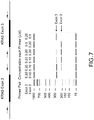
- Figure 7 is an example of the multiplex PCR amplification of KRAS exons 2 and 3, where equal concentrations of primers and gDNA template resulted in varying degrees of amplification of these PCR templates.
- Applicants found that when more than two PCR primer sets or primers/probes are used, changing one primer concentration will more probably than not result in changes in all PCR products or SNPE products (data not shown). Similar results were observed when more than 10 SNPE primers/probes were used to interrogate large number of mutation hotspots. Without wishing to be bound by any theory, Applicants believe that strong competition between primer pairs makes adjustment of relative peak heights difficult, in that changing the concentration of one primer will result in the re-equilibration of many other primers/probes.
- Probes for the SNPE reaction were chosen by selecting regions of genes immediately adjacent 3' to the base of interest. Both sense and antisense strands were used in designing the SNPE probes. Size based resolution of the probes was made possible by the addition of GATC repeats or poly T tails ( GATC , T10 up to T49 in Table 2) on the 5' end of the probe. In one instance a mixed poly T-C oligo tail (T10 C10 T36) was used.
- the detection of the BRAF double mutations, V600K and V600R presented special challenges in SNPE probe design.
- Applicants designed additional primers/probes directed to all of the potential mutations at these positions (Table 6).
- the BRAF-2 pool included the primer/probe 1799(A)/600 which detected any mutant with an A at the 1799 nucleotide position, including the double mutation such as V600R (GTG to AGG).
- This design was also needed to detect the more aggressive BRAF double mutation, V600K, which has reported as a possible link to the tumor metastasis ( Menzies, A. M., et al., Clin.
- oligonucleotide templates containing wild-type and mutant sequences in both sense and anti-sense directions, to validate the SNPE primers/probes for their specificity in detecting each mutation of interest.
- the oligonucleotide templates are synthetic, single-stranded DNA that mimics the respective PCR products. As such, there was no substantial difference between the use of the synthetic templates and the PCR products obtained from cell lines with the specific mutations.
- Synthetic oligonucleotides (Table 3) (SEQ ID NOS: 43-100), made up as 100 ⁇ M stock in distilled water, were diluted 1:200 in 1X calf intestine alkaline phosphatase (CIP) (New England BioLabs, Ipswich, MA) buffer. 15 ⁇ L of diluted stock was transferred to a microtiter plate and treated with 5 units CIP at 37°C for 60 minutes, followed by 70°C for 15 minutes. One microliter of the CIP treated oligonucleotide was mixed with 9 ⁇ L SNPE reaction mixture. The remaining incubation and analysis methods were the same as that described for gDNA (Example 2).
- PCR conditions were as follows: 96°C, 5 minutes, 40 cycles of amplification, wherein each cycle consists of 94°C, 30 seconds, 55°C, 55 seconds, 72°C, 45 seconds. After 40 cycles the reactions were incubated at 72°C for 10 minutes then held at 4°C until processed in the single nucleotide primer extension (SNPE) assay. Any remaining PCR product was stored at -20°C.
- SNPE single nucleotide primer extension
- each PCR product Prior to using the PCR product as a template in the single SNPE assay, 15 ⁇ L of each PCR product was treated with 5 units of calf intestine alkaline phosphatase (CIP) (New England BioLabs, Ipswich, MA) and 2 units of Exonuclease I (ExoI , Affymetrix/USB, Cleveland, OH) at 37°C, 1 hour, followed by 70°C, 0.25 hour in a final volume of 16 ⁇ l. 3 ⁇ L of the CIP/Exo I treated PCR product was then used in the SNPE assay without further adjustment to the DNA concentration.
- CIP calf intestine alkaline phosphatase
- ExoI Exonuclease I
- Each single nucleotide primer extension reaction consisted of 5 ⁇ L SNaPshot Multiplex Kit (Applied Biosystems, Life Technologies, Carlsbad, CA), 3 ⁇ L CIP/ExoI treated PCR product, 1 ⁇ L SNPE probe pool (see, Table 2 for probe sequence and concentrations used), and 1 ⁇ L water.
- the SNPE reaction cycling conditions were as follows: 96°C, 10 seconds, 50°C, 5 seconds, 60°C, 30 seconds, for 25 cycles. Samples were maintained at 4°C until analyzed. For analysis, each SNPE reaction was treated with 1 unit CIP, 37°C, for 1 hour followed by 70°C, for 0.25 hour.
- One-half microliter (0.5 ⁇ L) of the CIP treated single SNPE reaction product was mixed with 0.5 ⁇ L GeneScan 120 LIZ size standards (Applied Biosystems, Life Technologies, Carlsbad, CA) and 9.0 uL Hi-Di Formamide (Applied Biosystems, Life Technologies, Carlsbad, CA), denatured at 95°C for 5 minutes. After denaturing, the samples were immediately placed on ice for 5 minutes then centrifuged and loaded onto an ABI 3500 Genetic analyzer (Applied Biosystems, Life Technologies, Carlsbad, CA) equipped with a 50 cm capillary using POP 7 polymer (Applied Biosystems, Life Technologies, Carlsbad, CA).
- results of the capillary electrophoresis run were imported into ABI GeneMapper (version 4.1) (Applied Biosystems, Life Technologies, Carlsbad, CA) where pre-programmed base calling functions for KRAS, BRAF and NRAS identified the bases found at each position examined.
- Sanger sequencing often referred to as "First Generation” sequencing, was accomplished using ABI BigDye Direct Cycle Sequencing Kit (ABI # 4458687) (Applied Biosystems, Life Technologies, Carlsbad, CA) with M13 modified PCR primers and following the manufacturer's instructions.
- the reaction mixture was applied to an ABI 3500 Genetic analyzer (Applied Biosystems, Life Technologies, Carlsbad, CA) equipped with a 50 cm capillary using POP 7 polymer.
- gDNA from discordant samples were analyzed by ion semiconductor sequencing on the Ion PGM sequencer (Life Technologies, Carlsbad, CA). Targeted exon enrichment was performed using the GeneRead DNA panel. Enrichment was accomplished using 20-25 PCR cycles. Library construction was made using Life Tech's Ion Xpres Plus gDNA and Amplicon Library construction kit (Life Technologies, Carlsbad, CA). Variant calls were made using two algorithms, Ion Torrent Variant Caller (Life Technologies, Carlsbad, CA) and GATK (The Genome Analysis Tool Kit, Broad Institute, Boston, MA). Only variants identified by both algorithms were reported.
- the specificity of the ERK Mutation Assay is a measure of its ability to identify samples that are wild type for KRAS, BRAF and NRAS. A wild type result in a test with high specificity indicates a high probability of the mutation being absent. Specificity is defined as follows:
- Table 5 lists the mutations identified by the ERK Mutation Assay and their resulting amino acid change. It should be noted, that while the ERK Mutation Assay was able to detect and identify the known mutations listed in Table 5, because of the degeneracy of the genetic code and the nature of these single nucleotide mutations, the design of the PCR primers (Table 1) (SEQ ID NOS: 1-14) and the SNPE probes (Table 2) (SEQ ID NOS: 15-42) of the inventive assay would enable them to detect and identify additional single nucleotide mutations at the same loci within these genes.
- the sensitivity of the KBN-SNPE assay is a measure of its ability to identify samples that contain a mutation listed in Table 5 in KRAS, BRAF, and NRAS.
- the identification of a mutation present in a highly sensitive assay indicates a high probability of the mutation being present.
- the SNPE assay (Example 4), which specifically detects ERK Mutations, i.e, mutations to KRAS, BRAF, and NRAS, referred to herein as the ERK Mutation Assay, begins with the PCR amplification of regions of the KRAS, BRAF and NRAS genes from gDNA isolated from one or two 5 ⁇ m FFPE slides. A set of probes was hybridized to the amplicons, followed by a single base extension reaction of the probes and capillary electrophoresis separation, and automated base call using the GeneMapper® software (ABI-Life TechnologiesTM, Carlsbad, CA) based on size and incorporated fluorescent label. Examples of the output from a ERK Mutation Assay are shown in Figures 2-5 .
- the ERK Mutation Assay identified the mutations in KRAS, BRAF and NRAS listed in Table 5, which can be used to stratify patients in a clinical trial.
- the ERK Mutation Assay was designed and validated to perform well with FFPE samples, which generally yield low quality genomic DNA. To work with such samples, Applicants found it necessary to design PCR primers that amplify short stretches (100-150 bp) of DNA (Table 1) (SEQ ID NOS: 1-2) and to carefully balance primer concentrations ( Figure 7 ) so that all regions were equally amplified.
- the ERK Mutation Assay simultaneously interrogates nine and six nucleotide positions within KRAS and NRAS, respectively, and twelve positions within BRAF (Table 2 and Figures 6A-6C ).
- KRAS exons 2, 3 and 4, NRAS exons 2 and 3, and BRAF exons 11 and 15 were amplified in three separate PCR reactions (Table 1). These PCR products served as templates for the SNPE reaction used to identify possible mutations. Due to the number of SNPE probes used for BRAF (Table 2) (SEQ ID NOS: 15-18) and sequence similarity between KRAS and NRAS, Applicants found it necessary to perform the primer extension portion of the assay in four separate reactions (Table 2).
- a number of well characterized cell lines with previously defined mutations in KRAS and BRAF are commercially available from ATCC. Such samples mimic tumor samples, but have the advantage that gDNA isolated is free from contamination by gDNA from "normal adjacent" tissue, as would be the case when isolating gDNA from FFPE tumor samples. As such, gDNA isolated from cell lines that are heterozygous for particular mutations represent a pool of gDNA with a "known" percentage of mutant DNA in a wild type background and represent an ideal source of DNA for determining the limits of detection of an assay (see Example 15).
- C. ERK Mutation Assay detects mutations in FFPE tumor samples
- Genomic DNA was isolated from sixty FFPE samples, ten samples each of colorectal, ovarian, melanoma, lung (non-small cell carcinomas), pancreatic, and thyroid cancers. Each sample was subjected to mutation analysis by the ERK Mutation Assay (Example 12) and Sanger sequencing (Example 6). The analytical accuracy of the mutation analysis of the genomic DNA using the ERK Mutation Assay (EMA) and Sanger sequencing (Sanger) is shown in Table 8 (Colorectal: CRC; Ovarian: OVR; Thyroid: THY; Melanoma: MEL; Pancreatic: PNC; Lung: LNG).
- EMA ERK Mutation Assay
- Sanger Sanger sequencing
- Table 9 shows the percentage of tumors with mutations in KRAS, BRAF, or NRAS as reported in the COSMIC Database (Catalogue of Somatic Mutations in Cancer, Wellcome Trust Sanger Institute, Cambridge, UK) and from the ERK Mutation Assay (EMA).
- Example 7 Discordant results between the EMA and Sanger results were observed in eighteen out of sixty samples. To resolve these discordant results, samples were analyzed by Ion PGM to produce a consensus result (Example 7). There was insufficient material for NGS analysis for seven (one ovarian and six melanoma cancers) of the eighteen discordant samples. The NGS results for seven of the eleven discordant samples agreed with the EMA results. The remaining four samples agreed with the Sanger sequencing results. The results for samples for which there was sufficient material with which to carry out NGS are also shown in Table 8.
- LOD limit of detection
- Table 12 shows the lowest percentage of mutant DNA in a wild type background for BRAF and KRAS that can be detected in sequencing assays of the type used herein. Aliquots of gDNA from mutant cell lines (two BRAF and three KRAS) were mixed with wild type gDNA at ratios of 25%, 15%, 10%, 5%, and 1% of mutant to wild type gDNA and then analyzed by the KBN-SNPE assay.
- the LOD for NRAS was not performed as there were no NRAS mutant cell lines available from the American Type Culture Collection (ATCC) (Manassas, VA).
- ATCC American Type Culture Collection
- VA Manassas, VA
- the LOD observed herein was dependent on the specific mutation or cell line used for the experiment and varied from 1.6% to 12%. For example, one KRAS mutation was detected at 1.6% mutant: wild type DNA, while two other mutations were detected at 6% mutant DNA, and all mutations were detected at 12% mutant DNA.
- the ERK Mutation Assay is used as part of a clinical trial to identify and select patients diagnosed with cancer having an ERK Mutation for treatment with an ERK inhibitor.
- patients diagnosed with cancer can be classified into two subgroups, i.e., those having one or more ERK Mutations and those lacking an ERK Mutation.
- How well an assay performs in a binary classification of patients is measured by its concordance (accuracy), and its sensitivity and specificity.
- the sensitivity of the ERK Mutation Assay would be assessed by its ability to correctly identify samples containing a mutation listed in Table 2.
- the specificity of the KBN-SNPE assay would be assessed by its ability to correctly identify samples that lack any of the KRAS, BRAF and NRAS mutations listed in Table 5.
- An ideal assay would be one having 100% specificity and 100% sensitivity.
- Table 14 shows the performance characteristics (specificity, sensitivity, and overall concordance) for the ERK Mutation Assay at the gene level, while Table 15 shows these same performance characteristics at the sample or patient level. It should be noted that for classification of patients in a clinical setting, patient-level performance is considered to be more relevant than gene-level performance. As shown in Table 15, at the sample level the ERK Mutation assay herein had an overall concordance of 94% when samples were assayed as a single point versus 100% when assayed in triplicate. Similarly, specificity was 91%/100% for single point/triplicate assays, respectively, while sensitivity was 100% in both formats. The limit of detection varied from 2 -12%, depending on the mutation (data not shown).
Landscapes
- Chemical & Material Sciences (AREA)
- Health & Medical Sciences (AREA)
- Life Sciences & Earth Sciences (AREA)
- Organic Chemistry (AREA)
- Proteomics, Peptides & Aminoacids (AREA)
- Wood Science & Technology (AREA)
- Zoology (AREA)
- Engineering & Computer Science (AREA)
- Analytical Chemistry (AREA)
- Immunology (AREA)
- Genetics & Genomics (AREA)
- General Health & Medical Sciences (AREA)
- Pathology (AREA)
- General Engineering & Computer Science (AREA)
- Biotechnology (AREA)
- Physics & Mathematics (AREA)
- Biochemistry (AREA)
- Bioinformatics & Cheminformatics (AREA)
- Biophysics (AREA)
- Molecular Biology (AREA)
- Microbiology (AREA)
- Pharmacology & Pharmacy (AREA)
- Epidemiology (AREA)
- Animal Behavior & Ethology (AREA)
- Public Health (AREA)
- Veterinary Medicine (AREA)
- Hospice & Palliative Care (AREA)
- Oncology (AREA)
- Medicinal Chemistry (AREA)
- Chemical Kinetics & Catalysis (AREA)
- Measuring Or Testing Involving Enzymes Or Micro-Organisms (AREA)
Description
- This invention relates to methods for the detection of genetic mutations and uses thereof for the treatment of cancer.
- The processes involved in tumor growth, progression, and metastasis are mediated by signaling pathways that are activated in cancer cells. The mitogen-activated protein kinase (MAPK) pathway, of which extracellular regulated kinase (ERK) is a downstream member, plays a central role in regulating mammalian cell growth by relaying extracellular signals from ligand-bound cell surface tyrosine kinase receptors such as erbB family, PDGF, FGF, and VEGF receptor tyrosine kinase. Activation of the MAPK/ERK pathway is via a cascade of phosphorylation events that begins with activation of a small guanosine triphosphatase (GTPase), RAS. Activation of RAS leads to the recruitment and activation of RAF, a serine-threonine kinase. Activated RAF then phosphorylates and activates MEK1/2, which in turn phosphorylates and activates ERK1/2. When activated, ERK1/2 phosphorylates several downstream targets involved in a multitude of cellular events including cytoskeletal changes and transcriptional activation.
- The MAPK/ERK pathway is one of the most important for cell proliferation and it is believed that this pathway is frequently activated in many tumors. RAS genes, which are upstream of ERK1/2, are mutated in several cancers including colorectal, melanoma, breast and pancreatic tumors. The high RAS activity is accompanied by elevated ERK activity in many human tumors. In addition, mutations of BRAF, a serine-threonine kinase of the RAF family, are associated with increased kinase activity. Mutations in BRAF have been identified in melanomas (60%) (Fecher, L.A., et al., Curr. Opin. Oncol., 2008, 20:183-189), thyroid cancers (approximately 50%) (Robert, R.J., and Der, C. J., Oncogene, 2007, 26: 329103310), and colorectal cancers (Davies, H., et al., Nature, 2002, 417: 949-954; Kohno, M., and Pauyssegur, J., Ann. Med., 2006, 38: 200-211).
- KRAS also acts upstream of ERK to regulate proliferation, differentiation, and cell survival. Mutations in
codons codons - The mutational status of solid tumors is also becoming increasingly important for identifying the best treatment options for cancer patients (Yokota, T., Anti-Cancer Agents in Medicinal Chemistry, 2012, 12: 163-171). Treatments developed targeting a specific signaling pathway may not be effective when activating mutations are present downstream of the signal transduction pathway. For example, tumors harboring activating mutations in RAS and RAF do not generally respond well to anti-EGFR therapy, in that these mutations are believed to lead to EGFR-independent activation of an intracellular signaling pathway (Messersmith, W.A., and Ahnen, D. J., N. Engl., J. Med., 2008, 359(17): 1834-1836). As such, it is believed that inhibitors targeting proteins further downstream in this pathway may be more effective in treating these cancers.
- As such, proteins that lie downstream of RAS and, even further downstream of RAF and MEK, in the MAPK/ERK pathway are potential targets for pharmacological intervention (Downward, J., Nat. Rev. Cancer, 2003, 3: 11-22; Pearson, G., et al., Endocr. Rev., 2001, 22: 153-183; Fecher, L.A., et al., Curr. Opin. Oncol., 2008, 20: 183-189). Gain of function, i.e., activating, mutations in RAS and RAF that lead to constitutive activation of this pathway are frequently observed in human cancers and are associated with high rates of cancer cell proliferation (Id.). It has been reported that activating mutations of RAS were identified in about 25% of all cancers (Smalley, K.S.M., Int. J. Cancer, 2003, 104: 527-532). These mutations, especially for KRAS, are present at even higher rates in pancreatic cancer and colorectal cancer (KRAS+ in 90% and 50%, respectively, Downward, 2003, and Smalley, 2003). Other studies also reported that NRAS mutations were detected in about 10% to 25% of melanomas (Downard, 2003; Fecher, 2008; Smalley, 2003; Robert, P.J., and Der, C.J., Oncogene, 2007, 26: 3291-3310) and KRAS mutations were detected in about 30% non-small cell lung cancers (NSCLCs) (Downward, 2003; Wistuba, I.I., et al., Semin. Oncol., 2001, 28(2)(suppl 4): 3-13). In addition, RAS mutations (HRAS, KRAS, or NRAS) have been identified in about 55% to 60% of thyroid cancers (Fecher, 2008).
- Similarly, BRAF mutations have been identified in about 60% of malignant melanomas, where all mutations appear to be within the kinase domain and a single substitution (T → A, V600E) accounts for about 80% of the mutations (Davies, H., et al., Nature, 2002, 417: 949-954; Kohno, M., and Pouyssegur, J., Ann. Med., 2006, 38: 200-211). Activating BRAF mutations have also been documented in a variety of human cancers other than melanoma, including about 10% in colorectal cancer (CRC), approximately 50% in thyroid cancer (Roberts and Der, 2007), and several percent in NSCLC (Brose, M.S., et al., Cancer Res., 2002, 62: 6997-7000). The high frequency of RAS or BRAF mutations in these cancers makes targeting this pathway an attractive strategy for new anti-cancer agents that rely on patient stratification to identify individuals most likely to benefit from inhibitors that target the MAPK/ERK pathway (Pratilas, C.A., and Solit, D. B., Rev. Recent Clin. Trials, 2007, 2: 121-134).
- Scientific and clinical attention has recently focused on the major mutational hotspots in these genes (KRAS
codons - Sanger sequencing, a method of DNA sequencing based on the selective incorporation of chain terminating dideoxnucleotides by DNA polymerase during in vitro DNA replication, considered to be the "gold standard" for detecting genetic mutations, is generally considered not sensitive enough to detect genetic mutations present at low level or low copy number relative to wild type DNA. Commercially available BRAF and KRAS kits, such as, TheraScreen® (CE-IVD) KRAS Mutation kit (DxS Ltd., Manchester, UK) and the FDA approved cobas® KRAS and BRAF Mutation Test kits (Roche Diagnostics, Indianapolis, IN) for testing mutations in
codons codon 12 and 13 (Angulo, B., et al., J. Mol. Diagn., 2010 May 12(3): 292-299). The cobas® KRAS Mutation assay, a TaqMelt PCR assay, reports only mutations in general to codon 12/13 or 61, but not specific mutations needed for exploratory data analysis. The cobas® BRAF Mutation assay only detects the single BRAF V600E mutation. There is currently no commercial NRAS mutation detection assay. - The instant invention relates generally to methods for the detection of MAPK/ERK pathway mutations. In one embodiment, the invention comprises a sensitive and selective assay capable of detecting MAPK/ERK pathway mutations, specifically to BRAF, KRAS, and NRAS, and hereinafter referred to as an "ERK Mutation," at more than 30 "hotspots" or loci. In another aspect of this embodiment, the invention comprises primers and probes for the detection of the BRAF, KRAS, and NRAS mutations in a single reaction.
- In another embodiment, the invention comprises a method to identify and select patients diagnosed with cancer for treatment with an ERK inhibitor, wherein said patients to be treated have one or more ERK Mutations. In another aspect of this embodiment, the invention comprises an ERK inhibitor for use in treating a patient, having one or more mutations to BRAF, KRAS, and NRAS.
-
-
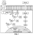
Figure 1 is a graphic illustration of the pathway for the growth and differentiation factor, ERK, showing the relationship of ERK to the RAS and RAF genes in the MAPK pathway (Kabbarah, O., and Chin, L., Frontiers in Bioscience, 2006, 11:928-942). -
Figures 2A-2C are graphics illustrating the ERK Mutation Assay results for the BRAF Probe Pool 7: wild type (WT) control (Figure 2A ); V600E mutation (MT) (Figure 2B ); and the V600D mutation (MT) (Figure 2C ). The position of the codons and nucleotides are indicated at the bottom of the figure. The nucleotide label for each peak has been converted to the sense strand. -
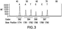
Figure 3 is a graphic illustrating the ERK Mutation Assay results for the BRAF Probe Pool 6: wild type (WT) control. The position of the codons and nucleotides are indicated at the bottom of the figure. The nucleotide label for each peak has been converted to the sense strand. -
Figures 4A-4C are graphics illustrating the ERK Mutation Assay results for the NRAS Probe Pool: wild type (WT) control (Figure 4A ); Q61K mutation (MT) (Figure 4B ); and the Q61L mutation (MT) (Figure 4C ). The position of the codons and nucleotides are indicated at the bottom of the figure. The nucleotide label for each peak has been converted to the sense strand. -
Figures 5A-5E are graphics illustrating the ERK Mutation Assay results for the KRAS Probe Pool: (wild type (WT) control (Figure 5A ); G12S mutation (MT) (Figure 5B ); G12D mutation (MT) (Figure 5C ); G12V mutation (MT) (Figure 5D ); and the G13D mutation (MT) (Figure 5E ). The position of the codons and nucleotides are indicated at the bottom of the figure. The nucleotide label for each peak has been converted to the sense strand. -
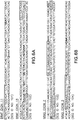
Figures 6A-6C illustrate the PCR primer locations and hotspots for BRAF (Figure 6A ), NRAS (Figure 6B ), and KRAS (Figure 6C ). The portion of the sequence highlighted in dark gray denotes the location of the PCR primers, while the portion highlighted in light gray are hot spot codons around which the SNPE probes were designed. -
Figure 7 illustrates the effect of varying the concentration of the PCR primer sets on the amplification of DNA fragments forKRAS exons -
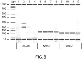
Figure 8 illustrates the inability to amplify large DNA fragments (amplicons) from formalin-fixed paraffin embedded (FFPE) derived genomic DNA (gDNA) by polymerase chain reaction (PCR). Replicate samples of FFPE gDNA from four tumor types (ovarian cancer:lanes lanes lanes lanes -
Figure 9 is an example of a data output for the BRAF probe 1799 (G)/600 (Table 2) with the variant caller, Gene Mapper (Applied Biosystems, Foster City, CA), for wild-type and mutants in a tabular format. The output shows thatAlelle 1 had the mutation corresponding to V600E (Table 5). - The invention herein relates to the field of nucleic acid sequence detection. Nucleic acid sequence detection can be used to determine the presence or absence of a particular genetic element or it can be used to determine the specific type of a particular genetic element that is present. The detection of nucleic acids in a sample and the subtypes thereof typically rely on the technique of specific nucleic acid hybridization, which are based on the tendency of two nucleic acid strands to pair at complementary regions. Nucleic acid hybridization assays are used to detect and identify unique DNA and RNA base sequences or specific genes in a complete DNA molecule in mixtures of nucleic acid, or in mixtures of nucleic acid fragments.
- The practical use of nucleic acid hybridization assays was greatly enhanced by methods that included amplifying or copying, with fidelity, precise sequences of nucleic acid found at low concentration to higher copy numbers, so as to make them more readily detectable. The original amplification method is the polymerase chain reaction described by
Mullis, et al., U.S. Pat. Nos. 4,683,195 ;4,683,202 ; and4,965,188 . Further improvements in the use of these assays was the introduction of multiplex assays, that allowed for the simultaneous amplification and detection of multiple nucleic acid target sequences through the use of optimized primers and reaction conditions. Commercial kits are available to carry out these multiplex hybridization assays. One such kit, the SNaPshot™ Multiplex Kit can be used to carry out a nucleic acid hybridization assay that can detect and identify single nucleotide polymorphisms (SNPs) at known locations on DNA templates (Lurkin, I., et al., 2010, PLoS ONE 5: e8802; Dias-Santagata, D., et al., 2011, PLoS ONE 6:e17948). The kit includes a reaction mix, comprising a DNA polymerase, fluorescent labels, and a reaction buffer, a control primer mix, comprising one or more primers for control reactions, and a control template. The chemistry of the kit is based on the dideoxy single-base extension of an unlabeled oligonucleotide primer (or primers). Each primer binds to a complementary template in the presence of fluorescently labeled ddNTPs and the DNA polymerase. The oligonucleotide probes are designed to hybridize immediately adjacent to the 3' side of the nucleotides of interest. The DNA polymerase probe is extended by the addition of a single fluorescently labeled dideoxynucleotide. The detection of the mutation is made by a combination of the identification of the fluorescently tagged nucleotide added and the size migration of the probe during capillary electrophoresis. - A PCR-based approach to detect all of the mutations to KRAS, BRAF, and NRAS would likely require more than 100 primer/probe sets and at least 30 individual reactions. Qualitative mutant-enriched PCR assays are commercially available, such as the KRAS StripAssay® (ViennaLab, Vienna, Austria), and they appear to be sensitive enough to detect low mutant copies (Sarasquenta, A.F., et al., J. Mol. Diag., 2011, 13(2):199-205), but these commercial tests require the use of a large number of evaluations/assays to detect all of the mutations and they provide no estimate of frequency for individual mutations in the corresponding sample, i.e., only yes (presence) or no (absence) answers as to the mutation are obtained. However, many treatment responses appear to be directly correlated to the mutation frequency for the mutant population detected in the wild-type background (unpublished data). Such information, when analyzed retrospectively, adds value to the final evaluation of a clinical trial and for the interpretation of patient treatment response.
- In that there is a lack of viable options to the standard PCR approach for clinical use, Applicants herein have developed and validated an efficient semi-quantitative multiplexed assay, referred to herein as the KRAS/BRAF/NRAS single nucleotide primer extension assay (KBN-SNPE assay) or, alternatively, as the ERK Mutation Assay, to detect a broader array of mutations to these genes. The ERK Mutation Assay employs a limited number of primers (14, Table 1) and probes (28, Table 2), capable of detecting single point polymorphism mutations for the KRAS, BRAF, and NRAS genes in this pathway in a single reaction. As such, this assay can be used as a high throughput screening assay for drug development or as a clinical, functional use assay, to identify and/or select patients diagnosed with cancer having an ERK Mutation for treatment with an ERK inhibitor.
- "ERK Mutation" means a genetic mutation in a gene which encodes a protein in the MAPK pathway to which ERK is a downstream member. As used herein, this term means generically any mutation to KRAS, BRAF, and NRAS in the MAPK/ERK pathway and specifically those mutations identified in Table 5.
- "ERK inhibitor" means any compound or agent that inhibits or affects the activity of an ERK protein, including ERK1 and ERK2, either directly or indirectly, such as, via inhibition or affecting the activity of a member of the MAPK pathway to which ERK is a downstream member. Members of the MAPK/ERK pathway include BRAF, KRAS, and NRAS. Such molecules would be useful for treating a broad spectrum of cancers, such as, for example, melanoma, pancreatic cancer, thyroid cancer, colorectal cancer, lung cancer, breast cancer, and ovarian cancer. Examples of small molecule ERK inhibitors useful for the treatment of such cancers are described in
WO 2011/16330 PCT publications WO 2007/070398 andWO 2008/153858 disclose polycyclic indazole derivatives that are useful as ERK inhibitors for the treatment of cancer. - "ERK Mutation Assay" means the KRAS/BRAF/NRAS single nucleotide primer extension assay (ERK MUTATION assay) described herein used to detect and quantify an ERK Mutation.
- The invention herein is a multiplex nucleic acid hybridization assay, herein referred to as the ERK Mutation assay, which allows for the specific detection and identification of over 30 mutations in the BRAF, KRAS, and NRAS genes, all members of the MAPK/ERK pathway, thought to activate or increase the activity of the MAPK/ERK pathway. Current commercially available assays target a small subset, typically 1-7 each, of the known mutations to KRAS and BRAF, which enables detection of roughly 80% of the known mutations. Moreover, the currently available assays can target the mutations of only one gene per reaction. The ERK Mutation Assay is a single nucleotide primer extension (SNPE) multiplexed assay consisting of multiplexed oligonucleotide primer extension reactions that determine the nucleotide sequence at 27 hot spot locations in the KRAS, BRAF and NRAS genes. Each reaction consists of multiplexed oligonucleotide primer extension reactions. BRAF consists of thirteen oligonucleotide probes, multiplexed in separate reactions of six and seven probes, respectively, while KRAS and NRAS are multiplexed in separate reactions with nine and six probes, respectively.
- The ERK Mutation Assay includes multiplexing of both the PCR template amplifications and the Single Nucleotide Primer Extension (SNPE) reactions, using the PCR primers of Table 1 (SEQ ID NOS: 1-2) and the SNPE probes of Table 2 (SEQ ID NOS: 15-18). Thus, the competition among the PCR primers is not only against the gDNA templates, but also against each other. These primers also compete against nucleotides and DNA polymerase both in the kinetic and thermodynamic manner depending on their size, sequence, and melting temperature.
- In one embodiment, the invention comprises the ERK Mutation Assay for detecting and identifying an ERK Mutation comprising:
- (a) contacting a biological sample from a patient diagnosed with cancer with a plurality of polymerase chain reaction (PCR) primers selected from the group consisting of the PCR primers of Table 1 (SEQ ID NO: 1-2), which target one or more ERK Mutations;
- (b) amplifying the section of said sample targeted by the PCR primers to form a PCR template containing one or more targeted ERK Mutations;
- (c) contacting the PCR template of step (b) with a plurality of single nucleotide primer extension (SNPE) probes, selected from the group consisting of the SNPE probes of Table 2 (SEQ ID NO: 15-18), under hybridization conditions suitable to form a duplex between the template and the SNPE probe, such that the nucleotide base to be identified is the first unpaired base in the template immediately downstream of the 3' end of the probe in said duplex;
- (d) contacting the PCR template-SNPE duplex of step (c) with i) a reaction mixture comprising a DNA polymerase and a mixture of ddNTPs terminators of a nucleic acid template-dependent primer extension reaction, wherein each terminator is capable of specifically terminating the extension reaction in a manner strictly dependent on the identity of the unpaired nucleotide base in the targeted ERK Mutation PCR template immediately downstream of the 3' end of the primer, wherein each terminator in the mixture is complementary to the nucleotide base to be identified, and wherein each terminator is labeled with a detectable marker, ii) under conditions sufficient to carry out a template-dependent primer extension reaction to form an SNPE product, wherein said conditions permit the base pairing of the complementary terminator with the nucleotide base to be identified and the incorporation of the complementary terminator onto the 3' end of the primer to thereby extend said 3' end of the primer by one terminator; and
- (e) determining the presence and identity of the nucleotide base at the specific position in the SNPE product by detecting the detectable marker of the incorporated terminator of step (d) while said terminator is incorporated at the 3' end of the extended primer, and wherein the ERK Mutation is determined by the identified nucleotide base at the specific position in the SNPE product.
- In another embodiment, the invention comprises the ERK Mutation Assay for detecting and identifying an ERK Mutation comprising:
- (a) contacting a biological sample from a patient diagnosed with cancer with a plurality of polymerase chain reaction (PCR) primers selected from the group consisting of the PCR primers of Table 1 (SEQ ID NOs: 1-2), which target one or more ERK Mutations;
- (b) amplifying the section of said sample targeted by the PCR primers to form a PCR template containing one or more targeted ERK Mutations;
- (c) contacting the PCR template of step (b) with a plurality of single nucleotide primer extension (SNPE) probes, selected from the group consisting of the SNPE probes of Table 2 (SEQ ID NO: 15-18), under hybridization conditions suitable to form a duplex between the template and the SNPE probe, such that the first unpaired nucleotide base in the template immediately downstream of the 3' end of the probe in said duplex is a nucleotide base to be identified, wherein the nucleotide base to be identified is an ERK Mutation;
- (d) contacting the PCR template-SNPE duplex of step (c) with
- (i) a reaction mixture comprising a DNA polymerase; and
- (ii) a mixture of ddNTP terminators of a nucleic acid template-dependent primer extension reaction, wherein each terminator is capable of specifically terminating the extension reaction in a manner strictly dependent on the identity of the unpaired nucleotide base in the PCR template that is immediately downstream of the 3' end of the primer, wherein each terminator in the mixture is complementary to the nucleotide base to be identified, and wherein each terminator is labeled with a detectable marker;
under conditions sufficient to carry out a template-dependent primer extension reaction to form an SNPE product, wherein said conditions permit base pairing of a complementary terminator with the nucleotide base to be identified and incorporation of the complementary terminator onto the 3' end of the primer, thereby extending said 3' end of the primer by one terminator;
- (e) determining the presence and identity of the nucleotide base to be identified at the specific position in the SNPE product by detecting the detectable marker of the incorporated terminator of step (d) while said terminator is incorporated at the 3' end of the extended primer.
- In one aspect of this embodiment, in step (d) of the method, the ddNTP terminators are a mixture of ddATP, ddCTP, ddGTP, and ddTTP. In another aspect of this embodiment, the ddNTP terminators are detectably labeled, each with a different detectable label.
- In another embodiment, the invention is the use of the ERK Mutation Assay to identify and select a patient diagnosed with cancer to be treated with an ERK inhibitor, wherein the patient identified for treatment has one or more ERK Mutations. In one aspect of this embodiment, the patient identified for treatment has one or more KRAS, BRAF, or NRAS mutations. In another aspect of this embodiment, the patient identified for treatment has one or more of the mutations listed in Table 5.
- In still another embodiment, the invention is an ERK inhibitor for use in treating a patient diagnosed with cancer, wherein the patient to be treated is characterized as having one or more ERK Mutations, and wherein the ERK Mutation is detected and identified using the ERK Mutation Assay. In one aspect of this embodiment, the patient to be treated with the ERK inhibitor has one or more KRAS, BRAF, or NRAS mutations that are detected and identified using the ERK Mutation assay. In another aspect of this embodiment, the patient to be treated with the ERK inhibitor has one or more of the mutations listed in Table 5 that are detected and identified using the ERK Mutation assay.
- In yet another embodiment, the invention is a kit to detect and identify an ERK Mutation in a patient diagnosed with cancer. In one aspect of this embodiment, the kit includes the PCR primers of Table 1 (SEQ ID NOS: 1-2) and the SNPE probes of Table 2 (SEQ ID NOS: 15-18). In another aspect of this embodiment, the kit is used in a clinical setting to identify and select patients for treatment with an ERK inhibitor.
- Any DNA sample may be used to carry out the present invention. The DNA may be obtained from any type of biological sample, including but not limited to, tissue or body fluid samples, such as, blood, plasma, or cerebral spinal fluid. In one embodiment, the inventive assay used genomic DNA obtained from a tissue sample, specifically, from a formalin-fixed paraffin embedded (FFPE) tissue sample obtained from a patient diagnosed with cancer. The DNA sample may be extracted from the biological sample using any of the numerous methods that are standard in the art. Those skilled in the art would understand and recognize that the method to be used will depend on the nature of the source of the sample. The amount of DNA to be extracted for use in the present invention will depend on the nature of the source of the sample, but is typically at least 5 ng. In one embodiment, the amount used by the Applicants was 15 ng. The DNA sample to be used in the inventive assay can be single or double-stranded. Those skilled in the art would recognize and appreciate that the standard conditions utilized while carrying out a polymerase chain reaction type reaction would typically be sufficient to separate double-stranded DNA into single-stranded DNA for purposes of the reaction.
- In one embodiment of the invention, the method uses a plurality of polymerase chain reaction (PCR) primers selected from the group of PCR primers listed in Table 1 (SEQ ID NOS: 1-2). Those skilled in the art would understand that a forward and reverse primer specific to each target or region to be amplified is needed to carry out a polymerase chain reaction to generate the templates for the SNPE assay. Thus, the inventive method employs the use of multiple pairs of PCR primers in order to generate the templates, containing the hotspots or targets, for each target gene, i.e. KRAS, NRAS, and BRAF.
- Similarly, in one embodiment of the invention, the method uses a plurality of SNPE probes selected from the SNPE probes listed in Table 2 (SEQ ID NOS: 15-18). Those skilled in the art would understand that while the SNPE probes target a specific ERK Mutation gene, i.e. KRAS, NRAS, or BRAF, unlike other PCR-based mutation detection methods that require a separate probe for the detection of each possible mutation, the inventive SNPE probes listed in Table 2 (SEQ ID NOS: 15-18) have been designed such that they will detect multiple mutations occurring at the same hot spot or loci. Those skilled in the art would also understand that there are limits to the number of probes, typically 10-12, that can be used per reaction in a multiplex/SNPE assay. Thus, the inventive SNPE probes not only reduce the number of SNPE probes needed to detect virtually any mutation occurring at the ERK gene hotspots, they allow for the detection of multiple mutations in all three ERK genes in one reaction. In one embodiment, the inventive method uses 9 KRAS probes, 6 NRAS probes, and 13 BRAF probes, broken down into two groups of 6 and 7 probes, BRAF-1 and BRAF-2, respectively (Table 2) (SEQ ID NOS: 15-18), to detect multiple mutations across the three genes in one reaction. In another aspect of this embodiment, the inventive method can be utilized as a high throughput screening assay.
- In practicing the invention, those skilled in the art would understand that prior to determining the presence and identity of the nucleotide base at the specific position in the primer extended reaction product, that the reaction product is denatured, using standard techniques known in the art, prior to loading onto a DNA sequencer for separation, detection, and identification. Further, those skilled in the art would also understand that the detection and identification of the nucleotide base at a specified position is based on standard techniques, such as the color and size of the incorporated labeled ddNTP and probe used in the reaction. As used herein, the inventive assay used a mixture of labeled ddATP, ddCTP, ddGTP, and ddTTP, each of which was labeled with a different detectable label, with the specified reaction probes (Table 2) (SEQ ID NOS: 15-18), which enables the detection and identification of the nucleotide in question based on color, i.e. identifies which nucleotide was incorporated, and size, i.e. the length of the tail on the probe.
- All cell lines were obtained from American Type Culture Collection (ATCC) (Manassas, VA) and cultivated using recommended conditions. Genomic DNA (gDNA) was extracted from cell pellets using a DNeasy Blood and Tissue Kit (Qiagen, Germantown, MD). Sixty formalin-fixed paraffin embedded (FFPE) samples, ten FFPE slides each, from the following cases of human cancers were purchased from BioChain's repository network (Newark, CA): colorectal, melanoma, ovarian, thyroid, pancreatic, and lung (non-small cell lung cancers). Tissue samples were immediately fixed in formalin and paraffin embedded. Five micron thick sections were cut and mounted on positively charged slides. FFPE samples were shipped at room temperature and stored at 4-8°C. Extraction of gDNA from FFPE tissue samples was performed using the Qiagen DNA FFPE Tissue Extraction Kit (Qiagen, Germantown, MD) following manufactures instructions. Briefly, the excess paraffin was removed from one or two 5 micron thick sections and the tissue scraped off the slide using a fresh surgical scalpel. A new scalpel was used for each section. The paraffin was removed by successive washes with xylene and ethanol and the tissue digested with proteinase K. The released gDNA was bound and eluted from a miniElute column (Qiagen, Germantown, MD) in 50 µL, quantified using a NanoDrop spectrometer (NanoDrop Products, Wilmington, DE) and adjusted to a concentration of 5 ng/µL and stored at -80°C.
- PCR primers (Table 1), SNPE probes (Table 2) and synthetic oligonucleotides (Table 3) were prepared by Sigma (St. Louis, MO) based on Applicants design and were either desalted or purified by HPLC. Except where noted, all reagents for the polymerase chain reaction (PCR) and single nucleotide primer extension (SNPE) reactions were obtained from ABI-Life Technologies (Carlsbad, CA).
- PCR primer sequences (Table 1) (SEQ ID NOS:1-2) were designed by Applicants to amplify gene fragments ranging in size from 100-150 nucleotides of the hot spot regions of KRAS, NRAS and BRAF. To accomplish this, DNA sequences surrounding the regions of interest were manually scanned to identify portions which could serve as primers to amplify the appropriately sized fragment of DNA (
Figures 6A-6C ). BLAST searches with the amplified regions were performed to confirm specificity of the PCR amplified product. Because individual exons were not equally amplified during the PCR amplification step, the concentration of the various PCR primers was adjusted until all exons were equally amplified. -
Figure 7 is an example of the multiplex PCR amplification ofKRAS exons Table 1 Gene Region Sequence (5' to 3') Tm (°C) Final Concentration (nM) KRAS Pool exon 2, forward 
59.32 500 exon 2, reverse
58.8 500 exon 3, forwardGGAAGCAAGTAGTAATTGATGG (SEQ ID NO: 3) illustrative 59.14 200 exon 3, reverseAAAGAAAGCCCTCCCC (SEQ ID NO:4) illustrative 59.07 200 exon 4, forwardGAACAGTAGACACAAAACAGG C (SEQ ID NO: 5) illustrative 60.23 200 exon 4, reverseTGCAGAAAACAGATCTGTATTT ATTT (SEQ ID NO:6) illustrative 60.73 200 NRAS Pool exon 2, forward GGTGTGAAATGACTGAGTAC (SEQ ID NO: 7) illustrative 54.21 100 exon 2, reverseGGGCCTCACCTCTATGGTG (SEQ ID NO: 8) illustrative 64.49 100 exon 3, forwardGGTGAAACCTGTTTGTTGGA (SEQ ID NO: 9) illustrative 62.38 150 exon 3, reverseATACACAGAGGAAGCCTTCG (SEQ ID NO: 10) illustrative 60.82 150 BRAF Pool Exon 15, forward TCTTCATGAAGACCTCACAGT (SEQ ID NO: 11) illustrative 58.85 100 Exon 15, reverseCCAGACAACTGTTCAAACTGA (SEQ ID NO: 12) illustrative 60.35 100 exon 11, forwardGTGATGATTGGGAGATTCCT (SEQ ID NO: 13) illustrative 60.3 150 exon 11, reverseCTGCCACATCACCATGCCA (SEQ ID NO:14) illustrative 69.62 150 - Probes for the SNPE reaction (Table 2) (SEQ ID NOS: 15-18) were chosen by selecting regions of genes immediately adjacent 3' to the base of interest. Both sense and antisense strands were used in designing the SNPE probes. Size based resolution of the probes was made possible by the addition of GATC repeats or poly T tails (GATC, T10 up to T49 in Table 2) on the 5' end of the probe. In one instance a mixed poly T-C oligo tail (T10 C10 T36) was used.
- The detection of the BRAF double mutations, V600K and V600R, presented special challenges in SNPE probe design. In addition to designing oligonucleotide primers/probes from both the sense and anti-sense directions, Applicants designed additional primers/probes directed to all of the potential mutations at these positions (Table 6). As shown in Table 2, the BRAF-2 pool included the primer/probe 1799(A)/600 which detected any mutant with an A at the 1799 nucleotide position, including the double mutation such as V600R (GTG to AGG). This design was also needed to detect the more aggressive BRAF double mutation, V600K, which has reported as a possible link to the tumor metastasis (Menzies, A. M., et al., Clin. Cancer Res., 2012, 18(12):3242-3249; Rosalyn, J., et al., Clin. Cancer Res., 2012, 18(24):6792).
Table 2 Reaction Region Sequence (5' to 3') Size (nt) Strand Probed Tm (°C)* Final Conc. (nM) KRAS 34/12 AACTTGTGGTAGTTGGAGCT (SEQ ID NO: 15) 20 sense 57.84 25 35/12 
26 sense 60.24 75 37/13 
32 sense 62.15 25 38/13 
38 sense 65.61 75 181/61 TTTTTTTTTTTTTTTTTTTTTTT TTTTTCTCATTGCACTGTACTC CTCTT (SEQ ID NO: 19) illustrative 50 antisense 59.95 25 182/61 TTTTTTTTTTTTTTTTTTTTTTT TTTTTTTTTTTTATTCTCGACA CAGCAGGTC (SEQ ID NO: 20) illustrative 54 sense 60.45 25 183/61 TTTTTTTTTTTTTTTTTTTTTTT TTTTTTTTTTTTTTTTTTTCCTC ATTGCACTGTACTCCTC (SEQ ID NO: 21) illustrative 63 antisense 61.29 65 436/146 TTTTTTTTTTTTTTTTTTTTTTT TTTTTTTTTTTTTTTTTTTTTTT TTTGTCTTACTTAGCTGTCTTG TCTTTG (SEQ ID NO: 22) illustrative 73 sense 59.23 400 437/146 TTTTTTTTTTCCCCCCCCCCTT TTTTTTTTTTTTTTTTTTTTTTT TTTTTTTTTTTGAATTCCTTTT ATTGAAACATCAG (SEQ ID NO: 23) illustrative 81 antisense 59.41 400 NRAS 35/12 TTTTTTTTTTTTTTTTTTTTTTC TGGTGGTGGTTGGAGCAG (SEQ ID NO: 24) illustrative 41 sense 66.93 75 37/13 TTTTTTTTTTTTTTTTTTTTTTT TTTTTGGTGGTGGTTGGAGCA GGT (SEQ ID NO: 25) illustrative 47 sense 67.46 3 38/13 TTTTTTTTTTTTTTTTTTTTTTT TTTTTTTTTGTCAGTGCGCTTT TCCCAACA (SEQ ID NO: 26) illustrative 53 antisense 69.39 125 181/61 TTTTTTTTTTTTTTTTTTTTTTT TTTTTTTTTTTTTTCTCATGGC ACTGTACTCTTCTT (SEQ ID NO: 27) illustrative 59 antisense 59.95 263 182/61 TTTTTTTTTTTTTTTTTTTTTTT TTTTTTTTTTTTTTTTTTTTGAC ATACTGGATACAGCTGGAC (SEQ ID NO: 28) illustrative 65 sense 60.04 35 183/61 TTTTTTTTTTTTTTTTTTTTTTT TTTTTTTTTTTTTTTTTTTTTTT TTTCTCTCATGGCACTGTACTC TTC (SEQ ID NO: 29) illustrative 71 antisense 60.48 70 BRAF-1 1774/592 TTTTTTTTTTTTTTTCATGAA GACCTCACAGTAAAA (SEQ ID NO: 30) illustrative 36 sense 56.8 250 1781/594 TTTTTTTTTTTTTTTTTTTTT ACCTCACAGTAAAAATAGGT G (SEQ ID NO: 31) illustrative 42 sense 54.84 250 1782/594 TTTTTTTTTTTTTTTTTTTTTT TTTGATTTCACTGTAGCTAG ACCAAA (SEQ ID NO: 32) illustrative 48 antisense 58.94 125 1786/596 TTTTTTTTTTTTTTTTTTTTTT TTTTTTTTTTTGAGATTTCAC TGTAGCTAGAC (SEQ ID NO: 33) illustrative 54 antisense 52.16 250 1789/597 TTTTTTTTTTTTTTTTTTTTTT TTTTTTTTTTTTTTTTTGTAA AAATAGGTGATTTTGGT (SEQ ID NO: 34) illustrative 60 sense 53.91 250 1790/597 TTTTTTTTTTTTTTTTTTTTTT TTTTTTTTTTTTTTTTTTTTTT TCATCGAGATTTCACTGTAG CT (SEQ ID NO: 35) illustrative 66 antisense 58.52 250 BRAF-2 1799(A)/600 TTTTTTTTTTTTTGGTGATTT TGGTCTAGCTACAA (SEQ ID NO: 36) illustrative 35 sense 59.65 62 1799(G)/600 TTTTTTTTTTTTTTTTTTTGG TGATTTTGGTCTAGCTACAG (SEQ ID NO: 37) illustrative 41 sense 59.29 62 1800/600 TTTTTTTTTTTTTTTTTTTTTT TTTTGGACCCACTCCATCGA GATTT (SEQ ID NO: 38) illustrative 47 antisense 66.21 62 1798/600 TTTTTTTTTTTTTTTTTTTTTT TTTTTTTTTTGGTGATTTTGG TCTAGCTACA(SEQ ID NO:39) illustrative 53 sense 58.28 31 1801/601 TTTTTTTTTTTTTTTTTTTTTT TTTTTTTTTTTTTTTTTGGAC CCACTCCATCGAGATT (SEQ ID NO: 40) illustrative 59 antisense 65.16 62 2/466 TTTTTTTTTTTTTTTTTTTTTT TTTTTTTTTTTTTTTTTTTTTT TGGGACAAAGAATTGGATCT G (SEQ ID NO: 41) illustrative 65 sense 60.87 62 3/466 TTTTTTTTTTTTTTTTTTTTTT TTTTTTTTTTTTTTTTTTTTTT TTTTTCCTTGTAGACTGTTCC AAATGA (SEQ ID NO:42) illustrative 71 antisense 60.55 93 * Poly T tails or GATC repeats were omitted for Tm calculations - Because of the difficulty in obtaining cell lines with all of the specific mutations of interest, Applicants employed synthetic oligonucleotide templates, containing wild-type and mutant sequences in both sense and anti-sense directions, to validate the SNPE primers/probes for their specificity in detecting each mutation of interest. The oligonucleotide templates are synthetic, single-stranded DNA that mimics the respective PCR products. As such, there was no substantial difference between the use of the synthetic templates and the PCR products obtained from cell lines with the specific mutations.
- Applicants found that the size of the amplicon, i.e., the PCR products generated from two PCR primers through PCR cycles, used for the PCR template was critical to successfully detect the mutations with FFPE tissue samples using the KBN-SNPE assay. Initial design efforts and assay optimization were conducted using normal human blood gDNA samples. While data generated using longer PCR templates containing the mutation hotspots was successful, when FFPE tumor tissue samples were used, amplification of the target regions was inconsistent (
Figure 8 ), presumably because the DNA from FFPE tissue samples was highly fragmented and/or degraded. Applicants shortened the length of the amplicons used herein to accommodate the low quality sample common with FFPE tissue. - Synthetic oligonucleotides (Table 3) (SEQ ID NOS: 43-100), made up as 100 µM stock in distilled water, were diluted 1:200 in 1X calf intestine alkaline phosphatase (CIP) (New England BioLabs, Ipswich, MA) buffer. 15 µL of diluted stock was transferred to a microtiter plate and treated with 5 units CIP at 37°C for 60 minutes, followed by 70°C for 15 minutes. One microliter of the CIP treated oligonucleotide was mixed with 9 µL SNPE reaction mixture. The remaining incubation and analysis methods were the same as that described for gDNA (Example 2).
Table 3 Gene Codon Number Codon Protein Description Sequence 466 GGA G466G 
466 GGA G466G 
466 GAA G466E 
466 GTA G466V 
592 ATA I592I 
592 GTA I592V 
594 GAT D594D 
594 GAT D594D 
594 GTT D594V 
BRAF 594 GAA D594E 
594 GAG D594E 
596 GGT G596G 
596 GGT G596G 
596 CGT G596R 
597 TCA L597S 
597 TCA L597S 
600 GTG V600V 
600 GTG V600V 
600 GAG V600E 
600 AGG V600R 
600 AAG V600K 
600 GAT V600D 
600 GAT V600D 
601 GAA K601E 
12 GGT G12G 
12 AGT G12S 
12 TGT G12C 
12 GAT G12D 
12 GCT G12A 
12 GTT G12V 
13 GGC G13G 
13 CGC G13R 
13 GAC G13D 
KRAS 61 CAA Q61Q 
61 CAA Q61Q 
61 AAA Q61K 
61 CTA Q61L 
61 CAC Q61H 
61 CAT Q61H 
146 GCA A146A 
146 GCA A146A 
146 ACA A146T 
146 ACA A146T 
146 GTA A146V 
146 GTA A146V 
NRAS 12 GGT G12G 
12 GGT G12G 
12 GAT G12D 
12 GTT G12V 
13 GAT G13D 
13 CGT G13R 
61 CAA Q61Q 
61 CAA Q61Q 
61 AAA Q61K 
61 CGA Q61R 
61 CTA Q61L 
61 CAC Q61H 
61 CAT Q61H 
- Fifteen (15) ng of isolated gDNA was used as a template for PCR amplification of
KRAS exons Figure 5A ),NRAS exons 2 and 3(Figure 5C ) andBRAF exons 11 and 15 (Figure 5B ). Each gene was independently amplified by PCR. For each PCR reaction 25 µL ABI AmpliTaq Gold Master Mix (ABI cat # 4398886), 5 µL PCR primer pool (Table 2S), 3 µL gDNA (5 ng/µL), and 17 µL distilled water were mixed and subjected to 40 rounds of PCR amplification. The PCR conditions were as follows: 96°C, 5 minutes, 40 cycles of amplification, wherein each cycle consists of 94°C, 30 seconds, 55°C, 55 seconds, 72°C, 45 seconds. After 40 cycles the reactions were incubated at 72°C for 10 minutes then held at 4°C until processed in the single nucleotide primer extension (SNPE) assay. Any remaining PCR product was stored at -20°C. - Prior to using the PCR product as a template in the single SNPE assay, 15 µL of each PCR product was treated with 5 units of calf intestine alkaline phosphatase (CIP) (New England BioLabs, Ipswich, MA) and 2 units of Exonuclease I (ExoI , Affymetrix/USB, Cleveland, OH) at 37°C, 1 hour, followed by 70°C, 0.25 hour in a final volume of 16 µl. 3 µL of the CIP/Exo I treated PCR product was then used in the SNPE assay without further adjustment to the DNA concentration. Each single nucleotide primer extension reaction consisted of 5 µL SNaPshot Multiplex Kit (Applied Biosystems, Life Technologies, Carlsbad, CA), 3 µL CIP/ExoI treated PCR product, 1 µL SNPE probe pool (see, Table 2 for probe sequence and concentrations used), and 1 µL water. The SNPE reaction cycling conditions were as follows: 96°C, 10 seconds, 50°C, 5 seconds, 60°C, 30 seconds, for 25 cycles. Samples were maintained at 4°C until analyzed. For analysis, each SNPE reaction was treated with 1 unit CIP, 37°C, for 1 hour followed by 70°C, for 0.25 hour. One-half microliter (0.5 µL) of the CIP treated single SNPE reaction product was mixed with 0.5 µL GeneScan 120 LIZ size standards (Applied Biosystems, Life Technologies, Carlsbad, CA) and 9.0 uL Hi-Di Formamide (Applied Biosystems, Life Technologies, Carlsbad, CA), denatured at 95°C for 5 minutes. After denaturing, the samples were immediately placed on ice for 5 minutes then centrifuged and loaded onto an
ABI 3500 Genetic analyzer (Applied Biosystems, Life Technologies, Carlsbad, CA) equipped with a 50 cmcapillary using POP 7 polymer (Applied Biosystems, Life Technologies, Carlsbad, CA). Results of the capillary electrophoresis run were imported into ABI GeneMapper (version 4.1) (Applied Biosystems, Life Technologies, Carlsbad, CA) where pre-programmed base calling functions for KRAS, BRAF and NRAS identified the bases found at each position examined. - In experiments where samples were assayed in triplicate, the rules for determining the consensus call are presented in Table 4. If the same call was determined in two of three or three of three reactions, then the majority call was the consensus. Where a call was not possible (ND) in two or more replicates, the consensus call was considered to be indeterminate. When performed in support of a clinical trial, re-do rules would be applied and samples where clear results could not be obtained would be re-assayed. Two failed attempts would result in a "No-Call" result and a new sample would be requested.
Table 4 Replicate/ Call 1 2 3 Consensus Wild Type Wild Type Wild Type Wild Type Wild Type Wild Type Mutation-1 Wild Type Wild Type Mutation-1 Mutation-1 Mutation-1 Mutation-1 Mutation-1 Mutation-1 Mutation-1 Wild Type Wild Type ND* Wild Type Mutation-1 Mutation-1 ND Mutation-1 Wild Type ND ND Indeterminate-Redo ND ND ND Indeterminate-Redo Mutation-1 ND ND Indeterminate-Redo Wild Type Mutation-1 ND Indeterminate-Redo Mutation-1 Mutation-1 Mutation-2 Mutation-1 Mutation-1 Wild Type ND Indeterminate-Redo *ND=Not Determined - Sanger sequencing, often referred to as "First Generation" sequencing, was accomplished using ABI BigDye Direct Cycle Sequencing Kit (ABI # 4458687) (Applied Biosystems, Life Technologies, Carlsbad, CA) with M13 modified PCR primers and following the manufacturer's instructions. The reaction mixture was applied to an
ABI 3500 Genetic analyzer (Applied Biosystems, Life Technologies, Carlsbad, CA) equipped with a 50 cmcapillary using POP 7 polymer. - 80 ng of gDNA from discordant samples were analyzed by ion semiconductor sequencing on the Ion PGM sequencer (Life Technologies, Carlsbad, CA). Targeted exon enrichment was performed using the GeneRead DNA panel. Enrichment was accomplished using 20-25 PCR cycles. Library construction was made using Life Tech's Ion Xpres Plus gDNA and Amplicon Library construction kit (Life Technologies, Carlsbad, CA). Variant calls were made using two algorithms, Ion Torrent Variant Caller (Life Technologies, Carlsbad, CA) and GATK (The Genome Analysis Tool Kit, Broad Institute, Boston, MA). Only variants identified by both algorithms were reported.
- Concordance was determined at the sample (patient) level. Each sample was subjected to mutation analysis by the SNPE assay (ERK Mutation Assay) and Sanger sequencing as described in Examples 4 (12) and 6, respectively. If the two methods agree, the mutation was assumed to be "truth." If only one method detected a mutation, the sample was analyzed by Ion PGM sequencing (Life Technologies, Carlsbad, CA), a type of Next Generation sequencing (NGS), i.e., a high-throughput sequencing technology that parallels the sequencing process. If the Ion PGM result matches either the SNPE (ERK Mutation) or Sanger result, the matched result was considered "truth" and was considered the consensus call. Performance of the SNPE assay (ERK Mutation Assay) was measured versus this consensus call. Samples where a clear result could not be obtained were not included in the concordance calculations.
- The specificity of the ERK Mutation Assay is a measure of its ability to identify samples that are wild type for KRAS, BRAF and NRAS. A wild type result in a test with high specificity indicates a high probability of the mutation being absent. Specificity is defined as follows:
- Truth: the consensus result obtained by two or more assay methods
(ERK Mutation Assay, Sanger sequencing, NGS) - True Positive = sample correctly identified as having a mutation
- False Positive = sample incorrectly identified as having a mutation
- True Negative = sample correctly identified as not having any mutations
(wild type) listed in Table 5 - False Negative = sample incorrectly identified as not having any mutation
(wild type) listed in Table 5
- Table 5 lists the mutations identified by the ERK Mutation Assay and their resulting amino acid change. It should be noted, that while the ERK Mutation Assay was able to detect and identify the known mutations listed in Table 5, because of the degeneracy of the genetic code and the nature of these single nucleotide mutations, the design of the PCR primers (Table 1) (SEQ ID NOS: 1-14) and the SNPE probes (Table 2) (SEQ ID NOS: 15-42) of the inventive assay would enable them to detect and identify additional single nucleotide mutations at the same loci within these genes. Thus, the potential number of single nucleotide polymorphic mutations that can be detected and identified by the inventive assay exceeds those specifically listed in Table 5, estimated to be in excess of 80 mutations.
Table 5 Codon Number WT Codon Mutant Codon Protein Description BRAF 466 GGA GAA G466E 466 GGA GTA G466V 592 ATA GTA I592V 594 GAT GTT D594V 594 GAT GAA D594E 594 GAT GAG D594E 596 GGT CGT G596R 597 CTA TCA L597S 600 GTG GAG V600E 600 GTG AGG V600R 600 GTG AAG V600K 600 GTG GAT V600D 601 AAA GAA K601E KRAS 12 GGT AGT G12S 12 GGT TGT G12C 12 GGT GAT G12D 12 GGT GCT G12A 12 GGT GTT G12V 13 GGC CGC G13R 13 GGC GAC G13D 61 CAA AAA Q61K 61 CAA CTA Q61L 61 CAA CAC Q61H 61 CAA CAT Q61H 146 GCA ACA A146T 146 GCA GTA A146T NRAS 12 GGT GAT G12D 12 GGT GTT G12V 13 GGT CGT G13R 13 GGT GAT G13D 61 CAA AAA Q61K 61 CAA CGA Q61R 61 CAA CTA Q61L 61 CAA CAC Q61H - The sensitivity of the KBN-SNPE assay is a measure of its ability to identify samples that contain a mutation listed in Table 5 in KRAS, BRAF, and NRAS. The identification of a mutation present in a highly sensitive assay indicates a high probability of the mutation being present.
- Sensitivity or sample level positive agreement is defined as follows:

- The overall accuracy of the KBN-SNPE assay to make the same wild type or mutation call as the consensus call is defined as follows:

- The SNPE assay (Example 4), which specifically detects ERK Mutations, i.e, mutations to KRAS, BRAF, and NRAS, referred to herein as the ERK Mutation Assay, begins with the PCR amplification of regions of the KRAS, BRAF and NRAS genes from gDNA isolated from one or two 5 µm FFPE slides. A set of probes was hybridized to the amplicons, followed by a single base extension reaction of the probes and capillary electrophoresis separation, and automated base call using the GeneMapper® software (ABI-Life Technologies™, Carlsbad, CA) based on size and incorporated fluorescent label. Examples of the output from a ERK Mutation Assay are shown in
Figures 2-5 . - The base called for in each location interrogated is given below each electropherogram (
Figures 2-5 ) and indicated whether the nucleotide occupying that position was wild type or mutant. Base calling was determined by the migration of the probe together with the color coding for the nucleotide added during the primer extension reaction. Owing to the slight difference in migration rate, which resulted from the addition of the fluorescently tagged nucleotide of each probe, Applicants identified which of four possible nucleotides was added to the probe. - The ERK Mutation Assay identified the mutations in KRAS, BRAF and NRAS listed in Table 5, which can be used to stratify patients in a clinical trial. The ERK Mutation Assay was designed and validated to perform well with FFPE samples, which generally yield low quality genomic DNA. To work with such samples, Applicants found it necessary to design PCR primers that amplify short stretches (100-150 bp) of DNA (Table 1) (SEQ ID NOS: 1-2) and to carefully balance primer concentrations (
Figure 7 ) so that all regions were equally amplified. - The ERK Mutation Assay simultaneously interrogates nine and six nucleotide positions within KRAS and NRAS, respectively, and twelve positions within BRAF (Table 2 and
Figures 6A-6C ).KRAS exons NRAS exons BRAF exons Figures 6A-6C ). In aggregate, Applicants detected 35 specific and 81 potential mutations (counting all possible A/T/G/C changes at each nucleotide location and excluding wild type) at 27 nucleotide positions in the three genes that were dispersed over seven exons. In one embodiment of the invention, the four primer extension reactions are carried out on a single 96 well plate. - To establish the analytical accuracy of the ERK Mutation Assay, three sets of samples from cell lines with previously documented mutations in BRAF or KRAS, synthetic oligonucleotides containing all mutations detected by the KBN-SNPE assay, and a set of sixty formalin-fixed paraffin embedded (FFPE) tumor samples were analyzed by both the KBN-SNPE assay and Sanger sequencing (Example 6)
- A. ERK Mutation Assay detects known mutations in cell lines
- A number of well characterized cell lines with previously defined mutations in KRAS and BRAF are commercially available from ATCC. Such samples mimic tumor samples, but have the advantage that gDNA isolated is free from contamination by gDNA from "normal adjacent" tissue, as would be the case when isolating gDNA from FFPE tumor samples. As such, gDNA isolated from cell lines that are heterozygous for particular mutations represent a pool of gDNA with a "known" percentage of mutant DNA in a wild type background and represent an ideal source of DNA for determining the limits of detection of an assay (see Example 15).
- Genomic DNA was isolated from nine cell lines with mutations previously identified by Sanger sequencing as containing mutations in KRAS, BRAF, or NRAS, and two wild type (WT) and compared to the results from the ERK Mutation Assay (Table 6). Of the eleven cell lines examined, ten produced the expected result in the ERK Mutation Assay. The single exception (GAK), which was expected to contain an NRAS mutation, was subjected to re-sequencing by Sanger sequencing and was determined to be wild type for BRAF, KRAS and NRAS, confirming the ERK Mutation result. Without wishing to be bound by any theory, Applicants suggest that the NRAS mutation expected to be present was lost during passage of this cell line.
Table 6 Cell Line (DNA) Cancer Type Expected Mutation KBN-SNPE Mutation A375 Melanoma BRAF, V600E BRAF, V600E ES-2 Ovarian BRAF, V600E BRAF, V600E WM-266-4 Melanoma BRAF, V600D BRAF, V600D NCI-H358 Lung KRAS, G12C KRAS, G12C HCT-8 Colon KRAS, G13D KRAS, G13D SW 527 Breast KRAS, G12V KRAS, G12V NCI-H2122 Lung KRAS, G12C KRAS, G12C Panc-1 Pancreatic KRAS, G12D KRAS, G12D GAK Melanoma NRAS, Q61L WT* HS-294T Melanoma WT WT ZR-75-1 Breast WT WT *Wild type status confirmed by re-sequencing of sample used in the KBN-SNPE assay. - To further establish that the ERK Mutation Assay was able to detect an ERK Mutation, a series of synthetic oligonucleotides (Table 3) (SEQ ID NOS: 43-100) were designed, owing to the limited availability of commercial cell lines containing known mutations, to demonstrate the ERK Mutation Assay's specificity and ability to detect all of the mutations listed in Table 5. These synthetic oligonucleotides replaced the PCR product produced from the genomic DNA in the ERK Mutation Assay. With this exception, the ERK Mutation Assay was performed the same as if genomic DNA was used. All appropriate wild type sequences were included as well. As shown in Table 7, the ERK Mutation Assay accurately identified (Obs'd) all wild type (WT) and mutations (MT) expected (Exp'd) at each position (Position) interrogated.
Table 7 BRAF KRAS NRAS Position Exp'd Obs'd Position Exp'd Obs'd Position Exp'd Obs'd B-G466G-S WT WT K-G12GAS WT WT N-G12G-S WT WT B-G466GAS WT WT K-G12S-AS MT MT N-G12GAS MT MT B-G466E-AS MT MT K-G12C-AS MT MT N-G12D-S MT MT B-G466V-S MT MT K-G12D-AS MT MT N-G12V-S WT WT B-I592I-AS WT WT K-G12A-AS MT MT N-G13D-S MT MT B-I592VAS MT MT K-G12VAS MT MT N-G13RAS MT MT B-D594D-S WT WT K-G13GAS WT WT N-Q61Q-S MT MT B-D594D-AS WT WT K-G13RAS MT MT N-Q61Q-AS WT WT B-D594VAS MT MT K-G13D-AS MT MT N-Q61K-S MT MT B-D594E-S MT MT K-Q61Q-S WT WT N-Q61RAS MT MT B-D594E-S MT MT K-Q61Q-AS MT MT N-Q61L-AS MT MT B-G596G-S WT WT K-Q61K-S MT MT N-Q61H-S MT MT B-G596GAS WT WT K-Q61L-AS MT MT N-Q61H-S MT MT B-G596R-S MT MT K-Q61H-S MT MT B-L597S-S MT MT K-Q61H-S MT MT B-L597S-AS MT MT K-A146A-S WT WT B-V600V-S WT WT K-A146A-AS MT MT B-V600VAS WT WT K-A146T-AS MT MT B-V600E-AS MT MT K-A146T-S MT MT B-V600RAS MT MT K-A146V-S MT MT B-V600K-AS MT MT K-A146VAS MT MT B-V600D-S MT MT B-V600D-AS MT MT B-K601E-S MT MT - Genomic DNA was isolated from sixty FFPE samples, ten samples each of colorectal, ovarian, melanoma, lung (non-small cell carcinomas), pancreatic, and thyroid cancers. Each sample was subjected to mutation analysis by the ERK Mutation Assay (Example 12) and Sanger sequencing (Example 6). The analytical accuracy of the mutation analysis of the genomic DNA using the ERK Mutation Assay (EMA) and Sanger sequencing (Sanger) is shown in Table 8 (Colorectal: CRC; Ovarian: OVR; Thyroid: THY; Melanoma: MEL; Pancreatic: PNC; Lung: LNG). Table 9 shows the percentage of tumors with mutations in KRAS, BRAF, or NRAS as reported in the COSMIC Database (Catalogue of Somatic Mutations in Cancer, Wellcome Trust Sanger Institute, Cambridge, UK) and from the ERK Mutation Assay (EMA).
- In general, Applicants found that the number and type of mutations present when analyzed with the ERK Mutation Assay was consistent with those reported in the COSMIC database (Table 9). The exceptions appear to be under reporting of BRAF and KRAS mutations in melanomas and pancreatic cancers respectively (10% and 20% versus 43% and 58%, for BRAF and KRAS in the EMA and the COSMIC database, respectively). BRAF mutations in thyroid cancers appear to be over reported (90% in the EMA versus 40% in the COSMIC database). It should also be noted that the rates of mutation reported in the COSMIC database differ from some rates reported in the literature (Downward, J., Nat. Rev. Cancer, 2003, 3:11-22; Pearson, G., et al., Endocr. Rev., 2001, 22:153-183; Fecher, L.A., et al., Curr.Opin. Oncol., 2008, 20:183-189; Smalley, K.S.M., Intl. J. Cancer, 2003,104:527-532; Roberts, P.J., and Der, C.J., Oncogene, 2007, 26:3291-3310; Wistuba, I.I., et al., Semin. Oncol., 2001, 28(2)(suppl 4):3-13; Davies, H., et al., Nature, 2002, 417:949-954; Kohno, M., and Pouyssegur, J., Ann. Med., 2006, 38:200-211; Brose, M.S., et al., Cancer Res., 2002, 62:6997-7000; Pratilas, C.A., and Solit, D.B., Rev. Recent Clin. Trials, 2007, 2:121-123). The mutation rates reported in the COSMIC database include all mutations reported for these genes, while rates reported in the literature, as well as this study, generally focus on a more restricted set of mutations. Without wishing to be bound by any theory, Applicants suggest that the small sample size used in this study may have also contributed to any discrepancy observed.
- Discordant results between the EMA and Sanger results were observed in eighteen out of sixty samples. To resolve these discordant results, samples were analyzed by Ion PGM to produce a consensus result (Example 7). There was insufficient material for NGS analysis for seven (one ovarian and six melanoma cancers) of the eighteen discordant samples. The NGS results for seven of the eleven discordant samples agreed with the EMA results. The remaining four samples agreed with the Sanger sequencing results. The results for samples for which there was sufficient material with which to carry out NGS are also shown in Table 8.
Table 8 KRAS NRAS BRAF Tumor Type EMA Sanger NGS EMA Sanger NGS EMA Sanger NGS CRC-1 WT WT Q61L Q61L WT WT CRC-2 WT WT WT WT WT WT CRC-3 WT WT WT WT WT WT CRC-4 G13D G13D WT WT WT WT CRC-5 WT WT WT WT WT WT CRC-6 G12S G12N G12S WT WT WT WT WT WT CRC-7 G12V G12V WT WT WT WT CRC-8 WT NC WT WT WT WT NC WT WT CRC-9 WT WT WT WT WT WT CRC-10 WT WT WT WT V600E V600E OVR-1 WT WT WT WT WT WT OVR-2 WT WT WT WT WT WT OVR-3 WT WT WT WT WT WT OVR-4 WT WT WT WT WT WT OVR-5** WT NC ND NC NC ND WT G466R* ND OVR-6 WT WT WT WT WT WT OVR-7 WT WT WT WT WT WT OVR-8 WT WT WT WT WT WT WT WT WT OVR-9 WT WT WT WT WT WT OVR10 WT WT WT WT WT WT THY-1 WT WT WT WT V600E V600E THY-2 WT WT WT WT V600E V600E THY-3 WT WT WT WT V600E V600E THY-4 WT Q61Stop* A146T WT WT WT WT V600E V600E V600E THY-5 WT WT WT WT V600E V600E THY-6 WT WT WT WT V600E V600E THY-7 WT WT WT WT G13D WT V600E V600E V600E THY-8 A146T* WT A146T WT WT WT V600E V600E WT THY-9 WT WT WT WT V600E V600E THY-10 WT WT WT WT WT WT MEL-1 WT WT WT WT WT WT MEL-2** WT NC ND WT WT ND WT WT ND MEL-3 WT WT Q61K Q61K WT WT MEL-4** WT NC ND WT WT ND WT WT ND MEL-5** WT NC ND WT NC ND WT NC ND MEL-6** WT WT ND WT G13D ND V600E WT ND MEL-7** WT NC ND WT WT ND WT WT ND MEL-8 WT WT WT WT WT WT WT WT WT MEL-9** WT NC ND WT WT ND NC WT ND MEL-10 WT WT WT WT WT WT PNC-1 WT WT WT WT WT WT WT NC WT PNC-2 WT G13D WT WT WT WT WT WT WT PNC-3 G12D G12D WT WT WT WT PNC-4 G12D NC G12D WT WT WT WT WT WT PNC-5 G12D WT WT WT WT WT WT NC WT PNC-6 G12D WT WT WT WT WT WT WT WT PNC-7 G12C NC WT WT WT WT WT WT WT PNC-8 WT WT WT WT WT WT PNC-9 WT WT WT WT WT WT PNC-10 WT WT WT WT WT WT LNG-1 WT WT WT WT WT WT LNG-2 WT NC WT WT G13D WT WT WT WT LNG-3 WT WT WT WT WT WT LNG-4 WT WT WT WT WT WT LNG-5 WT WT WT WT WT WT LNG-6 WT WT WT WT WT WT LNG-7 G12D G12D WT WT WT WT LNG-8 WT WT WT WT WT WT LNG-9 WT WT WT WT WT WT LNG-10 WT WT WT WT WT WT *These results were only observed once and have not been reproduced.
** Insufficient material remaining for NGS analysis (ND).Table 9 Cosmic Database* EMA* KRAS BRAF NRAS KRAS BRAF NRAS Colorectal 35 12 2 30 10 10 Melanoma 2 43 13 0 10 10 Ovarian 14 9 1 0 0 0 Pancreatic 58 2 1 20 0 0 Thyroid 2 40 0 10 90 0 Lung 16 2 1 10 0 0 - To assess intra-run precision (repeatability), i.e. how well the assay performs when similar samples are assayed , eight samples from the accuracy study (Example 13) across various tumor types (colorectal, thyroid, lung, pancreatic, and ovarian), were run in replicas of eight in a single batch using the ERK Mutation Assay (Example 12). The results shown in Tables 10A and 10B were compared to the consensus call (Example 5). With the exception of one discordant result for each of the two ovarian samples and one replica of Ovarian-1 for
KRAS exon 2 that failed to amplify, the repeatability was 100% for six of the eight samples for each tumor type across all three genes. Repeatability for the BRAF, KRAS and NRAS genes individually was 100% (64/64), 100% (63/63) and 97% (62/64), respectively (Table 14).Table 10A Colorectal-1 Colorectal-7 Thyroid-2 Thyroid-8 KRAS NRAS BRAF KRAS NRAS BRAF KRAS NRAS BRAF KRAS NRAS BRAF Truth WT Q61L WT G12V WT WT WT WT V600E WT WT V600E 1 WT Q61L WT G12V WT WT WT WT V600E WT WT V600E 2 WT Q61L WT G12V WT WT WT WT V600E WT WT V600E 3 WT Q61L WT G12V WT WT WT WT V600E WT WT V600E 4 WT Q61L WT G12V WT WT WT WT V600E WT WT V600E 5 WT Q61L WT G12V WT WT WT WT V600E WT WT V600E 6 WT Q61L WT G12V WT WT WT WT V600E WT WT V600E 7 WT Q61L WT G12V WT WT WT WT V600E WT WT V600E 8 WT Q61L WT G12V WT WT WT WT V600E WT WT V600E Table 10B Lung-7 Pancreactic-3 Ovarian-1 Ovarian-8 KRAS NRAS BRAF KRAS NRAS BRAF KRAS NRAS BRAF KRAS NRAS BRAF Truth G12D WT WT G12D WT WT WT WT WT WT WT WT 1 G12D WT WT G12D WT WT WT WT WT WT WT WT 2 G12D WT WT G12D WT WT WT WT WT WT WT WT 3 G12D WT WT G12D WT WT WT WT WT WT G13D WT 4 G12D WT WT G12D WT WT WT WT WT WT WT WT 5 G12D WT WT G12D WT WT No Exon 2 WT WT WT WT WT 6 G12D WT WT G12D WT WT WT G12D WT WT WT WT 7 G12D WT WT G12D WT WT WT WT WT WT WT WT 8 G12D WT WT G12D WT WT WT WT WT WT WT WT - Similarly, to assess the inter-run precision (reproducibility), i.e., how well the assay performs when the samples are assayed over a period of time, eight genomic DNA samples across various tumor types (colorectal, thyroid, lung, pancreatic, and ovarian),were assayed eight times in eight batches over a period of four days. As shown in Tables 11A and 11B, with the exception of a thyroid and a colorectal sample, each having one false positive in NRAS, and a melanoma sample having two false negatives in BRAF and a false positive in KRAS, 100% reproducibility was observed for five of the eight samples. Reproducibility for the BRAF, KRAS and NRAS genes individually was 97% (62/64), 98% (61/62) and 97% (62/64), respectively (Table 12).
Table 11A Colorectal-1 Colorectal-10 Thyroid-1q Thyroid-8 KRAS NRAS BRAF KRAS NRAS BRAF KRAS NRAS BRAF KRAS NRAS BRAF WT Q61L WT WT WT V600E WT WT V600E WT WT V600E WT Q61L WT WT WT V600E WT WT V600E WT WT V600E WT Q61L WT WT WT V600E WT WT V600E WT WT V600E WT Q61L WT WT WT V600E WT WT V600E WT WT V600E WT Q61L WT WT WT V600E no Ex 2 WT V600E WT WT V600E WT Q61L WT WT WT V600E no Ex2 WT V600E WT WT V600E WT Q61L WT WT G13D V600E WT G13D V600E WT WT V600E WT Q61L WT WT WT V600E WT WT V600E WT WT V600E WT Q61L WT WT WT V600E WT WT V600E WT WT V600E Table 11B Ovarian-1 Ovarian-8 Lung-7 Melanoma-6 KRAS NRAS BRAF KRAS NRAS BRAF KRAS NRAS BRAF KRAS NRAS BRAF WT WT WT WT WT WT G12D WT WT WT WT V600E WT WT WT WT WT WT G12D WT WT WT WT V600E WT WT WT WT WT WT G12D WT WT WT WT V600E WT WT WT WT WT WT G12D WT WT WT WT V600E WT WT WT WT WT WT G12D WT WT WT WT V600E WT WT WT WT WT WT G12D WT WT WT WT V600E WT WT WT WT WT WT G12D WT WT WT WT WT WT WT WT WT WT WT G12D WT WT WT WT WT WT WT WT WT WT WT G12D WT WT G12D WT V600E - For a qualitative assay, analytical sensitivity (limit of detection, LOD) is the lowest percentage of mutant gDNA mixed with wild type gDNA where a known mutation can be detected (CLSI guideline EP17, Protocols for Determination of Limits of Detection and Limits of Quantitation). Table 12 shows the lowest percentage of mutant DNA in a wild type background for BRAF and KRAS that can be detected in sequencing assays of the type used herein. Aliquots of gDNA from mutant cell lines (two BRAF and three KRAS) were mixed with wild type gDNA at ratios of 25%, 15%, 10%, 5%, and 1% of mutant to wild type gDNA and then analyzed by the KBN-SNPE assay. The LOD for NRAS was not performed as there were no NRAS mutant cell lines available from the American Type Culture Collection (ATCC) (Manassas, VA). The LOD observed herein was dependent on the specific mutation or cell line used for the experiment and varied from 1.6% to 12%. For example, one KRAS mutation was detected at 1.6% mutant: wild type DNA, while two other mutations were detected at 6% mutant DNA, and all mutations were detected at 12% mutant DNA.
- Without wishing to be bound by any theory, Applicants believe that the results observed were affected by several factors. First, the difficulty in accurately determining the starting percentage of mutant DNA present in any given sample required an assumption that each cell line was heterozygous for the mutation. Second, the differences in extinction coefficients for the fluorescent dyes used to identify the various bases incorporated into the SNPE probes made it difficult to accurately assess the relative ratios of one nucleotide verses another. As such, a conservative estimate of the LOD was between 6 and 12% mutant DNA.
Table 12 BRAF KRAS Cell Line ES2 A375 HCT8 Panc1 SW527 Mutation V600E V600E G13D G12D G12V % Mut DNA 25 Yes Yes Yes Yes Yes 12.5 Yes Yes Yes Yes Yes 6.25 Yes No No Yes No 3.125 No No No Yes No 1.56 No No No Yes No 0.78 No No No No No - In the experiments described above, Applicants noted that miscalls could occur at a rate of about 2-3 %. For clinical applications, it would be desirable to appreciably reduce this error rate. Applicants hypothesized that the error rate of the assay could be reduced by assaying samples in triplicate and applying a 'majority rules' approach (Example 5). Rules for calling the results of the ERK Mutation Assay are given in Table 4.
- The results for the fourteen samples, which were assayed in triplicate on three independent occasions, are shown for BRAF, KNAS, and NRAS in Tables 13A-13C, respectively (Consensus (Cnss.); Colorectal (CRC); Melanoma (MEL); Ovarian (OVR); Pancreactic (PNC); Thyroid (THY); Lung (LNG); and Concordance (Cncd.)). There were no missed calls in the BRAF and NRAS portions of the assay, while there were three indeterminate calls (one melanoma-2 and two with thyroid-1) for the KRAS portion of the assay. Under the "redo rules" for this assay, the melanoma-2 sample would have been repeated and its mutation status reported as "wild type." The thyroid-1 sample would have been repeated and its mutation status reported as "indeterminate." Thus, when samples were assayed in triplicate there was 100% (42/42) concordance between the ERK Mutation Assay result and the consensus result (defined by agreement with Sanger sequencing or NGS). Samples assayed in triplicate where two or more calls where indeterminate (ND) were excluded from calculations for concordance.
Table 13A BRAF Experiment 1 Experiment 2Experiment 3Sample Cnss. Rep 1Rep 2Rep 3Rep 1Rep 2Rep 3Rep 1Rep 2Rep 3Cncd. CRC-1 WT WT WT WT WT WT WT WT WT WT 3/3 CRC-4 WT WT WT WT WT WT WT WT WT WT 3/3 CRC-7 WT WT WT WT WT WT WT WT WT WT 3/3 CRC-10 V600E V600E V600E V600E V600E V600E V600E V600E V600E V600E 3/3 MEL-2 WT WT WT WT WT WT WT WT WT WT 3/3 MEL-3 WT WT WT WT WT WT WT WT WT WT 3/3 OVR-1 WT WT WT WT WT WT WT WT WT WT 3/3 OVR-8 WT WT WT WT WT WT WT WT WT WT 3/3 PNC-3 WT WT WT WT WT WT WT WT WT WT 3/3 PNC-8 WT WT WT WT WT WT WT WT WT WT 3/3 THY-1 V600E V600E V600E V600E V600E V600E V600E V600E V600E V600E 3/3 THY-8 V600E V600E V600E V600E V600E V600E V600E V600E V600E V600E 3/3 LNG-5 WT WT WT WT WT WT WT WT WT WT 3/3 LNG-7 WT WT WT WT WT WT WT WT WT WT 3/3 Table 13B KRAS Experiment 1 Experiment 2Experiment 3Sample Cnss. Rep 1Rep 2Rep 3Rep 1Rep 2Rep 3Rep 1Rep 2Rep 3Cncd. CRC-1 WT WT WT WT WT WT WT ND* WT WT 3/3 CRC-4 G13D G13D G13D G13D G13D G13D G13D G13D G13D G13D 3/3 CRC-7 G12V G12V G12V G12V G12V G12V G12V G12V G12V G12V 3/3 CRC-10 WT WT WT WT WT WT WT WT WT WT 3/3 MEL-2 WT WT ND* ND WT WT ND WT WT ND 2/2 MEL-3 WT WT WT WT WT WT WT WT WT WT 3/3 OVR-1 WT WT WT WT WT WT WT WT WT WT 3/3 OVR-8 WT WT WT WT WT WT WT WT G12D WT 3/3 PNC-3 G12D G12D G12D G12D G12D G12D G12D G12D G12D G12D 3/3 PNC-8 WT WT WT WT WT WT ND WT WT ND 3/3 THY-1 WT ND A146T WT ND ND WT ND WT WT 1/2 THY-8 WT WT WT A146V WT WT WT WT WT WT 3/3 LNG-5 WT WT WT WT WT WT WT WT WT WT 3/3 LNG-7 G12D G12D G12D G12D G12D G12D G12D G12D G12D G12D 3/3 *ND-No data due one or more exons missing Table 13C NRAS Experiment 1 Experiment 2Experiment 3Sample Cnss. Rep 1Rep 2Rep 3Rep 1Rep 2Rep 3Rep 1Rep 2Rep 3Cncd. CRC-1 QA1L Q61L Q61L Q61L Q61L Q61L Q61L Q61L Q61L Q61L 3/3 CRC-4 WT WT WT WT WT WT WT WT WT WT 3/3 CRC-7 WT WT WT WT WT WT WT WT WT WT 3/3 CRC-10 WT WT WT WT WT WT WT G12D WT WT 3/3 MEL-2 WT WT WT WT WT WT WT WT WT WT 3/3 MEL-3 Q61K Q61K Q61K Q61K Q61K Q61K Q61K Q61K Q61K Q61K 3/3 OVR-1 WT WT WT WT WT WT WT WT Q61L WT 3/3 OVR-8 WT WT WT WT WT WT WT WT WT WT 3/3 PNC-3 WT WT WT WT WT WT WT WT WT WT 3/3 PNC-8 WT WT WT WT WT WT WT WT WT WT 3/3 THY-1 WT WT WT WT WT WT WT G12D, G13V WT WT 3/3 THY-8 WT WT WT WT WT WT WT WT WT WT 3/3 LNG-5 WT WT WT WT WT WT WT WT WT WT 3/3 LNG-7 WT WT WT WT WT WT WT WT WT WT 3/3 - In one embodiment of the invention, the ERK Mutation Assay is used as part of a clinical trial to identify and select patients diagnosed with cancer having an ERK Mutation for treatment with an ERK inhibitor. In this instance, patients diagnosed with cancer can be classified into two subgroups, i.e., those having one or more ERK Mutations and those lacking an ERK Mutation. How well an assay performs in a binary classification of patients is measured by its concordance (accuracy), and its sensitivity and specificity. As used herein, the sensitivity of the ERK Mutation Assay would be assessed by its ability to correctly identify samples containing a mutation listed in Table 2. Similarly, the specificity of the KBN-SNPE assay would be assessed by its ability to correctly identify samples that lack any of the KRAS, BRAF and NRAS mutations listed in Table 5. An ideal assay would be one having 100% specificity and 100% sensitivity.
- Table 14 shows the performance characteristics (specificity, sensitivity, and overall concordance) for the ERK Mutation Assay at the gene level, while Table 15 shows these same performance characteristics at the sample or patient level. It should be noted that for classification of patients in a clinical setting, patient-level performance is considered to be more relevant than gene-level performance. As shown in Table 15, at the sample level the ERK Mutation assay herein had an overall concordance of 94% when samples were assayed as a single point versus 100% when assayed in triplicate. Similarly, specificity was 91%/100% for single point/triplicate assays, respectively, while sensitivity was 100% in both formats. The limit of detection varied from 2 -12%, depending on the mutation (data not shown).
Table 14 Samples Analyzed Specificity/Sample-Level Negative Agreement Sensitivity/Mutation-Level Positive Agreement Overall Concordance (Accuracy) KRAS NRAS BRAF KRAS NRAS BRAF KRAS NRAS BRAF FFPE Single Screen 100% (44/44) 100% (55/55) 100% (46/46) 70% (7/10) 100% (2/2) 100% (10/10) 94% (51/54) 100% (55/55) 100% (51/51) FFPE Triplicate Screen 100% (27/27) 100% (36/36) 100% (33/33) 100% (12/12) 100% (6/6) 100% (9/9) 100% (39/39) 100% (48/48) 100% (42/42) Intra-Run 100% 96% 100% 100% 80% 100% 100% 97% 100% Variation (39/39) (54/56) (48/48) (24/24) (8/10) (16/16) (63/63) (62/64) (64/64) Inter-Run Variation 98% (53/54) 96% (54/56) 100% (32/32) 100% (8/8) 100% (8/8) 94% (30/32) 98% (61/62) 97% (62/64) 97% (62/64) Cell line DNA 100% (6/6) 100% (11/11) 100% (8/8) 100% (5/5) 100% (0/0) 100% (3/3) 100% (11/11) 100% (11/11) 100% (11/11) Table 15 Samples Analyzed Specificity/Sample-Level Negative Agreement Sensitivity/ Mutation-Level Positive Agreement Overall Concordance (Accuracy) FFPE Single Screen 91% (32/35) 100% (18/18) 94% (50/53) FFPE Triplicate Screen 100% (13/13) 100% (25/25) 100% (38/38) Intra-Run Variation 87% (13/15) 100% (48/48) 97% (61/63) Inter-Run Variation 100% (16/16) 96%(44/46) 97% (60/62) Cell line DNA 100% (3/3) 100% (8/8) 100% (11/11) -
- <110> Merck Sharp & Dohme Corp.
- <120> METHODS AND COMPOSITIONS FOR TREATING CANCER
- <130> 23497-US-PCT
- <150>
US 61/809,612
<151> 2013-04-08 - <160> 107
- <170> PatentIn version 3.5
- <210> 1
<211> 27
<212> DNA
<213> Artificial Sequence - <220>
<223> PCR PRIMER - <400> 1
tgtgacatgt tctaatatag tcacatt 27 - <210> 2
<211> 23
<212> DNA
<213> Artificial Sequence - <220>
<223> PCR PRIMER - <400> 2
cacaaaatga ttctgaatta gct 23 - <210> 3
<211> 22
<212> DNA
<213> Artificial Sequence - <220>
<223> PCR PRIMER - <400> 3
ggaagcaagt agtaattgat gg 22 - <210> 4
<211> 16
<212> DNA
<213> Artificial Sequence - <220>
<223> PCR PRIMER - <400> 4
aaagaaagcc ctcccc 16 - <210> 5
<211> 22
<212> DNA
<213> Artificial Sequence - <220>
<223> PCR PRIMER - <400> 5
gaacagtaga cacaaaacag gc 22 - <210> 6
<211> 26
<212> DNA
<213> Artificial Sequence - <220>
<223> PCR PRIMER - <400> 6
tgcagaaaac agatctgtat ttattt 26 - <210> 7
<211> 20
<212> DNA
<213> Artificial Sequence - <220>
<223> PCR PRIMER - <400> 7
ggtgtgaaat gactgagtac 20 - <210> 8
<211> 19
<212> DNA
<213> Artificial Sequence - <220>
<223> PCR PRIMER - <400> 8
gggcctcacc tctatggtg 19 - <210> 9
<211> 20
<212> DNA
<213> Artificial Sequence - <220>
<223> PCR PRIMER - <400> 9
ggtgaaacct gtttgttgga 20 - <210> 10
<211> 20
<212> DNA
<213> Artificial Sequence - <220>
<223> PCR PRIMER - <400> 10
atacacagag gaagccttcg 20 - <210> 11
<211> 21
<212> DNA
<213> Artificial Sequence - <220>
<223> PCR PRIMER - <400> 11
tcttcatgaa gacctcacag t 21 - <210> 12
<211> 21
<212> DNA
<213> Artificial Sequence - <220>
<223> PCR PRIMER - <400> 12
ccagacaact gttcaaactg a 21 - <210> 13
<211> 20
<212> DNA
<213> Artificial Sequence - <220>
<223> PCR PRIMER - <400> 13
gtgatgattg ggagattcct 20 - <210> 14
<211> 19
<212> DNA
<213> Artificial Sequence - <220>
<223> PCR PRIMER - <400> 14
ctgccacatc accatgcca 19 - <210> 15
<211> 20
<212> DNA
<213> Artificial Sequence - <220>
<223> SNPE PROBE - <400> 15
aacttgtggt agttggagct 20 - <210> 16
<211> 26
<212> DNA
<213> Artificial Sequence - <220>
<223> SNPE PROBE - <400> 16
gatcgtactt gtggtagttg gagctg 26 - <210> 17
<211> 32
<212> DNA
<213> Artificial Sequence - <220>
<223> SNPE PROBE - <400> 17
gatcgatcga tcttgtggta gttggagctg gt 32 - <210> 18
<211> 38
<212> DNA
<213> Artificial Sequence - <220>
<223> SNPE PROBE - <400> 18
gatcgatcga tcgatcgatg tggtagttgg agctggtg 38 - <210> 19
<211> 50
<212> DNA
<213> Artificial Sequence - <220>
<223> SNPE PROBE - <400> 19
tttttttttt tttttttttt ttttttttct cattgcactg tactcctctt 50 - <210> 20
<211> 54
<212> DNA
<213> Artificial Sequence - <220>
<223> SNPE PROBE - <400> 20
tttttttttt tttttttttt tttttttttt tttttattct cgacacagca ggtc 54 - <210> 21
<211> 63
<212> DNA
<213> Artificial Sequence - <220>
<223> SNPE PROBE - <400> 21

- <210> 22
<211> 74
<212> DNA
<213> Artificial Sequence - <220>
<223> SNPE PROBE - <400> 22

- <210> 23
<211> 80
<212> DNA
<213> Artificial Sequence - <220>
<223> SNPE PROBE - <400> 23

- <210> 24
<211> 41
<212> DNA
<213> Artificial Sequence - <220>
<223> SNPE PROBE - <400> 24
tttttttttt tttttttttt ttctggtggt ggttggagca g 41 - <210> 25
<211> 47
<212> DNA
<213> Artificial Sequence - <220>
<223> SNPE PROBE - <400> 25
tttttttttt tttttttttt ttttttttgg tggtggttgg agcaggt 47 - <210> 26
<211> 53
<212> DNA
<213> Artificial Sequence - <220>
<223> SNPE PROBE - <400> 26
tttttttttt tttttttttt tttttttttt ttgtcagtgc gcttttccca aca 53 - <210> 27
<211> 59
<212> DNA
<213> Artificial Sequence - <220>
<223> SNPE PROBE - <400> 27
tttttttttt tttttttttt tttttttttt tttttttctc atggcactgt actcttctt 59 - <210> 28
<211> 65
<212> DNA
<213> Artificial Sequence - <220>
<223> SNPE PROBE - <400> 28

- <210> 29
<211> 71
<212> DNA
<213> Artificial Sequence - <220>
<223> SNPE PROBE - <400> 29

- <210> 30
<211> 36
<212> DNA
<213> Artificial Sequence - <220>
<223> SNPE PROBE - <400> 30
tttttttttt tttttcatga agacctcaca gtaaaa 36 - <210> 31
<211> 42
<212> DNA
<213> Artificial Sequence - <220>
<223> SNPE PROBE - <400> 31
tttttttttt tttttttttt tacctcacag taaaaatagg tg 42 - <210> 32
<211> 48
<212> DNA
<213> Artificial Sequence - <220>
<223> SNPE PROBE - <400> 32
tttttttttt tttttttttt tttttgattt cactgtagct agaccaaa 48 - <210> 33
<211> 54
<212> DNA
<213> Artificial Sequence - <220>
<223> SNPE PROBE - <400> 33
tttttttttt tttttttttt tttttttttt tttgagattt cactgtagct agac 54 - <210> 34
<211> 60
<212> DNA
<213> Artificial Sequence - <220>
<223> SNPE PROBE - <400> 34
tttttttttt tttttttttt tttttttttt tttttttttg taaaaatagg tgattttggt 60 - <210> 35
<211> 66
<212> DNA
<213> Artificial Sequence - <220>
<223> SNPE PROBE - <400> 35

- <210> 36
<211> 35
<212> DNA
<213> Artificial Sequence - <220>
<223> SNPE PROBE - <400> 36
tttttttttt tttggtgatt ttggtctagc tacaa 35 - <210> 37
<211> 41
<212> DNA
<213> Artificial Sequence - <220>
<223> SNPE PROBE - <400> 37
tttttttttt tttttttttg gtgattttgg tctagctaca g 41 - <210> 38
<211> 47
<212> DNA
<213> Artificial Sequence - <220>
<223> SNPE PROBE - <400> 38
tttttttttt tttttttttt ttttttggac ccactccatc gagattt 47 - <210> 39
<211> 53
<212> DNA
<213> Artificial Sequence - <220>
<223> SNPE PROBE - <400> 39
tttttttttt tttttttttt tttttttttt ttggtgattt tggtctagct aca 53 - <210> 40
<211> 59
<212> DNA
<213> Artificial Sequence - <220>
<223> SNPE PROBE - <400> 40
tttttttttt tttttttttt tttttttttt tttttttttg gacccactcc atcgagatt 59 - <210> 41
<211> 65
<212> DNA
<213> Artificial Sequence - <220>
<223> SNPE PROBE - <400> 41

- <210> 42
<211> 71
<212> DNA
<213> Artificial Sequence - <220>
<223> SNPE PROBE - <400> 42

- <210> 43
<211> 53
<212> DNA
<213> Artificial Sequence - <220>
<223> Synthetic Oligonucleotide - <400> 43
tacagtggga caaagaattg gatctggatc atttggaaca gtctacaagg gaa 53 - <210> 44
<211> 53
<212> DNA
<213> Artificial Sequence - <220>
<223> Synthetic Oligonucleotide - <400> 44
ttcccttgta gactgttcca aatgatccag atccaattct ttgtcccact gta 53 - <210> 45
<211> 53
<212> DNA
<213> Artificial Sequence - <220>
<223> Synthetic Oligonucleotide - <400> 45
ttcccttgta gactgttcca aatgattcag atccaattct ttgtcccact gta 53 - <210> 46
<211> 53
<212> DNA
<213> Artificial Sequence - <220>
<223> Synthetic Oligonucleotide - <400> 46
tacagtggga caaagaattg gatctgtatc atttggaaca gtctacaagg gaa 53 - <210> 47
<211> 53
<212> DNA
<213> Artificial Sequence - <220>
<223> Synthetic Oligonucleotide - <400> 47
tcactgtagc tagaccaaaa tcacctattt ttactgtgag gtcttcatga aga 53 - <210> 48
<211> 53
<212> DNA
<213> Artificial Sequence - <220>
<223> Synthetic Oligonucleotide - <400> 48
tcactgtagc tagaccaaaa tcacctagtt ttactgtgag gtcttcatga aga 53 - <210> 49
<211> 53
<212> DNA
<213> Artificial Sequence - <220>
<223> Synthetic Oligonucleotide - <400> 49
tgaagacctc acagtaaaaa taggtgattt tggtctagct acagtgaaat ctc 53 - <210> 50
<211> 53
<212> DNA
<213> Artificial Sequence - <220>
<223> Synthetic Oligonucleotide - <400> 50
gagatttcac tgtagctaga ccaaaatcac ctatttttac tgtgaggtct tca 53 - <210> 51
<211> 53
<212> DNA
<213> Artificial Sequence - <220>
<223> Synthetic Oligonucleotide - <400> 51
gagatttcac tgtagctaga ccaaaaacac ctatttttac tgtgaggtct tca 53 - <210> 52
<211> 53
<212> DNA
<213> Artificial Sequence - <220>
<223> Synthetic Oligonucleotide - <400> 52
tgaagacctc acagtaaaaa taggtgaatt tggtctagct acagtgaaat ctc 53 - <210> 53
<211> 53
<212> DNA
<213> Artificial Sequence - <220>
<223> Synthetic Oligonucleotide - <400> 53
tgaagacctc acagtaaaaa taggtgagtt tggtctagct acagtgaaat ctc 53 - <210> 54
<211> 53
<212> DNA
<213> Artificial Sequence - <220>
<223> Synthetic Oligonucleotide - <400> 54
cctcacagta aaaataggtg attttggtct agctacagtg aaatctcgat gga 53 - <210> 55
<211> 53
<212> DNA
<213> Artificial Sequence - <220>
<223> Synthetic Oligonucleotide - <400> 55
tccatcgaga tttcactgta gctagaccaa aatcacctat ttttactgtg agg 53 - <210> 56
<211> 53
<212> DNA
<213> Artificial Sequence - <220>
<223> Synthetic Oligonucleotide - <400> 56
cctcacagta aaaataggtg attttcgtct agctacagtg aaatctcgat gga 53 - <210> 57
<211> 53
<212> DNA
<213> Artificial Sequence - <220>
<223> Synthetic Oligonucleotide - <400> 57
cctcacagta aaaataggtg attttcgttc agctacagtg aaatctcgat gga 53 - <210> 58
<211> 53
<212> DNA
<213> Artificial Sequence - <220>
<223> Synthetic Oligonucleotide - <400> 58
tccatcgaga tttcactgta gctgaacgaa aatcacctat ttttactgtg agg 53 - <210> 59
<211> 53
<212> DNA
<213> Artificial Sequence - <220>
<223> Synthetic Oligonucleotide - <400> 59
aataggtgat tttggtctag ctacagtgaa atctcgatgg agtgggtccc atc 53 - <210> 60
<211> 53
<212> DNA
<213> Artificial Sequence - <220>
<223> Synthetic Oligonucleotide - <400> 60
gatgggaccc actccatcga gatttcactg tagctagacc aaaatcacct att 53 - <210> 61
<211> 53
<212> DNA
<213> Artificial Sequence - <220>
<223> Synthetic Oligonucleotide - <400> 61
gatgggaccc actccatcga gatttctctg tagctagacc aaaatcacct att 53 - <210> 62
<211> 53
<212> DNA
<213> Artificial Sequence - <220>
<223> Synthetic Oligonucleotide - <400> 62
gatgggaccc actccatcga gatttccttg tagctagacc aaaatcacct att 53 - <210> 63
<211> 53
<212> DNA
<213> Artificial Sequence - <220>
<223> Synthetic Oligonucleotide - <400> 63
gatgggaccc actccatcga gatttctttg tagctagacc aaaatcacct att 53 - <210> 64
<211> 53
<212> DNA
<213> Artificial Sequence - <220>
<223> Synthetic Oligonucleotide - <400> 64
aataggtgat tttggtctag ctacagataa atctcgatgg agtgggtccc atc 53 - <210> 65
<211> 53
<212> DNA
<213> Artificial Sequence - <220>
<223> Synthetic Oligonucleotide - <400> 65
gatgggaccc actccatcga gatttatctg tagctagacc aaaatcacct att 53 - <210> 66
<211> 53
<212> DNA
<213> Artificial Sequence - <220>
<223> Synthetic Oligonucleotide - <400> 66
aggtgatttt ggtctagcta cagtggaatc tcgatggagt gggtcccatc agt 53 - <210> 67
<211> 53
<212> DNA
<213> Artificial Sequence - <220>
<223> Synthetic Oligonucleotide - <400> 67
gtatcgtcaa ggcactcttg cctacgccac cagctccaac taccacaagt tta 53 - <210> 68
<211> 53
<212> DNA
<213> Artificial Sequence - <220>
<223> Synthetic Oligonucleotide - <400> 68
gtatcgtcaa ggcactcttg cctacgccac tagctccaac taccacaagt tta 53 - <210> 69
<211> 53
<212> DNA
<213> Artificial Sequence - <220>
<223> Synthetic Oligonucleotide - <400> 69
gtatcgtcaa ggcactcttg cctacgccac aagctccaac taccacaagt tta 53 - <210> 70
<211> 53
<212> DNA
<213> Artificial Sequence - <220>
<223> Synthetic Oligonucleotide - <400> 70
gtatcgtcaa ggcactcttg cctacgccat cagctccaac taccacaagt tta 53 - <210> 71
<211> 53
<212> DNA
<213> Artificial Sequence - <220>
<223> Synthetic Oligonucleotide - <400> 71
gtatcgtcaa ggcactcttg cctacgccag cagctccaac taccacaagt tta 53 - <210> 72
<211> 53
<212> DNA
<213> Artificial Sequence - <220>
<223> Synthetic Oligonucleotide - <400> 72
gtatcgtcaa ggcactcttg cctacgccaa cagctccaac taccacaagt tta 53 - <210> 73
<211> 53
<212> DNA
<213> Artificial Sequence - <220>
<223> Synthetic Oligonucleotide - <400> 73
gtatcgtcaa ggcactcttg cctacgccac cagctccaac taccacaagt tta 53 - <210> 74
<211> 53
<212> DNA
<213> Artificial Sequence - <220>
<223> Synthetic Oligonucleotide - <400> 74
gtatcgtcaa ggcactcttg cctacgcgac cagctccaac taccacaagt tta 53 - <210> 75
<211> 53
<212> DNA
<213> Artificial Sequence - <220>
<223> Synthetic Oligonucleotide - <400> 75
gtatcgtcaa ggcactcttg cctacgtcac cagctccaac taccacaagt tta 53 - <210> 76
<211> 53
<212> DNA
<213> Artificial Sequence - <220>
<223> Synthetic Oligonucleotide - <400> 76
cttggatatt ctcgacacag caggtcaaga ggagtacagt gcaatgaggg acc 53 - <210> 77
<211> 53
<212> DNA
<213> Artificial Sequence - <220>
<223> Synthetic Oligonucleotide - <400> 77
ggtccctcat tgcactgtac tcctcttgac ctgctgtgtc gagaatatcc aag 53 - <210> 78
<211> 53
<212> DNA
<213> Artificial Sequence - <220>
<223> Synthetic Oligonucleotide - <400> 78
cttggatatt ctcgacacag caggtaaaga ggagtacagt gcaatgaggg acc 53 - <210> 79
<211> 53
<212> DNA
<213> Artificial Sequence - <220>
<223> Synthetic Oligonucleotide - <400> 79
ggtccctcat tgcactgtac tcctctttac ctgctgtgtc gagaatatcc aag 53 - <210> 80
<211> 53
<212> DNA
<213> Artificial Sequence - <220>
<223> Synthetic Oligonucleotide - <400> 80
cttggatatt ctcgacacag caggtcacga ggagtacagt gcaatgaggg acc 53 - <210> 81
<211> 53
<212> DNA
<213> Artificial Sequence - <220>
<223> Synthetic Oligonucleotide - <400> 81
cttggatatt ctcgacacag caggtcatga ggagtacagt gcaatgaggg acc 53 - <210> 82
<211> 53
<212> DNA
<213> Artificial Sequence - <220>
<223> Synthetic Oligonucleotide - <400> 82
tggaattcct tttattgaaa catcagcaaa gacaagacag gtaagtaaca ctg 53 - <210> 83
<211> 53
<212> DNA
<213> Artificial Sequence - <220>
<223> Synthetic Oligonucleotide - <400> 83
cagtgttact tacctgtctt gtctttgctg atgtttcaat aaaaggaatt cca 53 - <210> 84
<211> 53
<212> DNA
<213> Artificial Sequence - <220>
<223> Synthetic Oligonucleotide - <400> 84
cagtgttact tacctgtctt gtctttgttg atgtttcaat aaaaggaatt cca 53 - <210> 85
<211> 53
<212> DNA
<213> Artificial Sequence - <220>
<223> Synthetic Oligonucleotide - <400> 85
tggaattcct tttattgaaa catcaacaaa gacaagacag gtaagtaaca ctg 53 - <210> 86
<211> 53
<212> DNA
<213> Artificial Sequence - <220>
<223> Synthetic Oligonucleotide - <400> 86
tggaattcct tttattgaaa catcagtaaa gacaagacag gtaagtaaca ctg 53 - <210> 87
<211> 53
<212> DNA
<213> Artificial Sequence - <220>
<223> Synthetic Oligonucleotide - <400> 87
cagtgttact tacctgtctt gtctttactg atgtttcaat aaaaggaatt cca 53 - <210> 88
<211> 53
<212> DNA
<213> Artificial Sequence - <220>
<223> Synthetic Oligonucleotide - <400> 88
caaactggtg gtggttggag caggtggtgt tgggaaaagc gcactgacaa tcc 53 - <210> 89
<211> 53
<212> DNA
<213> Artificial Sequence - <220>
<223> Synthetic Oligonucleotide - <400> 89
ggattgtcag tgcgcttttc ccaacaccac ctgctccaac caccaccagt ttg 53 - <210> 90
<211> 53
<212> DNA
<213> Artificial Sequence - <220>
<223> Synthetic Oligonucleotide - <400> 90
caaactggtg gtggttggag cagatggtgt tgggaaaagc gcactgacaa tcc 53 - <210> 91
<211> 53
<212> DNA
<213> Artificial Sequence - <220>
<223> Synthetic Oligonucleotide - <400> 91
caaactggtg gtggttggag cagttggtgt tgggaaaagc gcactgacaa tcc 53 - <210> 92
<211> 53
<212> DNA
<213> Artificial Sequence - <220>
<223> Synthetic Oligonucleotide - <400> 92
caaactggtg gtggttggag caggtgatgt tgggaaaagc gcactgacaa tcc 53 - <210> 93
<211> 53
<212> DNA
<213> Artificial Sequence - <220>
<223> Synthetic Oligonucleotide - <400> 93
ggattgtcag tgcgcttttc ccaacacgac ctgctccaac caccaccagt ttg 53 - <210> 94
<211> 53
<212> DNA
<213> Artificial Sequence - <220>
<223> Synthetic Oligonucleotide - <400> 94
gttggacata ctggatacag ctggacaaga agagtacagt gccatgagag acc 53 - <210> 95
<211> 53
<212> DNA
<213> Artificial Sequence - <220>
<223> Synthetic Oligonucleotide - <400> 95
ggtctctcat ggcactgtac tcttcttgtc cagctgtatc cagtatgtcc aac 53 - <210> 96
<211> 53
<212> DNA
<213> Artificial Sequence - <220>
<223> Synthetic Oligonucleotide - <400> 96
gttggacata ctggatacag ctggaaaaga agagtacagt gccatgagag acc 53 - <210> 97
<211> 53
<212> DNA
<213> Artificial Sequence - <220>
<223> Synthetic Oligonucleotide - <400> 97
ggtctctcat ggcactgtac tcttctcgtc cagctgtatc cagtatgtcc aac 53 - <210> 98
<211> 53
<212> DNA
<213> Artificial Sequence - <220>
<223> Synthetic Oligonucleotide - <400> 98
ggtctctcat ggcactgtac tcttctagtc cagctgtatc cagtatgtcc aac 53 - <210> 99
<211> 53
<212> DNA
<213> Artificial Sequence - <220>
<223> Synthetic Oligonucleotide - <400> 99
gttggacata ctggatacag ctggacacga agagtacagt gccatgagag acc 53 - <210> 100
<211> 53
<212> DNA
<213> Artificial Sequence - <220>
<223> Synthetic Oligonucleotide - <400> 100
gttggacata ctggatacag ctggacatga agagtacagt gccatgagag acc 53 - <210> 101
<211> 140
<212> DNA
<213> Artificial Sequence - <220>
<223>BRAF Exon 11 - <400> 101

- <210> 102
<211> 210
<212> DNA
<213> Artificial Sequence - <220>
<223>BRAF Exon 15 - <400> 102


- <210> 103
<211> 210
<212> DNA
<213> Artificial Sequence - <220>
<223> NRAsExon 2 - <400> 103

- <210> 104
<211> 140
<212> DNA
<213> Artificial Sequence - <220>
<223>NRAS Exon 3 - <400> 104

- <210> 105
<211> 280
<212> DNA
<213> Artificial Sequence - <220>
<223> KRAsExon 2 - <400> 105

- <210> 106
<211> 280
<212> DNA
<213> Artificial Sequence - <220>
<223> KRAsExon 3 - <400> 106

- <210> 107
<211> 180
<212> DNA
<213> Artificial Sequence - <220>
<223> KRAsExon 4 - <400> 107

Claims (15)
- A method for identifying an ERK Mutation in a patient diagnosed with cancer comprising:(a) contacting a biological sample from a patient diagnosed with cancer with a plurality of polymerase chain reaction (PCR) primers selected from the group consisting of the PCR primers of Table 1 (SEQ ID NOs: 1-2), which target one or more ERK Mutations;(b) amplifying the section of said sample targeted by the PCR primers to form a PCR template containing one or more targeted ERK Mutations;(c) contacting the PCR template of step (b) with a plurality of single nucleotide primer extension (SNPE) probes, selected from the group consisting of the SNPE probes of Table 2 (SEQ ID NO: 15-18), under hybridization conditions suitable to form a duplex between the template and the SNPE probe, such that the first unpaired nucleotide base in the template immediately downstream of the 3' end of the probe in said duplex is a nucleotide base to be identified, wherein the nucleotide base to be identified is an ERK Mutation;(d) contacting the PCR template-SNPE duplex of step (c) with(i) a reaction mixture comprising a DNA polymerase; and(ii) a mixture of ddNTP terminators of a nucleic acid template-dependent primer extension reaction, wherein each terminator is capable of specifically terminating the extension reaction in a manner strictly dependent on the identity of the unpaired nucleotide base in the PCR template that is immediately downstream of the 3' end of the primer, wherein each terminator in the mixture is complementary to the nucleotide base to be identified, and wherein each terminator is labeled with a detectable marker;
under conditions sufficient to carry out a template-dependent primer extension reaction to form an SNPE product, wherein said conditions permit base pairing of a complementary terminator with the nucleotide base to be identified and incorporation of the complementary terminator onto the 3' end of the primer, thereby extending said 3' end of the primer by one terminator;(e) determining the presence and identity of the nucleotide base to be identified at the specific position in the SNPE product by detecting the detectable marker of the incorporated terminator of step (d) while said terminator is incorporated at the 3' end of the extended primer. - The method of claim 1, wherein in step (d), the ddNTP terminators are a mixture of ddATP, ddCTP, ddGTP, and ddTTP.
- The method of claim 1, wherein in step (d), the duplex from step (c) is contacted with four labeled terminators, each with a different detectable label.
- The method of claim 1, wherein in step (e) the ERK Mutation identified is from the group consisting of the mutations of Table 5.
- A method for identifying a patient previously diagnosed with cancer as a patient who should undergo treatment with an ERK inhibitor comprising:(a) contacting a biological sample from a patient diagnosed with cancer with a plurality of polymerase chain reaction (PCR) primers selected from the group consisting of the PCR primers of Table 1 (SEQ ID NOs: 1-2), which target one or more ERK Mutations;(b) amplifying the section of said sample targeted by the PCR primers to form a PCR template containing one or more targeted ERK Mutations;(c) contacting the PCR template of step (b) with a plurality of single nucleotide primer extension (SNPE) probes, selected from the group consisting of the SNPE probes of Table 2 (SEQ ID NO: 15-18), under hybridization conditions suitable to form a duplex between the template and the SNPE probe, such that the first unpaired nucleotide base in the template immediately downstream of the 3' end of the probe in said duplex is a nucleotide base to be identified, wherein the nucleotide base to be identified is an ERK Mutation;(d) contacting the PCR template-SNPE duplex of step (c) with(i) a reaction mixture comprising a DNA polymerase; and(ii) a mixture of ddNTP terminators of a nucleic acid template-dependent primer extension reaction, wherein each terminator is capable of specifically terminating the extension reaction in a manner strictly dependent on the identity of the unpaired nucleotide base in the PCR template that is immediately downstream of the 3' end of the primer, wherein each terminator in the mixture is complementary to the nucleotide base to be identified, and wherein each terminator is labeled with a detectable marker;
under conditions sufficient to carry out a template-dependent primer extension reaction to form an SNPE product, wherein said conditions permit base pairing of a complementary terminator with the nucleotide base to be identified and incorporation of the complementary terminator onto the 3' end of the primer, thereby extending said 3' end of the primer by one terminator;(e) determining the presence and identity of the nucleotide base to be identified at the specific position in the SNPE product by detecting the detectable marker of the incorporated terminator of step (d) while said terminator is incorporated at the 3' end of the extended primer and
wherein the patient is identified as having an ERK Mutation as determined by the identified nucleotide base at the specific position in the SNPE product. - An ERK inhibitor for use in treating the patient identified by the method according to claim 5.
- The method of claim 5, wherein in step (d), the ddNTP terminators are a mixture of ddATP, ddCTP, ddGTP, and ddTTP.
- The method of claim 5, wherein in step (d), the duplex from step (c) is contacted with four labeled terminators, each with a different detectable label.
- The method of claim 5, wherein in step (e) the ERK Mutation identified is from the group consisting of the mutations of Table 5.
- An ERK inhibitor for use in treating a patient diagnosed with an ERK associated cancer, the use comprising:(a) contacting a biological sample from a patient diagnosed with cancer with a plurality of polymerase chain reaction (PCR) primers selected from the group consisting of the PCR primers of Table 1 (SEQ ID NOs: 1-2), which target one or more ERK Mutations;(b) amplifying the section of said sample targeted by the PCR primers to form a PCR template containing one or more targeted ERK Mutations;(c) contacting the PCR template of step (b) with a plurality of single nucleotide primer extension (SNPE) probes, selected from the group consisting of the SNPE probes of Table 2 (SEQ ID NO: 15-18), under hybridization conditions suitable to form a duplex between the template and the SNPE probe, such that the first unpaired nucleotide base in the template immediately downstream of the 3' end of the probe in said duplex is a nucleotide base to be identified, wherein the nucleotide base to be identified is an ERK Mutation;(d) contacting the PCR template-SNPE duplex of step (c) with(i) a reaction mixture comprising a DNA polymerase; and(ii) a mixture of ddNTP terminators of a nucleic acid template-dependent primer extension reaction, wherein each terminator is capable of specifically terminating the extension reaction in a manner strictly dependent on the identity of the unpaired nucleotide base in the PCR template that is immediately downstream of the 3' end of the primer, wherein each terminator in the mixture is complementary to the nucleotide base to be identified, and wherein each terminator is labeled with a detectable marker;
under conditions sufficient to carry out a template-dependent primer extension reaction to form an SNPE product, wherein said conditions permit base pairing of a complementary terminator with the nucleotide base to be identified and incorporation of the complementary terminator onto the 3' end of the primer, thereby extending said 3' end of the primer by one terminator;(e) determining the presence and identity of the nucleotide base to be identified at the specific position in the SNPE product by detecting the detectable marker of the incorporated terminator of step (d) while said terminator is incorporated at the 3' end of the extended primer.(f) determining the presence and identity of an ERK Mutation from the identified nucleotide base at the specific position in the SNPE product; and(g) treating the patient identified as having an ERK Mutation with an ERK inhibitor. - The ERK inhibitor for use of claim 10, wherein in step (d), the ddNTP terminators are a mixture of ddATP, ddCTP, ddGTP, and ddTTP.
- The ERK inhibitor for use of claim 10, wherein in step (d), the duplex from step (c) is contacted with four labeled terminators, each with a different detectable label.
- The ERK inhibitor for use of claim 10, wherein in step (f) the ERK Mutation identified is from the group consisting of the mutations of Table 5.
- The ERK inhibitor for use of claim 10, wherein the cancer is selected from the group consisting of melanoma, pancreatic cancer, thyroid cancer, colorectal cancer, lung cancer, breast cancer, and ovarian cancer.
- A kit for the detection of an ERK Mutation in a patient diagnosed with cancer comprising the PCR primers of Table 1 (SEQ ID NOs: 1-2) and the SNPE probes of Table 2 (SEQ ID NOs: 15-18).
Applications Claiming Priority (2)
| Application Number | Priority Date | Filing Date | Title |
|---|---|---|---|
| US201361809612P | 2013-04-08 | 2013-04-08 | |
| PCT/US2014/032745 WO2014168801A2 (en) | 2013-04-08 | 2014-04-03 | Methods and compositions for treating cancer |
Publications (3)
| Publication Number | Publication Date |
|---|---|
| EP2984185A2 EP2984185A2 (en) | 2016-02-17 |
| EP2984185A4 EP2984185A4 (en) | 2017-01-25 |
| EP2984185B1 true EP2984185B1 (en) | 2019-06-12 |
Family
ID=51690106
Family Applications (1)
| Application Number | Title | Priority Date | Filing Date |
|---|---|---|---|
| EP14783222.4A Active EP2984185B1 (en) | 2013-04-08 | 2014-04-03 | Methods and compositions for treating cancer |
Country Status (3)
| Country | Link |
|---|---|
| US (1) | US10053737B2 (en) |
| EP (1) | EP2984185B1 (en) |
| WO (1) | WO2014168801A2 (en) |
Families Citing this family (3)
| Publication number | Priority date | Publication date | Assignee | Title |
|---|---|---|---|---|
| US10053737B2 (en) | 2013-04-08 | 2018-08-21 | Merck Sharp & Dohme Corp. | Methods and compositions for treating cancer by identifying one or more ERK mutations |
| KR101997116B1 (en) * | 2016-10-14 | 2019-07-05 | 연세대학교 산학협력단 | Guide RNA complementary to KRAS gene and use thereof |
| CN107586851A (en) * | 2017-11-03 | 2018-01-16 | 深圳市优圣康生物科技有限公司 | A kind of KRAS, NRAS, BRAF gene mutation detection kit |
Family Cites Families (8)
| Publication number | Priority date | Publication date | Assignee | Title |
|---|---|---|---|---|
| WO2003071252A2 (en) * | 2002-02-15 | 2003-08-28 | Exact Sciences Corporation | Methods for analysis of molecular events |
| WO2009073513A1 (en) * | 2007-11-30 | 2009-06-11 | Schering Corporation | Braf biomarkers |
| US20100286143A1 (en) * | 2009-04-24 | 2010-11-11 | Dora Dias-Santagata | Methods and materials for genetic analysis of tumors |
| EP2494066B1 (en) * | 2009-10-27 | 2017-04-05 | Swift Biosciences, Inc. | Bimolecular primers |
| EP2426217A1 (en) * | 2010-09-03 | 2012-03-07 | Centre National de la Recherche Scientifique (CNRS) | Analytical methods for cell free nucleic acids and applications |
| WO2012049279A1 (en) * | 2010-10-14 | 2012-04-19 | Universitaet Des Saarlandes | MEANS AND METHODS APPLYING SINGLE NUCLEOTIDE PRIMER EXTENSION WITH ION PAIR-, REVERSED-PHASE HPLC (SIRPH) FOR THE DIAGNOSIS OF SNPs |
| WO2012129352A1 (en) * | 2011-03-21 | 2012-09-27 | Yale University | The kras variant and tumor biology |
| US10053737B2 (en) * | 2013-04-08 | 2018-08-21 | Merck Sharp & Dohme Corp. | Methods and compositions for treating cancer by identifying one or more ERK mutations |
-
2014
- 2014-04-03 US US14/781,952 patent/US10053737B2/en active Active
- 2014-04-03 EP EP14783222.4A patent/EP2984185B1/en active Active
- 2014-04-03 WO PCT/US2014/032745 patent/WO2014168801A2/en not_active Ceased
Non-Patent Citations (1)
| Title |
|---|
| None * |
Also Published As
| Publication number | Publication date |
|---|---|
| US20160040252A1 (en) | 2016-02-11 |
| EP2984185A2 (en) | 2016-02-17 |
| WO2014168801A2 (en) | 2014-10-16 |
| WO2014168801A3 (en) | 2015-10-08 |
| EP2984185A4 (en) | 2017-01-25 |
| US10053737B2 (en) | 2018-08-21 |
Similar Documents
| Publication | Publication Date | Title |
|---|---|---|
| AU2025283650A1 (en) | Methods for cancer detection and monitoring by means of personalized detection of circulating tumor DNA | |
| CA2869729C (en) | Novel markers for detecting microsatellite instability in cancer and determining synthetic lethality with inhibition of the dna base excision repair pathway | |
| JP2023093499A (en) | Methods for targeted nucleic acid sequence enrichment with application to error-corrected nucleic acid sequencing | |
| JP3693352B2 (en) | Methods for detecting genetic polymorphisms and monitoring allelic expression using probe arrays | |
| JP7198199B2 (en) | Methods to Identify Microsatellite Instability | |
| AU2017318669B2 (en) | Methods and composition for the prediction of the activity of enzastaurin | |
| JP2018530347A (en) | Method for preparing cell-free nucleic acid molecules by in situ amplification | |
| US20040229224A1 (en) | Allele-specific expression patterns | |
| EP2949763A2 (en) | Consensus coding sequences of human breast cancers | |
| AU2018298443B2 (en) | Target-enriched multiplexed parallel analysis for assessment of tumor biomarkers | |
| EP3494236B1 (en) | Method for conducting early detection of colon cancer and/or of colon cancer precursor cells and for monitoring colon cancer recurrence | |
| EP3322823A1 (en) | Methods for diagnosis, prognosis and monitoring of breast cancer and reagents therefor | |
| US20220025466A1 (en) | Differential methylation | |
| WO2017112738A1 (en) | Methods for measuring microsatellite instability | |
| Chang et al. | Somatic diseases (cancer): amplification-based next-generation sequencing | |
| US10053737B2 (en) | Methods and compositions for treating cancer by identifying one or more ERK mutations | |
| CN105247073A (en) | Method for detecting cystic fibrosis | |
| CA2194696A1 (en) | Method, reagents and kit for diagnosis and targeted screening for retinoblastoma | |
| KR102409747B1 (en) | Composition for predicting or diagnosing obesity using methylation level of SNX20 gene and method for providing information therefore | |
| US20220356467A1 (en) | Methods for duplex sequencing of cell-free dna and applications thereof | |
| CN112359116A (en) | Kit for detecting DNA (deoxyribonucleic acid) cross-damage synthesis repair pathway key mutant gene | |
| Alfaro et al. | Molecular testing for targeted therapies and pharmacogenomics | |
| CN120435568A (en) | Methods, kits and uses thereof for determining microsatellite instability status | |
| US12344900B2 (en) | Method for determining if a tumor has a mutation in a microsatellite | |
| Chang et al. | Development and validation of a clinical trial patient stratification assay that interrogates 27 mutation sites in MAPK pathway genes |
Legal Events
| Date | Code | Title | Description |
|---|---|---|---|
| PUAI | Public reference made under article 153(3) epc to a published international application that has entered the european phase |
Free format text: ORIGINAL CODE: 0009012 |
|
| AK | Designated contracting states |
Kind code of ref document: A2 Designated state(s): AL AT BE BG CH CY CZ DE DK EE ES FI FR GB GR HR HU IE IS IT LI LT LU LV MC MK MT NL NO PL PT RO RS SE SI SK SM TR |
|
| AX | Request for extension of the european patent |
Extension state: BA ME |
|
| 17P | Request for examination filed |
Effective date: 20160408 |
|
| RAX | Requested extension states of the european patent have changed |
Extension state: BA Payment date: 20160408 Extension state: ME Payment date: 20160408 |
|
| RBV | Designated contracting states (corrected) |
Designated state(s): AL AT BE BG CH CY CZ DE DK EE ES FI FR GB GR HR HU IE IS IT LI LT LU LV MC MK MT NL NO PL PT RO RS SE SI SK SM TR |
|
| RIC1 | Information provided on ipc code assigned before grant |
Ipc: C12Q 1/68 20060101AFI20161013BHEP |
|
| RIC1 | Information provided on ipc code assigned before grant |
Ipc: C12Q 1/68 20060101AFI20161215BHEP |
|
| A4 | Supplementary search report drawn up and despatched |
Effective date: 20161223 |
|
| RIC1 | Information provided on ipc code assigned before grant |
Ipc: C12Q 1/68 20060101AFI20161219BHEP |
|
| STAA | Information on the status of an ep patent application or granted ep patent |
Free format text: STATUS: EXAMINATION IS IN PROGRESS |
|
| 17Q | First examination report despatched |
Effective date: 20180219 |
|
| GRAP | Despatch of communication of intention to grant a patent |
Free format text: ORIGINAL CODE: EPIDOSNIGR1 |
|
| STAA | Information on the status of an ep patent application or granted ep patent |
Free format text: STATUS: GRANT OF PATENT IS INTENDED |
|
| INTG | Intention to grant announced |
Effective date: 20190114 |
|
| GRAS | Grant fee paid |
Free format text: ORIGINAL CODE: EPIDOSNIGR3 |
|
| GRAA | (expected) grant |
Free format text: ORIGINAL CODE: 0009210 |
|
| STAA | Information on the status of an ep patent application or granted ep patent |
Free format text: STATUS: THE PATENT HAS BEEN GRANTED |
|
| AK | Designated contracting states |
Kind code of ref document: B1 Designated state(s): AL AT BE BG CH CY CZ DE DK EE ES FI FR GB GR HR HU IE IS IT LI LT LU LV MC MK MT NL NO PL PT RO RS SE SI SK SM TR |
|
| AX | Request for extension of the european patent |
Extension state: BA ME |
|
| REG | Reference to a national code |
Ref country code: GB Ref legal event code: FG4D |
|
| REG | Reference to a national code |
Ref country code: CH Ref legal event code: EP |
|
| REG | Reference to a national code |
Ref country code: AT Ref legal event code: REF Ref document number: 1142599 Country of ref document: AT Kind code of ref document: T Effective date: 20190615 |
|
| REG | Reference to a national code |
Ref country code: DE Ref legal event code: R096 Ref document number: 602014048267 Country of ref document: DE |
|
| REG | Reference to a national code |
Ref country code: IE Ref legal event code: FG4D |
|
| REG | Reference to a national code |
Ref country code: NL Ref legal event code: MP Effective date: 20190612 |
|
| REG | Reference to a national code |
Ref country code: LT Ref legal event code: MG4D |
|
| PG25 | Lapsed in a contracting state [announced via postgrant information from national office to epo] |
Ref country code: NO Free format text: LAPSE BECAUSE OF FAILURE TO SUBMIT A TRANSLATION OF THE DESCRIPTION OR TO PAY THE FEE WITHIN THE PRESCRIBED TIME-LIMIT Effective date: 20190912 Ref country code: LT Free format text: LAPSE BECAUSE OF FAILURE TO SUBMIT A TRANSLATION OF THE DESCRIPTION OR TO PAY THE FEE WITHIN THE PRESCRIBED TIME-LIMIT Effective date: 20190612 Ref country code: HR Free format text: LAPSE BECAUSE OF FAILURE TO SUBMIT A TRANSLATION OF THE DESCRIPTION OR TO PAY THE FEE WITHIN THE PRESCRIBED TIME-LIMIT Effective date: 20190612 Ref country code: SE Free format text: LAPSE BECAUSE OF FAILURE TO SUBMIT A TRANSLATION OF THE DESCRIPTION OR TO PAY THE FEE WITHIN THE PRESCRIBED TIME-LIMIT Effective date: 20190612 Ref country code: AL Free format text: LAPSE BECAUSE OF FAILURE TO SUBMIT A TRANSLATION OF THE DESCRIPTION OR TO PAY THE FEE WITHIN THE PRESCRIBED TIME-LIMIT Effective date: 20190612 Ref country code: FI Free format text: LAPSE BECAUSE OF FAILURE TO SUBMIT A TRANSLATION OF THE DESCRIPTION OR TO PAY THE FEE WITHIN THE PRESCRIBED TIME-LIMIT Effective date: 20190612 |
|
| PG25 | Lapsed in a contracting state [announced via postgrant information from national office to epo] |
Ref country code: RS Free format text: LAPSE BECAUSE OF FAILURE TO SUBMIT A TRANSLATION OF THE DESCRIPTION OR TO PAY THE FEE WITHIN THE PRESCRIBED TIME-LIMIT Effective date: 20190612 Ref country code: LV Free format text: LAPSE BECAUSE OF FAILURE TO SUBMIT A TRANSLATION OF THE DESCRIPTION OR TO PAY THE FEE WITHIN THE PRESCRIBED TIME-LIMIT Effective date: 20190612 Ref country code: GR Free format text: LAPSE BECAUSE OF FAILURE TO SUBMIT A TRANSLATION OF THE DESCRIPTION OR TO PAY THE FEE WITHIN THE PRESCRIBED TIME-LIMIT Effective date: 20190913 Ref country code: BG Free format text: LAPSE BECAUSE OF FAILURE TO SUBMIT A TRANSLATION OF THE DESCRIPTION OR TO PAY THE FEE WITHIN THE PRESCRIBED TIME-LIMIT Effective date: 20190912 |
|
| REG | Reference to a national code |
Ref country code: AT Ref legal event code: MK05 Ref document number: 1142599 Country of ref document: AT Kind code of ref document: T Effective date: 20190612 |
|
| PG25 | Lapsed in a contracting state [announced via postgrant information from national office to epo] |
Ref country code: CZ Free format text: LAPSE BECAUSE OF FAILURE TO SUBMIT A TRANSLATION OF THE DESCRIPTION OR TO PAY THE FEE WITHIN THE PRESCRIBED TIME-LIMIT Effective date: 20190612 Ref country code: RO Free format text: LAPSE BECAUSE OF FAILURE TO SUBMIT A TRANSLATION OF THE DESCRIPTION OR TO PAY THE FEE WITHIN THE PRESCRIBED TIME-LIMIT Effective date: 20190612 Ref country code: PT Free format text: LAPSE BECAUSE OF FAILURE TO SUBMIT A TRANSLATION OF THE DESCRIPTION OR TO PAY THE FEE WITHIN THE PRESCRIBED TIME-LIMIT Effective date: 20191014 Ref country code: SK Free format text: LAPSE BECAUSE OF FAILURE TO SUBMIT A TRANSLATION OF THE DESCRIPTION OR TO PAY THE FEE WITHIN THE PRESCRIBED TIME-LIMIT Effective date: 20190612 Ref country code: AT Free format text: LAPSE BECAUSE OF FAILURE TO SUBMIT A TRANSLATION OF THE DESCRIPTION OR TO PAY THE FEE WITHIN THE PRESCRIBED TIME-LIMIT Effective date: 20190612 Ref country code: NL Free format text: LAPSE BECAUSE OF FAILURE TO SUBMIT A TRANSLATION OF THE DESCRIPTION OR TO PAY THE FEE WITHIN THE PRESCRIBED TIME-LIMIT Effective date: 20190612 Ref country code: EE Free format text: LAPSE BECAUSE OF FAILURE TO SUBMIT A TRANSLATION OF THE DESCRIPTION OR TO PAY THE FEE WITHIN THE PRESCRIBED TIME-LIMIT Effective date: 20190612 |
|
| PG25 | Lapsed in a contracting state [announced via postgrant information from national office to epo] |
Ref country code: ES Free format text: LAPSE BECAUSE OF FAILURE TO SUBMIT A TRANSLATION OF THE DESCRIPTION OR TO PAY THE FEE WITHIN THE PRESCRIBED TIME-LIMIT Effective date: 20190612 Ref country code: SM Free format text: LAPSE BECAUSE OF FAILURE TO SUBMIT A TRANSLATION OF THE DESCRIPTION OR TO PAY THE FEE WITHIN THE PRESCRIBED TIME-LIMIT Effective date: 20190612 Ref country code: IS Free format text: LAPSE BECAUSE OF FAILURE TO SUBMIT A TRANSLATION OF THE DESCRIPTION OR TO PAY THE FEE WITHIN THE PRESCRIBED TIME-LIMIT Effective date: 20191012 Ref country code: IT Free format text: LAPSE BECAUSE OF FAILURE TO SUBMIT A TRANSLATION OF THE DESCRIPTION OR TO PAY THE FEE WITHIN THE PRESCRIBED TIME-LIMIT Effective date: 20190612 |
|
| REG | Reference to a national code |
Ref country code: DE Ref legal event code: R097 Ref document number: 602014048267 Country of ref document: DE |
|
| PG25 | Lapsed in a contracting state [announced via postgrant information from national office to epo] |
Ref country code: TR Free format text: LAPSE BECAUSE OF FAILURE TO SUBMIT A TRANSLATION OF THE DESCRIPTION OR TO PAY THE FEE WITHIN THE PRESCRIBED TIME-LIMIT Effective date: 20190612 |
|
| PLBE | No opposition filed within time limit |
Free format text: ORIGINAL CODE: 0009261 |
|
| STAA | Information on the status of an ep patent application or granted ep patent |
Free format text: STATUS: NO OPPOSITION FILED WITHIN TIME LIMIT |
|
| PG25 | Lapsed in a contracting state [announced via postgrant information from national office to epo] |
Ref country code: PL Free format text: LAPSE BECAUSE OF FAILURE TO SUBMIT A TRANSLATION OF THE DESCRIPTION OR TO PAY THE FEE WITHIN THE PRESCRIBED TIME-LIMIT Effective date: 20190612 Ref country code: DK Free format text: LAPSE BECAUSE OF FAILURE TO SUBMIT A TRANSLATION OF THE DESCRIPTION OR TO PAY THE FEE WITHIN THE PRESCRIBED TIME-LIMIT Effective date: 20190612 |
|
| 26N | No opposition filed |
Effective date: 20200313 |
|
| PG25 | Lapsed in a contracting state [announced via postgrant information from national office to epo] |
Ref country code: SI Free format text: LAPSE BECAUSE OF FAILURE TO SUBMIT A TRANSLATION OF THE DESCRIPTION OR TO PAY THE FEE WITHIN THE PRESCRIBED TIME-LIMIT Effective date: 20190612 Ref country code: IS Free format text: LAPSE BECAUSE OF FAILURE TO SUBMIT A TRANSLATION OF THE DESCRIPTION OR TO PAY THE FEE WITHIN THE PRESCRIBED TIME-LIMIT Effective date: 20200224 |
|
| PG2D | Information on lapse in contracting state deleted |
Ref country code: IS |
|
| PG25 | Lapsed in a contracting state [announced via postgrant information from national office to epo] |
Ref country code: MC Free format text: LAPSE BECAUSE OF FAILURE TO SUBMIT A TRANSLATION OF THE DESCRIPTION OR TO PAY THE FEE WITHIN THE PRESCRIBED TIME-LIMIT Effective date: 20190612 |
|
| REG | Reference to a national code |
Ref country code: CH Ref legal event code: PL |
|
| PG25 | Lapsed in a contracting state [announced via postgrant information from national office to epo] |
Ref country code: LI Free format text: LAPSE BECAUSE OF NON-PAYMENT OF DUE FEES Effective date: 20200430 Ref country code: LU Free format text: LAPSE BECAUSE OF NON-PAYMENT OF DUE FEES Effective date: 20200403 Ref country code: CH Free format text: LAPSE BECAUSE OF NON-PAYMENT OF DUE FEES Effective date: 20200430 |
|
| REG | Reference to a national code |
Ref country code: BE Ref legal event code: MM Effective date: 20200430 |
|
| PG25 | Lapsed in a contracting state [announced via postgrant information from national office to epo] |
Ref country code: BE Free format text: LAPSE BECAUSE OF NON-PAYMENT OF DUE FEES Effective date: 20200430 |
|
| PG25 | Lapsed in a contracting state [announced via postgrant information from national office to epo] |
Ref country code: IE Free format text: LAPSE BECAUSE OF NON-PAYMENT OF DUE FEES Effective date: 20200403 |
|
| PG25 | Lapsed in a contracting state [announced via postgrant information from national office to epo] |
Ref country code: MT Free format text: LAPSE BECAUSE OF FAILURE TO SUBMIT A TRANSLATION OF THE DESCRIPTION OR TO PAY THE FEE WITHIN THE PRESCRIBED TIME-LIMIT Effective date: 20190612 Ref country code: CY Free format text: LAPSE BECAUSE OF FAILURE TO SUBMIT A TRANSLATION OF THE DESCRIPTION OR TO PAY THE FEE WITHIN THE PRESCRIBED TIME-LIMIT Effective date: 20190612 |
|
| PG25 | Lapsed in a contracting state [announced via postgrant information from national office to epo] |
Ref country code: MK Free format text: LAPSE BECAUSE OF FAILURE TO SUBMIT A TRANSLATION OF THE DESCRIPTION OR TO PAY THE FEE WITHIN THE PRESCRIBED TIME-LIMIT Effective date: 20190612 |
|
| REG | Reference to a national code |
Ref country code: DE Ref legal event code: R081 Ref document number: 602014048267 Country of ref document: DE Owner name: MERCK SHARP & DOHME LLC, RAHWAY, US Free format text: FORMER OWNER: MERCK SHARP & DOHME CORP., RAHWAY, N.J., US |
|
| REG | Reference to a national code |
Ref country code: GB Ref legal event code: 732E Free format text: REGISTERED BETWEEN 20221013 AND 20221019 |
|
| PGFP | Annual fee paid to national office [announced via postgrant information from national office to epo] |
Ref country code: GB Payment date: 20240314 Year of fee payment: 11 |
|
| PGFP | Annual fee paid to national office [announced via postgrant information from national office to epo] |
Ref country code: FR Payment date: 20240315 Year of fee payment: 11 |
|
| PGFP | Annual fee paid to national office [announced via postgrant information from national office to epo] |
Ref country code: DE Payment date: 20240315 Year of fee payment: 11 |
|
| REG | Reference to a national code |
Ref country code: DE Ref legal event code: R119 Ref document number: 602014048267 Country of ref document: DE |
|
| GBPC | Gb: european patent ceased through non-payment of renewal fee |
Effective date: 20250403 |
|
| PG25 | Lapsed in a contracting state [announced via postgrant information from national office to epo] |
Ref country code: DE Free format text: LAPSE BECAUSE OF NON-PAYMENT OF DUE FEES Effective date: 20251104 |
|
| PG25 | Lapsed in a contracting state [announced via postgrant information from national office to epo] |
Ref country code: GB Free format text: LAPSE BECAUSE OF NON-PAYMENT OF DUE FEES Effective date: 20250403 |
|
| PG25 | Lapsed in a contracting state [announced via postgrant information from national office to epo] |
Ref country code: FR Free format text: LAPSE BECAUSE OF NON-PAYMENT OF DUE FEES Effective date: 20250430 |