EP2977064B1 - Drug carrier having self-assembled 3-d nucleic acid nanostructure for delivering doxorubicin in the treatment of cancer - Google Patents
Drug carrier having self-assembled 3-d nucleic acid nanostructure for delivering doxorubicin in the treatment of cancer Download PDFInfo
- Publication number
- EP2977064B1 EP2977064B1 EP15177301.7A EP15177301A EP2977064B1 EP 2977064 B1 EP2977064 B1 EP 2977064B1 EP 15177301 A EP15177301 A EP 15177301A EP 2977064 B1 EP2977064 B1 EP 2977064B1
- Authority
- EP
- European Patent Office
- Prior art keywords
- dna
- vivo
- cancer
- dox
- carrier
- Prior art date
- Legal status (The legal status is an assumption and is not a legal conclusion. Google has not performed a legal analysis and makes no representation as to the accuracy of the status listed.)
- Not-in-force
Links
Images








Classifications
-
- A—HUMAN NECESSITIES
- A61—MEDICAL OR VETERINARY SCIENCE; HYGIENE
- A61K—PREPARATIONS FOR MEDICAL, DENTAL OR TOILETRY PURPOSES
- A61K9/00—Medicinal preparations characterised by special physical form
- A61K9/48—Preparations in capsules, e.g. of gelatin, of chocolate
- A61K9/50—Microcapsules having a gas, liquid or semi-solid filling; Solid microparticles or pellets surrounded by a distinct coating layer, e.g. coated microspheres, coated drug crystals
- A61K9/51—Nanocapsules; Nanoparticles
- A61K9/5107—Excipients; Inactive ingredients
- A61K9/513—Organic macromolecular compounds; Dendrimers
-
- A—HUMAN NECESSITIES
- A61—MEDICAL OR VETERINARY SCIENCE; HYGIENE
- A61K—PREPARATIONS FOR MEDICAL, DENTAL OR TOILETRY PURPOSES
- A61K31/00—Medicinal preparations containing organic active ingredients
- A61K31/70—Carbohydrates; Sugars; Derivatives thereof
- A61K31/7028—Compounds having saccharide radicals attached to non-saccharide compounds by glycosidic linkages
- A61K31/7034—Compounds having saccharide radicals attached to non-saccharide compounds by glycosidic linkages attached to a carbocyclic compound, e.g. phloridzin
- A61K31/704—Compounds having saccharide radicals attached to non-saccharide compounds by glycosidic linkages attached to a carbocyclic compound, e.g. phloridzin attached to a condensed carbocyclic ring system, e.g. sennosides, thiocolchicosides, escin, daunorubicin
-
- A—HUMAN NECESSITIES
- A61—MEDICAL OR VETERINARY SCIENCE; HYGIENE
- A61K—PREPARATIONS FOR MEDICAL, DENTAL OR TOILETRY PURPOSES
- A61K31/00—Medicinal preparations containing organic active ingredients
- A61K31/70—Carbohydrates; Sugars; Derivatives thereof
- A61K31/7088—Compounds having three or more nucleosides or nucleotides
-
- A—HUMAN NECESSITIES
- A61—MEDICAL OR VETERINARY SCIENCE; HYGIENE
- A61K—PREPARATIONS FOR MEDICAL, DENTAL OR TOILETRY PURPOSES
- A61K31/00—Medicinal preparations containing organic active ingredients
- A61K31/70—Carbohydrates; Sugars; Derivatives thereof
- A61K31/7088—Compounds having three or more nucleosides or nucleotides
- A61K31/713—Double-stranded nucleic acids or oligonucleotides
-
- A—HUMAN NECESSITIES
- A61—MEDICAL OR VETERINARY SCIENCE; HYGIENE
- A61K—PREPARATIONS FOR MEDICAL, DENTAL OR TOILETRY PURPOSES
- A61K38/00—Medicinal preparations containing peptides
- A61K38/02—Peptides of undefined number of amino acids; Derivatives thereof
-
- A—HUMAN NECESSITIES
- A61—MEDICAL OR VETERINARY SCIENCE; HYGIENE
- A61K—PREPARATIONS FOR MEDICAL, DENTAL OR TOILETRY PURPOSES
- A61K47/00—Medicinal preparations characterised by the non-active ingredients used, e.g. carriers or inert additives; Targeting or modifying agents chemically bound to the active ingredient
- A61K47/50—Medicinal preparations characterised by the non-active ingredients used, e.g. carriers or inert additives; Targeting or modifying agents chemically bound to the active ingredient the non-active ingredient being chemically bound to the active ingredient, e.g. polymer-drug conjugates
- A61K47/51—Medicinal preparations characterised by the non-active ingredients used, e.g. carriers or inert additives; Targeting or modifying agents chemically bound to the active ingredient the non-active ingredient being chemically bound to the active ingredient, e.g. polymer-drug conjugates the non-active ingredient being a modifying agent
- A61K47/54—Medicinal preparations characterised by the non-active ingredients used, e.g. carriers or inert additives; Targeting or modifying agents chemically bound to the active ingredient the non-active ingredient being chemically bound to the active ingredient, e.g. polymer-drug conjugates the non-active ingredient being a modifying agent the modifying agent being an organic compound
- A61K47/549—Sugars, nucleosides, nucleotides or nucleic acids
-
- A—HUMAN NECESSITIES
- A61—MEDICAL OR VETERINARY SCIENCE; HYGIENE
- A61K—PREPARATIONS FOR MEDICAL, DENTAL OR TOILETRY PURPOSES
- A61K49/00—Preparations for testing in vivo
- A61K49/0002—General or multifunctional contrast agents, e.g. chelated agents
-
- A—HUMAN NECESSITIES
- A61—MEDICAL OR VETERINARY SCIENCE; HYGIENE
- A61K—PREPARATIONS FOR MEDICAL, DENTAL OR TOILETRY PURPOSES
- A61K49/00—Preparations for testing in vivo
- A61K49/001—Preparation for luminescence or biological staining
- A61K49/0013—Luminescence
- A61K49/0017—Fluorescence in vivo
- A61K49/0019—Fluorescence in vivo characterised by the fluorescent group, e.g. oligomeric, polymeric or dendritic molecules
- A61K49/0021—Fluorescence in vivo characterised by the fluorescent group, e.g. oligomeric, polymeric or dendritic molecules the fluorescent group being a small organic molecule
- A61K49/0032—Methine dyes, e.g. cyanine dyes
-
- A—HUMAN NECESSITIES
- A61—MEDICAL OR VETERINARY SCIENCE; HYGIENE
- A61K—PREPARATIONS FOR MEDICAL, DENTAL OR TOILETRY PURPOSES
- A61K49/00—Preparations for testing in vivo
- A61K49/001—Preparation for luminescence or biological staining
- A61K49/0013—Luminescence
- A61K49/0017—Fluorescence in vivo
- A61K49/0019—Fluorescence in vivo characterised by the fluorescent group, e.g. oligomeric, polymeric or dendritic molecules
- A61K49/0021—Fluorescence in vivo characterised by the fluorescent group, e.g. oligomeric, polymeric or dendritic molecules the fluorescent group being a small organic molecule
- A61K49/0041—Xanthene dyes, used in vivo, e.g. administered to a mice, e.g. rhodamines, rose Bengal
- A61K49/0043—Fluorescein, used in vivo
-
- A—HUMAN NECESSITIES
- A61—MEDICAL OR VETERINARY SCIENCE; HYGIENE
- A61K—PREPARATIONS FOR MEDICAL, DENTAL OR TOILETRY PURPOSES
- A61K49/00—Preparations for testing in vivo
- A61K49/001—Preparation for luminescence or biological staining
- A61K49/0013—Luminescence
- A61K49/0017—Fluorescence in vivo
- A61K49/005—Fluorescence in vivo characterised by the carrier molecule carrying the fluorescent agent
- A61K49/0054—Macromolecular compounds, i.e. oligomers, polymers, dendrimers
-
- A—HUMAN NECESSITIES
- A61—MEDICAL OR VETERINARY SCIENCE; HYGIENE
- A61K—PREPARATIONS FOR MEDICAL, DENTAL OR TOILETRY PURPOSES
- A61K9/00—Medicinal preparations characterised by special physical form
- A61K9/0012—Galenical forms characterised by the site of application
- A61K9/0019—Injectable compositions; Intramuscular, intravenous, arterial, subcutaneous administration; Compositions to be administered through the skin in an invasive manner
-
- A—HUMAN NECESSITIES
- A61—MEDICAL OR VETERINARY SCIENCE; HYGIENE
- A61P—SPECIFIC THERAPEUTIC ACTIVITY OF CHEMICAL COMPOUNDS OR MEDICINAL PREPARATIONS
- A61P35/00—Antineoplastic agents
Definitions
- the present invention relates to a molecule delivery technology and a carrier technology, which may selectively deliver a material to a desired specific cell and living tissue.
- the field for which the technology of the present invention may be utilized is largely the field of a drug carrier which effectively delivers an imaging probe and a therapeutic agent to an affected part.
- Examples of the existing drug delivery technology include a liposome carrier, a polymer-based carrier, a peptide carrier, and the like.
- the liposome carrier has low vesicle stability, relatively low drug-encapsulation rate and limited size reproducibility
- the polymer-based carrier is disadvantageous in that it has low biodegradability and limited size reproducibility, its in vivo drug metabolism is unclear, as well as it shows immunogenicity.
- the peptide carrier is also disadvantageous in having low thermal stability, and limited modification possibility and immunogenicity.
- a carrier composed of nucleic acids is advantageous in that it is completely biodegradable into non-toxic small molecules, the size and shape thereof can be easily adjusted to a suitable level, it can be self-assembled and has monodispersity and non-immunogenicity, as well as it can be easily conjugated with functional molecules. Accordingly, DNA nanocarriers are an emerging class in drug delivery technologies.
- biocompatible DNA could self-assemble to construct various three dimensional (3D) DNA nanostructures including tetrahedron, pentahedron, cube, double pyramid, octahedron, dodecahedron, and fullerene-like structures.
- 3D DNA nanostructures including tetrahedron, pentahedron, cube, double pyramid, octahedron, dodecahedron, and fullerene-like structures.
- the DNA tetrahedron has been considered one of the most practical DNA nanostructures since it can be assembled simply from four DNA strands and prepared in high yield.
- a principle in which the DNA tetrahedron is assembled from 4 DNA strands is by mutual hybridization, and a schematic view explaining the principle is illustrated in FIG. 1 .
- DNA nanostructures have been utilized in intracellular delivery of bioactive molecules such as anticancer agents, aptamers, antisenses, immunogenic molecules, and siRNA.
- the present inventors have studied and tried to develop a pharmaceutical composition for use in the therapy of cancer comprising a drug carrier having a self-assembled 3-D L-DNA nanostructure for in vivo delivering a pharmaceutically active ingredient selectively to a cancer tissue by in vivo administration; wherein the pharmaceutically active ingredient is an anticancer agent; wherein the carrier does not comprise a targeting ligand for the cancer tissue, wherein the carrier has in vivo specificity to cancer tissue and wherein the carrier has a tetrahedron structure composed of 4 strands of L-DNA 55 mer or 4 strands of L-DNA 92 mer, wherein the anticancer agent is doxorubicin, and wherein the pharmaceutical composition is systemically administered to the cancer tissue.
- the present inventors have found that the self-assembled 3-D nucleic acid nanostructure not only has specificity for specific cells and tissues, but also may be specifically delivered only to the corresponding tissue during in vivo administration particularly without targeting ligands, thereby completing the present invention.
- the present invention provides a pharmaceutical composition for use in the therapy of cancer, comprising a drug carrier having a self-assembled 3-D nucleic acid nanoparticle having selectivity for specific tissues to cells as defined in claim 1.
- the pharmaceutical composition of the present invention provides a drug carrier for systemic administration having the nucleic acid nanostructure, which is specific for a specific tissue as defined in claim 1.
- the pharmaceutical composition of the present invention provides a drug carrier having a 3-D nucleic acid nanostructure, which is specific for a specific tissue as defined in claim 1.
- the 3-D nucleic acid nanoparticle drug carrier of the present invention delivers a pharmaceutically active ingredient only to a specific biotissue during in vivo administration, and has cell or tissue specificity particularly without targeting ligands.
- nucleic acid In order to specifically deliver nucleic acid to a specific cell such as a tumor cell in the related art, there has been an attempt to deliver nucleic acid using a ligand such as antibody or folate. However, it was impossible to achieve in vivo targeting without an additional process of binding the ligand, and there was a problem in that the ligand could cause an immune reaction and the like in an organism.
- a ligand such as antibody or folate
- the present inventors provide an in vivo drug delivery technology in which only a specific tissue is specifically targeted during administration of only nucleic acid nanostructures which are completely biodegraded into non-toxic small molecules in the organism.
- a drug carrier of the pharmaceutical composition of the present invention has an L-DNA nanostructure, and has in vivo cancer tissue specificity.
- the L-DNA nanoparticle has a tetrahedron structure composed of 4 strands of L-DNA 55 mer or 4 strands of L-DNA 92 mer.
- the drug carrier has a D-DNA nanostructure, and has specificity for liver, skin, or kidney tissue during in vivo administration.
- the carrier preferably may have in vivo liver tissue specificity as a tetrahedron nanostructure composed of 4 strands of D-DNA 55 mer, or have in vivo kidney tissue specificity as a tetrahedron nanostructure composed of 4 strands of D-DNA 92 mer, but is not necessarily limited thereto.
- the pharmaceutically active ingredient may be encapsulated in the tetrahedron structure, or be allowed to be bonded to the backbone of nucleic acid by an action in which linear nucleic acid strands are assembled into a 3-D tetrahedron nanostructure, for example, in a state where the linear nucleic strands are mixed with the active ingredient.
- the pharmaceutically active ingredient, which is delivered into cells using the drug carrier of the pharmaceutical composition of the present invention is an anticancer agent, and the anticancer agent is doxorubicin.
- the anticancer agent may be encapsulated in the nucleic acid drug carrier of the present invention or be bonded to the backbone of nucleic acid.
- the pharmaceutical composition may further include a pharmaceutically acceptable carrier, and the pharmaceutically acceptable carrier is typically used in formulations and includes lactose, dextrose, sucrose, sorbitol, mannitol, starch, gum acacia, calcium phosphate, alginate, gelatin, calcium silicate, microcrystalline cellulose, polyvinylpyrrolidone, cellulose, water, syrup, methyl cellulose, methyl hydroxybenzoate, propyl hydroxybenzoate, talc, magnesium stearate, mineral oil, and the like, but is not limited thereto.
- suitable pharmaceutically acceptable carriers and formulations are described in detail in Remington's Pharmaceutical Sciences (19th ed., 1995 ).
- the pharmaceutical composition of the present invention is systemically administered An adequate administration amount of the pharmaceutical composition of the present invention may vary depending on factors, such as formulation method, administration method, age, weight, sex or disease condition of the patient, diet, administration time, administration route, elimination rate and response sensitivity.
- a contrast medium composition for tissue specific imaging including the aforementioned 3-D nucleic acid nanoparticle drug carrier and a contrast medium.
- an inorganic or organic dye, a fluorescent substance, an isotope, a magnetic substance, a paramagnetic or superparamagnetic nanoparticle may be used as the contrast medium.
- the inorganic or organic dyes are used for enhancing image contrast in fluorescent or optical image, radiation imaging such as computed tomography (CT) photography, and non-radiation image such as sonogram or MRI including the existing dyes, and may include, for example, diatrizoate, metrizoate, ioxaglate, iopamidol, iohexyl, ioxilan, iopromide, iodixanol, barium-based dye such as barium sulfate, Gastrografin®, and the like, but are not necessarily limited thereto.
- the fluorescent probe may include, for example, indocyanine, NIR (near infrared) dye, fluorescein, phycoerythrin, rhodamine, lissamine, Cy3, Cy5 (Pharmacia), chromophore, chemical luminophore, mass labeling, electron dense particle, enzymes (alkaline phosphatase or horseradish peroxidase), but is not necessarily limited thereto.
- indocyanine NIR (near infrared) dye
- fluorescein fluorescein
- phycoerythrin phycoerythrin
- rhodamine rhodamine
- lissamine Cy3, Cy5 (Pharmacia)
- chromophore chemical luminophore
- mass labeling electron dense particle
- enzymes alkaline phosphatase or horseradish peroxidase
- the composition may be a magnetic substance, and a paramagnetic or superparamagnetic nanoparticle, and in this case, may be usefully utilized particularly in imaging using MRI.
- the paramagnetic nanoparticle is a material which may induce spin-lattice relaxation, and it is possible to use, for example, Magnevist (Schering, Germany), Gd chelating compounds such as Gd-DTPA (Gd-diethylene triamine pentaacetic acid), and a material such as Gd 2 O 3 ( C. Riviere et al. J. Am. Chem. Soc. 2007, 129, 5076 .) and MnO ( T. Hyeon et al. Angew. Chem. Int. Ed. 2007, 46, 5397 .).
- Magnevist Schering, Germany
- Gd chelating compounds such as Gd-DTPA (Gd-diethylene triamine pentaacetic acid)
- Gd 2 O 3 C. Riviere et al. J. Am. Chem. Soc. 2007, 129, 5076 .
- MnO T. Hyeon et al. Angew. Chem. Int. Ed. 2007, 46, 5397
- the superparamagnetic nanoparticle is a material which is magnetized by an externally given magnetic field, affects the spin-spin process of a hydrogen nuclear spin in surrounding water molecules by generating an induced magnetic field, and shows a dark or negative contrast effect typically compared with water by amplifying the magnetic resonance imaging signal, and for example, Feridex, Resovist, Combidex, MEIO (magnetism engineered iron oxide) and the like including oxidized iron components.
- the contrast medium composition of the present invention may be used appropriately for fluorescence imaging, optical imaging, radiation imaging, computed tomography (CT) photography or MRI.
- the contrast medium composition may be for imaging of liver, kidney, or cancer tissues.
- the 3-D nucleic nanoparticle drug carrier of the pharmaceutical composition of the present invention is an in vivo drug delivery technology which targets only a specific tissue even without including a target ligand during the administration in an organism.
- the drug carrier of the pharmaceutical composition of the present invention as described above may be completely biodegraded into non-toxic small molecules, and the size and shape thereof may be easily adjusted to a suitable level, and the carrier is advantageous in being capable of being self-assembled while having monodispersity and non-immunogenicity, and being easily conjugated with functional molecules, and simultaneously has specific cell or biotissue selectivity. Accordingly, the drug carrier of the pharmaceutical composition of the present invention may be usefully utilized as a drug carrier.
- DNA oligonucleotides required for the Tds structure were synthesized by using a DNA synthesis standard protocol.
- the base sequence of the 55mer DNA standard was adopted from the Tuberfield Td, and the base sequence of the 92mer DNA standard was adopted from the Anderson ( Lee et al. Nat. Nanotechnol. 2012, 7, 389-393 ).
- the oligonucleotide sequences used to constitute total 4 DNA nanostructures are shown in the following Table 1.
- a library of DNA nanostructures was prepared by performing the Td assembly as described in the document ( Kim et al. Chem. Sci., 2014, 5, 1533-1537 ) to change the sugar backbone and size (see FIG. 2a ), and then the self-assembled D and L-Td (D-DNA 55mer Td, L-DNA 55mer Td, D-DNA 92mer Td, and L-DNA 92mer Td) were confirmed by performing 6% non-denaturing polyacrylamide gel electrophoresis (PAGE) ( FIG. 2b ).
- PAGE polyacrylamide gel electrophoresis
- cancer cells were treated with the DNA nanostructures prepared above.
- HeLa, HepG2, A549 and MCF7 cells were each inoculated into a glass-bottom 35-mm petri dish including a DMEM medium (Gibco, USA) containing 10% fetal bovine serum inactivated with heat, 1% penicillin, and streptomycin, and then the dish was cultured in a wet atmosphere including 5% CO2 at 37°C.
- the growth medium was removed from each cell sample and washed twice with PBS (Gibco, USA), and the DNA nanostructures prepared were subjected to transfection treatment in each cell.
- FIG. 3 Cells transfected with the DNA nanostructures were observed under a fluorescent microscope (DeltaVision, Applied Precision, USA) and living cells were imaged, and the results are each shown in FIG. 3 (FIGS. 3a to 3d ).
- the HeLa, HepG2, A549 and MCF7 cells were cultured with DNA molecules fluorescently labeled using the method which is the same as that adopted in the transfection experiment, the fluorescence intensity of the cells was evaluated using a flow cytometer (FC500, Beckman coulter, USA), and then, the result is shown in FIG. 4 .
- mice were treated according to the regulations of the committee.
- a mouse was anesthesized by intraperitoneally injecting 0.5% pentobarbital sodium (0.01 mL/g).
- An animal disease model was established by using a BALB/c nude mouse (5 weeks old, male, Orient Bio Inc., Korea).
- a tumor was produced by subcutaneously inoculating SCC7 cells (1.0 ⁇ 10 6 cells suspended in a culture medium) into the thigh of the mouse.
- the DNA structures prepared were injected into the caudal vein of the mouse, and fluorescent images obtained by using a CCD camera performed in a highly sensitive imaging system (IVIS-spectrum, Perkin-Elmer, USA) are shown in FIG. 5 ( FIGS. 5a to 5e ).
- ex vivo near-infrared fluorescence images for excised organs and the other sites of the body were obtained by using an IVIS-spectrum imaging system including the same obtaining set as that used for in vivo imaging, and the results are shown in FIG. 5 (the second photograph in FIGS. 5a and 5b , the last photograph in FIGS. 5d and 5e , FIG. 5f, and FIG. 5g ).
- L-Td tetrahedron
- L-TP triangular prism
- L-Cb cube
- L-Od octahedron
- the strands constituting the structure were mixed so as to have a concentration of 1 ⁇ M based on each strand.
- a TM buffer (10 mM Tris-HCl, 5 mM MgCl 2 , pH 8.0) was used as a buffer.
- the mixture was denatured through heating at 95°C by using a RT-PCR machine, and was slowly cooled at 4°C to be annealed.
- FIGS. 6a to 6d FIGS. 6a to 6d ( FIG. 6a : L-Td, FIG. 6b : L-TP, FIG. 6c : L-Cb, and FIG. 6d : L-Od).
- a red fluorescent dye (SYTO® 62 Red Fluorescent Nucleic Acid Stain, S11344, life technologies) having binding properties with DNA was mixed so as to have a final concentration of 1 ⁇ M.
- the DNA structure and the dye were bonded to each other at a concentration ratio of 1:1.
- the red fluorescent dye used in the present experiment has properties which do not show fluorescence when being present alone, but show fluorescence while being bonded to DNA.
- a balb/c nude mouse (5 weeks old, male) was used as an experimental animal group, and a tumor was formed by subcutaneously inoculating SCC7 cells (1.0 ⁇ 10 6 cells suspended in the culture medium) into the left thigh of the mouse. When the tumor volume became 50 mm 3 or more, the tumor was used for the experiment.
- FIGS. 7a to 7f FIGS. 7a to 7f ( FIG. 7a : Free dye, FIG. 7b : L-Td, FIG. 7c : L-TP, FIG. 7d : L-Cb, FIG. 7e : L-Od, and FIG. 7f : Comparison of in vivo images of four structures on the same scale).
- FIGS. 8a to 8f FIGS. 8a to 8f
- FIG. 8a Free dye
- FIG. 8b L-Td
- FIG. 8c L-TP
- FIG. 8d L-Cb
- FIG. 8e L-Od
- FIG. 8f Comparison of ex vivo images of four structures on the same scale).
- L-Td L-Td
- L-Od L-structures
- Td and DOX were prepared at a concentration of 1 ⁇ M, respectively, in the case of L-Td55 and at a concentration of 1 ⁇ M and 3 ⁇ M, respectively, in the case of L-Td92, mixed at each volume ratio (see the following Table 6, and the total volume was fixed at 100 ⁇ l), and then incubated at normal temperature for 1 hour.
- a balb/c nude mouse (5 weeks old, male) was used as an experimental animal group, and a tumor was formed by subcutaneously inoculating SCC7 cells (1.0 ⁇ 10 6 cells suspended in the culture medium) into the left thigh of the mouse. When the tumor volume became approximately 50 mm 3 , the tumor was used for the experiment.
- DOX was each loaded into L-Td55 and L-Td92, which are a carrier having cancer tissue selectivity.
- the therapy experiment was performed by classifying the mice into total six groups (PBS, L-Td55, L-Td92, free DOX, DOX@L-Td55, and DOX@L-Td92), and 7 mice per group were used. Before the therapy was performed each time, the tumor volume and the mouse body weight were measured. In this case, the tumor volume was calculated by a method of (minor axis 2 ⁇ major axis)/2. Each sample was injected into the caudal vein of the mice completely subjected to measurement (see Slide # 2 for concentration), the therapy was performed once every three days, and the therapy was performed total 6 times.
- FIGS. 11 and 12 Photographs of external parts of mice and a photograph of tumors removed, FIG. 12a : Change in volume of tumor, FIG. 12b : Change in body weight, FIG. 12c : Number of survived mice, and FIG. 12d : Final tumor weight).
- the therapy was finished, and 6 organs of brain, heart, lung, liver, kidney, and spleen and tumor were removed by sacrificing the survived mouse for each group, thereby observing the presence of organ damage and the tumor state. All the organs and tumor were fixed in 4% formaldehyde (4°C, overnight), and then embedded in paraffin after the tissue dehydration process. The paraffin block thus prepared was cut into a thickness of 5 ⁇ m, stained with hematoxylin and eosin, and observed through optical microscopy, and the results are shown in FIG. 13 .
- the animal groups of individual 1 to 3 mice died during the therapy period.
- the increase in body weight is due to an increase in tumor volume.
- the tumor weights of the groups other than DOX@L55 or DOX@L92 exhibited values about 5 times higher than the tumor weights of DOX@L55 or DOX@L92.
Landscapes
- Health & Medical Sciences (AREA)
- Life Sciences & Earth Sciences (AREA)
- Veterinary Medicine (AREA)
- Animal Behavior & Ethology (AREA)
- General Health & Medical Sciences (AREA)
- Public Health (AREA)
- Chemical & Material Sciences (AREA)
- Epidemiology (AREA)
- Medicinal Chemistry (AREA)
- Pharmacology & Pharmacy (AREA)
- Engineering & Computer Science (AREA)
- Bioinformatics & Cheminformatics (AREA)
- Molecular Biology (AREA)
- Biomedical Technology (AREA)
- Biochemistry (AREA)
- Immunology (AREA)
- Optics & Photonics (AREA)
- Nanotechnology (AREA)
- Physics & Mathematics (AREA)
- Proteomics, Peptides & Aminoacids (AREA)
- Dermatology (AREA)
- Nuclear Medicine, Radiotherapy & Molecular Imaging (AREA)
- Chemical Kinetics & Catalysis (AREA)
- Organic Chemistry (AREA)
- General Chemical & Material Sciences (AREA)
- Pharmaceuticals Containing Other Organic And Inorganic Compounds (AREA)
- Medicines That Contain Protein Lipid Enzymes And Other Medicines (AREA)
- Medicines Containing Antibodies Or Antigens For Use As Internal Diagnostic Agents (AREA)
- Medicinal Preparation (AREA)
- Biotechnology (AREA)
- Genetics & Genomics (AREA)
- Radiology & Medical Imaging (AREA)
Description
- The present invention relates to a molecule delivery technology and a carrier technology, which may selectively deliver a material to a desired specific cell and living tissue.
- The field for which the technology of the present invention may be utilized is largely the field of a drug carrier which effectively delivers an imaging probe and a therapeutic agent to an affected part.
- Examples of the existing drug delivery technology include a liposome carrier, a polymer-based carrier, a peptide carrier, and the like. However, the liposome carrier has low vesicle stability, relatively low drug-encapsulation rate and limited size reproducibility, and the polymer-based carrier is disadvantageous in that it has low biodegradability and limited size reproducibility, its in vivo drug metabolism is unclear, as well as it shows immunogenicity. The peptide carrier is also disadvantageous in having low thermal stability, and limited modification possibility and immunogenicity.
- On the other hand, unlike the existing carriers, a carrier composed of nucleic acids is advantageous in that it is completely biodegradable into non-toxic small molecules, the size and shape thereof can be easily adjusted to a suitable level, it can be self-assembled and has monodispersity and non-immunogenicity, as well as it can be easily conjugated with functional molecules. Accordingly, DNA nanocarriers are an emerging class in drug delivery technologies.
- Previously, it has been shown that biocompatible DNA could self-assemble to construct various three dimensional (3D) DNA nanostructures including tetrahedron, pentahedron, cube, double pyramid, octahedron, dodecahedron, and fullerene-like structures. Among them, the DNA tetrahedron has been considered one of the most practical DNA nanostructures since it can be assembled simply from four DNA strands and prepared in high yield. A principle in which the DNA tetrahedron is assembled from 4 DNA strands is by mutual hybridization, and a schematic view explaining the principle is illustrated in
FIG. 1 . - A recent report in which DNA nanostructures are taken up into mammalian cells has opened an opportunity that the nanostructures may serve an important role in being applied to biomedicine. They are often introduced into mammalian cells even without transfection agents. Further, it is known that DNA nanostructures are significantly resistant to nucleases.
- Due to these characteristics, recently, DNA nanostructures have been utilized in intracellular delivery of bioactive molecules such as anticancer agents, aptamers, antisenses, immunogenic molecules, and siRNA.
- The present inventors have studied and tried to develop a pharmaceutical composition for use in the therapy of cancer comprising a drug carrier having a self-assembled 3-D L-DNA nanostructure for in vivo delivering a pharmaceutically active ingredient selectively to a cancer tissue by in vivo administration; wherein the pharmaceutically active ingredient is an anticancer agent; wherein the carrier does not comprise a targeting ligand for the cancer tissue, wherein the carrier has in vivo specificity to cancer tissue and wherein the carrier has a tetrahedron structure composed of 4 strands of L-
DNA 55 mer or 4 strands of L-DNA 92 mer, wherein the anticancer agent is doxorubicin, and wherein the pharmaceutical composition is systemically administered to the cancer tissue. As a result, the present inventors have found that the self-assembled 3-D nucleic acid nanostructure not only has specificity for specific cells and tissues, but also may be specifically delivered only to the corresponding tissue during in vivo administration particularly without targeting ligands, thereby completing the present invention. - Accordingly, the present invention provides a pharmaceutical composition for use in the therapy of cancer, comprising a drug carrier having a self-assembled 3-D nucleic acid nanoparticle having selectivity for specific tissues to cells as defined in
claim 1. - Thus, the pharmaceutical composition of the present invention provides a drug carrier for systemic administration having the nucleic acid nanostructure, which is specific for a specific tissue as defined in
claim 1. - The advantages of the present invention will be more apparent from the following detailed description, claims and drawings of the invention.
- The pharmaceutical composition of the present invention provides a drug carrier having a 3-D nucleic acid nanostructure, which is specific for a specific tissue as defined in
claim 1. - The 3-D nucleic acid nanoparticle drug carrier of the present invention delivers a pharmaceutically active ingredient only to a specific biotissue during in vivo administration, and has cell or tissue specificity particularly without targeting ligands.
- In order to specifically deliver nucleic acid to a specific cell such as a tumor cell in the related art, there has been an attempt to deliver nucleic acid using a ligand such as antibody or folate. However, it was impossible to achieve in vivo targeting without an additional process of binding the ligand, and there was a problem in that the ligand could cause an immune reaction and the like in an organism.
- Under these circumstances, the present inventors provide an in vivo drug delivery technology in which only a specific tissue is specifically targeted during administration of only nucleic acid nanostructures which are completely biodegraded into non-toxic small molecules in the organism.
- Accordingly, a drug carrier of the pharmaceutical composition of the present invention has an L-DNA nanostructure, and has in vivo cancer tissue specificity.
- The L-DNA nanoparticle has a tetrahedron structure composed of 4 strands of L-
DNA 55 mer or 4 strands of L-DNA 92 mer. - Not part of the invention, the drug carrier has a D-DNA nanostructure, and has specificity for liver, skin, or kidney tissue during in vivo administration. The carrier preferably may have in vivo liver tissue specificity as a tetrahedron nanostructure composed of 4 strands of D-
DNA 55 mer, or have in vivo kidney tissue specificity as a tetrahedron nanostructure composed of 4 strands of D-DNA 92 mer, but is not necessarily limited thereto. - The pharmaceutically active ingredient may be encapsulated in the tetrahedron structure, or be allowed to be bonded to the backbone of nucleic acid by an action in which linear nucleic acid strands are assembled into a 3-D tetrahedron nanostructure, for example, in a state where the linear nucleic strands are mixed with the active ingredient.
- The pharmaceutically active ingredient, which is delivered into cells using the drug carrier of the pharmaceutical composition of the present invention is an anticancer agent, and the anticancer agent is doxorubicin.
- According to an exemplary embodiment of the present invention, the anticancer agent may be encapsulated in the nucleic acid drug carrier of the present invention or be bonded to the backbone of nucleic acid.
- The pharmaceutical composition may further include a pharmaceutically acceptable carrier, and the pharmaceutically acceptable carrier is typically used in formulations and includes lactose, dextrose, sucrose, sorbitol, mannitol, starch, gum acacia, calcium phosphate, alginate, gelatin, calcium silicate, microcrystalline cellulose, polyvinylpyrrolidone, cellulose, water, syrup, methyl cellulose, methyl hydroxybenzoate, propyl hydroxybenzoate, talc, magnesium stearate, mineral oil, and the like, but is not limited thereto. Suitable pharmaceutically acceptable carriers and formulations are described in detail in Remington's Pharmaceutical Sciences (19th ed., 1995).
- The pharmaceutical composition of the present invention is systemically administered An adequate administration amount of the pharmaceutical composition of the present invention may vary depending on factors, such as formulation method, administration method, age, weight, sex or disease condition of the patient, diet, administration time, administration route, elimination rate and response sensitivity.
- Not part of the invention is a contrast medium composition for tissue specific imaging, including the aforementioned 3-D nucleic acid nanoparticle drug carrier and a contrast medium.
- In an exemplary embodiment not part of the invention, an inorganic or organic dye, a fluorescent substance, an isotope, a magnetic substance, a paramagnetic or superparamagnetic nanoparticle may be used as the contrast medium.
- Herein, the inorganic or organic dyes are used for enhancing image contrast in fluorescent or optical image, radiation imaging such as computed tomography (CT) photography, and non-radiation image such as sonogram or MRI including the existing dyes, and may include, for example, diatrizoate, metrizoate, ioxaglate, iopamidol, iohexyl, ioxilan, iopromide, iodixanol, barium-based dye such as barium sulfate, Gastrografin®, and the like, but are not necessarily limited thereto.
- The fluorescent probe may include, for example, indocyanine, NIR (near infrared) dye, fluorescein, phycoerythrin, rhodamine, lissamine, Cy3, Cy5 (Pharmacia), chromophore, chemical luminophore, mass labeling, electron dense particle, enzymes (alkaline phosphatase or horseradish peroxidase), but is not necessarily limited thereto.
- In the contrast medium composition not part of the invention, the composition may be a magnetic substance, and a paramagnetic or superparamagnetic nanoparticle, and in this case, may be usefully utilized particularly in imaging using MRI.
- The paramagnetic nanoparticle is a material which may induce spin-lattice relaxation, and it is possible to use, for example, Magnevist (Schering, Germany), Gd chelating compounds such as Gd-DTPA (Gd-diethylene triamine pentaacetic acid), and a material such as Gd2O3 (C. Riviere et al. J. Am. Chem. Soc. 2007, 129, 5076.) and MnO (T. Hyeon et al. Angew. Chem. Int. Ed. 2007, 46, 5397.).
- Further, the superparamagnetic nanoparticle is a material which is magnetized by an externally given magnetic field, affects the spin-spin process of a hydrogen nuclear spin in surrounding water molecules by generating an induced magnetic field, and shows a dark or negative contrast effect typically compared with water by amplifying the magnetic resonance imaging signal, and for example, Feridex, Resovist, Combidex, MEIO (magnetism engineered iron oxide) and the like including oxidized iron components.
- As described above, depending on what contrast medium is encapsulated in the nucleic acid nanoparticle drug carrier having a polyhedron structure of the present invention or is bonded to the backbone of nucleic acid, the contrast medium composition of the present invention may be used appropriately for fluorescence imaging, optical imaging, radiation imaging, computed tomography (CT) photography or MRI.
- In an embodiment not part of the invention, the contrast medium composition may be for imaging of liver, kidney, or cancer tissues.
- The 3-D nucleic nanoparticle drug carrier of the pharmaceutical composition of the present invention is an in vivo drug delivery technology which targets only a specific tissue even without including a target ligand during the administration in an organism.
- The drug carrier of the pharmaceutical composition of the present invention as described above may be completely biodegraded into non-toxic small molecules, and the size and shape thereof may be easily adjusted to a suitable level, and the carrier is advantageous in being capable of being self-assembled while having monodispersity and non-immunogenicity, and being easily conjugated with functional molecules, and simultaneously has specific cell or biotissue selectivity. Accordingly, the drug carrier of the pharmaceutical composition of the present invention may be usefully utilized as a drug carrier.
-
- 1.
FIG. 1 is a schematic view illustrating a principle in which a DNA polyhedron is assembled from 4 DNA strands by hybridization. - 2.
FIG. 2a is a schematic view illustrating a process in which a library of DNA nanostructures is prepared by changing the sugar backbone and size, and the cell and biotissue specificity thereof is confirmed, andFIG. 2b illustrates a result in which 4 DNA tetrahedrons (D55, L55, D92, and L92) are prepared and confirmed by PAGE. - 3.
FIGS. 3a to 3d illustrate the results in which cells transfected with 4 DNA tetrahedrons (D55, L55, D92, and L92) are observed through fluorescent microscopy (FIG. 3a : Hela cells,FIG. 3b : HepG2 cells,FIG. 3c : A549 cells, andFIG. 3d : MCF7 cells). - 4.
FIG. 4 illustrates the results in which cells transfected with 4 DNA tetrahedrons (D55, L55, D92, and L92) are subjected to flow cytometry. - 5.
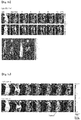
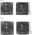
FIGS. 5a to 5g illustrates fluorescent images obtained by injecting 4 DNA polyhedrons (D55, L55, D92, and L92) into mice. Specifically,-
FIG. 5a : Fluorescent image obtained by injecting D55 Td, -
FIG. 5b : Fluorescent image obtained by injecting D92 Td, -
FIG. 5c : Comparison of in vivo fluorescent images obtained by injecting L55 and L92 and leaving the mice to stand for 0 minute to 24 hours after the injection, -
FIG. 5d : Fluorescent image obtained by injecting L55 and leaving the mouse to stand for 0 minute to 5 hours (in vivo +ex vivo 5 hours after injection), -
FIG. 5e : Fluorescent image obtained by injecting L92 and leaving the mouse to stand for 0 minute to 7 hours (in vivo +ex vivo 7 hours after injection), -
FIG. 5f : Comparison of ex vivo fluorescent images obtained by injecting L55 and L92 (L55 and L92 are obtained 5 hours after injection and 7 hours after injection, respectively), and -
FIG. 5g : Comparison of ex vivo fluorescent images obtained by injecting L55 and L92 (after adjusting scale).
-
- 6.
FIGS. 6a to 6d illustrate the results in which the formation of the 4 (L-Td, L-TP, L-Cb, and L-Od) structures are confirmed by PAGE (FIG. 6a : L-Td,FIG. 6b : L-TP,FIG. 6c : L-Cb, andFIG. 6d : L-Od). - 7.
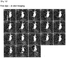
FIGS. 7a to 7f illustrate the results in which L-DNA nanostructures fluorescently labeled are injected into a mouse tumor model, and a change in distribution of nanostructures in the organism is observed (FIG. 7a : Free dye,FIG. 7b : L-Td,FIG. 7c : L-TP,FIG. 7d : L-Cb,FIG. 7e : L-Od, andFIG. 7f : Comparison of in vivo images of 4 structures on the same scale). - 8.
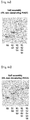
FIGS. 8a to 8f illustrate the results in which L-DNA nanostructures fluorescently labeled are injected into a tumor model, the mouse was sacrificed, and 6 organs of brain, heart, lung, liver, kidney, and spleen and tumor were removed and observed (FIG. 8a : Free dye,FIG. 8b : L-Td,FIG. 8c : L-TP,FIG. 8d : L-Cb,FIG. 8e : L-Od, andFIG. 8f : Comparison of ex vivo images of 4 structures on the same scale). - 9.
FIGS. 9a and 9b illustrate the results in which an optimal binding ratio is searched using a Job plot in order to load a doxorubicin (DOX) drug into L-Td55 (FIG. 9a ) and L-Td92 (FIG. 9b ). - 10.
FIG. 10 illustrates a schematic view of selective DOX delivery and therapy of a cancer tissue using a Xenograft cancer mouse animal model. - 11.
FIG. 11 illustrates photographs of external parts of mice which were classified intototal 6 groups (PBS, L-Td55, L-Td92, free DOX, DOX@L-Td55, and DOX@L-Td92) and treated for 18 days, and a photograph of tumors removed at the final day of therapy. - 12.
FIG. 12 is graphs illustrating changes in mice which were classified intototal 6 groups (PBS, L-Td55, L-Td92, free DOX, DOX@L-Td55, and DOX@L-Td92) and treated for 18 days during the therapy period (FIG. 12a : Change in volume of tumor,FIG. 12b : Change in body weight,FIG. 12c : Number of survived mice, andFIG. 12d : Final tumor weight). - 13.
FIG. 13 illustrates the results in which mice were classified intototal 6 groups (PBS, L-Td55, L-Td92, free DOX, DOX@L-Td55, and DOX@L-Td92), treated for 18 days, and sacrificed at the final day of therapy, and lung, liver, kidney, and cancer removed were observed through optical microscopy. - Hereinafter, the present invention will be described in more detail through the Examples. These Examples are provided only for more specifically describing the present invention, and it will be obvious to a person with ordinary skill in the art to which the present invention pertains that the scope of the present invention is not limited by these Examples.
- DNA oligonucleotides required for the Tds structure were synthesized by using a DNA synthesis standard protocol. The base sequence of the 55mer DNA standard was adopted from the Tuberfield Td, and the base sequence of the 92mer DNA standard was adopted from the Anderson (Lee et al. Nat. Nanotechnol. 2012, 7, 389-393). The oligonucleotide sequences used to constitute total 4 DNA nanostructures (D-DNA 55mer Td, L-DNA 55mer Td, D-DNA 92mer Td, and L-DNA 92mer Td) are shown in the following Table 1.
[Table 1] Sequence(5' to 3') Cy5.5-labeled D/L 55_S1 
Fluorescein-labeled D/L 55_S1 
D/L 55_S2 
D/L 55_S3 
D/L 55_S4 
Cy5.5-labeled D/L 92_S1 
Fluorescein-labeled D/L 92_S1 
D/L 92_S2 
D/L 92_S3 
D/L 92_S4 
- A library of DNA nanostructures was prepared by performing the Td assembly as described in the document (Kim et al. Chem. Sci., 2014, 5, 1533-1537) to change the sugar backbone and size (see
FIG. 2a ), and then the self-assembled D and L-Td (D-DNA 55mer Td, L-DNA 55mer Td, D-DNA 92mer Td, and L-DNA 92mer Td) were confirmed by performing 6% non-denaturing polyacrylamide gel electrophoresis (PAGE) (FIG. 2b ). - In order to confirm the cell selectivity of each DNA nanostructure, cancer cells were treated with the DNA nanostructures prepared above.
- Specifically, HeLa, HepG2, A549 and MCF7 cells were each inoculated into a glass-bottom 35-mm petri dish including a DMEM medium (Gibco, USA) containing 10% fetal bovine serum inactivated with heat, 1% penicillin, and streptomycin, and then the dish was cultured in a wet atmosphere including 5% CO2 at 37°C. The growth medium was removed from each cell sample and washed twice with PBS (Gibco, USA), and the DNA nanostructures prepared were subjected to transfection treatment in each cell.
- Cells transfected with the DNA nanostructures were observed under a fluorescent microscope (DeltaVision, Applied Precision, USA) and living cells were imaged, and the results are each shown in
FIG. 3 (FIGS. 3a to 3d ). - As a result of the experiment, it could be seen that the DNA nanostructures were entered into the cells, and it could be confirmed that the DNA nanostructures were in the cytoplasm region without being delivered to the cell nucleus.
- The HeLa, HepG2, A549 and MCF7 cells were cultured with DNA molecules fluorescently labeled using the method which is the same as that adopted in the transfection experiment, the fluorescence intensity of the cells was evaluated using a flow cytometer (FC500, Beckman coulter, USA), and then, the result is shown in
FIG. 4 . - Based on the result that the amount of each nanostructure delivered into the cells was quantified through a flow cytometry, the amounts delivered for each cell could be compared with each other, and as a result of the experiment, it was confirmed that D55 had HepG2 cell selectivity, L55 simultaneously had strong HeLa cell selectivity and considerable HepG2 cell selectivity, and L92 had HeLa and HepG2 cell selectivity (
FIG. 4 ). - As described above, since the kind of cell delivered in a large amount for each structure is present, it was confirmed that a structure having cell selectivity could be discovered through the construction of a library composed of various DNA nanostructures.
- The animal experiment was approved by the Institutional Animal Care and Use Committee of Korean Institute of Science and Technology, and all the mice were treated according to the regulations of the committee. For in vivo imaging and estalishment of a disease model, a mouse was anesthesized by intraperitoneally injecting 0.5% pentobarbital sodium (0.01 mL/g). An animal disease model was established by using a BALB/c nude mouse (5 weeks old, male, Orient Bio Inc., Korea). A tumor was produced by subcutaneously inoculating SCC7 cells (1.0 × 10⁶ cells suspended in a culture medium) into the thigh of the mouse.
- The DNA structures prepared were injected into the caudal vein of the mouse, and fluorescent images obtained by using a CCD camera performed in a highly sensitive imaging system (IVIS-spectrum, Perkin-Elmer, USA) are shown in
FIG. 5 (FIGS. 5a to 5e ). - After an in vivo imaging study, ex vivo near-infrared fluorescence images for excised organs and the other sites of the body were obtained by using an IVIS-spectrum imaging system including the same obtaining set as that used for in vivo imaging, and the results are shown in
FIG. 5 (the second photograph inFIGS. 5a and5b , the last photograph inFIGS. 5d and 5e ,FIG. 5f, and FIG. 5g ). - As a result of the experiment, it was confirmed that D55 was delivered more selectively to the liver, and distributed even in the skin tissue. On the contrary, D92 was delivered more selectively to the kidney. It was shown that L55 and L92 were accumulated, exhibiting high selectivity for the cancer tissue In addition, it was observed that as time elapsed, L-Td's had accumulated been more selectively in cancer instead of being distributed throughout the tissue than D-Td (after about 6 to 7 hours), and had escaped after 24 hours.
- The oligonucleotide sequences used to self-assemble total four L-DNA nanostructures having tetrahedron (L-Td), triangular prism (L-TP), cube (L-Cb) and octahedron (L-Od) shapes as an L-DNA nanostructure are shown in the following Tables 2 to 5.
[Table 2] Structure Sequence (5' to 3') Tetrahedron (L-Td) S1 
S2 
S3 
S4 
[Table 3] Structure Sequence (5' to 3') Triangular prism (L-TP) S1 
S2 
S3 
S4 
S5 
[Table 4] Structure Sequence (5' to 3') Cube (L-Cb) S1 
S2 
S3 
S4 
S5 
S6 
[Table 5] Structure Sequence (5' to 3') Octahedron (L-Od) S1 
S2 
S3 
S4 
S5 
S6 
S7 
S8 
- The strands constituting the structure were mixed so as to have a concentration of 1 µM based on each strand. In this case, a TM buffer (10 mM Tris-HCl, 5 mM MgCl2, pH 8.0) was used as a buffer. The mixture was denatured through heating at 95°C by using a RT-PCR machine, and was slowly cooled at 4°C to be annealed.
- As a result, it was confirmed by a non-denaturing PAGE that the resulting four (L-Td, L-TP, L-Cb, and L-Od) structures had been formed, and the results are each shown in
FIGS. 6a to 6d (FIG. 6a : L-Td,FIG. 6b : L-TP,FIG. 6c : L-Cb, andFIG. 6d : L-Od). - A red fluorescent dye (SYTO® 62 Red Fluorescent Nucleic Acid Stain, S11344, life technologies) having binding properties with DNA was mixed so as to have a final concentration of 1 µM. Herein, the DNA structure and the dye were bonded to each other at a concentration ratio of 1:1. The red fluorescent dye used in the present experiment has properties which do not show fluorescence when being present alone, but show fluorescence while being bonded to DNA.
- A balb/c nude mouse (5 weeks old, male) was used as an experimental animal group, and a tumor was formed by subcutaneously inoculating SCC7 cells (1.0 × 10⁶ cells suspended in the culture medium) into the left thigh of the mouse. When the tumor volume became 50 mm3 or more, the tumor was used for the experiment.
- A sample of the L-DNA nanostructure (a final concentration of 1 µM, 200 µL) labeled with fluorescence prepared in 1 was injected into the caudal vein of the mouse tumor model (I. V. injection). When compared to the case before the sample was injected, a change in in vivo distribution of the nanostructure was observed by using an IVIS imaging system apparatus at immediately after injection (0 min), 5, 10, 15, 20, 25, and 30 minutes, and 1, 2, 3, 4, 5, 6, 7, 8, 24, and 48 hours (filter set: Ex = 640 nm, Em = 680 nm), and the results are each shown in
FIGS. 7a to 7f (FIG. 7a : Free dye,FIG. 7b : L-Td,FIG. 7c : L-TP,FIG. 7d : L-Cb,FIG. 7e : L-Od, andFIG. 7f : Comparison of in vivo images of four structures on the same scale). - Based on the in vivo imaging results, ex vivo images were observed by sacrificing the mouse at the time when the fluorescent intensity of each structure in the tumor was highest and removing 6 organs of brain, heart, lung, liver, kidney, and spleen and tumor, and are each shown in
FIGS. 8a to 8f (FIG. 8a : Free dye,FIG. 8b : L-Td,FIG. 8c : L-TP,FIG. 8d : L-Cb,FIG. 8e : L-Od, andFIG. 8f : Comparison of ex vivo images of four structures on the same scale). - As a result of the experiment, it could be confirmed that all the four L-DNA nanostructures used in the experiment showed cancer tissue selectivity.
- Among them, the best cancer tissue selectivity was found in L-Td, and the next best selectivity was found in this order of L-TP, L-Cb, and L-Od. In the tissue other than the cancer tissue, it was found that L-structures were usually found from the kidney.
- In order to confirm the optimal binding ratio of Td and doxorubicin (DOX), the Job's plot method was used as in
FIG. 9 (see analytical chemistry, 1971, 43, 1265,FIG. 9a : L-Td55 andFIG. 9b : L-Td92). Td and DOX were prepared at a concentration of 1 µM, respectively, in the case of L-Td55 and at a concentration of 1 µM and 3 µM, respectively, in the case of L-Td92, mixed at each volume ratio (see the following Table 6, and the total volume was fixed at 100 µl), and then incubated at normal temperature for 1 hour.[Table 6] Sample (Td-DOX Complex) Td 10 9 8 7 6 5 4 3 2 1 0 Total : 100 µl DOX 0 1 2 3 4 5 6 7 8 9 10 Control (free DOX) TM buffer 10 9 8 7 6 5 4 3 2 1 0 Total: 100 µl DOX 0 1 2 3 4 5 6 7 8 9 10 - And then, the samples were scanned at a wavelength of 200 to 800 nm by using an UV-visible spectrophotometer, and free DOX to which a TM buffer was added instead of the Td sample was used as a control. At 480 nm where the highest absorbance value of DOX was shown, it was judged that the binding ratio used in the sample in which the difference in absorbance values of free DOX and Dox loaded Td was highest formed a composite best, and at a ratio thus determined, DOX was loaded into Td.
- A balb/c nude mouse (5 weeks old, male) was used as an experimental animal group, and a tumor was formed by subcutaneously inoculating SCC7 cells (1.0 × 10⁶ cells suspended in the culture medium) into the left thigh of the mouse. When the tumor volume became approximately 50 mm3, the tumor was used for the experiment.
- For the selective therapy of cancer tissue, DOX was each loaded into L-Td55 and L-Td92, which are a carrier having cancer tissue selectivity. A sample was prepared by mixing the nanostructure with DOX at the optimal binding ratio obtained through the Job plot method of the slide # 1 (L-Td55 : DOX = 1 : 24, L-Td92 : DOX = 1 : 48).
- The therapy experiment was performed by classifying the mice into total six groups (PBS, L-Td55, L-Td92, free DOX, DOX@L-Td55, and DOX@L-Td92), and 7 mice per group were used. Before the therapy was performed each time, the tumor volume and the mouse body weight were measured. In this case, the tumor volume was calculated by a method of (minor axis2∗major axis)/2. Each sample was injected into the caudal vein of the mice completely subjected to measurement (see
Slide # 2 for concentration), the therapy was performed once every three days, and the therapy was performed total 6 times. Onday 18 which was 3 days after the last 6th therapy, the final tumor weight was measured by measuring the tumor volume and the mouse body weight, and then measuring the mouse body weight to remove the tumor, and the results are shown inFIGS. 11 and12 (FIG. 11 : Photographs of external parts of mice and a photograph of tumors removed,FIG. 12a : Change in volume of tumor,FIG. 12b : Change in body weight,FIG. 12c : Number of survived mice, andFIG. 12d : Final tumor weight). - The therapy was finished, and 6 organs of brain, heart, lung, liver, kidney, and spleen and tumor were removed by sacrificing the survived mouse for each group, thereby observing the presence of organ damage and the tumor state. All the organs and tumor were fixed in 4% formaldehyde (4°C, overnight), and then embedded in paraffin after the tissue dehydration process. The paraffin block thus prepared was cut into a thickness of 5 µm, stained with hematoxylin and eosin, and observed through optical microscopy, and the results are shown in
FIG. 13 . - As a result of the experiment, it was confirmed that tumors were increased 4 to 5 times in the case of DOX@L55 and DOX@L92, while tumor volumes were increased up to about 40 times for the other groups, and thus the growth in tumor was delayed when DOX was loaded into the carrier.
- Due to the tumor growth rate of the tumor bearing mice using SCC7 known to be rapidly growing, it was judged that an increase in tumor volume had been observed in all the cases. When a tumor model is established using human-derived cells, a much better therapeutic effect is expected.
- For the control which was not subjected to therapy, the L55 treatment group, the L92 treatment group, and the free DOX treatment group showing slight therapeutic effects, the animal groups of individual 1 to 3 mice died during the therapy period.
- When the graph of change in body weight is observed, it is judged that the increase in body weight is due to an increase in tumor volume. When the weights of tumors obtained through ex vivo imaging were compared with each other, the tumor weights of the groups other than DOX@L55 or DOX@L92 exhibited values about 5 times higher than the tumor weights of DOX@L55 or DOX@L92.
- When the tissue fragments were observed, no part particularly damaged by DOX had been found in the tissues other than cancer tissues.
Claims (2)
- A pharmaceutical composition for use in the therapy of cancer, comprising a drug carrier having a self-assembled 3-D L-DNA nanostructure for in vivo delivering a pharmaceutically active ingredient selectively to a cancer tissue by in vivo administration; wherein the pharmaceutically active ingredient is an anticancer agent; wherein the carrier does not comprise a targeting ligand for the cancer tissue, wherein the carrier has in vivo specificity to cancer tissue and wherein the carrier has a tetrahedron structure composed of 4 strands of L-DNA 55 mer or 4 strands of L-DNA 92 mer; and wherein the anticancer agent is doxorubicin, and wherein the pharmaceutical composition is systemically administered to the cancer tissue.
- The pharmaceutical composition for use in the therapy of cancer of claim 1, wherein the pharmaceutically active ingredient is encapsulated within the nucleic acid nanostructure or bonded to the backbone of the nucleic acid nanostructure, and thereby delivered to the target tissue.
Applications Claiming Priority (1)
| Application Number | Priority Date | Filing Date | Title |
|---|---|---|---|
| KR20140093458 | 2014-07-23 |
Publications (2)
| Publication Number | Publication Date |
|---|---|
| EP2977064A1 EP2977064A1 (en) | 2016-01-27 |
| EP2977064B1 true EP2977064B1 (en) | 2021-10-27 |
Family
ID=53938061
Family Applications (1)
| Application Number | Title | Priority Date | Filing Date |
|---|---|---|---|
| EP15177301.7A Not-in-force EP2977064B1 (en) | 2014-07-23 | 2015-07-17 | Drug carrier having self-assembled 3-d nucleic acid nanostructure for delivering doxorubicin in the treatment of cancer |
Country Status (3)
| Country | Link |
|---|---|
| US (2) | US20160051483A1 (en) |
| EP (1) | EP2977064B1 (en) |
| KR (1) | KR101755617B1 (en) |
Families Citing this family (8)
| Publication number | Priority date | Publication date | Assignee | Title |
|---|---|---|---|---|
| KR101943515B1 (en) * | 2017-02-02 | 2019-01-30 | 한국과학기술연구원 | pH responsive composition for regulation of enzyme activity and uses thereof |
| CN108721634B (en) * | 2018-05-30 | 2021-06-04 | 郑州大学 | Construction method and application of double-target-point DNA nano treatment system |
| KR102315342B1 (en) | 2019-11-01 | 2021-10-22 | 한국과학기술연구원 | Three dimensional self-assembled nucleic acid nanoparticles and use thereof |
| CN111803511B (en) * | 2020-06-12 | 2022-11-08 | 南京邮电大学 | DNA tetrahedral nucleic acid frame type gastric cancer diagnosis and treatment integrated reagent and preparation method and application thereof |
| CN112345507B (en) * | 2020-11-06 | 2022-07-05 | 济南大学 | Biosensor for targeting cancer cells based on DNA triangular prism structure conformational change |
| CN112807445B (en) * | 2021-01-07 | 2023-06-20 | 中国药科大学 | pH-responsive DNA octahedron drug carrier and its preparation method and application |
| CN116327627B (en) * | 2023-03-10 | 2024-06-07 | 四川大学 | A tetrahedral framework nucleic acid-glabridin complex and its use in skin whitening |
| CN116407643A (en) * | 2023-03-27 | 2023-07-11 | 南通大学附属医院 | Application of MTX-loaded DNA tetrahedron in the preparation of drugs for treating rheumatoid arthritis |
Family Cites Families (7)
| Publication number | Priority date | Publication date | Assignee | Title |
|---|---|---|---|---|
| EP1877069A2 (en) | 2005-05-04 | 2008-01-16 | Noxxon Pharma AG | Novel use of spiegelmers |
| KR100637557B1 (en) | 2005-08-29 | 2006-10-20 | 이희란 | SiRNA with antiviral activity against bifolio enterovirus |
| GB0607866D0 (en) * | 2006-04-20 | 2006-05-31 | Isis Innovation | Nanostructures |
| US20110033706A1 (en) * | 2009-08-04 | 2011-02-10 | Yamuna Krishnan | Nanocapsules and methods for modular assembly |
| WO2012125987A2 (en) * | 2011-03-17 | 2012-09-20 | Massachusetts Institute Of Technology | Delivery system |
| US9266844B2 (en) | 2012-06-15 | 2016-02-23 | The Curators Of The University Of Missouri | Suppression of SARS replication by SARS helicase inhibitors |
| KR101494773B1 (en) * | 2013-11-18 | 2015-02-23 | 한국과학기술연구원 | Drug Carrier Having L-DNA Nanocage Structure |
-
2015
- 2015-07-16 KR KR1020150101204A patent/KR101755617B1/en active Active
- 2015-07-17 EP EP15177301.7A patent/EP2977064B1/en not_active Not-in-force
- 2015-07-20 US US14/803,343 patent/US20160051483A1/en not_active Abandoned
-
2019
- 2019-08-12 US US16/538,109 patent/US11446254B2/en active Active
Non-Patent Citations (1)
| Title |
|---|
| None * |
Also Published As
| Publication number | Publication date |
|---|---|
| KR101755617B1 (en) | 2017-07-10 |
| KR20160012079A (en) | 2016-02-02 |
| US20160051483A1 (en) | 2016-02-25 |
| US11446254B2 (en) | 2022-09-20 |
| EP2977064A1 (en) | 2016-01-27 |
| US20190365659A1 (en) | 2019-12-05 |
Similar Documents
| Publication | Publication Date | Title |
|---|---|---|
| Xu et al. | Engineered exosomes: desirable target-tracking characteristics for cerebrovascular and neurodegenerative disease therapies | |
| US11446254B2 (en) | Drug carrier having self-assembled 3-D nucleic acid nanostructure | |
| Wan et al. | Self-assembled magnetic theranostic nanoparticles for highly sensitive MRI of minicircle DNA delivery | |
| JP5802555B2 (en) | Chelated amphiphilic polymers | |
| Jarzyna et al. | Multifunctional imaging nanoprobes | |
| Wang et al. | Multifunctional reduction-responsive SPIO&DOX-loaded PEGylated polymeric lipid vesicles for magnetic resonance imaging-guided drug delivery | |
| EP2365796A1 (en) | Fusogenic properties of saposin c and related proteins and peptides for application to transmembrane drug delivery systems | |
| CN101698106A (en) | D-glucosamine-modified iron oxide nanoparticles and preparation method of lyophilized powder thereof | |
| CN101888857A (en) | Polymeric drug carriers for image-guided delivery | |
| US20200179295A1 (en) | Mri-detectable multilayer microcapsules for ultrasound-triggered delivery of pharmacologically active agents | |
| JP2014095025A (en) | Diamond composite particles | |
| US9669114B2 (en) | Contrast medium composition and method of bio imagination using the same | |
| Xu et al. | Piperazine-derived ionizable lipids for enhanced mRNA delivery and cancer immunotherapy | |
| Fan et al. | Charge reversal hairpin peptide modified synergy therapeutic nanoplatforms for tumor specific drug shuttling | |
| Bai et al. | Modular design of Bi-specific nanoplatform engaged in malignant lymphoma immunotherapy | |
| Ma et al. | Deposition of gadolinium onto the shell structure of micelles for integrated magnetic resonance imaging and robust drug delivery systems | |
| Chen et al. | Targeted nanoprobe for magnetic resonance imaging-guided enhanced antitumor via synergetic photothermal/immunotherapy | |
| CN113975389B (en) | A macrophage-loaded multifunctional mesoporous iron oxide nano-diagnosis and treatment preparation and its preparation method and anti-tumor application | |
| Chang et al. | A highly effective in vivo photothermal nanoplatform with dual imaging-guided therapy of cancer based on the charge reversal complex of dye and iron oxide | |
| CN112870387B (en) | Magnetic nano-drug carrier and preparation method and application thereof | |
| CN106880846A (en) | A kind of cancer target multifunctional nano delivery system and preparation method and purposes | |
| CN111807979B (en) | Zwitterionic fluorinated cross-linking agent, nanogel and application thereof | |
| WO2010148395A1 (en) | Paramagnetic porous polymersomes and uses thereof | |
| Nasr Azadani et al. | Nanoparticles in cancer theranostics: Focus on gliomas | |
| Li et al. | Cochlear transfection gene Guinea pigs mediates atoh1-EGFP based hyaluronic acid modified polyethyleneimine nanoparticles |
Legal Events
| Date | Code | Title | Description |
|---|---|---|---|
| PUAI | Public reference made under article 153(3) epc to a published international application that has entered the european phase |
Free format text: ORIGINAL CODE: 0009012 |
|
| 17P | Request for examination filed |
Effective date: 20150806 |
|
| AK | Designated contracting states |
Kind code of ref document: A1 Designated state(s): AL AT BE BG CH CY CZ DE DK EE ES FI FR GB GR HR HU IE IS IT LI LT LU LV MC MK MT NL NO PL PT RO RS SE SI SK SM TR |
|
| AX | Request for extension of the european patent |
Extension state: BA ME |
|
| RBV | Designated contracting states (corrected) |
Designated state(s): AL AT BE BG CH CY CZ DE DK EE ES FI FR GB GR HR HU IE IS IT LI LT LU LV MC MK MT NL NO PL PT RO RS SE SI SK SM TR |
|
| STAA | Information on the status of an ep patent application or granted ep patent |
Free format text: STATUS: EXAMINATION IS IN PROGRESS |
|
| 17Q | First examination report despatched |
Effective date: 20180320 |
|
| REG | Reference to a national code |
Ref country code: DE Ref legal event code: R079 Ref document number: 602015074405 Country of ref document: DE Free format text: PREVIOUS MAIN CLASS: A61K0047480000 Ipc: A61K0047500000 |
|
| GRAP | Despatch of communication of intention to grant a patent |
Free format text: ORIGINAL CODE: EPIDOSNIGR1 |
|
| STAA | Information on the status of an ep patent application or granted ep patent |
Free format text: STATUS: GRANT OF PATENT IS INTENDED |
|
| RIC1 | Information provided on ipc code assigned before grant |
Ipc: A61P 35/00 20060101ALI20210428BHEP Ipc: A61K 31/704 20060101ALI20210428BHEP Ipc: A61K 9/14 20060101ALI20210428BHEP Ipc: A61K 49/00 20060101ALI20210428BHEP Ipc: A61K 47/50 20170101AFI20210428BHEP |
|
| INTG | Intention to grant announced |
Effective date: 20210514 |
|
| GRAS | Grant fee paid |
Free format text: ORIGINAL CODE: EPIDOSNIGR3 |
|
| GRAA | (expected) grant |
Free format text: ORIGINAL CODE: 0009210 |
|
| STAA | Information on the status of an ep patent application or granted ep patent |
Free format text: STATUS: THE PATENT HAS BEEN GRANTED |
|
| AK | Designated contracting states |
Kind code of ref document: B1 Designated state(s): AL AT BE BG CH CY CZ DE DK EE ES FI FR GB GR HR HU IE IS IT LI LT LU LV MC MK MT NL NO PL PT RO RS SE SI SK SM TR |
|
| REG | Reference to a national code |
Ref country code: GB Ref legal event code: FG4D |
|
| REG | Reference to a national code |
Ref country code: CH Ref legal event code: EP |
|
| REG | Reference to a national code |
Ref country code: DE Ref legal event code: R096 Ref document number: 602015074405 Country of ref document: DE |
|
| REG | Reference to a national code |
Ref country code: AT Ref legal event code: REF Ref document number: 1441226 Country of ref document: AT Kind code of ref document: T Effective date: 20211115 |
|
| REG | Reference to a national code |
Ref country code: IE Ref legal event code: FG4D |
|
| REG | Reference to a national code |
Ref country code: LT Ref legal event code: MG9D |
|
| REG | Reference to a national code |
Ref country code: NL Ref legal event code: MP Effective date: 20211027 |
|
| REG | Reference to a national code |
Ref country code: AT Ref legal event code: MK05 Ref document number: 1441226 Country of ref document: AT Kind code of ref document: T Effective date: 20211027 |
|
| PG25 | Lapsed in a contracting state [announced via postgrant information from national office to epo] |
Ref country code: RS Free format text: LAPSE BECAUSE OF FAILURE TO SUBMIT A TRANSLATION OF THE DESCRIPTION OR TO PAY THE FEE WITHIN THE PRESCRIBED TIME-LIMIT Effective date: 20211027 Ref country code: LT Free format text: LAPSE BECAUSE OF FAILURE TO SUBMIT A TRANSLATION OF THE DESCRIPTION OR TO PAY THE FEE WITHIN THE PRESCRIBED TIME-LIMIT Effective date: 20211027 Ref country code: FI Free format text: LAPSE BECAUSE OF FAILURE TO SUBMIT A TRANSLATION OF THE DESCRIPTION OR TO PAY THE FEE WITHIN THE PRESCRIBED TIME-LIMIT Effective date: 20211027 Ref country code: BG Free format text: LAPSE BECAUSE OF FAILURE TO SUBMIT A TRANSLATION OF THE DESCRIPTION OR TO PAY THE FEE WITHIN THE PRESCRIBED TIME-LIMIT Effective date: 20220127 Ref country code: AT Free format text: LAPSE BECAUSE OF FAILURE TO SUBMIT A TRANSLATION OF THE DESCRIPTION OR TO PAY THE FEE WITHIN THE PRESCRIBED TIME-LIMIT Effective date: 20211027 |
|
| PG25 | Lapsed in a contracting state [announced via postgrant information from national office to epo] |
Ref country code: IS Free format text: LAPSE BECAUSE OF FAILURE TO SUBMIT A TRANSLATION OF THE DESCRIPTION OR TO PAY THE FEE WITHIN THE PRESCRIBED TIME-LIMIT Effective date: 20220227 Ref country code: SE Free format text: LAPSE BECAUSE OF FAILURE TO SUBMIT A TRANSLATION OF THE DESCRIPTION OR TO PAY THE FEE WITHIN THE PRESCRIBED TIME-LIMIT Effective date: 20211027 Ref country code: PT Free format text: LAPSE BECAUSE OF FAILURE TO SUBMIT A TRANSLATION OF THE DESCRIPTION OR TO PAY THE FEE WITHIN THE PRESCRIBED TIME-LIMIT Effective date: 20220228 Ref country code: PL Free format text: LAPSE BECAUSE OF FAILURE TO SUBMIT A TRANSLATION OF THE DESCRIPTION OR TO PAY THE FEE WITHIN THE PRESCRIBED TIME-LIMIT Effective date: 20211027 Ref country code: NO Free format text: LAPSE BECAUSE OF FAILURE TO SUBMIT A TRANSLATION OF THE DESCRIPTION OR TO PAY THE FEE WITHIN THE PRESCRIBED TIME-LIMIT Effective date: 20220127 Ref country code: NL Free format text: LAPSE BECAUSE OF FAILURE TO SUBMIT A TRANSLATION OF THE DESCRIPTION OR TO PAY THE FEE WITHIN THE PRESCRIBED TIME-LIMIT Effective date: 20211027 Ref country code: LV Free format text: LAPSE BECAUSE OF FAILURE TO SUBMIT A TRANSLATION OF THE DESCRIPTION OR TO PAY THE FEE WITHIN THE PRESCRIBED TIME-LIMIT Effective date: 20211027 Ref country code: HR Free format text: LAPSE BECAUSE OF FAILURE TO SUBMIT A TRANSLATION OF THE DESCRIPTION OR TO PAY THE FEE WITHIN THE PRESCRIBED TIME-LIMIT Effective date: 20211027 Ref country code: GR Free format text: LAPSE BECAUSE OF FAILURE TO SUBMIT A TRANSLATION OF THE DESCRIPTION OR TO PAY THE FEE WITHIN THE PRESCRIBED TIME-LIMIT Effective date: 20220128 Ref country code: ES Free format text: LAPSE BECAUSE OF FAILURE TO SUBMIT A TRANSLATION OF THE DESCRIPTION OR TO PAY THE FEE WITHIN THE PRESCRIBED TIME-LIMIT Effective date: 20211027 |
|
| REG | Reference to a national code |
Ref country code: DE Ref legal event code: R097 Ref document number: 602015074405 Country of ref document: DE |
|
| PG25 | Lapsed in a contracting state [announced via postgrant information from national office to epo] |
Ref country code: SM Free format text: LAPSE BECAUSE OF FAILURE TO SUBMIT A TRANSLATION OF THE DESCRIPTION OR TO PAY THE FEE WITHIN THE PRESCRIBED TIME-LIMIT Effective date: 20211027 Ref country code: SK Free format text: LAPSE BECAUSE OF FAILURE TO SUBMIT A TRANSLATION OF THE DESCRIPTION OR TO PAY THE FEE WITHIN THE PRESCRIBED TIME-LIMIT Effective date: 20211027 Ref country code: RO Free format text: LAPSE BECAUSE OF FAILURE TO SUBMIT A TRANSLATION OF THE DESCRIPTION OR TO PAY THE FEE WITHIN THE PRESCRIBED TIME-LIMIT Effective date: 20211027 Ref country code: EE Free format text: LAPSE BECAUSE OF FAILURE TO SUBMIT A TRANSLATION OF THE DESCRIPTION OR TO PAY THE FEE WITHIN THE PRESCRIBED TIME-LIMIT Effective date: 20211027 Ref country code: DK Free format text: LAPSE BECAUSE OF FAILURE TO SUBMIT A TRANSLATION OF THE DESCRIPTION OR TO PAY THE FEE WITHIN THE PRESCRIBED TIME-LIMIT Effective date: 20211027 Ref country code: CZ Free format text: LAPSE BECAUSE OF FAILURE TO SUBMIT A TRANSLATION OF THE DESCRIPTION OR TO PAY THE FEE WITHIN THE PRESCRIBED TIME-LIMIT Effective date: 20211027 |
|
| PLBE | No opposition filed within time limit |
Free format text: ORIGINAL CODE: 0009261 |
|
| STAA | Information on the status of an ep patent application or granted ep patent |
Free format text: STATUS: NO OPPOSITION FILED WITHIN TIME LIMIT |
|
| 26N | No opposition filed |
Effective date: 20220728 |
|
| PG25 | Lapsed in a contracting state [announced via postgrant information from national office to epo] |
Ref country code: AL Free format text: LAPSE BECAUSE OF FAILURE TO SUBMIT A TRANSLATION OF THE DESCRIPTION OR TO PAY THE FEE WITHIN THE PRESCRIBED TIME-LIMIT Effective date: 20211027 |
|
| PG25 | Lapsed in a contracting state [announced via postgrant information from national office to epo] |
Ref country code: SI Free format text: LAPSE BECAUSE OF FAILURE TO SUBMIT A TRANSLATION OF THE DESCRIPTION OR TO PAY THE FEE WITHIN THE PRESCRIBED TIME-LIMIT Effective date: 20211027 |
|
| PG25 | Lapsed in a contracting state [announced via postgrant information from national office to epo] |
Ref country code: MC Free format text: LAPSE BECAUSE OF FAILURE TO SUBMIT A TRANSLATION OF THE DESCRIPTION OR TO PAY THE FEE WITHIN THE PRESCRIBED TIME-LIMIT Effective date: 20211027 |
|
| REG | Reference to a national code |
Ref country code: CH Ref legal event code: PL |
|
| REG | Reference to a national code |
Ref country code: BE Ref legal event code: MM Effective date: 20220731 |
|
| PG25 | Lapsed in a contracting state [announced via postgrant information from national office to epo] |
Ref country code: LU Free format text: LAPSE BECAUSE OF NON-PAYMENT OF DUE FEES Effective date: 20220717 Ref country code: LI Free format text: LAPSE BECAUSE OF NON-PAYMENT OF DUE FEES Effective date: 20220731 Ref country code: CH Free format text: LAPSE BECAUSE OF NON-PAYMENT OF DUE FEES Effective date: 20220731 |
|
| PG25 | Lapsed in a contracting state [announced via postgrant information from national office to epo] |
Ref country code: IT Free format text: LAPSE BECAUSE OF FAILURE TO SUBMIT A TRANSLATION OF THE DESCRIPTION OR TO PAY THE FEE WITHIN THE PRESCRIBED TIME-LIMIT Effective date: 20211027 Ref country code: BE Free format text: LAPSE BECAUSE OF NON-PAYMENT OF DUE FEES Effective date: 20220731 |
|
| PG25 | Lapsed in a contracting state [announced via postgrant information from national office to epo] |
Ref country code: IE Free format text: LAPSE BECAUSE OF NON-PAYMENT OF DUE FEES Effective date: 20220717 |
|
| PG25 | Lapsed in a contracting state [announced via postgrant information from national office to epo] |
Ref country code: HU Free format text: LAPSE BECAUSE OF FAILURE TO SUBMIT A TRANSLATION OF THE DESCRIPTION OR TO PAY THE FEE WITHIN THE PRESCRIBED TIME-LIMIT; INVALID AB INITIO Effective date: 20150717 |
|
| PG25 | Lapsed in a contracting state [announced via postgrant information from national office to epo] |
Ref country code: MK Free format text: LAPSE BECAUSE OF FAILURE TO SUBMIT A TRANSLATION OF THE DESCRIPTION OR TO PAY THE FEE WITHIN THE PRESCRIBED TIME-LIMIT Effective date: 20211027 Ref country code: CY Free format text: LAPSE BECAUSE OF FAILURE TO SUBMIT A TRANSLATION OF THE DESCRIPTION OR TO PAY THE FEE WITHIN THE PRESCRIBED TIME-LIMIT Effective date: 20211027 |
|
| PG25 | Lapsed in a contracting state [announced via postgrant information from national office to epo] |
Ref country code: TR Free format text: LAPSE BECAUSE OF FAILURE TO SUBMIT A TRANSLATION OF THE DESCRIPTION OR TO PAY THE FEE WITHIN THE PRESCRIBED TIME-LIMIT Effective date: 20211027 |
|
| PG25 | Lapsed in a contracting state [announced via postgrant information from national office to epo] |
Ref country code: MT Free format text: LAPSE BECAUSE OF FAILURE TO SUBMIT A TRANSLATION OF THE DESCRIPTION OR TO PAY THE FEE WITHIN THE PRESCRIBED TIME-LIMIT Effective date: 20211027 |
|
| PGFP | Annual fee paid to national office [announced via postgrant information from national office to epo] |
Ref country code: DE Payment date: 20240722 Year of fee payment: 10 |
|
| PGFP | Annual fee paid to national office [announced via postgrant information from national office to epo] |
Ref country code: GB Payment date: 20240723 Year of fee payment: 10 |
|
| PGFP | Annual fee paid to national office [announced via postgrant information from national office to epo] |
Ref country code: FR Payment date: 20240723 Year of fee payment: 10 |
|
| REG | Reference to a national code |
Ref country code: DE Ref legal event code: R119 Ref document number: 602015074405 Country of ref document: DE |
|
| GBPC | Gb: european patent ceased through non-payment of renewal fee |
Effective date: 20250717 |
|
| PG25 | Lapsed in a contracting state [announced via postgrant information from national office to epo] |
Ref country code: GB Free format text: LAPSE BECAUSE OF NON-PAYMENT OF DUE FEES Effective date: 20250717 |
|
| PG25 | Lapsed in a contracting state [announced via postgrant information from national office to epo] |
Ref country code: DE Free format text: LAPSE BECAUSE OF NON-PAYMENT OF DUE FEES Effective date: 20260203 |
|
| PG25 | Lapsed in a contracting state [announced via postgrant information from national office to epo] |
Ref country code: FR Free format text: LAPSE BECAUSE OF NON-PAYMENT OF DUE FEES Effective date: 20250731 |