EP2970325B1 - Lysophosphatidic acid detection - Google Patents
Lysophosphatidic acid detection Download PDFInfo
- Publication number
- EP2970325B1 EP2970325B1 EP14765163.2A EP14765163A EP2970325B1 EP 2970325 B1 EP2970325 B1 EP 2970325B1 EP 14765163 A EP14765163 A EP 14765163A EP 2970325 B1 EP2970325 B1 EP 2970325B1
- Authority
- EP
- European Patent Office
- Prior art keywords
- lysophosphatidic acid
- lpa
- acid species
- sample
- phase
- Prior art date
- Legal status (The legal status is an assumption and is not a legal conclusion. Google has not performed a legal analysis and makes no representation as to the accuracy of the status listed.)
- Active
Links
- WRGQSWVCFNIUNZ-GDCKJWNLSA-N 1-oleoyl-sn-glycerol 3-phosphate Chemical compound CCCCCCCC\C=C/CCCCCCCC(=O)OC[C@@H](O)COP(O)(O)=O WRGQSWVCFNIUNZ-GDCKJWNLSA-N 0.000 title claims description 247
- AWUCVROLDVIAJX-UHFFFAOYSA-N alpha-glycerophosphate Natural products OCC(O)COP(O)(O)=O AWUCVROLDVIAJX-UHFFFAOYSA-N 0.000 title claims description 244
- 238000001514 detection method Methods 0.000 title description 38
- OKKJLVBELUTLKV-UHFFFAOYSA-N Methanol Chemical compound OC OKKJLVBELUTLKV-UHFFFAOYSA-N 0.000 claims description 168
- HEDRZPFGACZZDS-UHFFFAOYSA-N Chloroform Chemical compound ClC(Cl)Cl HEDRZPFGACZZDS-UHFFFAOYSA-N 0.000 claims description 92
- 238000000034 method Methods 0.000 claims description 77
- IAZDPXIOMUYVGZ-UHFFFAOYSA-N Dimethylsulphoxide Chemical compound CS(C)=O IAZDPXIOMUYVGZ-UHFFFAOYSA-N 0.000 claims description 56
- 239000000203 mixture Substances 0.000 claims description 54
- 238000002414 normal-phase solid-phase extraction Methods 0.000 claims description 48
- 150000001875 compounds Chemical class 0.000 claims description 47
- 238000004128 high performance liquid chromatography Methods 0.000 claims description 47
- 239000002904 solvent Substances 0.000 claims description 45
- YPGZWUVVEWKKDQ-UHFFFAOYSA-M 4-(4-dihexadecylaminostyryl)-N-methylpyridium iodide Chemical compound [I-].C1=CC(N(CCCCCCCCCCCCCCCC)CCCCCCCCCCCCCCCC)=CC=C1C=CC1=CC=[N+](C)C=C1 YPGZWUVVEWKKDQ-UHFFFAOYSA-M 0.000 claims description 38
- 239000008346 aqueous phase Substances 0.000 claims description 35
- XLYOFNOQVPJJNP-UHFFFAOYSA-N water Substances O XLYOFNOQVPJJNP-UHFFFAOYSA-N 0.000 claims description 34
- 229910001868 water Inorganic materials 0.000 claims description 33
- 210000002966 serum Anatomy 0.000 claims description 27
- 239000003480 eluent Substances 0.000 claims description 23
- 238000004007 reversed phase HPLC Methods 0.000 claims description 18
- NBIIXXVUZAFLBC-UHFFFAOYSA-N Phosphoric acid Chemical compound OP(O)(O)=O NBIIXXVUZAFLBC-UHFFFAOYSA-N 0.000 claims description 15
- 230000005526 G1 to G0 transition Effects 0.000 claims description 14
- 208000037265 diseases, disorders, signs and symptoms Diseases 0.000 claims description 14
- 208000035475 disorder Diseases 0.000 claims description 14
- 206010028980 Neoplasm Diseases 0.000 claims description 12
- VYPSYNLAJGMNEJ-UHFFFAOYSA-N Silicium dioxide Chemical compound O=[Si]=O VYPSYNLAJGMNEJ-UHFFFAOYSA-N 0.000 claims description 12
- 230000001594 aberrant effect Effects 0.000 claims description 12
- 238000002156 mixing Methods 0.000 claims description 12
- 206010033128 Ovarian cancer Diseases 0.000 claims description 10
- 206010061535 Ovarian neoplasm Diseases 0.000 claims description 10
- 230000007935 neutral effect Effects 0.000 claims description 10
- 239000000872 buffer Substances 0.000 claims description 8
- 201000011510 cancer Diseases 0.000 claims description 8
- 208000010110 spontaneous platelet aggregation Diseases 0.000 claims description 8
- 208000024172 Cardiovascular disease Diseases 0.000 claims description 7
- 206010016654 Fibrosis Diseases 0.000 claims description 7
- 208000027418 Wounds and injury Diseases 0.000 claims description 7
- 229910000147 aluminium phosphate Inorganic materials 0.000 claims description 7
- 230000006378 damage Effects 0.000 claims description 7
- 238000010828 elution Methods 0.000 claims description 7
- 230000004761 fibrosis Effects 0.000 claims description 7
- 208000014674 injury Diseases 0.000 claims description 7
- 208000028867 ischemia Diseases 0.000 claims description 7
- 208000004296 neuralgia Diseases 0.000 claims description 7
- 208000021722 neuropathic pain Diseases 0.000 claims description 7
- 230000010412 perfusion Effects 0.000 claims description 7
- 230000001850 reproductive effect Effects 0.000 claims description 7
- 239000000377 silicon dioxide Substances 0.000 claims description 5
- 238000001035 drying Methods 0.000 claims description 4
- 238000001704 evaporation Methods 0.000 claims description 4
- 238000011068 loading method Methods 0.000 claims description 3
- 125000000008 (C1-C10) alkyl group Chemical group 0.000 claims 4
- 125000001183 hydrocarbyl group Chemical group 0.000 claims 1
- 241000894007 species Species 0.000 description 96
- 239000000523 sample Substances 0.000 description 79
- 239000000243 solution Substances 0.000 description 45
- 150000003904 phospholipids Chemical class 0.000 description 27
- 238000000926 separation method Methods 0.000 description 19
- 239000003153 chemical reaction reagent Substances 0.000 description 16
- 239000012071 phase Substances 0.000 description 16
- 238000002101 electrospray ionisation tandem mass spectrometry Methods 0.000 description 15
- 238000004885 tandem mass spectrometry Methods 0.000 description 15
- HEDRZPFGACZZDS-MICDWDOJSA-N Trichloro(2H)methane Chemical compound [2H]C(Cl)(Cl)Cl HEDRZPFGACZZDS-MICDWDOJSA-N 0.000 description 14
- DTQVDTLACAAQTR-UHFFFAOYSA-N Trifluoroacetic acid Chemical compound OC(=O)C(F)(F)F DTQVDTLACAAQTR-UHFFFAOYSA-N 0.000 description 14
- -1 heteroaliphatic Chemical group 0.000 description 14
- 150000008103 phosphatidic acids Chemical class 0.000 description 14
- 238000011084 recovery Methods 0.000 description 14
- YMWUJEATGCHHMB-UHFFFAOYSA-N Dichloromethane Chemical compound ClCCl YMWUJEATGCHHMB-UHFFFAOYSA-N 0.000 description 13
- 239000010410 layer Substances 0.000 description 13
- 239000008363 phosphate buffer Substances 0.000 description 13
- FAZBDRGXCKPVJU-UHFFFAOYSA-N (2-hydroxy-3-phosphonooxypropyl) tetradecanoate Chemical compound CCCCCCCCCCCCCC(=O)OCC(O)COP(O)(O)=O FAZBDRGXCKPVJU-UHFFFAOYSA-N 0.000 description 12
- 150000002500 ions Chemical class 0.000 description 12
- LAYXSTYJRSVXIH-HXUWFJFHSA-N 1-stearoyl-sn-glycero-3-phosphate Chemical compound CCCCCCCCCCCCCCCCCC(=O)OC[C@@H](O)COP(O)(O)=O LAYXSTYJRSVXIH-HXUWFJFHSA-N 0.000 description 11
- ZRALSGWEFCBTJO-UHFFFAOYSA-N Guanidine Chemical compound NC(N)=N ZRALSGWEFCBTJO-UHFFFAOYSA-N 0.000 description 11
- 238000004458 analytical method Methods 0.000 description 11
- 239000007787 solid Substances 0.000 description 11
- WEVYAHXRMPXWCK-UHFFFAOYSA-N Acetonitrile Chemical compound CC#N WEVYAHXRMPXWCK-UHFFFAOYSA-N 0.000 description 10
- 238000011088 calibration curve Methods 0.000 description 10
- 238000000638 solvent extraction Methods 0.000 description 10
- 125000000217 alkyl group Chemical group 0.000 description 9
- 238000000605 extraction Methods 0.000 description 9
- 238000000622 liquid--liquid extraction Methods 0.000 description 9
- 238000011002 quantification Methods 0.000 description 9
- YNDYKPRNFWPPFU-UHFFFAOYSA-N 1-palmitoylglycerol 3-phosphate Chemical compound CCCCCCCCCCCCCCCC(=O)OCC(O)COP(O)(O)=O YNDYKPRNFWPPFU-UHFFFAOYSA-N 0.000 description 8
- 239000002253 acid Substances 0.000 description 8
- 238000002347 injection Methods 0.000 description 8
- 239000007924 injection Substances 0.000 description 8
- 150000002632 lipids Chemical class 0.000 description 8
- 239000012074 organic phase Substances 0.000 description 8
- 239000003643 water by type Substances 0.000 description 8
- 125000004432 carbon atom Chemical group C* 0.000 description 7
- 238000006243 chemical reaction Methods 0.000 description 7
- 230000005284 excitation Effects 0.000 description 7
- 230000004044 response Effects 0.000 description 7
- PYWVYCXTNDRMGF-UHFFFAOYSA-N rhodamine B Chemical compound [Cl-].C=12C=CC(=[N+](CC)CC)C=C2OC2=CC(N(CC)CC)=CC=C2C=1C1=CC=CC=C1C(O)=O PYWVYCXTNDRMGF-UHFFFAOYSA-N 0.000 description 7
- 239000000126 substance Substances 0.000 description 7
- RYCNUMLMNKHWPZ-SNVBAGLBSA-N 1-acetyl-sn-glycero-3-phosphocholine Chemical compound CC(=O)OC[C@@H](O)COP([O-])(=O)OCC[N+](C)(C)C RYCNUMLMNKHWPZ-SNVBAGLBSA-N 0.000 description 6
- QYCYIZLSHFMZKV-LTKCOYKYSA-N LysoPA(20:4(8Z,11Z,14Z,17Z)/0:0) Chemical compound CC\C=C/C\C=C/C\C=C/C\C=C/CCCCCCC(=O)OCC(O)COP(O)(O)=O QYCYIZLSHFMZKV-LTKCOYKYSA-N 0.000 description 6
- UIIMBOGNXHQVGW-UHFFFAOYSA-M Sodium bicarbonate Chemical class [Na+].OC([O-])=O UIIMBOGNXHQVGW-UHFFFAOYSA-M 0.000 description 6
- WQDUMFSSJAZKTM-UHFFFAOYSA-N Sodium methoxide Chemical compound [Na+].[O-]C WQDUMFSSJAZKTM-UHFFFAOYSA-N 0.000 description 6
- 125000005233 alkylalcohol group Chemical group 0.000 description 6
- 238000011156 evaluation Methods 0.000 description 6
- VLKZOEOYAKHREP-UHFFFAOYSA-N n-Hexane Chemical compound CCCCCC VLKZOEOYAKHREP-UHFFFAOYSA-N 0.000 description 6
- 239000002953 phosphate buffered saline Substances 0.000 description 6
- BWHMMNNQKKPAPP-UHFFFAOYSA-L potassium carbonate Chemical compound [K+].[K+].[O-]C([O-])=O BWHMMNNQKKPAPP-UHFFFAOYSA-L 0.000 description 6
- 238000000746 purification Methods 0.000 description 6
- 229940043267 rhodamine b Drugs 0.000 description 6
- 125000001424 substituent group Chemical group 0.000 description 6
- ZPDQFUYPBVXUKS-YADHBBJMSA-N 1-stearoyl-sn-glycero-3-phosphoserine Chemical compound CCCCCCCCCCCCCCCCCC(=O)OC[C@@H](O)COP(O)(=O)OC[C@H](N)C(O)=O ZPDQFUYPBVXUKS-YADHBBJMSA-N 0.000 description 5
- 238000001644 13C nuclear magnetic resonance spectroscopy Methods 0.000 description 5
- 238000005160 1H NMR spectroscopy Methods 0.000 description 5
- JZNWSCPGTDBMEW-UHFFFAOYSA-N Glycerophosphorylethanolamin Natural products NCCOP(O)(=O)OCC(O)CO JZNWSCPGTDBMEW-UHFFFAOYSA-N 0.000 description 5
- CHJJGSNFBQVOTG-UHFFFAOYSA-N N-methyl-guanidine Natural products CNC(N)=N CHJJGSNFBQVOTG-UHFFFAOYSA-N 0.000 description 5
- 239000007832 Na2SO4 Substances 0.000 description 5
- PMZURENOXWZQFD-UHFFFAOYSA-L Sodium Sulfate Chemical compound [Na+].[Na+].[O-]S([O-])(=O)=O PMZURENOXWZQFD-UHFFFAOYSA-L 0.000 description 5
- 150000007513 acids Chemical class 0.000 description 5
- 125000003118 aryl group Chemical group 0.000 description 5
- 125000004429 atom Chemical group 0.000 description 5
- 239000012043 crude product Substances 0.000 description 5
- SWSQBOPZIKWTGO-UHFFFAOYSA-N dimethylaminoamidine Natural products CN(C)C(N)=N SWSQBOPZIKWTGO-UHFFFAOYSA-N 0.000 description 5
- 238000001914 filtration Methods 0.000 description 5
- 238000003818 flash chromatography Methods 0.000 description 5
- 238000002114 high-resolution electrospray ionisation mass spectrometry Methods 0.000 description 5
- 150000002430 hydrocarbons Chemical group 0.000 description 5
- 239000000463 material Substances 0.000 description 5
- 150000008104 phosphatidylethanolamines Chemical class 0.000 description 5
- 150000003905 phosphatidylinositols Chemical class 0.000 description 5
- 229910052938 sodium sulfate Inorganic materials 0.000 description 5
- 239000011877 solvent mixture Substances 0.000 description 5
- 238000012360 testing method Methods 0.000 description 5
- 238000004809 thin layer chromatography Methods 0.000 description 5
- AXKVUJMUBAXXKG-UHFFFAOYSA-N (2-hydroxy-3-phosphonooxypropyl) heptadecanoate Chemical compound CCCCCCCCCCCCCCCCC(=O)OCC(O)COP(O)(O)=O AXKVUJMUBAXXKG-UHFFFAOYSA-N 0.000 description 4
- KGEWEYVLAJRBPJ-UHFFFAOYSA-N 3',6'-bis(diethylamino)-3-oxospiro[isoindole-1,9'-xanthene]-2-carboximidamide Chemical compound NC(=N)N1C(=O)C2=CC=CC=C2C21C1=CC=C(N(CC)CC)C=C1OC1=CC(N(CC)CC)=CC=C21 KGEWEYVLAJRBPJ-UHFFFAOYSA-N 0.000 description 4
- XKRFYHLGVUSROY-UHFFFAOYSA-N Argon Chemical compound [Ar] XKRFYHLGVUSROY-UHFFFAOYSA-N 0.000 description 4
- IJGRMHOSHXDMSA-UHFFFAOYSA-N Atomic nitrogen Chemical compound N#N IJGRMHOSHXDMSA-UHFFFAOYSA-N 0.000 description 4
- 101100511505 Bacillus subtilis lpa-14 gene Proteins 0.000 description 4
- LFQSCWFLJHTTHZ-UHFFFAOYSA-N Ethanol Chemical compound CCO LFQSCWFLJHTTHZ-UHFFFAOYSA-N 0.000 description 4
- XEKOWRVHYACXOJ-UHFFFAOYSA-N Ethyl acetate Chemical compound CCOC(C)=O XEKOWRVHYACXOJ-UHFFFAOYSA-N 0.000 description 4
- GWEVSGVZZGPLCZ-UHFFFAOYSA-N Titan oxide Chemical compound O=[Ti]=O GWEVSGVZZGPLCZ-UHFFFAOYSA-N 0.000 description 4
- 239000007864 aqueous solution Substances 0.000 description 4
- 239000012472 biological sample Substances 0.000 description 4
- 210000004369 blood Anatomy 0.000 description 4
- 239000008280 blood Substances 0.000 description 4
- 230000001419 dependent effect Effects 0.000 description 4
- LOKCTEFSRHRXRJ-UHFFFAOYSA-I dipotassium trisodium dihydrogen phosphate hydrogen phosphate dichloride Chemical compound P(=O)(O)(O)[O-].[K+].P(=O)(O)([O-])[O-].[Na+].[Na+].[Cl-].[K+].[Cl-].[Na+] LOKCTEFSRHRXRJ-UHFFFAOYSA-I 0.000 description 4
- 239000007850 fluorescent dye Substances 0.000 description 4
- 125000004435 hydrogen atom Chemical group [H]* 0.000 description 4
- BDAGIHXWWSANSR-UHFFFAOYSA-N methanoic acid Natural products OC=O BDAGIHXWWSANSR-UHFFFAOYSA-N 0.000 description 4
- 125000002924 primary amino group Chemical group [H]N([H])* 0.000 description 4
- 125000003607 serino group Chemical group [H]N([H])[C@]([H])(C(=O)[*])C(O[H])([H])[H] 0.000 description 4
- DUYSYHSSBDVJSM-KRWOKUGFSA-N sphingosine 1-phosphate Chemical compound CCCCCCCCCCCCC\C=C\[C@@H](O)[C@@H](N)COP(O)(O)=O DUYSYHSSBDVJSM-KRWOKUGFSA-N 0.000 description 4
- KUEVBEDQNSYXKS-UHFFFAOYSA-N 2-[3',6'-bis(diethylamino)-3-oxospiro[isoindole-1,9'-xanthene]-2-yl]guanidine Chemical compound NC(N)=NN1C(=O)C2=CC=CC=C2C21C1=CC=C(N(CC)CC)C=C1OC1=CC(N(CC)CC)=CC=C21 KUEVBEDQNSYXKS-UHFFFAOYSA-N 0.000 description 3
- 239000001763 2-hydroxyethyl(trimethyl)azanium Substances 0.000 description 3
- CHKMESCAQLVOQU-UHFFFAOYSA-N 3-n,3-n,6-n,6-n-tetramethyl-10-nonylacridin-10-ium-3,6-diamine Chemical compound C1=C(N(C)C)C=C2[N+](CCCCCCCCC)=C(C=C(C=C3)N(C)C)C3=CC2=C1 CHKMESCAQLVOQU-UHFFFAOYSA-N 0.000 description 3
- 235000019743 Choline chloride Nutrition 0.000 description 3
- YXFVVABEGXRONW-UHFFFAOYSA-N Toluene Chemical compound CC1=CC=CC=C1 YXFVVABEGXRONW-UHFFFAOYSA-N 0.000 description 3
- ZMANZCXQSJIPKH-UHFFFAOYSA-N Triethylamine Chemical compound CCN(CC)CC ZMANZCXQSJIPKH-UHFFFAOYSA-N 0.000 description 3
- 238000004220 aggregation Methods 0.000 description 3
- 125000001931 aliphatic group Chemical group 0.000 description 3
- SGMZJAMFUVOLNK-UHFFFAOYSA-M choline chloride Chemical compound [Cl-].C[N+](C)(C)CCO SGMZJAMFUVOLNK-UHFFFAOYSA-M 0.000 description 3
- 229960003178 choline chloride Drugs 0.000 description 3
- 238000004587 chromatography analysis Methods 0.000 description 3
- 230000002596 correlated effect Effects 0.000 description 3
- 230000000875 corresponding effect Effects 0.000 description 3
- 238000003745 diagnosis Methods 0.000 description 3
- 125000000524 functional group Chemical group 0.000 description 3
- 125000001072 heteroaryl group Chemical group 0.000 description 3
- 150000003951 lactams Chemical group 0.000 description 3
- 238000004895 liquid chromatography mass spectrometry Methods 0.000 description 3
- 230000014759 maintenance of location Effects 0.000 description 3
- LWJROJCJINYWOX-UHFFFAOYSA-L mercury dichloride Chemical compound Cl[Hg]Cl LWJROJCJINYWOX-UHFFFAOYSA-L 0.000 description 3
- 239000012454 non-polar solvent Substances 0.000 description 3
- 230000020477 pH reduction Effects 0.000 description 3
- 229910000027 potassium carbonate Inorganic materials 0.000 description 3
- 230000008569 process Effects 0.000 description 3
- 238000001044 reversed-phase solid-phase extraction Methods 0.000 description 3
- 235000004400 serine Nutrition 0.000 description 3
- 238000003860 storage Methods 0.000 description 3
- UQJXXWHAJKRDKY-UHFFFAOYSA-N tert-butyl n-[[(2-methylpropan-2-yl)oxycarbonylamino]-methylsulfanylmethylidene]carbamate Chemical compound CC(C)(C)OC(=O)NC(SC)=NC(=O)OC(C)(C)C UQJXXWHAJKRDKY-UHFFFAOYSA-N 0.000 description 3
- VPWFNCFRPQFWGS-UHFFFAOYSA-N tert-butyl n-[amino-[(2-methylpropan-2-yl)oxycarbonylamino]methylidene]carbamate Chemical compound CC(C)(C)OC(=O)NC(N)=NC(=O)OC(C)(C)C VPWFNCFRPQFWGS-UHFFFAOYSA-N 0.000 description 3
- 238000003260 vortexing Methods 0.000 description 3
- WSLDOOZREJYCGB-UHFFFAOYSA-N 1,2-Dichloroethane Chemical compound ClCCCl WSLDOOZREJYCGB-UHFFFAOYSA-N 0.000 description 2
- PORPENFLTBBHSG-MGBGTMOVSA-N 1,2-dihexadecanoyl-sn-glycerol-3-phosphate Chemical compound CCCCCCCCCCCCCCCC(=O)OC[C@H](COP(O)(O)=O)OC(=O)CCCCCCCCCCCCCCC PORPENFLTBBHSG-MGBGTMOVSA-N 0.000 description 2
- ZILVNHNSYBNLSZ-UHFFFAOYSA-N 2-(diaminomethylideneamino)guanidine Chemical compound NC(N)=NNC(N)=N ZILVNHNSYBNLSZ-UHFFFAOYSA-N 0.000 description 2
- DZNJMLVCIZGWSC-UHFFFAOYSA-N 3',6'-bis(diethylamino)spiro[2-benzofuran-3,9'-xanthene]-1-one Chemical compound O1C(=O)C2=CC=CC=C2C21C1=CC=C(N(CC)CC)C=C1OC1=CC(N(CC)CC)=CC=C21 DZNJMLVCIZGWSC-UHFFFAOYSA-N 0.000 description 2
- OSWFIVFLDKOXQC-UHFFFAOYSA-N 4-(3-methoxyphenyl)aniline Chemical compound COC1=CC=CC(C=2C=CC(N)=CC=2)=C1 OSWFIVFLDKOXQC-UHFFFAOYSA-N 0.000 description 2
- AIXZBGVLNVRQSS-UHFFFAOYSA-N 5-tert-butyl-2-[5-(5-tert-butyl-1,3-benzoxazol-2-yl)thiophen-2-yl]-1,3-benzoxazole Chemical compound CC(C)(C)C1=CC=C2OC(C3=CC=C(S3)C=3OC4=CC=C(C=C4N=3)C(C)(C)C)=NC2=C1 AIXZBGVLNVRQSS-UHFFFAOYSA-N 0.000 description 2
- OKTJSMMVPCPJKN-UHFFFAOYSA-N Carbon Chemical group [C] OKTJSMMVPCPJKN-UHFFFAOYSA-N 0.000 description 2
- IAZDPXIOMUYVGZ-WFGJKAKNSA-N Dimethyl sulfoxide Chemical compound [2H]C([2H])([2H])S(=O)C([2H])([2H])[2H] IAZDPXIOMUYVGZ-WFGJKAKNSA-N 0.000 description 2
- SQSPRWMERUQXNE-UHFFFAOYSA-N Guanylurea Chemical compound NC(=N)NC(N)=O SQSPRWMERUQXNE-UHFFFAOYSA-N 0.000 description 2
- 108010001441 Phosphopeptides Proteins 0.000 description 2
- MCMNRKCIXSYSNV-UHFFFAOYSA-N Zirconium dioxide Chemical compound O=[Zr]=O MCMNRKCIXSYSNV-UHFFFAOYSA-N 0.000 description 2
- JLPULHDHAOZNQI-JLOPVYAASA-N [(2r)-3-hexadecanoyloxy-2-[(9e,12e)-octadeca-9,12-dienoyl]oxypropyl] 2-(trimethylazaniumyl)ethyl phosphate Chemical compound CCCCCCCCCCCCCCCC(=O)OC[C@H](COP([O-])(=O)OCC[N+](C)(C)C)OC(=O)CCCCCCC\C=C\C\C=C\CCCCC JLPULHDHAOZNQI-JLOPVYAASA-N 0.000 description 2
- 238000000862 absorption spectrum Methods 0.000 description 2
- 230000002776 aggregation Effects 0.000 description 2
- 125000003545 alkoxy group Chemical group 0.000 description 2
- 239000012491 analyte Substances 0.000 description 2
- 239000012736 aqueous medium Substances 0.000 description 2
- 229910052786 argon Inorganic materials 0.000 description 2
- 230000015572 biosynthetic process Effects 0.000 description 2
- 238000005251 capillar electrophoresis Methods 0.000 description 2
- 231100000504 carcinogenesis Toxicity 0.000 description 2
- 238000005119 centrifugation Methods 0.000 description 2
- 229940125904 compound 1 Drugs 0.000 description 2
- 229940125782 compound 2 Drugs 0.000 description 2
- 125000004122 cyclic group Chemical group 0.000 description 2
- 238000010511 deprotection reaction Methods 0.000 description 2
- OKGXJRGLYVRVNE-UHFFFAOYSA-N diaminomethylidenethiourea Chemical group NC(N)=NC(N)=S OKGXJRGLYVRVNE-UHFFFAOYSA-N 0.000 description 2
- 238000009792 diffusion process Methods 0.000 description 2
- 238000010790 dilution Methods 0.000 description 2
- 239000012895 dilution Substances 0.000 description 2
- ZGSPNIOCEDOHGS-UHFFFAOYSA-L disodium [3-[2,3-di(octadeca-9,12-dienoyloxy)propoxy-oxidophosphoryl]oxy-2-hydroxypropyl] 2,3-di(octadeca-9,12-dienoyloxy)propyl phosphate Chemical compound [Na+].[Na+].CCCCCC=CCC=CCCCCCCCC(=O)OCC(OC(=O)CCCCCCCC=CCC=CCCCCC)COP([O-])(=O)OCC(O)COP([O-])(=O)OCC(OC(=O)CCCCCCCC=CCC=CCCCCC)COC(=O)CCCCCCCC=CCC=CCCCCC ZGSPNIOCEDOHGS-UHFFFAOYSA-L 0.000 description 2
- 230000009881 electrostatic interaction Effects 0.000 description 2
- 238000000295 emission spectrum Methods 0.000 description 2
- 238000005516 engineering process Methods 0.000 description 2
- VFRSADQPWYCXDG-LEUCUCNGSA-N ethyl (2s,5s)-5-methylpyrrolidine-2-carboxylate;2,2,2-trifluoroacetic acid Chemical compound OC(=O)C(F)(F)F.CCOC(=O)[C@@H]1CC[C@H](C)N1 VFRSADQPWYCXDG-LEUCUCNGSA-N 0.000 description 2
- 235000019439 ethyl acetate Nutrition 0.000 description 2
- 238000000105 evaporative light scattering detection Methods 0.000 description 2
- 238000002474 experimental method Methods 0.000 description 2
- 238000002189 fluorescence spectrum Methods 0.000 description 2
- 235000019253 formic acid Nutrition 0.000 description 2
- 238000004817 gas chromatography Methods 0.000 description 2
- 125000001188 haloalkyl group Chemical group 0.000 description 2
- 125000004404 heteroalkyl group Chemical group 0.000 description 2
- IKDUDTNKRLTJSI-UHFFFAOYSA-N hydrazine hydrate Chemical compound O.NN IKDUDTNKRLTJSI-UHFFFAOYSA-N 0.000 description 2
- 239000001257 hydrogen Substances 0.000 description 2
- 229910052739 hydrogen Inorganic materials 0.000 description 2
- 238000006460 hydrolysis reaction Methods 0.000 description 2
- 230000002209 hydrophobic effect Effects 0.000 description 2
- 230000003993 interaction Effects 0.000 description 2
- 230000002452 interceptive effect Effects 0.000 description 2
- 238000002955 isolation Methods 0.000 description 2
- 229910052747 lanthanoid Inorganic materials 0.000 description 2
- 239000007788 liquid Substances 0.000 description 2
- 238000001294 liquid chromatography-tandem mass spectrometry Methods 0.000 description 2
- 238000006386 neutralization reaction Methods 0.000 description 2
- 229910052757 nitrogen Inorganic materials 0.000 description 2
- 125000002347 octyl group Chemical group [H]C([*])([H])C([H])([H])C([H])([H])C([H])([H])C([H])([H])C([H])([H])C([H])([H])C([H])([H])[H] 0.000 description 2
- 125000002467 phosphate group Chemical group [H]OP(=O)(O[H])O[*] 0.000 description 2
- 150000008105 phosphatidylcholines Chemical class 0.000 description 2
- XHXFXVLFKHQFAL-UHFFFAOYSA-N phosphoryl trichloride Chemical compound ClP(Cl)(Cl)=O XHXFXVLFKHQFAL-UHFFFAOYSA-N 0.000 description 2
- 239000002798 polar solvent Substances 0.000 description 2
- 230000000717 retained effect Effects 0.000 description 2
- 230000035945 sensitivity Effects 0.000 description 2
- 229910000030 sodium bicarbonate Inorganic materials 0.000 description 2
- 238000003756 stirring Methods 0.000 description 2
- 238000003786 synthesis reaction Methods 0.000 description 2
- 125000000999 tert-butyl group Chemical group [H]C([H])([H])C(*)(C([H])([H])[H])C([H])([H])[H] 0.000 description 2
- 238000004448 titration Methods 0.000 description 2
- 230000007704 transition Effects 0.000 description 2
- 238000010792 warming Methods 0.000 description 2
- 230000029663 wound healing Effects 0.000 description 2
- BOBLSBAZCVBABY-WPWUJOAOSA-N 1,6-diphenylhexatriene Chemical compound C=1C=CC=CC=1\C=C\C=C\C=C\C1=CC=CC=C1 BOBLSBAZCVBABY-WPWUJOAOSA-N 0.000 description 1
- FAZBDRGXCKPVJU-MRXNPFEDSA-N 1-myristoyl-sn-glycerol 3-phosphate Chemical compound CCCCCCCCCCCCCC(=O)OC[C@@H](O)COP(O)(O)=O FAZBDRGXCKPVJU-MRXNPFEDSA-N 0.000 description 1
- WTDHTIVYKKLOTC-UHFFFAOYSA-N 2-amino-3',6'-bis(diethylamino)spiro[isoindole-3,9'-xanthene]-1-one Chemical compound NN1C(=O)C2=CC=CC=C2C21C1=CC=C(N(CC)CC)C=C1OC1=CC(N(CC)CC)=CC=C21 WTDHTIVYKKLOTC-UHFFFAOYSA-N 0.000 description 1
- 206010003178 Arterial thrombosis Diseases 0.000 description 1
- 201000001320 Atherosclerosis Diseases 0.000 description 1
- 206010006187 Breast cancer Diseases 0.000 description 1
- 208000026310 Breast neoplasm Diseases 0.000 description 1
- DAXNFMBPTPOLKO-UHFFFAOYSA-N CCN(CC)c1cc(Oc2c(C3(c4c5cccc4)N(C(N)=O)C5=O)ccc(N(CC)CC)c2)c3cc1 Chemical compound CCN(CC)c1cc(Oc2c(C3(c4c5cccc4)N(C(N)=O)C5=O)ccc(N(CC)CC)c2)c3cc1 DAXNFMBPTPOLKO-UHFFFAOYSA-N 0.000 description 1
- GZRYLXZJKABPPO-UHFFFAOYSA-N CCN(CC)c1ccc(C2(c(cccc3)c3C3=O)N3[N](C)(C(N)=N)c3cc(N(CC)CC)cc(O4)c23)c4c1 Chemical compound CCN(CC)c1ccc(C2(c(cccc3)c3C3=O)N3[N](C)(C(N)=N)c3cc(N(CC)CC)cc(O4)c23)c4c1 GZRYLXZJKABPPO-UHFFFAOYSA-N 0.000 description 1
- 239000004215 Carbon black (E152) Substances 0.000 description 1
- 208000005623 Carcinogenesis Diseases 0.000 description 1
- KCXVZYZYPLLWCC-UHFFFAOYSA-N EDTA Chemical compound OC(=O)CN(CC(O)=O)CCN(CC(O)=O)CC(O)=O KCXVZYZYPLLWCC-UHFFFAOYSA-N 0.000 description 1
- 102000004190 Enzymes Human genes 0.000 description 1
- 108090000790 Enzymes Proteins 0.000 description 1
- 102000003688 G-Protein-Coupled Receptors Human genes 0.000 description 1
- 108090000045 G-Protein-Coupled Receptors Proteins 0.000 description 1
- 206010027476 Metastases Diseases 0.000 description 1
- MXXFWTQMHLEKMX-UHFFFAOYSA-N [9-[2-(diaminomethylidenecarbamoyl)phenyl]-6-(diethylamino)xanthen-3-ylidene]-diethylazanium;2,2,2-trifluoroacetate Chemical compound [O-]C(=O)C(F)(F)F.C=12C=CC(=[N+](CC)CC)C=C2OC2=CC(N(CC)CC)=CC=C2C=1C1=CC=CC=C1C(=O)N=C(N)N MXXFWTQMHLEKMX-UHFFFAOYSA-N 0.000 description 1
- HFHDQRRBEBXKRR-UHFFFAOYSA-N [9-[2-[(diaminomethylideneamino)carbamoyl]phenyl]-6-(diethylamino)xanthen-3-ylidene]-diethylazanium;2,2,2-trifluoroacetate Chemical compound [O-]C(=O)C(F)(F)F.C=12C=CC(=[N+](CC)CC)C=C2OC2=CC(N(CC)CC)=CC=C2C=1C1=CC=CC=C1C(=O)NN=C(N)N HFHDQRRBEBXKRR-UHFFFAOYSA-N 0.000 description 1
- 238000002835 absorbance Methods 0.000 description 1
- 230000002378 acidificating effect Effects 0.000 description 1
- 125000002252 acyl group Chemical group 0.000 description 1
- 150000007824 aliphatic compounds Chemical class 0.000 description 1
- 125000003282 alkyl amino group Chemical group 0.000 description 1
- 230000033115 angiogenesis Effects 0.000 description 1
- 239000003146 anticoagulant agent Substances 0.000 description 1
- 229940127219 anticoagulant drug Drugs 0.000 description 1
- 238000000429 assembly Methods 0.000 description 1
- 230000000712 assembly Effects 0.000 description 1
- 230000007214 atherothrombosis Effects 0.000 description 1
- 230000008901 benefit Effects 0.000 description 1
- 125000001797 benzyl group Chemical group [H]C1=C([H])C([H])=C(C([H])=C1[H])C([H])([H])* 0.000 description 1
- 230000000975 bioactive effect Effects 0.000 description 1
- 239000000090 biomarker Substances 0.000 description 1
- 210000000601 blood cell Anatomy 0.000 description 1
- 230000036952 cancer formation Effects 0.000 description 1
- 125000004181 carboxyalkyl group Chemical group 0.000 description 1
- 150000001768 cations Chemical class 0.000 description 1
- 230000012292 cell migration Effects 0.000 description 1
- 230000004663 cell proliferation Effects 0.000 description 1
- 230000008859 change Effects 0.000 description 1
- 230000007073 chemical hydrolysis Effects 0.000 description 1
- 239000003795 chemical substances by application Substances 0.000 description 1
- 229960001231 choline Drugs 0.000 description 1
- OEYIOHPDSNJKLS-UHFFFAOYSA-N choline Chemical compound C[N+](C)(C)CCO OEYIOHPDSNJKLS-UHFFFAOYSA-N 0.000 description 1
- 229940125898 compound 5 Drugs 0.000 description 1
- 238000007796 conventional method Methods 0.000 description 1
- 238000001816 cooling Methods 0.000 description 1
- 230000009089 cytolysis Effects 0.000 description 1
- 230000000694 effects Effects 0.000 description 1
- 230000002255 enzymatic effect Effects 0.000 description 1
- 230000007071 enzymatic hydrolysis Effects 0.000 description 1
- 238000006047 enzymatic hydrolysis reaction Methods 0.000 description 1
- 230000008020 evaporation Effects 0.000 description 1
- 239000000706 filtrate Substances 0.000 description 1
- 238000004401 flow injection analysis Methods 0.000 description 1
- 238000001506 fluorescence spectroscopy Methods 0.000 description 1
- 239000003269 fluorescent indicator Substances 0.000 description 1
- 238000007421 fluorometric assay Methods 0.000 description 1
- 239000011521 glass Substances 0.000 description 1
- 229910052736 halogen Inorganic materials 0.000 description 1
- 150000002367 halogens Chemical class 0.000 description 1
- 230000036541 health Effects 0.000 description 1
- 125000000623 heterocyclic group Chemical group 0.000 description 1
- 229930195733 hydrocarbon Natural products 0.000 description 1
- 230000007062 hydrolysis Effects 0.000 description 1
- QWPPOHNGKGFGJK-UHFFFAOYSA-N hypochlorous acid Chemical compound ClO QWPPOHNGKGFGJK-UHFFFAOYSA-N 0.000 description 1
- 239000004615 ingredient Substances 0.000 description 1
- 150000002602 lanthanoids Chemical class 0.000 description 1
- 238000004811 liquid chromatography Methods 0.000 description 1
- 238000001972 liquid chromatography-electrospray ionisation mass spectrometry Methods 0.000 description 1
- 238000010859 live-cell imaging Methods 0.000 description 1
- 238000004519 manufacturing process Methods 0.000 description 1
- 238000005259 measurement Methods 0.000 description 1
- 239000012528 membrane Substances 0.000 description 1
- 229910052751 metal Inorganic materials 0.000 description 1
- 239000002184 metal Substances 0.000 description 1
- 230000009401 metastasis Effects 0.000 description 1
- 125000000250 methylamino group Chemical group [H]N(*)C([H])([H])[H] 0.000 description 1
- 238000013508 migration Methods 0.000 description 1
- 239000003607 modifier Substances 0.000 description 1
- 229910000403 monosodium phosphate Inorganic materials 0.000 description 1
- 235000019799 monosodium phosphate Nutrition 0.000 description 1
- 238000000655 nuclear magnetic resonance spectrum Methods 0.000 description 1
- 238000005457 optimization Methods 0.000 description 1
- 239000012044 organic layer Substances 0.000 description 1
- 125000001997 phenyl group Chemical group [H]C1=C([H])C([H])=C(*)C([H])=C1[H] 0.000 description 1
- WTJKGGKOPKCXLL-RRHRGVEJSA-N phosphatidylcholine Chemical compound CCCCCCCCCCCCCCCC(=O)OC[C@H](COP([O-])(=O)OCC[N+](C)(C)C)OC(=O)CCCCCCCC=CCCCCCCCC WTJKGGKOPKCXLL-RRHRGVEJSA-N 0.000 description 1
- 230000036470 plasma concentration Effects 0.000 description 1
- 239000011148 porous material Substances 0.000 description 1
- 238000002360 preparation method Methods 0.000 description 1
- 238000012545 processing Methods 0.000 description 1
- 238000003672 processing method Methods 0.000 description 1
- 239000000047 product Substances 0.000 description 1
- 230000005588 protonation Effects 0.000 description 1
- 238000010791 quenching Methods 0.000 description 1
- 230000000171 quenching effect Effects 0.000 description 1
- 239000011541 reaction mixture Substances 0.000 description 1
- 238000009877 rendering Methods 0.000 description 1
- 238000004366 reverse phase liquid chromatography Methods 0.000 description 1
- 230000011664 signaling Effects 0.000 description 1
- 239000000741 silica gel Substances 0.000 description 1
- 229910002027 silica gel Inorganic materials 0.000 description 1
- AJPJDKMHJJGVTQ-UHFFFAOYSA-M sodium dihydrogen phosphate Chemical compound [Na+].OP(O)([O-])=O AJPJDKMHJJGVTQ-UHFFFAOYSA-M 0.000 description 1
- 239000001488 sodium phosphate Substances 0.000 description 1
- 159000000000 sodium salts Chemical class 0.000 description 1
- 239000002594 sorbent Substances 0.000 description 1
- 230000003595 spectral effect Effects 0.000 description 1
- 238000001228 spectrum Methods 0.000 description 1
- 125000003003 spiro group Chemical group 0.000 description 1
- 239000012086 standard solution Substances 0.000 description 1
- 125000004079 stearyl group Chemical group [H]C([*])([H])C([H])([H])C([H])([H])C([H])([H])C([H])([H])C([H])([H])C([H])([H])C([H])([H])C([H])([H])C([H])([H])C([H])([H])C([H])([H])C([H])([H])C([H])([H])C([H])([H])C([H])([H])C([H])([H])C([H])([H])[H] 0.000 description 1
- 125000003107 substituted aryl group Chemical group 0.000 description 1
- 235000000346 sugar Nutrition 0.000 description 1
- 150000008163 sugars Chemical class 0.000 description 1
- 239000006228 supernatant Substances 0.000 description 1
- 230000004083 survival effect Effects 0.000 description 1
- 239000000725 suspension Substances 0.000 description 1
- JPXNGLLMJQQMEP-UHFFFAOYSA-N tert-butyl N-[N'-[3',6'-bis(diethylamino)-3-oxospiro[isoindole-1,9'-xanthene]-2-yl]-N-[(2-methylpropan-2-yl)oxycarbonyl]carbamimidoyl]carbamate Chemical compound C(C)(C)(C)OC(/N=C(\NC(=O)OC(C)(C)C)/NN1C(C2=CC=CC=C2C11C2=CC=C(C=C2OC=2C=C(C=CC1=2)N(CC)CC)N(CC)CC)=O)=O JPXNGLLMJQQMEP-UHFFFAOYSA-N 0.000 description 1
- WROMPOXWARCANT-UHFFFAOYSA-N tfa trifluoroacetic acid Chemical compound OC(=O)C(F)(F)F.OC(=O)C(F)(F)F WROMPOXWARCANT-UHFFFAOYSA-N 0.000 description 1
- ANRHNWWPFJCPAZ-UHFFFAOYSA-M thionine Chemical compound [Cl-].C1=CC(N)=CC2=[S+]C3=CC(N)=CC=C3N=C21 ANRHNWWPFJCPAZ-UHFFFAOYSA-M 0.000 description 1
- 229910001428 transition metal ion Inorganic materials 0.000 description 1
- 238000004704 ultra performance liquid chromatography Methods 0.000 description 1
- 229910021642 ultra pure water Inorganic materials 0.000 description 1
- 239000012498 ultrapure water Substances 0.000 description 1
- 235000008979 vitamin B4 Nutrition 0.000 description 1
- 238000005406 washing Methods 0.000 description 1
Images




Classifications
-
- G—PHYSICS
- G01—MEASURING; TESTING
- G01N—INVESTIGATING OR ANALYSING MATERIALS BY DETERMINING THEIR CHEMICAL OR PHYSICAL PROPERTIES
- G01N33/00—Investigating or analysing materials by specific methods not covered by groups G01N1/00 - G01N31/00
- G01N33/48—Biological material, e.g. blood, urine; Haemocytometers
- G01N33/50—Chemical analysis of biological material, e.g. blood, urine; Testing involving biospecific ligand binding methods; Immunological testing
- G01N33/92—Chemical analysis of biological material, e.g. blood, urine; Testing involving biospecific ligand binding methods; Immunological testing involving lipids, e.g. cholesterol, lipoproteins, or their receptors
-
- C—CHEMISTRY; METALLURGY
- C07—ORGANIC CHEMISTRY
- C07D—HETEROCYCLIC COMPOUNDS
- C07D491/00—Heterocyclic compounds containing in the condensed ring system both one or more rings having oxygen atoms as the only ring hetero atoms and one or more rings having nitrogen atoms as the only ring hetero atoms, not provided for by groups C07D451/00 - C07D459/00, C07D463/00, C07D477/00 or C07D489/00
- C07D491/02—Heterocyclic compounds containing in the condensed ring system both one or more rings having oxygen atoms as the only ring hetero atoms and one or more rings having nitrogen atoms as the only ring hetero atoms, not provided for by groups C07D451/00 - C07D459/00, C07D463/00, C07D477/00 or C07D489/00 in which the condensed system contains two hetero rings
- C07D491/10—Spiro-condensed systems
- C07D491/107—Spiro-condensed systems with only one oxygen atom as ring hetero atom in the oxygen-containing ring
-
- C—CHEMISTRY; METALLURGY
- C07—ORGANIC CHEMISTRY
- C07D—HETEROCYCLIC COMPOUNDS
- C07D491/00—Heterocyclic compounds containing in the condensed ring system both one or more rings having oxygen atoms as the only ring hetero atoms and one or more rings having nitrogen atoms as the only ring hetero atoms, not provided for by groups C07D451/00 - C07D459/00, C07D463/00, C07D477/00 or C07D489/00
- C07D491/22—Heterocyclic compounds containing in the condensed ring system both one or more rings having oxygen atoms as the only ring hetero atoms and one or more rings having nitrogen atoms as the only ring hetero atoms, not provided for by groups C07D451/00 - C07D459/00, C07D463/00, C07D477/00 or C07D489/00 in which the condensed system contains four or more hetero rings
-
- C—CHEMISTRY; METALLURGY
- C09—DYES; PAINTS; POLISHES; NATURAL RESINS; ADHESIVES; COMPOSITIONS NOT OTHERWISE PROVIDED FOR; APPLICATIONS OF MATERIALS NOT OTHERWISE PROVIDED FOR
- C09B—ORGANIC DYES OR CLOSELY-RELATED COMPOUNDS FOR PRODUCING DYES, e.g. PIGMENTS; MORDANTS; LAKES
- C09B11/00—Diaryl- or thriarylmethane dyes
- C09B11/04—Diaryl- or thriarylmethane dyes derived from triarylmethanes, i.e. central C-atom is substituted by amino, cyano, alkyl
- C09B11/10—Amino derivatives of triarylmethanes
- C09B11/24—Phthaleins containing amino groups ; Phthalanes; Fluoranes; Phthalides; Rhodamine dyes; Phthaleins having heterocyclic aryl rings; Lactone or lactame forms of triarylmethane dyes
Definitions
- Kits for performing the methods also are disclosed.
- Lysophosphatidic acid is a bioactive phospholipid that stimulates cell proliferation, migration, and survival.
- Five G-protein-coupled receptors have been identified as specific for LPA.
- Aberrant LPA production, expression, and signalling have been linked to cancer initiation (e.g., tumorigenesis), progression, angiogenesis, and metastasis.
- LPA is thought to play a role in a number of cancers, including ovarian cancer and other gynecological cancers.
- LPA also has been linked to cardiovascular disease (e.g., atherosclerosis, atherothrombosis), platelet aggregation, ischemia perfusion injury, wound healing, neuropathic pain, neuropsychiatric disorders, reproductive disorders, and fibrosis.
- LPA has been considered as an important and sensitive biomarker of ovarian cancer. Elevated LPA levels in plasma have been found in patients with ovarian cancer. There is evidence suggesting that only certain LPA species (below) are associated with ovarian cancer ( Sutphen et al., Cancer Epidemiol. Biomark. Prev., 2004, 13:1185-1191 ; Xu et al. JAMA, 1998, 280:719-732 ), therefore, quantification of individual LPA species would provide a better way to improve the accuracy of diagnosis. A need also exists for a colorimetric or fluorometric probe capable of detecting equal concentrations of individual LPAs with the same degree of response to more accurately determine total LPA concentration with a single probe.
- LC-MS/MS methods have disadvantages.
- Shan et al. J. Chomatogr. B, 2008, 864:22-28 ) found that some unknown compounds in plasma, which produced the same parent-to-daughter ion transition as LPA in a direct flow injection LC-ESI-MS/MS method, could reduce the accuracy of quantification of LPA.
- Zhao et al. J. Lipid Res., 2010, 51:652-659
- LPC lysophosphatidylcholine
- LPS lysophosphatidylserine
- phosphatidic acids may be fragmented by enzymes during separation from blood samples and/or fragmented in an ESI detector, in some instances losing one lipid chain and producing a false positive by appearing as LPA.
- Another disadvantage is that LPAs do not ionize well, and the best results for LPA typically are obtained by running the mass spectrometer in negative ion mode, which can be more technically challenging than the more typical positive ion mode.
- Huang et al. J. Inorg. Biochem. 2011, 105, 800 ) describe the use of a rhodamine probe, N-(Rhodamine-6G)lactam-hydrazinecarbothioamide, as a fluorescent and colorimetric chemodosimeter in aqueous solution for Cu 2+ .
- Zuo et al. J. Fluoresc. 2012, 22, 1201 ) describe a rhodamine probe for HClO, and the use of the probe in live cell imaging.
- Zhao et al. (Chemical Papers 2009, 63, 261 ) describe the use of a rhodamine probe for Hg 2+ .
- the probe is said to display excellent selective and sensitive response to Hg 2+ over other transition metal ions in neutral aqueous solutions.
- Zhao et al. (Sensors Actuators B 2009, 135, 625 ) describe the use of a rhodamine probe, 1-(rhodamine B)lactam-thiosemicarbazide, as a fluorescent chemosensor for Cu 2+ in aqueous media.
- US 2010/291689 described lanthanide complexes as fluorescent indicators for neutral sugars and cancer diagnosis.
- the complexes are water-soluble salophene-lanthanide complexes and they are said to be useful for the selective detection of lysophosphatidic acid (LPA) in the presence of phosphatidic acid.
- LPA lysophosphatidic acid
- the present invention provides a compound having the formula:
- the method for isolating lysophosphatidic acid species from a plasma or serum sample include obtaining a plasma or serum sample; combining one part plasma or serum sample with five parts solvent comprising methanol and chloroform in a ratio of 2:1 to form a mixture; incubating the mixture at 4 °C for 15-30 minutes; warming the mixture to ambient temperature; separating organic and aqueous layers of the mixture; extracting the aqueous layer with phosphate-buffered saline, pH 7.4 to form an extracted aqueous phase; mixing the extracted aqueous phase with chloroform; separating chloroform from the extracted aqueous phase to form a washed aqueous phase; repeating the steps of mixing the extracted phase with chloroform and separating chloroform from the extracted aqueous phase to form a washed aqueous phase; adding phosphoric acid to the washed aqueous phase to reduce pH to 2, thereby forming an acidified aqueous phase; loading the acidified aque
- the method may further includes evaporating methanol to form a dry residue comprising lysophosphatidic acid species; and dissolving the dry residue in 9:1 methanol:H 2 O to produce an extracted lysophosphatidic acid sample comprising one or more lysophosphatidic acid species.
- a total concentration of lysophatidic acid (LPA) species in the extracted lysophosphatidic acid sample may be determined.
- the sample may be obtained from a subject suspected of being at risk of a condition associated with an aberrant LPA level, the method further comprising determining a risk level for the condition, wherein the risk level is based at least in part on the lysophosphatidic acid concentration.
- the condition is cancer (for example, ovarian cancer), cardiovascular disease, platelet aggregation, ischemia perfusion injury, neuropathic pain, a neuropsychiatric disorder, a reproductive disorder, or fibrosis.
- Total concentration of LPA can be determined by combining the extracted lysophosphatidic acid sample with a compound of the invention, as described above in a solvent comprising 2.5-10% dimethylsulfoxide in methanol to form a solution; exposing the solution to a light source; measuring fluorescence intensity of the solution; and determining, based on the fluorescence intensity, the total concentration of lysophosphatidic acid species.
- the compound is and fluorescence intensity is measured at 570 nm.
- lysophosphatidic acid species in the extracted lysophosphatidic acid sample are separated using a reversed-phase high-performance liquid chromatography (HPLC) column, and individual lysophosphatidic acid species are detected as the separated lysophosphatidic acid species exit the reversed-phase HPLC column.
- LPA species may be separated using HPLC by flowing the sample into a reversed-phase HPLC column including a C8 stationary phase, and subsequently flowing 16:5 methanol/phosphate buffer (50 mM, pH 2.5) through the reversed-phase HPLC column, thereby forming an eluent comprising lysophosphatidic acid species.
- Individual LPA species may be detected by combining the eluent with 4-(4-(dihexadecylamino)styryl)-N-methylpyridinium iodide (DiA) as it exits the reversed-phase HPLC column to form a DiA-eluent mixture, flowing the DiA-eluent mixture through a detector, and detecting individual lysophosphatidic acid species by detecting fluorescence of the DiA-eluent mixture as the DiA-eluent mixture flows through the detector.
- DiA 4-(4-(dihexadecylamino)styryl)-N-methylpyridinium iodide
- the method further includes identifying an individual lysophosphatidic acid species by comparing an elution time for the individual lysophosphatidic acid species to elute from the reversed-phase HPLC column to elution times for known individual lysophosphatidic acid species, measuring fluorescence intensity of the DiA-eluent mixture, and determining, based on the fluorescence intensity, a concentration of the individual lysophosphatidic acid species.
- the sample is obtained from a subject suspected of being at risk of a condition associated with an aberrant LPA level
- the method includes determining a risk level for the condition, wherein the risk level is based at least in part on an identification of an individual lysophosphatidic acid species, the concentration of the individual lysophosphatidic acid species, or a combination thereof.
- Exemplary conditions that may be associated with an aberrant LPA level include cancer, cardiovascular disease, platelet aggregation, ischemia perfusion injury, neuropathic pain, a neuropsychiatric disorder, a reproductive disorder, or fibrosis.
- the condition is ovarian cancer.
- kits for detecting and quantifying lysophosphatidic acid include at least one compound of the invention, as described above.
- the kits may further include one or more lysophosphatidic acid species, and/or one or more solid-phase extraction cartridges wherein the stationary phase is C8.
- Methods and compounds of the invention for isolating and detecting lysophosphatidic acids are disclosed, along with kits for performing the method.
- Methods may include a liquid-liquid and solid-phase extraction process to isolate LPAs in plasma from interfering components. Following extraction, individual LPA species may be separated by HPLC and detected in a fluorometric assay. Alternatively, total LPA may be quantified using embodiments of a fluorescent compound capable of detecting a plurality of LPA species with substantially similar sensitivity.
- Compounds for detecting lysophosphatidic acids are selected from:
- kits for detecting and quantifying lysophosphatidic acid comprises at least one compound described above.
- the kit further comprises one or more lysophosphatidic acid species.
- the kit may further comprise one or more solid-phase extraction cartridges wherein the stationary phase is C8.
- the compound may be the invention
- a method for quantifying lysophosphatidic acid species comprises combining a sample that may include one or more lysophosphatidic acid species with a compound according to the invention in a solvent comprising dimethylsulfoxide in methanol to form a solution; exposing the solution to a light source; measuring fluorescence intensity of the solution; and determining, based on the fluorescence intensity, a total concentration of lysophosphatidic acid species in the sample.
- the solvent may comprise 2.5-10% dimethylsulfoxide in methanol.
- the sample is obtained by extracting lysophosphatidic acid species from a plasma or serum sample.
- a method for extracting lysophosphatidic acid species includes combining a sample of plasma or serum with a solvent comprising an alkyl alcohol having 1-10 carbon atoms and a relatively nonpolar solvent, e.g., chloroform, to form a mixture; separating organic and aqueous layers of the mixture; extracting the aqueous layer with a buffer at neutral pH to form an extracted aqueous phase; mixing the extracted aqueous phase with chloroform; separating chloroform from the extracted aqueous phase to form a washed aqueous phase; adding phosphoric acid to the washed aqueous phase to form an acidified aqueous phase; loading the acidified aqueous phase onto a solid-phase extraction (SPE) cartridge including a stationary phase comprising silica derivatized with hydrocarbon chains; flowing water and subsequently chloroform through the SPE cartridge; drying the SPE cartridge; and flowing an alkyl alcohol having 1-10 carbon atoms through the SPE cartridge, thereby eluti
- Combining the plasma or serum sample with the solvent may comprises combining one part of the plasma or serum sample with five parts of the solvent, the solvent comprising methanol and chloroform in a ratio of 2:1.
- the method may further comprise incubating the mixture at 4 °C for a period of time; and warming the mixture to ambient temperature before separating the organic and aqueous layers of the mixture.
- Extracting the aqueous layer with a buffer at neutral pH may comprise extracting with phosphate-buffered saline at pH 7.4.
- the steps of mixing the extracted aqueous phase with chloroform and separating chloroform from the extracted aqueous phase to form a washed aqueous phase may be repeated.
- Sufficient phosphoric acid may be added to the washed aqueous phase to reduce pH to 2.
- the stationary phase of the SPE cartridge may comprise silica derivatized with octyl chains.
- the alkyl alcohol having 1-10 carbon atoms may be methanol.
- the method may further include evaporating the having 1-10 carbon atoms alkyl alcohol to form a dry residue comprising lysophosphatidic acid species; and dissolving the dry residue in 9:1 methanol:H 2 O to produce an extracted lysophosphatidic acid sample comprising one or more lysophosphatidic acid species.
- the method further includes determining a total concentration of lysophatidic acid species in the extracted lysophosphatidic acid sample.
- the method may further includes determining a total concentration of lysophatidic acid species by combining the extracted lysophosphatidic acid sample with a compound as disclosed herein in a solvent comprising dimethylsulfoxide in methanol to form a solution; exposing the solution to a light source; measuring fluorescence intensity of the solution; and determining, based on the fluorescence intensity, the total concentration of lysophosphatidic acid species.
- the solvent may comprise 2.5-10% dimethylsulfoxide in methanol.
- the compound used for determining a total concentration of lysophosphatidic acid species is and fluorescence intensity is measured at 570 nm.
- the sample may be obtained from a subject suspected of being at risk of a condition associated with an aberrant LPA level, the method further comprising determining a risk level for the condition, wherein the risk level is based at least in part on the total concentration.
- the condition may be cancer, e.g ., ovarian cancer, cardiovascular disease, platelet aggregation, ischemia perfusion injury, neuropathic pain, a neuropsychiatric disorder, a reproductive disorder, or fibrosis.
- the method may further includes separating lysophosphatidic acid species in the extracted lysophosphatidic acid sample using a reversed-phase high-performance liquid chromatography (HPLC) column, and detecting individual lysophosphatidic acid species as the separated lysophosphatidic acid species exit the reversed-phase HPLC column.
- HPLC high-performance liquid chromatography
- Separating lysophosphatidic acid species using a reversed-phase high-performance liquid chromatography (HPLC) column may further comprise flowing the sample into the reversed-phase HPLC column, wherein the column has a C8 stationary phase, and subsequently flowing 16:5 methanol/phosphate buffer (50 mM, pH 2.5) through the reversed-phase HPLC column, thereby forming an eluent comprising lysophosphatidic acid species.
- HPLC high-performance liquid chromatography
- detecting individual lysophosphatidic acid species may comprise combining the eluent with 4-(4-(dihexadecylamino)styryl)-N-methylpyridinium iodide (DiA) as it exits the reversed-phase HPLC column to form a DiA-eluent mixture; flowing the DiA-eluent mixture through a detector; and detecting individual lysophosphatidic acid species by detecting fluorescence of the DiA-eluent mixture as the DiA-eluent mixture flows through the detector.
- DiA 4-(4-(dihexadecylamino)styryl)-N-methylpyridinium iodide
- the method may further comprise identifying an individual lysophosphatidic acid species by comparing an elution time for the individual lysophosphatidic acid species to elute from the reversed-phase HPLC column to elution times for known individual lysophosphatidic acid species, measuring fluorescence intensity of the DiA-eluent mixture, and determining, based on the fluorescence intensity, a concentration of the individual lysophosphatidic acid species.
- the plasma or serum sample may be obtained from a subject suspected of being at risk of a condition associated with an aberrant LPA level, the method further comprising determining a risk level for the condition, wherein the risk level is based at least in part on an identification of an individual lysophosphatidic acid species, the concentration of the individual lysophosphatidic acid species, or a combination thereof.
- the condition may be cancer, e.g ., ovarian cancer, cardiovascular disease, platelet aggregation, ischemia perfusion injury, neuropathic pain, a neuropsychiatric disorder, a reproductive disorder, or fibrosis.
- LPAs are found in plasma and serum. To accurately quantify total LPA and/or to separate, identify, and quantify LPA species, the LPAs must be extracted from the plasma or serum sample and separated from potential interferences, including other phospholipids (e.g., phosphatidic acids, phosphatidyl cholines, lysophophatidyl cholines, lysophosphatidyl serines, phosphatidyl ethanolamine, phosphatidyl inositol, and sphingosine-1-phosphate) that may interfere.
- phospholipids e.g., phosphatidic acids, phosphatidyl cholines, lysophophatidyl cholines, lysophosphatidyl serines, phosphatidyl ethanolamine, phosphatidyl inositol, and sphingosine-1-phosphate
- Embodiments of the disclosed method for LPA extraction include a liquid-liquid extraction followed by reversed-phase SPE.
- Liquid-liquid extraction may be performed with a solvent mixture comprising an alkyl alcohol having 1-10 carbon atoms (e.g ., methanol, ethanol) and a relatively nonpolar solvent.
- a solvent comprising methanol and chloroform such as 2:1 MeOH:CHCl 3
- a plasma or serum sample is mixed with a plasma or serum sample in a ratio of five parts solvent to one part plasma/serum.
- the combined solvent mixture and plasma/serum sample is incubated at 4 °C for an effective period of time, and then warmed to ambient temperature (e.g., 20 °C).
- the effective period of time is at least 15 minutes, such as 15-30 minutes. In certain examples, the effective period of time is 20 minutes.
- the organic and aqueous layers are separated (e.g., by centrifugation), and the upper aqueous layer is recovered.
- Phosphatidic acids are negatively charged at neutral pH.
- extraction at neutral pH selectively removes negatively charged LPAs from other neutral and positively charged phospholipids.
- the upper aqueous layer is extracted with a buffer near physiologic pH, such as phosphate-buffered saline (10 mM, pH 7.4).
- the aqueous phase is washed with chloroform, and then acidified with phosphoric acid to pH 2.
- Reversed-phase SPE separates compounds based on polarity.
- An SPE column includes a stationary phase (e.g., silica) derivatized with hydrocarbon chains.
- a solution comprising a mixture of compounds with varying polarities is passed through the column. Compounds with low-polarity are retained on the stationary phase while more polar compounds elute with the solvent. The less polar compounds then are eluted from the stationary phase using a nonpolar solvent.
- a C8 SPE cartridge is used.
- a C8 stationary phase comprises silica functionalized with octyl chains.
- the cartridge may be preconditioned with a polar solvent, or series of polar solvents, such as methanol followed by water.
- the acidified aqueous phase from the liquid:liquid extraction is loaded onto the cartridge.
- the cartridge is washed first with water to remove any polar species, and then with chloroform.
- the SPE cartridge is dried, with a nitrogen stream, to remove residual chloroform, and LPAs subsequently are eluted with an alkyl alcohol having 1-10 carbon atoms, e.g., methanol.
- the most hydrophobic species are retained on the stationary phase.
- isolating LPA includes obtaining a plasma or serum sample, e.g., from a subject having, or being at risk of developing, a condition associated with aberrant LPA levels.
- the plasma or serum sample is combined with a solvent comprising 2:1 methanol:chloroform; the combined mixture includes one part plasma or serum sample and five parts solvent.
- 0.8 mL of plasma or serum sample and 4 mL of solvent are used.
- the mixture is thoroughly mixed, e.g., by vortexing at 2,000 rpm for 30 seconds.
- the mixture is then incubated at 4 °C for 20 minutes, warmed to ambient temperature, and the aqueous and organic layers are separated, e.g., by centrifugation at 2000 rpm for 10 minutes.
- the upper aqueous layer is extracted with 2 mL phosphate-buffered saline (10 mM, pH 7.4) by vortexing at 2000 rpm for 30 seconds.
- the aqueous phase is separated and washed twice with chloroform (1.33 mL). After each washing, the layers are separated, and the chloroform layer is discarded.
- the washed aqueous phase is acidified to pH 2 with phosphoric acid.
- the acidified aqueous phase is then loaded on a C8 SPE cartridge that has been preconditioned with 6 mL of methanol, followed by 3 mL of water. After the sample is loaded onto the cartridge, the cartridge is rinsed with 3 mL water, and then 1 mL chloroform.
- the cartridge then is dried, e.g., with a nitrogen stream.
- LPAs are eluted from the dried cartridge with 4 mL methanol.
- the eluted LPAs may be dried, and reconstituted in 9:1 methanol:water for further analysis, such as HPLC separation.
- Embodiments of the disclosed method are capable of separating and quantifying at least six individual LPA species. In some embodiments, six individual LPA species at physiological levels in plasma can be separated and detected in 15 minutes. Compared to conventional LC-MS methods, embodiments of the disclosed method are more practical and reliable since the method is not prone to ionization-related issues and is relatively inexpensive for routine analysis.
- HPLC can be used to separate LPA species. Desirably, HPLC separation will provide complete separation of LPA species with minimal peak broadening and a reasonable separation time (e.g., less than 20 minutes). It was determined that reversed-phase HPLC would be suitable. An extensive evaluation of solvent systems determined that common solvent mixtures (methanol/water, acetonitrile/water) did not adequately resolve LPA species. Phosphate buffer was found to provide the best results. Complete separation of LPA species depends, at least in part, on buffer concentration and pH, and optimal separation was obtained with 50 mM phosphate buffer, pH 2.5. In some examples, LPAs were eluted using a 16:5 mixture of methanol:phosphate buffer (50 mM, pH 2.5).
- the LPA species are detected with DiA.
- DiA is mixed with the eluent exiting the HPLC column ( e.g., via a 3-way connector or mixing tee), and the mixture then flows into a fluorescence detector.
- fluorescence detector As individual LPA species exit the column, become mixed with DiA, and flow into the detector, fluorescence increases and the LPA species are detected.
- DiA concentration and flow rate are both result-effective variables. Both concentration and flow rate are selected to provide an optimal signal-to-noise ratio with a relatively low fluorescence background.
- a 10 ⁇ M aqueous DiA solution with a flow rate of 0.60 mL/minute to 0.65 mL/minute ( e.g., 0.62 mL/min.) was used.
- the lower limit of LPA detection was determined as being the amount of an LPA species that produced a signal-to-noise ratio of 3:1.
- the method may have a lower limit of detection of ⁇ 0.3 ⁇ M, such as ⁇ 0.2 ⁇ M or ⁇ 0.1 ⁇ M. Solutions of known LPA species can be used to determine retention times for identification of peaks.
- LPA species can be quantified by first preparing calibration curves using standard solutions of LPA species at known concentrations. Because DiA fluorescence intensity varies for different LPA species at equivalent concentrations, a calibration curve is prepared for each species. Once the calibration curves are prepared, the concentrations of LPA species in an unknown sample can be determined by comparing fluorescence intensity of each LPA species to the respective calibration curve.
- the combined eluent and DiA solution flows through the outlet of the mixing tee and into a fluorescence detector. Fluorescence is measured using an excitation wavelength of 450 nm and an emission wavelength of 570 nm. As each LPA species elutes from the HPLC column and flows through the detector, an increase in fluorescence is observed.
- Identification and quantification of individual LPA species can be used to evaluate a subject's risk level of having, or developing, a condition correlated with aberrant LPA levels.
- a subject's risk of having or developing an LPA-related condition is proportional to the concentration of one or more LPA species in the subject's blood.
- elevated levels of plasma LPA have been found in patients with ovarian cancer.
- Other conditions associated with elevated LPA levels include other cancers (e.g. breast cancer, gynecological cancers), cardiovascular disease, platelet aggregation, ischemia perfusion injury, wound healing, neuropathic pain, neuropsychiatric disorders, reproductive disorders, and fibrosis.
- Some conditions are correlated with elevated levels of only certain LPA species. Thus, determining the presence and concentration of individual LPA species may improve diagnosis accuracy.
- total LPA concentration may be predictive of a subject's risk of having, or developing, a condition associated with aberrant LPA levels.
- Fluorogenic probes commercially available for phospholipid detection can be classified as fluorescent lipid analogs.
- Phospholipid detection is based on the enhancement or quenching of fluorescence emission intensity as a result of aggregation/de-aggregation between the phospholipid and the fluorophore. Since this non-specific process is dependent on the type, length and number of alkyl chains present in the phospholipid; these probes are not useful for total phospholipid analysis since the fluorescence intensity will vary from phospholipid to phospholipid. Thus, if a plurality of phospholipids is present, the concentration cannot be accurately determined based upon the total fluorescence.
- the disclosed fluorogenic compounds are based on a rhodamine B framework, and are functionalized with a guanidine, biguanidine, guanylurea, or guanylthiourea group.
- a guanidine, biguanidine, guanylurea, or guanylthiourea group Upon interaction of the guanidine, biguanidine, guanylurea, or guanylthiourea group with the phosphate group of a phospholipid, electrostatic interactions will alter the compound's fluorescence.
- the compounds are selected from:
- the compounds above include a lactam ring.
- the lactam ring When the lactam ring is present, the compound is non-fluorescent.
- the guanidine group interacts with the phosphate group of lysophosphatidic acid, the bond between R 11 and the upper 3-ring portion of the structure is broken and the lactam ring opens, thereby altering electrostatic interactions within the compound, and rendering the compound fluorescent.
- the compounds of the invention are suitable for universal detection of LPA.
- substantially the same fluorescence intensity is obtained at a given LPA concentration irrespective of the particular LPA species present.
- the fluorescence intensity is stable over time.
- the fluorescence intensity may remain substantially constant for at least 10 minutes, at least 20 minutes, or at least 30 minutes.
- a sample that may include one or more LPAs is combined with a compound according to any one of general formulas I-IV in a suitable solvent to form a solution.
- the solution is exposed to a light source, and LPAs, if present, are detected by detecting fluorescence of the solution at a suitable wavelength.
- LPAs include those in which both the compound and the LPAs are soluble.
- Suitable solvents include chloroform/DMSO mixtures, such as mixtures comprising 2.5-10% (v/v) DMSO.
- a sample potentially comprising LPAs is combined with a compound according to any one of general formulas I-IV in a solvent comprising 9:1 chloroform:DMSO.
- the compound is present in an excess amount compared to the LPAs.
- the LPA sample may be obtained from a biological sample, e.g., plasma or serum obtained from a subject at risk of having, or developing, a condition correlated with aberrant LPA levels.
- the LPA sample may be prepared from the biological sample using liquid-liquid extraction followed by SPE extraction as described herein.
- a calibration curve can be prepared by combining a compound of the invention with a series of samples having known concentrations of one or more LPA species and measuring the fluorescence intensity of each solution.
- the concentration of LPA in an unknown sample is determined by combining the unknown sample with the compound, measuring the fluorescence intensity, and comparing the fluorescence intensity to the calibration curve.
- the methods and compounds disclosed herein are suitable for detecting LPAs in plasma and serum samples.
- Blood samples are stored at 4 °C before processing, such as for 4-8 hours.
- An anti-coagulant e.g., EDTA
- Plasma samples are stored at 4 °C before processing, such as for 4-8 hours.
- An anti-coagulant e.g., EDTA
- Platelets are removed from plasma samples before storage; otherwise, the platelets may undergo lysis and release additional amounts of LPA into the sample.
- the stored samples are centrifuged to remove blood cells before analysis. In some embodiments, the sample is centrifuged at 1750 x g for 15 minutes at ambient temperature ( ⁇ 20 °C).
- the supernatant (plasma or serum) is stored in low-binding siliconized tubes or glass tubes.
- kits for total LPA quantification are also a feature of this disclosure.
- the kits of the invention include at least one compound of the invention and suitable for detecting LPAs extracted from a sample (e.g., plasma or serum).
- the probe is GRBI or GRBII.
- the kits also include one or more solvents suitable for detecting fluorescence of LPAs when combined with the probe.
- the kit may include chloroform and dimethylsulfoxide, or a solution comprising 9:1 chloroform:DMSO.
- kits also may include one or more containers, such as a disposable test tube or cuvette, in which the detection can be performed.
- the compound may be premeasured into the one or more containers, and the detection is subsequently performed by adding the solvent(s) and test sample to the container).
- kits may further include instructions for performing the detection and, optionally, instructions for extracting LPAs from a biological sample.
- the kits also may include one or more solid-phase extraction cartridges suitable for the extraction, e.g., one or more C8 SPE cartridges.
- the kit further includes one or more control samples of LPAs. The control samples may be provided in solid form or in solution.
- All the lysophosphatidic acids including 1-myristoyl-2-hydroxy-sn-glycero-3-phosphate (LPA 14:0), 1-palmitoyl-2-hydroxy-sn-glycero-3-phosphate (LPA 16:0), 1-heptadecanoyl-2-hydroxy-sn-glycero-3-phosphate (LPA 17:0), 1-stearoyl-2-hydroxy-sn-glycero-3-phosphate (LPA 18:0), 1-oleoyl-2-hydroxy-sn-glycero-3-phosphate (LPA 18:1) and 1-arachidonoyl-2-hydroxy-sn-glycero-3-phosphate (LPA 20:4) were purchased from Avanti Polar Lipids (Alabaster, AL, USA).
- Rhodamine B Rhodamine B Base, 1,3-bis-Boc-2-methyl-2-thiopseudourea, Sodium methoxide solution (0.5 M), and 1,3-bis(tert-butoxycarbonyl)guanidine were purchased from Aldrich. Anhydrous potassium carbonate and trifluoroacetic acid were purchased from Fisher Scientific. Mercury(II) chloride was purchased from Acros Organics. LPA sodium salts were purchased from Avanti. Silica gel was purchased from Sorbent Technologies. All chemicals were used as received without further purification.
- Fluorescence measurements were performed on a Cary Eclipse fluorescence spectrophotometer and absorption spectra on a Cary 50 UV-Vis spectrophotometer (Agilent Technologies).
- the HPLC system consists of a 1525 binary HPLC delivery system, a 2475 multi lambda fluorescence detector (Waters).
- the reagent is pumped by a reagent manager (Waters).
- the post-column reagent and HPLC system are mixed and delivered to the detector by a metal mixing tee.
- the data was collected and processed with the EmpowerTM software suite (Waters).
- LPAs were separated in an Accela UPLC system (Thermo Fisher, San Jose, CA) and detected in an LTQ-Orbitrap XL Discovery instrument (San Jose, CA, USA), equipped with an ESI ion mass source. The data was collected in negative mode and processed with the Xcalibur ® software suite.
- Reversed-phase SPE materials provided better results.
- Three different commercial reversed-phase C8 SPE cartridges including Waters (Sep-pakTM Plus C-8, 200 mg, 37-55 ⁇ m), Supelco (DiscoveryTM DSC-8, 3 mL, 500 mg, 50 ⁇ m) and Waters (OASISTM HLB 3 mL, 60 mg, 30 ⁇ m) were evaluated. From the evaluated commercial cartridges, the OASISTM HLB proved to be the best in terms of LPA recoveries.
- a liquid-liquid extraction prior to the SPE procedure was desirable to remove other phospholipids that may interfere with the detection of LPAs.
- Typical procedures reported in the literature involve acidification of plasma prior to a liquid-liquid extraction. However, this procedure gave very low LPA recoveries and removal of other interferences.
- the aqueous phase was separated and washed with 1.33 mL CHCl 3 twice, then acidified to pH 2 with concentrated H 3 PO 4 .
- An SPE cartridge was preconditioned with 6 mL of MeOH, followed by 3 mL of H 2 O. The sample was loaded to the cartridge and rinsed with 3 mL of H 2 O and then with 1 mL of CHCl 3 .
- the SPE cartridge was dried by applying a N 2 stream, and LPAs were eluted with 4 mL of MeOH. The MeOH was evaporated, and the residue was reconstituted in 0.16 mL of MeOH:H 2 O 9:1.
- Samples (20 ⁇ L) obtained from the SPE purification step were injected and eluted with a mixture of MeOH:phosphate buffer (50 mM, pH 2.5) 16:5 through a C8 column. The end of the column was connected to a mixing tee allowing contact with the post-column reagent solution (DiA, 10 ⁇ M). The flow rate of the mobile phase was set to 0.32 mL/min and 0.62 mL/min for the post-column reagent. Each concentration was done three times.
- the recoveries of LPA 14:0, LPA 16:0, LPA 17:0, LPA 18:0, LPA 18:1 and LPA 20:4 are 93.45%, 94.15%, 85.14%, 76.87%, 76.86% and 73.79%, respectively.
- the contribution of other phospholipids that have been identified to be present in human plasma was evaluated. These other phospholipids can result in potential false positives for LPAs.
- Phosphatidic acids (PAs) hydrolyze producing their corresponding LPAs, adding to the apparent LPA concentrations.
- PCs Phosphatidyl cholines
- LPCs lysophosphatidyl serines
- LPC interference was removed by doing three consecutive liquid-liquid extractions instead of one, the presence of lysophosphatidyl serines (LPS) was determined not to be significant, since they are usually found at a much lower concentration than LPAs.
- LPS lysophosphatidyl serines
- Other phospholipids including phosphatidyl ethanolamine (PE), phosphatidyl inositol (PI) and S1P were also evaluated and found not to be interferences.
- LPA Detection Fluorescent probes used for post-column detection of phospholipids, typically rely on non-covalent interactions of the phospholipid with the probe's supramolecular assemblies that are solvent dependent. Examples of these probes include 2,5-bis-2-(5-tert-Butyl)benzoxazolylthiophene (BBOT) and 1,6-diphenyl-1,3,5-hexatriene (DPH). Evaluation of BBOT and DPH produced higher fluorescence emission enhancement for double-chain phosphatidic acids (PA) compared to single-chain analogues (lysophosphatidic acids, LPAs) investigated herein. Another class of fluorescent probes with amphiphilic properties was also evaluated.
- BBOT 2,5-bis-2-(5-tert-Butyl)benzoxazolylthiophene
- DPH 1,6-diphenyl-1,3,5-hexatriene
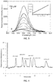
- N-nonyl acridine orange (NAO) has been proposed for the analysis of specific phospholipids like cardiolipin (CL), it did not exhibit a significant spectral response for LPA. Extending the alkyl chain of NAO to an octadecyl chain did not improve LPA detection. Evaluation of the amphiphilic cyanine type probe 4-(4-(dihexadecylamino)styryl)-N-methylpyridinium iodide (DiA) gave the most promising results for LPA detection. As shown in FIG. 1 , 3 ⁇ M DiA shows an increase in absorbance at 440-450 nm in the presence of 10 ⁇ M LPA 18:0.
- DiA (3 ⁇ M) exhibits weak fluorescence emission at 590 nm (excitation: 470 nm) in aqueous medium, and upon addition of 10 ⁇ M LPA 18:0, an increase in fluorescence emission is observed ( FIG. 2 ).
- LPA Separation The common solvent mixtures (MeOH/H 2 O, MeCN/H 2 O) used for reversed-phase chromatography were not able to resolve individual LPA species. From the various buffer systems evaluated, it was found that phosphate buffer gave better separation and peak shapes. LPA species separation was dependent on buffer pH and concentration. Optimal resolution was achieved with 50 mM phosphate buffer pH 2.5. The use of mobile phase modifiers, including trifluoroacetic acid (TFA) and choline chloride were also evaluated. Only choline chloride gave suitable separation, however, it increased significantly both the time of analysis and column back pressure.
- TFA trifluoroacetic acid
- the 3-D mode feature in the Waters 2475 fluorescence detector was used to determine suitable excitation and emission wavelengths for detection.
- the detector gain was set to 100.
- the acquisition mode was set to excitation scanning mode (330-530 nm) while keeping the emission wavelength constant (570 nm).
- Solutions of DiA with concentrations ranging from 3 to 20 ⁇ M were tested and compared. Among them, 10 ⁇ M showed optimal signal-to-noise ratio with a relatively low fluorescence background. Reagent flow rates from 0.15 to 0.70 mL/min were tested and compared. Higher flow rates resulted in higher signal to noise ratios; however, they also induced a weaker signal due to excessive dilution of the sample. The optimal reagent flow rate was found to be 0.62 mL/min.
- FIG. 4 shows a representative trace for the separation and subsequent detection of LPA 14:0, 16:0, 17:0, 18:0, 18:1 and 20:4 with DiA.
- An LPA mixture (10 ⁇ M LPA 14:0, 16:0, 18:0, 18:1, 20:4 and 20 ⁇ M LPA 17:0 as an internal standard) was injected in a 20 ⁇ L injection loop.
- LPAs in MeOH:H 2 O 9:1 with concentrations ranging from 0.5 - 40 ⁇ M were evaluated using the same procedure to determine the concentration range over which a linear response was obtained.
- LPA (17:0) a non-natural LPA, was added to these mixtures at a concentration of 20 ⁇ M to act as an internal standard for further quantification.
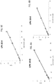
- LPA 14:0, LPA 18:0 and LPA 18:1 demonstrated a linear response throughout this range, while LPA 16:0 and LPA 20:4 had a linear response over the range of 0.5 - 25 ⁇ M ( FIGS. 5A-5E ).
- LPA Limit of detection
- Table 2 LPA species Retention time (min) Linear range ( ⁇ M) R 2 LOD ( ⁇ M) 14:0 3.50 0-40 0.9962 0.147 20:4 5.56 0-25 0.9960 0.161 16:0 6.64 0-25 0.9963 0.173 18:1 8.35 0-40 0.9949 0.074 18:0 13.75 0-40 0.9943 0.272
- FIGS. 6A-E show calibration curves for the individual LPA species evaluated with LC/ESI/MS/MS. Acceptable correlation factors ( R 2 ) were obtained for all the LPAs (Table 3).
- LPA Limit of detection
- LPA species Retention time (min) Linear range ( ⁇ M)
- R 2 LOD ( ⁇ M) 14:0 6.30 0-40 0.9982 0.0067 20:4 7.55 0-40 0.9981 0.0099 16:0 8.29 0-40 0.9986 0.0123 18:1 9.10 0-40 0.9989 0.0066 18:0 11.22 0-40 0.9985 0.0156
- Plasma Source for Donor A Lyophilized Human Plasma, Sigma-Aldrich (Catalog # P9523).
- donors B, C, D, and E plasma was collected by Lampire Biological Laboratories Inc., from female donors, processed to obtain platelet-free plasma, and frozen at -80 °C (Catalog # 7303809).
- the human plasma from these five donors was spiked with 0.5 ⁇ M of each individual LPA. Then, the individual LPA concentrations of these spiked plasma samples were determined. All the samples were analyzed by triplicate using both HPLC-post column and LC/ESI/MS/MS methods.
- HPLC-post column conditions Column - reversed phase C8, 3 ⁇ m, 50 x 2.0 mm.
- Mobile phase - methanol:phosphate buffer (pH 2.5) 16:5.
- Reagent flow rate - 0.62 mL/min.
- FIG. 7 shows typical traces obtained by the HPLC-post column method for a standard LPA mixture and LPAs isolated from human plasma samples.
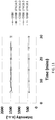
- FIGS. 8A and 8B show typical LC/ESI/MS/MS traces of a 10 ⁇ M mixture of standard LPAs ( FIG. 8A ) and a plasma sample from donor A ( FIG. 8B ).
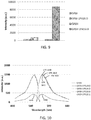
- spiro guanidine rhodamines GRBI and GRBII were evaluated as universal fluorophores for the detection and quantification of lysophosphatidic acid (LPA).
- LPA14:0, 16:0, 18:0, 18:1 were obtained by protonation of their corresponding sodium salt respectively.
- 100 mg of LPA sodium salt were dissolved in 15 mL of a solvent mixture
- the fluorescence emission intensity of GRBI and GRBII in the presence of 10 ⁇ M LPA16:0 in 2.5% DMSO/chloroform was evaluated. The final probe concentration was 5 ⁇ M. An excitation wavelength of 550 nm was used, and fluorescence emission at 570 nm was measured. As shown in FIG. 9 , GRBII exhibited greater fluorescence emission in the presence of LPA 16:0 as compared to GRBI.
Landscapes
- Chemical & Material Sciences (AREA)
- Organic Chemistry (AREA)
- Health & Medical Sciences (AREA)
- Life Sciences & Earth Sciences (AREA)
- Molecular Biology (AREA)
- Engineering & Computer Science (AREA)
- Immunology (AREA)
- Hematology (AREA)
- Biomedical Technology (AREA)
- Urology & Nephrology (AREA)
- Food Science & Technology (AREA)
- Physics & Mathematics (AREA)
- Biophysics (AREA)
- Microbiology (AREA)
- Biotechnology (AREA)
- Endocrinology (AREA)
- Medicinal Chemistry (AREA)
- Cell Biology (AREA)
- Analytical Chemistry (AREA)
- Biochemistry (AREA)
- General Health & Medical Sciences (AREA)
- General Physics & Mathematics (AREA)
- Pathology (AREA)
- Investigating Or Analysing Materials By The Use Of Chemical Reactions (AREA)
- Investigating Or Analysing Biological Materials (AREA)
- Investigating, Analyzing Materials By Fluorescence Or Luminescence (AREA)
Description
- This disclosure concerns methods and compounds for isolating and detecting lysophosphatidic acids. Kits for performing the methods also are disclosed.
- This invention was made with government support under Grant No. RO1 CA136491 awarded by the National Institutes of Health, and Grant No. 0741993, awarded by the National Science Foundation. The government has certain rights in the invention.
- Lysophosphatidic acid (LPA) is a bioactive phospholipid that stimulates cell proliferation, migration, and survival. Five G-protein-coupled receptors have been identified as specific for LPA. Aberrant LPA production, expression, and signalling have been linked to cancer initiation (e.g., tumorigenesis), progression, angiogenesis, and metastasis. LPA is thought to play a role in a number of cancers, including ovarian cancer and other gynecological cancers. LPA also has been linked to cardiovascular disease (e.g., atherosclerosis, atherothrombosis), platelet aggregation, ischemia perfusion injury, wound healing, neuropathic pain, neuropsychiatric disorders, reproductive disorders, and fibrosis.
- In recent years, LPA has been considered as an important and sensitive biomarker of ovarian cancer. Elevated LPA levels in plasma have been found in patients with ovarian cancer. There is evidence suggesting that only certain LPA species (below) are associated with ovarian cancer (Sutphen et al., Cancer Epidemiol. Biomark. Prev., 2004, 13:1185-1191; Xu et al. JAMA, 1998, 280:719-732), therefore, quantification of individual LPA species would provide a better way to improve the accuracy of diagnosis. A need also exists for a colorimetric or fluorometric probe capable of detecting equal concentrations of individual LPAs with the same degree of response to more accurately determine total LPA concentration with a single probe.

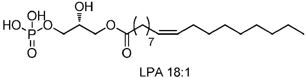

- Known methods suffer from disadvantages. Xu et al. (JAMA, 1998, 280:719-723) used a gas chromatography (GC) method to quantify total LPA plasma levels. Chen et al. (J. Chromatogr. B. Biomed. Sci. Appl., 2001, 753:355-363) used capillary electrophoresis (CE) to quantify individual LPAs with an indirect ultraviolet (UV) for the detection. However, to separate LPA from other lipids before the detection, these and several other studies rely on two-dimensional thin layer chromatography (TLC) as a step, which is time consuming and labor intensive.
- Holland et al. (J. Lipid Res., 2003, 44:854-858) used high performance liquid chromatography (HPLC) to separate LPA species and evaporative light-scattering detection (ELSD). This method avoids the two-dimensional TLC step, but LPA elutes at 38 min with a relatively low recovery. LC-MS based methodology has been used to quantify LPAs (Baker et al., Anal. Biochem., 2001, 292:287-295); however, it is not as accurate as LC-MS/MS because LC-MS only determines LPA by detecting the molecular mass ion rather than the parent to daughter ion transitions.
- According to some recent studies, LC-MS/MS methods have disadvantages. Shan et al. (J. Chomatogr. B, 2008, 864:22-28) found that some unknown compounds in plasma, which produced the same parent-to-daughter ion transition as LPA in a direct flow injection LC-ESI-MS/MS method, could reduce the accuracy of quantification of LPA. Zhao et al. (J. Lipid Res., 2010, 51:652-659) reported lysophosphatidylcholine (LPC) and lysophosphatidylserine (LPS) could lose the choline or serine group to generate LPA-like signals at the ion source. Additionally, phosphatidic acids may be fragmented by enzymes during separation from blood samples and/or fragmented in an ESI detector, in some instances losing one lipid chain and producing a false positive by appearing as LPA. Another disadvantage is that LPAs do not ionize well, and the best results for LPA typically are obtained by running the mass spectrometer in negative ion mode, which can be more technically challenging than the more typical positive ion mode.
- Huang et al. (J. Inorg. Biochem. 2011, 105, 800) describe the use of a rhodamine probe, N-(Rhodamine-6G)lactam-hydrazinecarbothioamide, as a fluorescent and colorimetric chemodosimeter in aqueous solution for Cu2+.
- Zuo et al. (J. Fluoresc. 2012, 22, 1201) describe a rhodamine probe for HClO, and the use of the probe in live cell imaging.
- Zhao et al. (Chemical Papers 2009, 63, 261) describe the use of a rhodamine probe for Hg2+. The probe is said to display excellent selective and sensitive response to Hg2+ over other transition metal ions in neutral aqueous solutions.
- Zhao et al. (Sensors Actuators B 2009, 135, 625) describe the use of a rhodamine probe, 1-(rhodamine B)lactam-thiosemicarbazide, as a fluorescent chemosensor for Cu2+ in aqueous media.
-
US 2010/291689 described lanthanide complexes as fluorescent indicators for neutral sugars and cancer diagnosis. The complexes are water-soluble salophene-lanthanide complexes and they are said to be useful for the selective detection of lysophosphatidic acid (LPA) in the presence of phosphatidic acid. - The present invention provides a compound having the formula:
-


- The method for isolating lysophosphatidic acid species from a plasma or serum sample include obtaining a plasma or serum sample; combining one part plasma or serum sample with five parts solvent comprising methanol and chloroform in a ratio of 2:1 to form a mixture; incubating the mixture at 4 °C for 15-30 minutes; warming the mixture to ambient temperature; separating organic and aqueous layers of the mixture; extracting the aqueous layer with phosphate-buffered saline, pH 7.4 to form an extracted aqueous phase; mixing the extracted aqueous phase with chloroform; separating chloroform from the extracted aqueous phase to form a washed aqueous phase; repeating the steps of mixing the extracted phase with chloroform and separating chloroform from the extracted aqueous phase to form a washed aqueous phase; adding phosphoric acid to the washed aqueous phase to reduce pH to 2, thereby forming an acidified aqueous phase; loading the acidified aqueous phase onto a solid-phase extraction (SPE) cartridge, wherein the SPE cartridge has a C8 stationary phase; flowing water and subsequently chloroform through the SPE cartridge; drying the SPE cartridge; and flowing methanol through the SPE cartridge, thereby eluting lysophosphatidic acid species in methanol from the SPE cartridge. The method may further includes evaporating methanol to form a dry residue comprising lysophosphatidic acid species; and dissolving the dry residue in 9:1 methanol:H2O to produce an extracted lysophosphatidic acid sample comprising one or more lysophosphatidic acid species.
- In the methods a total concentration of lysophatidic acid (LPA) species in the extracted lysophosphatidic acid sample may be determined. The sample may be obtained from a subject suspected of being at risk of a condition associated with an aberrant LPA level, the method further comprising determining a risk level for the condition, wherein the risk level is based at least in part on the lysophosphatidic acid concentration. In certain embodiments, the condition is cancer (for example, ovarian cancer), cardiovascular disease, platelet aggregation, ischemia perfusion injury, neuropathic pain, a neuropsychiatric disorder, a reproductive disorder, or fibrosis. Total concentration of LPA can be determined by combining the extracted lysophosphatidic acid sample with a compound of the invention, as described above in a solvent comprising 2.5-10% dimethylsulfoxide in methanol to form a solution; exposing the solution to a light source; measuring fluorescence intensity of the solution; and determining, based on the fluorescence intensity, the total concentration of lysophosphatidic acid species. In certain examples, the compound is
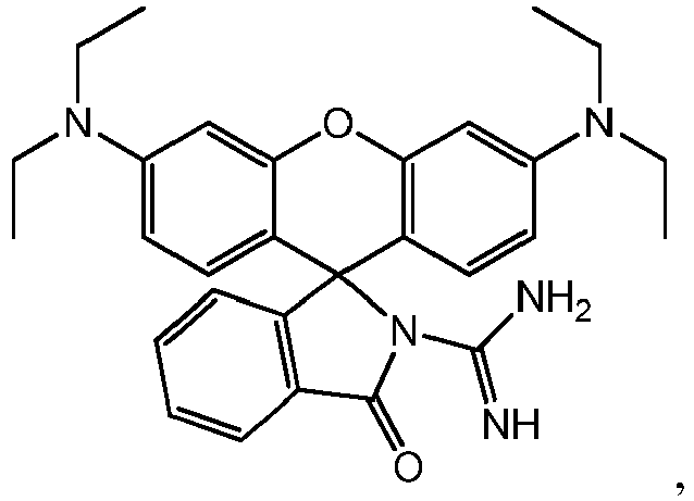
- In some embodiments, lysophosphatidic acid species in the extracted lysophosphatidic acid sample are separated using a reversed-phase high-performance liquid chromatography (HPLC) column, and individual lysophosphatidic acid species are detected as the separated lysophosphatidic acid species exit the reversed-phase HPLC column. LPA species may be separated using HPLC by flowing the sample into a reversed-phase HPLC column including a C8 stationary phase, and subsequently flowing 16:5 methanol/phosphate buffer (50 mM, pH 2.5) through the reversed-phase HPLC column, thereby forming an eluent comprising lysophosphatidic acid species. Individual LPA species may be detected by combining the eluent with 4-(4-(dihexadecylamino)styryl)-N-methylpyridinium iodide (DiA) as it exits the reversed-phase HPLC column to form a DiA-eluent mixture, flowing the DiA-eluent mixture through a detector, and detecting individual lysophosphatidic acid species by detecting fluorescence of the DiA-eluent mixture as the DiA-eluent mixture flows through the detector.
- In certain embodiments, the method further includes identifying an individual lysophosphatidic acid species by comparing an elution time for the individual lysophosphatidic acid species to elute from the reversed-phase HPLC column to elution times for known individual lysophosphatidic acid species, measuring fluorescence intensity of the DiA-eluent mixture, and determining, based on the fluorescence intensity, a concentration of the individual lysophosphatidic acid species. In some examples, the sample is obtained from a subject suspected of being at risk of a condition associated with an aberrant LPA level, and the method includes determining a risk level for the condition, wherein the risk level is based at least in part on an identification of an individual lysophosphatidic acid species, the concentration of the individual lysophosphatidic acid species, or a combination thereof. Exemplary conditions that may be associated with an aberrant LPA level include cancer, cardiovascular disease, platelet aggregation, ischemia perfusion injury, neuropathic pain, a neuropsychiatric disorder, a reproductive disorder, or fibrosis. In certain examples, the condition is ovarian cancer.
- Also provided by the present invention are kits for detecting and quantifying lysophosphatidic acid include at least one compound of the invention, as described above. The kits may further include one or more lysophosphatidic acid species, and/or one or more solid-phase extraction cartridges wherein the stationary phase is C8.
- The foregoing and other objects, features, and advantages of the invention will become more apparent from the following detailed description, which proceeds with reference to the accompanying figures.
-
-
FIG. 1 shows absorbance spectra of an aqueous solution of 3 µM 4-(4-(dihexadecylamino)styryl)-N-methylpyridinium iodide (DiA) in the absence (dashed line) or presence (solid line) of 10 µM lysophosphatidic acid (LPA) 18:0. -
FIG. 2 shows fluorescence emission spectra of 3 µM DiA in the absence (dashed line) or presence (solid line) of 10 µM LPA 18:0 (λex/em = 470/590 nm). -
FIG. 3 shows emission spectra and a calibration curve (inset) of 2.67 µM DiA upon titration with LPA (18:0). -
FIG. 4 is an HPLC trace of a LPA mixture (10 µM LPA 14:0, 16:0, 18:0, 18:1, 20:4 and 20 µM LPA 17:0) injected in a 20 µL injection loop. Chromatography conditions: Column: Luna™ C8, 3 µm, 50 x 2.1 mm, mobile phase: methanol:phosphate buffer (50 mM, pH 2.5) 16:5; flow rate: 0.32 mL/min. Injection volume: 20 µL. Sample concentration: 10 µM in methanol:H2O 9:1. Post-column reagent: 10 µM DiA in H2O. Reagent flow rate: 0.62 mL/min. Detection wavelength: ex/em 450/570 nm. -
FIGS. 5A-5E are a series of calibration curves of LPAs: 14:0 -FIG. 5A , 16:0 -FIG. 5B , 18:0 -FIG. 5C , 18:1 -FIG. 5D , and 20:4 -FIG. 5E . The curves were obtained using the disclosed HPLC post-column procedure. The area ratio (A/As) is the peak area of the individual LPA divided by the peak area of the internal standard (LPA 17.0). -
FIGS. 6A-6E are a series of calibration curves LPAs: 14:0 -FIG. 6A , 16:0 -FIG. 6B , 18:0 -FIG. 6C , 18:1 -FIG. 6D , and 20:4 -FIG. 6E . The curves were obtained using an LC/ESI/MS/MS method. The area ratio (A/As) is the peak area of the individual LPA divided by the peak area of the internal standard (LPA 17.0). -
FIG. 7 is an HPLC trace of 10 µM LPAs mixture (LPA 14:0, 16:0, 17:0, 18:0, 18:1 and 20:4) and LPAs isolated from Donor A human plasma. Chromatography conditions: Column - reversed phase C8, 3 µm, 50 x 2.0 mm; mobile phase - methanol:phosphate buffer (pH 2.5) = 16:5; flow rate - 0.32 mL/min; injection volume - 20 µL; sample concentration - 10 µM in MeOH:H2O 9:1; post-column reagent - 10 µM DiA in H2O; reagent flow rate - 0.62 mL/min. Detection wavelengths: ex/em 450/570 nm. -
FIGS. 8A and8B are LC/ESI/MS/MS traces of a 10 µM mixture of standard LPAs (FIG 8A ) and a plasma sample from donor A (FIG. 8B ). Column: Luna C8 (50 x 2 mm, 3 µm) at 40 °C. Injection volume; 10 µL. Mobile phase; MeOH:aqueous formic acid (pH 2.5) 9:1. Flow rate of 0.4 mL/min. Parent and daughter ions were detected in the negative ion mode, sprayer voltage; 3.0 kV, capillary temperature at 300 °C. -
FIG. 9 is a bar graph illustrating the relative fluorescence emission obtained from two embodiments of the disclosed probes (GRBI and GRBII) when combined with LPA 16:0; Ex/Em = 550nm/570nm, final probe concentration 5µM, final LPA16:0concentration 10 µM, solvent system DMSO 2.5% in chloroform. -
FIG. 10 shows fluorescence spectra of GRBII alone and GRBII in the presence of LPA14:0, 16:0, 18:0, 18:1, ex/em = 550 nm/570 nm,final probe concentration 5 µM,final LPA concentration 10 µM, solvent system CHCl3: DMSO 9:1. -
FIG. 11 is a graph of fluorescence intensity of GRBII alone and in the presence of LPA14:0, 16:0, 18:0, 18:1 over time, ex/em=550 nm/570 nm,final probe concentration 5 µM,final LPA concentration 10 µM, solvent system CHCl3: DMSO 9:1. - Methods and compounds of the invention for isolating and detecting lysophosphatidic acids (LPAs) are disclosed, along with kits for performing the method. Methods may include a liquid-liquid and solid-phase extraction process to isolate LPAs in plasma from interfering components. Following extraction, individual LPA species may be separated by HPLC and detected in a fluorometric assay. Alternatively, total LPA may be quantified using embodiments of a fluorescent compound capable of detecting a plurality of LPA species with substantially similar sensitivity.
- The following explanations of terms and abbreviations are provided to better describe the present disclosure and to guide those of ordinary skill in the art in the practice of the present disclosure. As used herein, "comprising" means "including" and the singular forms "a" or "an" or "the" include plural references unless the context clearly dictates otherwise. The term "or" refers to a single element of stated alternative elements or a combination of two or more elements, unless the context clearly indicates otherwise.
- Unless explained otherwise, all technical and scientific terms used herein have the same meaning as commonly understood to one of ordinary skill in the art to which this disclosure belongs. Although methods and materials similar or equivalent to those described herein can be used in the practice or testing of the present disclosure, suitable methods and materials are described below. The materials, methods, and examples are illustrative only and not intended to be limiting. Other features of the disclosure are apparent from the following detailed description and the claims.
- Unless otherwise indicated, all numbers expressing quantities of ingredients, properties such as molecular weight, percentages, and so forth, as used in the specification or claims are to be understood as being modified by the term "about." Accordingly, unless otherwise indicated, implicitly or explicitly, the numerical parameters set forth are approximations that may depend on the desired properties sought, limits of detection under standard test conditions/methods, limitations of the processing method, and/or the nature of the parameter or property. When directly and explicitly distinguishing embodiments from discussed prior art, the embodiment numbers are not approximates unless the word "about" is recited.
- Acyl: An organic functional group having the general formula -C(O)R, where R is alkyl, heteroalkyl, haloalkyl, aliphatic, heteroaliphatic, aryl, or heteroaryl.
- Alkoxy: A radical (or substituent) having the structure -O-R, where R is a substituted or unsubstituted alkyl. Methoxy (-OCH3) is an exemplary alkoxy group.
- Alkyl: A hydrocarbon group having a saturated carbon chain. The chain may be cyclic, branched or unbranched. An alkyl group may be a group with a saturated carbon chain having 1-10 carbon atoms.
- Alkylamino: An alkyl group where at least one hydrogen is substituted with an amino, mono-substituted amino or di-substituted amino group.
- Amino: A chemical functional group -N(R)R' where R and R' are independently hydrogen, alkyl, heteroalkyl, haloalkyl, aliphatic, heteroaliphatic, aryl (such as optionally substituted phenyl or benzyl), heteroaryl, alkylsulfano, or other functionality. A "primary amino" group is -NH2. "Mono-substituted amino" means a radical -N(H)R substituted as above and includes, e.g., methylamino, (1-methylethyl)amino, phenylamino, and the like. "Di-substituted amino" means a radical -N(R)R' substituted as above and includes, e.g., dimethylamino, methylethylamino, di(1-methylethyl)amino, and the like.
- Carboxyalkyl: A functional group with the formula -COOR where R is alkyl.
- DiA: 4-(4-(dihexadecylamino)styryl)-N-methylpyridinium iodide
- HPLC: high performance (or pressure) liquid chromatography
- LPA: lysophosphatidic acid
- LPS: lysophosphatidyl serine
- PA: phosphatidic acid
- PC: phosphatidyl choline
- PE: phosphatidylethanolamine
- PI: phosphatidyl inositol
- Probe: A substance used to detect or identify another substance in a sample.
- S1P: sphingosine-1-phosphate
- SPE: solid phase extraction
- Substituent: An atom or group of atoms that replaces another atom in a molecule as the result of a reaction. The term "substituent" typically refers to an atom or group of atoms that replaces a hydrogen atom on a parent hydrocarbon chain or ring.
- Substituted: A fundamental compound, such as an aryl or aliphatic compound, or a radical thereof, having coupled thereto, typically in place of a hydrogen atom, a second substituent. For example, substituted aryl compounds or substituents may have an aliphatic group coupled to the closed ring of the aryl base, such as with toluene. Again solely by way of example and without limitation, a long-chain hydrocarbon may have a substituent bonded thereto, such as one or more halogens, an aryl group, a cyclic group, a heteroaryl group or a heterocyclic group.
- Compounds for detecting lysophosphatidic acids are selected from:


- A kit for detecting and quantifying lysophosphatidic acid comprises at least one compound described above. In some embodiments, the kit further comprises one or more lysophosphatidic acid species. In any or all of the above embodiments, the kit may further comprise one or more solid-phase extraction cartridges wherein the stationary phase is C8. In any or all of the above embodiments, the compound may be the invention

- A method for quantifying lysophosphatidic acid species comprises combining a sample that may include one or more lysophosphatidic acid species with a compound according to the invention in a solvent comprising dimethylsulfoxide in methanol to form a solution; exposing the solution to a light source; measuring fluorescence intensity of the solution; and determining, based on the fluorescence intensity, a total concentration of lysophosphatidic acid species in the sample. The solvent may comprise 2.5-10% dimethylsulfoxide in methanol. In some embodiments, the sample is obtained by extracting lysophosphatidic acid species from a plasma or serum sample.
- A method for extracting lysophosphatidic acid species includes combining a sample of plasma or serum with a solvent comprising an alkyl alcohol having 1-10 carbon atoms and a relatively nonpolar solvent, e.g., chloroform, to form a mixture; separating organic and aqueous layers of the mixture; extracting the aqueous layer with a buffer at neutral pH to form an extracted aqueous phase; mixing the extracted aqueous phase with chloroform; separating chloroform from the extracted aqueous phase to form a washed aqueous phase; adding phosphoric acid to the washed aqueous phase to form an acidified aqueous phase; loading the acidified aqueous phase onto a solid-phase extraction (SPE) cartridge including a stationary phase comprising silica derivatized with hydrocarbon chains; flowing water and subsequently chloroform through the SPE cartridge; drying the SPE cartridge; and flowing an alkyl alcohol having 1-10 carbon atoms through the SPE cartridge, thereby eluting lysophosphatidic acid species in the alkyl alcohol having 1-10 carbon atoms from the SPE cartridge.
- Combining the plasma or serum sample with the solvent may comprises combining one part of the plasma or serum sample with five parts of the solvent, the solvent comprising methanol and chloroform in a ratio of 2:1. The method may further comprise incubating the mixture at 4 °C for a period of time; and warming the mixture to ambient temperature before separating the organic and aqueous layers of the mixture. Extracting the aqueous layer with a buffer at neutral pH may comprise extracting with phosphate-buffered saline at pH 7.4. In any or all of the above embodiments, the steps of mixing the extracted aqueous phase with chloroform and separating chloroform from the extracted aqueous phase to form a washed aqueous phase may be repeated. Sufficient phosphoric acid may be added to the washed aqueous phase to reduce pH to 2. The stationary phase of the SPE cartridge may comprise silica derivatized with octyl chains. The alkyl alcohol having 1-10 carbon atoms may be methanol. The method may further include evaporating the having 1-10 carbon atoms alkyl alcohol to form a dry residue comprising lysophosphatidic acid species; and dissolving the dry residue in 9:1 methanol:H2O to produce an extracted lysophosphatidic acid sample comprising one or more lysophosphatidic acid species. The method further includes determining a total concentration of lysophatidic acid species in the extracted lysophosphatidic acid sample.
- The method may further includes determining a total concentration of lysophatidic acid species by combining the extracted lysophosphatidic acid sample with a compound as disclosed herein in a solvent comprising dimethylsulfoxide in methanol to form a solution; exposing the solution to a light source; measuring fluorescence intensity of the solution; and determining, based on the fluorescence intensity, the total concentration of lysophosphatidic acid species. The solvent may comprise 2.5-10% dimethylsulfoxide in methanol.
- In some embodiments, the compound used for determining a total concentration of lysophosphatidic acid species is

- The sample may be obtained from a subject suspected of being at risk of a condition associated with an aberrant LPA level, the method further comprising determining a risk level for the condition, wherein the risk level is based at least in part on the total concentration. The condition may be cancer, e.g., ovarian cancer, cardiovascular disease, platelet aggregation, ischemia perfusion injury, neuropathic pain, a neuropsychiatric disorder, a reproductive disorder, or fibrosis.
- The method may further includes separating lysophosphatidic acid species in the extracted lysophosphatidic acid sample using a reversed-phase high-performance liquid chromatography (HPLC) column, and detecting individual lysophosphatidic acid species as the separated lysophosphatidic acid species exit the reversed-phase HPLC column. Separating lysophosphatidic acid species using a reversed-phase high-performance liquid chromatography (HPLC) column may further comprise flowing the sample into the reversed-phase HPLC column, wherein the column has a C8 stationary phase, and subsequently flowing 16:5 methanol/phosphate buffer (50 mM, pH 2.5) through the reversed-phase HPLC column, thereby forming an eluent comprising lysophosphatidic acid species. In any or all of the above embodiments, detecting individual lysophosphatidic acid species may comprise combining the eluent with 4-(4-(dihexadecylamino)styryl)-N-methylpyridinium iodide (DiA) as it exits the reversed-phase HPLC column to form a DiA-eluent mixture; flowing the DiA-eluent mixture through a detector; and detecting individual lysophosphatidic acid species by detecting fluorescence of the DiA-eluent mixture as the DiA-eluent mixture flows through the detector. The method may further comprise identifying an individual lysophosphatidic acid species by comparing an elution time for the individual lysophosphatidic acid species to elute from the reversed-phase HPLC column to elution times for known individual lysophosphatidic acid species, measuring fluorescence intensity of the DiA-eluent mixture, and determining, based on the fluorescence intensity, a concentration of the individual lysophosphatidic acid species.
- The plasma or serum sample may be obtained from a subject suspected of being at risk of a condition associated with an aberrant LPA level, the method further comprising determining a risk level for the condition, wherein the risk level is based at least in part on an identification of an individual lysophosphatidic acid species, the concentration of the individual lysophosphatidic acid species, or a combination thereof. The condition may be cancer, e.g., ovarian cancer, cardiovascular disease, platelet aggregation, ischemia perfusion injury, neuropathic pain, a neuropsychiatric disorder, a reproductive disorder, or fibrosis.
- LPAs are found in plasma and serum. To accurately quantify total LPA and/or to separate, identify, and quantify LPA species, the LPAs must be extracted from the plasma or serum sample and separated from potential interferences, including other phospholipids (e.g., phosphatidic acids, phosphatidyl cholines, lysophophatidyl cholines, lysophosphatidyl serines, phosphatidyl ethanolamine, phosphatidyl inositol, and sphingosine-1-phosphate) that may interfere.
- Conventional methods for extracting LPAs insufficiently remove interferences and/or produce poor LPA recoveries. These methods do not adequately prepare LPA samples for quantifying total LPA, or for HPLC separation and subsequent identification/quantification of individual LPA species as disclosed herein. For example, solid phase extraction (SPE) is an accepted method for the removal of potential interferences from biological samples, and has been used for the isolation and enrichment of the different classes of phospholipids. However, SPE alone does not sufficiently remove other phospholipids that can interfere with LPA separation and detection by HPLC. Additionally, although hybrid zirconia and titania support cartridges are known to be useful for recovery of phosphopeptides and removal of phospholipids from biological media, poor recoveries of LPAs were obtained under all evaluated conditions. Because SPE does not sufficiently remove interfering phospholipids, some reported methods include a liquid-liquid extraction step. Typical procedures require plasma acidification prior to extraction. However, evaluation of these methods demonstrated very low LPA recoveries, and insufficient removal of other interferences.
- Embodiments of the disclosed method for LPA extraction include a liquid-liquid extraction followed by reversed-phase SPE. Liquid-liquid extraction may be performed with a solvent mixture comprising an alkyl alcohol having 1-10 carbon atoms (e.g., methanol, ethanol) and a relatively nonpolar solvent. In some examples, a solvent comprising methanol and chloroform, such as 2:1 MeOH:CHCl3, is mixed with a plasma or serum sample in a ratio of five parts solvent to one part plasma/serum. The combined solvent mixture and plasma/serum sample is incubated at 4 °C for an effective period of time, and then warmed to ambient temperature (e.g., 20 °C). In some embodiments, the effective period of time is at least 15 minutes, such as 15-30 minutes. In certain examples, the effective period of time is 20 minutes. The organic and aqueous layers are separated (e.g., by centrifugation), and the upper aqueous layer is recovered.
- Phosphatidic acids are negatively charged at neutral pH. Thus, extraction at neutral pH selectively removes negatively charged LPAs from other neutral and positively charged phospholipids. Accordingly, the upper aqueous layer is extracted with a buffer near physiologic pH, such as phosphate-buffered saline (10 mM, pH 7.4). The aqueous phase is washed with chloroform, and then acidified with phosphoric acid to
pH 2. - Reversed-phase SPE separates compounds based on polarity. An SPE column includes a stationary phase (e.g., silica) derivatized with hydrocarbon chains. A solution comprising a mixture of compounds with varying polarities is passed through the column. Compounds with low-polarity are retained on the stationary phase while more polar compounds elute with the solvent. The less polar compounds then are eluted from the stationary phase using a nonpolar solvent. In some embodiments, a C8 SPE cartridge is used. A C8 stationary phase comprises silica functionalized with octyl chains. The cartridge may be preconditioned with a polar solvent, or series of polar solvents, such as methanol followed by water. The acidified aqueous phase from the liquid:liquid extraction is loaded onto the cartridge. The cartridge is washed first with water to remove any polar species, and then with chloroform. The SPE cartridge is dried, with a nitrogen stream, to remove residual chloroform, and LPAs subsequently are eluted with an alkyl alcohol having 1-10 carbon atoms, e.g., methanol. The most hydrophobic species are retained on the stationary phase.
- In certain examples, isolating LPA includes obtaining a plasma or serum sample, e.g., from a subject having, or being at risk of developing, a condition associated with aberrant LPA levels. The plasma or serum sample is combined with a solvent comprising 2:1 methanol:chloroform; the combined mixture includes one part plasma or serum sample and five parts solvent. In certain examples, 0.8 mL of plasma or serum sample and 4 mL of solvent are used. The mixture is thoroughly mixed, e.g., by vortexing at 2,000 rpm for 30 seconds. The mixture is then incubated at 4 °C for 20 minutes, warmed to ambient temperature, and the aqueous and organic layers are separated, e.g., by centrifugation at 2000 rpm for 10 minutes. The upper aqueous layer is extracted with 2 mL phosphate-buffered saline (10 mM, pH 7.4) by vortexing at 2000 rpm for 30 seconds. The aqueous phase is separated and washed twice with chloroform (1.33 mL). After each washing, the layers are separated, and the chloroform layer is discarded. The washed aqueous phase is acidified to
pH 2 with phosphoric acid. The acidified aqueous phase is then loaded on a C8 SPE cartridge that has been preconditioned with 6 mL of methanol, followed by 3 mL of water. After the sample is loaded onto the cartridge, the cartridge is rinsed with 3 mL water, and then 1 mL chloroform. The cartridge then is dried, e.g., with a nitrogen stream. LPAs are eluted from the dried cartridge with 4 mL methanol. The eluted LPAs may be dried, and reconstituted in 9:1 methanol:water for further analysis, such as HPLC separation. - Following solid-phase extraction of LPAs from plasma or serum, the LPA species are separated using HPLC and then detected. A fluorescent probe 4-(4-(dihexadecylamino)styryl)-N-methylpyridinium iodide (DiA, below) is used as a post-column reagent for the fluorescent detection and quantification of phospholipids. DiA fluorescence can be detected with an excitation wavelength of 450-470 nm and an emission wavelength of 570-590 nm. Embodiments of the disclosed method, including the initial LPA extraction, are capable of separating and quantifying at least six individual LPA species. In some embodiments, six individual LPA species at physiological levels in plasma can be separated and detected in 15 minutes. Compared to conventional LC-MS methods, embodiments of the disclosed method are more practical and reliable since the method is not prone to ionization-related issues and is relatively inexpensive for routine analysis.

- HPLC can be used to separate LPA species. Desirably, HPLC separation will provide complete separation of LPA species with minimal peak broadening and a reasonable separation time (e.g., less than 20 minutes). It was determined that reversed-phase HPLC would be suitable. An extensive evaluation of solvent systems determined that common solvent mixtures (methanol/water, acetonitrile/water) did not adequately resolve LPA species. Phosphate buffer was found to provide the best results. Complete separation of LPA species depends, at least in part, on buffer concentration and pH, and optimal separation was obtained with 50 mM phosphate buffer, pH 2.5. In some examples, LPAs were eluted using a 16:5 mixture of methanol:phosphate buffer (50 mM, pH 2.5).
- Several reversed-phase columns were evaluated, and C8-based columns were found to provide superior results. A shorter column (i.e., 50 mm vs. 100 mm) minimized peak broadening and provided a desirably short separation time of ∼15 minutes. Peak width is a result-effective variable, with narrower peaks providing better separation (less diffusion and overlap of adjacent peaks) and allowing a lower detection limit due to less dilution of the LPA species.
- The LPA species are detected with DiA. DiA is mixed with the eluent exiting the HPLC column (e.g., via a 3-way connector or mixing tee), and the mixture then flows into a fluorescence detector. As individual LPA species exit the column, become mixed with DiA, and flow into the detector, fluorescence increases and the LPA species are detected. DiA concentration and flow rate are both result-effective variables. Both concentration and flow rate are selected to provide an optimal signal-to-noise ratio with a relatively low fluorescence background. In some examples, a 10 µM aqueous DiA solution with a flow rate of 0.60 mL/minute to 0.65 mL/minute (e.g., 0.62 mL/min.) was used. The lower limit of LPA detection was determined as being the amount of an LPA species that produced a signal-to-noise ratio of 3:1. The method may have a lower limit of detection of ≤ 0.3 µM, such as ≤ 0.2 µM or ≤ 0.1 µM. Solutions of known LPA species can be used to determine retention times for identification of peaks.
- LPA species can be quantified by first preparing calibration curves using standard solutions of LPA species at known concentrations. Because DiA fluorescence intensity varies for different LPA species at equivalent concentrations, a calibration curve is prepared for each species. Once the calibration curves are prepared, the concentrations of LPA species in an unknown sample can be determined by comparing fluorescence intensity of each LPA species to the respective calibration curve.
- In
certain methods 20 µL of extracted LPAs in 9:1 methanol:water is injected onto a C8-based HPLC column (e.g., Luna™ C8, 50 x 2 mm, 3 µm) and eluted with a solvent comprising 16:5 methanol:phosphate buffer (50 mM, pH 2.5) at a flow rate of 0.32 mL/minute. The distal end of the HPLC column is coupled to one inlet of a mixing tee. An aqueous DiA solution (10 µM) is coupled to a second inlet of the mixing tee and set to a flow rate of 0.62 mL/minute. The combined eluent and DiA solution flows through the outlet of the mixing tee and into a fluorescence detector. Fluorescence is measured using an excitation wavelength of 450 nm and an emission wavelength of 570 nm. As each LPA species elutes from the HPLC column and flows through the detector, an increase in fluorescence is observed. - Identification and quantification of individual LPA species can be used to evaluate a subject's risk level of having, or developing, a condition correlated with aberrant LPA levels. In some embodiments, a subject's risk of having or developing an LPA-related condition is proportional to the concentration of one or more LPA species in the subject's blood. For example, elevated levels of plasma LPA have been found in patients with ovarian cancer. Other conditions associated with elevated LPA levels include other cancers (e.g. breast cancer, gynecological cancers), cardiovascular disease, platelet aggregation, ischemia perfusion injury, wound healing, neuropathic pain, neuropsychiatric disorders, reproductive disorders, and fibrosis. Some conditions are correlated with elevated levels of only certain LPA species. Thus, determining the presence and concentration of individual LPA species may improve diagnosis accuracy.
- In some conditions, total LPA concentration may be predictive of a subject's risk of having, or developing, a condition associated with aberrant LPA levels. Thus, it is desirable to have a single probe capable of detecting a plurality of LPA species with equivalent sensitivity.
- Fluorogenic probes commercially available for phospholipid detection can be classified as fluorescent lipid analogs. Phospholipid detection is based on the enhancement or quenching of fluorescence emission intensity as a result of aggregation/de-aggregation between the phospholipid and the fluorophore. Since this non-specific process is dependent on the type, length and number of alkyl chains present in the phospholipid; these probes are not useful for total phospholipid analysis since the fluorescence intensity will vary from phospholipid to phospholipid. Thus, if a plurality of phospholipids is present, the concentration cannot be accurately determined based upon the total fluorescence.
- The disclosed fluorogenic compounds are based on a rhodamine B framework, and are functionalized with a guanidine, biguanidine, guanylurea, or guanylthiourea group. Upon interaction of the guanidine, biguanidine, guanylurea, or guanylthiourea group with the phosphate group of a phospholipid, electrostatic interactions will alter the compound's fluorescence.

- The compounds are selected from


- The compounds above include a lactam ring. When the lactam ring is present, the compound is non-fluorescent. However, when the guanidine group interacts with the phosphate group of lysophosphatidic acid, the bond between R11 and the upper 3-ring portion of the structure is broken and the lactam ring opens, thereby altering electrostatic interactions within the compound, and rendering the compound fluorescent.
- The compounds of the invention are suitable for universal detection of LPA. In other words, substantially the same fluorescence intensity is obtained at a given LPA concentration irrespective of the particular LPA species present. In some embodiments, when the compound is combined with a sample comprising isolated LPAs, the fluorescence intensity is stable over time. For example, the fluorescence intensity may remain substantially constant for at least 10 minutes, at least 20 minutes, or at least 30 minutes.
- A sample that may include one or more LPAs is combined with a compound according to any one of general formulas I-IV in a suitable solvent to form a solution. The solution is exposed to a light source, and LPAs, if present, are detected by detecting fluorescence of the solution at a suitable wavelength. For GRBI and GRBII, an excitation wavelength of 550 nm and an emission wavelength of 570 nm may be used. Suitable solvents include those in which both the compound and the LPAs are soluble. Exemplary solvents include chloroform/DMSO mixtures, such as mixtures comprising 2.5-10% (v/v) DMSO. In certain examples, a sample potentially comprising LPAs is combined with a compound according to any one of general formulas I-IV in a solvent comprising 9:1 chloroform:DMSO. Desirably, the compound is present in an excess amount compared to the LPAs.
- The LPA sample may be obtained from a biological sample, e.g., plasma or serum obtained from a subject at risk of having, or developing, a condition correlated with aberrant LPA levels. The LPA sample may be prepared from the biological sample using liquid-liquid extraction followed by SPE extraction as described herein.
- A calibration curve can be prepared by combining a compound of the invention with a series of samples having known concentrations of one or more LPA species and measuring the fluorescence intensity of each solution. The concentration of LPA in an unknown sample is determined by combining the unknown sample with the compound, measuring the fluorescence intensity, and comparing the fluorescence intensity to the calibration curve.
- The methods and compounds disclosed herein are suitable for detecting LPAs in plasma and serum samples. Blood samples are stored at 4 °C before processing, such as for 4-8 hours. An anti-coagulant, e.g., EDTA, is added to plasma samples before storage to prevent platelet aggregation and inhibit cation-dependent lipid enzymatic processes. Platelets are removed from plasma samples before storage; otherwise, the platelets may undergo lysis and release additional amounts of LPA into the sample. The stored samples are centrifuged to remove blood cells before analysis. In some embodiments, the sample is centrifuged at 1750 x g for 15 minutes at ambient temperature (∼ 20 °C). The supernatant (plasma or serum) is stored in low-binding siliconized tubes or glass tubes. (Baker et al., JAMA, J. Am. Med. Assoc., 2002, 287:3081-3082; Xiao et al., Ann. N. Y. Acad. Sci., 2000, 905:242-259; Xu et al., JAMA, J. Am. Med. Assoc., 1998, 280:719-723; Yi et al., Functional Lipidomics, CRC Press, 2005, 125-146). Desirably, sufficient blood is collected to provide a plasma or serum sample with a volume of at least 3.5 mL, or at least 5 mL, such that the sample may be evaluated in triplicate.
- Kits for total LPA quantification are also a feature of this disclosure. The kits of the invention include at least one compound of the invention and suitable for detecting LPAs extracted from a sample (e.g., plasma or serum). In some embodiments, the probe is GRBI or GRBII. In some embodiments, the kits also include one or more solvents suitable for detecting fluorescence of LPAs when combined with the probe. For example, the kit may include chloroform and dimethylsulfoxide, or a solution comprising 9:1 chloroform:DMSO.
- The kits also may include one or more containers, such as a disposable test tube or cuvette, in which the detection can be performed. In certain embodiments, the compound may be premeasured into the one or more containers, and the detection is subsequently performed by adding the solvent(s) and test sample to the container).
- The kits may further include instructions for performing the detection and, optionally, instructions for extracting LPAs from a biological sample. The kits also may include one or more solid-phase extraction cartridges suitable for the extraction, e.g., one or more C8 SPE cartridges. In certain embodiments, the kit further includes one or more control samples of LPAs. The control samples may be provided in solid form or in solution.
- All the lysophosphatidic acids including 1-myristoyl-2-hydroxy-sn-glycero-3-phosphate (LPA 14:0), 1-palmitoyl-2-hydroxy-sn-glycero-3-phosphate (LPA 16:0), 1-heptadecanoyl-2-hydroxy-sn-glycero-3-phosphate (LPA 17:0), 1-stearoyl-2-hydroxy-sn-glycero-3-phosphate (LPA 18:0), 1-oleoyl-2-hydroxy-sn-glycero-3-phosphate (LPA 18:1) and 1-arachidonoyl-2-hydroxy-sn-glycero-3-phosphate (LPA 20:4) were purchased from Avanti Polar Lipids (Alabaster, AL, USA). 4-(4-(Dihexadecylamino)-styryl)-N-methylpyridinium iodide (DiA) was purchased from AnaSpec (Fremont, CA, USA). HPLC grade MeOH was purchased from Fisher Scientific. Ultra-pure water was obtained from a Milli-Q™ system. Phosphoric acid and monosodium phosphate were purchased from Sigma-Aldrich (St. Louis, MO, USA). Waters OASIS™ HLB (3 cc, 60 mg, 30 µm) SPE cartridges were purchased from Waters Corporation (Milford, MA, USA).
- Rhodamine B, Rhodamine B Base, 1,3-bis-Boc-2-methyl-2-thiopseudourea, Sodium methoxide solution (0.5 M), and 1,3-bis(tert-butoxycarbonyl)guanidine were purchased from Aldrich. Anhydrous potassium carbonate and trifluoroacetic acid were purchased from Fisher Scientific. Mercury(II) chloride was purchased from Acros Organics. LPA sodium salts were purchased from Avanti. Silica gel was purchased from Sorbent Technologies. All chemicals were used as received without further purification.
- Fluorescence measurements were performed on a Cary Eclipse fluorescence spectrophotometer and absorption spectra on a
Cary 50 UV-Vis spectrophotometer (Agilent Technologies). - The HPLC system consists of a 1525 binary HPLC delivery system, a 2475 multi lambda fluorescence detector (Waters). A Luna™ C8 (50 x 2 mm, 3 µm) column connected to a guard cartridge with 2.0 to 3.0mm internal diameters was used for all the separations (Phenomenex). The reagent is pumped by a reagent manager (Waters). The post-column reagent and HPLC system are mixed and delivered to the detector by a metal mixing tee. The data was collected and processed with the Empower™ software suite (Waters).
- For LC/ESI/MS/MS, LPAs were separated in an Accela UPLC system (Thermo Fisher, San Jose, CA) and detected in an LTQ-Orbitrap XL Discovery instrument (San Jose, CA, USA), equipped with an ESI ion mass source. The data was collected in negative mode and processed with the Xcalibur® software suite.
- NMR spectra were recorded on a Bruker spectrometer in CDCl3 and DMSO-d6 solutions, and chemical shifts were reported in δ units.
- High-resolution ESI/MS spectra were recorded on a ThermoElectron LTQ-Orbitrap Discovery equipped with an ESI ion mass source.
- Several solid supports were initially evaluated in LPA control mixtures containing LPA 14:0, 16:0, 17:0, 18:0, 18:1 and 20:4. Despite the common use of hybrid Zr and TiO2 support cartridges for the recovery and enrichment of phosphopeptides and removal of phospholipids from biological media, little or no LPA recovery was obtained under all the different conditions tested.
- Reversed-phase SPE materials provided better results. Three different commercial reversed-phase C8 SPE cartridges, including Waters (Sep-pak™ Plus C-8, 200 mg, 37-55 µm), Supelco (Discovery™ DSC-8, 3 mL, 500 mg, 50 µm) and Waters (
OASIS™ HLB 3 mL, 60 mg, 30 µm) were evaluated. From the evaluated commercial cartridges, the OASIS™ HLB proved to be the best in terms of LPA recoveries. - A liquid-liquid extraction prior to the SPE procedure was desirable to remove other phospholipids that may interfere with the detection of LPAs. Typical procedures reported in the literature involve acidification of plasma prior to a liquid-liquid extraction. However, this procedure gave very low LPA recoveries and removal of other interferences.
- Control of pH was discovered to have an important role in selective removal of interferences. Because phosphatidic acids have a pKa1 = 2.9 and pKa2 = 7.5 (Kooijman et al., Biochemistry, 2005, 44:17007), they are negatively charged at neutral pH. Thus performing liquid-liquid extraction at this condition separated neutral and positively charged phospholipids from the negatively-charged LPAs.
- Acidification of the aqueous phase at pH 5.0, 4.0, 3.0 and 2.5 in the SPE step was evaluated, revealing that lower pH during SPE improved LPA enrichment. The use of MeOH as the final solvent allowed the selective elution of LPAs, leaving behind the most hydrophobic species. The best recoveries (74-100%) were obtained at pH 2.5.
- Recovery tests were performed. Three concentrations including 0.25 µM, 3 µM and 5 µM of each LPA species mixed in PBS buffer were tested after liquid-liquid extraction and SPE. Each buffered LPA solution was mixed with 4 mL of MeOH:CHCl3 2:1 and vortexed at 2000 rpm for 30 seconds. The mixture was incubated at 4 °C for 20 minutes and warmed to room temperature. The mixture was then centrifuged at 2000 rpm for 10 minutes, and the upper layer was separated and extracted with 2 mL PBS (10 mM, pH 7.4) by vortexing at 2000 rpm for 30 seconds. The aqueous phase was separated and washed with 1.33 mL CHCl3 twice, then acidified to
pH 2 with concentrated H3PO4. An SPE cartridge was preconditioned with 6 mL of MeOH, followed by 3 mL of H2O. The sample was loaded to the cartridge and rinsed with 3 mL of H2O and then with 1 mL of CHCl3. The SPE cartridge was dried by applying a N2 stream, and LPAs were eluted with 4 mL of MeOH. The MeOH was evaporated, and the residue was reconstituted in 0.16 mL of MeOH:H2O 9:1. Samples (20 µL) obtained from the SPE purification step were injected and eluted with a mixture of MeOH:phosphate buffer (50 mM, pH 2.5) 16:5 through a C8 column. The end of the column was connected to a mixing tee allowing contact with the post-column reagent solution (DiA, 10 µM). The flow rate of the mobile phase was set to 0.32 mL/min and 0.62 mL/min for the post-column reagent. Each concentration was done three times. - As shown in Table 1, the recoveries of LPA 14:0, LPA 16:0, LPA 17:0, LPA 18:0, LPA 18:1 and LPA 20:4 are 93.45%, 94.15%, 85.14%, 76.87%, 76.86% and 73.79%, respectively. In addition, the contribution of other phospholipids that have been identified to be present in human plasma was evaluated. These other phospholipids can result in potential false positives for LPAs. In general, it is known that phospholipids are prone to either chemical or enzymatic hydrolysis. Phosphatidic acids (PAs) hydrolyze producing their corresponding LPAs, adding to the apparent LPA concentrations. Due to the acidic conditions in which the LPA solid phase extraction was carried out, control experiments were performed to measure at what extent PAs were hydrolyzed. The results demonstrated that none of PA 14:0, PA 16:0, PA 18:0 or PA 18:1 was hydrolyzed. Phosphatidyl cholines (PCs) represent the major components of biological membranes and are also prone to hydrolysis producing the corresponding lysophosphatidyl cholines (LPCs). It was determined that PCs do not hydrolyze under our enrichment conditions. On the other hand, LPCs and lysophosphatidyl serines (LPSs) were found to be potential interferences. While LPC interference was removed by doing three consecutive liquid-liquid extractions instead of one, the presence of lysophosphatidyl serines (LPS) was determined not to be significant, since they are usually found at a much lower concentration than LPAs. Other phospholipids including phosphatidyl ethanolamine (PE), phosphatidyl inositol (PI) and S1P were also evaluated and found not to be interferences.
Table 1 LPA Species Measured by HPLC- post column Measured by HPLC- MS/MS Recovery in control (%) RSD (%) Recovery in control (%) RSD (%) 14:0 93.45 4.8 93.67 2.9 20:4 73.79 4.3 76.60 1.2 16:0 94.15 5.4 95.72 4.6 18:1 76.86 4.9 77.55 1.7 17:0 85.14 3.8 84.97 4.6 18:0 76.87 4.4 73.14 2.4 - LPA Detection: Fluorescent probes used for post-column detection of phospholipids, typically rely on non-covalent interactions of the phospholipid with the probe's supramolecular assemblies that are solvent dependent. Examples of these probes include 2,5-bis-2-(5-tert-Butyl)benzoxazolylthiophene (BBOT) and 1,6-diphenyl-1,3,5-hexatriene (DPH). Evaluation of BBOT and DPH produced higher fluorescence emission enhancement for double-chain phosphatidic acids (PA) compared to single-chain analogues (lysophosphatidic acids, LPAs) investigated herein. Another class of fluorescent probes with amphiphilic properties was also evaluated. Although 10-N-nonyl acridine orange (NAO) has been proposed for the analysis of specific phospholipids like cardiolipin (CL), it did not exhibit a significant spectral response for LPA. Extending the alkyl chain of NAO to an octadecyl chain did not improve LPA detection. Evaluation of the amphiphilic cyanine type probe 4-(4-(dihexadecylamino)styryl)-N-methylpyridinium iodide (DiA) gave the most promising results for LPA detection. As shown in
FIG. 1 ,3 µM DiA shows an increase in absorbance at 440-450 nm in the presence of 10 µM LPA 18:0. DiA (3 µM) exhibits weak fluorescence emission at 590 nm (excitation: 470 nm) in aqueous medium, and upon addition of 10 µM LPA 18:0, an increase in fluorescence emission is observed (FIG. 2 ). - To determine the linear response of DiA in the presence of LPA, an initial evaluation using LPA (18:0) as the model compound was carried out using direct fluorescence spectrometry. Solutions of increased concentrations (0, 6.25, 12.5, 18.75, 25, 37.5, 50, 75, 100 and 150 µM) of LPA (18:0) were prepared in a mixture of MeOH:CHCl3 1:1. To avoid aggregation of the lipids, films for each concentration tested were prepared by evaporation under an Ar stream, and the films were reconstituted in MeOH. Choline chloride (final concentration 6.4 mM) was added before mixing with DiA (final concentration 2.67 µM) aqueous solution. Emission spectra for each solution were collected from 480 to 800 nm exciting at 470 nm (
FIG. 3 ). As shown in the inset ofFIG. 3 , the plot of maximum fluorescence emission versus concentration demonstrated a good linear relationship (R 2 = 0.997) between the fluorescence intensity and LPA (18:0) concentrations ranging from 1 to 16 µM. - LPA Separation: The common solvent mixtures (MeOH/H2O, MeCN/H2O) used for reversed-phase chromatography were not able to resolve individual LPA species. From the various buffer systems evaluated, it was found that phosphate buffer gave better separation and peak shapes. LPA species separation was dependent on buffer pH and concentration. Optimal resolution was achieved with 50 mM phosphate buffer pH 2.5. The use of mobile phase modifiers, including trifluoroacetic acid (TFA) and choline chloride were also evaluated. Only choline chloride gave suitable separation, however, it increased significantly both the time of analysis and column back pressure.
- Several reversed phase columns were evaluated. The best results were obtained from C8-based columns. The Discovery BIO™ wide pore C8 column (100 x 2.1 mm, 3 µm) was able to separate all the LPAs well, but caused very high pressure and limited optimization of the composition/flow rate of mobile phase, achieving only long analysis times (> 40 minutes). The Luna™ C8 (50 x 2 mm, 3 µm) allowed an increased flow rate, thereby producing sharper peaks and significantly reducing the analysis time (∼15 minutes). The length of the tubing was also important for achieving good peak resolution and lower limits of detection. Shorter tubing minimized peak broadening (and potential overlapping) due to diffusion as the LPA species flowed through the column. The effect of temperature was also studied when working with the longer tubing, with no appreciable change in the range 20 - 60 °C.
- Detection of separated LPA species: The 3-D mode feature in the Waters 2475 fluorescence detector was used to determine suitable excitation and emission wavelengths for detection. The detector gain was set to 100. The acquisition mode was set to excitation scanning mode (330-530 nm) while keeping the emission wavelength constant (570 nm).
- Solutions of DiA with concentrations ranging from 3 to 20 µM were tested and compared. Among them, 10 µM showed optimal signal-to-noise ratio with a relatively low fluorescence background. Reagent flow rates from 0.15 to 0.70 mL/min were tested and compared. Higher flow rates resulted in higher signal to noise ratios; however, they also induced a weaker signal due to excessive dilution of the sample. The optimal reagent flow rate was found to be 0.62 mL/min.
-
FIG. 4 shows a representative trace for the separation and subsequent detection of LPA 14:0, 16:0, 17:0, 18:0, 18:1 and 20:4 with DiA. An LPA mixture (10 µM LPA 14:0, 16:0, 18:0, 18:1, 20:4 and 20 µM LPA 17:0 as an internal standard) was injected in a 20 µL injection loop. Chromatography conditions: (i) column: Luna™ C8, 3 µm, 50 x 2.1 mm; (ii) mobile phase: MeOH:phosphate buffer (50 mM, pH 2.5) 16:5; (iii) flow rate: 0.32 mL/min.; (iv) injection volume: 20 µL; (v) sample concentration: 10 µM in MeOH:H2O 9:1; (vi) post-column reagent: 10 µM DiA in H2O; (vii) reagent flow rate: 0.62 mL/min; (viii) detection wavelength: ex/em 450/570 nm. - Mixtures of LPAs (in MeOH:H2O 9:1) with concentrations ranging from 0.5 - 40 µM were evaluated using the same procedure to determine the concentration range over which a linear response was obtained. LPA (17:0), a non-natural LPA, was added to these mixtures at a concentration of 20 µM to act as an internal standard for further quantification. LPA 14:0, LPA 18:0 and LPA 18:1 demonstrated a linear response throughout this range, while LPA 16:0 and LPA 20:4 had a linear response over the range of 0.5 - 25 µM (
FIGS. 5A-5E ). Correlation factors (R2) > 0.99 were obtained for all of the LPAs (Table 2). The limit of detection (LOD) for each LPA species was determined as the amount of analyte that corresponds to three times the signal of the background noise.Table 2 LPA species Retention time (min) Linear range (µM) R 2 LOD (µM) 14:0 3.50 0-40 0.9962 0.147 20:4 5.56 0-25 0.9960 0.161 16:0 6.64 0-25 0.9963 0.173 18:1 8.35 0-40 0.9949 0.074 18:0 13.75 0-40 0.9943 0.272 - To make a comparison with the HPLC post-column method, the individual LPA species also were evaluated with LC/ESI/MS/MS. A concentration range of 0-40 µM was selected. LPA (17:0) was also used as an internal standard.
FIGS. 6A-E show calibration curves for the individual LPA species evaluated with LC/ESI/MS/MS. Acceptable correlation factors (R 2) were obtained for all the LPAs (Table 3). The limit of detection (LOD) for each LPA species was determined as the amount of analyte that corresponds to three times the signal of the background noiseTable 3 LPA species Retention time (min) Linear range (µM) R 2 LOD (µM) 14:0 6.30 0-40 0.9982 0.0067 20:4 7.55 0-40 0.9981 0.0099 16:0 8.29 0-40 0.9986 0.0123 18:1 9.10 0-40 0.9989 0.0066 18:0 11.22 0-40 0.9985 0.0156 - Two blind samples which included some of the LPA subspecies at different concentrations were prepared as above and tested in triplicate using both the HPLC post column method and LC/ESI/MS/MS. The LPA concentrations were in agreement between the two methods and within acceptable error as compared to the theoretical values (Table 4).
Table 4 Blind A Blind B Theoretical Value HPLC Experimental LC/ESI/MS/MS Experimental Theoretical Value HPLC Experimental LC/ESI/MS/MS Experimental (µM) Concentration (µM) % error (µM) % error (µM) Concentration (µM) % error (µM) % error LPA 14:0 3.80 3.72 -2.11 3.81 0.26 - - - - - LPA 16:0 1.20 1.16 -3.33 1.21 0.83 0.80 0.86 7.50 0.72 -10.00 LPA 18:0 2.50 2.34 -6.40 2.62 4.80 4.20 4.26 1.43 3.83 -8.81 LPA 18:1 0.70 0.66 -5.71 0.71 1.43 - - - - - LPA 20:4 4.90 4.83 -1.43 5.19 5.92 2.20 2.32 5.45 2.33 5.91 - Native LPA concentrations were determined in human plasma from 5 different donors. Plasma Source for Donor A: Lyophilized Human Plasma, Sigma-Aldrich (Catalog # P9523). For donors B, C, D, and E, plasma was collected by Lampire Biological Laboratories Inc., from female donors, processed to obtain platelet-free plasma, and frozen at -80 °C (Catalog # 7303809).
- The human plasma from these five donors was spiked with 0.5 µM of each individual LPA. Then, the individual LPA concentrations of these spiked plasma samples were determined. All the samples were analyzed by triplicate using both HPLC-post column and LC/ESI/MS/MS methods.
- HPLC-post column conditions: Column - reversed phase C8, 3 µm, 50 x 2.0 mm. Mobile phase - methanol:phosphate buffer (pH 2.5) = 16:5. Flow rate - 0.32 mL/min. Injection volume - 20 µL. Sample concentration - 10 µM in MeOH:H2O 9:1. Post-column reagent - 10 µM DiA in H2O. Reagent flow rate - 0.62 mL/min. Detection wavelengths: ex/
em 450/570 nm. - LC/ESI/MS/MS conditions: Column - Luna C8 (50 × 2 mm, 3 µm) at 40 °C. Injection volume - 10 µL. Mobile phase - MeOH:aqueous formic acid (pH 2.5) 9:1. Flow rate of 0.4 mL/min. Parent and daughter ions were detected in the negative ion mode, sprayer voltage; 3.0 kV, capillary temperature at 300 °C.
- The LPA concentrations determined by these two methods correlate with each other (Tables 5A-5C). Acceptable standard deviations (σ) were obtained for each individual LPA for all donors: (a) HPLC-post column method, σ: 0.002 - 0.091; (b) LC/ESI/MS/MS method, σ: 0.002 - 0.063. The experimental concentration determined for spiked samples is in agreement with the actual spiked concentration. (a) HPLC-post column method: 0.406 to 0.595 µM. (b) LC/ESI/MS/MS method: 0.370 to 0.592 µM.
-
FIG. 7 shows typical traces obtained by the HPLC-post column method for a standard LPA mixture and LPAs isolated from human plasma samples.FIGS. 8A and8B show typical LC/ESI/MS/MS traces of a 10 µM mixture of standard LPAs (FIG. 8A ) and a plasma sample from donor A (FIG. 8B ). - Summary of results for LPA analysis in human plasma using the HPLC post-column fluorescence and LC/ESI/MS/MS methods.
Table 5A: Non Spiked Donor A Avg (σ)* Donor B Avg (σ)* Donor C Avg (σ)* Donor D Avg (σ)* Donor E Avg (σ)* HPLC Post-Column LC/ESI MS/MS HPLC Post-Column LC/ESI MS/MS HPLC Post-Column LC/ESI MS/MS HPLC Post-Column LC/ESI MS/MS HPLC Post-Column LC/ESI MS/MS LPA 14:0 0.90 (0.01) 0.92 (0.01) 0.97 (0.03) 1.03 (0.01) 0.76 (0.01) 0.68 (0.02) 0.24 (0.00) 0.23 (0.01) 0.17 (0.00) 0.18 (0.01) LPA 20:4 0.63 (0.02) 0.64 (0.03) 0.98 (0.01) 0.94 (0.01) 0.21 (0.02) 0.27 (0.02) 0.26 (0.01) 0.28 (0.01) 0.20 (0.02) 0.23 (0.01) LPA 16:0 0.76 (0.03) 0.74 (0.01) 0.96 (0.02) 1.04 (0.02) 0.55 (0.01) 0.42 (0.04) 0.45 (0.03) 0.43 (0.01) 0.29 (0.00) 0.28 (0.02) LPA 18:1 0.68 (0.01) 0.65 (0.02) 1.05 (0.00) 1.03 (0.02) 0.37 (0.01) 0.32 (0.01) 0.30 (0.02) 0.38 (0.01) 0.53 (0.01) 0.47 (0.02) LPA 18:0 0.56 (0.02) 0.60 (0.01) 0.99 (0.01) 0.93 (0.01) 0.29 (0.03) 0.23 (0.01) 0.33 (0.02) 0.31 (0.00) 0.33 (0.00) 0.30 (0.01) Total LPA 3.53 (0.03) 3.56 (0.02) 4.96 (0.04) 4.97 (0.04) 2.18 (0.02) 1.91 (0.09) 1.57 (0.03) 1.63 (0.03) 1.52 (0.02) 1.45 (0.05) Table 5B: Spiked with 0.5 µM LPA Donor A Avg (σ)* Donor B Avg (σ)* Donor C Avg (σ)* Donor D Avg (σ)* Donor E Avg (σ)* HPLC Post-Column LC/ESI MS/MS HPLC Post-Column LC/ESI MS/MS HPLC Post-Column LC/ESI MS/MS HPLC Post-Column LC/ESI MS/MS HPLC Post-Column LC/ESI MS/MS LPA 14:0 1.31 (0.09) 1.33 (0.05) 1.43 (0.975) 1.45 (0.03) 1.25 (0.02) 1.21 (0.04) 0.65 (0.02) 0.68 (0.02) 0.61 (0.02) 0.60 (0.00) LPA 20:4 1.09 (0.05) 1.06 (0.03) 1.41 (0.976) 1.43 (0.01) 0.64 (0.01) 0.67 (0.05) 0.67 (0.01) 0.65 (0.04) 0.75 (0.01) 0.77 (0.01) LPA 16:0 1.19 (0.09) 1.19 (0.01) 1.45 (1.015) 1.60 (0.03) 1.05 (0.05) 0.97 (0.04) 0.88 (0.02) 0.83 (0.04) 0.71 (0.00) 0.76 (0.01) LPA 18:1 1.18 (0.01) 1.10 (0.03) 1.47( 1.021) 1.55 (0.02) 0.96 (0.05) 0.79 (0.06) 0.87 (0.02) 0.85 (0.03) 0.97 (0.02) 1.03 (0.00) LPA 18:0 1.06 (0.03) 1.05 (0.01) 1.56 (1.097) 1.47 (0.01) 0.79 (0.01) 0.79 (0.02) 0.85 (0.02) 0.82 (0.01) 0.84 (0.01) 0.89 (0.00) Total LPA 5.83 (0.22) 5.73 (0.11) 7.33 (5.102) 7.50 (0.08) 4.69 (0.08) 4.44 (0.12) 3.9 1(0.03) 3.83 (0.12) 3.88 (0.04) 4.06 (0.01) Table 5C: Difference between spiked and non-spiked Donor A Exp Avg /Spiked LPA conc) Donor B Exp Avg /Spiked LPA conc) Donor C Exp Avg /Spiked LPA conc) Donor D Exp Avg /Spiked LPA conc) Donor E Exp Avg /Spiked LPA conc) HPLC Post-Column LC/ESI MS/MS HPLC Post-Column LC/ESI MS/MS HPLC Post-Column LC/ESI MS/MS HPLC Post-Column LC/ESI MS/MS HPLC Post-Column LC/ESI MS/MS LPA 14:0 0.41/ 0.50 0.41/ 0.50 0.47/ 0.50 0.41/ 0.50 0.49/ 0.50 0.53/ 0.50 0.41/ 0.50 0.46/ 0.50 0.43/ 0.50 0.42/ 0.50 LPA 20:4 0.47 /0.50 0.42/ 0.50 0.43/ 0.50 0.50/ 0.50 0.42/ 0.50 0.40/ 0.50 0.41/ 0.50 0.37/ 0.50 0.55/ 0.50 0.55/ 0.50 LPA 16:0 0.43/ 0.50 0.45/ 0.50 0.49/ 0.50 0.56/ 0.50 0.50/ 0.50 0.56/ 0.50 0.44/ 0.50 0.40/ 0.50 0.42/ 0.50 0.48/ 0.50 LPA 18:1 0.49/ 0.50 0.45/ 0.50 0.42/ 0.50 0.51/ 0.50 0.60/ 0.50 0.48/ 0.50 0.57/ 0.50 0.47/ 0.50 0.45/ 0.50 0.56/ 0.50 LPA 18:0 0.50/ 0.50 0.44/ 0.50 0.57/ 0.50 0.55/ 0.50 0.51/ 0.50 0.56/ 0.50 0.52/ 0.50 0.51/ 0.50 0.51/ 0.50 0.59/ 0.50 Total LPA 2.31/ 2.50 2.17/ 2.50 2.37/ 2.50 2.53/ 2.50 2.52/ 2.50 2.53/ 2.50 2.35/ 2.50 2.20/ 2.50 2.36/ 2.50 2.60/ 2.50 * Avg. : n =3 σ = standard deviation - The synthetic route for GRBI (1-(3',6'-bis(diethylamino)-3-oxospiro[isoindoline-1,9'-xanthene]-2-yl)guanidine) is described in
Scheme 1.Compound 1 can be synthesized in 4 steps; starting from the commercially availableRhodamine B. Compound 4 was obtained as described with a yield of 95%. The reaction of 4 with 1,3-bis-Boc-2-methyl-2-thiopseudourea and HgCl2 produced Boc-protectedcompound 5 in 90% yield. Guanidine deprotection was accomplished with TFA in DCM, which upon neutralization under basic condition gavecompound 1 in 60% yield.
- 2-amino-3',6'-bis(diethylamino)spiro[isoindoline-1,9'-xanthen]-3-one (4). To a stirred solution of 3 (1 g, 2.1 mmol) in ethanol (60 mL) at room temperature was added hydrazine monohydrate (98%, 0.6 mL, 12 mmol), and the mixture was refluxed for 12 h. After cooling to room temperature, the solvent was evaporated under reduced pressure. The resulting solid was dissolved in CHCl3 (50 mL) and the organic phase was washed with H2O (3×50 mL), followed by drying over anhydrous Na2SO4. After filtration and removal of solvent, the crude product was purified by flash chromatography (DCM:MeOH = 97.5:2.5) to afford a yellow oil product with a yield of 950 mg (95%). 1H-NMR (600 MHz, CDCl3) δ 1.16 (t, J = 7.1 Hz, 12H), 3.34 (q, J = 7.1 Hz, 8H), 3.61 (s, 2H), 6.29 (dd, J = 8.8 Hz, 2H), 6.42 (d, J = 2.5 Hz, 2H), 6.46 (d, J = 8.8 Hz, 2H), 7.0 (m, 1H), 7.44 (m, 2H), 7.93 (m, 1H). 13C-NMR (151 MHz, CDCl3) δ 12.75, 44.51, 66.04, 98.12, 104.74, 108.18, 123.13, 123.97, 128.23, 130.19, 132.63, 149.03, 151.71, 153.99, 166.28. HR-ESI-MS(m/z)457.2577[M+H]+.
- (E)-tert-butyl(3',6'-bis(diethylamino)-3-oxospiro[isoindoline-1,9'-xanthene]-2-ylamino)(tert-butoxy-carbonylamino) methylenecarbamate (5). To a stirred solution of 4 (290 mg, 0.64 mmol), HgCl2 (190 mg, 0.70 mmol) and 1,3-bis-Boc-2-methyl-2-thiopseudourea (188 mg, 0.70 mmol) in anhydrous DMF (10 mL) under argon was added triethylamine (0.45 mL, 3.18 mmol). The resulting suspension was stirred in an ice bath for 2 h, and then at room temperature for 12 h. The mixture was diluted with CHCl3 and filtered through a short Celite column. The filtrate was washed with saturated NaHCO3 solution (25 mL) and H2O (3x25 mL). The combined organic phase was dried over anhydrous Na2SO4. After filtration and removal of solvent, the crude product was purified by flash chromatography (EtOAc:hexane = 1:2) to afford a purple solid with a yield of 410 mg (90%). 1H-NMR (600 MHz, CDCl3) δ 1.16 (t, J = 6.9 Hz, 12H), 1.37 (s, J = 9.2 Hz, 9H), 1.38 (s, J = 20.5 Hz, 9H), 3.33 (m, J = 6.8 Hz, 8H), 6.27 (dd, J = 32.5, 8.2 Hz, 2H), 6.36 (d, J = 14.9 Hz, 2H), 6.77 (s, 1H), 7.10 (t, J = 7.3 Hz, 1H), 7.46 (m, 2H), 7.94 (t, J = 10.8 Hz, 1H), 9.33 (s, 1H), 11.16 (s, 1H). 13C NMR (151 MHz, CDCl3) δ 12.78, 28.07, 28.35, 44.47, 66.68, 78.80, 83.25, 97.81, 104.55, 108.02, 123.60, 124.19, 128.02, 128.23, 129.03, 133.21, 149.03, 152.35, 153.75, 156.39, 163.56. HR-ESI-MS(m/z) 699.3932 [M+H]+.
- N-(9-(2-(2-carbamimidoylhydrazinecarbonyl)phenyl)-6-(diethylamino)-3H-xanthen-3-ylidene)-N-ethyl-
ethanaminium - 1-(3',6'-bis(diethylamino)-3-oxospiro[isoindoline-1,9'-xanthene]-2-yl)guanidine (1). To a stirred solution of 6 in anhydrous MeOH (3 mL), 0.5 M NaOMe solution (3 mL) was added, and the mixture was stirred at room temperature for 1 h. The solvent was evaporated under reduced pressure, and the resulting solid was dissolved in CHCl3 (50 mL). The organic phase was washed with NaHCO3 solution (25 mL) and H2O (3x25 mL). The combined organic phase was dried over anhydrous Na2SO4. After filtration and removal of solvent, the crude product was purified by flash chromatography (DCM:MeOH = 9:1) to afford a purple solid with a yield of 160 mg (60%). 1H-NMR (600 MHz, DMSO) δ 1.08 (t, J = 7.0 Hz, 3H), 3.30 (q, J = 7.1 Hz, 8H), 5.51 (d, J = 293.9 Hz, 4H), 6.27 (dd, J = 8.9, 2.5 Hz, 2H), 6.30 (d, J = 2.5 Hz, 2H), 6.56 (d, J = 7.3 Hz, 2H), 6.88 (d, J = 6.9 Hz, 1H), 7.42 (m, 2H), 7.71 (d, J = 6.5 Hz, 1H). 13C-NMR (151 MHz, DMSO) δ 12.49, 43.63, 65.29, 97.15, 105.99, 107.39, 121.78, 123.17, 127.77, 128.72, 130.55, 131.59, 147.93, 152.34, 152.78, 158.53. HR-ESI-MS (m/z) 499.2827[M+H]+.
- The synthetic route for GRBII (3',6'-bis(diethylamino)-3-oxospiro[isoindoline-1,9'-xanthene]-2-carboximidamide) is described in
Scheme 2.Compound 2 can be synthesized in 4 steps; starting from the commercially available Rhodamine B Base.Compound 8 was obtained according to the procedure described below. Without further purification, the reaction of 8 with 1,3-Bis(tert-butoxycarbonyl)guanidine and K2CO3 produced Boc-protected compound 9 in 62% yield. Guanidine deprotection was accomplished with TFA in DCM, which upon neutralization under basic conditions gavecompound 2 in 52% yield.
- N-(9-(2-(chlorocarbonyl)phenyl)-6-(diethylamino)-3H-xanthen-3-ylidene)-N-ethylethanaminium(8). To a solution of 7 (500mg, 1.13mmol) in
anhydrous 1,2-dichloroethane (5 mL), was added a solution of phosphorus oxychloride (0.26 mL, 2.8 mmol) in 1,2-dichloroethane (5 mL) drop wise over 5 min, the mixture was refluxed for 4 h. After the reaction mixture was cooled to room temperature, the solvent was evaporated under reduced pressure to yield rhodamine B acyl chloride without further purification. - E)-tert-butyl (3',6'-bis(diethylamino)-3-oxospiro[isoindoline-1,9'-xanthene]-2-yl)methanediylidene-dicarbamate (9). 1,3-Bis(tert-butoxycarbonyl)guanidine (290 mg, 1.13 mmol) and K2CO3 (0.62 g, 4.5 mmol) were dissolved in anhydrous acetonitrile (5 mL) under argon. The crude rhodamine B acyl chloride was dissolved in anhydrous acetonitrile (5 mL) and added drop wise to the solution over 3 h. After 12 h, the solvent was evaporated under reduced pressure; the residue was dissolved in chloroform and washed with saturated NaHCO3 solution (25 mL) and H2O (3×25 mL), the organic phase was dried over anhydrous Na2SO4. After filtration and removal of solvent, the crude product was purified by flash chromatography (EtOAc:Hexane = 1:2) to afford a yellow solid with a yield of 476 mg (62%). 1H-NMR (600 MHz, CDCl3) δ 1.13 (t, J = 7.1 Hz, 12H), 1.35 (s, 9H), 1.41 (s, 9H), 3.30 (m, 8H), 6.18 (dd, J = 8.8, 2.6 Hz, 2H), 6.30 (d, J = 8.8 Hz, 2H), 6.35 (d, J = 2.6 Hz, 2H), 7.16 (d, J = 7.7 Hz, 1H), 7.51 (m, 1H), 7.58 (td, J = 7.5, 1.1 Hz, 1H), 7.94 (d, J = 7.6 Hz, 1H), 10.75 (s, 1H). 13C-NMR (151 MHz, CDCl3) δ 12.78, 28.15, 28.18, 44.40, 68.30, 78.74, 81.78, 97.72, 106.68, 107.23, 123.59, 125.09, 127.65, 128.75, 129.20, 135.16, 139.08, 148.97, 150.16, 153.42, 154.20, 156.14, 171.05. HR-ESI-MS (m/z) 684.3794 [M+H]+.
- N-(9-(2-(carbamimidoylcarbamoyl)phenyl)-6-(diethylamino)-3H-xanthen-3-ylidene)-N-
ethylethanaminium - 3',6'-bis(diethylamino)-3-oxospiro[isoindoline-1,9'-xanthene]-2-carboximidamide (2). To a stirred solution of 10 in anhydrous MeOH (3 mL), 0.5 M NaOMe solution (3 mL) was added and the mixture was stirred at room temperature for 1 h. The solvent was evaporated under reduced pressure and the resulting solid was dissolved in CHCl3 (50 mL), and the organic phase was washed with NaHCO3 solution (25 mL) and H2O (3x25 mL), the combined organic phase was dried over anhydrous Na2SO4. After filtration and removal of solvent, the crude product was purified by flash chromatography (DCM:MeOH = 9:1) to afford a purple solid with a yield of 110 mg (52%). 1H-NMR (600 MHz, DMSO) δ 1.09 (t, J = 7.0 Hz, 12H), 3.34 (q, J = 7.1 Hz, 8H), 6.42 (dd, J = 9.0, 2.6 Hz, 2H), 6.45 (d, J = 2.5 Hz, 2H), 6.72 (d, J = 8.9 Hz, 2H), 7.01 (d, J = 7.8 Hz, 1H), 7.58 (m, 1H), 7.68(m, J = 1.1 Hz, 1H), 7.69 (bs, 3H), 7.99 (d, J = 7.7 Hz, 1H). 13C-NMR (151 MHz, DMSO) δ 12.38, 43.69, 79.18, 97.67, 103.08, 108.69, 123.65, 124.39, 124.53, 127.22, 129.43, 136.51, 149.17, 151.72, 153.52, 153.64, 169.01. HR-ESI-MS (m/z) 484.2751 [M+H]+.
- The spiro guanidine rhodamines GRBI and GRBII were evaluated as universal fluorophores for the detection and quantification of lysophosphatidic acid (LPA).
- LPA14:0, 16:0, 18:0, 18:1 were obtained by protonation of their corresponding sodium salt respectively. 100 mg of LPA sodium salt were dissolved in 15 mL of a solvent mixture
- CHCl3 :MeOH (2:1), followed by the addition of 2 mL DI water. After shaking the solution, the phases were allowed to separate, and the pH of the aqueous phase was adjusted with 3 M HCl to pH 2.5-3.0. The organic phase was separated, and the solvent removed under reduced pressure.
- The fluorescence emission intensity of GRBI and GRBII in the presence of 10 µM LPA16:0 in 2.5% DMSO/chloroform was evaluated. The final probe concentration was 5 µM. An excitation wavelength of 550 nm was used, and fluorescence emission at 570 nm was measured. As shown in
FIG. 9 , GRBII exhibited greater fluorescence emission in the presence of LPA 16:0 as compared to GRBI. - Based on these results; the performance of the GRBII fluorophore for LPA detection was further explored. From the evaluation of a series of solvent systems, as well as LPA sample preparation experiments, it was found that GRBII gave the same fluorescence enhancement in the presence of the individual LPAs (LPA14:0, LPA16:0, LPA18:0 and LPA18:1) included in this study. A DMSO titration in chloroform showed that a CHCl3: DMSO 9:1 mixture gave the best results (
FIG. 10 ). In each case, the LPA concentration was 10 µM, and the final GRBII concentration was 5 µM. As seen inFIG. 9 , GRBII produced the same fluorescence intensity with each of the LPAs, demonstrating that GRBII can be used as a universal LPA detection agent. - The fluorescence stability was monitored over time. As shown in
FIG. 11 , fluorescence intensity remained substantially constant over 30 minutes.
Claims (11)
- A compound having the formula:

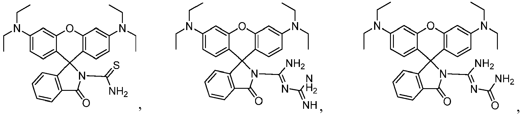
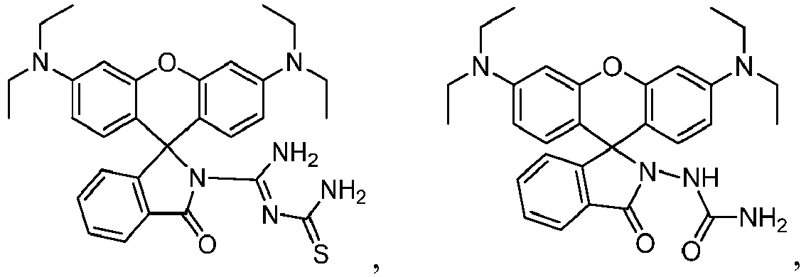


- A method for quantifying lysophosphatidic acid species, comprising:combining a sample that may include one or more lysophosphatidic acid species with a compound according to claim 1 in a solvent comprising dimethylsulfoxide in methanol to form a solution;exposing the solution to a light source;measuring fluorescence intensity of the solution; anddetermining, based on the fluorescence intensity, a total concentration of lysophosphatidic acid species in the sample.
- The method of claim 2, further comprising obtaining the sample by extracting lysophosphatidic acid species from a sample comprising plasma or serum.
- The method of claim 3, wherein extracting the lysophosphatidic acid species from the sample comprises:combining the sample comprising plasma or serum with a solvent comprising a C1-10 alkyl alcohol and chloroform to form a mixture;separating organic and aqueous layers of the mixture;extracting the aqueous layer with a buffer at neutral pH to form an extracted aqueous phase;mixing the extracted aqueous phase with chloroform;separating chloroform from the extracted aqueous phase to form a washed aqueous phase;adding phosphoric acid to the washed aqueous phase to form an acidified aqueous phase;loading the acidified aqueous phase onto a solid-phase extraction (SPE) cartridge including a stationary phase comprising silica derivatized with hydrocarbon chains;flowing water and subsequently chloroform through the SPE cartridge;drying the SPE cartridge;flowing a C1-10 alkyl alcohol through the SPE cartridge, thereby eluting lysophosphatidic acid species in the C1-10 alkyl alcohol from the SPE cartridge;evaporating the C1-10 alkyl alcohol to form a dry residue comprising lysophosphatidic acid species; anddissolving the dry residue in 9:1 methano1:H2O to produce the extracted lysophosphatidic acid sample comprising one or more lysophosphatidic acid species.
- The method of claim 4, further comprising:separating lysophosphatidic acid species in the extracted lysophosphatidic acid sample using a reversed-phase high-performance liquid chromatography (HPLC) column;detecting individual lysophosphatidic acid species as the separated lysophosphatidic acid species exit the reversed-phase HPLC column in an eluent comprising lysophosphatidic acid species bycombining the eluent with 4-(4-(dihexadecylamino)styryl)-N-methylpyridinium iodide (DiA) as it exits the reversed-phase HPLC column to form a DiA-eluent mixture,flowing the DiA-eluent mixture through a detector,detecting individual lysophosphatidic acid species by detecting fluorescence of the DiA-eluent mixture as the DiA-eluent mixture flows through the detector; andoptionally, identifying an individual lysophosphatidic acid species bycomparing an elution time for the individual lysophosphatidic acid species to elute from the reversed-phase HPLC column to elution times for known individual lysophosphatidic acid species,measuring fluorescence intensity of the DiA-eluent mixture, anddetermining, based on the fluorescence intensity, a concentration of the individual lysophosphatidic acid species.
- The method of claim 5, wherein the sample comprising plasma or serum is obtained from a subject suspected of being at risk of a condition associated with an aberrant LPA level, the method further comprising determining a risk level for the condition, wherein the risk level is based at least in part on an identification of an individual lysophosphatidic acid species, the concentration of the individual lysophosphatidic acid species, or a combination thereof.
- The method of claim 2 or claim 5, wherein the compound is

- The method of claim 3 or claim 5, wherein the sample comprising plasma or serum is obtained from a subject suspected of being at risk of a condition associated with an aberrant LPA level, the method further comprising determining a risk level for the condition, wherein the risk level is based at least in part on the total concentration of lysophosphatidic acid species.
- The method of claim 8, wherein the condition is cancer, cardiovascular disease, platelet aggregation, ischemia perfusion injury, neuropathic pain, a neuropsychiatric disorder, a reproductive disorder, or fibrosis, optionally wherein the cancer is ovarian cancer.
- A kit for detecting and quantifying lysophosphatidic acid, comprising at least one compound according to claim 1, and optionally comprising one or more lysophosphatidic acid species and/or one or more solid-phase extraction cartridges wherein the stationary phase is C8.
- The kit of claim 10, wherein the compound is

Applications Claiming Priority (2)
| Application Number | Priority Date | Filing Date | Title |
|---|---|---|---|
| US201361790443P | 2013-03-15 | 2013-03-15 | |
| PCT/US2014/029025 WO2014144561A2 (en) | 2013-03-15 | 2014-03-14 | Lysophosphatidic acid detection |
Publications (3)
| Publication Number | Publication Date |
|---|---|
| EP2970325A2 EP2970325A2 (en) | 2016-01-20 |
| EP2970325A4 EP2970325A4 (en) | 2016-08-31 |
| EP2970325B1 true EP2970325B1 (en) | 2017-09-27 |
Family
ID=51538340
Family Applications (1)
| Application Number | Title | Priority Date | Filing Date |
|---|---|---|---|
| EP14765163.2A Active EP2970325B1 (en) | 2013-03-15 | 2014-03-14 | Lysophosphatidic acid detection |
Country Status (4)
| Country | Link |
|---|---|
| US (2) | US9952231B2 (en) |
| EP (1) | EP2970325B1 (en) |
| ES (1) | ES2654371T3 (en) |
| WO (1) | WO2014144561A2 (en) |
Families Citing this family (11)
| Publication number | Priority date | Publication date | Assignee | Title |
|---|---|---|---|---|
| CN104560026A (en) * | 2014-12-25 | 2015-04-29 | 温州医科大学附属第二医院 | Fluorescent probe targeting MUC1 for in-vivo ovarian cancer tissue and preparation method of fluorescent probe |
| US20180149654A1 (en) | 2015-05-13 | 2018-05-31 | Econous Systems Inc. | Biosensors and methods for detection of lysophosphatidic acid for signaling of ovarian cancer |
| US10184932B2 (en) * | 2015-12-07 | 2019-01-22 | Zora Biosciences Oy | Use of ceramides and LPLs in diagnosing CVD |
| US11191850B2 (en) | 2016-05-23 | 2021-12-07 | Portland State University | Molecular probes for detection and imaging of pancreatic cancer |
| CN106478646B (en) * | 2016-09-26 | 2018-10-26 | 西北大学 | A kind of photosensitive probe of rhodamine-guanidine radicals benzimidazole functionalization and its application |
| CN108218880B (en) * | 2016-12-22 | 2020-09-15 | 王可 | Mercury ion optical probe and preparation method and application thereof |
| CN108218881B (en) * | 2016-12-22 | 2020-09-15 | 王可 | Mercury ion fluorescent probe based on rhodamine B and preparation method and application thereof |
| CN110272637B (en) * | 2018-03-16 | 2021-06-04 | 中国科学院大连化学物理研究所 | An acid-resistant light-controlled fluorescent molecular switch and its synthesis method and application |
| CN118652275A (en) * | 2019-01-30 | 2024-09-17 | 四川科伦博泰生物医药股份有限公司 | Camptothecin derivatives and water-soluble prodrugs thereof, pharmaceutical compositions containing the same, and preparation methods and uses thereof |
| US20250283800A1 (en) * | 2022-04-02 | 2025-09-11 | The General Hospital Corporation | Probes for fluorescence imaging |
| KR102723430B1 (en) * | 2022-09-13 | 2024-10-28 | 경북대학교 산학협력단 | Novel rhodamine derivative and use thereof for detecting mercury ion |
Family Cites Families (1)
| Publication number | Priority date | Publication date | Assignee | Title |
|---|---|---|---|---|
| US20100291689A1 (en) | 2006-03-21 | 2010-11-18 | Board Of Supervisors Of Louisiana State University & Agricultural And Mechanical College | Lanthanide Complexes as Fluorescent Indicators for Neutral Sugars and Cancer Diagnosis |
-
2014
- 2014-03-14 US US14/776,273 patent/US9952231B2/en not_active Ceased
- 2014-03-14 ES ES14765163.2T patent/ES2654371T3/en active Active
- 2014-03-14 EP EP14765163.2A patent/EP2970325B1/en active Active
- 2014-03-14 US US16/858,380 patent/USRE48928E1/en active Active
- 2014-03-14 WO PCT/US2014/029025 patent/WO2014144561A2/en not_active Ceased
Non-Patent Citations (1)
| Title |
|---|
| None * |
Also Published As
| Publication number | Publication date |
|---|---|
| WO2014144561A2 (en) | 2014-09-18 |
| ES2654371T3 (en) | 2018-02-13 |
| USRE48928E1 (en) | 2022-02-15 |
| US20160047830A1 (en) | 2016-02-18 |
| EP2970325A4 (en) | 2016-08-31 |
| EP2970325A2 (en) | 2016-01-20 |
| WO2014144561A3 (en) | 2014-11-06 |
| US9952231B2 (en) | 2018-04-24 |
Similar Documents
| Publication | Publication Date | Title |
|---|---|---|
| US9952231B2 (en) | Lysophosphatidic acid detection | |
| Erdemir et al. | Fast, visual, and quantitative monitoring of N2H4 by two ratiometric fluorescent probes in environmental media and biological systems | |
| Wei et al. | NBD-based colorimetric and fluorescent turn-on probes for hydrogen sulfide | |
| Li et al. | A mitochondria-targeted dual-response sensor for monitoring viscosity and peroxynitrite in living cells with distinct fluorescence signals | |
| Hao et al. | A naphthalimide-based chemodosimetric probe for ratiometric detection of hydrazine | |
| Zhao et al. | A novel pH probe based on a rhodamine–rhodamine platform | |
| Tong et al. | A ratiometric fluorescent pH probe based on keto–enol tautomerization for imaging of living cells in extreme acidity | |
| Wu et al. | A red-to-near-infrared fluorescent probe for the detection of thiophenol based on a novel hydroxylflavone-quinoline-amino molecular system with large Stokes shift | |
| Wu et al. | A highly selective visible light excitable boron dipyrromethene probe for cysteine over homocysteine and glutathione based on a Michael addition reaction | |
| WO2016025382A2 (en) | Probes for quantitative imaging of thiols in various environments | |
| Wei et al. | A highly sensitive and specific fluorescent probe for detecting ONOO–in liver-injured cells and tissues | |
| Li et al. | Novel coumarin-based sensitive and selective fluorescent probes for biothiols in aqueous solution and in living cells | |
| Liu et al. | Ruthenium (II) complex-based long-lived two-photon luminescence probe for dynamic monitoring of glutathione S-transferases in mouse models of drug-induced liver injury | |
| KR102513508B1 (en) | FLUOROGENIC pH-SENSITIVE DYES, FILM AND KIT COMPRISING THE SAME | |
| Wa-Yi et al. | A highly selective quinolizinium-based fluorescent probe for cysteine detection | |
| Firmino et al. | Bifunctionalised long-wavelength fluorescent probes for biological applications | |
| Gu et al. | A novel fluorescent probe based on β-C-glycoside for quantification of bovine serum albumin | |
| CN115894293A (en) | A ratio-type near-infrared fluorescent probe for detecting hydrazine and its synthesis method and application | |
| CN109180695A (en) | The preparation and application of nitric oxide fluorescent probe based on deoxidation rhodamine | |
| Hong et al. | A novel near-infrared fluorescent probe with a “donor–π–acceptor” type structure and its application in the selective detection of cysteine in living cells | |
| Lu et al. | A near-infrared fluorescent probe with remarkably large stokes shift for specifical imaging of peroxynitrite fluctuations in Hela cells | |
| Zhong et al. | Near-infrared multi-functional aggregation-induced emission fluorescent probe for detection of viscosity and peroxynitrite and its imaging application | |
| Fan et al. | A novel ratiometric BenzoBODIPY-Based fluorescent probe for the detection and imaging of Cysteine in living cells and zebrafish models | |
| CN109897080A (en) | High selection hypersensitive liver cancer-specific peroxynitrite probe and its application | |
| Xu et al. | A mitochondrial targeted dual ratiometric near-infrared fluorescent probe based on ICT effect for the detection of SO2 derivative and its bioimaging |
Legal Events
| Date | Code | Title | Description |
|---|---|---|---|
| PUAI | Public reference made under article 153(3) epc to a published international application that has entered the european phase |
Free format text: ORIGINAL CODE: 0009012 |
|
| 17P | Request for examination filed |
Effective date: 20151014 |
|
| AK | Designated contracting states |
Kind code of ref document: A2 Designated state(s): AL AT BE BG CH CY CZ DE DK EE ES FI FR GB GR HR HU IE IS IT LI LT LU LV MC MK MT NL NO PL PT RO RS SE SI SK SM TR |
|
| AX | Request for extension of the european patent |
Extension state: BA ME |
|
| DAX | Request for extension of the european patent (deleted) | ||
| A4 | Supplementary search report drawn up and despatched |
Effective date: 20160801 |
|
| RIC1 | Information provided on ipc code assigned before grant |
Ipc: G01N 33/92 20060101ALI20160726BHEP Ipc: C09K 11/06 20060101ALI20160726BHEP Ipc: G01N 33/52 20060101ALI20160726BHEP Ipc: G01N 21/64 20060101ALI20160726BHEP Ipc: C07D 491/107 20060101AFI20160726BHEP Ipc: C07B 63/00 20060101ALI20160726BHEP Ipc: C07D 491/22 20060101ALI20160726BHEP |
|
| GRAP | Despatch of communication of intention to grant a patent |
Free format text: ORIGINAL CODE: EPIDOSNIGR1 |
|
| INTG | Intention to grant announced |
Effective date: 20170413 |
|
| GRAS | Grant fee paid |
Free format text: ORIGINAL CODE: EPIDOSNIGR3 |
|
| GRAA | (expected) grant |
Free format text: ORIGINAL CODE: 0009210 |
|
| AK | Designated contracting states |
Kind code of ref document: B1 Designated state(s): AL AT BE BG CH CY CZ DE DK EE ES FI FR GB GR HR HU IE IS IT LI LT LU LV MC MK MT NL NO PL PT RO RS SE SI SK SM TR |
|
| REG | Reference to a national code |
Ref country code: GB Ref legal event code: FG4D |
|
| REG | Reference to a national code |
Ref country code: CH Ref legal event code: EP |
|
| REG | Reference to a national code |
Ref country code: AT Ref legal event code: REF Ref document number: 931836 Country of ref document: AT Kind code of ref document: T Effective date: 20171015 |
|
| REG | Reference to a national code |
Ref country code: IE Ref legal event code: FG4D |
|
| REG | Reference to a national code |
Ref country code: DE Ref legal event code: R096 Ref document number: 602014015142 Country of ref document: DE |
|
| PG25 | Lapsed in a contracting state [announced via postgrant information from national office to epo] |
Ref country code: FI Free format text: LAPSE BECAUSE OF FAILURE TO SUBMIT A TRANSLATION OF THE DESCRIPTION OR TO PAY THE FEE WITHIN THE PRESCRIBED TIME-LIMIT Effective date: 20170927 Ref country code: SE Free format text: LAPSE BECAUSE OF FAILURE TO SUBMIT A TRANSLATION OF THE DESCRIPTION OR TO PAY THE FEE WITHIN THE PRESCRIBED TIME-LIMIT Effective date: 20170927 Ref country code: NO Free format text: LAPSE BECAUSE OF FAILURE TO SUBMIT A TRANSLATION OF THE DESCRIPTION OR TO PAY THE FEE WITHIN THE PRESCRIBED TIME-LIMIT Effective date: 20171227 Ref country code: HR Free format text: LAPSE BECAUSE OF FAILURE TO SUBMIT A TRANSLATION OF THE DESCRIPTION OR TO PAY THE FEE WITHIN THE PRESCRIBED TIME-LIMIT Effective date: 20170927 Ref country code: LT Free format text: LAPSE BECAUSE OF FAILURE TO SUBMIT A TRANSLATION OF THE DESCRIPTION OR TO PAY THE FEE WITHIN THE PRESCRIBED TIME-LIMIT Effective date: 20170927 |
|
| REG | Reference to a national code |
Ref country code: NL Ref legal event code: MP Effective date: 20170927 |
|
| REG | Reference to a national code |
Ref country code: LT Ref legal event code: MG4D |
|
| REG | Reference to a national code |
Ref country code: ES Ref legal event code: FG2A Ref document number: 2654371 Country of ref document: ES Kind code of ref document: T3 Effective date: 20180213 |
|
| REG | Reference to a national code |
Ref country code: AT Ref legal event code: MK05 Ref document number: 931836 Country of ref document: AT Kind code of ref document: T Effective date: 20170927 |
|
| PG25 | Lapsed in a contracting state [announced via postgrant information from national office to epo] |
Ref country code: LV Free format text: LAPSE BECAUSE OF FAILURE TO SUBMIT A TRANSLATION OF THE DESCRIPTION OR TO PAY THE FEE WITHIN THE PRESCRIBED TIME-LIMIT Effective date: 20170927 Ref country code: GR Free format text: LAPSE BECAUSE OF FAILURE TO SUBMIT A TRANSLATION OF THE DESCRIPTION OR TO PAY THE FEE WITHIN THE PRESCRIBED TIME-LIMIT Effective date: 20171228 Ref country code: BG Free format text: LAPSE BECAUSE OF FAILURE TO SUBMIT A TRANSLATION OF THE DESCRIPTION OR TO PAY THE FEE WITHIN THE PRESCRIBED TIME-LIMIT Effective date: 20171227 Ref country code: RS Free format text: LAPSE BECAUSE OF FAILURE TO SUBMIT A TRANSLATION OF THE DESCRIPTION OR TO PAY THE FEE WITHIN THE PRESCRIBED TIME-LIMIT Effective date: 20170927 |
|
| REG | Reference to a national code |
Ref country code: FR Ref legal event code: PLFP Year of fee payment: 5 |
|
| PG25 | Lapsed in a contracting state [announced via postgrant information from national office to epo] |
Ref country code: NL Free format text: LAPSE BECAUSE OF FAILURE TO SUBMIT A TRANSLATION OF THE DESCRIPTION OR TO PAY THE FEE WITHIN THE PRESCRIBED TIME-LIMIT Effective date: 20170927 |
|
| PG25 | Lapsed in a contracting state [announced via postgrant information from national office to epo] |
Ref country code: CZ Free format text: LAPSE BECAUSE OF FAILURE TO SUBMIT A TRANSLATION OF THE DESCRIPTION OR TO PAY THE FEE WITHIN THE PRESCRIBED TIME-LIMIT Effective date: 20170927 Ref country code: RO Free format text: LAPSE BECAUSE OF FAILURE TO SUBMIT A TRANSLATION OF THE DESCRIPTION OR TO PAY THE FEE WITHIN THE PRESCRIBED TIME-LIMIT Effective date: 20170927 |
|
| PG25 | Lapsed in a contracting state [announced via postgrant information from national office to epo] |
Ref country code: EE Free format text: LAPSE BECAUSE OF FAILURE TO SUBMIT A TRANSLATION OF THE DESCRIPTION OR TO PAY THE FEE WITHIN THE PRESCRIBED TIME-LIMIT Effective date: 20170927 Ref country code: IS Free format text: LAPSE BECAUSE OF FAILURE TO SUBMIT A TRANSLATION OF THE DESCRIPTION OR TO PAY THE FEE WITHIN THE PRESCRIBED TIME-LIMIT Effective date: 20180127 Ref country code: SK Free format text: LAPSE BECAUSE OF FAILURE TO SUBMIT A TRANSLATION OF THE DESCRIPTION OR TO PAY THE FEE WITHIN THE PRESCRIBED TIME-LIMIT Effective date: 20170927 Ref country code: SM Free format text: LAPSE BECAUSE OF FAILURE TO SUBMIT A TRANSLATION OF THE DESCRIPTION OR TO PAY THE FEE WITHIN THE PRESCRIBED TIME-LIMIT Effective date: 20170927 Ref country code: AT Free format text: LAPSE BECAUSE OF FAILURE TO SUBMIT A TRANSLATION OF THE DESCRIPTION OR TO PAY THE FEE WITHIN THE PRESCRIBED TIME-LIMIT Effective date: 20170927 |
|
| REG | Reference to a national code |
Ref country code: DE Ref legal event code: R097 Ref document number: 602014015142 Country of ref document: DE |
|
| PG25 | Lapsed in a contracting state [announced via postgrant information from national office to epo] |
Ref country code: DK Free format text: LAPSE BECAUSE OF FAILURE TO SUBMIT A TRANSLATION OF THE DESCRIPTION OR TO PAY THE FEE WITHIN THE PRESCRIBED TIME-LIMIT Effective date: 20170927 |
|
| PLBE | No opposition filed within time limit |
Free format text: ORIGINAL CODE: 0009261 |
|
| STAA | Information on the status of an ep patent application or granted ep patent |
Free format text: STATUS: NO OPPOSITION FILED WITHIN TIME LIMIT |
|
| PG25 | Lapsed in a contracting state [announced via postgrant information from national office to epo] |
Ref country code: PL Free format text: LAPSE BECAUSE OF FAILURE TO SUBMIT A TRANSLATION OF THE DESCRIPTION OR TO PAY THE FEE WITHIN THE PRESCRIBED TIME-LIMIT Effective date: 20170927 |
|
| 26N | No opposition filed |
Effective date: 20180628 |
|
| REG | Reference to a national code |
Ref country code: CH Ref legal event code: PL |
|
| PG25 | Lapsed in a contracting state [announced via postgrant information from national office to epo] |
Ref country code: SI Free format text: LAPSE BECAUSE OF FAILURE TO SUBMIT A TRANSLATION OF THE DESCRIPTION OR TO PAY THE FEE WITHIN THE PRESCRIBED TIME-LIMIT Effective date: 20170927 Ref country code: MC Free format text: LAPSE BECAUSE OF FAILURE TO SUBMIT A TRANSLATION OF THE DESCRIPTION OR TO PAY THE FEE WITHIN THE PRESCRIBED TIME-LIMIT Effective date: 20170927 |
|
| REG | Reference to a national code |
Ref country code: BE Ref legal event code: MM Effective date: 20180331 |
|
| REG | Reference to a national code |
Ref country code: IE Ref legal event code: MM4A |
|
| PG25 | Lapsed in a contracting state [announced via postgrant information from national office to epo] |
Ref country code: LU Free format text: LAPSE BECAUSE OF NON-PAYMENT OF DUE FEES Effective date: 20180314 |
|
| PG25 | Lapsed in a contracting state [announced via postgrant information from national office to epo] |
Ref country code: IE Free format text: LAPSE BECAUSE OF NON-PAYMENT OF DUE FEES Effective date: 20180314 |
|
| PG25 | Lapsed in a contracting state [announced via postgrant information from national office to epo] |
Ref country code: LI Free format text: LAPSE BECAUSE OF NON-PAYMENT OF DUE FEES Effective date: 20180331 Ref country code: CH Free format text: LAPSE BECAUSE OF NON-PAYMENT OF DUE FEES Effective date: 20180331 Ref country code: BE Free format text: LAPSE BECAUSE OF NON-PAYMENT OF DUE FEES Effective date: 20180331 |
|
| PGFP | Annual fee paid to national office [announced via postgrant information from national office to epo] |
Ref country code: LU Payment date: 20190325 Year of fee payment: 6 |
|
| PGFP | Annual fee paid to national office [announced via postgrant information from national office to epo] |
Ref country code: ES Payment date: 20190401 Year of fee payment: 6 |
|
| PG25 | Lapsed in a contracting state [announced via postgrant information from national office to epo] |
Ref country code: MT Free format text: LAPSE BECAUSE OF NON-PAYMENT OF DUE FEES Effective date: 20180314 |
|
| PG25 | Lapsed in a contracting state [announced via postgrant information from national office to epo] |
Ref country code: TR Free format text: LAPSE BECAUSE OF FAILURE TO SUBMIT A TRANSLATION OF THE DESCRIPTION OR TO PAY THE FEE WITHIN THE PRESCRIBED TIME-LIMIT Effective date: 20170927 |
|
| PG25 | Lapsed in a contracting state [announced via postgrant information from national office to epo] |
Ref country code: PT Free format text: LAPSE BECAUSE OF FAILURE TO SUBMIT A TRANSLATION OF THE DESCRIPTION OR TO PAY THE FEE WITHIN THE PRESCRIBED TIME-LIMIT Effective date: 20170927 |
|
| PG25 | Lapsed in a contracting state [announced via postgrant information from national office to epo] |
Ref country code: CY Free format text: LAPSE BECAUSE OF FAILURE TO SUBMIT A TRANSLATION OF THE DESCRIPTION OR TO PAY THE FEE WITHIN THE PRESCRIBED TIME-LIMIT Effective date: 20170927 Ref country code: HU Free format text: LAPSE BECAUSE OF FAILURE TO SUBMIT A TRANSLATION OF THE DESCRIPTION OR TO PAY THE FEE WITHIN THE PRESCRIBED TIME-LIMIT; INVALID AB INITIO Effective date: 20140314 Ref country code: MK Free format text: LAPSE BECAUSE OF NON-PAYMENT OF DUE FEES Effective date: 20170927 |
|
| PG25 | Lapsed in a contracting state [announced via postgrant information from national office to epo] |
Ref country code: AL Free format text: LAPSE BECAUSE OF FAILURE TO SUBMIT A TRANSLATION OF THE DESCRIPTION OR TO PAY THE FEE WITHIN THE PRESCRIBED TIME-LIMIT Effective date: 20170927 |
|
| REG | Reference to a national code |
Ref country code: ES Ref legal event code: FD2A Effective date: 20210809 |
|
| REG | Reference to a national code |
Ref country code: ES Ref legal event code: FD2A Effective date: 20210810 |
|
| PG25 | Lapsed in a contracting state [announced via postgrant information from national office to epo] |
Ref country code: IT Free format text: LAPSE BECAUSE OF NON-PAYMENT OF DUE FEES Effective date: 20200314 |
|
| PG25 | Lapsed in a contracting state [announced via postgrant information from national office to epo] |
Ref country code: ES Free format text: LAPSE BECAUSE OF NON-PAYMENT OF DUE FEES Effective date: 20200315 |
|
| PGFP | Annual fee paid to national office [announced via postgrant information from national office to epo] |
Ref country code: DE Payment date: 20250305 Year of fee payment: 12 |
|
| PGFP | Annual fee paid to national office [announced via postgrant information from national office to epo] |
Ref country code: FR Payment date: 20250310 Year of fee payment: 12 |
|
| PGFP | Annual fee paid to national office [announced via postgrant information from national office to epo] |
Ref country code: GB Payment date: 20250306 Year of fee payment: 12 |