EP2964755B1 - Menstrual stems cells for the efficient support and expansion of cd34+ cd133+ hematopoietic stem cells in vitro - Google Patents
Menstrual stems cells for the efficient support and expansion of cd34+ cd133+ hematopoietic stem cells in vitro Download PDFInfo
- Publication number
- EP2964755B1 EP2964755B1 EP13877167.0A EP13877167A EP2964755B1 EP 2964755 B1 EP2964755 B1 EP 2964755B1 EP 13877167 A EP13877167 A EP 13877167A EP 2964755 B1 EP2964755 B1 EP 2964755B1
- Authority
- EP
- European Patent Office
- Prior art keywords
- cells
- stem cells
- menstrual
- cultured
- menscs
- Prior art date
- Legal status (The legal status is an assumption and is not a legal conclusion. Google has not performed a legal analysis and makes no representation as to the accuracy of the status listed.)
- Not-in-force
Links
Images






Classifications
-
- C—CHEMISTRY; METALLURGY
- C12—BIOCHEMISTRY; BEER; SPIRITS; WINE; VINEGAR; MICROBIOLOGY; ENZYMOLOGY; MUTATION OR GENETIC ENGINEERING
- C12N—MICROORGANISMS OR ENZYMES; COMPOSITIONS THEREOF; PROPAGATING, PRESERVING, OR MAINTAINING MICROORGANISMS; MUTATION OR GENETIC ENGINEERING; CULTURE MEDIA
- C12N5/00—Undifferentiated human, animal or plant cells, e.g. cell lines; Tissues; Cultivation or maintenance thereof; Culture media therefor
- C12N5/06—Animal cells or tissues; Human cells or tissues
- C12N5/0602—Vertebrate cells
- C12N5/0634—Cells from the blood or the immune system
- C12N5/0647—Haematopoietic stem cells; Uncommitted or multipotent progenitors
-
- C—CHEMISTRY; METALLURGY
- C12—BIOCHEMISTRY; BEER; SPIRITS; WINE; VINEGAR; MICROBIOLOGY; ENZYMOLOGY; MUTATION OR GENETIC ENGINEERING
- C12N—MICROORGANISMS OR ENZYMES; COMPOSITIONS THEREOF; PROPAGATING, PRESERVING, OR MAINTAINING MICROORGANISMS; MUTATION OR GENETIC ENGINEERING; CULTURE MEDIA
- C12N5/00—Undifferentiated human, animal or plant cells, e.g. cell lines; Tissues; Cultivation or maintenance thereof; Culture media therefor
- C12N5/06—Animal cells or tissues; Human cells or tissues
- C12N5/0602—Vertebrate cells
- C12N5/0652—Cells of skeletal and connective tissues; Mesenchyme
- C12N5/0662—Stem cells
- C12N5/0665—Blood-borne mesenchymal stem cells, e.g. from umbilical cord blood
-
- C—CHEMISTRY; METALLURGY
- C12—BIOCHEMISTRY; BEER; SPIRITS; WINE; VINEGAR; MICROBIOLOGY; ENZYMOLOGY; MUTATION OR GENETIC ENGINEERING
- C12N—MICROORGANISMS OR ENZYMES; COMPOSITIONS THEREOF; PROPAGATING, PRESERVING, OR MAINTAINING MICROORGANISMS; MUTATION OR GENETIC ENGINEERING; CULTURE MEDIA
- C12N2501/00—Active agents used in cell culture processes, e.g. differentation
- C12N2501/06—Anti-neoplasic drugs, anti-retroviral drugs, e.g. azacytidine, cyclophosphamide
-
- C—CHEMISTRY; METALLURGY
- C12—BIOCHEMISTRY; BEER; SPIRITS; WINE; VINEGAR; MICROBIOLOGY; ENZYMOLOGY; MUTATION OR GENETIC ENGINEERING
- C12N—MICROORGANISMS OR ENZYMES; COMPOSITIONS THEREOF; PROPAGATING, PRESERVING, OR MAINTAINING MICROORGANISMS; MUTATION OR GENETIC ENGINEERING; CULTURE MEDIA
- C12N2501/00—Active agents used in cell culture processes, e.g. differentation
- C12N2501/10—Growth factors
- C12N2501/105—Insulin-like growth factors [IGF]
-
- C—CHEMISTRY; METALLURGY
- C12—BIOCHEMISTRY; BEER; SPIRITS; WINE; VINEGAR; MICROBIOLOGY; ENZYMOLOGY; MUTATION OR GENETIC ENGINEERING
- C12N—MICROORGANISMS OR ENZYMES; COMPOSITIONS THEREOF; PROPAGATING, PRESERVING, OR MAINTAINING MICROORGANISMS; MUTATION OR GENETIC ENGINEERING; CULTURE MEDIA
- C12N2501/00—Active agents used in cell culture processes, e.g. differentation
- C12N2501/10—Growth factors
- C12N2501/113—Acidic fibroblast growth factor (aFGF, FGF-1)
-
- C—CHEMISTRY; METALLURGY
- C12—BIOCHEMISTRY; BEER; SPIRITS; WINE; VINEGAR; MICROBIOLOGY; ENZYMOLOGY; MUTATION OR GENETIC ENGINEERING
- C12N—MICROORGANISMS OR ENZYMES; COMPOSITIONS THEREOF; PROPAGATING, PRESERVING, OR MAINTAINING MICROORGANISMS; MUTATION OR GENETIC ENGINEERING; CULTURE MEDIA
- C12N2501/00—Active agents used in cell culture processes, e.g. differentation
- C12N2501/10—Growth factors
- C12N2501/125—Stem cell factor [SCF], c-kit ligand [KL]
-
- C—CHEMISTRY; METALLURGY
- C12—BIOCHEMISTRY; BEER; SPIRITS; WINE; VINEGAR; MICROBIOLOGY; ENZYMOLOGY; MUTATION OR GENETIC ENGINEERING
- C12N—MICROORGANISMS OR ENZYMES; COMPOSITIONS THEREOF; PROPAGATING, PRESERVING, OR MAINTAINING MICROORGANISMS; MUTATION OR GENETIC ENGINEERING; CULTURE MEDIA
- C12N2501/00—Active agents used in cell culture processes, e.g. differentation
- C12N2501/10—Growth factors
- C12N2501/145—Thrombopoietin [TPO]
-
- C—CHEMISTRY; METALLURGY
- C12—BIOCHEMISTRY; BEER; SPIRITS; WINE; VINEGAR; MICROBIOLOGY; ENZYMOLOGY; MUTATION OR GENETIC ENGINEERING
- C12N—MICROORGANISMS OR ENZYMES; COMPOSITIONS THEREOF; PROPAGATING, PRESERVING, OR MAINTAINING MICROORGANISMS; MUTATION OR GENETIC ENGINEERING; CULTURE MEDIA
- C12N2501/00—Active agents used in cell culture processes, e.g. differentation
- C12N2501/90—Polysaccharides
- C12N2501/91—Heparin
-
- C—CHEMISTRY; METALLURGY
- C12—BIOCHEMISTRY; BEER; SPIRITS; WINE; VINEGAR; MICROBIOLOGY; ENZYMOLOGY; MUTATION OR GENETIC ENGINEERING
- C12N—MICROORGANISMS OR ENZYMES; COMPOSITIONS THEREOF; PROPAGATING, PRESERVING, OR MAINTAINING MICROORGANISMS; MUTATION OR GENETIC ENGINEERING; CULTURE MEDIA
- C12N2502/00—Coculture with; Conditioned medium produced by
- C12N2502/24—Genital tract cells, non-germinal cells from gonads
- C12N2502/243—Cells of the female genital tract, non-germinal ovarian cells
Definitions
- the invention relates to highly proliferative stem cell obtained from menstrual fluid.
- the menstrual stem cells may be cultured in vitro and exhibit mesenchymal stem cell (MSC)-like properties and some culture and expansion advantages.
- MSC mesenchymal stem cell
- This population of MenSCs compared to the broadly studied bone marrow derived stem cells (BM-MSCs) out-performs bone marrow derived mesenchymal stem cells in proliferation rate and support of hematopoietic stem cell (HSC) expansion in vitro.
- BM-MSCs bone marrow derived stem cells
- HSC hematopoietic stem cell
- MenSCs demonstrate to maintain their stem cell properties over 2 years, being genetically stable, without expressing surface differentiation markers, and they also show the ability to differentiate into adipocytes, chondrocytes and osteoblast cells. MenSCs can be easily and periodically obtained, isolated and cultured.
- MSC Mesenchymal stem cell
- BM-MSCs bone marrow derived stem cells
- HSC hematopoietic stem cells
- HSC have to be expanded in vitro successfully in order to have enough quantities to be used for clinical research and for the treatment of a wide range of diseases. Nevertheless, these known MSC types used to support expansion of stem cells have a low availability and they are the main limiting factor when co-culturing cells.
- BM-MSCs cannot be easily and periodically obtained, isolated and cultured as requested, due to the restricted source.
- HSC stem cell markers
- WO1993018137 discloses the co-culture of HSC with stromal cells or stromal cells' conditioned media.
- the document describes HSC co-culture directly over a layer of stromal cells or alternatively, HSC are co-cultured separated from the stromal cells using a porous membrane.
- the aim of the invention is the growing of stem cells inhibiting the differentiation of the cells to improve their expansion and the culture of genetic modified human hematopoietic stem cells.
- the differentiation is reduced by using culture media supplemented with Leukemia Inhibitory Factor (LIF), where the inhibition of the differentiation also improves the integration of the construct.
- LIF Leukemia Inhibitory Factor
- WO2009/058356 and related application WO2013/025189 disclose methods for obtaining expanded human cord blood cells that may express CD34, SSEA-4, and HLA-II through co-culture with menstrual cells collected, isolated and cultured according to methods in WO2008/109063 .
- the present invention provides a method to obtain highly proliferative stem cell from menstrual fluid for clinical and research applications, including in vivo and in vitro cultures.
- the MenSCs obtained using the method described in the present invention have culture and expansion advantages in the proliferation rate and in their ability for supporting hematopoietic stem cell (HSC) expansion in vitro.
- HSC hematopoietic stem cell
- the method comprises the easy and periodically obtention, isolation and culture of MenSCs, where the MenSCs are obtained from menstrual blood.
- the method comprises obtaining MenSCs from menstrual blood, where the isolated MenSCs have high proliferation rates, determined at different time points of the culture.
- the proliferation rates can be compared to BM-MSCs isolated under standard conditions, as described below.
- the method provides an in vitro method for culturing menstrual stem cells (MenSCs) that exhibit mesenchymal stem cell (MSC)-like properties.
- MenSCs menstrual stem cells
- MSC mesenchymal stem cell
- the method comprises the selection of the MenSCs according to the immunophenotype based on mesenchymal and pluripotential stem cell markers.
- the method comprises the co-culture of MenSCs with cord blood stem cells in a growth factor-defined medium.
- the method comprises the ex vivo proliferation and support of HSCs.
- the method comprises the adipogenic, chondrogenic, and osteogenic differentiation of the MenSCs.
- the mesenchymal stem cells obtained according to the present invention can be harvested from menstrual blood, which is a clear difference regarding the invasive procedures and the ethical controversy compared to embryonic stem cells related procedures.
- MenSCs display stem cell-like phenotypic markers and show great potential for self-renewal, plasticity, and undifferentiated proliferation for long periods of time in vitro. A large number of studies have shown that MenSCs are a population of pluripotent cells.
- the present invention positions the cells isolated from menstrual fluids on the top of the list of cells candidates to fill up that clinical need.
- Menstrual cells have been collected, isolated and cultured according to classic methods as described by Meng et al. 2007 and document WO2008/109063 .
- Those methods disclose the isolation and processing of menstrual cells, where isolation may be performed with at least one of the operations of centrifugation, density gradient sedimentation or filtration.
- the processing stages to obtain the menstrual cell include the steps of obtain, isolate and preserve the menstrual cells where menstrual cells can be used later fresh or cryopreserved.
- those processes show a source of stem cells, where viable menstrual cells are prepared for cryopreservation and are stored at cell banks being a more available source than cord blood cells, that is easily accessible, highly expandable in vitro and may be an alternative for autologous or heterologous stem cell applications.
- MenSCs A detailed characterization of the MenSCs has been made for a complete comparison with related cells from other sources in order to allow the evaluation and comparative analysis of the advantages of MenSCs and also their safety/efficacy profile for clinical applications.
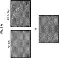
- the menstrual blood-derived stem cells morphology in culture is shown in Figure 1(A) , having a fibroblast-like spindle-shaped morphology.
- the morphology is maintained during the successive culture passages, and can be observed in freshly isolated menstrual stem cells (without passages or passage zero), after 24 hours of culture and after 2-week cell culture and a confluent culture at the third passage.
- MenSCs have been shown to be positive for mesenchymal stem cell markers including CD9, CD29, CD105, and CD73, and negative for hematopoietic markers such as CD34, CD45 and CD133, as expected for those cell types.
- MenSCs menstrual stem cells
- BM-MSCs bone marrow
- MenSCs display positive expression for mesenchymal stromal cells such as CD44, CD105, and CD73 while negative for CD14, CD34, CD45, HLA-DR, EPCAM, CD31, CD117, and CD271, and mildly positive expression for CD146.
- MenSCs do not express pluripotent surface markers such as SSEA-3, SSEA-4, and TRA-1-60.
- the frequency of stem cell precursors defined by their ability to form a cell colony in vitro (colony-forming unit, CFU) is another important property when characterizing a new stem cell source.
- MSCs can be virtually isolated from many organs, their low frequency or lack of clonogenicity can be a limiting factor for their clinical application.
- MSCs isolated from umbilical cord blood have comparable differentiation properties as cells isolated from bone marrow (BM-MSC); however, these cells are found at a much lower rate than in the later case, hampering their therapeutically usage.
- BM-MSC bone marrow
- MenSC possess a high or a low precursor frequency in comparison to MSCs isolated from other known sources.
- cells were cultured at different serial dilution to determine their CFU-fibroblast frequency in comparison to BM-MSCs.
- MenSCs have shown a higher proliferation rate compared to other sources, with doubling times of MenSCs within 18-24 hr depending on culture technique and media used (Meng et al., 2007).
- a good proliferation rate is essential for feasibility of clinical applications since stem cell-based therapies are usually efficient starting at a specific dose.
- MenSCs have been largely expanded in vitro without any mutation or visible abnormality at the chromosomal level. They maintained a telomerase activity greater than 50% even at passage 12 compared with human embryonic stem cells (Patel et al., 2008), and also appear to mildly express the chemokine receptor CXCR4 and the respective receptor for stromal cell-derived factor-1 (SDF-1), which play a significant role in the mediation of mesenchymal stem cell migration (Patel et al., 2008).
- SDF-1 stromal cell-derived factor-1
- these MenSCs cells did not show any sign of stem cell exhaustion evidenced by a steady expression of all stromal stem markers, a stable proliferation rate and CFU potential when compared as shown in Figure 2 (A) .
- Such a high proliferative rate in the face of genetic stability and apparent preservation of multipotency indicates MenSCs as a type of stem cell that could present unexpected therapeutic properties, a fact that is also implied by their extensive differentiation capabilities.
- MenSCs to differentiate into adipose, bone, cartilage, cardiac, neural, hepatic and pancreatic cell types has been shown using standard differentiation techniques and media conditions.
- these multipotent cells have the ability to differentiate into various functional types, including respiratory epithelial cells, neurocytes, myocytes, endothelial cells, pancreatic cells, hepatocytes, adipocytes and osteocytes (Meng et al., 2007).
- MenSCs show the ability to differentiate into adipose, bone and chondrocytes tissues after culture under appropriate differentiation media at varying degrees compared with BM-MSCs. They showed a lower adipogenic differentiation, an equal osteocytic differentiation and a higher chondrocytic differentiation as shown in the different staining as shown in Figure 3 .
- HSCs human hematopoietic stem cells
- MSCs isolated from the bone marrow have been used as a support for HSC expansion in vitro in preclinical and clinical settings.
- BM-MSCs bone marrow
- CD34+ CD133+ cells were immunomagneticaly isolated from human umbilical cords using CD34 beads, then cultured alone or co-cultured at a ratio of 1:4 to1:5 in different feeder layers from BM-MSCs and MenSCs. Total number of cells and the expression of CD34+ CD133+ were evaluated at different time point during the course of the culture. Starting from 3 days post-culture, a statistically significant different in favor of the co-culture with MenSCs was observed. By the end of the culture, a difference about 4 fold was observed for total cell numbers when compared with cells cultured alone or with BM-MSCs.
- MenSCs Compared with BM-MSCs, MenSCs elicit a more precocious early stem/progenitor formation and faster proliferation and differentiation of hematopoietic progenitors. This property to support hematopoiesis in vitro provides the rationale for their use in supporting hematopoietic reconstitution in clinical settings. The mechanism behind this superior yield is currently under investigation in our laboratory.
- the HSCs were co-cultured in the presence of MenSCs, in similar conditions, with the difference of the presence or absence of a separation membrane, denoted as T (Transwell®, Corning).
- Menstrual blood is collected from a healthy female subject during the earliest days of a menstrual cycle. Samples are collected in a menstrual silicone cup. The cells are harvested with the informed consent of the donor as approved by an Institutional Review Board of Universidad de los Andes. Menstrual blood samples are transferred into a tube with phosphate buffered saline (PBS) containing amphotericin B, penicillin, streptomycin and EDTA.
- PBS phosphate buffered saline
- Menstrual blood mononuclear cells are separated by Ficoll-Paque density gradient and washed in PBS. Cells were subsequently cultured in a flask containing Dulbecco's modified Eagle's medium (DMEM) high Glucose supplemented with % penicillin/streptomycin, amphotericin B, glutamine and FBS at 35-40°C, using 2-10% CO2 in order to obtain adherent cells. Media were changed the next day to wash non adherent cells. Cells were replated as follows: adherent cells were detached using Trypsin-Edta, counted and sub-cultured. Afterwards, these cells were used as menstrual blood-derived stem cells (MenSCs).
- MenSCs menstrual blood-derived stem cells
- BM-MSCs Bone marrow mesenchymal stem cells
- HSCs Hematopoietic Stem Cells
- Mononuclear cell were isolated after centrifugation on Ficoll-Paque.
- CD34+ cells were enriched with a CD34 Positive Selection Kit. Labeled cells are separated using a magnet separation kit.
- MenSCs and BM-MSCs are seed in adipogenic differentiation medium: complete DMEM media, dexamethasone, 3-isobutyl-1-methyl-xanthine (IBMX), recombinant human insulin, and indomethacin; and cultured for up 21 days with media changes every 3-4 days.
- adipogenic differentiation medium complete DMEM media, dexamethasone, 3-isobutyl-1-methyl-xanthine (IBMX), recombinant human insulin, and indomethacin
- Control cells are cultured in completed DMEM media. Cells were subsequently stained with Oil Red O.
- Cells are seed in osteogenic differentiation medium: complete DMEM media, dexamethasone, b-glycerophosphate, and ascorbate; and cultured for up 21 days with media changes every 3-4 days. Control cells were cultured in completed DMEM media. Cells are later stained with Alizarin.
- Cells are seed in chondrogenic differentiation medium: complete DMEM media, dexamethasone, b-glycerophosphate, and ascorbate; and cultured for up 21 days with media changes every 3-4 days. Control cells are cultured in completed DMEM media and subsequently stained with Safranina O.
- MenSCs and BM-MSCs are cultured in a 24-well plate in complete DMEM media. Cell proliferation and viability is determined at different days measuring cellular mitochondrial dehydrogenase and quantified by spectrophotometry using absorbance 440 nm.
- CFU-F Colony-Forming Unit-Fibroblast
- cytometer buffer PBS + BSA + Sodium Azide
- specific antibody for 20 min a 4°C in cytometer buffer containing the specific fluorescein isothiocynate (FITC), allophycocyanin (APC), phycoerythrin (PE), or phycoerythrin with cyanin-7 (PECy7) labeled antibodies.
- FITC fluorescein isothiocynate
- APC allophycocyanin
- PE phycoerythrin
- PECy7 phycoerythrin-7
- Antibodies for human cell surface antigens CD14, CD44, CD90, CD271, CD105, CD73, CD117, CD45, CD34, HLA-ABC, HLA-DR, CD146, EPCAM, CD31, TRA-1-60, CD49a, SSEA3, SSEA4, CD133 and CD3 were used.
- the corresponding isotype-matched antibodies are used as negative controls.
- a dead cell stain kits is used to determine the viability cells by flow cytometry. Data is collected and analyzed using FACS software.
- CD34+ CD133+ hematopoietic stem cells are then co-cultured with feeder layers using StemSpan medium supplemented with human growth factors: FGF-1, SCF, TPO, IGFBP2, heparin and penicillin/streptomycin.
- CD34+ CD133+cells are plated directly on the feeder layer or in Transwell above the feeder layer (noncontact growth conditions) in an appropriate ratio in growth factor-supplemented StemSpan media in 24-well plates. The medium was half changed every day and additionally supplemented. All the expanded cord blood cells were harvested after different days by vigorous pipetting, washed in PBS, and the cells total number were determined by trypan blue.
- MSCs were seeded in 24 well plate and cultured on MenSC media prior to the coculture.
- MenSC media consists of DMEM-high glucose supplemented with FBS, penicillin streptomycin, amphotericin B and L-glutamine. The assays were preformed in triplicate. A fourth well was seeded for the cell quantification before radiation procedure. At day 1, once the MSCs cultures are in confluent layers, the cells were irradiated (15 Gy).
- HSCs CD34+CD133+ cells derived from umbilical cord blood were obtained using CD34 immunomagnetic purification. The HCS cells were thawed as follows:
- HSCs are co-cultured over adherent monolayer MSCs at 1:4 ratio of HSC:MSC cells /well in a 24 well plat.
- the culture was feed with supplemented StemSpan® media. Each following day the media was fed removing the residual media, and then adding fresh media.
- the quantification of the HSCs cells is performed after resuspending the cells after carefully pipetting 10 times avoiding the disturbance of the MSCs layer.
- the HSCs cells are analyzed by FACS.
- the analysis is made on different days for FITC, APC, PE, PECy7, CD45, CD3, CD133, CD34 antibodies.
- Menstrual blood was collected in a menstrual silicone cup (Mialuna®, Santiago, Chile). Menstrual blood samples were transferred into a 50 ml tube with 10 ml phosphate buffered saline (PBS) containing 0.25 mg/mL amphotericin B (Gibco, Paisley, UK), penicillin 100 IU, streptomycin 100 mg/mL and 2 mM EDTA (all from Sigma, USA).
- PBS phosphate buffered saline
- Menstrual blood mononuclear cells were separated by Ficoll-Paque (Sigma-Aldrich, St Lois, USA) (1.077 g/ml) density gradient according to the manufacturer's instructions and washed in PBS. Cells were subsequently cultured in a T25 flask (Falcon®, Becton Dickinson) containing Dulbecco's modified Eagle's medium (DMEM) high Glucose (Gibco, Paisley, UK) supplemented with 1% penicillin/streptomycin, 1% amphotericin B, 1% glutamine (Gibco, Paisley, UK) and 15% FBS (Lonza, Walkersville, MD USA) at 37°C, 5% CO2 in order to obtain adherent cells.
- DMEM Dulbecco's modified Eagle's medium
- BM-MSCs bone marrow mesenchymal stem cells
- MenSCs and BM-MSCs were seeded at a concentration of 1,5x10E4 cells/cm2 in adipogenic differentiation medium: complete DMEM media, 1mM dexamethasone, 0.5 mM 3-isobutyl-1-methyl-xanthine (IBMX), 10ug/ml recombinant human insulin, and 100 mM indomethacin; and cultured for up 21 days with media changes every 3-4 days.
- adipogenic differentiation medium complete DMEM media, 1mM dexamethasone, 0.5 mM 3-isobutyl-1-methyl-xanthine (IBMX), 10ug/ml recombinant human insulin, and 100 mM indomethacin
- Control cells were cultured in completed DMEM media. Cells were subsequently stained with Oil Red O (Sigma-Aldrich, St Lois, USA).
- Cells were seeded at a concentration of 2x10E4 cells/cm2 in osteogenic differentiation medium: complete DMEM media, 100nM dexamethasone, 10mM b-glycerophosphate, and 0.2MM ascorbate; and cultured for up 21 days with media changes every 3-4 days.
- Control cells were cultured in completed DMEM media. Cells were stained with Alizarin Red (Sigma-Aldrich, St Lois, USA).
- Cells were seeded at a concentration of 3x10E4 cells/10uL in chondrogenic differentiation medium: complete DMEM media, 100nM dexamethasone, 10mM b-glycerophosphate, and 0.2mM ascorbate; and cultured for up 21 days with media changes every 3-4 days.
- Control cells were cultured in completed DMEM media. Cells were subsequently stained with Safranina O (Sigma-Aldrich, St Lois, USA).
- MenSCs and BM-MSCs were cultured at 1000 cells/cm2 in a 24-well plate (Falcon®, Becton Dickinson) in complete DMEM media.
- CFU Colony Forming Unit
- CFU-F Colony-Forming Unit-Fibroblast
- Results were expressed as CFU/number of cells plated. Then, cell-incorporated crystal violet was solubilized by incubation with acetic acid 33% and quantified spectrophotometrically (absorbance, 620 nm).
- cytometer buffer PBS + 0.2% BSA + 0.01% Sodium Azide [all from Sigma-Aldrich, St Lois, USA]) and incubated with the specific antibody at concentrations recommended by the respective manufacturer for 20 min a 4°C in cytometer buffer containing the specific fluorescein isothiocynate (FITC), allophycocyanin (APC), phycoerythrin (PE), or phycoerythrin with cyanin-7 (PECy7) labeled antibodies.
- FITC fluorescein isothiocynate
- APC allophycocyanin
- PE phycoerythrin
- PECy7 phycoerythrin-7
- Antibodies for human cell surface antigens CD14, CD44, CD90, CD271, CD105, CD73, CD117, CD45, CD34, HLA-ABC, HLA-DR, CD146, EPCAM, CD31, TRA-1-60, CD49a, SSEA3, SSEA4, CD133 and CD3 were purchase from BD PharmingenTM (BD Biosciences, San Jose, CA), R&D System (Minneapolis, MN, USA) and Biolegend (San Diego, CA, USA).
- CD34+ CD133+ hematopoietic stem cells were then co-culture with these feeder layers using StemSpan (Stem Cell Technologies, Vancouver, Canada) supplemented with human growth factors: 10 ng/ml FGF-1, 10 ng/ml SCF, 20 ng/ml TPO, 100 ng/ml IGFBP2 -all R&D systems (Minneapolis, MN, USA)-, 100 micrograms/ml of heparin and 1 x penicillin/streptomycin (Gibco, Paisley, UK).
- StemSpan StemSpan
- human growth factors 10 ng/ml FGF-1, 10 ng/ml SCF, 20 ng/ml TPO, 100 ng/ml IGFBP2 -all R&D systems (Minneapolis, MN, USA)-, 100 micrograms/ml of heparin and 1 x penicillin/streptomycin (Gibco, Paisley, UK).
- CD34+ CD133+cells were plated directly on these feeder layer or in Transwell (0.4 ⁇ m pore size; BD Biosciences) above the feeder layer (noncontact growth conditions) in a 1:4-1:5 ratio in growth factor-supplemented StemSpan in a 24-well plates (Falcon®, Becton Dickinson), at 500 ⁇ l/well. The medium was half changed every day and additionally supplemented with 100 ⁇ l/well. All the expanded cord blood cells were harvested after 3, 5 and 7 days by vigorous pipetting, washed in PBS, and the cells total number were determined by trypan blue.
- MSCs were seeded in 24 well plate at a concentration of 5x10 4 cells/well. Cells were cultured on MenSC media prior to the coculture. MenSC media consists of DMEM-high glucose (Gibco #10313021) supplemented with 15% FBS, 1% penicillin streptomycin (Gibco #15140122), 1% amphotericin B (Biological #03-028-1B) and 1% L-glutamine (Gibco #25030-081). The assays were preformed in triplicate. A fourth well was seeded for the cell quantification before radiation procedure.
- HSCs CD34+CD133+ cells derived from umbilical cord blood were obtained using CD34 immunomagnetic purification. The HCS cells were thawed as follows:
- HSCs are cocultured over adherent monolayer MSCs at 1:4 and/or 1:5 ratio of HSC: MSC, using 10 4 HSC cells and 4x10 4 MSC cells /well in a 24 well plat.
- the culture was feed with supplemented StemSpan® media (StemCell Technologies #9610) 500uL/well (initial volume). Each following day the media was fed removing the residual media over 200ml, and then adding 100ml of fresh media.
- the quantification of the HSCs cells is performed after resuspending the cells after carefully pipetting 10 times avoiding the disturbance of the MSCs layer.
- the HSCs cells are analyzed by FACS.
- the analysis is made on days 3, 5 and 7 for FITC, APC, PE, PECy7, CD45, CD3, CD133, CD34 antibodies.
Landscapes
- Health & Medical Sciences (AREA)
- Life Sciences & Earth Sciences (AREA)
- Engineering & Computer Science (AREA)
- Biomedical Technology (AREA)
- Genetics & Genomics (AREA)
- Zoology (AREA)
- Organic Chemistry (AREA)
- Biotechnology (AREA)
- Chemical & Material Sciences (AREA)
- Bioinformatics & Cheminformatics (AREA)
- Wood Science & Technology (AREA)
- Hematology (AREA)
- Developmental Biology & Embryology (AREA)
- Microbiology (AREA)
- Cell Biology (AREA)
- Biochemistry (AREA)
- General Engineering & Computer Science (AREA)
- General Health & Medical Sciences (AREA)
- Immunology (AREA)
- Rheumatology (AREA)
- Micro-Organisms Or Cultivation Processes Thereof (AREA)
Description
- The invention relates to highly proliferative stem cell obtained from menstrual fluid. The menstrual stem cells (MenSCs) may be cultured in vitro and exhibit mesenchymal stem cell (MSC)-like properties and some culture and expansion advantages. This population of MenSCs, compared to the broadly studied bone marrow derived stem cells (BM-MSCs) out-performs bone marrow derived mesenchymal stem cells in proliferation rate and support of hematopoietic stem cell (HSC) expansion in vitro. MenSCs demonstrate to maintain their stem cell properties over 2 years, being genetically stable, without expressing surface differentiation markers, and they also show the ability to differentiate into adipocytes, chondrocytes and osteoblast cells. MenSCs can be easily and periodically obtained, isolated and cultured.
- Mesenchymal stem cell (MSC)-like are well known for exhibiting desirable properties in culture and expansion in vitro.
Particularly, bone marrow derived stem cells (BM-MSCs) are known for having the ability to support expansion in vitro of stem cells, such as hematopoietic stem cells (HSC). - HSC have to be expanded in vitro successfully in order to have enough quantities to be used for clinical research and for the treatment of a wide range of diseases. Nevertheless, these known MSC types used to support expansion of stem cells have a low availability and they are the main limiting factor when co-culturing cells.
- Moreover, BM-MSCs cannot be easily and periodically obtained, isolated and cultured as requested, due to the restricted source.
- Therefore, a high proliferation and a good differentiation of HSC is a key issue, but taking special attention on maintaining the expression of their stem cell markers (CD34+ and CD133+).
- In the prior art, document
US2007041948 has been described the co-culture of human endometrial stromal cell with HSC expressing CD34+, nevertheless, this method is uses endometrial stromal cells or epithelial cells obtained from fresh tissue, having a very limited availability for research and clinical purposes. The human endometrial stem cells are obtained by culturing finely cut fresh uterine tissue in a medium containing collagenase, the tissue is disaggregated, the cells are filtered and stromal cells are isolated, collecting the human endometrial stromal cells adhered to the cell culture dish. -
WO1993018137 discloses the co-culture of HSC with stromal cells or stromal cells' conditioned media. The document describes HSC co-culture directly over a layer of stromal cells or alternatively, HSC are co-cultured separated from the stromal cells using a porous membrane. The aim of the invention is the growing of stem cells inhibiting the differentiation of the cells to improve their expansion and the culture of genetic modified human hematopoietic stem cells. The differentiation is reduced by using culture media supplemented with Leukemia Inhibitory Factor (LIF), where the inhibition of the differentiation also improves the integration of the construct. -
WO2009/058356 and related applicationWO2013/025189 disclose methods for obtaining expanded human cord blood cells that may express CD34, SSEA-4, and HLA-II through co-culture with menstrual cells collected, isolated and cultured according to methods inWO2008/109063 . - The present invention provides a method to obtain highly proliferative stem cell from menstrual fluid for clinical and research applications, including in vivo and in vitro cultures.
- The MenSCs obtained using the method described in the present invention have culture and expansion advantages in the proliferation rate and in their ability for supporting hematopoietic stem cell (HSC) expansion in vitro.
- In an embodiment of the invention, the method comprises the easy and periodically obtention, isolation and culture of MenSCs, where the MenSCs are obtained from menstrual blood.
- In an embodiment of the invention, the method comprises obtaining MenSCs from menstrual blood, where the isolated MenSCs have high proliferation rates, determined at different time points of the culture. The proliferation rates can be compared to BM-MSCs isolated under standard conditions, as described below.
- In an embodiment of the invention, the method provides an in vitro method for culturing menstrual stem cells (MenSCs) that exhibit mesenchymal stem cell (MSC)-like properties.
- In an embodiment of the invention, the method comprises the selection of the MenSCs according to the immunophenotype based on mesenchymal and pluripotential stem cell markers.
- In an embodiment of the invention, the method comprises the co-culture of MenSCs with cord blood stem cells in a growth factor-defined medium.
- In an embodiment of the invention, the method comprises the ex vivo proliferation and support of HSCs.
- In an embodiment of the invention, the method comprises the adipogenic, chondrogenic, and osteogenic differentiation of the MenSCs.
-
-
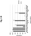
Figure 1 illustrates the menstrual blood-derived stem cells morphology and surface markers expression. -
Figure 1(A) . Morphology of MenSCs in culture, of freshly isolated menstrual stem cells (without passages or passage zero P0), after 2-week cell culture, and at the third passage (P3) showing a fibroblast-like spindle-shaped morphology.Scale bars 100 um. -
Figure 1(B) . Menstrual blood cells display stem cell-like phenotypic markers. In order to determine the immunophenotype MenSCs and BM-MSCs were stained by FITC, APC, PE or PECy7-conjugated antibodies against mesenchymal stromal, hematopoietic and pluripotential stem cell markers. Viable cells were determined through the stain LIVE/DEAD®Fixable dead cell kit and then analyzed by FC. -
Figure 1(C) . As shown in these figures, MenSCs display positive expression for mesenchymal stromal cells such as CD44, CD105, and CD73 while negative for CD14, CD34, CD45, HLA-DR, EPCAM, CD31, CD117, and CD271, and mildly positive for CD146. In addition, FC analysis showed that MenSCs do not express pluripotent surface markers such as SSEA-3, SSEA-4, and TRA-1-60.
Abbreviations: MenSCs, menstrual blood-derived stem cells; BM-MSCs, bone marrow mesenchymal stem cells; FC, flow cytometry. -
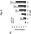
Figure 2 illustrates that menstrual blood-derived stem cells display high proliferation potential and CFU-F capacity. -
Figure 2(A) . MenSCs show high proliferation potential. In order to determine the proliferation potential, cells were cultured at 1000 cells/cm2 in a 24-well plate and the cellular mitochondrial dehydrogenase was quantified at different time points. Starting from 6-days post-culture, MenSCs showed significantly high proliferation kinetics when compared to BM-MSCs. -
Figure 2(B) . CFU-F capacity. Serial dilutions of a defined number of cells were cultured and their potential for the formation of CFU was evaluated. Only colonies formed by more than 50 fibroblast-like cells were assessed. MenSCs significantly generated more CFUs than BM-MSCs. -
Figure 2(C) . Relative CFU-F capacity. After 21 days of cell culture, colonies were evaluated according to crystal violet staining (absorbance, 620 nm) with the purpose of reflecting their potential growing. MenSCs showed similar colonies size than BM-MSC. Bars represent the SE of triplicate (*p ≤ 0.05). Abbreviations: CFU-F, colonies forming unit of fibroblast; SE, standard error. -
Figure 3 illustrates that menstrual blood-derived stem cells differentiate into mesodermal tissues. MenSCs have the ability to differentiate into adipose, bone and chondrocytes tissues after culture under appropriate differentiation media at varying degrees compared with BM-MSCs. -
Figure 3(A) . Adipocytic differentiation. Staining with oil red O for fat vacuoles showed lowest adipogenic differentiation in MenSCs respect to BM-MSC. -
Figure 3(B) . Osteocytic differentiation. Staining with alizarin red S for calcium deposits showed high differential potential of MenSCs compare to BM-MSCs. -
Figure 3(C) . Condrocytic differentiation. MenSCs stain positive with safranina O for sulfated proteoglycans. -
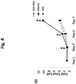
Figure 4 illustrates that MenSCs efficiently support the proliferation of CD34+ CD133+ hematopoietic stem cells. Cord blood CD34+CD133+ cells were immunomagneticaly isolated using CD34 beads, then cultured alone or co-cultured at a ratio of 1:4 to 1:5 in two different feeder layers from BM-MSCs and MenSCs, or without feeder (alone). Total number of cells and the expression of CD34+ CD133+ were evaluated at different time point during the course of the culture. Starting from 3 days post-culture, a statistically significant difference in favor of the co-culture with MenSCs was observed. By the end of the culture, a difference about 3 folds was observed when compared with cells cultured alone or with BM-MSCs. -
Figure 4(A) . Expansion of CD34+ cells cultured on feeder layers from BM.MSCs and MenSCs, or alone. -
Figure 4(B) . Total cells numbers fold increase compared to initial number of cells. -
Figure 4(C) . Expansion of CD34+CD133+ cells cultured on feeder layers from BM-MSCs and MenSCs, or alone. Bars represent the SE of triplicate (*p ≤ 0.05). -
Figure 4 (D and E). Illustrates the co-culture of CD34+CD133+ HSC with MenSCs under direct contact (MenSC) and non-contact conditions (MenSC-T), using a separating membrane, Transwell® (T). CD34+CD133+ were cultured under similar media condition with the difference of the presence or absence of the separating membrane, Transwell®. -
Figure 4(D) shows histogram representing the fold increase of total number of cells with or without the Transwell. -
Figure 4(E) shows the fold increase of CD34+CD133+ cells population. -
Figure 4(F) illustrates the fold increase of CD34+ cells population. - The following detailed description does not limit the scope of the invention to the examples and analysis described. While embodiments of the invention may be described, modifications, adaptations, and other implementations are possible. For example, substitutions, additions, or modifications may be made to the steps, equipments and compounds used in the methods described herein and may be modified by substituting, reordering, or adding stages equipments and compounds to the disclosed embodiments.
- While stages and methods are described in terms of "comprising" various compounds or steps, the methods can also consist of more steps or using more equipments and compounds, unless stated otherwise. Additionally, the terms "a," "an," and "the" are intended to include plural alternatives, unless stated otherwise specified.
- The mesenchymal stem cells obtained according to the present invention can be harvested from menstrual blood, which is a clear difference regarding the invasive procedures and the ethical controversy compared to embryonic stem cells related procedures.
- MenSCs display stem cell-like phenotypic markers and show great potential for self-renewal, plasticity, and undifferentiated proliferation for long periods of time in vitro. A large number of studies have shown that MenSCs are a population of pluripotent cells.
- Due to many limiting factors related to their practical usage in routine applications as detailed in the introductory section, there is an urge of creating a more accessible "product" that can fill the gap and as well as the supply and demand of an expanding stem cell therapy field. Supported by recently published data and our results, the present invention positions the cells isolated from menstrual fluids on the top of the list of cells candidates to fill up that clinical need.
- Several lines of MenSCs associated with novel regenerative properties have been isolated. Menstrual cells have been collected, isolated and cultured according to classic methods as described by Meng et al. 2007 and document
WO2008/109063 . - Those methods disclose the isolation and processing of menstrual cells, where isolation may be performed with at least one of the operations of centrifugation, density gradient sedimentation or filtration. The processing stages to obtain the menstrual cell include the steps of obtain, isolate and preserve the menstrual cells where menstrual cells can be used later fresh or cryopreserved.
- Thus, those processes show a source of stem cells, where viable menstrual cells are prepared for cryopreservation and are stored at cell banks being a more available source than cord blood cells, that is easily accessible, highly expandable in vitro and may be an alternative for autologous or heterologous stem cell applications.
- A detailed characterization of the MenSCs has been made for a complete comparison with related cells from other sources in order to allow the evaluation and comparative analysis of the advantages of MenSCs and also their safety/efficacy profile for clinical applications.
- The menstrual blood-derived stem cells morphology in culture is shown in
Figure 1(A) , having a fibroblast-like spindle-shaped morphology. - The morphology is maintained during the successive culture passages, and can be observed in freshly isolated menstrual stem cells (without passages or passage zero), after 24 hours of culture and after 2-week cell culture and a confluent culture at the third passage.
- In the present invention, it has been completed the full phenotypical and functional characterization of the MenSCs, while concentrating on their yet unexplored properties for the support of hematopoietic stem cells (CD34+ and CD133+) population.
- MenSCs have been shown to be positive for mesenchymal stem cell markers including CD9, CD29, CD105, and CD73, and negative for hematopoietic markers such as CD34, CD45 and CD133, as expected for those cell types.
- However, some groups have reported positive expression of embryonic markers such as SSEA-4 and Nanog in MenSCs, which were not found to be present on MSCs from other sources. This raises the question whether these cells presenting earlier markers of stemness represent a more primitive progenitor than MSCs from other sources, although there was a second group showing a different expression pattern in cells isolated and cultured under comparable conditions* (Meng et al. 2007).
- It was analyzed an exhaustive list of mesenchymal phenotypic markers on menstrual stem cells (MenSCs) from 3 different donors and they were compared to the phenotypic markers on MSCs isolated from the bone marrow (BM-MSCs). Additionally, these cells were further characterized, using a large battery of markers looking not only for mesenchymal and embryonic markers, but also for endothelial and epithelial markers, as other cell types might represent a source of contamination of the MenSC culture.
- These quality-control parameters are essential when comparing similar cells from different sources as shown in
Figure 1(B) . For this analysis, MenSCs and BM-MSCs were stained by FITC, APC, PE or PECy7-conjugated antibodies against mesenchymal stromal, hematopoietic and pluripotential stem cell markers. Viable cells were determined through the stain LIVE/DEAD® Fixable dead cell kit and then analyzed by flow cytometry, FC. - As shown in
Figure 1(C) , MenSCs display positive expression for mesenchymal stromal cells such as CD44, CD105, and CD73 while negative for CD14, CD34, CD45, HLA-DR, EPCAM, CD31, CD117, and CD271, and mildly positive expression for CD146. - In addition, flow cytometry analysis showed that MenSCs do not express pluripotent surface markers such as SSEA-3, SSEA-4, and TRA-1-60.
- The frequency of stem cell precursors, defined by their ability to form a cell colony in vitro (colony-forming unit, CFU) is another important property when characterizing a new stem cell source.
- While MSCs can be virtually isolated from many organs, their low frequency or lack of clonogenicity can be a limiting factor for their clinical application. For instance, MSCs isolated from umbilical cord blood have comparable differentiation properties as cells isolated from bone marrow (BM-MSC); however, these cells are found at a much lower rate than in the later case, hampering their therapeutically usage.
- It is thus essential to determine if MenSC possess a high or a low precursor frequency in comparison to MSCs isolated from other known sources. For this purpose cells were cultured at different serial dilution to determine their CFU-fibroblast frequency in comparison to BM-MSCs.
- As shown in
Figure 2 (B) , at 21 day post-culture, it was noted a significantly higher number of CFUs where the MenSCs were cultured indicating a higher frequency of stem/progenitor cells. - In addition, MenSCs have shown a higher proliferation rate compared to other sources, with doubling times of MenSCs within 18-24 hr depending on culture technique and media used (Meng et al., 2007).
- A good proliferation rate is essential for feasibility of clinical applications since stem cell-based therapies are usually efficient starting at a specific dose.
- In most human clinical trials, 1 million-cells/kg appears to be the dose of choice; however, when allogenic or repeated usage seems possible, escalating the yield of cultures can become of utmost importance.
- Nonetheless, a high proliferation is a two-edged sword that could also lead to genetic instability or the exhaustion of a specific stem cell pool. In fact, these MenSCs have been largely expanded in vitro without any mutation or visible abnormality at the chromosomal level. They maintained a telomerase activity greater than 50% even at
passage 12 compared with human embryonic stem cells (Patel et al., 2008), and also appear to mildly express the chemokine receptor CXCR4 and the respective receptor for stromal cell-derived factor-1 (SDF-1), which play a significant role in the mediation of mesenchymal stem cell migration (Patel et al., 2008). - More interestingly, according to the present invention, these MenSCs cells did not show any sign of stem cell exhaustion evidenced by a steady expression of all stromal stem markers, a stable proliferation rate and CFU potential when compared as shown in
Figure 2 (A) . Such a high proliferative rate in the face of genetic stability and apparent preservation of multipotency indicates MenSCs as a type of stem cell that could present unexpected therapeutic properties, a fact that is also implied by their extensive differentiation capabilities. - The ability of MenSCs to differentiate into adipose, bone, cartilage, cardiac, neural, hepatic and pancreatic cell types has been shown using standard differentiation techniques and media conditions.
- A recent study by (Hida et al., 2008)* using co-culture with fetal mouse cardiomyocytes went still further, evidencing immortalization mediated by human telomerase reverse transcriptase (hTERT) on MenSCs (Hida et al., 2008). They also demonstrated that spontaneous beating can occur upon cardiogenic differentiation.
- In addition, these multipotent cells have the ability to differentiate into various functional types, including respiratory epithelial cells, neurocytes, myocytes, endothelial cells, pancreatic cells, hepatocytes, adipocytes and osteocytes (Meng et al., 2007).
- For the present invention it has been investigated the mesodermal differentiation potential of MenSCs and has been compared to BM-MSCs. MenSCs show the ability to differentiate into adipose, bone and chondrocytes tissues after culture under appropriate differentiation media at varying degrees compared with BM-MSCs. They showed a lower adipogenic differentiation, an equal osteocytic differentiation and a higher chondrocytic differentiation as shown in the different staining as shown in
Figure 3 . - The supportive effect of MSCs on hematopoiesis has been well documented in many studies including a published work of the inventor and a patent application Khoury et al, 2011 and patent application
WO2010/138873 , where expanding human hematopoietic stem cells (HSCs) to increase their numbers while maintaining their stem cell properties has therefore become an important area of research. - MSCs isolated from the bone marrow (BM-MSCs) have been used as a support for HSC expansion in vitro in preclinical and clinical settings. However, the ability of MenSCs to support hematopoietic stem cells in vitro and in vivo has not been established yet. Thus, their usefulness in supporting hematopoietic stem cell engraftment is not as clear as with BM-MSCs.
- CD34+ CD133+ cells were immunomagneticaly isolated from human umbilical cords using CD34 beads, then cultured alone or co-cultured at a ratio of 1:4 to1:5 in different feeder layers from BM-MSCs and MenSCs. Total number of cells and the expression of CD34+ CD133+ were evaluated at different time point during the course of the culture. Starting from 3 days post-culture, a statistically significant different in favor of the co-culture with MenSCs was observed. By the end of the culture, a difference about 4 fold was observed for total cell numbers when compared with cells cultured alone or with BM-MSCs.
- More interestingly, it was observed a significant higher expansion of CD34+ CD133+ cells cultured on feeder layers from BM-MSCs, MenSCs, or alone as shown in
Figure 4 (C) . - The results clearly demonstrate that HSCs co-cultured with MenSCs in a growth factor-defined medium, show a high proliferation rate while maintaining their stem cell markers (CD34+ and CD133+).
- Compared with BM-MSCs, MenSCs elicit a more precocious early stem/progenitor formation and faster proliferation and differentiation of hematopoietic progenitors. This property to support hematopoiesis in vitro provides the rationale for their use in supporting hematopoietic reconstitution in clinical settings. The mechanism behind this superior yield is currently under investigation in our laboratory.
- It is also important to take into consideration parameters such as safety issues, in order to avoid any undesired immune reaction. This issue can be addressed by isolating the supporting MenSCs from the culture used as supporting layer.
- To determine whether the cell to cell contact is essential for the expansion of the CD34+ CD133+ population, the HSCs were co-cultured in the presence of MenSCs, in similar conditions, with the difference of the presence or absence of a separation membrane, denoted as T (Transwell®, Corning).
- At
day 7 of the expansion, cells were harvested, counted and then stained for the analysis of the different surface markers and analyzed by flow cytometry. - Expansion of HSC and HPC was significantly higher under MSC contact respect to noncontact growth conditions
- Menstrual blood is collected from a healthy female subject during the earliest days of a menstrual cycle. Samples are collected in a menstrual silicone cup. The cells are harvested with the informed consent of the donor as approved by an Institutional Review Board of Universidad de los Andes. Menstrual blood samples are transferred into a tube with phosphate buffered saline (PBS) containing amphotericin B, penicillin, streptomycin and EDTA.
- Menstrual blood mononuclear cells are separated by Ficoll-Paque density gradient and washed in PBS. Cells were subsequently cultured in a flask containing Dulbecco's modified Eagle's medium (DMEM) high Glucose supplemented with % penicillin/streptomycin, amphotericin B, glutamine and FBS at 35-40°C, using 2-10% CO2 in order to obtain adherent cells. Media were changed the next day to wash non adherent cells. Cells were replated as follows: adherent cells were detached using Trypsin-Edta, counted and sub-cultured. Afterwards, these cells were used as menstrual blood-derived stem cells (MenSCs).
- Bone marrow mesenchymal stem cells (BM-MSCs) were maintained in the same medium. Hematopoietic Stem Cells (HSCs) were collected from fresh umbilical cord blood after informed consent. Mononuclear cell were isolated after centrifugation on Ficoll-Paque. CD34+ cells were enriched with a CD34 Positive Selection Kit. Labeled cells are separated using a magnet separation kit.
- All the co-cultures are performed using MenSCs of different donors and BM-MSCs at early passages, no higher than 6 passages.
- MenSCs and BM-MSCs are seed in adipogenic differentiation medium: complete DMEM media, dexamethasone, 3-isobutyl-1-methyl-xanthine (IBMX), recombinant human insulin, and indomethacin; and cultured for up 21 days with media changes every 3-4 days.
- Control cells are cultured in completed DMEM media. Cells were subsequently stained with Oil Red O.
- Cells are seed in osteogenic differentiation medium: complete DMEM media, dexamethasone, b-glycerophosphate, and ascorbate; and cultured for up 21 days with media changes every 3-4 days. Control cells were cultured in completed DMEM media. Cells are later stained with Alizarin.
- Cells are seed in chondrogenic differentiation medium: complete DMEM media, dexamethasone, b-glycerophosphate, and ascorbate; and cultured for up 21 days with media changes every 3-4 days. Control cells are cultured in completed DMEM media and subsequently stained with Safranina O.
- MenSCs and BM-MSCs are cultured in a 24-well plate in complete DMEM media. Cell proliferation and viability is determined at different days measuring cellular mitochondrial dehydrogenase and quantified by spectrophotometry using absorbance 440 nm.
- Various independent experiments are performed in triplicate. The Student's t-test was used to calculate statistical difference. Data with a p value of p ≤ 0.05 is considered to be statistically significant.
- In order to quantify functional mesenchymal stem cells, MenSCs and BM-MSCs were evaluated for frequency of Colony-Forming Unit-Fibroblast (CFU-F). CFU-F were evaluated plating between 25-250 cells per well of a 6-well plate at five different densities in complete DMEM media, in triplicates.
- After 21 days, cells were stained with crystal violet in diluted methanol. After six washes, colonies formed by more than 50 fibroblast-like cells were counted under light microscope at low magnification. Results were expressed as CFU/number of cells plated. Then, cell-incorporated crystal violet was solubilized by incubation with diluted acetic acid and quantified by spectrophotometry at absorbance, 620 nm. The Student's t-test was used to calculate statistical difference. Data with a p value of p ≤ 0.05 was considered to be statistically significant.
- For cell surface antigen analysis, cells were harvested, washed with cytometer buffer (PBS + BSA + Sodium Azide) and incubated with the specific antibody for 20 min a 4°C in cytometer buffer containing the specific fluorescein isothiocynate (FITC), allophycocyanin (APC), phycoerythrin (PE), or phycoerythrin with cyanin-7 (PECy7) labeled antibodies. Antibodies for human cell surface antigens CD14, CD44, CD90, CD271, CD105, CD73, CD117, CD45, CD34, HLA-ABC, HLA-DR, CD146, EPCAM, CD31, TRA-1-60, CD49a, SSEA3, SSEA4, CD133 and CD3 were used.
- In the analysis, the corresponding isotype-matched antibodies are used as negative controls. In addition, a dead cell stain kits is used to determine the viability cells by flow cytometry. Data is collected and analyzed using FACS software.
- Confluent layers of MenSCs and BM-MSCs were irradiated (15Gy). CD34+ CD133+ hematopoietic stem cells are then co-cultured with feeder layers using StemSpan medium supplemented with human growth factors: FGF-1, SCF, TPO, IGFBP2, heparin and penicillin/streptomycin.
- Briefly, CD34+ CD133+cells are plated directly on the feeder layer or in Transwell above the feeder layer (noncontact growth conditions) in an appropriate ratio in growth factor-supplemented StemSpan media in 24-well plates. The medium was half changed every day and additionally supplemented. All the expanded cord blood cells were harvested after different days by vigorous pipetting, washed in PBS, and the cells total number were determined by trypan blue.
- In addition, cells were stained with CD3-APC, CD45-FITC, CD34-PECy7, CD133-PE, and a dead cell stain kit. Expression of surface markers was then analyzed by flow cytometry. Various independent analyses are performed. The Student's t-test was used to calculate statistical difference. Data with a p value of p ≤ 0.05 is considered to be statically significant.
- MSCs were seeded in 24 well plate and cultured on MenSC media prior to the coculture. MenSC media consists of DMEM-high glucose supplemented with FBS, penicillin streptomycin, amphotericin B and L-glutamine. The assays were preformed in triplicate. A fourth well was seeded for the cell quantification before radiation procedure. At
day 1, once the MSCs cultures are in confluent layers, the cells were irradiated (15 Gy). - HSCs CD34+CD133+ cells derived from umbilical cord blood were obtained using CD34 immunomagnetic purification. The HCS cells were thawed as follows:
- The cryotube was placed in a 30-40°C bath and cold DMEM media is slowly added to the tube (without using FBS). The cells were collected when adding the DMEM cold media. This procedure is repeated until recovering all the cells. Then, the mixture is centrifugated at room temperature. The supernatant is removed, resuspending the pellet on DMEM without supplemented FBS. Cells are quantified after adding trypan blue.
- HSCs are co-cultured over adherent monolayer MSCs at 1:4 ratio of HSC:MSC cells /well in a 24 well plat. The culture was feed with supplemented StemSpan® media. Each following day the media was fed removing the residual media, and then adding fresh media.
- The quantification of the HSCs cells is performed after resuspending the cells after carefully pipetting 10 times avoiding the disturbance of the MSCs layer. The HSCs cells are analyzed by FACS.
- The analysis is made on different days for FITC, APC, PE, PECy7, CD45, CD3, CD133, CD34 antibodies.
- Menstrual blood was collected in a menstrual silicone cup (Mialuna®, Santiago, Chile). Menstrual blood samples were transferred into a 50 ml tube with 10 ml phosphate buffered saline (PBS) containing 0.25 mg/mL amphotericin B (Gibco, Paisley, UK),
penicillin 100 IU,streptomycin 100 mg/mL and 2 mM EDTA (all from Sigma, USA). - Menstrual blood mononuclear cells were separated by Ficoll-Paque (Sigma-Aldrich, St Lois, USA) (1.077 g/ml) density gradient according to the manufacturer's instructions and washed in PBS. Cells were subsequently cultured in a T25 flask (Falcon®, Becton Dickinson) containing Dulbecco's modified Eagle's medium (DMEM) high Glucose (Gibco, Paisley, UK) supplemented with 1% penicillin/streptomycin, 1% amphotericin B, 1% glutamine (Gibco, Paisley, UK) and 15% FBS (Lonza, Walkersville, MD USA) at 37°C, 5% CO2 in order to obtain adherent cells.
- Media were changed the next day to wash non adherent cells. Cells were replated as follows: adherent cells were detached using 0.05% Trypsin-Edta (Gibco, UK), counted and sub-cultured. Afterwards, these cells were used as menstrual blood-derived stem cells (MenSCs).
- The cocultures were performed using MenSCs of 3 different donors and BM-MSCs at early passages (2 to 6 passages) using bone marrow mesenchymal stem cells (BM-MSCs) gifts from L. Salazar, Laboratorio Clínico, Cells for Cells, Chile; Hematopoietic Stem Cells (HSCs); and Mononuclear cell isolated after centrifugation on Ficoll-Paque. CD34+ cells were enriched with EasySep™ Human Cord Blood CD34 Positive Selection Kit (Stem Cell Technologies, Vancouver, Canada) according to the manufacturer's instructions. Labeled cells are separated using an EasySep™ magnet (Stem Cell Technologies).
- MenSCs and BM-MSCs were seeded at a concentration of 1,5x10E4 cells/cm2 in adipogenic differentiation medium: complete DMEM media, 1mM dexamethasone, 0.5 mM 3-isobutyl-1-methyl-xanthine (IBMX), 10ug/ml recombinant human insulin, and 100 mM indomethacin; and cultured for up 21 days with media changes every 3-4 days.
- Control cells were cultured in completed DMEM media. Cells were subsequently stained with Oil Red O (Sigma-Aldrich, St Lois, USA).
- Cells were seeded at a concentration of 2x10E4 cells/cm2 in osteogenic differentiation medium: complete DMEM media, 100nM dexamethasone, 10mM b-glycerophosphate, and 0.2MM ascorbate; and cultured for up 21 days with media changes every 3-4 days.
- Control cells were cultured in completed DMEM media. Cells were stained with Alizarin Red (Sigma-Aldrich, St Lois, USA).
- Cells were seeded at a concentration of 3x10E4 cells/10uL in chondrogenic differentiation medium: complete DMEM media, 100nM dexamethasone, 10mM b-glycerophosphate, and 0.2mM ascorbate; and cultured for up 21 days with media changes every 3-4 days.
- Control cells were cultured in completed DMEM media. Cells were subsequently stained with Safranina O (Sigma-Aldrich, St Lois, USA).
- MenSCs and BM-MSCs were cultured at 1000 cells/cm2 in a 24-well plate (Falcon®, Becton Dickinson) in complete DMEM media.
- Cell proliferation and viability were determined at
days - Three independent experiments were performed in triplicate
- In order to quantify functional mesenchymal stem cells, MenSCs and BM-MSCs were evaluated for frequency of Colony-Forming Unit-Fibroblast (CFU-F). CFU-F were evaluated plating between 25-250 cells per well of a 6-well plate (Falcon®, Becton Dickinson) at five different densities in complete DMEM media, in triplicates.
- After 21 days, cells were stained with 0.5% crystal violet (Sigma-Aldrich, St.Louis, MO) in 10% methanol for 20 minutes. After six washes, colonies formed by more than 50 fibroblast-like cells were counted under light microscope at low magnification.
- Results were expressed as CFU/number of cells plated. Then, cell-incorporated crystal violet was solubilized by incubation with acetic acid 33% and quantified spectrophotometrically (absorbance, 620 nm).
- For cell surface antigen analysis, cells were harvested, washed with cytometer buffer (PBS + 0.2% BSA + 0.01% Sodium Azide [all from Sigma-Aldrich, St Lois, USA]) and incubated with the specific antibody at concentrations recommended by the respective manufacturer for 20 min a 4°C in cytometer buffer containing the specific fluorescein isothiocynate (FITC), allophycocyanin (APC), phycoerythrin (PE), or phycoerythrin with cyanin-7 (PECy7) labeled antibodies.
- Antibodies for human cell surface antigens CD14, CD44, CD90, CD271, CD105, CD73, CD117, CD45, CD34, HLA-ABC, HLA-DR, CD146, EPCAM, CD31, TRA-1-60, CD49a, SSEA3, SSEA4, CD133 and CD3 were purchase from BD Pharmingen™ (BD Biosciences, San Jose, CA), R&D System (Minneapolis, MN, USA) and Biolegend (San Diego, CA, USA).
- In all experiments, the corresponding isotype-matched antibodies were used as negative controls. In addition, LIVE/DEAD®Fixable dead cell stain kits (Invitrogen) were used to determine the viability cells by flow cytometry according to the manufacturer's protocol. Data (10,000 events) were collected using a FACS Canto II Flow cytometer (BD Biosciences, San Jose, CA) and analyzed on FACS Diva software (BD Biosciences).
- Confluent layers of MenSCs and BM-MSCs were irradiated (15Gy) in the Chilean Commission for Nuclear Energy Facilities.
- CD34+ CD133+ hematopoietic stem cells were then co-culture with these feeder layers using StemSpan (Stem Cell Technologies, Vancouver, Canada) supplemented with human growth factors: 10 ng/ml FGF-1, 10 ng/ml SCF, 20 ng/ml TPO, 100 ng/ml IGFBP2 -all R&D systems (Minneapolis, MN, USA)-, 100 micrograms/ml of heparin and 1 x penicillin/streptomycin (Gibco, Paisley, UK).
- Briefly, CD34+ CD133+cells were plated directly on these feeder layer or in Transwell (0.4 µm pore size; BD Biosciences) above the feeder layer (noncontact growth conditions) in a 1:4-1:5 ratio in growth factor-supplemented StemSpan in a 24-well plates (Falcon®, Becton Dickinson), at 500 µl/well. The medium was half changed every day and additionally supplemented with 100 µl/well. All the expanded cord blood cells were harvested after 3, 5 and 7 days by vigorous pipetting, washed in PBS, and the cells total number were determined by trypan blue.
- In addition, cells were stained with CD3-APC, CD45-FITC, CD34-PECy7, CD133-PE, and LIVE/DEAD®Fixable dead cell stain. Expression of surface markers was then analyzed by flow cytometry. Three independent experiments were performed in triplicate. The Student's t-test was used to calculate statistical difference. A p value of p ≤ 0.05 was considered to be statically significant.
- MSCs were seeded in 24 well plate at a concentration of 5x10⁴cells/well. Cells were cultured on MenSC media prior to the coculture. MenSC media consists of DMEM-high glucose (Gibco #10313021) supplemented with 15% FBS, 1% penicillin streptomycin (Gibco #15140122), 1% amphotericin B (Biological #03-028-1B) and 1% L-glutamine (Gibco #25030-081). The assays were preformed in triplicate. A fourth well was seeded for the cell quantification before radiation procedure.
- At
day 1, once the MSCs cultures are in confluent layers, the cells were irradiated (15 Gy) in the Chilean Commission for Nuclear Energy Facilities, CCHEN. - HSCs CD34+CD133+ cells derived from umbilical cord blood were obtained using CD34 immunomagnetic purification. The HCS cells were thawed as follows:
- The cryotube was placed for 10" in a 37°C bath and 1ml of cold DMEM media was slowly added to the tube (without using FBS). The cells were collected when adding the DMEM cold media. This procedure is repeated until recovering all the cells. Then, the mixture is centrifugated 5' at 1200rpm at room temperature. The supernatant is removed, resuspending the pellet on DMEM without supplemented FBS. Cells are quantified after adding trypan blue.
- HSCs are cocultured over adherent monolayer MSCs at 1:4 and/or 1:5 ratio of HSC: MSC, using 104 HSC cells and 4x10⁴ MSC cells /well in a 24 well plat. The culture was feed with supplemented StemSpan® media (StemCell Technologies #9610) 500uL/well (initial volume). Each following day the media was fed removing the residual media over 200ml, and then adding 100ml of fresh media.
- The quantification of the HSCs cells is performed after resuspending the cells after carefully pipetting 10 times avoiding the disturbance of the MSCs layer. The HSCs cells are analyzed by FACS.
- The analysis is made on
days
Claims (12)
- A method for culturing and proliferating hematopoietic stem cells, comprising the steps of:(a) culturing hematopoietic stem cells expressing CD34+ and CD133+;(b) culturing menstrual stem cells obtained from a human donor during the earliest days of menstruation, said menstrual stem cells having positive expression for CD44, CD105 and CD73 and negative expression for CD14, CD34, CD45, HLA-DR, EPCAM, CD31, CD117, CD271, SSEA-3, SSEA-4 and TRA-1-60;(c) co-culturing the hematopoietic stem cells cultured in step (a) and the menstrual stem cells cultured in step (b), whereby the hematopoietic stem cells proliferate.
- The method of claim 1, wherein the cultured menstrual stem cells in step (b) are irradiated.
- The method of claim 2, wherein the cultured menstrual stem cells are irradiated with 15Gy.
- The method of claim 1, wherein the cultured menstrual stem cells in step (b) are cultured in supplemented DMEM-high glucose media.
- The method of claim 4, wherein DMEM-high glucose media is supplemented with 15% FBS, 1 % penicillin streptomycin, 1 % amphotericin B and 1 % L-glutamine.
- The method of claim 1, wherein the co-cultured cells of step (c) are cultured with supplemented culture media.
- The method of claim 6, wherein the supplemented culture media is supplemented with FGF-1 , SCF, TPO, IGFBP2, heparin and penicillin/streptomycin.
- The method of claim 7, wherein the supplemented media contains 10 ng/ml FGF-1, 10 ng/ml SCF, 20 ng/ml TPO, 100 ng/ml IGFBP2, 100 micrograms/ml of heparin and 1 x penicillin/streptomycin.
- The method of claim 1, wherein prior to culturing in step (a) the hematopoietic stem cells are obtained by the steps of:(i) collecting the hematopoietic stem cells from fresh umbilical cord blood;(ii) concentrating cells using antibody complexes and dextran-coated magnetic particles; and(iii) separating cells attached to the magnetic particles using high-gradient magnetic field.
- The method according claim 9, wherein the antibodies antibody complexes are antibodies to CD 34 and CD133.
- The method according claim 9, wherein the antibodies antibody complexes are antibodies to FITC, APC, PE, PECy7, CD45, CD3, CD133, CD34.
- The method of claim 1, wherein the menstrual stem cells are obtained by the steps of:(i) collecting the menstrual blood;(ii) washing the menstrual cells by adding phosphate buffered saline;(iii) separating the menstrual blood mononuclear cells through a Ficoll density gradient;(iv) washing the menstrual blood mononuclear cells by adding phosphate buffered saline;(v) culturing the menstrual blood mononuclear cells in supplemented media;(vi) removing non adherent cells;(vii) sub-culturing and cryo-preserving the adherent cells;(viii) reacting the adherent cells with antibodies to human cell surface antigens; and.(ix) isolating the cells bound to the antibody.
Applications Claiming Priority (1)
| Application Number | Priority Date | Filing Date | Title |
|---|---|---|---|
| PCT/IB2013/051861 WO2014135924A1 (en) | 2013-03-08 | 2013-03-08 | Menstrual stems cells for the efficient support and expansion of cd34+ cd133+ hematopoietic stem cells in vitro |
Publications (3)
| Publication Number | Publication Date |
|---|---|
| EP2964755A1 EP2964755A1 (en) | 2016-01-13 |
| EP2964755A4 EP2964755A4 (en) | 2016-08-17 |
| EP2964755B1 true EP2964755B1 (en) | 2017-11-01 |
Family
ID=51490683
Family Applications (1)
| Application Number | Title | Priority Date | Filing Date |
|---|---|---|---|
| EP13877167.0A Not-in-force EP2964755B1 (en) | 2013-03-08 | 2013-03-08 | Menstrual stems cells for the efficient support and expansion of cd34+ cd133+ hematopoietic stem cells in vitro |
Country Status (3)
| Country | Link |
|---|---|
| US (1) | US20160024473A1 (en) |
| EP (1) | EP2964755B1 (en) |
| WO (1) | WO2014135924A1 (en) |
Families Citing this family (7)
| Publication number | Priority date | Publication date | Assignee | Title |
|---|---|---|---|---|
| CN107629998A (en) * | 2017-08-24 | 2018-01-26 | 浙江大学 | A kind of stem cell in vitro reparation with menstrual blood source is damaged the model of endometrium |
| CN119776281A (en) * | 2018-06-13 | 2025-04-08 | 浙江生创精准医疗科技有限公司 | Immortalized Uterine Blood Stem Cell Line |
| CN110218698A (en) * | 2019-06-21 | 2019-09-10 | 东华理工大学 | A kind of preparation method and application of the people through hemocytoblast |
| CN111979199B (en) * | 2020-07-09 | 2023-08-01 | 浙江普慧医疗科技有限公司 | Uterine blood stem cells and exosomes for the treatment of uterine cavity adhesions |
| CN115386538A (en) * | 2021-05-24 | 2022-11-25 | 上海长一干细胞研究中心有限公司 | Method for separating, extracting and culturing endometrial stem cells from menstrual blood and special culture medium |
| CN115011556B (en) * | 2022-08-10 | 2022-10-25 | 山东省齐鲁干细胞工程有限公司 | Separation method of umbilical cord blood hematopoietic stem cells |
| WO2026020255A1 (en) * | 2024-07-26 | 2026-01-29 | Cells For Cells S.A. | Cd34+ cells modified with mesenchymal cell mitochondria |
Family Cites Families (12)
| Publication number | Priority date | Publication date | Assignee | Title |
|---|---|---|---|---|
| JPH07504331A (en) | 1992-03-04 | 1995-05-18 | システミックス,インコーポレイティド | Culture of hematopoietic stem cells and their genetic engineering |
| US5843633A (en) * | 1996-04-26 | 1998-12-01 | Amcell Corporation | Characterization of a human hematopoietic progenitor cell antigen |
| US20040143863A1 (en) * | 2003-01-10 | 2004-07-22 | Linheng Li | Hematopoietic stem cell niche cells |
| EP1842068A1 (en) * | 2004-06-21 | 2007-10-10 | Fondazione Santa Lucia I.R.C.C.S. | Use of multiparametric flow cytometry for the diagnosis, prognosis, and validation of immunotherapies in autoimmune, hematologic, and lymphoproliferative diseases |
| US20070041948A1 (en) | 2005-07-20 | 2007-02-22 | Seoul National University Industry Foundation | Method for culturing and proliferating hematopoietic stem cells and progenitor cells using human endometrial cells |
| JP2010530214A (en) * | 2007-03-01 | 2010-09-09 | クライオ−セル インターナショナル インコーポレイテッド | Acquisition, isolation and cryopreservation of endometrial / menstrual cells |
| AU2008319284A1 (en) * | 2007-10-31 | 2009-05-07 | Cryo-Cell International, Inc. | Methods for co-culturing cord blood derived cells with menstrual stem cells |
| US8160827B2 (en) | 2007-11-02 | 2012-04-17 | Emerson Climate Technologies, Inc. | Compressor sensor module |
| WO2010138873A1 (en) * | 2009-05-29 | 2010-12-02 | Maroun Khoury | Long term expansion of human hematopoietic stem cells |
| US20110306516A1 (en) * | 2010-06-15 | 2011-12-15 | The New York Stem Cell Foundation | Methods for producing induced pluripotent stem cells |
| WO2013007284A1 (en) * | 2011-07-08 | 2013-01-17 | Mikael Sigvardsson | A method for enriching mesenchymal stem cells and applications thereof |
| WO2013025189A1 (en) * | 2011-08-12 | 2013-02-21 | Cryo-Cell International, Inc. | Methods for co-culturing cord blood cells or cord tissue cells with menstrual multipotent cells |
-
2013
- 2013-03-08 WO PCT/IB2013/051861 patent/WO2014135924A1/en not_active Ceased
- 2013-03-08 EP EP13877167.0A patent/EP2964755B1/en not_active Not-in-force
- 2013-03-08 US US14/773,746 patent/US20160024473A1/en not_active Abandoned
Non-Patent Citations (1)
| Title |
|---|
| None * |
Also Published As
| Publication number | Publication date |
|---|---|
| WO2014135924A1 (en) | 2014-09-12 |
| US20160024473A1 (en) | 2016-01-28 |
| EP2964755A4 (en) | 2016-08-17 |
| EP2964755A1 (en) | 2016-01-13 |
Similar Documents
| Publication | Publication Date | Title |
|---|---|---|
| Maleki et al. | Comparison of mesenchymal stem cell markers in multiple human adult stem cells | |
| Zeddou et al. | The umbilical cord matrix is a better source of mesenchymal stem cells (MSC) than the umbilical cord blood | |
| Rojewski et al. | Phenotypic characterization of mesenchymal stem cells from various tissues | |
| Debnath et al. | Standardization and quality assessment for clinical grade mesenchymal stem cells from human adipose tissue | |
| Meng et al. | Endometrial regenerative cells: a novel stem cell population | |
| Lindroos et al. | Serum-free, xeno-free culture media maintain the proliferation rate and multipotentiality of adipose stem cells in vitro | |
| Patel et al. | Multipotent menstrual blood stromal stem cells: isolation, characterization, and differentiation | |
| EP1733027B1 (en) | Method for obtaining mesenchymal stem cells | |
| US20160024473A1 (en) | Menstrual stems cells for the efficient support and expansion of cd34+ cd133+ hematopoietic stem cells in vitro | |
| Saulnier et al. | Mesenchymal stromal cells multipotency and plasticity: induction toward the hepatic lineage. | |
| US20100105132A1 (en) | Human Mesenchymal stem cells and preparation thereof | |
| Zhu et al. | Adipose‐derived stem cell: a better stem cell than BMSC | |
| EP2630234B1 (en) | Human multipotent embryonic stem cell-like progenitor cells | |
| US20110014704A1 (en) | Isolation of multi-lineage stem cells | |
| CN107937338A (en) | A kind of mesodermal lineage mescenchymal stem cell from multipotential stem cell and preparation method thereof | |
| Yew et al. | Efficient expansion of mesenchymal stem cells from mouse bone marrow under hypoxic conditions | |
| TWI487788B (en) | Proliferation without tumorigenic pluripotent stem cells | |
| Issaragrisil | Isolation, characterization and neural differentiation potential of amnion derived mesenchymal stem cells | |
| US8796020B2 (en) | Manufacturing process for fresh and frozen stem cells | |
| CN113774020B (en) | A method for constructing adipose-derived mesenchymal stem cell bank | |
| CN100453640C (en) | A method for isolating multipotent adult progenitor cells from umbilical cord blood | |
| US9574175B2 (en) | Endometrial polyp stem cell | |
| CN101928694A (en) | A kind of establishment method of human amniotic fluid stem cell line | |
| Kögler | The unrestricted somatic stem cell (USSC) from cord blood for regenerative medicine | |
| Somasundaram | Isolation, characterization, and differentiation of endometrial stem cells |
Legal Events
| Date | Code | Title | Description |
|---|---|---|---|
| PUAI | Public reference made under article 153(3) epc to a published international application that has entered the european phase |
Free format text: ORIGINAL CODE: 0009012 |
|
| 17P | Request for examination filed |
Effective date: 20150908 |
|
| AK | Designated contracting states |
Kind code of ref document: A1 Designated state(s): AL AT BE BG CH CY CZ DE DK EE ES FI FR GB GR HR HU IE IS IT LI LT LU LV MC MK MT NL NO PL PT RO RS SE SI SK SM TR |
|
| AX | Request for extension of the european patent |
Extension state: BA ME |
|
| DAX | Request for extension of the european patent (deleted) | ||
| A4 | Supplementary search report drawn up and despatched |
Effective date: 20160720 |
|
| RIC1 | Information provided on ipc code assigned before grant |
Ipc: C12N 5/0789 20100101ALI20160714BHEP Ipc: C12N 5/0775 20100101ALI20160714BHEP Ipc: C12N 5/0787 20100101AFI20160714BHEP |
|
| GRAP | Despatch of communication of intention to grant a patent |
Free format text: ORIGINAL CODE: EPIDOSNIGR1 |
|
| INTG | Intention to grant announced |
Effective date: 20170531 |
|
| GRAS | Grant fee paid |
Free format text: ORIGINAL CODE: EPIDOSNIGR3 |
|
| GRAA | (expected) grant |
Free format text: ORIGINAL CODE: 0009210 |
|
| AK | Designated contracting states |
Kind code of ref document: B1 Designated state(s): AL AT BE BG CH CY CZ DE DK EE ES FI FR GB GR HR HU IE IS IT LI LT LU LV MC MK MT NL NO PL PT RO RS SE SI SK SM TR |
|
| REG | Reference to a national code |
Ref country code: GB Ref legal event code: FG4D |
|
| REG | Reference to a national code |
Ref country code: CH Ref legal event code: EP Ref country code: AT Ref legal event code: REF Ref document number: 942044 Country of ref document: AT Kind code of ref document: T Effective date: 20171115 |
|
| REG | Reference to a national code |
Ref country code: IE Ref legal event code: FG4D |
|
| REG | Reference to a national code |
Ref country code: DE Ref legal event code: R096 Ref document number: 602013028966 Country of ref document: DE |
|
| REG | Reference to a national code |
Ref country code: NL Ref legal event code: MP Effective date: 20171101 |
|
| REG | Reference to a national code |
Ref country code: FR Ref legal event code: PLFP Year of fee payment: 6 |
|
| REG | Reference to a national code |
Ref country code: LT Ref legal event code: MG4D |
|
| REG | Reference to a national code |
Ref country code: AT Ref legal event code: MK05 Ref document number: 942044 Country of ref document: AT Kind code of ref document: T Effective date: 20171101 |
|
| PG25 | Lapsed in a contracting state [announced via postgrant information from national office to epo] |
Ref country code: SE Free format text: LAPSE BECAUSE OF FAILURE TO SUBMIT A TRANSLATION OF THE DESCRIPTION OR TO PAY THE FEE WITHIN THE PRESCRIBED TIME-LIMIT Effective date: 20171101 Ref country code: FI Free format text: LAPSE BECAUSE OF FAILURE TO SUBMIT A TRANSLATION OF THE DESCRIPTION OR TO PAY THE FEE WITHIN THE PRESCRIBED TIME-LIMIT Effective date: 20171101 Ref country code: LT Free format text: LAPSE BECAUSE OF FAILURE TO SUBMIT A TRANSLATION OF THE DESCRIPTION OR TO PAY THE FEE WITHIN THE PRESCRIBED TIME-LIMIT Effective date: 20171101 Ref country code: ES Free format text: LAPSE BECAUSE OF FAILURE TO SUBMIT A TRANSLATION OF THE DESCRIPTION OR TO PAY THE FEE WITHIN THE PRESCRIBED TIME-LIMIT Effective date: 20171101 Ref country code: NL Free format text: LAPSE BECAUSE OF FAILURE TO SUBMIT A TRANSLATION OF THE DESCRIPTION OR TO PAY THE FEE WITHIN THE PRESCRIBED TIME-LIMIT Effective date: 20171101 Ref country code: NO Free format text: LAPSE BECAUSE OF FAILURE TO SUBMIT A TRANSLATION OF THE DESCRIPTION OR TO PAY THE FEE WITHIN THE PRESCRIBED TIME-LIMIT Effective date: 20180201 |
|
| PG25 | Lapsed in a contracting state [announced via postgrant information from national office to epo] |
Ref country code: GR Free format text: LAPSE BECAUSE OF FAILURE TO SUBMIT A TRANSLATION OF THE DESCRIPTION OR TO PAY THE FEE WITHIN THE PRESCRIBED TIME-LIMIT Effective date: 20180202 Ref country code: IS Free format text: LAPSE BECAUSE OF FAILURE TO SUBMIT A TRANSLATION OF THE DESCRIPTION OR TO PAY THE FEE WITHIN THE PRESCRIBED TIME-LIMIT Effective date: 20180301 Ref country code: BG Free format text: LAPSE BECAUSE OF FAILURE TO SUBMIT A TRANSLATION OF THE DESCRIPTION OR TO PAY THE FEE WITHIN THE PRESCRIBED TIME-LIMIT Effective date: 20180201 Ref country code: LV Free format text: LAPSE BECAUSE OF FAILURE TO SUBMIT A TRANSLATION OF THE DESCRIPTION OR TO PAY THE FEE WITHIN THE PRESCRIBED TIME-LIMIT Effective date: 20171101 Ref country code: RS Free format text: LAPSE BECAUSE OF FAILURE TO SUBMIT A TRANSLATION OF THE DESCRIPTION OR TO PAY THE FEE WITHIN THE PRESCRIBED TIME-LIMIT Effective date: 20171101 Ref country code: AT Free format text: LAPSE BECAUSE OF FAILURE TO SUBMIT A TRANSLATION OF THE DESCRIPTION OR TO PAY THE FEE WITHIN THE PRESCRIBED TIME-LIMIT Effective date: 20171101 Ref country code: HR Free format text: LAPSE BECAUSE OF FAILURE TO SUBMIT A TRANSLATION OF THE DESCRIPTION OR TO PAY THE FEE WITHIN THE PRESCRIBED TIME-LIMIT Effective date: 20171101 |
|
| PG25 | Lapsed in a contracting state [announced via postgrant information from national office to epo] |
Ref country code: SK Free format text: LAPSE BECAUSE OF FAILURE TO SUBMIT A TRANSLATION OF THE DESCRIPTION OR TO PAY THE FEE WITHIN THE PRESCRIBED TIME-LIMIT Effective date: 20171101 Ref country code: DK Free format text: LAPSE BECAUSE OF FAILURE TO SUBMIT A TRANSLATION OF THE DESCRIPTION OR TO PAY THE FEE WITHIN THE PRESCRIBED TIME-LIMIT Effective date: 20171101 Ref country code: CY Free format text: LAPSE BECAUSE OF FAILURE TO SUBMIT A TRANSLATION OF THE DESCRIPTION OR TO PAY THE FEE WITHIN THE PRESCRIBED TIME-LIMIT Effective date: 20171101 Ref country code: EE Free format text: LAPSE BECAUSE OF FAILURE TO SUBMIT A TRANSLATION OF THE DESCRIPTION OR TO PAY THE FEE WITHIN THE PRESCRIBED TIME-LIMIT Effective date: 20171101 Ref country code: CZ Free format text: LAPSE BECAUSE OF FAILURE TO SUBMIT A TRANSLATION OF THE DESCRIPTION OR TO PAY THE FEE WITHIN THE PRESCRIBED TIME-LIMIT Effective date: 20171101 |
|
| REG | Reference to a national code |
Ref country code: DE Ref legal event code: R097 Ref document number: 602013028966 Country of ref document: DE |
|
| PG25 | Lapsed in a contracting state [announced via postgrant information from national office to epo] |
Ref country code: SM Free format text: LAPSE BECAUSE OF FAILURE TO SUBMIT A TRANSLATION OF THE DESCRIPTION OR TO PAY THE FEE WITHIN THE PRESCRIBED TIME-LIMIT Effective date: 20171101 Ref country code: IT Free format text: LAPSE BECAUSE OF FAILURE TO SUBMIT A TRANSLATION OF THE DESCRIPTION OR TO PAY THE FEE WITHIN THE PRESCRIBED TIME-LIMIT Effective date: 20171101 Ref country code: PL Free format text: LAPSE BECAUSE OF FAILURE TO SUBMIT A TRANSLATION OF THE DESCRIPTION OR TO PAY THE FEE WITHIN THE PRESCRIBED TIME-LIMIT Effective date: 20171101 Ref country code: RO Free format text: LAPSE BECAUSE OF FAILURE TO SUBMIT A TRANSLATION OF THE DESCRIPTION OR TO PAY THE FEE WITHIN THE PRESCRIBED TIME-LIMIT Effective date: 20171101 |
|
| PLBE | No opposition filed within time limit |
Free format text: ORIGINAL CODE: 0009261 |
|
| STAA | Information on the status of an ep patent application or granted ep patent |
Free format text: STATUS: NO OPPOSITION FILED WITHIN TIME LIMIT |
|
| 26N | No opposition filed |
Effective date: 20180802 |
|
| REG | Reference to a national code |
Ref country code: CH Ref legal event code: PL |
|
| PG25 | Lapsed in a contracting state [announced via postgrant information from national office to epo] |
Ref country code: SI Free format text: LAPSE BECAUSE OF FAILURE TO SUBMIT A TRANSLATION OF THE DESCRIPTION OR TO PAY THE FEE WITHIN THE PRESCRIBED TIME-LIMIT Effective date: 20171101 Ref country code: MC Free format text: LAPSE BECAUSE OF FAILURE TO SUBMIT A TRANSLATION OF THE DESCRIPTION OR TO PAY THE FEE WITHIN THE PRESCRIBED TIME-LIMIT Effective date: 20171101 |
|
| REG | Reference to a national code |
Ref country code: BE Ref legal event code: MM Effective date: 20180331 |
|
| REG | Reference to a national code |
Ref country code: IE Ref legal event code: MM4A |
|
| PG25 | Lapsed in a contracting state [announced via postgrant information from national office to epo] |
Ref country code: LU Free format text: LAPSE BECAUSE OF NON-PAYMENT OF DUE FEES Effective date: 20180308 |
|
| PG25 | Lapsed in a contracting state [announced via postgrant information from national office to epo] |
Ref country code: IE Free format text: LAPSE BECAUSE OF NON-PAYMENT OF DUE FEES Effective date: 20180308 |
|
| PG25 | Lapsed in a contracting state [announced via postgrant information from national office to epo] |
Ref country code: CH Free format text: LAPSE BECAUSE OF NON-PAYMENT OF DUE FEES Effective date: 20180331 Ref country code: LI Free format text: LAPSE BECAUSE OF NON-PAYMENT OF DUE FEES Effective date: 20180331 Ref country code: BE Free format text: LAPSE BECAUSE OF NON-PAYMENT OF DUE FEES Effective date: 20180331 |
|
| PGFP | Annual fee paid to national office [announced via postgrant information from national office to epo] |
Ref country code: DE Payment date: 20190315 Year of fee payment: 7 Ref country code: GB Payment date: 20190315 Year of fee payment: 7 |
|
| PG25 | Lapsed in a contracting state [announced via postgrant information from national office to epo] |
Ref country code: MT Free format text: LAPSE BECAUSE OF NON-PAYMENT OF DUE FEES Effective date: 20180308 |
|
| PG25 | Lapsed in a contracting state [announced via postgrant information from national office to epo] |
Ref country code: TR Free format text: LAPSE BECAUSE OF FAILURE TO SUBMIT A TRANSLATION OF THE DESCRIPTION OR TO PAY THE FEE WITHIN THE PRESCRIBED TIME-LIMIT Effective date: 20171101 |
|
| PG25 | Lapsed in a contracting state [announced via postgrant information from national office to epo] |
Ref country code: PT Free format text: LAPSE BECAUSE OF FAILURE TO SUBMIT A TRANSLATION OF THE DESCRIPTION OR TO PAY THE FEE WITHIN THE PRESCRIBED TIME-LIMIT Effective date: 20171101 |
|
| PG25 | Lapsed in a contracting state [announced via postgrant information from national office to epo] |
Ref country code: HU Free format text: LAPSE BECAUSE OF FAILURE TO SUBMIT A TRANSLATION OF THE DESCRIPTION OR TO PAY THE FEE WITHIN THE PRESCRIBED TIME-LIMIT; INVALID AB INITIO Effective date: 20130308 Ref country code: MK Free format text: LAPSE BECAUSE OF NON-PAYMENT OF DUE FEES Effective date: 20171101 |
|
| PG25 | Lapsed in a contracting state [announced via postgrant information from national office to epo] |
Ref country code: AL Free format text: LAPSE BECAUSE OF FAILURE TO SUBMIT A TRANSLATION OF THE DESCRIPTION OR TO PAY THE FEE WITHIN THE PRESCRIBED TIME-LIMIT Effective date: 20171101 |
|
| REG | Reference to a national code |
Ref country code: DE Ref legal event code: R119 Ref document number: 602013028966 Country of ref document: DE |
|
| PG25 | Lapsed in a contracting state [announced via postgrant information from national office to epo] |
Ref country code: DE Free format text: LAPSE BECAUSE OF NON-PAYMENT OF DUE FEES Effective date: 20201001 |
|
| GBPC | Gb: european patent ceased through non-payment of renewal fee |
Effective date: 20200308 |
|
| PG25 | Lapsed in a contracting state [announced via postgrant information from national office to epo] |
Ref country code: GB Free format text: LAPSE BECAUSE OF NON-PAYMENT OF DUE FEES Effective date: 20200308 |
|
| PGFP | Annual fee paid to national office [announced via postgrant information from national office to epo] |
Ref country code: FR Payment date: 20240313 Year of fee payment: 12 |
|
| PG25 | Lapsed in a contracting state [announced via postgrant information from national office to epo] |
Ref country code: FR Free format text: LAPSE BECAUSE OF NON-PAYMENT OF DUE FEES Effective date: 20250331 |