EP2958589B1 - Caix stratification based cancer treatment - Google Patents
Caix stratification based cancer treatment Download PDFInfo
- Publication number
- EP2958589B1 EP2958589B1 EP14706543.7A EP14706543A EP2958589B1 EP 2958589 B1 EP2958589 B1 EP 2958589B1 EP 14706543 A EP14706543 A EP 14706543A EP 2958589 B1 EP2958589 B1 EP 2958589B1
- Authority
- EP
- European Patent Office
- Prior art keywords
- caix
- antibody
- score
- staining
- tumor cells
- Prior art date
- Legal status (The legal status is an assumption and is not a legal conclusion. Google has not performed a legal analysis and makes no representation as to the accuracy of the status listed.)
- Active
Links
Images


Classifications
-
- G—PHYSICS
- G01—MEASURING; TESTING
- G01N—INVESTIGATING OR ANALYSING MATERIALS BY DETERMINING THEIR CHEMICAL OR PHYSICAL PROPERTIES
- G01N33/00—Investigating or analysing materials by specific methods not covered by groups G01N1/00 - G01N31/00
- G01N33/48—Biological material, e.g. blood, urine; Haemocytometers
- G01N33/50—Chemical analysis of biological material, e.g. blood, urine; Testing involving biospecific ligand binding methods; Immunological testing
-
- G—PHYSICS
- G01—MEASURING; TESTING
- G01N—INVESTIGATING OR ANALYSING MATERIALS BY DETERMINING THEIR CHEMICAL OR PHYSICAL PROPERTIES
- G01N33/00—Investigating or analysing materials by specific methods not covered by groups G01N1/00 - G01N31/00
- G01N33/48—Biological material, e.g. blood, urine; Haemocytometers
- G01N33/50—Chemical analysis of biological material, e.g. blood, urine; Testing involving biospecific ligand binding methods; Immunological testing
- G01N33/53—Immunoassay; Biospecific binding assay; Materials therefor
- G01N33/575—Immunoassay; Biospecific binding assay; Materials therefor for cancer
- G01N33/57525—Immunoassay; Biospecific binding assay; Materials therefor for cancer of the liver or pancreas
-
- A—HUMAN NECESSITIES
- A61—MEDICAL OR VETERINARY SCIENCE; HYGIENE
- A61K—PREPARATIONS FOR MEDICAL, DENTAL OR TOILETRY PURPOSES
- A61K39/00—Medicinal preparations containing antigens or antibodies
- A61K39/395—Antibodies; Immunoglobulins; Immune serum, e.g. antilymphocytic serum
-
- A—HUMAN NECESSITIES
- A61—MEDICAL OR VETERINARY SCIENCE; HYGIENE
- A61K—PREPARATIONS FOR MEDICAL, DENTAL OR TOILETRY PURPOSES
- A61K9/00—Medicinal preparations characterised by special physical form
- A61K9/08—Solutions
-
- A—HUMAN NECESSITIES
- A61—MEDICAL OR VETERINARY SCIENCE; HYGIENE
- A61P—SPECIFIC THERAPEUTIC ACTIVITY OF CHEMICAL COMPOUNDS OR MEDICINAL PREPARATIONS
- A61P35/00—Antineoplastic agents
-
- C—CHEMISTRY; METALLURGY
- C07—ORGANIC CHEMISTRY
- C07K—PEPTIDES
- C07K16/00—Immunoglobulins [IG], e.g. monoclonal or polyclonal antibodies
- C07K16/18—Immunoglobulins [IG], e.g. monoclonal or polyclonal antibodies against material from animals or humans
- C07K16/28—Immunoglobulins [IG], e.g. monoclonal or polyclonal antibodies against material from animals or humans against receptors, cell surface antigens or cell surface determinants
- C07K16/30—Immunoglobulins [IG], e.g. monoclonal or polyclonal antibodies against material from animals or humans against receptors, cell surface antigens or cell surface determinants from tumour cells
-
- C—CHEMISTRY; METALLURGY
- C07—ORGANIC CHEMISTRY
- C07K—PEPTIDES
- C07K16/00—Immunoglobulins [IG], e.g. monoclonal or polyclonal antibodies
- C07K16/40—Immunoglobulins [IG], e.g. monoclonal or polyclonal antibodies against enzymes
-
- A—HUMAN NECESSITIES
- A61—MEDICAL OR VETERINARY SCIENCE; HYGIENE
- A61K—PREPARATIONS FOR MEDICAL, DENTAL OR TOILETRY PURPOSES
- A61K39/00—Medicinal preparations containing antigens or antibodies
- A61K2039/505—Medicinal preparations containing antigens or antibodies comprising antibodies
-
- C—CHEMISTRY; METALLURGY
- C07—ORGANIC CHEMISTRY
- C07K—PEPTIDES
- C07K2317/00—Immunoglobulins specific features
- C07K2317/20—Immunoglobulins specific features characterized by taxonomic origin
- C07K2317/24—Immunoglobulins specific features characterized by taxonomic origin containing regions, domains or residues from different species, e.g. chimeric, humanized or veneered
-
- G—PHYSICS
- G01—MEASURING; TESTING
- G01N—INVESTIGATING OR ANALYSING MATERIALS BY DETERMINING THEIR CHEMICAL OR PHYSICAL PROPERTIES
- G01N2333/00—Assays involving biological materials from specific organisms or of a specific nature
- G01N2333/90—Enzymes; Proenzymes
- G01N2333/988—Lyases (4.), e.g. aldolases, heparinase, enolases, fumarase
Definitions
- the present invention relates to an anti-CAIX (carbonic anhydrase IX) antibody for the use in the treatment of renal clear cell carcinoma (ccRCC), wherein the use comprises quantifying CAIX expression as well as the determination of a CAIX score based on the CAIX expression.
- CAIX carbonic anhydrase IX
- CAIX transmembrane protein carbonic anhydrase IX
- CAIX The transmembrane protein carbonic anhydrase IX (CAIX) is a member of the large family of carbonic anhydrase enzymes which share the ability to catalyze the reversible hydration of carbon dioxide to carbonic acid, leading to a decrease in pH ( Opavsky R, et al., Genomics 1996, 33: 480 487 ).
- Up-regulation of CAIX gene expression occurs in response to hypoxia via direct transcriptional activation by hypoxia inducible factor-1 alpha (HIF-1 ⁇ ), and is believed to be involved in sensing and maintaining the acidic environment of hypoxic cells, particularly within the hypoxic regions of tumors ( Wykoff et al., Cancer Res 2000, 60: 7075-7083 ; Svastova et al., FEBS Letters 2004 577: 439-445 .)
- HIF-1 ⁇ hypoxia inducible factor-1 alpha
- the G250 antigen is closely associated with numerous carcinomas such as renal cell carcinoma.
- the G250 antigen was first described as a kidney cancer-associated antigen ( WO 88/08854 ). Later, it was found to be identical with the tumor-associated antigen MN, a cell surface antigen with carbonic anhydrase activity, also referred to as CAIX.
- CAIX expression is found in gastric, intestinal and biliary mucosa, where its physiological role resides in pH regulation. Besides its normal expression pattern, CAIX expression is found in a variety of tumors including cervical, esophageal, colorectal, lung, pancreas, biliary and renal cell clear cell carcinomas (RCC).
- CRC renal cell clear cell carcinomas
- Antibodies against CAIX therefore, can be employed for cancer therapy.
- Anti-G250 antibodies are described, for example, in EP 637 336 , WO 93/18152 , WO 95/34650 , WO 00/24913 , WO 02/063010 , WO 04/025302 , WO 05/037083 , WO 2011/139375 and foreign counterparts thereof.
- WO 02/062972 describes a hybridoma cell line DSM ACC 2526 which produces the monoclonal antibody G250.
- the monoclonal antibody G250 recognizes an antigen preferably expressed on membranes of renal cell carcinoma cells (RCC), but not expressed in normal proximal tubular epithelium.
- the G250 antibody binds to the antigen G250, which is also called MN antigen (cf., for example, WO 93/18152 ) or CAIX (carbonic anhydrase IX).
- the CAIX score is determined, or calculated respectively, on the basis of CAIX expression described by a combination of tumor cell extent and staining intensity.
- the CAIX expression can be considered to be a measure of the antigen density preferably a measure of the CAIX polypeptide density within the tumor tissue of the patient suffering from the cancer to be treated wherein the cancer is ccRCC.
- the CAIX score is a value describing the amount of immunoreactivity of a marker such as CAIX in a sample.
- the score considers both staining intensity and the percentage of cells stained at a specific range of intensities.
- the score is determined by summing the products of the percentage cells stained at a given staining intensity (0-100) in a sample (such as a tissue section of a ccRCC cancer) and the staining intensity (0-3).
- a sample such as a tissue section of a ccRCC cancer
- the staining intensity (0-3).
- the score can be rendered for a specific compartment for example cytoplasm, membrane, nucleus.
- the CAIX score can take any value between 0 (no staining) and 3 (100% viable tumor cells with an intensity scaling of 3+).
- tumor cell extent or the amount of CAIX antigen expressing tumor cells is considered in the CAIX score formula according to the present invention by the indication of the percentage rate of viable tumor cells and their staining intensity.
- viable tumor cells may be counted or determined by methods known to a person skilled in the art. For example, the number of cells and/or the respective percentage rate may be determined by visual counting or computer-assisted programs such as an automatic imaging system.
- Tumor cell extent as part of the CAIX score is graded into a four-tired scaling factor (0, 1+, 2+ and 3+).
- the scaling factor 0 is derived from the CAIX staining comparable to control samples and represents lack of CAIX expression.
- the scaling factors 1+ to 3+ are derived from an average sample of tumor cells expressing CAIX, which show tumor cell staining from barely perceptible to strong.
- a histologist or pathologist of average skill in the art can make the classification by conventional analysis by eye ( Cregger et al., Arch. Pathol. Lab. Med., 2006, 130: 1026-1030 ).
- the absolute value of staining intensity may depend, e.g., on the applied staining reagent, the tumor tissue to be stained, etc.
- this problem can be overcome by using the above scaling system comprising grades +1 to +3.
- the staining intensity in tumor cells increases almost proportionally from barely perceptible staining to strong staining.
- the tumor cells expressing CAIX are divided into three groups having a scaling from 1+ to 3+, with the respective increase in staining intensity being the same for each group.
- the viable tumor cells are separated into cells with faint or barely perceptible staining intensity (scaling 1+), moderate staining intensity (2+) and strong staining intensity (3+).
- a score system Once a score system has been established for a specific staining agent and specific tumor cells in an average sample of tumor cells defining a reference system, it can be used for further samples of tumor cells, i.e. the respective CAIX score system does, of course, not have to be established for each sample of tumor cells.
- “Staining intensity” means the percentage of tumor cells within each of the four-tired scaling factor. The percentage can be any between 0% and 100%.
- the intensity of staining is determined as a percentage of tumor cells displaying faint or barely perceptible membrane staining for scaling +1, as a percentage of tumor cells displaying moderate membrane staining for scaling +2, as a percentage of tumor cells displaying moderate membrane staining for scaling +3 for each tissue sample (Table 1).
- Table 1 Staining intensity Scaling % of tumor cells displayed faint / barely perceptible membrane staining 1+ % of tumor cells displayed moderate membrane staining 2+ % of tumor cells displayed strong membrane staining 3+
- the classification of tumor cells expressing CAIX in the respective scaling group can easily be made by comparing the staining intensity of the sample with the reference tumor sample on which the CAIX scoring system was based. Thus, it is possible that samples of tumor cells do not include all three scaling groups.
- a person of average skill in the art knows methods for determining staining intensity, either for defining a score system or for comparing different samples of cells or classifying cells into a score system for the purpose of comparing the staining intensity (cf. Cregger et al., 1026, Arch. Pathol. Lab. Med., vol. 130, July 2006 ).
- the determination of the staining intensity or the classification into a score system may be carried out manually or via a computer-assisted program or any other suitable method.
- the score system is applied using standardized control tumor assays in combination with a device that provides quantitative and objective output.
- the CAIX score is based on the CAIX expression which has to be determined.
- CAIX expression and CAIX protein expression can be used interchangeable.
- CAIX expression is related to the amount of CAIX protein expressed.
- Treatment of ccRCC comprises administering a therapeutically effective amount of an anti-CAIX antibody to the patient if the patient's ccRCC has been found to have a CAIX score of equal to or greater than 2.6.
- CAIX is quantified, if any, present in one or more samples derived from a subject to be treated.
- the sample derived from the subject to be treated is a tissue sample of the ccRCC to be treated.
- CAIX is quantified by immunohistochemical staining, preferably immunohistochemical staining wherein CAIX protein or CAIX polypeptide is specifically bound by a monoclonal antibody, more preferably by a monoclonal antibody with an epitope binding domain at the N-terminus of CAIX, even more preferably by the monoclonal antibody M75, most preferably by the monoclonal antibody that is secreted from the hybridoma VU-M75 which has accession No. ATCC HB 11128.
- a person skilled in the art knows suitable methods of immunohistochemical staining methods.
- Kits for immunohistochemical staining are commercially available and include but are not limited to the CAIX IHC Kit offered by Oncogene Science 100 Acorn Park Drive, Cambridge, MA 02140, USA. Another example for a method of immunohistochemical staining is, e.g., described in EP 1 501 939 B1 .
- Immunohistochemical staining of tissue sections can be done with anti-CAIX antibody using a peroxidase technique with antigen retrieval using heat treatment as described by Dako staining systems (Dako Corporation, Carpenteria, CA).
- the antibodies used in the immunoassay and/or immunohistochemical staining may be polyclonal or monoclonal. Preferably, monoclonal antibodies are used.
- Labelled detection means including labels that may comprise fluorescent means, enzymes, co-enzymes, radionucleotides, chemiluminescent means, enzyme substrates, particles, dyes, etc.
- the respective assays include enzyme immunoassays, fluorescent immunoassays, radioimmunoassays, ELISAS, etc.
- Detection antibodies of use in the immunohistochemical staining tissue include monoclonal antibodies M75, MN9, MN12 , MN7.
- a detection antibody that is preferably used according to the present invention is M75.
- the M75 antibody is further described in US 5,981,711 and is part of the CAIX IHC Kit offered by Oncogene Science (see supra). Particularly preferred are staining methods which provide measurable and/or comparable results such as dye intensity, fluorescent intensity, radio signals, enzyme activity, etc.
- the expressed CAIX comprises a CAIX polypeptide or fragment of CAIX polypeptide.
- the expressed CAIX comprises an mRNA that encodes a CAIX polypeptide or fragment of CAIX polypeptide.
- methods to determine the amount of mRNA in the sample of a cell such as Northern Blot, microassays, real-time PCR etc.
- the subject is to be treated if the CAIX score in the tumor sample provided from the subject is ⁇ 2.6.
- An analysis for all possible CAIX scores from 0.0 to 3.0 reveals that the more the CAIX score increases the more significant the treatment effect is.
- the patients having a CAIX score of ⁇ 2.6 show a statistically significant treatment effect when treated with an anti-CAIX antibody, in particular an anti-G250 antibody.
- the tumor sample provided from the subject is at least one tissue sample derived from the ccRCC of the subject to be treated.
- the anti-CAIX antibody specifically interacts with a CAIX polypeptide. Specifically interacting with (e.g.
- recognising or binding to means that the anti-CAIX antibody has a greater affinity to CAIX than to other polypeptides.
- the anti-CAIX antibody interacts with (i.e. binds to or recognizes) or modulates the activity of CAIX polypeptide and/or mediates an antibody dependent cell cytotoxicity (ADCC) and/or complement mediated cytotoxicity (CDC).
- ADCC antibody dependent cell cytotoxicity
- CDC complement mediated cytotoxicity
- the antibody is a CAIX inhibitor acting on the protein level
- CAIX inhibitor acting on the protein level
- Examples of anti-CAIX antibodies or antibodies against CAIX are described in EP 637 336 , WO 93/18152 , WO 95/34650 , WO 00/24913 , WO 02/063010 , WO 04/025302 , WO 05/037083 , WO 2011/139375 , Murri-Plesko et al., Eur J Pharmacol 2011, 657: 173-183 .
- the antibody is selected from the group consisting of polyclonal antibodies, monoclonal antibodies, antigen-binding fragments thereof such as F(ab') 2 , Fab', sFv, dsFv and chimerized, humanized and fully human variants thereof.
- this anti-CAIX antibody or epitope-binding fragment thereof binds to the amino acid sequence LSTAFARV and/or ALGPGREYRAL.
- the CAIX targeting compound is the antibody anti-G250 and/or an epitope-binding fragment thereof.
- Anti-G250 antibodies are, e.g., described in EP-B-0 637 336 . It is particularly preferred that the anti-tumor antibody is chimeric or humanised G250 antibody and/or a fragment thereof.
- the antibodies for use in the present invention may be produced by any suitable method known in the art including but not limited by methods as described in PCT/EP02/01282 and PCT/EP02/01283 .
- An especially preferred antibody is cG250, preferably girentuximab (INN).
- Another especially preferred embodiment is the monoclonal antibody G250 produced by the hybridoma cell line DSM ACC 2526.
- the antibody cG250 is an IgG1 kappa light chain chimeric version of an originally murine monoclonal antibody mG250.
- the cancer to be treated is a ccRCC which is characterised by expressing CAIX.
- the cancer is characterised by having a CAIX score ⁇ 2.6 as set out above.
- renal clear cell carcinoma is treated by administering the antibody G250, and in particular cG250, preferably girentuximab.
- the subject to be treated has been diagnosed with a non-metastatic type of cancer or non-metastatic disease, more preferably with a non-metastatic type of cancer or non-metastatic disease or/and has to be diagnosed as having a high risk of recurrence.
- the anti-CAIX antibody and in particular the anti-G250 antibody, is to be used after the subject to be treated has undergone a primary tumor resection, preferably a tumor nephrectomy, more preferably a tumor nephrectomy and lymphadenectomy.
- this primary tumor is a G250 antigen-expressing renal clear cell carcinoma (ccRCC).
- the anti-CAIXantibody and in particular the anti-G250 antibody, is used as an adjuvant therapy.
- adjuvant therapy describes a way to target any remaining cancer cells that cannot be seen.
- Adjuvant therapies are usually used after primary treatments such as surgery or radiation to guard any cancer recurrences.
- An example of adjuvant therapy is the additional treatment usually given after surgery where all detectable disease has been removed, but where there remains a statistical risk of relapse due to occult disease.
- Types of adjuvant therapy comprise chemotherapy, hormone therapy, radiation therapy and/or immunotherapy.
- radiotherapy or systemic therapy is commonly given as adjuvant treatment after surgery for breast cancer.
- Systemic therapy may consist of chemotherapy, immunotherapy or biological response modifiers or hormone therapy.
- an adjuvant therapy may be combined with any of these types of known adjuvant therapies.
- a further aspect of the present document relates to a method for diagnosing, prognosing and/or classifying a cancer disease comprising
- the method of diagnosing may further comprise the determination of a CAIX score as described above.
- the cancer to be diagnosed and/or classified is a cancer as described above, namely renal clear cell carcinoma.This cancer has a CAIX score ⁇ 2.6.
- the prognosis includes the use of the CAIX score to predict whether a patient to be treated would respond to treatment with an anti-CAIX antibody or not, in particular to treatment with an anti-G250 antibody. It is particularly used to predict if a patient would respond to treatment with the cG250 antibody, preferably with girentuximab. In other words it can be prognosed which patients will probably benefit from a treatment with CAIX targeting compounds. For example, patients having renal clear cell carcinoma, wherein the corresponding tumor cell sample shows a CAIX score ⁇ 2.6 will most probably benefit from a treatment with the cG250 antibody, preferably girentuximab.
- T3/T4 Patients with a high T-stage (T3/T4), a low grading (G1/G2), no lymph node involvement (NO/NX) and no metastatic disease (M0) have a worse prognosis with a low CAIX score as compared to patients with a high CAIX score.
- a further aspect of the present document relates to a method for treating ccRCC, said method comprising
- the anti-CAIX antibody and in particular the anti-G250 antibody may be administered as monotherapy protocol.
- the anti-CAIX antibody, and in particular the anti-G250 antibody, preferably the cG250 antibody, more preferably girentuximab may also be administered in a combination therapy protocol or as a combination therapy, e.g., as already outlined above, an adjuvant anti-G250 antibody treatment may be combined with any other type of adjuvant therapy.
- a cytokine may be co-administered together with the antibody to increase antibody-dependent cellular cytotoxicity (ADCC) and/or to activate the immune system of the patient, e.g., the NK cells.
- This cytokine may be selected from the group consisting of interleukins such as IL-2, -3, -4, -5, -6, -7, -8, -9, -10, -12, -13, - 14, and -15, interferon, e.g., IFN- ⁇ , IFN- ⁇ , and IFN- ⁇ , TFN- ⁇ , TNF- ⁇ , nerve growth factor, ligands of CD40, Fas, CD27 and CD30, macrophage-inhibiting protein, Rantes, active fragments of pharmaceutically acceptable analogues and derivatives, and mixtures thereof. More preferably, the cytokine is selected from IL-2 and IFN- ⁇ .
- further cytotoxic means e.g, doxorubicin, cis-platin or carboplatin and other neoplastic means, is possible.
- the artisan of ordinary skill can determine the dosage of the anti-CAIX antibody used, and in particular G250-antigen-specific antibody, based on age, weight, condition and the severity of the disease, for example.
- a dosing regimen of either 20 mg or 50 mg cG250 antibody, for example, per patient on a weekly cycle, will deliver concentrations above 0.5 ⁇ g/ml preferably 1 ⁇ g/ml and therefore should be adequate for efficacy. Therefore, a weekly dose of the G250-specific antibody of 5-250 mg/week, preferably 10-100 mg/week, and more preferably 20 mg/week to 50 mg/week can be administered.
- the G250 antigen-specific antibody and/or antibody fragment thereof can be administered to a subject in need thereof in at least two treatment stages in which different, preferably decreasing amounts of the antibody are administered.
- the above described method may comprise the administration of a G250-antigen-specific antibody or/and an antibody fragment thereof to a subject in need thereof in two stages, wherein
- the first treatment stage comprises administration of 50 mg/week of the G250-specific antibody
- the second treatment stage comprises administration of 20 mg/week.
- the antitumor antibody is preferably administered intravenously, preferably by infusion or intravenous injections.
- the administration of the antibody by infusion is preferably performed in up to about 30 minutes, more preferably in about 15 minutes.
- the CAIX targeting compound can also be applied intraperitoneally or intramuscularly.
- the first treatment stage comprises up to 12 weeks, preferably up to 6 weeks, even more preferably up to one week and the second treatment stage comprises up to 156 weeks, preferably up to 104 weeks, more preferably up to 52 weeks, even more preferably up to 12-24 weeks.
- the first treatment stage comprises up to one week and the administration of a single loading dose of 50 mg/week of cG250 antibody
- the second treatment stage comprises up to 24 weeks and the administration of 20 mg/week of cG250 antibody for the treatment of renal clear cell carcinoma.
- the first treatment stage may comprise up to one week and the administration of a single loading dose of 50 mg/week of cG250 antibody intravenously
- the second treatment stage may comprise up to 24 weeks and the administration of 20 mg/week of cG250 antibody for the treatment of renal clear cell carcinoma
- the second treatment stage comprises at least 8 consecutive intravenous administration of 20 mg/week of cG250 antibody for the treatment of renal clear cell carcinoma.
- the anti-CAIX antibody in particular the cG250 antibody, may, of course, be administered in a pharmaceutical composition.
- This pharmaceutically acceptable composition may comprise acceptable carriers, diluents and/or adjuvants.
- carrier when used herein includes carriers, excipients and/or stabilisers that are non-toxic to the cell or mammal being exposed thereto at the dosages and concentrations employed. Often the physiologically acceptable carriers are aqueous pH buffered solutions or liposomes.
- physiologically acceptable carriers include buffers such as phosphate, citrate and other organic acids; anti-oxidants including ascorbic acid, low molecular weight (less than about 10 residues) polypeptides; proteins such as serum albumin, gelatine or immunoglobulins; hydrophilic polymers such as polyvinyl pyrrolidone; amino acids such as glycine, glutamine, asparagine, arginine or lysine; monosaccharides, disaccharides, and other carbohydrates including glucose, mannose or dextrins, gelating agents such as EDTA, sugar, alcohols such as mannitol or sorbitol; salt-forming counterions such as sodium; and/or non-ionic surfactants such as TWEEN, polyethylene or polyethylene glycol.
- buffers such as phosphate, citrate and other organic acids
- anti-oxidants including ascorbic acid, low molecular weight (less than about 10 residues) polypeptides
- proteins such as serum albumin, gelatine or
- the anti-CAIX antibody is administered in normal saline (0.9% sterile sodium chloride in water).
- the antibody cG250 is administered in normal saline by intravenous infusion, e.g., 100 ml solution over 15 min.
- carbonic anhydrase IX and "CAIX”, “CA9”, “MN” and “G250” are herein considered to be synonymous.
- Antigen refers to a ligand that can be bound by e.g. an antibody.
- the portions of the antigen that makes contact with the antibody are denominated “epitopes”.
- staining refers to any method which makes tumor cells visible.
- the “staining” method used according to the invention is immunohistochemical staining as described above.
- the objectives in this study were to evaluate disease free survival (DFS) on girentuximab therapy as compared to placebo and to evaluate overall survival (OS) on girentuximab therapy as compared to placebo.
- DFS disease free survival
- OS overall survival
- Girentuximab (cG250; an IgG1 kappa light chain chimeric version of an original murine monoclonal antibody mG250) was applied in a single loading dose of 50 mg (week 1) followed by weekly infusions of 20 mg of girentuximab (weeks 2-24).
- the drug was diluted in 100 ml normal saline (0.9% sterile sodium chloride in water) and was applied by intravenous infusion over 15 minutes.
- the placebo group received phosphate-buffered saline with polysorbate 20 (Tween® 20) without active ingredient. Duration of treatment was 24 weeks.
- Paraffin embedded nephrectomized tissue specimens were cut into section of 3-5 ⁇ m and collected on respective adhesive slides. Deparaffinisation and dehydration was done with EZ Prep mixture from Ventana Medical Systems, Inc.. The tissue material was blocked and stained with a solution of the diluted antibody (CAIX M75, 1:150). The antibody was detected using a biotinylated IgG anti-mouse IgG antibody (Ventana Medical Systems, Inc.) followed by conjugated streptavidin horseradish peroxidase (Ventana Medical Systems, Inc.). The signal detection was done with diaminobenzidine and hydrogen peroxide (Ventana Medical Systems, Inc.). Positive and negative tissue controls were included as a reference.
- the efficacy evaluation was primarily analysed for the Intent-to-treat (ITT) population consisting of all 864 patients randomised. In addition the analysis was repeated for the Per Protocol (PP) Population consisting of 766 patients. Patients who received at least eight consecutive administrations of study medication (week 1 to 8) and had no major protocol deviation as defined in the deviation criteria prior to unblinding were evaluated as Per Protocol (PP) Population.
- ITT Intent-to-treat
- PP Per Protocol
- the sample size calculation was performed using the program Pass 2002. Statistical summaries were produced using SAS® software version 8.1 or higher. For the efficacy analysis, incomplete/partial dates were imputed; missing safety dates were not imputed. Adverse events and medical histories were coded using the Medical Dictionary for Regulatory Activities; medications were coded using the World Health Organisation Drug Dictionary. The primary efficacy analysis was based on the Intent-to-treat (ITT) Population and was repeated using the Per Protocol Population.
- the potential effect of prognostic factors on both DFS and OS was investigated using the Cox proportional hazards model.
- the hazard ratio (girentuximab treatment versus placebo) was estimated together with its associated 95% confidence interval and p-value using a proportional hazards model.
- the 151 patients in the ITT population with a high CAIX score of ⁇ 2.6 were evenly distributed between the cG250 and the placebo arm.
Landscapes
- Health & Medical Sciences (AREA)
- Chemical & Material Sciences (AREA)
- Life Sciences & Earth Sciences (AREA)
- Immunology (AREA)
- Medicinal Chemistry (AREA)
- Organic Chemistry (AREA)
- General Health & Medical Sciences (AREA)
- Molecular Biology (AREA)
- Engineering & Computer Science (AREA)
- Biochemistry (AREA)
- Proteomics, Peptides & Aminoacids (AREA)
- Biophysics (AREA)
- Genetics & Genomics (AREA)
- Cell Biology (AREA)
- Veterinary Medicine (AREA)
- Public Health (AREA)
- Pharmacology & Pharmacy (AREA)
- Urology & Nephrology (AREA)
- Animal Behavior & Ethology (AREA)
- Biomedical Technology (AREA)
- Hematology (AREA)
- Microbiology (AREA)
- Epidemiology (AREA)
- Analytical Chemistry (AREA)
- Physics & Mathematics (AREA)
- Food Science & Technology (AREA)
- General Physics & Mathematics (AREA)
- Pathology (AREA)
- Chemical Kinetics & Catalysis (AREA)
- Biotechnology (AREA)
- General Chemical & Material Sciences (AREA)
- Nuclear Medicine, Radiotherapy & Molecular Imaging (AREA)
- Mycology (AREA)
- Bioinformatics & Cheminformatics (AREA)
- Peptides Or Proteins (AREA)
- Medicines Containing Antibodies Or Antigens For Use As Internal Diagnostic Agents (AREA)
- Investigating Or Analysing Biological Materials (AREA)
- Medicines That Contain Protein Lipid Enzymes And Other Medicines (AREA)
- Measuring Or Testing Involving Enzymes Or Micro-Organisms (AREA)
- Gastroenterology & Hepatology (AREA)
Description
- The present invention relates to an anti-CAIX (carbonic anhydrase IX) antibody for the use in the treatment of renal clear cell carcinoma (ccRCC), wherein the use comprises quantifying CAIX expression as well as the determination of a CAIX score based on the CAIX expression.
- The transmembrane protein carbonic anhydrase IX (CAIX) is a member of the large family of carbonic anhydrase enzymes which share the ability to catalyze the reversible hydration of carbon dioxide to carbonic acid, leading to a decrease in pH (Opavsky R, et al., Genomics 1996, 33: 480 487). Up-regulation of CAIX gene expression occurs in response to hypoxia via direct transcriptional activation by hypoxia inducible factor-1 alpha (HIF-1α), and is believed to be involved in sensing and maintaining the acidic environment of hypoxic cells, particularly within the hypoxic regions of tumors (Wykoff et al., Cancer Res 2000, 60: 7075-7083; Svastova et al., FEBS Letters 2004 577: 439-445.)
- The G250 antigen is closely associated with numerous carcinomas such as renal cell carcinoma. The G250 antigen was first described as a kidney cancer-associated antigen (
WO 88/08854 - Normal CAIX expression is found in gastric, intestinal and biliary mucosa, where its physiological role resides in pH regulation. Besides its normal expression pattern, CAIX expression is found in a variety of tumors including cervical, esophageal, colorectal, lung, pancreas, biliary and renal cell clear cell carcinomas (RCC).
- Antibodies against CAIX, therefore, can be employed for cancer therapy. Anti-G250 antibodies are described, for example, in
EP 637 336 WO 93/18152 WO 95/34650 WO 00/24913 WO 02/063010 WO 04/025302 WO 05/037083 WO 2011/139375 and foreign counterparts thereof. Further,WO 02/062972 WO 93/18152 - A press release from Wilex AG on 16-10-2012 reported the results of the Phase III ARISER trial wherein Girentuximab was used to treat clear cell renal cell carcinoma (ccRCC). The trial did not meet its primary endpoint. Methods for determining CAIX expression are known to a person skilled in the art and include but are not limited to immunohistochemical staining of tissue sections which are e.g. described in Bui et al., Clinical Cancer Research 2003, 9: 802-811, Atkins et al., Clin. Cancer Res 2005, 11: 3714-3721,
US 5,989,838 ,US 6,004,535 ,US 6,093,548 ,US 7,524,634 ,US 7,851,455 . Such methods may be used in human renal clear cell carcinoma prognosis. - It was the object of the present invention to provide methods, which make it possible to characterise a CAIX-associated cancer disease further and predict how efficient or successful treatment with a CAIX targeting compound would be. According to the present invention, this object is achieved by the use of a specific CAIX score in the treatment, and classification of cancer as defined in the claims.
- Thus, the present invention relates to an anti-CAlX (carbonic anhydrase IX) antibody for use in the treatment of renal clear cell carcinoma (ccRCC), wherein the use comprises quantifying CAIX expression in at least one tumor sample provided from a subject to be treated, determining a CAIX score based on the CAIX expression in the tumor sample, and the subject is treated if a CAIX score ≥ 2.6 is reached, wherein CAIX expression is quantified by immunohistochemical staining and wherein the CAIX score is determined and/or calculated by the formula:

- In terms of the present invention the CAIX score is determined, or calculated respectively, on the basis of CAIX expression described by a combination of tumor cell extent and staining intensity. In other words, the amount/percentage of CAIX antigen expressing tumor cells as well as the staining intensity of the stained tumor cells are both considered in the CAIX score used herein. The CAIX expression can be considered to be a measure of the antigen density preferably a measure of the CAIX polypeptide density within the tumor tissue of the patient suffering from the cancer to be treated wherein the cancer is ccRCC. The CAIX score is a value describing the amount of immunoreactivity of a marker such as CAIX in a sample. The score considers both staining intensity and the percentage of cells stained at a specific range of intensities. The score is determined by summing the products of the percentage cells stained at a given staining intensity (0-100) in a sample (such as a tissue section of a ccRCC cancer) and the staining intensity (0-3). Preferably the score can be rendered for a specific compartment for example cytoplasm, membrane, nucleus.
- According to the invention, the CAIX score is determined and/or calculated by the Formula

- The CAIX score can take any value between 0 (no staining) and 3 (100% viable tumor cells with an intensity scaling of 3+).
- In the context of the present invention "tumor cell extent" or the amount of CAIX antigen expressing tumor cells is considered in the CAIX score formula according to the present invention by the indication of the percentage rate of viable tumor cells and their staining intensity. The viable tumor cells may be counted or determined by methods known to a person skilled in the art. For example, the number of cells and/or the respective percentage rate may be determined by visual counting or computer-assisted programs such as an automatic imaging system.
- Tumor cell extent as part of the CAIX score is graded into a four-tired scaling factor (0, 1+, 2+ and 3+). The
scaling factor 0 is derived from the CAIX staining comparable to control samples and represents lack of CAIX expression. Thescaling factors 1+ to 3+ are derived from an average sample of tumor cells expressing CAIX, which show tumor cell staining from barely perceptible to strong. Usually a histologist or pathologist of average skill in the art can make the classification by conventional analysis by eye (Cregger et al., Arch. Pathol. Lab. Med., 2006, 130: 1026-1030). The absolute value of staining intensity may depend, e.g., on the applied staining reagent, the tumor tissue to be stained, etc. However, inter alia, this problem can be overcome by using the above scaling system comprising grades +1 to +3. - The staining intensity in tumor cells increases almost proportionally from barely perceptible staining to strong staining. Along this linear increase of staining intensity the tumor cells expressing CAIX are divided into three groups having a scaling from 1+ to 3+, with the respective increase in staining intensity being the same for each group. In other words, the viable tumor cells are separated into cells with faint or barely perceptible staining intensity (scaling 1+), moderate staining intensity (2+) and strong staining intensity (3+).
- Once a score system has been established for a specific staining agent and specific tumor cells in an average sample of tumor cells defining a reference system, it can be used for further samples of tumor cells, i.e. the respective CAIX score system does, of course, not have to be established for each sample of tumor cells.
- "Staining intensity" according to the present invention means the percentage of tumor cells within each of the four-tired scaling factor. The percentage can be any between 0% and 100%. The intensity of staining is determined as a percentage of tumor cells displaying faint or barely perceptible membrane staining for scaling +1, as a percentage of tumor cells displaying moderate membrane staining for scaling +2, as a percentage of tumor cells displaying moderate membrane staining for scaling +3 for each tissue sample (Table 1).
Table 1: Staining intensity Scaling % of tumor cells displayed faint / barely perceptible membrane staining 1+ % of tumor cells displayed moderate membrane staining 2+ % of tumor cells displayed strong membrane staining 3+ - The classification of tumor cells expressing CAIX in the respective scaling group can easily be made by comparing the staining intensity of the sample with the reference tumor sample on which the CAIX scoring system was based. Thus, it is possible that samples of tumor cells do not include all three scaling groups.
- A person of average skill in the art knows methods for determining staining intensity, either for defining a score system or for comparing different samples of cells or classifying cells into a score system for the purpose of comparing the staining intensity (cf. Cregger et al., 1026, Arch. Pathol. Lab. Med., vol. 130, July 2006). The determination of the staining intensity or the classification into a score system may be carried out manually or via a computer-assisted program or any other suitable method. In a preferred embodiment, the score system is applied using standardized control tumor assays in combination with a device that provides quantitative and objective output.
- An example for calculating the CAIX score for 100% CAIX antigen extent expression with 40% moderate and 60% strong staining intensity will be shown in the following. Further examples for different extents of CAIX expression, a different content of staining intensity and the respective scores are shown in Table 2.
- 100% CAIX antigen extent expression with 40% moderate & 60%
strong staining intensity 
Table 2 extent CAIX expression 1+ 2+ 3+ Score 100 0 40 60 2.60 100 10 60 30 2.20 100 5 25 70 2.65 100 50 30 20 1.70 100 0 10 90 2.90 10 0 100 0 0.20 10 100 0 0 0.10 50 100 0 0 0.50 50 30 30 40 1.05 50 50 30 20 0.85 50 20 70 10 0.95 50 70 20 10 0.70 - According to the present invention the CAIX score is based on the CAIX expression which has to be determined. Herein, the terms CAIX expression and CAIX protein expression can be used interchangeable. Preferably, the term CAIX expression is related to the amount of CAIX protein expressed.
- Treatment of ccRCC comprises administering a therapeutically effective amount of an anti-CAIX antibody to the patient if the patient's ccRCC has been found to have a CAIX score of equal to or greater than 2.6.
Thereby expressed CAIX is quantified, if any, present in one or more samples derived from a subject to be treated. Preferably, the sample derived from the subject to be treated is a tissue sample of the ccRCC to be treated. - Expressed CAIX is quantified by immunohistochemical staining, preferably immunohistochemical staining wherein CAIX protein or CAIX polypeptide is specifically bound by a monoclonal antibody, more preferably by a monoclonal antibody with an epitope binding domain at the N-terminus of CAIX, even more preferably by the monoclonal antibody M75, most preferably by the monoclonal antibody that is secreted from the hybridoma VU-M75 which has accession No. ATCC HB 11128. A person skilled in the art knows suitable methods of immunohistochemical staining methods. Kits for immunohistochemical staining are commercially available and include but are not limited to the CAIX IHC Kit offered by
Oncogene Science 100 Acorn Park Drive, Cambridge, MA 02140, USA. Another example for a method of immunohistochemical staining is, e.g., described inEP 1 501 939 B1US 5,981,711 and is part of the CAIX IHC Kit offered by Oncogene Science (see supra). Particularly preferred are staining methods which provide measurable and/or comparable results such as dye intensity, fluorescent intensity, radio signals, enzyme activity, etc. - According to a preferred embodiment, the expressed CAIX comprises a CAIX polypeptide or fragment of CAIX polypeptide. According to another preferred embodiment, the expressed CAIX comprises an mRNA that encodes a CAIX polypeptide or fragment of CAIX polypeptide. A person skilled in the art knows methods to determine the amount of mRNA in the sample of a cell, such as Northern Blot, microassays, real-time PCR etc.
- According to the invention, the subject is to be treated if the CAIX score in the tumor sample provided from the subject is ≥ 2.6. An analysis for all possible CAIX scores from 0.0 to 3.0 reveals that the more the CAIX score increases the more significant the treatment effect is. The patients having a CAIX score of ≥ 2.6 show a statistically significant treatment effect when treated with an anti-CAIX antibody, in particular an anti-G250 antibody. Preferably the tumor sample provided from the subject is at least one tissue sample derived from the ccRCC of the subject to be treated. The anti-CAIX antibody specifically interacts with a CAIX polypeptide. Specifically interacting with (e.g. recognising or binding to) means that the anti-CAIX antibody has a greater affinity to CAIX than to other polypeptides. In one embodiment the anti-CAIX antibody interacts with (i.e. binds to or recognizes) or modulates the activity of CAIX polypeptide and/or mediates an antibody dependent cell cytotoxicity (ADCC) and/or complement mediated cytotoxicity (CDC). Thus, according to one embodiment, the antibody is a CAIX inhibitor acting on the protein level Examples of anti-CAIX antibodies or antibodies against CAIX are described in
EP 637 336 WO 93/18152 WO 95/34650 WO 00/24913 WO 02/063010 WO 04/025302 WO 05/037083 WO 2011/139375 , Murri-Plesko et al., Eur J Pharmacol 2011, 657: 173-183. - In a preferred embodiment, the antibody is selected from the group consisting of polyclonal antibodies, monoclonal antibodies, antigen-binding fragments thereof such as F(ab')2, Fab', sFv, dsFv and chimerized, humanized and fully human variants thereof. According to a further preferred embodiment, this anti-CAIX antibody or epitope-binding fragment thereof binds to the amino acid sequence LSTAFARV and/or ALGPGREYRAL.
- According to a further particularly preferred embodiment, the CAIX targeting compound is the antibody anti-G250 and/or an epitope-binding fragment thereof. Anti-G250 antibodies are, e.g., described in
EP-B-0 637 336 . It is particularly preferred that the anti-tumor antibody is chimeric or humanised G250 antibody and/or a fragment thereof. The antibodies for use in the present invention may be produced by any suitable method known in the art including but not limited by methods as described inPCT/EP02/01282 andPCT/EP02/01283 . - An especially preferred antibody is cG250, preferably girentuximab (INN). Another especially preferred embodiment is the monoclonal antibody G250 produced by the hybridoma cell line DSM ACC 2526. The antibody cG250 is an IgG1 kappa light chain chimeric version of an originally murine monoclonal antibody mG250.
- The cancer to be treated is a ccRCC which is characterised by expressing CAIX. In particular, the cancer is characterised by having a CAIX score ≥ 2.6 as set out above.
- In the most preferred embodiment of the present invention renal clear cell carcinoma is treated by administering the antibody G250, and in particular cG250, preferably girentuximab.
- It is preferred that the subject to be treated has been diagnosed with a non-metastatic type of cancer or non-metastatic disease, more preferably with a non-metastatic type of cancer or non-metastatic disease or/and has to be diagnosed as having a high risk of recurrence.
- According to a further embodiment, the anti-CAIX antibody, and in particular the anti-G250 antibody, is to be used after the subject to be treated has undergone a primary tumor resection, preferably a tumor nephrectomy, more preferably a tumor nephrectomy and lymphadenectomy. Preferably, this primary tumor is a G250 antigen-expressing renal clear cell carcinoma (ccRCC).
- Accordingly, in a further preferred embodiment the anti-CAIXantibody, and in particular the anti-G250 antibody, is used as an adjuvant therapy. In terms of the present invention, adjuvant therapy describes a way to target any remaining cancer cells that cannot be seen. Adjuvant therapies are usually used after primary treatments such as surgery or radiation to guard any cancer recurrences. An example of adjuvant therapy is the additional treatment usually given after surgery where all detectable disease has been removed, but where there remains a statistical risk of relapse due to occult disease. Types of adjuvant therapy comprise chemotherapy, hormone therapy, radiation therapy and/or immunotherapy. For example, radiotherapy or systemic therapy is commonly given as adjuvant treatment after surgery for breast cancer. Systemic therapy may consist of chemotherapy, immunotherapy or biological response modifiers or hormone therapy. Preferably, if the anti-CAIX antibody according to the present invention is used, an adjuvant therapy may be combined with any of these types of known adjuvant therapies.
- A further aspect of the present document relates to a method for diagnosing, prognosing and/or classifying a cancer disease comprising
- (a) quantifying the expression of CAIX in a cancer sample by staining and
- (b) classifying the cancer disease according to the extent and staining intensity of stained cancer cells.
- The method of diagnosing may further comprise the determination of a CAIX score as described above.
- The cancer to be diagnosed and/or classified is a cancer as described above, namely renal clear cell carcinoma.This cancer has a CAIX score ≥ 2.6.
- The prognosis includes the use of the CAIX score to predict whether a patient to be treated would respond to treatment with an anti-CAIX antibody or not, in particular to treatment with an anti-G250 antibody. It is particularly used to predict if a patient would respond to treatment with the cG250 antibody, preferably with girentuximab. In other words it can be prognosed which patients will probably benefit from a treatment with CAIX targeting compounds. For example, patients having renal clear cell carcinoma, wherein the corresponding tumor cell sample shows a CAIX score ≥ 2.6 will most probably benefit from a treatment with the cG250 antibody, preferably girentuximab.
- Patients with a high T-stage (T3/T4), a low grading (G1/G2), no lymph node involvement (NO/NX) and no metastatic disease (M0) have a worse prognosis with a low CAIX score as compared to patients with a high CAIX score.
- A further aspect of the present document relates to a method for treating ccRCC, said method comprising
- (a) defining the expression of a CAIX in a cancer cell derived from ccRCC obtained from a subject to be treated by staining,
- (b) determining a CAIX score according to the extent and staining intensity of the cancer cells and
- (c) administering an anti-CAIX antibody if in step (b) a predefined CAIX threshold value has been exceeded.
- The anti-CAIX antibody and in particular the anti-G250 antibodymay be administered as monotherapy protocol.
- The anti-CAIX antibody, and in particular the anti-G250 antibody, preferably the cG250 antibody, more preferably girentuximab may also be administered in a combination therapy protocol or as a combination therapy, e.g., as already outlined above, an adjuvant anti-G250 antibody treatment may be combined with any other type of adjuvant therapy. A cytokine may be co-administered together with the antibody to increase antibody-dependent cellular cytotoxicity (ADCC) and/or to activate the immune system of the patient, e.g., the NK cells. This cytokine may be selected from the group consisting of interleukins such as IL-2, -3, -4, -5, -6, -7, -8, -9, -10, -12, -13, - 14, and -15, interferon, e.g., IFN-α, IFN-β, and IFN-γ, TFN-α, TNF-β, nerve growth factor, ligands of CD40, Fas, CD27 and CD30, macrophage-inhibiting protein, Rantes, active fragments of pharmaceutically acceptable analogues and derivatives, and mixtures thereof. More preferably, the cytokine is selected from IL-2 and IFN-α. Of course, a combination with further cytotoxic means, e.g, doxorubicin, cis-platin or carboplatin and other neoplastic means, is possible.
- The artisan of ordinary skill can determine the dosage of the anti-CAIX antibody used, and in particular G250-antigen-specific antibody, based on age, weight, condition and the severity of the disease, for example. A dosing regimen of either 20 mg or 50 mg cG250 antibody, for example, per patient on a weekly cycle, will deliver concentrations above 0.5 µg/ml preferably 1 µg/ml and therefore should be adequate for efficacy. Therefore, a weekly dose of the G250-specific antibody of 5-250 mg/week, preferably 10-100 mg/week, and more preferably 20 mg/week to 50 mg/week can be administered.
- The G250 antigen-specific antibody and/or antibody fragment thereof can be administered to a subject in need thereof in at least two treatment stages in which different, preferably decreasing amounts of the antibody are administered.
- The above described method may comprise the administration of a G250-antigen-specific antibody or/and an antibody fragment thereof to a subject in need thereof in two stages, wherein
- (a) a dose of 10-250 mg/week, preferably 20-100 mg/week, more preferably 30-100 mg/week, even more preferably 30-55 mg/week and most preferably 50 mg/week of the G250-antigen-specific antibody is administered in the first treatment stage and
- (b) a dose of 5-100 mg, preferably 10-50 mg, more preferably 15-25 mg, most preferably 20 mg/week of the G250-antigen-specific antibody is administered in the second treatment stage.
- It is even more preferred that the first treatment stage comprises administration of 50 mg/week of the G250-specific antibody, and the second treatment stage comprises administration of 20 mg/week.
- The antitumor antibody is preferably administered intravenously, preferably by infusion or intravenous injections. The administration of the antibody by infusion is preferably performed in up to about 30 minutes, more preferably in about 15 minutes. Of course, the CAIX targeting compound can also be applied intraperitoneally or intramuscularly.
- Dosing schemes with weekly infusions of either 20 or 50 mg of cG250 antibody for up to 20 weeks appear to be well tolerated and do not lead to significant HACA development.
- It is therefore preferred that the first treatment stage comprises up to 12 weeks, preferably up to 6 weeks, even more preferably up to one week and the second treatment stage comprises up to 156 weeks, preferably up to 104 weeks, more preferably up to 52 weeks, even more preferably up to 12-24 weeks.
- Most preferably, the first treatment stage comprises up to one week and the administration of a single loading dose of 50 mg/week of cG250 antibody, and the second treatment stage comprises up to 24 weeks and the administration of 20 mg/week of cG250 antibody for the treatment of renal clear cell carcinoma.
- The first treatment stage may comprise up to one week and the administration of a single loading dose of 50 mg/week of cG250 antibody intravenously, and the second treatment stage may comprise up to 24 weeks and the administration of 20 mg/week of cG250 antibody for the treatment of renal clear cell carcinoma, preferably the second treatment stage comprises at least 8 consecutive intravenous administration of 20 mg/week of cG250 antibody for the treatment of renal clear cell carcinoma.
- The anti-CAIX antibody, in particular the cG250 antibody, may, of course, be administered in a pharmaceutical composition. This pharmaceutically acceptable composition may comprise acceptable carriers, diluents and/or adjuvants. The term "carrier" when used herein includes carriers, excipients and/or stabilisers that are non-toxic to the cell or mammal being exposed thereto at the dosages and concentrations employed. Often the physiologically acceptable carriers are aqueous pH buffered solutions or liposomes. Examples of physiologically acceptable carriers include buffers such as phosphate, citrate and other organic acids; anti-oxidants including ascorbic acid, low molecular weight (less than about 10 residues) polypeptides; proteins such as serum albumin, gelatine or immunoglobulins; hydrophilic polymers such as polyvinyl pyrrolidone; amino acids such as glycine, glutamine, asparagine, arginine or lysine; monosaccharides, disaccharides, and other carbohydrates including glucose, mannose or dextrins, gelating agents such as EDTA, sugar, alcohols such as mannitol or sorbitol; salt-forming counterions such as sodium; and/or non-ionic surfactants such as TWEEN, polyethylene or polyethylene glycol.
- In one aspect, the anti-CAIX antibody is administered in normal saline (0.9% sterile sodium chloride in water).
- According to a particular aspect, the antibody cG250 is administered in normal saline by intravenous infusion, e.g., 100 ml solution over 15 min.
- The term "carbonic anhydrase IX" and "CAIX", "CA9", "MN" and "G250" are herein considered to be synonymous.
- "cG250" and girentuximab are used interchangeable.
- "Antigen" refers to a ligand that can be bound by e.g. an antibody. The portions of the antigen that makes contact with the antibody are denominated "epitopes".
- If not mentioned otherwise, "staining" as used herein refers to any method which makes tumor cells visible. The "staining" method used according to the invention is immunohistochemical staining as described above.
-
-
Figure 1 : HR (hazard ratio) and p-values for increasing CAIX-score values for the ITT (intended to treat) and PP populations (per protocol population) -
Figure 2 : DFS by treatment arm (cG250 vs. Placebo) for CAIX cut-off of 2.6 -
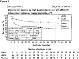
Figure 3 : DFS curve for PP population with CAIX scores ≥ 2.6 -
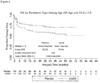
Figure 4 : High CAIX Score (≥2.0) and Age <65 years (ITT population) - The following results are based on a randomised double blind Phase III study to evaluate adjuvant cG250 treatment versus placebo in patients with renal clear cell carcinoma (ccRCC) and high risk of recurrence.
- The objectives in this study were to evaluate disease free survival (DFS) on girentuximab therapy as compared to placebo and to evaluate overall survival (OS) on girentuximab therapy as compared to placebo.
- 864 patients (ITT) were enrolled in a prospective, two armed, randomized, double blinded, placebo controlled study. The evaluation of the disease free survival was assessed by an independent Radiological Review Committee. 855 patients received at least one study treatment (cG250 or placebo) and were analysed for safety by an Independent Data Monitoring Committee. Inclusion criteria for the patients were: age ≥ 18 years, prior (partial or total) nephrectomy of primary renal cell carcinoma with documented clear cell histology, no evidence of macroscopic and microscopic residual disease. Patients should have one of the following High Risk Group (RG) (referring to TNM classification, 6th edition UICC, 2002):
- ∘ RG I: T3aN0/XM0 or T3bN0/XM0 or T3cN0/XM0 or T4N0/XM0
- ∘ RG II: any T stage and N+ disease and M0
- ∘ RG III: T1bN0/XM0 or T2N0/XM0, each with grading G≥3(Fuhrman or any other nuclear grading system with at least 3 grades),
not more than 12 weeks between date of nephrectomy and randomisation, ECOG of 0 to 1 - Girentuximab (cG250; an IgG1 kappa light chain chimeric version of an original murine monoclonal antibody mG250) was applied in a single loading dose of 50 mg (week 1) followed by weekly infusions of 20 mg of girentuximab (weeks 2-24). The drug was diluted in 100 ml normal saline (0.9% sterile sodium chloride in water) and was applied by intravenous infusion over 15 minutes. The placebo group received phosphate-buffered saline with polysorbate 20 (Tween® 20) without active ingredient. Duration of treatment was 24 weeks.
- Paraffin embedded nephrectomized tissue specimens were cut into section of 3-5µm and collected on respective adhesive slides. Deparaffinisation and dehydration was done with EZ Prep mixture from Ventana Medical Systems, Inc.. The tissue material was blocked and stained with a solution of the diluted antibody (CAIX M75, 1:150). The antibody was detected using a biotinylated IgG anti-mouse IgG antibody (Ventana Medical Systems, Inc.) followed by conjugated streptavidin horseradish peroxidase (Ventana Medical Systems, Inc.). The signal detection was done with diaminobenzidine and hydrogen peroxide (Ventana Medical Systems, Inc.). Positive and negative tissue controls were included as a reference.
- The efficacy evaluation was primarily analysed for the Intent-to-treat (ITT) population consisting of all 864 patients randomised. In addition the analysis was repeated for the Per Protocol (PP) Population consisting of 766 patients. Patients who received at least eight consecutive administrations of study medication (
week 1 to 8) and had no major protocol deviation as defined in the deviation criteria prior to unblinding were evaluated as Per Protocol (PP) Population. - The sample size calculation was performed using the program Pass 2002. Statistical summaries were produced using SAS® software version 8.1 or higher. For the efficacy analysis, incomplete/partial dates were imputed; missing safety dates were not imputed. Adverse events and medical histories were coded using the Medical Dictionary for Regulatory Activities; medications were coded using the World Health Organisation Drug Dictionary. The primary efficacy analysis was based on the Intent-to-treat (ITT) Population and was repeated using the Per Protocol Population.
- Continuous variables were described using: number of observations, arithmetic mean, standard deviation, minimum, median and maximum. Categorical variables were presented using the number of observations and percentages. Hierarchical testing was applied for DFS and OS to keep the global significance level to ≤ 5% for both. Both DFS and OS were compared between the girentuximab arm and the placebo arm using the log-rank test and the Kaplan-Meier method. The significance levels for OS were adjusted using the O'Brien-Fleming approach for group sequential methods with an overall significance level of 5%. 95% confidence intervals for proportions were calculated using the exact method (Pearson-Clopper).
- The potential effect of prognostic factors on both DFS and OS was investigated using the Cox proportional hazards model. The hazard ratio (girentuximab treatment versus placebo) was estimated together with its associated 95% confidence interval and p-value using a proportional hazards model.
- At the time of the cut-off for data analysis 360 patients (41.7%) had experienced a DFS event and 504 (58.3%) were still without disease according to local investigator assessment. 0.6% of patients were locally assessed as having metastatic disease at baseline. Overall 181 patients had died. After the IRRC central reading assessment, a DFS event could be assigned for 389 patients (45%) whereas a censor date could be assigned for the remaining 475 (55%) patients. The number of DFS events (293 excluding patients with metastatic disease at baseline) that occurred in the ITT population was comparable between the treatment arms (cG250: 142, 32.8%; Placebo: 151, 35.0%), as was the rate of metastases at baseline (DFS=0), seen in 11.5% of girentuximab patients and 10.7% of placebo patients.
- The primary analysis for DFS based on the IRRC evaluation for the ITT population showed no statistically significant difference in overall median DFS between the treatment arms (hazard ratio [HR]=0.999, p=0.737). The median DFS for girentuximab was 71.4 months and the median DFS for the placebo group was not reached.
- There was no statistically significant difference in DFS between treatment arms, regardless of High Risk Group (RG) classification (RG I: HR=0.934, p=0.596; RG II: HR=1.73, p=0.084; RG III: HR=0.984, p=0.627).
- In addition, exploratory analyses done using a classification based on the CAIX antigen expression alone according to Bui et al. (Bui et al., Clinical Cancer Research 2003, 9: 802-811) have not delivered statistical significant results neither for the prognosis nor for the treatment effect. However, surprisingly the combination of the CAIX antigen expression with the intensity of the staining, the CAIX score, which can be considered as a measure of the antigen density, showed significant results for the treatment effect as well as for the prognosis.
- Multivariate analysis for the CAIX score cut-offs (1.91 for ITT and 1.52 for ITT Placebo) derived from survival tree analyses show that the CAIX score is a prognostic, but not independent factor for prognosis in DFS ITT, OS ITT and OS Placebo. The results suggest the CAIX score cut-off of 1.52 (ITT Placebo) may be useful for prognosis.
- For patients with a low CAIX score the Kaplan-Meier-Curves for the different treatment arms overlap independently of the cut-off used. This clearly shows that patients with a low CAIX score do not benefit from treatment.
- A high CAIX score of ≥1.91 showed a positive trend for the treatment effect (HR=0.879, p-value 0.248). Subgroup analyses for all CAIX scores from 0.0 to 3.0 revealed that as the CAIX score increases, the more significant the treatment effect becomes (
Figure 1 ). - A CAIX score of ≥ 2.6 results in a statistically significant treatment effect with median DFS increasing from 51.2 months in the placebo arm to 73.6 months in cG250 patients (17% of the whole patient population, HR=0.568; p=0.022;
Figure 2 ). The 151 patients in the ITT population with a high CAIX score of ≥2.6 were evenly distributed between the cG250 and the placebo arm. Interestingly, patients in the placebo arm showed a 5 year DFS time (48.4 months versus 51.7 months) similar to patients with a CAIX score <2.6 regardless of treatment arm. Adjusting the population for patients with metastasis at baseline or for patients with positive lymph nodes did not change the outcome. Taking the PP population with a CAIX score ≥ 2.6 (N=139) improved both the hazard ratio and the significance level (HR=0.508; p = 0.007;Figure 2 ). - Overall, the outcomes seen in the ITT population were reflected by the outcomes in the PP population (
Figure 3 ). - A further subgroup analysis revealed a statistically significant treatment effect with median DFS increasing from 55.2 months in the placebo arm to 70.9 months in cG250 patients with a CAIX score of ≥ 2.0 and <65 years (HR=0.60; 95% CI 0.40-0.89; p-value=0.01;
Figure 4 ). - In addition, the analyses have shown that clear cell RCC patients with a high T-stage (T3/T4), a low Grading (G1/G2), no lymph node involvement (NO/NX) and no metastatic disease have a significantly worse prognosis with a low CAIX score as compared to patients with a high CAIX score.
-
- <110> Wilex AG
- <120> CAIX stratification based cancer treatment
- <130> 55302P WO
- <140>
PCT/EP2014/053420
<141> 2014-02-21 - <150>
US 61/768,084
<151> 2013-02-22 - <150>
US 61/829,349
<151> 2013-05-31 - <160> 2
- <170> PatentIn version 3.5
- <210> 1
<211> 8
<212> PRT
<213> Homo sapiens - <400> 1

- <210> 2
<211> 11
<212> PRT
<213> Homo sapiens - <400> 2

Claims (7)
- An anti-CAIX (carbonic anhydrase IX) antibody for use in the treatment of renal clear cell carcinoma (ccRCC), wherein the use comprises quantifying CAIX expression in at least one tumor sample provided from a subject to be treated, determining a CAIX score based on the CAIX expression in the tumor sample, and the subject is treated if a CAIX score ≥ 2.6 is reached,
wherein CAIX expression is quantified by immunohistochemical staining and
wherein the CAIX score is determined and/or calculated by the formula:
- The anti-CAIX antibody for use according to claim 1, wherein the expressed CAIX comprises a CAIX polypeptide or a fragment of a CAIX polypeptide.
- The anti-CAIX antibody for use according to claim 1 or 2, wherein the expressed CAIX comprises an mRNA that encodes a CAIX polypeptide.
- The anti-CAIX antibody for use according to claims 1-3, wherein the anti-CAIX antibody is the antibody girentuximab and/or a fragment thereof.
- The anti-CAIX antibody for use according to claims 1-4, wherein the antibody binds to the amino acid sequence LSTAFARV and/or ALGPGREYRAL.
- The anti-CAIX antibody for use according to claims 1-5, wherein the antibody is to be used after the subject to be treated has undergone primary tumor resection.
- The anti-CAIX antibody for use according to claims 1-6 in an adjuvant therapy.
Applications Claiming Priority (3)
| Application Number | Priority Date | Filing Date | Title |
|---|---|---|---|
| US201361768084P | 2013-02-22 | 2013-02-22 | |
| US201361829349P | 2013-05-31 | 2013-05-31 | |
| PCT/EP2014/053420 WO2014128258A1 (en) | 2013-02-22 | 2014-02-21 | Caix stratification based cancer treatment |
Publications (2)
| Publication Number | Publication Date |
|---|---|
| EP2958589A1 EP2958589A1 (en) | 2015-12-30 |
| EP2958589B1 true EP2958589B1 (en) | 2018-11-21 |
Family
ID=50179596
Family Applications (1)
| Application Number | Title | Priority Date | Filing Date |
|---|---|---|---|
| EP14706543.7A Active EP2958589B1 (en) | 2013-02-22 | 2014-02-21 | Caix stratification based cancer treatment |
Country Status (17)
| Country | Link |
|---|---|
| US (1) | US10620208B2 (en) |
| EP (1) | EP2958589B1 (en) |
| JP (1) | JP2016511766A (en) |
| KR (1) | KR102172403B1 (en) |
| CN (2) | CN110208535A (en) |
| AU (1) | AU2014220705C1 (en) |
| BR (1) | BR112015020061A8 (en) |
| CA (1) | CA2901531C (en) |
| ES (1) | ES2703572T3 (en) |
| HK (1) | HK1215195A1 (en) |
| IL (1) | IL240499B (en) |
| MX (1) | MX363845B (en) |
| NZ (1) | NZ712023A (en) |
| RU (1) | RU2663694C2 (en) |
| SG (1) | SG11201506635TA (en) |
| WO (1) | WO2014128258A1 (en) |
| ZA (1) | ZA201505937B (en) |
Families Citing this family (8)
| Publication number | Priority date | Publication date | Assignee | Title |
|---|---|---|---|---|
| ES2703572T3 (en) | 2013-02-22 | 2019-03-11 | Heidelberg Pharma Ag | Cancer treatment based on CAIX stratification |
| EP4031667B1 (en) | 2019-09-16 | 2023-11-01 | Bracco Imaging SPA | Ca-ix aptamers and diagnostic and therapeutic uses thereof |
| CN110862457B (en) * | 2019-12-05 | 2021-09-24 | 中国人民解放军陆军特色医学中心 | Camel source nano antibody capable of being specifically combined with carbonic anhydrase IX and application thereof |
| EP4232820A1 (en) * | 2020-10-23 | 2023-08-30 | Centre National de la Recherche Scientifique | Circulating microvesicles expressing carbonic anhydrase 9 for the prognosis of renal cell carcinoma |
| WO2024023138A2 (en) | 2022-07-28 | 2024-02-01 | Bracco Imaging Spa | Ca-ix targeting fluorescent probes |
| WO2024023144A1 (en) | 2022-07-28 | 2024-02-01 | Bracco Imaging Spa | Ca-ix targeting fluorescent probes |
| JP2026507228A (en) | 2023-03-03 | 2026-02-27 | アーセナル バイオサイエンシズ インコーポレイテッド | PSMA and CA9 Targeting Systems |
| CN117659128B (en) * | 2023-11-30 | 2024-07-02 | 北京大学第一医院 | Carbonic anhydrase IX targeting compound and application thereof |
Family Cites Families (24)
| Publication number | Priority date | Publication date | Assignee | Title |
|---|---|---|---|---|
| EP0366707A1 (en) | 1987-05-06 | 1990-05-09 | OOSTERWIJK, Egbert | Monoclonal antibodies to renal cell carcinoma |
| US5989838A (en) | 1992-03-11 | 1999-11-23 | Institute Of Virology, Slovak Academy Of Sciences | Immunological methods of detecting MN proteins and MN polypeptides |
| WO2000024913A2 (en) | 1998-10-23 | 2000-05-04 | Bayer Corporation | Mn gene and protein |
| US6093548A (en) | 1992-03-11 | 2000-07-25 | Institute Of Virology, Slovak Academy Of Sciences | Detection and quantitation of MN-specific antibodies. |
| US5387676A (en) | 1992-03-11 | 1995-02-07 | Ciba Corning Diagnostics Corp. | MN gene and protein |
| US5981711A (en) | 1992-03-11 | 1999-11-09 | Institute Of Virology, Slovak Academy Of Sciences | MN-specific antibodies and hybridomas |
| AU2861695A (en) | 1994-06-15 | 1996-01-05 | Ciba Corning Diagnostics Corp. | Mn gene and protein |
| CA2435683C (en) | 2001-02-07 | 2012-03-20 | Wilex Ag | Hybridoma cell line g250 and its use for producing monoclonal antibodies |
| JP4619651B2 (en) * | 2001-06-01 | 2011-01-26 | コーネル・リサーチ・ファンデーション・インコーポレイテッド | Modified antibodies against prostate-specific membrane antigen and uses thereof |
| EP1461082A4 (en) * | 2001-12-03 | 2006-06-28 | Abgenix Inc | Antibodies against carboxic anhydrase ix (ca ix) tumor antigen |
| US7910549B2 (en) | 2001-12-13 | 2011-03-22 | Institute Of Virology Of The Slovak Academy Of Sciences | MN gene and protein |
| US8936914B2 (en) * | 2002-04-16 | 2015-01-20 | The Regents Of The University Of California | Methods of renal cell carcinoma prognosis and treatment selection with carbonic anhydrase IX |
| DE10240118A1 (en) | 2002-08-30 | 2004-03-11 | Wilex Ag | Characterizing and identifying proteins specific for renal cell carcinoma, useful for diagnosis, prognosis, prevention and treatment, comprises comparing proteome patterns of healthy and diseased tissue |
| US7786279B2 (en) * | 2003-02-27 | 2010-08-31 | Yeda Research And Development Co. Ltd. | Nucleic acid molecules, polypeptides, antibodies and compositions for treating and detecting influenza virus infection |
| KR20110094361A (en) * | 2003-04-11 | 2011-08-23 | 메디뮨 엘엘씨 | Recombinant IL-1 antibodies and uses thereof |
| WO2005037083A2 (en) | 2003-10-16 | 2005-04-28 | Bayer Healthcare | Mn/ca ix and cancer prognosis |
| AU2005241093B2 (en) * | 2004-05-04 | 2008-06-05 | Institute Of Virology Of Slovak Academy Of Sciences | MN/CA IX/ CA9 and renal cancer prognosis |
| US7691375B2 (en) * | 2004-07-02 | 2010-04-06 | Wilex Ag | Adjuvant theraphy of G250-expressing tumors |
| US7820159B2 (en) * | 2006-10-31 | 2010-10-26 | Instiute of Virology of the Slovak Academy of Sciences | MN/CA IX and EGFR pathway inhibition |
| EP2219668A1 (en) * | 2007-11-02 | 2010-08-25 | Wilex AG | Binding epitopes for g250 antibody |
| AU2009204133B2 (en) * | 2008-01-09 | 2014-04-10 | Molecular Insight Pharmaceuticals, Inc. | Inhibitors of carbonic anhydrase IX |
| EP2477661B1 (en) * | 2009-09-15 | 2017-06-14 | Wilex AG | Selective detection of bone metastases in renal clear cell carcinoma |
| WO2011139375A1 (en) | 2010-05-06 | 2011-11-10 | Ludwig Institute For Cancer Research Ltd | Antibodies directed against carbonic anhydrase ix and methods and uses thereof |
| ES2703572T3 (en) | 2013-02-22 | 2019-03-11 | Heidelberg Pharma Ag | Cancer treatment based on CAIX stratification |
-
2014
- 2014-02-21 ES ES14706543T patent/ES2703572T3/en active Active
- 2014-02-21 RU RU2015140139A patent/RU2663694C2/en active
- 2014-02-21 JP JP2015558463A patent/JP2016511766A/en active Pending
- 2014-02-21 CN CN201910448310.2A patent/CN110208535A/en active Pending
- 2014-02-21 AU AU2014220705A patent/AU2014220705C1/en active Active
- 2014-02-21 NZ NZ71202314A patent/NZ712023A/en unknown
- 2014-02-21 BR BR112015020061A patent/BR112015020061A8/en not_active Application Discontinuation
- 2014-02-21 WO PCT/EP2014/053420 patent/WO2014128258A1/en not_active Ceased
- 2014-02-21 CA CA2901531A patent/CA2901531C/en active Active
- 2014-02-21 KR KR1020157025649A patent/KR102172403B1/en active Active
- 2014-02-21 EP EP14706543.7A patent/EP2958589B1/en active Active
- 2014-02-21 US US14/769,541 patent/US10620208B2/en active Active
- 2014-02-21 SG SG11201506635TA patent/SG11201506635TA/en unknown
- 2014-02-21 MX MX2015010740A patent/MX363845B/en active IP Right Grant
- 2014-02-21 CN CN201480018654.5A patent/CN105377295A/en active Pending
- 2014-02-21 HK HK16103308.3A patent/HK1215195A1/en unknown
-
2015
- 2015-08-10 IL IL240499A patent/IL240499B/en active IP Right Grant
- 2015-08-18 ZA ZA201505937A patent/ZA201505937B/en unknown
Non-Patent Citations (1)
| Title |
|---|
| None * |
Also Published As
| Publication number | Publication date |
|---|---|
| US20160002350A1 (en) | 2016-01-07 |
| CA2901531C (en) | 2022-03-29 |
| BR112015020061A8 (en) | 2018-01-23 |
| AU2014220705A1 (en) | 2015-09-10 |
| AU2014220705C1 (en) | 2018-10-18 |
| CN110208535A (en) | 2019-09-06 |
| EP2958589A1 (en) | 2015-12-30 |
| BR112015020061A2 (en) | 2017-07-18 |
| IL240499B (en) | 2020-02-27 |
| WO2014128258A1 (en) | 2014-08-28 |
| SG11201506635TA (en) | 2015-09-29 |
| CN105377295A (en) | 2016-03-02 |
| RU2663694C2 (en) | 2018-08-08 |
| JP2016511766A (en) | 2016-04-21 |
| HK1215195A1 (en) | 2016-08-19 |
| KR20150119406A (en) | 2015-10-23 |
| KR102172403B1 (en) | 2020-11-02 |
| ZA201505937B (en) | 2019-10-30 |
| MX363845B (en) | 2019-04-05 |
| ES2703572T3 (en) | 2019-03-11 |
| IL240499A0 (en) | 2015-09-24 |
| MX2015010740A (en) | 2016-04-11 |
| US10620208B2 (en) | 2020-04-14 |
| CA2901531A1 (en) | 2014-08-28 |
| AU2014220705B2 (en) | 2018-05-17 |
| NZ712023A (en) | 2019-10-25 |
Similar Documents
| Publication | Publication Date | Title |
|---|---|---|
| US10620208B2 (en) | Classifying a cancer disease using CAIX expression | |
| US12527867B2 (en) | Uses for and article of manufacture including HER2 dimerization inhibitor Pertuzumab | |
| JP2021178832A (en) | Treatment of advanced her2 expressing cancer | |
| CN115350276A (en) | Methods for determining and achieving therapeutically effective doses of anti-CD 47 agents for treating cancer | |
| KR20190015407A (en) | Anti-PD-1 antibody for use in the treatment of recurrent small cell lung cancer | |
| US20160347836A1 (en) | Treatment of hodgkin's lymphoma using an anti-pd-1 antibody | |
| JP2024038034A (en) | Anti-PD-L1 antibody treatment for bladder cancer | |
| US20240218066A1 (en) | Use of anti-pd-1 antibody in combination with first-line chemotherapy in treatment of advanced non-small cell lung cancer | |
| WO2023134787A2 (en) | Use of combination of anti-pd-1 antibody and anti-vegf antibody in treatment of hepatocellular carcinoma | |
| CA2788253C (en) | Combined use of pertuzumab, trastuzumab, and carboplatin for neoadjuvant therapy of early-stage her2-positive breast cancer | |
| CN115364209A (en) | Use of anti-PD-1 antibody in combination with chemotherapy for treating esophageal cancer | |
| CN116710071A (en) | Subcutaneous administration of PD1/PD-L1 antibodies | |
| CN119562830A (en) | Antibody compositions and methods of use thereof | |
| CN115364208A (en) | Medicine and method for treating patient with completely resected mucosal melanoma | |
| CN116209466A (en) | Use of anti-PD-1 antibodies in the treatment of nasopharyngeal carcinoma | |
| NZ732854B2 (en) | Uses for and article of manufacture including her2 dimerization inhibitor pertuzumab |
Legal Events
| Date | Code | Title | Description |
|---|---|---|---|
| PUAI | Public reference made under article 153(3) epc to a published international application that has entered the european phase |
Free format text: ORIGINAL CODE: 0009012 |
|
| 17P | Request for examination filed |
Effective date: 20150922 |
|
| AK | Designated contracting states |
Kind code of ref document: A1 Designated state(s): AL AT BE BG CH CY CZ DE DK EE ES FI FR GB GR HR HU IE IS IT LI LT LU LV MC MK MT NL NO PL PT RO RS SE SI SK SM TR |
|
| AX | Request for extension of the european patent |
Extension state: BA ME |
|
| DAX | Request for extension of the european patent (deleted) | ||
| REG | Reference to a national code |
Ref country code: HK Ref legal event code: DE Ref document number: 1215195 Country of ref document: HK |
|
| STAA | Information on the status of an ep patent application or granted ep patent |
Free format text: STATUS: EXAMINATION IS IN PROGRESS |
|
| 17Q | First examination report despatched |
Effective date: 20170124 |
|
| RAP1 | Party data changed (applicant data changed or rights of an application transferred) |
Owner name: HEIDELBERG PHARMA AG |
|
| GRAP | Despatch of communication of intention to grant a patent |
Free format text: ORIGINAL CODE: EPIDOSNIGR1 |
|
| STAA | Information on the status of an ep patent application or granted ep patent |
Free format text: STATUS: GRANT OF PATENT IS INTENDED |
|
| INTG | Intention to grant announced |
Effective date: 20180601 |
|
| GRAJ | Information related to disapproval of communication of intention to grant by the applicant or resumption of examination proceedings by the epo deleted |
Free format text: ORIGINAL CODE: EPIDOSDIGR1 |
|
| STAA | Information on the status of an ep patent application or granted ep patent |
Free format text: STATUS: EXAMINATION IS IN PROGRESS |
|
| GRAR | Information related to intention to grant a patent recorded |
Free format text: ORIGINAL CODE: EPIDOSNIGR71 |
|
| GRAS | Grant fee paid |
Free format text: ORIGINAL CODE: EPIDOSNIGR3 |
|
| STAA | Information on the status of an ep patent application or granted ep patent |
Free format text: STATUS: GRANT OF PATENT IS INTENDED |
|
| GRAA | (expected) grant |
Free format text: ORIGINAL CODE: 0009210 |
|
| STAA | Information on the status of an ep patent application or granted ep patent |
Free format text: STATUS: THE PATENT HAS BEEN GRANTED |
|
| INTC | Intention to grant announced (deleted) | ||
| AK | Designated contracting states |
Kind code of ref document: B1 Designated state(s): AL AT BE BG CH CY CZ DE DK EE ES FI FR GB GR HR HU IE IS IT LI LT LU LV MC MK MT NL NO PL PT RO RS SE SI SK SM TR |
|
| INTG | Intention to grant announced |
Effective date: 20181015 |
|
| REG | Reference to a national code |
Ref country code: CH Ref legal event code: EP |
|
| REG | Reference to a national code |
Ref country code: IE Ref legal event code: FG4D |
|
| REG | Reference to a national code |
Ref country code: DE Ref legal event code: R096 Ref document number: 602014036405 Country of ref document: DE |
|
| REG | Reference to a national code |
Ref country code: AT Ref legal event code: REF Ref document number: 1066784 Country of ref document: AT Kind code of ref document: T Effective date: 20181215 |
|
| REG | Reference to a national code |
Ref country code: NL Ref legal event code: FP |
|
| REG | Reference to a national code |
Ref country code: SE Ref legal event code: TRGR |
|
| REG | Reference to a national code |
Ref country code: CH Ref legal event code: NV Representative=s name: BOVARD AG PATENT- UND MARKENANWAELTE, CH |
|
| REG | Reference to a national code |
Ref country code: ES Ref legal event code: FG2A Ref document number: 2703572 Country of ref document: ES Kind code of ref document: T3 Effective date: 20190311 |
|
| REG | Reference to a national code |
Ref country code: AT Ref legal event code: MK05 Ref document number: 1066784 Country of ref document: AT Kind code of ref document: T Effective date: 20181121 |
|
| PG25 | Lapsed in a contracting state [announced via postgrant information from national office to epo] |
Ref country code: AT Free format text: LAPSE BECAUSE OF FAILURE TO SUBMIT A TRANSLATION OF THE DESCRIPTION OR TO PAY THE FEE WITHIN THE PRESCRIBED TIME-LIMIT Effective date: 20181121 Ref country code: LV Free format text: LAPSE BECAUSE OF FAILURE TO SUBMIT A TRANSLATION OF THE DESCRIPTION OR TO PAY THE FEE WITHIN THE PRESCRIBED TIME-LIMIT Effective date: 20181121 Ref country code: LT Free format text: LAPSE BECAUSE OF FAILURE TO SUBMIT A TRANSLATION OF THE DESCRIPTION OR TO PAY THE FEE WITHIN THE PRESCRIBED TIME-LIMIT Effective date: 20181121 Ref country code: HR Free format text: LAPSE BECAUSE OF FAILURE TO SUBMIT A TRANSLATION OF THE DESCRIPTION OR TO PAY THE FEE WITHIN THE PRESCRIBED TIME-LIMIT Effective date: 20181121 Ref country code: BG Free format text: LAPSE BECAUSE OF FAILURE TO SUBMIT A TRANSLATION OF THE DESCRIPTION OR TO PAY THE FEE WITHIN THE PRESCRIBED TIME-LIMIT Effective date: 20190221 Ref country code: FI Free format text: LAPSE BECAUSE OF FAILURE TO SUBMIT A TRANSLATION OF THE DESCRIPTION OR TO PAY THE FEE WITHIN THE PRESCRIBED TIME-LIMIT Effective date: 20181121 Ref country code: IS Free format text: LAPSE BECAUSE OF FAILURE TO SUBMIT A TRANSLATION OF THE DESCRIPTION OR TO PAY THE FEE WITHIN THE PRESCRIBED TIME-LIMIT Effective date: 20190321 Ref country code: NO Free format text: LAPSE BECAUSE OF FAILURE TO SUBMIT A TRANSLATION OF THE DESCRIPTION OR TO PAY THE FEE WITHIN THE PRESCRIBED TIME-LIMIT Effective date: 20190221 |
|
| PG25 | Lapsed in a contracting state [announced via postgrant information from national office to epo] |
Ref country code: GR Free format text: LAPSE BECAUSE OF FAILURE TO SUBMIT A TRANSLATION OF THE DESCRIPTION OR TO PAY THE FEE WITHIN THE PRESCRIBED TIME-LIMIT Effective date: 20190222 Ref country code: AL Free format text: LAPSE BECAUSE OF FAILURE TO SUBMIT A TRANSLATION OF THE DESCRIPTION OR TO PAY THE FEE WITHIN THE PRESCRIBED TIME-LIMIT Effective date: 20181121 Ref country code: RS Free format text: LAPSE BECAUSE OF FAILURE TO SUBMIT A TRANSLATION OF THE DESCRIPTION OR TO PAY THE FEE WITHIN THE PRESCRIBED TIME-LIMIT Effective date: 20181121 Ref country code: PT Free format text: LAPSE BECAUSE OF FAILURE TO SUBMIT A TRANSLATION OF THE DESCRIPTION OR TO PAY THE FEE WITHIN THE PRESCRIBED TIME-LIMIT Effective date: 20190321 |
|
| PG25 | Lapsed in a contracting state [announced via postgrant information from national office to epo] |
Ref country code: DK Free format text: LAPSE BECAUSE OF FAILURE TO SUBMIT A TRANSLATION OF THE DESCRIPTION OR TO PAY THE FEE WITHIN THE PRESCRIBED TIME-LIMIT Effective date: 20181121 Ref country code: PL Free format text: LAPSE BECAUSE OF FAILURE TO SUBMIT A TRANSLATION OF THE DESCRIPTION OR TO PAY THE FEE WITHIN THE PRESCRIBED TIME-LIMIT Effective date: 20181121 Ref country code: CZ Free format text: LAPSE BECAUSE OF FAILURE TO SUBMIT A TRANSLATION OF THE DESCRIPTION OR TO PAY THE FEE WITHIN THE PRESCRIBED TIME-LIMIT Effective date: 20181121 |
|
| REG | Reference to a national code |
Ref country code: DE Ref legal event code: R097 Ref document number: 602014036405 Country of ref document: DE |
|
| PG25 | Lapsed in a contracting state [announced via postgrant information from national office to epo] |
Ref country code: RO Free format text: LAPSE BECAUSE OF FAILURE TO SUBMIT A TRANSLATION OF THE DESCRIPTION OR TO PAY THE FEE WITHIN THE PRESCRIBED TIME-LIMIT Effective date: 20181121 Ref country code: SK Free format text: LAPSE BECAUSE OF FAILURE TO SUBMIT A TRANSLATION OF THE DESCRIPTION OR TO PAY THE FEE WITHIN THE PRESCRIBED TIME-LIMIT Effective date: 20181121 Ref country code: SM Free format text: LAPSE BECAUSE OF FAILURE TO SUBMIT A TRANSLATION OF THE DESCRIPTION OR TO PAY THE FEE WITHIN THE PRESCRIBED TIME-LIMIT Effective date: 20181121 Ref country code: EE Free format text: LAPSE BECAUSE OF FAILURE TO SUBMIT A TRANSLATION OF THE DESCRIPTION OR TO PAY THE FEE WITHIN THE PRESCRIBED TIME-LIMIT Effective date: 20181121 |
|
| PLBE | No opposition filed within time limit |
Free format text: ORIGINAL CODE: 0009261 |
|
| STAA | Information on the status of an ep patent application or granted ep patent |
Free format text: STATUS: NO OPPOSITION FILED WITHIN TIME LIMIT |
|
| 26N | No opposition filed |
Effective date: 20190822 |
|
| PG25 | Lapsed in a contracting state [announced via postgrant information from national office to epo] |
Ref country code: SI Free format text: LAPSE BECAUSE OF FAILURE TO SUBMIT A TRANSLATION OF THE DESCRIPTION OR TO PAY THE FEE WITHIN THE PRESCRIBED TIME-LIMIT Effective date: 20181121 Ref country code: MC Free format text: LAPSE BECAUSE OF FAILURE TO SUBMIT A TRANSLATION OF THE DESCRIPTION OR TO PAY THE FEE WITHIN THE PRESCRIBED TIME-LIMIT Effective date: 20181121 |
|
| PG25 | Lapsed in a contracting state [announced via postgrant information from national office to epo] |
Ref country code: TR Free format text: LAPSE BECAUSE OF FAILURE TO SUBMIT A TRANSLATION OF THE DESCRIPTION OR TO PAY THE FEE WITHIN THE PRESCRIBED TIME-LIMIT Effective date: 20181121 |
|
| PG25 | Lapsed in a contracting state [announced via postgrant information from national office to epo] |
Ref country code: MT Free format text: LAPSE BECAUSE OF NON-PAYMENT OF DUE FEES Effective date: 20190221 |
|
| PG25 | Lapsed in a contracting state [announced via postgrant information from national office to epo] |
Ref country code: CY Free format text: LAPSE BECAUSE OF FAILURE TO SUBMIT A TRANSLATION OF THE DESCRIPTION OR TO PAY THE FEE WITHIN THE PRESCRIBED TIME-LIMIT Effective date: 20181121 |
|
| PG25 | Lapsed in a contracting state [announced via postgrant information from national office to epo] |
Ref country code: HU Free format text: LAPSE BECAUSE OF FAILURE TO SUBMIT A TRANSLATION OF THE DESCRIPTION OR TO PAY THE FEE WITHIN THE PRESCRIBED TIME-LIMIT; INVALID AB INITIO Effective date: 20140221 |
|
| PG25 | Lapsed in a contracting state [announced via postgrant information from national office to epo] |
Ref country code: MK Free format text: LAPSE BECAUSE OF FAILURE TO SUBMIT A TRANSLATION OF THE DESCRIPTION OR TO PAY THE FEE WITHIN THE PRESCRIBED TIME-LIMIT Effective date: 20181121 |
|
| REG | Reference to a national code |
Ref country code: HK Ref legal event code: WD Ref document number: 1215195 Country of ref document: HK |
|
| P01 | Opt-out of the competence of the unified patent court (upc) registered |
Free format text: CASE NUMBER: UPC_APP_118514/2023 Effective date: 20230510 |
|
| PGFP | Annual fee paid to national office [announced via postgrant information from national office to epo] |
Ref country code: ES Payment date: 20250331 Year of fee payment: 12 |
|
| REG | Reference to a national code |
Ref country code: CH Ref legal event code: U11 Free format text: ST27 STATUS EVENT CODE: U-0-0-U10-U11 (AS PROVIDED BY THE NATIONAL OFFICE) Effective date: 20260301 |
|
| PGFP | Annual fee paid to national office [announced via postgrant information from national office to epo] |
Ref country code: LU Payment date: 20260218 Year of fee payment: 13 Ref country code: NL Payment date: 20260218 Year of fee payment: 13 |
|
| PGFP | Annual fee paid to national office [announced via postgrant information from national office to epo] |
Ref country code: SE Payment date: 20260218 Year of fee payment: 13 |
|
| PGFP | Annual fee paid to national office [announced via postgrant information from national office to epo] |
Ref country code: GB Payment date: 20260219 Year of fee payment: 13 |
|
| PGFP | Annual fee paid to national office [announced via postgrant information from national office to epo] |
Ref country code: DE Payment date: 20260123 Year of fee payment: 13 Ref country code: IE Payment date: 20260218 Year of fee payment: 13 |
|
| PGFP | Annual fee paid to national office [announced via postgrant information from national office to epo] |
Ref country code: BE Payment date: 20260218 Year of fee payment: 13 Ref country code: IT Payment date: 20260224 Year of fee payment: 13 |
|
| PGFP | Annual fee paid to national office [announced via postgrant information from national office to epo] |
Ref country code: FR Payment date: 20260218 Year of fee payment: 13 |
|
| PGFP | Annual fee paid to national office [announced via postgrant information from national office to epo] |
Ref country code: CH Payment date: 20260301 Year of fee payment: 13 |