EP2943508B1 - Combination therapy of anti-her3 and anti-her2 antibodies - Google Patents
Combination therapy of anti-her3 and anti-her2 antibodies Download PDFInfo
- Publication number
- EP2943508B1 EP2943508B1 EP14700281.0A EP14700281A EP2943508B1 EP 2943508 B1 EP2943508 B1 EP 2943508B1 EP 14700281 A EP14700281 A EP 14700281A EP 2943508 B1 EP2943508 B1 EP 2943508B1
- Authority
- EP
- European Patent Office
- Prior art keywords
- antibody
- seq
- her2
- her3
- cancer
- Prior art date
- Legal status (The legal status is an assumption and is not a legal conclusion. Google has not performed a legal analysis and makes no representation as to the accuracy of the status listed.)
- Active
Links
Images


Classifications
-
- C—CHEMISTRY; METALLURGY
- C07—ORGANIC CHEMISTRY
- C07K—PEPTIDES
- C07K16/00—Immunoglobulins [IG], e.g. monoclonal or polyclonal antibodies
- C07K16/18—Immunoglobulins [IG], e.g. monoclonal or polyclonal antibodies against material from animals or humans
- C07K16/28—Immunoglobulins [IG], e.g. monoclonal or polyclonal antibodies against material from animals or humans against receptors, cell surface antigens or cell surface determinants
- C07K16/2863—Immunoglobulins [IG], e.g. monoclonal or polyclonal antibodies against material from animals or humans against receptors, cell surface antigens or cell surface determinants against receptors for growth factors, growth regulators
-
- A—HUMAN NECESSITIES
- A61—MEDICAL OR VETERINARY SCIENCE; HYGIENE
- A61K—PREPARATIONS FOR MEDICAL, DENTAL OR TOILETRY PURPOSES
- A61K39/00—Medicinal preparations containing antigens or antibodies
- A61K39/395—Antibodies; Immunoglobulins; Immune serum, e.g. antilymphocytic serum
- A61K39/39533—Antibodies; Immunoglobulins; Immune serum, e.g. antilymphocytic serum against materials from animals
- A61K39/3955—Antibodies; Immunoglobulins; Immune serum, e.g. antilymphocytic serum against materials from animals against proteinaceous materials, e.g. enzymes, hormones, lymphokines
-
- A—HUMAN NECESSITIES
- A61—MEDICAL OR VETERINARY SCIENCE; HYGIENE
- A61P—SPECIFIC THERAPEUTIC ACTIVITY OF CHEMICAL COMPOUNDS OR MEDICINAL PREPARATIONS
- A61P1/00—Drugs for disorders of the alimentary tract or the digestive system
-
- A—HUMAN NECESSITIES
- A61—MEDICAL OR VETERINARY SCIENCE; HYGIENE
- A61P—SPECIFIC THERAPEUTIC ACTIVITY OF CHEMICAL COMPOUNDS OR MEDICINAL PREPARATIONS
- A61P1/00—Drugs for disorders of the alimentary tract or the digestive system
- A61P1/18—Drugs for disorders of the alimentary tract or the digestive system for pancreatic disorders, e.g. pancreatic enzymes
-
- A—HUMAN NECESSITIES
- A61—MEDICAL OR VETERINARY SCIENCE; HYGIENE
- A61P—SPECIFIC THERAPEUTIC ACTIVITY OF CHEMICAL COMPOUNDS OR MEDICINAL PREPARATIONS
- A61P13/00—Drugs for disorders of the urinary system
- A61P13/08—Drugs for disorders of the urinary system of the prostate
-
- A—HUMAN NECESSITIES
- A61—MEDICAL OR VETERINARY SCIENCE; HYGIENE
- A61P—SPECIFIC THERAPEUTIC ACTIVITY OF CHEMICAL COMPOUNDS OR MEDICINAL PREPARATIONS
- A61P15/00—Drugs for genital or sexual disorders; Contraceptives
-
- A—HUMAN NECESSITIES
- A61—MEDICAL OR VETERINARY SCIENCE; HYGIENE
- A61P—SPECIFIC THERAPEUTIC ACTIVITY OF CHEMICAL COMPOUNDS OR MEDICINAL PREPARATIONS
- A61P35/00—Antineoplastic agents
-
- A—HUMAN NECESSITIES
- A61—MEDICAL OR VETERINARY SCIENCE; HYGIENE
- A61P—SPECIFIC THERAPEUTIC ACTIVITY OF CHEMICAL COMPOUNDS OR MEDICINAL PREPARATIONS
- A61P43/00—Drugs for specific purposes, not provided for in groups A61P1/00-A61P41/00
-
- C—CHEMISTRY; METALLURGY
- C07—ORGANIC CHEMISTRY
- C07K—PEPTIDES
- C07K16/00—Immunoglobulins [IG], e.g. monoclonal or polyclonal antibodies
- C07K16/18—Immunoglobulins [IG], e.g. monoclonal or polyclonal antibodies against material from animals or humans
- C07K16/28—Immunoglobulins [IG], e.g. monoclonal or polyclonal antibodies against material from animals or humans against receptors, cell surface antigens or cell surface determinants
- C07K16/30—Immunoglobulins [IG], e.g. monoclonal or polyclonal antibodies against material from animals or humans against receptors, cell surface antigens or cell surface determinants from tumour cells
- C07K16/3015—Breast
-
- C—CHEMISTRY; METALLURGY
- C07—ORGANIC CHEMISTRY
- C07K—PEPTIDES
- C07K16/00—Immunoglobulins [IG], e.g. monoclonal or polyclonal antibodies
- C07K16/18—Immunoglobulins [IG], e.g. monoclonal or polyclonal antibodies against material from animals or humans
- C07K16/32—Immunoglobulins [IG], e.g. monoclonal or polyclonal antibodies against material from animals or humans against translation products of oncogenes
-
- A—HUMAN NECESSITIES
- A61—MEDICAL OR VETERINARY SCIENCE; HYGIENE
- A61K—PREPARATIONS FOR MEDICAL, DENTAL OR TOILETRY PURPOSES
- A61K39/00—Medicinal preparations containing antigens or antibodies
- A61K2039/505—Medicinal preparations containing antigens or antibodies comprising antibodies
-
- A—HUMAN NECESSITIES
- A61—MEDICAL OR VETERINARY SCIENCE; HYGIENE
- A61K—PREPARATIONS FOR MEDICAL, DENTAL OR TOILETRY PURPOSES
- A61K39/00—Medicinal preparations containing antigens or antibodies
- A61K2039/505—Medicinal preparations containing antigens or antibodies comprising antibodies
- A61K2039/507—Comprising a combination of two or more separate antibodies
-
- C—CHEMISTRY; METALLURGY
- C07—ORGANIC CHEMISTRY
- C07K—PEPTIDES
- C07K2317/00—Immunoglobulins specific features
- C07K2317/20—Immunoglobulins specific features characterized by taxonomic origin
- C07K2317/24—Immunoglobulins specific features characterized by taxonomic origin containing regions, domains or residues from different species, e.g. chimeric, humanized or veneered
-
- C—CHEMISTRY; METALLURGY
- C07—ORGANIC CHEMISTRY
- C07K—PEPTIDES
- C07K2317/00—Immunoglobulins specific features
- C07K2317/40—Immunoglobulins specific features characterized by post-translational modification
- C07K2317/41—Glycosylation, sialylation, or fucosylation
-
- C—CHEMISTRY; METALLURGY
- C07—ORGANIC CHEMISTRY
- C07K—PEPTIDES
- C07K2317/00—Immunoglobulins specific features
- C07K2317/50—Immunoglobulins specific features characterized by immunoglobulin fragments
- C07K2317/56—Immunoglobulins specific features characterized by immunoglobulin fragments variable (Fv) region, i.e. VH and/or VL
- C07K2317/565—Complementarity determining region [CDR]
-
- C—CHEMISTRY; METALLURGY
- C07—ORGANIC CHEMISTRY
- C07K—PEPTIDES
- C07K2317/00—Immunoglobulins specific features
- C07K2317/70—Immunoglobulins specific features characterized by effect upon binding to a cell or to an antigen
- C07K2317/73—Inducing cell death, e.g. apoptosis, necrosis or inhibition of cell proliferation
-
- C—CHEMISTRY; METALLURGY
- C07—ORGANIC CHEMISTRY
- C07K—PEPTIDES
- C07K2317/00—Immunoglobulins specific features
- C07K2317/70—Immunoglobulins specific features characterized by effect upon binding to a cell or to an antigen
- C07K2317/73—Inducing cell death, e.g. apoptosis, necrosis or inhibition of cell proliferation
- C07K2317/732—Antibody-dependent cellular cytotoxicity [ADCC]
-
- C—CHEMISTRY; METALLURGY
- C07—ORGANIC CHEMISTRY
- C07K—PEPTIDES
- C07K2317/00—Immunoglobulins specific features
- C07K2317/70—Immunoglobulins specific features characterized by effect upon binding to a cell or to an antigen
- C07K2317/76—Antagonist effect on antigen, e.g. neutralization or inhibition of binding
-
- C—CHEMISTRY; METALLURGY
- C07—ORGANIC CHEMISTRY
- C07K—PEPTIDES
- C07K2317/00—Immunoglobulins specific features
- C07K2317/90—Immunoglobulins specific features characterized by (pharmaco)kinetic aspects or by stability of the immunoglobulin
- C07K2317/92—Affinity (KD), association rate (Ka), dissociation rate (Kd) or EC50 value
Definitions
- the present invention relates to the combination therapy of anti-HER3 antibodies with certain anti-HER antibodies.
- Human HER3 encodes a member of the epidermal growth factor receptor (EGFR) family of receptor tyrosine kinases which also includes HER1 (also known as EGFR), HER2, and HER4 ( Kraus, M.H. et al, PNAS 86 (1989) 9193-9197 ; Plowman, G.D. et al, PNAS 87 (1990) 4905-4909 ; Kraus, M.H. et al, PNAS 90 (1993) 2900-2904 ).
- EGFR epidermal growth factor receptor
- the transmembrane receptor HER3 consists of an extracellular ligand-binding domain (ECD), a dimerization domain within the ECD, a transmembrane domain, an intracellular protein tyrosine kinase domain (TKD) and a C-terminal phosphorylation domain.
- ECD extracellular ligand-binding domain
- TKD transmembrane domain
- This membrane-bound protein has HER3 a Heregulin (HRG) binding domain within the extracellular domain but not an active kinase domain. It therefore can bind this ligand but not convey the signal into the cell through protein phosphorylation. However, it does form heterodimers with other HER family members which do have kinase activity.
- HRG Heregulin
- Heterodimerization leads to the activation of the receptor-mediated signaling pathway and transphosphorylation of its intracellular domain. Dimer formation between HER family members expands the signaling potential of HER3 and is a means not only for signal diversification but also signal amplification. For example the HER2/HER3 heterodimer induces one of the most important mitogenic signals via the PI3K and AKT pathway among HER family members ( Sliwkowski M.X., et al, J. Biol. Chem. 269 (1994) 14661-14665 ; Alimandi M, et al, Oncogene. 10 (1995) 1813-1821 ; Hellyer, N.J., J. Biol. Chem. 276 (2001) 42153-4261 ; Singer, E., J. Biol. Chem. 276 (2001) 44266-44274 ; Schaefer, K.L., Neoplasia 8 (2006) 613-622 ).
- WO 97/35885 relates to HER3 antibodies.
- WO 2003/013602 relates to inhibitors of HER activity, including HER antibodies.
- WO 2007/077028 , WO 2008/100624 , WO2011076683 , WO2011044311 , WO2011136911 , WO2012019024 , WO2012022814 , WO2012031198 , WO2012044612 , WO2012052230 , WO2012059858 relate to HER3 antibodies.
- Human HER2 refers to 185-kDa growth factor receptor also referred to as neu and c-erbB-2 ( Slamon, et al., Science 235 (1987) 177-182 ; Swiss-Prot P04626) whose function is related to neoplastic transformation in human breast cancer cells. Overexpression of this protein has been identified in 20-30% of breast cancer patients where it correlates with regionally advanced disease, increased probability of tumor recurrence, and reduced patient survival. As many as 30-40% of patients having gastric, endometrial, salivary gland, non-small cell lung, pancreatic, ovarian, peritoneal, prostate, or colorectal cancers may also exhibit overexpression of this protein.
- the HER receptor will generally comprise an extracellular domain, which may bind an HER ligand; a lipophilic transmembrane domain, a conserved intracellular tyrosine kinase domain, and a carboxyl-terminal signaling domain harboring several tyrosine residues which can be phosphorylated.
- the extracellular domain of HER2 comprises four domains, Domain I (amino acid residues from about 1-195), Domain II (amino acid residues from about 196-320), Domain III (amino acid residues from about 321 488), and Domain IV (amino acid residues from about 489-632) (residue numbering without signal peptide). See Garrett, et al., Mol. Cell.
- Trastuzumab (sold under the tradename Herceptin®) is a recombinant humanized anti-HER2 monoclonal antibody used for the treatment of HER2 over-expressed/HER2 gene amplified metastatic breast cancer. Trastuzumab binds specifically to the same epitope of HER2 as the murine anti-HER2 antibody 4D5 described in Hudziak, et al., Mol. Cell. BioL 9 (1989) 1165-1172 .
- Trastuzumab is a recombinant humanized version of the murine anti-HER2 antibody 4D5, referred to as rhuMAb 4D5 or trastuzumab) and has been clinically active in patients with HER2-overexpressing metastatic breast cancers that had received extensive prior anticancer therapy. ( Baselga, et al, J. Clin. Oncol. 14 (1996) 737-744 ). Trastuzumab and its method of preparation are described in US 5,821,337 .
- Pertuzumab (Omnitarg®) is another recombinant humanized anti-HER2 monoclonal antibody used for the treatment of HER2 positive cancers.
- Pertuzumab binds specifically to the 2C4 epitope, a different epitope on the extracellular domain of HER2 as trastuzumab.
- Pertuzumab is the first in a new class of HER2 dimerisation inhibitors (HDIs). Through its binding to the HER2 extracellular domain, pertuzumab inhibits dimerization of HER2 (with other HER family members), thereby inhibiting downstream signalling pathways and cellular processes associated with tumour growth and progression ( Franklin, M.C., et al.
- Pertuzumab is a recombinant humanized version of the murine anti-HER2 antibody 2C4 (referred to as rhuMAb 2C4 or pertuzumab) and it is described together with the respective method of preparation in WO 01/00245 and WO 2006/007398 .
- epitope 2C4 is the region in the extracellular domain of HER2 to which the antibody 2C4 binds.
- a routine cross-blocking assay such as that described in " Ed. Harlow and David Lane, Antibodies, A Laboratory Manual, Cold Spring Harbor Laboratory, (1988 )" can be performed.
- epitope mapping can be performed to assess whether the antibody binds to the 2C4 epitope of HER2 (e.g. any one or more residues in the region from about residue 22 to about residue 584 of HER2, inclusive).
- Epitope 2C4 comprises residues from domain II in the extracellular domain of HER2.
- 2C4 and pertuzumab bind to the extracellular domain of HER2 at the junction of domains I, II and III. See also Franklin, et al., Cancer Cell 5 (2004) 317-328 .
- WO 2012/125864 , US 2012/107234 and WO 2011/103242 relate inter alia a combined anti-HER3 and anti-HER2 treatment of of HER2 overexpressing/HER2 positive cancers.
- the invention provides a combination therapy of an anti-HER3 antibody for the treatment of cancer characterized by a HER3 expression and a HER2 immunohistochemistry (IHC) score of 2+ or 1+ and in situ hybridization (ISH) amplification ratio ⁇ 2.0; wherein said antibody is to be administerd in combination with an antibody which binds to human HER2 and which inhibits dimerization of HER2, and wherein the antibody which binds to human HER3 is characterized in comprising as heavy chain variable domain a CDR3H region of SEQ ID NO: 1, a CDR2H region of SEQ ID NO: 2, and a CDR1H region of SEQ ID NO:3, and the light chain variable domain comprises a CDR3L region of SEQ ID NO: 4, a CDR2L region of SEQ ID NO:5, and a CDR1L region of SEQ ID NO:7.
- IHC immunohistochemistry
- ISH in situ hybridization
- the antibody which binds to human HER3 is characterized in that the heavy chain variable domain VH is SEQ ID NO:8; and the light chain variable domain VL is SEQ ID NO:10.
- the antibody which binds to human HER3 described above is further characterized in that is glycosylated with a sugar chain at Asn297 whereby the amount of fucose within said sugar chain is 65 % or lower.
- the antibody which binds to human HER2 and which inhibits dimerization of HER2 is pertuzumab.
- the cancer is breast cancer, ovarian cancer, gastric cancer, prostate cancer, pancreatic cancer or cancer of the head or neck breast cancer.
- an anti-HER3 antibody described above with an antibody which binds to human HER2 and which inhibits dimerization of HER2 showed strong tumor growth inhibition of HER2 normal expressing cancers, even in tumors where the an antibody which binds to human HER2 and which inhibits dimerization of HER2, only showed low to medium tumor growth inhibition when administered alone.
- an antibody for use in the combination therapies described herein wherein the antibody binds to human HER3, characterized in that the heavy chain variable domain comprises a CDR3H region of SEQ ID NO: 1, a CDR2H region of SEQ ID NO: 2, and a CDR1H region of SEQ ID NO:3, and the light chain variable domain comprises a CDR3L region of SEQ ID NO: 4, a CDR2L region of SEQ ID NO:5, and a CDR1L region of SEQ ID NO:6 or a CDR1L region of SEQ ID NO:7.
- an antibody for use in the combination therapies described herein characterized in that the heavy chain variable domain VH is SEQ ID NO:8; and the light chain variable domain VL is SEQ ID NO:9, or the light chain variable domain VL is SEQ ID NO:10, or the light chain variable domain VL is SEQ ID NO:11; or a humanized version thereof. Also provided is an antibody for use in the combination therapies described herein, characterized in that the heavy chain variable domain VH is SEQ ID NO:8; and the light chain variable domain VL is SEQ ID NO:9, or the light chain variable domain VL is SEQ ID NO:10, or the light chain variable domain VL is SEQ ID NO:1.
- the antibody for use in the combination therapies described herein is characterized in comprising as heavy chain variable domain a CDR3H region of SEQ ID NO: 1, a CDR2H region of SEQ ID NO: 2, and a CDR1H region of SEQ ID NO:3, and the light chain variable domain comprises a CDR3L region of SEQ ID NO: 4, a CDR2L region of SEQ ID NO:5, and a CDR1L region of SEQ ID NO:6 for use in the combination therapies described herein.
- the antibody for use in the combination therapies described herein is characterized in that the heavy chain variable domain VH is SEQ ID NO:8; and the light chain variable domain VL is SEQ ID NO:9 or the light chain variable domain VL is SEQ ID NO:11.
- the antibody for use in the combination therapies described herein is characterized in comprising as heavy chain variable domain a CDR3H region of SEQ ID NO: 1, a CDR2H region of SEQ ID NO: 2, and a CDR1H region of SEQ ID NO:3, and the light chain variable domain comprises a CDR3L region of SEQ ID NO: 4, a CDR2L region of SEQ ID NO:5, and a CDR1L region of SEQ ID NO:7.
- the antibody for use in the combination therapies described herein is characterized in that the heavy chain variable domain VH is SEQ ID NO:8; and the light chain variable domain VL is SEQ ID NO:10
- the antibody for use in the combination therapies described herein can be monoclonal; humanized or human; of IgG1 or IgG4 subclass. In a preferred aspect, it is glycosylated with a sugar chain at Asn297 whereby the amount of fucose within said sugar chain is 65 % or lower.
- the humanized antibodies Mab 205.10.1, Mab 205.10.2 and Mab 205.10.3 with their respective VH and VL or CDRs can be used in the combination therapies described herein.
- such antibodies comprise constant regions of human origin e.g. SEQ ID NO:12-16, preferably of SEQ ID NO:12-13.
- the term "antibody” encompasses the various forms of antibody structures including, but not being limited to, whole antibodies and antibody fragments.
- the antibody according to the invention is preferably a human antibody, humanized antibody, chimeric antibody, or further genetically engineered antibody as long as the characteristic properties according to the invention are retained.
- Antibody fragments comprise a portion of a full length antibody, preferably the variable domain thereof, or at least the antigen binding site thereof.
- Examples of antibody fragments include diabodies, single-chain antibody molecules, and multispecific antibodies formed from antibody fragments.
- scFv antibodies are, e.g., described in Huston, J.S., Methods in Enzymol. 203 (1991) 46-88 .
- antibody fragments comprise single chain polypeptides having the characteristics of a V H domain, namely being able to assemble together with a V L domain, or of a V L domain binding to the respective antigen being able to assemble together with a V H domain to a functional antigen binding site and thereby providing the properties of an antibody according to the invention.
- monoclonal antibody or “monoclonal antibody composition” as used herein refer to a preparation of antibody molecules of a single amino acid composition.
- chimeric antibody refers to a monoclonal antibody comprising a variable region, i.e., binding region, from mouse and at least a portion of a constant region derived from a different source or species, usually prepared by recombinant DNA techniques. Chimeric antibodies comprising a mouse variable region and a human constant region are especially preferred. Such rat/human chimeric antibodies are the product of expressed immunoglobulin genes comprising DNA segments encoding rat immunoglobulin variable regions and DNA segments encoding human immunoglobulin constant regions. Other forms of "chimeric antibodies” encompassed by the present invention are those in which the class or subclass has been modified or changed from that of the original antibody.
- Such “chimeric” antibodies are also referred to as "class-switched antibodies.”
- Methods for producing chimeric antibodies involve conventional recombinant DNA and gene transfection techniques now well known in the art. See, e.g., Morrison, S.L., et al., Proc. Natl. Acad Sci. USA 81 (1984) 6851-6855 ; US 5,202,238 and US 5,204,244 .
- humanized antibody or “humanized version of an antibody” refers to antibodies in which the framework or “complementarity determining regions” (CDR) have been modified to comprise the CDR of an immunoglobulin of different specificity as compared to that of the parent immunoglobulin.
- CDR complementarity determining regions
- the CDRs of the VH and VL are grafted into the framework region of human antibody to prepare the "humanized antibody.” See e.g. Riechmann, L., et al., Nature 332 (1988) 323-327 ; and Neuberger, M.S., et al., Nature 314 (1985) 268-270 .
- the heavy and light chain variable framework regions can be derived from the same or different human antibody sequences.
- the human antibody sequences can be the sequences of naturally occurring human antibodies.
- Human heavy and light chain variable framework regions are listed e.g. in Lefranc, M.-P., Current Protocols in Immunology (2000) - Appendix IP A.1P.1-A.1P.37 and are accessible via IMGT, the international ImMunoGeneTics information system® (http://imgt.cines.fr) or via http://vbase.mrc-cpe.cam.ac.uk.
- the framework region can be modified by further mutations.
- Particularly preferred CDRs correspond to those representing sequences recognizing the antigens noted above for chimeric antibodies.
- Preferably such humanized version is chimerized with a human constant region (see e.g.
- humanized antibody as used herein also comprises such antibodies which are modified in the constant region to generate the properties according to the invention, especially in regard to C1q binding and/or FcR binding, e.g. by "class switching” i.e. change or mutation of Fc parts (e.g. from IgG1 to IgG4 and/or IgG1/IgG4 mutation).
- human antibody is intended to include antibodies having variable and constant regions derived from human germ line immunoglobulin sequences. Human antibodies are well-known in the state of the art ( van Dijk, M.A., and van de Winkel, J.G., Curr. Opin. Chem. Biol. 5 (2001) 368-374 ). Human antibodies can also be produced in transgenic animals (e.g., mice) that are capable, upon immunization, of producing a full repertoire or a selection of human antibodies in the absence of endogenous immunoglobulin production.
- Human antibodies can also be produced in phage display libraries ( Hoogenboom, H.R., and Winter, G., J. Mol. Biol.
- human antibody as used herein also comprises such antibodies which are modified in the constant region to generate the properties according to the invention, especially in regard to C1q binding and/or FcR binding, e.g. by "class switching” i.e. change or mutation of Fc parts (e.g. from IgG1 to IgG4 and/or IgG1/IgG4 mutation).
- class switching i.e. change or mutation of Fc parts (e.g. from IgG1 to IgG4 and/or IgG1/IgG4 mutation).
- recombinant human antibody is intended to include all human antibodies that are prepared, expressed, created or isolated by recombinant means, such as antibodies isolated from a host cell such as a NS0 or CHO cell or from an animal (e.g. a mouse) that is transgenic for human immunoglobulin genes or antibodies expressed using a recombinant expression vector transfected into a host cell.
- recombinant human antibodies have variable and constant regions in a rearranged form.
- the recombinant human antibodies according to the invention have been subjected to in vivo somatic hypermutation.
- the amino acid sequences of the VH and VL regions of the recombinant antibodies are sequences that, while derived from and related to human germ line VH and VL sequences, may not naturally exist within the human antibody germ line repertoire in vivo.
- the terms "which binds to human HER3", “which specifically binds to human HER3", or “ anti-HER3 antibody” are interchangeable and refer to an antibody which specifically binds to the human HER3 antigen with a binding affinity of KD-value of 1.0 x 10 -8 mol/l or lower at 25°C, in one embodiment of a KD-value of 1.0 x10 -9 mol/l or lower at 25°C.
- the binding affinity is determined with a standard binding assay at 25°C, such as surface plasmon resonance technique (BIAcore®, GE-Healthcare Uppsala, Sweden). A method for determining the KD-value of the binding affinity is described in Example 2b).
- an "antibody which binds to human HER3" as used herein refers to an antibody specifically which binds to the human HER3 antigen with a binding affinity of KD 1.0 x 10 -8 mol/l or lower (in one embodiment of KD 1.0 x 10 -8 mol/l - 1.0 x 10 -13 mol/l) at 25°C.
- the terms "which binds to human HER2", “which specifically binds to human HER2", or “ anti-HER2 antibody” are interchangeable and refer to an antibody which specifically binds to the human HER2 antigen with a binding affinity of KD-value of 1.0 x 10 -8 mol/l or lower at 25°C, in one embodiment of a KD-value of 1.0 x10 -9 mol/l or lower at 25°C.
- the binding affinity is determined with a standard binding assay at 25°C, such as surface plasmon resonance technique (BIAcore®, GE-Healthcare Uppsala, Sweden). A method for determining the KD-value of the binding affinity is described in Example 2b).
- an "antibody which binds to human HER2" as used herein refers to an antibody specifically which binds to the human HER2 antigen with a binding affinity of KD 1.0 x 10 -8 mol/l or lower (in one embodiment of KD 1.0 x 10 -8 mol/l - 1.0 x 10 -13 mol/l) at 25°C.
- MAPK Mitogen-activated protein kinase
- PI3K Phosphoinositide 3-kinase
- an antibody which binds to human HER2 and which inhibits dimerization of HER2 refer to an anti-HER2 antibody which specifically binds to the human HER2 antigen and which inhibits/blocks ligand-dependent HER2 heterodimerization with HER1, HER3, and HER4, and especially inhibits HER2/HER3 dimerization (see e.g.
- Human HER3 encodes a member of the epidermal growth factor receptor (EGFR) family of receptor tyrosine kinases which also includes HER1 (also known as EGFR), HER2, and HER4 ( Kraus, M.H. et al, PNAS 86 (1989), 9193-9197 ; Plowman, G.D. et al, PNAS 87 (1990), 4905-4909 ; Kraus, M.H. et al, PNAS 90 (1993), 2900-2904 ).
- EGFR epidermal growth factor receptor
- the transmembrane receptor HER3 consists of an extracellular ligand-binding domain (ECD), a dimerization domain within the ECD, a transmembrane domain, an intracellular protein tyrosine kinase domain (TKD) and a C-terminal phosphorylation domain.
- ECD extracellular ligand-binding domain
- TKD transmembrane domain
- This membrane-bound protein has HER3 a Heregulin (HRG) binding domain within the extracellular domain but not an active kinase domain. It therefore can bind this ligand but not convey the signal into the cell through protein phosphorylation. However, it does form heterodimers with other HER family members which do have kinase activity.
- HRG Heregulin
- Heterodimerization leads to the activation of the receptor-mediated signaling pathway and transphosphorylation of its intracellular domain. Dimer formation between HER family members expands the signaling potential of HER3 and is a means not only for signal diversification but also signal amplification. For example the HER2/HER3 heterodimer induces one of the most important mitogenic signals via the PI3K and AKT pathway among HER family members ( Sliwkowski, M.X., et al, J. Biol. Chem. 269 (1994) 14661-14665 ; Alimandi, M., et al, Oncogene 10 (1995) 1813-1821 ; Hellyer, N.J., J. Biol. Chem. 276 (2001) 42153-421561 ; Singer, E., J. Biol. Chem. 276 (2001) 44266-44274 ; Schaefer, K.L., Neoplasia 8 (2006) 613-622 ).
- HER3 antibodies Mab205.10.1, Mab205.10.2, and Mab205.10.3 showed a competitive binding with the ligand Heregulin (HRG) to HER3.
- HRG Heregulin
- human HER2 refers to 185-kDa growth factor receptor also referred to as neu and c-erbB-2 ( Slamon, et al., Science 235 (1987) 177-182 ; Swiss-Prot P04626; SEQ ID NO:18 including signal peptide) whose function is related to neoplastic transformation in human breast cancer cells.
- the HER receptor will generally comprise an extracellular domain, which may bind an HER ligand; a lipophilic transmembrane domain, a conserved intracellular tyrosine kinase domain, and a carboxyl-terminal signaling domain harboring several tyrosine residues which can be phosphorylated.
- the extracellular domain of HER2 comprises four domains, Domain I (amino acid residues from about 1-195), Domain II (amino acid residues from about 196-320), Domain III (amino acid residues from about 321 488), and Domain IV (amino acid residues from about 489-632) (residue numbering without signal peptide).
- Domain I amino acid residues from about 1-195
- Domain II amino acid residues from about 196-320
- Domain III amino acid residues from about 321 488
- Domain IV amino acid residues from about 489-632
- Pertuzumab (Omnitarg®) is another recombinant humanized anti-HER2 monoclonal antibody used for the treatment of HER2 positive cancers. Pertuzumab binds specifically to the 2C4 epitope, a different epitope on the extracellular domain of HER2 as trastuzumab. Pertuzumab is the first in a new class of HER dimerisation inhibitors (HDIs). Through its binding to the HER2 extracellular domain, pertuzumab inhibits dimerization of HER2 (especially ligand-activated heterodimerization with other HER family members), thereby inhibiting downstream signalling pathways and cellular processes associated with tumour growth and progression ( Franklin, M.C., et al.
- Pertuzumab is a recombinant humanized version of the murine anti-HER2 antibody 2C4 (referred to as rhuMAb 2C4 or pertuzumab) and it is described together with the respective method of preparation in WO 01/00245 and WO 2006/007398 .
- epitope 2C4 is the region in the extracellular domain of HER2 to which the antibody 2C4 binds.
- a routine cross-blocking assay such as that described in " Ed. Harlow and David Lane, Antibodies, A Laboratory Manual, Cold Spring Harbor Laboratory, (1988 )" can be performed.
- epitope mapping can be performed to assess whether the antibody binds to the 2C4 epitope of HER2 (e.g. any one or more residues in the region from about residue 22 to about residue 584 of HER2, inclusive).
- Epitope 2C4 comprises residues from domain II in the extracellular domain of HER2.
- 2C4 and pertuzumab bind to the extracellular domain of HER2 at the junction of domains I, II and III. See also Franklin, et al., Cancer Cell 5 (2004) 317-328 .
- epitope includes any polypeptide determinant capable of specific binding to an antibody.
- epitope determinant include chemically active surface groupings of molecules such as amino acids, sugar side chains, phosphoryl, or sulfonyl, and, in certain embodiments, may have specific three dimensional structural characteristics, and or specific charge characteristics.
- An epitope is a region of an antigen that is bound by an antibody.
- variable domain of an antibody according to the invention denotes each of the pair of light and heavy chain domains which are involved directly in binding the antibody to the antigen.
- the variable light and heavy chain domains have the same general structure and each domain comprises four framework (FR) regions whose sequences are widely conserved, connected by three "hypervariable regions” (or complementary determining regions, CDRs).
- the framework regions adopt a ⁇ -sheet conformation and the CDRs may form loops connecting the ⁇ -sheet structure.
- the CDRs in each chain are held in their three-dimensional structure by the framework regions and form together with the CDRs from the other chain the antigen binding site.
- the antibody's heavy and light chain CDR3 regions play a particularly important role in the binding specificity/affinity of the antibodies according to the invention and therefore provide a further object of the invention.
- antigen-binding portion of an antibody when used herein refer to the amino acid residues of an antibody which are responsible for antigen-binding.
- the antigen-binding portion of an antibody comprises amino acid residues from the "complementary determining regions" or "CDRs".
- CDRs complementarity determining regions
- the term “antigen-binding portion” of an antibody of the invention contains six complementarity determining regions (CDRs) which contribute in varying degrees to the affinity of the binding site for antigen. There are three heavy chain variable domain CDRs (CDRH1, CDRH2 and CDRH3) and three light chain variable domain CDRs (CDRL1, CDRL2 and CDRL3).
- CDRH1 denotes the CDR1 region of the heavy chain variable region calculated according to Kabat.
- CDRH2, CDRH3, CDRL1, CDRL2 and CDRL3 mean the respective regions from the heavy (H) or light(L) chain.
- the extent of CDR and framework regions (FRs) is determined by comparison to a compiled database of amino acid sequences in which those regions have been defined according to variability among the sequences according to Kabat et al, Sequences of Proteins of Immunological Interest, 5th Ed. Public Health Service, National Institutes of Health, Bethesda, Md. (1991 ).
- the "Fc part” of an antibody is not involved directly in binding of an antibody to an antigen, but exhibit various effector functions.
- a “Fc part of an antibody” is a term well known to the skilled artisan and defined on the basis of papain cleavage of antibodies.
- antibodies or immunoglobulins are divided in the classes: IgA, IgD, IgE, IgG and IgM, and several of these may be further divided into subclasses (isotypes), e.g. IgG1, IgG2, IgG3, and IgG4, IgA1, and IgA2.
- the different classes of immunoglobulins are called ⁇ , ⁇ , ⁇ , ⁇ , and ⁇ , respectively.
- the Fc part of an antibody is directly involved in ADCC (antibody-dependent cell-mediated cytotoxicity) and CDC (complement-dependent cytotoxicity) based on complement activation, C1q binding and Fc receptor binding.
- the term "complement-dependent cytotoxicity (CDC)” denotes a process initiated by binding of complement factor C1q to the Fc part of most IgG antibody subclasses. Binding of C1q to an antibody is caused by defined protein-protein interactions at the so called binding site. Such binding sites are known in the state of the art and described e.g.
- binding sites are e.g. L234, L235, D270, N297, E318, K320, K322, P331 and P329 (numbering according to EU index of Kabat, E.A., see below).
- Antibodies of subclass IgG1, IgG2 and IgG3 usually show complement activation and C1q and C3 binding, whereas IgG4 do not activate the complement system and do not bind C1q and C3.
- the antibody to be used as described herein can comprise a Fc part derived from human origin and preferably all other parts of the human constant regions.
- Fc part derived from human origin denotes a Fc part which is either a Fc part of a human antibody of the subclass IgG1, IgG2, IgG3 or IgG4, e.g. a Fc part from human IgG1 subclass, a mutated Fc part from human IgG1 subclass (preferably with a mutation on L234A + L235A), a Fc part from human IgG4 subclass or a mutated Fc part from human IgG4 subclass (preferably with a mutation on S228P).
- the human heavy chain constant regions of SEQ ID NO: 13 (human IgG1 subclass), SEQ ID NO: 14 (human IgG1 subclass with mutations L234A and L235A).
- the antibody to be used as described herein can be of human IgG1 subclass or of human IgG3 subclass. Preferably, it is of human IgG1 subclass.
- the antibody to be used as described herein can be characterized in that the constant chains are of human origin. Such constant chains are well known in the state of the art and e.g. described by Kabat, E.A., (see e.g. Johnson, G. and Wu, T.T., Nucleic Acids Res. 28 (2000) 214-218 ).
- a useful human heavy chain constant region comprises an amino acid sequence of SEQ ID NO: 13.
- a useful human light chain constant region comprises an amino acid sequence of a kappa-light chain constant region of SEQ ID NO: 12.
- amino acid denotes the group of naturally occurring carboxy ⁇ -amino acids comprising alanine (three letter code: ala, one letter code: A), arginine (arg, R), asparagine (asn, N), aspartic acid (asp, D), cysteine (cys, C), glutamine (gln, Q), glutamic acid (glu, E), glycine (gly, G), histidine (his, H), isoleucine (ile, I), leucine (leu, L), lysine (lys, K), methionine (met, M), phenylalanine (phe, F), proline (pro, P), serine (ser, S), threonine (thr, T), tryptophan (trp, W), tyrosine (tyr, Y), and valine (val, V).
- nucleic acid or “nucleic acid molecule”, as used herein, are intended to include DNA molecules and RNA molecules.
- a nucleic acid molecule may be single-stranded or double-stranded, but preferably is double-stranded DNA.
- a nucleic acid is "operably linked" when it is placed into a functional relationship with another nucleic acid.
- DNA for a presequence or secretory leader is operable linked to DNA for a polypeptide if it is expressed as a preprotein that participates in the secretion of the polypeptide; a promoter or enhancer is operable linked to a coding sequence if it affects the transcription of the sequence; or a ribosome binding site is operable linked to a coding sequence if it is positioned so as to facilitate translation.
- operable linked means that the DNA sequences being linked are colinear, and, in the case of a secretory leader, contiguous and in reading frame. However, enhancers do not have to be contiguous. Linking is accomplished by ligation at convenient restriction sites.
- oligonucleotide adaptors or linkers are used in accordance with conventional practice.
- the expressions "cell”, “cell line”, and “cell culture” are used interchangeably and all such designations include progeny.
- the words “transformants” and “transformed cells” include the primary subject cell and cultures derived there from without regard for the number of transfers. It is also understood that all progeny may not be precisely identical in DNA content, due to deliberate or inadvertent mutations. Variant progeny that have the same function or biological activity as screened for in the originally transformed cell are included.
- the anti-HER3 antibody described herein is preferably characterized in that the constant chains are of human origin.
- Such constant chains are well known in the state of the art and described, e.g., by Kabat et al., Sequences of Proteins of Immunological Interest, 5th ed., Public Health Service, National Institutes of Health, Bethesda, MD (1991 ).
- a useful human light chain constant region comprises an amino acid sequence of a kappa-light chain constant region of SEQ ID NO:12.
- useful human heavy chain constant region comprises SEQ ID NO:13 to 16.
- an anti-HER3 antibody for the respective combination therapy comprising a VH as in any of the embodiments provided above, and a VL as in any of the embodiments provided above.
- the antibody comprises the VH and VL sequences in SEQ ID NO:8 and SEQ ID NO:10, respectively,; and having one or more of the following properties (determined in assays as described in Example 3 and 2):
- ADCC antibody-dependent cellular cytotoxicity
- IgG1 type antibodies the most commonly used therapeutic antibodies, are glycoproteins that have a conserved N-linked glycosylation site at Asn297 in each CH2 domain.
- ADCC antibody dependent cellular cytotoxicity
- the antibody to be used as described herein can be afucosylated wich means the antibody is glycosylated (if it comprises an Fc part of IgG1 or IgG3 subclass) with a sugar chain at Asn297 whereby the amount of fucose within said sugar chain is 80% or lower (Numbering according to Kabat), e.g. between 80% and 1 %.
- the amount of fucose within said sugar chain is 65% or lower, between 5% and 65%, from 0% to 65%, or 0%.
- Such antibodies are referred to in the following as “afucosylated antibodies” or “non-fucosylated antibodies”. Such afucosylated antibodies show enhanced ADCC whereas other antibody properties remain substantially unaffected.
- the amount of N-glycolylneuraminic acid can be 1% or less and/or the amount of N-terminal alpha-1,3-galactose is 1% or less within said sugar chain.
- the sugar chain show preferably the characteristics of N-linked glycans attached to Asn297 of an antibody recombinantly expressed in a CHO cell.
- “Asn297” according to the invention means amino acid asparagine located at about position 297 in the Fc region. Based on minor sequence variations of antibodies, Asn297 can also be located some amino acids (usually not more than +3 amino acids) upstream or downstream of position 297, i.e. between position 294 and 300.
- the sugar chains show characteristics of N-linked glycans attached to Asn297 of an antibody recombinantly expressed in a CHO cell
- NGNA glycosylation of human IgG1 or IgG3 occurs at Asn297 as core fucosylated biantennary complex oligosaccharide glycosylation terminated with up to two Gal residues.
- Human constant heavy chain regions of the IgG1 or IgG3 subclass are reported in detail by Kabat, E., A., et al., Sequences of Proteins of Immunological Interest, 5th Ed. Public Health Service, National Institutes of Health, Bethesda, MD. (1991 ), and by Brueggemann, M., et al., J. Exp. Med.
- Antibodies which are recombinantly expressed in non-glycomodified CHO host cells usually are fucosylated at Asn297 in an amount of at least 85%.
- the modified oligosaccharides of the full length parent antibody may be hybrid or complex.
- the bisected, reduced/not-fucosylated oligosaccharides are hybrid.
- the bisected, reduced/not-fucosylated oligosaccharides are complex.
- amount of fucose means the amount of said sugar within the sugar chain at Asn297, related to the sum of all glycostructures attached to Asn297 (e.g.
- the relative amount of fucose is the percentage of fucose-containing structures related to all glycostructures identified in an N-Glycosidase F treated sample (e.g. complex, hybrid and oligo- and high-mannose structures, resp.) by MALDI-TOF.
- the antibodies to be used as described herein are preferably produced by recombinant means. Such methods are widely known in the state of the art and comprise protein expression in prokaryotic and eukaryotic cells with subsequent isolation of the antibody polypeptide and usually purification to a pharmaceutically acceptable purity.
- nucleic acids encoding light and heavy chains or fragments thereof are inserted into expression vectors by standard methods. Expression is performed in appropriate prokaryotic or eukaryotic host cells, such as CHO cells, NS0 cells, SP2/0 cells, HEK293 cells, COS cells, yeast, or E. coli cells, and the antibody is recovered from the cells (from the supernatant or after cells lysis).
- the antibodies may be present in whole cells, in a cell lysate, or in a partially purified, or substantially pure form.
- NS0 cells Purification is performed in order to eliminate other cellular components or other contaminants, e.g., other cellular nucleic acids or proteins, by standard techniques, including, column chromatography and others well known in the art (see Ausubel, F., et al., ed. Current Protocols in Molecular Biology, Greene Publishing and Wiley Interscience, New York (1987 )). Expression in NS0 cells is described by, e.g., Barnes, L.M., et al., Cytotechnology 32 (2000) 109-123 ; Barnes, L.M., et al., Biotech. Bioeng. 73 (2001) 261-270 .
- Transient expression is described by, e.g., Durocher, Y., et al., Nucl. Acids. Res. 30 (2002) E9 . Cloning of variable domains is described by Orlandi, R., et al., Proc. Natl. Acad. Sci. USA 86 (1989) 3833-3837 ; Carter, P., et al., Proc. Natl. Acad. Sci. USA 89 (1992) 4285-4289 ; Norderhaug, L., et al., J. Immunol. Methods 204 (1997) 77-87 . A preferred transient expression system (HEK 293) is described by Schlaeger, E.-J.
- Monoclonal antibodies are suitably separated from the culture medium by conventional immunoglobulin purification procedures such as, for example, protein A-Sepharose, hydroxylapatite chromatography, gel electrophoresis, dialysis, or affinity chromatography.
- DNA and RNA encoding the monoclonal antibodies is readily isolated and sequenced using conventional procedures.
- the hybridoma cells can serve as a source of such DNA and RNA.
- the DNA may be inserted into expression vectors, which are then transfected into host cells, such as HEK 293 cells, CHO cells, or myeloma cells that do not otherwise produce immunoglobulin protein, to obtain the synthesis of recombinant monoclonal antibodies in the host cells.
- host cells such as HEK 293 cells, CHO cells, or myeloma cells that do not otherwise produce immunoglobulin protein.
- Antibodies obtainable from said cell lines are preferred.
- Afocusylated antibodies are preferably prepared via glycoengineering as descrined above.
- the nucleic acid sequeces encoding the heavy and light chain variable domains are combined with sequences of promoter, translation initiation, nucleic acid sequences encoding the region, 3' untranslated region, polyadenylation, and transcription termination to form expression vector constructs.
- the heavy and light chain expression constructs can be combined into a single vector, co-transfected, serially transfected, or separately transfected into host cells which are then fused to form a single host cell expressing both chains.
- the antibody to be used as described herein can be administered as a pharmaceutical composition comprising the respective antibody.
- a composition e.g. a pharmaceutical composition, containing an antibody as described herein, can be formulated together with a pharmaceutical carrier.
- pharmaceutical carrier includes any and all solvents, dispersion media, coatings, antibacterial and antifungal agents, isotonic and absorption delaying agents, and the like that are physiologically compatible.
- the carrier is suitable for intravenous, intramuscular, subcutaneous, parenteral, spinal or epidermal administration (e.g. by injection or infusion).
- Such a composition can be administered by a variety of methods known in the art.
- the route and/or mode of administration will vary depending upon the desired results.
- To administer a compound of the invention by certain routes of administration it may be necessary to coat the compound with, or co-administer the compound with, a material to prevent its inactivation.
- the compound may be administered to a subject in an appropriate carrier, for example, liposomes, or a diluent.
- suitable diluents include saline and aqueous buffer solutions.
- Pharmaceutical carriers include sterile aqueous solutions or dispersions and sterile powders for the extemporaneous preparation of sterile injectable solutions or dispersion. The use of such media and agents for pharmaceutically active substances is known in the art.
- parenteral administration and “administered parenterally” as used herein means modes of administration other than enteral and topical administration, usually by injection, and includes, without limitation, intravenous, intramuscular, intra-arterial, intrathecal, intracapsular, intraorbital, intracardiac, intradermal, intraperitoneal, transtracheal, subcutaneous, subcuticular, intra-articular, subcapsular, subarachnoid, intraspinal, epidural and intrasternal injection and infusion.
- cancer as used herein may be, for example, lung cancer, non small cell lung (NSCL) cancer, bronchioloalviolar cell lung cancer, bone cancer, pancreatic cancer, skin cancer, cancer of the head or neck, cutaneous or intraocular melanoma, uterine cancer, ovarian cancer, rectal cancer, cancer of the anal region, stomach cancer, gastric cancer, colorectal cancer, breast cancer, uterine cancer, carcinoma of the fallopian tubes, carcinoma of the endometrium, carcinoma of the cervix, carcinoma of the vagina, carcinoma of the vulva, Hodgkin's Disease, cancer of the esophagus, cancer of the small intestine, cancer of the endocrine system, cancer of the thyroid gland, cancer of the parathyroid gland, cancer of the adrenal gland, sarcoma of soft tissue, cancer of the urethra, cancer of the penis, prostate cancer, cancer of the bladder, cancer of the kidney or ureter, renal cell carcinoma, carcinoma of the renal pelvis,
- an anti-HER3-antibody for the treatment of cancer in combination with an antibody which binds to human HER2 and which inhibits dimerization of HER2, wherein the cancer is a HER2-normal cancer. Also disclosed is the use of an antibody which binds to human HER3 for the manufacture of a medicament for the treatment of cancer in combination with an antibody which binds to human HER2 and which inhibits dimerization of HER2, wherein the cancer is a HER2-normal cancer.
- a method of treatment of a patient suffering from cancer by administering an anti-HER3-antibody antibody to said patient in the need of such treatment in combination with an antibody which binds to human HER2 and which inhibits dimerization of HER2, wherein the cancer is a HER2-normal cancer.
- the anti-HER3 antibody used in this combination is characterized in comprising as VH an amino acid sequence of SEQ ID NO:8 and an as VL an amino acid sequence of SEQ ID NO: 10,
- the anti-HER2 antibody used in this combination is pertzumab
- the cancer is breast cancer, ovarian cancer, gastric cancer, prostate cancer, pancreatic cancer or cancer of the head or neck (or breast cancer).
- an anti-HER3 antibody for the treatment of cancer in combination with an antibody which binds to human HER1 wherein both, the antibody which binds to human HER3 and the antibody which binds to human HER1, are characterized in being glycosylated with a sugar chain at Asn297 whereby the amount of fucose within said sugar chain is 65 % or lower.
- an antibody which binds to human HER3 for the manufacture of a medicament for the treatment of cancer in combination with an antibody which binds to human HER1 wherein both, the antibody which binds to human HER3 and the antibody which binds to human HER1, are characterized in being glycosylated with a sugar chain at Asn297 whereby the amount of fucose within said sugar chain is 65 % or lower.
- the anti-HER3 antibody used in this combination is characterized in comprising as VH an amino acid sequence of SEQ ID NO:8 and an as VL an amino acid sequence of SEQ ID NO: 10
- the anti-HER1 antibody used in this combination is characterized in comprising as VH an amino acid sequence of SEQ ID NO:20 and an as VL an amino acid sequence of SEQ ID NO: 21, and
- the cancer is lung cancer, breast cancer, colorectal cancer, or head and neck cancer.
- HER3 expression refers to HER3 protein nad/or gene expression (amplification).
- the expression level of HER3 may be detected by an immunohistochemical method, whereas said HER3 gene amplification status can be measured with in situ hybridization methods, like fluorescence in situ hybridization techniques (FISH).
- FISH fluorescence in situ hybridization techniques
- Corresponding assays and kits are well known in the art, for protein expression assays as well as for the detection of gene amplifications. Alternatively other methods like qRT-PCR might be used to detect levels of HER3 gene expression.
- the expression level of HER3 can, inter alia, be detected by an immunohistochemical method.
- the cancer is characterized by a high (increased) pHER3/HER3 ratio (analyzed by e.g. by IHC of fresh frozen tumor tissue; in preclinical setting Western blot Standard SDS-PAGE and Western blotting was performed using a phosphor-HER3 antibody ( ⁇ Phospho-HER3 clone 21D3 [Tyr1289]; Cell Signaling Technologies, #4791) or anti-HER3 antibody ( ⁇ HER3 clone C-17; Santa Cruz, #sc-285).
- a phosphor-HER3 antibody ⁇ Phospho-HER3 clone 21D3 [Tyr1289]; Cell Signaling Technologies, #4791
- anti-HER3 antibody ⁇ HER3 clone C-17; Santa Cruz, #sc-285.
- HER3 receptor phosphorylation can be detected using electrochemiluminescence (Amersham, RPN2209) and percent inhibition of HER3 receptor phosphorylation calculated for each concentration of HER3 antibody is tested.
- tumor lysates are prepared and equal amounts (20 ⁇ g/lane) and are separated on SDS page.
- Western blotting for HER3 and phosphorylated HER3 (pHER3) is performed as above)
- HER2-normal cancer refers to a cancer/tumorous tissue etc. which comprises cancer cells which have normal levels of HER2, meaning they don't have HER2 overexpression, as defined for HER2-positive cancer, or they are not negative for HER2 expression.
- HER2-normal cancer has an immunohistochemistry (IHC) score of 2+ and an in situ hybridization (ISH) amplification ratio ⁇ 2.0 (i.e.
- HER2-normal cancer is present if a low (IHC 1+) or moderate (IHC 2+) HER2 (protein) expression level detected e.g.
- HER2-normal cancer is defined as an immunohistochemistry (IHC) score of HER2(2+) and ISH negative or immunohistochemistry (IHC) score of HER2(1+) and ISH negative (IHC 1+/ISH-negative or IHC 2+/ISH-negative).
- the expression level of HER2 may be detected by an immunohistochemical method, whereas said HER2 gene amplification status can be measured with in situ hybridization methods, like fluorescence in situ hybridization techniques (FISH).
- FISH fluorescence in situ hybridization techniques
- HER2 gene amplification status can be measured with in situ hybridization methods, like fluorescence in situ hybridization techniques (FISH).
- FISH fluorescence in situ hybridization techniques
- HER2 gene expression can, inter alia, be detected by an immunohistochemical method. Such methods are well known in the art and corresponding commercial kits are available.
- kits which may be used in accordance with the present disclosure are, inter alia, HerceptTestTM produced and distributed by the company Dako or the test called Ventana PathwayTM.
- the level of HER2 protein expression may be assessed by using the reagents provided with and following the protocol of the HercepTestTM.
- a skilled person will be aware of further means and methods for determining the expression level of HER2 by immunohistochemical methods; see for example WO 2005/117553 . Therefore, the expression level of HER2 can be easily and reproducibly determined by a person skilled in the art without undue burden. However, to ensure accurate and reproducible results, the testing must be performed in a specialized laboratory, which can ensure validation of the testing procedures.
- the expression level of HER2 can be classified in a low expression level, an intermediate expression level and a high expression level. It is preferred in context of this invention that HER2-normal disease is defined by a low or weak expression level of HER2 (e.g. HER2(1+ or 2+) by IHC) and a negative ISH result, for example determined in a sample of a cancer patient. Therefore parallel testing using immunohistochemistry and in situ hybridisation is preferred.
- the recommended scoring system to evaluate the IHC staining patterns in breast cancer which reflect the expression levels of HER2 designated herein HER2(0), HER2(+), HER2(++) and HER2(+++), is as follows: The below IHC staining patterns are recommended for determining HER2 status in breast cancer (see Dako Herceptest package insert). Staining Intensity Score Staining Pattern HER2 overexpression assessment 0 No staining is observed or membrane staining is observed in ⁇ 10 % of the tumor cells negative 1+ A faint/barely perceptible membrane staining is detected in > 10 % of the tumor cells. The cells are only stained in part of their membrane. negative 2+ A weak to moderate complete membrane staining is detected in > 10 % of the tumor cells. weakly positive. 3+ A strong complete membrane staining is detected in > 10 % of the tumor cells. strongly positive
- HER2(+), HER2(++) and HER2(+++) used herein are equivalent to the terms HER2(1+), HER2(2+) and HER2(3+).
- a "normal HER2 protein expression level" used in context of this invention corresponds to a 1+ score ("negative assessment” according to the table shown herein above), and a 2+ score "weakly positive". As described herein above in detail, the evaluation of the protein expression level (i.e. the scoring system as shown in the table) is based on results obtained by immunohistochemical methods.
- the HER-2 status is, accordingly, performed by immunohistochemistry with one of two FDA-approved commercial kits available; namely the Dako HerceptestTM and the Ventana PathwayTM. These are semi-quantitative assays which stratify expression levels into 0 ( ⁇ 20,000 receptors per cell, no expression visible by IHC staining), 1+ ( ⁇ 100,000 receptors per cell, partial membrane staining, ⁇ 10% of cells overexpressing HER-2), 2+ ( ⁇ 500,000 receptors per cell, light to moderate complete membrane staining, > 10% of cells overexpressing HER-2), and 3+ ( ⁇ 2,000,000 receptors per cell, strong complete membrane staining, > 10% of cells overexpressing HER-2).
- the sample to be assessed can be (obtained) from a patient with HER2-normal cancer as defined above.
- the sample may be obtained from a tumorous tissue, (a) tumor(s) and, accordingly, is (a) tumor cell(s) or (a) tumor tissue(s) suspected of being HER2 expressing tumour, like a breast tumor.
- a person skilled in the art is in the position to identify such tumors and/or individuals/patients suffering from corresponding cancer using standard techniques known in the art and methods disclosed herein.
- said tumor cell or cancer cell may be obtained from any biological source/organism, particularly any biological source/organism, suffering from the above-mentioned cancer.
- particular useful cells are, preferably, human cells.
- the tumor/cancer/tumor cell/cancer cell is a solid tumor/ cancer/tumor cell/cancer cell.
- the cancer/tumor cell may be a breast cancer/tumor cell or said sample comprises a cancer/tumor cell, such as a breast cancer/tumor cell.
- said tumor/cancer may be a breast tumor/cancer.
- compositions of the antibodies described herein may also contain adjuvants such as preservatives, wetting agents, emulsifying agents and dispersing agents. Prevention of presence of microorganisms may be ensured both by sterilization procedures, supra, and by the inclusion of various antibacterial and antifungal agents, for example, paraben, chlorobutanol, phenol, sorbic acid, and the like. It may also be desirable to include isotonic agents, such as sugars, sodium chloride, and the like into the compositions. In addition, prolonged absorption of the injectable pharmaceutical form may be brought about by the inclusion of agents which delay absorption such as aluminum monostearate and gelatin.
- the may be used in a suitable hydrated form, and/or the pharmaceutical compositions may be formulated into pharmaceutically acceptable dosage forms by conventional methods known to those of skill in the art.
- Actual dosage levels of the active ingredients in the pharmaceutical compositions may be varied so as to obtain an amount of the active ingredient which is effective to achieve the desired therapeutic response for a particular patient, composition, and mode of administration, without being toxic to the patient.
- the selected dosage level will depend upon a variety of pharmacokinetic factors including the activity of the particular compositions employed, the route of administration, the time of administration, the rate of excretion of the particular compound being employed, the duration of the treatment, other drugs, compounds and/or materials used in combination with the particular compositions employed, the age, sex, weight, condition, general health and prior medical history of the patient being treated, and like factors well known in the medical arts.
- composition must be sterile and fluid to the extent that the composition is deliverable by syringe.
- carrier preferably is an isotonic buffered saline solution.
- Proper fluidity can be maintained, for example, by use of coating such as lecithin, by maintenance of required particle size in the case of dispersion and by use of surfactants.
- isotonic agents for example, sugars, polyalcohols such as mannitol or sorbitol, and sodium chloride in the composition.
- treating means reversing, alleviating, inhibiting the progress of, or preventing, either partially or completely, the growth of tumors, tumor metastases, or other cancer-causing or neoplastic cells in a patient.
- treatment refers to the act of treating.
- the antibodies are administered to the patient in therapeutically effective amount which is the amount of the subject compound or combination that will elicit the biological or medical response of a tissue, system, animal or human that is being sought by the researcher, veterinarian, medical doctor or other clinician.
- the term "in combination with” refers to the "co-administration” or “co-administering" of the anti-HER3 antibody which is administered additionally to the anti-HER2 antibody,.
- the "co-administration” means that the first antibody is administered additionally to the second antibody either simultaneously or sequentially.
- the coadministration can be simultanous or sequential in either order, wherein preferably there is a time period while both (or all) active agents simultaneously exert their biological activities.
- both antibodies are administered simultaneously the dose is administered on the same day in one administration, e.g. during one continuous infusion.
- both antibodies are administered sequentially the dose is administered either on the same day in two separate administrations, e.g.
- the maintenance doses of the first antibody and the second antibody mean that the maintenance doses can be either administered simultaneously, e.g. during one continuous infusion, if the treatment cycle is appropriate for both antibodies. Or the maintenance doses are administered sequentially, either within one or several days, e.g. the maintenance dose of the first antibody is administered every 3 weeks, and the maintenance dose of the second is administered every 2 weeks. Also other treatment cycles /usually from 1 to 4 weeks, preferably from 2 to 3 weeks, may be used for both antibodies.
- the amount of antibody co-administration and the timing of administration will depend on the type (species, gender, age, weight, etc.) and condition of the patient being treated and the severity of the disease or condition being treated.
- typical dosages antibodies are used.
- the dosages for administration of the antibodies according to the invention can be about 1 ⁇ g /kg to 50 mg/kg (e.g. 0.1-20 mg/kg) of antibody by one or more separate administrations, or by continuous infusion.
- a typical daily dosage might range from about 1 ⁇ g /kg to about 100 mg/kg.
- the antibodies are administered every two to three weeks, at a dose ranged from about 1 mg/kg to about 15 mg/kg.
- a preferred dose for trastuzumab is a loading dose of 4 mg/kg administered as continuous infusion and subsequent 3-weekly infusions of 2 mg/kg to 6 mg/kg, preferably 2 mg/kg, administered as continuous infusion until disease progression is detected.
- cytotoxic, chemotherapeutic or anti-cancer agents or compounds that enhance the effects of such agents may be used in combination treatment described herein.
- agents include, for example: alkylating agents or agents with an alkylating action, such as cyclophosphamide (CTX; e.g. cytoxan®), chlorambucil (CHL; e.g. leukeran®), cisplatin (CisP; e.g. platinol®) busulfan (e.g.
- myleran® myleran®
- melphalan carmustine (BCNU)
- streptozotocin triethylenemelamine (TEM)
- mitomycin C and the like
- anti-metabolites such as methotrexate (MTX), etoposide (VP16; e.g. vepesid®), 6-mercaptopurine (6MP), 6-thiocguanine (6TG), cytarabine (Ara-C), 5-fluorouracil (5-FU), capecitabine (e.g. Xeloda®), dacarbazine (DTIC), and the like
- antibiotics such as actinomycin D, doxorubicin (DXR; e.g.
- adriamycin® daunorubicin (daunomycin), bleomycin, mithramycin and the like
- alkaloids such as vinca alkaloids such as vincristine (VCR), vinblastine, and the like
- antitumor agents such as paclitaxel (e.g. taxol®) and paclitaxel derivatives, the cytostatic agents, glucocorticoids such as dexamethasone (DEX; e.g.
- decadron® and corticosteroids such as prednisone, nucleoside enzyme inhibitors such as hydroxyurea, amino acid depleting enzymes such as asparaginase, leucovorin and other folic acid derivatives, and similar, diverse antitumor agents.
- the following agents may also be used as additional agents: arnifostine (e.g. ethyol®), dactinomycin, mechlorethamine (nitrogen mustard), streptozocin, cyclophosphamide, lomustine (CCNU), doxorubicin lipo (e.g. doxil®), gemcitabine (e.g. gemzar®), daunorubicin lipo (e.g.
- daunoxome® procarbazine, mitomycin, docetaxel (e.g. taxotere®), aldesleukin, carboplatin, oxaliplatin, cladribine, camptothecin, CPT 11 (irinotecan), 10-hydroxy 7-ethyl-camptothecin (SN38), floxuridine, fludarabine, ifosfamide, idarubicin, mesna, interferon beta, interferon alpha, mitoxantrone, topotecan, leuprolide, megestrol, melphalan, mercaptopurine, plicamycin, mitotane, pegaspargase, pentostatin, pipobroman, plicamycin, tamoxifen, teniposide, testolactone, thioguanine, thiotepa, uracil mustard, vinorelbine, chlorambucil.
- taxotere® aldes
- the combination treatment described herein is used without such additional cytotoxic, chemotherapeutic or anti-cancer agents, or compounds that enhance the effects of such agents.
- an anti-hormonal agent may be used in combination treatment described herein.
- the term "anti-hormonal agent” includes natural or synthetic organic or peptidic compounds that act to regulate or inhibit hormone action on tumors.
- Antihormonal agents include, for example: steroid receptor antagonists, anti-estrogens such as tamoxifen, raloxifene, aromatase inhibiting 4(5)-imidazoles, other aromatase inhibitors, 42-hydroxytamoxifen, trioxifene, keoxifene, LY 117018, onapristone, and toremifene (e.g.
- anti-androgens such as flutamide, nilutamide, bicalutamide, leuprolide, and goserelin; and pharmaceutically acceptable salts, acids or derivatives of any of the above; agonists and/or antagonists of glycoprotein hormones such as follicle stimulating hormone (FSH), thyroid stimulating hormone (TSH), and luteinizing hormone (LH) and LHRH (leuteinizing hormone-releasing hormone); the LHRH agonist goserelin acetate, commercially available as Zoladex® (AstraZeneca); the LHRH antagonist D-alaninamide N-acetyl-3-(2-naphthalenyl)-D-alanyl-4-chloro-D-phenylalanyl-3-(3-pyridinyl)-D-alanyl-L-seryl-N6-(3-pyridinylcarbonyl)-L-lysyl-N6-(3-pyridinyl-carbonyl)-
- the combination treatment described herein is used without such additional anti-hormonal agent.
- the use of the cytotoxic and other anticancer agents described above in chemotherapeutic regimens is generally well characterized in the cancer therapy arts, and their use herein falls under the same considerations for monitoring tolerance and effectiveness and for controlling administration routes and dosages, with some adjustments.
- the actual dosages of the cytotoxic agents may vary depending upon the patient's cultured cell response determined by using histoculture methods. Generally, the dosage will be reduced compared to the amount used in the absence of additional other agents.
- Typical dosages of an effective cytotoxic agent can be in the ranges recommended by the manufacturer, and where indicated by in vitro responses or responses in animal models, can be reduced by up to about one order of magnitude concentration or amount.
- the actual dosage will depend upon the judgment of the physician, the condition of the patient, and the effectiveness of the therapeutic method based on the in vitro responsiveness of the primary cultured malignant cells or histocultured tissue sample, or the responses observed in the appropriate animal models.
- Additional antiproliferative agents may be used in the combination treatment described herein including, for example: Inhibitors of the enzyme farnesyl protein transferase and inhibitors of the receptor tyrosine kinase PDGFR, including the compounds disclosed and claimed in U.S. patent Nos.
- the combination treatment described herein is used without such additional antiproliferative agents.
- An effective amount of ionizing radiation may be carried out and/or a radiopharmaceutical may be used in addition to combination treatment described herein.
- the source of radiation can be either external or internal to the patient being treated. When the source is external to the patient, the therapy is known as external beam radiation therapy (EBRT).
- EBRT external beam radiation therapy
- Radioactive atoms for use in the context of this invention can be selected from the group including, but not limited to, radium, cesium-137, iridium-192, americium-241, gold-198, cobalt-57, copper-67, technetium-99, iodine-123, iodine-131, and indium-Ill.
- the EGFR kinase inhibitor according to this invention is an antibody, it is also possible to label the antibody with such radioactive isotopes.
- the combination treatment described herein is used without such additional ionizing radiation.
- Radiation therapy is a standard treatment for controlling unresectable or inoperable tumors and/or tumor metastases. Improved results have been seen when radiation therapy has been combined with chemotherapy. Radiation therapy is based on the principle that high-dose radiation delivered to a target area will result in the death of reproductive cells in both tumor and normal tissues.
- the radiation dosage regimen is generally defined in terms of radiation absorbed dose (Gy), time and fractionation, and must be carefully defined by the oncologist.
- the amount of radiation a patient receives will depend on various considerations, but the two most important are the location of the tumor in relation to other critical structures or organs of the body, and the extent to which the tumor has spread.
- a typical course of treatment for a patient undergoing radiation therapy will be a treatment schedule over a 1 to 6 week period, with a total dose of between 10 and 80 Gy administered to the patient in a single daily fraction of about 1.8 to 2.0 Gy, 5 days a week.
- the inhibition of tumor growth by means of the agents comprising the combination or single therapy is enhanced when combined with radiation, optionally with additional chemotherapeutic or anticancer agents.
- Parameters of adjuvant radiation therapies are, for example, contained in International Patent Publication WO 99/60023 .
- the antibodies are administered to a patient according to known methods, by intravenous administration as a bolus or by continuous infusion over a period of time, by intramuscular, intraperitoneal, intracerobrospinal, subcutaneous, intra-articular, intrasynovial, or intrathecal routes. Intravenous or subcutaneous administration of the antibodies is preferred.
- the present disclosure further provides an article of manufacture comprising a container, a composition within the container comprising an anti-HER3 antibody and a package insert instructing the user of the composition to administer said anti-HER3 antibody to a patient suffering from HER2 normal cancer in combination with an anti-HER2 antibody which inhibits the dimerization of HER2.
- the present disclosure further provides an article of manufacture comprising a container, a composition within the container comprising an anti-HER3 antibody and a package insert instructing the user of the composition to administer said anti-HER3 antibody to a patient suffering from cancer in combination with an anti-HER2 antibody which inhibits the dimerization of HER2.
- the present disclosure further provides an article of manufacture comprising a container, a composition within the container comprising an anti-HER3 antibody and a package insert instructing the user of the composition to administer said anti-HER3 antibody to a patient suffering from cancer in combination with an anti-HER1 antibody wherein both, the antibody which binds to human HER3 and the antibody which binds to human HER1, are characterized in being glycosylated with a sugar chain at Asn297 whereby the amount of fucose within said sugar chain is 65 % or lower.
- the term "package insert" refers to instructions customarily included in commercial packages of therapeutic products, which may include information about the indications, usage, dosage, administration, contraindications and/or warnings concerning the use of such therapeutic products.
- the article of manufacture containers may further include a pharmaceutically acceptable carrier.
- the article of manufacture may further include a sterile diluent, which is preferably stored in a separate additional container.
- NMRI mice were immunized with hHER3-ECD (inhouse) and boosted with hu-HER3-ECD. The immune response was monitored by testing serum samples against the HER1/2/3- ECD-ELISA. Spleen cells from mice with sufficient titers of anti-HER3 immunoglobulin were frozen for later immortalization by fusion with mouse myeloma cell line P3X63 Ag8.653. One fusion was done and hybridoma supernatants screened by HER1/2/-ECD- ELISA showing no cross-reacivity, but binding to HER3-ECD and anti-HER3 selective hybridomas were selected. The relevant hybridomas were cloned by single cell FACS sorting.
- Single cell clones from different hybridomas were cultured in vitro to produce antibody in tissue culture medium for characterization.
- Antibodies were selected by determining their ability to inhibit HER3 phosphorylation, AKT phosphorylation and tumor cell proliferation of MDA-MB-175 cells (see Examples below). From the obtained antibodies, one was further humanized to give the following antibodies Mab 205.10.1, Mab 205.10.2 and Mab 205.10.3 with their respective VH and VL or CDRs.
- such antibodies were prepared using constant regions of human origin e.g. SEQ ID NO:12-13.
- Soluble human HER3 extracellular domain fused to Streptavidin Binding Protein was captured on a sreptavidine plate.
- SBP Streptavidin Binding Protein
- Antibodies according to the invention antibody were tested either undiluted or using a 12-step-dilution. 12.5 ⁇ l per well of each sample was incubated for 90 min at room temperature. After intensive washing using PBS-T (0.1% Tween 20 in PBS) 25 ⁇ l goat anti-human IgG antibodies coupled with HRP (Jackson ImmunoResearch, Code No: 109-036-098, dilution 1:10000) for human antibodies were added and incubated for 1 hour. After intensive washing the binding of the antibodies was detected with ABTS tablets (Roche Diagnostics GmbH, Cat.No.: 1112422). Absorbance at 405 nm/492 nm was measured using a standard photometer.
- the table shows the relative binding ratios of the different antibodies.
- antibody hu_HER3-ECD-ELISA c( ⁇ g/ml) IgG-ELISA c( ⁇ g/ml) activity (ratio binding to hu_HER3-ECD/IgG) Mab 205.10.1 583.,1 785.,0 0.74 Mab 205.10.2 396.,4 508.,0 0.,78 Mab 205.10.3 505.4 608.4 0.83
- CM-5 sensor chip For affinity measurements, 30 ⁇ g/ml of anti Fc ⁇ antibodies (from goat, Jackson Immuno Research) were coupled to the surface of a CM-5 sensor chip by standard amine-coupling and blocking chemistry on a SPR instrument (Biacore T100). After conjugation, anti-HER3 antibodies were injected at 25°C at a flow rate of 5 ⁇ L/min, followed by a dilution series (0 nM to 1000 nM) of human HER3 ECD at 30 ⁇ L/min. As running buffer for the binding experiment PBS/0.1% BSA was used. The chip was then regenerated with a 60s pulse of 10 mM glycine-HCl, pH 2.0 solution.
- Mab205.10.1, Mab205.10.2, and Mab205.10.3 all showed binding to the same epitope.
- the anti-HER3-antibodies U1-7, U-53 and U1-59 described in WO 2007/077028 and Ab#6 described in WO 2008/100624 were investigated in such assay and revealed to bind to different epitopes than antibodies Mab205.10.1. Mab205.10.2, and Mab205.10.3.
- Assays were performed in MCF7 and FaDu cells according to the following protocol: Seed cells with 500,000 cells/well into Poly-D-Lysine coated 6-well plate in RPMI1640 medium with 10% FCS. Incubate for 24h. Remove medium by aspirating, incubate overnight with 500 ⁇ l/well RPMI 1640 with 0.5% FCS. Add antibodies in 500 ⁇ l RPMI 1640 with 0.5% FCS. Incubate for 1h. Add HRG-1b (final concentration 500ng/ml) for 10 min. To lyse the cells remove medium and add 80 ⁇ l ice cold Triton-X-100 cell lysis buffer and incubate for 5 minutes on ice.
- % inhibition of anti-HER3 antibodies on receptor phosphorylation in FaDu cells is shown below.
- % Inhibition of HER3 phosphorylation in FaDu cells Antibody pHER3 % Inhibition [ 0.03 ⁇ g/ml] pHER3 % Inhibition [ 0.10 ⁇ g/ml] pHER3 % Inhibition [ 0.30 ⁇ g/ml] Control 0 0 0 Mab205.10.2 88 93 97 U1-59 31 25 90
- the anti-HER3 antibody Mab205.10.2, and also the anti-HER3-antibodies 8B8.2D9 described in WO 97/35885 , and 1B4C3 and 2D1D12 described in WO 2003/013602 , and 105.5 from (Millipore, Cat.no. 05-47, named ⁇ -HER ECD in WO 2003/013602 ) were investigated in Mel-Juso cells. Assays in Mel-Juso cells were performed according to the aforementioned protocol for MCF7 and FaDu cells. Cell numbers and media volumes were adapted to 12-well plates Percent (%) inhibition of anti-HER3 antibodies on receptor phosphorylation in Mel-Juso cells is shown below and in Figure 1C .
- the cells To lyse the cells add 100 ⁇ l ice cold cell lysis buffer on ice and resuspend by pipetting ca.5x. Centrifuge plate at 3000rpm for 10min at 4°C and transfer 80 ⁇ l supernatant (or aliquots) into fresh polypropylene plate and shock-freeze in LN2. Store at -80°C until assay.
- AKT1,2(phospho-Ser473) EIA Kit Assay Designs #900-162:Samples (1:10 diluted) are added to the plate coated with a mouse MAB specific for the N-terminus of AKT. Incubation 1h at RT with shaking. Wash 5x, incubation with biotinylated anti-phospho-AKT(Ser473) 1h at RT with shaking. Wash 5x, incubation with streptavidin-HRP conjugate 30min at RT with shaking. Wash 5x, incubate with TMB substrate 30min at RT with shaking. Stop and read at 450nm.
- Mab 205.10.2 showed an IC50 of the AKT phosphorylation inhibition of 0.06 ⁇ g/ml.
- HER3 antibodies Mab205.10.1, Mab205.10.2, and Mab205.10.3 in a cell proliferation assay was assessed.
- MDA-MB-175 cells VII Human Breast Carcinoma Cells, ATCC catalog no. HTB-25
- 20,000 cells per well were seeded into sterile 96 well tissue culture plates with DMEM/F12 cell culture medium, containing 10% FCS and incubated at 37°C ⁇ 1°C with 5 % ⁇ 1% CO 2 for one day.
- the cells are slow growing cells with a doubling time of ca. 1.5 days.
- Anti-HER3 antibodies were added in dilution series and further incubated for 6 days. Cell viability was then assessed using the alamarBlue® readout.
- IC50 values were calculated using means of triplicates for each antibody concentration; otherwise, if the % inhibition of cell viability at the highest concentration was below 50%, no IC50 could be calculated and it is indicated that IC 50 [ ⁇ g/ml] is above (>) the highest concentration. Also the anti-HER3-antibodies U1-59 described in WO 2007/077028 and Ab#6 described in WO 2008/100624 were investigated. antibody IC 50 [ ⁇ g/ml] Mab205.10.1 8.0 Mab205.10.2 3.8 Mab205.10.3 6.8 U1-59 12.4 Ab#6 > 60 ⁇ g/ml
- anti-HER3 antibodies 8B8.2D9 described in WO 97/35885 , and 1B4C3 described in WO 2003/013602 were investigated.
- the target cells KPL4 (ADCC), breast carcinoma, cultivation in RPMI1640 + 2 mM L-alanyl-L-Glutamine + 10 % FCS) were collected with trypsin/EDTA (Gibco # 25300-054) in exponential growth phase. After a washing step and checking cell number and viability, the aliquot needed was labeled for 30 min at 37°C in the cell incubator with calcein (Invitrogen #C3100MP; 1 vial was resuspended in 50 ⁇ l DMSO for 5 Mio cells in 5 ml medium). Afterwards, the cells were washed three times with AIM-V medium, the cell number and viability was checked and the cell number adjusted to 0.3 Mio/ml.
- PBMC Peripheral Blood Mononuclear Cells
- 100 ⁇ l calcein-stained target cells were plated in round-bottom 96-well plates, 50 ⁇ l diluted, afucosylated antibody (Mab205.10.1, Mab205.10.2, Mab205.10.3, preparation see below) which was added and 50 ⁇ l effector cells.
- the target cells were mixed with Redimune ® NF Liquid (ZLB Behring) at a concentration of 10 mg/ml Redimune.
- the killing of target cells was assessed by measuring LDH (Lactate Dehydrogenase) release from damaged cells using the Cytotoxicity Detection kit (LDH Detection Kit, Roche # 1 644 793) according to the manufacturer's instruction. Briefly, 100 ⁇ l supernatant from each well was mixed with 100 ⁇ l substrate from the kit in a transparent flat bottom 96 well plate. The Vmax values of the substrate's colour reaction was determined in an ELISA reader at 490 nm for at least 10 min.
- Percentage of specific antibody-mediated killing was calculated as follows: ((A - SR)/(MR - SR)x100, where A is the mean of Vmax at a specific antibody concentration, SR is the mean of Vmax of the spontaneous release and MR is the mean of Vmax of the maximal release.
- the afucosylated antibody (Mab205.10.1, Mab205.10.2, Mab205.10.3) were prepared by co-transfection with four plasmids, two for antibody expression, one for a fusion GnTIII polypeptide expression (a GnT-III expression vector), and one for mannosidase II expression (a Golgi mannosidase II expression vector) at a ratio of 4:4:1:1, respectively in HEK293 or CHO cells.
- the full antibody heavy and light chain DNA sequences were subcloned into mammalian expression vectors (one for the light chain and one for the heavy chain) under the control of the MPSV promoter and upstream of a synthetic polyA site, each vector carrying an EBV OriP sequence.
- Antibodies were produced by co-transfecting HEK293-EBNA cells or CHO cells with the antibody heavy and light chain expression vectors using a calcium phosphate-transfection approach. Exponentially growing HEK293-EBNA cells were transfected by the calcium phosphate method.
- the cells were co-transfected with four plasmids, two for antibody expression, one for a fusion GnTIII polypeptide expression (a GnT-III expression vector), and one for mannosidase II expression (a Golgi mannosidase II expression vector) at a ratio of 4:4:1:1, respectively.
- Cells were grown as adherent monolayer cultures in T flasks using DMEM culture medium supplemented with 10% FCS, and were transfected when they were between 50 and 80% confluent.
- a solution of DNA, CaCl2 and water was prepared by mixing 188 ⁇ g total plasmid vector DNA (four plasmids, two for antibody expression (one light chain and one heavy chain), one for a fusion GnTIII polypeptide expression (a GnT-III expression vector), and one for mannosidase II expression (a Golgi mannosidase II expression vector) at a ratio of 4:4:1:1, respectively), water to a final volume of 938 ⁇ l and 938 ⁇ l of a 1M CaCl2 solution.
- the cells were incubated at 37°C, 5% CO2 for about 17 to 20 hours, then medium was replaced with 25 ml DMEM, 10% FCS.
- the conditioned culture medium was harvested 7 days post-transfection by centrifugation for 15 min at 210 x g, the solution was sterile filtered (0.22 ⁇ m filter) and sodium azide in a final concentration of 0.01 % w/v was added, and kept at 4°C.
- the secreted afucosylated antibodies were purified and the oligosaccharides attached to the Fc region of the antibodies were analysed e.g. by MALDI/TOF-MS (as described in e.g. WO 2008/077546 ).
- MALDI/TOF-MS as described in e.g. WO 2008/077546 .
- oligosaccharides were enzymatically released from the antibodies by PNGaseF digestion, with the antibodies being either immobilized on a PVDF membrane or in solution.
- the resulting digest solution containing the released oligosaccharides either prepared directly for MALDI/TOF-MS analysis or was further digested with EndoH glycosidase prior to sample preparation for MALDI/TOF-MS analysis.
- the analyzed amount of fucose within the sugar chain at Asn297 was between 50-20%.
- Afucosylated Mab205.10.2 (designated Mab 205 in Figures 2 , 3 , 4 ) was provided as stock solution from Roche, Penzberg, Germany.
- Antibody buffer included histidine.
- Antibody solution was diluted appropriately in buffer from stock prior injections.
- FaDu human HNSCC cells were originally obtained from ATCC.
- the tumor cell line was routinely cultured in MEM Eagle medium supplemented with 10 % fetal bovine serum, 2 mM L-glutamine, 1 mM sodium pyruvate and 0.1 mM NEAA at 37 °C in a water-saturated atmosphere at 5 % CO 2 .
- Culture passage was performed with trypsin / EDTA 1x splitting every third day.
- Tumor fragments were originally taken from patients and transplanted s.c. to nude donor mice. Subsequently tumor fragments are serial passaged in vivo. For a preclinical study small tumor fragments were generated from donor mice and placed s.c. on further nude mice (MAXF449, 7177).
- mice Female SCID beige or nude mice were purchased from breeder (e.g. Charles River, Sulzfeld, Germany) and maintained under specific-pathogen-free condition with daily cycles of 12 h light /12 h darkness according to committed guidelines (GV-Solas; Felasa; TierschG). Experimental study protocol was reviewed and approved by local government. After arrival animals were maintained in the quarantine part of the animal facility for one week to get accustomed to new environment and for observation. Continuous health monitoring was carried out on regular basis. Diet food (Provimi Kliba 3337) and water (acidified pH 2.5-3) were provided ad libitum.
- Antibody was administered as single agent at 10 or 25mg/kg i.p. q7d once weekly for 3-6 weeks depending of the model. The corresponding vehicle was administered on the same days.
- FaDu HNSCC head and neck squamous cell cancer
- xenograft bearing mice were treated with antibody Mab205.10.2 from study day 14 to 35.
- treatment with the Mab205.10.2 antibody showed significant anti-tumor efficacy with tumors stasis of s.c. FaDu xenografts.
- the Tumor Growth Inhibition (TGI) was calculated at 98%.
- MAXF449 breast cancer xenograft bearing mice were treated with antibody Mab205.10.2 from study day 64 to 91.
- treatment with the Mab205.10.2 antibody showed significant anti-tumor efficacy with tumors stasis of MAXF449 xenografts.
- the Tumor Growth Inhibition (TGI) was over 100%.
- TGI Tumor Growth Inhibition
- the human breast cancer cell line ZR-75-1 which is a HER2 normal cancer cell line and expresses HER3, was subcutaneously (s.c.) inoculated into the right flank of female Balb/c nude mice (5 ⁇ 10 6 cells per animal). Animals were systemically supplemented with 17 ⁇ -estradiol pellets and the antibiotic cefocein (20 mg/kg) was administered s.c. in once-weekly intervals throughout the whole study period.
- TV Primary tumor volume
- TV day z represents the tumor volume of an individual animal at a defined study day (day z) and TV day x represents the tumor volume of an individual animal at the staging day (day x).
- TGI % 100 ⁇ median TV treated day z ⁇ TV treated day x median TV resp . control day z ⁇ TV resp . control day x ⁇ 100
- Subcutaneous xenograft models were generated by using either human tumor cell lines (BxPC3, QG56, A549, NCI-H322M, NCI-H1975, HCC827, HCC827GR, NCI-H441, FaDu) or by implantation of human tumor tissue fragments. Cell lines and fragments were selected based on a high pHER3/HER3 ratio (analyzed by Western blot). All experiments were conducted according to the guidelines of the German Animal Welfare Act (Tierschutz für).
- TGI tumor growth inhibition
- PDX Subcutaneous patient-derived tumor xenografts models
- RG7116 treatment results in strong TGI of mouse xenograft tumors
- RG7116 The in vivo activity of RG7116 was investigated using subcutaneous mouse xenograft models representing different tumor entities (pancreas, TNBC, SSCHN and NSCLC). All models expressed HER3 which is significantly phosphorylated, indicating that HER3 is activated in these models; therefore cell growth could be dependent on HER3 signaling. Since subcutaneous xenograft models lack immune effector cells at the site of the tumor, these models reflect only anti-tumor efficacy mediated via HER3 signaling inhibition; there is no contribution from ADCC.
- RG7116 demonstrated dose-dependent TGI in a BxPC3 mouse xenograft model ( Figure 6A ). Intraperitoneal doses of RG7116 in the range 0.3 to 25 mg/kg were highly efficacious and resulted in TGI of >90% compared with control mice. Only at a dose of 0.1 mg/kg was partial TGI achieved. At the end of the study (Day 56) mice were sacrificed and explanted tumor tissue examined for the presence of HER3 and pHER3. Levels of pHER3 were markedly reduced in mice treated with single-agent RG7116 at doses of 0.3-25 mg/kg compared control animals ( Figure 4B ).
- c-Met expression status and KRAS mutation status were known for all of the NSCLC tumor models.
- the efficacy of RG7116 mediated by HER3 signal inhibition was low in the three adenocarcinoma cell lines that overexpressed c-Met (HCC827, Lu7397 and NCI-H441), whereas TGI of >50% was seen in two KRAS-mutant models (A549 and LXFA983). No TGI was seen in the third KRAS-mutant cell line (NCI-H441) which also overexpressed c-Met.
- RG7116 was enhanced when combined with antibodies targeting or HER2 (pertuzumab; Figure 7C ) in models in which HER2 (MAXF449 cells, HER2-normal cancer cells) is the preferred heterodimerisation partner respectively. Combination treatment led to long lasting and complete tumor regression.
Landscapes
- Health & Medical Sciences (AREA)
- Chemical & Material Sciences (AREA)
- Organic Chemistry (AREA)
- Life Sciences & Earth Sciences (AREA)
- Medicinal Chemistry (AREA)
- General Health & Medical Sciences (AREA)
- Immunology (AREA)
- Pharmacology & Pharmacy (AREA)
- Animal Behavior & Ethology (AREA)
- Public Health (AREA)
- Veterinary Medicine (AREA)
- Engineering & Computer Science (AREA)
- Bioinformatics & Cheminformatics (AREA)
- Nuclear Medicine, Radiotherapy & Molecular Imaging (AREA)
- General Chemical & Material Sciences (AREA)
- Chemical Kinetics & Catalysis (AREA)
- Proteomics, Peptides & Aminoacids (AREA)
- Genetics & Genomics (AREA)
- Molecular Biology (AREA)
- Biochemistry (AREA)
- Biophysics (AREA)
- Endocrinology (AREA)
- Microbiology (AREA)
- Mycology (AREA)
- Epidemiology (AREA)
- Oncology (AREA)
- Cell Biology (AREA)
- Reproductive Health (AREA)
- Urology & Nephrology (AREA)
- Peptides Or Proteins (AREA)
- Medicines Containing Antibodies Or Antigens For Use As Internal Diagnostic Agents (AREA)
- Pharmaceuticals Containing Other Organic And Inorganic Compounds (AREA)
- Acyclic And Carbocyclic Compounds In Medicinal Compositions (AREA)
Description
- The present invention relates to the combination therapy of anti-HER3 antibodies with certain anti-HER antibodies.
- Human HER3 (ErbB-3, ERBB3, c-erbB-3,c-erbB3, receptor tyrosine-protein kinase erbB-3, SEQ ID NO: 17) encodes a member of the epidermal growth factor receptor (EGFR) family of receptor tyrosine kinases which also includes HER1 (also known as EGFR), HER2, and HER4 (Kraus, M.H. et al, PNAS 86 (1989) 9193-9197; Plowman, G.D. et al, PNAS 87 (1990) 4905-4909; Kraus, M.H. et al, PNAS 90 (1993) 2900-2904). Like the prototypical epidermal growth factor receptor, the transmembrane receptor HER3 consists of an extracellular ligand-binding domain (ECD), a dimerization domain within the ECD, a transmembrane domain, an intracellular protein tyrosine kinase domain (TKD) and a C-terminal phosphorylation domain. This membrane-bound protein has HER3 a Heregulin (HRG) binding domain within the extracellular domain but not an active kinase domain. It therefore can bind this ligand but not convey the signal into the cell through protein phosphorylation. However, it does form heterodimers with other HER family members which do have kinase activity. Heterodimerization leads to the activation of the receptor-mediated signaling pathway and transphosphorylation of its intracellular domain. Dimer formation between HER family members expands the signaling potential of HER3 and is a means not only for signal diversification but also signal amplification. For example the HER2/HER3 heterodimer induces one of the most important mitogenic signals via the PI3K and AKT pathway among HER family members (Sliwkowski M.X., et al, J. Biol. Chem. 269 (1994) 14661-14665; Alimandi M, et al, Oncogene. 10 (1995) 1813-1821; Hellyer, N.J., J. Biol. Chem. 276 (2001) 42153-4261; Singer, E., J. Biol. Chem. 276 (2001) 44266-44274; Schaefer, K.L., Neoplasia 8 (2006) 613-622).
- Expression of this gene and/or expression of its protein have been reported in numerous cancers, including prostate, bladder, and breast tumors. Alternate transcriptional splice variants encoding different isoforms have been characterized. One isoform lacks the intermembrane region and is secreted outside the cell. This form acts to modulate the activity of the membrane-bound form. Additional splice variants have also been reported, but they have not been thoroughly characterized.
-
WO 97/35885 WO 2003/013602 relates to inhibitors of HER activity, including HER antibodies.WO 2007/077028 ,WO 2008/100624 ,WO2011076683 ,WO2011044311 ,WO2011136911 ,WO2012019024 ,WO2012022814 ,WO2012031198 ,WO2012044612 ,WO2012052230 ,WO2012059858 relate to HER3 antibodies. - Human HER2 refers to 185-kDa growth factor receptor also referred to as neu and c-erbB-2 (Slamon, et al., Science 235 (1987) 177-182; Swiss-Prot P04626) whose function is related to neoplastic transformation in human breast cancer cells. Overexpression of this protein has been identified in 20-30% of breast cancer patients where it correlates with regionally advanced disease, increased probability of tumor recurrence, and reduced patient survival. As many as 30-40% of patients having gastric, endometrial, salivary gland, non-small cell lung, pancreatic, ovarian, peritoneal, prostate, or colorectal cancers may also exhibit overexpression of this protein.
- The HER receptor will generally comprise an extracellular domain, which may bind an HER ligand; a lipophilic transmembrane domain, a conserved intracellular tyrosine kinase domain, and a carboxyl-terminal signaling domain harboring several tyrosine residues which can be phosphorylated. The extracellular domain of HER2 comprises four domains, Domain I (amino acid residues from about 1-195), Domain II (amino acid residues from about 196-320), Domain III (amino acid residues from about 321 488), and Domain IV (amino acid residues from about 489-632) (residue numbering without signal peptide). See Garrett, et al., Mol. Cell. 11 (2003) 495-505, Cho, et al., Nature 421 (2003) 756-760, Franklin, et al., Cancer Cell 5 (2004) 317-328, or Plowman, et al., Proc. Natl. Acad. Sci. 90 (1993) 1746-1750 and
WO 2006/007398 . - Trastuzumab (sold under the tradename Herceptin®) is a recombinant humanized anti-HER2 monoclonal antibody used for the treatment of HER2 over-expressed/HER2 gene amplified metastatic breast cancer. Trastuzumab binds specifically to the same epitope of HER2 as the murine anti-HER2 antibody 4D5 described in Hudziak, et al., Mol. Cell. BioL 9 (1989) 1165-1172. Trastuzumab is a recombinant humanized version of the murine anti-HER2 antibody 4D5, referred to as rhuMAb 4D5 or trastuzumab) and has been clinically active in patients with HER2-overexpressing metastatic breast cancers that had received extensive prior anticancer therapy. (Baselga, et al, J. Clin. Oncol. 14 (1996) 737-744). Trastuzumab and its method of preparation are described in
US 5,821,337 . - Pertuzumab (Omnitarg®) is another recombinant humanized anti-HER2 monoclonal antibody used for the treatment of HER2 positive cancers. Pertuzumab binds specifically to the 2C4 epitope, a different epitope on the extracellular domain of HER2 as trastuzumab. Pertuzumab is the first in a new class of HER2 dimerisation inhibitors (HDIs). Through its binding to the HER2 extracellular domain, pertuzumab inhibits dimerization of HER2 (with other HER family members), thereby inhibiting downstream signalling pathways and cellular processes associated with tumour growth and progression (Franklin, M.C., et al. Cancer Cell 5 (2004) 317-328 and Friess, T, et al. Clin Cancer Res 11 (2005) 5300-5309). Pertuzumab is a recombinant humanized version of the murine anti-HER2 antibody 2C4 (referred to as rhuMAb 2C4 or pertuzumab) and it is described together with the respective method of preparation in
WO 01/00245WO 2006/007398 . - The "epitope 2C4" is the region in the extracellular domain of HER2 to which the antibody 2C4 binds. In order to screen for antibodies which bind to the 2C4 epitope, a routine cross-blocking assay such as that described in "Ed. Harlow and David Lane, Antibodies, A Laboratory Manual, Cold Spring Harbor Laboratory, (1988)", can be performed. Alternatively, epitope mapping can be performed to assess whether the antibody binds to the 2C4 epitope of HER2 (e.g. any one or more residues in the region from about
residue 22 to about residue 584 of HER2, inclusive). Epitope 2C4 comprises residues from domain II in the extracellular domain of HER2. 2C4 and pertuzumab bind to the extracellular domain of HER2 at the junction of domains I, II and III. See also Franklin, et al., Cancer Cell 5 (2004) 317-328. -
WO 2012/125864 ,US 2012/107234 andWO 2011/103242 relate inter alia a combined anti-HER3 and anti-HER2 treatment of of HER2 overexpressing/HER2 positive cancers. - The invention is defined in the appended claims and any subject matter not falling within the scope of the claims is for information only.
- The invention provides a combination therapy of an anti-HER3 antibody for the treatment of cancer characterized by a HER3 expression and a HER2 immunohistochemistry (IHC) score of 2+ or 1+ and in situ hybridization (ISH) amplification ratio <2.0; wherein said antibody is to be administerd in combination with an antibody which binds to human HER2 and which inhibits dimerization of HER2,
and wherein the antibody which binds to human HER3 is characterized in comprising as heavy chain variable domain a CDR3H region of SEQ ID NO: 1, a CDR2H region of SEQ ID NO: 2, and a CDR1H region of SEQ ID NO:3, and the light chain variable domain comprises a CDR3L region of SEQ ID NO: 4, a CDR2L region of SEQ ID NO:5, and a CDR1L region of SEQ ID NO:7. - In one embodiment the antibody which binds to human HER3 is characterized in that
the heavy chain variable domain VH is SEQ ID NO:8; and the light chain variable domain VL is SEQ ID NO:10. - In one embodiment the antibody which binds to human HER3 described above is further characterized in that is glycosylated with a sugar chain at Asn297 whereby the amount of fucose within said sugar chain is 65 % or lower.
- In one embodiment the antibody which binds to human HER2 and which inhibits dimerization of HER2 is pertuzumab.
- In one embodiment the cancer is breast cancer, ovarian cancer, gastric cancer, prostate cancer, pancreatic cancer or cancer of the head or neck breast cancer.
- Surprisingly it was found that the combination therapy an anti-HER3 antibody described above with an antibody which binds to human HER2 and which inhibits dimerization of HER2 showed strong tumor growth inhibition of HER2 normal expressing cancers, even in tumors where the an antibody which binds to human HER2 and which inhibits dimerization of HER2, only showed low to medium tumor growth inhibition when administered alone.
-
- Figure 1A and B:
- Percent (%) inhibition of anti-HER3 antibodies on receptor phosphorylation in MCF7 cells in different concentrations.
- Figure 1C
- Percent (%) inhibition of anti-HER3 antibodies on receptor phosphorylation in Mel-Juso cells in different concentrations.
- Figure 2
- Treatment with Mab 205 (10mg/kg q7dx3, i.p.) resulted in tumor stasis of head and neck cancer FaDu SCCHN transplanted xenografts.
- Figure 3
- Treatment with Mab 205 (10mg/kg q7d, i.p.) resulted in tumor stasis of HER2-normal MAXF449 breast cancer transplanted xenografts.
- Figure 4
- Treatment with Mab 205 (25mg/kg q7d, i.p.) resulted in tumor stasis of 7177 NSCLC transplanted xenografts.
- Figure 5
- Treatment of HER2-normal breast cancer cell ZR-75-1 xenografts with Mab 205.10.2 in combination with pertuzumab resulted in tumor growth inhibition.
- Figure 6
- In vivo efficacy of RG7116 in SCID-beige mice (n=10 per group) bearing BxPC3 human pancreatic adenocarcinoma subcutaneous xenografts. (A) Mice were treated with five weekly i.p. doses of RG7116 beginning on Day 24 and tumor size measured by caliper. RG7116 at 0.3 mg/kg and above was highly efficacious and significantly inhibited tumor growth. Mice were sacrificed on day 56 and explanted tumor tissue was examined by Western blotting for expression of HER3 and pHER3 (B) and for HER3 expression by immunohistochemistry (C). Efficacious doses of RG7116 inhibited HER3 phosphorylation and down-modulated membrane HER3 levels.
- Figure 7
- Tumor growth inhibition mediated by HER3 signal inhibition. (A) NSCLC cell lines or patient-derived tumor tissue fragments established as s.c. xenografts in SCID-beige or Balb/c nude mice (n=10 per group) and treated with 4-6 weekly doses of RG7116 (10-25 mg/kg). Substantial TGI was seen in squamous lung models shown as black bars (including complete remission in half the xenograft models examined) and in adenocarcinoma models shown as grey bars(* indicates c-Met high overexpressing models, † indicates KRAS-mutant models). (B) Time course for one of the patient-derived squamous tumor xenografts (LXFE772) in which complete remission was achieved with 6 cycles of 22 mg/kg RG7116. Tumors were undetectable by Day 95. Combination of RG7116 with other anti-HER antibodies enhanced efficacy. Complete tumor regression was achieved when RG7116 was combined with pertuzumab (anti-HER2) in an s.c. patient-derived breast cancer tumor xenografts model MAXF 449 (
Figure 7C ). - The invention is defined in the appended claims and any subject matter not falling within the scope of the claims is for information only. Provided is an antibody for use in the combination therapies described herein, wherein the antibody binds to human HER3, characterized in that the heavy chain variable domain comprises a CDR3H region of SEQ ID NO: 1, a CDR2H region of SEQ ID NO: 2, and a CDR1H region of SEQ ID NO:3, and the light chain variable domain comprises a CDR3L region of SEQ ID NO: 4, a CDR2L region of SEQ ID NO:5, and a CDR1L region of SEQ ID NO:6 or a CDR1L region of SEQ ID NO:7. Also provided herein is an antibody for use in the combination therapies described herein, characterized in that the heavy chain variable domain VH is SEQ ID NO:8; and the light chain variable domain VL is SEQ ID NO:9, or the light chain variable domain VL is SEQ ID NO:10, or the light chain variable domain VL is SEQ ID NO:11; or a humanized version thereof. Also provided is an antibody for use in the combination therapies described herein, characterized in that the heavy chain variable domain VH is SEQ ID NO:8; and the light chain variable domain VL is SEQ ID NO:9, or the light chain variable domain VL is SEQ ID NO:10, or the light chain variable domain VL is SEQ ID NO:1. Preferably, the antibody for use in the combination therapies described herein is characterized in comprising as heavy chain variable domain a CDR3H region of SEQ ID NO: 1, a CDR2H region of SEQ ID NO: 2, and a CDR1H region of SEQ ID NO:3, and the light chain variable domain comprises a CDR3L region of SEQ ID NO: 4, a CDR2L region of SEQ ID NO:5, and a CDR1L region of SEQ ID NO:6 for use in the combination therapies described herein. Further preferably, the antibody for use in the combination therapies described herein is characterized in that the heavy chain variable domain VH is SEQ ID NO:8; and the light chain variable domain VL is SEQ ID NO:9 or the light chain variable domain VL is SEQ ID NO:11.
- In one aspect, the antibody for use in the combination therapies described herein is characterized in comprising as heavy chain variable domain a CDR3H region of SEQ ID NO: 1, a CDR2H region of SEQ ID NO: 2, and a CDR1H region of SEQ ID NO:3, and the light chain variable domain comprises a CDR3L region of SEQ ID NO: 4, a CDR2L region of SEQ ID NO:5, and a CDR1L region of SEQ ID NO:7.
- In another aspect, the antibody for use in the combination therapies described herein is characterized in that the heavy chain variable domain VH is SEQ ID NO:8; and the light chain variable domain VL is SEQ ID NO:10 The antibody for use in the combination therapies described herein can be monoclonal; humanized or human; of IgG1 or IgG4 subclass. In a preferred aspect, it is glycosylated with a sugar chain at Asn297 whereby the amount of fucose within said sugar chain is 65 % or lower. For example, the humanized antibodies Mab 205.10.1, Mab 205.10.2 and Mab 205.10.3 with their respective VH and VL or CDRs can be used in the combination therapies described herein.
Antibody VH VL Mab 205.10.1 SEQ ID NO: 8 SEQ ID NO: 9 Mab 205.10.2 SEQ ID NO: 8 SEQ ID NO: 10 Mab 205.10.3 SEQ ID NO: 8 SEQ ID NO: 11 Antibody CDR3H CDR2H CDR1H CDR3L CDR2L CDR1L Mab 205.10.1 SEQ ID NO: 1 SEQ ID NO: 2 SEQ ID NO: 3 SEQ ID NO: 4 SEQ ID NO: 5 SEQ ID NO: 6 Mab 205.10.2 SEQ ID NO: 1 SEQ ID NO: 2 SEQ ID NO: 3 SEQ ID NO: 4 SEQ ID NO: 5 SEQ ID NO: 7 Mab 205.10.3 SEQ ID NO: 1 SEQ ID NO: 2 SEQ ID NO: 3 SEQ ID NO: 4 SEQ ID NO: 5 SEQ ID NO: 6 - In one aspect, such antibodies comprise constant regions of human origin e.g. SEQ ID NO:12-16, preferably of SEQ ID NO:12-13. The term "antibody" encompasses the various forms of antibody structures including, but not being limited to, whole antibodies and antibody fragments. The antibody according to the invention is preferably a human antibody, humanized antibody, chimeric antibody, or further genetically engineered antibody as long as the characteristic properties according to the invention are retained.
- "Antibody fragments" comprise a portion of a full length antibody, preferably the variable domain thereof, or at least the antigen binding site thereof. Examples of antibody fragments include diabodies, single-chain antibody molecules, and multispecific antibodies formed from antibody fragments. scFv antibodies are, e.g., described in Huston, J.S., Methods in Enzymol. 203 (1991) 46-88. In addition, antibody fragments comprise single chain polypeptides having the characteristics of a VH domain, namely being able to assemble together with a VL domain, or of a VL domain binding to the respective antigen being able to assemble together with a VH domain to a functional antigen binding site and thereby providing the properties of an antibody according to the invention.
- The terms "monoclonal antibody" or "monoclonal antibody composition" as used herein refer to a preparation of antibody molecules of a single amino acid composition.
- The term "chimeric antibody" refers to a monoclonal antibody comprising a variable region, i.e., binding region, from mouse and at least a portion of a constant region derived from a different source or species, usually prepared by recombinant DNA techniques. Chimeric antibodies comprising a mouse variable region and a human constant region are especially preferred. Such rat/human chimeric antibodies are the product of expressed immunoglobulin genes comprising DNA segments encoding rat immunoglobulin variable regions and DNA segments encoding human immunoglobulin constant regions. Other forms of "chimeric antibodies" encompassed by the present invention are those in which the class or subclass has been modified or changed from that of the original antibody. Such "chimeric" antibodies are also referred to as "class-switched antibodies." Methods for producing chimeric antibodies involve conventional recombinant DNA and gene transfection techniques now well known in the art. See, e.g., Morrison, S.L., et al., Proc. Natl. Acad Sci. USA 81 (1984) 6851-6855;
US 5,202,238 andUS 5,204,244 . - The term "humanized antibody" or "humanized version of an antibody" refers to antibodies in which the framework or "complementarity determining regions" (CDR) have been modified to comprise the CDR of an immunoglobulin of different specificity as compared to that of the parent immunoglobulin. In a preferred embodiment, the CDRs of the VH and VL are grafted into the framework region of human antibody to prepare the "humanized antibody." See e.g. Riechmann, L., et al., Nature 332 (1988) 323-327; and Neuberger, M.S., et al., Nature 314 (1985) 268-270. The heavy and light chain variable framework regions can be derived from the same or different human antibody sequences. The human antibody sequences can be the sequences of naturally occurring human antibodies. Human heavy and light chain variable framework regions are listed e.g. in Lefranc, M.-P., Current Protocols in Immunology (2000) - Appendix IP A.1P.1-A.1P.37 and are accessible via IMGT, the international ImMunoGeneTics information system® (http://imgt.cines.fr) or via http://vbase.mrc-cpe.cam.ac.uk. Optionally the framework region can be modified by further mutations. Particularly preferred CDRs correspond to those representing sequences recognizing the antigens noted above for chimeric antibodies. Preferably such humanized version is chimerized with a human constant region (see e.g. Sequences SEQ ID NO:12-16). The term "humanized antibody" as used herein also comprises such antibodies which are modified in the constant region to generate the properties according to the invention, especially in regard to C1q binding and/or FcR binding, e.g. by "class switching" i.e. change or mutation of Fc parts (e.g. from IgG1 to IgG4 and/or IgG1/IgG4 mutation).
- The term "human antibody", as used herein, is intended to include antibodies having variable and constant regions derived from human germ line immunoglobulin sequences. Human antibodies are well-known in the state of the art (van Dijk, M.A., and van de Winkel, J.G., Curr. Opin. Chem. Biol. 5 (2001) 368-374). Human antibodies can also be produced in transgenic animals (e.g., mice) that are capable, upon immunization, of producing a full repertoire or a selection of human antibodies in the absence of endogenous immunoglobulin production. Transfer of the human germ-line immunoglobulin gene array in such germ-line mutant mice will result in the production of human antibodies upon antigen challenge (see, e.g., Jakobovits, A., et al., Proc. Natl. Acad. Sci. USA 90 (1993) 2551-2555; Jakobovits, A., et al., Nature 362 (1993) 255-258; Brueggemann, M.D., et al., Year Immunol. 7 (1993) 33-40). Human antibodies can also be produced in phage display libraries (Hoogenboom, H.R., and Winter, G., J. Mol. Biol. 227 (1992) 381-388; Marks, J.D., et al., J. Mol. Biol. 222 (1991) 581-597). The techniques of Cole, A., et al. and Boerner, P., et al. are also available for the preparation of human monoclonal antibodies (Cole, A., et al., Monoclonal Antibodies and Cancer Therapy, Liss, A.L., p. 77 (1985); and Boerner, P., et al., J. Immunol. 147 (1991) 86-95). As already mentioned for and humanized antibodies according to the invention the term "human antibody" as used herein also comprises such antibodies which are modified in the constant region to generate the properties according to the invention, especially in regard to C1q binding and/or FcR binding, e.g. by "class switching" i.e. change or mutation of Fc parts (e.g. from IgG1 to IgG4 and/or IgG1/IgG4 mutation).
- The term "recombinant human antibody", as used herein, is intended to include all human antibodies that are prepared, expressed, created or isolated by recombinant means, such as antibodies isolated from a host cell such as a NS0 or CHO cell or from an animal (e.g. a mouse) that is transgenic for human immunoglobulin genes or antibodies expressed using a recombinant expression vector transfected into a host cell. Such recombinant human antibodies have variable and constant regions in a rearranged form. The recombinant human antibodies according to the invention have been subjected to in vivo somatic hypermutation. Thus, the amino acid sequences of the VH and VL regions of the recombinant antibodies are sequences that, while derived from and related to human germ line VH and VL sequences, may not naturally exist within the human antibody germ line repertoire in vivo.
- As used herein, the terms "which binds to human HER3", "which specifically binds to human HER3", or " anti-HER3 antibody" are interchangeable and refer to an antibody which specifically binds to the human HER3 antigen with a binding affinity of KD-value of 1.0 x 10⁻⁸ mol/l or lower at 25°C, in one embodiment of a KD-value of 1.0 x10⁻⁹ mol/l or lower at 25°C. The binding affinity is determined with a standard binding assay at 25°C, such as surface plasmon resonance technique (BIAcore®, GE-Healthcare Uppsala, Sweden). A method for determining the KD-value of the binding affinity is described in Example 2b). Thus an "antibody which binds to human HER3" as used herein refers to an antibody specifically which binds to the human HER3 antigen with a binding affinity of KD 1.0 x 10⁻⁸ mol/l or lower (in one embodiment of KD 1.0 x 10⁻⁸ mol/l - 1.0 x 10⁻¹³ mol/l) at 25°C.
- As used herein, the terms "which binds to human HER2", "which specifically binds to human HER2", or " anti-HER2 antibody" are interchangeable and refer to an antibody which specifically binds to the human HER2 antigen with a binding affinity of KD-value of 1.0 x 10⁻⁸ mol/l or lower at 25°C, in one embodiment of a KD-value of 1.0 x10⁻⁹ mol/l or lower at 25°C. The binding affinity is determined with a standard binding assay at 25°C, such as surface plasmon resonance technique (BIAcore®, GE-Healthcare Uppsala, Sweden). A method for determining the KD-value of the binding affinity is described in Example 2b). Thus an "antibody which binds to human HER2" as used herein refers to an antibody specifically which binds to the human HER2 antigen with a binding affinity of KD 1.0 x 10⁻⁸ mol/l or lower (in one embodiment of KD 1.0 x 10⁻⁸ mol/l - 1.0 x 10⁻¹³ mol/l) at 25°C.
- The pairing of HER receptors on the cell surface is referred to as dimerization. HER2 dimerizes with the other members of the HER family, including HER1, HER3, and HER4; HER2:HER3 dimerization is believed to produce the strongest mitogenic signaling and activate 2 key pathways that regulate cell survival and growth (Mitogen-activated protein kinase (MAPK) pathway and Phosphoinositide 3-kinase (PI3K) pathway).As used herein, the term "an antibody which binds to human HER2 and which inhibits dimerization of HER2" refer to an anti-HER2 antibody which specifically binds to the human HER2 antigen and which inhibits/blocks ligand-dependent HER2 heterodimerization with HER1, HER3, and HER4, and especially inhibits HER2/HER3 dimerization (see e.g. PERJETA Prescribing Information. Genentech, Inc. June 2012. Baselga J, et al; N Engl J Med. 2012;366:109-119; Baselga J, et al Nat Rev Cancer. 2009;9:463-475; Hynes NE, et al Nat Rev Cancer. 2005;5:341-354; Yarden Y, et al, Nat Rev Mol Cell Biol. 2001;2:127-137; Hsieh AC, et al, Br J Cancer. 2007;97:453-457; Soltoff SP, et al, Mol Cell Biol. 1994;14:3550-3558). Examples of such anti-HER2 antibodies which inhibit HER2 dimerization are described e.g. in
WO 01/00245 WO 2006/007398 wherein pertuzumab (referred to as rhuMAb 2C4 or pertuzumab) is described as one example. - Human HER3 (ErbB-3, ERBB3, c-erbB-3,c-erbB3, receptor tyrosine-protein kinase erbB-3, SEQ ID NO: 17 including signal peptide) encodes a member of the epidermal growth factor receptor (EGFR) family of receptor tyrosine kinases which also includes HER1 (also known as EGFR), HER2, and HER4 (Kraus, M.H. et al, PNAS 86 (1989), 9193-9197; Plowman, G.D. et al, PNAS 87 (1990), 4905-4909; Kraus, M.H. et al, PNAS 90 (1993), 2900-2904). Like the prototypical epidermal growth factor receptor, the transmembrane receptor HER3 consists of an extracellular ligand-binding domain (ECD), a dimerization domain within the ECD, a transmembrane domain, an intracellular protein tyrosine kinase domain (TKD) and a C-terminal phosphorylation domain. This membrane-bound protein has HER3 a Heregulin (HRG) binding domain within the extracellular domain but not an active kinase domain. It therefore can bind this ligand but not convey the signal into the cell through protein phosphorylation. However, it does form heterodimers with other HER family members which do have kinase activity. Heterodimerization leads to the activation of the receptor-mediated signaling pathway and transphosphorylation of its intracellular domain. Dimer formation between HER family members expands the signaling potential of HER3 and is a means not only for signal diversification but also signal amplification. For example the HER2/HER3 heterodimer induces one of the most important mitogenic signals via the PI3K and AKT pathway among HER family members (Sliwkowski, M.X., et al, J. Biol. Chem. 269 (1994) 14661-14665; Alimandi, M., et al, Oncogene 10 (1995) 1813-1821; Hellyer, N.J., J. Biol. Chem. 276 (2001) 42153-421561; Singer, E., J. Biol. Chem. 276 (2001) 44266-44274; Schaefer, K.L., Neoplasia 8 (2006) 613-622).
- HER3 antibodies Mab205.10.1, Mab205.10.2, and Mab205.10.3 showed a competitive binding with the ligand Heregulin (HRG) to HER3.
- Expression of this gene and/or expression of its protein have been reported in numerous cancers, including prostate, bladder, and breast tumors. Alternate transcriptional splice variants encoding different isoforms have been characterized. One isoform lacks the intermembrane region and is secreted outside the cell. This form acts to modulate the activity of the membrane-bound form. Additional splice variants have also been reported, but they have not been thoroughly characterized.
- The term "human HER2" according to the invention refers to 185-kDa growth factor receptor also referred to as neu and c-erbB-2 (Slamon, et al., Science 235 (1987) 177-182; Swiss-Prot P04626; SEQ ID NO:18 including signal peptide) whose function is related to neoplastic transformation in human breast cancer cells. The HER receptor will generally comprise an extracellular domain, which may bind an HER ligand; a lipophilic transmembrane domain, a conserved intracellular tyrosine kinase domain, and a carboxyl-terminal signaling domain harboring several tyrosine residues which can be phosphorylated. The extracellular domain of HER2 comprises four domains, Domain I (amino acid residues from about 1-195), Domain II (amino acid residues from about 196-320), Domain III (amino acid residues from about 321 488), and Domain IV (amino acid residues from about 489-632) (residue numbering without signal peptide). See Garrett, et al., Mol. Cell. 11 (2003) 495-505, Cho, et al., Nature 421 (2003) 756-760, Franklin, et al., Cancer Cell 5 (2004) 317-328, or Plowman, et al., Proc. Natl. Acad. Sci. 90 (1993) 1746-1750 and
WO 2006/007398 . - Pertuzumab (Omnitarg®) is another recombinant humanized anti-HER2 monoclonal antibody used for the treatment of HER2 positive cancers. Pertuzumab binds specifically to the 2C4 epitope, a different epitope on the extracellular domain of HER2 as trastuzumab. Pertuzumab is the first in a new class of HER dimerisation inhibitors (HDIs). Through its binding to the HER2 extracellular domain, pertuzumab inhibits dimerization of HER2 (especially ligand-activated heterodimerization with other HER family members), thereby inhibiting downstream signalling pathways and cellular processes associated with tumour growth and progression (Franklin, M.C., et al. Cancer Cell 5 (2004) 317-328 and Friess, T, et al. Clin Cancer Res 11 (2005) 5300-5309). Pertuzumab is a recombinant humanized version of the murine anti-HER2 antibody 2C4 (referred to as rhuMAb 2C4 or pertuzumab) and it is described together with the respective method of preparation in
WO 01/00245 WO 2006/007398 . - The "epitope 2C4" is the region in the extracellular domain of HER2 to which the antibody 2C4 binds. In order to screen for antibodies which bind to the 2C4 epitope, a routine cross-blocking assay such as that described in "Ed. Harlow and David Lane, Antibodies, A Laboratory Manual, Cold Spring Harbor Laboratory, (1988)", can be performed. Alternatively, epitope mapping can be performed to assess whether the antibody binds to the 2C4 epitope of HER2 (e.g. any one or more residues in the region from about
residue 22 to about residue 584 of HER2, inclusive). Epitope 2C4 comprises residues from domain II in the extracellular domain of HER2. 2C4 and pertuzumab bind to the extracellular domain of HER2 at the junction of domains I, II and III. See also Franklin, et al., Cancer Cell 5 (2004) 317-328. - The term "epitope" includes any polypeptide determinant capable of specific binding to an antibody. In certain embodiments, epitope determinant include chemically active surface groupings of molecules such as amino acids, sugar side chains, phosphoryl, or sulfonyl, and, in certain embodiments, may have specific three dimensional structural characteristics, and or specific charge characteristics. An epitope is a region of an antigen that is bound by an antibody.
- The "variable domain of an antibody according to the invention" (variable domain of a light chain (VL), variable domain of a heavy chain (VH)) as used herein denotes each of the pair of light and heavy chain domains which are involved directly in binding the antibody to the antigen. The variable light and heavy chain domains have the same general structure and each domain comprises four framework (FR) regions whose sequences are widely conserved, connected by three "hypervariable regions" (or complementary determining regions, CDRs). The framework regions adopt a β-sheet conformation and the CDRs may form loops connecting the β-sheet structure. The CDRs in each chain are held in their three-dimensional structure by the framework regions and form together with the CDRs from the other chain the antigen binding site. The antibody's heavy and light chain CDR3 regions play a particularly important role in the binding specificity/affinity of the antibodies according to the invention and therefore provide a further object of the invention.
- The term "antigen-binding portion of an antibody" when used herein refer to the amino acid residues of an antibody which are responsible for antigen-binding. The antigen-binding portion of an antibody comprises amino acid residues from the "complementary determining regions" or "CDRs". The term "antigen-binding portion" of an antibody of the invention contains six complementarity determining regions (CDRs) which contribute in varying degrees to the affinity of the binding site for antigen. There are three heavy chain variable domain CDRs (CDRH1, CDRH2 and CDRH3) and three light chain variable domain CDRs (CDRL1, CDRL2 and CDRL3). The term "CDRH1" denotes the CDR1 region of the heavy chain variable region calculated according to Kabat. CDRH2, CDRH3, CDRL1, CDRL2 and CDRL3 mean the respective regions from the heavy (H) or light(L) chain. The extent of CDR and framework regions (FRs) is determined by comparison to a compiled database of amino acid sequences in which those regions have been defined according to variability among the sequences according to Kabat et al, Sequences of Proteins of Immunological Interest, 5th Ed. Public Health Service, National Institutes of Health, Bethesda, Md. (1991).
- The "Fc part" of an antibody is not involved directly in binding of an antibody to an antigen, but exhibit various effector functions. A "Fc part of an antibody" is a term well known to the skilled artisan and defined on the basis of papain cleavage of antibodies. Depending on the amino acid sequence of the constant region of their heavy chains, antibodies or immunoglobulins are divided in the classes: IgA, IgD, IgE, IgG and IgM, and several of these may be further divided into subclasses (isotypes), e.g. IgG1, IgG2, IgG3, and IgG4, IgA1, and IgA2. According to the heavy chain constant regions the different classes of immunoglobulins are called α, δ, ε, γ, and µ, respectively. The Fc part of an antibody is directly involved in ADCC (antibody-dependent cell-mediated cytotoxicity) and CDC (complement-dependent cytotoxicity) based on complement activation, C1q binding and Fc receptor binding. The term "complement-dependent cytotoxicity (CDC)" denotes a process initiated by binding of complement factor C1q to the Fc part of most IgG antibody subclasses. Binding of C1q to an antibody is caused by defined protein-protein interactions at the so called binding site. Such binding sites are known in the state of the art and described e.g. by Boackle, R.J., et al., Nature 282 (1979) 742-743, Lukas, T.J., et al., J. Immunol. 127 (1981) 2555-2560, Brunhouse, R., and Cebra, J.J., Mol. Immunol. 16 (1979) 907-917, Burton, D.R., et al., Nature 288 (1980) 338-344, Thommesen, J.E., et al., Mol. Immunol. 37 (2000) 995-1004, Idusogie, E.E., et al., J. Immunol.164 (2000) 4178-4184, Hezareh, M., et al., J. Virology 75 (2001) 12161-12168, Morgan, A., et al., Immunology 86 (1995) 319-324,
EP 0 307 434 - In one aspect, the antibody to be used as described herein can comprise a Fc part derived from human origin and preferably all other parts of the human constant regions. As used herein the term "Fc part derived from human origin" denotes a Fc part which is either a Fc part of a human antibody of the subclass IgG1, IgG2, IgG3 or IgG4, e.g. a Fc part from human IgG1 subclass, a mutated Fc part from human IgG1 subclass (preferably with a mutation on L234A + L235A), a Fc part from human IgG4 subclass or a mutated Fc part from human IgG4 subclass (preferably with a mutation on S228P). Preferred are the human heavy chain constant regions of SEQ ID NO: 13 (human IgG1 subclass), SEQ ID NO: 14 (human IgG1 subclass with mutations L234A and L235A). In another aspect, the antibody to be used as described herein can be of human IgG1 subclass or of human IgG3 subclass. Preferably, it is of human IgG1 subclass. In a furter aspect, the antibody to be used as described herein can be characterized in that the constant chains are of human origin. Such constant chains are well known in the state of the art and e.g. described by Kabat, E.A., (see e.g. Johnson, G. and Wu, T.T., Nucleic Acids Res. 28 (2000) 214-218). For example, a useful human heavy chain constant region comprises an amino acid sequence of SEQ ID NO: 13. For example, a useful human light chain constant region comprises an amino acid sequence of a kappa-light chain constant region of SEQ ID NO: 12.
- The term "amino acid" as used within this application denotes the group of naturally occurring carboxy α-amino acids comprising alanine (three letter code: ala, one letter code: A), arginine (arg, R), asparagine (asn, N), aspartic acid (asp, D), cysteine (cys, C), glutamine (gln, Q), glutamic acid (glu, E), glycine (gly, G), histidine (his, H), isoleucine (ile, I), leucine (leu, L), lysine (lys, K), methionine (met, M), phenylalanine (phe, F), proline (pro, P), serine (ser, S), threonine (thr, T), tryptophan (trp, W), tyrosine (tyr, Y), and valine (val, V).
- The terms "nucleic acid" or "nucleic acid molecule", as used herein, are intended to include DNA molecules and RNA molecules. A nucleic acid molecule may be single-stranded or double-stranded, but preferably is double-stranded DNA. A nucleic acid is "operably linked" when it is placed into a functional relationship with another nucleic acid. For example, DNA for a presequence or secretory leader is operable linked to DNA for a polypeptide if it is expressed as a preprotein that participates in the secretion of the polypeptide; a promoter or enhancer is operable linked to a coding sequence if it affects the transcription of the sequence; or a ribosome binding site is operable linked to a coding sequence if it is positioned so as to facilitate translation. Generally, "operable linked" means that the DNA sequences being linked are colinear, and, in the case of a secretory leader, contiguous and in reading frame. However, enhancers do not have to be contiguous. Linking is accomplished by ligation at convenient restriction sites. If such sites do not exist, synthetic oligonucleotide adaptors or linkers are used in accordance with conventional practice. As used herein, the expressions "cell", "cell line", and "cell culture" are used interchangeably and all such designations include progeny. Thus, the words "transformants" and "transformed cells" include the primary subject cell and cultures derived there from without regard for the number of transfers. It is also understood that all progeny may not be precisely identical in DNA content, due to deliberate or inadvertent mutations. Variant progeny that have the same function or biological activity as screened for in the originally transformed cell are included.
- The anti-HER3 antibody described herein is preferably characterized in that the constant chains are of human origin. Such constant chains are well known in the state of the art and described, e.g., by Kabat et al., Sequences of Proteins of Immunological Interest, 5th ed., Public Health Service, National Institutes of Health, Bethesda, MD (1991). For example, a useful human light chain constant region comprises an amino acid sequence of a kappa-light chain constant region of SEQ ID NO:12. For example, useful human heavy chain constant region comprises SEQ ID NO:13 to 16.
- In another aspect, an anti-HER3 antibody for the respective combination therapy is provided, wherein the antibody comprises a VH as in any of the embodiments provided above, and a VL as in any of the embodiments provided above. In one embodiment, the antibody comprises the VH and VL sequences in SEQ ID NO:8 and SEQ ID NO:10, respectively,; and having one or more of the following properties (determined in assays as described in Example 3 and 2):
- the anti-HER3 antibody inhibits the HER3 phosphorylation in tumor cells such as MCF7 cells, FaDu cells or Mel-Juso cell (in one embodiment the anti-HER3 antibody shows an inhibition of the HER3 phosphorylation in MCF7 cells of at least 80% (in one embodiment at least 90%) at a concentration of 1.0 µg/ml ; in one embodiment the anti-HER3 antibody shows an inhibition of the HER3 phosphorylation in FaDu cells of at least 80% (in one embodiment at least 90%) at a concentration of 0.1 µg/ml ; in one embodiment the anti-HER3 antibody shows an inhibition of the HER3 phosphorylation in Mel-Juso cells of at least 60% (in one embodiment at least 70%) at a concentration of 0.1 µg/ml)
- the anti-HER3 antibody inhibits the AKT phosphorylation in tumor cells such as Mel-Juso cell (in one embodiment the anti-HER3 antibody inhibits the AKT phosphorylation in Mel-Juso cells with an IC50 value of less than 0.50 µg/ml, in one embodiment with IC50 value of less than 0.35 µg/ml)
- the anti-HER3 antibody inhibits the proliferation of tumor cells such as MDA-MB-175 cells (in one embodiment the anti-HER3 antibody inhibits the proliferation of MDA-MB-175 cells with an IC50 value of less than 10 µg/ml)
- the anti-HER3 antibody binds to HER3 with a KD value of less than 5.0x 10⁻⁹ M, in one embodiment with a KD value of less than 3.0x 10⁻⁹ M.
- The term "antibody-dependent cellular cytotoxicity (ADCC)" refers to lysis of human target cells by an antibody according to the invention in the presence of effector cells. ADCC is measured preferably by the treatment of a preparation of HER3 expressing cells with an antibody according to the invention in the presence of effector cells such as freshly isolated PBMC or purified effector cells from buffy coats, like monocytes or natural killer (NK) cells or a permanently growing NK cell line.
- Cell-mediated effector functions like ADCC of monoclonal antibodies can be enhanced by engineering their oligosaccharide component as described in Umana, P., et al., Nature Biotechnol. 17 (1999) 176-180, and
US 6,602,684 . IgG1 type antibodies, the most commonly used therapeutic antibodies, are glycoproteins that have a conserved N-linked glycosylation site at Asn297 in each CH2 domain. The two complex biantennary oligosaccharides attached to Asn297 are buried between the CH2 domains, forming extensive contacts with the polypeptide backbone, and their presence is essential for the antibody to mediate effector functions such as antibody dependent cellular cytotoxicity (ADCC) (Lifely, M.R., et al., Glycobiology 5 (1995) 813-822; Jefferis, R., et al., Immunol. Rev. 163 (1998) 59-76; Wright, A., and Morrison, S.L., Trends Biotechnol. 15 (1997) 26-32). Umana, P., et al., Nature Biotechnol. 17 (1999) 176-180 andWO 99/54342 WO 2005/044859 ,WO 2004/065540 ,WO2007/031875 , Umana, P., et al., Nature Biotechnol. 17 (1999) 176-180,WO 99/154342 WO 2005/018572 ,WO 2006/116260 ,WO 2006/114700 ,WO 2004/065540 ,WO 2005/011735 ,WO 2005/027966 ,WO 1997/028267 ,US 2006/0134709 ,US 2005/0054048 ,US 2005/0152894 ,WO 2003/035835 andWO 2000/061739 or e.g. in Niwa, R., et al., J. Immunol. Methods 306 (2005) 151-160; Shinkawa, T., et al, J Biol Chem, 278 (2003) 3466-3473;WO 03/055993 US 2005/0249722 . In one aspect, the antibody to be used as described herein can be afucosylated wich means the antibody is glycosylated (if it comprises an Fc part of IgG1 or IgG3 subclass) with a sugar chain at Asn297 whereby the amount of fucose within said sugar chain is 80% or lower (Numbering according to Kabat), e.g. between 80% and 1 %. In aspect, the amount of fucose within said sugar chain is 65% or lower, between 5% and 65%, from 0% to 65%, or 0%. Such antibodies are referred to in the following as "afucosylated antibodies" or "non-fucosylated antibodies". Such afucosylated antibodies show enhanced ADCC whereas other antibody properties remain substantially unaffected. - In a further aspect the amount of N-glycolylneuraminic acid (NGNA) can be 1% or less and/or the amount of N-terminal alpha-1,3-galactose is 1% or less within said sugar chain. The sugar chain show preferably the characteristics of N-linked glycans attached to Asn297 of an antibody recombinantly expressed in a CHO cell. "Asn297" according to the invention means amino acid asparagine located at about position 297 in the Fc region. Based on minor sequence variations of antibodies, Asn297 can also be located some amino acids (usually not more than +3 amino acids) upstream or downstream of position 297, i.e. between
position 294 and 300. - The term "the sugar chains show characteristics of N-linked glycans attached to Asn297 of an antibody recombinantly expressed in a CHO cell" denotes that the sugar chain at Asn297 of the full length parent antibody according to the invention has the same structure and sugar residue sequence except for the fucose residue as those of the same antibody expressed in unmodified CHO cells, e.g. as those reported in
WO 2006/103100 . - The term "NGNA" as used herein denotes the sugar residue N-glycolyl-neuraminic acid. Glycosylation of human IgG1 or IgG3 occurs at Asn297 as core fucosylated biantennary complex oligosaccharide glycosylation terminated with up to two Gal residues. Human constant heavy chain regions of the IgG1 or IgG3 subclass are reported in detail by Kabat, E., A., et al., Sequences of Proteins of Immunological Interest, 5th Ed. Public Health Service, National Institutes of Health, Bethesda, MD. (1991), and by Brueggemann, M., et al., J. Exp. Med. 166 (1987) 1351-1361; Love, T.W., et al., Methods Enzymol. 178 (1989) 515-527. These structures are designated as G0, G1 (α-1,6- or α-1,3-), or G2 glycan residues, depending from the amount of terminal Gal residues (Raju, T.S., Bioprocess Int. 1 (2003) 44-53). CHO type glycosylation of antibody Fc parts is e.g. described by Routier, F.H., Glycoconjugate J. 14 (1997) 201-207. Antibodies which are recombinantly expressed in non-glycomodified CHO host cells usually are fucosylated at Asn297 in an amount of at least 85%. The modified oligosaccharides of the full length parent antibody may be hybrid or complex. Preferably the bisected, reduced/not-fucosylated oligosaccharides are hybrid. In another embodiment, the bisected, reduced/not-fucosylated oligosaccharides are complex. As used herein, "amount of fucose" means the amount of said sugar within the sugar chain at Asn297, related to the sum of all glycostructures attached to Asn297 (e.g. complex, hybrid and high mannose structures) measured by MALDI-TOF mass spectrometry (e.g. in LC/MS system) and calculated as average value (see e.g
WO 2008/077546 ). The relative amount of fucose is the percentage of fucose-containing structures related to all glycostructures identified in an N-Glycosidase F treated sample (e.g. complex, hybrid and oligo- and high-mannose structures, resp.) by MALDI-TOF. - The antibodies to be used as described herein are preferably produced by recombinant means. Such methods are widely known in the state of the art and comprise protein expression in prokaryotic and eukaryotic cells with subsequent isolation of the antibody polypeptide and usually purification to a pharmaceutically acceptable purity. For the protein expression nucleic acids encoding light and heavy chains or fragments thereof are inserted into expression vectors by standard methods. Expression is performed in appropriate prokaryotic or eukaryotic host cells, such as CHO cells, NS0 cells, SP2/0 cells, HEK293 cells, COS cells, yeast, or E. coli cells, and the antibody is recovered from the cells (from the supernatant or after cells lysis). Recombinant production of antibodies is well-known in the state of the art and described, for example, in the review articles of Makrides, S.C., Protein Expr. Purif. 17 (1999) 183-202; Geisse, S., et al., Protein Expr. Purif. 8 (1996) 271-282; Kaufman, R.J., Mol. Biotechnol. 16 (2000) 151-161; Werner, R.G., Drug Res. 48 (1998) 870-880. The antibodies may be present in whole cells, in a cell lysate, or in a partially purified, or substantially pure form. Purification is performed in order to eliminate other cellular components or other contaminants, e.g., other cellular nucleic acids or proteins, by standard techniques, including, column chromatography and others well known in the art (see Ausubel, F., et al., ed. Current Protocols in Molecular Biology, Greene Publishing and Wiley Interscience, New York (1987)). Expression in NS0 cells is described by, e.g., Barnes, L.M., et al., Cytotechnology 32 (2000) 109-123; Barnes, L.M., et al., Biotech. Bioeng. 73 (2001) 261-270. Transient expression is described by, e.g., Durocher, Y., et al., Nucl. Acids. Res. 30 (2002) E9. Cloning of variable domains is described by Orlandi, R., et al., Proc. Natl. Acad. Sci. USA 86 (1989) 3833-3837; Carter, P., et al., Proc. Natl. Acad. Sci. USA 89 (1992) 4285-4289; Norderhaug, L., et al., J. Immunol. Methods 204 (1997) 77-87. A preferred transient expression system (HEK 293) is described by Schlaeger, E.-J. and Christensen, K., in Cytotechnology 30 (1999) 71-83, and by Schlaeger, E.-J., in J. Immunol. Methods 194 (1996) 191-199. Monoclonal antibodies are suitably separated from the culture medium by conventional immunoglobulin purification procedures such as, for example, protein A-Sepharose, hydroxylapatite chromatography, gel electrophoresis, dialysis, or affinity chromatography. DNA and RNA encoding the monoclonal antibodies is readily isolated and sequenced using conventional procedures. The hybridoma cells can serve as a source of such DNA and RNA. Once isolated, the DNA may be inserted into expression vectors, which are then transfected into host cells, such as HEK 293 cells, CHO cells, or myeloma cells that do not otherwise produce immunoglobulin protein, to obtain the synthesis of recombinant monoclonal antibodies in the host cells. Antibodies obtainable from said cell lines are preferred. Afocusylated antibodies are preferably prepared via glycoengineering as descrined above.
- The nucleic acid sequeces encoding the heavy and light chain variable domains are combined with sequences of promoter, translation initiation, nucleic acid sequences encoding the region, 3' untranslated region, polyadenylation, and transcription termination to form expression vector constructs. The heavy and light chain expression constructs can be combined into a single vector, co-transfected, serially transfected, or separately transfected into host cells which are then fused to form a single host cell expressing both chains.
- In one aspect, the antibody to be used as described herein can be administered as a pharmaceutical composition comprising the respective antibody. In another aspect, a composition, e.g. a pharmaceutical composition, containing an antibody as described herein, can be formulated together with a pharmaceutical carrier.
- As used herein, "pharmaceutical carrier" includes any and all solvents, dispersion media, coatings, antibacterial and antifungal agents, isotonic and absorption delaying agents, and the like that are physiologically compatible. Preferably, the carrier is suitable for intravenous, intramuscular, subcutaneous, parenteral, spinal or epidermal administration (e.g. by injection or infusion). Such a composition can be administered by a variety of methods known in the art. As will be appreciated by the skilled artisan, the route and/or mode of administration will vary depending upon the desired results. To administer a compound of the invention by certain routes of administration, it may be necessary to coat the compound with, or co-administer the compound with, a material to prevent its inactivation. For example, the compound may be administered to a subject in an appropriate carrier, for example, liposomes, or a diluent. Pharmaceutically acceptable diluents include saline and aqueous buffer solutions. Pharmaceutical carriers include sterile aqueous solutions or dispersions and sterile powders for the extemporaneous preparation of sterile injectable solutions or dispersion. The use of such media and agents for pharmaceutically active substances is known in the art.
- The phrases "parenteral administration" and "administered parenterally" as used herein means modes of administration other than enteral and topical administration, usually by injection, and includes, without limitation, intravenous, intramuscular, intra-arterial, intrathecal, intracapsular, intraorbital, intracardiac, intradermal, intraperitoneal, transtracheal, subcutaneous, subcuticular, intra-articular, subcapsular, subarachnoid, intraspinal, epidural and intrasternal injection and infusion.
- The term "cancer" as used herein may be, for example, lung cancer, non small cell lung (NSCL) cancer, bronchioloalviolar cell lung cancer, bone cancer, pancreatic cancer, skin cancer, cancer of the head or neck, cutaneous or intraocular melanoma, uterine cancer, ovarian cancer, rectal cancer, cancer of the anal region, stomach cancer, gastric cancer, colorectal cancer, breast cancer, uterine cancer, carcinoma of the fallopian tubes, carcinoma of the endometrium, carcinoma of the cervix, carcinoma of the vagina, carcinoma of the vulva, Hodgkin's Disease, cancer of the esophagus, cancer of the small intestine, cancer of the endocrine system, cancer of the thyroid gland, cancer of the parathyroid gland, cancer of the adrenal gland, sarcoma of soft tissue, cancer of the urethra, cancer of the penis, prostate cancer, cancer of the bladder, cancer of the kidney or ureter, renal cell carcinoma, carcinoma of the renal pelvis, mesothelioma, hepatocellular cancer, biliary cancer, neoplasms of the central nervous system (CNS), spinal axis tumors, brain stem glioma, glioblastoma multiforme, astrocytomas, schwanomas, ependymonas, medulloblastomas, meningiomas, squamous cell carcinomas, pituitary adenoma, lymphoma, lymphocytic leukemia, including refractory versions of any of the above cancers, or a combination of one or more of the above cancers. Also disclosed herein is an anti-HER3-antibody for the treatment of cancer in combination with an antibody which binds to human HER2 and which inhibits dimerization of HER2, wherein the cancer is a HER2-normal cancer. Also disclosed is the use of an antibody which binds to human HER3 for the manufacture of a medicament for the treatment of cancer in combination with an antibody which binds to human HER2 and which inhibits dimerization of HER2, wherein the cancer is a HER2-normal cancer. Also disclosed is a method of treatment of a patient suffering from cancer by administering an anti-HER3-antibody antibody to said patient in the need of such treatment in combination with an antibody which binds to human HER2 and which inhibits dimerization of HER2, wherein the cancer is a HER2-normal cancer. In one aspect a) the anti-HER3 antibody used in this combination is characterized in comprising as VH an amino acid sequence of SEQ ID NO:8 and an as VL an amino acid sequence of SEQ ID NO: 10, b) the the anti-HER2 antibody used in this combination is pertzumab, and c) the cancer is breast cancer, ovarian cancer, gastric cancer, prostate cancer, pancreatic cancer or cancer of the head or neck (or breast cancer). Also disclosed is an anti-HER3 antibody for the treatment of cancer in combination with an antibody which binds to human HER1 wherein both, the antibody which binds to human HER3 and the antibody which binds to human HER1, are characterized in being glycosylated with a sugar chain at Asn297 whereby the amount of fucose within said sugar chain is 65 % or lower. Also disclosed is the use of an antibody which binds to human HER3 for the manufacture of a medicament for the treatment of cancer in combination with an antibody which binds to human HER1 wherein both, the antibody which binds to human HER3 and the antibody which binds to human HER1, are characterized in being glycosylated with a sugar chain at Asn297 whereby the amount of fucose within said sugar chain is 65 % or lower. Also disclosed is a method of treatment of a patient suffering from cancer by administering an anti-HER3 antibody antibody to said patient in the need of such treatment in combination with an antibody which binds to human HER1 wherein both, the antibody which binds to human HER3 and the antibody which binds to human HER1, are characterized in being glycosylated with a sugar chain at Asn297 whereby the amount of fucose within said sugar chain is 65 % or lower. In one aspect a) the anti-HER3 antibody used in this combination is characterized in comprising as VH an amino acid sequence of SEQ ID NO:8 and an as VL an amino acid sequence of SEQ ID NO: 10, b) the anti-HER1 antibody used in this combination is characterized in comprising as VH an amino acid sequence of SEQ ID NO:20 and an as VL an amino acid sequence of SEQ ID NO: 21, and c) the cancer is lung cancer, breast cancer, colorectal cancer, or head and neck cancer.
- In one preferred aspect of this disclosure, all such cancer mentioned above are further characterized by HER3 expression. HER3 expression refers to HER3 protein nad/or gene expression (amplification). The expression level of HER3 may be detected by an immunohistochemical method, whereas said HER3 gene amplification status can be measured with in situ hybridization methods, like fluorescence in situ hybridization techniques (FISH). Corresponding assays and kits are well known in the art, for protein expression assays as well as for the detection of gene amplifications. Alternatively other methods like qRT-PCR might be used to detect levels of HER3 gene expression. The expression level of HER3 can, inter alia, be detected by an immunohistochemical method. Such methods are well known in the art (see e.g. analogous methods and test for HER2 expression levels below). Preferably, the cancer is characterized by a high (increased) pHER3/HER3 ratio (analyzed by e.g. by IHC of fresh frozen tumor tissue; in preclinical setting Western blot Standard SDS-PAGE and Western blotting was performed using a phosphor-HER3 antibody (αPhospho-HER3 clone 21D3 [Tyr1289]; Cell Signaling Technologies, #4791) or anti-HER3 antibody (αHER3 clone C-17; Santa Cruz, #sc-285). E.g. Signal can be detected using electrochemiluminescence (Amersham, RPN2209) and percent inhibition of HER3 receptor phosphorylation calculated for each concentration of HER3 antibody is tested. For analysis of HER3 phosphorylation in tumors, tumor lysates are prepared and equal amounts (20µg/lane) and are separated on SDS page. Western blotting for HER3 and phosphorylated HER3 (pHER3) is performed as above)
- In the context of the combination therapy of HER3 antibody with anti-HER2 antibodies, which anti-HER2 antibodies inhibit HER2 dimerization, the term "HER2-normal cancer" as used herein refers to a cancer/tumorous tissue etc. which comprises cancer cells which have normal levels of HER2, meaning they don't have HER2 overexpression, as defined for HER2-positive cancer, or they are not negative for HER2 expression. For the purpose of the present invention, "HER2-normal cancer" has an immunohistochemistry (IHC) score of 2+ and an in situ hybridization (ISH) amplification ratio <2.0 (i.e. is ISH-negative) or an immunohistochemistry (IHC) score of 1+ and an in situ hybridization (ISH) amplification ratio <2.0 (i.e. is ISH-negative) . Accordingly, HER2-normal cancer is present if a low (
IHC 1+) or moderate (IHC 2+) HER2 (protein) expression level detected e.g. by immunohistochemical methods and no HER2 gene amplification, detected by in-situ-hybridization (ISH negative, like a HER2 gene copy ≤4 copies of the HER2 gene per tumor cell or ratio of < 2.0 for the number of HER2 gene copies to the number of signals for CEP17.), is found in samples obtained from the patients such as breast tissue biopsies or breast tissue resections or in tissue derived from metastatic sites. In one embodiment "HER2-normal cancer" is defined as an immunohistochemistry (IHC) score of HER2(2+) and ISH negative or immunohistochemistry (IHC) score of HER2(1+) and ISH negative (IHC 1+/ISH-negative or IHC 2+/ISH-negative). - The expression level of HER2 may be detected by an immunohistochemical method, whereas said HER2 gene amplification status can be measured with in situ hybridization methods, like fluorescence in situ hybridization techniques (FISH). Corresponding assays and kits are well known in the art, for protein expression assays as well as for the detection of gene amplifications. Alternatively other methods like qRT-PCR might be used to detect levels of HER2 gene expression. The expression level of HER2 can, inter alia, be detected by an immunohistochemical method. Such methods are well known in the art and corresponding commercial kits are available. Exemplary kits which may be used in accordance with the present disclosure are, inter alia, HerceptTest™ produced and distributed by the company Dako or the test called Ventana Pathway™. The level of HER2 protein expression may be assessed by using the reagents provided with and following the protocol of the HercepTest™. A skilled person will be aware of further means and methods for determining the expression level of HER2 by immunohistochemical methods; see for example
WO 2005/117553 . Therefore, the expression level of HER2 can be easily and reproducibly determined by a person skilled in the art without undue burden. However, to ensure accurate and reproducible results, the testing must be performed in a specialized laboratory, which can ensure validation of the testing procedures.
The expression level of HER2 can be classified in a low expression level, an intermediate expression level and a high expression level. It is preferred in context of this invention that HER2-normal disease is defined by a low or weak expression level of HER2 (e.g. HER2(1+ or 2+) by IHC) and a negative ISH result, for example determined in a sample of a cancer patient. Therefore parallel testing using immunohistochemistry and in situ hybridisation is preferred.
The recommended scoring system to evaluate the IHC staining patterns in breast cancer which reflect the expression levels of HER2 designated herein HER2(0), HER2(+), HER2(++) and HER2(+++), is as follows:
The below IHC staining patterns are recommended for determining HER2 status in breast cancer (see Dako Herceptest package insert).Staining Intensity Score Staining Pattern HER2 overexpression assessment 0 No staining is observed or membrane staining is observed in < 10 % of the tumor cells negative 1+ A faint/barely perceptible membrane staining is detected in > 10 % of the tumor cells. The cells are only stained in part of their membrane. negative 2+ A weak to moderate complete membrane staining is detected in > 10 % of the tumor cells. weakly positive. 3+ A strong complete membrane staining is detected in > 10 % of the tumor cells. strongly positive - The above IHC staining patterns are routinely used in determining HER2 status in breast cancer. The terms HER2(+), HER2(++) and HER2(+++) used herein are equivalent to the terms HER2(1+), HER2(2+) and HER2(3+). A "normal HER2 protein expression level" used in context of this invention corresponds to a 1+ score ("negative assessment" according to the table shown herein above), and a 2+ score "weakly positive". As described herein above in detail, the evaluation of the protein expression level (i.e. the scoring system as shown in the table) is based on results obtained by immunohistochemical methods. As a standard or routinely, the HER-2 status is, accordingly, performed by immunohistochemistry with one of two FDA-approved commercial kits available; namely the Dako Herceptest™ and the Ventana Pathway™. These are semi-quantitative assays which stratify expression levels into 0 (<20,000 receptors per cell, no expression visible by IHC staining), 1+ (∼100,000 receptors per cell, partial membrane staining, < 10% of cells overexpressing HER-2), 2+ (∼500,000 receptors per cell, light to moderate complete membrane staining, > 10% of cells overexpressing HER-2), and 3+ (∼2,000,000 receptors per cell, strong complete membrane staining, > 10% of cells overexpressing HER-2).
- Alternatively, further methods for the evaluation of the protein expression level of HER2 may be used, e.g. Western Blots, ELISA-based detection systems and so on.
- The below IHC staining patterns are recommended for determining HER2 status in gastric cancer (see Dako Herceptest package insert):
Staining Intensity Score Surgical specimen - staining pattern Biopsy specimen - staining pattern HER2 Overexpressio n Assessment 0 No reactivity or no membranous reactivity in < 10% of tumour cells No reactivity or no membranous reactivity in any tumour cell Negative 1+ Faint/ barely perceptible membranous reactivity in ≥ 10% of tumour cells; cells are reactive only in part of their membrane Tumour cell cluster (≥ 5 cells) with a faint / barely perceptible membranous reactivity irrespective of percentage of tumour cells stained Negative 2+ Weak to moderate complete, basolateral or lateral membranous reactivity in ≥ 10% of tumour cells Tumour cell cluster (≥ 5 cells) with a weak to moderate complete, basolateral or lateral membranous reactivity irrespective of percentage of tumour cells stained Equivocal 3+ Strong complete, basolateral or lateral membranous reactivity in ≥ 10% of tumour cells Tumour cell cluster (≥ 5 cells) with a strong complete, basolateral or lateral membranous reactivity irrespective of percentage of tumour cells stained Positive - In accordance with the above, the sample to be assessed can be (obtained) from a patient with HER2-normal cancer as defined above. For example, the sample may be obtained from a tumorous tissue, (a) tumor(s) and, accordingly, is (a) tumor cell(s) or (a) tumor tissue(s) suspected of being HER2 expressing tumour, like a breast tumor. A person skilled in the art is in the position to identify such tumors and/or individuals/patients suffering from corresponding cancer using standard techniques known in the art and methods disclosed herein. Generally, said tumor cell or cancer cell may be obtained from any biological source/organism, particularly any biological source/organism, suffering from the above-mentioned cancer. In context of this invention particular useful cells are, preferably, human cells. These cells can be obtained from e.g. biopsies or from biological samples. The tumor/cancer/tumor cell/cancer cell is a solid tumor/ cancer/tumor cell/cancer cell. In accordance with the above, the cancer/tumor cell may be a breast cancer/tumor cell or said sample comprises a cancer/tumor cell, such as a breast cancer/tumor cell. In line with the above, said tumor/cancer may be a breast tumor/cancer.
- Compositions of the antibodies described herein may also contain adjuvants such as preservatives, wetting agents, emulsifying agents and dispersing agents. Prevention of presence of microorganisms may be ensured both by sterilization procedures, supra, and by the inclusion of various antibacterial and antifungal agents, for example, paraben, chlorobutanol, phenol, sorbic acid, and the like. It may also be desirable to include isotonic agents, such as sugars, sodium chloride, and the like into the compositions. In addition, prolonged absorption of the injectable pharmaceutical form may be brought about by the inclusion of agents which delay absorption such as aluminum monostearate and gelatin.
- Regardless of the route of administration selected, the may be used in a suitable hydrated form, and/or the pharmaceutical compositions may be formulated into pharmaceutically acceptable dosage forms by conventional methods known to those of skill in the art.
- Actual dosage levels of the active ingredients in the pharmaceutical compositions may be varied so as to obtain an amount of the active ingredient which is effective to achieve the desired therapeutic response for a particular patient, composition, and mode of administration, without being toxic to the patient. The selected dosage level will depend upon a variety of pharmacokinetic factors including the activity of the particular compositions employed, the route of administration, the time of administration, the rate of excretion of the particular compound being employed, the duration of the treatment, other drugs, compounds and/or materials used in combination with the particular compositions employed, the age, sex, weight, condition, general health and prior medical history of the patient being treated, and like factors well known in the medical arts.
- The composition must be sterile and fluid to the extent that the composition is deliverable by syringe. In addition to water, the carrier preferably is an isotonic buffered saline solution.
- Proper fluidity can be maintained, for example, by use of coating such as lecithin, by maintenance of required particle size in the case of dispersion and by use of surfactants. In many cases, it is preferable to include isotonic agents, for example, sugars, polyalcohols such as mannitol or sorbitol, and sodium chloride in the composition.
- The term "treating" as used herein, unless otherwise indicated, means reversing, alleviating, inhibiting the progress of, or preventing, either partially or completely, the growth of tumors, tumor metastases, or other cancer-causing or neoplastic cells in a patient. The term "treatment" as used herein, unless otherwise indicated, refers to the act of treating.
- It is self-evident that the antibodies are administered to the patient in therapeutically effective amount which is the amount of the subject compound or combination that will elicit the biological or medical response of a tissue, system, animal or human that is being sought by the researcher, veterinarian, medical doctor or other clinician.
- The term "in combination with" refers to the "co-administration" or "co-administering" of the anti-HER3 antibody which is administered additionally to the anti-HER2 antibody,. The "co-administration" means that the first antibody is administered additionally to the second antibody either simultaneously or sequentially. The coadministration can be simultanous or sequential in either order, wherein preferably there is a time period while both (or all) active agents simultaneously exert their biological activities. When both antibodies are administered simultaneously the dose is administered on the same day in one administration, e.g. during one continuous infusion. When both antibodies are administered sequentially the dose is administered either on the same day in two separate administrations, e.g. two separate continuous infusions, or one of the antibodies is administered on
day 1 and the second antibody is administered on day 2 to day 7, preferably on day 2 to 4. The terms "co-administration" or "co-administering" with respect to the maintenance doses of the first antibody and the second antibody mean that the maintenance doses can be either administered simultaneously, e.g. during one continuous infusion, if the treatment cycle is appropriate for both antibodies. Or the maintenance doses are administered sequentially, either within one or several days, e.g. the maintenance dose of the first antibody is administered every 3 weeks, and the maintenance dose of the second is administered every 2 weeks. Also other treatment cycles /usually from 1 to 4 weeks, preferably from 2 to 3 weeks, may be used for both antibodies. - The amount of antibody co-administration and the timing of administration will depend on the type (species, gender, age, weight, etc.) and condition of the patient being treated and the severity of the disease or condition being treated. Usually typical dosages antibodies are used. For example, the dosages for administration of the antibodies according to the invention can be about 1 µg /kg to 50 mg/kg (e.g. 0.1-20 mg/kg) of antibody by one or more separate administrations, or by continuous infusion. A typical daily dosage might range from about 1 µg /kg to about 100 mg/kg. In a preferred aspect, the antibodies are administered every two to three weeks, at a dose ranged from about 1 mg/kg to about 15 mg/kg. A preferred dose for trastuzumab is a loading dose of 4 mg/kg administered as continuous infusion and subsequent 3-weekly infusions of 2 mg/kg to 6 mg/kg, preferably 2 mg/kg, administered as continuous infusion until disease progression is detected.
- In addition, other cytotoxic, chemotherapeutic or anti-cancer agents, or compounds that enhance the effects of such agents may be used in combination treatment described herein. Such agents include, for example: alkylating agents or agents with an alkylating action, such as cyclophosphamide (CTX; e.g. cytoxan®), chlorambucil (CHL; e.g. leukeran®), cisplatin (CisP; e.g. platinol®) busulfan (e.g. myleran®), melphalan, carmustine (BCNU), streptozotocin, triethylenemelamine (TEM), mitomycin C, and the like; anti-metabolites, such as methotrexate (MTX), etoposide (VP16; e.g. vepesid®), 6-mercaptopurine (6MP), 6-thiocguanine (6TG), cytarabine (Ara-C), 5-fluorouracil (5-FU), capecitabine (e.g. Xeloda®), dacarbazine (DTIC), and the like; antibiotics, such as actinomycin D, doxorubicin (DXR; e.g. adriamycin®), daunorubicin (daunomycin), bleomycin, mithramycin and the like; alkaloids, such as vinca alkaloids such as vincristine (VCR), vinblastine, and the like; and other antitumor agents, such as paclitaxel (e.g. taxol®) and paclitaxel derivatives, the cytostatic agents, glucocorticoids such as dexamethasone (DEX; e.g. decadron®) and corticosteroids such as prednisone, nucleoside enzyme inhibitors such as hydroxyurea, amino acid depleting enzymes such as asparaginase, leucovorin and other folic acid derivatives, and similar, diverse antitumor agents. The following agents may also be used as additional agents: arnifostine (e.g. ethyol®), dactinomycin, mechlorethamine (nitrogen mustard), streptozocin, cyclophosphamide, lomustine (CCNU), doxorubicin lipo (e.g. doxil®), gemcitabine (e.g. gemzar®), daunorubicin lipo (e.g. daunoxome®), procarbazine, mitomycin, docetaxel (e.g. taxotere®), aldesleukin, carboplatin, oxaliplatin, cladribine, camptothecin, CPT 11 (irinotecan), 10-hydroxy 7-ethyl-camptothecin (SN38), floxuridine, fludarabine, ifosfamide, idarubicin, mesna, interferon beta, interferon alpha, mitoxantrone, topotecan, leuprolide, megestrol, melphalan, mercaptopurine, plicamycin, mitotane, pegaspargase, pentostatin, pipobroman, plicamycin, tamoxifen, teniposide, testolactone, thioguanine, thiotepa, uracil mustard, vinorelbine, chlorambucil. In one aspect, the combination treatment described herein is used without such additional cytotoxic, chemotherapeutic or anti-cancer agents, or compounds that enhance the effects of such agents. Also an anti-hormonal agent may be used in combination treatment described herein. As used herein, the term "anti-hormonal agent" includes natural or synthetic organic or peptidic compounds that act to regulate or inhibit hormone action on tumors. Antihormonal agents include, for example: steroid receptor antagonists, anti-estrogens such as tamoxifen, raloxifene, aromatase inhibiting 4(5)-imidazoles, other aromatase inhibitors, 42-hydroxytamoxifen, trioxifene, keoxifene, LY 117018, onapristone, and toremifene (e.g. Fareston®); anti-androgens such as flutamide, nilutamide, bicalutamide, leuprolide, and goserelin; and pharmaceutically acceptable salts, acids or derivatives of any of the above; agonists and/or antagonists of glycoprotein hormones such as follicle stimulating hormone (FSH), thyroid stimulating hormone (TSH), and luteinizing hormone (LH) and LHRH (leuteinizing hormone-releasing hormone); the LHRH agonist goserelin acetate, commercially available as Zoladex® (AstraZeneca); the LHRH antagonist D-alaninamide N-acetyl-3-(2-naphthalenyl)-D-alanyl-4-chloro-D-phenylalanyl-3-(3-pyridinyl)-D-alanyl-L-seryl-N6-(3-pyridinylcarbonyl)-L-lysyl-N6-(3-pyridinyl-carbonyl)-D-lysyl-L-leucyl-N6-(1-methylethyl)-L-lysyl-L-proline (e.g Antide®, Ares-Serono); the LHRH antagonist ganirelix acetate; the steroidal anti-androgens cyproterone acetate (CPA) and megestrol acetate, commercially available as Megace® (Bristol-Myers Oncology); the nonsteroidal anti-androgen flutamide (2-methyl-N-[4, 20-nitro-3-(trifluoromethyl) phenylpropanamide), commercially available as Eulexin® (Schering Corp.); the non-steroidal anti-androgen nilutamide, (5,5-dimethyl-3-[4-nitro-3-(trifluoromethyl-4'-nitrophenyl)-4,4-dimethyl-imidazolidine-dione); and antagonists for other non-permissive receptors, such as antagonists for RAR (retinoic acid receptor), RXR (retinoid X receptor), TR (thyroid receptor), VDR (vitamin-D receptor), and the like. In one aspect the combination treatment described herein is used without such additional anti-hormonal agent. The use of the cytotoxic and other anticancer agents described above in chemotherapeutic regimens is generally well characterized in the cancer therapy arts, and their use herein falls under the same considerations for monitoring tolerance and effectiveness and for controlling administration routes and dosages, with some adjustments. For example, the actual dosages of the cytotoxic agents may vary depending upon the patient's cultured cell response determined by using histoculture methods. Generally, the dosage will be reduced compared to the amount used in the absence of additional other agents.
- Typical dosages of an effective cytotoxic agent can be in the ranges recommended by the manufacturer, and where indicated by in vitro responses or responses in animal models, can be reduced by up to about one order of magnitude concentration or amount. Thus, the actual dosage will depend upon the judgment of the physician, the condition of the patient, and the effectiveness of the therapeutic method based on the in vitro responsiveness of the primary cultured malignant cells or histocultured tissue sample, or the responses observed in the appropriate animal models. Additional antiproliferative agents may be used in the combination treatment described herein including, for example: Inhibitors of the enzyme farnesyl protein transferase and inhibitors of the receptor tyrosine kinase PDGFR, including the compounds disclosed and claimed in
U.S. patent Nos. 6,080,769 ,6,194,438 ,6,258,824 ,6,586,447 ,6,071,935 ,6,495,564 ,6,150,377 ,6,596,735 and6,479,513 , and International Patent PublicationWO 01/40217 - Radiation therapy is a standard treatment for controlling unresectable or inoperable tumors and/or tumor metastases. Improved results have been seen when radiation therapy has been combined with chemotherapy. Radiation therapy is based on the principle that high-dose radiation delivered to a target area will result in the death of reproductive cells in both tumor and normal tissues. The radiation dosage regimen is generally defined in terms of radiation absorbed dose (Gy), time and fractionation, and must be carefully defined by the oncologist. The amount of radiation a patient receives will depend on various considerations, but the two most important are the location of the tumor in relation to other critical structures or organs of the body, and the extent to which the tumor has spread. A typical course of treatment for a patient undergoing radiation therapy will be a treatment schedule over a 1 to 6 week period, with a total dose of between 10 and 80 Gy administered to the patient in a single daily fraction of about 1.8 to 2.0 Gy, 5 days a week. In a preferred aspect there is synergy when tumors in human patients are treated with the combination treatment and radiation. In other words, the inhibition of tumor growth by means of the agents comprising the combination or single therapy is enhanced when combined with radiation, optionally with additional chemotherapeutic or anticancer agents. Parameters of adjuvant radiation therapies are, for example, contained in International Patent Publication
WO 99/60023 - The antibodies are administered to a patient according to known methods, by intravenous administration as a bolus or by continuous infusion over a period of time, by intramuscular, intraperitoneal, intracerobrospinal, subcutaneous, intra-articular, intrasynovial, or intrathecal routes. Intravenous or subcutaneous administration of the antibodies is preferred.
- The present disclosure further provides an article of manufacture comprising a container, a composition within the container comprising an anti-HER3 antibody and a package insert instructing the user of the composition to administer said anti-HER3 antibody to a patient suffering from HER2 normal cancer in combination with an anti-HER2 antibody which inhibits the dimerization of HER2.
- The present disclosure further provides an article of manufacture comprising a container, a composition within the container comprising an anti-HER3 antibody and a package insert instructing the user of the composition to administer said anti-HER3 antibody to a patient suffering from cancer in combination with an anti-HER2 antibody which inhibits the dimerization of HER2.
- The present disclosure further provides an article of manufacture comprising a container, a composition within the container comprising an anti-HER3 antibody and a package insert instructing the user of the composition to administer said anti-HER3 antibody to a patient suffering from cancer in combination with an anti-HER1 antibody wherein both, the antibody which binds to human HER3 and the antibody which binds to human HER1, are characterized in being glycosylated with a sugar chain at Asn297 whereby the amount of fucose within said sugar chain is 65 % or lower.
- The term "package insert" refers to instructions customarily included in commercial packages of therapeutic products, which may include information about the indications, usage, dosage, administration, contraindications and/or warnings concerning the use of such therapeutic products. The article of manufacture containers may further include a pharmaceutically acceptable carrier. The article of manufacture may further include a sterile diluent, which is preferably stored in a separate additional container.
- The following examples, sequence listing and figures are provided to aid the understanding of the present invention, the true scope of which is set forth in the appended claims.
-
SEQ ID NO: 1 heavy chain CDR3H, Mab 205.10 SEQ ID NO: 2 heavy chain CDR2H, Mab 205.10 SEQ ID NO: 3 heavy chain CDR1H, Mab 205.10 SEQ ID NO: 4 light chain CDR3L, Mab 205.10 SEQ ID NO: 5 light chain CDR2L, Mab 205.10 SEQ ID NO: 6 light chain CDR1L (variant 1), Mab 205.10 SEQ ID NO: 7 light chain CDR1L (variant 2), Mab 205.10 SEQ ID NO: 8 heavy chain variable domain VH, Mab 205.10 SEQ ID NO: 9 light chain variable domain VL, Mab 205.10.1 SEQ ID NO: 10 light chain variable domain VL, Mab 205.10.2 SEQ ID NO: 11 light chain variable domain VL, Mab 205.10.3 SEQ ID NO: 12 human kappa light chain constant region SEQ ID NO: 13 human heavy chain constant region derived from IgG1 SEQ ID NO: 14 human heavy chain constant region derived from IgG1 mutated on L234A and L235A SEQ ID NO: 15 human heavy chain constant region derived from IgG4 SEQ ID NO: 16 human heavy chain constant region derived from IgG4 mutated on S228P SEQ ID NO: 17 human HER3 (including signal peptide) SEQ ID NO: 18 human HER2 (including signal peptide) SEQ ID NO: 19 human HER1 (including signal peptide) SEQ ID NO: 20 heavy chain variable domain VH of anti-HER1 antibody GA201 SEQ ID NO: 21 light chain variable domain VL of anti-HER1 antibody GA201 - NMRI mice were immunized with hHER3-ECD (inhouse) and boosted with hu-HER3-ECD. The immune response was monitored by testing serum samples against the HER1/2/3- ECD-ELISA. Spleen cells from mice with sufficient titers of anti-HER3 immunoglobulin were frozen for later immortalization by fusion with mouse myeloma cell line P3X63 Ag8.653. One fusion was done and hybridoma supernatants screened by HER1/2/-ECD- ELISA showing no cross-reacivity, but binding to HER3-ECD and anti-HER3 selective hybridomas were selected. The relevant hybridomas were cloned by single cell FACS sorting. Single cell clones from different hybridomas were cultured in vitro to produce antibody in tissue culture medium for characterization. Antibodies were selected by determining their ability to inhibit HER3 phosphorylation, AKT phosphorylation and tumor cell proliferation of MDA-MB-175 cells (see Examples below). From the obtained antibodies, one was further humanized to give the following antibodies Mab 205.10.1, Mab 205.10.2 and Mab 205.10.3 with their respective VH and VL or CDRs.
Antibody VH VL Mab 205.10.1 SEQ ID NO: 8 SEQ ID NO: 9 Mab 205.10.2 SEQ ID NO: 8 SEQ ID NO: 10 Mab 205.10.3 SEQ ID NO: 8 SEQ ID NO: 11 Antibody CDR3H CDR2H CDR1H CDR3L CDR2L CDR1L Mab 205.10.1 SEQ ID NO: 1 SEQ ID NO: 2 SEQ ID NO: 3 SEQ ID NO: 4 SEQ ID NO: 5 SEQ ID NO: 6 Mab 205.10.2 SEQ ID NO: 1 SEQ ID NO: 2 SEQ ID NO: 3 SEQ ID NO: 4 SEQ ID NO: 5 SEQ ID NO: 7 Mab 205.10.3 SEQ ID NO: 1 SEQ ID NO: 2 SEQ ID NO: 3 SEQ ID NO: 4 SEQ ID NO: 5 SEQ ID NO: 6 - In one embodiment such antibodies were prepared using constant regions of human origin e.g. SEQ ID NO:12-13.
- Soluble human HER3 extracellular domain fused to Streptavidin Binding Protein (SBP) was captured on a sreptavidine plate. To define optimal binding of the antibody to SPB-CDCP1, 384-well polystyrene plates (NUNC, streptavidin-coated) delivered by MicroCoat, Bernried, Germany (ID-No.1734776-001) have been coated with pure and stepwise diluted HEK293 supernatant (in BSA/IMDM buffer:100 mg/ml BSA Fraction V, Roche 10735078001, dissolved in Iscove's Modified Dulbeccos Medium). Using mouse a calibration curve of chimeric 205 antibodies the optimal dilution factor of the HEK293 supernatant in relation to the streptavidin binding capazity of the microtiter plate was identified. For the standard coating, SBP-HER3 containing HEK293 supernatant was diluted (between 1:15 and 1: 40) and incubated overnight at 2-80C (25µl per well). Intensive washing of the microtiter plate is necessary to remove remaining unbound SBP-HER3.
- Antibodies according to the invention antibody were tested either undiluted or using a 12-step-dilution. 12.5µl per well of each sample was incubated for 90 min at room temperature. After intensive washing using PBS-T (0.1
% Tween 20 in PBS) 25µl goat anti-human IgG antibodies coupled with HRP (Jackson ImmunoResearch, Code No: 109-036-098, dilution 1:10000) for human antibodies were added and incubated for 1 hour. After intensive washing the binding of the antibodies was detected with ABTS tablets (Roche Diagnostics GmbH, Cat.No.: 1112422). Absorbance at 405 nm/492 nm was measured using a standard photometer. - The table shows the relative binding ratios of the different antibodies.
antibody hu_HER3-ECD-ELISA c(µg/ml) IgG-ELISA c(µg/ml) activity (ratio binding to hu_HER3-ECD/IgG) Mab 205.10.1 583.,1 785.,0 0.74 Mab 205.10.2 396.,4 508.,0 0.,78 Mab 205.10.3 505.4 608.4 0.83 - For affinity measurements, 30 µg/ml of anti Fcγ antibodies (from goat, Jackson Immuno Research) were coupled to the surface of a CM-5 sensor chip by standard amine-coupling and blocking chemistry on a SPR instrument (Biacore T100). After conjugation, anti-HER3 antibodies were injected at 25°C at a flow rate of 5 µL/min, followed by a dilution series (0 nM to 1000 nM) of human HER3 ECD at 30 µL/min. As running buffer for the binding experiment PBS/0.1% BSA was used. The chip was then regenerated with a 60s pulse of 10 mM glycine-HCl, pH 2.0 solution.
- Calculation of thermodynamic parameters (KD, binding constant to HER3) were calculated using a Langmuir 1:1 binding model.
Antibody Binding Affinity KD [M] Mab 205.10.1 2.0x 10⁻⁹ Mab 205.10.2 1.1x10⁻⁹ Mab 205.10.3 2.0x10⁻⁹ - In a competitive binding assay (Biacore) Mab205.10.1, Mab205.10.2, and Mab205.10.3 all showed binding to the same epitope. The anti-HER3-antibodies U1-7, U-53 and U1-59 described in
WO 2007/077028 andAb# 6 described inWO 2008/100624 were investigated in such assay and revealed to bind to different epitopes than antibodies Mab205.10.1. Mab205.10.2, and Mab205.10.3. - Assays were performed in MCF7 and FaDu cells according to the following protocol: Seed cells with 500,000 cells/well into Poly-D-Lysine coated 6-well plate in RPMI1640 medium with 10% FCS. Incubate for 24h. Remove medium by aspirating, incubate overnight with 500µl/well RPMI 1640 with 0.5% FCS. Add antibodies in 500 µl RPMI 1640 with 0.5% FCS. Incubate for 1h. Add HRG-1b (final concentration 500ng/ml) for 10 min. To lyse the cells remove medium and add 80 µl ice cold Triton-X-100 cell lysis buffer and incubate for 5 minutes on ice. After transferring the lysate into 1.5 ml reaction tube and centrifugation at 14000 rpm for 15 min at 4°C, transfer supernatant into fresh reaction tubes. Samepes containing equal amounts of protein in SDS loading buffer were separated on SDS PAGE and blotted by using a semi-dry Western Blot to nitrocellulose membranes. Membrans were blocked by 1xNET-buffer + 0.25% gelatine for 1h hour and pHER3 is detected by the antibody αPhospho-HER3/ErbB3 (Tyr1289)(21D3), Cell Signaling, #4791 and HER3 by the antibody αErbB3 (C-17), Santa Cruz, #sc-285 respectively. After washing und detection of the signals by an POD coupled secondary antibody, bands were densometricaly scanned. The anti-HER3 antibodies Mab205.10.1, Mab205.10.2, and Mab205.10.3 and also anti-HER3 antibodies U1-7, U-53 and U1-59 described in
WO 2007/077028 andAb# 6 described inWO 2008/100624 were investigated. Percent (%) inhibition of anti-HER3 antibodies on receptor phosphorylation in MCF7 cells is shown below and inFig 1A .% Inhibition of HER3 phosphorylation in MCF7 cells Antibody pHER3 % inhibition [0.1µg/ml] pHER3 % inhibition [1.0 µg/ml] control 0 0 Mab205.10.2 62 96 U1-7 36 44 U1-53 54 51 U1-59 15 70 Ab# 613 64 - In a further experiment the anti-HER3 antibody Mab205.10.2, and also the anti-HER3-antibodies 8B8.2D9 described in
WO 97/35885 WO 2003/013602 were investigated. Percent (%) inhibition of anti-HER3 antibodies on receptor phosphorylation in MCF7 cells is shown below and inFig 1B .% Inhibition of HER3 phosphorylation in MCF7 cells Antibody pHER3 % inhibition [0.1µg/ml] pHER3 % inhibition [1.0 µg/ml] control 0 0 Mab205.10.2 68 91 8B8.2D9 13 28 1B4C3 49 46 2D1D12 34 34 - Percent (%) inhibition of anti-HER3 antibodies on receptor phosphorylation in FaDu cells is shown below.
% Inhibition of HER3 phosphorylation in FaDu cells Antibody pHER3 % Inhibition [0.03 µg/ml] pHER3 % Inhibition [0.10 µg/ml] pHER3 % Inhibition [0.30 µg/ml] Control 0 0 0 Mab205.10.2 88 93 97 U1-59 31 25 90 - In a further experiment, the anti-HER3 antibody Mab205.10.2, and also the anti-HER3-antibodies 8B8.2D9 described in
WO 97/35885 WO 2003/013602 , and 105.5 from (Millipore, Cat.no. 05-47, named α-HERECD inWO 2003/013602 ) were investigated in Mel-Juso cells. Assays in Mel-Juso cells were performed according to the aforementioned protocol for MCF7 and FaDu cells. Cell numbers and media volumes were adapted to 12-well plates Percent (%) inhibition of anti-HER3 antibodies on receptor phosphorylation in Mel-Juso cells is shown below and inFigure 1C .% Inhibition of HER3 phosphorylation in Mel-Juso cells Antibody pHER3 % inhibition [0.1µg/ml] pHER3 % inhibition [1.0 µg/ml] control 0 0 Mab205.10.2 75.9 78.8 105.5 (α-HERECD) 22.2 19.5 8B8.2D9 31.3 20.3 1B4C3 20.7 17.5 2D1D12 3.4 39.3 - Assays were performed in MCF7 cellsaccordign toth efollowing protocol:Seed MCF7 cells with 30000 cells/well into Poly-D-Lysine coated 96-well plate in RPMI1640 medium with 10% FCS and incubate for 24h. Remove medium by tapping on a clean paper towel, wash carefully with 200µl serum-free medium, incubate overnight with 100µl/well RPMI 1640 with 0.5% FCS. Remove medium as above; add antibodies in 100µl RPMI 1640 with 0.5% FCS and incubate 1.5h. Add HRG-1b (final concentration 5ng/ml) for 10 min. Remove medium as above. To lyse the cells add 100µl ice cold cell lysis buffer on ice and resuspend by pipetting ca.5x. Centrifuge plate at 3000rpm for 10min at 4°C and transfer 80µl supernatant (or aliquots) into fresh polypropylene plate and shock-freeze in LN2. Store at -80°C until assay.
- AKT1,2(phospho-Ser473) EIA Kit Assay Designs #900-162:Samples (1:10 diluted) are added to the plate coated with a mouse MAB specific for the N-terminus of AKT. Incubation 1h at RT with shaking. Wash 5x, incubation with biotinylated anti-phospho-AKT(Ser473) 1h at RT with shaking. Wash 5x, incubation with streptavidin-HRP conjugate 30min at RT with shaking. Wash 5x, incubate with TMB substrate 30min at RT with shaking. Stop and read at 450nm.
- Mab 205.10.2 showed an IC50 of the AKT phosphorylation inhibition of 0.06 µg/ml.
- In an pAKT ELISA in Mel-Juso cell performed as described for MCF7 cells Mab 205.10.2 showed an IC50 of AKT phosporylation inhibition of 0.28 µg/ml all the other analyses antibodies show an IC50 above (>) 50.
% AKT phosporylation inhibition in Mel-Juso cells Antibody IC50 [µg/ml] Mab 205.10.2 0.28 105.5 (α-HERECD) 0.81 1B4C3 >50 2D1D12 >50 8B8D9 >50 - The anti-tumor efficacy of HER3 antibodies Mab205.10.1, Mab205.10.2, and Mab205.10.3 in a cell proliferation assay, using MDA-MB-175 cells (VII Human Breast Carcinoma Cells, ATCC catalog no. HTB-25), was assessed. 20,000 cells per well were seeded into sterile 96 well tissue culture plates with DMEM/F12 cell culture medium, containing 10% FCS and incubated at 37°C±1°C with 5 % ±1% CO2 for one day. The cells are slow growing cells with a doubling time of ca. 1.5 days. Anti-HER3 antibodies were added in dilution series and further incubated for 6 days. Cell viability was then assessed using the alamarBlue® readout. If the cell viability was reduced to more than 50 % of control, IC50 values were calculated using means of triplicates for each antibody concentration; otherwise, if the % inhibition of cell viability at the highest concentration was below 50%, no IC50 could be calculated and it is indicated that IC50 [µg/ml] is above (>) the highest concentration. Also the anti-HER3-antibodies U1-59 described in
WO 2007/077028 andAb# 6 described inWO 2008/100624 were investigated.antibody IC50 [µg/ml] Mab205.10.1 8.0 Mab205.10.2 3.8 Mab205.10.3 6.8 U1-59 12.4 Ab# 6> 60 µg/ml - In a further experiment the anti-HER3 antibodies 8B8.2D9 described in
WO 97/35885 WO 2003/013602 were investigated.antibody IC50 [µg/ml] 8B8.2D9 > 100 µg/ml (29 % inhibition at 100 µg/ml) 1B4C3 > 100 µg/ml (26 % inhibition at 100 µg/ml) - The target cells KPL4 (ADCC), breast carcinoma, cultivation in RPMI1640 + 2 mM L-alanyl-L-Glutamine + 10 % FCS) were collected with trypsin/EDTA (Gibco # 25300-054) in exponential growth phase. After a washing step and checking cell number and viability, the aliquot needed was labeled for 30 min at 37°C in the cell incubator with calcein (Invitrogen #C3100MP; 1 vial was resuspended in 50 µl DMSO for 5 Mio cells in 5 ml medium). Afterwards, the cells were washed three times with AIM-V medium, the cell number and viability was checked and the cell number adjusted to 0.3 Mio/ml.
- Meanwhile, PBMC (Peripheral Blood Mononuclear Cells) as effector cells were prepared by density gradient centrifugation (Histopaque-1077, Sigma # H8889) according to the manufacturer's protocol (washing steps 1x at 400g and 2x at
350g 10 min each). The cell number and viability was checked and the cell number adjusted to 15 Mio/ml. - 100µl calcein-stained target cells were plated in round-bottom 96-well plates, 50µl diluted, afucosylated antibody (Mab205.10.1, Mab205.10.2, Mab205.10.3, preparation see below) which was added and 50µl effector cells. In some experiments the target cells were mixed with Redimune ® NF Liquid (ZLB Behring) at a concentration of 10 mg/ml Redimune.
- As controls served the spontaneous lysis, determined by co-culturing target and effector cells without antibody and the maximal lysis, determined by 1 % Triton X-100 lysis of target cells only. The plate was incubated for 4 hours at 37°C in a humidified cell incubator.
- The killing of target cells was assessed by measuring LDH (Lactate Dehydrogenase) release from damaged cells using the Cytotoxicity Detection kit (LDH Detection Kit,
Roche # 1 644 793) according to the manufacturer's instruction. Briefly, 100 µl supernatant from each well was mixed with 100 µl substrate from the kit in a transparent flat bottom 96 well plate. The Vmax values of the substrate's colour reaction was determined in an ELISA reader at 490 nm for at least 10 min. Percentage of specific antibody-mediated killing was calculated as follows: ((A - SR)/(MR - SR)x100, where A is the mean of Vmax at a specific antibody concentration, SR is the mean of Vmax of the spontaneous release and MR is the mean of Vmax of the maximal release. - As additional readout the calcein retention of intact target cells was assessed by lysing the remaining target cells in borate buffer (5 mM sodium borate + 0.1 % Triton) and measuring the calcein fluorescence in a fluorescence plate reader. Mab205.10.1, Mab205.10.2, Mab205.10.3 showed and ADCC [KPL-4] by 1µg/ml of specific Lysis of about 40-60%.
- The afucosylated antibody (Mab205.10.1, Mab205.10.2, Mab205.10.3) were prepared by co-transfection with four plasmids, two for antibody expression, one for a fusion GnTIII polypeptide expression (a GnT-III expression vector), and one for mannosidase II expression (a Golgi mannosidase II expression vector) at a ratio of 4:4:1:1, respectively in HEK293 or CHO cells.
- The full antibody heavy and light chain DNA sequences were subcloned into mammalian expression vectors (one for the light chain and one for the heavy chain) under the control of the MPSV promoter and upstream of a synthetic polyA site, each vector carrying an EBV OriP sequence. Antibodies were produced by co-transfecting HEK293-EBNA cells or CHO cells with the antibody heavy and light chain expression vectors using a calcium phosphate-transfection approach. Exponentially growing HEK293-EBNA cells were transfected by the calcium phosphate method. For the production of the glycoengineered antibody, the cells were co-transfected with four plasmids, two for antibody expression, one for a fusion GnTIII polypeptide expression (a GnT-III expression vector), and one for mannosidase II expression (a Golgi mannosidase II expression vector) at a ratio of 4:4:1:1, respectively. Cells were grown as adherent monolayer cultures in T flasks using DMEM culture medium supplemented with 10% FCS, and were transfected when they were between 50 and 80% confluent. For the transfection of a T150 flask, 15 million cells were seeded 24 hours before transfection in 25 ml DMEM culture medium supplemented with FCS (at 10% V/V final), and cells were placed at 37°C in an incubator with a 5% CO2 atmosphere overnight. For every antibody to be produced, a solution of DNA, CaCl2 and water was prepared by mixing 188 µg total plasmid vector DNA (four plasmids, two for antibody expression (one light chain and one heavy chain), one for a fusion GnTIII polypeptide expression (a GnT-III expression vector), and one for mannosidase II expression (a Golgi mannosidase II expression vector) at a ratio of 4:4:1:1, respectively), water to a final volume of 938 µl and 938 µl of a 1M CaCl2 solution. To this solution, 1876 µl of a 50 mM HEPES, 280 mM NaCl, 1.5 mM Na2HPO4 solution at pH 7.05 were added, mixed immediately for 10 sec and left to stand at room temperature for 20 sec. The suspension was diluted with 46 ml of DMEM supplemented with 2% FCS, and divided into two T150 flasks in place of the existing medium.
- The cells were incubated at 37°C, 5% CO2 for about 17 to 20 hours, then medium was replaced with 25 ml DMEM, 10% FCS. The conditioned culture medium was harvested 7 days post-transfection by centrifugation for 15 min at 210 x g, the solution was sterile filtered (0.22 µm filter) and sodium azide in a final concentration of 0.01 % w/v was added, and kept at 4°C.
- The secreted afucosylated antibodies were purified and the oligosaccharides attached to the Fc region of the antibodies were analysed e.g. by MALDI/TOF-MS (as described in e.g.
WO 2008/077546 ). For this analysis oligosaccharides were enzymatically released from the antibodies by PNGaseF digestion, with the antibodies being either immobilized on a PVDF membrane or in solution. The resulting digest solution containing the released oligosaccharides either prepared directly for MALDI/TOF-MS analysis or was further digested with EndoH glycosidase prior to sample preparation for MALDI/TOF-MS analysis. The analyzed amount of fucose within the sugar chain at Asn297 was between 50-20%. - The in vivo antitumor efficacy of the antibodies Mab205.10.1, Mab205.10.2, Mab205.10.3 could be detected in cell and fragment based models of various tumor origin (e.g. lung cancer, SCCHN, breast- and pancreatic cancer) transplanted on SCID beige or nude mice. As examples data are shown for the SCCHN xenograft model FaDu (cell line based), breast cancer model MAXF449 (fragment-based) and NSCLC model 7177 (fragment-based).
- Afucosylated Mab205.10.2 (designated
Mab 205 inFigures 2 ,3 ,4 ) was provided as stock solution from Roche, Penzberg, Germany. Antibody buffer included histidine. Antibody solution was diluted appropriately in buffer from stock prior injections. - FaDu human HNSCC cells were originally obtained from ATCC. The tumor cell line was routinely cultured in MEM Eagle medium supplemented with 10 % fetal bovine serum, 2 mM L-glutamine, 1 mM sodium pyruvate and 0.1 mM NEAA at 37 °C in a water-saturated atmosphere at 5 % CO2. Culture passage was performed with trypsin / EDTA 1x splitting every third day.
- Tumor fragments were originally taken from patients and transplanted s.c. to nude donor mice. Subsequently tumor fragments are serial passaged in vivo. For a preclinical study small tumor fragments were generated from donor mice and placed s.c. on further nude mice (MAXF449, 7177).
- Female SCID beige or nude mice were purchased from breeder (e.g. Charles River, Sulzfeld, Germany) and maintained under specific-pathogen-free condition with daily cycles of 12 h light /12 h darkness according to committed guidelines (GV-Solas; Felasa; TierschG). Experimental study protocol was reviewed and approved by local government. After arrival animals were maintained in the quarantine part of the animal facility for one week to get accustomed to new environment and for observation. Continuous health monitoring was carried out on regular basis. Diet food (Provimi Kliba 3337) and water (acidified pH 2.5-3) were provided ad libitum.
- Animals were controlled daily for clinical symptoms and detection of adverse effects. For monitoring throughout the experiment body weight of animals was documented.
- Animal treatment started after animal randomisation after cell or fragment transplantation when median tumor size was about 100-150mm3. Antibody was administered as single agent at 10 or 25mg/kg i.p. q7d once weekly for 3-6 weeks depending of the model. The corresponding vehicle was administered on the same days.
- FaDu HNSCC (head and neck squamous cell cancer) xenograft bearing mice were treated with antibody Mab205.10.2 from study day 14 to 35. As a result, treatment with the Mab205.10.2 antibody showed significant anti-tumor efficacy with tumors stasis of s.c. FaDu xenografts. The Tumor Growth Inhibition (TGI) was calculated at 98%.
- Treatment with Mab 205 (10mg/kg q7dx3, i.p.) resulted in tumor stasis of FaDu HNSCCHN transplanted xenografts (see
Figure 2 ). - MAXF449 breast cancer xenograft bearing mice were treated with antibody Mab205.10.2 from
study day 64 to 91. As a result, treatment with the Mab205.10.2 antibody showed significant anti-tumor efficacy with tumors stasis of MAXF449 xenografts. The Tumor Growth Inhibition (TGI) was over 100%. - Treatment with Mab 205 (10mg/kg q7d, i.p.) resulted in tumor stasis of MAXF449 breast cancer transplanted xenografts (see
Figure 3 ). - 7177 NSCLC xenograft bearing mice were treated with antibody Mab205.10.2 from
study day 28 to 56. As a result, treatment with the Mab205.10.2 antibody showed significant anti-tumor efficacy with tumors stasis of 7177 NSCLC xenografts. The Tumor Growth Inhibition (TGI) was over 100%. - Treatment with Mab 205 (25mg/kg q7d, i.p.) resulted in tumor stasis of 7177 NSCLC transplanted xenografts (see
Figure 4 ). - The human breast cancer cell line ZR-75-1, which is a HER2 normal cancer cell line and expresses HER3, was subcutaneously (s.c.) inoculated into the right flank of female Balb/c nude mice (5×10⁶ cells per animal). Animals were systemically supplemented with 17β-estradiol pellets and the antibiotic cefocein (20 mg/kg) was administered s.c. in once-weekly intervals throughout the whole study period.
- On
day 40 after tumor cell inoculation, animals were randomized and allocated to 3 treatment groups and one vehicle group, resulting in a median tumor volume of ∼100 mm3 in all groups. On the day of randomization, treatment was started in once-weekly intervals by intra-peritoneal administration of Mab205.10.2 (10 mg/kg), pertuzumab (30 mg/kg loading dose followed by a 15 mg/kg maintenance dose), a combination of Mab205.10.2 plus pertuzumab. Animals were sacrificed on day 81, which was 41 days after start of treatment and 6 days after the last (6th) medication. - Primary tumor volume (TV) was calculated according to the NCI protocol (TV = (length x width2) / 2), where "length" and "width" are long and short diameters of tumor mass in mm (Corbett et al., 1997). Calculation was executed from staging (
day 40 after tumor inoculation) until study termination (day 81 after tumor inoculation). - For calculation of percentage tumor growth inhibition (TGI) during the treatment period, every treated group was compared with its respective vehicle control. TV day z represents the tumor volume of an individual animal at a defined study day (day z) and TVday x represents the tumor volume of an individual animal at the staging day (day x).
- The following formula was applied:

-
Compound TGI (%) anti-HER2 Pertuzumab i.p. 32.9 anti-HER3 Mab205.10.2 i.p. 27.6 anti-HER3 Mab205.10.2 i.p. + anti-HER2 Pertuzumab i.p. 53.2 - Subcutaneous xenograft models were generated by using either human tumor cell lines (BxPC3, QG56, A549, NCI-H322M, NCI-H1975, HCC827, HCC827GR, NCI-H441, FaDu) or by implantation of human tumor tissue fragments. Cell lines and fragments were selected based on a high pHER3/HER3 ratio (analyzed by Western blot). All experiments were conducted according to the guidelines of the German Animal Welfare Act (Tierschutzgesetz).
- For cell line-based xenograft models, cells (5-10 x 106 cells) were injected subcutaneously (s.c.) into female SCID/beige (BxPC3, QG56, A549, NCI-H322M, NCI-H441, FaDu) or Balb/c nude mice (HCC827, HCC827GR, NCI-H1975) (both Charles River, Germany). Mice (n=10 per group) were randomized on Day 21-24 (depending on the model) stratified for primary tumor size with treatment beginning thereafter. Mab205.10.2 (abbreviated in this example as RG7116) treatments (dose 10-25mg/kg) (n=2-5 doses) were given once weekly intraperitoneally (i.p.). Saline was used as vehicle control. Tumor volume was measured by caliper once weekly (½ (length x (width)2)) and the percentage tumor growth inhibition (TGI) compared with control animals was calculated as described in the Supplemental Material.
- Subcutaneous patient-derived tumor xenografts models (PDX) were evaluated at Oncontest GmbH (Freiburg, Germany) or Experimental Pharmacology & Oncology Berlin-Buch GmbH (Berlin, Germany) by transplantation of small human tumor fragments onto NMRI nude mice. Mice were randomized (n=10 per group) and therapy performed similar to cell based models.
- An orthotopic cell line-based xenograft mouse model was used to assess the contribution of ADCC. Therefore, HER3 recombinant A549-B34 transfectant cells were injected i.v. (3 x 10⁶ cells) into female SCID-beige mice (Taconics). Mice were randomized on Day 23 (n=15 per group) when evidence of tumor growth in the lung was confirmed in scout animals. Mice received 10-13 weekly i.p. injections of 25 mg/kg RG7116 or non-glycoengineered RG7116 or saline control. The termination criterion was sickness with locomotion impairment. Median survival was defined as the experimental day when at least 50% of animals in the group were sacrificed. Survival data were represented using Kaplan-Meier curves and differences in median survival between each treatment group were compared by means of the Pairwise Log-Rank test.
- The in vivo activity of RG7116 was investigated using subcutaneous mouse xenograft models representing different tumor entities (pancreas, TNBC, SSCHN and NSCLC). All models expressed HER3 which is significantly phosphorylated, indicating that HER3 is activated in these models; therefore cell growth could be dependent on HER3 signaling. Since subcutaneous xenograft models lack immune effector cells at the site of the tumor, these models reflect only anti-tumor efficacy mediated via HER3 signaling inhibition; there is no contribution from ADCC.
- RG7116 demonstrated dose-dependent TGI in a BxPC3 mouse xenograft model (
Figure 6A ). Intraperitoneal doses of RG7116 in the range 0.3 to 25 mg/kg were highly efficacious and resulted in TGI of >90% compared with control mice. Only at a dose of 0.1 mg/kg was partial TGI achieved. At the end of the study (Day 56) mice were sacrificed and explanted tumor tissue examined for the presence of HER3 and pHER3. Levels of pHER3 were markedly reduced in mice treated with single-agent RG7116 at doses of 0.3-25 mg/kg compared control animals (Figure 4B ). Only the non-efficacious dose (0.1 mg/kg) of RG7116 failed to inhibit HER3 phosphorylation completely. When explanted tissue was examined by immunohistochemistry, the efficacious doses of RG7116 appeared to down-modulate levels of membrane HER3 compared with tumor explants treated with vehicle control and 0.1 mg/kg RG7116 (Figure 4C ), with the same kinetics as seen with Western blotting. - In HER3-positive human NSCLC (adenocarcinoma and squamous) models (cell line and fragment based), single-agent RG7116 induced potent TGI (
Figure 7 ). Treatment with 4-6 cycles of weekly RG7116 at doses of 10-25 mg/kg resulted in strong TGI in 5/6 squamous NSCLC models, including tumor stasis or complete remission in 3/6 (Figure 7A ).Figure 5B shows an example squamous NSCLC model (LXFE722) in which complete remission was achieved. Substantial TGI (>50%) was also observed in 5/10 adenocarcinoma NSCLC xenograft models (Figure 7A ). - c-Met expression status and KRAS mutation status were known for all of the NSCLC tumor models. The efficacy of RG7116 mediated by HER3 signal inhibition was low in the three adenocarcinoma cell lines that overexpressed c-Met (HCC827, Lu7397 and NCI-H441), whereas TGI of >50% was seen in two KRAS-mutant models (A549 and LXFA983). No TGI was seen in the third KRAS-mutant cell line (NCI-H441) which also overexpressed c-Met.
- Furthermore, the efficacy of RG7116 was enhanced when combined with antibodies targeting or HER2 (pertuzumab;
Figure 7C ) in models in which HER2 (MAXF449 cells, HER2-normal cancer cells) is the preferred heterodimerisation partner respectively. Combination treatment led to long lasting and complete tumor regression. -
- <110> F. Hoffmann-La Roche AG
- <120> Combination therapy of anti-HER3 antibodies
- <130> 31412 WO
- <150>
EP 13151076.0
<151> 2013-01-11 - <160> 21
- <170> PatentIn version 3.5
- <210> 1
<211> 11
<212> PRT
<213> Artificial - <220>
<223> heavy chain CDR3H, Mab 205.10 - <400> 1

- <210> 2
<211> 17
<212> PRT
<213> Artificial - <220>
<223> heavy chain CDR2H, Mab 205.10 - <400> 2

- <210> 3
<211> 5
<212> PRT
<213> Artificial - <220>
<223> heavy chain CDR1H, Mab 205.10 - <400> 3

- <210> 4
<211> 9
<212> PRT
<213> Artificial - <220>
<223> light chain CDR3L, Mab 205.10 - <400> 4

- <210> 5
<211> 7
<212> PRT
<213> Artificial - <220>
<223> light chain CDR2L, Mab 205.10 - <400> 5

- <210> 6
<211> 17
<212> PRT
<213> Artificial - <220>
<223> light chain CDR1L (variant 1), Mab 205.10 - <400> 6

- <210> 7
<211> 17
<212> PRT
<213> Artificial - <220>
<223> light chain CDR1L (variant 2), Mab 205.10 - <400> 7

- <210> 8
<211> 120
<212> PRT
<213> Artificial - <220>
<223> heavy chain variable domain VH, Mab 205.10 - <400> 8

- <210> 9
<211> 113
<212> PRT
<213> Artificial - <220>
<223> light chain variable domain VL, Mab 205.10.1 - <400> 9


- <210> 10
<211> 113
<212> PRT
<213> Artificial - <220>
<223> light chain variable domain VL, Mab 205.10.2 - <400> 10

- <210> 11
<211> 113
<212> PRT
<213> Artificial - <220>
<223> light chain variable domain VL, Mab 205.10.3 - <400> 11

- <210> 12
<211> 107
<212> PRT
<213> Homo sapiens - <400> 12
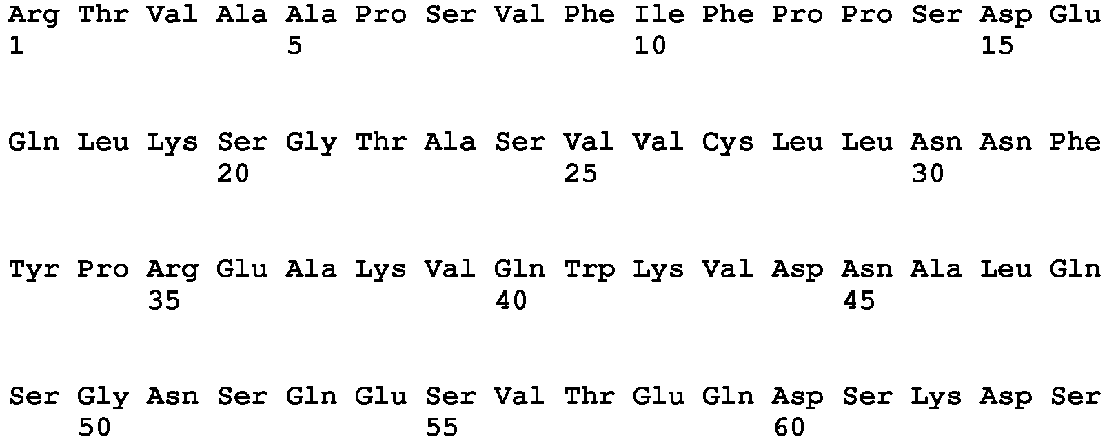

- <210> 13
<211> 330
<212> PRT
<213> Homo sapiens - <400> 13


- <210> 14
<211> 330
<212> PRT
<213> Artificial - <220>
<223> human heavy chain constant region derived from IgG1 mutated on L234A and L235A - <400> 14


- <210> 15
<211> 327
<212> PRT
<213> Homo sapiens - <400> 15

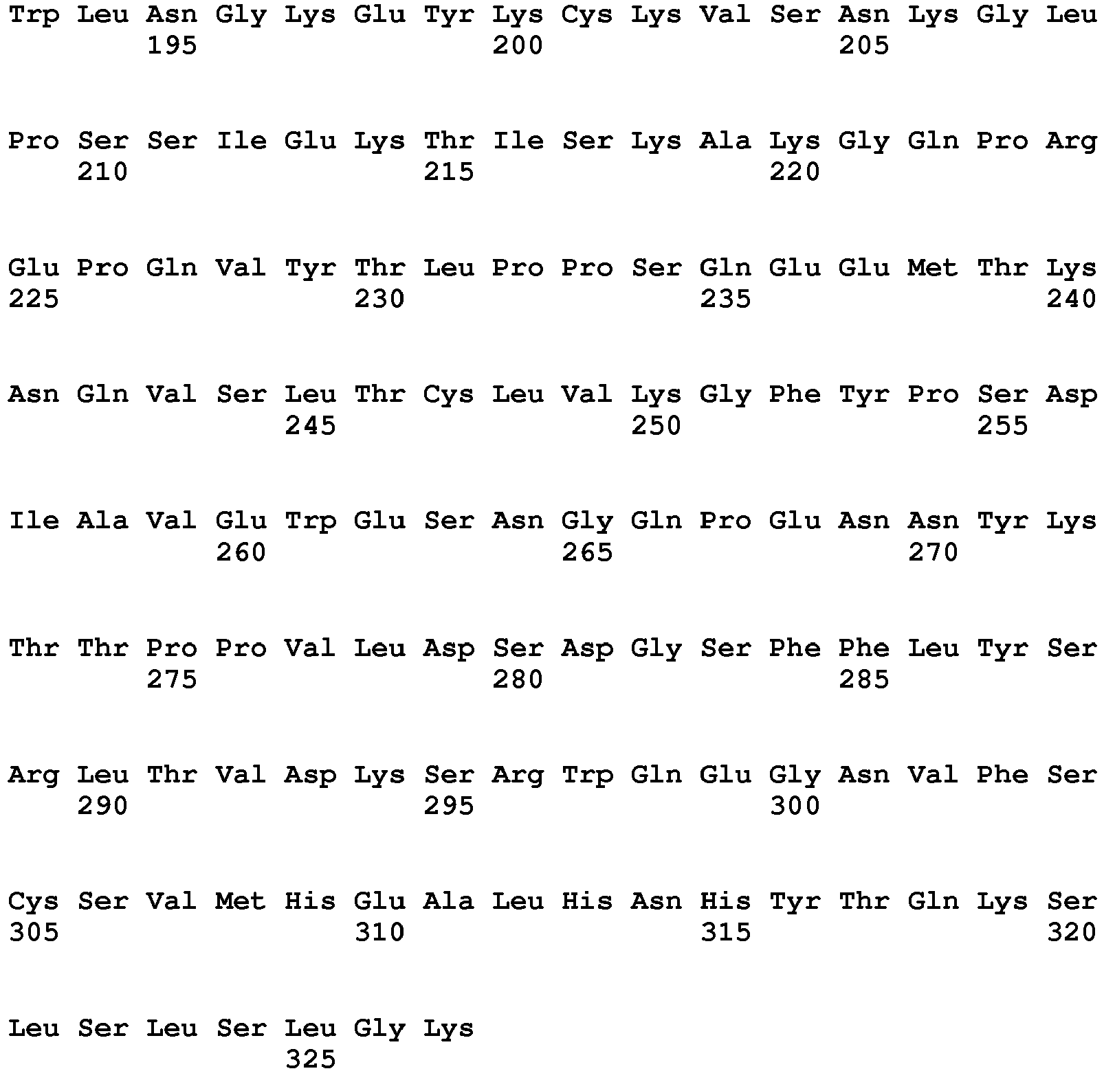
- <210> 16
<211> 327
<212> PRT
<213> Artificial - <220>
<223> human heavy chain constant region derived from IgG4 mutated onS228P - <400> 16
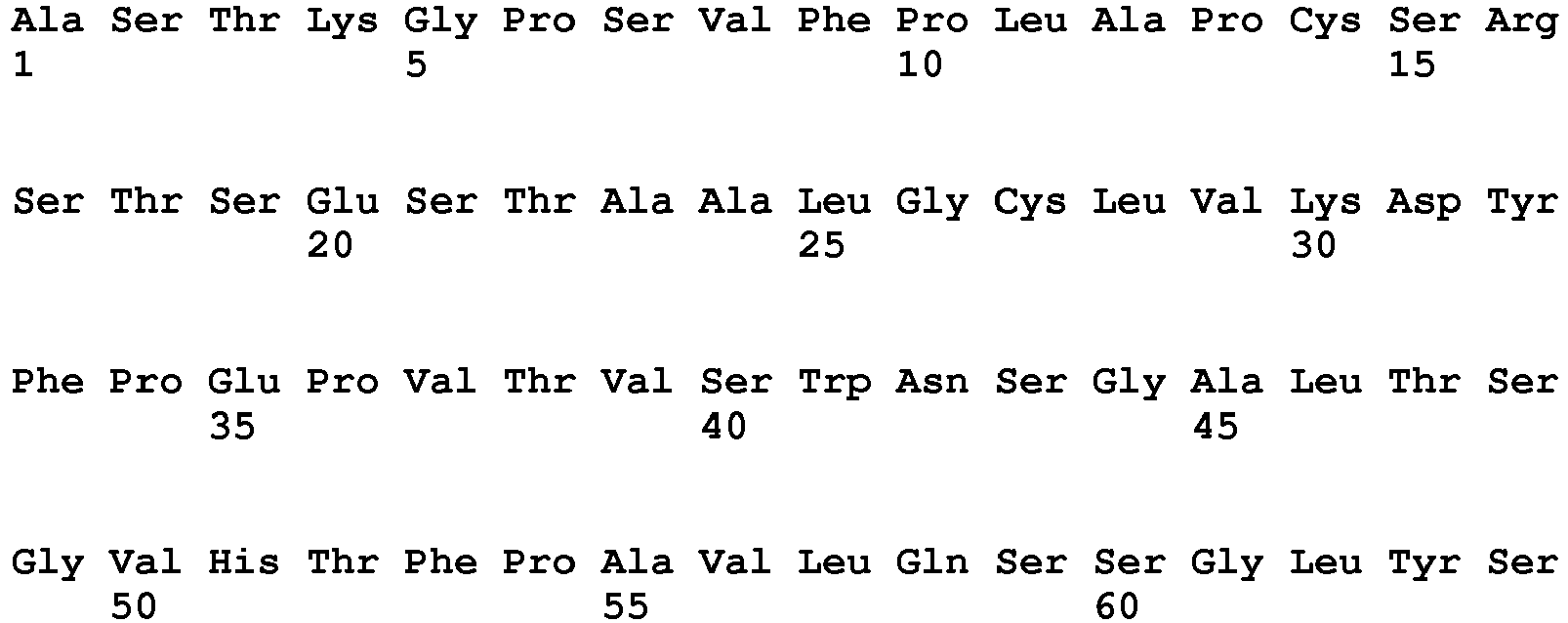


- <210> 17
<211> 1342
<212> PRT
<213> homo sapiens - <400> 17


- <210> 18
<211> 1255
<212> PRT
<213> homo sapiens - <400> 18


- <210> 19
<211> 1210
<212> PRT
<213> homo sapiens - <400> 19


- <210> 20
<211> 120
<212> PRT
<213> Artificial - <220>
<223> heavy chain variable domain VH of anti-HER1 antibody GA201 - <400> 20

- <210> 21
<211> 108
<212> PRT
<213> Artificial - <220>
<223> light chain variable domain VL of anti-HER1 antibody GA201 - <400> 21


Claims (5)
- An antibody which binds to human HER3 for use in the treatment of cancer characterized by a HER3 expression and a HER2 immunohistochemistry (IHC) score of 2+ or 1+ and in situ hybridization (ISH) amplification ratio <2.0; wherein said antibody is to be administerd in combination with an antibody which binds to human HER2 and which inhibits dimerization of HER2,
and wherein the antibody which binds to human HER3 is characterized in comprising as heavy chain variable domain a CDR3H region of SEQ ID NO: 1, a CDR2H region of SEQ ID NO: 2, and a CDR1H region of SEQ ID NO:3, and the light chain variable domain comprises a CDR3L region of SEQ ID NO: 4, a CDR2L region of SEQ ID NO:5, and a CDR1L region of SEQ ID NO:7. - The antibody for use of claim 1, wherein the antibody which binds to human HER3 is characterized in that
the heavy chain variable domain VH is SEQ ID NO:8; and the light chain variable domain VL is SEQ ID NO: 10. - The antibody for use of any one of claims 1 to 2, wherein the antibody which binds to human HER3 is characterized in that is glycosylated with a sugar chain at Asn297 whereby the amount of fucose within said sugar chain is 65 % or lower.
- The antibody for use of any one of claims 1 to 3, wherein the antibody which binds to human HER2 and which inhibits dimerization of HER2 is pertuzumab.
- The antibody for use of any one of claims 1 to 4, wherein the cancer is breast cancer, ovarian cancer, gastric cancer, prostate cancer, pancreatic cancer or head and neck cancer.
Priority Applications (1)
| Application Number | Priority Date | Filing Date | Title |
|---|---|---|---|
| EP14700281.0A EP2943508B1 (en) | 2013-01-11 | 2014-01-10 | Combination therapy of anti-her3 and anti-her2 antibodies |
Applications Claiming Priority (3)
| Application Number | Priority Date | Filing Date | Title |
|---|---|---|---|
| EP13151076 | 2013-01-11 | ||
| EP14700281.0A EP2943508B1 (en) | 2013-01-11 | 2014-01-10 | Combination therapy of anti-her3 and anti-her2 antibodies |
| PCT/EP2014/050344 WO2014108484A2 (en) | 2013-01-11 | 2014-01-10 | Combination therapy of anti-her3 antibodies |
Publications (2)
| Publication Number | Publication Date |
|---|---|
| EP2943508A2 EP2943508A2 (en) | 2015-11-18 |
| EP2943508B1 true EP2943508B1 (en) | 2020-07-15 |
Family
ID=47504772
Family Applications (1)
| Application Number | Title | Priority Date | Filing Date |
|---|---|---|---|
| EP14700281.0A Active EP2943508B1 (en) | 2013-01-11 | 2014-01-10 | Combination therapy of anti-her3 and anti-her2 antibodies |
Country Status (19)
| Country | Link |
|---|---|
| US (2) | US9180185B2 (en) |
| EP (1) | EP2943508B1 (en) |
| JP (1) | JP6257646B2 (en) |
| KR (1) | KR20150093231A (en) |
| CN (1) | CN104797602A (en) |
| AR (1) | AR094403A1 (en) |
| AU (1) | AU2014204795A1 (en) |
| CA (1) | CA2887508A1 (en) |
| CL (1) | CL2015001049A1 (en) |
| CR (1) | CR20150284A (en) |
| EA (1) | EA201500744A1 (en) |
| HK (1) | HK1211950A1 (en) |
| IL (1) | IL238393A0 (en) |
| MA (1) | MA38273A1 (en) |
| MX (1) | MX2015008313A (en) |
| PH (1) | PH12015501071A1 (en) |
| SG (1) | SG11201503802UA (en) |
| TW (1) | TW201431559A (en) |
| WO (1) | WO2014108484A2 (en) |
Families Citing this family (19)
| Publication number | Priority date | Publication date | Assignee | Title |
|---|---|---|---|---|
| JO3000B1 (en) | 2004-10-20 | 2016-09-05 | Genentech Inc | Antibody Formulations. |
| TWI472339B (en) | 2008-01-30 | 2015-02-11 | Genentech Inc | Composition comprising antibody that binds to domain ii of her2 and acidic variants thereof |
| BRPI0812682A2 (en) | 2008-06-16 | 2010-06-22 | Genentech Inc | metastatic breast cancer treatment |
| PT4241849T (en) | 2011-10-14 | 2024-10-30 | Hoffmann La Roche | Uses for and article of manufacture including her2 dimerization inhibitor pertuzumab |
| AR094403A1 (en) * | 2013-01-11 | 2015-07-29 | Hoffmann La Roche | ANTI-HER3 ANTIBODY COMBINATION THERAPY |
| AR095863A1 (en) | 2013-04-16 | 2015-11-18 | Genentech Inc | VARIATIONS OF PERTUZUMAB, ITS ASSESSMENT, METHOD OF TREATMENT, METHOD OF PREPARATION AND ARTICLE OF MANUFACTURE |
| MX2016014862A (en) | 2014-05-14 | 2017-02-27 | Hoffmann La Roche | Her3/her2 bispecific antibodies binding to the beta-hairpin of her3 and domain ii of her2. |
| EP3091033A1 (en) * | 2015-05-06 | 2016-11-09 | Gamamabs Pharma | Anti-human-her3 antibodies and uses thereof |
| CN108602890A (en) | 2015-12-11 | 2018-09-28 | 瑞泽恩制药公司 | Method for reducing or preventing the tumour growth resistant to EGFR and/or ERBB3 retarding agents |
| WO2017102789A1 (en) * | 2015-12-17 | 2017-06-22 | F. Hoffmann-La Roche Ag | Combination therapy of anti-her3 antibodies and anti-her2 antibodies |
| MY189536A (en) | 2017-01-17 | 2022-02-16 | Hoffmann La Roche | Subcutaneous her2 antibody formulations |
| WO2018159582A1 (en) | 2017-02-28 | 2018-09-07 | 学校法人近畿大学 | Method for treating egfr-tki-resistant non-small cell lung cancer by administration of anti-her3 antibody-drug conjugate |
| US11077189B2 (en) | 2017-03-02 | 2021-08-03 | Genentech Inc. | Adjuvant treatment of HER2-positive breast cancer |
| CN116891534A (en) | 2017-06-27 | 2023-10-17 | 瑞泽恩制药公司 | Tropically modified recombinant virions and their use for targeted introduction of genetic material into human cells |
| AU2019301070B2 (en) * | 2018-07-09 | 2025-09-11 | Precigen, Inc. | Fusion constructs and methods of using thereof |
| CN112912109A (en) | 2018-09-20 | 2021-06-04 | 第一三共株式会社 | Treatment of HER 3-mutated cancer by administration of anti-HER 3 antibody-drug conjugates |
| EP4171748A1 (en) | 2020-06-29 | 2023-05-03 | Genentech, Inc. | Pertuzumab plus trastuzumab fixed dose combination |
| JP2024536719A (en) * | 2021-09-03 | 2024-10-08 | キャンサー・リサーチ・テクノロジー・リミテッド | Treatment and prevention of cancer using HER3 antigen binding molecules |
| CN121219016A (en) | 2023-06-02 | 2025-12-26 | 第一三共株式会社 | Combination of anti-HER3 antibody-drug conjugate and RASG12C inhibitor |
Family Cites Families (85)
| Publication number | Priority date | Publication date | Assignee | Title |
|---|---|---|---|---|
| EP0307434B2 (en) | 1987-03-18 | 1998-07-29 | Scotgen Biopharmaceuticals, Inc. | Altered antibodies |
| US5202238A (en) | 1987-10-27 | 1993-04-13 | Oncogen | Production of chimeric antibodies by homologous recombination |
| US5204244A (en) | 1987-10-27 | 1993-04-20 | Oncogen | Production of chimeric antibodies by homologous recombination |
| US5183884A (en) | 1989-12-01 | 1993-02-02 | United States Of America | Dna segment encoding a gene for a receptor related to the epidermal growth factor receptor |
| ATE255131T1 (en) | 1991-06-14 | 2003-12-15 | Genentech Inc | HUMANIZED HEREGULIN ANTIBODIES |
| US6750334B1 (en) | 1996-02-02 | 2004-06-15 | Repligen Corporation | CTLA4-immunoglobulin fusion proteins having modified effector functions and uses therefor |
| AU727082B2 (en) | 1996-03-27 | 2000-11-30 | Genentech Inc. | ErbB3 antibodies |
| AU2710597A (en) | 1996-06-27 | 1998-01-14 | Pfizer Inc. | Derivatives of 2-(2-oxo-ethylidene)-imidazolidin-4-one and their use as farnesyl protein transferase inhibitors |
| JPH11236333A (en) | 1997-12-30 | 1999-08-31 | Pfizer Prod Inc | Imidazolin-4-one derivative as anticancer agent |
| AU3657899A (en) | 1998-04-20 | 1999-11-08 | James E. Bailey | Glycosylation engineering of antibodies for improving antibody-dependent cellular cytotoxicity |
| WO1999060023A1 (en) | 1998-05-15 | 1999-11-25 | Imclone Systems Incorporated | Treatment of human tumors with radiation and inhibitors of growth factor receptor tyrosine kinases |
| ATE289602T1 (en) | 1998-08-27 | 2005-03-15 | Pfizer Prod Inc | QUINOLIN-2-ONE DERIVATIVES USABLE AS ANTI-CANCER AGENT |
| TR200101343T2 (en) | 1998-08-27 | 2001-09-21 | Pfizer Products Inc. | Alkynyl-substituted quinolin-2-one derivatives as agents used against cancer |
| EP1006113A1 (en) | 1998-12-02 | 2000-06-07 | Pfizer Products Inc. | Derivatives of 2-(2-oxo-ethylidene)-imidazolidin-4-one and their use to inhibit abnormal cell growth |
| KR20010102073A (en) | 1999-02-11 | 2001-11-15 | 실버스타인 아써 에이. | Heteroaryl-substituted quinolin-2-one derivatives useful as anticancer agents |
| US6586447B1 (en) | 1999-04-01 | 2003-07-01 | Pfizer Inc | 3,3-disubstituted-oxindole derivatives useful as anticancer agents |
| PT1176195E (en) | 1999-04-09 | 2013-07-18 | Kyowa Hakko Kirin Co Ltd | Method for controlling the activity of immunologically functional molecule |
| AUPQ105799A0 (en) | 1999-06-18 | 1999-07-08 | Victor Chang Cardiac Research Institute, The | Cell growth inhibition |
| AT500848B1 (en) | 1999-06-25 | 2008-01-15 | Genentech Inc | HUMANIZED ANTI-ERBB2 ANTIBODIES |
| DE60008206T2 (en) | 1999-11-30 | 2004-12-02 | Pfizer Products Inc., Groton | Quinoline derivatives can be used to inhibit farnesyl protein transferase |
| UA75055C2 (en) | 1999-11-30 | 2006-03-15 | Пфайзер Продактс Інк. | Benzoimidazole derivatives being used as antiproliferative agent, pharmaceutical composition based thereon |
| HN2000000266A (en) | 2000-01-21 | 2001-05-21 | Pfizer Prod Inc | ANTI-TARGET COMPOUND AND METHOD OF SEPARATION OF ENANTIOMERS USEFUL TO SYNTHEIZE SUCH COMPOUND. |
| FR2807767B1 (en) | 2000-04-12 | 2005-01-14 | Lab Francais Du Fractionnement | MONOCLONAL ANTIBODIES ANTI-D |
| EP1228766A1 (en) | 2001-01-31 | 2002-08-07 | Max-Planck-Gesellschaft zur Förderung der Wissenschaften e.V. | PYK2 phosphorylation by HER3 induces tumor invasion |
| US7744882B2 (en) | 2001-05-31 | 2010-06-29 | Tumor Biology Investment Group, Inc. | Soluble ErbB3 methods of detection and antibodies |
| EP1283053A1 (en) | 2001-08-09 | 2003-02-12 | Max-Planck-Gesellschaft zur Förderung der Wissenschaften e.V. | Inhibitors of HER3 activity |
| KR100988949B1 (en) | 2001-10-25 | 2010-10-20 | 제넨테크, 인크. | Glycoprotein Composition |
| US20040093621A1 (en) | 2001-12-25 | 2004-05-13 | Kyowa Hakko Kogyo Co., Ltd | Antibody composition which specifically binds to CD20 |
| US7919098B2 (en) | 2002-03-26 | 2011-04-05 | Zensun ( Shanghai ) Sci & Tech Co., Ltd. | ErbB-3 based methods and compositions for treating neoplasms |
| CZ308720B6 (en) | 2003-01-22 | 2021-03-24 | Roche Glycart Ag | Fusion constructs and their use in preparing antibodies with increased binding affinity to Fc receptors and effector function |
| EP1648511A1 (en) | 2003-07-29 | 2006-04-26 | Morphotek, Inc. | Antibodies and methods for generating genetically altered antibodies with enhanced effector function |
| CN1871259A (en) | 2003-08-22 | 2006-11-29 | 比奥根艾迪克Ma公司 | Improved antibodies having altered effector function and methods for making the same |
| AU2004273791A1 (en) | 2003-09-05 | 2005-03-31 | Genentech, Inc. | Antibodies with altered effector functions |
| PT1692182E (en) | 2003-11-05 | 2010-05-13 | Roche Glycart Ag | Cd20 antibodies with increased fc receptor binding affinity and effector function |
| EP2527460B1 (en) | 2004-05-27 | 2014-12-24 | The Regents of The University of Colorado | Methods for prediction of clinical outcome to epidermal growth factor receptor inhibitors by cancer patients |
| SV2006002143A (en) | 2004-06-16 | 2006-01-26 | Genentech Inc | USE OF AN ANTIBODY FOR THE TREATMENT OF CANCER RESISTANT TO PLATINUM |
| US20090175854A1 (en) | 2004-09-08 | 2009-07-09 | Genentech, Inc. | Methods of using death receptor ligands and CD20 antibodies |
| WO2007024249A2 (en) | 2004-11-10 | 2007-03-01 | Macrogenics, Inc. | Engineering fc antibody regions to confer effector function |
| ZA200704796B (en) | 2004-12-07 | 2008-11-26 | Genentech Inc | Selecting patients for therapy with a HER inhibitor |
| ES2755976T3 (en) * | 2005-02-07 | 2020-04-24 | Roche Glycart Ag | Antigen-binding molecules that bind to EGFR, vectors that encode them, and uses thereof |
| KR101253576B1 (en) * | 2005-02-23 | 2013-04-11 | 제넨테크, 인크. | Extending time to disease progression or survival in cancer patients using a her dimerization inhibitor |
| US20060204505A1 (en) * | 2005-03-08 | 2006-09-14 | Sliwkowski Mark X | Methods for identifying tumors responsive to treatment with HER dimerization inhibitors (HDIs) |
| TW200720289A (en) | 2005-04-01 | 2007-06-01 | Hoffmann La Roche | Antibodies against CCR5 and uses thereof |
| US8008443B2 (en) | 2005-04-26 | 2011-08-30 | Medimmune, Llc | Modulation of antibody effector function by hinge domain engineering |
| CA2605697A1 (en) | 2005-04-26 | 2006-11-02 | Bioren, Inc. | Method of producing human igg antibodies with enhanced effector functions |
| PE20070207A1 (en) | 2005-07-22 | 2007-03-09 | Genentech Inc | COMBINED TREATMENT OF TUMORS THAT EXPRESS HER |
| RU2482132C2 (en) | 2005-08-26 | 2013-05-20 | Роше Гликарт Аг | Modified antigen-binding molecules with changed cell signal activity |
| AR056857A1 (en) | 2005-12-30 | 2007-10-24 | U3 Pharma Ag | DIRECTED ANTIBODIES TO HER-3 (RECEIVER OF THE HUMAN EPIDERMAL GROWTH FACTOR-3) AND ITS USES |
| EP2518508A1 (en) | 2006-11-28 | 2012-10-31 | U3 Pharma GmbH | Activated HER3 as a marker for predicting therapeutic efficacy |
| US20080226635A1 (en) | 2006-12-22 | 2008-09-18 | Hans Koll | Antibodies against insulin-like growth factor I receptor and uses thereof |
| SG178789A1 (en) | 2007-02-16 | 2012-03-29 | Merrimack Pharmaceuticals Inc | Antibodies against erbb3 and uses thereof |
| CA2687368C (en) | 2007-06-01 | 2018-10-30 | The Henry M. Jackson Foundation For The Advancement Of Military Medicine, Inc. | Vaccine for the prevention of breast cancer relapse |
| US8529901B2 (en) | 2007-06-06 | 2013-09-10 | Hoffmann-La Roche, Inc. | Composition of a first non-labeled monoclonal antibody binding to a tumor antigen and a non-cross reactive second monoclonal antibody labeled with a NIR fluorescence label |
| EP2138511A1 (en) | 2008-06-27 | 2009-12-30 | Max-Planck-Gesellschaft zur Förderung der Wissenschaften e.V. | HER3 as a determinant for the prognosis of melanoma |
| WO2010019952A2 (en) | 2008-08-15 | 2010-02-18 | Merrimack Pharmaceuticals, Inc. | Methods, systems and products for predicting response of tumor cells to a therapeutic agent and treating a patient according to the predicted response |
| WO2010052225A1 (en) | 2008-11-04 | 2010-05-14 | F. Hoffmann-La Roche Ag | Modulators for her2 signaling in normal her2 expressing settings |
| SG172983A1 (en) | 2009-01-15 | 2011-08-29 | Lab Corp America Holdings | Methods of determining patient response by measurement of her-3 |
| SG10201402742YA (en) | 2009-03-20 | 2014-08-28 | Genentech Inc | Bispecific anti-her antibodies |
| JP5587975B2 (en) | 2009-04-07 | 2014-09-10 | ロシュ グリクアート アクチェンゲゼルシャフト | Bispecific anti-ErbB-3 / anti-c-Met antibody |
| WO2010127181A1 (en) | 2009-04-29 | 2010-11-04 | Trellis Bioscience, Inc. | Improved antibodies immunoreactive with heregulin-coupled her3 |
| KR20120059568A (en) | 2009-08-21 | 2012-06-08 | 메리맥 파마슈티컬즈, 인크. | Antibodies against the ectodomain of erbb3 and uses thereof |
| JP2013507378A (en) | 2009-10-09 | 2013-03-04 | メルク・シャープ・エンド・ドーム・コーポレイション | Production, characterization and use of anti-HER3 antibodies |
| EP2496598B1 (en) | 2009-11-04 | 2017-08-02 | Affibody AB | Her3 binding polypeptides |
| BR112012012160B1 (en) * | 2009-11-13 | 2022-04-26 | Amgen, Inc. | Material and methods to treat or prevent diseases associated with her-3 |
| PH12012501157A1 (en) * | 2009-12-22 | 2012-10-29 | Roche Glycart Ag | Anti-her3 antibodies and uses thereof |
| KR20130000384A (en) | 2010-02-18 | 2013-01-02 | 더 보드 어브 트러스티스 어브 더 리랜드 스탠포드 주니어 유니버시티 | Neuregulin antagonists and use thereof in treating cancer |
| KR101798679B1 (en) | 2010-03-11 | 2017-11-16 | 메리맥 파마슈티컬즈, 인크. | Use of erbb3 inhibitors in the treatment of triple negative and basal-like breast cancers |
| US8481687B2 (en) | 2010-04-09 | 2013-07-09 | Aveo Pharmaceuticals, Inc. | Anti-ErbB3 antibodies |
| WO2012019024A2 (en) | 2010-08-04 | 2012-02-09 | Immunogen, Inc. | Her3-binding molecules and immunoconjugates thereof |
| CA2815154A1 (en) | 2010-08-06 | 2012-02-09 | U3 Pharma Gmbh | Use of her3 binding agents in prostate treatment |
| SG187908A1 (en) | 2010-08-20 | 2013-03-28 | Novartis Ag | Antibodies for epidermal growth factor receptor 3 (her3) |
| TW201302793A (en) | 2010-09-03 | 2013-01-16 | Glaxo Group Ltd | Novel antigen binding protein |
| AU2011307251A1 (en) | 2010-09-30 | 2013-02-28 | Merck Sharp & Dohme Corp. | Generation, characterization and uses thereof of anti-HER3 antibodies |
| AU2011317743B2 (en) | 2010-10-18 | 2015-05-21 | Mediapharma S.R.L. | ErbB3 binding antibody |
| KR101862832B1 (en) | 2010-11-01 | 2018-05-30 | 심포젠 에이/에스 | Pan-her antibody composition |
| US9155802B2 (en) | 2010-11-01 | 2015-10-13 | Symphogen A/S | Pan-HER antibody composition |
| ITRM20100577A1 (en) | 2010-11-02 | 2012-05-03 | Takis Srl | IMMUNOTHERAPY AGAINST THE ERBB-3 RECEPTOR |
| JP2014509593A (en) | 2011-03-15 | 2014-04-21 | メリマック ファーマシューティカルズ インコーポレーティッド | Overcoming resistance to ErbB pathway inhibitors |
| PT4241849T (en) | 2011-10-14 | 2024-10-30 | Hoffmann La Roche | Uses for and article of manufacture including her2 dimerization inhibitor pertuzumab |
| MX350957B (en) | 2011-11-23 | 2017-09-27 | Medimmune Llc | Binding molecules specific for her3 and uses thereof. |
| KR102083957B1 (en) * | 2011-12-05 | 2020-03-04 | 노파르티스 아게 | Antibodies for epidermal growth factor receptor 3 (her3) |
| EP2793941A1 (en) | 2011-12-23 | 2014-10-29 | F.Hoffmann-La Roche Ag | Articles of manufacture and methods for co-administration of antibodies |
| US8859797B1 (en) | 2012-04-27 | 2014-10-14 | Air Liquide Electronics U.S. Lp | Synthesis methods for carbosilanes |
| HK1202431A1 (en) | 2012-05-02 | 2015-10-02 | Symphogen A/S | Humanized pan-her antibody compositions |
| AR094403A1 (en) * | 2013-01-11 | 2015-07-29 | Hoffmann La Roche | ANTI-HER3 ANTIBODY COMBINATION THERAPY |
-
2014
- 2014-01-09 AR ARP140100081A patent/AR094403A1/en unknown
- 2014-01-09 US US14/151,626 patent/US9180185B2/en active Active
- 2014-01-10 CN CN201480003140.2A patent/CN104797602A/en active Pending
- 2014-01-10 TW TW103101044A patent/TW201431559A/en unknown
- 2014-01-10 EP EP14700281.0A patent/EP2943508B1/en active Active
- 2014-01-10 JP JP2015552051A patent/JP6257646B2/en not_active Expired - Fee Related
- 2014-01-10 CA CA 2887508 patent/CA2887508A1/en not_active Abandoned
- 2014-01-10 EA EA201500744A patent/EA201500744A1/en unknown
- 2014-01-10 KR KR1020157018413A patent/KR20150093231A/en not_active Ceased
- 2014-01-10 SG SG11201503802UA patent/SG11201503802UA/en unknown
- 2014-01-10 WO PCT/EP2014/050344 patent/WO2014108484A2/en not_active Ceased
- 2014-01-10 AU AU2014204795A patent/AU2014204795A1/en not_active Abandoned
- 2014-01-10 MA MA38273A patent/MA38273A1/en unknown
- 2014-01-10 MX MX2015008313A patent/MX2015008313A/en unknown
- 2014-01-10 HK HK15112676.9A patent/HK1211950A1/en unknown
-
2015
- 2015-04-20 IL IL238393A patent/IL238393A0/en unknown
- 2015-04-23 CL CL2015001049A patent/CL2015001049A1/en unknown
- 2015-05-14 PH PH12015501071A patent/PH12015501071A1/en unknown
- 2015-05-28 CR CR20150284A patent/CR20150284A/en unknown
- 2015-10-07 US US14/877,569 patent/US20160159912A1/en not_active Abandoned
Non-Patent Citations (1)
| Title |
|---|
| WEITSMAN GREGORY ET AL: "HER2-HER3 dimer quantification by FLIM-FRET predicts breast cancer metastatic relapse independently of HER2 IHC status.", ONCOTARGET 09 AUG 2016, vol. 7, no. 32, 9 August 2016 (2016-08-09), pages 51012 - 51026, ISSN: 1949-2553 * |
Also Published As
| Publication number | Publication date |
|---|---|
| HK1211950A1 (en) | 2016-06-03 |
| KR20150093231A (en) | 2015-08-17 |
| EA201500744A1 (en) | 2015-12-30 |
| PH12015501071A1 (en) | 2015-08-03 |
| JP6257646B2 (en) | 2018-01-10 |
| TW201431559A (en) | 2014-08-16 |
| US20160159912A1 (en) | 2016-06-09 |
| MX2015008313A (en) | 2015-11-11 |
| EP2943508A2 (en) | 2015-11-18 |
| AU2014204795A1 (en) | 2015-04-30 |
| IL238393A0 (en) | 2015-06-30 |
| CL2015001049A1 (en) | 2015-10-16 |
| AR094403A1 (en) | 2015-07-29 |
| CN104797602A (en) | 2015-07-22 |
| JP2016504399A (en) | 2016-02-12 |
| MA38273A1 (en) | 2017-05-31 |
| WO2014108484A3 (en) | 2014-09-04 |
| CA2887508A1 (en) | 2014-07-17 |
| WO2014108484A2 (en) | 2014-07-17 |
| SG11201503802UA (en) | 2015-08-28 |
| CR20150284A (en) | 2015-06-30 |
| US9180185B2 (en) | 2015-11-10 |
| US20140227255A1 (en) | 2014-08-14 |
Similar Documents
| Publication | Publication Date | Title |
|---|---|---|
| US9180185B2 (en) | Combination therapy of anti-HER3 antibodies | |
| US10196456B2 (en) | Anti-HER3 antibodies and uses thereof | |
| RU2733315C2 (en) | Combined therapy for malignant tumor treatment | |
| PT2417156E (en) | Trivalent, bispecific antibodies | |
| AU2007236199A1 (en) | Antibodies against insulin-like growth factor i receptor and uses thereof | |
| WO2017102789A1 (en) | Combination therapy of anti-her3 antibodies and anti-her2 antibodies | |
| HK1175183B (en) | Anti-her3 antibodies and uses thereof |
Legal Events
| Date | Code | Title | Description |
|---|---|---|---|
| PUAI | Public reference made under article 153(3) epc to a published international application that has entered the european phase |
Free format text: ORIGINAL CODE: 0009012 |
|
| 17P | Request for examination filed |
Effective date: 20150811 |
|
| AK | Designated contracting states |
Kind code of ref document: A2 Designated state(s): AL AT BE BG CH CY CZ DE DK EE ES FI FR GB GR HR HU IE IS IT LI LT LU LV MC MK MT NL NO PL PT RO RS SE SI SK SM TR |
|
| AX | Request for extension of the european patent |
Extension state: BA ME |
|
| DAX | Request for extension of the european patent (deleted) | ||
| 17Q | First examination report despatched |
Effective date: 20161014 |
|
| STAA | Information on the status of an ep patent application or granted ep patent |
Free format text: STATUS: EXAMINATION IS IN PROGRESS |
|
| GRAP | Despatch of communication of intention to grant a patent |
Free format text: ORIGINAL CODE: EPIDOSNIGR1 |
|
| STAA | Information on the status of an ep patent application or granted ep patent |
Free format text: STATUS: GRANT OF PATENT IS INTENDED |
|
| INTG | Intention to grant announced |
Effective date: 20191009 |
|
| GRAJ | Information related to disapproval of communication of intention to grant by the applicant or resumption of examination proceedings by the epo deleted |
Free format text: ORIGINAL CODE: EPIDOSDIGR1 |
|
| STAA | Information on the status of an ep patent application or granted ep patent |
Free format text: STATUS: EXAMINATION IS IN PROGRESS |
|
| GRAS | Grant fee paid |
Free format text: ORIGINAL CODE: EPIDOSNIGR3 |
|
| STAA | Information on the status of an ep patent application or granted ep patent |
Free format text: STATUS: GRANT OF PATENT IS INTENDED |
|
| GRAP | Despatch of communication of intention to grant a patent |
Free format text: ORIGINAL CODE: EPIDOSNIGR1 |
|
| INTC | Intention to grant announced (deleted) | ||
| RIN1 | Information on inventor provided before grant (corrected) |
Inventor name: FRIESS, THOMAS Inventor name: THOMAS, MARLENE Inventor name: GERDES, CHRISTIAN Inventor name: HASMANN, MAX Inventor name: BAUSS, FRIEDER Inventor name: BOSSENMAIER, BIRGIT Inventor name: WEISSER, MARTIN |
|
| INTG | Intention to grant announced |
Effective date: 20200221 |
|
| GRAA | (expected) grant |
Free format text: ORIGINAL CODE: 0009210 |
|
| STAA | Information on the status of an ep patent application or granted ep patent |
Free format text: STATUS: THE PATENT HAS BEEN GRANTED |
|
| AK | Designated contracting states |
Kind code of ref document: B1 Designated state(s): AL AT BE BG CH CY CZ DE DK EE ES FI FR GB GR HR HU IE IS IT LI LT LU LV MC MK MT NL NO PL PT RO RS SE SI SK SM TR |
|
| REG | Reference to a national code |
Ref country code: GB Ref legal event code: FG4D Ref country code: CH Ref legal event code: EP |
|
| REG | Reference to a national code |
Ref country code: IE Ref legal event code: FG4D |
|
| REG | Reference to a national code |
Ref country code: DE Ref legal event code: R096 Ref document number: 602014067692 Country of ref document: DE |
|
| REG | Reference to a national code |
Ref country code: AT Ref legal event code: REF Ref document number: 1290929 Country of ref document: AT Kind code of ref document: T Effective date: 20200815 |
|
| REG | Reference to a national code |
Ref country code: LT Ref legal event code: MG4D |
|
| REG | Reference to a national code |
Ref country code: AT Ref legal event code: MK05 Ref document number: 1290929 Country of ref document: AT Kind code of ref document: T Effective date: 20200715 |
|
| REG | Reference to a national code |
Ref country code: NL Ref legal event code: MP Effective date: 20200715 |
|
| PG25 | Lapsed in a contracting state [announced via postgrant information from national office to epo] |
Ref country code: LT Free format text: LAPSE BECAUSE OF FAILURE TO SUBMIT A TRANSLATION OF THE DESCRIPTION OR TO PAY THE FEE WITHIN THE PRESCRIBED TIME-LIMIT Effective date: 20200715 Ref country code: ES Free format text: LAPSE BECAUSE OF FAILURE TO SUBMIT A TRANSLATION OF THE DESCRIPTION OR TO PAY THE FEE WITHIN THE PRESCRIBED TIME-LIMIT Effective date: 20200715 Ref country code: PT Free format text: LAPSE BECAUSE OF FAILURE TO SUBMIT A TRANSLATION OF THE DESCRIPTION OR TO PAY THE FEE WITHIN THE PRESCRIBED TIME-LIMIT Effective date: 20201116 Ref country code: HR Free format text: LAPSE BECAUSE OF FAILURE TO SUBMIT A TRANSLATION OF THE DESCRIPTION OR TO PAY THE FEE WITHIN THE PRESCRIBED TIME-LIMIT Effective date: 20200715 Ref country code: BG Free format text: LAPSE BECAUSE OF FAILURE TO SUBMIT A TRANSLATION OF THE DESCRIPTION OR TO PAY THE FEE WITHIN THE PRESCRIBED TIME-LIMIT Effective date: 20201015 Ref country code: NO Free format text: LAPSE BECAUSE OF FAILURE TO SUBMIT A TRANSLATION OF THE DESCRIPTION OR TO PAY THE FEE WITHIN THE PRESCRIBED TIME-LIMIT Effective date: 20201015 Ref country code: AT Free format text: LAPSE BECAUSE OF FAILURE TO SUBMIT A TRANSLATION OF THE DESCRIPTION OR TO PAY THE FEE WITHIN THE PRESCRIBED TIME-LIMIT Effective date: 20200715 Ref country code: SE Free format text: LAPSE BECAUSE OF FAILURE TO SUBMIT A TRANSLATION OF THE DESCRIPTION OR TO PAY THE FEE WITHIN THE PRESCRIBED TIME-LIMIT Effective date: 20200715 Ref country code: GR Free format text: LAPSE BECAUSE OF FAILURE TO SUBMIT A TRANSLATION OF THE DESCRIPTION OR TO PAY THE FEE WITHIN THE PRESCRIBED TIME-LIMIT Effective date: 20201016 Ref country code: FI Free format text: LAPSE BECAUSE OF FAILURE TO SUBMIT A TRANSLATION OF THE DESCRIPTION OR TO PAY THE FEE WITHIN THE PRESCRIBED TIME-LIMIT Effective date: 20200715 |
|
| PG25 | Lapsed in a contracting state [announced via postgrant information from national office to epo] |
Ref country code: IS Free format text: LAPSE BECAUSE OF FAILURE TO SUBMIT A TRANSLATION OF THE DESCRIPTION OR TO PAY THE FEE WITHIN THE PRESCRIBED TIME-LIMIT Effective date: 20201115 Ref country code: RS Free format text: LAPSE BECAUSE OF FAILURE TO SUBMIT A TRANSLATION OF THE DESCRIPTION OR TO PAY THE FEE WITHIN THE PRESCRIBED TIME-LIMIT Effective date: 20200715 Ref country code: LV Free format text: LAPSE BECAUSE OF FAILURE TO SUBMIT A TRANSLATION OF THE DESCRIPTION OR TO PAY THE FEE WITHIN THE PRESCRIBED TIME-LIMIT Effective date: 20200715 Ref country code: PL Free format text: LAPSE BECAUSE OF FAILURE TO SUBMIT A TRANSLATION OF THE DESCRIPTION OR TO PAY THE FEE WITHIN THE PRESCRIBED TIME-LIMIT Effective date: 20200715 |
|
| PG25 | Lapsed in a contracting state [announced via postgrant information from national office to epo] |
Ref country code: NL Free format text: LAPSE BECAUSE OF FAILURE TO SUBMIT A TRANSLATION OF THE DESCRIPTION OR TO PAY THE FEE WITHIN THE PRESCRIBED TIME-LIMIT Effective date: 20200715 |
|
| REG | Reference to a national code |
Ref country code: DE Ref legal event code: R097 Ref document number: 602014067692 Country of ref document: DE |
|
| PG25 | Lapsed in a contracting state [announced via postgrant information from national office to epo] |
Ref country code: DK Free format text: LAPSE BECAUSE OF FAILURE TO SUBMIT A TRANSLATION OF THE DESCRIPTION OR TO PAY THE FEE WITHIN THE PRESCRIBED TIME-LIMIT Effective date: 20200715 Ref country code: EE Free format text: LAPSE BECAUSE OF FAILURE TO SUBMIT A TRANSLATION OF THE DESCRIPTION OR TO PAY THE FEE WITHIN THE PRESCRIBED TIME-LIMIT Effective date: 20200715 Ref country code: CZ Free format text: LAPSE BECAUSE OF FAILURE TO SUBMIT A TRANSLATION OF THE DESCRIPTION OR TO PAY THE FEE WITHIN THE PRESCRIBED TIME-LIMIT Effective date: 20200715 Ref country code: IT Free format text: LAPSE BECAUSE OF FAILURE TO SUBMIT A TRANSLATION OF THE DESCRIPTION OR TO PAY THE FEE WITHIN THE PRESCRIBED TIME-LIMIT Effective date: 20200715 Ref country code: RO Free format text: LAPSE BECAUSE OF FAILURE TO SUBMIT A TRANSLATION OF THE DESCRIPTION OR TO PAY THE FEE WITHIN THE PRESCRIBED TIME-LIMIT Effective date: 20200715 Ref country code: SM Free format text: LAPSE BECAUSE OF FAILURE TO SUBMIT A TRANSLATION OF THE DESCRIPTION OR TO PAY THE FEE WITHIN THE PRESCRIBED TIME-LIMIT Effective date: 20200715 |
|
| PLBE | No opposition filed within time limit |
Free format text: ORIGINAL CODE: 0009261 |
|
| STAA | Information on the status of an ep patent application or granted ep patent |
Free format text: STATUS: NO OPPOSITION FILED WITHIN TIME LIMIT |
|
| PG25 | Lapsed in a contracting state [announced via postgrant information from national office to epo] |
Ref country code: AL Free format text: LAPSE BECAUSE OF FAILURE TO SUBMIT A TRANSLATION OF THE DESCRIPTION OR TO PAY THE FEE WITHIN THE PRESCRIBED TIME-LIMIT Effective date: 20200715 |
|
| 26N | No opposition filed |
Effective date: 20210416 |
|
| PG25 | Lapsed in a contracting state [announced via postgrant information from national office to epo] |
Ref country code: SK Free format text: LAPSE BECAUSE OF FAILURE TO SUBMIT A TRANSLATION OF THE DESCRIPTION OR TO PAY THE FEE WITHIN THE PRESCRIBED TIME-LIMIT Effective date: 20200715 |
|
| PG25 | Lapsed in a contracting state [announced via postgrant information from national office to epo] |
Ref country code: MC Free format text: LAPSE BECAUSE OF FAILURE TO SUBMIT A TRANSLATION OF THE DESCRIPTION OR TO PAY THE FEE WITHIN THE PRESCRIBED TIME-LIMIT Effective date: 20200715 Ref country code: SI Free format text: LAPSE BECAUSE OF FAILURE TO SUBMIT A TRANSLATION OF THE DESCRIPTION OR TO PAY THE FEE WITHIN THE PRESCRIBED TIME-LIMIT Effective date: 20200715 |
|
| PG25 | Lapsed in a contracting state [announced via postgrant information from national office to epo] |
Ref country code: LU Free format text: LAPSE BECAUSE OF NON-PAYMENT OF DUE FEES Effective date: 20210110 |
|
| REG | Reference to a national code |
Ref country code: BE Ref legal event code: MM Effective date: 20210131 |
|
| PG25 | Lapsed in a contracting state [announced via postgrant information from national office to epo] |
Ref country code: IE Free format text: LAPSE BECAUSE OF NON-PAYMENT OF DUE FEES Effective date: 20210110 |
|
| PG25 | Lapsed in a contracting state [announced via postgrant information from national office to epo] |
Ref country code: BE Free format text: LAPSE BECAUSE OF NON-PAYMENT OF DUE FEES Effective date: 20210131 |
|
| PGFP | Annual fee paid to national office [announced via postgrant information from national office to epo] |
Ref country code: GB Payment date: 20221214 Year of fee payment: 10 Ref country code: FR Payment date: 20221216 Year of fee payment: 10 |
|
| PGFP | Annual fee paid to national office [announced via postgrant information from national office to epo] |
Ref country code: CH Payment date: 20230201 Year of fee payment: 10 |
|
| PG25 | Lapsed in a contracting state [announced via postgrant information from national office to epo] |
Ref country code: HU Free format text: LAPSE BECAUSE OF FAILURE TO SUBMIT A TRANSLATION OF THE DESCRIPTION OR TO PAY THE FEE WITHIN THE PRESCRIBED TIME-LIMIT; INVALID AB INITIO Effective date: 20140110 |
|
| PGFP | Annual fee paid to national office [announced via postgrant information from national office to epo] |
Ref country code: DE Payment date: 20221215 Year of fee payment: 10 |
|
| P01 | Opt-out of the competence of the unified patent court (upc) registered |
Effective date: 20230326 |
|
| P02 | Opt-out of the competence of the unified patent court (upc) changed |
Effective date: 20230408 |
|
| PG25 | Lapsed in a contracting state [announced via postgrant information from national office to epo] |
Ref country code: CY Free format text: LAPSE BECAUSE OF FAILURE TO SUBMIT A TRANSLATION OF THE DESCRIPTION OR TO PAY THE FEE WITHIN THE PRESCRIBED TIME-LIMIT Effective date: 20200715 |
|
| REG | Reference to a national code |
Ref country code: DE Ref legal event code: R082 Ref document number: 602014067692 Country of ref document: DE Representative=s name: KRAUS & LEDERER PARTGMBB, DE |
|
| PG25 | Lapsed in a contracting state [announced via postgrant information from national office to epo] |
Ref country code: MK Free format text: LAPSE BECAUSE OF FAILURE TO SUBMIT A TRANSLATION OF THE DESCRIPTION OR TO PAY THE FEE WITHIN THE PRESCRIBED TIME-LIMIT Effective date: 20200715 |
|
| PG25 | Lapsed in a contracting state [announced via postgrant information from national office to epo] |
Ref country code: TR Free format text: LAPSE BECAUSE OF FAILURE TO SUBMIT A TRANSLATION OF THE DESCRIPTION OR TO PAY THE FEE WITHIN THE PRESCRIBED TIME-LIMIT Effective date: 20200715 |
|
| REG | Reference to a national code |
Ref country code: DE Ref legal event code: R119 Ref document number: 602014067692 Country of ref document: DE |
|
| REG | Reference to a national code |
Ref country code: CH Ref legal event code: PL |
|
| GBPC | Gb: european patent ceased through non-payment of renewal fee |
Effective date: 20240110 |
|
| PG25 | Lapsed in a contracting state [announced via postgrant information from national office to epo] |
Ref country code: MT Free format text: LAPSE BECAUSE OF FAILURE TO SUBMIT A TRANSLATION OF THE DESCRIPTION OR TO PAY THE FEE WITHIN THE PRESCRIBED TIME-LIMIT Effective date: 20200715 |
|
| PG25 | Lapsed in a contracting state [announced via postgrant information from national office to epo] |
Ref country code: DE Free format text: LAPSE BECAUSE OF NON-PAYMENT OF DUE FEES Effective date: 20240801 |
|
| PG25 | Lapsed in a contracting state [announced via postgrant information from national office to epo] |
Ref country code: GB Free format text: LAPSE BECAUSE OF NON-PAYMENT OF DUE FEES Effective date: 20240110 |
|
| PG25 | Lapsed in a contracting state [announced via postgrant information from national office to epo] |
Ref country code: FR Free format text: LAPSE BECAUSE OF NON-PAYMENT OF DUE FEES Effective date: 20240131 |
|
| PG25 | Lapsed in a contracting state [announced via postgrant information from national office to epo] |
Ref country code: CH Free format text: LAPSE BECAUSE OF NON-PAYMENT OF DUE FEES Effective date: 20240131 |
|
| PG25 | Lapsed in a contracting state [announced via postgrant information from national office to epo] |
Ref country code: GB Free format text: LAPSE BECAUSE OF NON-PAYMENT OF DUE FEES Effective date: 20240110 Ref country code: FR Free format text: LAPSE BECAUSE OF NON-PAYMENT OF DUE FEES Effective date: 20240131 Ref country code: DE Free format text: LAPSE BECAUSE OF NON-PAYMENT OF DUE FEES Effective date: 20240801 Ref country code: CH Free format text: LAPSE BECAUSE OF NON-PAYMENT OF DUE FEES Effective date: 20240131 |