EP2943147B1 - Surgical implant for the treatment of the prolapse of a pelvic organ by laparoscopy via sacrocolpopexy and/or sacrohysteropexy - Google Patents
Surgical implant for the treatment of the prolapse of a pelvic organ by laparoscopy via sacrocolpopexy and/or sacrohysteropexy Download PDFInfo
- Publication number
- EP2943147B1 EP2943147B1 EP14700291.9A EP14700291A EP2943147B1 EP 2943147 B1 EP2943147 B1 EP 2943147B1 EP 14700291 A EP14700291 A EP 14700291A EP 2943147 B1 EP2943147 B1 EP 2943147B1
- Authority
- EP
- European Patent Office
- Prior art keywords
- fabric
- section
- implant
- sections
- implant according
- Prior art date
- Legal status (The legal status is an assumption and is not a legal conclusion. Google has not performed a legal analysis and makes no representation as to the accuracy of the status listed.)
- Active
Links
Images



Classifications
-
- A—HUMAN NECESSITIES
- A61—MEDICAL OR VETERINARY SCIENCE; HYGIENE
- A61F—FILTERS IMPLANTABLE INTO BLOOD VESSELS; PROSTHESES; DEVICES PROVIDING PATENCY TO, OR PREVENTING COLLAPSING OF, TUBULAR STRUCTURES OF THE BODY, e.g. STENTS; ORTHOPAEDIC, NURSING OR CONTRACEPTIVE DEVICES; FOMENTATION; TREATMENT OR PROTECTION OF EYES OR EARS; BANDAGES, DRESSINGS OR ABSORBENT PADS; FIRST-AID KITS
- A61F2/00—Filters implantable into blood vessels; Prostheses, i.e. artificial substitutes or replacements for parts of the body; Appliances for connecting them with the body; Devices providing patency to, or preventing collapsing of, tubular structures of the body, e.g. stents
- A61F2/0004—Closure means for urethra or rectum, i.e. anti-incontinence devices or support slings against pelvic prolapse
- A61F2/0031—Closure means for urethra or rectum, i.e. anti-incontinence devices or support slings against pelvic prolapse for constricting the lumen; Support slings for the urethra
- A61F2/0036—Closure means for urethra or rectum, i.e. anti-incontinence devices or support slings against pelvic prolapse for constricting the lumen; Support slings for the urethra implantable
- A61F2/0045—Support slings
-
- A—HUMAN NECESSITIES
- A61—MEDICAL OR VETERINARY SCIENCE; HYGIENE
- A61F—FILTERS IMPLANTABLE INTO BLOOD VESSELS; PROSTHESES; DEVICES PROVIDING PATENCY TO, OR PREVENTING COLLAPSING OF, TUBULAR STRUCTURES OF THE BODY, e.g. STENTS; ORTHOPAEDIC, NURSING OR CONTRACEPTIVE DEVICES; FOMENTATION; TREATMENT OR PROTECTION OF EYES OR EARS; BANDAGES, DRESSINGS OR ABSORBENT PADS; FIRST-AID KITS
- A61F2/00—Filters implantable into blood vessels; Prostheses, i.e. artificial substitutes or replacements for parts of the body; Appliances for connecting them with the body; Devices providing patency to, or preventing collapsing of, tubular structures of the body, e.g. stents
- A61F2/0063—Implantable repair or support meshes, e.g. hernia meshes
Definitions
- the present invention relates to an implant for the surgical repair of the pelvic floor by laparoscopy which adjusts to the techniques called sacrocolpopexy and/or sacrohysteropexy.
- the present invention relates to an implant for the treatment of vaginal vault prolapse or uterine prolapse using said techniques.
- Uterine prolapse or vaginal vault prolapse is a disorder of the pelvic floor organs produced by different causes that give rise to numerous problems such as, for example, pelvic pain, problems in sexual relations, it may affect urinary incontinence, etc. decreasing the quality of life of the person suffering from it.
- Vaginal vault prolapse can be treated using the surgical technique called colposacropexy or sacrocolpopexy and uterine prolapse can be treated by the surgical technique called sacrohysteropexy.
- These techniques entail suspension of the vagina or uterus to a point of the pelvic cavity, preferably, to the patient's sacral promontory, via a surgical implant which typically consists of a fabric of implantable polymeric material in the form of mesh. The objective of the mesh is to replace the displaced fascias and substitute the damaged native tissue.
- the sacrocolpopexy technique and the sacrohysteropexy technique can be performed via an abdominal incision, although it is currently performed by laparoscopy since this technique is less aggressive for the patient and also offers numerous advantages.
- laparoscopy is the only technique that makes it possible to repair the double angle of the vagina (posterior and anterior angle) with a constant vision and direct access to the deepest areas of the pelvis, achieving a suitable and lasting securing of the vagina to the sacral promontory through a surgical mesh.
- Patents WO2010/138894 , WO2010/87923 and WO02/19946 disclose implantable fabrics in the form of mesh which have the special characteristic that all have a general Y-shaped configuration.
- This configuration includes a first portion for the fastening of the fabric to the sacral promontory and a second portion connected to the first portion for the fastening of the fabric to the pelvic organ.
- the second portion includes two superimposed sections of fabric to respectively fix in the anterior and posterior part of the pelvic organ. These two sections of fabric have the special characteristic that they are joined together by way of a pin, each one of the sections determining one arm of said pin.
- Patent document US2011/297161 refers to a method of treating prolapse of a vagina including the step of securing a base of a Y- shaped support to tissue by inserting an anchor into the tissue, the anchor coupled to the Y-shaped support via an interconnecting member, the step of asymmetrically securing the base of a Y-shaped support to one anterior sacroiliac ligament connected to and located alongside of the sacrum bone and securing each of a pair of legs of the Y-shaped support to opposing sides of the vagina, and the final step of adjusting elevation of the cuff of the vagina relative to the sacrum bone by sliding the interconnecting member relative to the adjustable anchor.
- the Y-shaped implantable meshes or fabrics of the patent documents mentioned have the drawback that they are difficult to handle for the surgeon, especially when the operation is carried out by laparoscopy which entails the use of a trocar introduced in the abdomen.
- the Y-shaped implantable meshes or fabric have joints or welds in the points of intersection of the arms or sections that define the fastening pin. These joints or welds mean that the mesh is less malleable when being introduced in the trocar cannula, which entails more work for the surgeon and, furthermore, in some cases, the risk of tearing the mesh.
- Patent WO2007/118260 discloses an implantable mesh for the surgical reconstruction in the area of the pelvic floor including an anterior mesh segment between the bladder and the vagina, a posterior mesh segment between the vagina and the rectum, a pair of distal transobturator holding straps extending from the anterior mesh segment, a pair of proximal transobturator holding straps extending from the anterior mesh segment and a pair of lower dorsal holding straps extending from the posterior mesh segment.
- the mesh further includes an intermediate segment located between the anterior mesh segment and the posterior mesh segment.
- the pair of lower dorsal holding straps extends from the region of the posterior mesh segment bordering on the intermediate segment and a pair of upper dorsal holding straps extends from the region of the anterior mesh segment bordering on the intermediate segment.
- Patent document US2011/105836 discloses a system and method of treating vaginal prolapse and incontinence comprising a kit.
- the kit includes a mesh graft configured for Attachment to the anterior and posterior vaginal walls to thereby treat the vaginal prolapse.
- a graft delivery device is also provided for introducing and placing the mesh graft to a location deep within the peritoneal cavity and for attaching the graft thereto.
- a leg assembly is provided and coupled to an end of the mesh graft and cooperates with the graft delivery device to anchor and affix the mesh graft to the desired anatomical structures.
- Patent document FR 2976788 discloses a mesh design for sacrocolpopexy procedure that has a configuration for covering the vagina and an extension to the sacrum to provide for apical support.
- Patent EP 2340784 discloses a textile mesh, in particular for pelvic floor reconstruction or for preventing dyspareunia, comprising a central mesh section and two lateral mesh sections on both sides of the central mesh section.
- the objective of the present invention is to resolve said drawbacks, developing a surgical implant for the treatment of the prolapse of a pelvic organ by laparoscopy and via sacrocolpopexy and/or sacrohysteropexy which resolve the aforementioned problems and have the advantages described below.
- the present invention provides a surgical implant for the treatment of the prolapse of a pelvic organ by laparoscopy and via sacrocolpopexy and/or sacrohysteropexy, comprising a fabric of implantable material including;
- the surgical implant of the present invention has numerous advantages with respect to the implants of the state of the art.
- the implant is much easier to place, since it has two sections of fabric to fix to the pelvic organ which are situated in the same plane and are separated from one another by a pre-established distance "d".
- This distance "d" between the sections of fabric situated in the same plane makes it easier for the surgeon to position the implant, providing him with a much greater margin of manoeuvre that those provided by the pin of the Y-shaped implants of the state of the art.
- the surgeon is obliged to separate the sections of superimposed fabric of the pin to place the implant, meaning that the manoeuvre is much more complicated and slower, especially when the operation is carried out by laparoscopy.
- Another advantage of the implant claimed lies in the fact that it has a third section of fabric which acts as a bridge between the two sections mentioned to cover said distance "d" and comfortably and laterally surround the organ.
- This third bridge section provides an additional fastening coverage of the pelvic organ and enables, on the other hand, the safe and comfortable treatment of vaginal prolapse that conserves the uterus.
- Another advantage of the implant claimed lies in the fact that it has the advantage that it can be fixed by laparoscopy to the pelvic organ without tensions. This is due to the fact that as the sections of fabric are situated in the same plane, it is easier to adjust the implant to the anatomy of the pelvic organ without problems, avoiding the formation of folds and guaranteeing at all times a correct distribution of forces in the organ.
- the implant of the present invention has a configuration substantially in the form of a chair which has the advantage that it is very easy for the surgeon to handle and substantially reduces the formation of folds or creases. Indeed, as the fixing section to the sacrum and one of said parallel fixing sections to the pelvic organ are aligned, there are fewer steps to be performed by the surgeon to place the implant and the adaptation to the organ's anatomy is easier.
- said first and second portion of fabric are integrated in a single piece of fabric of substantially flat structure without welds or joints. Furthermore, advantageously, the structure and density of said fabric is uniform for all the sections mentioned of the implant.
- said section of fabric to fix to the sacrum extends from the end of one of said parallel sections maintaining one of its lateral edges substantially aligned with the lateral edge of the section whereto it is joined.
- the distance "d" of separation between said two parallel sections of fabric, or the width of the section acting as a bridge is dimensioned to laterally surround the pelvic organ to enable the safe and comfortable treatment of vaginal prolapse that conserves the uterus.
- said distance "d” ranges between 0.035 m (35mm) and 0.045 m (45 mm).
- this distance is suitable to connect the anterior and posterior area of a pelvic organ such as, for example, the vagina to repair the vaginal and/or uterine prolapse.
- said two parallel sections of fabric are each configured in the form of a rectangle with suitable dimensions to anatomically cover the anterior or posterior part of said pelvic organ.
- one of said parallel sections in the form of a rectangle has an area of magnitude greater than that of the other section to anatomically cover the anterior part of said vagina.
- said section of fabric to fix to the sacral promontory is configured in the form of a rectangle with a width less than the width of the section of fabric of the second portion of the implant whereto it is joined.
- said fabric of implantable material is a mesh, preferably, a mesh of monofilament polypropylene yarns classified within group I of the AMID classification.
- This mesh may be composed of a biological material, such as, for example, collagen, or of a mixture of a biological and synthetic material. Furthermore, the mesh may contain antimicrobial agents.
- the pores of said mesh are of a size equal to or greater than 0.0010 m (1 mm) and, preferably, the geometric structure of said pores is substantially regular, such as, for example, a geometric structure in the form of a hexagon. Nevertheless, this geometric structure can also be irregular.
- the pore size claimed facilitates the integration of the mesh.
- its substantially regular pore structure allows that the tensile forces and elongation of the fabric in the longitudinal and transversal directions are very similar. This detail reduces the appearance of tensions with the biological fabrics.
- the implant comprises a section of fabric to fix to the sacral promontory, a section of fabric to fix to the posterior part of the pelvic organ, a section of fabric to fix to the anterior part of the pelvic organ, said two sections of fabric to fix to the anterior and the posterior part of the organ being disposed substantially parallel in the same plane and separated from one another by a distance "d", said implant further to cover said distance "d", said bridge section being dimensioned and configured to laterally surround said pelvic organ to enable the safe and comfortable treatment of vaginal prolapse that conserves the uterus.
- the dimensions of the four sections of the fabric comprising the implant may vary in accordance with the dimensions of the pelvic organ to be treated. Nevertheless, these dimensions will preferably be those stated below, where "A” corresponds to the width and “h” corresponds to the length of each one of the sections, hereinafter, sections 3a, 5a, 5b and 5c.
- Section 3a to fix to the sacral promontory
- Section 5a to fix to the posterior part of the pelvic organ
- Section 5b to fix to the anterior part of the pelvic organ
- Section 5c which acts as a bridge and surrounds the pelvic organ:
- the method to treat the prolapse of a pelvic organ by the implant claimed is very fast and comfortable, since the surgeon can laterally surround the pelvic organ with the implant to situate the position of the fixing arms or sections. In the implants of the state of the art, this manoeuvre is not possible since the arms or sections of the pin are superimposed.
- the present invention relates to a surgical kit comprising the implant claimed, a laparoscopic instrument to introduce the implant and means to fix the position of the implant in the organ once placed.
- Said means to fix the position of the implant may comprise absorbable or non-absorbable sutures and/or “tackers", or biocompatible adhesive, for example an adhesive including fibrin or cyanoacrylates, etc...
- the implant claimed has the advantage that it makes viable and facilitates the use of the adhesive on the parts of the organ thanks to the additional coverage provided by the sections of the second portion of the implant.
- This adhesive can be applied by a specially designed applicator for laparoscopic use.
- pelvic organ is understood to be any organ of the pelvic cavity of a woman, preferably, a vagina or uterus.
- FIG. 1 The implant of the present invention is described below, making reference to figures 1 to 7 .
- FIG. 1 These figures show an implant for the treatment of the prolapse of a vagina 1b which conserves the uterus 1a via sacrohysteropexy.
- the implant claimed comprises a fabric 2 of implantable material which, in the embodiment described, is a mesh of uniform structure and density manufactured from monofilament polypropylene yarns and classified within group I of the AMID classification.
- the pore size of this mesh is between 0.0028 m (2.8 mm) and 0.0036 m (3.6 mm) and the geometric structure of the pore is regular in the form of a hexagon.
- This fabric 2 in the form of mesh includes a first portion 3 for the fastening of the fabric 2 to a point of the pelvic cavity, in this case, the sacral promontory 4, and a second portion 5 for the fastening of the fabric 2 to the pelvic organ, in this case, the vagina 1b.
- the second portion 5 of the fabric 2 comprises two sections 5a, 5b of fabric 2 in the form of a rectangle to respectively fix in the anterior and posterior part of the pelvic organ. As can be seen in figure 1 , these two sections 5a, 5b of fabric 2 are disposed substantially parallel in the same plane and separated from one another by a distance "d". This same second portion 5 of fabric 2 includes a third section 5c of fabric 2 which acts as a connecting bridge of the parallel sections 5a, 5b to be able to surround the pelvic organ.
- the first portion 3 of fabric 2 comprises a single section 3a of fabric 2 in the form of a rectangle to fix to the sacral promontory 4.
- This section 3a of fabric 2 extends aligned with section 5a of the second portion 5 maintaining one of its lateral edges 6 substantially aligned with the lateral edge 7 of said section 5a and forming a substantially right angle with the third section 5c of fabric 2 which acts as a bridge.
- the third section 5c which acts as a bridge extends between the two parallel sections 5a, 5b maintaining its upper lateral edge 8 substantially aligned with the upper edge 9,10 of the ends of said two sections 5a, 5b.
- the fabric 2 of the implant acquires a general chair-shaped configuration which has the advantage that it is very easy to handle for the surgeon and substantially avoids the formation of folds or creases, since it is easier to adapt to the patient's anatomy.
- the four sections 3a, 5a, 5b, 5c of fabric 2 which define the implant are integrated in a single piece of fabric 2 of substantially flat structure without welds or joints. Thanks to this, the implant is highly malleable for which reason it is particularly interesting for use in laparoscopy, since it can be coiled and uncoiled and penetrate in the trocar without the risk of it breaking.
- the pelvic organ to treat is a vagina 1b which conserves the uterus 1 a of approximately 0.08 m - 0.11 m (8- 11 cm), with an anterior area of approximately 0.07 m (7 cm) and a posterior area of approximately 0.09 m (9 cm).
- the uterus 1a has general anatomical measurements of 0.08 m (8 cm) in length, 0.05 m (5 cm) in width and 0.025 m (2.5 cm) thickness.
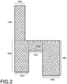
- the dimensions of the fabric 2 of the implant used in this embodiment are the following (see figure 2 ):
- the fabric 2 used is a mesh composed of monofilament polypropylene fibres with the following (general) technical characteristics:
- section 5a of fabric 2 is situated in the posterior area of the vagina 1b (see figures 3 and 4 ). Therefore, the peritoneum is opened just in the right side of the sacral promontory 4. After this step has been performed, the mesorectum is dissected towards the right uterosacral ligaments. The retrorectal space must be found, but not the presacral space. Then, the implant is placed in the posterior area, i.e. section 5a is placed in the posterior part of the vagina 1b, in front of the rectum, and it is fixed to the sides of the space of the rectum levator, being secured in the lower posterior area of the vagina 1b.
- section 5a is placed and fixed, the vagina 1b is surrounded with section 5c of fabric 2 which acts as a bridge and section 5b is situated in the anterior area of the vagina 1, behind the rectum (see figure 4 ). Then, section 5b is fixed in the lower anterior area of the vagina 1b. The dimensions of this section 5b make it possible to anatomically cover the entire anterior area of the vagina 1b (see figure 5 ).
- the bridge section 5c is secured in the vaginal vault. Although this fastening is not strictly necessary, it is recommended to guarantee that the implant is completely fixed.
- the last step of the treatment consists of fixing in distended form the section 3a of fabric 2 to the sacral promontory 4 of the patient.
- Figures 6 and 7 represent the implant situated and fixed in the organ of the patient suffering the prolapse, in this case, the vagina 1b.
- the implant of the present invention enables a complete fastening that is much greater than that enabled by the implants of the state of the art. This greater coverage also guarantees a more uniform distribution of holding forces of the organ which avoids tensions, subsequent problems of pain and the appearance of recurrences.
- the composition and uniform geometric structure of the fabric 2, as well as its uniform mechanical properties both in longitudinal and transversal direction, contribute to improving the distribution of forces and favours the integration of the fabric 2 in the patient's body.
Landscapes
- Health & Medical Sciences (AREA)
- Urology & Nephrology (AREA)
- Cardiology (AREA)
- Oral & Maxillofacial Surgery (AREA)
- Transplantation (AREA)
- Engineering & Computer Science (AREA)
- Biomedical Technology (AREA)
- Heart & Thoracic Surgery (AREA)
- Vascular Medicine (AREA)
- Life Sciences & Earth Sciences (AREA)
- Animal Behavior & Ethology (AREA)
- General Health & Medical Sciences (AREA)
- Public Health (AREA)
- Veterinary Medicine (AREA)
- Prostheses (AREA)
Description
- The present invention relates to an implant for the surgical repair of the pelvic floor by laparoscopy which adjusts to the techniques called sacrocolpopexy and/or sacrohysteropexy. In particular, the present invention relates to an implant for the treatment of vaginal vault prolapse or uterine prolapse using said techniques.
- Uterine prolapse or vaginal vault prolapse is a disorder of the pelvic floor organs produced by different causes that give rise to numerous problems such as, for example, pelvic pain, problems in sexual relations, it may affect urinary incontinence, etc. decreasing the quality of life of the person suffering from it.
- Vaginal vault prolapse can be treated using the surgical technique called colposacropexy or sacrocolpopexy and uterine prolapse can be treated by the surgical technique called sacrohysteropexy. These techniques entail suspension of the vagina or uterus to a point of the pelvic cavity, preferably, to the patient's sacral promontory, via a surgical implant which typically consists of a fabric of implantable polymeric material in the form of mesh. The objective of the mesh is to replace the displaced fascias and substitute the damaged native tissue.
- The sacrocolpopexy technique and the sacrohysteropexy technique can be performed via an abdominal incision, although it is currently performed by laparoscopy since this technique is less aggressive for the patient and also offers numerous advantages. In particular, experts consider that laparoscopy is the only technique that makes it possible to repair the double angle of the vagina (posterior and anterior angle) with a constant vision and direct access to the deepest areas of the pelvis, achieving a suitable and lasting securing of the vagina to the sacral promontory through a surgical mesh.
- Patents
WO2010/138894 ,WO2010/87923 WO02/19946 - Patent document
US2011/297161 refers to a method of treating prolapse of a vagina including the step of securing a base of a Y- shaped support to tissue by inserting an anchor into the tissue, the anchor coupled to the Y-shaped support via an interconnecting member, the step of asymmetrically securing the base of a Y-shaped support to one anterior sacroiliac ligament connected to and located alongside of the sacrum bone and securing each of a pair of legs of the Y-shaped support to opposing sides of the vagina, and the final step of adjusting elevation of the cuff of the vagina relative to the sacrum bone by sliding the interconnecting member relative to the adjustable anchor. - The Y-shaped implantable meshes or fabrics of the patent documents mentioned have the drawback that they are difficult to handle for the surgeon, especially when the operation is carried out by laparoscopy which entails the use of a trocar introduced in the abdomen.
- In particular, it has been observed that the Y-shaped implantable meshes or fabric have joints or welds in the points of intersection of the arms or sections that define the fastening pin. These joints or welds mean that the mesh is less malleable when being introduced in the trocar cannula, which entails more work for the surgeon and, furthermore, in some cases, the risk of tearing the mesh.
- Another drawback of the meshes or fabrics disclosed in said patents lies in the fact that to be able to fix the mesh to the anterior and posterior part of the pelvic organ it is necessary to separate the two arms or sections of superimposed fabric that define the pin. However, this operation is not at all easy to carry out through the trocar, which again entails more work for the surgeon.
- On the other hand, it is important to highlight that a fundamental principal in the practice of sacrocolpopexy is that the fixing of the mesh or fabric to the sacral promontory must be performed without tension, since the tensions are translated into problems of pain in the patient. To eradicate these tensions it is essential to ensure a correct distribution of forces in the organ.
- Patent
WO2007/118260 discloses an implantable mesh for the surgical reconstruction in the area of the pelvic floor including an anterior mesh segment between the bladder and the vagina, a posterior mesh segment between the vagina and the rectum, a pair of distal transobturator holding straps extending from the anterior mesh segment, a pair of proximal transobturator holding straps extending from the anterior mesh segment and a pair of lower dorsal holding straps extending from the posterior mesh segment. According to this patent document, the mesh further includes an intermediate segment located between the anterior mesh segment and the posterior mesh segment. The pair of lower dorsal holding straps extends from the region of the posterior mesh segment bordering on the intermediate segment and a pair of upper dorsal holding straps extends from the region of the anterior mesh segment bordering on the intermediate segment. - Patent document
US2011/105836 discloses a system and method of treating vaginal prolapse and incontinence comprising a kit. The kit includes a mesh graft configured for Attachment to the anterior and posterior vaginal walls to thereby treat the vaginal prolapse. A graft delivery device is also provided for introducing and placing the mesh graft to a location deep within the peritoneal cavity and for attaching the graft thereto. A leg assembly is provided and coupled to an end of the mesh graft and cooperates with the graft delivery device to anchor and affix the mesh graft to the desired anatomical structures. - Patent document
FR 2976788 - Patent
EP 2340784 discloses a textile mesh, in particular for pelvic floor reconstruction or for preventing dyspareunia, comprising a central mesh section and two lateral mesh sections on both sides of the central mesh section. - It has been observed that the Y-shaped fabrics or meshes of the documents of the state of the art do not guarantee the desired distribution of forces, unwanted folds forming in many cases during its placement which constitute, furthermore, a risk factor for infections and possible erosions.
- The objective of the present invention is to resolve said drawbacks, developing a surgical implant for the treatment of the prolapse of a pelvic organ by laparoscopy and via sacrocolpopexy and/or sacrohysteropexy which resolve the aforementioned problems and have the advantages described below.
- In accordance with this objective, according to a first aspect, the present invention provides a surgical implant for the treatment of the prolapse of a pelvic organ by laparoscopy and via sacrocolpopexy and/or sacrohysteropexy, comprising a fabric of implantable material including;
- a first portion for the fastening of said fabric to one or more points of the pelvic cavity and,
- a second portion which is joined to said first portion for the fastening of said fabric to said pelvic organ, said second portion of fabric including;
- two sections of fabric to respectively fix in the anterior and posterior part of said pelvic organ, said two sections of fabric being disposed substantially parallel in the same plane and separated from one another by a distance "d", said second portion of fabric further comprising;
- a third section of fabric which acts as connecting bridge of said two sections to cover said distance "d" of separation, wherein said third section of fabric which act as a bridge extends between said two parallel sections with its lateral edge substantially aligned with the edge of the ends of said two parallel sections, toward the first portion and wherein said first portion for the fastening of the fabric to the pelvic cavity includes;
- a section of fabric to fix to the sacral promontory which extends substantially aligned with and joined to the end of one of said two parallel sections, said section of fabric to fix to the sacral promontory forming a substantially right angle with the third section which acts as a bridge, making it possible for said third bridge section to partially and laterally surround said pelvic organ to place said two parallel sections in the anterior or posterior part of said organ.
- The surgical implant of the present invention has numerous advantages with respect to the implants of the state of the art. In particular, the implant is much easier to place, since it has two sections of fabric to fix to the pelvic organ which are situated in the same plane and are separated from one another by a pre-established distance "d". This distance "d" between the sections of fabric situated in the same plane makes it easier for the surgeon to position the implant, providing him with a much greater margin of manoeuvre that those provided by the pin of the Y-shaped implants of the state of the art. Indeed, in the implants of the state of the art, the surgeon is obliged to separate the sections of superimposed fabric of the pin to place the implant, meaning that the manoeuvre is much more complicated and slower, especially when the operation is carried out by laparoscopy.
- Another advantage of the implant claimed lies in the fact that it has a third section of fabric which acts as a bridge between the two sections mentioned to cover said distance "d" and comfortably and laterally surround the organ. This third bridge section provides an additional fastening coverage of the pelvic organ and enables, on the other hand, the safe and comfortable treatment of vaginal prolapse that conserves the uterus.
- In the implants of the state of the art, the treatment of uterine or vaginal prolapse which conserves the uterus is very difficult since the adjustment of the arms of the pin around the vagina and the fastening to the sacrum is very complex, for which reason, in practice, these Y-shaped implants are only used in the sacrocolpopexy technique. Other known implants, as the one described in patent
WO2007/118260 , likewise do not enable comfortable treatment of a vagina in a patient that conserves the uterus since in this case an intermediate segment of the implant is designed to be folded over the prolapsed vagina. - Another advantage of the implant claimed lies in the fact that it has the advantage that it can be fixed by laparoscopy to the pelvic organ without tensions. This is due to the fact that as the sections of fabric are situated in the same plane, it is easier to adjust the implant to the anatomy of the pelvic organ without problems, avoiding the formation of folds and guaranteeing at all times a correct distribution of forces in the organ.
- The implant of the present invention has a configuration substantially in the form of a chair which has the advantage that it is very easy for the surgeon to handle and substantially reduces the formation of folds or creases. Indeed, as the fixing section to the sacrum and one of said parallel fixing sections to the pelvic organ are aligned, there are fewer steps to be performed by the surgeon to place the implant and the adaptation to the organ's anatomy is easier.
- Preferably, said first and second portion of fabric are integrated in a single piece of fabric of substantially flat structure without welds or joints. Furthermore, advantageously, the structure and density of said fabric is uniform for all the sections mentioned of the implant.
- In this way, a very malleable fabric is obtained which is particularly interesting for use in laparoscopy since it can be coiled and uncoiled without problems as it does not have welds or joints which make the implant more rigid. On the other hand, as it is a fabric with uniform structure and density, it favours a better distribution of the tensile forces in the fixing points.
- Advantageously, according to the same preferred embodiment, said section of fabric to fix to the sacrum extends from the end of one of said parallel sections maintaining one of its lateral edges substantially aligned with the lateral edge of the section whereto it is joined.
Preferably, the distance "d" of separation between said two parallel sections of fabric, or the width of the section acting as a bridge, is dimensioned to laterally surround the pelvic organ to enable the safe and comfortable treatment of vaginal prolapse that conserves the uterus. Advantageously, said distance "d" ranges between 0.035 m (35mm) and 0.045 m (45 mm). - It has been observed that this distance is suitable to connect the anterior and posterior area of a pelvic organ such as, for example, the vagina to repair the vaginal and/or uterine prolapse.
- Again preferably, said two parallel sections of fabric are each configured in the form of a rectangle with suitable dimensions to anatomically cover the anterior or posterior part of said pelvic organ.
- Advantageously, if said pelvic organ is a vagina, one of said parallel sections in the form of a rectangle has an area of magnitude greater than that of the other section to anatomically cover the anterior part of said vagina.
- Again advantageously, said section of fabric to fix to the sacral promontory is configured in the form of a rectangle with a width less than the width of the section of fabric of the second portion of the implant whereto it is joined.
- According to a preferred embodiment, said fabric of implantable material is a mesh, preferably, a mesh of monofilament polypropylene yarns classified within group I of the AMID classification.
- This mesh may be composed of a biological material, such as, for example, collagen, or of a mixture of a biological and synthetic material. Furthermore, the mesh may contain antimicrobial agents.
Advantageously, the pores of said mesh are of a size equal to or greater than 0.0010 m (1 mm) and, preferably, the geometric structure of said pores is substantially regular, such as, for example, a geometric structure in the form of a hexagon. Nevertheless, this geometric structure can also be irregular. - The pore size claimed facilitates the integration of the mesh. On the other hand, its substantially regular pore structure allows that the tensile forces and elongation of the fabric in the longitudinal and transversal directions are very similar. This detail reduces the appearance of tensions with the biological fabrics.
- According to one preferred embodiment, the implant comprises a section of fabric to fix to the sacral promontory, a section of fabric to fix to the posterior part of the pelvic organ, a section of fabric to fix to the anterior part of the pelvic organ, said two sections of fabric to fix to the anterior and the posterior part of the organ being disposed substantially parallel in the same plane and separated from one another by a distance "d", said implant further to cover said distance "d", said bridge section being dimensioned and configured to laterally surround said pelvic organ to enable the safe and comfortable treatment of vaginal prolapse that conserves the uterus.
- The dimensions of the four sections of the fabric comprising the implant may vary in accordance with the dimensions of the pelvic organ to be treated. Nevertheless, these dimensions will preferably be those stated below, where "A" corresponds to the width and "h" corresponds to the length of each one of the sections, hereinafter,
sections -
- A3a= between 0.020 m (20 mm) and 0.040 m (40 mm)
- h3a= between 0.080 m (80 mm) and 0.120 m (120mm)
-
- A5a = between 0.030 m (30mm) and 0.050 m (50 mm)
- h5a= 0.070 m (70 mm) and 0.110 m (110 mm)
-
- A5b= between 0.040 m (40 mm) and 0.060 m (60 mm)
- h5b= 0.080 m (80 mm) and 0.120 m (120 mm)
-
- A5c= between 0.035 m (35 mm) and 0.045 m (45 mm), this dimension corresponding to the distance "d" claimed.
- h5c= between 0.010 m (10 mm) and 0.050 m (50 mm)
- a) situating one of the two sections of fabric of the second portion of the implant in the posterior or anterior area of the pelvic organ,
- b) situating another of the two sections of fabric of the second portion of the implant in the anterior or posterior area of the pelvic organ, partially and laterally surrounding for this the pelvic organ with the third bridge section of fabric of the implant,
- c) fixing the two sections of fabric respectively to the anterior and posterior area of the pelvic organ and, optionally, also fixing the third bridge section of fabric to the vaginal vault when said organ is a vagina, and
- d) later, fixing the first portion of the implant to a point of the pelvic cavity, preferably, to the sacral promontory of the patient.
- The method to treat the prolapse of a pelvic organ by the implant claimed is very fast and comfortable, since the surgeon can laterally surround the pelvic organ with the implant to situate the position of the fixing arms or sections. In the implants of the state of the art, this manoeuvre is not possible since the arms or sections of the pin are superimposed.
- In accordance with a second aspect, the present invention relates to a surgical kit comprising the implant claimed, a laparoscopic instrument to introduce the implant and means to fix the position of the implant in the organ once placed.
Said means to fix the position of the implant may comprise absorbable or non-absorbable sutures and/or "tackers", or biocompatible adhesive, for example an adhesive including fibrin or cyanoacrylates, etc... - The implant claimed has the advantage that it makes viable and facilitates the use of the adhesive on the parts of the organ thanks to the additional coverage provided by the sections of the second portion of the implant. This adhesive can be applied by a specially designed applicator for laparoscopic use.
- In the present invention, pelvic organ is understood to be any organ of the pelvic cavity of a woman, preferably, a vagina or uterus.
- For a better understanding of all that disclosed, drawings are attached wherein, diagrammatically and only as a non-limitative example, one embodiment of the invention is represented.
-
-
Figures 1 and2 show plan views of an embodiment of the implant. In these figures, the different sections of the implant's fabric have been delimited diagrammatically using broken lines to facilitate the description. -
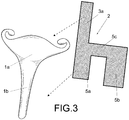
Figure 3 is a perspective view showing a vagina with uterus and the implant of the embodiment of thefigure 1 designed to secure said vagina for the treatment of the prolapse. -
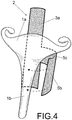
Figure 4 is a perspective view showing the manner in which the surgeon situates the implant on the pelvic organ, in this case, a vagina. -
Figure 5 is a perspective view showing the implant already disposed on the vagina. -
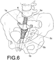
Figure 6 is a perspective view showing the manner in which the implant is situated and secured inside the pelvic cavity. -
Figure 7 is a section of anatomy showing the manner in which the implant is situated and secured inside the pelvic cavity. - The implant of the present invention is described below, making reference to
figures 1 to 7 . These figures show an implant for the treatment of the prolapse of avagina 1b which conserves theuterus 1a via sacrohysteropexy. - The implant claimed comprises a
fabric 2 of implantable material which, in the embodiment described, is a mesh of uniform structure and density manufactured from monofilament polypropylene yarns and classified within group I of the AMID classification. The pore size of this mesh is between 0.0028 m (2.8 mm) and 0.0036 m (3.6 mm) and the geometric structure of the pore is regular in the form of a hexagon. - This
fabric 2 in the form of mesh includes afirst portion 3 for the fastening of thefabric 2 to a point of the pelvic cavity, in this case, thesacral promontory 4, and asecond portion 5 for the fastening of thefabric 2 to the pelvic organ, in this case, thevagina 1b. - The
second portion 5 of thefabric 2 comprises twosections fabric 2 in the form of a rectangle to respectively fix in the anterior and posterior part of the pelvic organ. As can be seen infigure 1 , these twosections fabric 2 are disposed substantially parallel in the same plane and separated from one another by a distance "d". This samesecond portion 5 offabric 2 includes athird section 5c offabric 2 which acts as a connecting bridge of theparallel sections - The
first portion 3 offabric 2 comprises asingle section 3a offabric 2 in the form of a rectangle to fix to thesacral promontory 4. Thissection 3a offabric 2 extends aligned withsection 5a of thesecond portion 5 maintaining one of itslateral edges 6 substantially aligned with thelateral edge 7 of saidsection 5a and forming a substantially right angle with thethird section 5c offabric 2 which acts as a bridge. On the other hand, thethird section 5c which acts as a bridge extends between the twoparallel sections lateral edge 8 substantially aligned with theupper edge sections - In this way, as can be seen in
figures 1 and2 , thefabric 2 of the implant acquires a general chair-shaped configuration which has the advantage that it is very easy to handle for the surgeon and substantially avoids the formation of folds or creases, since it is easier to adapt to the patient's anatomy. - In the embodiment described, the four
sections fabric 2 which define the implant are integrated in a single piece offabric 2 of substantially flat structure without welds or joints. Thanks to this, the implant is highly malleable for which reason it is particularly interesting for use in laparoscopy, since it can be coiled and uncoiled and penetrate in the trocar without the risk of it breaking. - Intervention by laparoscopy has the following advantages in the repair of vaginal prolapse with uterus via the implant of the present invention:
- ▪ The vagina is situated in horizontal position with respect to the plane of the levators and its angle can be corrected, and, as the uterus is conserved, the fixing of the cervix in the sacral cavity is physiological.
- ▪ The fixing of the implant is safer and more stable.
- ▪ A complementary laparoscopic colposuspension can be performed (for example, according to Bursch's technique).
- ▪ As the uterus is conserved, there are high possibilities of pregnancy and low possibilities of miscarriage.
- ▪ Lack of scars and without lateralization of the vagina.
- ▪ Greater length of the vagina after fixing to the sacrum, especially in android type vaginas and where is a shorter distance between the introitus and the ischiatic spine, which conditions a short vagina.
- ▪ Capacity of re-assessing all defects in the same surgical act and fully repairing them after readjusting the therapeutic strategy.
- In the embodiment described of the implant, the pelvic organ to treat is a
vagina 1b which conserves theuterus 1 a of approximately 0.08 m - 0.11 m (8- 11 cm), with an anterior area of approximately 0.07 m (7 cm) and a posterior area of approximately 0.09 m (9 cm). Theuterus 1a has general anatomical measurements of 0.08 m (8 cm) in length, 0.05 m (5 cm) in width and 0.025 m (2.5 cm) thickness. - The dimensions of the
fabric 2 of the implant used in this embodiment are the following (seefigure 2 ): -
Section 3a to fix to the sacral promontory: A3a= 0.03 m (30 mm), h3a= 0.100 m (100 mm) -
Section 5a to fix to the posterior part of thevagina 1b: A5a = 0.04 m (40 mm), h5a= 0.09 m (90 mm) -
Section 5b to fix to the anterior part of thevagina 1b: A5b= 0.050 m (50 mm), h5b= 0.100 m (100 mm) -
Section 5c which acts as a bridge and surrounds thevagina 1b: A5c= 0.040 m (40 mm), h5c= 0.030 m (30 mm): The dimension A5c corresponds to the distance "d" claimed ofsection 5c. - The
fabric 2 used is a mesh composed of monofilament polypropylene fibres with the following (general) technical characteristics: - Thickness of the fabric 2: 0.0005 m (0.5 mm)
- Weight: 0.048 Kg/m2
- Tensile force in longitudinal direction: equal to or greater than 25 N
- Tensile force in transversal direction: equal to or greater than 25N
- Elongation in longitudinal direction: equal to or greater than 60%
- Elongation in transversal direction: equal to or greater than 60 %
- Tear strength of suture in longitudinal direction: 31N
- Tear strength of suture in transversal direction: 35 N
- Diameter of the yarn: 0.000150 m (0.150 mm)
- Pore size of the mesh: macropore of 0.0030 m (3.0 mm)
- Firstly,
section 5a offabric 2 is situated in the posterior area of thevagina 1b (seefigures 3 and4 ). Therefore, the peritoneum is opened just in the right side of thesacral promontory 4. After this step has been performed, the mesorectum is dissected towards the right uterosacral ligaments. The retrorectal space must be found, but not the presacral space. Then, the implant is placed in the posterior area, i.e.section 5a is placed in the posterior part of thevagina 1b, in front of the rectum, and it is fixed to the sides of the space of the rectum levator, being secured in the lower posterior area of thevagina 1b. - Next, once
section 5a is placed and fixed, thevagina 1b is surrounded withsection 5c offabric 2 which acts as a bridge andsection 5b is situated in the anterior area of the vagina 1, behind the rectum (seefigure 4 ). Then,section 5b is fixed in the lower anterior area of thevagina 1b. The dimensions of thissection 5b make it possible to anatomically cover the entire anterior area of thevagina 1b (seefigure 5 ). - Once
sections vagina 1b, thebridge section 5c is secured in the vaginal vault. Although this fastening is not strictly necessary, it is recommended to guarantee that the implant is completely fixed. - The last step of the treatment consists of fixing in distended form the
section 3a offabric 2 to thesacral promontory 4 of the patient.Figures 6 and7 represent the implant situated and fixed in the organ of the patient suffering the prolapse, in this case, thevagina 1b. - As can be seen in these
figures 6 and7 , the implant of the present invention enables a complete fastening that is much greater than that enabled by the implants of the state of the art. This greater coverage also guarantees a more uniform distribution of holding forces of the organ which avoids tensions, subsequent problems of pain and the appearance of recurrences. The composition and uniform geometric structure of thefabric 2, as well as its uniform mechanical properties both in longitudinal and transversal direction, contribute to improving the distribution of forces and favours the integration of thefabric 2 in the patient's body. - Despite the fact that reference has been made to a specific embodiment of the present invention, it is evident for a person skilled in the art that numerous variations and modifications can be made to the implant of the invention, without departing from the scope of protection defined by the attached claims.
Claims (15)
- Surgical implant for the treatment of the prolapse of a pelvic organ (1a,1b) by laparoscopy and via sacrocolpopexy and/or sacrohysteropexy, comprising a fabric (2) of implantable material including;- a first portion (3) for the fastening of said fabric to one or more points of the pelvic cavity,- a second portion (5) which is joined to said first portion (3) for the fastening of said fabric (2) to said pelvic organ (1a,1b), said second portion (5) of fabric (2) including;- two sections (5a,5b) of fabric (2) to respectively fix in the anterior and posterior part of said pelvic organ (1a,1b), said two sections (5a,5b) of fabric (2) being disposed substantially parallel in the same plane and separated from one another by a distance "d", said second portion (5) of fabric (2) further comprising;- a third section (5c) of fabric which acts as connecting bridge of said two parallel sections (5a,5b) to cover said distance "d" of separation, wherein said third section of fabric which act as a bridge extends between said two parallel sections with its lateral edge substantially aligned with the edge of the ends of said two parallel sections, toward the first portion (3) and whereinsaid first portion (3) for the fastening of the fabric to the pelvic cavity includes;- a section (3a) of fabric (2) to fix to the sacral promontory which extends substantially aligned with and joined to the end of one of said two parallel sections (5a,5b), said section (3a) of fabric (2) to fix to the sacral promontory forming a substantially right angle with the third section (5c) which acts as a bridge, making it possible for said third bridge section (5c) to partially and laterally surround said pelvic organ (1a,1b) to place said two parallel sections (5a,5b) in the anterior or posterior part of said organ (1a, 1b).
- Implant according to claim 1, wherein said first and second portion (3,5) of fabric (2) are integrated in a single piece of fabric (2) of substantially flat structure without welds or joints between said sections (3,5).
- Implant according to claim 1, wherein the structure and density of said fabric (2) is uniform for all the sections (3,5a,5b,5c) of the implant.
- Implant according to any of claims 1 to 3, wherein said section of fabric (3a) to fix to the sacrum (4) extends from the end of one of said parallel sections (5a) maintaining one of its lateral edges (6) substantially aligned with the lateral edge (7) of the section (5a) whereto it is joined.
- Implant according to any of the preceding claims, wherein said distance "d" of separation, or the width (A5c) of the third section which acts as a bridge (5c), is dimensioned to laterally surround the pelvic organ (1b) to enable the safe and comfortable treatment of vaginal (1b) prolapse that conserves the uterus (1a).
- Implant according to claim 5, wherein said distance "d" ranges between 0.035 m and 0.045 m.
- Implant according to any of the preceding claims, wherein said two parallel sections (5a,5b) of fabric (2) are each configured in the form of a rectangle with suitable dimensions (A5a, A5b, h5a,h5b) to cover the anatomy of the anterior or posterior part of said pelvic organ (1b).
- Implant according to claim 7, wherein said pelvic organ is a vagina (1b) and one of said parallel sections (5b) in the form of rectangle has an area of magnitude greater than that of the other section (5a) to anatomically cover the anterior part of said vagina (1b).
- Implant according to claim 7, wherein said section (3a) of fabric to fix to the sacral promontory (4) is configured in the form of a rectangle with a width (A3a) less than the width (A5a) of the section (5a) of fabric (2) of the second portion (3) of the implant whereto it is joined.
- Implant according to claim 1, wherein said bridge section (5c) is dimensioned and configured to laterally surround said pelvic organ (1b) to enable the safe and comfortable treatment of vaginal (1b) prolapse that conserves the uterus (1a).
- Implant according to claim 1, wherein said fabric (2) is a mesh, preferably, a mesh of monofilament polypropylene yarns classified within group I of the AMID classification.
- Implant according to claim 11, wherein the pores of said mesh are of a size equal to or greater than 0.0010 m.
- Implant according to any of claims 11 to 12, wherein the geometric structure of said pores is substantially regular, preferably, a geometric structure, in the form of a hexagon.
- Implant according to any of the preceding claims, wherein said pelvic organ is a vagina (1b) or a uterus (1a) of a patient.
- Surgical kit comprising an implant according to any of the preceding claims, a laparoscopic instrument to introduce the implant and means to fix the position of the implant in the organ once placed, said means preferably including a biocompatible adhesive to fix the sections (5a,5b,5c) of the second portion (5) of the implant on the pelvic organ (1a, 1b).
Priority Applications (2)
| Application Number | Priority Date | Filing Date | Title |
|---|---|---|---|
| EP14700291.9A EP2943147B1 (en) | 2013-01-10 | 2014-01-10 | Surgical implant for the treatment of the prolapse of a pelvic organ by laparoscopy via sacrocolpopexy and/or sacrohysteropexy |
| PL14700291T PL2943147T3 (en) | 2013-01-10 | 2014-01-10 | Surgical implant for the treatment of the prolapse of a pelvic organ by laparoscopy via sacrocolpopexy and/or sacrohysteropexy |
Applications Claiming Priority (3)
| Application Number | Priority Date | Filing Date | Title |
|---|---|---|---|
| EP13382005.0A EP2754412A1 (en) | 2013-01-10 | 2013-01-10 | A surgical implant for the treatment of pelvic organ prolapse by sacrocolpopexy and/or sacrohysteropexy |
| EP14700291.9A EP2943147B1 (en) | 2013-01-10 | 2014-01-10 | Surgical implant for the treatment of the prolapse of a pelvic organ by laparoscopy via sacrocolpopexy and/or sacrohysteropexy |
| PCT/EP2014/050361 WO2014108493A1 (en) | 2013-01-10 | 2014-01-10 | Surgical implant for the treatment of the prolapse of a pelvic organ by laparoscopy via sacrocolpopexy and/or sacrohysteropexy |
Publications (2)
| Publication Number | Publication Date |
|---|---|
| EP2943147A1 EP2943147A1 (en) | 2015-11-18 |
| EP2943147B1 true EP2943147B1 (en) | 2017-09-27 |
Family
ID=48040136
Family Applications (2)
| Application Number | Title | Priority Date | Filing Date |
|---|---|---|---|
| EP13382005.0A Withdrawn EP2754412A1 (en) | 2013-01-10 | 2013-01-10 | A surgical implant for the treatment of pelvic organ prolapse by sacrocolpopexy and/or sacrohysteropexy |
| EP14700291.9A Active EP2943147B1 (en) | 2013-01-10 | 2014-01-10 | Surgical implant for the treatment of the prolapse of a pelvic organ by laparoscopy via sacrocolpopexy and/or sacrohysteropexy |
Family Applications Before (1)
| Application Number | Title | Priority Date | Filing Date |
|---|---|---|---|
| EP13382005.0A Withdrawn EP2754412A1 (en) | 2013-01-10 | 2013-01-10 | A surgical implant for the treatment of pelvic organ prolapse by sacrocolpopexy and/or sacrohysteropexy |
Country Status (4)
| Country | Link |
|---|---|
| EP (2) | EP2754412A1 (en) |
| ES (1) | ES2660693T3 (en) |
| PL (1) | PL2943147T3 (en) |
| WO (1) | WO2014108493A1 (en) |
Families Citing this family (3)
| Publication number | Priority date | Publication date | Assignee | Title |
|---|---|---|---|---|
| US9480546B2 (en) * | 2013-08-05 | 2016-11-01 | Coloplast A/S | Hysteropexy mesh apparatuses and methods |
| ITUB20153642A1 (en) * | 2015-09-15 | 2017-03-15 | Assut Europe Spa | Functional biological prosthesis for the treatment of pelvic floor prolapse |
| CA3213536A1 (en) * | 2021-04-29 | 2022-11-03 | Salwan Hamoudi Musa Al-Salihi | A surgical device |
Family Cites Families (8)
| Publication number | Priority date | Publication date | Assignee | Title |
|---|---|---|---|---|
| US6592515B2 (en) | 2000-09-07 | 2003-07-15 | Ams Research Corporation | Implantable article and method |
| KR20090012217A (en) * | 2006-04-14 | 2009-02-02 | 에이엠아이 어젠시 포 메디컬 이노베이션스 게엠베하 | Implantable Mesh for Surgical Reconstruction in the Pelvic Floor Region |
| CN102341061A (en) | 2009-02-02 | 2012-02-01 | Cr巴德公司 | Implant for pelvic organ prolapse repair |
| US8720446B2 (en) * | 2010-06-04 | 2014-05-13 | Coloplast A/S | Sacrocolpopexy support and method of implantation |
| WO2010138894A2 (en) | 2009-05-28 | 2010-12-02 | Rosenblatt Associates, Llc | Tubular mesh for sacrocolpopexy and related procedures |
| US9414902B2 (en) * | 2009-11-04 | 2016-08-16 | Boston Scientific Scimed, Inc. | Method for treating prolapse and incontinence |
| DE102009060799A1 (en) * | 2009-12-31 | 2011-07-07 | Aesculap AG, 78532 | Textile network, in particular for the treatment or treatment of prolapse or for the prevention of dyspareunia |
| FR2976788B1 (en) * | 2011-06-22 | 2014-04-04 | Ct Hospitalier Universitaire Nimes | PROSTHESIS FOR RECTOPEXIE AND CYSTOPEXIE |
-
2013
- 2013-01-10 EP EP13382005.0A patent/EP2754412A1/en not_active Withdrawn
-
2014
- 2014-01-10 WO PCT/EP2014/050361 patent/WO2014108493A1/en not_active Ceased
- 2014-01-10 PL PL14700291T patent/PL2943147T3/en unknown
- 2014-01-10 ES ES14700291.9T patent/ES2660693T3/en active Active
- 2014-01-10 EP EP14700291.9A patent/EP2943147B1/en active Active
Non-Patent Citations (1)
| Title |
|---|
| None * |
Also Published As
| Publication number | Publication date |
|---|---|
| WO2014108493A1 (en) | 2014-07-17 |
| EP2754412A1 (en) | 2014-07-16 |
| EP2943147A1 (en) | 2015-11-18 |
| ES2660693T3 (en) | 2018-03-23 |
| PL2943147T3 (en) | 2018-02-28 |
Similar Documents
| Publication | Publication Date | Title |
|---|---|---|
| US8328825B2 (en) | Method and implant for curing cystocele | |
| JP4241389B2 (en) | Method and apparatus for treatment of pelvic prolapse in female patients | |
| US7588598B2 (en) | Implant for treating rectocele and a device for putting said implant into place | |
| US7204801B2 (en) | Pubovaginal support for treating female urinary incontinence | |
| JP2006525097A (en) | Prosthesis used for surgical treatment of urogenital prolapse and female urinary incontinence | |
| AU2004229195B2 (en) | Implant for the treatment of a cystocele and device for placement of said implant | |
| US20150099924A1 (en) | Methods and devices for repair of vaginal wall or uterus | |
| CN102137633A (en) | Minimally invasive implant and method | |
| US9161829B2 (en) | Implantable medical device and methods of delivering the implantable medical device | |
| JP6490179B2 (en) | Medical devices and instruments for adjustment and cutting | |
| EP2754412A1 (en) | A surgical implant for the treatment of pelvic organ prolapse by sacrocolpopexy and/or sacrohysteropexy | |
| US10213286B2 (en) | Implantable medical device and methods of delivering the implantable medical device | |
| US10039627B2 (en) | Medical device and method of delivering the medical device | |
| US8986190B2 (en) | Implant for treating a genital prolapse, and associated treatment method | |
| US9283063B2 (en) | Medical device and methods of delivering the medical device | |
| US9333062B2 (en) | Bodily implants and methods of treating fecal incontinence using bodily implants | |
| RU2020135066A (en) | Laparoscopic bilateral hysterocervicocoloposuspension using titanium mesh implants in patients with apical prolapse |
Legal Events
| Date | Code | Title | Description |
|---|---|---|---|
| PUAI | Public reference made under article 153(3) epc to a published international application that has entered the european phase |
Free format text: ORIGINAL CODE: 0009012 |
|
| 17P | Request for examination filed |
Effective date: 20150730 |
|
| AK | Designated contracting states |
Kind code of ref document: A1 Designated state(s): AL AT BE BG CH CY CZ DE DK EE ES FI FR GB GR HR HU IE IS IT LI LT LU LV MC MK MT NL NO PL PT RO RS SE SI SK SM TR |
|
| AX | Request for extension of the european patent |
Extension state: BA ME |
|
| DAX | Request for extension of the european patent (deleted) | ||
| GRAP | Despatch of communication of intention to grant a patent |
Free format text: ORIGINAL CODE: EPIDOSNIGR1 |
|
| STAA | Information on the status of an ep patent application or granted ep patent |
Free format text: STATUS: GRANT OF PATENT IS INTENDED |
|
| INTG | Intention to grant announced |
Effective date: 20170508 |
|
| GRAS | Grant fee paid |
Free format text: ORIGINAL CODE: EPIDOSNIGR3 |
|
| GRAA | (expected) grant |
Free format text: ORIGINAL CODE: 0009210 |
|
| STAA | Information on the status of an ep patent application or granted ep patent |
Free format text: STATUS: THE PATENT HAS BEEN GRANTED |
|
| AK | Designated contracting states |
Kind code of ref document: B1 Designated state(s): AL AT BE BG CH CY CZ DE DK EE ES FI FR GB GR HR HU IE IS IT LI LT LU LV MC MK MT NL NO PL PT RO RS SE SI SK SM TR |
|
| REG | Reference to a national code |
Ref country code: GB Ref legal event code: FG4D |
|
| REG | Reference to a national code |
Ref country code: CH Ref legal event code: EP |
|
| REG | Reference to a national code |
Ref country code: AT Ref legal event code: REF Ref document number: 931282 Country of ref document: AT Kind code of ref document: T Effective date: 20171015 |
|
| REG | Reference to a national code |
Ref country code: IE Ref legal event code: FG4D |
|
| REG | Reference to a national code |
Ref country code: DE Ref legal event code: R096 Ref document number: 602014015044 Country of ref document: DE |
|
| PG25 | Lapsed in a contracting state [announced via postgrant information from national office to epo] |
Ref country code: HR Free format text: LAPSE BECAUSE OF FAILURE TO SUBMIT A TRANSLATION OF THE DESCRIPTION OR TO PAY THE FEE WITHIN THE PRESCRIBED TIME-LIMIT Effective date: 20170927 Ref country code: NO Free format text: LAPSE BECAUSE OF FAILURE TO SUBMIT A TRANSLATION OF THE DESCRIPTION OR TO PAY THE FEE WITHIN THE PRESCRIBED TIME-LIMIT Effective date: 20171227 Ref country code: FI Free format text: LAPSE BECAUSE OF FAILURE TO SUBMIT A TRANSLATION OF THE DESCRIPTION OR TO PAY THE FEE WITHIN THE PRESCRIBED TIME-LIMIT Effective date: 20170927 Ref country code: LT Free format text: LAPSE BECAUSE OF FAILURE TO SUBMIT A TRANSLATION OF THE DESCRIPTION OR TO PAY THE FEE WITHIN THE PRESCRIBED TIME-LIMIT Effective date: 20170927 Ref country code: SE Free format text: LAPSE BECAUSE OF FAILURE TO SUBMIT A TRANSLATION OF THE DESCRIPTION OR TO PAY THE FEE WITHIN THE PRESCRIBED TIME-LIMIT Effective date: 20170927 |
|
| REG | Reference to a national code |
Ref country code: NL Ref legal event code: MP Effective date: 20170927 |
|
| REG | Reference to a national code |
Ref country code: LT Ref legal event code: MG4D |
|
| REG | Reference to a national code |
Ref country code: AT Ref legal event code: MK05 Ref document number: 931282 Country of ref document: AT Kind code of ref document: T Effective date: 20170927 |
|
| PG25 | Lapsed in a contracting state [announced via postgrant information from national office to epo] |
Ref country code: LV Free format text: LAPSE BECAUSE OF FAILURE TO SUBMIT A TRANSLATION OF THE DESCRIPTION OR TO PAY THE FEE WITHIN THE PRESCRIBED TIME-LIMIT Effective date: 20170927 Ref country code: RS Free format text: LAPSE BECAUSE OF FAILURE TO SUBMIT A TRANSLATION OF THE DESCRIPTION OR TO PAY THE FEE WITHIN THE PRESCRIBED TIME-LIMIT Effective date: 20170927 Ref country code: GR Free format text: LAPSE BECAUSE OF FAILURE TO SUBMIT A TRANSLATION OF THE DESCRIPTION OR TO PAY THE FEE WITHIN THE PRESCRIBED TIME-LIMIT Effective date: 20171228 Ref country code: BG Free format text: LAPSE BECAUSE OF FAILURE TO SUBMIT A TRANSLATION OF THE DESCRIPTION OR TO PAY THE FEE WITHIN THE PRESCRIBED TIME-LIMIT Effective date: 20171227 |
|
| REG | Reference to a national code |
Ref country code: ES Ref legal event code: FG2A Ref document number: 2660693 Country of ref document: ES Kind code of ref document: T3 Effective date: 20180323 |
|
| PG25 | Lapsed in a contracting state [announced via postgrant information from national office to epo] |
Ref country code: NL Free format text: LAPSE BECAUSE OF FAILURE TO SUBMIT A TRANSLATION OF THE DESCRIPTION OR TO PAY THE FEE WITHIN THE PRESCRIBED TIME-LIMIT Effective date: 20170927 |
|
| PG25 | Lapsed in a contracting state [announced via postgrant information from national office to epo] |
Ref country code: RO Free format text: LAPSE BECAUSE OF FAILURE TO SUBMIT A TRANSLATION OF THE DESCRIPTION OR TO PAY THE FEE WITHIN THE PRESCRIBED TIME-LIMIT Effective date: 20170927 Ref country code: CZ Free format text: LAPSE BECAUSE OF FAILURE TO SUBMIT A TRANSLATION OF THE DESCRIPTION OR TO PAY THE FEE WITHIN THE PRESCRIBED TIME-LIMIT Effective date: 20170927 |
|
| PG25 | Lapsed in a contracting state [announced via postgrant information from national office to epo] |
Ref country code: EE Free format text: LAPSE BECAUSE OF FAILURE TO SUBMIT A TRANSLATION OF THE DESCRIPTION OR TO PAY THE FEE WITHIN THE PRESCRIBED TIME-LIMIT Effective date: 20170927 Ref country code: SK Free format text: LAPSE BECAUSE OF FAILURE TO SUBMIT A TRANSLATION OF THE DESCRIPTION OR TO PAY THE FEE WITHIN THE PRESCRIBED TIME-LIMIT Effective date: 20170927 Ref country code: IS Free format text: LAPSE BECAUSE OF FAILURE TO SUBMIT A TRANSLATION OF THE DESCRIPTION OR TO PAY THE FEE WITHIN THE PRESCRIBED TIME-LIMIT Effective date: 20180127 Ref country code: SM Free format text: LAPSE BECAUSE OF FAILURE TO SUBMIT A TRANSLATION OF THE DESCRIPTION OR TO PAY THE FEE WITHIN THE PRESCRIBED TIME-LIMIT Effective date: 20170927 Ref country code: AT Free format text: LAPSE BECAUSE OF FAILURE TO SUBMIT A TRANSLATION OF THE DESCRIPTION OR TO PAY THE FEE WITHIN THE PRESCRIBED TIME-LIMIT Effective date: 20170927 |
|
| REG | Reference to a national code |
Ref country code: DE Ref legal event code: R097 Ref document number: 602014015044 Country of ref document: DE |
|
| PG25 | Lapsed in a contracting state [announced via postgrant information from national office to epo] |
Ref country code: DK Free format text: LAPSE BECAUSE OF FAILURE TO SUBMIT A TRANSLATION OF THE DESCRIPTION OR TO PAY THE FEE WITHIN THE PRESCRIBED TIME-LIMIT Effective date: 20170927 |
|
| PLBE | No opposition filed within time limit |
Free format text: ORIGINAL CODE: 0009261 |
|
| STAA | Information on the status of an ep patent application or granted ep patent |
Free format text: STATUS: NO OPPOSITION FILED WITHIN TIME LIMIT |
|
| REG | Reference to a national code |
Ref country code: CH Ref legal event code: PL |
|
| 26N | No opposition filed |
Effective date: 20180628 |
|
| GBPC | Gb: european patent ceased through non-payment of renewal fee |
Effective date: 20180110 |
|
| PG25 | Lapsed in a contracting state [announced via postgrant information from national office to epo] |
Ref country code: FR Free format text: LAPSE BECAUSE OF NON-PAYMENT OF DUE FEES Effective date: 20180131 Ref country code: LU Free format text: LAPSE BECAUSE OF NON-PAYMENT OF DUE FEES Effective date: 20180110 |
|
| REG | Reference to a national code |
Ref country code: IE Ref legal event code: MM4A |
|
| REG | Reference to a national code |
Ref country code: FR Ref legal event code: ST Effective date: 20180928 |
|
| REG | Reference to a national code |
Ref country code: BE Ref legal event code: MM Effective date: 20180131 |
|
| PG25 | Lapsed in a contracting state [announced via postgrant information from national office to epo] |
Ref country code: LI Free format text: LAPSE BECAUSE OF NON-PAYMENT OF DUE FEES Effective date: 20180131 Ref country code: BE Free format text: LAPSE BECAUSE OF NON-PAYMENT OF DUE FEES Effective date: 20180131 Ref country code: CH Free format text: LAPSE BECAUSE OF NON-PAYMENT OF DUE FEES Effective date: 20180131 Ref country code: SI Free format text: LAPSE BECAUSE OF FAILURE TO SUBMIT A TRANSLATION OF THE DESCRIPTION OR TO PAY THE FEE WITHIN THE PRESCRIBED TIME-LIMIT Effective date: 20170927 Ref country code: GB Free format text: LAPSE BECAUSE OF NON-PAYMENT OF DUE FEES Effective date: 20180110 |
|
| PG25 | Lapsed in a contracting state [announced via postgrant information from national office to epo] |
Ref country code: IE Free format text: LAPSE BECAUSE OF NON-PAYMENT OF DUE FEES Effective date: 20180110 |
|
| PG25 | Lapsed in a contracting state [announced via postgrant information from national office to epo] |
Ref country code: MC Free format text: LAPSE BECAUSE OF FAILURE TO SUBMIT A TRANSLATION OF THE DESCRIPTION OR TO PAY THE FEE WITHIN THE PRESCRIBED TIME-LIMIT Effective date: 20170927 |
|
| PG25 | Lapsed in a contracting state [announced via postgrant information from national office to epo] |
Ref country code: MT Free format text: LAPSE BECAUSE OF NON-PAYMENT OF DUE FEES Effective date: 20180110 |
|
| PG25 | Lapsed in a contracting state [announced via postgrant information from national office to epo] |
Ref country code: TR Free format text: LAPSE BECAUSE OF FAILURE TO SUBMIT A TRANSLATION OF THE DESCRIPTION OR TO PAY THE FEE WITHIN THE PRESCRIBED TIME-LIMIT Effective date: 20170927 |
|
| PG25 | Lapsed in a contracting state [announced via postgrant information from national office to epo] |
Ref country code: PT Free format text: LAPSE BECAUSE OF FAILURE TO SUBMIT A TRANSLATION OF THE DESCRIPTION OR TO PAY THE FEE WITHIN THE PRESCRIBED TIME-LIMIT Effective date: 20170927 |
|
| PG25 | Lapsed in a contracting state [announced via postgrant information from national office to epo] |
Ref country code: HU Free format text: LAPSE BECAUSE OF FAILURE TO SUBMIT A TRANSLATION OF THE DESCRIPTION OR TO PAY THE FEE WITHIN THE PRESCRIBED TIME-LIMIT; INVALID AB INITIO Effective date: 20140110 Ref country code: CY Free format text: LAPSE BECAUSE OF FAILURE TO SUBMIT A TRANSLATION OF THE DESCRIPTION OR TO PAY THE FEE WITHIN THE PRESCRIBED TIME-LIMIT Effective date: 20170927 Ref country code: MK Free format text: LAPSE BECAUSE OF NON-PAYMENT OF DUE FEES Effective date: 20170927 |
|
| PG25 | Lapsed in a contracting state [announced via postgrant information from national office to epo] |
Ref country code: AL Free format text: LAPSE BECAUSE OF FAILURE TO SUBMIT A TRANSLATION OF THE DESCRIPTION OR TO PAY THE FEE WITHIN THE PRESCRIBED TIME-LIMIT Effective date: 20170927 |
|
| PGFP | Annual fee paid to national office [announced via postgrant information from national office to epo] |
Ref country code: ES Payment date: 20260217 Year of fee payment: 13 |
|
| PGFP | Annual fee paid to national office [announced via postgrant information from national office to epo] |
Ref country code: DE Payment date: 20260120 Year of fee payment: 13 |
|
| PGFP | Annual fee paid to national office [announced via postgrant information from national office to epo] |
Ref country code: IT Payment date: 20260130 Year of fee payment: 13 |
|
| PGFP | Annual fee paid to national office [announced via postgrant information from national office to epo] |
Ref country code: PL Payment date: 20260109 Year of fee payment: 13 |