EP2938746B1 - Probe kit for detecting a single strand target nucleotide sequence - Google Patents
Probe kit for detecting a single strand target nucleotide sequence Download PDFInfo
- Publication number
- EP2938746B1 EP2938746B1 EP13831873.8A EP13831873A EP2938746B1 EP 2938746 B1 EP2938746 B1 EP 2938746B1 EP 13831873 A EP13831873 A EP 13831873A EP 2938746 B1 EP2938746 B1 EP 2938746B1
- Authority
- EP
- European Patent Office
- Prior art keywords
- probe
- nucleotide sequence
- hybr
- target nucleotide
- kit
- Prior art date
- Legal status (The legal status is an assumption and is not a legal conclusion. Google has not performed a legal analysis and makes no representation as to the accuracy of the status listed.)
- Not-in-force
Links
Images



Classifications
-
- C—CHEMISTRY; METALLURGY
- C12—BIOCHEMISTRY; BEER; SPIRITS; WINE; VINEGAR; MICROBIOLOGY; ENZYMOLOGY; MUTATION OR GENETIC ENGINEERING
- C12Q—MEASURING OR TESTING PROCESSES INVOLVING ENZYMES, NUCLEIC ACIDS OR MICROORGANISMS; COMPOSITIONS OR TEST PAPERS THEREFOR; PROCESSES OF PREPARING SUCH COMPOSITIONS; CONDITION-RESPONSIVE CONTROL IN MICROBIOLOGICAL OR ENZYMOLOGICAL PROCESSES
- C12Q1/00—Measuring or testing processes involving enzymes, nucleic acids or microorganisms; Compositions therefor; Processes of preparing such compositions
- C12Q1/68—Measuring or testing processes involving enzymes, nucleic acids or microorganisms; Compositions therefor; Processes of preparing such compositions involving nucleic acids
- C12Q1/6813—Hybridisation assays
- C12Q1/6816—Hybridisation assays characterised by the detection means
- C12Q1/6818—Hybridisation assays characterised by the detection means involving interaction of two or more labels, e.g. resonant energy transfer
-
- C—CHEMISTRY; METALLURGY
- C12—BIOCHEMISTRY; BEER; SPIRITS; WINE; VINEGAR; MICROBIOLOGY; ENZYMOLOGY; MUTATION OR GENETIC ENGINEERING
- C12Q—MEASURING OR TESTING PROCESSES INVOLVING ENZYMES, NUCLEIC ACIDS OR MICROORGANISMS; COMPOSITIONS OR TEST PAPERS THEREFOR; PROCESSES OF PREPARING SUCH COMPOSITIONS; CONDITION-RESPONSIVE CONTROL IN MICROBIOLOGICAL OR ENZYMOLOGICAL PROCESSES
- C12Q1/00—Measuring or testing processes involving enzymes, nucleic acids or microorganisms; Compositions therefor; Processes of preparing such compositions
- C12Q1/68—Measuring or testing processes involving enzymes, nucleic acids or microorganisms; Compositions therefor; Processes of preparing such compositions involving nucleic acids
- C12Q1/6813—Hybridisation assays
- C12Q1/6816—Hybridisation assays characterised by the detection means
Definitions
- the present invention relates to a probe kit for detecting a single strand target nucleotide sequence.
- amplification of the sample is required for all these assays in order to allow a sufficient detection sensitivity.
- a first example of this strategy consists in molecular beacons (MB), single strand oligonucleotides with a stem-loop structure formed by 4 parts: a) a loop consisting of a region of 15-30 nucleotides complementary to the target sequence; b) a stem, i.e. a short duplex segment formed by 5-8 base pairs; c) a reporter which is generally bound to the 5' end and emits fluorescent light; d) a quencher bound to the 3' end, which absorbs the light radiation of the reporter.
- MB molecular beacons
- the stem-loop structure of the molecular beacon undergoes a spontaneous fluorogenic conformational change.
- the molecular beacon design provides a mechanism for both molecular recognition and transduction of the hybridization events in one single step and thus dramatically accelerates the molecular detection process.
- molecular beacons have been used for real-time quantitative determination of nucleic acids, for the construction of self-reporting oligonucleotide arrays, and even for analysis in vivo ( Li Y. Zhou X. and Ye D. Biochem. Biophys. Res. Communications 2008, 373, 457-461 ).
- a second example of the nucleic acid displacement strategy consists in the double-stranded assay, in which a nucleotide sequence containing a fluorophore labelled on the 5' end is designed to be complementary to the nucleotide sequence of interest.
- a complementary sequence is designed with respect to the fluorophore probe but with a shorter length and its 3' end is labelled with a quencher.
- the fluorophore and quencher probes are in close proximity diminishing the fluorescence signal. With the target, the quencher probe is replaced due to the thermodynamically driven binding event between the fluorophore probe and the target. Therefore, the fluorophore is separated from the quencher and is able to fluoresce.
- advantages of dsDNA probes include the possibility of adjusting the quencher-to-fluorophore ratio for noise minimization and the flexibility of modifying the lengths of the quencher sequence and the sticky end for improving the specificity, selectivity and kinetics of the assay.
- these assays have generally been shown to be effective only for lengths longer than 50 bases.
- Kit 10 per for detecting a single strand target nucleotide sequence 3 comprises at least one first nucleic acid probe 1 and at least one second nucleic acid probe 2.
- both probe 1 and probe 2 are made of DNA.
- Fluorophore 11 bound to the 5' end of probe 1 is preferably selected from the group consisting of FAM, TET, JOE, HEX, Oregon Green ® , TAMRA, ROX, Cy3, Cy3.5, Cy5, Cy5.5, CAL RedTM, Red 640, Cy5, and Cy5.5.
- Quencher 23 of probe 2 is preferably selected from the group consisting of DDQ-I, Dabcyl, Eclipse, Iowa Black FQ, BHQ-1, QSY-7, BHQ-2, DDQ-II, Iowa Black RQ, QSY-21, and BHQ-3 and is compatible with fluorophores 11 bound to the 5' end of probe 1.
- probe 1 and probe 2 are designed so that the following relation is met: ⁇ G hybr . target 3 ⁇ probe 2 > ⁇ G hybr . probe 1 ⁇ probe 2 , where:
- probe 1 and probe 2 are designed so that 10 Kcal / mol > ⁇ G hybr . target 3 ⁇ probe 2 ⁇
- probe 1 and probe 2 are even more preferably designed so that 35 Kcal / mol > ⁇ G hybr .
- probe 1 and probe 2 are even more preferably designed so that 10 Kcal/mol >
- this software uses the value of ⁇ G as a measure of the affinity between two nucleotide sequences, where the affinity represents the measure of the thermodynamic stability of the duplex formed by the two single strand oligonucleotides.
- ⁇ H (enthalpy) represents the total energy exchange between the system and the surrounding environment (kcal/mole)
- ⁇ S entropy
- T represents the absolute temperature of the system in Kelvin degrees (Celsius + 273.15).
- Target nucleotide sequence 3 preferably has a length from 15 to 100 bases, even more preferably from 20 to 40 bases.
- Kit 10 allows to detect target nucleotide sequences 3 in a range of concentrations from 1 ⁇ 10 -11 M to 1 ⁇ 10 -22 M, i.e. in a very broad range. In particular, kit 10 allows to detect target nucleotide sequences 3 at concentrations from 1 ⁇ 10 -17 M to 1.10 -19 M, i.e. a very low concentrations.
- probe 1, probe 2 and target nucleotide sequence 3 are shown.
- probe 1 is x bases long (for example, 12 bases) and has a Cy5 molecule bound at the 5' end.
- Probe 2 is x+y bases long (for example, 39 bases) and comprises, from the 5' to the 3' end, a first segment 21 which is x bases long (for example, 12 bases long) and complementary to probe 1, a quencher 23 (BHQ), and a second segment 22 which is y bases long (for example, 27 bases long) and complementary to at least part of target nucleotide sequence 3.
- Target nucleotide sequence 3 is x+y bases long (for example, 39 bases long).
- probe 1 and probe 2 In the presence of probe 1 and probe 2, these form a duplex having formation free energy ⁇ G hybr.probe1-probe2. In this situation, quencher 23 BHQ quenches the signal emitted by fluorophore 11 Cy5 and there is no fluorescence emission.
- Probe 1 and probe 2 are designed on the basis of target nucleotide sequence 3 and their thermodynamic affinity is modulated so that the affinity of probe 2 for target nucleotide sequence 3 is higher than the affinity of the initial duplex between probe 1 and probe 2.
- and the length of probe 1 are selected so as to optimize the displacement of probe 1 and the formation of the duplex between probe 2 and target nucleotide sequence 3.
- kit 10 also comprises at least one microparticle 4 covalently bound to the 3' end of probe 1, preferably by means of an amide bond. Even more preferably, the bond between microparticle 4 and probe 1 is obtained by means of an appropriate amino-linker on probe 1.
- multilayer particle 4 according to the present invention comprises:
- First fluorophore 51 and second fluorophore 71 are different, first layer 5 and third layer -7 are not in contact with one another.
- First fluorophore 51 and second fluorophore 71 can be selected from the group consisting of rhodamine, fluorescein, Cy2, Oregon Green, Alexa (488, 532, 546, 555) and others as long as the emission wave length do not overlap.
- multilayer microparticle 4 also comprises:
- Third fluorophore 91 is different from second fluorophore 71 and from first fluorophore 51, and third layer 7 and fifth layer 9 are not in contact with one another.
- the third fluorophore can be selected from the group consisting of rhodamine, fluorescein, Cy2, Oregon Green, Alexa (488, 532, 546, 555) and others as long as the emission wavelengths do not overlap with the wavelengths of first and second fluorophore 51, 71.
- Multilayer microparticle 4 preferably has a size from 0.5 ⁇ m to 2 ⁇ m.
- Each layer of multilayer microparticle 4 preferably comprises esters and amides of acrylic acid or of methacrylic acid or vinyls or allyls, which are optionally substituted.
- esters and amides of acrylic acid or of methacrylic acid or vinyls or allyls, which are optionally substituted there is also intended compounds equivalent thereto.
- This definition also includes difunctional polymers used as cross-linkers such as, for example, bisacrylammide, polyethylenoxide-acrylate/-methacrylate etc.
- Fluorophores 51, 71, 91 included in layers 5, 7, 9 may be used in the form of acrylates or methacrylates or vinyls or allyls with other chemical groups which allow the chemical bond to the polymer network of layers 5, 7, 9.
- Figure 2B shows the process for the preparation of a specific multilayer microparticle 4.
- a first layer 5 of polyethylene glycol dimethacrylate (PEGDA, 500 MW) and rhodamine B acrylate monomers is produced by free-radical precipitation polymerization.
- a second layer 6 of polyethylene glycol dimethacrylate is produced around first layer 5 by seeded polymerization.
- a third layer 7 of acrylic acid and fluorescein is produced around second layer 6 by seeded polymerization.
- Different groups of microparticles with different fluorescence codes can be obtained by using different concentration ratios between rhodamine and fluorescein.
- first layer 5 Microgels of polyethylene glycol dimethacrylate have been prepared by free-radical precipitation polymerization, using a concentration of total monomers of 1% (w/v). Polymerization has been performed in a 100 ml three-neck flask with round bottom, in which a filtered aqueous solution of monomers and 1% (w/v) polyvinyl alcohol (PVA) as surfactant have been added. This solution was heated to ⁇ 65 °C while being purged with N 2 gas and stirred vigorously for ⁇ 1 h. Then the reaction was immediately initiated by injection of a potassium persulfate (KPS) aqueous solution (to make a final KPS concentration of 0.06 % w/v).
- KPS potassium persulfate
- Second layer 6 The rhodamine-labelled microgel was resuspended in deionised water to a concentration of 10 mg/ml. These microgels were then used as seed particles, upon which a PEGDMA cross-linked layer was added.
- a solution of rhodamine-labelled core microgels (100 mg, 10 ml) in deionised water (25 ml) was heated to 65 °C under a gentle stream of N 2 .
- PEGDMA 240 mg was dissolved in water (10 ml), purged with N 2 at room temperature and then slowly added to the heated core solution.
- the reaction was allowed to proceed for 6 h.
- the microgels were dialyzed for 5 days, purified several times by centrifugation (for 15 minutes at 6500 rpm) and resuspended in deionised water to remove unreacted monomers, oligomers and surfactants, then stored at 4 °C prior to use until further use.
- the conjugate between probe 1 and multilayer microparticle 4 was precipitated down by centrifugation at 6000 rpm for 15 min at room temperature. The supernatant was removed carefully with a pipette and the precipitant was resuspended in 1 ml Of Tris HCl, pH 8 buffer by agitating with a pipette tip and brief vortexing. This washing step was repeated three more times.
- Example 2 microparticles with different ratios between first fluorophore 51 and second fluorophore 71
- Polyethylene glycol (PEG) microgels were produced (particle size of about 1 ⁇ m).
- the outer layer of these microparticles 4 was functionalised with carboxylic groups.
- Two concentrations of fluorescein 71 (0.1 ⁇ m and 0.2 ⁇ m) were used for third layer 7, and three different concentrations of rhodamine 51 (0.1 ⁇ m, 0.01 ⁇ m e 0.005 ⁇ m) were used for first layer 5.
- Six microgels were distinguished by means of a spectrofluorometer, on the basis of combinations of different concentrations of rhodamine 51 and fluorescein 71 in the production solution of multilayer microparticles 4 ( figures 7A and 7B ).
- the approach for multilayer microparticle 4 production allows to define an univocal code on the particles in virtue of the fact that fluorophores 51, 71 are confined in space.
- confocal microscope images were obtained of three microgels with different codes obtained by means of different concentration ratios between rhodamine 51 and fluorescein 71 respectively in first layer 5 and in third layer 7 of multilayer microparticle 4.
- Figure 4 shows how the length of probe 1 influences the formation of the duplex between probe 1 and probe 2 of probe kit 10 according to the invention.
- the results for a probe 1 of 8 bases and a probe 1 of 12 bases are shown.
- 0.1 ⁇ M solutions of probe 1 (8 and 12 bases long) and probe 2 were reacted for 12 hours and fluorescence emission was evaluated.
- the bond between probe 1 (8 bases long) and probe 2 is poorly stable as can be seen from the measurements of fluorescence relating to the formation of the duplex. In this case a very low loss in the percentage of fluorescence is registered.
- the duplex between probe 1 (12 bases) and probe 2 results more stable as confirmed by a greater percent loss of fluorescence emission of the complex probe 1 - probe 2 if compared to sole reference probe 1.
- ⁇ G The values of ⁇ G have been computed by means of the Oligocalc software.
- Table 1 Probe name Sequence Length ⁇ G (Kcal/mol) HIV probes (on the basis of Genbank sequence:AF033819.3 positions 6520-6559) HIV first probe (tail-Cy5) 5' Cy5 ACT GCT GTT AAA C6 NH 2 -3' 12
- HCV probes on the basis of Genbank sequence: M67463.1 positions 160-195) HCV first probe (tail-Cy5) 5' Cy5 TTC CGG TGT ACT-C6 NH2-3' 12
- Genb on the basis of human coronavirus bank:AF304460 positions 16710-167 sequence 747) 229E, whole SARS first probe (tail-Cy5) 5' Cy5 GGC TCC AGT ATA -C6 NH2- 3' 12
- probe 1 40 pmoles of probe 1 were mixed with 40 pmoles of probe 2 in Tris HCl, pH 8. Quenched samples were used as reference in order to evaluate the displacement efficiency.
- target nucleotide sequences miR155 can be detected up to concentrations of 5 nM with a range from 50 nM to 5 nM and a LOD of 1 nM.
- probe kit 10 is capable of capturing and distinguishing target nucleotide sequences 2 even within longer sequences (as would occur in an actual context, since target nucleotide sequence 3 would be within a gene), displacement experiments were carried out using the 99 base long nucleotide sequences shown in table 2. These experiments were carried out in homogeneous phase.
- Table 2 Probe name Sequences Length (nt) HIV 100 R 99 HIV 100 M 99 HIV 100 L 99
- the HIV 100 R, HIV 100 M and HIV 100 L probes were designed so that target nucleotide sequence 3 is respectively at the 5' end, in the middle and at the 3' end of the 99 base long sequence.
- Example 7 heterogeneous phase assay (microparticle conjugated probes)
- FIG. 3 diagrammatically shows the heterogeneous phase assay.
- first probe conjugated with the microgel in 250 ⁇ l of Tris HCl hybridization buffer pH 8 was mixed with 350 pmoles of second probe (250 ⁇ l) . The mixture was incubated at room temperature overnight. The microgels were then washed with hybridization buffer and resuspended in 1 ml of buffer at a final concentration of 1 ⁇ g/ ⁇ l. 50 ⁇ l (50 ⁇ g) of quenched microgel were mixed to 450 ⁇ l of a solution containing target probe sequences 3 at different concentrations ranging from 10 -11 to 10 -22 M and incubated at room temperature overnight. The microgel was precipitated down by centrifugation at 6000 rpm for 15 min at 4°C. The supernatant was removed carefully with a pipette and the precipitant was resuspended in 1 ml of Tris HCl, pH 8 buffer by agitating with a pipette tip and brief vortexing.
- Example 8 heterogeneous assay with HIV-DNA and miRNA21 as target nucleotide sequence
- Probes 1 (12 bases) specific for each target nucleotide sequence 3 and functionalised with an amine group at the 3' end were conjugated with the carboxylic groups on the surface of the microgel. Fluorophore 11 bound at the 5' of each probe 1 was Cy5. Respective probes 2 (39 bases) carrying BHQ2 as quencher 23 were hybridized to probe 1.
- Figure 8 shows images acquired by CLSM and the intensities measured along the axis of the microgel.
- the channel for the analysis of the code and for the detection of the displacement are shown in the lower part.
- Figure 9A shows the recovery of fluorescence on the surface of microgels as a function of the different concentrations of the target sequence (HIV).
- the disclosed kit allows to obtain a linear response in the emission of fluorescence in the range of concentrations between 10 -17 M and 10 -19 M.
- the graph in figure 9b shows the linear regression of the fluorescence emissions as a function of the concentration of the target sequence in the range between 10 -17 M and 10 -19 M.
- kit 10 for detecting a single-strand target nucleotide sequence 3 From an analysis of the features of kit 10 for detecting a single-strand target nucleotide sequence 3 according to the present invention, the advantages it allows to obtain are apparent.
- kit 10 allows to detect target nucleotide sequences 3:
- probes 1 and 2 by means of very specific parameters, a very high specificity can be obtained, allowing to obtain a very low aspecific signal even when complex samples with several protein species are analysed.
- the kit according to the invention allows a very high multiplexing.
- Kit 10 works in assays for target nucleotide sequences 3 both of DNA and RNA.
- a high number of probes 1 can be concentrated in an extremely limited area. This allows to increase the sensitivity of the assay.
- kit 10 The combination between multilayer microparticles 4 and kit 10 allows to:
Landscapes
- Chemical & Material Sciences (AREA)
- Organic Chemistry (AREA)
- Life Sciences & Earth Sciences (AREA)
- Zoology (AREA)
- Wood Science & Technology (AREA)
- Proteomics, Peptides & Aminoacids (AREA)
- Health & Medical Sciences (AREA)
- Engineering & Computer Science (AREA)
- Microbiology (AREA)
- Biochemistry (AREA)
- Physics & Mathematics (AREA)
- Molecular Biology (AREA)
- Biotechnology (AREA)
- Biophysics (AREA)
- Analytical Chemistry (AREA)
- Immunology (AREA)
- Bioinformatics & Cheminformatics (AREA)
- General Engineering & Computer Science (AREA)
- General Health & Medical Sciences (AREA)
- Genetics & Genomics (AREA)
- Measuring Or Testing Involving Enzymes Or Micro-Organisms (AREA)
- Investigating, Analyzing Materials By Fluorescence Or Luminescence (AREA)
Description
- The present invention relates to a probe kit for detecting a single strand target nucleotide sequence.
- Currently, assays for detecting single strand nucleic acids at low.concentrations in multiplex are based on real-time PCR and Southern blotting methods, which require several amplification steps and different calibration systems, and also imply a considerable amount of time, money and labour. As an alternative, microarrays are known, which are however still manufactured by complex manufacturing procedures and are subsequently an expensive option.
- In any case, amplification of the sample is required for all these assays in order to allow a sufficient detection sensitivity.
- Some techniques based on the displacement of nucleic acids have been developed to increase the sensitivity of assays for the detection of single strand nucleic acids (Wang K. et al. , Angew.. Chem. Int. Ed. 2009, 48, 856-870; Li, Q. et al., Nucleic Acids Res. 2002 30, e5).
- A first example of this strategy consists in molecular beacons (MB), single strand oligonucleotides with a stem-loop structure formed by 4 parts: a) a loop consisting of a region of 15-30 nucleotides complementary to the target sequence; b) a stem, i.e. a short duplex segment formed by 5-8 base pairs; c) a reporter which is generally bound to the 5' end and emits fluorescent light; d) a quencher bound to the 3' end, which absorbs the light radiation of the reporter.
- During hybridization to the target sequence, the stem-loop structure of the molecular beacon undergoes a spontaneous fluorogenic conformational change. The molecular beacon design provides a mechanism for both molecular recognition and transduction of the hybridization events in one single step and thus dramatically accelerates the molecular detection process. In virtue of high sensitivity and high specificity, molecular beacons have been used for real-time quantitative determination of nucleic acids, for the construction of self-reporting oligonucleotide arrays, and even for analysis in vivo (Li Y. Zhou X. and Ye D. Biochem. Biophys. Res. Communications 2008, 373, 457-461).
- A second example of the nucleic acid displacement strategy consists in the double-stranded assay, in which a nucleotide sequence containing a fluorophore labelled on the 5' end is designed to be complementary to the nucleotide sequence of interest. To allow homogeneous detection of the target, a complementary sequence is designed with respect to the fluorophore probe but with a shorter length and its 3' end is labelled with a quencher. In the absence of the target, the fluorophore and quencher probes are in close proximity diminishing the fluorescence signal. With the target, the quencher probe is replaced due to the thermodynamically driven binding event between the fluorophore probe and the target. Therefore, the fluorophore is separated from the quencher and is able to fluoresce.
- Compared to other homogeneous assays for nucleic acids, such as those employing molecular beacons, advantages of dsDNA probes include the possibility of adjusting the quencher-to-fluorophore ratio for noise minimization and the flexibility of modifying the lengths of the quencher sequence and the sticky end for improving the specificity, selectivity and kinetics of the assay.
- However, most dsDNA assays require target amplification, such as PCR, to obtain a sufficient sensitivity (Riahi R. et al., Anal. Chem. 2011, 83 6349-6354). Gidwani V. et al., The Analyst, 2009, 134(8) 1675 and Wang Z et al., JALA, 2008, 13(4) 243-248 also disclose methods of detecting a single strand target nucleotide by double-stranded DNA probes.
- Furthermore, double-strand DNA assays have further important drawbacks.
- In particular, as regards the length of the nucleic acids to analyse, these assays have generally been shown to be effective only for lengths longer than 50 bases.
- Furthermore, it is difficult to control and modulate the displacement process on the basis of the gain of free energy of the considered systems.
- Moreover, they are expensive and have a limited stability and shelf life.
- Finally, they often display a high aspecific signal when used in complex biological samples, for example blood or plasma, due to aspecific interactions with several proteins. In view of this, a sample separation step is generally required before the actual detection step.
- It is therefore an object of the present invention to develop a kit for detecting a single strand target nucleotide sequence allowing to overcome at least one of the above said problems in a simple, effective and cost-effective manner.
- The above said object is achieved by the present invention as it relates to a kit for detecting a single strand target nucleotide sequence as defined in
claim 1. - For a better understanding of the present invention a preferred embodiment is disclosed hereinafter by way of non-limitative example and with reference to the accompanying drawings, in which:
-
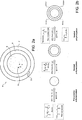
figure 1 is a diagrammatic representation of the probe kit according to the present invention and of the operation thereof; -
figure 2A is a diagrammatic representation of a microparticle according to the present invention; -
figure 2B is a diagram of the process for preparing a microparticle according to the present invention; -
figure 3 is a diagrammatic representation of the conjugation of a probe kit and a microparticle according to the present invention; -
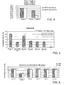
figure 4 is a diagram showing the effects of the length of a first probe on the formation of the duplex between the first and the second probe of the probe kit offigure 1 ; -
figure 5 is a diagram showing the fluorescence recovery as a function of the different concentrations of the target sequence; -
figure 6 is a diagram showing the selectivity and the specificity in the formation of the duplex between a second probe of the probe kit offigure 1 and a target sequence inserted in longer nucleotide sequences (100 nt); -
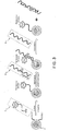
figures 7A and 7B show the fluorescence codes of 6 microgels (i.e. a set of microparticles) obtained using different concentrations of first and second fluorophore respectively in the first and third layer of microparticles shown infigure 2A ; -
figure 7C shows an image of three microgels with different codes obtained using different concentrations of first and second fluorophore respectively in the first and third layer of microparticles shown infigure 2A ; -
figure 8 shows images obtained by CLSM and intensities measured along the axis of the microgel. The channel for the code analysis and detection of the displacement are shown in the lower part; -
figure 9A is a diagram showing the fluorescence recovery on the surface of the microgels as a function of the different concentrations of the target sequence (HIV); -
figure 9B is a diagram showing the linear regression of the fluorescence emissions as a function of the concentration of a target sequence for the computation of the detection limit (HIV); -
figure 10 shows an example of microparticles conjugated with the first probe of the probe kit offigure 1 after displacement, on which the analysis of emitted fluorescence is performed; -
figure 11a is a diagram showing the fluorescence recovery on the surface of the microgels as a function of the different concentrations of the target sequence (mir21); -
figure 11b is a diagram showing the linear regression of the fluorescence emissions as a function of the concentration of the target sequence for the computation of the detection limit (mir21). - Kit 10 per for detecting a single strand
target nucleotide sequence 3 according to the present invention comprises at least one firstnucleic acid probe 1 and at least one secondnucleic acid probe 2. -
Probe 1 has a length from 10 to 14 bases, preferably from 11 to 13 bases, and has at least onefluorophore 11 bound at the 5' end. -
Probe 2 has a length from 35 to 50 bases and comprises, from the 5' end to the 3' end:- a
first segment 21 of nucleotide sequence complementary to probe 1, - at least one
quencher 23, and - a
second segment 22 of nucleotide sequence complementary to at least part of thetarget nucleotide sequence 3.
- a
- In the case shown, both
probe 1 andprobe 2 are made of DNA. -
Fluorophore 11 bound to the 5' end ofprobe 1 is preferably selected from the group consisting of FAM, TET, JOE, HEX, Oregon Green®, TAMRA, ROX, Cy3, Cy3.5, Cy5, Cy5.5, CAL Red™, Red 640, Cy5, and Cy5.5. -
Quencher 23 ofprobe 2 is preferably selected from the group consisting of DDQ-I, Dabcyl, Eclipse, Iowa Black FQ, BHQ-1, QSY-7, BHQ-2, DDQ-II, Iowa Black RQ, QSY-21, and BHQ-3 and is compatible withfluorophores 11 bound to the 5' end ofprobe 1. - Advantageously,
probe 1 andprobe 2 are designed so that the following relation is met:
- ΔG hybr.target3-probe2 is the free energy of duplex formation between
target nucleotide sequence 3 and secondnucleic acid probe 2, and - ΔG hybr.probe1-probe2 is the free energy of duplex formation between the first
nucleic acid probe 1 and the secondnucleic acid probe 2. - More preferably,
probe 1 andprobe 2 are designed so that
- When
target nucleotide sequence 3 is DNA,probe 1 andprobe 2 are even more preferably designed so that
- When
target nucleotide sequence 3 is miRNA,probe 1 andprobe 2 are even more preferably designed so that 10 Kcal/mol > |ΔG hybr.target3 - probe2| — |ΔG hybr.probe1 - probe2|> 25 Kcal/mol. - In the case at issue, the Oligocalc software (Nucl. Acids Res. (2007) 35 (suppl2):W43-W46) was used to compute the ΔG values.
- As most of the software packages commercially available for the design of oligonucleotides, this software uses the value of ΔG as a measure of the affinity between two nucleotide sequences, where the affinity represents the measure of the thermodynamic stability of the duplex formed by the two single strand oligonucleotides.
- The transition from one state (2 single strands) to another state (duplex) results in an energy variation in the system.
- ΔG is the variation in Gibbs free energy (unit: kcal/mole) and represents the net exchange in energy between the system and its environment and is described by the following equation

- The description of ΔG indicates that this amount depends on the temperature. In the case at issue, reactions have been performed at room temperature. Therefore, ΔG has been computed for T=25°C (298.15 Kelvin).
- At a given temperature a positive ΔG value indicates that the system tends to evolve towards single strand reagents (non spontaneous). A negative value of ΔG indicates, instead, that the system tends to evolve towards a duplex product (spontaneous).
- For greater clarity and simplicity, the values of ΔG are indicated as an absolute value.
-
Target nucleotide sequence 3 preferably has a length from 15 to 100 bases, even more preferably from 20 to 40 bases. - Kit 10 allows to detect
target nucleotide sequences 3 in a range of concentrations from 1·10-11 M to 1·10-22 M, i.e. in a very broad range. In particular, kit 10 allows to detecttarget nucleotide sequences 3 at concentrations from 1·10-17 M to 1.10-19 M, i.e. a very low concentrations. - With reference to
figure 1 ,probe 1,probe 2 andtarget nucleotide sequence 3 are shown. In the case at issue,probe 1 is x bases long (for example, 12 bases) and has a Cy5 molecule bound at the 5' end.Probe 2 is x+y bases long (for example, 39 bases) and comprises, from the 5' to the 3' end, afirst segment 21 which is x bases long (for example, 12 bases long) and complementary to probe 1, a quencher 23 (BHQ), and asecond segment 22 which is y bases long (for example, 27 bases long) and complementary to at least part oftarget nucleotide sequence 3.Target nucleotide sequence 3 is x+y bases long (for example, 39 bases long). - In the presence of
probe 1 andprobe 2, these form a duplex having formation free energy ΔG hybr.probe1-probe2. In this situation, quencher 23 BHQ quenches the signal emitted byfluorophore 11 Cy5 and there is no fluorescence emission. - When
target nucleotide sequence 3 is added toprobes target nucleotide sequence 3 andprobe 2, because |ΔG hybr.target3-probe2|>|ΔG hybr.probe1-probe2|. The displacement ofquencher 23 BHQ fromfluorophore 11 Cy5 caused by the displacement ofprobe 1 fromprobe 2 results in the emission of fluorescence. -
Probe 1 andprobe 2 are designed on the basis oftarget nucleotide sequence 3 and their thermodynamic affinity is modulated so that the affinity ofprobe 2 fortarget nucleotide sequence 3 is higher than the affinity of the initial duplex betweenprobe 1 andprobe 2. The difference in free energy |ΔG hybr.target3 - probe2| - |ΔG hybr.probe1 - probe2| and the length ofprobe 1 are selected so as to optimize the displacement ofprobe 1 and the formation of the duplex betweenprobe 2 andtarget nucleotide sequence 3. - With reference to
figure 3 , in a preferred embodiment, kit 10 also comprises at least one microparticle 4 covalently bound to the 3' end ofprobe 1, preferably by means of an amide bond. Even more preferably, the bond between microparticle 4 andprobe 1 is obtained by means of an appropriate amino-linker onprobe 1. - With reference to
figure 2A , multilayer particle 4 according to the present invention comprises: - at least one
first layer 5 comprising afirst fluorophore 51, - at least one second layer 6 in contact with
first layer 5, and - at least one
third layer 7 in contact with second layer 6 and comprising asecond fluorophore 71. -
First fluorophore 51 andsecond fluorophore 71 are different,first layer 5 and third layer -7 are not in contact with one another. -
First fluorophore 51 andsecond fluorophore 71 can be selected from the group consisting of rhodamine, fluorescein, Cy2, Oregon Green, Alexa (488, 532, 546, 555) and others as long as the emission wave length do not overlap. - Preferably, multilayer microparticle 4 also comprises:
- at least one
fourth layer 8 in contact withthird layer 7, and - a least one
fifth layer 9 in contact withfourth layer 8 and comprises a third fluorophore 91. - Third fluorophore 91 is different from
second fluorophore 71 and fromfirst fluorophore 51, andthird layer 7 andfifth layer 9 are not in contact with one another. - The third fluorophore can be selected from the group consisting of rhodamine, fluorescein, Cy2, Oregon Green, Alexa (488, 532, 546, 555) and others as long as the emission wavelengths do not overlap with the wavelengths of first and
second fluorophore - Multilayer microparticle 4 preferably has a size from 0.5 µm to 2 µm.
- Each layer of multilayer microparticle 4 preferably comprises esters and amides of acrylic acid or of methacrylic acid or vinyls or allyls, which are optionally substituted.
- By "esters and amides of acrylic acid or of methacrylic acid or vinyls or allyls, which are optionally substituted" there is also intended compounds equivalent thereto. This definition also includes difunctional polymers used as cross-linkers such as, for example, bisacrylammide, polyethylenoxide-acrylate/-methacrylate etc.
-
Fluorophores layers layers -
Figure 2B shows the process for the preparation of a specific multilayer microparticle 4. In a first step, afirst layer 5 of polyethylene glycol dimethacrylate (PEGDA, 500 MW) and rhodamine B acrylate monomers is produced by free-radical precipitation polymerization. In a following step, a second layer 6 of polyethylene glycol dimethacrylate is produced aroundfirst layer 5 by seeded polymerization. In a following step, athird layer 7 of acrylic acid and fluorescein is produced around second layer 6 by seeded polymerization. Different groups of microparticles with different fluorescence codes can be obtained by using different concentration ratios between rhodamine and fluorescein. - With reference to
figure 2B an example of synthesis of a specific type of microparticles is disclosed in detail. - Synthesis of
first layer 5. Microgels of polyethylene glycol dimethacrylate have been prepared by free-radical precipitation polymerization, using a concentration of total monomers of 1% (w/v). Polymerization has been performed in a 100 ml three-neck flask with round bottom, in which a filtered aqueous solution of monomers and 1% (w/v) polyvinyl alcohol (PVA) as surfactant have been added. This solution was heated to ∼ 65 °C while being purged with N2 gas and stirred vigorously for ∼ 1 h. Then the reaction was immediately initiated by injection of a potassium persulfate (KPS) aqueous solution (to make a final KPS concentration of 0.06 % w/v). The solution turned turbid, indicating successful initiation. Methacryloxy thiocarbonyl rhodamine B, dissolved in dimethyl sulfoxide (0.1 ml) and diluted with water (1.9 ml), was then added to the stirred mixture at a final concentration ranging from 0.005 to 0.3 mM to obtain different dye amounts. The solution was allowed to heat and stir for an additional 7 h while being purged with N2 gas. The microgels were dialyzed for 2 days against distilled water, purified several times by centrifuging for 15 minutes at 12000 rpm and resuspending in deionised water to remove unreacted monomers, oligomers and surfactants and stored at 4 °C until further use. - Synthesis of second layer 6. The rhodamine-labelled microgel was resuspended in deionised water to a concentration of 10 mg/ml. These microgels were then used as seed particles, upon which a PEGDMA cross-linked layer was added. A solution of rhodamine-labelled core microgels (100 mg, 10 ml) in deionised water (25 ml) was heated to 65 °C under a gentle stream of N2. Separately, PEGDMA (240 mg) was dissolved in water (10 ml), purged with N2 at room temperature and then slowly added to the heated core solution. After the temperature remained stable at 65 °C for ∼ 1 h, 2 ml of aqueous solution of KPS (final concentration of 0.03 % w/v) was added to initiate the polymerization. The reaction was allowed to proceed, for 6 h. The microgels were purified several times by centrifugation (15 minutes at 9000 rpm) and resuspended in deionised water.
- Synthesis of third layer 6. A solution of two layer (core-shell) microgels (10 ml, [C] = 10 mg/ml) in deionised water (25 ml) was heated to 65 °C, followed by the slow addition of 10 ml of aqueous monomer solution containing PEGDMA (240 mg) and acrylic acid (125 mg). After the temperature remained stable at 65 °C for ∼ 1 h, 2 ml of aqueous solution of KPS (final concentration of 0.03 % w/v) was added to initiate the polymerization. Fluorescein O-methacrylate diluted in water (2 ml), was then added to the stirred mixture at a final concentration ranging from 0.05 to 0.2 mM to obtain different dye amounts. The reaction was allowed to proceed for 6 h. The microgels were dialyzed for 5 days, purified several times by centrifugation (for 15 minutes at 6500 rpm) and resuspended in deionised water to remove unreacted monomers, oligomers and surfactants, then stored at 4 °C prior to use until further use.
- 1 mg of encoded microgels was dissolved in 250 µl of coupling buffer, 100 mM MES pH 4.8, and kept at 4°C with occasional vortexing for at least 1 h to disperse the colloidal particles. 1-ethyl-3-(3-dimethylaminopropyl)carbodiimide (EDC) (500 mM, final concentration, dissolved in the coupling buffer that was freshly prepared just before use) was added to this suspension, followed by the addition of 1200 pmol of
probe 1. The total reaction volume was approximately 500 µl. The reaction mixture was carried on in dark and left at 4°C in a shaker over night. The conjugate betweenprobe 1 and multilayer microparticle 4 was precipitated down by centrifugation at 6000 rpm for 15 min at room temperature. The supernatant was removed carefully with a pipette and the precipitant was resuspended in 1 ml Of Tris HCl,pH 8 buffer by agitating with a pipette tip and brief vortexing. This washing step was repeated three more times. - Polyethylene glycol (PEG) microgels were produced (particle size of about 1 µm). The outer layer of these microparticles 4 was functionalised with carboxylic groups. Two concentrations of fluorescein 71 (0.1 µm and 0.2 µm) were used for
third layer 7, and three different concentrations of rhodamine 51 (0.1 µm, 0.01 µm e 0.005 µm) were used forfirst layer 5. Six microgels were distinguished by means of a spectrofluorometer, on the basis of combinations of different concentrations ofrhodamine 51 andfluorescein 71 in the production solution of multilayer microparticles 4 (figures 7A and 7B ). - As may be noted in
figure 7C , the approach for multilayer microparticle 4 production allows to define an univocal code on the particles in virtue of the fact thatfluorophores rhodamine 51 andfluorescein 71 respectively infirst layer 5 and inthird layer 7 of multilayer microparticle 4. -
Figure 4 shows how the length ofprobe 1 influences the formation of the duplex betweenprobe 1 andprobe 2 of probe kit 10 according to the invention. In particular, the results for aprobe 1 of 8 bases and aprobe 1 of 12 bases are shown. 0.1 µM solutions of probe 1 (8 and 12 bases long) andprobe 2 were reacted for 12 hours and fluorescence emission was evaluated. From the data offigure 4 it is apparent that the bond between probe 1 (8 bases long) andprobe 2 is poorly stable as can be seen from the measurements of fluorescence relating to the formation of the duplex. In this case a very low loss in the percentage of fluorescence is registered. Instead, the duplex between probe 1 (12 bases) andprobe 2 results more stable as confirmed by a greater percent loss of fluorescence emission of the complex probe 1 -probe 2 if compared tosole reference probe 1. - The values of ΔG have been computed by means of the Oligocalc software.
Table 1 Probe name Sequence Length ΔG (Kcal/mol) HIV probes (on the basis of Genbank sequence:AF033819.3 positions 6520-6559) HIV first probe (tail-Cy5) 5' Cy5 ACT GCT GTT AAA C6 NH2-3' 12 |ΔGhybr.probel-probe2| 11.2 HIV second probe (quencher) 
39 |ΔGhybr.target3.probe2| 50.9 HIV target nucl. seq. (target) 
39 |ΔGhybr.target3-probe2|- |ΔGhybr.probe1-probe2| 39.7 HCV probes (on the basis of Genbank sequence: M67463.1 positions 160-195) HCV first probe (tail-Cy5) 5' Cy5 TTC CGG TGT ACT-C6 NH2-3' 12 |ΔGhybr.probe1-probe2| 13.3 HCV second probe (quencher) 
35 |ΔGhybr.target3-probe2| 53.7 HCV target nucl. seq. (target) 
35 |ΔGhybr.target3-probe2|-|ΔGhybr.probe1-probe2| 40.4 SARS probes genome, Genb (on the basis of human coronavirus bank:AF304460 positions 16710-167 sequence 747) 229E, whole SARS first probe (tail-Cy5) 5' Cy5 GGC TCC AGT ATA -C6 NH2- 3' 12 |ΔGhybr.probe1-probe2| 11.9 SARS second probe (quencher) 
37 |ΔGhybr.target3-probe2| 52.4 SARS target nucl. seq. (target) 
37 |ΔGhybr.target3-probe2|-|ΔGhybr.probe1-probe2 40.5 Hsa_miRNA 155 (from www.mirbase.org) miR155 first probe (tail(12)-Cy5) 5'- Cy5 CGT GAT AGG GGT NH2-3' 12 |ΔGhybr.probe1-probe2| 13.6 miR155 second probe (quencher 12) 
23 |ΔGhybr.probe1-probe2| 6.1 miR155 first probe (tail(8)-Cy5) 5'- Cy5 AT AGG GGT NH2-3' 9 miR155 second probe (quencher 8) 
23 miR155 target nucl. seq. (target) 5'-TTAATGCTAATCGTGATAGGGGT-3' 23 miR155 target nucl. seq. (target) 5'-UUAAUGCUAAUCGUGAUAGGGGU-3' 23 ΔGhybr.target3-probe2| 28 |ΔGhybr.target3-probe2|-|ΔGhybr.probel-probe2| (12) 14.4 |ΔGhybr.target3-probe2|-|ΔGhybr.probe1-probe2| (8) 21.9 Hsa_miRNA 21 (from www.mirbase.org) miR21 first probe (tail-Cy5) 5'-Cy5 GACTGATGTTGA NH2-3' 12 |ΔGhybr.probe1-probe2| 11.2 miR21 second probe (quencher 12) 5'-TCAACATCAGTBHQTGATAAGCTA-3' 22 |ΔGhybr.target3-probe2| 25.1 miR21 target nucl. seq. (target) 5'-UAGCWAUCAGACUGAUGWGA-3' 22 |ΔGhybr.target3-probe2|-|ΔGhybr.probe1-probe2| 13.9 - An experiment was performed with a probe kit, to which no microparticles were conjugated, i.e. in homogeneous phase.
- 40 pmoles of
probe 1 were mixed with 40 pmoles ofprobe 2 in Tris HCl,pH 8. Quenched samples were used as reference in order to evaluate the displacement efficiency. The specificity of double strand probes was evaluated by using scrambled or non specific sequences. Each sample was loaded onto a 96-well microplate and the fluorescence emission intensity was measured in 2300 EnSpire multilabel reader (Perkin-Elmer, Waltham, MA) by setting the λex=633 and λem=654. - The indicated experimental uncertainties represent the standard deviation of three replicates.
- As may be noted in
figure 5 , target nucleotide sequences miR155 can be detected up to concentrations of 5 nM with a range from 50 nM to 5 nM and a LOD of 1 nM. - Starting from a concentration of
probe 1 andprobe 2 of 50 nM, displacement experiments have been carried out using different concentrations oftarget nucleotide sequences 3 in a range from 50 nM to 0.05 nM. It may be noted that for concentrations below 0.5 nM there are no variations in the fluorescence with respect to the duplex between probe 1 -probe 2, so that it is not possible to observe such a variation by means of spectrofluorimetry. In the range from 50 nM to 5 nM there are significant variations in fluorescence. The data of fluorescence emission as a function of the concentration were processed by means of linear regression and the value of limit of detection was extrapolated (LOD=1nM). - To prove that probe kit 10 according to the invention is capable of capturing and distinguishing
target nucleotide sequences 2 even within longer sequences (as would occur in an actual context, sincetarget nucleotide sequence 3 would be within a gene), displacement experiments were carried out using the 99 base long nucleotide sequences shown in table 2. These experiments were carried out in homogeneous phase.Table 2 Probe name Sequences Length (nt) HIV 100 R 
99 HIV 100 M 
99 HIV 100 L 
99 - The HIV 100 R, HIV 100 M and HIV 100 L probes were designed so that
target nucleotide sequence 3 is respectively at the 5' end, in the middle and at the 3' end of the 99 base long sequence. - The results shown in
figure 6 prove that the probes are capable of hybridizing and displacingprobe 2 in Tris buffer as well as in a complex environment such as that of serum proteins (FBS). With respect to the short 99 base long target sequences, they are recognised without significant statistical differences. -
Figure 3 diagrammatically shows the heterogeneous phase assay. - Approximately 1 mg of first probe conjugated with the microgel (in 250 µl of Tris HCl hybridization buffer pH 8) was mixed with 350 pmoles of second probe (250 µl). The mixture was incubated at room temperature overnight. The microgels were then washed with hybridization buffer and resuspended in 1 ml of buffer at a final concentration of 1 µg/µl. 50 µl (50 µg) of quenched microgel were mixed to 450 µl of a solution containing
target probe sequences 3 at different concentrations ranging from 10-11 to 10-22 M and incubated at room temperature overnight. The microgel was precipitated down by centrifugation at 6000 rpm for 15 min at 4°C. The supernatant was removed carefully with a pipette and the precipitant was resuspended in 1 ml of Tris HCl,pH 8 buffer by agitating with a pipette tip and brief vortexing. - 30 µl of coupled, quenched and strand displaced microgels were loaded onto µ-slide channels (Ibidi, Martinsried, DE), illuminated at confocal laser scanning microscope and fluorescence images of microparticles were collected. All captured images were analysed with a public domain image-processing Image J (
version 1,43i, NIH, Bethesda, MD). The image was then further processed with the Analyze Particles function Image J to determine the number of single fluorescence particles computationally. The size of the particles was set to reduce false positive signals generated from noises. For each experiment, at least 200 microparticles were selected for each sample to be analysed. - Two case studies are hereinafter disclosed to prove the ability of the assay to capture single strand target nucleotide sequences. In particular, an HIV target DNA and an RNA (miRNA 21) were used.
- The steps of conjugation of
probe 1 with microparticles 4 and of design of probe kit 10 are the same in the two cases. The difference resides only intarget nucleotide sequence 3. In the case of the miRNA the formation of a heteroduplex is also shown. - Probes 1 (12 bases) specific for each
target nucleotide sequence 3 and functionalised with an amine group at the 3' end were conjugated with the carboxylic groups on the surface of the microgel.Fluorophore 11 bound at the 5' of eachprobe 1 was Cy5. Respective probes 2 (39 bases) carrying BHQ2 asquencher 23 were hybridized toprobe 1. -
Figure 8 shows images acquired by CLSM and the intensities measured along the axis of the microgel. The channel for the analysis of the code and for the detection of the displacement are shown in the lower part. - The close proximity between Cy5 and BHQ2 results in the quenching of the fluorescence of Cy5. Solutions containing target nucleotide sequences 3 (39 bases) were brought in contact with 50 µg of microparticles 4 inducing the hybridisation of each
probe 2 with respectivetarget nucleotide sequences 3 and the subsequent emission of fluorescence by Cy5. The emission of Cy5 can be calibrated to evaluate the correspondence between the fluorescence emission (recovery) and the concentration oftarget nucleotide sequence 3. -
Figure 9A shows the recovery of fluorescence on the surface of microgels as a function of the different concentrations of the target sequence (HIV). - 30 µl of coupled, quenched and strand displaced microgels were loaded onto µ-slide channels (Ibidi, Martinsried, DE), illuminated at confocal laser scanning microscope and fluorescence images of microparticles were collected. All captured images were analysed with a public domain image-processing Image J (
version 1,43i, NIH, Bethesda, MD). The image was then further processed with the Analyze Particles function Image J to determine the number of single fluorescence particles computationally. The size of the particles was set to reduce false positive signals generated from noises or aggregates formation. For each experiment, at least 200 microparticles were selected for each sample to be analysed.Table 3 ID microparticle Fluorescence emission ID microparticle Fluorescence emission ID microparticle Fluorescence emission ID microparticle Fluorescence emission 1 1001 51 1018 101 1041 150 1062 2 1001 52 1019 102 1042 151 1063 3 1002 53 1019 103 1042 152 1063 4 1002 54 1019 104 1043 153 1064 5 1002 55 1020 105 1044 154 1064 6 1003 56 1020 106 1044 155 1064 7 1003 57 1020 107 1044 156 1064 8 1003 58 1020 108 1046 157 1065 9 1003 59 1021 109 1047 158 1065 10 1004 60 1022 110 1047 159 1066 11 1004 61 1023 111 1047 160 1067 12 1004 62 1024 112 1048 161 1067 13 1005 63 1024 113 1048 162 1067 14 1005 64 1024 114 1049 163 1068 15 1006 65 1024 115 1049 164 1068 16 1006 66 1024 116 1049 165 1068 17 1007 67 1024 117 1049 166 1068 18 1007 68 1025 118 1049 167 1068 19 1007 69 1025 119 1050 168 1068 20 1007 70 1025 120 1051 169 1069 21 1007 71 1026 121 1051 170 1070 22 1008 72 1026 122 1052 171 1070 23 1008 73 1026 123 1052 172 1070 24 1009 74 1026 124 1053 173 1071 25 1009 75 1027 125 1053 174 1072 26 1009 76 1027 126 1054 175 1074 27 1010 77 1028 127 1055 176 1074 28 1010 78 1028 128 1055 177 1075 29 1011 79 1029 129 1056 178 1075 30 1011 80 1029 130 1056 179 1076 31 1012 81 1029 131 1056 180 1077 32 1012 82 1030 132 1056 181 1077 33 1012 83 1031 133 1057 182 1077 34 1013 84 1031 134 1058 183 1079 35 1013 85 1031 135 1059 184 1079 36 1014 86 1032 136 1059 185 1080 37 1014 87 1032 137 1059 186 1080 38 1014 88 1032 138 1060 187 1081 39 1014 89 1033 139 1060 188 1081 40 1015 90 1033 140 1060 189 1082 41 1015 91 1033 141 1060 190 1082 42 1015 92 1034 142 1060 191 1083 43 1015 93 1035 143 1060 192 1083 44 1015 94 1036 144 1060 193 1084 45 1016 95 1037 145 1061 194 1085 46 1016 96 1038 146 1061 195 1086 47 1016 97 1038 147 1062 196 1086 48 1016 98 1038 148 1062 197 1087 49 1017 99 1039 149 1062 198 1087 50 1017 100 1040 150 1062 199 1087 mean±sd 1041±25 - The disclosed kit allows to obtain a linear response in the emission of fluorescence in the range of concentrations between 10-17 M and 10-19 M. The graph in
figure 9b shows the linear regression of the fluorescence emissions as a function of the concentration of the target sequence in the range between 10-17 M and 10-19 M. The limit of detection (LOD) for HIV has been computed considering the value of fluorescence emission of the background (intercept of the linear regression line) plus 3 times the standard deviation of the value itself (LOD = 0.790 aM). - From an analysis of the features of kit 10 for detecting a single-strand
target nucleotide sequence 3 according to the present invention, the advantages it allows to obtain are apparent. - In particular, kit 10 allows to detect target nucleotide sequences 3:
- avoiding the separation of the sample and/or the amplification of target nucleotide sequence 3 (leading to a simple, faster and more cost-effective assay),
- with very low concentrations (< 1·10-17 M) of
target nucleotide sequence 3, - with very short target nucleotide sequences 3 (20-40 nucleotides).
- In virtue of the design of
probes - In virtue of the possibility of using a virtually indefinite number of fluorophores and the ever greater availability of fluorophores on the market, the kit according to the invention allows a very high multiplexing.
- Kit 10 works in assays for
target nucleotide sequences 3 both of DNA and RNA. - Moreover, in virtue of the conjugation on the surface of microparticles 4 of
probe 1, a high number ofprobes 1 can be concentrated in an extremely limited area. This allows to increase the sensitivity of the assay. - The combination between multilayer microparticles 4 and kit 10 allows to:
- obtain a very fast assay (in virtue of a high reaction kinetics),
- assemble and handle multilayer microparticles 4 in miniaturised devices (lab-on-chips).
-
- <110> FONDAZIONE ISTITUTO ITALIANO DI TECNOLOGIA
- <120> KIT DI SONDE PER RIVELARE UNA SEQUENZA NUCLEOTIDICA BERSAGLIO A
SINGOLO FILAMENTO - <160> 21
- <170> BiSSAP 1.0
- <210> 1
<211> 12
<212> DNA
<213> Human immunodeficiency virus - <220>
<221> source
<222> 1..12
<223> /mol_type="DNA" /organism="Human immunodeficiency virus" - <400> 1
actgctgtta aa 12 - <210> 2
<211> 39
<212> DNA
<213> Human immunodeficiency virus - <220>
<221> source
<222> 1..39
<223> /mol_type="DNA" /organism="Human immunodeficiency virus" - <400> 2
tttaacagca gttgagttga tactactggc ctaattcca 39 - <210> 3
<211> 39
<212> DNA
<213> Human immunodeficiency virus - <220>
<221> source
<222> 1..39
<223> /mol_type="DNA" /organism="Human immunodeficiency virus" - <400> 3
tggaattagg ccagtagtat caactcaact gctgttaaa 39 - <210> 4
<211> 12
<212> DNA
<213> Hepatitis C virus - <220>
<221> source
<222> 1..12
<223> /mol_type="DNA" /organism="Hepatitis C virus" - <400> 4
ttccggtgta ct 12 - <210> 5
<211> 35
<212> DNA
<213> Hepatitis C virus - <220>
<221> source
<222> 1..35
<223> /mol_type="DNA" /organism="Hepatitis C virus" - <400> 5
agtacaccgg aattgccagg acgaccgggt ccttt 35 - <210> 6
<211> 35
<212> DNA
<213> Hepatitis C virus - <220>
<221> source
<222> 1..35
<223> /mol_type="DNA" /organism="Hepatitis C virus" - <400> 6
aaaggacccg gtcgtcctgg caattccggt gtact 35 - <210> 7
<211> 12
<212> DNA
<213> SARS coronavirus - <220>
<221> source
<222> 1..12
<223> /mol_type="DNA" /organism="SARS coronavirus" - <400> 7
ggctccagta ta 12 - <210> 8
<211> 37
<212> DNA
<213> SARS coronavirus - <220>
<221> source
<222> 1..37
<223> /mol_type="DNA" /organism="SARS coronavirus" - <400> 8
tatactggag ccattgtcta cctgaacact accgcgt 37 - <210> 9
<211> 37
<212> DNA
<213> SARS coronavirus - <220>
<221> source
<222> 1..37
<223> /mol_type="DNA" /organism="SARS coronavirus" - <400> 9
acgcggtagt gttcaggtag acaatggctc cagtata 37 - <210> 10
<211> 12
<212> DNA
<213> Homo sapiens - <220>
<221> source
<222> 1..12
<223> /mol_type="DNA" /organism="Homo sapiens" - <400> 10
cgtgataggg gt 12 - <210> 11
<211> 23
<212> DNA
<213> Homo sapiens - <220>
<221> source
<222> 1..23
<223> /mol_type="DNA" /organism="Homo sapiens" - <400> 11
acccctatca cgattagcat taa 23 - <210> 12
<211> 9
<212> DNA
<213> Homo sapiens - <220>
<221> source
<222> 1..12
<223> /mol_type="DNA" /organism="Homo sapiens" - <400> 12
ataggggt 9 - <210> 13
<211> 23
<212> DNA
<213> Homo sapiens - <220>
<221> source
<222> 1..23
<223> /mol_type="DNA" /organism="Homo sapiens" - <400> 13
acccctatca cgattagcat taa 23 - <210> 14
<211> 23
<212> DNA
<213> Homo sapiens - <220>
<221> source
<222> 1..23
<223> /mol_type="DNA" /organism="Homo sapiens" - <400> 14
ttaatgctaa tcgtgatagg ggt 23 - <210> 15
<211> 23
<212> RNA
<213> Homo sapiens - <220>
<221> source
<222> 1..23
<223> /mol_type="RNA" /organism="Homo sapiens" - <400> 15
uuaaugcuaa ucgugauagg ggu 23 - <210> 16
<211> 12
<212> DNA
<213> Homo sapiens - <220>
<221> source
<222> 1..12
<223> /mol_type="DNA" /organism="Homo sapiens" - <400> 16
gactgatgtt ga 12 - <210> 17
<211> 22
<212> DNA
<213> Homo sapiens - <220>
<221> source
<222> 1..22
<223> /mol_type="DNA" /organism="Homo sapiens" - <400> 17
tcaacatcag tctgataagc ta 22 - <210> 18
<211> 22
<212> RNA
<213> Homo sapiens - <220>
<221> source
<222> 1..22
<223> /mol_type="RNA" /organism="Homo sapiens" - <400> 18
uagcuuauca gacugauguuga 22 - <210> 19
<211> 99
<212> DNA
<213>Human immunodeficiency virus 1 - <220>
<221> source
<222> 1..99
<223> /mol_type="DNA" /organism="Human immunodeficiency virus 1" - <400> 19

- <210> 20
<211> 99
<212> DNA
<213>Human immunodeficiency virus 1 - <220>
<221> source
<222> 1..99
<223> /mol_type="DNA" /organism="Human immunodeficiency virus 1" - <400> 20

- <210> 21
<211> 99
<212> DNA
<213>Human immunodeficiency virus 1 - <220>
<221> source
<222> 1..99
<223> /mol_type="DNA" /organism="Human immunodeficiency virus 1" - <400> 21

Claims (8)
- A kit (10) for detecting a single strand target nucleotide sequence (3) comprising:- at least one first nucleic acid probe (1) from 10 to 14 bases, to the 5' end of which at least one fluorophore (11) is bound;- at least one second nucleic acid probe (2) from 35 to 50 bases, comprising, from the 5' to the 3' end:- a first segment (21) having a nucleotide sequence complementary to the first nucleic acid probe (1),- at least one quencher (23), and- a second segment (22) having a nucleotide sequence complementary to at least part of the target nucleotide sequence (3), wherein:- the following relation is met:
 - ΔG hybr.target3-probe2 is the free energy of duplex formation between the target nucleotide sequence (3) and the second nucleic acid probe (2), and- ΔG hybr.probel-probe2 is the free energy of duplex formation between the first nucleic acid probe (1) and the second nucleic acid probe (2); and- the concentrations of the at least one first nucleic acid probe (1) and of the at least one second nucleic acid probe (2) are from 5 nM to 50 nM.
 - The kit (10) according to claim 1, wherein the single strand target nucleotide sequence (3) is DNA and 35 Kcal/mol > |ΔG hybr.target3 - probe2| - |ΔG hybr.probe1 - probe2|> 45 Kcal/mol.
- The kit (10) according to claim 1, wherein the single strand target nucleotide sequence (3) is miRNA and 10 Kcal/mol > |ΔG hybr.target3 - probe2| - |ΔG hybr.probe1 - probe2|> 25 Kcal/mol.
- The kit (10) according to any of the preceding claims, wherein the at least one first nucleic acid probe (1) has a length from 11 to 13 bases.
- The kit (10) according to any of the preceding claims, wherein the single strand target nucleotide sequence (3) has a length from 15 to 100 bases.
- The kit (10) according to claim 5, wherein the single strand target nucleotide sequence (3) has a length from 20 to 40 bases.
- The kit (10) according to any of the preceding claims, wherein the single strand target nucleotide sequence (3) is in a concentration from 1·10-17 M to 1·10-19 M.
- The kit (10) according to any of the preceding claims, also comprising a microparticle (4) bound covalently to the 3' end of the at least one first nucleic acid probe (1).
Applications Claiming Priority (2)
| Application Number | Priority Date | Filing Date | Title |
|---|---|---|---|
| IT001154A ITTO20121154A1 (en) | 2012-12-27 | 2012-12-27 | SYSTEM OF PROBES FOR DETECTING A SINGLE-strand TARGET NUCLEOTIDE SEQUENCE |
| PCT/IB2013/061377 WO2014102748A1 (en) | 2012-12-27 | 2013-12-27 | Probe kit for detecting a single strand target nucleotide sequence |
Publications (2)
| Publication Number | Publication Date |
|---|---|
| EP2938746A1 EP2938746A1 (en) | 2015-11-04 |
| EP2938746B1 true EP2938746B1 (en) | 2017-04-26 |
Family
ID=47683967
Family Applications (1)
| Application Number | Title | Priority Date | Filing Date |
|---|---|---|---|
| EP13831873.8A Not-in-force EP2938746B1 (en) | 2012-12-27 | 2013-12-27 | Probe kit for detecting a single strand target nucleotide sequence |
Country Status (4)
| Country | Link |
|---|---|
| US (1) | US9834812B2 (en) |
| EP (1) | EP2938746B1 (en) |
| IT (1) | ITTO20121154A1 (en) |
| WO (1) | WO2014102748A1 (en) |
Family Cites Families (8)
| Publication number | Priority date | Publication date | Assignee | Title |
|---|---|---|---|---|
| NZ212419A (en) * | 1984-06-25 | 1988-08-30 | Mucan Diagnostics Pty Ltd | In vitro diagnostic test for detecting cancer cells producing mucin antigens |
| US20030096351A1 (en) * | 1998-03-27 | 2003-05-22 | Genentech, Inc. | Secreted and transmembrane polypeptides and nucleic acids encoding the same |
| US20030108927A1 (en) * | 2000-04-03 | 2003-06-12 | Kathryn Leishman | Compositions and methods for the prevention, treatment and detection of tuberculosis and other diseases |
| WO2002069898A2 (en) * | 2001-03-02 | 2002-09-12 | Stratagene | Compositions and methods using platinum compounds for nucleic acid labeling |
| US7385043B1 (en) * | 2003-04-30 | 2008-06-10 | The Public Health Research Institute Of The City Of New York, Inc. | Homogeneous multiplex screening assays and kits |
| US7445898B2 (en) * | 2004-04-01 | 2008-11-04 | Bio-Rad Laboratories, Inc. | Quantitative amplification with a labeled probe and 3′ to 5′ exonuclease activity |
| AU2006213907A1 (en) * | 2005-02-09 | 2006-08-17 | Stratagene California | Key probe compositions and methods for polynucleotide detection |
| WO2009140374A2 (en) * | 2008-05-13 | 2009-11-19 | Gen-Probe Incorporated | Inactivatable target capture oligomers for use in the selective hybridization and capture of target nucleic acid sequences |
-
2012
- 2012-12-27 IT IT001154A patent/ITTO20121154A1/en unknown
-
2013
- 2013-12-27 WO PCT/IB2013/061377 patent/WO2014102748A1/en not_active Ceased
- 2013-12-27 EP EP13831873.8A patent/EP2938746B1/en not_active Not-in-force
- 2013-12-27 US US14/655,276 patent/US9834812B2/en not_active Expired - Fee Related
Non-Patent Citations (1)
| Title |
|---|
| None * |
Also Published As
| Publication number | Publication date |
|---|---|
| WO2014102748A1 (en) | 2014-07-03 |
| ITTO20121154A1 (en) | 2014-06-28 |
| EP2938746A1 (en) | 2015-11-04 |
| US20160053308A1 (en) | 2016-02-25 |
| US9834812B2 (en) | 2017-12-05 |
Similar Documents
| Publication | Publication Date | Title |
|---|---|---|
| US20060234261A1 (en) | Colorimetric readout of hybridization chain reaction | |
| Vilaivan | Fluorogenic PNA probes | |
| US7632641B2 (en) | Hybridization chain reaction | |
| US20130210653A1 (en) | Scanning multifunctional particles | |
| JP2012509078A (en) | Real-time multiplex PCR detection on solid surface using double-stranded nucleic acid specific dye | |
| JP7618563B2 (en) | Reaction compositions and methods for intensely labeled sequencing | |
| CN104726605B (en) | A kind of molecular beacon based on nanometer gold and its preparation and application | |
| JP2013523131A (en) | Tools and methods for nanopore dissociation-dependent nucleic acid sequencing | |
| US20020177136A1 (en) | Peptide nucleic acid based molecular sensors for nucleic acids | |
| US20140106983A1 (en) | Methods for Detecting and Measuring Specific Nucleic Acid Sequences | |
| JP2005523027A5 (en) | ||
| EP3237890A2 (en) | Triggered assembly of metafluorophores | |
| EP1766089A1 (en) | Analog probe complexes | |
| Zavoiura et al. | Quantum dot-PNA conjugates for target-catalyzed RNA detection | |
| EP3246414A1 (en) | Kit and method for detecting or quantifying one or multiple nucleic acid targets | |
| US10323270B2 (en) | Kit for detecting nucleic acid and method for detecting nucleic acid | |
| AU2019279371B2 (en) | Method for detection of unstable cell-free DNA and device using same | |
| Wu et al. | Engineering molecular beacons for intracellular imaging | |
| US9834812B2 (en) | Probe kit for detecting a single strand target nucleotide sequence | |
| EP4522770A1 (en) | Methods and kits for detection of targets using pcr and padlock probes | |
| JP4306881B2 (en) | Target nucleic acid detection / quantification method by dry fluorescence measurement | |
| KR102043662B1 (en) | Oligomer-gold nanoparticle complex for detecting Apple Scar Skin Viroid and detection method using the same | |
| US20240384343A1 (en) | Graphene oxide-based fluorescent sensor for biomolecular detection | |
| KR102384471B1 (en) | Complex of LNA probe and graphene oxide and nucleic acid detection method using same | |
| Du et al. | Fluorescent Platforms for RNA Chemical Biology Research. Genes 2022, 13, 1348 |
Legal Events
| Date | Code | Title | Description |
|---|---|---|---|
| PUAI | Public reference made under article 153(3) epc to a published international application that has entered the european phase |
Free format text: ORIGINAL CODE: 0009012 |
|
| 17P | Request for examination filed |
Effective date: 20150616 |
|
| AK | Designated contracting states |
Kind code of ref document: A1 Designated state(s): AL AT BE BG CH CY CZ DE DK EE ES FI FR GB GR HR HU IE IS IT LI LT LU LV MC MK MT NL NO PL PT RO RS SE SI SK SM TR |
|
| AX | Request for extension of the european patent |
Extension state: BA ME |
|
| RIN1 | Information on inventor provided before grant (corrected) |
Inventor name: BATTISTA, EDMONDO Inventor name: CUSANO, ANGELA MARIA Inventor name: CAUSA, FILIPPO Inventor name: NETTI, PAOLO Inventor name: ALIBERTI, ANNA |
|
| DAX | Request for extension of the european patent (deleted) | ||
| GRAP | Despatch of communication of intention to grant a patent |
Free format text: ORIGINAL CODE: EPIDOSNIGR1 |
|
| INTG | Intention to grant announced |
Effective date: 20161118 |
|
| GRAS | Grant fee paid |
Free format text: ORIGINAL CODE: EPIDOSNIGR3 |
|
| GRAA | (expected) grant |
Free format text: ORIGINAL CODE: 0009210 |
|
| AK | Designated contracting states |
Kind code of ref document: B1 Designated state(s): AL AT BE BG CH CY CZ DE DK EE ES FI FR GB GR HR HU IE IS IT LI LT LU LV MC MK MT NL NO PL PT RO RS SE SI SK SM TR |
|
| REG | Reference to a national code |
Ref country code: GB Ref legal event code: FG4D |
|
| REG | Reference to a national code |
Ref country code: CH Ref legal event code: EP |
|
| REG | Reference to a national code |
Ref country code: AT Ref legal event code: REF Ref document number: 887947 Country of ref document: AT Kind code of ref document: T Effective date: 20170515 |
|
| REG | Reference to a national code |
Ref country code: IE Ref legal event code: FG4D |
|
| REG | Reference to a national code |
Ref country code: DE Ref legal event code: R096 Ref document number: 602013020433 Country of ref document: DE |
|
| REG | Reference to a national code |
Ref country code: NL Ref legal event code: MP Effective date: 20170426 |
|
| REG | Reference to a national code |
Ref country code: LT Ref legal event code: MG4D |
|
| REG | Reference to a national code |
Ref country code: AT Ref legal event code: MK05 Ref document number: 887947 Country of ref document: AT Kind code of ref document: T Effective date: 20170426 |
|
| PG25 | Lapsed in a contracting state [announced via postgrant information from national office to epo] |
Ref country code: NL Free format text: LAPSE BECAUSE OF FAILURE TO SUBMIT A TRANSLATION OF THE DESCRIPTION OR TO PAY THE FEE WITHIN THE PRESCRIBED TIME-LIMIT Effective date: 20170426 |
|
| PG25 | Lapsed in a contracting state [announced via postgrant information from national office to epo] |
Ref country code: ES Free format text: LAPSE BECAUSE OF FAILURE TO SUBMIT A TRANSLATION OF THE DESCRIPTION OR TO PAY THE FEE WITHIN THE PRESCRIBED TIME-LIMIT Effective date: 20170426 Ref country code: FI Free format text: LAPSE BECAUSE OF FAILURE TO SUBMIT A TRANSLATION OF THE DESCRIPTION OR TO PAY THE FEE WITHIN THE PRESCRIBED TIME-LIMIT Effective date: 20170426 Ref country code: AT Free format text: LAPSE BECAUSE OF FAILURE TO SUBMIT A TRANSLATION OF THE DESCRIPTION OR TO PAY THE FEE WITHIN THE PRESCRIBED TIME-LIMIT Effective date: 20170426 Ref country code: NO Free format text: LAPSE BECAUSE OF FAILURE TO SUBMIT A TRANSLATION OF THE DESCRIPTION OR TO PAY THE FEE WITHIN THE PRESCRIBED TIME-LIMIT Effective date: 20170726 Ref country code: LT Free format text: LAPSE BECAUSE OF FAILURE TO SUBMIT A TRANSLATION OF THE DESCRIPTION OR TO PAY THE FEE WITHIN THE PRESCRIBED TIME-LIMIT Effective date: 20170426 Ref country code: HR Free format text: LAPSE BECAUSE OF FAILURE TO SUBMIT A TRANSLATION OF THE DESCRIPTION OR TO PAY THE FEE WITHIN THE PRESCRIBED TIME-LIMIT Effective date: 20170426 Ref country code: GR Free format text: LAPSE BECAUSE OF FAILURE TO SUBMIT A TRANSLATION OF THE DESCRIPTION OR TO PAY THE FEE WITHIN THE PRESCRIBED TIME-LIMIT Effective date: 20170727 |
|
| PG25 | Lapsed in a contracting state [announced via postgrant information from national office to epo] |
Ref country code: IS Free format text: LAPSE BECAUSE OF FAILURE TO SUBMIT A TRANSLATION OF THE DESCRIPTION OR TO PAY THE FEE WITHIN THE PRESCRIBED TIME-LIMIT Effective date: 20170826 Ref country code: PL Free format text: LAPSE BECAUSE OF FAILURE TO SUBMIT A TRANSLATION OF THE DESCRIPTION OR TO PAY THE FEE WITHIN THE PRESCRIBED TIME-LIMIT Effective date: 20170426 Ref country code: RS Free format text: LAPSE BECAUSE OF FAILURE TO SUBMIT A TRANSLATION OF THE DESCRIPTION OR TO PAY THE FEE WITHIN THE PRESCRIBED TIME-LIMIT Effective date: 20170426 Ref country code: SE Free format text: LAPSE BECAUSE OF FAILURE TO SUBMIT A TRANSLATION OF THE DESCRIPTION OR TO PAY THE FEE WITHIN THE PRESCRIBED TIME-LIMIT Effective date: 20170426 Ref country code: LV Free format text: LAPSE BECAUSE OF FAILURE TO SUBMIT A TRANSLATION OF THE DESCRIPTION OR TO PAY THE FEE WITHIN THE PRESCRIBED TIME-LIMIT Effective date: 20170426 Ref country code: BG Free format text: LAPSE BECAUSE OF FAILURE TO SUBMIT A TRANSLATION OF THE DESCRIPTION OR TO PAY THE FEE WITHIN THE PRESCRIBED TIME-LIMIT Effective date: 20170726 |
|
| REG | Reference to a national code |
Ref country code: FR Ref legal event code: PLFP Year of fee payment: 5 |
|
| REG | Reference to a national code |
Ref country code: DE Ref legal event code: R097 Ref document number: 602013020433 Country of ref document: DE |
|
| PG25 | Lapsed in a contracting state [announced via postgrant information from national office to epo] |
Ref country code: DK Free format text: LAPSE BECAUSE OF FAILURE TO SUBMIT A TRANSLATION OF THE DESCRIPTION OR TO PAY THE FEE WITHIN THE PRESCRIBED TIME-LIMIT Effective date: 20170426 Ref country code: CZ Free format text: LAPSE BECAUSE OF FAILURE TO SUBMIT A TRANSLATION OF THE DESCRIPTION OR TO PAY THE FEE WITHIN THE PRESCRIBED TIME-LIMIT Effective date: 20170426 Ref country code: RO Free format text: LAPSE BECAUSE OF FAILURE TO SUBMIT A TRANSLATION OF THE DESCRIPTION OR TO PAY THE FEE WITHIN THE PRESCRIBED TIME-LIMIT Effective date: 20170426 Ref country code: EE Free format text: LAPSE BECAUSE OF FAILURE TO SUBMIT A TRANSLATION OF THE DESCRIPTION OR TO PAY THE FEE WITHIN THE PRESCRIBED TIME-LIMIT Effective date: 20170426 Ref country code: SK Free format text: LAPSE BECAUSE OF FAILURE TO SUBMIT A TRANSLATION OF THE DESCRIPTION OR TO PAY THE FEE WITHIN THE PRESCRIBED TIME-LIMIT Effective date: 20170426 |
|
| PG25 | Lapsed in a contracting state [announced via postgrant information from national office to epo] |
Ref country code: SM Free format text: LAPSE BECAUSE OF FAILURE TO SUBMIT A TRANSLATION OF THE DESCRIPTION OR TO PAY THE FEE WITHIN THE PRESCRIBED TIME-LIMIT Effective date: 20170426 |
|
| PLBE | No opposition filed within time limit |
Free format text: ORIGINAL CODE: 0009261 |
|
| STAA | Information on the status of an ep patent application or granted ep patent |
Free format text: STATUS: NO OPPOSITION FILED WITHIN TIME LIMIT |
|
| 26N | No opposition filed |
Effective date: 20180129 |
|
| PG25 | Lapsed in a contracting state [announced via postgrant information from national office to epo] |
Ref country code: SI Free format text: LAPSE BECAUSE OF FAILURE TO SUBMIT A TRANSLATION OF THE DESCRIPTION OR TO PAY THE FEE WITHIN THE PRESCRIBED TIME-LIMIT Effective date: 20170426 |
|
| REG | Reference to a national code |
Ref country code: CH Ref legal event code: PL |
|
| REG | Reference to a national code |
Ref country code: IE Ref legal event code: MM4A |
|
| PG25 | Lapsed in a contracting state [announced via postgrant information from national office to epo] |
Ref country code: MT Free format text: LAPSE BECAUSE OF NON-PAYMENT OF DUE FEES Effective date: 20171227 Ref country code: LU Free format text: LAPSE BECAUSE OF NON-PAYMENT OF DUE FEES Effective date: 20171227 |
|
| REG | Reference to a national code |
Ref country code: BE Ref legal event code: MM Effective date: 20171231 |
|
| PG25 | Lapsed in a contracting state [announced via postgrant information from national office to epo] |
Ref country code: IE Free format text: LAPSE BECAUSE OF NON-PAYMENT OF DUE FEES Effective date: 20171227 |
|
| PG25 | Lapsed in a contracting state [announced via postgrant information from national office to epo] |
Ref country code: BE Free format text: LAPSE BECAUSE OF NON-PAYMENT OF DUE FEES Effective date: 20171231 Ref country code: LI Free format text: LAPSE BECAUSE OF NON-PAYMENT OF DUE FEES Effective date: 20171231 Ref country code: CH Free format text: LAPSE BECAUSE OF NON-PAYMENT OF DUE FEES Effective date: 20171231 |
|
| PG25 | Lapsed in a contracting state [announced via postgrant information from national office to epo] |
Ref country code: MC Free format text: LAPSE BECAUSE OF FAILURE TO SUBMIT A TRANSLATION OF THE DESCRIPTION OR TO PAY THE FEE WITHIN THE PRESCRIBED TIME-LIMIT Effective date: 20170426 Ref country code: HU Free format text: LAPSE BECAUSE OF FAILURE TO SUBMIT A TRANSLATION OF THE DESCRIPTION OR TO PAY THE FEE WITHIN THE PRESCRIBED TIME-LIMIT; INVALID AB INITIO Effective date: 20131227 |
|
| PG25 | Lapsed in a contracting state [announced via postgrant information from national office to epo] |
Ref country code: CY Free format text: LAPSE BECAUSE OF FAILURE TO SUBMIT A TRANSLATION OF THE DESCRIPTION OR TO PAY THE FEE WITHIN THE PRESCRIBED TIME-LIMIT Effective date: 20170426 |
|
| PG25 | Lapsed in a contracting state [announced via postgrant information from national office to epo] |
Ref country code: MK Free format text: LAPSE BECAUSE OF FAILURE TO SUBMIT A TRANSLATION OF THE DESCRIPTION OR TO PAY THE FEE WITHIN THE PRESCRIBED TIME-LIMIT Effective date: 20170426 |
|
| PG25 | Lapsed in a contracting state [announced via postgrant information from national office to epo] |
Ref country code: TR Free format text: LAPSE BECAUSE OF FAILURE TO SUBMIT A TRANSLATION OF THE DESCRIPTION OR TO PAY THE FEE WITHIN THE PRESCRIBED TIME-LIMIT Effective date: 20170426 |
|
| PG25 | Lapsed in a contracting state [announced via postgrant information from national office to epo] |
Ref country code: PT Free format text: LAPSE BECAUSE OF FAILURE TO SUBMIT A TRANSLATION OF THE DESCRIPTION OR TO PAY THE FEE WITHIN THE PRESCRIBED TIME-LIMIT Effective date: 20170426 |
|
| PG25 | Lapsed in a contracting state [announced via postgrant information from national office to epo] |
Ref country code: AL Free format text: LAPSE BECAUSE OF FAILURE TO SUBMIT A TRANSLATION OF THE DESCRIPTION OR TO PAY THE FEE WITHIN THE PRESCRIBED TIME-LIMIT Effective date: 20170426 |
|
| PGFP | Annual fee paid to national office [announced via postgrant information from national office to epo] |
Ref country code: GB Payment date: 20211223 Year of fee payment: 9 Ref country code: FR Payment date: 20211227 Year of fee payment: 9 |
|
| PGFP | Annual fee paid to national office [announced via postgrant information from national office to epo] |
Ref country code: DE Payment date: 20211228 Year of fee payment: 9 |
|
| P01 | Opt-out of the competence of the unified patent court (upc) registered |
Effective date: 20230414 |
|
| REG | Reference to a national code |
Ref country code: DE Ref legal event code: R119 Ref document number: 602013020433 Country of ref document: DE |
|
| GBPC | Gb: european patent ceased through non-payment of renewal fee |
Effective date: 20221227 |
|
| PG25 | Lapsed in a contracting state [announced via postgrant information from national office to epo] |
Ref country code: GB Free format text: LAPSE BECAUSE OF NON-PAYMENT OF DUE FEES Effective date: 20221227 Ref country code: DE Free format text: LAPSE BECAUSE OF NON-PAYMENT OF DUE FEES Effective date: 20230701 |
|
| PG25 | Lapsed in a contracting state [announced via postgrant information from national office to epo] |
Ref country code: FR Free format text: LAPSE BECAUSE OF NON-PAYMENT OF DUE FEES Effective date: 20221231 |
|
| PGFP | Annual fee paid to national office [announced via postgrant information from national office to epo] |
Ref country code: IT Payment date: 20231117 Year of fee payment: 11 |
|
| PG25 | Lapsed in a contracting state [announced via postgrant information from national office to epo] |
Ref country code: IT Free format text: LAPSE BECAUSE OF NON-PAYMENT OF DUE FEES Effective date: 20241227 |