FIELD OF THE INVENTION
-
The invention relates to human tumor-specific binding proteins and all uses thereof. In particular, the invention relates to antibodies or antibody fragments specific for antigens or molecules on cancer cells and to immunoconjugates comprising the binding proteins of the invention, and methods of use thereof.
BACKGROUND OF THE INVENTION
-
In the year 2000, an estimated 22 million people were suffering from cancer worldwide and 6.2 millions deaths were attributed to this class of diseases. Every year, there are over 10 million new cases and this estimate is expected to grow by 50% over the next 15 years (WHO, World Cancer Report. Bernard W. Stewart and Paul Kleihues, eds. IARC Press, Lyon, 2003). Current cancer treatments are limited to invasive surgery, radiation therapy and chemotherapy, all of which cause either potentially severe side-effects, non-specific toxicity and/or traumatizing changes to ones body image and/or quality of life. Cancer can become refractory to chemotherapy reducing further treatment options and likelihood of success. The prognosis for some cancer is worse than for others and some, like lung or pancreatic cancer are almost always fatal. In addition, some cancers with a relatively high treatment success rate, such as breast cancer, also have a very high incidence rate and, thus, remain major killers.
-
For instance, there are over 1 million new cases of breast cancer, worldwide, each year. Treatments consist of minimal to radical surgical removal of breast tissue and lymph nodes with radiation and chemotherapy for metastatic disease. Prognosis for localized disease is relatively good with a 5 years survival rate of around 50% but once the cancer has metastasized, it is incurable with an average survival of around 2 years. Despite improving treatment success rates, nearly 400,000 women die of breast cancer each year, the highest number of deaths to cancer in woman, ahead of deaths to lung cancer. Among the short and long term survivors, most will suffer the lifelong trauma of the invasive and disfiguring surgical treatment.
-
Another example is liver cancer, with more than half a million new cases each year and nearly the same number of deaths due to poor treatment efficacy. Hepatocellular carcinomas represent around 80% of all liver cancers and are rarely curable. Five-year survival rate is only about 10% and survival after diagnosis often less than 6 months. Although surgical resection of diseased tissue can be effective, it is not an option for the majority of cases because of the presence of cirrhosis of the liver. Hepatocellular carcinomas are largely radiation resistant and response to chemotherapy is poor.
-
Yet another example is that of pancreatic cancer with around 200,000 new cases per year and a very poor prognosis. In fact, the majority of patients die within a year of diagnosis and only a few percent of patients survive five years. Surgery is the only available treatment but is associated with high morbidity and complication rates because it involves not only the resection of at least part of the pancreas, but also of all of the duodenum, part of the jejunum, bile duct and gallbladder and a distal gastrectomy. In some cases, the spleen and lymph nodes are also removed.
-
Bladder cancer is the 9th most common cancer worldwide with an estimated 330,000 new cases and 130,000 deaths each year. In Europe, this disease is the cause of death for approximately 50,000 people each year. Current treatment includes the intravesicular delivery of chemotherapy and immunotherapy with the bacille Calmette-Guerin (BCG) vaccine that involves the additional risk of systemic infection with the tuberculosis bacterium. Despite this aggressive treatment regime, 70% of these superficial papillary tumors will recur over a prolonged clinical course some will progress into invasive carcinomas. The high rate of recurrence of this disease and associated repeated course of treatment makes this form of cancer one of the most expensive to treat over a patient's lifetime. For patients with recurring disease, the only options are to undergo multiple anesthetic-requiring cystoscopy surgery or major, radical, life-altering surgery (usually cystectomy). Radical cystectomy consists of excision of the bladder, prostate and seminal vesicle in males and of the ovaries, uterus, urethra and part of the vagina in females.
-
There are many more examples of cancer where current treatments do not meet the needs of patients either due to their lack of efficacy and/or because they have high morbidity rates and severe side-effects. Those selected statistics and facts however, illustrate well the need for cancer treatments with better safety and efficacy profiles.
-
One of the causes for the inadequacy of current cancer treatments is their lack of selectivity for affected tissues and cells. Surgical resection always involves the removal of apparently normal tissue as a "safety margin" which can increase morbidity and risk of complications. It also always removes some of the healthy tissue that may be interspersed with tumor cells and that could potentially maintain or restore the function of the affected organ or tissue. Radiation and chemotherapy will kill or damage many normal cells due to their non-specific mode of action. This can result in serious side-effects such as severe nausea, weight loss and reduced stamina, loss of hair etc., as well as increasing the risk of developing secondary cancer later in life. Treatment with greater selectivity for cancer cells would leave normal cells unharmed thus improving outcome, side-effect profile and quality of life.
-
The selectivity of cancer treatment can be improved by using antibodies that are specific for molecules present only or mostly on cancer cells. Such antibodies can be used to modulate the immune system and enhance the recognition and destruction of the cancer by the patient's own immune system. They can also block or alter the function of the target molecule and, thus, of the cancer cells. They can also be used to target drugs, genes, toxins or other medically relevant molecules to the cancer cells. Such antibody-drug complexes are usually referred to as immunotoxins or immunoconjugates and a number of such compounds have been tested in recent year [Kreitman RJ (1999) Immunotoxins in cancer therapy. Curr Opin Immunol 11:570-578; Kreitman RJ (2000) Immunotoxins. Expert Opin Pharmacother 1:1117-1129; Wahl RL (1994) Experimental radioimmunotherapy. A brief overview. Cancer 73:989-992; Grossbard ML, Fidias P (1995) Prospects for immunotoxin therapy of non-Hodgkin's lymphoma. Clin Immunol Immunopathol 76:107-114; Jurcic JG, Caron PC, Scheinberg DA (1995) Monoclonal antibody therapy of leukemia and lymphoma. Adv Pharmacol 33:287-314; Lewis JP, DeNardo GL, DeNardo SJ (1995) Radioimmunotherapy of lymphoma: a UC Davis experience. Hybridoma 14:115-120; Uckun FM, Reaman GH (1995) Immunotoxins for treatment of leukemia and lymphoma. Leuk Lymphoma 18:195-201; Kreitman RJ, Wilson WH, Bergeron K, Raggio M, Stetler-Stevenson M, FitzGerald DJ, Pastan I (2001) Efficacy of the anti-CD22 recombinant immunotoxin BL22 in chemotherapy-resistant hairy-cell leukemia. N Engl J Med 345:241-247]. Most antibodies tested to date have been raised against known cancer markers in the form of mouse monoclonal antibodies, sometimes "humanized" through molecular engineering. Unfortunately, their targets can also be present in significant quantities on a subset of normal cells thus raising the risk of non-specific toxic effects. Furthermore, these antibodies are basically mouse proteins that are being seen by the human patient's immune system as foreign proteins. The ensuing immune reaction and antibody response can result in a loss of efficacy or in side-effects.
-
The inventors have used a different approach in their development of antibodies for cancer treatment. Instead of immunizing experimental animals with cancer cells or isolated cancer cell markers, they have sought out only those markers that are recognized by the patient's own immune system or, in other words, that are seen by the immune system as a foreign molecule. This implies that the markers or antigens are usually substantially absent on normal cells and, thus, the risk of non-specific toxicity is further reduced. Hybridoma libraries are generated from cancer patient-derived lymphocytes and the antibodies they secrete are tested for binding to normal and tumor cells. Only antibodies showing high selectivity for cancer cells are retained for further evaluation and development as a cancer therapeutic or diagnostic agent. One such highly selective antibody is the subject of this patent application. In addition to being selective, this antibody is fully compatible with the patient's immune system by virtue of being a fully-human protein. The antibody of the invention can be used for diagnostic or therapeutic uses or as a basis for engineering other binding molecules for the target antigen.
-
The basic structure of an antibody molecule consists of four protein chains, two heavy chains and two light chains. These chains are interconnected by disulfide bonds. Each light chain is comprised of a light chain variable region and a light chain constant region. Each heavy chain is comprised of a heavy chain variable region and a heavy chain constant region. The light chain and heavy chain variable regions can be further subdivided into framework regions and regions of hypervariability, termed complementarity determining regions (CDR). Each light chain and heavy chain variable region is composed of three CDRs and four framework regions.
-
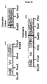
CD44 represents a family of cell surface glycoproteins encoded by a single gene comprising a total of 20 exons. Exons 19 and 20 are expressed together as the cytoplasmic tail and therefore grouped as "exon 19" by most research groups (Liao et al. J. Immunol 151:6490-99, 1993). The term exon 19 will be used henceforth to designate genomic exons 19 and 20. Structural and functional diversity is achieved by alternative splicing of the messenger RNA involving 10 "variant" exons identified as exons 6-15 or, most often, as "variant exons" 1-10 (v1-v10). In human, variant exon 1 contains a stop codon and is not usually expressed. The longest potential CD44 variant is therefore CD44v2-10 (see Naor et al. Adv Cancer Res 71:241-319, 1997 for review of CD44).
-
Exons 1-5 and all variant exons are part of the extracellular domain and contain many potential sites for post-translational modifications. The transmembrane domain is highly conserved across species but the intracellular tail can be truncated leading to another type of variant. One such variant comprises variant exons 8-10 but lacks part of exon 19. Changes to the intracellular domain has been shown to change the function of CD44, in part with respect to binding and internalization of hyaluronic acid (HA). CD44 is not only involved in binding to the extracellular molecules but it also has cell signaling properties (see Turley et al. J Biol Chem 277(7):4589-4592, 2002 for review).
-
The "standard" CD44 (CD44s), the most commonly expressed form of CD44, contains exons 1-5 and 16-19 and none of the variant exons. The molecular weight for the core protein is 37-38kDa but posttranslational modification can result in a molecule of 85-95kDa or more (Drillenburg et al., Blood 95(6):1900, 2000). It binds hyaluronic acid (HA), an extracellular glycosaminoglycan, constitutively and CD44 is often referred to as the HA receptor. It is interesting that the presence of variant exons can reduce the binding of HA by CD44 such that CD44 variants cannot be said to constitutively bind HA but such binding can be inducible (reviewed in Naor et al. Adv Cancer Res 71:241-319, 1997). See Figure 17 for some examples of variants.
-
CD44E, also called CD44v8-10, contains variant exons 8-10 in addition to the exons 1-5 and 16-19. Other variants include CD44v3-10, CD44v6, CD44v7-8 and many others. The variant exons are part of the extracellular domain of the CD44.
-
CD44E can be present on certain normal epithelial cells, particularly by generative cells of the basal cell of stratified squamous epithelium and of glandular epithelium (Mackay et al. J Cell Biol 124(1-2):71-82, 1994) and in the fetus at certain stages development. But importantly, it has been shown to be overexpressed on various types of cancer cells. Using RT-PCR, lida & Bourguignon (J Cell Physiol 162(1):127-133, 1995) and Kalish et al. (Frontiers Bioscience 4(a):1-8, 1999) have shown that CD44E is present in normal breast tissue and is more abundant than CD44s. They have also shown that CD44, including CD44E and CD44s are overexpressed, and preferentially located in metastatic breast cancer tissues. Miyake et al. (J Urol 167(3):1282-87, 2002) reported that CD44v8-10 mRNA is strongly expressed in urothelial cancer and can even be detected in urinary exfoliated cells of patients with invasive vs superficial urothelial cancer. The ratio of CD44v8-10 to CD44v10 mRNA increases in cancer and was shown to have diagnostic value in breast, lung, laryngeal and bladder. The presence of CD44v8-10 was also confirmed by immunohistochemistry with a polyclonal antibody (Okamoto et al. J Natl Cancer Inst 90(4): 307-15, 1997). CD44v8-10 can also be overexpressed in gallbladder cancer (Yamaguchi et al. Oncol Rep 7(3):541-4, 2000), renal cell carcinoma (Hara et al. Urology 54(3):562-6, 1999), testicular germ cell tumors
-
(Miyake et al. Am J Pathol 152(5):1157-60, 1998), non-small cell lung carcinomas (Sasaki et al. Int J Oncol 12(3):525-33, 1998), colorectal cancer (Yamaguchi et al. J Clin Oncol 14(4):1122-27, 1996) and gastric cancer (Yamaguchi et al. Jpn J Cancer Res 86(12): 1166-71, 1995). Overexpression of CD44v8-10 was also shown to have diagnostic value for prostate cancer (Martegani et al. Amer J Pathol 154(1): 291-300, 1999).
-
Alpha-fetoprotein (AFP) is a major serum protein synthesized during fetal life. Its presence in adults is usually indicative of carcinomas, particularly those of the liver and teratocarcinomas. It is part of the albuminoid gene family that also comprises serum and alpha albumins and vitamin D-binding protein. AFP comprises 590 amino acids for a molecular weight of about 69-70 kDa and has one site for glycosylation. (Morinaga et al., Proc Natl Acad Sci 80:4604-08, 1983; Mizejewski Exp Biol Med 226(5):377-408, 2002). Molecular variants have been studied and identified in rodents, but in humans there are no reports of variant proteins being detected. A recent report has identified a variant mRNA that, if expressed, would code for a 65kDa protein. This protein is expected to remain in the cytoplasm (Fukusawa et al. J Soc Gynecol Investig May 20, e-publication, 2005).
SUMMARY OF THE INVENTION
-
The present inventors have prepared human tumor-specific antibodies that bind to several types of tumor cells including bladder, breast, ovary, prostate and uterus. Importantly, the antibodies do not significantly bind to normal tissue making them suitable candidates for tumor therapy. The inventors have cloned and sequenced the antibodies and determined the sequence of the antibody light and heavy chain variable regions and complementarity determining regions 1, 2 and 3. Accordingly, the invention provides isolated light chain complementarity determining regions 1, 2 and 3, comprising the amino acid sequences SGDNLGNKYVC (SEQ ID NO:1), EDTKRPS (SEQ ID NO:2) and QAWDSRTEI (SEQ ID NO:3), respectively; and isolated heavy chain complementarity determining regions 1, 2 and 3, comprising the amino acid sequences GDEYYWS (SEQ ID NO:4), YMSYRGSSYYSPSLQS (SEQ ID NO:5) and KYCGGDCRSGFDI (SEQ ID NO:6), respectively.
-
The invention also provides isolated nucleic acid sequences encoding light chain complementarity determining regions 1, 2 and/or 3, comprising the amino acid sequences SGDNLGNKYVC (SEQ ID NO:1), EDTKRPS (SEQ ID NO:2) and QAWDSRTEI (SEQ ID NO:3), respectively; and isolated nucleic acid sequences encoding heavy chain complementarity determining regions 1, 2 and/or 3, comprising the amino acid sequences GDEYYWS (SEQ ID NO:4), YMSYRGSSYYSPSLQS (SEQ ID NO:5) and KYCGGDCRSGFDI (SEQ ID NO:6), respectively.
-
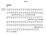
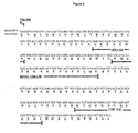
Additional aspects of the invention are isolated light chain variable regions comprising light chain complementarity determining regions 1, 2 and/or 3 of the invention (SEQ ID NOS:1-3), and isolated heavy chain variable regions comprising heavy chain complementarity determining regions 1, 2 and/or 3 of the invention (SEQ ID NOS:4-6). In one embodiment, the light chain variable region comprises the amino acid sequence shown in Figure 1 (SEQ ID NO:7). In another embodiment, the heavy chain variable region comprises the amino acid sequence shown in Figure 2 (SEQ ID NO:9).
-
The invention also provides an isolated nucleic acid sequence encoding the light chain variable region of the invention, and an isolated nucleic acid sequence encoding the heavy chain variable region of the invention. In one embodiment, the light chain variable region comprises the nucleic acid sequence shown in Figure 1 (SEQ ID NO: 8). In another embodiment, the heavy chain variable region comprises the nucleic acid sequence shown in Figure 2 (SEQ ID NO:10).
-
Another aspect of the invention is a binding protein, preferably an antibody or antibody fragment, that comprises at least one light chain complementarity determining region of the invention (i.e. one or more of the SEQ ID NOS:1-3) and/or at least one heavy chain complementarity determining region of the invention (i.e. one or more of SEQ ID NO:4-6). The invention also provides a binding protein, preferably an antibody or antibody fragment that comprises the light chain variable regions of the invention and/or the heavy chain variable regions of the invention.
-
The inventors have also identified the antigen that binds to the binding proteins of the invention. Accordingly, the invention provides the binding protein of the invention that binds to a protein comprising the 5-v8 interface of CD44E, the v8 exon of CD44 or amino acid sequence ATNMDSSHSIT. The invention also provides a binding protein of the invention that binds to CD44E; alpha-fetoprotein; a protein having a molecular weight between 47-53 kDa and an isoelectric point between 5.2-5.5, preferably 5.4; a protein having a molecular weight between 48-54 kDa and an isoelectric point between 5.1-5.4, preferably 5.2; or a protein comprising the amino acid sequence 107 to . 487 of AFP (SEQ ID NO:14), 107 to 590 of AFP (SEQ ID NO: 15) or 107 to 609 of AFP (SEQ ID NO:16).
-
In addition, the invention provides compositions comprising the binding proteins of the invention, such as antibodies and antibody fragments, with a pharmaceutically acceptable excipient, carrier, buffer or stabilizer.
-
Another aspect of the invention is an immunoconjugate comprising (1) binding protein of the invention, preferably an antibody or antibody fragment that binds to an antigen or molecule on or in a cancer cell, attached to (2) an effector molecule. A further aspect of the invention is an immunoconjugate comprising (1) binding protein of the invention, preferably an antibody or antibody fragment that binds to an antigen or molecule that is internalized by a cancer cell, attached to (2) an effector molecule. In a preferred embodiment, the effector molecule is (i) a label, which can generate a detectable signal, directly or indirectly, or (ii) a cancer therapeutic agent, which is either cytotoxic, cytostatic or otherwise prevents or reduces the ability of the cancer cells to divide and/or metastasize. Preferably, the cancer therapeutic agent is a toxin.
-
The invention also provides compositions comprising the immunoconjugate of the invention and uses of the immunoconjugate for the manufacture of a medicament for treating or preventing cancer, and diagnostic purposes. In addition, the invention provides methods of treating or preventing cancer using the immunoconjugate of the invention and related kits.
-
A further aspect of the invention is a method of diagnosing cancer in a mammal comprising the steps of:
- (1) contacting a test sample taken from said mammal with a binding protein of the invention that binds to an antigen on or in the cancer cell under conditions that permit the formulation of a binding protein-antigen complex;
- (2) measuring the amount of binding protein-antigen complex in the test sample; and
- (3) comparing the amount of binding protein-antigen complex in the test sample to a control.
-
The invention also includes a method of diagnosing cancer in a mammal comprising the steps of:
- (1) contacting a test sample taken from said mammal with a binding protein of the invention that binds specifically to alpha-fetoprotein or a variant thereof under conditions that permit the formulation of a binding protein-alpha-fetoprotein complex;
- (2) measuring the amount of binding protein-alpha-fetoprotein complex in the test sample; and
- (3) comparing the amount of binding protein-alpha-fetoprotein complex in the test sample to a control.
-
Another aspect of the invention is a diagnostic agent comprising the immunoconjugate of the invention, wherein the effector molecule is a label, which can generate a detectable signal, directly or indirectly.
-
The invention also includes an isolated protein that can specifically bind with one of the binding proteins of the invention, nucleic acid sequences and uses thereof.
-
Other features and advantages of the present invention will become apparent from the following detailed description. It should be understood, however, that the detailed description and the specific examples while indicating preferred embodiments of the invention are given by way of illustration only, since various changes and modifications within the spirit and scope of the invention will become apparent to those skilled in the art from this detailed description.
BRIEF DESCRIPTION OF THE DRAWINGS
-
The invention will now be described in relation to the drawings in which:
- Figure 1 is the nucleic acid and amino acid sequence of the light chain variable region of VB1-008.
- Figure 2 is the nucleic acid and amino acid sequence of the heavy chain variable region of VB1-008.
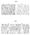
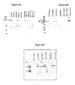
- Figure 3 is SKBR-3 (400X mag) fixed-cell pellet stained with VB1-008 (A) and the isotype control antibody 4B5 (B). Notice prominent membrane staining (arrow).
- Figure 4 are representative photographs of immunohistochemical staining of normal testis with VB1-008 and the isotype control antibody 4B5. (A) Sample 925 testes tissue (400X mag) stained with VB1-008. Membrane staining in mature sperm cells is indicated by an arrow. (B) Sample 925 testes tissue (400X mag) stained with IgG isotype control 4B5. Notice absence of staining. Arrow points to mature sperm cell for contrast to staining with VB1-008 in (A).
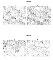
- Figure 5 shows Sample 3427A1 breast adenocarcinoma (400X) stained with VB1-008 and IgG isotype control 4B5. Notice staining of cell membrane of tumor cells, especially of cells in contact with the extracellular matrix (white arrow). Cells close to the center of the tumor show primarily cytoplasmic staining (black arrow). Arrow points to unstained tumor cells. Tumor cells are stained with VB1-008.
- Figure 6 shows Sample 946 B1 bladder carcinoma (400X) stained with VB1-008 (A) and IgG isotype control 4B5 (B). Arrows indicate membrane staining of the tumor cells with VB1-008 (A) but not with the control antibody (B).
- Figure 7 shows sample 4036A2 uterus carcinoma (200X mag) stained with VB1-008 and the IgG control antibody 4B5. Notice membrane staining (arrow) with VB1-008 (A & C) but not with the control antibody (B). Higher magnification of uterus carcinoma (600X) shows membrane staining (C).
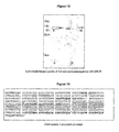
- Figure 8 is a demonstration of antibody cell surface binding after incubation of A-375 cells at different temperatures as determined by flow cytometry. Fluorescence labeling of A-375 cells after incubation of cell suspensions at 4°C: 4B5 (1) and VB1-008 (2) Fluorescence labeling of A-375 cells after warming antibody-bound cells to 37°C: VB1-008 for 60 min (3), for 120 min (4).
- Figure 9 shows confocal microscopy assessment of VB1-008 internalization. A-375 cells were incubated with antibody at 4°C, washed and warmed to 37°C for 60 min. Cells were fixed, permeabilized and labeled with fluorescent-labeled second antibody. Fluorescence labeling of A-375 cells after incubation of VB1-008 at 4°C for 60 min, displaying circumferential surface distribution of labeling, (60X x 4) magnification (A). Following incubation of antibody-bound cells at 37°C for 60 min the cells show strong intracellular staining by internalized antibody, (60X x 4) magnification (B).
- Figures 10A, B and C show a western analysis of immunoprecipitation reactions using VB1-008. Figure 10A shows the results of the experiment under non-reducing conditions, while Figures 10B and C show the results of the experiment under reducing conditions.
- Figures 11A and B show the presence of two distinct protein spots in the purified antigen complex, very close in molecular weight. The proteins were probably not perceived as two bands in 1 D-PAGE due to protein stacking. Figure 11A represents the western blot profile of the 2D-gel and Figure 11B represents the Coomassie stained counterpart.
- Figures 12A and B show the mapping of the peptides obtained and the sequence coverage of the original AFP molecule, Accession # GI|4501989. Figure 12A shows the mapping of peptides obtained from the 2D gel. The amino acids in bolded font represent the sequences of amino acids identified from MS analysis. The shaded regions represent the homology of peptide sequences and thereby depict the sequence coverage. Figure 12B shows the complete mapping of the peptides obtained from the 1 D and 2D gels. The amino acids in bolded font represent the sequences of amino acids from MS analysis. The shaded regions represent the homology of peptide sequences and thereby detect the sequence coverage. The underlined amino acids were not detected.
- Figure 13 shows immunopurification of the VB1-008 antigen using 1000 µg of MDA-MB-435S membranes as the source. The purified antigen(s) was resolved on SDS-PAGE under non-reducing sample conditions. Reducing agents such as DTT or β-mercaptoethanol were avoided so as to preserve the native conformation of the binding antigen(s). The sample was resolved on two lanes of the gel. One lane (A), was stained for protein; the other (B) was subjected to western blotting and probed with VB1-008, to ensure the presence of the specific antigen. Band "E" from the coomassie stained portion of the gel was excised and sent for MS analysis.
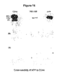
- Figure 14 shows the complete mapping of the peptides obtained and the sequence coverage of CD44 molecule, Accession # GI|105583. The amino acids in red font represent the sequences of amino acids identified from MS analysis. The shaded regions represent the homology of peptide sequences and thereby depict the sequence coverage. The amino acids in underlined area constitute the variable region (v8-v10) characteristic of the isoform3 or CD44E.
- Figure 15A shows the reactivity of VB1-008 to recombinant AFP molecule, commercially available from RDI systems. The recombinant AFP was electrophoresed, transferred on to nitrocellulose membrane and probed with VB1-008. The results are clearly indicative of the reactivity of VB1-008 to AFP.
- Figures 15B and C are 2D-gel profiles of "B" and "C", which were immunoprecipitates obtained using VB1-008. The gels were transferred to nitrocellulose and probed with anti-CD44 and anti-AFP, both mouse-monoclonal antibodies respectively.
- Figure 16 is a western analysis under non-reducing conditions. Anti-CD44 was used to immunopurify CD44 proteins from MDA-MB-435S cells and the purified fraction was subjected to SDS-PAGE and WB analysis under non-reducing conditions. The experiment was performed in three sets and each set was identical to the other. Each of the sets was probed with 5 µg/mL of anti-CD44, anti-AFP and VB1-008. Anti-CD44 and anti-AFP were mouse monoclonal antibodies, whereas, VB1-008 is VBI's human monoclonal antibody.
- Figure 17 is a schematic representation of the distribution of different exons in the CD44 gene in humans. Alternative splicing in the variable region results in the creation of a number of isoforms, a few of the reported isoforms are represented schematically in the corresponding figure.
- Figure 18A depicts the amino acid sequence of CD44E. The highlighted portion represents the stretch of 17 amino acids used to generate peptides 1-3. The negative control peptide is highlighted in the C-terminal region of the protein. Figure 18B shows the results of a binding experiment with VB1-008 to peptides 1-3.
- Figure 19A shows the results of a competition study using peptides 1-3 against binding of VB1-008. Figure 19B shows the results of a competition study using peptides 1-3 against a control antibody (anti-EGFR).
- Figure 20 shows the nucleotide sequence of the immunoconjugate VB6-008 (SEQ ID NO:11). The sequence of the PelB leader sequence is in lower case with the initiation codon bolded. The stop codes are in uppercase and bolded.
- Figure 21 shows the amino acid sequences of the heavy chain and light chain of the immunoconjugate VB6-0008 (SEQ ID NO:12 and 13).
- Figure 22 shows the complete VB6-008 construct.
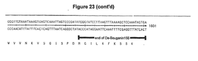
- Figure 23 shows the VB6-008 unit #1, which includes the PeIB-VH-CH-Furin-De-Bouganin.
- Figure 24 shows the VB6-008 #2 unit which consists of PelB-VL-CL.
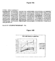
- Figure 25 shows the results of an in vitro cytotoxicity experiment using VB6-008.
- Figure 26 is a depiction of the gamma cassette.
- Figure 27 is a depiction of the assembly of the Fab-bouganin immunotoxin.
DETAILED DESCRIPTION OF THE INVENTION
(A) Definitions
-
The term "administered systemically" as used herein means that the immunoconjugate and/or other cancer therapeutic may be administered systemically in a convenient manner such as by injection (subcutaneous, intravenous, intramuscular, etc.), oral administration, inhalation, transdermal administration or topical application (such as topical cream or ointment, etc.), suppository applications, or means of an implant. An implant can be of a porous, non-porous, or gelatinous material, including membranes, such as sialastic membranes, or fibers. Suppositories generally contain active ingredients in the range of 0.5% to 10% by weight.
-
The term "antibody" as used herein is intended to include monoclonal antibodies, polyclonal antibodies, and chimeric antibodies. The antibody may be from recombinant sources and/or produced in transgenic animals. The term "antibody fragment" as used herein is intended to include Fab, Fab', F(ab')2, scFv, dsFv, ds-scFv, dimers, minibodies, diabodies, and multimers thereof and bispecific antibody fragments. Antibodies can be fragmented using conventional techniques. For example, F(ab')2 fragments can be generated by treating the antibody with pepsin. The resulting F(ab')2 fragment can be treated to reduce disulfide bridges to produce Fab' fragments. Papain digestion can lead to the formation of Fab fragments. Fab, Fab' and F(ab')2, scFv, dsFv, ds-scFv, dimers, minibodies, diabodies, bispecific antibody fragments and other fragments can also be synthesized by recombinant techniques.
-
The term "antibody or antibody fragment of the invention" as used herein comprises at least one light chain complementarity determining region of the invention (i.e. one or more of SEQ ID NOS:1-3) and/or at least one heavy chain complementarity determining region of the invention (i.e. one or more of SEQ ID NOS:4-6). Preferably, the antibody or antibody fragment comprises the light chain CDR sequences (SEQ ID NOS:1-3) and/or the heavy chain CDR sequences (SEQ ID NOS:4-6) or functional variants of the sequences so that the antibody or antibody fragment can bind to the tumor cell without substantially binding to normal cells. Antibodies or antibody fragments of the invention also include antibodies or antibody fragments that bind to CD44E; alpha-fetoprotein; a protein having a molecular weight between 47-53 kDa and an isoelectric point between 5.2-5.5, preferably 5.4; a protein having a molecular weight between 48-54 kDa and an isoelectric point between 5.1-5.4, preferably 5.2; or a protein comprising the amino acid sequence 107 to 487 of AFP (SEQ ID NO:14), 107 to 590 of AFP (SEQ ID NO: 15) or 107 to 609 of AFP (SEQ ID NO:16). In addition, antibodies or antibody fragments of the invention include antibodies or antibody fragments that bind to a protein comprising the 5-v8 interface of CD44E, the v8 exon of CD44 or amino acid sequence ATNMDSSHSIT.
-
By "at least moderately stringent hybridization conditions" it is meant that conditions are selected which promote selective hybridization between two complementary nucleic acid molecules in solution. Hybridization may occur to all or a portion of a nucleic acid sequence molecule. The hybridizing portion is typically at least 15 (e.g. 20, 25, 30, 40 or 50) nucleotides in length. Those skilled in the art will recognize that the stability of a nucleic acid duplex, or hybrids, is determined by the Tm, which in sodium containing buffers is a function of the sodium ion concentration and temperature (Tm = 81.5°C-16.6 (Log10 [Na+]) + 0.41(%(G+C) - 600/l), or similar equation). Accordingly, the parameters in the wash conditions that determine hybrid stability are sodium ion concentration and temperature. In order to identify molecules that are similar, but not identical, to a known nucleic acid molecule a 1% mismatch may be assumed to result in about a 1°C decrease in Tm, for example if nucleic acid molecules are sought that have a >95% identity, the final wash temperature will be reduced by about 5°C. Based on these considerations those skilled in the art will be able to readily select appropriate hybridization conditions. In preferred embodiments, stringent hybridization conditions are selected. By way of example the following conditions may be employed to achieve stringent hybridization: hybridization at 5x sodium chloride/sodium citrate (SSC)/5x Denhardt's solution/1.0% SDS at Tm - 5°C based on the above equation, followed by a wash of 0.2x SSC/0.1% SDS at 60°C. Moderately stringent hybridization conditions include a washing step in 3x SSC at 42°C. It is understood, however, that equivalent stringencies may be achieved using alternative buffers, salts and temperatures. Additional guidance regarding hybridization conditions may be found in: Current Protocols in Molecular Biology, John Wiley & Sons, N.Y., 1989, 6.3.1-6.3.6 and in: Sambrook et al., Molecular Cloning, a Laboratory Manual, Cold Spring Harbor Laboratory Press, 1989, Vol.3.
-
The term "binding protein" as used herein refers to proteins that specifically bind to another substance. In an embodiment, binding proteins are antibodies or antibody fragments.
-
The term "binding proteins of the invention" as used herein includes antibodies or antibody fragments of the invention.
-
By "biologically compatible form suitable for administration in vivo" is meant a form of the substance to be administered in which any toxic effects are outweighed by the therapeutic effects.
-
The term "cancer" as used herein includes any cancer that can be bound by a binding protein of the invention, preferably an antibody or antibody fragment of the invention.
-
The term "CD44" as used herein refers to the family of CD44 molecules encoded by a single gene comprising a total of 19 exons. There are 10 variable exons. Alternative splicing in the variable regions results in the creation of a number of different CD44 variants (See Figure 17). The term "CD44E", also known as CD44v8-10, refers to the epithelial variant of CD44. CD44E contains variant exons 8-10 in addition to exons 1-5 and 16-19. The term "v8 exon of CD44" refers to variable exon 8 of CD44. The term "5-v8 interface of CD44E" refers to the region where exon 5 connects with variable exon 8 in CD44E. It is a continuous sequence that includes part of the region of exon 5 and part of the variable exon 8 of CD44E.
-
A "conservative amino acid substitution", as used herein, is one in which one amino acid residue is replaced with another amino acid residue without abolishing the protein's desired properties.
-
The term "controlled release system" as used means the immunoconjugate and/or other cancer therapeutic of the invention can be administered in a controlled fashion. For example, a micropump may deliver controlled doses directly into the area of the tumor, thereby finely regulating the timing and concentration of the pharmaceutical composition (see, e.g., Goodson, 1984, in Medical Applications of Controlled Release, vol. 2, pp. 115-138).
-
The term "direct administration" as used herein means the immunoconjugate and/or other cancer therapeutic may be administered, without limitation, intratumorally, intravascularly, and peritumorally. For example, the immunoconjugate may be administered by one or more direct injections into the tumor, by continuous or discontinuous perfusion into the tumor, by introduction of a reservoir of the immunoconjugate, by introduction of a slow-release apparatus into the tumor, by introduction of a slow-release formulation into the tumor, and/or by direct application onto the tumor. By the mode of administration "into the tumor," introduction of the immunoconjugate and/or other cancer therapeutic to the area of the tumor, or into a blood vessel or lymphatic vessel that substantially directly flows into the area of the tumor, is included.
-
As used herein, the phrase "effective amount" means an amount effective, at dosages and for periods of time necessary to achieve the desired result. Effective amounts of an immunoconjugate may vary according to factors such as the disease state, age, sex, weight of the animal. Dosage regime may be adjusted to provide the optimum therapeutic response. For example, several divided doses may be administered daily or the dose may be proportionally reduced as indicated by the exigencies of the therapeutic situation.
-
The term "heavy chain complementarity determining region" as used herein refers to regions of hypervariability within the heavy chain variable region of an antibody molecule. The heavy chain variable region has three complementarity determining regions termed heavy chain complementarity determining region 1, heavy chain complementarity determining region 2 and heavy chain complementarity determining region 3 from the amino terminus to carboxy terminus.
-
The term "heavy chain variable region" as used herein refers to the variable region of a heavy chain.
-
The term "immunoconjugate of the invention" is used herein comprises (1) a binding protein, preferably an antibody or antibody fragment, of the invention attached to (2) an effector molecule. The effector molecule can be any molecule that one wishes to deliver to the cancer cell, including, but not limited to (i) a label, which can generate a detectable signal, directly or indirectly, or (ii) a cancer therapeutic agent, such as a toxin that is either cytotoxic, cytostatic or otherwise prevents or reduces the ability of the cancer cells to divide and/or metastasize.
-
The term "isolated nucleic acid sequences" as used herein refers to a nucleic acid substantially free of cellular material or culture medium when produced by recombinant DNA techniques, or chemical precursors, or other chemicals when chemically synthesized. An isolated nucleic acid is also substantially free of sequences which naturally flank the nucleic acid (i.e. sequences located at the 5' and 3' ends of the nucleic acid) from which the nucleic acid is derived. The term "nucleic acid" is intended to include DNA and RNA and can be either double stranded or single stranded.
-
The term "isolated proteins", such as light chain complementarity regions 1, 2 and 3, heavy chain complementarity regions 1, 2 and 3, light chain variable regions, heavy chain variable regions, and binding proteins of the invention, refers to a protein substantially free of cellular material or culture medium when produced by recombinant DNA techniques, or chemical precursors or other chemicals when chemically synthesized.
-
The term "light chain complementarity determining region" as used herein refers to regions of hypervariability within the light chain variable region of an antibody molecule. Light chain variable regions have three complementarity determining regions termed light chain complementarity determining region 1, light chain complementarity determining region 2 and light chain complementarity determining region 3 from the amino terminus to the carboxy terminus.
-
The term "light chain variable region" as used herein refers to the variable region of a light chain.
-
The term "modified bouganin" as used here means a modified bouganin that has a reduced propensity to activate an immune response as described in
PCT/CA2005/000410 and United States Patent Application No.
11.084,080 . In one example, the modified bouganin has the amino acid sequence (SEQ ID NO: 17):
-
The term "nucleic acid sequence" as used herein refers to a sequence of nucleoside or nucleotide monomers consisting of naturally occurring bases, sugars and intersugar (backbone) linkages. The term also includes modified or substituted sequences comprising non-naturally occurring monomers or portions thereof. The nucleic acid sequences of the present invention may be deoxyribonucleic acid sequences (DNA) or ribonucleic acid sequences (RNA) and may include naturally occurring bases including adenine, guanine, cytosine, thymidine and uracil. The sequences may also contain modified bases. Examples of such modified bases include aza and deaza adenine, guanine, cytosine, thymidine and uracil; and xanthine and hypoxanthine.
-
The term "sequence identity" as used herein refers to the percentage of sequence identity between two polypeptide sequences. In order to determine the percentage of identity between two polypeptide sequences, the amino acid sequences of such two sequences are aligned, preferably using the Clustal W algorithm (Thompson, JD, Higgins DG, Gibson TJ, 1994, Nucleic Acids Res. 22 (22): 4673-4680), together with BLOSUM 62 scoring matrix (Henikoff S. and Henikoff J.G., 1992, Proc. Natl. Acad. Sci. USA 89: 10915-10919) and a gap opening penalty of 10 and gap extension penalty of 0.1, so that the highest order match is obtained between two sequences wherein at least 50% of the total length of one of the sequences is involved in the alignment. Other methods that may be used to align sequences are the alignment method of Needleman and Wunsch (J. Mol. Biol., 1970, 48: 443), as revised by Smith and Waterman (Adv. Appl. Math., 1981, 2: 482) so that the highest order match is obtained between the two sequences and the number of identical amino acids is determined between the two sequences. Other methods to calculate the percentage identity between two amino acid sequences are generally art recognized and include, for example, those described by Carillo and Lipton (SIAM J. Applied Math., 1988, 48:1073) and those described in Computational Molecular Biology, Lesk, e.d. Oxford University Press, New York, 1988, Biocomputing: Informatics and Genomics Projects. Generally, computer programs will be employed for such calculations. Computer programs that may be used in this regard include, but are not limited to, GCG (Devereux et al., Nucleic Acids Res., 1984, 12: 387) BLASTP, BLASTN and FASTA (Altschul et al., J. Molec. Biol., 1990: 215: 403).
-
As used herein, the phrase "treating cancer" refers to inhibition of cancer cell replication, inhibition of cancer spread (metastasis), inhibition of tumor growth, reduction of cancer cell number or tumor growth, decrease in the malignant grade of a cancer (e.g., increased differentiation), or improved cancer-related symptoms.
-
The term "variant" as used herein includes modifications or chemical equivalents of the amino acid and nucleotide sequences of the present invention that perform substantially the same function as the proteins or nucleic acid molecules of the invention in substantially the same way. For example, variants of proteins of the invention include, without limitation, conservative amino acid substitutions. Variants of proteins of the invention also include additions and deletions to the proteins of the invention.
-
The term "variant of alpha-fetoprotein" includes variants of alpha-fetoprotein, such as a protein comprising the amino acid sequence of SEQ ID NO:14, 15 or 16; or a protein that is a truncated version of alpha-fetoprotein and has the molecular weight of 48-54 kDa and an isoelectric point between 5.1-5.4.
(B) Proteins and Nucleic Acids of the Invention
(i) Light and Heavy Chain Complementarity Determining Regions and Light and Heavy Chain Variable Regions
-
The invention provides isolated light chain complementarity determining region 1 comprising the amino acid sequence SGDNLGNKYVC (SEQ ID NO:1). The invention also provides isolated light chain complementarity determining region 2 comprising the amino acid sequence EDTKRPS (SEQ ID NO:2). In addition, the invention provides isolated light chain complementarity determining region 3 comprising the amino acid sequence QAWDSRTEI (SEQ ID NO:3).
-
The invention provides isolated light chain complementarity determining region 1 comprising the amino acid sequence GDEYYWS (SEQ ID NO:4). The invention also provides isolated light chain complementarity determining region 2 comprising the amino acid sequence YMSYRGSSYYSPSLQS (SEQ ID NO:5). In addition, the invention provides isolated light chain complementarity determining region 3 comprising the amino acid sequence KYCGGDCRSGFDI (SEQ ID NO:6).
-
The invention provides isolated light chain complementarity determining regions 1, 2 and 3, comprising the amino acid sequences SGDNLGNKYVC (SEQ ID NO:1), EDTKRPS (SEQ ID NO:2) and QAWDSRTEI (SEQ ID NO:3), respectively; and isolated heavy chain complementarity determining regions 1, 2 and 3, comprising the amino acid sequences GDEYYWS (SEQ ID NO:4), YMSYRGSSYYSPSLQS (SEQ ID NO:5) and KYCGGDCRSGFDI (SEQ ID NO:6), respectively.
-
The invention also includes variants of the CDR sequences that can bind to the same epitope or antigen recognized by the CDR sequences disclosed above.
-
Additional aspects of the invention are isolated light chain variable regions comprising light chain complementarity determining regions 1, 2 and/or 3 of the invention (SEQ ID NOS:1-3); and heavy chain variable regions comprising the heavy chain complementarity determining regions 1, 2 and/or 3 of the invention (SEQ ID NOS:4-6). In one embodiment, the light chain variable region comprises the amino acid sequence shown in Figure 1 (SEQ ID NO:7), and the heavy chain variable region comprises the amino acid sequence shown in Figure 2 (SEQ ID NO:9).
-
The invention also includes variants of the isolated light chain variable regions and heavy chain variable regions that can bind to the same epitope or antigen recognized by the isolated light chain variable regions and isolated heavy chain variable regions disclosed above.
-
A person skilled in the art will appreciate that the invention includes variants to the amino acid sequences of SEQ ID NOS:1-6, 7 and 9, including chemical equivalents to the sequences disclosed by the present invention. Such equivalents include proteins that perform substantially the same function as the specific proteins disclosed herein in substantially the same way. A functional variant of a CDR sequence will be able to bind to the antigen or epitope recognized by the native CDR sequence. For example, equivalents include, without limitation, conservative amino acid substitutions.
-
In one embodiment, the variant amino acid sequences of the light chain complementarity determining regions 1, 2 and 3, and the heavy chain complementarity determining regions 1, 2 and 3 have at least 50%, preferably at least 60%, more preferably at least 70%, most preferably at least 80%, and even more preferably at least 90% sequence identity to SEQ ID NOS:1-6, respectively.
-
In another embodiment, the variant amino acid sequences of the light chain variable region and the heavy chain variable region have at least 50%, preferably at least 60%, more preferably at least 70%, most preferably at least 80%, and even more preferably at least 90% sequence identity to SEQ ID NOS:7 and 9, respectively.
-
The invention also provides an isolated nucleic acid sequence encoding the light chain variable region of the invention, and an isolated nucleic acid sequence encoding the heavy chain variable region of the invention. In one embodiment, the light chain variable region comprises the nucleic acid sequence shown in Figure 1 (SEQ ID NO: 8). In another embodiment, the heavy chain variable region comprises the nucleic acid sequence shown in Figure 2 (SEQ ID NO:10). The invention also includes variants to the nucleic acid sequences that encode for the light chain variable region and heavy chain variable region of the invention. For example, the variants include nucleotide sequences that hybridize to the nucleic acid sequences encoding the light chain variable region and heavy chain variable region of the invention under at least moderately stringent hybridization conditions.
-
The invention also provides isolated nucleic acid sequences encoding light chain complementarity determining regions 1, 2 and/or 3, comprising the amino acid sequences SGDNLGNKYVC (SEQ ID NO:1), EDTKRPS (SEQ ID NO:2) and QAWDSRTEI (SEQ ID NO:3), respectively; and isolated nucleic acid sequences encoding heavy chain complementarity determining regions 1, 2 and/or 3, comprising the amino acid sequences GDEYYWS (SEQ ID NO:4), YMSYRGSSYYSPSLQS (SEQ ID NO:5) and KYCGGDCRSGFDI (SEQ ID NO:6), respectively. The invention also includes isolated nucleic acid sequences encoding variants of the CDR sequences discussed above. Nucleic acid sequences encoding variants of the CDR sequences of the invention include nucleic acid sequences that hybridize to the CDR sequences encoding the amino acid sequences shown in SEQ ID NOS:1-6 under at least moderately stringent hybridization conditions.
(ii) Binding proteins
-
Another aspect of the invention is a binding protein, preferably an antibody or antibody fragment, that comprises at least one light chain complementarity determining region of the invention (i.e. one or more of SEQ ID NOS:1-3) and/or at least one heavy chain complementarity determining region of the invention (i.e. one or more of SEQ ID NOS:4-6). Such a binding protein can be generally referred to herein as "a binding protein of the invention", or preferably "an antibody or antibody fragment of the invention".
-
In one embodiment, the binding protein, preferably an antibody or antibody fragment, comprises the light chain complementarity determining regions 1, 2 and 3, comprising the amino acid sequences SGDNLGNKYVC (SEQ ID NO:1), EDTKRPS (SEQ ID NO:2) and QAWDSRTEI (SQ ID NO:3), respectively; and heavy chain complementarity determining regions 1, 2 and 3, comprising the amino acid sequences GDEYYWS (SEQ ID NO:4), YMSYRGSSYYSPSLQS (SEQ ID NO:5) and KYCGGDCRSGFDI (SEQ ID NO:6), respectively. The invention also provides a binding protein, preferably an antibody or antibody fragment, that comprises the light chain variable region of the invention and/or the heavy chain variable region of the invention.
-
A person skilled in the art will appreciate that the invention includes variants to the specific binding proteins disclosed above, including chemical equivalents to the sequences disclosed above that perform substantially the same function as the binding proteins disclosed above in substantially the same way. A functional variant of a binding protein will be able to bind to a protein comprising 5-v8 interface of CD44E, the v8 exon of CD44, the amino acid sequence ATNMDSSHSIT, amino acid SEQ ID NOS:14, 15 or 16, or to a protein having a molecular weight between 47-53 kDa and an isoelectric point between 5.2-5.5; a protein having a molecular weight between 48-54 kDa and an isoelectric point between 5.1-5.4, CD44E, or alpha-fetoprotein or a variant thereof.
-
As stated above, the inventors have identified the antigen that binds to the binding protein of the invention. In particular, the inventors have shown that the binding proteins of the invention bind to the extracellular domain of CD44E. In addition, the inventors have shown that the binding proteins of the invention bind to AFP or a variant thereof.
-
It is important to recognize that CD44 molecules have a high potential for N- and O-glycosylation and for the addition of chondroitin sulfate and heparan sulfate. However, the pattern of these post-translational modifications is variable, and appears to be cell-specific and can potentially affect the ability of CD44 to bind HA or other extracellular molecules. The variable pattern of post-translational modifications is particularly relevant to the preparation of anti-CD44 monoclonal antibodies since antibody binding has been shown to be affected by the presence of these modifications, despite the primary structure of the molecule being the same as that of the antigen used to raise the antibody (Matzuki et al. Cancer Res 63:8278-83, 2003; Martegani et al. Amer J Pathol 154(1): 291-300, 1999). This also limits the usefulness of recombinant CD44 as an immunogen since its glycosylation pattern would likely differ from that of tumor cells. The binding proteins of the invention is, therefore, particularly unique since it recognizes a form of the CD44 that is present on human tumor cells.
-
Accordingly, the invention provides a binding protein of the invention that binds to a protein comprising the 5-v8 interface of CD44E, the v8 exon of CD44 or amino acid sequence ATNMDSSHSIT. The invention also provides a binding protein of the invention that binds to CD44E; alpha-fetoprotein; a protein having a molecular weight between 47-53 kDa and an isoelectric point between 5.2-5.5, preferably 5.4; a protein having a molecular weight between 48-54 kDa and an isoelectric point between 5.1-5.4, preferably 5.2; or a protein comprising the amino acid sequence 107 to 487 of AFP (SEQ ID NO:14), 107 to 590 of AFP (SEQ ID NO: 15) or 107 to 609 of AFP (SEQ ID NO:16). The invention also provides a binding protein of the invention that binds to a protein comprising SEQ ID NOS: 38, 39, 40, 41, 42, 43, 44 or 45 and having a molecular weight between 47-53 kDa and an isoelectric point between 5.2-5.5; or a protein comprising SEQ ID NOS: 46, 47, 48, 49, 50, 51, 52, 53, 54, 55, 56, 57, 58, 59, 60, 61, 62, 63, 64, 65, 66, 67, 68, 69, 70, 71, 72, 73, 74 or 75 and having a molecular weight between 48-54 kDa and an isoelectric point between 5.1-5.4.
-
The invention also includes binding proteins that bind to the amino acid sequence ATNMDSSHSIT.
-
In certain embodiments, the antibody or antibody fragment comprises all or a portion of a heavy chain constant region, such as an IgG1, IgG2, IgG3, IgG4, IgA1, IgA2, IgE, IgM or IgD constant region. Preferably, the heavy chain constant region is an IgG1 heavy chain constant region. Furthermore, the antibody or antibody fragment can comprise all or a portion of a kappa light chain constant region or a lambda light chain constant region. Preferably, the light chain constant region is a lambda light chain constant region.
-
To produce monoclonal antibodies derived from humans, antibody producing cells (lymphocytes) can be harvested from a human having cancer and fused with myeloma cells by standard somatic cell fusion procedures thus immortalizing these cells and yielding hybridoma cells. Such techniques are well known in the art, (e.g. the hybridoma technique originally developed by
Kohler and Milstein (Nature 256:495-497 (1975)) as well as other techniques such as the human B-cell hybridoma technique (
Kozbor et al., Immunol. Today 4:72 (1983)), the EBV-hybridoma technique to produce human monoclonal antibodies (
Cole et al., Methods Enzymol, 121:140-67 (1986)), and screening of combinatorial antibody libraries (
Huse et al., Science 246:1275 (1989)). Another example of making human monoclonal antibodies is described in
WO/9947929 . In another example, a myeloma-like fusion partner, as described in
Dan et al. (J Neurosurgery 76:660-69, 1992) can be used. Hybridoma cells can be screened immunochemically for production of antibodies specifically reactive with cancer cells and the monoclonal antibodies can be isolated.
-
Specific antibodies, or antibody fragments, reactive against particular antigens or molecules, such as antigens or molecules on a cancer cell, may also be generated by screening expression libraries encoding immunoglobulin genes, or portions thereof, expressed in bacteria with cell surface components. For example, complete Fab fragments, VH regions and FV regions can be expressed in bacteria using phage expression libraries (See for example Ward et al., Nature 341:544-546 (1989); Huse et al., Science 246:1275-1281 (1989); and McCafferty et al., Nature 348:552-554 (1990)).
-
The present invention includes all antibodies and antibody fragments that bind to the same antigen as the antibodies or antibody fragments of the invention. A person skilled in the art will appreciate that binding assays can be used to find other antibodies and antibody fragments with the same binding specificities as the antibodies and antibody fragments of the invention. As exemplified, below, a competition binding assay can be used to find such other antibodies.
-
Before a competition assay is performed using flow cytometry, the minimal concentration of antibody of the invention (Ab1) that gives maximal binding against a fixed number of tumor cells (for example, A-375 cells for VB1-008) is determined. A total of 106 cells are harvested from exponentially growing cultures and incubated with various antibody concentrations for 1 hr at 4°C. The cells are washed and incubated with a suitable detection antibody for an additional hour at 4°C. After washing, the cells are analyzed by flow cytometry. For each test antibody, a saturation curve is generated from the data by plotting median fluorescence against the antibody concentration.
-
For the competition assay, tumor cells are prepared as above and treated in duplicate with a fixed concentration of antibody (Ab1). The fixed concentration is the minimal concentration of antibody that generates maximal binding against a fixed number of tumor cells as determined above. Immediately following the addition of the Ab1, varying concentrations of the potential inhibitory antibody (Ab2) is added to each tube and the mixture incubated for 1 hr at 4°C. Both the percent inhibition and change over maximum median fluorescence are calculated by subtracting the background fluorescence (PBS-5% FCS) from the median fluorescence reading for each test sample (Ab1 + Ab2). The result is then divided by the median fluorescence of Ab1 alone (maximal binding) minus the background (see below). The percent of inhibition result is obtained by multiplying by 100. The mean of the replicates along with their respective standard error is plotted against antibody concentration. The following formula is used to calculate the percent inhibition:
where
PI = percent inhibition;
MF(Ab1+Ab2) = median fluorescence measured for Ab1+Ab2 mixture; and
MFBgd = background median fluorescence with PBS-5% FCS.
-
Accordingly, the invention provides a binding protein capable of binding an antigen on a tumor cell wherein the binding protein can be identified by a method comprising:
- (1) incubating a fixed number of tumor cells with a minimal concentration of a binding protein of the invention, preferably an antibody or antibody fragment (Ab1) that generates maximal binding against the fixed number of tumor cells and measuring median fluorescence of Ab1 (MFAb1);
- (2) testing two or more concentrations of a test binding protein (Ab2) by adding Ab2 to the Ab1 and tumor cells, and measuring median fluorescence (MF(Ab1+Ab2));
- (3) measuring background median fluorescence (MFbgd);
- (4) calculating PI, wherein
and - (5) comparing the PI to a control PI value;
wherein, a PI that has a statistically significant difference from the control PI indicates that the test binding protein is capable of binding the antigen on the tumor cell.
-
The competition binding assay can also be done with peptides, preferably the peptide defined by SEQ ID NO:28. Similar to the method described above, before the competition assay is performed, the minimal concentration of test binding protein (Ab2) that gives maximal binding against a fixed number of tumor cells is determined.
-
Accordingly, an embodiment of the invention provides a binding protein capable of binding an antigen on a tumor cell wherein the binding protein can be identified by a method comprising:
- (1) incubating a fixed number of tumor cells with a minimal concentration of a test binding protein (Ab2) that generates maximal binding against the fixed number of tumor cells and measuring median fluorescence of Ab2 (MFAb2);
- (2) preparing a peptide and Ab2 mixture by incubating a molar excess of a peptide defined by SEQ ID NO:28 with said minimal concentration of the test binding protein (Ab2);
- (3) adding said mixture to tumor cells and measuring median fluorescence (MF(Ab2+peptide));
- (4) measuring background median fluorescence (MFbgd);
- (5) calculating PI, wherein
and - (6) comparing the PI to a control PI value;
wherein, a PI that has a statistically significant difference from the control PI indicates that the test binding protein is capable of binding the antigen on the tumor cell.
-
A person skilled in the art will appreciate that affinity maturation techniques could be used modify the binding proteins or immunoconjugates of the invention either by increasing its affinity for both CD44E and AFP or by selecting out the binding to one antigen. The latter can lead to the development of 2 separate antibodies or immunoconjugates with preferential binding to either AFP or to CD44E.
-
Two strategies are routinely used to enhance the binding affinity of an antibody. One approach utilizes the resolution of the crystal structure of the Ab-Ag complex to identify the key residues involved in the antigen binding (Davies D.R., Cohen G.H. 1996. Interactions of protein antigens with antibodies. Proc Natl. Acad. Sci. U.S A. 93, 7-12). Subsequently, those residues can be mutated to enhance the interaction. The other approach mimics an in vivo antigen stimulation that drives the affinity maturation of immunoglobulin produced by B cells. During the maturation of the immune response, the variable regions of the immunoglobulins are subjected to somatic mutations (Mc Heyzer-Williams M. 2003. B-cell signaling mechanism and activation. Fundamental Immunology, Fifth edition, 195-225). This process, highly specific for the immune system, is characterized by the introduction of point mutations at a very high rate. It occurs only within the DNA fragments encoding the variable regions and excludes the conserved domains. The B cells expressing the somatically mutated variant antibody are then subjected to an antigen-mediated selection resulting in the selection of higher affinity immunoglobulin. In order to replicate this phenomenon in vitro, several approaches have been used to introduce mutations either by random or targeted processes. The random mutations can be introduced using error-prone PCR, chain shuffling or mutator E. coli strains (Clackson T. Hoogenboom N.R., Griffiths A.D. and Winter G. 1991 Making antibody fragments using phage display libraries. Nature 352, 624-628, Hawkins R.E., Russell S.J. and Winter G. 1992. Selection of phage antibodies by binding affinity. Mimicking affinity maturation. J. Mol. Biol. 226, 889-896, Low N., Holliger P. and Winter G. 1996. Mimicking somatic hypermutation: affinity maturation of antibodies displayed on bacteriophage using a bacterial mutator strain. J Mol. Biol. 260, 359-368). This strategy leads to the creation of large libraries in which reactive clones are selected with a display technology such as ribosome, phage or yeast (Min L. (2000). Applications of display technology in protein analysis. Nat. Biotechnol. 18, 1251-1256).
-
The targeted mutations of the CDRs, especially CDR3 of the light and heavy chains, have been shown to be an effective technique for increasing antibody affinity. Blocks of 3 to 4 amino acids of the CDR3 or specific regions called "hot-spots" are targeted for mutagenesis. Yang et al reported an increase of 420 fold of an anti-HIV gp120 Fab fragment by mutating four CDR residues (Yang W.P., Green K., Pinz-Sweeney S., Briones A.T., Burton D.R. and Barbas C.F. III. 1995. CDR walking mutagenesis for the affinity maturation of a potent human anti-HIV-1 antibody into picomolar range. J.Mol.Biol., 254, 392-403). One mutation in the VL CDR3 combined with three mutations in the VH CDR3 of the C6.5 scFv yielded a 1230 fold increased affinity (Schier R., McCall A., Adams G.P., Marshall K.W., Merrit H., Yin M., Crawford R.S. Weiner L.M., Marks C. and Marks J.D. 1996. Isolation of picomolar affinity anti-c-erbB-2 single-chain Fv by molecular evolution of the complementary determining regions in the center of the antibody binding site. J. Mol. Biol., 263, 551-567).
-
"Hot spots" are the sequences where somatic hypermutation takes place in vivo (Neuberger M.S and Milstein C. 1995. Somatic hypermutation. Curr. Opin. Immunol. 7, 248-254). The hotspot sequences can be defined as consensus nucleotide sequences in certain codons. The consensus sequence is the tetranucleotide, RGYW, in which R can be either A or G, Y can be C or T and W can be either A or T (Neuberger M.S and Milstein C. 1995. Somatic hypermutation. Curr. Opin. Immunol. 7, 248-254). In addition, the serine residues encoded by the nucleotides AGY are predominantly present in the CDRs regions of the variable domain over those encoded by TCN corresponding to a potential hot-spot sequences (Wagner S.D., Milstein C. and Neuberger M.S. 1995. Codon bias targets mutation. Nature, 376, 732). The structural analysis has shown that the CDR loops contribute the most to the antigen binding, especially the CDR3 loops (Giudicelli V., Chaume D. and Lefranc M.P. 2004. IMGT/V-QUEST, an integrated software program for immunoglobulin and T cell receptor V-J and V-D-J rearrangement analysis. Nucleis Acids Res. 32, 435-440). Therefore, the nucleotide sequence of the CDRs of the heavy and light chains of each antibody of the invention is scanned for the presence of the hot-spot sequences and AGY codons. The identified hot-spots of the CDR regions of the light and heavy chain are compared to the germinal sequences of the heavy and light chains using the International ImMunoGen Tics database (IMGT, http://imgt.cines.fr/textes/vquest/) (Davies D.R., Padlan E.A. and Sheriff S. 1990. Antibody-antigen complexes. Annu. Rev. Biochem. 59, 439-473). A sequence, identical to the germ line, suggest that somatic mutation has not occurred; therefore the random mutations are introduced mimicking the somatic events occurring in vivo. In contrast, a different sequence shows that some somatic mutations have already occurred. It will remain to be determined if the in vivo somatic mutation was optimal. The hot-spots that code for buried or conserved amino acids within the CDRs are not mutagenized. These residues are usually critical for the overall structure and are unlikely to interact with the antigen since they are buried. In addition, the sequences can be compared to the predicted locations in the germ line sequences where somatic mutations occurred predominantly (Tomlinson I.M., Cox J.P.L., Gherardi E., Lesk A.M. and Chotia C. 1995. The structural repertoire of the human VIdomain. EMBO J. 14, 4628-4638, Tomlinson I.M., Walter G., Jones P.T., Dear P.H., Sonnhammer E.L.L. and Winter G. 1996. The imprint of somatic hypermutation on the repertoire of human germline V genes. J.Mol.Biol. 256, 813-817). A similar strategy was applied for the affinity maturation of BL22 scFv. A point mutation introduced in the CDR3 of the heavy resulted in 5 to 10 fold increase in binding activity on various CD22-positive cell lines (Salvatore G., Beers R., Margulies I., Kreitman R.J. and Pastan I. 2002. Improved cytotoxic activity toward cell lines and fresh leukemia cells of a mutant anti-CD22 immunotoxin obtained by antibody phage display. Clinical Cancer research, 8, 995-1002). Also, the mutation of various amino acids in the CDR1 and CDR2 loops also produced mutant with increase affinity ranging from 3 fold to 7 fold (Ho M., Kreitman J., Onda M. and Pastan I. 2005. In vitro antibody evolution targeting germline hot spots to increase activity of an anti-CD22 immunotoxin. J.Biol. Chem., 280, 607-617).
-
After mutations are introduced, either by random or targeted processes, the antibodies are expressed and assessed for function. For instance, functional screening can be based on binding. Once function is assessed, then DNA sequencing of the chosen antibodies can be carried out using known methods.
-
In another embodiment, the anchored periplasmic expression (APEx) method described by Harvey, B et al (PNAS 2004 June 22; 101(25): 9193-8) is used for affinity maturation of the binding proteins or immunoconjugates of the invention.
-
Accordingly, the invention includes binding proteins of the invention that have been affinity maturized to increase the affinity of the binding protein to CD44E and AFP or a variant thereof, or to select a binding protein that has affinity to CD44E or AFP or a variant thereof.
-
The invention also provides compositions comprising the binding proteins of the invention, preferably antibodies and antibody fragments, with a pharmaceutically acceptable excipient, carrier, buffer or stabilizer.
(C) Immunoconjugates
-
The invention also includes an immunoconjugate comprising (1) a binding protein of the invention, preferably an antibody or antibody fragment, that has been attached to (2) an effector molecule. In one embodiment, the binding protein of the invention binds to an antigen or molecule on or in a cancer cell.
-
The antigen can be a protein comprising the 5-v8 interface of CD44E; a protein comprising the v8 exon of CD44; CD44E; a protein comprising amino acid sequence ATNMDSSHSIT; alpha-fetoprotein or a variant thereof; a protein having a molecular weight between 47-53 kDa and an isoelectric point between 5.2-5.5, preferably 5.4; a protein having a molecular weight between 48-54 kDa and an isoelectric point between 5.1-5.4, preferably 5.2; or a protein comprising the amino acid sequence 107 to 487 of AFP (SEQ ID NO:14), 107 to 590 of AFP (SEQ ID NO: 15) or 107 to 609 of AFP (SEQ ID NO:16). In another example the antigen is a protein comprising amino acid SEQ ID NOS: 38, 39, 40, 41, 42, 43, 44 or 45 and having a molecular weight between 47-53 kDa and an isoelectric point between 5.2-5.5; or a protein comprising amino acid SEQ ID NOS: 46, 47, 48, 49, 50, 51, 52, 53, 54, 55, 56, 57, 58, 59, 60, 61, 62, 63, 64, 65, 66, 67, 68, 69, 70, 71, 72, 73, 74 or 75 and having a molecular weight between 48-54 kDa and an isoelectric point between 5.1-5.4.
-
In a preferred, embodiment the effector molecule is (i) a label, which can generate a detectable signal, directly or indirect, or (ii) a cancer therapeutic agent, which is either cytotoxic, cytostatic or otherwise prevents or reduces the ability of the cancer cells to divide and/or metastasize. Such an immunoconjugate can be generally referred to as "the immunoconjugate of the invention" herein.
-
In the embodiment of the invention the effector molecule is a cancer therapeutic agent. The cancer therapeutic agent is preferably a toxin that is either cytotoxic, cytostatic or otherwise prevents or reduces the ability of the cancer cells to divide and/or metastasize. Accordingly, one aspect of the invention is an immunoconjugate comprising (1) a binding protein of the invention, preferably an antibody or antibody fragment, attached to (2) a cancer therapeutic agent, such as a toxin.
-
In another embodiment, the immunoconjugate is internalized and the cancer therapeutic agent is a toxin that blocks the protein synthesis of the cell, therein leading to cell death. Importantly, since most normal cells do not widely express the antigen present on the cancer cells, they cannot bind and internalize the immunoconjugate, and are protected from the killing effect of the toxin or other cancer therapeutic agents.
-
A variety of effector molecules may be used in the immunoconjugates of the invention and a number of such effector molecules are intracellularly active molecules. Accordingly, in an embodiment of the invention, the immunoconjugate is internalized by the cancer cell.
-
In preferred embodiments, the effector molecule is a cancer therapeutic agent, more preferably a toxin that comprises a polypeptide having ribosome-inactivating activity including, without limitation, gelonin, bouganin, saporin, ricin, ricin A chain, bryodin, diphtheria toxin, restrictocin, Pseudomonas exotoxin A and variants thereof. When the protein is a ribosome-inactivating protein, the immunoconjugate must be internalized upon binding to the cancer cell in order for the toxin to be cytotoxic to the cells. Accordingly, in an embodiment of the invention, the effector molecule is a toxin and the immunoconjugate is internalized by the cancer cell.
-
In one embodiment of the invention, the toxin is bouganin or Pseudomonas exotoxin A, and variants thereof. In another embodiment, the toxin is modified bouganin or a truncated form of Pseudomonas exotoxin A that consists of amino acids 252-608.
-
The invention includes an immunoconjugate comprising a protein encoded by nucleic acid sequence of SEQ ID NO:11 (Figure 20). The invention also includes an immunoconjugate comprising the amino acid sequences of SEQ ID NO: 12 and 13 (Figure 21).
-
In other nonlimiting embodiments, the cancer therapeutic agent comprises an agent that acts to disrupt DNA. Thus, the cancer therapeutic agents may be selected, without limitation, from enediynes (e.g., calicheamicin and esperamicin) and non-enediyne small molecule agents (e.g., bleomycin, methidiumpropyl-EDTA-Fe(II)). Other cancer therapeutic agents useful in accordance with the invention include, without limitation, daunorubicin, doxorubicin, distamycin A, cisplatin, mitomycin C, ecteinascidins, duocarmycin/CC-1065, and bleomycin/pepleomycin.
-
In other nonlimiting embodiments, the cancer therapeutic agent comprises an agent that acts to disrupt tubulin. Such agents may comprise, without limitation, rhizoxin/maytansine, paclitaxel, vincristine and vinblastine, colchicine, auristatin dolastatin 10 MMAE, and peloruside A.
-
In other nonlimiting embodiments, the cancer therapeutic portion of an immunoconjugate of the invention may comprise an alkylating agent including, without limitation, Asaley NSC 167780, AZQ NSC 182986, BCNU NSC 409962, Busulfan NSC 750, carboxyphthalatoplatinum NSC 271674, CBDCA NSC 241240, CCNU NSC 79037, CHIP NSC 256927, chlorambucil NSC 3088, chlorozotocin NSC 178248, cis-platinum NSC 119875, clomesone NSC 338947, cyanomorpholinodoxorubicin NSC 357704, cyclodisone NSC 348948, dianhydrogalactitol NSC 132313, fluorodopan NSC 73754, hepsulfam NSC 329680, hycanthone NSC 142982, melphalan NSC 8806, methyl CCNU NSC 95441, mitomycin C NSC 26980, mitozolamide NSC 353451, nitrogen mustard NSC 762, PCNU NSC 95466, piperazine NSC 344007, piperazinedione NSC 135758, pipobroman NSC 25154, porfiromycin NSC 56410, spirohydantoin mustard NSC 172112, teroxirone NSC 296934, tetraplatin NSC 363812, thio-tepa NSC 6396, triethylenemelamine NSC 9706, uracil nitrogen mustard NSC 34462, and Yoshi-864 NSC 102627.
-
In other nonlimiting embodiments, the cancer therapeutic agent portion of the immunoconjugate of the invention may comprise an antimitotic agent including, without limitation, allocolchicine NSC 406042, Halichondrin B NSC 609395, colchicine NSC 757, colchicine derivative NSC 33410, dolastatin 10 NSC 376128 (NG - auristatin derived), maytansine NSC 153858, rhizoxin NSC 332598, taxol NSC 125973, taxol derivative NSC 608832, thiocolchicine NSC 361792, trityl cysteine NSC 83265, vinblastine sulfate NSC 49842, and vincristine sulfate NSC 67574.
-
In other nonlimiting embodiments, the cancer therapeutic agent portion of the immunoconjugate of the invention may comprise an topoisomerase I inhibitor including, without limitation, camptothecin NSC 94600, camptothecin, Na salt NSC 100880, aminocamptothecin NSC 603071, camptothecin derivative NSC 95382, camptothecin derivative NSC 107124, camptothecin derivative NSC 643833, camptothecin derivative NSC 629971, camptothecin derivative NSC 295500, camptothecin derivative NSC 249910, camptothecin derivative NSC 606985, camptothecin derivative NSC 374028, camptothecin derivative NSC 176323, camptothecin derivative NSC 295501, camptothecin derivative NSC 606172, camptothecin derivative NSC 606173, camptothecin derivative NSC 610458, camptothecin derivative NSC 618939, camptothecin derivative NSC 610457, camptothecin derivative NSC 610459, camptothecin derivative NSC 606499, camptothecin derivative NSC 610456, camptothecin derivative NSC 364830, camptothecin derivative NSC 606497, and morpholinodoxorubicin NSC 354646.
-
In other nonlimiting embodiments, cancer therapeutic agent portion of the immunoconjugate of the invention may comprise an topoisomerase II inhibitor including, without limitation, doxorubicin NSC 123127, amonafide NSC 308847, m-AMSA NSC 249992, anthrapyrazole derivative NSC 355644, pyrazoloacridine NSC 366140, bisantrene HCL NSC 337766, daunorubicin NSC 82151, deoxydoxorubicin NSC 267469, mitoxantrone NSC 301739, menogaril NSC 269148, N,N-dibenzyl daunomycin NSC 268242, oxanthrazole NSC 349174, rubidazone NSC 164011, VM-26 NSC 122819, and VP-16 NSC 141540.
-
In other nonlimiting embodiments, the cancer therapeutic agent portion of the immunoconjugate of the invention may comprise an RNA or DNA antimetabolite including, without limitation, L-alanosine NSC 153353, 5-azacytidine NSC 102816, 5-fluorouracil NSC 19893, acivicin NSC 163501, aminopterin derivative NSC 132483, aminopterin derivative NSC 184692, aminopterin derivative NSC 134033, an antifol NSC 633713, an antifol NSC 623017, Baker's soluble antifol NSC 139105, dichlorallyl lawsone NSC 126771, brequinar NSC 368390, ftorafur (pro-drug) NSC 148958, 5,6-dihydro-5-azacytidine NSC 264880, methotrexate NSC 740, methotrexate derivative NSC 174121, N-(phosphonoacetyl)-L-aspartate (PALA) NSC 224131, pyrazofurin NSC 143095, trimetrexate NSC 352122, 3-HP NSC 95678, 2'-deoxy-5-fluorouridine NSC 27640, 5-HP NSC 107392, alpha-TGDR NSC 71851, aphidicolin glycinate NSC 303812, ara-C NSC 63878, 5-aza-2'-deoxycytidine NSC 127716, beta-TGDR NSC 71261, cyclocytidine NSC 145668, guanazole NSC 1895, hydroxyurea NSC 32065, inosine glycodialdehyde NSC 118994, macbecin II NSC 330500, pyrazoloimidazole NSC 51143, thioguanine NSC 752, and thiopurine NSC 755.
-
The present invention further provides immunoconjugates comprising (i) a binding protein of the invention, preferably an antibody or antibody fragment, attached to (2) an effector molecule, wherein the effector molecule is a label, which can generate a detectable signal, indirectly or directly. These immunoconjugates can be used for research or diagnostic applications, such as for the in vivo detection of cancer. The label is preferably capable of producing, either directly or indirectly, a detectable signal. For example, the label may be radio-opaque or a radioisotope, such as 3H, 14C, 32P, 35S, 123I, 125I, 131I; a fluorescent (fluorophore) or chemiluminescent (chromophore) compound, such as fluorescein isothiocyanate, rhodamine or luciferin; an enzyme, such as alkaline phosphatase, beta-galactosidase or horseradish peroxidase; an imaging agent; or a metal ion.
-
In another embodiment, the immunoconjugate is detectable indirectly. For example, a secondary antibody that is specific for the immunoconjugate and contains a detectable label can be used to detect the immunoconjugate.
-
The binding protein of the invention, preferably an antibody of antibody fragment, may be "attached to" the effector molecule by any means by which the binding protein can be associated with, or linked to, the effector molecule. For example, the binding protein may be attached to the effector molecule by chemical or recombinant means. Chemical means for preparing fusions or conjugates are known in the art and can be used to prepare the immunoconjugate. The method used to conjugate the binding protein and effector molecule must be capable of joining the binding protein with the effector molecule without interfering with the ability of the binding protein to bind to the antigen on the cancer cell.
-
In one embodiment, the binding protein, preferably an antibody or antibody fragment, and effector molecule are both proteins and can be conjugated using techniques well known in the art. There are several hundred crosslinkers available that can conjugate two proteins. (See for example "Chemistry of Protein Conjugation and Crosslinking". 1991, Shans Wong, CRC Press, Ann Arbor). The crosslinker is generally chosen based on the reactive functional groups available or inserted on the binding protein, preferably an antibody or antibody fragment, and/or effector molecule. In addition, if there are no reactive groups, a photoactivatible crosslinker can be used. In certain instances, it may be desirable to include a spacer between the binding protein, preferably an antibody or antibody fragment, and effector molecule. Crosslinking agents known to the art include the homobifunctional agents: glutaraldehyde, dimethyladipimidate and Bis(diazobenzidine) and the heterobifunctional agents: m Maleimidobenzoyl-N-Hydroxysuccinimide and Sulfo-m Maleimidobenzoyl-N-Hydroxysuccinimide.
-
A binding protein-effector molecule protein fusion may also be prepared using recombinant DNA techniques. In such a case a DNA sequence encoding the binding protein is fused to a DNA sequence encoding the effector molecule, resulting in a chimeric DNA molecule. The chimeric DNA sequence is transfected into a host cell that expresses the fusion protein. The fusion protein can be recovered from the cell culture and purified using techniques known in the art.
-
Examples of attaching an effector molecule, which is a label, to the binding protein include the methods described in
Hunter, et al., Nature 144:945 (1962);
David, et al., Biochemistry 13:1014 (1974);
Pain, et al., J. Immunol. Meth. 40:219 (1981);
Nygren, J. Histochem. and Cytochem. 30:407 (1982);
Wensel and Meares, Radioimmunoimaging And Radioimmunotherapy, Elsevier, N.Y. (1983); and
Colcher et al., "Use Of Monoclonal Antibodies As Radiopharmaceuticals For The Localization Of Human Carcinoma Xenografts In Athymic Mice", Meth. Enzymol., 121:802-16 (1986).
(D)
Preparation of Proteins of the Invention
-
A person skilled in the art will appreciate that the proteins of the invention, such as the light and heavy complementarity determining regions, the light and heavy chain variable regions, antibodies and antibody fragments, and immunoconjugates, may be prepared in any of several ways, but is most preferably prepared using recombinant methods.
-
Accordingly, the nucleic acid molecules of the present invention may be incorporated in a known manner into an appropriate expression vector which ensures good expression of the proteins of the invention. Possible expression vectors include but are not limited to cosmids, plasmids, or modified viruses (e.g. replication defective retroviruses, adenoviruses and adeno-associated viruses), so long as the vector is compatible with the host cell used. The expression vectors are "suitable for transformation of a host cell", which means that the expression vectors contain a nucleic acid molecule of the invention and regulatory sequences selected on the basis of the host cells to be used for expression, which is operatively linked to the nucleic acid molecule. Operatively linked is intended to mean that the nucleic acid is linked to regulatory sequences in a manner which allows expression of the nucleic acid.
-
The invention therefore contemplates a recombinant expression vector of the invention containing a nucleic acid molecule of the invention, or a fragment thereof, and the necessary regulatory sequences for the transcription and translation of the inserted protein-sequence.
-
Suitable regulatory sequences may be derived from a variety of sources, including bacterial, fungal, viral, mammalian, or insect genes (For example, see the regulatory sequences described in Goeddel, Gene Expression Technology: Methods in Enzymology 185, Academic Press, San Diego, CA (1990)). Selection of appropriate regulatory sequences is dependent on the host cell chosen as discussed below, and may be readily accomplished by one of ordinary skill in the art. Examples of such regulatory sequences include: a transcriptional promoter and enhancer or RNA polymerase binding sequence, a ribosomal binding sequence, including a translation initiation signal. Additionally, depending on the host cell chosen and the vector employed, other sequences, such as an origin of replication, additional DNA restriction sites, enhancers, and sequences conferring inducibility of transcription may be incorporated into the expression vector.
-
The recombinant expression vectors of the invention may also contain a selectable marker gene which facilitates the selection of host cells transformed or transfected with a recombinant molecule of the invention. Examples of selectable marker genes are genes encoding a protein such as G418 and hygromycin which confer resistance to certain drugs, β-galactosidase, chloramphenicol acetyltransferase, firefly luciferase, or an immunoglobulin or portion thereof such as the Fc portion of an immunoglobulin preferably IgG. Transcription of the selectable marker gene is monitored by changes in the concentration of the selectable marker protein such as β-galactosidase, chloramphenicol acetyltransferase, or firefly luciferase. If the selectable marker gene encodes a protein conferring antibiotic resistance such as neomycin resistance transformant cells can be selected with G418. Cells that have incorporated the selectable marker gene will survive, while the other cells die. This makes it possible to visualize and assay for expression of recombinant expression vectors of the invention and in particular to determine the effect of a mutation on expression and phenotype. It will be appreciated that selectable markers can be introduced on a separate vector from the nucleic acid of interest.
-
The recombinant expression vectors may also contain genes which encode a fusion moiety which provides increased expression of the recombinant protein; increased solubility of the recombinant protein; and aid in the purification of the target recombinant protein by acting as a ligand in affinity purification. For example, a proteolytic cleavage site may be added to the target recombinant protein to allow separation of the recombinant protein from the fusion moiety subsequent to purification of the fusion protein. Typical fusion expression vectors include pGEX (Amrad Corp., Melbourne, Australia), pMal (New England Biolabs, Beverly, MA) and pRIT5 (Pharmacia, Piscataway, NJ) which fuse glutathione S-transferase (GST), maltose E binding protein, or protein A, respectively, to the recombinant protein.
-
Recombinant expression vectors can be introduced into host cells to produce a transformed host cell. The terms "transformed with", "transfected with", "transformation" and "transfection" are intended to encompass introduction of nucleic acid (e.g. a vector) into a cell by one of many possible techniques known in the art. The term "transformed host cell" as used herein is intended to also include cells capable of glycosylation that have been transformed with a recombinant expression vector of the invention. Prokaryotic cells can be transformed with nucleic acid by, for example, electroporation or calcium-chloride mediated transformation. For example, nucleic acid can be introduced into mammalian cells via conventional techniques such as calcium phosphate or calcium chloride co-precipitation, DEAE-dextran mediated transfection, lipofectin, electroporation or microinjection. Suitable methods for transforming and transfecting host cells can be found in Sambrook et al. (Molecular Cloning: A Laboratory Manual, 2nd Edition, Cold Spring Harbor Laboratory press (1989)), and other laboratory textbooks.
-
Suitable host cells include a wide variety of eukaryotic host cells and prokaryotic cells. For example, the proteins of the invention may be expressed in yeast cells or mammalian cells. Other suitable host cells can be found in Goeddel, Gene Expression Technology: Methods in Enzymology 185, Academic Press, San Diego, CA (1991). In addition, the proteins of the invention may be expressed in prokaryotic cells, such as Escherichia coli (Zhang et al., Science 303(5656): 371-3 (2004)).
-
Yeast and fungi host cells suitable for carrying out the present invention include, but are not limited to Saccharomyces cerevisiae, the genera Pichia or Kluyveromyces and various species of the genus Aspergillus. Examples of vectors for expression in yeast S. cerevisiae include pYepSec1 (Baldari. et al., Embo J. 6:229-234 (1987)), pMFa (Kurjan and Herskowitz, Cell 30:933-943 (1982)), pJRY88 (Schultz et al., Gene 54:113-123 (1987)), and pYES2 (Invitrogen Corporation, San Diego, CA). Protocols for the transformation of yeast and fungi are well known to those of ordinary skill in the art (see Hinnen et al., Proc. Natl. Acad. Sci. USA 75:1929 (1978); Itoh et al., J. Bacteriology 153:163 (1983), and Cullen et al. (BiolTechnology 5:369 (1987)).
-
Mammalian cells suitable for carrying out the present invention include, among others: COS (e.g., ATCC No. CRL 1650 or 1651), BHK (e.g. ATCC No. CRL 6281), CHO (ATCC No. CCL 61), HeLa (e.g., ATCC No. CCL 2), 293 (ATCC No. 1573) and NS-1 cells. Suitable expression vectors for directing expression in mammalian cells generally include a promoter (e.g., derived from viral material such as polyoma, Adenovirus 2, cytomegalovirus and Simian Virus 40), as well as other transcriptional and translational control sequences. Examples of mammalian expression vectors include pCDM8 (Seed, B., Nature 329:840 (1987)) and pMT2PC (Kaufman et al., EMBO J. 6:187-195 (1987)).
-
Given the teachings provided herein, promoters, terminators, and methods for introducing expression vectors of an appropriate type into plant, avian, and insect cells may also be readily accomplished. For example, within one embodiment, the proteins of the invention may be expressed from plant cells (see Sinkar et al., J. Biosci (Bangalore) 11:47-58 (1987), which reviews the use of Agrobacterium rhizogenes vectors; see also Zambryski et al., Genetic Engineering, Principles and Methods, Hollaender and Setlow (eds.), Vol. VI, pp. 253-278, Plenum Press, New York (1984), which describes the use of expression vectors for plant cells, including, among others, PAPS2022, PAPS2023, and PAPS2034)
-
Insect cells suitable for carrying out the present invention include cells and cell lines from
Bombyx, Trichoplusia or
Spodotera species. Baculovirus vectors available for expression of proteins in cultured insect cells (
SF 9 cells) include the pAc series (
Smith et al., Mol. Cell Biol. 3:2156-2165 (1983)) and the pVL series (
Lucklow, V.A., and Summers, M.D., Virology 170:31-39 (1989)). Some baculovirus-insect cell expression systems suitable for expression of the recombinant proteins of the invention are described in
PCT/US/02442 .
-
Alternatively, the proteins of the invention may also be expressed in non-human transgenic animals such as, rats, rabbits, sheep and pigs (
Hammer et al. Nature 315:680-683 (1985);
Palmiter et al. Science 222:809-814 (1983);
Brinster et al. Proc. Natl. Acad. Sci. USA 82:4438-4442 (1985);
Palmiter and Brinster Cell 41:343-345 (1985) and
U.S. Patent No. 4,736,866 ).
-
The proteins of the invention may also be prepared by chemical synthesis using techniques well known in the chemistry of proteins such as solid phase synthesis (Merrifield, J. Am. Chem. Assoc. 85:2149-2154 (1964); Frische et al., J. Pept. Sci. 2(4): 212-22 (1996)) or synthesis in homogenous solution (Houbenweyl, Methods of Organic Chemistry, ed. E. Wansch, Vol. 15 I and II, Thieme, Stuttgart (1987)).
-
N-terminal or C-terminal fusion proteins comprising the proteins of the invention conjugated with other molecules, such as proteins may be prepared by fusing, through recombinant techniques. The resultant fusion proteins contain a protein of the invention fused to the selected protein or marker protein as described herein. The recombinant protein of the invention may also be conjugated to other proteins by known techniques. For example, the proteins may be coupled using heterobifunctional thiol-containing linkers as described in
WO 90/10457 , N-succinimidyl-3-(2-pyridyldithio-proprionate) or N-succinimidyl-5 thioacetate. Examples of proteins which may be used to prepare fusion proteins or conjugates include cell binding proteins such as immunoglobulins, hormones, growth factors, lectins, insulin, low density lipoprotein, glucagon, endorphins, transferrin, bombesin, asialoglycoprotein glutathione-S-transferase (GST), hemagglutinin (HA), and truncated myc.
-
Accordingly, the invention provides a recombinant expression vector comprising the nucleic acid sequences that encode the proteins of the invention, such as the light and heavy chain complementarity determining regions, the light and heavy chain variable regions, the binding proteins, such as antibodies and antibody fragments, and immunoconjugates of the invention. Further, the invention provides a host cell comprising the recombinant expression vector of the invention.
(E) Therapeutic Methods and Pharmaceutical Compositions
-
The inventors have shown that binding proteins of the invention bind to the extracellular domain of CD44E and that binding proteins of the invention are internalized. Thus, the binding proteins of invention can be used for the targeted delivery of bioactive or medically relevant agents, such as imaging, radioactive or cytotoxic agents.
-
The inventors have also shown that the binding proteins of the invention bind to AFP or a variant thereof. Full length AFP can be found in free form in circulation and it is internalized upon binding to its receptor. Targeting circulating AFP with the binding proteins of the invention can thus also be used for targeted drug delivery.
-
In one embodiment, the invention provides a method of treating or preventing cancer, comprising administering to a patient suspected of having cancer an effective amount of the immunoconjugate of the invention, wherein the effector molecule is a cancer therapeutic agent. In another embodiment, the invention provides the use of an effective amount of the immunoconjugate of the invention, wherein the effector molecule is a cancer therapeutic agent, for the manufacture of a medicament for treating or preventing cancer. Furthermore, the invention provides the use of an effective amount of the immunoconjugate of the invention, wherein the effector molecule is a cancer therapeutic agent, comprising the use of an additional cancer therapeutic for the manufacture of a medicament for simultaneous, separate or sequential treatment or prevention of cancer.
-
In one embodiment of the invention, cancer includes, without limitation, cervical cancer, uterine cancer, ovarian cancer, pancreatic cancer, kidney cancer, gallbladder cancer, liver cancer, head and neck cancer, squamous cell carcinoma, gastrointestinal cancer, breast cancer (such as carcinoma, ductal, lobular, and nipple), prostate cancer, testicular cancer, lung cancer, non-small cell lung cancer, non-Hodgkin's lymphoma, multiple myeloma, leukemia (such as acute lymphocytic leukemia, chronic lymphocytic leukemia, acute myelogenous leukemia, and chronic myelogenous leukemia), brain cancer, neuroblastoma, sarcomas, colon cancer, rectum cancer, stomach cancer, bladder cancer, pancreatic cancer, endometrial cancer, plasmacytoma, lymphoma, and melanoma. In a preferred embodiment, the cancer includes, without limitation, bladder cancer, breast cancer, cervical cancer, colon cancer, kidney cancer, liver cancer, lung cancer, ovarian cancer, pancreatic cancer, prostate cancer, rectal cancer, skin cancer, stomach cancer, uterine cancer, and head and neck cancer.
-
The ability of the immunoconjugate of the invention to selectively inhibit or destroy cancerous cells may be readily tested in vitro using cancer cell lines. The selective inhibitory effect of the immunoconjugates of the invention may be determined, for example by demonstrating the selective inhibition of cellular proliferation of the cancer cells.
-
Toxicity may also be measured based on cell viability, for example, the viability of cancer and normal cell cultures exposed to the immunoconjugate may be compared. Cell viability may be assessed by known techniques, such as trypan blue exclusion assays.
-
In another example, a number of models may be used to test the effectiveness of the immunoconjugates of the invention. Thompson, E.W. et al. (Breast Cancer Res. Treatment 31:357-370 (1994)) has described a model for the determination of invasiveness of human breast cancer cells in vitro by measuring tumor cell-mediated proteolysis of extracellular matrix and tumor cell invasion of reconstituted basement membrane (collagen, laminin, fibronectin, Matrigel or gelatin). Other applicable cancer cell models include cultured ovarian adenocarcinoma cells (Young, T.N. et al. Gynecol. Oncol. 62:89-99 (1996); Moore, D.H. et al. Gynecol. Oncol. 65:78-82 (1997)), human follicular thyroid cancer cells (Demeure, M.J. et al., World J. Surg. 16:770-776 (1992)), human melanoma (A-2058) and fibrosarcoma (HT-1080) cell lines (Mackay, A.R. et al. Lab. Invest. 70:781 783 (1994)), and lung squamous (HS-24) and adenocarcinoma (SB-3) cell lines (Spiess, E. et al. J. Histochem. Cytochem. 42:917-929 (1994)). An in vivo test system involving the implantation of tumors and measurement of tumor growth and metastasis in athymic nude mice has also been described (Thompson, E.W. et al., Breast Cancer Res. Treatment 31:357-370 (1994); Shi, Y.E. et al., Cancer Res. 53:1409-1415 (1993)).
-
The immunoconjugates of the invention may be formulated into pharmaceutical compositions for administration to subjects in a biologically compatible form suitable for administration in vivo. The substances may be administered to living organisms including humans, and animals. Administration of a therapeutically active amount of the pharmaceutical compositions of the present invention is defined as an amount effective, at dosages and for periods of time necessary to achieve the desired result. For example, a therapeutically active amount of a substance may vary according to factors such as the disease state, age, sex, and weight of the individual, and the ability of the recombinant protein of the invention to elicit a desired response in the individual. Dosage regime may be adjusted to provide the optimum therapeutic response. For example, several divided doses may be administered daily or the dose may be proportionally reduced as indicated by the exigencies of the therapeutic situation.
-
Accordingly, the present invention provides a pharmaceutical composition for treating or preventing cancer comprising the immunoconjugates of the invention, and a pharmaceutically acceptable carrier, diluent or excipient. In a preferred embodiment, the effector molecule of the immunoconjugate in the pharmaceutical composition is a cancer therapeutic agent, more preferably a toxin.
-
The pharmaceutical preparation comprising the immunoconjugate of the invention may be administered systemically. The pharmaceutical preparation may be administered directly to the cancer site. Depending on the route of administration, the immunoconjugate may be coated in a material to protect the compound from the action of enzymes, acids and other natural conditions that may inactivate the compound.
-
In accordance with one aspect of the present invention, the immunoconjugate is delivered to the patient by direct administration. The invention contemplates the pharmaceutical composition being administered in at least an amount sufficient to achieve the endpoint, and if necessary, comprises a pharmaceutically acceptable carrier.
-
The invention also provides methods for reducing the risk of post-surgical complications comprising administering an effective amount of the immunoconjugate of the invention before, during, or after surgery to treat cancer.
-
The compositions described herein can be prepared by per se known methods for the preparation of pharmaceutically acceptable compositions that can be administered to subjects, such that an effective quantity of the active substance is combined in a mixture with a pharmaceutically acceptable vehicle. Suitable vehicles are described, for example, in Remington's Pharmaceutical Sciences (Remington's Pharmaceutical Sciences, Mack Publishing Company, Easton, Pa., USA 1985). On this basis, the compositions include, albeit not exclusively, solutions of the substances in association with one or more pharmaceutically acceptable vehicles or diluents, and contained in buffered solutions with a suitable pH and isoosmotic with the physiological fluids.
-
Pharmaceutical compositions include, without limitation, lyophilized powders or aqueous or non-aqueous sterile injectable solutions or suspensions, which may further contain antioxidants, buffers, bacteriostats and solutes that render the compositions substantially compatible with the tissues or the blood of an intended recipient. Other components that may be present in such compositions include water, alcohols, polyols, glycerin and vegetable oils, for example. Extemporaneous injection solutions and suspensions may be prepared from sterile powders, granules, tablets, or concentrated solutions or suspensions. Immunoconjugate may be supplied, for example but not by way of limitation, as a lyophilized powder which is reconstituted with sterile water or saline prior to administration to the patient.
-
Pharmaceutical compositions of the invention may comprise a pharmaceutically acceptable carrier. Suitable pharmaceutically acceptable carriers include essentially chemically inert and nontoxic compositions that do not interfere with the effectiveness of the biological activity of the pharmaceutical composition. Examples of suitable pharmaceutical carriers include, but are not limited to, water, saline solutions, glycerol solutions, ethanol, N-(1 (2,3-dioleyloxy)propyl)N,N,N-trimethylammonium chloride (DOTMA), diolesylphosphotidyl-ethanolamine (DOPE), and liposomes. Such compositions should contain a therapeutically effective amount of the compound, together with a suitable amount of carrier so as to provide the form for direct administration to the patient.
-
The composition may be in the form of a pharmaceutically acceptable salt which includes, without limitation, those formed with free amino groups such as those derived from hydrochloric, phosphoric, acetic, oxalic, tartaric acids, etc., and those formed with free carboxyl groups such as those derived from sodium, potassium, ammonium, calcium, ferric hydroxides, isopropylamine, triethylamine, 2-ethylarnino ethanol, histidine, procaine, etc.
-
In various embodiments of the invention, the pharmaceutical composition is directly administered systemically or directly to the area of the tumor(s).
-
The pharmaceutical compositions may be used in methods for treating animals, including mammals, preferably humans, with cancer. The dosage and type of immunoconjugate to be administered will depend on a variety of factors which may be readily monitored in human subjects. Such factors include the etiology and severity (grade and stage) of the cancer.
-
Clinical outcomes of cancer treatments using the immunoconjugates of the invention are readily discernable by one of skill in the relevant art, such as a physician. For example, standard medical tests to measure clinical markers of cancer may be strong indicators of the treatment's efficacy. Such tests may include, without limitation, physical examination, performance scales, disease markers, 12-lead ECG, tumor measurements, tissue biopsy, cytoscopy, cytology, longest diameter of tumor calculations, radiography, digital imaging of the tumor, vital signs, weight, recordation of adverse events, assessment of infectious episodes, assessment of concomitant medications, pain assessment, blood or serum chemistry, urinalysis, CT scan, and pharmacokinetic analysis. Furthermore, synergistic effects of a combination therapy comprising the immunoconjugate and another cancer therapeutic may be determined by comparative studies with patients undergoing monotherapy.
-
Another embodiment of the invention is a kit for treating or preventing cancer comprising an effective amount of the immunoconjugate of the invention, and directions for the use thereof to treat the cancer.
-
In the majority of approved anticancer therapies, the anticancer therapy is used in combination with other anticancer therapies. Accordingly, the invention provides a method of preventing or treating cancer using the immunoconjugate of the invention in combination with at least one additional anticancer therapy. The other cancer therapy may be administered prior to, overlapping with, concurrently, and/or after administration of the immunoconjugate. When administered concurrently, the immunoconjugate and the other cancer therapeutic may be administered in a single formulation or in separate formulations, and if separately, then optionally, by different modes of administration. The combination of one or more immunoconjugates and one or more other cancer therapies may synergistically act to combat the tumor or cancer. The other cancer therapies include, without limitation, radiation and other anticancer therapeutic agents. These other cancer therapeutics may include, without limitation, 2,2',2"trichlorotriethylamine, 6-azauridine, 6-diazo-5-oxo-L-norleucine, 6-mercaptopurine, aceglarone, aclacinomycins actinomycin, altretamine, aminoglutethimide, aminoglutethimide, amsacrine, anastrozole, ancitabine, angiogenin antisense oligonucleotide, anthramycin, azacitidine, azaserine, aziridine, batimastar, bcl-2 antisense oligonucleotide, benzodepa, bicalutamide, bisantrene, bleomycin, buserelin, busulfan, cactinomycin, calusterone, carboplatin, carboquone, carminomycin, carmofur, carmustine, carubicin, carzinophilin, chlorambucil, chlornaphazine, chlormadinone acetate, chlorozotocin, chromomycins, cisplatin, cladribine, cyclophosphamide, cytarabine, dacarbazine, dactinomycin, daunorubicin, defosfamide, demecolcine, denopterin, detorubicin, diaziquone, docetaxel, doxifluridine, doxorubicin, droloxifene, dromostanolone, edatrexate, eflomithine, elliptinium acetate, emitefur, enocitabune, epirubicin, epitiostanol, esorubicin, estramustine, etoglucid, etoposide, fadrozole, fenretinide, floxuridine, fludarabine, fluorouracil, flutamide, folinic acid, formestane, fosfestrol, fotemustine, gallium nitrate, gemcitabine, goserelin, hexestrol, hydroxyurea, idarubicin, ifosfamide, improsulfan, interferon-alpha, interferon-beta, interferon-gamma, interleukin-2, L-asparaginase, lentinan, letrozole, leuprolide, lomustine, lonidamine, mannomustine, marcellomycin, mechlorethamine, mechlorethamine oxide hydrochloride, medroxyprogesterone, megestrol acetate, melengestrol, melphalan, menogaril, mepitiostane, methotrexate, meturedepa, miboplatin, miltefosine, mitobronitol, mitoguazone, mitolactol, mitomycins, mitotane, mitoxantrone, mopidamol, mycophenolic acid, nilutamide, nimustine, nitracine, nogalamycin, novembichin, olivomycins, oxaliplatin, paclitaxel, pentostatin, peplomycin, perfosfamide, phenamet, phenesterine, pipobroman, piposulfan, pirarubicin, piritrexim, plicamycin, podophyllinic acid 2-ethyl-hydrazide, polyestradiol phosphate, porfimer sodium, porfiromycin, prednimustine, procabazine, propagermanium, PSK, pteropterin, puromycin, quelamycin, ranimustine, razoxane, rodorubicin, roquinimex, sizofican, sobuzoxane, spirogermanium, streptonigrin, streptozocin, tamoxifen, taxotere, tegafur, temozolomide, teniposide, tenuzonic acid, testolacone, thiamiprine, thioguanine, thiotepa, Tomudex, topotecan, toremifene, triaziquone, triethylenemelamine, triethylenephosphoramide, triethylenethiophosphoramide, trilostane, trimetrexate, triptorelin, trofosfamide, trontecan, tubercidin, ubenimex, uracil mustard, uredepa, urethan, vinblastine, vincristine, zinostatin, and zorubicin, cytosine arabinoside, gemtuzumab, thioepa, cyclothosphamide, antimetabolites (e.g., methotrexate, 6-mercaptopurine, 6-thioguanine, cytarabine, 5-fluorouracil, fludarabine, gemcitabine, dacarbazine, temozoamide), hexamethylmelamine, LYSODREN, nucleoside analogues, plant alkaloids (e.g., Taxol, paclitaxel, camptothecin, topotecan, irinotecan (CAMPTOSAR,CPT-11), vincristine, vinca alkyloids such as vinblastine.) podophyllotoxin, epipodophyllotoxin, VP-16 (etoposide), cytochalasin B, gramicidin D, ethidium bromide, emetine, anthracyclines (e.g., daunorubicin), doxorubicin liposomal, dihydroxyanthracindione, mithramycin, actinomycin D, aldesleukin, allutamine, biaomycin, capecitabine, carboplain, chlorabusin, cyclarabine, daclinomycin, floxuridhe, lauprolide acetate, levamisole, lomusline, mercaptopurino, mesna, mitolanc, pegaspergase, pentoslatin, picamycin, riuxlmab, campath-1, straplozocin, tretinoin, VEGF antisense oligonucleotide, vindesine, and vinorelbine. Compositions comprising one or more cancer therapeutics (e.g., FLAG, CHOP) are also contemplated by the present invention. FLAG comprises fludarabine, cytosine arabinoside (Ara-C) and G-CSF. CHOP comprises cyclophosphamide, vincristine, doxorubicin, and prednisone. For a full listing of cancer therapeutics known in the art, see, e.g., the latest editions of The Merck Index and the Physician's Desk Reference.
-
Pharmaceutical compositions for combination therapy may also include, without limitation, antibiotics (e.g., dactinomycin, bleomycin, mithramycin, anthramycin), asparaginase, Bacillus and Guerin, diphtheria toxin, procaine, tetracaine, lidocaine, propranolol, anti-mitotic agents, abrin, ricinA, Pseudomonas exotoxin, nerve growth factor, platelet derived growth factor, tissue plasminogen activator, antihistaminic agents, anti-nausea agents, etc.
-
Indeed, administration of an effective amount of an immunoconjugate to a patient in need of such treatment may result in reduced doses of another cancer therapeutic having clinically significant efficacy. Such efficacy of the reduced dose of the other cancer therapeutic may not be observed absent administration with an immunoconjugate. Accordingly, the present invention provides methods for treating a tumor or cancer comprising administering a reduced dose of one or more other cancer therapeutics.
-
Moreover, combination therapy comprising an immunoconjugate to a patient in need of such treatment may permit relatively short treatment times when compared to the duration or number of cycles of standard treatment regimens. Accordingly, the present invention provides methods for treating a tumor or cancer comprising administering one or more other cancer therapeutics for relatively short duration and/or in fewer treatment cycles.
-
Thus, in accordance with the present invention, combination therapies comprising an immunoconjugate and another cancer therapeutic may reduce toxicity (i.e., side effects) of the overall cancer treatment. For example, reduced toxicity, when compared to a monotherapy or another combination therapy, may be observed when delivering a reduced dose of immunoconjugate and/or other cancer therapeutic, and/or when reducing the duration of a cycle (i.e., the period of a single administration or the period of a series of such administrations), and/or when reducing the number of cycles.
-
Accordingly, the invention provides a pharmaceutical composition comprising an immunoconjugate and one or more additional anticancer therapeutic, optionally in a pharmaceutically acceptable carrier.
-
The present invention also provides a kit comprising an effective amount of an immunoconjugate, optionally, in combination with one or more other cancer therapeutic, together with instructions for the use thereof to treat cancer.
-
As stated above, combination therapy with an immunoconjugate may sensitize the cancer or tumor to administration of an additional cancer therapeutic. Accordingly, the present invention contemplates combination therapies for preventing, treating, and/or preventing recurrence of cancer comprising administering an effective amount of an immunoconjugate prior to, subsequently, or concurrently with a reduced dose of a cancer therapeutic. For example, initial treatment with an immunoconjugate may increase the sensitivity of a cancer or tumor to subsequent challenge with a dose of cancer therapeutic. This dose is near, or below, the low range of standard dosages when the cancer therapeutic is administered alone, or in the absence of an immunoconjugate. When concurrently administered, the immunoconjugate may be administered separately from the cancer therapeutic, and optionally, via a different mode of administration.
-
Accordingly, in one embodiment, the additional cancer therapeutic comprises cisplatin, e.g., PLATINOL or PLATINOL-AQ (Bristol Myers), at a dose ranging from approximately 5 to 10, 11 to 20, 21 to 40, or 41 to 75 mg/m2/cycle.
-
In another embodiment, the additional cancer therapeutic comprises carboplatin, e.g., PARAPLATIN (Bristol Myers), at a dose ranging from approximately 2 to 3, 4 to 8, 9 to 16, 17 to 35, or 36 to 75 mg/m2/cycle.
-
In another embodiment, the additional cancer therapeutic comprises cyclophosphamide, e.g., CYTOXAN (Bristol Myers Squibb), at a dose ranging from approximately 0.25 to 0.5, 0.6 to 0.9, 1 to 2, 3 to 5, 6 to 10, 11 to 20, or 21 to 40 mg/kg/cycle.
-
In another embodiment, the additional cancer therapeutic comprises cytarabine, e.g., CYTOSAR-U (Pharmacia & Upjohn), at a dose ranging from approximately 0.5 to 1, 2 to 4, 5 to 10, 11 to 25, 26 to 50, or 51 to 100 mg/m2/cycle. In another embodiment, the additional cancer therapeutic comprises cytarabine liposome, e.g., DEPOCYT (Chiron Corp.), at a dose ranging from approximately 5 to 50 mg/m2/cycle.
-
In another embodiment, the additional cancer therapeutic comprises dacarbazine, e.g., DTIC or DTICDOME (Bayer Corp.), at a dose ranging from approximately 15 to 250 mg/m2/cycle or ranging from approximately 0.2 to 2 mg/kg/cycle.
-
In another embodiment, the additional cancer therapeutic comprises topotecan, e.g., HYCAMTIN (SmithKline Beecham), at a dose ranging from approximately 0.1 to 0.2, 0.3 to 0.4, 0.5 to 0.8, or 0.9 to 1.5 mg/m2/Cycle. In another embodiment, the additional cancer therapeutic comprises irinotecan, e.g., CAMPTOSAR (Pharmacia & Upjohn), at a dose ranging from approximately 5 to 9, 10 to 25, or 26 to 50 mg/m2/cycle.
-
In another embodiment, the additional cancer therapeutic comprises fludarabine, e.g., FLUDARA (Berlex Laboratories), at a dose ranging from approximately 2.5 to 5, 6 to 10, 11 to 15, or 16 to 25 mg/m2/cycle.
-
In another embodiment, the additional cancer therapeutic comprises cytosine arabinoside (Ara-C) at a dose ranging from approximately 200 to 2000 mg/m2/cycle, 300 to 1000 mg/m2/cycle, 400 to 800 mg/m2/cycle, or 500 to 700 mg/m2/cycle.
-
In another embodiment, the additional cancer therapeutic comprises docetaxel, e.g., TAXOTERE (Rhone Poulenc Rorer) at a dose ranging from approximately 6 to 10, 11 to 30, or 31 to 60 mg/m2/cycle.
-
In another embodiment, the additional cancer therapeutic comprises paclitaxel, e.g., TAXOL (Bristol Myers Squibb), at a dose ranging from approximately 10 to 20, 21 to 40, 41 to 70, or 71 to 135 mg/kg/cycle.
-
In another embodiment, the additional cancer therapeutic comprises 5-fluorouracil at a dose ranging from approximately 0.5 to 5 mg/kg/cycle, 1 to 4 mg/kg/cycle, or 2-3 mg/kg/cycle.
-
In another embodiment, the additional cancer therapeutic comprises doxorubicin, e.g., ADRIAMYCIN (Pharmacia & Upjohn), DOXIL (Alza), RUBEX (Bristol Myers Squibb), at a dose ranging from approximately 2 to 4, 5 to 8, 9 to 15, 16 to 30, or 31 to 60 mg/kg/cycle.
-
In another embodiment, the additional cancer therapeutic comprises etoposide, e.g., VEPESID (Pharmacia & Upjohn), at a dose ranging from approximately 3.5 to 7, 8 to 15, 16 to 25, or 26 to 50 mg/m2/cycle.
-
In another embodiment, the additional cancer therapeutic comprises vinblastine, e.g., VELBAN (Eli Lilly), at a dose ranging from approximately 0.3 to 0.5, 0.6 to 0.9, 1 to 2, or 3 to 3.6 mg/m2/cycle.
-
In another embodiment, the additional cancer therapeutic comprises vincristine, e.g., ONCOVIN (Eli Lilly), at a dose ranging from approximately 0.1, 0.2, 0.3, 0.4, 0.5, 0.6 or 0.7 mg/m2/cycle.
-
In another embodiment, the additional cancer therapeutic comprises methotrexate at a dose ranging from approximately 0.2 to 0.9, 1 to 5, 6 to 10, or 11 to 20 mg/m2/cycle.
-
In another embodiment, an immunoconjugate is administered in combination with at least one other immunotherapeutic which includes, without limitation, rituxan, rituximab, campath-1, gemtuzumab, and trastuzutmab.
-
In another embodiment, an immunoconjugate is administered in combination with one or more anti-angiogenic agents which include, without limitation, angiostatin, thalidomide, kringle 5, endostatin, Serpin (Serine Protease Inhibitor), anti-thrombin, 29 kDa N-terminal and a 40 kDa C-terminal proteolytic fragments of fibronectin, 16 kDa proteolytic fragment of prolactin, 7.8 kDa proteolytic fragment of platelet factor-4, a 13 amino acid peptide corresponding to a fragment of platelet factor-4 (Maione et al., 1990, Cancer Res. 51:2077-2083), a 14-amino acid peptide corresponding to a fragment of collagen I (Tolma et al., 1993, J. Cell Biol. 122:497-51 1), a 19 amino acid peptide corresponding to a fragment of Thrombospondin I (Tolsma et al., 1993, J. Cell Biol. 122:497-511), a 20-amino acid peptide corresponding to a fragment of SPARC (Sage et al., 1995, J. Cell. Biochem. 57:1329-1334), and a variant thereof, including a pharmaceutically acceptable salt thereof.
-
In another embodiment, an immunoconjugate is administered in combination with a regimen of radiation therapy. The therapy may also comprise surgery and/or chemotherapy. For example, the immunoconjugate may be administered in combination with radiation therapy and cisplatin (Platinol), fluorouracil (5-FU, Adrucil), carboplatin (Paraplatin), and/or paclitaxel (Taxol). Treatment with the immunoconjugate may allow use of lower doses of radiation and/or less frequent radiation treatments, which may for example, reduce the incidence of severe sore throat that impedes swallowing function potentially resulting in undesired weight loss or dehydration.
-
In another embodiment, an immunoconjugate is administered in combination with one or more cytokines which include, without limitation, a lymphokine, tumor necrosis factors, tumor necrosis factor-like cytokine, lymphotoxin, interferon, macrophage inflammatory protein, granulocyte monocyte colony stimulating factor, interleukin (including, without limitation, interleukin-1, interleukin-2, interleukin-6, interleukin-12, interleukin-15, interleukin-18), and a variant thereof, including a pharmaceutically acceptable salt thereof.
-
In yet another embodiment, an immunoconjugate is administered in combination with a cancer vaccine or biological agents including, without limitation, autologous cells or tissues, non-autologous cells or tissues, carcinoembryonic antigen, alpha-fetoprotein, human chorionic gonadotropin, BCG live vaccine, Mycobacterial cell wall-DNA complexes, melanocyte lineage proteins, and mutated, tumor-specific antigens.
-
In yet another embodiment, an immunoconjugate is administered in association with hormonal therapy. Hormonal therapeutics include, without limitation, a hormonal agonist, hormonal antagonist (e.g., flutamide, tamoxifen, leuprolide acetate (LUPRON)), and steroid (e.g., dexamethasone, retinoid, betamethasone, cortisol, cortisone, prednisone, dehydrotestosterone, glucocorticoid, mineralocorticoid, estrogen, testosterone, progestin).
-
In yet another embodiment, an immunoconjugate is administered in association with a gene therapy program to treat or prevent cancer.
-
Combination therapy may thus increase the sensitivity of the cancer or tumor to the administered immunoconjugate and/or additional cancer therapeutic. In this manner, shorter treatment cycles may be possible thereby reducing toxic events. The cycle duration may vary according to the specific cancer therapeutic in use. The invention also contemplates continuous or discontinuous administration, or daily doses divided into several partial administrations. An appropriate cycle duration for a specific cancer therapeutic will be appreciated by the skilled artisan, and the invention contemplates the continued assessment of optimal treatment schedules for each cancer therapeutic. Specific guidelines for the skilled artisan are known in the art. See, e.g.,
Therasse et al., 2000, "New guidelines to evaluate the response to treatment in solid tumors. European Organization for Research and Treatment of Cancer, National Cancer Institute of the United States, National Cancer Institute of Canada," J Natl Cancer Inst. .
-
It is contemplated that the immunoconjugate may be administered by any suitable method such as injection, oral administration, inhalation, transdermal or intratumorally, whereas any other cancer therapeutic may be delivered to the patient by the same or by another mode of administration. Additionally, where multiple cancer therapeutics are intended to be delivered to a patient, the immunoconjugate and one or more of the other cancer therapeutics may be delivered by one method, whereas other cancer therapeutics may be delivered by another mode of administration.
(F) Diagnostic Methods and Agents
-
The binding proteins of the invention bind selectively to cancer cells or molecules internalized by cancer cells, and not significantly to normal cells. Therefore the binding proteins can be used in the diagnosis of cancer. As stated above, the inventors have shown that the binding proteins of the invention binds to the extracellular domain of CD44E. The inventors have also shown that the binding proteins of the invention bind to AFP or a variant thereof. AFP is associated with abnormal growth, cell transformation and cancer. Thus, the specificity of the binding proteins for tumor antigens makes it useful in the diagnosis of cancer.
-
In a preferred embodiment, the binding proteins are antibodies or antibody fragments of the invention. In addition, cancer cells may be evaluated to determine their susceptibility to the treatment methods of the invention by, for example, obtaining a sample of the cancer cells and determining the ability of the sample to bind to the binding proteins of the invention, preferably antibodies or antibody fragments.
-
Accordingly, the present invention includes diagnostic methods, agents, and kits that can be used by themselves or prior to, during or subsequent to the therapeutic method of the invention in order to determine whether or not cancer cells are present that express the antigen and can bind to the binding proteins of the invention, preferably antibodies and antibody fragments.
-
In one embodiment, the invention provides a method of diagnosing cancer in a mammal comprising the steps of
- (1) contacting a test sample taken from said mammal with the binding proteins of the invention that binds to an antigen on or in the cancer cell under conditions that permit the formation of a binding protein-antigen complex;
- (2) measuring the amount of binding protein-antigen complex in the test sample; and
- (3) comparing the amount of binding protein-antigen complex in the test sample to a control.
-
In one embodiment, the antigen is CD44E; a protein having a molecular weight between 47-53 kDa and an isoelectric point between 5.2-5.5, preferably 5.4; or a protein comprising the 5-v8 interface of CD44E, v8 exon of CD44 or the amino acid sequence ATNMDSSHSIT. In another embodiment, the antigen is alpha-fetoprotein or a variant thereof; a protein having a molecular weight between 48-54 kDa and an isoelectric point between 5.1-5.4, preferably 5.2; or a protein comprising amino acid SEQ ID NOS: 14, 15 or 16. In another example, the antigen is a protein comprising amino acid SEQ ID NOS: 38, 39, 40, 41, 42, 43,44 or 45 and has a molecular weight between 47-53 kDa and an isoelectric point between 5.2-5.5; or a protein comprising amino acid SEQ ID NOS: 46, 47, 48, 49, 50, 51, 52, 53, 54, 55, 56, 57, 58, 59, 60, 61, 62, 63, 64, 65, 66, 67, 68, 69, 70, 71, 72, 73, 74 or 75 and has a molecular weight between 48-54 kDa and an isoelectric point between 5.1-5.4.
-
Another embodiment of the invention is a method of diagnosing cancer in a mammal comprising the steps:
- (1) contacting a test sample from said mammal with an antibody that binds to alpha-fetoprotein or a variant thereof under conditions that permit the formation of an antibody-alpha-fetoprotein complex and an antibody that binds to CD44E under conditions that permit the formation of an antibody-CD44E complex;
- (2) measuring the amount of antibody-alpha-fetoprotein complex and antibody-CD44E complex in the test sample; and
- (3) comparing the amount of antibody-alpha-fetoprotein complex and antibody-CD44E complex in the test sample to a control.
-
The invention further includes a kit for diagnosing cancer comprising any one of the binding proteins of the invention and instructions for the use thereof to diagnose the cancer. The invention also includes a kit for diagnosing cancer comprising an antibody that binds to alpha-fetoprotein and an antibody that binds to CD44E and instructions for the use thereof to diagnose cancer.
-
For use in the diagnostic applications, the binding proteins of the invention, preferably antibodies or antibody fragments, may be labeled with a detectable marker such as a radio-opaque or radioisotope, such as 3H, 14C, 32P, 35S, 123I, 125I, 131I; a fluorescent (fluorophore) or chemiluminescent (chromophore) compound, such as fluorescein isothiocyanate, rhodamine or luciferin; an enzyme, such as alkaline phosphatase, beta-galactosidase or horseradish peroxidase; an imaging agent; or a metal ion. As described above, methods of attaching a label to a binding protein, such as an antibody or antibody fragment, are known in the art.
-
Another aspect of the invention is a method of diagnosing cancer in a mammal comprising the steps of
- (1) measuring the amount of antibodies of the invention in a test sample taken from said mammal; and
- (2) comparing the amount of antibodies of the invention in the test sample to a control.
-
In one embodiment, the amount of antibodies of the invention is measured by measuring the amount of antibodies of the invention in the test sample, for example by ELISA. In another embodiment, the amount of antibodies of the invention is measured by measuring the expression levels of nucleic acids encoding the antibodies of the invention in the test sample, for example by RT-PCR.
(G) Antigens
-
As mentioned above, the inventors have identified the antigen of the binding proteins of the invention. Accordingly, the invention includes an isolated protein that can specifically bind with one of the binding proteins of the invention, and nucleic acid sequences and uses thereof.
-
In one example, the isolated protein has a molecular weight between 47-53 kDa and an isoelectric point between 5.2-5.5, preferably 5.4; a protein having a molecular weight between 48-54 kDa and an isoelectric point between 5.1-5.4, preferably 5.2; or a protein comprising the amino acid sequence 107 to 487 of AFP (SEQ ID NO:14), 107 to 590 of AFP (SEQ ID NO: 15) or 107 to 609 of AFP (SEQ ID NO:16). In another example, the isolated protein comprises amino acid SEQ ID NOS: 38, 39, 40, 41, 42, 43, 44 or 45 and has a molecular weight between 47-53 kDa and an isoelectric point between 5.2-5.5; or comprises SEQ ID NOS: 46, 47, 48, 49, 50, 51, 52, 53, 54, 55, 56, 57, 58, 59, 60, 61, 62, 63, 64, 65, 66, 67, 68, 69, 70, 71, 72, 73, 74 or 75 and has a molecular weight between 48-54 kDa and an isoelectric point between 5.1-5.4, preferably 5.2.
(H) Other Uses of the Binding Proteins of the Invention
-
Antibodies to CD44 have been shown to block the PMA-induced binding of CD44H (the standard form, also called CD44s) and CD44E to hyaluronic acid (HA) (Liao et al. J Immunol 151(11):6490-99, 1993). Clustering of CD44 variants, particularly those that contain variant exon 9 appears to be important for binding to HA and can be induced by PMA. Down stream intracellular signaling is related to this clustering and interfering with it can affect cell function (Suzuki et al., JBC 277(10):8022-32, 2002). It is possible that the blocking effect of antibodies on HA binding is mediated by interference with clustering. Regardless of the mechanism, the binding proteins of the invention could be used to modulate the binding of CD44 to the extracellular molecules and the downstream cell signaling resulting from clustering, or the binding to HA or/or other extracellular molecules.
-
Accordingly, the invention includes the use of the binding proteins of the invention to modulate the activity of CD44E. For example, the binding proteins of the invention can be used to interfere with the binding of CD44E to HA. The binding proteins of the invention may also be used to enhance CD44E activity.
-
The following non-limiting examples are illustrative of the present invention:
EXAMPLES
Examples 1: Generation of VB1-008 Monoclonal Antibody
-
The VB1-008 monoclonal antibody was generated from the peripheral blood lymphocytes of a breast cancer patient. TM-SH-P2 was used as the fusion partner to generate the monoclonal antibody. VB1-008 is an IgG1, lambda monoclonal antibody.
-
Messenger RNA (mRNA) was isolated from hybridoma cells and first strand complement DNA (cDNA) was synthesized. The cDNA was then used to isolate antibody H and L chain genes by PCR. PCR primers were designed (see note) according to the consensus framework regions of the H (Gamma) and L (Lambda) chain isotypes. The PCR products were individually cloned into the TOPO-pCR 2.1 vector and transformed into E. coli cells. Individual clones containing the inserts in TOPO-pCR 2.1 were isolated and grown. Plasmid DNA was purified and sequenced.
- Gamma Primers:
- 1) 5' TCT AAA GAA GCC CCT GGG AGC ACA GCT CAT CAC CAT G 3' (SEQ ID NO:18)
- 2) 5' GCC CGG GGA GCG GGG GCT TGC CGG CCG TCG CAC TCA 3' (SEQ ID NO:19)
- 3) 5: ACC ATG AGT GAG AAA AAC TGG ATT TGT GTG GCA 3' (SEQ ID NO:20)
- 4) 5' GGA GCC GGT GAC CAG GGT TCC CTG GCC CCA 3' (SEQ ID NO:21)
- 5) 5' CTC ACC ATG GAG TTT GGG CTG AGC TGG GTT 3' (SEQ ID NO:22)
- 6) 5' GGA GGC TGA GGA GAC GGT GAC CAG GGT TCC CTG GCC 3' (SEQ ID NO:23)
- Lambda Primers:
- 7) 5' GGC TCG AGA TGR CCT GSW CYC CTC TCY TYC TSW YC 3' (SEQ ID NO:24)
- 8) 5' CCC GTC GAC GAA GCT CCT TCA GAG GAG GG 3' * (SEQ ID NO:25)
-
Note: In order to isolate as many varieties as possible using a single primer, mixed bases are used for certain consensus primers: R =A + G, D = A + T + G, Y = C + T, H = A + C + T, V = A + C +G, K = T + G, S = C + G, W = A + T.
-
Each PCR reaction comprised the following components in a 50 µL reaction volume.
- 10x PCR buffer 5µL
- 2 mM dNTPs 5µL
- 50 mM MgCl2 2µL
- 5' Primer 20 pmoL
- 3' Primer 20 pmoL
- Taq DNA Polymerase 2.5 U
- DNA template 50 ng
-
The PCR cycling conditions were: 94°C for 1 min., 62°C for 1 min., 72°C for 1.5 min. for 30 cycles and a final extension for 10 min. at 72°C. Amplified PCR products were electrophoretically separated on a 1 % agarose gel, excised, purified using a Qiaquick gel extraction kit, cloned into the TOPO pCR 2.1 cloning vector and then DNA sequenced using the 373 DNA sequencer stretch (Griffin G.H. and Griffin M.A.: PCR technology, Current innovations. CRC Press, Boca. Raton. Florida3431.USA; Cloning vector pCR 2.1, Catalogue #205184. Invitrogen, Carlsbad, CA; Qiagen, Qiaquick gel extraction kit, Catalogue # 28706. Qiagen Inc., Mississauga, ON; and 373 DNA Stretch. PE Applied Biosystems, Mississauga ON.).
-
The CDR sequences for VB1-008 are shown in Table 1.
-
The light chain variable region and the heavy chain variable region are shown in Figure 1 and 2, respectively.
Example 2: Antibody Profiling by Measuring Tumor Cell Reactivity
-
VB1-008 was tested by flow cytometry for tumor cell reactivity against two panels of cell lines. The first panel comprises fifteen different types of epithelial cancers while a second panel consists of five types of normal cells. The VB1-008 results are summarized in Table 2. VB1-008 had an MF > 2.0 for all cancer types tested. MF values indicate the mean calculated from the sum of the mean fold increase in median fluorescence over the control antibody from all cell lines in each indication. The strongest indications were, but not limited to, breast, lung, melanoma and prostate. In comparison, VB1-008 was more reactive with most of the tumor cell lines than with the normal cell lines. The two exceptions were the kidney and lung cell lines; however, they were still lower than the corresponding tumor cell type. See Table 2. The fold-increase in VB1-008 reactivity of tumor: normal varied from ∼2 to 7.
Example 3: Normal Tissue Microarray
-
VB1-008 was tested against the flow positive tumor cell line SKBR-3 to assess the appropriate tissue format to demonstrate membrane staining and to define the optimal conditions for staining. This antibody demonstrated cytoplasmic and cell membrane staining in all the experimental groups, including fixed embedded cells. In fixed cell pellets incubated overnight with VB1-008, 80% of the cells showed cytoplasmic staining, and 10% of them showed cell membrane staining. Representative pictures of cell membrane staining of formalin-fixed cell pellet cores are shown in Figure 3.
-
Once the optimal staining conditions were identified, the antibody was tested in comparison with an isotype control (4B5) on a low density (LD) array of critical normal for normal tissue reactivity. The results for VB1-008 are summarized in Table 3. No significant membrane staining of any of the normal critical tissues was observed. High density (HD) array staining of noncritical normal tissue showed that cell surface staining was limited to epithelial cells associated with reproduction-related tissues (testis and fallopian tubes, Figure 4, Table 4). Otherwise, no significant staining was observed of any of the tissues was observed.
Example 4: Tumor Tissue Microarray
-
VB1-008 was tested in a HD formalin-fixed tumor TMA for tumor tissue reactivity. See Table 5. VB1-008 exhibited moderate cell surface reactivity against a wide variety of indications including, bladder, breast, colon, kidney, liver, ovary, prostate, rectum, stomach and uterus. VB1-008 cell surface binding was lesser represented and at a lower reactivity with cancers of the cervix, lung, pancreas, and skin. Representative pictures illustrating the cell surface reactivity VB1-008 but not the isotype-matched control antibody to some of the cancers are shown in Figures 5-7.
Example 5: Assessment of VB1-008 Binding and Internalization by Flow Cytometry and Confocal Microscopy:
-
VB1-008 and two control antibodies (5E9 and MA-103) that demonstrate strong reactivity against the tumor cell line A-375 were used to assess VB1-008 for internalization. A representative experiment is shown in Table 6. VB1-008 binding results at different temperatures were not different from the internalizing antibody 5E9. After 60 min at 37°C, the membrane-bound VB1-008 disappeared from the cell surface, with a 57.5% reduction in median fluorescence. Increasing the incubation time at 37°C was associated with a further decline in median fluorescence. By 120 min, the median fluorescence had decreased by 62.2%. Flow histograms demonstrating cell-surface binding are illustrated in Figure 8. To confirm if the cell-surface bound VB1-008 internalized into A-375 cells or instead was shed from the plasma membrane, antibody-treated cells were further evaluated by direct visualization of fluorescence distribution and intracellular staining with the aid of laser scanning confocal microscopy. Like MA-103 and 5E9, incubation of A-375 cells with VB1-008 at 4°C for 60 min demonstrated a circumferential surface distribution of fluorescence label (Figure 9A). Warming the VB1-008 antibody bound cells to 37°C revealed a punctuated pattern of intracellular staining by the internalized antibody within 60 minutes, as shown in Figure 9B.
Example 6: Binding Affinity
-
Flow cytometry was used to assess functional affinity [Benedict, C.A., NacKrell, A.J. and Anderson, W.F. (1997) J. Immunol. Methods, 201:223-231]. A range of antibody concentrations were tested against a fixed number of tumor cells (A-375) for 2-hours to construct a saturation curve. Values and graphical analysis were generated using Sigma Plot (Jandel Scientific, San Rafael, CA). The inverse of the determined median fluorescence was plotted as a function of the inverse of antibody concentration to determine KD by the Lineweaver-Burk method. A straight line was generated and the KD was calculated from the slope of the curve. The dissociation constant KD values were determined by the following equation: 1/F= 1/Fmax + (KD/Fmax)(1/IgG or IgM or scFv), where F= background subtracted median fluorescence and Fmax was calculated from the plot. The dissociation constant for VB1-008 was shown to be 5.88x10⁻⁸M.
Example 7: VB1-008 Antigen Identification
Cells
-
Breast cancer cell lines, MDA-MB 435S, MDA-MB-231; MCF-7; melanoma cell line, A-375; pancreatic tumor cell line, PANC-1 and T-cell lines, Daudi and Ramos were used in the study (Table 7). These cell lines were selected based on the results of tumor cell line profiling by flow cytometry.
Growth and Maintenance of Tumor cell lines
-
The cell lines in the study were purchased from ATCC and cultured in accordance with the guidelines and recommendations of ATCC. Cells were harvested at 90% confluence with viability >90%.
Preliminary characterization of the antigen binding to VB1-008
-
Preliminary characterization data was obtained from experiments designed to assess the feasibility of the gel-based approach by dot blot assays; and from experiments performed to determine the nature of the epitope associated with the antigens.
-
The data from these experiments classified the VB1-008 antigen as a "blottable" antigen with a peptide epitope, i.e., the epitope involved in binding to VB1-008 on the antigen was neither glycosylated nor lipid associated. It should be noted that the antigen could be glycosylated at sites other than the binding site.
VB1-008 Ag enrichment and purification
Immunoprecipitation
-
A minimum of 500 µg membrane protein was used for immuno-affinity purification. A pre-clearing step using protein-G sepharose alone was the first step in the purification of the antigen prior to the addition of the antibody. In certain cases, pre-clearing was performed twice to add more stringency to the assay. A total of 15-20 µg of antibody was used as the precipitating agent in the mixture. The antigen-antibody mixtures were nutated overnight at 4°C using buffer conditions that mimicked physiologic conditions. Care was taken to ensure that protease inhibitors were used in every step of the antigen isolation process.
-
Immune complexes were centrifuged, washed with RIP-A lysis buffer and eluted with 0.2 M glycine pH 2.5. Supernatants representing the unbound fractions were stored to test the proteins that were not isolated by affinity purification. Immunoprecipitations were carried out on two very positive cell lines, i.e., A-375 and MDA-MB-435S, one moderately positive cell line, MDA-MB-231; one weakly positive cell line, i.e., MCF-7; and three negative cell lines, i.e., Panc-1; Daudi and Ramos, with VB1-008 and equal amounts of 4B5 (isotype-matched control) processed in parallel at all times.
Gel-based analysis and Western blotting
1D-PAGE
-
The purified proteins were subjected to reducing and non-reducing conditions of sample preparation and were subsequently analyzed by SDS-PAGE/Western Blotting. When reducing conditions were used, the isolated antigens were treated with sample buffer containing 1 % β-mercaptoethanol at 65 °C for 15 minutes and when non-reducing conditions were used, the antigens were mixed with sample buffer devoid of any reducing agent. The resulting blots were probed with the required antibodies and corresponding secondary antibodies conjugated to HRP, to visualize the immuno-purified proteins by chemiluminescence.
2D-PAGE
-
The immunoprecipitated proteins were separated by two-dimensional gel electrophoresis to resolve any protein stacking effect that may have occurred in the 1 D-PAGE analysis. The 2D-gel electrophoresis resolved proteins according to their isoelectric points (Pi) in the first dimension and on the basis of their molecular weights in the second dimension. The proteins thus resolved were transferred to nitrocellulose membranes, overnight, and processed as in the case of 1D-PAGE. Western blots were probed with VB1-008, anti-CD44 and anti-AFP as required and reacting proteins visualized by chemiluminescence.
Peptide extraction and antigen ID
-
The proteins were excised from 1D-gel and 2D-gels and analyzed. Raw data was obtained predicting the probable proteins based on the number of peptides received. The LC-MS/MS runs were carried out on 'QSTAR- and LCQ-dodeca LC-MS/MS from Thermo Finnigan. De-novo sequencing of the identified proteins was also performed at the same facility.
Example 7(a) 1D-PAGE/Western analysis
-
Only one specific band was detected after separation on a 1D-PAGE at ∼110 kDa under non-reducing conditions (Figure 10A) in antigen-positive cell lines (A-375, MDA-MB-435S,). The same band was weakly detected in the weakly positive cell lines (MCF-7) and absent in the antigen-negative cell line (Daudi). When samples were separated on SDS-PAGE under reducing conditions of sample preparation, a predominant band at ∼50 kDa and a faint 110 kDa band were observed expressed strongly in antigen-positive cell lines, MDA-MB-435S, A-375, MDA-MB-231, weakly expressed in MCF-7, and absent in antigen-negative cell lines, such as Daudi and Panc-1 (Figure 10B; Figure 10C); Ramos was an exception to the above observations (Figures 11B and 10C). None of the cell lines showed positive immunoprecipitation with 4B5. The Western data is summarized in Table 8.
-
To determine the specificity of binding of the antigens detected by IP and Western blotting, four cell lines were pre-cleared twice and the resulting solutions immunoprecipitated with VB1-008. As can be seen in Figure 10B, no band was detected in MCF-7, but the rest of the cell lines, showed the same 2 specific bands at ∼50 kDa and ∼110 kDa (faint). Apart from these, as seen in Figure 10A as well, immunoprecipitation with 4B5 did not yield any detectable reactive proteins with VB1-008, indicating specificity in the purification technique employed. The binding profiles of VB1-008 to these seven cell lines, measured by flow cytometry, were comparable to the results observed in the immunopurification experiments (Table 8).
Example 7(b) 2D-PAGE analysis
-
In order to determine isoelectric points (Pi) and assess the possibility of protein stacking in the 1D-PAGE analysis, the purified antigens for VB1-008 were separated on two-dimensional polyacrylamide gel electrophoresis (2D-PAGE), where the separation in the first dimension was on the basis of Pi and the second dimension on the basis of molecular weight. The gels were then transferred to nitrocellulose membranes and subjected to standard Western blotting processing. Since the amounts required for the detection of proteins on a 2D gel is ∼ 4 times higher than the requirement for a 1 D gel, purified antigens from 4 separate immunoprecipitation reactions were pooled together for one 2D-PAGE analysis. Two separate gels were processed simultaneously for Western blot analysis to ensure that the proteins detected on the Coomassie stained gels were the same as those observed in the Western blots. The 2D Western blots were probed with VB1-008 and detected by ECL (chemiluniscence). As can be seen in Figure 11A, two spots were detected at ∼49 kDa /Pi = 5.2-5.6.
-
Figure 11B represents the coomassie stained profile of the immunoprecipitates from MDA-MB-435S separated by two-dimensional gel electrophoresis. The two spots that were observed, labeled as spots "C" and "D" were excised for MS analysis. The details of the proteins identified are given in the Tables 9A and 9B, respectively.
Peptide extraction and protein analysis
-
A-375 and MDA-MB-435S membranes were used to immunopurify antigen(s) that bind specifically to VB1-008. Under reducing conditions of gel separation, ∼50 kDa band was observed in both the cell lines and under non-reducing conditions, -110 kDa band was observed, referred to as "E" from MDA-MB-435S cells. These protein bands were excised from the coomassie stained gels for MS analysis.
-
Proteins from 1D-gel band and 2D-spots were digested with trypsin to release them from the gel and analyzed on a reverse-phase LC-MS/MS system. The identities of the proteins were revealed by database analysis using bioinformatic tools. Raw data included peptides obtained, and a list of suggested proteins including contaminants such as keratin. To obtain the analysis MS/MS spectra were submitted directly to Mascot search engines available at www.Matrixscience.com.
Analysis of peptide masses and their identities
-
The connection between the isoelectric point (Pi) and the molecular weight of the putative protein candidate is a critical parameter for protein ID. Care was taken during analysis to ensure that the identified peptide masses and their Pi were within ± 3 kDa range and ± 0.2 Pi respectively. This is because of the inherent possibility of peptides to exist in different modified states, resulting in their deviation from the theoretically calculated masses and Pi. Any acceptable deviation should not be more than the values specified earlier. In cases, where the number of peptides was very low, an additional MS step was required to obtain more information by a process known as "de-novo sequencing". De-novo sequencing is a process where a second MS step fragments each of the peptides obtained in the first MS run into peptide fragment ions (y and b ions), each representing an ionized form of an amino acid. The sequence of each peptide can then be deduced from the resulting mass spectrum.
-
Peptides have a general tendency to undergo modifications such as oxidation of methionines; esterification of acidic "R" groups, acetamide formations of amine groups and hydroxylations of proline, hydroxyproline and glycine residues during MS/MS fragmentations. When these modifications occur, the peptide masses, although identical are perceived as different peptides, resulting in a false increase in scoring pattern of the protein ID, which is otherwise a cumulative unit of all the individual peptides identified. If the peptides are not analyzed properly, spurious scores may arise leading to incorrect protein identification. Therefore, it was critical to assess and select "unique" peptides that were not repetitive or represented elsewhere and award scores correctly on the basis of these unique peptides. In addition, several other parameters such as the SE window, the number of missed cleavages, metastable fragmentation, single amino acid modifications, etc., were taken into account before the final analysis was performed in-house. As a consequence of these stringent steps, a large number of peptides were drastically reduced to a fewer number. The database searches using these edited lists pulled down mapped proteins. Since the procedure employed here is immunopurification, the presence of remnant antibody also was considered as a contaminant along with well-known contaminants such as actin, vimentin, keratin, cytokeratin and tubulin. The resulting 3-4 final proteins were legitimate IDs, selected or eliminated based on the Pi and molecular weights of the proteins deduced by 2D-PAGE.
Analysis of 2D spot "C"
-
Spot "C" excised from the 2D-gel identified only alpha-fetoprotein (AFP), while the other two proteins listed were protease inhibitors added for the integrity of the protein during the study. The Pi also matches the possibility of the molecule being AFP. The MS analysis revealed 65 peptides, but only 30 unique peptides were retrieved which constituted 54% sequence coverage for human AFP with each peptide showing 100% homology to the original protein. However, the AFP molecule lacked the first 160 aa from the N-terminus. Sequence analysis of the human AFP molecule showed clear presence of lysine and arginine residues in these first 106 aa, which could be cleaved as peptides, should they be present in the molecule. De-novo sequencing information of the 2D spot "C", showed a lack of 160 aa from the N-terminus, which has been a recurrent phenomenon when the identity of AFP was established (Figure 12A). The combined results of De-novo sequencing from the 1D gel and the 2D gel is shown in Figure 12B. The results show a lack of 106 aa from the N-terminus. Table 11A lists the peptides identified.
Analysis of 2D-spot"D"
-
Spot "D" from the 2D-gel revealed the identities of 3 proteins in addition to co-purifying contaminants, actin and actin-binding protein actinin. However, except for CD44, the Pi of the other two proteins were distinctly different from the one observed for the 2D spot, therefore they were excluded as protein IDs. The molecular weight of the CD44 isoform 3 was determined to be 53.585±3 kDa making it a complete match for the molecular weight and Pi observed on 2D-PAGE analysis for the spot "D".
Analysis of the 110 kDa antigen band
-
As mentioned earlier, under reducing conditions the -110 kDa band was visualized by both Coomassie and Western blot analysis. From the 2D-PAGE analysis, it was clear that there were two components each around ∼50kDa, individually identified as CD44 and AFP, contributing together to form a 110 kDa band when the conformation was preserved under non-reducing conditions of gel separation. Thus for confirmation, the 110 kDa band was excised and analyzed to identify the protein components. The ∼110 kDa band seen in Figure 13A, was excised (E) for MS analysis. The details of the proteins identified from the 100 kDa band are given in Table 10.
MS analysis of protein band "E"
-
The results of the MS analysis for protein band "E" are given in Table 10. Apart from the co-purifying contaminants, i.e., actin, actinin and vimentin, three protein identities were obtained. Among them were CD44, AFP and heat shock protein 90. Heat shock protein 90 was not a match for the molecular weight identified, and was therefore excluded as a potential candidate. Since CD44 is membrane-associated, it is likely the cognate antigen. It has also been demonstrated that AFP co-purifies with CD44 (Figure 15A), however, AFP was not detected on the membrane surface.
-
Using top-down proteomics approach, it was clear that the molecular weight of the isolated antigen (50 kDa) corresponded to the predicted molecular weight of CD44E. Flow experiments and the binding rank order to the given cell lines also validate this finding. Data in Tables 11B and 12 describe the details associated with the mapping of the peptides identified by MS/MS analysis. Specifically, a set of 8 peptides were isolated that mapped to 3 different regions on the CD44 molecule. Particularly, one peptide mapped to v8-v9 region which is unique to CD44E in addition to being present in the parent molecule.
-
Figure 14 represents the sequence coverage obtained from mapping the peptides obtained in the protein database. A set of 8 peptides were obtained in all mapping the extracellular region, one in the variable region and 4 in the cytoplasmic region of the CD44 molecule. The homology searches and mapping of peptides to CD44 variants indicate that CD44R1 and CD44 R2 also express v8-v10 exons in the variable region. However, they lack a major portion of the cytoplasmic tail from the exon 19. Therefore show homology only to 4 peptides out of 8 identified from our analysis, hence do not fit into the criterion of Molecular weight/Pi observed from the antigen purified by immunoprecipitation. The predicted molecular weight of 53.8 kDa for CD44E and the observed molecular weight and Pi proved to be an exact match. Therefore, the CD44 isoform that is the possible antigen for VB1-008 is CD44E or the epithelial form, also referred to as Isoform-3.
Example 7(c) Validation of
VB1-008 antigen
(1) Cell surface reactivity of anti-CD44 and anti-AFP by flow cytometry
-
The possibility of CD44 being the cognate antigen for VB1-008 has been clearly established through immunopurification, gel-based analysis and MS analysis. Membrane preparations have been used in all the studies performed with VB1-008 based on the preliminary characterization experiments that clearly suggested the membrane localization of the antigen binding to VB1-008. To determine the orientation of the two components of the antigen on the cell surface, reactivity was measured by flow cytometry on a panel of cell lines, with VB1-008, anti-CD44, anti-AFP and anti-EGFR. Appropriate isotype-matched controls were also used in the study.
-
A panel of cell lines expressing different levels of VB1-008 Ag was selected for comparative cell surface reactivity experiments. Approximately, 300,000 cells from each cell line were used and the fold-increase in median fluorescence of VB1-008/anti-CD44/anti-AFP was measured and compared to the respective isotype-matched controls. The antigen intensity column was a compilation of the signal intensity observed on WB analysis for each cell line, probed with the corresponding antibodies. The isotype-matched control for VB1-008 was 4B5-IgG and the control for anti-CD44, anti-AFP and anti-EGFR were mouse IgG, since the latter three antibodies were mouse monoclonal antibodies.
-
As seen in Table 13, the rank order of the binding of anti-CD44 was similar to VB1-008. Anti-AFP did not show any detectable binding over the isotype-matched control. Since anti-CD44 and anti-AFP were mouse monoclonal antibodies, anti-EGFR, a mouse monoclonal antibody was used as a positive control. Not only was the rank order of binding comparable, anti-CD44 showed an enormous increase of over 48-fold compared to the binding of VB1-008, suggesting the presence of a cognate antigen-antibody interaction. The antigen intensity as observed from Western blotting profiles also was comparable to the profile obtained by flow.
(2) 1D-PAGE/Western blotting analysis of recombinant AFP
-
AFP is a serum glycoprotein that is available commercially as a 67 kDa recombinant molecule. This molecule was purchased from RDI laboratories and 0.3 µg of the pure protein, AFP and 0.3 µg of BSA were electrophoresed on SDS-PAGE, transferred to nitrocellulose membrane and probed with VB1-008. As can be seen from Figure 15A, positive reactivity was observed indicating the presence of an epitope on AFP that is recognized by VB1-008. Since AFP was one of the two identified protein molecules purified by immunoprecipitation with VB1-008 and identified by MS analysis, the current western blotting experiment proves the presence of AFP in the immunopurified sample by VB1-008.
(3) Western blot analysis of VB1-008 Ag and reactivity with anti-AFP and anti-CD44
-
2D-PAGE separation of the eluates from the VB1-008 immunoprecipitation reaction of MDA-MB-435S membranes revealed the presence of two distinct spots, "C" and "D", in the Pi range of 5.1-5.4 and molecular weight 51±3 kDa, and Pi range 5.2-5.5 and 50±3 kDa respectively. The two spots were visualized when probed with VB1-008 as well. LC-MS/MS analysis of these two spots revealed the identities of AFP and CD44, whose presence was confirmed even in the 110 kDa band seen under non-reducing conditions. Therefore, as a next step, the same conditions of immunopurification were repeated, resolved on 2D-PAGE, transferred to nitrocellulose membranes and the Western blots were probed with anti-AFP and anti-CD44. The results are shown in Figure 15B and Figure 15C.
-
Each of the commercially available antibodies, anti-AFP and anti-CD44 reacted specifically with the cognate spots identified by MS analysis from Figures 11A and 11B as spots "C" and "D" respectively. In Figure 15B and C, two spots around the same Pi, differing by 2-3 kDa were seen interacting to anti-CD44, possibly due to some random loss of a few amino acids as a processing by-product or due to the sensitivity of anti-CD44 to recognize the presence of surrounding CD44 epitopes. The point that needs to be emphasized is that the two spots that reacted with VB1-008, identified to be AFP and CD44 have been visualized with the respective antibodies at the appropriate positions of mass and Pi.
(4) Cross-reactivity of AFP to CD44
-
In order to understand the relationship of AFP to CD44, an experiment was designed to immunoprecipitate all CD44 isoforms, using anti-CD44. These proteins selectively purified were subjected to SDS-PAGE and WB. Three sets of identical experiments were carried out simultaneously. Western blots were probed with anti-CD44.
-
As can be seen in Figure 16, AFP very strongly reacts with CD44 between 115-200 kDa range when experimented under non-reducing conditions. VB1-008 reacts with CD44 as expected and is seen as a clean single band at ∼110 kDa range as has been seen in previous cases. Therefore it is possible that AFP is yet another co-purifying protein that possesses an inherent capacity to interact with CD44. As a result of being bound to CD44, it gets pulled down when immunopurified with VB1-008.
DISCUSSION
-
Immunopurification experiments with VB1-008 showed a single specific band at ∼110 kDa under non-reducing conditions and a single 50+3 kDa band under reducing conditions of 1 D-PAGE. In order to resolve protein stacking possibilities and to determine the isoelectric point of the protein, 2D-PAGE analysis was performed. Results from 2D-PAGE analysis showed the presence of two spots at Pi = 5.1-5.4 and 5.2-5.5 with molecular weights of 51±3 kDa and 50±3 kDa, respectively. MS/MS analysis of the 2D spots recovered 32 and 8 peptides, spanning 54% and 28% of each protein identified, respectively. The two putative antigens identified were CD44 isoform 3 and low molecular weight form of alpha-fetoprotein.
-
Validation experiments were performed to confirm the presence of the suggested antigens. SDS-PAGE/Western blot analysis of recombinant AFP molecule probed with VB1-008 showed positive reactivity in the 67 kDa range as one strong single band, thus confirming the presence of AFP. To confirm the presence of CD44, the same panel of cells was tested using anti-CD44by flow cytometry. CD44 showed a dramatic increase in binding compared to VB1-008, also preserving the same rank order. AFP failed to bind to any of the cell lines tested. These results suggest that CD44 is the cell surface antigen that is recognized by VB1-008. Also, immunopurification and subsequent MS/MS analysis clearly implicate the involvement of AFP.
CD44E as the VB1-008 Ag
-
Protein identification was done with m/z measurements of tryptic peptides from VB1-008 Ag purified by immunoprecipitation. Thorough searches of the protein databases led to one perfect hit corresponding to a set of 8 peptides identified from the immunopurified VB1-008 Ag, pointing to CD44 isoform 3 also known as CD44E or the epithelial form. The molecular weight of the purified antigen, rules out the possibility of both isoforms (1 and 2) as the antigen recognized by VB1-008 on the cells lines. Other isoforms such as isoform 2 which encodes all the exons except v1 or CD44v3, 8-10 could also be expected to react with VB1-008 but their molecular weight and/or pi are not consistent with those observed for the VB1-008 cell surface antigen.
-
We show evidence for the occurrence of the predicted molecular weight of the CD44E or isoform 3 as 50±3 kDa on both 2D-PAGE, probed with anti-CD44 and on 1 D-PAGE under reducing conditions of sample preparation, which under non-reducing conditions was observed as 110±10 kDa on 1 D-PAGE and Western blot analysis. LC-MS/MS analysis of the proteins confirms the presence of CD44E.
Example 8: Epitope Mapping - Binding Experiments
-
As described above, immunoprecipitation and MS analysis have identified CD44E (isoform 3) as the VB1-008 antigen. CD44E differs from other splice variants in having exons v8-v10 in between the conserved sequences, exons 1-5 and 16-20. Peptides were then synthesized from the unique region of CD44E (i.e., the amino acid sequence that spans the exon 5-v8 junction) in order to identify the reactive epitope of VB1-008. A peptide of the same length taken from the C-terminal region of CD44E was used the negative control.
METHODS AND REAGENTS
Peptides from the unique region of CD44E:
-
Synthetic peptides spanning the exon 5-V8 junction of CD44E were ordered from Global peptide services, LLC. The amino acid sequence (17 aa) from CD44E spans a length of 6 amino acids from exon 5 and 11 amino acids from the unique peptide of the v8 region. The highlighted portion of Figure 18A represents the stretch of 17 amino acids which has been split into 3 peptides, and the negative control peptide sequence is as highlighted in the C-terminal region of the protein.
-
The amino acid sequence of each peptide is as follows:
- Peptide 1: Biotin-STDRIPATNMD -1445.2 amu (SEQ ID NO: 26)
- Peptide 2: Biotin-RIPATNMDSSH -1453.27 amu (SEQ ID NO: 27)
- Peptide 3: Biotin-ATNMDSSHSIT -1387.58 amu (SEQ ID NO: 28)
- Negative: Biotin-AVEDRKPSGLN -1410.19 amu (SEQ ID NO: 29)
Solubilizing peptides:
-
All peptides were solubilized in PBS. The pH of the solution was adjusted with 0.01 N HCl or 0.01 N NaOH if any difficulty in solubility was observed. The peptide was stored in stock solutions (1000 nM) at -20°C.
Coating the peptides on an ELISA plate:
-
Peptide solutions were diluted 1-in-100 with Hank's buffered saline solution (HBSS) containing 0.5% formaldehyde. 100µL of diluted peptide solution was distributed to each well in a 96-well plate. The plates were incubated at room temperature for 1hour. The supernatant was removed and the plates were placed uncovered in a 37°C incubator for 16-18 hours. The peptide-coated plates were placed in plastic bags and stored at 2-8°C until required.
-
Alternatively, the peptides were diluted in carbonate/bicarbonate buffer pH 9.6 and coated on the plates. All the other steps with the exception of a change in the coating buffer were the same.
Binding of VB1-008 to the peptide-coated ELISA plates:
-
VB1-008 binding to immobilized peptides was performed according to SOP 2.1.19 and SOP 2.2.7:
- Following overnight incubation of the peptide-coated plates, 300 µL of wash buffer (PBS containing 0.5% Tween20) was manually added to each plate, with the help of a repeator pipette equipped with an 8-channel adaptor. The contents of the plates were discarded; the plates were inverted and patted on 3-4 inches of paper towel to remove excess liquid. The above steps were repeated two more times.
Blocking:
-
The peptide-coated plates were blocked with 300 µL/well with blocking buffer (PBS containing 1% BSA). The plates were incubated for 30-60 minutes at room temperature. The block buffer was discarded after the incubation.
Binding:
-
Aliquots equivalent to 75 µg/mL of VB1-008 were added to each of the wells and incubated at 37°C for two hours. The plates were washed as previously described with the wash buffer (PBS containing 0.5% Tween 20). The plates were incubated with 1:6000 dilution of anti-human IgG-HRP for one hour at room temperature. The plates were washed as previously described. 100 µL of TMB substrate (TMB peroxidase substrate KPL cat# 50-76-00) was added to each well and incubated for 5-10 minutes in the dark. The reaction was terminated by adding 100 µL of 1 M phosphoric acid to each well. The optical density was measured at 450 nm using an ELISA plate reader.
-
Alternatively, ELISA plates were coated with 100 µg/mL of VB1-008, according to the SOP 2.1.111, and binding of the biotinylated peptides to VB1-008 were assayed according to SOP 2.1.41 for the detection of biotinylated probes.
RESULTS
-
Screening of synthetic peptides from the unique region of CD44E (i.e., the amino acid sequence that spans the exon 5-v8 junction), revealed that Peptide 3 showed the strongest binding, followed by peptide 2 which demonstrated 50-60% of the binding observed with Peptide 3. A peptide of the same length taken from the C-terminal region of CD44E used as negative control did not show any reactivity as was the case with Peptide 1. Reactivity of VB1-008 with peptide 3 demonstrated that this region of CD44E contains the reactive epitope of VB1-008. See Figure 18B.
Example 9: Epitope Mapping - Competition Experiments
-
The competing efficiency of the peptides for VB1-008 binding was then assayed.
METHODS AND REAGENTS
Growth and maintenance of tumor cell lines:
-
Cell lines that are VB1-008-positive, i.e., MDA-MB-435S were cultured and maintained according to ATCC guidelines.
Synthetic peptides:
-
All peptides were solubilized in PBS and stored at 1.428 mM (2 mg/mL) and as 100 µM solutions at-20 °C.
Competition Assay:
-
VB1-008 (75 µg/mL) - 0.5 µM concentration, was used as the non-competed control. Molar excesses, i.e., 20X, 40X, 100X and 200X of peptides were used to compete with VB1-008. The peptides/VB1-008 mixtures were incubated on ice for 10 minutes prior to binding by flow. 4B5-IgG was used as the Isotype-matched control and anti-EGFR was used as the unrelated antibody. These two antibodies were processed exactly the same as VB1-008.
Binding of VB1-008:
-
The binding of VB1-008, along with the anti-EGFR and 4B5-IgG antibodies to MDA-MB435S cells was assessed by flow cytometry; and was performed according to the optimized protocol previously described. Cells treated with peptides and those that were untreated were processed similarly.
RESULTS
-
As seen in Figure 19A, peptide 1 did not compete with VB1-008 binding to MDA-MB435S, peptide 2 competed at 60% efficiency with VB1-008 binding to MDA-MB435S and peptide 3 competed at 96% efficiency with VB1-008 binding to MDA-MB435S. The control showed no competition to VB1-008.
-
Figure 19B shows the results of the isotype-matched control. None of the peptides or controls compete with anti-EGFR for binding.
Example 10: Cytotoxicity of VB1-008 Immunotoxin
METHODS AND REAGENTS
-
The VB6-008 construct, comprising VB1-008 attached to a modified bouganin was constructed using the methods disclosed in
PCT/CA2005/000410 and United States Patent Application No.
11/084,080 .
-
A dicistronic expression unit was generated comprising the VH-CH domain of VB1-008 linked to modified bouganin using a furin-sensitive linker immediately followed by the VL-CL of VB1-008 domain. Both the VH and VL were preceded by a PelB leader sequence (See Figures 26 and 27). The dicistronic unit was cloned into the pING3302 Xoma vector and was under the control of the arabinose-inducible araBAD promoter. The presence of the PelB leader sequence, adjacent to VH-CH Bouganin and VL-CL, will result in secretion of the proteins into the periplasmic space where the reducing environment will allow the formation of the disulphide bridge between the two constant domains. Ultimately, the Fab-bouganin fusion protein will be secreted into the culture supernatant. A histidine affinity tag, placed at the N-terminal of the VL-CL enables the Fab-bouganin protein to be purified using a Ni2+-chelating capture method. The VH fragment of VB6-008 (395 bp) was amplified with the following primers and cloned into PelB-VB6-011-F-boug gamma cassette using Pvull and NheI restriction sites.
- 5' PvuII-QVQL
- 5' ATG GCG CAG GTG CAG CTG CAG GAG TTG GGT CCA (SEQ ID NO: 30)
- 3' VB4-008-NheI
-
VB1-008 light chain is a lambda and since the lambda CL domain contains a SpeI restriction site, a different restriction site was used to assemble VB6-008. Therefore, in the 5' end of the VB6-008 light chain fragment, the HindIII restriction site (in bouganin) was used to assemble the final construct into pSP73 plasmid (See Figure 27). No restriction site was found around the VL-CL junction therefore the VL-CL of each clones was obtained by the Splice Overlapping Extension PCR approach. The following primers were used along with D-bouganin 156, PelB signal and cDNA of VB1-008 hybridoma as templates:
- HindIII-boug-PelB-VB6-008 lambda was assembled by the Splice Overlapping Extension Polymerase Chain Reaction method using the following primers:
- 5' Furin Linker D-bouganin
- 3' 008-PelB
- 5' PelB-SalI
- 3' 008-VL CL
- 5' 008-VL CL
- 3' 008 CL STOP
-
A three-step Splice Overlapping Extension PCR approach was undertaken using all 6 primers listed above for amplification.
Step 1
-
Primers 1 and 2 was used to amplify bouganin containing a portion of the PelB promoter (820 bp) in the 3' end. In a second PCR reaction, primers 3 and 4 was used to amplify the PelB containing in the 3' end a His tag and a portion of VB6-008 VL (179 bp). In a third PCR reaction, primers 5 and 6 was used to amplify the VB6-008 lambda chain with two stop codons and the Xhol site (666 bp) in the 3' end.
Step 2
-
In the second PCR reaction, primers 1 and 6 was used with 1µl from each PCR product to produce the HindIII-bouganin-PelB-VB6-008 lambda chain (1591 bp).
-
Electrophoresis on a 1% agarose gel was used to separate the amplified PCR products. The bands of interest was excised and purified using a Qiaquick gel extraction kit, cloned into the TOPO pCR 2.1 cloning vector and sequenced using the 373 DNA sequencer.
-
The PCR product was purified and sequenced. A verified clone was digested with HindIII and Xhol and ligated into the PeIB-VB4-008-F-boug/pSP73 previously digested with the corresponding enzymes (Figure 27). The VB6-008 fragment was then be digested with EcoRI and Xhol and cloned into the pING3302 expression vector and transformed into E104 cells.
-
E104 cells were propagated in 30 mL of TB media (1% innoculum) in a 250 mL shake flask at 37°C, shaken at 225 rpm for approximately 5 hours until the optical density (O.D. 600 nm) reached 2. At this time, the culture was induced with a final concentration of 0.1% L- (+) arabinose for 16 hours and incubated at 25°C. Subsequently, the cell pellet and supernatant was collected by centrifugation at 14000 rpm for 5 minutes. Both the cell pellet and supernatant was analyzed by Western blot using an anti-His (Amersham Biosciences 27-4710-01) and an anti-human kappa light chain (Sigma A-7164) or anti-human lambda light chain (Sigma A-5175) under reducing and non-reducing conditions to confirm the presence and size of the immunotoxin. A Research Cell Bank of the clone with the highest expression level was made and three independent vials will be tested for induction at a scale of 500 mL TB in 2L shake flasks. Every 6 hours, the cell pellet and supernatant was isolated and Western blot analysis was used to indicate the optimal post-induction time for harvesting.
-
Flow cytometry was used to demonstrate that the purified VB6 immunotoxins retain the binding specificity of their respective parent antibody using antigen positive and negative cell lines. Binding will be detected using a mouse anti-His monoclonal antibody (Amersham Biosciences 27-4710-01). The specificity of the binding was assessed by competition assay. Briefly, the VB6-immunotoxin (at a fixed concentration) and the corresponding VB1 antibody or an isotype-matched control antibody (at varying concentrations) was incubated simultaneously with antigen positive cells. Binding was detected using a mouse anti-His monoclonal antibody. Decreased binding using the anti-His monoclonal antibody indicated that the VB6 immunotoxins and the corresponding VB1 antibody bind to the same antigen. It is expected that the level of binding of the VB6 immunotoxins will not be altered in the presence of the isotype-matched control antibody. The functional affinity of the VB6 immunotoxins was calculated with a titration curve using an antigen positive cell line. An MTS assay was used to measure the IC50 of each VB6 immunotoxin using antigen positive and negative cell lines. VB6-4B5 was used as a negative control. The specificity of the cytotoxicity was measured by the difference in IC50 between the VB6 immunotoxins and VB6-4B5.
RESULTS
-
An immunoconjugate (VB6-008) comprising VB1-008 attached to a modified bouganin was constructed. The nucleotide sequence of the immunoconjugate is depicted in Figure 20 (SEQ ID NO:11). The amino acid sequence of the immunoconjugate is depicted in Figure 21 (SEQ ID NO:12). Figure 22 shows the complete VB6-080 construct. Figure 23 shows VB6-008 unit #1, which includes PelB-VH-CH-Furin-De-Bouganin. Figure 24 shows VB6-008 unit #2, which consists of PelB-VL-CL.
-
The cytotoxicity of VB6-008 was assessed in vitro against the antigen-positive cells, MB-435SC. Colo-320 was used as the negative control. The cells were incubated with VB8-008 ranging from 1000 to 1 nm and after 5 days of incubation variability was measured. As can be seen, in Figure 25, the VB6-008 immunoconjugate significantly killed the antigen-positive cells as compared to the negative control.
-
While the present invention has been described with reference to what are presently considered to be the preferred examples, it is to be understood that the invention is not limited to the disclosed examples. To the contrary, the invention is intended to cover various modifications and equivalent arrangements included within the spirit and scope of the appended claims.
-
Accordingly, the present application also discloses embodiments of the invention in the following numbered paragraphs:
- 1. An isolated light chain complementarity determining region 1 comprising the amino acid sequence of SEQ ID NO:1 or a variant thereof.
- 2. An isolated nucleic acid sequence encoding the light chain complementarity determining region 1 of paragraph 1, or a variant thereof.
- 3. An isolated light chain complementarity determining region 2 comprising the amino acid sequence SEQ ID NO:2 or a variant thereof.
- 4. An isolated nucleic acid sequence encoding the light chain complementarity determining region 2 of paragraph 3, or a variant thereof.
- 5. An isolated light chain complementarity determining region 3 comprising the amino acid sequence SEQ ID NO:3 or a variant thereof.
- 6. An isolated nucleic acid sequence encoding the light chain complementarity determining region 3 of paragraph 5, or a variant thereof.
- 7. An isolated heavy chain complementarity determining region 1 comprising the amino acid sequence SEQ ID NO:4 or a variant thereof.
- 8. An isolated nucleic acid sequence encoding the heavy chain complementarity determining region 1 of paragraph 7, or a variant thereof.
- 9. An isolated heavy chain complementarity determining region 2 comprising the amino acid sequence SEQ ID NO:5 or a variant thereof.
- 10.An isolated nucleic acid sequence encoding the heavy chain complementarity determining region 2 of paragraph 9, or a variant thereof.
- 11.An isolated heavy chain complementarity determining region 3 comprising the amino acid sequence SEQ ID NO:6 or a variant thereof.
- 12. An isolated nucleic acid sequence encoding the heavy chain complementarity determining region 3 of paragraph 11, or a variant thereof.
- 13.An isolated light chain variable region comprising the light chain complementarity determining regions of paragraph 1, 3 and/or 5, or a variant thereof.
- 14.An isolated nucleic acid sequence encoding the light chain variable region of paragraph 13, or a variant thereof.
- 15.An isolated heavy chain variable region comprising the heavy chain complementarity determining regions of paragraphs 7, 9 and/or 11, or a variant thereof.
- 16.An isolated nucleic acid sequence encoding the heavy chain variable region of paragraph 15, or a variant thereof.
- 17.An isolated light chain variable region comprising the amino acid sequence of SEQ ID NO:7 (Figure 1), or a variant thereof.
- 18.An isolated nucleic acid sequence comprising the light chain variable region of SEQ ID NO:6 (Figure 1), or a variant thereof.
- 19.An isolated heavy chain variable region comprising the amino acid sequence of SEQ ID NO:9 (Figure 2, or a variant thereof).
- 20.An isolated nucleic acid sequence comprising the heavy chain variable region of SEQ ID NO:10 (Figure 2), or a variant thereof.
- 21.A binding protein comprising the light chain complementarity determining regions of paragraphs 1, 3 and/or 5, or a variant thereof.
- 22.A binding protein comprising the heavy chain complementarity determining regions of paragraphs 7, 9 and/or 11, or a variant thereof.
- 23.A binding protein comprising the light chain complementarity determining regions of paragraphs 1, 3 and/or 5 and the heavy chain complementarity determining regions of paragraphs 7, 9 and/or 11, or a variant thereof.
- 24.A binding protein comprising the light chain variable region of paragraph 13 or 17, or a variant thereof.
- 25. A binding protein comprising the heavy chain variable region of paragraph 15 or 19, or a variant thereof.
- 26.A binding protein comprising the light chain variable region of paragraph 13 or 17 and the heavy chain variable region of paragraph 15 or 19, or a variant thereof.
- 27. The binding protein of any one of paragraphs 21 to 26, wherein the binding protein is an antibody.
- 28.The binding protein of paragraph 27, wherein the antibody is an antibody fragment.
- 29.The binding protein of paragraph 28, wherein the antibody fragment is a Fab, Fab', F(ab')2, scFv, dsFv, ds-scFv, dimers, minibodies, diabodies, and multimers thereof and bispecific antibody fragments.
- 30.The binding protein of any one of paragraphs 21 to 29, wherein the binding protein binds to a protein comprising 5-v8 interface of CD44E.
- 31.The binding protein of any one of paragraphs 21 to 29, wherein the binding protein binds to a protein comprising v8 exon of CD44.
- 32. The binding protein of any one of paragraphs 21 to 29, wherein the binding protein binds to CD44E.
- 33.The binding protein of any one of paragraphs 21 to 29, wherein the binding protein binds to a protein comprising amino acid sequence ATNMDSSHSIT.
- 34. The binding protein of any one of paragraphs 21 to 29, wherein the binding protein binds to a peptide consisting of amino acid sequence ATNMDSSHSIT.
- 35.The binding protein of any one of paragraphs 21 to 29, wherein the binding protein binds to alpha-fetoprotein or a variant thereof.
- 36. The binding protein of any one of paragraphs 21 to 29, wherein the binding protein binds to a protein having a molecular weight of between 47-53 kDa and an isoelectric point between 5.2-5.5.
- 37.The binding protein of paragraph 36, wherein the binding protein binds to the protein further comprising amino acid sequence of SEQ ID NOS: 38, 39, 40, 41, 42, 43, 44, and/or 45.
- 38.The binding protein of any one of paragraphs 21 to 29, wherein the binding protein binds to a protein having a molecular weight of between 48-54 kDa and an isoelectric point between 5.1-5.5.
- 39.The binding protein of paragraph 38, wherein the binding protein binds to the protein further comprising amino acid sequence of SEQ ID NOS: 46, 47, 48, 49, 50, 51, 52, 53, 54, 55, 56, 57, 58, 59, 60, 61, 62, 63, 64, 65, 66, 67, 68, 69, 70, 71, 72, 73, 74 and/or 75.
- 40. The binding protein of any one of paragraphs 21 to 29, wherein the binding protein binds to a protein comprising amino acid sequence of SEQ ID NO: 14.
- 41. The binding protein of any one of paragraphs 21 to 29, wherein the binding protein binds to a protein comprising amino acid sequence of SEQ ID NO: 15.
- 42. The binding protein of any one of paragraphs 21 to 29, wherein the binding protein binds to a protein comprising amino acid sequence of SEQ ID NO: 16.
- 43. A binding protein that binds to amino acid sequence ATNMDSSHSIT.
- 44. The binding protein of any one of paragraphs 21 to 43, wherein the binding protein is an antibody.
- 45. The binding protein of paragraph 44, wherein the antibody is an antibody fragment.
- 46. The binding protein of paragraph 45, wherein the antibody fragment is a Fab, Fab', F(ab')2, scFv, dsFv, ds-scFv, dimers, minibodies, diabodies, and multimers thereof and bispecific antibody fragments.
- 47.A binding protein capable of binding an antigen on a tumor cell, wherein the binding protein can be identified by a competitive binding assay.
- 48. The binding protein of paragraph 47, wherein the competitive binding assay comprises:
- (1) incubating a fixed number of tumor cells with a minimal concentration of a binding protein according to any one of paragraphs 21 to 46 (Ab1) that generates maximal binding against the fixed number of tumor cells and measuring median fluorescence of Ab1 (MFAb1);
- (2) testing two or more concentrations of a test binding protein (Ab2) by adding Ab2 to the Ab1 and tumor cells, and measuring median fluorescence (MF(Ab1+Ab2));
- (3) measuring background median fluorescence (MFbgd);
- (4) calculating PI, wherein
and - (5) comparing the PI to a control PI value;
wherein, a PI that has a statistically significant difference from the control PI indicates that the test binding protein is capable of binding the antigen on the tumor cell. - 49. The binding protein of paragraph47, wherein the competitive binding assay comprises:
- (1) incubating a fixed number of tumor cells with a minimal concentration of a test binding protein (Ab2) that generates maximal binding against the fixed number of tumor cells and measuring median fluorescence of Ab2 (MFAb2);
- (2) preparing a peptide and Ab2 mixture by incubating a molar excess of a peptide defined by SEQ ID NO:28 with said minimal concentration of the test binding protein (Ab2);
- (3) adding said mixture to tumor cells and measuring median fluorescence (MF(Ab2+peptide));
- (4) measuring background median fluorescence (MFbgd);
- (5) calculating PI, wherein
and - (6) comparing the PI to a control PI value;
wherein, a PI that has a statistically significant difference from the control PI indicates that the test binding protein is capable of binding the antigen on the tumor cell. - 50.The binding protein of any one of paragraphs21 to 49, wherein affinity maturation is used to increase the affinity of the binding protein for CD44E and/or AFP or a variant thereof.
- 51.A composition comprising any one of the binding proteins of paragraphs 21 to 50 with a pharmaceutically acceptable excipient, carrier, buffer or stabilizer.
- 52.An immunoconjugate comprising (1) a binding protein according to any one of paragraphs 21 to 50 that binds to an antigen on or in a cancer cell attached to (2) a cancer therapeutic that is cytotoxic, cytostatic or otherwise prevents or reduces the ability of the cancer cells to divide and/or metastasize.
- 53.The immunoconjugate of paragraph 52, wherein the antigen is a protein comprising 5-v8 interface of CD44E.
- 54.The immunoconjugate of paragraph 52, wherein the antigen a protein comprising v8 exon of CD44.
- 55. The immunoconjugate of paragraph 52, wherein the antigen is CD44E.
- 56.The immunoconjugate of paragraph 52, wherein the antigen is a protein having a molecular weight of between 47-53 kDa and an isoelectric point between 5.2-5.5.
- 57.The immunoconjugate of paragraph 56, wherein the antigen comprises amino acid sequence of SEQ ID NOS: 38, 39, 40, 41, 42, 43, 44, and/or 45.
- 58.The immunoconjugate of paragraph 52, wherein the antigen is a protein comprising amino acid sequence ATNMDSSHSIT.
- 59.The immunoconjugate of paragraph 52, wherein the antigen is alpha-fetoprotein or a variant thereof.
- 60.The immunoconjugate of paragraph 52, wherein the antigen is a protein having a molecular weight of between 48-54 kDa and an isoelectric point between 5.1-5.4.
- 61.The immunoconjugate of paragraph 60, wherein the antigen comprises amino acid sequence of SEQ ID NOS: 46, 47, 48, 49, 50, 51, 52, 53, 54, 55, 56, 57, 58, 59, 60, 61, 62, 63, 64, 65, 66, 67, 68, 69, 70, 71, 72, 73, 74 and/or 75.
- 62.The immunoconjugate of paragraph 52, wherein the antigen is a protein comprising amino acid sequence of SEQ ID NO: 14.
- 63.The immunoconjugate of paragraph 52, wherein the antigen is a protein comprising amino acid sequence of SEQ ID NO: 15.
- 64.The immunoconjugate of paragraph 52, wherein the antigen is a protein comprising amino acid sequence of SEQ ID NO: 16.
- 65.The immunoconjugate of any one of paragraph 52-64, wherein the cancer therapeutic is a toxin.
- 66.The immunoconjugate of paragraph 65, wherein the toxin is a ribosome-inactivating polypeptide.
- 67.The immunoconjugate of paragraph 66, wherein the toxin is selected from the group consisting of gelonin, bouganin, saporin, ricin, ricin A chain, bryodin, diphtheria, restrictocin and Pseudomonas exotoxin A or variants thereof.
- 68.The immunoconjugate of paragraph 66, wherein the toxin is modified bouganin or a variant thereof.
- 69.The immunoconjugate of paragraph 66, wherein the toxin is a truncated for of Pseudomonas exotoxin A that consists of amino acids 252-608 or a variant thereof.
- 70.The immunoconjugate of paragraph 52 comprising a protein encoded by the nucleotide sequence of SEQ ID NO: 11.
- 71.The immunoconjugate of paragraph 52 comprising amino acid sequence of SEQ ID NO: 12 and 13.
- 72.The immunoconjugate of any one of paragraphs 52-71 wherein the immunotoxin is internalized by the cancer cell.
- 73.A composition comprising the immunoconjugate of any one of paragraphs 52-72 with a pharmaceutically acceptable excipient, carrier, buffer or stabilizer.
- 74.A use of an effective amount of the immunoconjugate of any one of paragraphs 52-72 for the manufacture of a medicament for treating or preventing cancer.
- 75. The use of paragraph 74 additionally comprising the use of one or more further cancer therapeutic agent for the manufacture of a medicament for simultaneous, separate or sequential treatment or prevention of cancer.
- 76. A method of treating or preventing cancer comprising administering to a patient suspected of having cancer an effective amount of the immunoconjugate of any one of paragraphs 52-72.
- 77.A kit for treating or preventing cancer comprising an effective amount of the immunoconjugate of any one of paragraphs 52-72, and directions for the use thereof to treat or prevent the cancer.
- 78.A method of diagnosing cancer in a mammal comprising the steps of:
- (1) contacting a test sample taken from said mammal with any one of the binding proteins of paragraphs 21 to 50 that binds to an antigen on or in the cancer cell under conditions that permit the formation of a binding protein-antigen complex;
- (2) measuring the amount of binding protein-antigen complex in the test sample; and
- (3) comparing the amount of binding protein-antigen complex in the test sample to a control.
- 79.The method of paragraph 78, wherein the antigen is a protein comprising 5-v8 interface of CD44E
- 80. The method of paragraph 78, wherein the antigen a protein comprising v8 exon of CD44.
- 81. The method of paragraph 78, wherein the antigen is CD44E.
- 82.The method of paragraph 78, wherein the antigen is a protein having a molecular weight of between 47-53 kDa and an isoelectric point between 5.2-5.5.
- 83.The method of paragraph 82, wherein the antigen comprises amino acid sequence of SEQ ID NO: 38, 39, 40, 41, 42, 43, 44, and/or 45.
- 84.The method of paragraph 78, wherein the antigen is a protein comprising amino acid sequence ATNMDSSHSIT.
- 85. The method of paragraph 78, wherein the antigen is alpha-fetoprotein or a variant thereof.
- 86.The method of paragraph 78, wherein the antigen is a protein having a molecular weight of between 48-54 kDa and an isoelectric point between 5.1-5.4.
- 87.The method of paragraph 86, wherein the antigen comprises amino acid sequence of SEQ ID NOS: 46, 47, 48, 49, 50, 51, 52, 53, 54, 55, 56, 57, 58, 59, 60, 61, 62, 63, 64, 65, 66, 67, 68, 69, 70, 71, 72, 73, 74 and/or 75.
- 88.The method of paragraph 78, wherein the antigen is a protein comprising amino acid sequence of SEQ ID NO: 14.
- 89. The method of paragraph 78, wherein the antigen is a protein comprising amino acid sequence of SEQ ID NO: 15.
- 90. The method of paragraph 78, wherein the antigen is a protein comprising amino acid sequence of SEQ ID NO: 16.
- 91.A method of diagnosing cancer in a mammal comprising the steps:
- (1) contacting a test sample from said mammal with an antibody that binds to alpha-fetoprotein or a variant thereof under conditions that permit the formation of an antibody-alpha-fetoprotein complex and an antibody that binds to CD44E under conditions that permit the formation of an antibody-CD44E complex;
- (2) measuring the amount of antibody-alpha-fetoprotein complex and antibody-CD44E complex in the test sample; and
- (3) comparing the amount of antibody-alpha-fetoprotein complex and antibody-CD44E complex in the test sample to a control.
- 92.A kit for diagnosing cancer comprising any one of the binding proteins of paragraphs 21 to 50 and instructions for the use thereof to diagnose cancer.
- 93.A kit for diagnosing cancer comprising an antibody that binds to alpha-fetoprotein or a variant thereof, an antibody that binds to CD44E and instructions for the use thereof to diagnose cancer.
- 94.A diagnostic agent comprising (1) a binding protein according to any one of paragraphs 21 to 50 attached to (2) a label that produces a detectable signal, directly or indirectly.
- 95.The diagnostic agent of paragraph 94, wherein the label is a radioisotype, a fluorescent compound, a chemiluminescent compound, an enzyme, an imaging agent or a metal ion.
- 96.A kit comprising the diagnostic agent of paragraphs 94 or 95 and instructions for the use thereof.
- 97.A recombinant expression vector comprising the nucleic acid molecule of any one of paragraphs 2, 4, 6, 8, 10, 12, 14, 16, 18 and 20.
- 98.A recombinant expression vector comprising the nucleic acid molecule according to SEQ ID NO: 11.
- 99. A host cell comprising the recombinant expression vector of any one of paragraphs 97-98.
- 100. An isolated protein comprising an amino acid sequence that can specifically bind with one of the binding proteins of paragraphs 21 to 50.
- 101. The isolated protein according to paragraph 100, wherein the isolated protein has a molecular weight of 48-54 kDa and an isoelectric point between 5.1-5.4.
- 102. The isolated protein of paragraph 101 that comprises amino acid sequence of SEQ ID NOS: 46, 47, 48, 49, 50, 51, 52, 53, 54, 55, 56, 57, 58, 59, 60, 61, 62, 63, 64, 65, 66, 67, 68, 69, 70, 71, 72, 73, 74 and/or 75.
- 103. The isolated protein according to paragraph 100, wherein the isolated protein has a molecular weight of 47-53 kD and an isoelectric point between 5.2-5.5.
- 104. The isolated protein of paragraph 103 comprising amino acid sequence of SEQ ID NOS: 38, 39, 40, 41, 42, 43, 44, and/or 45
- 105. The isolated protein according to paragraph 100, wherein the isolated protein comprises amino acid sequence of SEQ ID NO: 14, SEQ ID NO: 15 or SEQ ID NO: 16.
- 106. An isolated nucleic acid sequence encoding the protein of any one of paragraph 100-105.
- 107. A use of the isolated protein according to any one of paragraphs 100-105 in the manufacture of a medicament to treat or prevent cancer.
All publications, patents and patent applications are herein incorporated by reference in their entirety to the same extent as if each individual publication, patent or patent application was specifically and individually indicated to be incorporated by reference in its entirety.
Table 1: CDR Sequences | CDR Sequences |
| | VB1-008 |
| | L-chain | H-chain |
| CDR1 | SGDNLGNKYVC | SEQ ID NO:1 | GDEYYWS | SEQ ID NO:4 |
| CDR2 | EDTKRPS | SEQ ID NO:2 | YMSYRGSSYYSPSLQS | SEQ ID NO:5 |
| CDR3 | QAWDSRTEI | SEQ ID NO:3 | KYCGGDCRSGFDI | SEQ ID NO:6 |
Table 3: LD Array of Critical Normal Tissue for VB1-008 | Tissue | Membrane Staining | Score Range |
| Brain | None (0/2) | 0 |
| Colon | None (0/5) | 0 |
| Heart | None (0/5) | 0 |
| Kidney | 2/3 | 0-1 (10%) |
| Liver | None (0/5) | 0 |
| Lung | None (0/5) | 0 |
| Pancreas | 1/5 | 1 (30%) |
| Stomach | 1/5 | 1 (70%) |
| * Scoring was evaluated on a 0-3+ scale, with 0 = no staining and trace being less than 1+ but greater than 0. Grades 1+ to 3+ represent increased intensity of staining, with 3+ being strong, dark brown staining. In general, a single specimen of 6 different patients was screened. Where fewer than 6 patients were screened indicates cores were either missing or were not representative of the tissue to be stained. Values in parentheses indicate the percentage of cells stained in the scored range. |
Table 4: HD Normal TMA for VB1-008 | Tissue | Membrane Staining | Score Range |
| Adrenal | None (0/2) | 0 |
| Aorta | None (0/5) | 0 |
| Artery | None (0/5) | 0 |
| Bladder | None (0/5) | 0 |
| Brain | None (0/5) | 0 |
| Breast | None (0/5) | 0 |
| Fallopian tube | 3/4 | 1-2 (30-60%) |
| LN | None (0/3) | 0 |
| Muscle | None (0/4) | 0 |
| Ovary | None (0/5) | 0 |
| Pituitary | None (0/5) | 0 |
| Placenta | None (0/4) | 0 |
| Prostate | 4/5 | 0-1 (10-20%) |
| Skin | ND | |
| Spinal cord | None (0/1) | 0 |
| Spleen | None (0/2) | 0 |
| Testis | 3/5 | 1-2 (95%) |
| Thymus | None (0/1) | 0 |
| Thyroid | None (0/5) | 0 |
| Ureter | 1/2 | |
| Uterus | None (0/5) | 0 |
| * Scoring was evaluated on a 0-3+ scale, with 0 = no staining and trace being less than 1+ but greater than 0. Grades 1+ to 3+ represent increased intensity of staining, with 3+ being strong, dark brown staining. In general, 2 specimens of 8 different patients were screened. Where fewer than 8 patients were screened indicates cores were either missing or were not representative of the tissue to be stained. Values in parentheses indicate the percentage of cells stained in the scored range. |
Table 5: HD Tumor TMA for VB1-008 | Tissue | Membrane Staining | Score Range* |
| Bladder | 6/6 | 1-2 (100%) |
| Breast | 6/7 | 1-2 (100%) |
| Cervix | 2/7 | 1 (100%) |
| Colon | 3/3 | 1-2 (100%) |
| Kidney | 5/8 | 1-2 (100%) |
| Liver | 5/7 | 1-2 (100%) |
| Lung | 1/8 | 1 (100%) |
| Ovary | 6/7 | 1-2 (100%) |
| Pancreas | 4/7 | 1 (100%) |
| Prostate | 5/5 | 1-2 (100%) |
| Rectum | 4/6 | 1-2 (100%) |
| Skin | 1/4 | 1 (100%) |
| Stomach | 4/5 | 1-2 (100%) |
| Uterus | 8/8 | 1-2 (100%) |
| Head & Neck | 4/8 | 1 (100%) |
| * Scoring was evaluated on a 0-3+ scale, with 0 = no staining and trace being less than 1+ but greater than 0. Grades 1+ to 3+ represent increased intensity of staining, with 3+ being strong, dark brown staining. In general, 2 specimens of 8 different proteins were screened. Where fewer than 8 proteins were screened indicates cores were either missing or were not representative of the tissue to be stained. Head & neck cancers included carcinomas of the throat, lip, larynx, mouth, tonsil, and gingival surface. Values in parentheses indicate the percentage of cells stained in the scored range. |
Table 6: Flow cytometry assessment of antibody binding as a function of time and temperature | MAb ID | Antibodies1 | Incubation Time(min) at 37°C | Median Fluorescence (MF) | Fold-increase in MF2 | % Reduction in MF3 |
| VB1-008 | 17P2/C12 | -4 | 134.0±11 | 31.7 | - |
| 60 | 57.0±1.0 | 13.5 | 57.5 |
| 120 | 50.7±1.1 | 12.0 | 62.2 |
| Non-Internalizing Control | MA-103 | - | 536.1±31.3 | 112.8 | - |
| 120 | 535.5±16.8 | 113.0 | - |
| Internalizing Control | 5E9 | - | 246±11 | 60.0 | - |
| 60 | 53.5±1.5 | 13.0 | 78.3 |
| 120 | 48±4 | 11.7 | 80.5 |
| 1 A representative experiment is shown. 2 MF increase above the negative control, mouse myeloma IgG or human IgG (4B5). 3Percent reduction of MF from the cell-surface of tumor cells. 4(-) cells incubated on ice for 120 minutes. |
Table 7: Increase in median fluorescence for VB1-008 over an isotype-matched control for each cell line used in the study | Cell line | MF* |
| A-375 | 13.3 |
| MDA-MB-435S | 15.8 |
| MDA-MB-231 | 14.2 |
| MCF-7 | 4.67 |
| PANC-1 | 8.3 |
| DAUDI | 1.1 |
| RAMOS | 1.3 |
Table 9A: Summary of the proteins identified by LC-MS/MS from 2D spot - 'C' | 2D Spot 'C' - 48.8 kDa from MDA-MB-435S |
| Accession # | Protein ID | Mw/Pi | Peptides | Match to 2DE |
| gi|4501989 | alpha-fetoprotein [Homo sapiens] (AFP) | 68813/5.2 | 30 | ✔ |
| gi|231315 | alpha-1proteinase inhibitor | 39099/5.27 | 7 | C |
| gi|224224 | alpha-1 antitrypsin | 46731/4.35 | 6 | C |
C- Co-purifying contaminant;
X - does not match Pi and/or Mw observed;
✔ = matches Pi and Mw |
Table 9B: Summary of the proteins identified by LC-MS/MS from 2D spot-'D' | 2D Spot 'D' - 45+2kDa |
| Accession # | Protein ID | Mw/PI | Peptides | Match to 2DE |
| gi|105583 | cell adhesion molecule CD44 - human | 53585/5.4 | 3 | ✔ |
| gi|87056 | nucleolin-related protein - human | 77453/4.5 | 3 | X |
| gi|2804273 | alpha-actinin 4 [Homo sapiens] | 102661/5.27 | 5 | C |
| gi|34862435 | ER protein 99/integrin | 92713/4.72 | 2 | X |
| gi|71620 | actin-beta - bovine | 41786/5.22 | 1 | C |
C- Co-purifying contaminant;
X - does not match Pi and Mw observed;
✔ = matches pl and Mw within acceptable range |
Table 10: Summary of the proteins identified by LC-MS/MS from protein band 'E' | Protein band 'E' -110 kDa band from VB1-008 IP (non-reducing conditions) |
| Accession # | Protein ID | Mw/Pi | Peptides | Match to 2DE |
| gi|4501989 | alpha-fetoprotein [Homo sapiens] (AFP) | 68813/5.2 | 16 | ✔ |
| Gi|105583 | cell adhesion molecule CD44 - human | 53585/5.4 | 8 | ✔ |
| gi|20177936 | heat shock protein Hsp90-beta[Hsp 84] | 81912/4.77 | 10 | X |
| gi|34862435 | Alpha-actinin | 92713/4.72 | 2 | C |
| gi|71620 | actin-beta - bovine | 41786/5.22 | 5 | C |
| gi|55408 | vimentin [Mus musculus] | 54418/5.01 | 3 | C |
C- Co-purifying contaminant;
X - does not match Pi and Mw observed;
✔ = matches pi and Mw within acceptable range |
Table 11A: List of peptides recovered from MS/MS for AFP | Peptide | SEQ ID NO |
| YGHSDCCSQSEEGR | 46 |
| HNCFLAHK | 47 |
| FIYEIAR | 48 |
| HPFLYAPTILLWAAR | 49 |
| IIPSCCK | 50 |
| AENAVECFQTK | 51 |
| ESSLLNQHACAVMK | 52 |
| TFQAITVTK | 53 |
| LSQKFTK | 54 |
| LVLDVAHVHEHCCR | 55 |
| GDVLDCLQDGEK | 56 |
| IMSYICSQQDTLSNK | 57 |
| GQCIIHAENDEKPEGLSPNLNR | 58 |
| FLGDRDFNQFSSGEK | 59 |
| DFNQFSSGEK | 60 |
| DFNQFSSGEKNIFLASFVHEYSR | 61 |
| NIFLASFVHEYSR | 62 |
| RHPQLAVSVILR | 63 |
| HPQLAVSVILR | 64 |
| GYQELLEK | 65 |
| YIQESQALAKR | 66 |
| RSCGLFQK | 67 |
| LGEYYLQNAFLVAYTKK | 68 |
| KAPQLTSSELMAITR | 69 |
| APQLTSSELMAITR | 70 |
| MAATAATCCQLSEDKLLACGEGAADIIIGHLCIR | 71 |
| LLACGEGAADIIIGHLCIR | 72 |
| DLCQAQGVALQTMKQEFLINLVK | 73 |
| QEFLINLVK | 74 |
| QKPQITEEQLEAVIADFSGLLEK | 75 |
Table 11B: List of peptides recovered from MS analysis of immunopurified CD44 | NLQNVDMK - Exon 20 (SEQ ID NO:38) |
| YVQKGEYR - Exon 5 (SEQ ID NO:39) |
| KPSGLNGEASK - Exon 20 (SEQ ID NO:40) |
| YGFIEGHVVIPR - Exon 3 (SEQ ID NO:41) |
| TEAADLCK - Exon 2 (SEQ ID NO:42) |
| LVINSGNGAVEDR - Exon 19 (SEQ ID NO:43) |
| ESSETPDQFMTADETR - Exon 20 (SEQ ID NO:44) |
| TGPLSMTTQQSNSQSFSTSHEGLEED - Exon v8-v9 (SEQ ID NO:45) |
Table 12: Peptide matches between different CD44 isoforms | CD44 isoform | Accession Number | peptide matches | homology |
| CD44 PGP Hutch protein | Gi|87056 | 2 | 100% |
| CD44 E/Isoform 3 | GI|105583/ GI|48255939 | 8* | 100% |
| CD44 M4 isoform | GI|346672 | 1 | 58.7% |
| CD44 Isoform 1 (parent) | GI|48255935 | 8 | 100% |
| CD44H/CD44s Isoform 2 (standard) | GI|48255937 | 7 | 100% |
| CD44 isoform 4 | GI|48255941 | 7 | 100% |
| CD44 isoform 5/isoform RC | GI|48255943 | 1 | 100% |
| CD44 isoform v3-v6 | GI|11139066 | 2 | 100% |
| CD44 homing antigen | GI|10432374 | 3 | 78.7% |
| CD44 T-cell antigen | GI|13936302 | 1 | 100% |
| CD44 M3 isoform | GI|346670 | 0 | - |
| CD44 isoform v6 | GI|11139062 | 0 | - |
| CD44 isoform R1, R2 | GI|87053 | 4 | 100% |