EP2917342B1 - Restoring phosphorylation of a novel pink1 substrate to treat parkinson's disease - Google Patents
Restoring phosphorylation of a novel pink1 substrate to treat parkinson's disease Download PDFInfo
- Publication number
- EP2917342B1 EP2917342B1 EP13792867.7A EP13792867A EP2917342B1 EP 2917342 B1 EP2917342 B1 EP 2917342B1 EP 13792867 A EP13792867 A EP 13792867A EP 2917342 B1 EP2917342 B1 EP 2917342B1
- Authority
- EP
- European Patent Office
- Prior art keywords
- ndufa10
- pink1
- phosphorylation
- complex
- mitochondrial
- Prior art date
- Legal status (The legal status is an assumption and is not a legal conclusion. Google has not performed a legal analysis and makes no representation as to the accuracy of the status listed.)
- Not-in-force
Links
- 230000026731 phosphorylation Effects 0.000 title claims description 113
- 238000006366 phosphorylation reaction Methods 0.000 title claims description 113
- 208000018737 Parkinson disease Diseases 0.000 title claims description 62
- 239000000758 substrate Substances 0.000 title claims description 35
- 101100190541 Caenorhabditis elegans pink-1 gene Proteins 0.000 title description 7
- 101100240300 Drosophila melanogaster ND-42 gene Proteins 0.000 claims description 135
- 102100038376 Serine/threonine-protein kinase PINK1, mitochondrial Human genes 0.000 claims description 66
- 101000605835 Homo sapiens Serine/threonine-protein kinase PINK1, mitochondrial Proteins 0.000 claims description 65
- 230000035772 mutation Effects 0.000 claims description 44
- 150000007523 nucleic acids Chemical class 0.000 claims description 39
- 238000000034 method Methods 0.000 claims description 36
- 108090000765 processed proteins & peptides Proteins 0.000 claims description 27
- 108020004707 nucleic acids Proteins 0.000 claims description 26
- 102000039446 nucleic acids Human genes 0.000 claims description 26
- 108091000080 Phosphotransferase Proteins 0.000 claims description 22
- 102000020233 phosphotransferase Human genes 0.000 claims description 22
- 150000001875 compounds Chemical class 0.000 claims description 20
- 239000000203 mixture Substances 0.000 claims description 20
- 230000009467 reduction Effects 0.000 claims description 10
- 238000012216 screening Methods 0.000 claims description 9
- 239000003814 drug Substances 0.000 claims description 8
- 102000004160 Phosphoric Monoester Hydrolases Human genes 0.000 claims description 7
- 108090000608 Phosphoric Monoester Hydrolases Proteins 0.000 claims description 7
- 238000001727 in vivo Methods 0.000 claims description 6
- 210000004027 cell Anatomy 0.000 description 66
- 108090000623 proteins and genes Proteins 0.000 description 53
- 230000002438 mitochondrial effect Effects 0.000 description 37
- 102000004169 proteins and genes Human genes 0.000 description 37
- 230000000694 effects Effects 0.000 description 35
- 235000018102 proteins Nutrition 0.000 description 35
- 210000001700 mitochondrial membrane Anatomy 0.000 description 29
- 210000002950 fibroblast Anatomy 0.000 description 26
- 101150066884 Pink1 gene Proteins 0.000 description 25
- 230000007547 defect Effects 0.000 description 25
- 230000002950 deficient Effects 0.000 description 21
- 241000255581 Drosophila <fruit fly, genus> Species 0.000 description 20
- 241000282414 Homo sapiens Species 0.000 description 20
- WEVYAHXRMPXWCK-UHFFFAOYSA-N Acetonitrile Chemical compound CC#N WEVYAHXRMPXWCK-UHFFFAOYSA-N 0.000 description 18
- 210000003470 mitochondria Anatomy 0.000 description 18
- 238000011002 quantification Methods 0.000 description 18
- 239000013598 vector Substances 0.000 description 18
- 241000699666 Mus <mouse, genus> Species 0.000 description 17
- 238000001415 gene therapy Methods 0.000 description 17
- 239000000523 sample Substances 0.000 description 17
- 108020004414 DNA Proteins 0.000 description 16
- 108091028043 Nucleic acid sequence Proteins 0.000 description 13
- MTCFGRXMJLQNBG-UHFFFAOYSA-N Serine Natural products OCC(N)C(O)=O MTCFGRXMJLQNBG-UHFFFAOYSA-N 0.000 description 12
- 125000003607 serino group Chemical group [H]N([H])[C@]([H])(C(=O)[*])C(O[H])([H])[H] 0.000 description 12
- 210000004556 brain Anatomy 0.000 description 11
- 230000001965 increasing effect Effects 0.000 description 11
- 210000002569 neuron Anatomy 0.000 description 10
- 238000006722 reduction reaction Methods 0.000 description 10
- 238000004458 analytical method Methods 0.000 description 9
- 238000003556 assay Methods 0.000 description 9
- 230000027721 electron transport chain Effects 0.000 description 9
- 230000006870 function Effects 0.000 description 9
- NPCOQXAVBJJZBQ-UHFFFAOYSA-N reduced coenzyme Q9 Natural products COC1=C(O)C(C)=C(CC=C(C)CCC=C(C)CCC=C(C)CCC=C(C)CCC=C(C)CCC=C(C)CCC=C(C)CCC=C(C)CCC=C(C)C)C(O)=C1OC NPCOQXAVBJJZBQ-UHFFFAOYSA-N 0.000 description 9
- 230000001225 therapeutic effect Effects 0.000 description 9
- ACTIUHUUMQJHFO-UHFFFAOYSA-N Coenzym Q10 Natural products COC1=C(OC)C(=O)C(CC=C(C)CCC=C(C)CCC=C(C)CCC=C(C)CCC=C(C)CCC=C(C)CCC=C(C)CCC=C(C)CCC=C(C)CCC=C(C)C)=C(C)C1=O ACTIUHUUMQJHFO-UHFFFAOYSA-N 0.000 description 8
- 235000017471 coenzyme Q10 Nutrition 0.000 description 8
- ACTIUHUUMQJHFO-UPTCCGCDSA-N coenzyme Q10 Chemical compound COC1=C(OC)C(=O)C(C\C=C(/C)CC\C=C(/C)CC\C=C(/C)CC\C=C(/C)CC\C=C(/C)CC\C=C(/C)CC\C=C(/C)CC\C=C(/C)CC\C=C(/C)CCC=C(C)C)=C(C)C1=O ACTIUHUUMQJHFO-UPTCCGCDSA-N 0.000 description 8
- 238000007619 statistical method Methods 0.000 description 8
- 229940035936 ubiquinone Drugs 0.000 description 8
- 241000255925 Diptera Species 0.000 description 7
- 102000003839 Human Proteins Human genes 0.000 description 7
- 108090000144 Human Proteins Proteins 0.000 description 7
- 238000000692 Student's t-test Methods 0.000 description 7
- 230000003247 decreasing effect Effects 0.000 description 7
- 230000003957 neurotransmitter release Effects 0.000 description 7
- 239000013603 viral vector Substances 0.000 description 7
- 241000699670 Mus sp. Species 0.000 description 6
- 108700019146 Transgenes Proteins 0.000 description 6
- 230000006735 deficit Effects 0.000 description 6
- 238000002474 experimental method Methods 0.000 description 6
- 210000004185 liver Anatomy 0.000 description 6
- 210000000715 neuromuscular junction Anatomy 0.000 description 6
- 229930027945 nicotinamide-adenine dinucleotide Natural products 0.000 description 6
- BOPGDPNILDQYTO-NNYOXOHSSA-N nicotinamide-adenine dinucleotide Chemical compound C1=CCC(C(=O)N)=CN1[C@H]1[C@H](O)[C@H](O)[C@@H](COP(O)(=O)OP(O)(=O)OC[C@@H]2[C@H]([C@@H](O)[C@@H](O2)N2C3=NC=NC(N)=C3N=C2)O)O1 BOPGDPNILDQYTO-NNYOXOHSSA-N 0.000 description 6
- 102000004196 processed proteins & peptides Human genes 0.000 description 6
- 230000000638 stimulation Effects 0.000 description 6
- 230000000946 synaptic effect Effects 0.000 description 6
- 210000001519 tissue Anatomy 0.000 description 6
- 108010030844 2-methylcitrate synthase Proteins 0.000 description 5
- VMEGFMNVSYVVOM-UHFFFAOYSA-N 6-decylubiquinone Chemical compound CCCCCCCCCCC1=C(C)C(=O)C(OC)=C(OC)C1=O VMEGFMNVSYVVOM-UHFFFAOYSA-N 0.000 description 5
- 108010071536 Citrate (Si)-synthase Proteins 0.000 description 5
- 238000013459 approach Methods 0.000 description 5
- 239000013604 expression vector Substances 0.000 description 5
- 238000000338 in vitro Methods 0.000 description 5
- 238000005259 measurement Methods 0.000 description 5
- 108010007425 oligomycin sensitivity conferring protein Proteins 0.000 description 5
- 230000010627 oxidative phosphorylation Effects 0.000 description 5
- 238000002360 preparation method Methods 0.000 description 5
- 229940080817 rotenone Drugs 0.000 description 5
- JUVIOZPCNVVQFO-UHFFFAOYSA-N rotenone Natural products O1C2=C3CC(C(C)=C)OC3=CC=C2C(=O)C2C1COC1=C2C=C(OC)C(OC)=C1 JUVIOZPCNVVQFO-UHFFFAOYSA-N 0.000 description 5
- 241000894007 species Species 0.000 description 5
- 102000006732 Citrate synthase Human genes 0.000 description 4
- VZUVCAGXYLMFEC-UHFFFAOYSA-L FM 1-43 dye Chemical compound [Br-].[Br-].C1=CC(N(CCCC)CCCC)=CC=C1C=CC1=CC=[N+](CCC[N+](CC)(CC)CC)C=C1 VZUVCAGXYLMFEC-UHFFFAOYSA-L 0.000 description 4
- 101000619542 Homo sapiens E3 ubiquitin-protein ligase parkin Proteins 0.000 description 4
- 102000002023 NADH:ubiquinone oxidoreductases Human genes 0.000 description 4
- 108050009313 NADH:ubiquinone oxidoreductases Proteins 0.000 description 4
- 108010081575 PTEN-induced putative kinase Proteins 0.000 description 4
- 102000001253 Protein Kinase Human genes 0.000 description 4
- 235000001014 amino acid Nutrition 0.000 description 4
- 150000001413 amino acids Chemical class 0.000 description 4
- 230000027455 binding Effects 0.000 description 4
- 230000001413 cellular effect Effects 0.000 description 4
- 239000002299 complementary DNA Substances 0.000 description 4
- 230000030609 dephosphorylation Effects 0.000 description 4
- 238000006209 dephosphorylation reaction Methods 0.000 description 4
- 208000037265 diseases, disorders, signs and symptoms Diseases 0.000 description 4
- 229940079593 drug Drugs 0.000 description 4
- 230000002255 enzymatic effect Effects 0.000 description 4
- 238000003384 imaging method Methods 0.000 description 4
- 238000002372 labelling Methods 0.000 description 4
- 238000001294 liquid chromatography-tandem mass spectrometry Methods 0.000 description 4
- 239000000463 material Substances 0.000 description 4
- 239000012528 membrane Substances 0.000 description 4
- 230000004065 mitochondrial dysfunction Effects 0.000 description 4
- 102000045222 parkin Human genes 0.000 description 4
- 230000037361 pathway Effects 0.000 description 4
- 125000002467 phosphate group Chemical group [H]OP(=O)(O[H])O[*] 0.000 description 4
- -1 phospho Chemical class 0.000 description 4
- 108091005981 phosphorylated proteins Proteins 0.000 description 4
- 229920001184 polypeptide Polymers 0.000 description 4
- 239000013641 positive control Substances 0.000 description 4
- 108060006633 protein kinase Proteins 0.000 description 4
- 230000002829 reductive effect Effects 0.000 description 4
- 238000006467 substitution reaction Methods 0.000 description 4
- 230000009261 transgenic effect Effects 0.000 description 4
- XLYOFNOQVPJJNP-UHFFFAOYSA-N water Substances O XLYOFNOQVPJJNP-UHFFFAOYSA-N 0.000 description 4
- 238000002965 ELISA Methods 0.000 description 3
- 102000008013 Electron Transport Complex I Human genes 0.000 description 3
- 108010089760 Electron Transport Complex I Proteins 0.000 description 3
- 241000282412 Homo Species 0.000 description 3
- WHUUTDBJXJRKMK-VKHMYHEASA-N L-glutamic acid Chemical compound OC(=O)[C@@H](N)CCC(O)=O WHUUTDBJXJRKMK-VKHMYHEASA-N 0.000 description 3
- AYFVYJQAPQTCCC-GBXIJSLDSA-N L-threonine Chemical compound C[C@@H](O)[C@H](N)C(O)=O AYFVYJQAPQTCCC-GBXIJSLDSA-N 0.000 description 3
- 102000045595 Phosphoprotein Phosphatases Human genes 0.000 description 3
- 108700019535 Phosphoprotein Phosphatases Proteins 0.000 description 3
- 102000004142 Trypsin Human genes 0.000 description 3
- 108090000631 Trypsin Proteins 0.000 description 3
- 125000000539 amino acid group Chemical group 0.000 description 3
- 230000008901 benefit Effects 0.000 description 3
- 230000015572 biosynthetic process Effects 0.000 description 3
- 210000004369 blood Anatomy 0.000 description 3
- 239000008280 blood Substances 0.000 description 3
- 230000007423 decrease Effects 0.000 description 3
- 230000007812 deficiency Effects 0.000 description 3
- 238000001514 detection method Methods 0.000 description 3
- 201000010099 disease Diseases 0.000 description 3
- 238000007824 enzymatic assay Methods 0.000 description 3
- 230000004992 fission Effects 0.000 description 3
- 230000002068 genetic effect Effects 0.000 description 3
- 210000004263 induced pluripotent stem cell Anatomy 0.000 description 3
- 230000002401 inhibitory effect Effects 0.000 description 3
- 150000002500 ions Chemical class 0.000 description 3
- 230000001418 larval effect Effects 0.000 description 3
- 230000000670 limiting effect Effects 0.000 description 3
- 238000011068 loading method Methods 0.000 description 3
- 230000001404 mediated effect Effects 0.000 description 3
- 238000007491 morphometric analysis Methods 0.000 description 3
- 230000002093 peripheral effect Effects 0.000 description 3
- 230000001766 physiological effect Effects 0.000 description 3
- 239000013612 plasmid Substances 0.000 description 3
- 230000001177 retroviral effect Effects 0.000 description 3
- 238000010186 staining Methods 0.000 description 3
- 239000012588 trypsin Substances 0.000 description 3
- MTCFGRXMJLQNBG-REOHCLBHSA-N (2S)-2-Amino-3-hydroxypropansäure Chemical compound OC[C@H](N)C(O)=O MTCFGRXMJLQNBG-REOHCLBHSA-N 0.000 description 2
- 108091032973 (ribonucleotides)n+m Proteins 0.000 description 2
- PLRACCBDVIHHLZ-UHFFFAOYSA-N 1-methyl-4-phenyl-1,2,3,6-tetrahydropyridine Chemical compound C1N(C)CCC(C=2C=CC=CC=2)=C1 PLRACCBDVIHHLZ-UHFFFAOYSA-N 0.000 description 2
- 102000004190 Enzymes Human genes 0.000 description 2
- 108090000790 Enzymes Proteins 0.000 description 2
- MBMLMWLHJBBADN-UHFFFAOYSA-N Ferrous sulfide Chemical class [Fe]=S MBMLMWLHJBBADN-UHFFFAOYSA-N 0.000 description 2
- 101001023507 Homo sapiens NADH dehydrogenase [ubiquinone] 1 alpha subcomplex subunit 10, mitochondrial Proteins 0.000 description 2
- QNAYBMKLOCPYGJ-REOHCLBHSA-N L-alanine Chemical compound C[C@H](N)C(O)=O QNAYBMKLOCPYGJ-REOHCLBHSA-N 0.000 description 2
- CKLJMWTZIZZHCS-REOHCLBHSA-N L-aspartic acid Chemical compound OC(=O)[C@@H](N)CC(O)=O CKLJMWTZIZZHCS-REOHCLBHSA-N 0.000 description 2
- 241000124008 Mammalia Species 0.000 description 2
- 241001465754 Metazoa Species 0.000 description 2
- 102100035390 NADH dehydrogenase [ubiquinone] 1 alpha subcomplex subunit 10, mitochondrial Human genes 0.000 description 2
- 108020004485 Nonsense Codon Proteins 0.000 description 2
- 108091005804 Peptidases Proteins 0.000 description 2
- 108010001441 Phosphopeptides Proteins 0.000 description 2
- 108010089430 Phosphoproteins Proteins 0.000 description 2
- 102000007982 Phosphoproteins Human genes 0.000 description 2
- 239000004365 Protease Substances 0.000 description 2
- 102100037486 Reverse transcriptase/ribonuclease H Human genes 0.000 description 2
- 101710168567 Serine/threonine-protein kinase PINK1, mitochondrial Proteins 0.000 description 2
- AYFVYJQAPQTCCC-UHFFFAOYSA-N Threonine Natural products CC(O)C(N)C(O)=O AYFVYJQAPQTCCC-UHFFFAOYSA-N 0.000 description 2
- 239000004473 Threonine Substances 0.000 description 2
- 230000005856 abnormality Effects 0.000 description 2
- 235000004279 alanine Nutrition 0.000 description 2
- 235000003704 aspartic acid Nutrition 0.000 description 2
- OQFSQFPPLPISGP-UHFFFAOYSA-N beta-carboxyaspartic acid Natural products OC(=O)C(N)C(C(O)=O)C(O)=O OQFSQFPPLPISGP-UHFFFAOYSA-N 0.000 description 2
- 230000002715 bioenergetic effect Effects 0.000 description 2
- 230000033228 biological regulation Effects 0.000 description 2
- 210000004958 brain cell Anatomy 0.000 description 2
- 239000000969 carrier Substances 0.000 description 2
- 230000001419 dependent effect Effects 0.000 description 2
- 238000003745 diagnosis Methods 0.000 description 2
- 230000029087 digestion Effects 0.000 description 2
- 208000018620 early-onset Parkinson disease Diseases 0.000 description 2
- 238000002001 electrophysiology Methods 0.000 description 2
- 230000007831 electrophysiology Effects 0.000 description 2
- 230000009088 enzymatic function Effects 0.000 description 2
- 238000011156 evaluation Methods 0.000 description 2
- 230000002964 excitative effect Effects 0.000 description 2
- 230000004927 fusion Effects 0.000 description 2
- 238000001476 gene delivery Methods 0.000 description 2
- 230000009368 gene silencing by RNA Effects 0.000 description 2
- 229930195712 glutamate Natural products 0.000 description 2
- 230000002209 hydrophobic effect Effects 0.000 description 2
- 230000006872 improvement Effects 0.000 description 2
- 238000002347 injection Methods 0.000 description 2
- 239000007924 injection Substances 0.000 description 2
- 238000003780 insertion Methods 0.000 description 2
- 230000037431 insertion Effects 0.000 description 2
- 238000011835 investigation Methods 0.000 description 2
- 238000002955 isolation Methods 0.000 description 2
- 239000002502 liposome Substances 0.000 description 2
- 238000012423 maintenance Methods 0.000 description 2
- 230000007246 mechanism Effects 0.000 description 2
- 108091070501 miRNA Proteins 0.000 description 2
- 230000003278 mimic effect Effects 0.000 description 2
- 230000021125 mitochondrion degradation Effects 0.000 description 2
- 230000004048 modification Effects 0.000 description 2
- 238000012986 modification Methods 0.000 description 2
- 210000003205 muscle Anatomy 0.000 description 2
- 238000002703 mutagenesis Methods 0.000 description 2
- 231100000350 mutagenesis Toxicity 0.000 description 2
- 239000013642 negative control Substances 0.000 description 2
- 229910052754 neon Inorganic materials 0.000 description 2
- GKAOGPIIYCISHV-UHFFFAOYSA-N neon atom Chemical compound [Ne] GKAOGPIIYCISHV-UHFFFAOYSA-N 0.000 description 2
- 230000004770 neurodegeneration Effects 0.000 description 2
- 208000015122 neurodegenerative disease Diseases 0.000 description 2
- 230000037434 nonsense mutation Effects 0.000 description 2
- 239000002773 nucleotide Substances 0.000 description 2
- 125000003729 nucleotide group Chemical group 0.000 description 2
- 230000003647 oxidation Effects 0.000 description 2
- 238000007254 oxidation reaction Methods 0.000 description 2
- 230000008506 pathogenesis Effects 0.000 description 2
- 239000000047 product Substances 0.000 description 2
- 230000001737 promoting effect Effects 0.000 description 2
- 230000001105 regulatory effect Effects 0.000 description 2
- 230000010076 replication Effects 0.000 description 2
- 230000035806 respiratory chain Effects 0.000 description 2
- 230000000284 resting effect Effects 0.000 description 2
- 102220000955 rs45539432 Human genes 0.000 description 2
- 150000003354 serine derivatives Chemical class 0.000 description 2
- 210000001626 skin fibroblast Anatomy 0.000 description 2
- 238000002415 sodium dodecyl sulfate polyacrylamide gel electrophoresis Methods 0.000 description 2
- 230000004936 stimulating effect Effects 0.000 description 2
- 210000003523 substantia nigra Anatomy 0.000 description 2
- 238000004885 tandem mass spectrometry Methods 0.000 description 2
- 238000012546 transfer Methods 0.000 description 2
- 230000003612 virological effect Effects 0.000 description 2
- 238000005406 washing Methods 0.000 description 2
- 238000001262 western blot Methods 0.000 description 2
- HNSDLXPSAYFUHK-UHFFFAOYSA-N 1,4-bis(2-ethylhexyl) sulfosuccinate Chemical compound CCCCC(CC)COC(=O)CC(S(O)(=O)=O)C(=O)OCC(CC)CCCC HNSDLXPSAYFUHK-UHFFFAOYSA-N 0.000 description 1
- JKMHFZQWWAIEOD-UHFFFAOYSA-N 2-[4-(2-hydroxyethyl)piperazin-1-yl]ethanesulfonic acid Chemical compound OCC[NH+]1CCN(CCS([O-])(=O)=O)CC1 JKMHFZQWWAIEOD-UHFFFAOYSA-N 0.000 description 1
- 108010054662 2-acylglycerophosphate acyltransferase Proteins 0.000 description 1
- ODHCTXKNWHHXJC-VKHMYHEASA-N 5-oxo-L-proline Chemical compound OC(=O)[C@@H]1CCC(=O)N1 ODHCTXKNWHHXJC-VKHMYHEASA-N 0.000 description 1
- 230000002407 ATP formation Effects 0.000 description 1
- ATRRKUHOCOJYRX-UHFFFAOYSA-N Ammonium bicarbonate Chemical compound [NH4+].OC([O-])=O ATRRKUHOCOJYRX-UHFFFAOYSA-N 0.000 description 1
- 229910000013 Ammonium bicarbonate Inorganic materials 0.000 description 1
- 108020005544 Antisense RNA Proteins 0.000 description 1
- 241000283690 Bos taurus Species 0.000 description 1
- OYPRJOBELJOOCE-UHFFFAOYSA-N Calcium Chemical compound [Ca] OYPRJOBELJOOCE-UHFFFAOYSA-N 0.000 description 1
- UXVMQQNJUSDDNG-UHFFFAOYSA-L Calcium chloride Chemical compound [Cl-].[Cl-].[Ca+2] UXVMQQNJUSDDNG-UHFFFAOYSA-L 0.000 description 1
- UGTJLJZQQFGTJD-UHFFFAOYSA-N Carbonylcyanide-3-chlorophenylhydrazone Chemical compound ClC1=CC=CC(NN=C(C#N)C#N)=C1 UGTJLJZQQFGTJD-UHFFFAOYSA-N 0.000 description 1
- 108090000994 Catalytic RNA Proteins 0.000 description 1
- 102000053642 Catalytic RNA Human genes 0.000 description 1
- 102000053602 DNA Human genes 0.000 description 1
- 230000004568 DNA-binding Effects 0.000 description 1
- 101150006098 Dnm1l gene Proteins 0.000 description 1
- 108700026979 Drosophila PINK1 Proteins 0.000 description 1
- 241000255601 Drosophila melanogaster Species 0.000 description 1
- 108091006149 Electron carriers Proteins 0.000 description 1
- 102100030013 Endoribonuclease Human genes 0.000 description 1
- 101710199605 Endoribonuclease Proteins 0.000 description 1
- YQYJSBFKSSDGFO-UHFFFAOYSA-N Epihygromycin Natural products OC1C(O)C(C(=O)C)OC1OC(C(=C1)O)=CC=C1C=C(C)C(=O)NC1C(O)C(O)C2OCOC2C1O YQYJSBFKSSDGFO-UHFFFAOYSA-N 0.000 description 1
- 238000012413 Fluorescence activated cell sorting analysis Methods 0.000 description 1
- 241000287828 Gallus gallus Species 0.000 description 1
- WHUUTDBJXJRKMK-UHFFFAOYSA-N Glutamic acid Natural products OC(=O)C(N)CCC(O)=O WHUUTDBJXJRKMK-UHFFFAOYSA-N 0.000 description 1
- 241000282575 Gorilla Species 0.000 description 1
- 108010043121 Green Fluorescent Proteins Proteins 0.000 description 1
- 239000007995 HEPES buffer Substances 0.000 description 1
- 239000012981 Hank's balanced salt solution Substances 0.000 description 1
- 206010019799 Hepatitis viral Diseases 0.000 description 1
- 241000175212 Herpesvirales Species 0.000 description 1
- FFEARJCKVFRZRR-BYPYZUCNSA-N L-methionine Chemical group CSCC[C@H](N)C(O)=O FFEARJCKVFRZRR-BYPYZUCNSA-N 0.000 description 1
- QEFRNWWLZKMPFJ-ZXPFJRLXSA-N L-methionine (R)-S-oxide Chemical compound C[S@@](=O)CC[C@H]([NH3+])C([O-])=O QEFRNWWLZKMPFJ-ZXPFJRLXSA-N 0.000 description 1
- QEFRNWWLZKMPFJ-UHFFFAOYSA-N L-methionine sulphoxide Natural products CS(=O)CCC(N)C(O)=O QEFRNWWLZKMPFJ-UHFFFAOYSA-N 0.000 description 1
- 125000002842 L-seryl group Chemical group O=C([*])[C@](N([H])[H])([H])C([H])([H])O[H] 0.000 description 1
- OUYCCCASQSFEME-QMMMGPOBSA-N L-tyrosine Chemical compound OC(=O)[C@@H](N)CC1=CC=C(O)C=C1 OUYCCCASQSFEME-QMMMGPOBSA-N 0.000 description 1
- 108010020246 Leucine-Rich Repeat Serine-Threonine Protein Kinase-2 Proteins 0.000 description 1
- 102100032693 Leucine-rich repeat serine/threonine-protein kinase 2 Human genes 0.000 description 1
- 239000007993 MOPS buffer Substances 0.000 description 1
- 108010058682 Mitochondrial Proteins Proteins 0.000 description 1
- 102000006404 Mitochondrial Proteins Human genes 0.000 description 1
- 108010006519 Molecular Chaperones Proteins 0.000 description 1
- 241000699660 Mus musculus Species 0.000 description 1
- 101100240303 Mus musculus Ndufa10 gene Proteins 0.000 description 1
- 206010028289 Muscle atrophy Diseases 0.000 description 1
- 102000006746 NADH Dehydrogenase Human genes 0.000 description 1
- 108010086428 NADH Dehydrogenase Proteins 0.000 description 1
- 108090000974 NADH dehydrogenase (quinone) Proteins 0.000 description 1
- 101710150058 NADH dehydrogenase [ubiquinone] 1 alpha subcomplex subunit 10, mitochondrial Proteins 0.000 description 1
- BZQFBWGGLXLEPQ-UHFFFAOYSA-N O-phosphoryl-L-serine Natural products OC(=O)C(N)COP(O)(O)=O BZQFBWGGLXLEPQ-UHFFFAOYSA-N 0.000 description 1
- 102000004316 Oxidoreductases Human genes 0.000 description 1
- 108090000854 Oxidoreductases Proteins 0.000 description 1
- 208000027089 Parkinsonian disease Diseases 0.000 description 1
- 206010034010 Parkinsonism Diseases 0.000 description 1
- OAICVXFJPJFONN-UHFFFAOYSA-N Phosphorus Chemical compound [P] OAICVXFJPJFONN-UHFFFAOYSA-N 0.000 description 1
- 241000288906 Primates Species 0.000 description 1
- 108010029485 Protein Isoforms Proteins 0.000 description 1
- 102000001708 Protein Isoforms Human genes 0.000 description 1
- 108091008103 RNA aptamers Proteins 0.000 description 1
- 238000012228 RNA interference-mediated gene silencing Methods 0.000 description 1
- 108091030071 RNAI Proteins 0.000 description 1
- 101100026132 Rattus norvegicus Ndufa10 gene Proteins 0.000 description 1
- 240000004808 Saccharomyces cerevisiae Species 0.000 description 1
- 235000014680 Saccharomyces cerevisiae Nutrition 0.000 description 1
- 101710113029 Serine/threonine-protein kinase Proteins 0.000 description 1
- 108091027967 Small hairpin RNA Proteins 0.000 description 1
- 229930006000 Sucrose Natural products 0.000 description 1
- CZMRCDWAGMRECN-UGDNZRGBSA-N Sucrose Chemical compound O[C@H]1[C@H](O)[C@@H](CO)O[C@@]1(CO)O[C@@H]1[C@H](O)[C@@H](O)[C@H](O)[C@@H](CO)O1 CZMRCDWAGMRECN-UGDNZRGBSA-N 0.000 description 1
- 241000589499 Thermus thermophilus Species 0.000 description 1
- 239000007983 Tris buffer Substances 0.000 description 1
- 108700005077 Viral Genes Proteins 0.000 description 1
- 241000700605 Viruses Species 0.000 description 1
- PFRQBZFETXBLTP-UHFFFAOYSA-N Vitamin K2 Natural products C1=CC=C2C(=O)C(CC=C(C)CCC=C(C)CCC=C(C)CCC=C(C)CCC=C(C)CCC=C(C)C)=C(C)C(=O)C2=C1 PFRQBZFETXBLTP-UHFFFAOYSA-N 0.000 description 1
- 241000269370 Xenopus <genus> Species 0.000 description 1
- 230000021736 acetylation Effects 0.000 description 1
- 238000006640 acetylation reaction Methods 0.000 description 1
- 230000003213 activating effect Effects 0.000 description 1
- VREFGVBLTWBCJP-UHFFFAOYSA-N alprazolam Chemical compound C12=CC(Cl)=CC=C2N2C(C)=NN=C2CN=C1C1=CC=CC=C1 VREFGVBLTWBCJP-UHFFFAOYSA-N 0.000 description 1
- 230000004075 alteration Effects 0.000 description 1
- 235000012538 ammonium bicarbonate Nutrition 0.000 description 1
- 239000001099 ammonium carbonate Substances 0.000 description 1
- 238000010171 animal model Methods 0.000 description 1
- 210000001367 artery Anatomy 0.000 description 1
- 230000004900 autophagic degradation Effects 0.000 description 1
- 230000001580 bacterial effect Effects 0.000 description 1
- 239000011230 binding agent Substances 0.000 description 1
- 238000001574 biopsy Methods 0.000 description 1
- 210000000601 blood cell Anatomy 0.000 description 1
- 239000000872 buffer Substances 0.000 description 1
- 239000011575 calcium Substances 0.000 description 1
- 229910052791 calcium Inorganic materials 0.000 description 1
- 239000001110 calcium chloride Substances 0.000 description 1
- 229910001628 calcium chloride Inorganic materials 0.000 description 1
- 238000005277 cation exchange chromatography Methods 0.000 description 1
- 125000002091 cationic group Chemical group 0.000 description 1
- 210000003169 central nervous system Anatomy 0.000 description 1
- 210000000038 chest Anatomy 0.000 description 1
- 239000013611 chromosomal DNA Substances 0.000 description 1
- 238000003776 cleavage reaction Methods 0.000 description 1
- 239000003184 complementary RNA Substances 0.000 description 1
- 230000008094 contradictory effect Effects 0.000 description 1
- 239000013078 crystal Substances 0.000 description 1
- 230000001351 cycling effect Effects 0.000 description 1
- 235000018417 cysteine Nutrition 0.000 description 1
- XUJNEKJLAYXESH-UHFFFAOYSA-N cysteine Natural products SCC(N)C(O)=O XUJNEKJLAYXESH-UHFFFAOYSA-N 0.000 description 1
- 230000003412 degenerative effect Effects 0.000 description 1
- 238000012217 deletion Methods 0.000 description 1
- 230000037430 deletion Effects 0.000 description 1
- 229950006137 dexfosfoserine Drugs 0.000 description 1
- 238000001085 differential centrifugation Methods 0.000 description 1
- 210000005064 dopaminergic neuron Anatomy 0.000 description 1
- 230000009977 dual effect Effects 0.000 description 1
- 230000004064 dysfunction Effects 0.000 description 1
- 238000004520 electroporation Methods 0.000 description 1
- 238000010828 elution Methods 0.000 description 1
- 230000012202 endocytosis Effects 0.000 description 1
- 210000003038 endothelium Anatomy 0.000 description 1
- 238000005516 engineering process Methods 0.000 description 1
- 230000002708 enhancing effect Effects 0.000 description 1
- 238000011067 equilibration Methods 0.000 description 1
- 125000004494 ethyl ester group Chemical group 0.000 description 1
- 230000005284 excitation Effects 0.000 description 1
- 229940013640 flavin mononucleotide Drugs 0.000 description 1
- FVTCRASFADXXNN-SCRDCRAPSA-N flavin mononucleotide Chemical compound OP(=O)(O)OC[C@@H](O)[C@@H](O)[C@@H](O)CN1C=2C=C(C)C(C)=CC=2N=C2C1=NC(=O)NC2=O FVTCRASFADXXNN-SCRDCRAPSA-N 0.000 description 1
- FVTCRASFADXXNN-UHFFFAOYSA-N flavin mononucleotide Natural products OP(=O)(O)OCC(O)C(O)C(O)CN1C=2C=C(C)C(C)=CC=2N=C2C1=NC(=O)NC2=O FVTCRASFADXXNN-UHFFFAOYSA-N 0.000 description 1
- 239000011768 flavin mononucleotide Substances 0.000 description 1
- 239000012530 fluid Substances 0.000 description 1
- 239000012634 fragment Substances 0.000 description 1
- 239000011521 glass Substances 0.000 description 1
- 235000013922 glutamic acid Nutrition 0.000 description 1
- 239000004220 glutamic acid Substances 0.000 description 1
- ZDXPYRJPNDTMRX-UHFFFAOYSA-N glutamine Natural products OC(=O)C(N)CCC(N)=O ZDXPYRJPNDTMRX-UHFFFAOYSA-N 0.000 description 1
- 125000000404 glutamine group Chemical group N[C@@H](CCC(N)=O)C(=O)* 0.000 description 1
- PCHJSUWPFVWCPO-UHFFFAOYSA-N gold Chemical compound [Au] PCHJSUWPFVWCPO-UHFFFAOYSA-N 0.000 description 1
- 239000010931 gold Substances 0.000 description 1
- 229910052737 gold Inorganic materials 0.000 description 1
- 238000004128 high performance liquid chromatography Methods 0.000 description 1
- 230000013632 homeostatic process Effects 0.000 description 1
- 210000005260 human cell Anatomy 0.000 description 1
- 238000003312 immunocapture Methods 0.000 description 1
- 238000003364 immunohistochemistry Methods 0.000 description 1
- 230000010354 integration Effects 0.000 description 1
- 230000003993 interaction Effects 0.000 description 1
- 230000002452 interceptive effect Effects 0.000 description 1
- 238000001361 intraarterial administration Methods 0.000 description 1
- 238000010255 intramuscular injection Methods 0.000 description 1
- 239000007927 intramuscular injection Substances 0.000 description 1
- 238000001990 intravenous administration Methods 0.000 description 1
- 238000000021 kinase assay Methods 0.000 description 1
- 239000003446 ligand Substances 0.000 description 1
- 239000007788 liquid Substances 0.000 description 1
- 230000004777 loss-of-function mutation Effects 0.000 description 1
- 238000004519 manufacturing process Methods 0.000 description 1
- 239000011159 matrix material Substances 0.000 description 1
- DKHGMERMDICWDU-GHDNBGIDSA-N menaquinone-4 Chemical compound C1=CC=C2C(=O)C(C/C=C(C)/CC/C=C(C)/CC/C=C(C)/CCC=C(C)C)=C(C)C(=O)C2=C1 DKHGMERMDICWDU-GHDNBGIDSA-N 0.000 description 1
- 229930182817 methionine Chemical group 0.000 description 1
- 239000002679 microRNA Substances 0.000 description 1
- 230000027829 mitochondrial depolarization Effects 0.000 description 1
- 238000002712 mitochondrial membrane potential assay Methods 0.000 description 1
- 235000013379 molasses Nutrition 0.000 description 1
- 238000010369 molecular cloning Methods 0.000 description 1
- 238000003032 molecular docking Methods 0.000 description 1
- 230000009456 molecular mechanism Effects 0.000 description 1
- 230000003990 molecular pathway Effects 0.000 description 1
- 239000003068 molecular probe Substances 0.000 description 1
- 230000004660 morphological change Effects 0.000 description 1
- 210000002161 motor neuron Anatomy 0.000 description 1
- 239000002105 nanoparticle Substances 0.000 description 1
- 210000005036 nerve Anatomy 0.000 description 1
- 230000001537 neural effect Effects 0.000 description 1
- 230000003955 neuronal function Effects 0.000 description 1
- 230000006576 neuronal survival Effects 0.000 description 1
- 210000000056 organ Anatomy 0.000 description 1
- 230000001717 pathogenic effect Effects 0.000 description 1
- 230000001575 pathological effect Effects 0.000 description 1
- 239000008177 pharmaceutical agent Substances 0.000 description 1
- 230000000865 phosphorylative effect Effects 0.000 description 1
- BZQFBWGGLXLEPQ-REOHCLBHSA-N phosphoserine Chemical compound OC(=O)[C@@H](N)COP(O)(O)=O BZQFBWGGLXLEPQ-REOHCLBHSA-N 0.000 description 1
- 239000002243 precursor Substances 0.000 description 1
- 230000008569 process Effects 0.000 description 1
- 230000002035 prolonged effect Effects 0.000 description 1
- QLNJFJADRCOGBJ-UHFFFAOYSA-N propionamide Chemical compound CCC(N)=O QLNJFJADRCOGBJ-UHFFFAOYSA-N 0.000 description 1
- 229940080818 propionamide Drugs 0.000 description 1
- 238000009163 protein therapy Methods 0.000 description 1
- 238000005086 pumping Methods 0.000 description 1
- 229940043131 pyroglutamate Drugs 0.000 description 1
- 108010054624 red fluorescent protein Proteins 0.000 description 1
- 230000000241 respiratory effect Effects 0.000 description 1
- 230000002441 reversible effect Effects 0.000 description 1
- 235000019231 riboflavin-5'-phosphate Nutrition 0.000 description 1
- 108091092562 ribozyme Proteins 0.000 description 1
- 210000003296 saliva Anatomy 0.000 description 1
- 230000007017 scission Effects 0.000 description 1
- 238000002864 sequence alignment Methods 0.000 description 1
- 238000002741 site-directed mutagenesis Methods 0.000 description 1
- 239000004055 small Interfering RNA Substances 0.000 description 1
- 238000002798 spectrophotometry method Methods 0.000 description 1
- 238000001228 spectrum Methods 0.000 description 1
- 230000035882 stress Effects 0.000 description 1
- 239000005720 sucrose Substances 0.000 description 1
- 239000013589 supplement Substances 0.000 description 1
- 208000024891 symptom Diseases 0.000 description 1
- 210000000225 synapse Anatomy 0.000 description 1
- 230000005062 synaptic transmission Effects 0.000 description 1
- 210000002504 synaptic vesicle Anatomy 0.000 description 1
- 230000008685 targeting Effects 0.000 description 1
- WGTODYJZXSJIAG-UHFFFAOYSA-N tetramethylrhodamine chloride Chemical compound [Cl-].C=12C=CC(N(C)C)=CC2=[O+]C2=CC(N(C)C)=CC=C2C=1C1=CC=CC=C1C(O)=O WGTODYJZXSJIAG-UHFFFAOYSA-N 0.000 description 1
- 239000003053 toxin Substances 0.000 description 1
- 231100000765 toxin Toxicity 0.000 description 1
- 108700012359 toxins Proteins 0.000 description 1
- 238000001890 transfection Methods 0.000 description 1
- 230000032258 transport Effects 0.000 description 1
- OUYCCCASQSFEME-UHFFFAOYSA-N tyrosine Natural products OC(=O)C(N)CC1=CC=C(O)C=C1 OUYCCCASQSFEME-UHFFFAOYSA-N 0.000 description 1
- 125000001493 tyrosinyl group Chemical group [H]OC1=C([H])C([H])=C(C([H])=C1[H])C([H])([H])C([H])(N([H])[H])C(*)=O 0.000 description 1
- 229940040064 ubiquinol Drugs 0.000 description 1
- QNTNKSLOFHEFPK-UPTCCGCDSA-N ubiquinol-10 Chemical compound COC1=C(O)C(C)=C(C\C=C(/C)CC\C=C(/C)CC\C=C(/C)CC\C=C(/C)CC\C=C(/C)CC\C=C(/C)CC\C=C(/C)CC\C=C(/C)CC\C=C(/C)CCC=C(C)C)C(O)=C1OC QNTNKSLOFHEFPK-UPTCCGCDSA-N 0.000 description 1
- 150000003669 ubiquinones Chemical class 0.000 description 1
- 238000011870 unpaired t-test Methods 0.000 description 1
- 239000003981 vehicle Substances 0.000 description 1
- 210000003462 vein Anatomy 0.000 description 1
- 201000001862 viral hepatitis Diseases 0.000 description 1
- 238000012800 visualization Methods 0.000 description 1
- 235000019143 vitamin K2 Nutrition 0.000 description 1
- 239000011728 vitamin K2 Substances 0.000 description 1
- 208000018878 young-onset Parkinson disease Diseases 0.000 description 1
Images



Classifications
-
- C—CHEMISTRY; METALLURGY
- C12—BIOCHEMISTRY; BEER; SPIRITS; WINE; VINEGAR; MICROBIOLOGY; ENZYMOLOGY; MUTATION OR GENETIC ENGINEERING
- C12N—MICROORGANISMS OR ENZYMES; COMPOSITIONS THEREOF; PROPAGATING, PRESERVING, OR MAINTAINING MICROORGANISMS; MUTATION OR GENETIC ENGINEERING; CULTURE MEDIA
- C12N9/00—Enzymes; Proenzymes; Compositions thereof; Processes for preparing, activating, inhibiting, separating or purifying enzymes
- C12N9/0004—Oxidoreductases (1.)
- C12N9/0012—Oxidoreductases (1.) acting on nitrogen containing compounds as donors (1.4, 1.5, 1.6, 1.7)
- C12N9/0036—Oxidoreductases (1.) acting on nitrogen containing compounds as donors (1.4, 1.5, 1.6, 1.7) acting on NADH or NADPH (1.6)
-
- A—HUMAN NECESSITIES
- A61—MEDICAL OR VETERINARY SCIENCE; HYGIENE
- A61K—PREPARATIONS FOR MEDICAL, DENTAL OR TOILETRY PURPOSES
- A61K38/00—Medicinal preparations containing peptides
- A61K38/16—Peptides having more than 20 amino acids; Gastrins; Somatostatins; Melanotropins; Derivatives thereof
- A61K38/43—Enzymes; Proenzymes; Derivatives thereof
- A61K38/44—Oxidoreductases (1)
-
- A—HUMAN NECESSITIES
- A61—MEDICAL OR VETERINARY SCIENCE; HYGIENE
- A61K—PREPARATIONS FOR MEDICAL, DENTAL OR TOILETRY PURPOSES
- A61K48/00—Medicinal preparations containing genetic material which is inserted into cells of the living body to treat genetic diseases; Gene therapy
-
- C—CHEMISTRY; METALLURGY
- C12—BIOCHEMISTRY; BEER; SPIRITS; WINE; VINEGAR; MICROBIOLOGY; ENZYMOLOGY; MUTATION OR GENETIC ENGINEERING
- C12Q—MEASURING OR TESTING PROCESSES INVOLVING ENZYMES, NUCLEIC ACIDS OR MICROORGANISMS; COMPOSITIONS OR TEST PAPERS THEREFOR; PROCESSES OF PREPARING SUCH COMPOSITIONS; CONDITION-RESPONSIVE CONTROL IN MICROBIOLOGICAL OR ENZYMOLOGICAL PROCESSES
- C12Q1/00—Measuring or testing processes involving enzymes, nucleic acids or microorganisms; Compositions therefor; Processes of preparing such compositions
- C12Q1/26—Measuring or testing processes involving enzymes, nucleic acids or microorganisms; Compositions therefor; Processes of preparing such compositions involving oxidoreductase
- C12Q1/32—Measuring or testing processes involving enzymes, nucleic acids or microorganisms; Compositions therefor; Processes of preparing such compositions involving oxidoreductase involving dehydrogenase
-
- C—CHEMISTRY; METALLURGY
- C12—BIOCHEMISTRY; BEER; SPIRITS; WINE; VINEGAR; MICROBIOLOGY; ENZYMOLOGY; MUTATION OR GENETIC ENGINEERING
- C12Y—ENZYMES
- C12Y106/00—Oxidoreductases acting on NADH or NADPH (1.6)
- C12Y106/05—Oxidoreductases acting on NADH or NADPH (1.6) with a quinone or similar compound as acceptor (1.6.5)
- C12Y106/05003—NADH dehydrogenase (ubiquinone) (1.6.5.3)
-
- C—CHEMISTRY; METALLURGY
- C12—BIOCHEMISTRY; BEER; SPIRITS; WINE; VINEGAR; MICROBIOLOGY; ENZYMOLOGY; MUTATION OR GENETIC ENGINEERING
- C12Y—ENZYMES
- C12Y106/00—Oxidoreductases acting on NADH or NADPH (1.6)
- C12Y106/99—Oxidoreductases acting on NADH or NADPH (1.6) with other acceptors (1.6.99)
- C12Y106/99003—NADH dehydrogenase (1.6.99.3)
Definitions
- the present application relates to the field of Parkinson's disease (PD), particularly Parkinson's disease with an important mitochondrial component, such as sporadic PD or PD associated with mutations in the mitochondrial kinase PINK1.
- PD Parkinson's disease
- an important mitochondrial component such as sporadic PD or PD associated with mutations in the mitochondrial kinase PINK1.
- NdufA10 A new substrate for this kinase, NdufA10, is identified herein.
- this protein is dephosphorylated, which is linked to a loss of mitochondrial membrane potential. It is shown that restoring or mimicking phosphorylation of NdufA10 restores the phenotypic defects associated with Parkinson's disease.
- Parkinson's disease is a degenerative disorder of the central nervous system. Most cases are sporadic, although a subset of cases has a genetic origin. Increasing evidence points to a role of mitochondrial dysfunction in many forms of Parkinsonism. Indeed, mitochondrial toxins, such as MPTP (1-methyl 4-phenyl-1,2,3,6-tetrahydropyridine) and rotenone, but also various defects in the electron transport chain (ETC) are associated with sporadic PD 6,7 .
- MPTP 1-methyl 4-phenyl-1,2,3,6-tetrahydropyridine
- ETC electron transport chain
- Defects in mitochondrial clearance are linked to mitochondrial fusion/fission defects 10 , which could explain thorax muscle degeneration and flight deficits observed in pink1 and parkin Drosophila models 12-14 . These defects are indeed rescued by expression of the fission promoting gene Drp1 or ablating the fusion promoting gene Opa1, respectively 15,16 .
- Pink1 -l- mice display very subtle, and somewhat controversial, phenotypes of altered mitochondrial morphology in resting state 3,8,19,20 , and it is unclear to what extent decreased mitophagy or Complex I deficiency contributes to these defects.
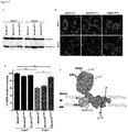
- NdufA10 a particular subunit of Complex I, NdufA10, was identified as a novel substrate. Specific involvement of PINK1 in the phosphorylation of Ser250 in this subunit could be demonstrated. This is physiologically relevant, as phosphorylation of Ser250 activates ubiquinone reduction by Complex I. Also, a phosphomimetic NdufA10 mutant reverses Pink1 deficits in mouse knockout cells and rescues mitochondrial depolarization and synaptic neurotransmission in pinkB9 null mutant Drosophila. Thus we identify a conserved molecular pathway regulating ⁇ m in Drosophila, mouse and human. Importantly, phosphomimetic NdufA10 rescues the deficit in human cells derived from PINK1 patients.
- these results reveal a novel mechanism contributing to the pathogenesis of PD. Moreover, as correcting (or mimicking) the phosphorylation of this novel target overcomes the disease phenotype, these results also provide a new therapeutic opportunity in the treatment of PD, particularly PD associated with loss of mitochondrial membrane potential (such as e.g. many cases of sporadic PD, and PD as a result of mutations in PINK1).
- NdufA10 phosphomimetic mutants or nucleic acids encoding such phosphomimetic mutants, wherein the phosphomimetic mutants harbor a phosphomimetic mutation at position 250 (or the equivalent position in a non-human protein).
- the mutation is a S250D mutation.
- the nucleic acids provided herein encode for phosphomimetic mutants with a phosphomimetic mutation at position 250 or equivalent position, particularly the S250D mutation.
- the phosphomimetic mutants are provided for use as a medicament.
- the phosphomimetic mutants are provided for use in the treatment of Parkinson's disease.
- Particularly envisaged forms of Parkinson's disease include, but are not limited to, sporadic Parkinson's disease and Parkinson's characterized by mutations in PINK1.
- methods of diagnosing Parkinson's disease in a subject comprising determining the phosphorylation status of NdufA10 at position 250 in a sample obtained from the subject, wherein the absence or reduction in NdufA10 phosphorylation is indicative of the the presence of Parkinson's disease.
- samples include, but are not limited to, brain samples, fibroblasts, muscle tissue, and blood samples. The latter are particularly envisaged, since these are the least invasive to obtain.
- NdufA10 phosphorylation can be assessed in different ways known by the skilled person, some of which are detailed further in the specification. Particularly envisaged forms of Parkinson's disease for diagnosis are the same as those for treatment.
- screening methods are provided to screen for compounds able to restore or increase NdufA10 phosphorylation, comprising the steps of:
- the kinase able to affect phosphorylation of NdufA10 is PINK1.
- Parkinson's disease or "PD” as used throughout the application is meant to specifically include mitochondrial forms of Parkinson's disease, grouped as entry #556500 in the OMIM database (Online Mendelian Inheritance in Man, McKusick-Nathans Institute of Genetic Medicine, Johns Hopkins University School of Medicine). This includes sporadic forms of PD as well as genetic forms of PD, e.g. those characterized by mutations in the Pink1 gene.
- Pink1 as used in the application is meant to refer to the gene PTEN induced putative kinase 1 (GeneID: 65018 in humans) or its encoded protein. The gene is also sometimes referred to as PARK6 or BRPK.
- This gene encodes a serine/threonine protein kinase that localizes to mitochondria. It is thought to protect cells from stress-induced mitochondrial dysfunction. Mutations in this gene are known to cause forms of autosomal recessive early-onset Parkinson disease, grouped as entry *608309 in the OMIM database.
- NdufA10 refers to the gene NADH dehydrogenase (ubiquinone) 1 alpha subcomplex, 10, 42kDa (Gene ID: 4705 in humans), or its encoded protein.
- the gene is sometimes also referred to as CI-42kD in the literature.
- the protein encoded by this gene belongs to the complex I 42kDA subunit family.
- Mammalian complex I is the first enzyme complex in the electron transport chain of mitochondria. It is composed of 45 different subunits.
- NdufA10 is a component of the hydrophobic protein fraction and has NADH dehydrogenase activity and oxidoreductase activity. It transfers electrons from NADH to the respiratory chain.
- NdufA10 is a phosphoprotein, meaning that it is a protein that can be phosphorylated at one or more positions.
- phosphorylation of NdufA10 typically means serine phosphorylation of the NdufA10 protein. Most particularly, phosphorylation of a particular serine residue is meant. In the human protein (GI:4758768, SEQ ID NO: 1), this is the serine residue at position 250. This serine residue is conserved throughout evolution and is present in species such as Drosophila or mice. The skilled person can readily determine the relevant serine residue (or the position thereof) in other species, e.g. using algorithms for sequence alignments such as BLAST.
- NdufA10 protein of primates such as chimp (GI:115392071) and gorilla (GI:115502295), it is also at position 250.
- the serine is at position 244; in pig protein (GI:311273371) at position 252; in cattle (GI:28603782) at position 238.
- mouse GI:13195624
- rat NdufA10 protein GI:170295834
- the proteins of all these mammals show over 75% sequence identity when aligned over their entire length, showing the high evolutionary conservation of these sequences.
- phosphorylated NdufA10 means a NdufA10 protein that is phosphorylated at this position (even when the rest of the protein is not phosphorylated), while “dephosphorylated NdufA10” means a protein that is not phosphorylated at that particular serine residue (even when other serine, threonine or tyrosine residues in the protein may be phosphorylated).
- determining the phosphorylation status typically refers to determining whether (or to what extent) this specific residue is phosphorylated. As will be explained further, this can be done directly (by assessing phosphorylation) or indirectly (e.g. by checking for mutations or evaluating an effect depending on the phosphorylation, such as mitochondrial membrane potential).
- phosphorylation of NdufA10 is inadequate or even absent, resulting in physiological defects, most particularly loss of mitochondrial membrane potential ⁇ m .
- the phrase "restoring the phosphorylation" of NdufA10 refers to increasing the phosphorylation of the NdufA10 protein, most particularly this serine phosphorylation, be it directly or indirectly. This results in an improvement of the physiological symptoms, particularly an improvement in membrane potential.
- a particular example of restoring the phosphorylation is "mimicking the phosphorylation".
- This phrase is meant to refer to a situation where the actual phosphorylation levels are not changed, since there is no addition of a phosphate group to the relevant amino acid residue, but the physiological effects of the phosphorylation are achieved by different means. Typically, this is done by substitution of an amino acid (particularly the one that is normally phosphorylated) by another amino acid that has a charge (particularly a negative charge, mimicking the charge of the phosphate group) and/or has increased bulk (mimicking the size of the phosphorylated amino acid residue). Although mimicking the phosphorylation does not effectively add a phosphate group, the physiological effects, particularly the effects on membrane potential, are identical to those obtained with a correctly phosphorylated protein.
- a kinase able to affect phosphorylation of NdufA10 refers to a kinase the activity of which results in increased phosphorylation of NdufA10 in physiological settings. The latter is important, since NdufA10 is part of a much larger complex, and it can be foreseen that a kinase that is able to phosphorylate purified NdufA10 protein in vitro has no relevant effect in vivo. On the other hand, it should be kept in mind that relevant kinases not necessarily phosphorylate NdufA10 directly.
- PINK1 kinase affects phosphorylation of NdufA10 in different physiologically relevant contexts, but it cannot be excluded that this is the result of indirect phosphorylation, e.g. through intermediate phosphorylation steps. Nevertheless, since modulating PINK1 activity has effects on the phosphorylation status of NdufA10, PINK1 is explicitly envisaged as a kinase able to affect phosphorylation of NdufA10. For phosphatases able to affect phosphorylation of NdufA10, the same considerations apply mutatis mutandis.
- peptide substrate of NdufA10 is used herein to refer to a peptide with a site that can be phosphorylated and is recognized by the same kinases/phosphatases that act on NdufA10.
- the peptide mimics the NdufA10 substrate.
- this is because it is derived from the NdufA10 protein (i.e. its sequence is integrally part of the NdufA10 amino acid sequence).
- the peptide provides the same linear epitope as NdufA10.
- Particularly envisaged linear epitopes are the ones with amino acids that are phosphorylated in vivo, such as the S250 residue.
- peptides that do not have the exact same sequence, but provide the same topology of the phosphosite, i.e. the same conformational epitope as NdufA10, are also envisaged herein.
- the linear and conformational epitope examples are not mutually exclusive.
- Phosphorylated sites in NdufA10 are disclosed herein, and others can be found e.g. using PhosphositePlus 32 .
- the present application relates to new diagnostic and therapeutic options in the field of Parkinson's disease.
- methods of diagnosing Parkinson's disease in a subject comprise a step of determining the phosphorylation status of NdufA10 in a sample obtained from the subject.
- a 'subject' as used herein refers to a eukaryotic organism, particularly a (individual) mammal, more in particular a human.
- a sample from said subject may be a fluid sample, e.g. blood, saliva, etc. or a tissue sample, e.g. a tissue biopsy, etc.
- the sample may be provided as such or can be pre-processed.
- PD is a neurodegenerative disease
- the sample is not necessarily a brain sample (although this is also possible).
- the NdufA10 protein is also present in non-brain (peripheral) cells, and the phosphorylation status of NdufA10 can be determined in these other cells (as illustrated for e.g. fibroblasts in the Examples section). Because obtaining samples from other tissues typically is less invasive, these samples are envisaged within the scope of the application.
- a particularly envisaged sample is a blood sample, since this is easy to obtain.
- Determining or detecting the phosphorylation of NdufA10 can be qualitative (e.g. is the protein phosphorylated or not), semi-quantitative (e.g. is there more or less phosphorylation present) and/or quantitative (e.g. how much of the protein is phosphorylated).
- quantitative e.g. how much of the protein is phosphorylated.
- the term "quantifying" when used in the context of phosphorylation of a protein in a sample can refer to absolute or to relative quantification. Absolute quantification may be accomplished by inclusion of known concentration(s) of one or more appropriate controls and referencing the detected levels to those of the known controls (e.g. through generation of a standard curve).
- relative quantification can be accomplished by comparison of detected levels or amounts between samples (or phosphoproteins in a sample) to provide a relative quantification.
- Evaluating phosphorylation may involve comparison with a positive control (e.g. to assess whether phosphorylation can be detected in the sample, in particular whether the detection method works), a negative control or a blank (typically to assess whether no false positive signal is being generated), one or more standards (either internal or external standards, typically to allow more accurate quantification), or a combination thereof.
- the positive control may additionally or alternatively be an internal positive control, typically a gene product known to be present in the sample. Detection of expression and/or activity is well known in the art, and a skilled person is capable of choosing appropriate controls and/or standards.
- phosphorylation-deficient NdufA10 mutant such as e.g. the S250A mutant for the human protein
- a phosphomimetic mutant such as e.g. the S250D mutant for the human protein
- NdufA10 phosphorylation can be measured directly (e.g. by an ELISA or equivalent assay measuring the amount of phosphorylated protein), but also indirectly.
- NdufA10 phosphorylation can be measured indirectly. For instance, one can look at functional effects of NdufA10 phosphorylation. Examples include, but are not limited to, enzymatic activity of Complex I (this will be decreased if NdufA10 is not properly phosphorylated), overall ATP levels (these will be decreased if NdufA10 is not properly phosphorylated) or mitochondrial membrane potential ( ⁇ m will be decreased if NdufA10 is not properly phosphorylated).
- peripheral cells such as blood cells or fibroblasts (e.g. de Wit et al., Methods in Enzymology 2009; 456:169-181 ; Mitoprofile kit from Invitrogen).
- NdufAlO contains a mutation at or near the site to be phosphorylated (e.g. a S250A mutation in the human protein) phosphorylation will be absent or reduced.
- phosphorylation of NdufAlO will also be absent of reduced.
- determining phosphorylation levels of NdufA10 may also comprise evaluating functional effects or assessing the presence of mutations in proteins (particularly PINK1 and/or NdufA10).
- methods of treating Parkinson's disease in a subject contain a step of increasing or restoring the phosphorylation of NdufAlO in said subject.
- the phosphorylation may be restored in all cells of the subject, it is particularly envisaged to restore NdufA10 phosphorylation in brain cells (particularly neurons), since Parkinson's is a neurodegenerative disease.
- NdufA10 phosphorylation is increased or restored in the substantia nigra of the subject.
- the methods comprise a step of determining the phosphorylation status of NdufAlO in a sample obtained from a subject, and, if the phosphorylation is reduced or absent, restoring the phosphorylation of NdufAlO in said subject.
- Correcting or restoring the phosphorylation of NdufA10 may be achieved in different ways. For instance, phosphorylation may be restored by administration of protein, either directly or through nucleic acid encoding the protein. According to particular examples, restoring the phosphorylation of NdufA10 is done through gene therapy. Briefly, gene therapy is the use of DNA as a pharmaceutical agent to treat disease and it typically entails the insertion of a nucleic acid to be expressed (often referred to as transgene) in cells of the subject undergoing the gene therapy.
- transgene a nucleic acid to be expressed
- transgene' refers to particular nucleic acid sequences encoding a polypeptide or a portion of a polypeptide to be expressed in a cell into which the nucleic acid sequence is inserted.
- transgenes are expressed as RNA, typically to control (e.g. lower) the amount of a particular polypeptide in a cell into which the nucleic acid sequence is inserted.
- RNA molecules include but are not limited to molecules that exert their function through RNA interference (shRNA, RNAi), micro-RNA regulation (miR) (which can be used to control expression of specific genes), catalytic RNA, antisense RNA, RNA aptamers, etc.
- nucleic acid sequence is introduced into a cell is not essential to the application, it may for instance be through integration in the genome or as an episomal plasmid.
- expression of the transgene may be restricted to a subset of the cells into which the nucleic acid sequence is inserted.
- transgene' is meant to include (1) a nucleic acid sequence that is not naturally found in the cell (i.e., a heterologous nucleic acid sequence); (2) a nucleic acid sequence that is a mutant form of a nucleic acid sequence naturally found in the cell into which it has been introduced ; (3) a nucleic acid sequence that serves to add additional copies of the same (i.e., homologous) or a similar nucleic acid sequence naturally occurring in the cell into which it has been introduced ; or (4) a silent naturally occurring or homologous nucleic acid sequence whose expression is induced in the cell into which it has been introduced.
- mutant form' is meant a nucleic acid sequence that contains one or more nucleotides that are different from the wild-type or naturally occurring sequence, i.e., the mutant nucleic acid sequence contains one or more nucleotide substitutions, deletions, and/or insertions.
- DNA that encodes a therapeutic protein is packaged within a vector, which is used to get the DNA inside cells within the body. Once inside, the DNA becomes expressed by the cell machinery, resulting in the production of therapeutic protein, which in turn treats the patient's disease.
- transgenes are provided on a vector.
- the term 'vector' as used in the application refers to nucleic acid molecules, usually double-stranded DNA, which may have inserted into it another nucleic acid molecule (the insert nucleic acid molecule) such as, but not limited to, a cDNA molecule.
- the vector is used to transport the insert nucleic acid molecule into a suitable host cell.
- a vector may contain the necessary elements that permit transcribing the insert nucleic acid molecule, and, optionally, translating the transcript into a polypeptide.
- the insert nucleic acid molecule may be derived from the host cell, or may be derived from a different cell or organism. Once in the host cell, the vector can replicate independently of, or coincidental with, the host chromosomal DNA, and several copies of the vector and its inserted nucleic acid molecule may be generated.
- the term 'vector' may thus also be defined as a gene delivery vehicle that facilitates gene transfer into a target cell. This definition includes both non-viral and viral vectors.
- Non-viral vectors include but are not limited to cationic lipids, liposomes, nanoparticles, PEG, PEI, etc.
- Viral vectors are derived from viruses and include but are not limited to retroviral, lentiviral, adeno-associated viral, adenoviral, herpes viral, hepatitis viral vectors or the like.
- viral vectors are replication-deficient as they have lost the ability to propagate in a given cell since viral genes essential for replication have been eliminated from the viral vector.
- the vectors can be episomal vectors (i.e., that do not integrate into the genome of a host cell), or can be vectors that integrate into the host cell genome.
- episomal vectors examples include (extrachromosomal) plasmids and so-called mini-circles, which are composed of the expression cassette only and are devoid of bacterial sequences, and examples of vectors that integrate into the host cell genome include e.g. retroviral vectors.
- Vectors may contain additional elements that target them to a particular tissue, especially in case that expression is particularly desired in a given tissue. In the context of the present application, vectors that target the brain (or are adapted for expression of proteins in the brain) are particularly envisaged.
- the most common form of gene therapy involves using DNA that encodes a functional, therapeutic gene in order to replace a mutated gene.
- Other forms involve directly correcting a mutation, or using DNA that encodes a therapeutic protein drug (rather than a natural human gene) to provide treatment.
- gene therapy may be done with a gene encoding for a wild type NdufAlO.
- gene therapy with a wild type Pink1 gene is more appropriate.
- a particular form of gene therapy is gene therapy with a phosphomimetic mutant of NdufA10.
- This has the advantage of restoring (or rather mimicking) the phosphorylation of NdufAlO regardless of the underlying defect.
- a phosphomimetic mutant does not restore phosphorylation sensu strictu
- the phosphomimetic protein shares functions with the phosphorylated protein that the phosphorylation-deficient protein does not have.
- the phosphomimetic protein displays at least part of the physiological effects associated with the phosphorylated protein.
- a NdufAlO phosphomimetic mutant is provided.
- the phosphomimetic mutant is a mutant of a serine residue in NdufAlO.
- the serine residue is the serine residue equivalent to the serine at position 250 of the human protein (SEQ ID NO: 1). Which serine residue this is in proteins from other species can easily be determined by the skilled person, e.g. using alignment algorithms.
- the phosphomimetic mutant is obtained through substitution of the phosphorylated residue (i.e.
- the NdufAlO phosphomimetic mutant is characterized by a substitution of serine with aspartic acid at position 250 (or the equivalent position), typically designated herein as NdufAlO S250D.
- these phosphomimetic mutants are provided for use as medicament. According to more specific examples, these phosphomimetic mutants are provided for use in the treatment of Parkinson's disease.
- Parkinson's disease in general, specific forms of PD that are envisaged are sporadic Parkinson's disease, Parkinson's characterized by mutations in PINK1 kinase, or Parkinson's characterized by mutations in Parkin. All of these forms of Parkinson's have been linked to mitochondrial dysfunction. Particularly envisaged forms of Parkinson's are those where the mitochondrial dysfunction is characterized by loss of mitochondrial membrane potential (or characterized at least by loss of ⁇ m. This is documented best for PD characterized by mutations in PINK1 kinase, or for sporadic PD with defective oxidative phosphorylation.
- nucleic acids encoding such NdufAlO mutant are also provided.
- expression vectors are suitable for use in gene therapy.
- nucleic acids and expression vectors are also provided for use as a medicament, particularly for use in the treatment of PD.
- methods for treating Parkinson's disease comprising administering a nucleic acid encoding a NdufAlO phosphomimetic mutant (or an expression vector containing such nucleic acid) to a subject in need thereof.
- nucleic acids and the vectors described herein can be used in gene therapy.
- Gene therapy protocols intended to achieve therapeutic gene product expression in target cells, in vitro, but also particularly in vivo, have been extensively described in the art. These include, but are not limited to, intramuscular injection of plasmid DNA (naked or in liposomes), interstitial injection, instillation in airways, application to endothelium, intra-hepatic parenchyme, and intravenous or intra-arterial administration (e.g. intra-hepatic artery, intra-hepatic vein).
- Various devices have been developed for enhancing the availability of DNA to the target cell. A simple approach is to contact the target cell physically with catheters or implantable materials containing DNA.
- Another approach is to utilize needle-free, jet injection devices which project a column of liquid directly into the target tissue under high pressure. These delivery paradigms can also be used to deliver viral vectors.
- Another approach to targeted gene delivery is the use of molecular conjugates, which consist of protein or synthetic ligands to which a nucleic acid-or DNA-binding agent has been attached for the specific targeting of nucleic acids to cells (Cristiano et al., 1993).
- the use of the nucleic acids or expression vectors as described herein is envisaged for gene therapy of brain cells.
- the use of the regulatory elements, expression cassettes or vectors is for gene therapy in vivo.
- the use is for a method of gene therapy to treat PD.
- compounds may be used to restore NdufAlO phosphorylation.
- Such compounds may have a direct effect on NdufAlO phosphorylation, or may act indirectly.
- compounds that increase the activity of PINK1 kinase will also increase the phosphorylation of NdufA10.
- compounds that inhibit dephosphorylation of NdufAlO, by inhibiting a phosphatase acting on NdufAlO will also increase phosphorylation of NdufA1lO.
- screening methods are provided to identify compounds able to restore or increase NdufAlO phosphorylation.
- Such screening methods can be set up in different ways, as the skilled person will be well aware of.
- compounds can be screened for that increase the activity of a kinase that phosphorylates NdufAlO.
- Methods according to this example typically will entail the contacting of a composition comprising the NdufA10 protein (or a peptide substrate) and a kinase able to affect phosphorylation of NdufA10 with a compound, and evaluating phosphorylation status of NdufA10.
- a peptide substrate can be used instead of using the NdufA10 protein.
- This substrate mimics the relevant NdufA10 substrate, but has the advantage that it is more amenable to screening procedures (e.g. using ELISA). Also, since NdufA10 is part of a complex, the isolated protein may not accurately reflect the physiological situation. Therefore, it may be more relevant to screen using the entire Complex I substrate (but this is challenging, particularly in vitro), or using a peptide that mimics the substrate, in sequence (i.e. linear epitope) and/or in structure (i.e. conformational epitope).
- a particularly envisaged kinase that can be used in these assays is PINK1, since PINK1 function increases phosphorylation of NdufA10.
- a composition comprising the NdufA10 protein (or a peptide substrate) and a phosphatase able to affect phosphorylation of NdufA10 with a compound, and evaluating phosphorylation status of NdufAlO.
- a peptide substrate can be used instead of using the NdufA10 protein. This substrate mimics the relevant NdufA10 substrate, but has the advantage that it is more amenable to screening procedures (e.g.
- NdufAlO is part of a complex
- the isolated protein may not accurately reflect the physiological situation. Therefore, it may be more relevant to screen using the entire Complex I substrate (but this is challenging, particularly in vitro), or using a peptide that mimics the substrate, in sequence (i.e. linear epitope) and/or in structure (i.e. conformational epitope).
- dufA10 form or relevant peptide substrate
- the skilled person is capable of picking the appropriate dufA10 form (or relevant peptide substrate) for each assay.
- dufA10 form or relevant peptide substrate
- it is appropriate to screen using a dephosphorylated NdufAlO substrate.
- dephosphorylation is studied, it makes more sense to use a phosphorylated substrate.
- the restoration or increase in NdufA10 phosphorylation can be screened for indirectly, by screening for a functional result of said phosphorylation. Since NdufA10 is part of Complex I, and the phosphorylation of NdufA10 is essential for correct functioning of Complex I, an increase of NdufA10 phosphorylation will result in increased Complex I enzymatic activity. Methods according to this example will typically entail the contacting of a composition comprising complex I with a compound, and evaluating Complex I enzymatic activity upon contact with the compound.
- compositions that contain Complex I include, but are not limited to, cellular or mitochondrial preparations. Particularly envisaged compositions are those that also contain relevant kinases (i.e. those kinases able to affect phosphorylation of NdufAlO, such as Pink1) or relevant phosphatases, in addition to containing NdufA10 (which they do by default, since the protein is a part of Complex I). These compositions more closely resemble the physiological situation. Methods to measure Complex I activity have been described in the art. Examples include, but are not limited to, spectrophotometric assays as described e.g.
- compositions or samples are mitochondrial samples, or mitochondrial fractions of samples, as they allow a more direct way of evaluating mitochondrial ATP levels.
- compositions are those that also contain relevant kinases (i.e. those kinases able to affect phosphorylation of NdufA10, such as Pink1) or relevant phosphatases, in addition to containing NdufA10.
- screening methods are provided that evaluate an increase or restoration of NdufAlO phosphorylation levels by evaluating the increase in mitochondrial membrane potential.
- Such methods will entail contacting a composition comprising mitochondria with a compound and evaluating ⁇ m .
- These compositions typically are compositions of cells (or containing cells), or mitochondrial fractions of cellular composition.
- Particularly envisaged compositions are those that also contain relevant kinases (i.e. those kinases able to affect phosphorylation of NdufA10, such as Pink1) or relevant phosphatases, in addition to containing NdufA10.
- Typical ways in which mitochondrial membrane potential may be evaluated include, but are not limited to, staining (e.g. using JC-1 dye) or FACS analysis of TMRE (tetramethylrhodamine, ethyl ester) uptake (e.g. using the TMRE-Mitochondrial Membrane Potential Assay Kit of Abcam).
- PINK1 patient material human fibroblasts harbouring the homozygous p.Q456X nonsense mutation (L2122) and the homozygous p.V170G missense mutation (L1703) and age-matched controls (L2134, L2132) were used.
- Complex I phosphoproteome immunocaptured Complex I from mouse brain and liver was analysed by LC-MS/MS. NdufA10 cDNA was obtained from Origene and phosphomimetic mutants were constructed using the multisite-directed mutagenesis kit as described by supplier's protocol (Stratagene), where Ser250 was mutated to either an Alanine (phospho-deficient mutant) or to a Glutamate (phosphomimetic mutant).
- TMRE and JC-1 were used on MEFs or third instar larvae, respectively.
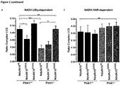
- Complex I enzymatic assays spectrophotometric measurements were performed on mitochondrial homogenates from fibroblast cells where either decylubiquinone (NADH:ubiquinone oxidoreductase, rotenone sensitive) or with hexamminerithenium (HAR) were used as Complex I substrates.
- Antibodies used mouse anti-Flag (1:1000), mouse anti-Hsp60 (1:5000); mouse anti-CV subunit. For Drosophila genetics, pink189 null and pink1rev mutants were crossed with transgenic UAS-(CG6343) (wild type; AlOwt). UAS-CG6343S-A (phospho-deficient; A10SA) and UAS-CG6343S-D (phosphomimetic; A10SD) flies ( CG6343; dNdufA10; ND-42).
- Electrophysiology was performed on third instar larvae where basal nurotransmitter release was measured at 1 Hz in 2 mM Ca2+ and relative excitatory junction potentials (EJP) amplitudes were measured in 2 mM Ca2+ during 10 min of 10 Hz stimulation.
- Reserve pool mobilization was performed using FM 1-43 dye.
- Human fibroblasts derived from PINK1 PD-causing mutations Human fibroblasts derived from PINK1 PD-causing mutations.
- Human fibroblasts harbouring the homozygous p.Q456X nonsense mutation (L2122) and the homozygous p. V170G missense mutation (L1703) and age-matched controls (L2134, L2132) were previously described ( Grunewald et al., Exp Neurol 2009; 219:266-273 ). Where indicated human fibroblasts were electroporated with GFP-tagged NdufA10 mutants using the NEON system (Invitrogen) according to the supplier's protocol.
- the PINK1 derived iPS cells were obtained according to the previously described protocol 31
- mtRFP mitochondrial targeted red fluorescent protein
- mtRFP mitochondrial targeted red fluorescent protein
- NEON Invitrogen
- Mitochondria were isolated from Pink1+ / + and Pink1 -/- mice by standard differential centrifugation and resuspended in Isolation buffer (IB: 0.2M sucrose, 10 mM Tris-MOPS pH 7.4, 0.1 mM EGTA-Tris pH 7.4) as previously described ( Frezza et al., Nat Protocol 2007; 2:287-295 ).
- IB Isolation buffer
- Complex I was immunocaptured from mitochondrial enriched fraction treated with 1% DDM according to manufacturer's protocol (Mitoscience).
- the generated peptide mixtures were vacuum-dried and re-dissolved in 20 ⁇ l of 2% acetonitrile and 0.1% TFA. These peptide mixtures were then analyzed on an Ultimate 3000 HPLC system (Dionex, Amsterdam, The Netherlands) in-line connected to an LTQ Orbitrap Velos mass spectrometer (Thermo Electron, Bremen, Germany). Here, a 30 min gradient of 2% acetonitrile to 50% acetonitrile, followed by a washing and re-equilibration step, on an in-house packed 15 cm long and 75 ⁇ m inner diameter column (Reprosil-Pur Basic C18-HD 3 ⁇ m, Dr. Maisch, Germany) was used.
- Acetylation of the protein N-terminus, pyroglutamate formation of N-terminal glutamine, methionine oxidation to methionine-sulfoxide, propionamide formation of cysteine and phosphorylation of serine, threonine and tyrosine were set as variable modifications.
- the protease was set to trypsin with one missed cleavage allowed.
- the mass tolerance on the precursor ion was set to ⁇ 10 ppm and on fragment ions to ⁇ 0.5 Da.
- Mascot's C13 setting was set to 1.
- mice anti-CV subunit (Mitosciences) was used at 1:500.
- Secondary antibodies Alexa-488 conjugated antibodies (Invitrogen) was used at 1:1000.
- Flies were kept on standard molasses medium. w pink1REV controls and w pink1B9 flies were kindly provided by Jeehye Park and Jongkyeong Chung (Korea Advanced Institute of Science and Technology) ( Park et al., Nature 2006; 441:1157-1161 ) and mutant larvae were selected using GFP balancers.
- dNDUFA10 (CG6343) cDNA was ordered from DGRC (Drosophila Genomics Resource Center) DGC ( Drosophila Gold Collection) clone LD29280 and cloned with 5'-CAGAATTCCAAAATGACCGCCGTGTTCCGCG-3' (SEQ ID NO:2) and 5'-GTGCGGCCGCCTAGTGGTGATGGTGATGATGGATGCCCTGGTTGATGCCTATTTTC-3' (SEQ ID NO:3) using 2x BIO-X-ACT Short Mix (BIOLINE), cloned in the EcoR1 and Not1 site of pUAST-Attb (PMID: 17360644) and sequenced (A10wt).
- UAS-CG6343S-A (A10SA) and UAS-CG6343S-D (A10SD) were generated similarly, with the following primers to introduce point mutations for S-A: 5'-CGGCATGGGTGGCGATGTCCTTG-3' (SEQ ID NO:4) and 5'-CAAGGACATCGCCACCCATGCCG-3' (SEQ ID NO:5) and for S-D: 5'-CGGCATGGGTGTCGATGTCCTTG-3' (SEQ ID NO:6) and 5'-CAAGGACATCGACACCCATGCCG-3' (SEQ ID NO:7).
- Transgenic flies were created at GenetiVision Inc. (Houston, USA) using PhiC31 mediated transgenesis in the VK1 docking site (2R, 59D3) (PMID: 17138868).
- TMRE mitochondrial membrane potential
- Human fibroblasts and MEFs were grown in 3 cm plastic dishes with glass coverslips (Nunc) and treated with 10 nM TMRE for 10 min at 37 °C as previously described ( Narendra et al., Autophagy 2009; 5:706-708 ).
- TMRE fluorescence intensity was measured using the ImageJ software.
- Third instar larval fillets were labelled with JC-1 (Molecular Probes) as described ( Verstreken et al., Neuron 2005; 47:365-378 ). Images were captured on a Nikon FN-1 microscope with a DS-2MBWc digital camera, 63x water objective, NA 0.8 and quantification of labelling intensity was performed using NIS-Elements software.
- FM 1-43 labelling and unloading were performed as described11. Images were captured on a Nikon FN-1 microscope with a DS-2MBWc digital camera, 63x water objective, NA 0.8 and quantification of labelling intensity was performed using NIS-Elements software.
- ⁇ m was significantly decreased in the patient fibroblasts as assessed by the electrochemical potentiometric dye tetramethyl rhodamine ethyl ester (TMRE) ( Fig. 1c, d ), in line with previous investigations showing bioenergetic abnormalities in mitochondria from PINK1 -mutant carriers 21 .
- TMRE electrochemical potentiometric dye tetramethyl rhodamine ethyl ester
- NdufA10 is located in the subunit l ⁇ of Complex I in close vicinity to the ND1 and ND3 subunits 22,23 .
- This effect is not due to defects in basal neurotransmitter release efficiency as release during low frequency stimulation in 2 mM external CaCl 2 is not affected ( Fig. 3c ).
- This neurotransmitter release defect is the consequence of defects in reserve pool (RP) vesicle mobilisation 3 , which can be assessed using FM 1-43, a lipophilic dye that internalizes in newly formed synaptic vesicles and upon prolonged stimulation also labels RP vesicles. Uploading of the RP is indeed rescued upon expression of the A10SD mutant in pink1B9 flies ( Fig. 3d, e ), but not when expressing A10 wt or A10 SA mutants.
- Example 4 Restoring NdufA10 phosphorylation can restore PD-associated defects in mammalian and patient-derived fibroblasts
- iPS induced pluripotent stem
- vitamin K2 an alternative electron carrier for ubiquinone
- mutations in PINK1 known to cause PD apparently affect the phosphorylation of this site, explaining the defects in ⁇ m seen in patient cells. While the most parsimonious explanation for our observations is that PINK1 is directly involved in the phosphorylation of NdufA10, we have not been able to directly demonstrate this with purified enzyme and substrate.
- PINK1 has a dual function in the maintenance of mitochondrial homeostasis. Under steady state conditions, PINK1 maintains a certain degree of phosphorylation to keep Complex I (and ETC) activity in check. When mutated, phosphorylation becomes deficient and ETC function becomes destabilized with latent alteration of ⁇ m , which manifests clinically as a late-onset defect in the dopaminergic neurons of the substantia nigra. The cell specificity of the clinical symptomatology remains a tantalizing and unfortunately unexplained issue, but our experiments suggest that patients harbouring PINK1 mutations should be explored for additional subtle phenotypes in organs that strongly depend on oxidative phosphorylation.
Landscapes
- Health & Medical Sciences (AREA)
- Life Sciences & Earth Sciences (AREA)
- Chemical & Material Sciences (AREA)
- Engineering & Computer Science (AREA)
- Organic Chemistry (AREA)
- Bioinformatics & Cheminformatics (AREA)
- Genetics & Genomics (AREA)
- Zoology (AREA)
- Wood Science & Technology (AREA)
- General Health & Medical Sciences (AREA)
- Medicinal Chemistry (AREA)
- Biochemistry (AREA)
- General Engineering & Computer Science (AREA)
- Molecular Biology (AREA)
- Biotechnology (AREA)
- Proteomics, Peptides & Aminoacids (AREA)
- Pharmacology & Pharmacy (AREA)
- Epidemiology (AREA)
- Veterinary Medicine (AREA)
- Animal Behavior & Ethology (AREA)
- Microbiology (AREA)
- Immunology (AREA)
- Public Health (AREA)
- Biomedical Technology (AREA)
- Gastroenterology & Hepatology (AREA)
- Physics & Mathematics (AREA)
- Analytical Chemistry (AREA)
- Biophysics (AREA)
- Medicines That Contain Protein Lipid Enzymes And Other Medicines (AREA)
- Pharmaceuticals Containing Other Organic And Inorganic Compounds (AREA)
Description
- The present application relates to the field of Parkinson's disease (PD), particularly Parkinson's disease with an important mitochondrial component, such as sporadic PD or PD associated with mutations in the mitochondrial kinase PINK1. A new substrate for this kinase, NdufA10, is identified herein. In Parkinson's disease, this protein is dephosphorylated, which is linked to a loss of mitochondrial membrane potential. It is shown that restoring or mimicking phosphorylation of NdufA10 restores the phenotypic defects associated with Parkinson's disease.
- Parkinson's disease (PD) is a degenerative disorder of the central nervous system. Most cases are sporadic, although a subset of cases has a genetic origin. Increasing evidence points to a role of mitochondrial dysfunction in many forms of Parkinsonism. Indeed, mitochondrial toxins, such as MPTP (1-methyl 4-phenyl-1,2,3,6-tetrahydropyridine) and rotenone, but also various defects in the electron transport chain (ETC) are associated with sporadic PD 6,7.
- Mutations in PINK1, a mitochondrial targeted Ser/Thr kinase, cause a monogenic form of PD1,2. In contrast to the recently identified LRRK2 kinase that has been shown to affect synaptic endocytosis in Parkinson's disease by phosphorylating Endophilin A (Matta et al 2012 Neuron 75: 1008-1021), the pink1 human mutations, as well as absence of PINK1 in diverse animal models, are associated with ETC deficiencies3,5,8, supporting the hypothesis that defects in ETC are a major culprit in the pathogenesis of PD. However, no mechanisms have been proposed, and the alternative, not necessarily contradictory, hypothesis that mutations in PINK1 cause PD by interfering with the PARKIN-mediated CCCP-induced mitophagy pathway (clearance of defective mitochondria) has gained ground considerably9-11.
- Defects in mitochondrial clearance are linked to mitochondrial fusion/fission defects10, which could explain thorax muscle degeneration and flight deficits observed in pink1 and parkin Drosophila models12-14. These defects are indeed rescued by expression of the fission promoting gene Drp1 or ablating the fusion promoting gene Opa1, respectively15,16. Intriguingly, other pink1-related phenotypes, such as defective neurotransmitter release and loss of mitochondrial membrane potential (ΔΨm) in Drosophila neurons, cannot be rescued by fission gene Drp117, but by genes restoring the proton motive force18, or by NDi1, a rotenone-insensitive NADH-quinone oxidoreductase of Saccharomyces cerevisiae 17 . These results confirm that two parallel pathways are affected in pink1B9 null Drosophila, one involved in clearance of defective mitochondria and another in the maintenance of ΔΨ m. Each pathway might explain a different subset of phenotypes in the fly. In contrast, Pink1-l- mice display very subtle, and somewhat controversial, phenotypes of altered mitochondrial morphology in resting state3,8,19,20, and it is unclear to what extent decreased mitophagy or Complex I deficiency contributes to these defects.
- It would be advantageous to identify defects underlying the mitochondrial deficiencies observed in PD. Particularly useful would be if these defects not only can help in diagnosing PD, but also point the way to new therapeutic paradigms.
- Mutations in the mitochondrial kinase PINK1 cause recessively inherited early onset Parkinson's disease (PD)1,2. Under resting conditions mouse Pink1 knockout cells and cells derived from patients with PINK1 mutations display a loss of mitochondrial membrane potential, ΔΨ m, reflecting a primordial role for PINK1 in the regulation of Complex I activity3-5.
As PINK1 is a kinase, it was reasoned that deficient phosphorylation of one or more specific substrates could underlie this pathologic phenotype. To identify possibly relevant PINK1 substrates, we have analysed the complete phosphoproteome of Complex I in brain and liver from Pink1 +/+ and Pink1 -/- mice. As will be detailed in the Examples section, a particular subunit of Complex I, NdufA10, was identified as a novel substrate. Specific involvement of PINK1 in the phosphorylation of Ser250 in this subunit could be demonstrated. This is physiologically relevant, as phosphorylation of Ser250 activates ubiquinone reduction by Complex I. Also, a phosphomimetic NdufA10 mutant reverses Pink1 deficits in mouse knockout cells and rescues mitochondrial depolarization and synaptic neurotransmission in pinkB9 null mutant Drosophila. Thus we identify a conserved molecular pathway regulating ΔΨ m in Drosophila, mouse and human. Importantly, phosphomimetic NdufA10 rescues the deficit in human cells derived from PINK1 patients. Summarizing, these data reveal a novel mechanism contributing to the pathogenesis of PD. Moreover, as correcting (or mimicking) the phosphorylation of this novel target overcomes the disease phenotype, these results also provide a new therapeutic opportunity in the treatment of PD, particularly PD associated with loss of mitochondrial membrane potential (such as e.g. many cases of sporadic PD, and PD as a result of mutations in PINK1). - Accordingly, it is an object of the invention to provide NdufA10 phosphomimetic mutants, or nucleic acids encoding such phosphomimetic mutants, wherein the phosphomimetic mutants harbor a phosphomimetic mutation at position 250 (or the equivalent position in a non-human protein). According to further particular embodiments, the mutation is a S250D mutation. Likewise, the nucleic acids provided herein encode for phosphomimetic mutants with a phosphomimetic mutation at position 250 or equivalent position, particularly the S250D mutation.
- According to another embodiment, the phosphomimetic mutants (or nucleic acids encoding the phosphomimetic mutants) are provided for use as a medicament. Particularly, the phosphomimetic mutants (or nucleic acids encoding the phosphomimetic mutants) are provided for use in the treatment of Parkinson's disease. Particularly envisaged forms of Parkinson's disease include, but are not limited to, sporadic Parkinson's disease and Parkinson's characterized by mutations in PINK1.
- In a further embodiment, methods of diagnosing Parkinson's disease in a subject are provided, comprising determining the phosphorylation status of NdufA10 at position 250 in a sample obtained from the subject, wherein the absence or reduction in NdufA10 phosphorylation is indicative of the the presence of Parkinson's disease. Particularly envisaged samples include, but are not limited to, brain samples, fibroblasts, muscle tissue, and blood samples. The latter are particularly envisaged, since these are the least invasive to obtain. NdufA10 phosphorylation can be assessed in different ways known by the skilled person, some of which are detailed further in the specification. Particularly envisaged forms of Parkinson's disease for diagnosis are the same as those for treatment.
- According to further aspects, screening methods are provided to screen for compounds able to restore or increase NdufA10 phosphorylation, comprising the steps of:
- contacting a composition comprising
- ∘ the NdufA10 protein, or a NdufA10 peptide substrate; and
- ∘ a kinase or phosphatase able to affect phosphorylation of NdufA10 at position 250
- evaluating phosphorylation status of NdufA10.
- According to particular embodiments, the kinase able to affect phosphorylation of NdufA10 is PINK1.
-
-
Figure 1 - Human PINK1 patient derived fibroblasts present mitochondrial membrane potential deficits.
a and b, Mitochondrial morphology analysis and quantification in fibroblasts of PINK1 patients (L1703 and L2122) and age-matched controls (L2134 and L2132). Following electroporation of cells with mitochondrial targeted RFP, fluorescent visualization and corresponding morphometric analysis was performed using ImageJ. Absence of PINK1 does not lead to mitochondrial morphological changes. c and d, Analysis and quantification of the mitochondrial membrane potential in these fibroblasts using the potentiometric dye TMRE. Quantification of TMRE fluorescence was performed using ImageJ software.
The mitochondrial membrane potential is decreased in the patient derived fibroblasts. Statistical analysis: student t-test; **, p<0.01; *, p<0.05; mean ±s.d.; n = 100. Scale bar, 10 µm. e and f, Schematic overview of the analysis of Complex I phosphoproteome. Mitochondrial enriched fractions from Pink1+/+ and Pink1-/- mouse brain and liver where treated with 1% DDM and the solubilized mitochondrial protein complexes were immunocaptured using 20 kDa Complex I subunit antibody. The immunocaptured Complex I was further analysed on SDS-PAGE followed by colloidal coomassie staining (f). The visualized protein bands were excised from the gel, digested with trypsin and fractionated by cation exchange chromatography to enrich for phosphopeptides. Phosphopeptides were further analysed on a Nano-LC-LTQ-Orbitrap-MS. g, Alignment of human (SEQ ID NO: 8), mouse (SEQ ID NO: 9) and Drosophila (SEQ ID NO: 10) NdufA10 revealed that the identified PINK1 dependent phospho-serine (in magenta) is conserved across species. -
Figure 2 - Expression of phosphomimetic NdufA10 rescues Complex I activity in PINK1 deficient mouse fibroblasts.
a, Pink1+/+ and Pink1-/- MEFs were stably transduced with 3xFLAG-tagged wild type NdufA10wt, phosphorylation-deficient NdufA10S250A and phosphomimetic NdufA10S250D. Expression levels were analysed by Western blotting of mitochondria-enriched fractions using anti-FLAG and anti-Hsp60 (loading control) antibody. b and c, Phosphomimetic NdufA10S250D restores the mitochondrial membrane potential in Pink1-/- MEFs. Cells were stably transduced with NdufA10 constructs as indicated and loaded with 10nM TMRE (b) and quantification of TMRE intensity (c) over mitochondrial regions of interest was performed using ImageJ software. Statistical analysis: student t-test; **, p<0.01; ns, not significant; mean ±s.d.; n = 100. Scale bar, 10 µm. d, Schematic representation of Complex I. Complex I is composed by 45 different subunits that assemble into a structure of approximately 1MDa. Electrons that arise from the oxidation of NADH are transferred to a non-covalently bound flavin mononucleotide and sub-sequentially passed through a series of iron-sulfur clusters (Fe-S) finally reaching the acceptor ubiquinone (Q), that is reduced to ubiquinol (QH2). NdufA10 is located in subunit ly within the membrane arm domain of Complex I, in close vicinity to the predicted ubiquinone binding pocket. HAR, hexamminerithenium. e and f, Analysis of enzymatic function of Complex I. Spectrophotometric assays were performed to measure Complex I (NADH:ubiquinone oxidoreductase) and citrate synthase activities on mitochondria homogenates from Pink1+/+ and Pink1-/- MEFs rescued with NdufA10 mutants as indicated. In (e), NADH:ubiquinone reduction (rotenone sensitive) and in (f) NADH:HAR reduction were measured. Values were normalized to citrate synthase acitivity. Statistical analysis: student t-test; ***; p<0.001; **, p<0.01; *, p<0.05; ns, not significant; mean ±s.d.; n = 3 independent experiments. -
Figure 3 - Restoration of synaptic defects in Drosophila pink1B9 null mutants by expressing phosphomimetic NdufA10
a, Schematic representation of a neuromuscular junction (NMJ) from a Drosophila third instar larva. The position of the stimulating electrode and nerves, and the recording electrode atmuscle 7 and 6, respectively, are illustrated. b, Relative excitatory junction potentials (EJP) amplitudes measured in 2 mM Ca2+ during 10 min of 10 Hz stimulation in pink1B9 null mutants expressing wild type (A10wt), phospho-deficient (A10SA) and phoshomimetic (A10SD) NdufA10 mutants. EJP amplitudes were binned per 30 s and normalized to the average amplitude of the first 15s of recordings. Inset represents an overlay of a raw data trace of EJPs recorded for 10 min at 10 Hz in 2 mM calcium of pink1B9+A10SD (black) and pink1B9+A10wt (grey). The deficit to maintain normal EJP amplitude during a 10 Hz stimulation train observed in pink1B9 mutant expressing A10wt is restored when phosphomimetic A10SD is present. Mean ±s.e.m.; n = 4 for A10wt, 7 for A10SA and 8 for A10SD. c, Depicted traces show basal neurotransmitter release measured at 1 Hz in 2 mM Ca2+ in pink1B9 null mutants expressing NdufA10 mutants. The average EJP amplitudes recorded are: pink1B9+A10wt: 56.4mV+/-1.9mV, pink1B9+A10SA: 52.4mV+/-2.1mV and pink1B9+A10SD: 57.0mV+/-3.3mV. Notice that basal neurotransmitter release is not affected in pink1B9 animals expressing the NdufA10 mutants. d and e, Reserve pool (RP) labelling in controls (pink1REV) and pink1 mutants (pink1B9). Both the exo/endo cycling pool (ECP) and RP were labelled by electrically stimulating motor neurons of third instar filets in the presence of 2 mM Ca2+ for 10 min and then leaving the dye with the preparation for 5 min. ECP and RP vesicles are labelled. Depolarisation with 90 mM KCI, 2 mM Ca2+ and following washing in Ca2+ free medium results in unloading of the ECP vesicles but not RP vesicles. Synapses were imaged after this unloading procedure. Quantification (d) of fluorescence intensity of loaded RP vesicles was normalized to loading intensity of controls. Notice that the loading defect in pink1B9 is restored upon expression of phosphomimetic NdufA10. f and g, Imaging of mitochondrial membrane potential in third instar Drosophila larval NMJs in controls (pink1REV) and pink1 mutants (pink1B9) using the ratiometric dye JC-1 . For quantification (f), the red JC-1 fluorescence emission to green emission (in the same area) is compared. The mitochondrial membrane potential was restored upon expression of phosphomimetic NdufA10. Statistical analysis: student t-test; ***; p<0.001; **, p<0.01; *, p<0.05; mean ±s.d.; n = 8 animals. Scale bar, 4.5 um. -
Figure 4 - Restoration of mitochondrial membrane potential in Pink1 deficient MEFs expressing PINK1 PD-causing mutants and PINK1 patient-derived fibroblasts upon expression of phosphomimetic NdufA10.
a, Respiratory chain measurements performed on mitochondria homogenates from Pink1-/- MEFs rescued with human PINK1 wild type (wt) or PD-causing mutants or artificial kinase dead mutant (KD). The cells were stably transduced with NdufA10 phosphomimetic mutants and analysed by spectrophotometric assays for the measurement of Complex I (NADH:ubiquinone oxidoreductase, rotenone sensitive) and citrate synthase enzyme activities. Values were normalized to citrate synthase acitivity. Note that the enzymatic activity of Complex I is rescued in the presence of NdufA10S250D mutant. b, Quantification of mitochondrial membrane potential in the same cell lines. Cells were loaded with 10nM TMRE and quantification of TMRE intensity over mitochondrial regions of interest was performed using ImageJ software. The mitochondrial membrane potential is restored when phosphomimetic NdufA10S250D is co-expressed. Statistical analysis: student t-test; **, p<0.01; *, p<0.05; ns, not significant; mean ±s.d.; for (a) n = 3 independent experiments, for (b) n = 70. Scale bar, 10 µm. c Quantification of mitochondrial membrane potential in control (L2134 and L2132) and PINK1 patient (L1703 and L2122) fibroblasts electroporated with GFP-tagged NdufA10 mutants loaded with 10 nM TMRE. TMRE fluorescence over mitochondrial regions of interest was quantified using ImageJ software; only cells that were GFP positive were analysed. Statistical analysis: student t-test; **, p<0.01; *, p<0.05; ns, not significant; mean ±s.d.; n = 80. Scale bar, 10 µm. -
Figure 5 - Human PINK1 patient derived neurons from induced pluripotent stem cells present mitochondrial membrane potential deficits.
a, Generation of iPS cells from a PD patient harboring a PINK1 mutation and a healthy control individual. IPS cells were established from two PD patient with mutant PINK1 (c.1366C>T) and from a healthy family member. Neuronal type morphology is observed in these preparations.
b and c, Analysis and quantification of the mitochondrial membrane potential in fibroblasts of PINK1 patients (L2124 and L2122) and age-matched controls (L2134 and L2135) using the potentiometric dye TMRE. Quantification of TMRE fluorescence was performed using ImageJ software. The mitochondrial membrane potential is decreased in the patient derived iPS cells.
Statistical analysis: student t-test; *, p<0.05; mean ±s.d.; n = 100. Scale bar, 10 µm. - The present application will be described with respect to particular examples and with reference to certain drawings but the application is not limited thereto but only by the claims. Any reference signs in the claims shall not be construed as limiting the scope. The drawings described are only schematic and are non-limiting. In the drawings, the size of some of the elements may be exaggerated and not drawn on scale for illustrative purposes. Where the term "comprising" is used in the present description and claims, it does not exclude other elements or steps. Where an indefinite or definite article is used when referring to a singular noun e.g. "a" or "an", "the", this includes a plural of that noun unless something else is specifically stated.
- Furthermore, the terms first, second, third and the like in the description and in the claims, are used for distinguishing between similar elements and not necessarily for describing a sequential or chronological order. It is to be understood that the terms so used are interchangeable under appropriate circumstances and that the examples of the application described herein are capable of operation in other sequences than described or illustrated herein.
- The following terms or definitions are provided solely to aid in the understanding of the application. Unless specifically defined herein, all terms used herein have the same meaning as they would to one skilled in the art of the present application. Practitioners are particularly directed to Sambrook et al., Molecular Cloning: A Laboratory Manual, 2nd ed., Cold Spring Harbor Press, Plainsview, New York (1989); and Ausubel et al., Current Protocols in Molecular Biology (Supplement 47), John Wiley & Sons, New York (1999), for definitions and terms of the art. The definitions provided herein should not be construed to have a scope less than understood by a person of ordinary skill in the art.
- The term "Parkinson's disease" or "PD" as used throughout the application is meant to specifically include mitochondrial forms of Parkinson's disease, grouped as entry #556500 in the OMIM database (Online Mendelian Inheritance in Man, McKusick-Nathans Institute of Genetic Medicine, Johns Hopkins University School of Medicine). This includes sporadic forms of PD as well as genetic forms of PD, e.g. those characterized by mutations in the Pink1 gene.
"Pink1" as used in the application is meant to refer to the gene PTEN induced putative kinase 1 (GeneID: 65018 in humans) or its encoded protein. The gene is also sometimes referred to as PARK6 or BRPK. This gene encodes a serine/threonine protein kinase that localizes to mitochondria. It is thought to protect cells from stress-induced mitochondrial dysfunction. Mutations in this gene are known to cause forms of autosomal recessive early-onset Parkinson disease, grouped as entry *608309 in the OMIM database. - The term "NdufA10" as used herein refers to the gene NADH dehydrogenase (ubiquinone) 1 alpha subcomplex, 10, 42kDa (Gene ID: 4705 in humans), or its encoded protein. The gene is sometimes also referred to as CI-42kD in the literature. The protein encoded by this gene belongs to the complex I 42kDA subunit family. Mammalian complex I is the first enzyme complex in the electron transport chain of mitochondria. It is composed of 45 different subunits. NdufA10 is a component of the hydrophobic protein fraction and has NADH dehydrogenase activity and oxidoreductase activity. It transfers electrons from NADH to the respiratory chain.
- NdufA10 is a phosphoprotein, meaning that it is a protein that can be phosphorylated at one or more positions. In the context of the application, phosphorylation of NdufA10 typically means serine phosphorylation of the NdufA10 protein. Most particularly, phosphorylation of a particular serine residue is meant. In the human protein (GI:4758768, SEQ ID NO: 1), this is the serine residue at position 250. This serine residue is conserved throughout evolution and is present in species such as Drosophila or mice. The skilled person can readily determine the relevant serine residue (or the position thereof) in other species, e.g. using algorithms for sequence alignments such as BLAST. By way of example, in the NdufA10 protein of primates such as chimp (GI:115392071) and gorilla (GI:115502295), it is also at position 250. This is also the case for the protein in horse (GI:149711435). In the protein of dog (GI:345790818), the serine is at position 244; in pig protein (GI:311273371) at position 252; in cattle (GI:28603782) at position 238. In mouse (GI:13195624) and rat NdufA10 protein (GI:170295834), the serine is also at position 250. It should be noted that the proteins of all these mammals show over 75% sequence identity when aligned over their entire length, showing the high evolutionary conservation of these sequences. In more distantly related species, although the overall sequence identity is lower, the serine is nevertheless conserved: in chicken protein (GI:71895153), this is at position 256; in Xenopus, both isoforms of the protein (GI:147898604 and GI:148237107) carry the serine at position 246.
- As typically used in the application, "phosphorylated NdufA10" means a NdufA10 protein that is phosphorylated at this position (even when the rest of the protein is not phosphorylated), while "dephosphorylated NdufA10" means a protein that is not phosphorylated at that particular serine residue (even when other serine, threonine or tyrosine residues in the protein may be phosphorylated). Accordingly, the phrase "determining the phosphorylation status" of NdufA10 typically refers to determining whether (or to what extent) this specific residue is phosphorylated. As will be explained further, this can be done directly (by assessing phosphorylation) or indirectly (e.g. by checking for mutations or evaluating an effect depending on the phosphorylation, such as mitochondrial membrane potential).
- In some situations, phosphorylation of NdufA10, most particularly serine phosphorylation at the position equivalent to S250 in the human protein, is inadequate or even absent, resulting in physiological defects, most particularly loss of mitochondrial membrane potential ΔΨ m. In this context, the phrase "restoring the phosphorylation" of NdufA10 refers to increasing the phosphorylation of the NdufA10 protein, most particularly this serine phosphorylation, be it directly or indirectly. This results in an improvement of the physiological symptoms, particularly an improvement in membrane potential. A particular example of restoring the phosphorylation is "mimicking the phosphorylation". This phrase is meant to refer to a situation where the actual phosphorylation levels are not changed, since there is no addition of a phosphate group to the relevant amino acid residue, but the physiological effects of the phosphorylation are achieved by different means. Typically, this is done by substitution of an amino acid (particularly the one that is normally phosphorylated) by another amino acid that has a charge (particularly a negative charge, mimicking the charge of the phosphate group) and/or has increased bulk (mimicking the size of the phosphorylated amino acid residue). Although mimicking the phosphorylation does not effectively add a phosphate group, the physiological effects, particularly the effects on membrane potential, are identical to those obtained with a correctly phosphorylated protein.
- The words "a kinase able to affect phosphorylation of NdufA10" as used in the application refers to a kinase the activity of which results in increased phosphorylation of NdufA10 in physiological settings. The latter is important, since NdufA10 is part of a much larger complex, and it can be foreseen that a kinase that is able to phosphorylate purified NdufA10 protein in vitro has no relevant effect in vivo. On the other hand, it should be kept in mind that relevant kinases not necessarily phosphorylate NdufA10 directly. For instance, it is shown herein that the PINK1 kinase affects phosphorylation of NdufA10 in different physiologically relevant contexts, but it cannot be excluded that this is the result of indirect phosphorylation, e.g. through intermediate phosphorylation steps. Nevertheless, since modulating PINK1 activity has effects on the phosphorylation status of NdufA10, PINK1 is explicitly envisaged as a kinase able to affect phosphorylation of NdufA10. For phosphatases able to affect phosphorylation of NdufA10, the same considerations apply mutatis mutandis.
The phrase "peptide substrate of NdufA10" is used herein to refer to a peptide with a site that can be phosphorylated and is recognized by the same kinases/phosphatases that act on NdufA10. Thus, the peptide mimics the NdufA10 substrate. Typically, this is because it is derived from the NdufA10 protein (i.e. its sequence is integrally part of the NdufA10 amino acid sequence). In other words, the peptide provides the same linear epitope as NdufA10. Particularly envisaged linear epitopes are the ones with amino acids that are phosphorylated in vivo, such as the S250 residue. However, peptides that do not have the exact same sequence, but provide the same topology of the phosphosite, i.e. the same conformational epitope as NdufA10, are also envisaged herein. Of course, the linear and conformational epitope examples are not mutually exclusive. Phosphorylated sites in NdufA10 are disclosed herein, and others can be found e.g. using PhosphositePlus32. - The present application relates to new diagnostic and therapeutic options in the field of Parkinson's disease. According to a first aspect, methods of diagnosing Parkinson's disease in a subject are provided, wherein the methods comprise a step of determining the phosphorylation status of NdufA10 in a sample obtained from the subject.
- A 'subject' as used herein refers to a eukaryotic organism, particularly a (individual) mammal, more in particular a human. A sample from said subject may be a fluid sample, e.g. blood, saliva, etc. or a tissue sample, e.g. a tissue biopsy, etc. The sample may be provided as such or can be pre-processed. Although PD is a neurodegenerative disease, the sample is not necessarily a brain sample (although this is also possible). Indeed, the NdufA10 protein is also present in non-brain (peripheral) cells, and the phosphorylation status of NdufA10 can be determined in these other cells (as illustrated for e.g. fibroblasts in the Examples section). Because obtaining samples from other tissues typically is less invasive, these samples are envisaged within the scope of the application. A particularly envisaged sample is a blood sample, since this is easy to obtain.
- Determining or detecting the phosphorylation of NdufA10 can be qualitative (e.g. is the protein phosphorylated or not), semi-quantitative (e.g. is there more or less phosphorylation present) and/or quantitative (e.g. how much of the protein is phosphorylated). As such, the term "quantifying" when used in the context of phosphorylation of a protein in a sample can refer to absolute or to relative quantification. Absolute quantification may be accomplished by inclusion of known concentration(s) of one or more appropriate controls and referencing the detected levels to those of the known controls (e.g. through generation of a standard curve). Alternatively, relative quantification can be accomplished by comparison of detected levels or amounts between samples (or phosphoproteins in a sample) to provide a relative quantification. Evaluating phosphorylation may involve comparison with a positive control (e.g. to assess whether phosphorylation can be detected in the sample, in particular whether the detection method works), a negative control or a blank (typically to assess whether no false positive signal is being generated), one or more standards (either internal or external standards, typically to allow more accurate quantification), or a combination thereof. The positive control may additionally or alternatively be an internal positive control, typically a gene product known to be present in the sample. Detection of expression and/or activity is well known in the art, and a skilled person is capable of choosing appropriate controls and/or standards. Particularly for determining phosphorylation of NdufA10, specific mutants may be used as appropriate controls. For instance, a phosphorylation-deficient NdufA10 mutant (such as e.g. the S250A mutant for the human protein) can be an appropriate negative control, while a phosphomimetic mutant (such as e.g. the S250D mutant for the human protein) can be an appropriate positive control (depending on the read-out).
- Phosphorylation can be measured directly (e.g. by an ELISA or equivalent assay measuring the amount of phosphorylated protein), but also indirectly. There are different ways in which NdufA10 phosphorylation can be measured indirectly. For instance, one can look at functional effects of NdufA10 phosphorylation. Examples include, but are not limited to, enzymatic activity of Complex I (this will be decreased if NdufA10 is not properly phosphorylated), overall ATP levels (these will be decreased if NdufA10 is not properly phosphorylated) or mitochondrial membrane potential (ΔΨ m will be decreased if NdufA10 is not properly phosphorylated). It should be mentioned here that such assays have been described also for peripheral cells such as blood cells or fibroblasts (e.g. de Wit et al., Methods in Enzymology 2009; 456:169-181; Mitoprofile kit from Invitrogen).
- Alternatively, or additionally, one can look at mutations of proteins. If NdufAlO contains a mutation at or near the site to be phosphorylated (e.g. a S250A mutation in the human protein) phosphorylation will be absent or reduced. Likewise, if the PINK1 kinase, which phosphorylates NdufAlO, harbours loss-of-function mutations, phosphorylation of NdufAlO will also be absent of reduced. Thus, determining phosphorylation levels of NdufA10 may also comprise evaluating functional effects or assessing the presence of mutations in proteins (particularly PINK1 and/or NdufA10).
- The absence or reduction in Nduf10 phosphorylation is indicative of the presence of PD.
- According to a further aspect, methods of treating Parkinson's disease in a subject are provided, which methods contain a step of increasing or restoring the phosphorylation of NdufAlO in said subject. Although the phosphorylation may be restored in all cells of the subject, it is particularly envisaged to restore NdufA10 phosphorylation in brain cells (particularly neurons), since Parkinson's is a neurodegenerative disease. According to further particular examples, NdufA10 phosphorylation is increased or restored in the substantia nigra of the subject.
- Of note, both the diagnosis and treatment methods may be combined into a single method. According to these examples, the methods comprise a step of determining the phosphorylation status of NdufAlO in a sample obtained from a subject, and, if the phosphorylation is reduced or absent, restoring the phosphorylation of NdufAlO in said subject.
- Correcting or restoring the phosphorylation of NdufA10 may be achieved in different ways. For instance, phosphorylation may be restored by administration of protein, either directly or through nucleic acid encoding the protein. According to particular examples, restoring the phosphorylation of NdufA10 is done through gene therapy. Briefly, gene therapy is the use of DNA as a pharmaceutical agent to treat disease and it typically entails the insertion of a nucleic acid to be expressed (often referred to as transgene) in cells of the subject undergoing the gene therapy.
- The term 'transgene' as used herein refers to particular nucleic acid sequences encoding a polypeptide or a portion of a polypeptide to be expressed in a cell into which the nucleic acid sequence is inserted. However, it is also possible that transgenes are expressed as RNA, typically to control (e.g. lower) the amount of a particular polypeptide in a cell into which the nucleic acid sequence is inserted. These RNA molecules include but are not limited to molecules that exert their function through RNA interference (shRNA, RNAi), micro-RNA regulation (miR) (which can be used to control expression of specific genes), catalytic RNA, antisense RNA, RNA aptamers, etc. How the nucleic acid sequence is introduced into a cell is not essential to the application, it may for instance be through integration in the genome or as an episomal plasmid. Of note, expression of the transgene may be restricted to a subset of the cells into which the nucleic acid sequence is inserted. The term 'transgene' is meant to include (1) a nucleic acid sequence that is not naturally found in the cell (i.e., a heterologous nucleic acid sequence); (2) a nucleic acid sequence that is a mutant form of a nucleic acid sequence naturally found in the cell into which it has been introduced ; (3) a nucleic acid sequence that serves to add additional copies of the same (i.e., homologous) or a similar nucleic acid sequence naturally occurring in the cell into which it has been introduced ; or (4) a silent naturally occurring or homologous nucleic acid sequence whose expression is induced in the cell into which it has been introduced. By 'mutant form' is meant a nucleic acid sequence that contains one or more nucleotides that are different from the wild-type or naturally occurring sequence, i.e., the mutant nucleic acid sequence contains one or more nucleotide substitutions, deletions, and/or insertions.
- In gene therapy, DNA that encodes a therapeutic protein is packaged within a vector, which is used to get the DNA inside cells within the body. Once inside, the DNA becomes expressed by the cell machinery, resulting in the production of therapeutic protein, which in turn treats the patient's disease. Thus, typically, transgenes are provided on a vector. The term 'vector' as used in the application refers to nucleic acid molecules, usually double-stranded DNA, which may have inserted into it another nucleic acid molecule (the insert nucleic acid molecule) such as, but not limited to, a cDNA molecule. The vector is used to transport the insert nucleic acid molecule into a suitable host cell. A vector may contain the necessary elements that permit transcribing the insert nucleic acid molecule, and, optionally, translating the transcript into a polypeptide. The insert nucleic acid molecule may be derived from the host cell, or may be derived from a different cell or organism. Once in the host cell, the vector can replicate independently of, or coincidental with, the host chromosomal DNA, and several copies of the vector and its inserted nucleic acid molecule may be generated. The term 'vector' may thus also be defined as a gene delivery vehicle that facilitates gene transfer into a target cell. This definition includes both non-viral and viral vectors. Non-viral vectors include but are not limited to cationic lipids, liposomes, nanoparticles, PEG, PEI, etc. Viral vectors are derived from viruses and include but are not limited to retroviral, lentiviral, adeno-associated viral, adenoviral, herpes viral, hepatitis viral vectors or the like. Typically, but not necessarily, viral vectors are replication-deficient as they have lost the ability to propagate in a given cell since viral genes essential for replication have been eliminated from the viral vector. The vectors can be episomal vectors (i.e., that do not integrate into the genome of a host cell), or can be vectors that integrate into the host cell genome. Examples of episomal vectors include (extrachromosomal) plasmids and so-called mini-circles, which are composed of the expression cassette only and are devoid of bacterial sequences, and examples of vectors that integrate into the host cell genome include e.g. retroviral vectors. Vectors may contain additional elements that target them to a particular tissue, especially in case that expression is particularly desired in a given tissue. In the context of the present application, vectors that target the brain (or are adapted for expression of proteins in the brain) are particularly envisaged.
- The most common form of gene therapy involves using DNA that encodes a functional, therapeutic gene in order to replace a mutated gene. Other forms involve directly correcting a mutation, or using DNA that encodes a therapeutic protein drug (rather than a natural human gene) to provide treatment.
- In cases where the lack of phosphorylation of NdufA10 is due to a mutation in NdufAlO, gene therapy may be done with a gene encoding for a wild type NdufAlO. In cases where lack of phosphorylation of NdufAlO is due to defects in function and/or expression of PINK1 kinase, gene therapy with a wild type Pink1 gene is more appropriate.
- A particular form of gene therapy that is envisaged however, is gene therapy with a phosphomimetic mutant of NdufA10. This has the advantage of restoring (or rather mimicking) the phosphorylation of NdufAlO regardless of the underlying defect. Although a phosphomimetic mutant does not restore phosphorylation sensu strictu, the phosphomimetic protein shares functions with the phosphorylated protein that the phosphorylation-deficient protein does not have. In other words, the phosphomimetic protein displays at least part of the physiological effects associated with the phosphorylated protein.
- Accordingly, in a further aspect of the application, a NdufAlO phosphomimetic mutant is provided. According to specific examples, the phosphomimetic mutant is a mutant of a serine residue in NdufAlO. According to further specific examples, the serine residue is the serine residue equivalent to the serine at position 250 of the human protein (SEQ ID NO: 1). Which serine residue this is in proteins from other species can easily be determined by the skilled person, e.g. using alignment algorithms. According to particular examples, the phosphomimetic mutant is obtained through substitution of the phosphorylated residue (i.e. typically the serine residue) with a negatively charged amino acid residue (to mimic the negative charge of the phosphate group), such as aspartic acid (D) or glutamic acid (E). According to most particular examples, the NdufAlO phosphomimetic mutant is characterized by a substitution of serine with aspartic acid at position 250 (or the equivalent position), typically designated herein as NdufAlO S250D.
- According to specific examples, these phosphomimetic mutants are provided for use as medicament. According to more specific examples, these phosphomimetic mutants are provided for use in the treatment of Parkinson's disease.
- This is equivalent as stating that methods are provided for treating Parkinson's disease, comprising administering a phosphomimetic mutant to a subject in need thereof.
- Although it is envisaged to treat Parkinson's disease in general, specific forms of PD that are envisaged are sporadic Parkinson's disease, Parkinson's characterized by mutations in PINK1 kinase, or Parkinson's characterized by mutations in Parkin. All of these forms of Parkinson's have been linked to mitochondrial dysfunction. Particularly envisaged forms of Parkinson's are those where the mitochondrial dysfunction is characterized by loss of mitochondrial membrane potential (or characterized at least by loss of ΔΨm. This is documented best for PD characterized by mutations in PINK1 kinase, or for sporadic PD with defective oxidative phosphorylation.
- Of note, since it is particularly envisaged to use the phosphomimetic NdufAlO mutant in methods of gene therapy, also provided are nucleic acids encoding such NdufAlO mutant, and expression vectors containing such nucleic acids. Most particularly, the expression vectors are suitable for use in gene therapy. These nucleic acids and expression vectors are also provided for use as a medicament, particularly for use in the treatment of PD.
- Again, this is equivalent as saying that methods are provided for treating Parkinson's disease, comprising administering a nucleic acid encoding a NdufAlO phosphomimetic mutant (or an expression vector containing such nucleic acid) to a subject in need thereof.
- Thus, the nucleic acids and the vectors described herein can be used in gene therapy. Gene therapy protocols, intended to achieve therapeutic gene product expression in target cells, in vitro, but also particularly in vivo, have been extensively described in the art. These include, but are not limited to, intramuscular injection of plasmid DNA (naked or in liposomes), interstitial injection, instillation in airways, application to endothelium, intra-hepatic parenchyme, and intravenous or intra-arterial administration (e.g. intra-hepatic artery, intra-hepatic vein). Various devices have been developed for enhancing the availability of DNA to the target cell. A simple approach is to contact the target cell physically with catheters or implantable materials containing DNA. Another approach is to utilize needle-free, jet injection devices which project a column of liquid directly into the target tissue under high pressure. These delivery paradigms can also be used to deliver viral vectors. Another approach to targeted gene delivery is the use of molecular conjugates, which consist of protein or synthetic ligands to which a nucleic acid-or DNA-binding agent has been attached for the specific targeting of nucleic acids to cells (Cristiano et al., 1993).
- According to a particular example, the use of the nucleic acids or expression vectors as described herein is envisaged for gene therapy of brain cells. According to a further particular example, the use of the regulatory elements, expression cassettes or vectors is for gene therapy in vivo. According to yet a further particular example, the use is for a method of gene therapy to treat PD.
- Apart from gene therapy or protein therapy to restore or mimic the phosphorylation of NdufAlO and thus treat PD, also compounds may be used to restore NdufAlO phosphorylation. Such compounds may have a direct effect on NdufAlO phosphorylation, or may act indirectly. For instance, compounds that increase the activity of PINK1 kinase will also increase the phosphorylation of NdufA10. On the other hand, compounds that inhibit dephosphorylation of NdufAlO, by inhibiting a phosphatase acting on NdufAlO, will also increase phosphorylation of NdufA1lO.
- According to a further aspect, screening methods are provided to identify compounds able to restore or increase NdufAlO phosphorylation.
- Such screening methods can be set up in different ways, as the skilled person will be well aware of. According to a first set-up, compounds can be screened for that increase the activity of a kinase that phosphorylates NdufAlO. Methods according to this example typically will entail the contacting of a composition comprising the NdufA10 protein (or a peptide substrate) and a kinase able to affect phosphorylation of NdufA10 with a compound, and evaluating phosphorylation status of NdufA10. As mentioned, instead of using the NdufA10 protein, a peptide substrate can be used. This substrate mimics the relevant NdufA10 substrate, but has the advantage that it is more amenable to screening procedures (e.g. using ELISA). Also, since NdufA10 is part of a complex, the isolated protein may not accurately reflect the physiological situation. Therefore, it may be more relevant to screen using the entire Complex I substrate (but this is challenging, particularly in vitro), or using a peptide that mimics the substrate, in sequence (i.e. linear epitope) and/or in structure (i.e. conformational epitope). A particularly envisaged kinase that can be used in these assays is PINK1, since PINK1 function increases phosphorylation of NdufA10.
- The reverse approach is also possible: rather than screen for compounds that increase phosphorylation, screen for compounds that decrease dephosphorylation, e.g. by inhibiting a phosphatase that dephosphorylates NdufA10. Methods according to this example typically will entail the contacting of a composition comprising the NdufA10 protein (or a peptide substrate) and a phosphatase able to affect phosphorylation of NdufA10 with a compound, and evaluating phosphorylation status of NdufAlO. As mentioned, instead of using the NdufA10 protein, a peptide substrate can be used. This substrate mimics the relevant NdufA10 substrate, but has the advantage that it is more amenable to screening procedures (e.g. using ELISA). Also, since NdufAlO is part of a complex, the isolated protein may not accurately reflect the physiological situation. Therefore, it may be more relevant to screen using the entire Complex I substrate (but this is challenging, particularly in vitro), or using a peptide that mimics the substrate, in sequence (i.e. linear epitope) and/or in structure (i.e. conformational epitope).
- It goes without saying that the skilled person is capable of picking the appropriate dufA10 form (or relevant peptide substrate) for each assay. E.g., when one wants to increase phosphorylation, it is appropriate to screen using a dephosphorylated NdufAlO substrate. When dephosphorylation is studied, it makes more sense to use a phosphorylated substrate.
- According to further examples, the restoration or increase in NdufA10 phosphorylation can be screened for indirectly, by screening for a functional result of said phosphorylation. Since NdufA10 is part of Complex I, and the phosphorylation of NdufA10 is essential for correct functioning of Complex I, an increase of NdufA10 phosphorylation will result in increased Complex I enzymatic activity. Methods according to this example will typically entail the contacting of a composition comprising complex I with a compound, and evaluating Complex I enzymatic activity upon contact with the compound.
- Typical examples of compositions that contain Complex I include, but are not limited to, cellular or mitochondrial preparations. Particularly envisaged compositions are those that also contain relevant kinases (i.e. those kinases able to affect phosphorylation of NdufAlO, such as Pink1) or relevant phosphatases, in addition to containing NdufA10 (which they do by default, since the protein is a part of Complex I). These compositions more closely resemble the physiological situation. Methods to measure Complex I activity have been described in the art. Examples include, but are not limited to, spectrophotometric assays as described e.g. in Janssen et al., Clinical Chemistry 2007; 53 (4): 729-734; Bénit et al., Clin Chim Acta. 2006; 374(1-2):81-6; or de Wit et al., Methods in Enzymology 2009; 456: 169-181.
- Another way in which increase or restoration of NdufA10 phosphorylation levels can be screened for indirectly, is by evaluating ATP levels or mitochondrial membrane potential. Indeed, mitochondria are essential for neuronal function and survival. Energy-demanding neurons require large numbers of functional mitochondria to provide most of their ATP via oxidative phosphorylation, a process where electrons traversing the electron transport chain (complexes I-IV) are coupled to proton pumping to establish a mitochondrial membrane potential subsequently used to synthesize ATP (complex V). Thus, increasing NdufA10 phosphorylation will result in increased ATP production by complex V, and will result in increased ΔΨ m.
- Accordingly, methods are provided that entail contacting a composition comprising cells (e.g. a cellular sample, or the cellular fraction of a sample) with a compound and evaluating the overall ATP levels, or more particularly, the mitochondrial ATP levels. Particularly envisaged as compositions or samples are mitochondrial samples, or mitochondrial fractions of samples, as they allow a more direct way of evaluating mitochondrial ATP levels. Particularly envisaged compositions are those that also contain relevant kinases (i.e. those kinases able to affect phosphorylation of NdufA10, such as Pink1) or relevant phosphatases, in addition to containing NdufA10. Methods of measuring mitochondrial ATP levels have been described in the art, e.g. by Drew and Leeuwenburgh, Am J Physiol Regul Integr Comp Physiol. 2003; 285(5):R1259-67.
It is envisaged, particularly but not exclusively in cases where overall ATP levels are used as a read-out, that an additional step is incorporated in the screening procedure involving evaluation of Complex V activity. This can be done for all compounds or, more economical, only for those that result in an increase in ATP levels. The evaluation of increased Complex V activity ensures that the compounds indeed act on the mitochondrial level. Methods of evaluating complex V activity are known in the art (see also the paragraph above on evaluating complex I activity). - According to further examples, screening methods are provided that evaluate an increase or restoration of NdufAlO phosphorylation levels by evaluating the increase in mitochondrial membrane potential. Typically, such methods will entail contacting a composition comprising mitochondria with a compound and evaluating ΔΨ m. These compositions typically are compositions of cells (or containing cells), or mitochondrial fractions of cellular composition. Particularly envisaged compositions are those that also contain relevant kinases (i.e. those kinases able to affect phosphorylation of NdufA10, such as Pink1) or relevant phosphatases, in addition to containing NdufA10. Typical ways in which mitochondrial membrane potential may be evaluated include, but are not limited to, staining (e.g. using JC-1 dye) or FACS analysis of TMRE (tetramethylrhodamine, ethyl ester) uptake (e.g. using the TMRE-Mitochondrial Membrane Potential Assay Kit of Abcam).
- It is to be understood that although particular examples, specific configurations as well as materials and/or molecules, have been discussed herein for cells and methods according to the present application, various changes or modifications in form and detail may be made without departing from the scope and spirit of this application. The following examples are provided to better illustrate particular examples, and they should not be considered limiting the application. The application is limited only by the claims.
- For PINK1 patient material, human fibroblasts harbouring the homozygous p.Q456X nonsense mutation (L2122) and the homozygous p.V170G missense mutation (L1703) and age-matched controls (L2134, L2132) were used. For Complex I phosphoproteome, immunocaptured Complex I from mouse brain and liver was analysed by LC-MS/MS. NdufA10 cDNA was obtained from Origene and phosphomimetic mutants were constructed using the multisite-directed mutagenesis kit as described by supplier's protocol (Stratagene), where Ser250 was mutated to either an Alanine (phospho-deficient mutant) or to a Glutamate (phosphomimetic mutant). For mitomorphic analysis, cells were electroporated with a mitochondrial targeted RFP. For determination of mitochondrial membrane potential, TMRE and JC-1 were used on MEFs or third instar larvae, respectively. For Complex I enzymatic assays, spectrophotometric measurements were performed on mitochondrial homogenates from fibroblast cells where either decylubiquinone (NADH:ubiquinone oxidoreductase, rotenone sensitive) or with hexamminerithenium (HAR) were used as Complex I substrates.
- Antibodies used: mouse anti-Flag (1:1000), mouse anti-Hsp60 (1:5000); mouse anti-CV subunit. For Drosophila genetics, pink189 null and pink1rev mutants were crossed with transgenic UAS-(CG6343) (wild type; AlOwt). UAS-CG6343S-A (phospho-deficient; A10SA) and UAS-CG6343S-D (phosphomimetic; A10SD) flies (CG6343; dNdufA10; ND-42). Experiments were performed on the following genotype: w pink1B9/Y; UAS-CG6343/+; daGal4/+and pink1rev/Y; UAS-CG6343/+; daGal4/+.
- Electrophysiology was performed on third instar larvae where basal nurotransmitter release was measured at 1 Hz in 2 mM Ca2+ and relative excitatory junction potentials (EJP) amplitudes were measured in 2 mM Ca2+ during 10 min of 10 Hz stimulation. Reserve pool mobilization was performed using FM 1-43 dye.
- Pink1 / MEFs stably transduced with PINK1 PD-causing mutations (G309D; W437X), and the artificial kinase inactive mutant (K219S) were previously described (Morais et al., EMBO Mol Med 2009; 1:99-111). Mouse NdufA10 cDNA was obtained from Origene (USA), and for detection purposes a 3xFLAG-tag or a GFP-tag was inserted. NdufA10 mutants were constructed using the multisite-directed mutagenesis kit as described by supplier's protocol (Stratagene), where Serine250 was mutated to either an Alanine (phosphor-deficient mutant) or to a Glutamate (phosphomimetic mutant). To generate stable cell lines, MEFs were transduced using a replication-defective recombinant retroviral expression system (Clontech) and where selected based on their acquired hygromycin resistance.
- Human fibroblasts harbouring the homozygous p.Q456X nonsense mutation (L2122) and the homozygous p. V170G missense mutation (L1703) and age-matched controls (L2134, L2132) were previously described (Grunewald et al., Exp Neurol 2009; 219:266-273). Where indicated human fibroblasts were electroporated with GFP-tagged NdufA10 mutants using the NEON system (Invitrogen) according to the supplier's protocol.
- The PINK1 derived iPS cells were obtained according to the previously described protocol31
- Human derived fibroblast cells were electroporated with mitochondrial targeted red fluorescent protein (mtRFP) corresponding to pDsRed2-Mito obtained from Clontech (Mountain View, CA) using the NEON system (Invitrogen) according to the supplier's protocol. For epifluorescent imaging of the mitochondrial network, 48h post-transfection medium was replaced with Hanks balanced salt solution (HBBS) and 10 mM HEPES and cells were placed on the stage of an Olympus IX81 inverted microscope equipped with a CellR imaging system (Olympus). Cells were excited using a 525±20nm excitation filter and emitted light was collected using a 40 x 1.4 NA Plan Apo objective (Olympus). Morphometric analysis was performed using ImageJ software as previously described (Cipolat et al., PNAS 2004; 101:15927-15932).
- Mitochondria were isolated from Pink1+/+ and Pink1-/- mice by standard differential centrifugation and resuspended in Isolation buffer (IB: 0.2M sucrose, 10 mM Tris-MOPS pH 7.4, 0.1 mM EGTA-Tris pH 7.4) as previously described (Frezza et al., Nat Protocol 2007; 2:287-295). Complex I was immunocaptured from mitochondrial enriched fraction treated with 1% DDM according to manufacturer's protocol (Mitoscience).
- After elution in 1% SDS, immunocaptured Complex I was analysed on an SDSPAGE followed by coomassie staining. The gel lanes were cut into 15 slices, and these gel slices were then washed with water, followed by acetonitrile/water (1/1, v/v) and acetonitrile, and then vacuum dried. The dried gel slices were then re-swollen in 10% acetonitrile and 50 mM ammonium bicarbonate (pH 8) containing 0.1 µg sequencing-grade modified trypsin (Promega, Madison, WI, USA). Digestion was allowed to proceed overnight at 37°C. After digestion, the generated peptide mixtures were vacuum-dried and re-dissolved in 20 µl of 2% acetonitrile and 0.1% TFA. These peptide mixtures were then analyzed on an Ultimate 3000 HPLC system (Dionex, Amsterdam, The Netherlands) in-line connected to an LTQ Orbitrap Velos mass spectrometer (Thermo Electron, Bremen, Germany). Here, a 30 min gradient of 2% acetonitrile to 50% acetonitrile, followed by a washing and re-equilibration step, on an in-house packed 15 cm long and 75 µm inner diameter column (Reprosil-Pur Basic C18-HD 3 µm, Dr. Maisch, Germany) was used. Per LC-MS/MS analysis, 2.5 µl of the peptide mixture was consumed. Instrument settings for LC-MS/MS analysis and the generation of MS/MS peak lists were as previously described (Ghesquiere et al., Mol Cell Proteomics 2009; 8:2642-2652). These MS/MS peak lists were then searched using the Mascot Daemon interface (version 2.3.0, Matrix Science, London, UK). The Mascot search parameters were as follows. The spectra were searched in the mouse subsection of the Swiss-Prot database. Acetylation of the protein N-terminus, pyroglutamate formation of N-terminal glutamine, methionine oxidation to methionine-sulfoxide, propionamide formation of cysteine and phosphorylation of serine, threonine and tyrosine were set as variable modifications. The protease was set to trypsin with one missed cleavage allowed. The mass tolerance on the precursor ion was set to ± 10 ppm and on fragment ions to ± 0.5 Da. In addition, Mascot's C13 setting was set to 1. Only peptides that were ranked one and had an ion score at least equal to the corresponding identity threshold at 99% confidence were withheld and further data handling was done in the ms_lims database (Helsens et al., Proteomics 2010; 10:1261-1264).
- Oxidative phosphorylation complex measurements performed on mitochondrial homogenates from fibroblast cells were analysed by spectrophotometric assays as previously described (de Paepe et al., Pediatr Res 2006; 59:2-6; Sled and Vinogradov, BBA 1193; 1141:262-268). Briefly, measurements of Complex I were performed with either decylubiquinone (NADH:ubiquinone oxidoreductase, rotenone sensitive) or with hexamminerithenium (HAR). The protein concentration was in the range of 2-4 mg/ml. Values were plotted according to the ratio between the specific complex's activity and citrate synthase activity.
- Primary antibodies used: mouse anti-CV subunit (Mitosciences) was used at 1:500. Secondary antibodies: Alexa-488 conjugated antibodies (Invitrogen) was used at 1:1000.
- Primary antibodies used: mouse anti-FLAG 1:1000, mouse anti-Hsp60 1:5000 (BD laboratories). Secondary antibodies: anti-rabbit HRP conjugated and anti-mouse HRP conjugated (Bio-Rad) 1:10000.
- Flies were kept on standard molasses medium. w pink1REV controls and w pink1B9 flies were kindly provided by Jeehye Park and Jongkyeong Chung (Korea Advanced Institute of Science and Technology) (Park et al., Nature 2006; 441:1157-1161) and mutant larvae were selected using GFP balancers.
- dNDUFA10 (CG6343) cDNA was ordered from DGRC (Drosophila Genomics Resource Center) DGC (Drosophila Gold Collection) clone LD29280 and cloned with 5'-CAGAATTCCAAAATGACCGCCGTGTTCCGCG-3' (SEQ ID NO:2) and 5'-GTGCGGCCGCCTAGTGGTGATGGTGATGATGGATGCCCTGGTTGATGCCTATTTTC-3' (SEQ ID NO:3) using 2x BIO-X-ACT Short Mix (BIOLINE), cloned in the EcoR1 and Not1 site of pUAST-Attb (PMID: 17360644) and sequenced (A10wt). UAS-CG6343S-A (A10SA) and UAS-CG6343S-D (A10SD) were generated similarly, with the following primers to introduce point mutations for S-A: 5'-CGGCATGGGTGGCGATGTCCTTG-3' (SEQ ID NO:4) and 5'-CAAGGACATCGCCACCCATGCCG-3' (SEQ ID NO:5) and for S-D: 5'-CGGCATGGGTGTCGATGTCCTTG-3' (SEQ ID NO:6) and 5'-CAAGGACATCGACACCCATGCCG-3' (SEQ ID NO:7). Transgenic flies were created at GenetiVision Inc. (Houston, USA) using PhiC31 mediated transgenesis in the VK1 docking site (2R, 59D3) (PMID: 17138868).
- For Drosophila genetics, pink1B9 null and pink1rev mutants were crossed with transgenic UAS-(CG6343) (wild type; A10wt). UAS-CG6343S-A (phospho-deficient; A10SA) and UAS-CG6343S-D (phosphomimetic; A10SD) flies (CG6343; dNdufA10; ND-42). Experiments were performed on the following genotype: w pink1B9/Y; UAS-CG6343/+; daGal4/+and pink1rev/Y; UAS-CG6343/+; daGal4/+.
- The dye TMRE was used to evaluate mitochondrial membrane potential. Human fibroblasts and MEFs were grown in 3 cm plastic dishes with glass coverslips (Nunc) and treated with 10 nM TMRE for 10 min at 37 °C as previously described (Narendra et al., Autophagy 2009; 5:706-708). TMRE fluorescence intensity was measured using the ImageJ software. Third instar larval fillets were labelled with JC-1 (Molecular Probes) as described (Verstreken et al., Neuron 2005; 47:365-378). Images were captured on a Nikon FN-1 microscope with a DS-2MBWc digital camera, 63x water objective, NA 0.8 and quantification of labelling intensity was performed using NIS-Elements software.
- Larval electrophysiological recordings were performed as described (Verstreken et al., Neuron 2005; 47:365-378; Verstreken et al., Neuron 2003; 40:733-748). Data was recorded with a Multiclamp 700B amplifier (Molecular Devices) and stored using
pClamp 10. A 2 mM Ca2+ concentration was used. - FM 1-43 labelling and unloading were performed as described11. Images were captured on a Nikon FN-1 microscope with a DS-2MBWc digital camera, 63x water objective, NA 0.8 and quantification of labelling intensity was performed using NIS-Elements software.
- The statistical significance of differences between a set of two groups was evaluated using student two-tailed unpaired t-tests (*, p<0.05; **, p<0.01; ***, p<0.001) in GraphPad Prism (Ghesquiere et al., Mol Cell Proteomics 2009; 8:2642-2652). Mean was calculated using standard deviation (s.d.) or standard error (s.e.m.).
- We first sought to confirm that clinically relevant mutations in PINK1 affect ETC function. Previously, we reported ΔΨ m defects in Pink1 deficient mice and Drosophila mutants, which could not be rescued by PINK1 harbouring PD-causing mutations3. We confirm this here in human fibroblasts derived from PINK1 patients with homozygous p.Q456X nonsense (L2122) or p.V170G missense (L1703) mutations21. When assessing the integrity of the mitochondrial network scoring fragmented versus elongated mito-RFP labelled mitochondria, no significant differences were observed between control (L2134; L2132) and patient fibroblasts (L1703; L2122) (
Fig. 1a, b ). However, ΔΨ m was significantly decreased in the patient fibroblasts as assessed by the electrochemical potentiometric dye tetramethyl rhodamine ethyl ester (TMRE) (Fig. 1c, d ), in line with previous investigations showing bioenergetic abnormalities in mitochondria from PINK1-mutant carriers21. - Pink1 deficiency in mouse embryonic fibroblast (MEF) cells affects Complex I activity3. Since PINK1 is a kinase, we immunocaptured Complex I from isolated mitochondria (
Fig. 1e, f ) and obtained independent phosphoproteomes from three brain and three liver preparations from mouse, covering 40 out of 46 Complex I subunits (Table I). Eight previously unknown phosphosites were identified in Complex I subunits in wild type brain or liver (not shown), one of which is S250 in NdufA10. Phosphosite Serine 250 in Complex I subunit NdufA10 was found to be dependent on the presence of PINK1. - We next analysed the phosphoproteome of 6 similar preparations of Pink1-/- mice. Remarkably, from all identified phosphosites only Ser250 in Complex I subunit NdufA10 (
Fig. 1g ) was neither identified in brain nor in liver, whereas the unphosphorylated peptide was identified in 3 out of the 6 samples (not shown). Interestingly, Ser250 is in a site conserved across human, mouse and Drosophila (Fig. 1g ).
- We stably transfected Pink1+/+ and Pink1-/- MEFs with wild type NdufA10wt, phosphorylation-deficient NdufA10S250A and phosphomimetic NdufA10S250D (
Fig. 2a ). While the defect in ΔΨ m assessed with TMRE dye was not rescued using NdufA10wt or NdufA10S250A, the phosphomimetic NdufA10S250D completely restored ΔΨ m to wild type levels (Fig. 2b, c ). No effects on ΔΨ m were observed in Pink1+/+ MEFs expressing the same NdufA10 mutants (Fig. 2b, c ). Hence, a phosphomimetic mutation at Ser250 in NdufA10 is sufficient to restore the defect in ΔΨ m in Pink1 -/- mutant cells. - NdufA10 is located in the subunit lγ of Complex I in close vicinity to the ND1 and ND3 subunits22,23. We therefore hypothesized that the identified phosphorylation site on NdufA10 could structurally influence the ubiquinone binding cavity. We performed colorimetric enzymatic assays for Complex I, in which the substrate NADH is not rate-limiting, to assess the reduction of decylubiquinone, an ubiquinone analogue (
Fig. 2d ). Reduction of decylubiquinone was significantly affected in Complex I prepared from Pink1 -/- MEFs expressing NdufA10wt but was restored in Pink1 -/- MEFs expressing NdufA10S250D (Fig. 2e ). The effect on Complex I was specific for its ubiquinone reductase enzymatic function since another enzymatic assay that employs only the NADH-binding site of Complex I and which is based on the reduction of the artificial substrate hexamminerithenium (HAR)24 (Fig. 2d ) was not affected in Pink1 deficient cells, nor in Pink1 -/- or wild type cells expressing NdufA10wt, NdufA10S250A or NdufA10S250D (Fig. 2f ). These data collectively show that NdufA10 is required for the binding and/or reduction of the physiological Complex I substrate ubiquinone. - We next assessed to what extent Drosophila pink1 mutant phenotypes could be rescued by expression of wild type (A10wt), phosphorylation-deficient (A10SA) or phosphomimetic (A10SD) NdufA10. Previously we reported that pink1B9 null mutant Drosophila fail to maintain neurotransmitter release at neuromuscular junctions (NMJ) (
Fig. 3a ) during high frequency stimulation (10Hz)3. However, this decline in neurotransmitter release was almost completely rescued when expressing phosphomimetic A10SD, but not with A10wt or A10SA (Fig. 3b ). This effect is not due to defects in basal neurotransmitter release efficiency as release during low frequency stimulation in 2 mM external CaCl2 is not affected (Fig. 3c ). This neurotransmitter release defect is the consequence of defects in reserve pool (RP) vesicle mobilisation3, which can be assessed using FM 1-43, a lipophilic dye that internalizes in newly formed synaptic vesicles and upon prolonged stimulation also labels RP vesicles. Uploading of the RP is indeed rescued upon expression of the A10SD mutant in pink1B9 flies (Fig. 3d, e ), but not when expressing A10wt or A10SA mutants. We finally assessed ΔΨ m in mitochondria at NMJ using JC-1, a green fluorescent potentiometric dye that shifts to red fluorescence as a function of a normal negative ΔΨ m 25. Synaptic mitochondria of pink1B9 mutants expressing A10SD showed red JC-1 aggregates comparable to control (Fig. 3f, g ), contrary to synaptic mitochondria of pink1B9 mutants expressing A10wt or A10SA which displayed similar signals as the pink1B9 mutant. The ΔΨ m was also not disturbed in control pinklrev NMJ expressing NdufA10 mutants (not shown). Thus, this previously identified synaptic phenotype is fully rescued by phosphomimetic NdufA10SD. - We further scrutinized whether the pathway linking PINK1 to Complex I activity has a pathogenic relevance in humans. Pink1 -/- cells expressing human wild type or PINK1 containing PD-causing mutations were stably transduced with NdufA10wt, NdufA10S250A and NdufA10S250D respectively (data not shown). NdufA10S250D was able to fully restore the decylubiquinone reduction reaction in Complex I from cells expressing PINK1 clinical mutants G309D and W437X, or the artificial kinase inactive (KD) PINK1 mutant (
Fig. 4a ), while NdufA10wt and NdufA10S250A had no effect. The functional consequence is clear from ΔΨ m measurements which confirm full restoration upon NdufA10S250D expression in the cells expressing the PD-causing mutations (Fig. 4b ). Ultimately, we electroporated human fibroblasts derived from PINK1mutant patients and controls (Fig. 1 ) with GFP-tagged forms of NdufA10 mutants. The ΔΨ m was normalized as assessed by TMRE in the GFP positive patient-derived fibroblasts expressing NdufA10S250D but not those expressing NdufA10wt or NdufA10S250A (Fig. 4c , d). Thus, restoration of the (pseudo)phosphorylation status of NdufA10 rescues Complex I activity in cells harbouring PD-causing mutations in PINK1. - To further scrutinize if the observations obtained from patient skin fibroblasts were phenocopied in a human neuronal cell type, we examined the role of endogenous PINK1 in induced pluripotent stem (iPS) cells from skin fibroblasts taken from two PD patients with nonsense (c.1366C>T; p.Q456X) mutation in the PINK1 gene. These cells were differentiated into neurons according to the previously established protocol31. When assessing the mitochondrial membrane potential (ΔΨm) between control (L2134; L2135) and patient derived iPS cells (L2124; L2122) using the electrochemical potentiometric dye tetramethyl rhodamine ethyl ester (TMRE) we observed a decrease of the ΔΨm in line with previous investigations showing bioenergetic abnormalities in mitochondria from PINK1-mutant carriers (
Figure 5 ). - Our work demonstrates that phosphorylation of Ser250 in NdufA10 regulates the ubiquinone reductase ability of Complex I. Crystal structures from Thermus thermophilus Complex I reveal that the subunits NdufS2 and NdufS7 are involved in electron donation to ubiquinone26,27, and that the contacts between the peripheral arm and the membrane domain of Complex I are mediated by NdufS2, ND1 and ND3, which leads to the formation of a cavity capable of harbouring the large hydrophobic substrate ubiquinone22,28. NdufA10 is located closely to ND1 and ND3 and we, therefore, speculate that phosphorylation of this site regulates the interaction of Complex I with ubiquinone. It should be noted that vitamin K2, an alternative electron carrier for ubiquinone, can rescue the pink1B9 mutant phenotype in Drosophila18, further arguing for a specific effect at the level of the ubiquinone reductase activity of Complex I. From a clinical perspective, it is extremely interesting that mutations in PINK1 known to cause PD apparently affect the phosphorylation of this site, explaining the defects in ΔΨ m seen in patient cells. While the most parsimonious explanation for our observations is that PINK1 is directly involved in the phosphorylation of NdufA10, we have not been able to directly demonstrate this with purified enzyme and substrate. Obtaining reproducible in vitro kinase assays with PINK1 has been notoriously challenging and the few substrates that have been identified for PINK1, such as mitochondrial protease Htr2A/OMI29, mitochondrial chaperone TRAP130 and Parkin, cannot explain the disruption of oxidative phosphorylation as shown by our experiments.
- We conclude that PINK1 has a dual function in the maintenance of mitochondrial homeostasis. Under steady state conditions, PINK1 maintains a certain degree of phosphorylation to keep Complex I (and ETC) activity in check. When mutated, phosphorylation becomes deficient and ETC function becomes destabilized with latent alteration of ΔΨ m, which manifests clinically as a late-onset defect in the dopaminergic neurons of the substantia nigra. The cell specificity of the clinical symptomatology remains a tantalizing and unfortunately unexplained issue, but our experiments suggest that patients harbouring PINK1 mutations should be explored for additional subtle phenotypes in organs that strongly depend on oxidative phosphorylation. In any event, our work provides a molecular mechanism for the dysfunction of Complex I in these patients. Bearing in mind that Complex I defects are also commonly observed in sporadic PD patients, therapeutic approaches that rescue Complex I activity by activating phosphorylation or inhibiting dephosphorylation of NdufA10 at residue Ser250 in PINK1 patients might yield excellent candidate drugs to be tested in sporadic cases as well.
-
- <110> VIB VZW KATHOLIEKE UNIVERSITEIT LEUVEN, K.U.LEUVEN R&D
- <120> Restoring phosphorylation of a novel PINK1 substrate to treat Parkinson's disease
- <130> BDS/PINK/435
- <150>
US 61/723,705
<151> 2012-11-07 - <160> 10
- <170> PatentIn version 3.5
- <210> 1
<211> 355
<212> PRT
<213> Homo sapiens - <400> 1


- <210> 2
<211> 31
<212> DNA
<213> Artificial Sequence - <220>
<223> Primer - <400> 2
cagaattcca aaatgaccgc cgtgttccgc g 31 - <210> 3
<211> 56
<212> DNA
<213> Artificial Sequence - <220>
<223> Primer - <400> 3
gtgcggccgc ctagtggtga tggtgatgat ggatgccctg gttgatgcct attttc 56 - <210> 4
<211> 23
<212> DNA
<213> Artificial Sequence - <220>
<223> Primer - <400> 4
cggcatgggt ggcgatgtcc ttg 23 - <210> 5
<211> 23
<212> DNA
<213> Artificial Sequence - <220>
<223> Primer - <400> 5
caaggacatc gccacccatg ccg 23 - <210> 6
<211> 23
<212> DNA
<213> Artificial Sequence - <220>
<223> Primer - <400> 6
cggcatgggt gtcgatgtcc ttg 23 - <210> 7
<211> 23
<212> DNA
<213> Artificial Sequence - <220>
<223> Primer - <400> 7
caaggacatc gacacccatg ccg 23 - <210> 8
<211> 35
<212> PRT
<213> Homo sapiens - <400> 8

- <210> 9
<211> 35
<212> PRT
<213> Mus musculus - <400> 9

- <210> 10
<211> 37
<212> PRT
<213> Drosophila melanogaster - <400> 10

-
- 1. Valente, E. M. et al. Hereditary early-onset Parkinson's disease caused by mutations in PINK1. Science 304, 1158-1160, doi:10.1126/science.1096284 (2004).
- 2. Valente, E. M. et al. PINK1 mutations are associated with sporadic early-onset parkinsonism. Ann Neurol 56, 336-341, doi:10.1002/ana.20256 (2004).
- 3. Morais, V. A. et al. Parkinson's disease mutations in PINK1 result in decreased Complex I activity and deficient synaptic function. ).
- 4. Parker, W. D., Jr. & Swerdlow, R. H. Mitochondrial dysfunction in idiopathic Parkinson disease. Am ).
- 5. Abramov, A. Y. et al. Bioenergetic consequences of PINK1 mutations in Parkinson disease. PLoS ONE 6, e25622, doi:10.1371/journal.pone.0025622 (2011).
- 6. Panov, A. et al. Rotenone model of Parkinson disease: multiple brain mitochondria dysfunctions after short term systemic rotenone intoxication. J Biol Chem 280, 42026-42035, doi:10.1074/jbc.M508628200 (2005).
- 7. Heikkila, R. E., Cabbat, F. S., Manzino, L. & Duvoisin, R. C. Effects of 1-methyl-4-phenyl-1,2,5,6-tetrahydropyridine on neostriatal dopamine in mice. Neuropharmacology 23, 711-713 (1984).
- 8. Gautier, C. A., Kitada, T. & Shen, J. Loss of PINK1 causes mitochondrial functional defects and increased sensitivity to oxidative stress. Proc Natl Acad Sci U S A 105, 11364-11369, doi:10.1073/pnas.0802076105 (2008).
- 9. Narendra, D. P. et al. PINK1 is selectively stabilized on impaired mitochondria to activate Parkin. ).
- 10. Youle, R. J. & Narendra, D. P. Mechanisms of mitophagy. Nature reviews. Molecular cell biology 12, 9-14, doi:10.1038/nrm3028 (2011).
- 11. Narendra, D. P. & Youle, R. J. Targeting mitochondrial dysfunction: role for PINK1 and Parkin in mitochondrial quality control. Antioxidants & redox signaling 14, 1929-1938, doi:10.1089/ars.2010.3799 (2011).
- 12. Park, J. et al. Mitochondrial dysfunction in Drosophila PINK1 mutants is complemented by parkin. Nature 441, 1157-1161 (2006).
- 13. Clark, I. E. et al. Drosophila pink1 is required for mitochondrial function and interacts genetically with parkin. Nature 441, 1162-1166, doi:10.1038/nature04779 (2006).
- 14. Yang, Y. et al. Mitochondrial pathology and muscle and dopaminergic neuron degeneration caused by inactivation of Drosophila Pink1 is rescued by Parkin. Proc Natl Acad Sci U S A 103, 10793-10798, doi:10.1073/pnas.0602493103 (2006).
- 15. Deng, H., Dodson, M. W., Huang, H. & Guo, M. The Parkinson's disease genes pink1 and parkin promote mitochondrial fission and/or inhibit fusion in Drosophila. Proc Natl Acad Sci U S A 105, 14503-14508, doi:10.1073/pnas.0803998105 (2008).
- 16. Yang, Y. et al. Pink1 regulates mitochondrial dynamics through interaction with the fission/fusion machinery. Proc Natl Acad Sci USA 105, 7070-7075, doi:10.1073/pnas.0711845105 (2008).
- 17. Vilain, S. et al. The yeast complex I equivalent NADH dehydrogenase rescues pink1 mutants. ).
- 18. Vos, M. et al. Vitamin K2 is a mitochondrial electron carrier that rescues pink1 deficiency. Science 336, 1306-1310, doi:10.1126/science.1218632 (2012).
- 19. Exner, N. et al. Loss-of-function of human PINK1 results in mitochondrial pathology and can be rescued by parkin. J Neurosci 27, 12413-12418, doi:10.1523/JNEUROSCI.0719-07.2007 (2007).
- 20. Lutz, A. K. et al. Loss of parkin or PINK1 function increases Drp1-dependent mitochondrial fragmentation. J Biol Chem 284, 22938-22951, doi:10.1074/jbc.M109.035774 (2009).
- 21. Grunewald, A. et al. Differential effects of PINK1 nonsense and missense mutations on mitochondrial function and morphology. Exp Neurol 219, 266-273, doi:10.1016/j.expneurol.2009.05.027 (2009).
- 22. Efremov, R. G., Baradaran, R. & Sazanov, L. A. The architecture of respiratory complex I. Nature 465, 441-445, doi:10.1038/nature09066 (2010).
- 23. Janssen, R. J., Nijtmans, L. G., van den Heuvel, L. P. & Smeitink, J. A. Mitochondrial complex I: structure, function and pathology. J Inherit Metab Dis 29, 499-515, doi:10.1007/s10545-006-0362-4 (2006).
- 24. Sled, V. D. & Vinogradov, A. D. Kinetics of the mitochondrial NADH-ubiquinone oxidoreductase interaction with hexammineruthenium(III). Biochim Biophys Acta 1141, 262-268 (1993).
- 25. Verstreken, P. et al. Synaptic mitochondria are critical for mobilization of reserve pool vesicles at Drosophila neuromuscular junctions. Neuron 47, 365-378, doi:10.1016/j.neuron.2005.06.018 (2005).
- 26. Sazanov, L. A. & Hinchliffe, P. Structure of the hydrophilic domain of respiratory complex I from Thermus thermophilus. Science 311, 1430-1436, doi:10.1126/science.1123809 (2006).
- 27. Berrisford, J. M. & Sazanov, L. A. Structural basis for the mechanism of respiratory complex I. J Biol Chem 284, 29773-29783, doi:10.1074/jbc.M109.032144 (2009).
- 28. Tocilescu, M. A., Zickermann, V., Zwicker, K. & Brandt, U. Quinone binding and reduction by respiratory complex I. Biochim Biophys Acta 1797, 1883-1890, doi:10.1016/j.bbabio.2010.05.009 (2010).
- 29. Plun-Favreau, H. et al. The mitochondrial protease HtrA2 is regulated by Parkinson's disease associated kinase PINK1. ).
- 30. Pridgeon, J. W., Olzmann, J. A., Chin, L. S. & Li, L. PINK1 protects against oxidative stress by phosphorylating mitochondrial chaperone TRAP1. PLoS Biol 5, e172, doi:10.1371/journal.pbio.0050172 (2007).
- 31. Seibler P, Graziotto J, Jeong H, Simunovic F, Klein C, Krainc D. Mitochondrial Parkin recruitment is impaired in neurons derived from mutant PINK1 induced pluripotent stem cells. J Neurosci. 2011; 31(16):5970-6.
- 32. Hornbeck PV, Kornhauser JM, Tkachev S, Zhang B, Skrzypek E, Murray B, Latham V, Sullivan M. PhosphoSitePlus: a comprehensive resource for investigating the structure and function of experimentally determined post-translational modifications in man and mouse. Nucleic Acids Res. 2012;40(Database issue):D261-70.
Claims (8)
- A NdufA10 phosphomimetic mutant or a nucleic acid encoding such phosphomimetic mutant, wherein the phosphomimetic mutation is at position 250.
- The phosphomimetic mutant or nucleic acid of claim 1, which is a S250D mutation.
- The phosphomimetic mutant or nucleic acid according to claim 1 or 2, for use as a medicament.
- The phosphomimetic mutant or nucleic acid according to claim 1 or 2, for use in the treatment of Parkinson's disease.
- The phosphomimetic mutant or nucleic acid for use according to claim 4, wherein the Parkinson's disease is sporadic Parkinson's disease or Parkinson's characterized by mutations in PINK1.
- A method of diagnosing Parkinson's disease in a subject, comprising determining the phosphorylation status of NdufA10 at position 250 in a sample obtained from the subject, wherein the absence or reduction in NdufA10 phosphorylation is indicative of the presence of Parkinson's disease.
- A screening method for compounds able to restore or increase NdufA10 phosphorylation, comprising:- contacting a composition comprising the NdufA10 protein or a peptide substrate thereof and a kinase or phosphatase able to affect phosphorylation of NdufA10 at position 250 with a compound, wherein said peptide substrate is a peptide that provides the same linear epitope as NdufA10, said linear epitope comprising a S250 residue that can be phosphorylated in vivo;- and evaluating phosphorylation status of NdufA10.
- The method according to claim 7, wherein the kinase is PINK1.
Applications Claiming Priority (2)
| Application Number | Priority Date | Filing Date | Title |
|---|---|---|---|
| US201261723705P | 2012-11-07 | 2012-11-07 | |
| PCT/EP2013/073280 WO2014072412A1 (en) | 2012-11-07 | 2013-11-07 | Restoring phosphorylation of a novel pink1 substrate to treat parkinson's disease |
Publications (2)
| Publication Number | Publication Date |
|---|---|
| EP2917342A1 EP2917342A1 (en) | 2015-09-16 |
| EP2917342B1 true EP2917342B1 (en) | 2017-06-21 |
Family
ID=49622790
Family Applications (1)
| Application Number | Title | Priority Date | Filing Date |
|---|---|---|---|
| EP13792867.7A Not-in-force EP2917342B1 (en) | 2012-11-07 | 2013-11-07 | Restoring phosphorylation of a novel pink1 substrate to treat parkinson's disease |
Country Status (4)
| Country | Link |
|---|---|
| US (1) | US9879233B2 (en) |
| EP (1) | EP2917342B1 (en) |
| CA (1) | CA2890485A1 (en) |
| WO (1) | WO2014072412A1 (en) |
Families Citing this family (1)
| Publication number | Priority date | Publication date | Assignee | Title |
|---|---|---|---|---|
| CN113227788B (en) * | 2019-03-19 | 2023-11-21 | 中南大学湘雅医院 | Method and reagent for assisting diagnosis and treatment of Parkinson's disease |
-
2013
- 2013-11-07 CA CA2890485A patent/CA2890485A1/en not_active Abandoned
- 2013-11-07 EP EP13792867.7A patent/EP2917342B1/en not_active Not-in-force
- 2013-11-07 US US14/440,746 patent/US9879233B2/en not_active Expired - Fee Related
- 2013-11-07 WO PCT/EP2013/073280 patent/WO2014072412A1/en not_active Ceased
Also Published As
| Publication number | Publication date |
|---|---|
| EP2917342A1 (en) | 2015-09-16 |
| US20150299672A1 (en) | 2015-10-22 |
| CA2890485A1 (en) | 2014-05-15 |
| WO2014072412A1 (en) | 2014-05-15 |
| US9879233B2 (en) | 2018-01-30 |
Similar Documents
| Publication | Publication Date | Title |
|---|---|---|
| Shekarabi et al. | Mutations in the nervous system–specific HSN2 exon of WNK1 cause hereditary sensory neuropathy type II | |
| LaConte et al. | CASK stabilizes neurexin and links it to liprin-α in a neuronal activity-dependent manner | |
| Garnham et al. | The chemical complexity of cellular microtubules: tubulin post‐translational modification enzymes and their roles in tuning microtubule functions | |
| Dagda et al. | Beyond the mitochondrion: cytosolic PINK 1 remodels dendrites through Protein Kinase A | |
| Choudhury et al. | Differential clathrin binding and subcellular localization of OCRL1 splice isoforms | |
| KR20120089743A (en) | Novel viral vector construct for neuron specific optimized continuous dopa synthesis in vivo | |
| Strobbe et al. | Distinct mechanisms of pathogenic DJ-1 mutations in mitochondrial quality control | |
| Kowalski et al. | Monomeric α‐synuclein activates the plasma membrane calcium pump | |
| US9598677B2 (en) | Use of human biliverdin reductase and fragments thereof for the treatment of protein kinase C-delta and ERK related conditions | |
| Nagano et al. | Tomosyn is a novel Akt substrate mediating insulin-dependent GLUT4 exocytosis | |
| Matsuo et al. | Nuclear choline acetyltransferase activates transcription of a high-affinity choline transporter | |
| US20180087041A1 (en) | Tagged form of mut enzyme, genetic constructs incorporating it, and its use in gene thereapy | |
| Kobayashi et al. | Identification and characterization of GSRP-56, a novel Golgi-localized spectrin repeat-containing protein | |
| US9879233B2 (en) | Restoring phosphorylation of a novel PINK1 substrate to treat parkinson's disease | |
| Vallentin et al. | RBCK1, a protein kinase CβI (PKCβI)-interacting protein, regulates PKCβ-dependent function | |
| Nguyen et al. | Amino acid changes at arginine 204 of troponin I result in increased calcium sensitivity of force development | |
| CA2662026A1 (en) | Compositions and methods for modulating ampa receptor-mediated excitotoxicity | |
| Takeya et al. | Expression and function of Noxo1γ, an alternative splicing form of the NADPH oxidase organizer 1 | |
| Chen et al. | Pro-death signaling of GRK2 in cardiac myocytes after ischemic stress occurs via ERK-dependent, Hsp90-mediated mitochondrial targeting | |
| JP4869685B2 (en) | Rab27A inactivating agent | |
| Mullen | Investigating the Mechanisms Linking Mutations in the Protein Synthesis Apparatus to Neurological Disease | |
| Liu | Mitochondrial Division: Synergizing in Mitochondrial Divisome | |
| Matsell | Characterizing the function of ATP8A2 and its role in neurodegenerative disease | |
| Congreve et al. | Palmitoylation regulates the magnitude of HCN4-mediated | |
| Moïse et al. | Characterization of a novel MK3 splice variant from murine ventricular myocardium |
Legal Events
| Date | Code | Title | Description |
|---|---|---|---|
| PUAI | Public reference made under article 153(3) epc to a published international application that has entered the european phase |
Free format text: ORIGINAL CODE: 0009012 |
|
| 17P | Request for examination filed |
Effective date: 20150601 |
|
| AK | Designated contracting states |
Kind code of ref document: A1 Designated state(s): AL AT BE BG CH CY CZ DE DK EE ES FI FR GB GR HR HU IE IS IT LI LT LU LV MC MK MT NL NO PL PT RO RS SE SI SK SM TR |
|
| AX | Request for extension of the european patent |
Extension state: BA ME |
|
| DAX | Request for extension of the european patent (deleted) | ||
| 17Q | First examination report despatched |
Effective date: 20160513 |
|
| GRAP | Despatch of communication of intention to grant a patent |
Free format text: ORIGINAL CODE: EPIDOSNIGR1 |
|
| INTG | Intention to grant announced |
Effective date: 20161201 |
|
| GRAS | Grant fee paid |
Free format text: ORIGINAL CODE: EPIDOSNIGR3 |
|
| GRAA | (expected) grant |
Free format text: ORIGINAL CODE: 0009210 |
|
| AK | Designated contracting states |
Kind code of ref document: B1 Designated state(s): AL AT BE BG CH CY CZ DE DK EE ES FI FR GB GR HR HU IE IS IT LI LT LU LV MC MK MT NL NO PL PT RO RS SE SI SK SM TR |
|
| REG | Reference to a national code |
Ref country code: GB Ref legal event code: FG4D |
|
| REG | Reference to a national code |
Ref country code: CH Ref legal event code: EP |
|
| REG | Reference to a national code |
Ref country code: IE Ref legal event code: FG4D |
|
| REG | Reference to a national code |
Ref country code: AT Ref legal event code: REF Ref document number: 902968 Country of ref document: AT Kind code of ref document: T Effective date: 20170715 |
|
| REG | Reference to a national code |
Ref country code: DE Ref legal event code: R096 Ref document number: 602013022628 Country of ref document: DE |
|
| REG | Reference to a national code |
Ref country code: NL Ref legal event code: MP Effective date: 20170621 |
|
| PG25 | Lapsed in a contracting state [announced via postgrant information from national office to epo] |
Ref country code: GR Free format text: LAPSE BECAUSE OF FAILURE TO SUBMIT A TRANSLATION OF THE DESCRIPTION OR TO PAY THE FEE WITHIN THE PRESCRIBED TIME-LIMIT Effective date: 20170922 Ref country code: NO Free format text: LAPSE BECAUSE OF FAILURE TO SUBMIT A TRANSLATION OF THE DESCRIPTION OR TO PAY THE FEE WITHIN THE PRESCRIBED TIME-LIMIT Effective date: 20170921 Ref country code: FI Free format text: LAPSE BECAUSE OF FAILURE TO SUBMIT A TRANSLATION OF THE DESCRIPTION OR TO PAY THE FEE WITHIN THE PRESCRIBED TIME-LIMIT Effective date: 20170621 Ref country code: HR Free format text: LAPSE BECAUSE OF FAILURE TO SUBMIT A TRANSLATION OF THE DESCRIPTION OR TO PAY THE FEE WITHIN THE PRESCRIBED TIME-LIMIT Effective date: 20170621 Ref country code: LT Free format text: LAPSE BECAUSE OF FAILURE TO SUBMIT A TRANSLATION OF THE DESCRIPTION OR TO PAY THE FEE WITHIN THE PRESCRIBED TIME-LIMIT Effective date: 20170621 |
|
| REG | Reference to a national code |
Ref country code: LT Ref legal event code: MG4D |
|
| REG | Reference to a national code |
Ref country code: AT Ref legal event code: MK05 Ref document number: 902968 Country of ref document: AT Kind code of ref document: T Effective date: 20170621 |
|
| PG25 | Lapsed in a contracting state [announced via postgrant information from national office to epo] |
Ref country code: NL Free format text: LAPSE BECAUSE OF FAILURE TO SUBMIT A TRANSLATION OF THE DESCRIPTION OR TO PAY THE FEE WITHIN THE PRESCRIBED TIME-LIMIT Effective date: 20170621 Ref country code: SE Free format text: LAPSE BECAUSE OF FAILURE TO SUBMIT A TRANSLATION OF THE DESCRIPTION OR TO PAY THE FEE WITHIN THE PRESCRIBED TIME-LIMIT Effective date: 20170621 Ref country code: RS Free format text: LAPSE BECAUSE OF FAILURE TO SUBMIT A TRANSLATION OF THE DESCRIPTION OR TO PAY THE FEE WITHIN THE PRESCRIBED TIME-LIMIT Effective date: 20170621 Ref country code: BG Free format text: LAPSE BECAUSE OF FAILURE TO SUBMIT A TRANSLATION OF THE DESCRIPTION OR TO PAY THE FEE WITHIN THE PRESCRIBED TIME-LIMIT Effective date: 20170921 Ref country code: LV Free format text: LAPSE BECAUSE OF FAILURE TO SUBMIT A TRANSLATION OF THE DESCRIPTION OR TO PAY THE FEE WITHIN THE PRESCRIBED TIME-LIMIT Effective date: 20170621 |
|
| PG25 | Lapsed in a contracting state [announced via postgrant information from national office to epo] |
Ref country code: EE Free format text: LAPSE BECAUSE OF FAILURE TO SUBMIT A TRANSLATION OF THE DESCRIPTION OR TO PAY THE FEE WITHIN THE PRESCRIBED TIME-LIMIT Effective date: 20170621 Ref country code: RO Free format text: LAPSE BECAUSE OF FAILURE TO SUBMIT A TRANSLATION OF THE DESCRIPTION OR TO PAY THE FEE WITHIN THE PRESCRIBED TIME-LIMIT Effective date: 20170621 Ref country code: SK Free format text: LAPSE BECAUSE OF FAILURE TO SUBMIT A TRANSLATION OF THE DESCRIPTION OR TO PAY THE FEE WITHIN THE PRESCRIBED TIME-LIMIT Effective date: 20170621 Ref country code: AT Free format text: LAPSE BECAUSE OF FAILURE TO SUBMIT A TRANSLATION OF THE DESCRIPTION OR TO PAY THE FEE WITHIN THE PRESCRIBED TIME-LIMIT Effective date: 20170621 Ref country code: CZ Free format text: LAPSE BECAUSE OF FAILURE TO SUBMIT A TRANSLATION OF THE DESCRIPTION OR TO PAY THE FEE WITHIN THE PRESCRIBED TIME-LIMIT Effective date: 20170621 |
|
| PG25 | Lapsed in a contracting state [announced via postgrant information from national office to epo] |
Ref country code: SM Free format text: LAPSE BECAUSE OF FAILURE TO SUBMIT A TRANSLATION OF THE DESCRIPTION OR TO PAY THE FEE WITHIN THE PRESCRIBED TIME-LIMIT Effective date: 20170621 Ref country code: IS Free format text: LAPSE BECAUSE OF FAILURE TO SUBMIT A TRANSLATION OF THE DESCRIPTION OR TO PAY THE FEE WITHIN THE PRESCRIBED TIME-LIMIT Effective date: 20171021 Ref country code: PL Free format text: LAPSE BECAUSE OF FAILURE TO SUBMIT A TRANSLATION OF THE DESCRIPTION OR TO PAY THE FEE WITHIN THE PRESCRIBED TIME-LIMIT Effective date: 20170621 Ref country code: ES Free format text: LAPSE BECAUSE OF FAILURE TO SUBMIT A TRANSLATION OF THE DESCRIPTION OR TO PAY THE FEE WITHIN THE PRESCRIBED TIME-LIMIT Effective date: 20170621 Ref country code: IT Free format text: LAPSE BECAUSE OF FAILURE TO SUBMIT A TRANSLATION OF THE DESCRIPTION OR TO PAY THE FEE WITHIN THE PRESCRIBED TIME-LIMIT Effective date: 20170621 |
|
| REG | Reference to a national code |
Ref country code: DE Ref legal event code: R097 Ref document number: 602013022628 Country of ref document: DE |
|
| PLBE | No opposition filed within time limit |
Free format text: ORIGINAL CODE: 0009261 |
|
| STAA | Information on the status of an ep patent application or granted ep patent |
Free format text: STATUS: NO OPPOSITION FILED WITHIN TIME LIMIT |
|
| PG25 | Lapsed in a contracting state [announced via postgrant information from national office to epo] |
Ref country code: DK Free format text: LAPSE BECAUSE OF FAILURE TO SUBMIT A TRANSLATION OF THE DESCRIPTION OR TO PAY THE FEE WITHIN THE PRESCRIBED TIME-LIMIT Effective date: 20170621 |
|
| 26N | No opposition filed |
Effective date: 20180322 |
|
| REG | Reference to a national code |
Ref country code: DE Ref legal event code: R119 Ref document number: 602013022628 Country of ref document: DE |
|
| PG25 | Lapsed in a contracting state [announced via postgrant information from national office to epo] |
Ref country code: MC Free format text: LAPSE BECAUSE OF FAILURE TO SUBMIT A TRANSLATION OF THE DESCRIPTION OR TO PAY THE FEE WITHIN THE PRESCRIBED TIME-LIMIT Effective date: 20170621 |
|
| GBPC | Gb: european patent ceased through non-payment of renewal fee |
Effective date: 20171107 |
|
| PG25 | Lapsed in a contracting state [announced via postgrant information from national office to epo] |
Ref country code: CH Free format text: LAPSE BECAUSE OF NON-PAYMENT OF DUE FEES Effective date: 20171130 Ref country code: LI Free format text: LAPSE BECAUSE OF NON-PAYMENT OF DUE FEES Effective date: 20171130 |
|
| PG25 | Lapsed in a contracting state [announced via postgrant information from national office to epo] |
Ref country code: LU Free format text: LAPSE BECAUSE OF NON-PAYMENT OF DUE FEES Effective date: 20171107 Ref country code: SI Free format text: LAPSE BECAUSE OF FAILURE TO SUBMIT A TRANSLATION OF THE DESCRIPTION OR TO PAY THE FEE WITHIN THE PRESCRIBED TIME-LIMIT Effective date: 20170621 |
|
| REG | Reference to a national code |
Ref country code: FR Ref legal event code: ST Effective date: 20180731 Ref country code: BE Ref legal event code: MM Effective date: 20171130 |
|
| REG | Reference to a national code |
Ref country code: IE Ref legal event code: MM4A |
|
| PG25 | Lapsed in a contracting state [announced via postgrant information from national office to epo] |
Ref country code: MT Free format text: LAPSE BECAUSE OF NON-PAYMENT OF DUE FEES Effective date: 20171107 |
|
| PG25 | Lapsed in a contracting state [announced via postgrant information from national office to epo] |
Ref country code: DE Free format text: LAPSE BECAUSE OF NON-PAYMENT OF DUE FEES Effective date: 20180602 Ref country code: FR Free format text: LAPSE BECAUSE OF NON-PAYMENT OF DUE FEES Effective date: 20171130 Ref country code: IE Free format text: LAPSE BECAUSE OF NON-PAYMENT OF DUE FEES Effective date: 20171107 |
|
| PG25 | Lapsed in a contracting state [announced via postgrant information from national office to epo] |
Ref country code: BE Free format text: LAPSE BECAUSE OF NON-PAYMENT OF DUE FEES Effective date: 20171130 Ref country code: GB Free format text: LAPSE BECAUSE OF NON-PAYMENT OF DUE FEES Effective date: 20171107 |
|
| PG25 | Lapsed in a contracting state [announced via postgrant information from national office to epo] |
Ref country code: HU Free format text: LAPSE BECAUSE OF FAILURE TO SUBMIT A TRANSLATION OF THE DESCRIPTION OR TO PAY THE FEE WITHIN THE PRESCRIBED TIME-LIMIT; INVALID AB INITIO Effective date: 20131107 |
|
| PG25 | Lapsed in a contracting state [announced via postgrant information from national office to epo] |
Ref country code: CY Free format text: LAPSE BECAUSE OF FAILURE TO SUBMIT A TRANSLATION OF THE DESCRIPTION OR TO PAY THE FEE WITHIN THE PRESCRIBED TIME-LIMIT Effective date: 20170621 |
|
| PG25 | Lapsed in a contracting state [announced via postgrant information from national office to epo] |
Ref country code: MK Free format text: LAPSE BECAUSE OF FAILURE TO SUBMIT A TRANSLATION OF THE DESCRIPTION OR TO PAY THE FEE WITHIN THE PRESCRIBED TIME-LIMIT Effective date: 20170621 |
|
| PG25 | Lapsed in a contracting state [announced via postgrant information from national office to epo] |
Ref country code: TR Free format text: LAPSE BECAUSE OF FAILURE TO SUBMIT A TRANSLATION OF THE DESCRIPTION OR TO PAY THE FEE WITHIN THE PRESCRIBED TIME-LIMIT Effective date: 20170621 |
|
| PG25 | Lapsed in a contracting state [announced via postgrant information from national office to epo] |
Ref country code: PT Free format text: LAPSE BECAUSE OF FAILURE TO SUBMIT A TRANSLATION OF THE DESCRIPTION OR TO PAY THE FEE WITHIN THE PRESCRIBED TIME-LIMIT Effective date: 20170621 |
|
| PG25 | Lapsed in a contracting state [announced via postgrant information from national office to epo] |
Ref country code: AL Free format text: LAPSE BECAUSE OF FAILURE TO SUBMIT A TRANSLATION OF THE DESCRIPTION OR TO PAY THE FEE WITHIN THE PRESCRIBED TIME-LIMIT Effective date: 20170621 |