EP2915878A1 - Method and device for conserving viable and functional human polymorphonuclear neutrophils - Google Patents
Method and device for conserving viable and functional human polymorphonuclear neutrophils Download PDFInfo
- Publication number
- EP2915878A1 EP2915878A1 EP14158437.5A EP14158437A EP2915878A1 EP 2915878 A1 EP2915878 A1 EP 2915878A1 EP 14158437 A EP14158437 A EP 14158437A EP 2915878 A1 EP2915878 A1 EP 2915878A1
- Authority
- EP
- European Patent Office
- Prior art keywords
- neutrophils
- leukocytes
- polymorphonuclear neutrophils
- medium
- neutrophil
- Prior art date
- Legal status (The legal status is an assumption and is not a legal conclusion. Google has not performed a legal analysis and makes no representation as to the accuracy of the status listed.)
- Withdrawn
Links
- 238000000034 method Methods 0.000 title claims abstract description 51
- 210000003622 mature neutrocyte Anatomy 0.000 title claims description 26
- 241000282414 Homo sapiens Species 0.000 title claims description 22
- 210000000265 leukocyte Anatomy 0.000 claims abstract description 46
- WQZGKKKJIJFFOK-GASJEMHNSA-N Glucose Natural products OC[C@H]1OC(O)[C@H](O)[C@@H](O)[C@@H]1O WQZGKKKJIJFFOK-GASJEMHNSA-N 0.000 claims abstract description 33
- 239000008103 glucose Substances 0.000 claims abstract description 33
- 206010021143 Hypoxia Diseases 0.000 claims abstract description 12
- 238000000338 in vitro Methods 0.000 claims abstract description 11
- 230000001146 hypoxic effect Effects 0.000 claims abstract description 7
- 210000000440 neutrophil Anatomy 0.000 claims description 177
- 210000004027 cell Anatomy 0.000 claims description 59
- 238000003860 storage Methods 0.000 claims description 53
- BNJOZDZCRHCODO-UHFFFAOYSA-N dimethyloxalylglycine Chemical compound COC(=O)CNC(=O)C(=O)OC BNJOZDZCRHCODO-UHFFFAOYSA-N 0.000 claims description 32
- 238000001890 transfection Methods 0.000 claims description 28
- 210000004369 blood Anatomy 0.000 claims description 26
- 239000008280 blood Substances 0.000 claims description 26
- 239000001963 growth medium Substances 0.000 claims description 18
- 239000002609 medium Substances 0.000 claims description 16
- 108010028501 Hypoxia-Inducible Factor 1 Proteins 0.000 claims description 15
- 102000016878 Hypoxia-Inducible Factor 1 Human genes 0.000 claims description 15
- 150000001875 compounds Chemical class 0.000 claims description 13
- 210000003714 granulocyte Anatomy 0.000 claims description 7
- 101001046870 Homo sapiens Hypoxia-inducible factor 1-alpha Proteins 0.000 claims description 6
- 102100022875 Hypoxia-inducible factor 1-alpha Human genes 0.000 claims description 6
- 230000003750 conditioning effect Effects 0.000 claims description 6
- 210000003819 peripheral blood mononuclear cell Anatomy 0.000 claims description 5
- 229940078467 Prolyl hydroxylase inhibitor Drugs 0.000 claims description 2
- 230000004083 survival effect Effects 0.000 description 37
- 108090001007 Interleukin-8 Proteins 0.000 description 28
- 102000004890 Interleukin-8 Human genes 0.000 description 28
- 108020004459 Small interfering RNA Proteins 0.000 description 27
- QVGXLLKOCUKJST-UHFFFAOYSA-N atomic oxygen Chemical compound [O] QVGXLLKOCUKJST-UHFFFAOYSA-N 0.000 description 25
- 229910052760 oxygen Inorganic materials 0.000 description 25
- 239000001301 oxygen Substances 0.000 description 25
- 238000002474 experimental method Methods 0.000 description 17
- 102000000412 Annexin Human genes 0.000 description 15
- 108050008874 Annexin Proteins 0.000 description 15
- KRKNYBCHXYNGOX-UHFFFAOYSA-K Citrate Chemical compound [O-]C(=O)CC(O)(CC([O-])=O)C([O-])=O KRKNYBCHXYNGOX-UHFFFAOYSA-K 0.000 description 15
- LOKCTEFSRHRXRJ-UHFFFAOYSA-I dipotassium trisodium dihydrogen phosphate hydrogen phosphate dichloride Chemical compound P(=O)(O)(O)[O-].[K+].P(=O)(O)([O-])[O-].[Na+].[Na+].[Cl-].[K+].[Cl-].[Na+] LOKCTEFSRHRXRJ-UHFFFAOYSA-I 0.000 description 14
- 239000002953 phosphate buffered saline Substances 0.000 description 14
- 238000000684 flow cytometry Methods 0.000 description 13
- 230000014509 gene expression Effects 0.000 description 13
- 229920000669 heparin Polymers 0.000 description 13
- 239000013612 plasmid Substances 0.000 description 13
- HTTJABKRGRZYRN-UHFFFAOYSA-N Heparin Chemical compound OC1C(NC(=O)C)C(O)OC(COS(O)(=O)=O)C1OC1C(OS(O)(=O)=O)C(O)C(OC2C(C(OS(O)(=O)=O)C(OC3C(C(O)C(O)C(O3)C(O)=O)OS(O)(=O)=O)C(CO)O2)NS(O)(=O)=O)C(C(O)=O)O1 HTTJABKRGRZYRN-UHFFFAOYSA-N 0.000 description 12
- 241000699666 Mus <mouse, genus> Species 0.000 description 12
- 239000002158 endotoxin Substances 0.000 description 12
- 229960002897 heparin Drugs 0.000 description 12
- 229920006008 lipopolysaccharide Polymers 0.000 description 12
- 241000607762 Shigella flexneri Species 0.000 description 11
- 210000002381 plasma Anatomy 0.000 description 11
- 239000003146 anticoagulant agent Substances 0.000 description 10
- 229940127219 anticoagulant drug Drugs 0.000 description 10
- 210000003743 erythrocyte Anatomy 0.000 description 10
- 238000000746 purification Methods 0.000 description 10
- JKMHFZQWWAIEOD-UHFFFAOYSA-N 2-[4-(2-hydroxyethyl)piperazin-1-yl]ethanesulfonic acid Chemical compound OCC[NH+]1CCN(CCS([O-])(=O)=O)CC1 JKMHFZQWWAIEOD-UHFFFAOYSA-N 0.000 description 9
- 239000008187 granular material Substances 0.000 description 9
- 239000000463 material Substances 0.000 description 8
- 102000004121 Annexin A5 Human genes 0.000 description 7
- 108090000672 Annexin A5 Proteins 0.000 description 7
- 241000894006 Bacteria Species 0.000 description 7
- 108091003079 Bovine Serum Albumin Proteins 0.000 description 7
- 102100025470 Carcinoembryonic antigen-related cell adhesion molecule 8 Human genes 0.000 description 7
- 101000914320 Homo sapiens Carcinoembryonic antigen-related cell adhesion molecule 8 Proteins 0.000 description 7
- 238000005119 centrifugation Methods 0.000 description 7
- 230000000694 effects Effects 0.000 description 7
- 239000012091 fetal bovine serum Substances 0.000 description 7
- 230000001965 increasing effect Effects 0.000 description 7
- 239000011325 microbead Substances 0.000 description 7
- 238000005457 optimization Methods 0.000 description 7
- 230000000638 stimulation Effects 0.000 description 7
- 230000009469 supplementation Effects 0.000 description 7
- PHEDXBVPIONUQT-UHFFFAOYSA-N Cocarcinogen A1 Natural products CCCCCCCCCCCCCC(=O)OC1C(C)C2(O)C3C=C(C)C(=O)C3(O)CC(CO)=CC2C2C1(OC(C)=O)C2(C)C PHEDXBVPIONUQT-UHFFFAOYSA-N 0.000 description 6
- 108020004414 DNA Proteins 0.000 description 6
- 101000917858 Homo sapiens Low affinity immunoglobulin gamma Fc region receptor III-A Proteins 0.000 description 6
- 101000917839 Homo sapiens Low affinity immunoglobulin gamma Fc region receptor III-B Proteins 0.000 description 6
- 102100029185 Low affinity immunoglobulin gamma Fc region receptor III-B Human genes 0.000 description 6
- 239000004698 Polyethylene Substances 0.000 description 6
- 239000012980 RPMI-1640 medium Substances 0.000 description 6
- 230000030279 gene silencing Effects 0.000 description 6
- 238000012226 gene silencing method Methods 0.000 description 6
- 239000013642 negative control Substances 0.000 description 6
- PHEDXBVPIONUQT-RGYGYFBISA-N phorbol 13-acetate 12-myristate Chemical compound C([C@]1(O)C(=O)C(C)=C[C@H]1[C@@]1(O)[C@H](C)[C@H]2OC(=O)CCCCCCCCCCCCC)C(CO)=C[C@H]1[C@H]1[C@]2(OC(C)=O)C1(C)C PHEDXBVPIONUQT-RGYGYFBISA-N 0.000 description 6
- XJMOSONTPMZWPB-UHFFFAOYSA-M propidium iodide Chemical compound [I-].[I-].C12=CC(N)=CC=C2C2=CC=C(N)C=C2[N+](CCC[N+](C)(CC)CC)=C1C1=CC=CC=C1 XJMOSONTPMZWPB-UHFFFAOYSA-M 0.000 description 6
- 206010002660 Anoxia Diseases 0.000 description 5
- 241000976983 Anoxia Species 0.000 description 5
- 238000002965 ELISA Methods 0.000 description 5
- 206010057249 Phagocytosis Diseases 0.000 description 5
- 238000004458 analytical method Methods 0.000 description 5
- 230000007953 anoxia Effects 0.000 description 5
- 238000003556 assay Methods 0.000 description 5
- 210000001185 bone marrow Anatomy 0.000 description 5
- 238000005516 engineering process Methods 0.000 description 5
- 108020004999 messenger RNA Proteins 0.000 description 5
- 230000008782 phagocytosis Effects 0.000 description 5
- 229920000136 polysorbate Polymers 0.000 description 5
- 230000002195 synergetic effect Effects 0.000 description 5
- 102000007469 Actins Human genes 0.000 description 4
- 108010085238 Actins Proteins 0.000 description 4
- KCXVZYZYPLLWCC-UHFFFAOYSA-N EDTA Chemical compound OC(=O)CN(CC(O)=O)CCN(CC(O)=O)CC(O)=O KCXVZYZYPLLWCC-UHFFFAOYSA-N 0.000 description 4
- 241000283973 Oryctolagus cuniculus Species 0.000 description 4
- FAPWRFPIFSIZLT-UHFFFAOYSA-M Sodium chloride Chemical compound [Na+].[Cl-] FAPWRFPIFSIZLT-UHFFFAOYSA-M 0.000 description 4
- 230000015572 biosynthetic process Effects 0.000 description 4
- BJQHLKABXJIVAM-UHFFFAOYSA-N bis(2-ethylhexyl) phthalate Chemical compound CCCCC(CC)COC(=O)C1=CC=CC=C1C(=O)OCC(CC)CCCC BJQHLKABXJIVAM-UHFFFAOYSA-N 0.000 description 4
- 230000001419 dependent effect Effects 0.000 description 4
- ZDXPYRJPNDTMRX-UHFFFAOYSA-N glutamine Natural products OC(=O)C(N)CCC(N)=O ZDXPYRJPNDTMRX-UHFFFAOYSA-N 0.000 description 4
- 230000034659 glycolysis Effects 0.000 description 4
- 210000004698 lymphocyte Anatomy 0.000 description 4
- 238000011002 quantification Methods 0.000 description 4
- 230000001105 regulatory effect Effects 0.000 description 4
- 210000002966 serum Anatomy 0.000 description 4
- 108010050327 trypticase-soy broth Proteins 0.000 description 4
- 238000001262 western blot Methods 0.000 description 4
- 108091032973 (ribonucleotides)n+m Proteins 0.000 description 3
- 102000040650 (ribonucleotides)n+m Human genes 0.000 description 3
- 102100035716 Glycophorin-A Human genes 0.000 description 3
- 101001074244 Homo sapiens Glycophorin-A Proteins 0.000 description 3
- 229930040373 Paraformaldehyde Natural products 0.000 description 3
- 238000011529 RT qPCR Methods 0.000 description 3
- 230000001640 apoptogenic effect Effects 0.000 description 3
- 210000003651 basophil Anatomy 0.000 description 3
- 239000012472 biological sample Substances 0.000 description 3
- 230000008859 change Effects 0.000 description 3
- 230000001143 conditioned effect Effects 0.000 description 3
- 230000007423 decrease Effects 0.000 description 3
- 230000003247 decreasing effect Effects 0.000 description 3
- 238000001514 detection method Methods 0.000 description 3
- 210000003979 eosinophil Anatomy 0.000 description 3
- 239000012909 foetal bovine serum Substances 0.000 description 3
- 230000033444 hydroxylation Effects 0.000 description 3
- 238000005805 hydroxylation reaction Methods 0.000 description 3
- 238000010166 immunofluorescence Methods 0.000 description 3
- 238000003125 immunofluorescent labeling Methods 0.000 description 3
- 230000006698 induction Effects 0.000 description 3
- 238000002955 isolation Methods 0.000 description 3
- 238000004519 manufacturing process Methods 0.000 description 3
- 230000035800 maturation Effects 0.000 description 3
- 229920002866 paraformaldehyde Polymers 0.000 description 3
- 239000008188 pellet Substances 0.000 description 3
- 230000008569 process Effects 0.000 description 3
- 108090000623 proteins and genes Proteins 0.000 description 3
- 230000002103 transcriptional effect Effects 0.000 description 3
- BJQHLKABXJIVAM-BGYRXZFFSA-N 1-o-[(2r)-2-ethylhexyl] 2-o-[(2s)-2-ethylhexyl] benzene-1,2-dicarboxylate Chemical compound CCCC[C@H](CC)COC(=O)C1=CC=CC=C1C(=O)OC[C@H](CC)CCCC BJQHLKABXJIVAM-BGYRXZFFSA-N 0.000 description 2
- IJRKANNOPXMZSG-SSPAHAAFSA-N 2-hydroxypropane-1,2,3-tricarboxylic acid;(2r,3s,4r,5r)-2,3,4,5,6-pentahydroxyhexanal Chemical compound OC[C@@H](O)[C@@H](O)[C@H](O)[C@@H](O)C=O.OC(=O)CC(O)(C(O)=O)CC(O)=O IJRKANNOPXMZSG-SSPAHAAFSA-N 0.000 description 2
- FWBHETKCLVMNFS-UHFFFAOYSA-N 4',6-Diamino-2-phenylindol Chemical compound C1=CC(C(=N)N)=CC=C1C1=CC2=CC=C(C(N)=N)C=C2N1 FWBHETKCLVMNFS-UHFFFAOYSA-N 0.000 description 2
- 102100035248 Alpha-(1,3)-fucosyltransferase 4 Human genes 0.000 description 2
- 101100447432 Danio rerio gapdh-2 gene Proteins 0.000 description 2
- MQIUGAXCHLFZKX-UHFFFAOYSA-N Di-n-octyl phthalate Natural products CCCCCCCCOC(=O)C1=CC=CC=C1C(=O)OCCCCCCCC MQIUGAXCHLFZKX-UHFFFAOYSA-N 0.000 description 2
- MYMOFIZGZYHOMD-UHFFFAOYSA-N Dioxygen Chemical compound O=O MYMOFIZGZYHOMD-UHFFFAOYSA-N 0.000 description 2
- 102000004190 Enzymes Human genes 0.000 description 2
- 108090000790 Enzymes Proteins 0.000 description 2
- 101150112014 Gapdh gene Proteins 0.000 description 2
- 108091005250 Glycophorins Proteins 0.000 description 2
- 102000028180 Glycophorins Human genes 0.000 description 2
- 241000282412 Homo Species 0.000 description 2
- 101001022185 Homo sapiens Alpha-(1,3)-fucosyltransferase 4 Proteins 0.000 description 2
- 229940122439 Hydroxylase inhibitor Drugs 0.000 description 2
- WHUUTDBJXJRKMK-VKHMYHEASA-N L-glutamic acid Chemical compound OC(=O)[C@@H](N)CCC(O)=O WHUUTDBJXJRKMK-VKHMYHEASA-N 0.000 description 2
- 229920012485 Plasticized Polyvinyl chloride Polymers 0.000 description 2
- 239000006146 Roswell Park Memorial Institute medium Substances 0.000 description 2
- 210000001744 T-lymphocyte Anatomy 0.000 description 2
- 102100035100 Transcription factor p65 Human genes 0.000 description 2
- 230000009471 action Effects 0.000 description 2
- 230000004913 activation Effects 0.000 description 2
- 230000006538 anaerobic glycolysis Effects 0.000 description 2
- 230000000844 anti-bacterial effect Effects 0.000 description 2
- 238000002617 apheresis Methods 0.000 description 2
- 230000006907 apoptotic process Effects 0.000 description 2
- 239000012148 binding buffer Substances 0.000 description 2
- 230000003833 cell viability Effects 0.000 description 2
- 230000001413 cellular effect Effects 0.000 description 2
- 239000000306 component Substances 0.000 description 2
- 229910001882 dioxygen Inorganic materials 0.000 description 2
- -1 e.g. Polymers 0.000 description 2
- MHMNJMPURVTYEJ-UHFFFAOYSA-N fluorescein-5-isothiocyanate Chemical compound O1C(=O)C2=CC(N=C=S)=CC=C2C21C1=CC=C(O)C=C1OC1=CC(O)=CC=C21 MHMNJMPURVTYEJ-UHFFFAOYSA-N 0.000 description 2
- 238000010353 genetic engineering Methods 0.000 description 2
- 230000006872 improvement Effects 0.000 description 2
- 238000011534 incubation Methods 0.000 description 2
- 230000002401 inhibitory effect Effects 0.000 description 2
- 230000003834 intracellular effect Effects 0.000 description 2
- 210000002540 macrophage Anatomy 0.000 description 2
- 239000003550 marker Substances 0.000 description 2
- 230000007246 mechanism Effects 0.000 description 2
- 239000012528 membrane Substances 0.000 description 2
- 239000000203 mixture Substances 0.000 description 2
- 210000001616 monocyte Anatomy 0.000 description 2
- 230000031972 neutrophil apoptotic process Effects 0.000 description 2
- 230000035479 physiological effects, processes and functions Effects 0.000 description 2
- 210000004623 platelet-rich plasma Anatomy 0.000 description 2
- 229920000515 polycarbonate Polymers 0.000 description 2
- 239000004417 polycarbonate Substances 0.000 description 2
- 229920000728 polyester Polymers 0.000 description 2
- 229920000098 polyolefin Polymers 0.000 description 2
- 229920002635 polyurethane Polymers 0.000 description 2
- 239000004814 polyurethane Substances 0.000 description 2
- 229920000915 polyvinyl chloride Polymers 0.000 description 2
- 239000004800 polyvinyl chloride Substances 0.000 description 2
- 102000004169 proteins and genes Human genes 0.000 description 2
- 239000001397 quillaja saponaria molina bark Substances 0.000 description 2
- 230000002829 reductive effect Effects 0.000 description 2
- 238000011160 research Methods 0.000 description 2
- 229930182490 saponin Natural products 0.000 description 2
- 150000007949 saponins Chemical class 0.000 description 2
- 239000011780 sodium chloride Substances 0.000 description 2
- 230000006641 stabilisation Effects 0.000 description 2
- 238000011105 stabilization Methods 0.000 description 2
- 238000010186 staining Methods 0.000 description 2
- 239000006228 supernatant Substances 0.000 description 2
- 238000012546 transfer Methods 0.000 description 2
- QKNYBSVHEMOAJP-UHFFFAOYSA-N 2-amino-2-(hydroxymethyl)propane-1,3-diol;hydron;chloride Chemical compound Cl.OCC(N)(CO)CO QKNYBSVHEMOAJP-UHFFFAOYSA-N 0.000 description 1
- XZKIHKMTEMTJQX-UHFFFAOYSA-N 4-Nitrophenyl Phosphate Chemical compound OP(O)(=O)OC1=CC=C([N+]([O-])=O)C=C1 XZKIHKMTEMTJQX-UHFFFAOYSA-N 0.000 description 1
- UZOVYGYOLBIAJR-UHFFFAOYSA-N 4-isocyanato-4'-methyldiphenylmethane Chemical compound C1=CC(C)=CC=C1CC1=CC=C(N=C=O)C=C1 UZOVYGYOLBIAJR-UHFFFAOYSA-N 0.000 description 1
- 229920001817 Agar Polymers 0.000 description 1
- 108700031361 Brachyury Proteins 0.000 description 1
- 241000283707 Capra Species 0.000 description 1
- 108020004635 Complementary DNA Proteins 0.000 description 1
- 229920002307 Dextran Polymers 0.000 description 1
- 102000016680 Dioxygenases Human genes 0.000 description 1
- 108010028143 Dioxygenases Proteins 0.000 description 1
- 102100037249 Egl nine homolog 1 Human genes 0.000 description 1
- 101710111663 Egl nine homolog 1 Proteins 0.000 description 1
- 241000588724 Escherichia coli Species 0.000 description 1
- 229920000219 Ethylene vinyl alcohol Polymers 0.000 description 1
- 102000013382 Gelatinases Human genes 0.000 description 1
- 108010026132 Gelatinases Proteins 0.000 description 1
- CEAZRRDELHUEMR-URQXQFDESA-N Gentamicin Chemical compound O1[C@H](C(C)NC)CC[C@@H](N)[C@H]1O[C@H]1[C@H](O)[C@@H](O[C@@H]2[C@@H]([C@@H](NC)[C@@](C)(O)CO2)O)[C@H](N)C[C@@H]1N CEAZRRDELHUEMR-URQXQFDESA-N 0.000 description 1
- 229930182566 Gentamicin Natural products 0.000 description 1
- 102000004269 Granulocyte Colony-Stimulating Factor Human genes 0.000 description 1
- 108010017080 Granulocyte Colony-Stimulating Factor Proteins 0.000 description 1
- 102000004457 Granulocyte-Macrophage Colony-Stimulating Factor Human genes 0.000 description 1
- 108010017213 Granulocyte-Macrophage Colony-Stimulating Factor Proteins 0.000 description 1
- 101001055222 Homo sapiens Interleukin-8 Proteins 0.000 description 1
- 108010001336 Horseradish Peroxidase Proteins 0.000 description 1
- 206010061218 Inflammation Diseases 0.000 description 1
- 102100034343 Integrase Human genes 0.000 description 1
- ZDXPYRJPNDTMRX-VKHMYHEASA-N L-glutamine Chemical compound OC(=O)[C@@H](N)CCC(N)=O ZDXPYRJPNDTMRX-VKHMYHEASA-N 0.000 description 1
- 108010015302 Matrix metalloproteinase-9 Proteins 0.000 description 1
- 102100030412 Matrix metalloproteinase-9 Human genes 0.000 description 1
- 102000008109 Mixed Function Oxygenases Human genes 0.000 description 1
- 108010074633 Mixed Function Oxygenases Proteins 0.000 description 1
- 239000000020 Nitrocellulose Substances 0.000 description 1
- BELBBZDIHDAJOR-UHFFFAOYSA-N Phenolsulfonephthalein Chemical compound C1=CC(O)=CC=C1C1(C=2C=CC(O)=CC=2)C2=CC=CC=C2S(=O)(=O)O1 BELBBZDIHDAJOR-UHFFFAOYSA-N 0.000 description 1
- 102000004160 Phosphoric Monoester Hydrolases Human genes 0.000 description 1
- 108090000608 Phosphoric Monoester Hydrolases Proteins 0.000 description 1
- 108010004729 Phycoerythrin Proteins 0.000 description 1
- 108010039918 Polylysine Proteins 0.000 description 1
- 102000004079 Prolyl Hydroxylases Human genes 0.000 description 1
- 108010043005 Prolyl Hydroxylases Proteins 0.000 description 1
- 102100037248 Prolyl hydroxylase EGLN2 Human genes 0.000 description 1
- 101710170760 Prolyl hydroxylase EGLN2 Proteins 0.000 description 1
- 238000002123 RNA extraction Methods 0.000 description 1
- 108010092799 RNA-directed DNA polymerase Proteins 0.000 description 1
- 238000011530 RNeasy Mini Kit Methods 0.000 description 1
- 241001353148 Shigella flexneri 5a Species 0.000 description 1
- 108091027967 Small hairpin RNA Proteins 0.000 description 1
- 238000000692 Student's t-test Methods 0.000 description 1
- 238000002835 absorbance Methods 0.000 description 1
- 230000003213 activating effect Effects 0.000 description 1
- 230000006978 adaptation Effects 0.000 description 1
- 239000008272 agar Substances 0.000 description 1
- 108010004469 allophycocyanin Proteins 0.000 description 1
- 230000002424 anti-apoptotic effect Effects 0.000 description 1
- 230000002924 anti-infective effect Effects 0.000 description 1
- 230000000845 anti-microbial effect Effects 0.000 description 1
- 239000000427 antigen Substances 0.000 description 1
- 108091007433 antigens Proteins 0.000 description 1
- 102000036639 antigens Human genes 0.000 description 1
- 239000004019 antithrombin Substances 0.000 description 1
- 238000013459 approach Methods 0.000 description 1
- 210000001367 artery Anatomy 0.000 description 1
- 125000000613 asparagine group Chemical group N[C@@H](CC(N)=O)C(=O)* 0.000 description 1
- 238000013475 authorization Methods 0.000 description 1
- 210000003719 b-lymphocyte Anatomy 0.000 description 1
- 239000011324 bead Substances 0.000 description 1
- 230000009286 beneficial effect Effects 0.000 description 1
- WQZGKKKJIJFFOK-VFUOTHLCSA-N beta-D-glucose Chemical compound OC[C@H]1O[C@@H](O)[C@H](O)[C@@H](O)[C@@H]1O WQZGKKKJIJFFOK-VFUOTHLCSA-N 0.000 description 1
- 239000013060 biological fluid Substances 0.000 description 1
- 230000033228 biological regulation Effects 0.000 description 1
- 229960000074 biopharmaceutical Drugs 0.000 description 1
- 230000017531 blood circulation Effects 0.000 description 1
- 239000012503 blood component Substances 0.000 description 1
- 239000000872 buffer Substances 0.000 description 1
- 244000309466 calf Species 0.000 description 1
- 238000004113 cell culture Methods 0.000 description 1
- 230000030833 cell death Effects 0.000 description 1
- 230000006721 cell death pathway Effects 0.000 description 1
- 238000006243 chemical reaction Methods 0.000 description 1
- 230000015271 coagulation Effects 0.000 description 1
- 238000005345 coagulation Methods 0.000 description 1
- 230000000052 comparative effect Effects 0.000 description 1
- 238000010226 confocal imaging Methods 0.000 description 1
- IQFVPQOLBLOTPF-HKXUKFGYSA-L congo red Chemical compound [Na+].[Na+].C1=CC=CC2=C(N)C(/N=N/C3=CC=C(C=C3)C3=CC=C(C=C3)/N=N/C3=C(C4=CC=CC=C4C(=C3)S([O-])(=O)=O)N)=CC(S([O-])(=O)=O)=C21 IQFVPQOLBLOTPF-HKXUKFGYSA-L 0.000 description 1
- 238000011161 development Methods 0.000 description 1
- 230000001210 effect on neutrophils Effects 0.000 description 1
- 239000012636 effector Substances 0.000 description 1
- 229920001971 elastomer Polymers 0.000 description 1
- 238000001962 electrophoresis Methods 0.000 description 1
- 238000004520 electroporation Methods 0.000 description 1
- HDERJYVLTPVNRI-UHFFFAOYSA-N ethene;ethenyl acetate Chemical class C=C.CC(=O)OC=C HDERJYVLTPVNRI-UHFFFAOYSA-N 0.000 description 1
- 239000005038 ethylene vinyl acetate Substances 0.000 description 1
- 238000011156 evaluation Methods 0.000 description 1
- 238000000605 extraction Methods 0.000 description 1
- 230000002349 favourable effect Effects 0.000 description 1
- 238000000799 fluorescence microscopy Methods 0.000 description 1
- 239000000499 gel Substances 0.000 description 1
- 239000011521 glass Substances 0.000 description 1
- 210000003958 hematopoietic stem cell Anatomy 0.000 description 1
- 238000013537 high throughput screening Methods 0.000 description 1
- 238000010191 image analysis Methods 0.000 description 1
- 238000003384 imaging method Methods 0.000 description 1
- 210000002865 immune cell Anatomy 0.000 description 1
- 230000001976 improved effect Effects 0.000 description 1
- 238000011065 in-situ storage Methods 0.000 description 1
- 230000002779 inactivation Effects 0.000 description 1
- 208000015181 infectious disease Diseases 0.000 description 1
- 230000004054 inflammatory process Effects 0.000 description 1
- 230000005764 inhibitory process Effects 0.000 description 1
- 238000002347 injection Methods 0.000 description 1
- 239000007924 injection Substances 0.000 description 1
- 238000011835 investigation Methods 0.000 description 1
- HJHXYSBRTVFEDD-UHFFFAOYSA-N isoquinoline-1-carbonitrile Chemical class C1=CC=C2C(C#N)=NC=CC2=C1 HJHXYSBRTVFEDD-UHFFFAOYSA-N 0.000 description 1
- 238000002372 labelling Methods 0.000 description 1
- 210000004185 liver Anatomy 0.000 description 1
- 238000002826 magnetic-activated cell sorting Methods 0.000 description 1
- 230000014759 maintenance of location Effects 0.000 description 1
- 230000001404 mediated effect Effects 0.000 description 1
- 239000008267 milk Substances 0.000 description 1
- 210000004080 milk Anatomy 0.000 description 1
- 235000013336 milk Nutrition 0.000 description 1
- 210000003470 mitochondria Anatomy 0.000 description 1
- 210000005087 mononuclear cell Anatomy 0.000 description 1
- 210000001167 myeloblast Anatomy 0.000 description 1
- 229920001220 nitrocellulos Polymers 0.000 description 1
- 230000004987 nonapoptotic effect Effects 0.000 description 1
- 230000036961 partial effect Effects 0.000 description 1
- 239000000137 peptide hydrolase inhibitor Substances 0.000 description 1
- 230000002093 peripheral effect Effects 0.000 description 1
- 229960003531 phenolsulfonphthalein Drugs 0.000 description 1
- 230000004962 physiological condition Effects 0.000 description 1
- 229920003023 plastic Polymers 0.000 description 1
- 239000004033 plastic Substances 0.000 description 1
- 229920001200 poly(ethylene-vinyl acetate) Polymers 0.000 description 1
- 229920000656 polylysine Polymers 0.000 description 1
- 229920001296 polysiloxane Polymers 0.000 description 1
- 125000001436 propyl group Chemical group [H]C([*])([H])C([H])([H])C([H])([H])[H] 0.000 description 1
- 230000004063 proteosomal degradation Effects 0.000 description 1
- 238000003753 real-time PCR Methods 0.000 description 1
- 238000011084 recovery Methods 0.000 description 1
- 230000007115 recruitment Effects 0.000 description 1
- 230000009467 reduction Effects 0.000 description 1
- 230000029058 respiratory gaseous exchange Effects 0.000 description 1
- 230000004043 responsiveness Effects 0.000 description 1
- 102200024899 rs121918123 Human genes 0.000 description 1
- 239000000523 sample Substances 0.000 description 1
- 210000004739 secretory vesicle Anatomy 0.000 description 1
- 238000004062 sedimentation Methods 0.000 description 1
- 239000004055 small Interfering RNA Substances 0.000 description 1
- 239000001509 sodium citrate Substances 0.000 description 1
- NLJMYIDDQXHKNR-UHFFFAOYSA-K sodium citrate Chemical compound O.O.[Na+].[Na+].[Na+].[O-]C(=O)CC(O)(CC([O-])=O)C([O-])=O NLJMYIDDQXHKNR-UHFFFAOYSA-K 0.000 description 1
- 238000002415 sodium dodecyl sulfate polyacrylamide gel electrophoresis Methods 0.000 description 1
- 239000000243 solution Substances 0.000 description 1
- 210000000952 spleen Anatomy 0.000 description 1
- 230000000087 stabilizing effect Effects 0.000 description 1
- 238000007619 statistical method Methods 0.000 description 1
- 230000001954 sterilising effect Effects 0.000 description 1
- 238000004659 sterilization and disinfection Methods 0.000 description 1
- 239000000126 substance Substances 0.000 description 1
- 239000000758 substrate Substances 0.000 description 1
- 239000012134 supernatant fraction Substances 0.000 description 1
- 230000008685 targeting Effects 0.000 description 1
- 238000000954 titration curve Methods 0.000 description 1
- 238000003146 transient transfection Methods 0.000 description 1
- 238000010798 ubiquitination Methods 0.000 description 1
- 230000034512 ubiquitination Effects 0.000 description 1
- 210000003462 vein Anatomy 0.000 description 1
- 230000035899 viability Effects 0.000 description 1
- 230000001018 virulence Effects 0.000 description 1
- 238000005406 washing Methods 0.000 description 1
Images







Classifications
-
- C—CHEMISTRY; METALLURGY
- C12—BIOCHEMISTRY; BEER; SPIRITS; WINE; VINEGAR; MICROBIOLOGY; ENZYMOLOGY; MUTATION OR GENETIC ENGINEERING
- C12N—MICROORGANISMS OR ENZYMES; COMPOSITIONS THEREOF; PROPAGATING, PRESERVING, OR MAINTAINING MICROORGANISMS; MUTATION OR GENETIC ENGINEERING; CULTURE MEDIA
- C12N5/00—Undifferentiated human, animal or plant cells, e.g. cell lines; Tissues; Cultivation or maintenance thereof; Culture media therefor
- C12N5/06—Animal cells or tissues; Human cells or tissues
- C12N5/0602—Vertebrate cells
- C12N5/0634—Cells from the blood or the immune system
- C12N5/0642—Granulocytes, e.g. basopils, eosinophils, neutrophils, mast cells
-
- C—CHEMISTRY; METALLURGY
- C12—BIOCHEMISTRY; BEER; SPIRITS; WINE; VINEGAR; MICROBIOLOGY; ENZYMOLOGY; MUTATION OR GENETIC ENGINEERING
- C12N—MICROORGANISMS OR ENZYMES; COMPOSITIONS THEREOF; PROPAGATING, PRESERVING, OR MAINTAINING MICROORGANISMS; MUTATION OR GENETIC ENGINEERING; CULTURE MEDIA
- C12N2500/00—Specific components of cell culture medium
- C12N2500/02—Atmosphere, e.g. low oxygen conditions
-
- C—CHEMISTRY; METALLURGY
- C12—BIOCHEMISTRY; BEER; SPIRITS; WINE; VINEGAR; MICROBIOLOGY; ENZYMOLOGY; MUTATION OR GENETIC ENGINEERING
- C12N—MICROORGANISMS OR ENZYMES; COMPOSITIONS THEREOF; PROPAGATING, PRESERVING, OR MAINTAINING MICROORGANISMS; MUTATION OR GENETIC ENGINEERING; CULTURE MEDIA
- C12N2500/00—Specific components of cell culture medium
- C12N2500/30—Organic components
- C12N2500/34—Sugars
-
- C—CHEMISTRY; METALLURGY
- C12—BIOCHEMISTRY; BEER; SPIRITS; WINE; VINEGAR; MICROBIOLOGY; ENZYMOLOGY; MUTATION OR GENETIC ENGINEERING
- C12N—MICROORGANISMS OR ENZYMES; COMPOSITIONS THEREOF; PROPAGATING, PRESERVING, OR MAINTAINING MICROORGANISMS; MUTATION OR GENETIC ENGINEERING; CULTURE MEDIA
- C12N2501/00—Active agents used in cell culture processes, e.g. differentation
- C12N2501/70—Enzymes
- C12N2501/73—Hydrolases (EC 3.)
Definitions
- the present invention pertains to the fields of transfusion and of cell culture. More particularly, the invention relates to new storage conditions for leukocytes, in particular for polymorphonuclear neutrophils (neutrophils), which allow an increased survival of functional cells, as well as to a method for storing said leukocytes and a device for storing and transporting them.
- neutrophils maturation occurs in the bone marrow from hematopoietic stem cells differentiating from myeloblasts to mature neutrophils 7 . These events occur in an hypoxic environment in which maturating neutrophils remain 3-5 days 8 . In this context, maturating cells produce energy required for their development through glycolysis rather than respiration in an HIF-1-dependent manner 9 .
- HIF-1 is an heterodimeric ( ⁇ - ⁇ ) transcriptional regulator; its ⁇ subunit abundance is regulated upon hydroxylation by prolyl-hydroxylases (PHD1, PHD2 and PHD3) under normoxic conditions 10,11 , followed by ubiquitination and proteasomal degradation 12 . Following their production, mature neutrophils remain within the bone marrow for 4-6 additional days 13 .
- DMOG Dimethyloxalylglycine
- the inventors now demonstrated a synergistic effect of anoxia, glycolysis induction and HIF stabilization on neutrophils survival.
- Optimum storage conditions are disclosed, which, following collection and purification of neutrophils with citrate, allow survival rates of 74 ⁇ 2% after 20 h and 49 ⁇ 3% after 48 h storage in an appropriate culture medium, and even more after storage in plasma under anoxic conditions; moreover, the viable neutrophils remain functional under these conditions.
- Preserving neutrophils in such an environment allowed for the first to perform plasmid transfection and siRNA gene silencing on human purified neutrophils, which opens new perspectives for neutrophil cell biology and physiopathological studies.
- a first aspect of the present invention is a method for keeping leukocytes alive ex vivo or in vitro, comprising maintaining the leukocytes in an appropriate medium comprising from 3 to 10 mM of glucose, preferably from 3 to 6 mm mM of glucose, in hypoxic conditions with P(O 2 ) ⁇ 10 mM Hg.
- the partial pressure of dioxygen is inferior or equal to 1 mM Hg (P(O 2 ) ⁇ 1 mM Hg), which will be considered here after as "anoxic conditions".
- the leukocytes are preferably obtained from a donor by a process preventing their contact with oxygen, for example through apheresis performed in conditions such that the cells are not contacted by O 2 . They are then kept in anaerobic conditions, for example in a closed (and preferably gas-free) oxygen-tight device, or in an anaerobic incubator.
- an "oxygen-tight", or “oxigen- impermeable” device is a storage container or storage container system that is impermeable to oxygen.
- an oxygen-impermeable storage container is a container, pouch, bag, or bottle that is constructed of a material compatible with a biological fluid, such as whole blood or a blood component and is preferably capable of withstanding centrifugation and sterilization.
- a biological fluid such as whole blood or a blood component and is preferably capable of withstanding centrifugation and sterilization.
- Such containers are known in the art and include, e.g., blood collection and satellite bags.
- Storage containers of use in the instant method can be made of plasticized polyvinyl chloride, e.g., PVC plasticized with dioctylphthalate, diethylhexylphthalate, or trioetyltrimellitate.
- the bags may also be formed from polyolefin, polyurethane, polyester, and polycarbonate.
- the storage container itself is constructed of an oxygen--impermeable material. Impermeable materials are routinely used in the art and any suitable material can be used.
- Existing systems use oxygen-impermeable receptacles composed of layers of ethylene vinyl alcohol copolymer and modified ethylene vinyl acetate copolymer, impermeable to oxygen ingress.
- the leukocytes are transferred into a first storage container which is a component of a storage container system that is impermeable to oxygen.
- a storage container system that is impermeable to oxygen.
- Such systems include, but are not limited to, use of an oxygen-impermeable over wrap or over bag which encloses the storage container.
- an "appropriate medium” can be either plasma or any culture medium appropriate for leukocytes survival.
- culture media appropriate for performing the method according to the invention include:
- the fetal bovine serum can be replaced by newborn calf serum (NBS) or by inactivated human serum.
- the culture medium is a synthetic medium which has been treated for removing dioxygen therefrom prior to transferring the leukocytes into it.
- This can be done by any technique known by the skilled artisan, such as gazing the medium with an oxygen-free gaz (for example CO 2 /N 2 or CO 2 /H 2 /N 2 ).
- the medium is a culture medium, it will preferably comprise an effective amount of a compound that stabilizes the hypoxia inducible factor-1 (HIF-1).
- HIF-1 hypoxia inducible factor-1
- the compound used to stabilize HIF-1 can do so either by a direct action of one of HIF-1 subunits (in particular, HIF-1 ⁇ ), or by an indirect action on HIF-1.
- HIF-1 ⁇ subunit is regulated by hydroxylation, in particular by the oxygen-sensitive propyl hydroxylase-3 (PHD-3) and by transcriptional inactivation following asparaginyl hydroxylation by factor inhibiting HIF (FIH)
- PFD-3 oxygen-sensitive propyl hydroxylase-3
- FISH factor inhibiting HIF
- a compound that inhibits these, or that inhibits the 2-oxoglutarate dioxygenase activity of any other enzyme implicated in HIF stability is also considered as a "compound that stabilizes HIF-1", according to the present invention.
- the compound that stabilizes the hypoxia inducible factor-1 comprises or consists of a prolyl hydroxylase inhibitor.
- the HIF-1-stabilizing compound must be appropriate for injection to a patient.
- Non-limitative examples of compounds which can be used to stabilize HIF-1 when performing the above method include dimethyloxalylglycine (DMOG), the cyanoisoquinoline compounds disclosed in WO 2007/090068 , the compounds capable of inhibiting HIF hydroxylase enzyme activity disclosed in WO 2012/106472 , the compounds disclosed in US 2013/310565 and those disclosed in US 2013/245037 .
- DMOG dimethyloxalylglycine
- the compound that stabilizes HIF-1 ⁇ is the dimethyloxalylglycine (DMOG).
- DMOG is preferably present in the medium at a concentration ranging from 8 to 50 ⁇ g/mL, more preferably from 25 to 40 ⁇ g/mL, for example in a concentration of about 32 ⁇ g/mL.
- the leukocytes are maintained in plasma, under hypoxic (P(O 2 ) ⁇ 10 mM Hg) or anoxic conditions. If necessary, glucose is added to the plasma so that the final glucose concentration is between 3 and 10 mM, preferably between 3 and 6 mM.
- the plasma is preferably from the same donor as the leukocytes.
- the present invention is particularly useful for maintaining leukocytes which are known to have a short lifespan in vitro, such as human granulocytes, and in particular polymorphonuclear neutrophils.
- the leukocytes comprise granulocytes.
- the leukocytes consist of granulocytes.
- Granulocytes include polymorphonuclear neutrophils, basophils and eosinophils.
- the leukocytes comprise polymorphonuclear neutrophils.
- the leukocytes consist of polymorphonuclear neutrophils. In this case, the polymorphonuclear neutrophils are preferentially purified.
- more than 70% of the polymorphonuclear neutrophils remain viable after 20 hours storage.
- more than 80%, and even 90% of the polymorphonuclear neutrophils remain viable after 20 hours storage.
- more than 45% of the polymorphonuclear neutrophils remain viable after 48 hours storage.
- the viable polymorphonuclear neutrophils remain functional.
- the leukocytes comprise basophils. According to another particular embodiment of the present invention, the leukocytes consist of basophils.
- the leukocytes comprise eosinophils. According to another particular embodiment of the present invention, the leukocytes consist of eosinophils.
- the leukocytes comprise peripheral blood mononuclear cells (PBMCs).
- PBMCs peripheral blood mononuclear cells
- the leukocytes consist of PBMCs.
- the leukocytes comprise monocytes. According to another particular embodiment of the present invention, the leukocytes consist of monocytes.
- the leukocytes comprise lymphocytes. According to another particular embodiment of the present invention, the leukocytes consist of lymphocytes.
- the leukocytes comprise T lymphocytes (LT). According to another particular embodiment of the present invention, the leukocytes consist of LT.
- the leukocytes comprise B lymphocytes (LB). According to another particular embodiment of the present invention, the leukocytes consist of LB.
- the leukocytes comprise macrophages. According to another particular embodiment of the present invention, the leukocytes consist of macrophages.
- anticoagulant molecules are available for treating blood samples, including heparin, citrate, ACD (acid citrate dextrose or anticoagulant citrate-dextrose) and EDTA.
- Heparin and citrate are the most commonly used anticoagulants for clinical analysis or in vitro basic research.
- the inventors have tested the impact of the anticoagulant molecule used during blood collection on the survival of neutrophils subsequently purified from the blood samples, and noted that neutrophils survival rates 20 h- and 48 h-post purification were improved when blood was collected in the presence of citrate as compared to heparin (Example 2 below). They obtained even better results by using EDTA instead of citrate at blood collection.
- the leukocytes have been purified from citrated venous blood or from blood treated with EDTA.
- polymorphonuclear neutrophils have been purified from citrated venous blood or from blood treated with EDTA.
- the present invention pertains to an oxygen-tight container containing live leukocytes in a medium with P(O 2 ) ⁇ 10 mM Hg and from 3 to 10 mM of glucose.
- containers include bags, bottles etc.
- Oxygen-impermeable materials are routinely used in the art and any suitable material can be used, such as, for example, plasticized polyvinyl chloride, e.g., PVC plasticized with dioctylphthalate, diethylhexylphthalate, trioetyltrimellitate, polyolefin, polyurethane, polyester, and polycarbonate.
- the volume of the container is comprised between 5 and 300 mL.
- the medium is plasma or essentially plasma, possibly complemented with glucose.
- the present invention pertains to a device for transporting leukocytes, comprising an oxygen-tight container containing a culture medium as described above, and means appropriate for introducing cells into the culture medium.
- the oxygen-impermeable container is as described above and the means for introducing cells into the culture medium are preferentially designed for avoiding an important contact between the culture medium and air during the transfer of the cells into the device.
- examples of such means include caps having a small diameter, for example a diameter of less than 5 mm, as well as silicone, rubber or plastic plugs which can be pierced by a needle, etc.
- the leukocytes are preferably obtained by a process preventing their contact with oxygen, for example through apheresis performed in conditions such that the cells are not contacted by O 2 , such as a closed device entirely made of oxygen-tight materials, including the tubes etc.
- the leukocytes comprise neutrophils.
- the leucocytes are neutrophils.
- the present invention concerns a culture medium for neutrophils, characterized in that it comprises between 3 and 6 mM of glucose and between 25 and 40 ⁇ g/mL of DMOG.
- the present invention also pertains to a method for transfecting polymorphonuclear neutrophils, comprising (i) a step of pre-conditioning said polymorphonuclear neutrophils by transferring them into a medium as described above, or by maintaining them in plasma, (ii) a step of incubating them in hypoxic or anoxic conditions, and (iii) a transfection step.
- a population of polymorphonuclear neutrophils which has been transfected according to the above method is also part of the present invention. In a preferred embodiment, at least 30% of these cells have been effectively transfected.
- Example 1 Neutrophils survival increases upon synergistic action of glucose and DMOG supplementation in anoxic conditions
- neutrophil survival rate was evaluated in various conditions by flow cytometry, by quantifying the proportion of viable (propidium iodide negative staining, PI-) and non-apoptotic (Annexin V negative staining, Annexin V-) cells.
- heparin and citrate are the most commonly used anticoagulants for clinical analysis or in vitro basic research 6,22 .
- Heparin acts as an anticoagulant by activating antithrombin 23 .
- the inventors demonstrated that neutrophils purified with heparin (Method 1) were viable upon collection and purification in the presence of atmospheric oxygen (20% O 2 , +O 2 ) (89 ⁇ 4%, Figure 1A ). However, a drastic decrease of their survival rate was observed as soon as 20 h (16 ⁇ 7% vs . 89 ⁇ 4%, P ⁇ 0.001) ( Figure 1A ).
- neutrophils survival rate 20 h post purification was significantly higher than in the presence of oxygen (35 ⁇ 11% vs. 16 ⁇ 7%, P ⁇ 0.01) ( Figure 1A , B ) but remained limited.
- Glucose supplementation did not have any effect on survival rate in the presence of oxygen at 20 h or 48 h (P > 0.05) ( Figure 1A ), but a significant increase was observed after 20 h storage in anoxia (P ⁇ 0.05) ( Figure 1B ). This effect was no longer observed after 48 h storage (P > 0.05) ( Figure 1B ).
- Low oxygen level and high glucose concentration synergistic effect on neutrophils survival may be the consequence of anaerobic glycolysis optimization, since glycolysis is the main energy production source of neutrophils 20,21 .
- DMOG Dimethyloxalylglycine
- HIF-1 ⁇ 24 Dimethyloxalylglycine
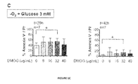
- Various DMOG concentrations (8 to 40 ⁇ g/mL) were assessed; a significant survival rate increase was observed after 20 h and 48 h storage with 32 ⁇ g/mL DMOG (P ⁇ 0.05, Fig. 1C ), reaching 54 ⁇ 10% and 14 ⁇ 10% survival rates respectively.
- Example 2 Neutrophils isolation with citrate increases cell survival rates as compared to heparin
- Anticoagulant molecule used for blood collection impacts on neutrophil survival and may have diverse effects on neutrophils activation 6 .
- Citrate impairs several enzymes involved in the coagulation cascade, through calcium-chelating property 22 . As reported previously, although heparin is widely used, it has been shown to activate neutrophils as a side effect 26 , whereas citrate limits neutrophils activation 6 and was assessed in similar storage conditions.
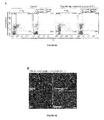
- Neutrophils purified with citrate were subsequently separated from remaining red blood cells with CD235a Microbeads ( Figure 6 ).
- Method 2 Neutrophils purified with citrate
- collecting blood in the presence of citrate allowed a significant increase of neutrophils survival rate as compared to heparin in the presence or in the absence of oxygen 20 h- and 48 h-post purification (P ⁇ 0.01) ( Fig. 2A , B ).
- supplementation with 3 mM glucose increased neutrophils survival 20 h- and 48 h-post purification when stored under anoxic conditions (P ⁇ 0.01) ( Figure 2A ), which was not observed when stored in the presence of oxygen (p>0.05) ( Figure 2A ).
- CD63 is a primary (azurophil) granule marker 27
- CD66b is specifically localised in secondary (specific) granules 28
- CD11b is a tertiary (gelatinase) granule marker 29
- CD16 (or FcgR IIIb) is present in secretory vesicles, but also on na ⁇ ve neutrophils surface 30 .
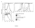
- the inventors first showed that granule markers detection on neutrophil surface (CD63, CD66b, CD11b and CD16) was comparable on na ⁇ ve neutrophils either freshly purified or upon 20 h storage ( Figure 3C ). These results demonstrated that if CD63 and CD11b were not detected, conversely CD66b and CD 16 were significantly exposed on neutrophils surface upon collection and purification. They then demonstrated that, in the presence of S . flexneri, all types of granules markers were similarly detected either on freshly purified neutrophils or upon 20 h storage ( Figure 3C ). S. flexneri stimulation increased CD63, CD66b and CD11b signal detection ( Figure 3C ).
- CD16 abundance decreased upon stimulation, both on freshly purified neutrophils and upon storage ( Figure 3C ), probably due to its recruitment and internalisation upon bacteria phagocytosis.
- neutrophils Prior to nucleofection, neutrophils were pre-conditioned in storage conditions; transfected cells remained stored for 20 h in similar conditions immediately after nucleofection.
- the inventors demonstrated that pmaxGFP plasmid could be efficiently transfected in neutrophils by nucleofection, using Y-01 program ( Fig. 4 ).
- 20 h post-transfection the majority of neutrophils remained viable (51% Annexin V-/PI-, representative experiment; Figure 4A ) as observed upon storage only (76% Annexin V-/PI-, representative experiment; Figure 4A ). Only 10% of nucleofected neutrophils were PI+, consistently with previous reports (8%) 31,32 .
- Annexin V-/PI- neutrophils proportion decreased drastically (24% vs . 51 %, representative experiment; Figure 4A ). This result demonstrates that conditioning neutrophils is required for nucleofection optimization.
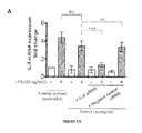
- the inventors aimed at determining if storing neutrophils would allow siRNA gene silencing by nucleofection. To do so, they assessed the IL-8-siRNA neutrophil nucleofection effect on the well-characterized induction of IL-8 expression and release upon lipopolysaccharide (LPS) stimulation 33,34 . Neutrophils responsiveness was assessed on freshly purified cells, stored neutrophils (20 h) and transfected neutrophils stored for 20 h post-transfection (IL-8 siRNA or Negative control siRNA).
- LPS lipopolysaccharide
- IL-8 release by neutrophils stimulated with LPS for 3 h was quantified.
- an increase of p65 expression was observed upon LPS stimulation ( FIG. 5B ) as previously reported 35,36 .
- the inventors observed that LPS-dependent IL-8 release was significantly reduced upon IL-8 siRNA transfection as compared to untreated stored neutrophils (P ⁇ 0.001, Fig. 5B ). No change was observed upon neutrophils storage as compared to freshly purified cells or upon negative control siRNA transfection as compared to stored cells (P > 0.05, Fig. 5B ).
- Neutrophils have long been known as "short-lived cells" 37 . This assumption is mostly based on inappropriate storage conditions in vitro rather than on their lifespan in humans, which appears to be longer as previously expected 17 . Neutrophil global lifespan in humans should also include their period of storage in the bone marrow, which lasts 4-6 additional days 15,13 . Previous reports describing improvement in conservation of viable and functional neutrophils in vitro did not lead to technical improvement of neutrophil genetic manipulation (plasmid or siRNA transfection) 38,39 . Until now, plasmid transfection was described by nucleofection but appeared not to be efficient, with less than 5% transfected cells 31,32 .
- the inventors defined optimized storage conditions based on neutrophil adaptation to low oxygen conditions, as observed during their maturation in the bone marrow 7 but also recently highlighted in vitro under the control of HIF 19,25 . They demonstrated that in conditions of (i) anoxia, (ii) glycolysis promotion by glucose supplementation (3 mM) and (iii) HIF stabilization in the presence of DMOG (32 ⁇ g/mL), neutrophil survival could be extended up to 70 ⁇ 8% after 20 h and 49 ⁇ 2% after 48 h storage ( Fig. 1C and 2B ).
- neutrophils bactericidal activities were not affected by storing cells for 20 h in these conditions as compared to freshly purified cells; since their ability to phagocytose bacteria, to produce NETs upon PMA stimulation or to release granules upon S. flexneri infection remained intact ( Fig. 4 ).
- neutrophils were functional, genetic manipulation could be envisaged, such as plasmid and siRNA transfection.
- pre-conditioning neutrophils increased the efficiency of plasmid transfection by nucleofection, as 37% of viable neutrophils were transfected ( Fig. 4F ). This will allow further studies focusing on neutrophil fundamental physiology but also on neutrophil defence mechanisms subversion by bacteria virulence effector as described in other infected cell types 40 .
Landscapes
- Health & Medical Sciences (AREA)
- Engineering & Computer Science (AREA)
- Biomedical Technology (AREA)
- Life Sciences & Earth Sciences (AREA)
- Bioinformatics & Cheminformatics (AREA)
- Wood Science & Technology (AREA)
- Biotechnology (AREA)
- Organic Chemistry (AREA)
- Immunology (AREA)
- Genetics & Genomics (AREA)
- Chemical & Material Sciences (AREA)
- Zoology (AREA)
- Hematology (AREA)
- Microbiology (AREA)
- Biochemistry (AREA)
- General Engineering & Computer Science (AREA)
- General Health & Medical Sciences (AREA)
- Cell Biology (AREA)
- Micro-Organisms Or Cultivation Processes Thereof (AREA)
Abstract
A method for keeping leukocytes alive ex vivo or in vitro, comprising maintaining the leukocytes in a medium comprising from 3 to 10 mM of glucose, in hypoxic conditions with P(O2) ≤ 10 mM Hg.
Description
- The present invention pertains to the fields of transfusion and of cell culture. More particularly, the invention relates to new storage conditions for leukocytes, in particular for polymorphonuclear neutrophils (neutrophils), which allow an increased survival of functional cells, as well as to a method for storing said leukocytes and a device for storing and transporting them.
- Polymorphonuclear neutrophils are the most abundant circulating white blood cell population (70% in human). However, human neutrophils are certainly the most difficult immune cells to study in vitro, regarding their short lifespan under atmospheric conditions (pO2 atm. = 160 mmHg), which does not exceed 8 hours. Neutrophils survival and cell death mechanisms are tightly regulated regarding their anti-infectious function and successful inflammation resolution respectively1. Apoptosis is the main cell death pathway2 and several anti-apoptotic factors (eg.,. G-CSF3, GM-CSF4) were shown to extend purified neutrophil longevity.
- Increasing purified neutrophils survival represents a major challenge to promote both their study in vitro and their clinical use, especially for neutrophil transfusion, which efficiency is still discussed, as rapid neutrophil apoptosis may be responsible for its failure5. Several strategies have been evaluated for their potential beneficial effect on neutrophil survival, including the evaluation of optimal anticoagulant molecules6.
- To the aim of increasing purified neutrophils survival, the inventors inspired of and mimicked physiological conditions encountered by neutrophils in their niche during the maturation process. Neutrophils maturation occurs in the bone marrow from hematopoietic stem cells differentiating from myeloblasts to mature neutrophils7. These events occur in an hypoxic environment in which maturating neutrophils remain 3-5 days8. In this context, maturating cells produce energy required for their development through glycolysis rather than respiration in an HIF-1-dependent manner9. HIF-1 is an heterodimeric (α-β) transcriptional regulator; its α subunit abundance is regulated upon hydroxylation by prolyl-hydroxylases (PHD1, PHD2 and PHD3) under normoxic conditions10,11, followed by ubiquitination and proteasomal degradation12. Following their production, mature neutrophils remain within the bone marrow for 4-6 additional days13.
- Once released into the blood circulation through sinusoids, neutrophils encounter oxygenated environments. Sinusoidal oxygen pressure is not precisely known in situ, however it is estimated between artery (pO2 a. = 75-100 mmHg) and venous (pO2 v. = 30-50 mmHg) oxygen pressures14. In this oxygenated context, human neutrophils lifespan remains discussed as varying among studies from 10 h to 90 h13,15,16,17 before clearance occurring equally in the bone marrow, the liver and the spleen18. A low oxygen environment is favourable to neutrophil lifespan, which is mediated by HIF-1α12. Inhibition of PHD3 by addition of Dimethyloxalylglycine (DMOG), a pan-hydroxylase inhibitor, decreases apoptosis under normoxic conditions19. Consistently, containing few mitochondria, neutrophils rely mainly on anaerobic glycolysis for energy production20,21.
- As disclosed in the experimental part below, the inventors now demonstrated a synergistic effect of anoxia, glycolysis induction and HIF stabilization on neutrophils survival. Optimum storage conditions are disclosed, which, following collection and purification of neutrophils with citrate, allow survival rates of 74±2% after 20 h and 49±3% after 48 h storage in an appropriate culture medium, and even more after storage in plasma under anoxic conditions; moreover, the viable neutrophils remain functional under these conditions. Preserving neutrophils in such an environment allowed for the first to perform plasmid transfection and siRNA gene silencing on human purified neutrophils, which opens new perspectives for neutrophil cell biology and physiopathological studies.
- A first aspect of the present invention is a method for keeping leukocytes alive ex vivo or in vitro, comprising maintaining the leukocytes in an appropriate medium comprising from 3 to 10 mM of glucose, preferably from 3 to 6 mm mM of glucose, in hypoxic conditions with P(O2) ≤ 10 mM Hg.
- According to a preferred embodiment of the method, the partial pressure of dioxygen is inferior or equal to 1 mM Hg (P(O2) ≤ 1 mM Hg), which will be considered here after as "anoxic conditions". To this aim, the leukocytes are preferably obtained from a donor by a process preventing their contact with oxygen, for example through apheresis performed in conditions such that the cells are not contacted by O2. They are then kept in anaerobic conditions, for example in a closed (and preferably gas-free) oxygen-tight device, or in an anaerobic incubator.
- As used herein, an "oxygen-tight", or "oxigen- impermeable" device is a storage container or storage container system that is impermeable to oxygen. In accordance with the present invention, an oxygen-impermeable storage container is a container, pouch, bag, or bottle that is constructed of a material compatible with a biological fluid, such as whole blood or a blood component and is preferably capable of withstanding centrifugation and sterilization. Such containers are known in the art and include, e.g., blood collection and satellite bags. Storage containers of use in the instant method can be made of plasticized polyvinyl chloride, e.g., PVC plasticized with dioctylphthalate, diethylhexylphthalate, or trioetyltrimellitate. The bags may also be formed from polyolefin, polyurethane, polyester, and polycarbonate. In one embodiment, the storage container itself is constructed of an oxygen--impermeable material. Impermeable materials are routinely used in the art and any suitable material can be used. Existing systems use oxygen-impermeable receptacles composed of layers of ethylene vinyl alcohol copolymer and modified ethylene vinyl acetate copolymer, impermeable to oxygen ingress. In another embodiment, the leukocytes are transferred into a first storage container which is a component of a storage container system that is impermeable to oxygen. Such systems include, but are not limited to, use of an oxygen-impermeable over wrap or over bag which encloses the storage container.
- In what precedes, an "appropriate medium" can be either plasma or any culture medium appropriate for leukocytes survival. Examples of culture media appropriate for performing the method according to the invention include:
- RMPI 1640 + 10 mM Hepes + 10% FBS,
- RMPI 1640 with L-Glu + 25 mM Hepes + 10% FBS,
- IMDM + 10% autologous serum, etc.
- Of course, in the above media, the fetal bovine serum (FBS) can be replaced by newborn calf serum (NBS) or by inactivated human serum.
- According to a particular embodiment of the invention, the culture medium is a synthetic medium which has been treated for removing dioxygen therefrom prior to transferring the leukocytes into it. This can be done by any technique known by the skilled artisan, such as gazing the medium with an oxygen-free gaz (for example CO2/N2 or CO2/H2/N2).
- In case the medium is a culture medium, it will preferably comprise an effective amount of a compound that stabilizes the hypoxia inducible factor-1 (HIF-1).
- Of course, the compound used to stabilize HIF-1 can do so either by a direct action of one of HIF-1 subunits (in particular, HIF-1α), or by an indirect action on HIF-1. For example, since the HIF-1α subunit is regulated by hydroxylation, in particular by the oxygen-sensitive propyl hydroxylase-3 (PHD-3) and by transcriptional inactivation following asparaginyl hydroxylation by factor inhibiting HIF (FIH), a compound that inhibits these, or that inhibits the 2-oxoglutarate dioxygenase activity of any other enzyme implicated in HIF stability, is also considered as a "compound that stabilizes HIF-1", according to the present invention.
- According to a preferred embodiment, the compound that stabilizes the hypoxia inducible factor-1 (HIF-1) comprises or consists of a prolyl hydroxylase inhibitor.
- Due to safety and regulatory constraints, when the leukocytes are stored in view of a transfusion, the HIF-1-stabilizing compound must be appropriate for injection to a patient.
- Non-limitative examples of compounds which can be used to stabilize HIF-1 when performing the above method include dimethyloxalylglycine (DMOG), the cyanoisoquinoline compounds disclosed in
WO 2007/090068 , the compounds capable of inhibiting HIF hydroxylase enzyme activity disclosed inWO 2012/106472 , the compounds disclosed inUS 2013/310565 and those disclosed inUS 2013/245037 . - According to a preferred embodiment, the compound that stabilizes HIF-1α is the dimethyloxalylglycine (DMOG). In this case, DMOG is preferably present in the medium at a concentration ranging from 8 to 50 µg/mL, more preferably from 25 to 40 µg/mL, for example in a concentration of about 32 µg/mL.
- According to another preferred embodiment of the method according to the invention, the leukocytes are maintained in plasma, under hypoxic (P(O2) ≤ 10 mM Hg) or anoxic conditions. If necessary, glucose is added to the plasma so that the final glucose concentration is between 3 and 10 mM, preferably between 3 and 6 mM. According to this embodiment, the plasma is preferably from the same donor as the leukocytes.
- The present invention is particularly useful for maintaining leukocytes which are known to have a short lifespan in vitro, such as human granulocytes, and in particular polymorphonuclear neutrophils. Hence, according to a preferred embodiment of the present invention, the leukocytes comprise granulocytes. According to another preferred embodiment of the present invention, the leukocytes consist of granulocytes.
- Granulocytes include polymorphonuclear neutrophils, basophils and eosinophils. According to a preferred embodiment of the present invention, the leukocytes comprise polymorphonuclear neutrophils. According to another preferred embodiment of the present invention, the leukocytes consist of polymorphonuclear neutrophils. In this case, the polymorphonuclear neutrophils are preferentially purified.
- According to a preferred embodiment of the invention, illustrated in the experimental part below, more than 70% of the polymorphonuclear neutrophils remain viable after 20 hours storage. According to another embodiment of the invention, more than 80%, and even 90% of the polymorphonuclear neutrophils remain viable after 20 hours storage.
- According to a preferred embodiment of the invention, illustrated in the experimental part below, more than 45% of the polymorphonuclear neutrophils remain viable after 48 hours storage.
- According to a preferred embodiment of the invention, illustrated in the experimental part below, the viable polymorphonuclear neutrophils remain functional.
- According to a particular embodiment of the present invention, the leukocytes comprise basophils. According to another particular embodiment of the present invention, the leukocytes consist of basophils.
- According to a particular embodiment of the present invention, the leukocytes comprise eosinophils. According to another particular embodiment of the present invention, the leukocytes consist of eosinophils.
- According to a particular embodiment of the present invention, the leukocytes comprise peripheral blood mononuclear cells (PBMCs). According to another particular embodiment of the present invention, the leukocytes consist of PBMCs.
- According to a particular embodiment of the present invention, the leukocytes comprise monocytes. According to another particular embodiment of the present invention, the leukocytes consist of monocytes.
- According to a particular embodiment of the present invention, the leukocytes comprise lymphocytes. According to another particular embodiment of the present invention, the leukocytes consist of lymphocytes.
- According to a particular embodiment of the present invention, the leukocytes comprise T lymphocytes (LT). According to another particular embodiment of the present invention, the leukocytes consist of LT.
- According to a particular embodiment of the present invention, the leukocytes comprise B lymphocytes (LB). According to another particular embodiment of the present invention, the leukocytes consist of LB.
- According to a particular embodiment of the present invention, the leukocytes comprise macrophages. According to another particular embodiment of the present invention, the leukocytes consist of macrophages.
- Several anticoagulant molecules are available for treating blood samples, including heparin, citrate, ACD (acid citrate dextrose or anticoagulant citrate-dextrose) and EDTA. Heparin and citrate are the most commonly used anticoagulants for clinical analysis or in vitro basic research. The inventors have tested the impact of the anticoagulant molecule used during blood collection on the survival of neutrophils subsequently purified from the blood samples, and noted that
neutrophils survival rates 20 h- and 48 h-post purification were improved when blood was collected in the presence of citrate as compared to heparin (Example 2 below). They obtained even better results by using EDTA instead of citrate at blood collection. Hence, according to preferred embodiments of the present invention, the leukocytes have been purified from citrated venous blood or from blood treated with EDTA. According to preferred embodiments of the present invention, polymorphonuclear neutrophils have been purified from citrated venous blood or from blood treated with EDTA. - According to another aspect, the present invention pertains to an oxygen-tight container containing live leukocytes in a medium with P(O2) ≤ 10 mM Hg and from 3 to 10 mM of glucose. Examples of containers include bags, bottles etc. Oxygen-impermeable materials are routinely used in the art and any suitable material can be used, such as, for example, plasticized polyvinyl chloride, e.g., PVC plasticized with dioctylphthalate, diethylhexylphthalate, trioetyltrimellitate, polyolefin, polyurethane, polyester, and polycarbonate. According to a preferred embodiment, the volume of the container is comprised between 5 and 300 mL. According to a preferred embodiment, the medium is plasma or essentially plasma, possibly complemented with glucose.
- According to another aspect, the present invention pertains to a device for transporting leukocytes, comprising an oxygen-tight container containing a culture medium as described above, and means appropriate for introducing cells into the culture medium. According to this embodiment, the oxygen-impermeable container is as described above and the means for introducing cells into the culture medium are preferentially designed for avoiding an important contact between the culture medium and air during the transfer of the cells into the device. Examples of such means include caps having a small diameter, for example a diameter of less than 5 mm, as well as silicone, rubber or plastic plugs which can be pierced by a needle, etc.
- As recalled above, the leukocytes are preferably obtained by a process preventing their contact with oxygen, for example through apheresis performed in conditions such that the cells are not contacted by O2, such as a closed device entirely made of oxygen-tight materials, including the tubes etc.
- According to a preferred embodiment of devices according to the invention, the leukocytes comprise neutrophils. According to another embodiment, the leucocytes are neutrophils.
- According to another aspect, the present invention concerns a culture medium for neutrophils, characterized in that it comprises between 3 and 6 mM of glucose and between 25 and 40 µg/mL of DMOG.
- The present invention also pertains to a method for transfecting polymorphonuclear neutrophils, comprising (i) a step of pre-conditioning said polymorphonuclear neutrophils by transferring them into a medium as described above, or by maintaining them in plasma, (ii) a step of incubating them in hypoxic or anoxic conditions, and (iii) a transfection step.
- A population of polymorphonuclear neutrophils which has been transfected according to the above method is also part of the present invention. In a preferred embodiment, at least 30% of these cells have been effectively transfected.
- The invention is further illustrated by the following figure and examples.
-
-
Figure 1 . Optimization of human neutrophil survival conditions in the presence and absence of oxygen, with heparin as an anticoagulant. (A-B) Human neutrophils were purified on heparin-coated collection tubes (Method 1). Neutrophil survival was quantified by flow cytometry (Annexin V-/PI-) either under atmospheric conditions (20% O2; (A)) or in the absence of oxygen (0% O2; (B)). Purified neutrophil survival was determined after 3, 20 and 48 h storage in the presence of 0, 1, 2 or 3 mM glucose as indicated. Error bars indicate SD. 'ns' indicates P > 0.05, * indicates P < 0.05. Data represent n=7 individual blood samples, which were collected and analysed independently. (C) Neutrophil survival was quantified as described in (A) in the absence of oxygen (-O2) with 3mM glucose and in the presence of 0, 8, 16, 32 or 40 µg/mL DMOG as indicated, after 20 or 48 h storage. Error bars indicate SD. * indicates P < 0.05. Data represent n=7 individual blood samples, which were collected and analysed independently. -
Figure 2 . Optimization of human neutrophil survival conditions, with citrate as an anticoagulant. (A) Human neutrophils were purified in the presence of citrate (Method 2). Neutrophil survival was quantified by flow cytometry (Annexin V-/PI-) either under atmospheric conditions (20% O2) or in the absence of oxygen (0% O2); with or without 3 mM glucose. Purified neutrophil survival was determined after 3, 20 and 48 h storage in the presence of 0, 1, 2 or 3 mM glucose as indicated. Error bars indicate SD. * indicates P < 0.05, ** indicates P < 0.01. Data represent n=5 individual blood samples, which were collected and analysed independently. (B) Neutrophil survival was quantified as described in (A) in the presence (+O2) or absence of oxygen (-O2) with 3mM glucose and in the presence or absence of 32 µg/mL DMOG as indicated, after 3, 20 or 48 h storage. Error bars indicate SD. * indicates P < 0.05, ** indicates P < 0.01, *** indicates P < 0.001. Data represent n=5 individual blood samples, which were collected and analysed independently. -
Figure 3 . Stored neutrophils remain functional as compared to freshly purified neutrophils. Neutrophils stored under anoxic conditions with 3mM glucose and 32 mg/mL DMOG were sorted on Annexin V-coated beads (Figure 6 ). Their phagocytosis (A), NET formation (B) and degranulation (C) properties were compared to freshly purified neutrophils. (A) S. flexneri phagocytosis assay was performed with either freshly purified or stored (20, 48 h) and viable neutrophils, atMOI 20 during 15 min at 37°C. Results are averaged from three independent experiments (n=3) performed in triplicate. Error bars indicate SD. 'ns' indicates P > 0.05. (B) Neutrophil NET formation was induced from either freshly purified or stored (20, 48 h) and viable neutrophils, in the presence of 25 nM PMA for 3 hours at 37°C. Immunofluorescence staining was performed with α-MMP-9 (green). DNA was stained with DAPI (blue). Results are representative of three independent experiments. Bars are 20 µm.(C) Neutrophil degranulation was assessed by flow cytometry detecting specific primary (CD63), secondary (CD66b), tertiary (CD11b) granules or CD16 (FcγR IIb). Freshly purified or stored (20, 48 h) and viable neutrophils (naive) were activated in the presence of S. flexneri (MOI 20). Results are representative of three independent experiments. -
Figure 4 . Neutrophil plasmid nucleofection optimization upon storage. Neutrophil transfection efficiency by nucleofection was evaluated in the presence of oxygen (+O2, 20%) or under anoxic conditions (-O2, 0%) with 3 mM glucose and 32 mg/mL DMOG. Neutrophils purified on citrate were pre-conditioned in storage media during 2 h prior nucleofection. Using Y-01 program, neutrophils (2.106 cells) were transfected with 2 µg pmaxGFP (Amaxa biosystems) with Nucleofector (Lonza). After 20 h transfection and cell recovery, neutrophils viability (A) and transfection efficiency (B-E) were analysed by flow cytometry. (A) Cell viability (Annexin V-/PI-) after nucleofection was compared to non-transfected cells (Control) in the presence of oxygen (+O2) or upon storage (-O2, + 3mM glucose + 32 µg/mL DMOG). Results are representative of three independent experiments. (B) Confocal imaging of whole transfected neutrophil population, detecting GFP (green), Annexin V (Red) and DNA (PI, blue) positive cells. Bar is 60 µm. Result is representative of three independent experiments. (C-D) Flow cytometry quantification of the GFP+ neutrophil proportion within the whole neutrophil population (C) or within viable (Annexin V-/PI-) population (D). Results are representative of three independent experiments. (E) pmaxGFP transfection efficiency by nucleofection on stored neutrophils (pre-conditioning and storage for 20 h post-transfection) or in the absence of treatment (+O2 conditions). (E-F) Flow cytometry quantification of the GFP+ neutrophil proportion within the whole neutrophil population (E) or within viable (Annexin V-/PI-) population (F). Results are averaged from three independent experiments (n=3) performed in triplicate. Error bars indicate SD. *** indicates P < 0.001. -
Figure 5 . Neutrophil storage allows IL-8 siRNA gene silencing by nucleofection. (A) qRT-PCR analysis of IL-8 mRNA expression in freshly purified neutrophils (5.105 cells) upon storage (-O2, + 3mM glucose + 32 µg/mL DMOG). Stored neutrophils were nucleofected with IL-8 siRNA or Negative control siRNA when indicated. Neutrophils were either untreated (whit bars) or stimulated with 10 ng/mL LPS (grey bar) during 3 h at 37°C. IL-8 mRNA expression fold change were calculated as compared to untreated freshly purified neutrophil. Error bars indicate SD. 'ns' indicates P > 0.05, *** indicates P < 0.001, (n=3). (B) IL-8 release in the supernatant fractions (Sup.) by neutrophils stored as described in (A) and stimulated with 10 ng/mL LPS for 3 h at 37°C. IL-8 quantifications were performed by ELISA (5.105 cells), Error bars indicate SD. 'ns' indicates P > 0.05, *** indicates P < 0.001, (n=3). As controls, p65 and actin were detected by Western blot in cellular (Cell.) fractions with specific antibodies. Results are representative of three independent biological samples. -
Figure 6 . Viable neutrophils purification from apoptotic neutrophils and red blood cells (RBCs) by flow cytometry. (A) Neutrophils purified with Method 2 (citrated venous blood) were contaminated by RBC at final purification step. Samples were additionally purified by negative selection using CD235a (glycophorin) microbeads, allowing RBC retention. (B) Upon storage, apoptotic neutrophils were eliminated using Annexin V microbeads, allowing negative selection of viable cells. Experiments are representative of at least five individual experiments. - The examples have been performed using the following materials and methods:
- Ethical approval
All participants gave written informed consent in accordance with the Declaration of Helsinki principles. Peripheral Human blood was collected from healthy patients at the ICAReB service of the Pasteur Institute (authorization DC No.2008-68). - Human blood collection
Human blood was collected from the antecubital vein into tubes containing sodium heparinate (1% final concentration) (Method 1) or sodium citrate (3,8% final) (Method 2) as anticoagulant molecules (see below). - Neutrophils isolation
Immediately after blood collection, neutrophils were isolated from total blood samples following two different methods as described below.-
Method 1. Heparinized venous blood was layered over 7 ml Histopaque®1119 (Sigma-Aldrich) and centrifuged at 800 x g for 20 min. at room temperature (RT). Plasma and the mononuclear cell layer were discarded. The diffuse red phase containing neutrophils, above Red Blood Cell (RBC) pellet, was collected and washed with RPMI 1640 (without glutamine and Phenol Red) (Gibco Invitrogen) with 10 mM Hepes and layered on 10 ml of discontinuous Percoll® Gradient (GE Healthcare) prepared in Ca2+/Mg2+-free phosphate buffered saline (PBS) with final Percoll® concentrations of 65, 70, 75, 80 and 85%. After centrifugation at 800 x g for 20 min, neutrophil buffy coat located at the 70/75% interface was collected and washed in RPMI 1640 with 10 mM Hepes before culture. Neutrophils were resuspended at 1x10⁶ cells/ml in RPMI 1640 with L Glutamine and 25 Mm Hepes (Gibco, Life Technologies), 10% of heat inactivated Foetal Bovine Serum (FBS) (Invitrogen, Gibco). -
Method 2. Citrated venous blood (45 mL) was centrifuged at 445 g for 15 min. at room temperature. Platelet rich plasma (PRP) was collected and centrifuged at 2500 x g for 20 min to form platelet poor plasma (PPP). 6 ml of 6% dextran were added to cells, to up to 50 ml with sterile saline (0.9% NaCl). Cells were subsequently mixed gently by inverting tubes. After red blood cells sedimentation, the upper layer containing neutrophils was transferred to clean 50 ml falcon tubes and centrifuged at 239 x g for 6 min. Pellet was resuspended in 1 ml of platelet poor plasma (PPP) and deposited on a Percoll® gradient with a lower phase (51% Percoll®+ 49% PPP) and an upper phase (42% Percoll®+ 58% PPP). After centrifugation at 239 x g for 15 min, the granulocyte layer was washed in 25% PPP (10 ml PPP + 30 ml Hanks without Ca2+ and Mg2+) and cells were counted.
-
- Culture media
Human purified neutrophils were cultured in RPMI 1640 (Life Technologies) supplemented with 10 mM Hepes or in RPMI 1640 with L Glutamine, 25 mM Hepes (Gibco, Life Technologies) and 10% of heat inactivated Foetal Bovine Serum (FBS) (Invitrogen, Gibco) when indicated.
Shigella flexneri 5a (S. flexneri) was grown in trypticase soy (TCS) broth or on TCS agar plates supplemented with 0.01% Congo Red (Sigma). - Neutrophils storage conditions
Neutrophils fractions with a >95% purity obtained by applyingMethod 1 orMethod 2 were resuspended at a 5x10⁶ cells/mL concentration in RPMI 1640 with L Glutamine and 25 mM Hepes (Gibco, Life Technologies), 10% of heat inactivated Foetal Bovine Serum (FBS) (Invitrogen, Gibco) and indicated concentrations of glucose (Sigma-Aldrich) and Dimethyloxalylglycine or DMOG (Cayman Chemical Company).
For comparative experiments aiming at characterizing the role of oxygen on neutrophils survival, purified neutrophils (Method 1 or Method 2) were split in two identical fractions. One was stored at 37°C with 5% CO2 under atmospheric conditions (20% O2 condition, +O2). The second one was stored at 37°C in an anoxic chamber (Minimacs, Don Withley) under an atmosphere containing 90% N2, 5% CO2 and 5% H2 (0% O2 condition, -O2), during indicated time. - Antibodies
For immunofluorescence, MMP-9 was detected using a rabbit polyclonal antibody (Novus Biologicals, NBP1-45719).
For flow cytometry, apoptotic cells were labelled with (allophycocyanin) APC-Annexin V (BD Bioscience). Cell surface exposed antigens were labelled with (phycoerythrin) PE Mouse anti-human CD16 (clone 3G8, BD Pharmingen), APC mouse anti-human CD11b (clone D12, BD Biosciences), PE mouse anti-human CD66b (clone G10F5, BD Pharmingen), PE, mouse anti-human CD15 (clone W6D3, BD Pharmingen) or FITC mouse anti-human CD63 (clone H5C6, BD Pharmingen). For western blot analysis, a monoclonal α-IL-8 antibody (SAB1409243, Sigma-Aldrich), a rabbit polyclonal α-p65 antibody (ab7970, Abcam) and a rabbit polyclonal α-actin antibody (A2066, Sigma-Aldrich) were used. - Flow cytometry
Cell viability. For neutrophil survival analysis, human neutrophils (1x10⁶) were washed twice in PBS and then resuspended in 1ml of Annexin V binding Buffer. A 100 µl sample (1x10⁵ cells) was stained with 5 µl of APC-Annexin V and 5 µl of Propidium iodide (PI) (0.5 µg/ml final concentration) as recommended by the manufacturer. At least 104 events were acquired for each condition on a FACSCalibur™ flow cytometer (BD Biosciences), and data were analyzed using CellQuest™ Pro Software (BD Biosciences). Viable cells were defined as Annexin V-/PI- quantifications.
Neutrophil degranulation assay. For neutrophil degranulation assays, 2.106 neutrophils purified withMethod 2 were stimulated with S. flexneri atMOI 20. After 10 min centrifugation at 300 x g, infected neutrophils were incubated during 15 min at 37°C. Cell surface markers exposure was quantified by flow cytometry as follows: 2.5x10⁵ cells were collected and incubated with 5 µL of PE Mouse anti-human CD16, APC mouse anti-human CD11b, PE mouse anti-human CD66b, PE, mouse anti-human CD15 or FITC mouse anti-human CD63. Cell labelling was analysed using a FACSCalibur™ flow cytometer (BD Biosciences), and data were analysed using CellQuest™ Pro Software (BD Biosciences). Results are representative of two independent experiments performed. - Plasmid or siRNA nucleofection
Plasmid or siRNA transfection were performed by nucleofection (Lonza). Neutrophils purified with Method 2 (2.106 cells/transfection) were pre-incubated 2 hours in 500 µL of supplemented culture media (RPMI + 10% SVF + 3 mM glucose + 32 µg/mL DMOG) at 37°C under anoxic conditions. Cells were collected by centrifugation (300 x g, 10 min at room temperature). Cells were washed in RPMI only. After an additional centrifugation, pellets were carefully resuspended in 100 µl supplemented Mouse T cell Nucleofector® solution (Lonza). 2 µg of pmaxGFP plasmid (Lonza) or 2 pmole of siRNA (SMARTpool®: Accell IL8 siRNA (Thermo Fischer Scientific) or Negative control siRNA (Qiagen)) were added before transferring the mixture into certified nucleofection cuvettes. DNA transfer was performed by electroporation using Nucleofector Program Y-01 (Amaxa® Nucleofector® device (Lonza)). Transfected samples were immediately transferred first in 500 µL of pre-equilibrated supplemented culture media and subsequently into 24-well plate containing 1 mL of pre-equilibrated supplemented culture media. 20 h post-transfection, transfected cells were analyzed by flow cytometry, fluorescence imaging (plasmid); quantitative PCR, ELISA or Western blot (siRNA). - Statistical analysis
Data were analysed using Prism 5.0 software (GraphPad) using Student's T-test. Significance was accepted when P<0.05. - Western Blot
Neutrophils stimulated with LPS or S. flexneri (see above) cellular extract and supernatants were incubated with anti-protease Cocktail (Roche). Proteins were separated by electrophoresis on SDS-Page gel and transferred onto nitrocellulose membrane (Life technologies) and blocked inPBS milk 5% for 1 hour at room temperature. Membranes were further incubated with α-p65 and α-actin primary antibodies (1:1000) overnight: antibody binding was detected with chemiluminescence (ECL kit, GE Healthcare) using conjugated antibodies to (Horseradish peroxidase) HRP (Dako) (1:5000). - Immunofluorescence and Imaging
Immunofluorescence detection was performed in PBS/Saponin 0.1% using a rabbit α-MMP-9 (1:1000) and α-rabbit-FITC conjugated secondary antibodies (1:1000). Actin was stained with phalloidin-Rhodamin (1:1000), DNA was stained with Dapi (1:1000).
Fluorescent-labelled cells were observed using a laser-scanning TCS SP5 confocal microscope (Leica). Image analysis was performed using Fiji software. - Phagocytosis assay
Shigella flexneri pGFP (M90T pGFP) or S. aureus pGFP strains were grown until an OD600=0.5 was reached and were incubated with purified human neutrophils in RMPI 1640 with 10 mM Hepes at aMOI 20 with inactivated human serum and centrifuged at 300 x g during 10 min. An additional incubation of 15 min at 37°C was performed to allow the phagocytosis of bacteria. For immunofluorescence staining, after three washes in PBS, infected cells were fixed with paraformaldehyde (PFA) 3% during 30 min. After an additional washing in PBS, infected cells were stained as described below. For intracellular bacteria counting, infected cells were washed three times with PBS and incubated with 50 µg/mL gentamycin for 30 min at 37°C and further washed three times in PBS. Intracellular bacteria were counted after cells were lysed in 1% saponin. - NET formation assay
Purified Neutrophils (5.105 per well) were seeded on glass coverslips treated with 0.001% polylysine, centrifuged at 300 x g for 10 min and treated 25 nM PMA (phorbol 12-myristate 13-acetate) (Sigma-Aldrich) for 3 hours at 37°C. Cells were subsequently fixed with 4% PFA prior immunofluorescence staining. - RNA extraction
IL-8 expression induction was performed on 2.105 human neutrophils either freshly purified, stored 20h in anoxic conditions with 3 mM glucose and 32 µg/mL DMOG, or stored and transfected 20 h with IL-8 siRNA or Negative control siRNA (see above). Neutrophils IL-8 expression was induced with 10 ng/mL E. coli LPS (InvivoGen-Cayla) for 180 min. RNAs were extracted with RNeasy Mini kit (Qiagen) from each condition; total RNAs were extracted from three independent biological samples. - Transcriptional analysis
Following RNAs extraction, cDNAs were produced with SuperScript II Reverse Transcriptase (Invitrogen) and oligo(dT)12-18 primer (Invitrogen) as recommended by the supplier. The following primers (purchased from Invitrogen) were used: IL-8, 5'-GCCTTCCTGATTTCTGCAGC-3' (SEQ ID No: 1) and 5'-TGCACCCAGTTTTCCTTGG-3' (SEQ ID No: 2); gapdh 5'-TCGCTCTCTGCTCCTCC-3' (SEQ ID No: 3) and 5'-TTAAAAGCAGCCCTGGTGAC-3' (SEQ ID No: 4). qPCR reactions were run on an ABI 7900HT (Applied Biosystems) using Power SYBR® Green mix (Applied Biosystems) according to the manufacturer's instructions. gapdh was used as an internal control gene to obtain relative expression compared to untreated freshly purified neutrophils and expressed as IL-8 mRNA expression fold change. Data were analysed with SDS 2.2 software (Applied Biosystems). Means and SD were calculated from three independent samples and performed in triplicate. - ELISA
Release of IL-8 was determined by ELISA as described previously (Hattar et al, 2001). Mouse monoclonal α-IL-8 antibody (4 µg/mL) was bound to 96-well ELISA plates at 4°C overnight and washed 5 times with PBS, 0.05% Tween. Wells were blocked in PBS, 0.05% Tween, 0.1% BSA for 1h at room temperature and washed 5 times with PBS, 0.05% Tween. Neutrophil supernatants (concentrated to reach 100 µg/mL) were bound to α-IL-8 coated plates (100 µL, 10 µg total protein) at room temperature during 2h and washed 5 times with PBS, 0.05% Tween. Recombinant human IL-8 (Sigma-Aldrich) was used for standard titration curves. IL-8 was detected with mouse monoclonal α-IL-8 antibody followed by phosphatase alkaline goat α-mouse conjugated antibody (Beckman Coulter) (200 ng per well). Antibodies diluted in PBS, 0.05% Tween were incubated with the plates for 90 min. Following washes, substrate PNPP (0.1%) was added in a Tris HCl 0.1M pH 9 with NaCl 4.5M buffer to each well and incubated for 60 min at room temperature. Absorbance was measured at 405 nm on a Sunrise microplate reader (Tecan). Mean and SD were calculated from experiments performed in triplicate on three independent biological samples. IL-8 concentration (pg/mL) was calculated from standard curve. - In this study, neutrophil survival rate was evaluated in various conditions by flow cytometry, by quantifying the proportion of viable (propidium iodide negative staining, PI-) and non-apoptotic (Annexin V negative staining, Annexin V-) cells.
- In routine, heparin and citrate are the most commonly used anticoagulants for clinical analysis or in vitro basic research6,22. Heparin acts as an anticoagulant by activating antithrombin23. The inventors demonstrated that neutrophils purified with heparin (Method 1) were viable upon collection and purification in the presence of atmospheric oxygen (20% O2, +O2) (89±4%,
Figure 1A ). However, a drastic decrease of their survival rate was observed as soon as 20 h (16±7% vs. 89±4%, P < 0.001) (Figure 1A ). Conversely, under anoxic conditions (0% O2, -O2),neutrophils survival rate 20 h post purification was significantly higher than in the presence of oxygen (35±11% vs. 16±7%, P < 0.01) (Figure 1A ,B ) but remained limited. Glucose supplementation (3mM) did not have any effect on survival rate in the presence of oxygen at 20 h or 48 h (P > 0.05) (Figure 1A ), but a significant increase was observed after 20 h storage in anoxia (P < 0.05) (Figure 1B ). This effect was no longer observed after 48 h storage (P > 0.05) (Figure 1B ). Low oxygen level and high glucose concentration synergistic effect on neutrophils survival may be the consequence of anaerobic glycolysis optimization, since glycolysis is the main energy production source of neutrophils20,21. - Dimethyloxalylglycine (DMOG) is a pan-hydroxylase inhibitor stabilizing HIF-1α24, which was shown to decrease neutrophil apoptosis25. Its effect on neutrophil survival in the absence of oxygen with 3mM glucose supplementation was evaluated in a concentration-dependent manner. Various DMOG concentrations (8 to 40 µg/mL) were assessed; a significant survival rate increase was observed after 20 h and 48 h storage with 32 µg/mL DMOG (P < 0.05,
Fig. 1C ), reaching 54±10% and 14±10% survival rates respectively. - Anticoagulant molecule used for blood collection impacts on neutrophil survival and may have diverse effects on neutrophils activation6.
- Citrate impairs several enzymes involved in the coagulation cascade, through calcium-chelating property22. As reported previously, although heparin is widely used, it has been shown to activate neutrophils as a side effect26, whereas citrate limits neutrophils activation6 and was assessed in similar storage conditions.
- Neutrophils purified with citrate (Method 2) were subsequently separated from remaining red blood cells with CD235a Microbeads (
Figure 6 ). As a first statement, collecting blood in the presence of citrate allowed a significant increase of neutrophils survival rate as compared to heparin in the presence or in the absence ofoxygen 20 h- and 48 h-post purification (P < 0.01) (Fig. 2A ,B ). As described with heparin, supplementation with 3 mM glucose increasedneutrophils survival 20 h- and 48 h-post purification when stored under anoxic conditions (P < 0.01) (Figure 2A ), which was not observed when stored in the presence of oxygen (p>0.05) (Figure 2A ). Synergistic effect of anoxia and 3 mM glucose supplementation resulted in survival rates of 64±8% and 41±3%, 20 h- and 48 h-post purification (Figure 2A ), which further increased to 70±8% (P < 0.05) and 49±2% (P < 0.05) upon addition of 32 µg/mL DMOG, respectively (Figure 2B ). These results demonstrate neutrophils survival rate optimization was due to a synergistic effect of anoxia, supplementation with 3 mM glucose and 32 µg/mL DMOG (hereinafter referred to as 'storage conditions' or 'storage'). - The above results demonstrated that neutrophils purified on citrate and stored for 20 h or 48 h under anoxic conditions with 3 mM glucose and 32 µg/mL DMOG remain mainly viable. To determine if viable neutrophil population remain functional, its ability to phagocytose bacteria was analysed, as well as its ability to form NET upon PMA stimulation and to release granule components as compared to freshly purified neutrophils.
- After 20 h or 48 h storage, viable neutrophils were purified by negative selection on Annexin V binding Microbeads to obtain a pure Annexin V-/PI- neutrophil population (Figure S1B). As shown in
Figure 3A , purified viable neutrophils obtained after 20 h or 48 h storage phagocytosed S. flexneri with the same efficiency as freshly purified neutrophils (P > 0.05). In addition, they remained sensitive to PMA-dependent NET formation (Figure 3B ). To evaluate neutrophil degranulation efficiency of stored viable neutrophils as compared to freshly purified neutrophils, the inventors quantified by flow cytometry cell-surface exposure of granule markers upon contact with S. flexneri; CD63 is a primary (azurophil) granule marker27, CD66b is specifically localised in secondary (specific) granules28 and CD11b is a tertiary (gelatinase) granule marker29. CD16 (or FcgR IIIb) is present in secretory vesicles, but also on naïve neutrophils surface30. - The inventors first showed that granule markers detection on neutrophil surface (CD63, CD66b, CD11b and CD16) was comparable on naïve neutrophils either freshly purified or upon 20 h storage (
Figure 3C ). These results demonstrated that if CD63 and CD11b were not detected, conversely CD66b andCD 16 were significantly exposed on neutrophils surface upon collection and purification. They then demonstrated that, in the presence of S. flexneri, all types of granules markers were similarly detected either on freshly purified neutrophils or upon 20 h storage (Figure 3C ). S. flexneri stimulation increased CD63, CD66b and CD11b signal detection (Figure 3C ). CD16 abundance decreased upon stimulation, both on freshly purified neutrophils and upon storage (Figure 3C ), probably due to its recruitment and internalisation upon bacteria phagocytosis. These results altogether demonstrate that viable stored neutrophils remain functional and their bactericidal properties are not altered, as compared to freshly purified neutrophils. - To date, DNA transfection into neutrophils remained difficult due to the short lifespan of purified cells under atmospheric conditions. It has recently been demonstrated that nucleofection was a successful approach to transfect plasmids into neutrophils31,32, although the efficiency remained limited to 5% of total cells; nucleofection per se lead to 8% of PI+ neutrophils. The inventors hypothesized that storing neutrophils prior to transfection would result in enhancement of nucleofection efficiency and cell survival.
- Prior to nucleofection, neutrophils were pre-conditioned in storage conditions; transfected cells remained stored for 20 h in similar conditions immediately after nucleofection. The inventors demonstrated that pmaxGFP plasmid could be efficiently transfected in neutrophils by nucleofection, using Y-01 program (
Fig. 4 ). Upon nucleofection, 20 h post-transfection, the majority of neutrophils remained viable (51% Annexin V-/PI-, representative experiment;Figure 4A ) as observed upon storage only (76% Annexin V-/PI-, representative experiment;Figure 4A ). Only 10% of nucleofected neutrophils were PI+, consistently with previous reports (8%)31,32. Without storage conditions (+O2), Annexin V-/PI- neutrophils proportion decreased drastically (24% vs. 51 %, representative experiment;Figure 4A ). This result demonstrates that conditioning neutrophils is required for nucleofection optimization. The inventors could detect pmaxGFP expression in 36.7% of the whole neutrophil population, including 38.6% of the Annexin V-/PI- neutrophil population (representative experiment,Figure 4B ,C,D ). Nucleofection efficiency was stable through independent replicates and averaged at 38±7% of the whole neutrophil population and 37±6% of the Annexin V-/PI- neutrophil population (n=3,Figure 4E,F ). Without storage, neutrophil nucleofection efficiency remained low in both the whole and the Annexin V-/PI- neutrophil populations (11±2% and 7±2%), consistently with previous reports31,32. In conclusion, pre-conditioning and storing neutrophils allowed a significant increase of nucleofection efficiency (P < 0.001;Figure 4E,F ). - Beyond optimization of plasmid transfection efficiency, the inventors aimed at determining if storing neutrophils would allow siRNA gene silencing by nucleofection. To do so, they assessed the IL-8-siRNA neutrophil nucleofection effect on the well-characterized induction of IL-8 expression and release upon lipopolysaccharide (LPS) stimulation33,34. Neutrophils responsiveness was assessed on freshly purified cells, stored neutrophils (20 h) and transfected neutrophils stored for 20 h post-transfection (IL-8 siRNA or Negative control siRNA). By qRT-PCR, they observed that neutrophils storage did not modulate significantly IL-8 expression upon LPS (10 ng/mL) stimulation during 3 h, as compared to stimulated freshly purified cells (P > 0.05,
Fig. 5A ). Transfection of IL-8 siRNA was efficient 20 h post-nucleofection, as compared to stimulated stored neutrophils control; a significant reduction of IL-8 mRNA expression was observed (P < 0.001,Fig. 5A ), which was not observed with negative control siRNA (P > 0.05,Fig. 5A ). - To further confirm the efficiency of IL-8 gene silencing, IL-8 release by neutrophils stimulated with LPS for 3 h was quantified. As control, in all conditions tested, an increase of p65 expression was observed upon LPS stimulation (
Fig. 5B ) as previously reported35,36. Consistently with gene expression regulations (Fig. 5A ), the inventors observed that LPS-dependent IL-8 release was significantly reduced upon IL-8 siRNA transfection as compared to untreated stored neutrophils (P < 0.001,Fig. 5B ). No change was observed upon neutrophils storage as compared to freshly purified cells or upon negative control siRNA transfection as compared to stored cells (P > 0.05,Fig. 5B ). These results demonstrate that storing neutrophils allows efficient and functional siRNA transfection in neutrophils. - Neutrophils have long been known as "short-lived cells"37. This assumption is mostly based on inappropriate storage conditions in vitro rather than on their lifespan in humans, which appears to be longer as previously expected17. Neutrophil global lifespan in humans should also include their period of storage in the bone marrow, which lasts 4-6 additional days15,13. Previous reports describing improvement in conservation of viable and functional neutrophils in vitro did not lead to technical improvement of neutrophil genetic manipulation (plasmid or siRNA transfection)38,39. Until now, plasmid transfection was described by nucleofection but appeared not to be efficient, with less than 5% transfected cells31,32.
- In the experiments described above, the inventors defined optimized storage conditions based on neutrophil adaptation to low oxygen conditions, as observed during their maturation in the bone marrow7 but also recently highlighted in vitro under the control of HIF19,25. They demonstrated that in conditions of (i) anoxia, (ii) glycolysis promotion by glucose supplementation (3 mM) and (iii) HIF stabilization in the presence of DMOG (32 µg/mL), neutrophil survival could be extended up to 70±8% after 20 h and 49±2% after 48 h storage (
Fig. 1C and2B ). Moreover, neutrophils bactericidal activities were not affected by storing cells for 20 h in these conditions as compared to freshly purified cells; since their ability to phagocytose bacteria, to produce NETs upon PMA stimulation or to release granules upon S. flexneri infection remained intact (Fig. 4 ). - As stored neutrophils were functional, genetic manipulation could be envisaged, such as plasmid and siRNA transfection. The inventors demonstrated that pre-conditioning neutrophils increased the efficiency of plasmid transfection by nucleofection, as 37% of viable neutrophils were transfected (
Fig. 4F ). This will allow further studies focusing on neutrophil fundamental physiology but also on neutrophil defence mechanisms subversion by bacteria virulence effector as described in other infected cell types40. - As so far, gene silencing was not permitted on purified neutrophils exposed to atmospheric conditions, its efficiency was assessed on neutrophils pre-conditioned and stored in preserving conditions herein described. Targeting IL-8 mRNA through IL-8 siRNA transfection reduced significantly LPS-induced level of expression (
Fig. 5A ). IL-8 siRNA transfection decreased IL-8 release by stored neutrophils upon LPS stimulation (Fig. 5B ). Broad range of potential applications of siRNA transfection allowed by storage will promote further investigations on neutrophils physiology and antimicrobial activities, including siRNA high-throughput screening and shRNA introduction through transient transfection. -
- 1. Serhan CN, Savill J. Resolution of inflammation: the beginning programs the end. Nat. Immunol. 2005;6(12):1191-1197.
- 2. Savill J. Apoptosis in resolution of inflammation. J. Leukoc. Biol. 1997;61(4):375-380.
- 3. Maianski NA, Roos D, Kuijpers TW. Bid truncation, bid/bax targeting to the mitochondria, and caspase activation associated with neutrophil apoptosis are inhibited by granulocyte colony-stimulating factor. J. Immunol. 2004; 172(11):7024-7030.
- 4. Kobayashi SD, Voyich JM, Whitney AR, DeLeo FR. Spontaneous neutrophil apoptosis and regulation of cell survival by granulocyte macrophage-colony stimulating factor. J. Leukoc. Biol. 2005;78(6):1408-1418.
- 5. Strauss RG. Role of granulocyte/neutrophil transfusions for haematology/oncology patients in the modern era. Br. J Haematol. 2012; 158(3):299-306.
- 6. Freitas M, Porto G, Lima JLFC, Fernandes E. Isolation and activation of human neutrophils in vitro. The importance of the anticoagulant used during blood collection. Clin. Biochem. 2008;41(7-8):570-575.
- 7. Sigurdsson F, Khanna-Gupta A, Lawson N, Berliner N. Control of late neutrophil-specific gene expression: insights into regulation of myeloid differentiation. Semin. Hematol. 1997;34(4):303-310.
- 8. Eliasson P, Jönsson J-I. The hematopoietic stem cell niche: low in oxygen but a nice place to be. J. Cell. Physiol. 2010;222(1):17-22.
- 9. Simsek T, Kocabas F, Zheng J, et al. The distinct metabolic profile of hematopoietic stem cells reflects their location in a hypoxic niche. Cell Stem Cell. 2010;7(3):380-390.
- 10. Epstein AC, Gleadle JM, McNeill LA, et al. C. elegans EGL-9 and mammalian homologs define a family of dioxygenases that regulate HIF by prolyl hydroxylation. Cell. 2001;107(1):43-54.
- 11. Bruick RK, McKnight SL. A conserved family of prolyl-4-hydroxylases that modify HIF. Science. 2001;294(5545):1337-1340.
- 12. Maxwell PH, Wiesener MS, Chang GW, et al. The tumour suppressor protein VHL targets hypoxia-inducible factors for oxygen-dependent proteolysis. Nature. 1999;399(6733):271-275.
- 13. Dancey JT, Deubelbeiss KA, Harker LA, Finch CA. Neutrophil kinetics in man. J. Clin. Invest. 1976;58(3):705-715.
- 14. Pittman RN. Oxygen gradients in the microcirculation. Acta Physiol (Oxf). 2011;202(3):311-322.
- 15. ATHENS JW, HAAB OP, RAAB SO, et al. Leukokinetic studies. IV. The total blood, circulating and marginal granulocyte pools and the granulocyte turnover rate in normal subjects. J. Clin. Invest. 1961;40:989-995.
- 16. Peters AM, Roddie ME, Danpure HJ, et al. 99Tcm-HMPAO labelled leucocytes: comparison with 111In-tropolonate labelled granulocytes. Nucl Med Commun. 1988;9(6):449-463.
- 17. Pillay J, Braber den I, Vrisekoop N, et al. In vivo labeling with 2H2O reveals a human neutrophil lifespan of 5.4 days. Blood. 2010;116(4):625-627.
- 18. Rankin SM. The bone marrow: a site of neutrophil clearance. J. Leukoc. Biol. 2010;88(2):241-251.
- 19. Walmsley SR, Chilvers ER, Thompson AA, et al. Prolyl hydroxylase 3 (PHD3) is essential for hypoxic regulation of neutrophilic inflammation in humans and mice. J. Clin. Invest. 2011;1211(3):1053-1063.
- 20. Borregaard N, Herlin T. Energy metabolism of human neutrophils during phagocytosis. J. Clip. Invest. 1982;70(3):550-557.
- 21. Maianski NA, Geissler J, Srinivasula SM, et al. Functional characterization of mitochondria in neutrophils: a role restricted to apoptosis. Cell Death Differ. 2004;11(2):143-153.
- 22. Lee G, Arepally GM. Anticoagulation techniques in apheresis: from heparin to citrate and beyond. J Clin Apher. 2012;27(3):117-125.
- 23. Engstad CS, Gutteberg TJ, Osterud B. Modulation of blood cell activation by four commonly used anticoagulants. Thromb. Haemost. 1997;77(4):690-696.
- 24. Jaakkola P, Mole DR, Tian YM, et al. Targeting of HIF-alpha to the von Hippel-Lindau ubiquitylation complex by 02-regulated prolyl hydroxylation. Science. 2001;292(5516):468-472.
- 25. Walmsley SR, Print C, Farahi N, et al. Hypoxia-induced neutrophil survival is mediated by HIF-1alpha-dependent NF-kappaB activity. J. Exp. Med. 2005;201(1):105-115.
- 26. Brown RA, Leung E, Kankaanranta H, Moilanen E, Page CP. Effects of heparin and related drugs on neutrophil function. Pulm Pharmacol Ther. 2012;25(2):185-192.
- 27. Pols MS, Klumperman J. Trafficking and function of the tetraspanin CD63. Exp. Cell Res. 2009;315(9):1584-1592.
- 28. Jog NR, Rane MJ, Lominadze G, et al. The actin cytoskeleton regulates exocytosis of all neutrophil granule subsets. Am. J. Physiol., Cell Physiol. 2007;292(5):C 1690-700.
- 29. Mollinedo F, Nakajima M, Llorens A, et al. Major co-localization of the extracellular-matrix degradative enzymes heparanase and gelatinase in tertiary granules of human neutrophils. Biochem. J. 1997;327 (Pt 3):917-923.
- 30. Faurschou M, Borregaard N. Neutrophil granules and secretory vesicles in inflammation. Microbes Infect. 2003;5(14):1317-1327.
- 31. Johnson JL, Ellis BA, Munafo DB, Brzezinska AA, Catz SD. Gene transfer and expression in human neutrophils. The phox homology domain of p47phox translocates to the plasma membrane but not to the membrane of mature phagosomes. BMC Immunol. 2006;7:28.
- 32. Magalhães MAO, Zhu F, Sarantis H, et al. Expression and translocation of fluorescent-tagged p21-activated kinase-binding domain and PH domain of protein kinase B during murine neutrophil chemotaxis. J. Leukoc. Biol. 2007;82(3):559-566.
- 33. Strieter RM, Kunkel SL, Showell HJ, et al. Endothelial cell gene expression of a neutrophil chemotactic factor by TNF-alpha, LPS, and IL-1 beta. Science. 1989;243(4897):1467-1469.
- 34. Hattar K, Fink L, Fietzner K, et al. Cell density regulates neutrophil IL-8 synthesis: role of IL-1 receptor antagonist and soluble TNF receptors. J. Immunol. 2001;166(10):6287-6293.
- 35. McDonald PP, Bald A, Cassatella MA. Activation of the NF-kappaB pathway by inflammatory stimuli in human neutrophils. Blood. 1997;89(9):3421-3433.
- 36. Miskolci V, Rollins J, Vu HY, et al. NFkappaB is persistently activated in continuously stimulated human neutrophils. Mol. Med. 2007;13(3-4):134-142.
- 37. Cassatella MA, Locati M, Mantovani A. Never underestimate the power of a neutrophil. Immunity. 2009;31(5):698-700.
- 38. Hubel K, Rodger E, Gaviria JM, et al. Effective storage of granulocytes collected by centrifugation leukapheresis from donors stimulated with granulocyte-colony-stimulating factor. Transfusion. 2005;45(12):1876-1889.
- 39. Price TH, Dale DC. Neutrophil transfusion: effect of storage and of collection method of neutrophil blood kinetics. Blood. 1978;51(5):789-798.
- 40. Phalipon A, Sansonetti PJ. Shigella's ways of manipulating the host intestinal innate and adaptive immune system: a tool box for survival? Immunol Cell Biol. 2007;85(2):119-129.
- 41. Witko-Sarsat V, Mocek J, Bouayad D, et al. Proliferating cell nuclear antigen acts as a cytoplasmic platform controlling human neutrophil survival. J. Exp. Med. 2010;207(12):2631-2645.
- 42. Rossi AG, Sawatzky DA, Walker A, et al. Cyclin-dependent kinase inhibitors enhance the resolution of inflammation by promoting inflammatory cell apoptosis. Nat. Med. 2006;12(9):1056-1064.
- 43. Li Y, Prasad A, Jia Y, et al. Pretreatment with phosphatase and tensin homolog deleted on chromosome 10 (PTEN) inhibitor SF1670 augments the efficacy of granulocyte transfusion in a clinically relevant mouse model. Blood. 2011;117(24):6702-6713.
- 44. Zhu D, Hattori H, Jo H, et al. Deactivation of .
- 45. Gabelloni ML, Trevani AS, Sabatté J, Geffner J. Mechanisms regulating neutrophil survival and cell death. Semin Immunopathol. 2013;35(4):423-437.
- 46. Katja Hattar, Ludger Fink, Karin Fietzner, Barbara Himmel, Friedrich Grimminger, Werner Seeger and Ulf Sibelius, Cell Density Regulates Neutrophil IL-8 Synthesis: Role of IL-1 Receptor Antagonist and Soluble TNF Receptors, The Journal of Immunology, May 15, 2001, vol. 166 no. 10 6287-6293

Claims (23)
- A method for keeping leukocytes alive ex vivo or in vitro, comprising maintaining the leukocytes in a medium comprising from 3 to 10 mM of glucose, in hypoxic conditions with P(O2) ≤ 10 mM Hg.
- The method of claim 1, wherein P(O2) ≤ 1 mM Hg.
- The method of claim 1 or claim 2, wherein the glucose concentration is comprised between 3 and 6 mM.
- The method of any of claims 1 to 3, wherein said medium is plasma.
- The method of any of claims 1 to 3, wherein said medium is a culture medium comprising an effective amount of a compound that stabilizes the hypoxia inducible factor-1 (HIF-1).
- The method of claim 5, wherein said compound that stabilizes the hypoxia inducible factor-1 (HIF-1) comprises or consists of a prolyl hydroxylase inhibitor.
- The method of claim 6, wherein the compound that stabilizes HIF-1α is the dimethyloxalylglycine (DMOG).
- The method of claim 7, wherein DMOG is present in the medium in a concentration ranging from 8 to 50 µg/mL.
- The method of claim 8, wherein DMOG is present in the medium in a concentration of 32 µg/mL.
- The method according to any of the preceding claims, wherein said leukocytes comprise human granulocytes.
- The method according to claim 10, wherein said human granulocytes comprise polymorphonuclear neutrophils.
- The method of claim 11, wherein said polymorphonuclear neutrophils are purified.
- The method of claim 12, wherein the polymorphonuclear neutrophils have been purified from citrated venous blood.
- The method according to any of the preceding claims, wherein more than 70% of the polymorphonuclear neutrophils remain viable after 20 hours storage.
- The method according to any of the preceding claims, wherein more than 45% of the polymorphonuclear neutrophils remain viable after 48 hours storage.
- The method according to any of the preceding claims, wherein the viable polymorphonuclear neutrophils remain functional.
- The method according to claims 1 to 9, wherein said leukocytes comprise human peripheral blood mononuclear cells (PBMCs).
- An oxygen-tight container containing live leukocytes in a medium with P(O2) ≤ 10 mM Hg and from 3 to 10 mM of glucose.
- A device for transporting leukocytes, comprising an oxygen-tight container containing a culture medium as recited in any of claims 1 to 9, and means appropriate for introducing cells into the culture medium.
- The device of claim 19, comprising polymorphonuclear neutrophils.
- A culture medium for neutrophils, characterized in that it comprises between 3 and 6 mM of glucose and between 25 and 40 µg/mL of DMOG.
- A method for transfecting polymorphonuclear neutrophils, comprising a step of pre-conditioning said polymorphonuclear neutrophils by transferring them into a medium as recited in any of claims 1-9 and incubating them in hypoxic or anoxic conditions, followed by a transfection step.
- A population of polymorphonuclear neutrophils which has been transfected according to the method of claim 22, characterized in that at least 30% of the cells have been effectively transfected.
Priority Applications (6)
| Application Number | Priority Date | Filing Date | Title |
|---|---|---|---|
| EP14158437.5A EP2915878A1 (en) | 2014-03-07 | 2014-03-07 | Method and device for conserving viable and functional human polymorphonuclear neutrophils |
| EP15714027.8A EP3114216A1 (en) | 2014-03-07 | 2015-03-09 | Method and device for conserving viable and functional human polymorphonuclear neutrophils |
| PCT/IB2015/051711 WO2015132774A1 (en) | 2014-03-07 | 2015-03-09 | Method and device for conserving viable and functional human polymorphonuclear neutrophils |
| US15/119,158 US20170051253A1 (en) | 2014-03-07 | 2015-03-09 | Method and device for conserving viable and functional human polymorphonuclear neutrophils |
| EP19157000.1A EP3502239A1 (en) | 2014-03-07 | 2015-03-09 | Method and device for conserving viable and functional human polymorphonuclear neutrophils |
| US16/040,852 US20190017026A1 (en) | 2014-03-07 | 2018-07-20 | Method and device for conserving viable and functional human polymorphonuclear neutrophils |
Applications Claiming Priority (1)
| Application Number | Priority Date | Filing Date | Title |
|---|---|---|---|
| EP14158437.5A EP2915878A1 (en) | 2014-03-07 | 2014-03-07 | Method and device for conserving viable and functional human polymorphonuclear neutrophils |
Publications (1)
| Publication Number | Publication Date |
|---|---|
| EP2915878A1 true EP2915878A1 (en) | 2015-09-09 |
Family
ID=50236068
Family Applications (3)
| Application Number | Title | Priority Date | Filing Date |
|---|---|---|---|
| EP14158437.5A Withdrawn EP2915878A1 (en) | 2014-03-07 | 2014-03-07 | Method and device for conserving viable and functional human polymorphonuclear neutrophils |
| EP15714027.8A Withdrawn EP3114216A1 (en) | 2014-03-07 | 2015-03-09 | Method and device for conserving viable and functional human polymorphonuclear neutrophils |
| EP19157000.1A Withdrawn EP3502239A1 (en) | 2014-03-07 | 2015-03-09 | Method and device for conserving viable and functional human polymorphonuclear neutrophils |
Family Applications After (2)
| Application Number | Title | Priority Date | Filing Date |
|---|---|---|---|
| EP15714027.8A Withdrawn EP3114216A1 (en) | 2014-03-07 | 2015-03-09 | Method and device for conserving viable and functional human polymorphonuclear neutrophils |
| EP19157000.1A Withdrawn EP3502239A1 (en) | 2014-03-07 | 2015-03-09 | Method and device for conserving viable and functional human polymorphonuclear neutrophils |
Country Status (3)
| Country | Link |
|---|---|
| US (2) | US20170051253A1 (en) |
| EP (3) | EP2915878A1 (en) |
| WO (1) | WO2015132774A1 (en) |
Families Citing this family (2)
| Publication number | Priority date | Publication date | Assignee | Title |
|---|---|---|---|---|
| EP3427833A1 (en) | 2017-07-10 | 2019-01-16 | Victor Lind | A device for test preparation of blood for determination of glucose concentration in blood plasma |
| WO2022061570A1 (en) * | 2020-09-23 | 2022-03-31 | 苏州市立医院 | Method for detecting neutrophil chemotaxis |
Citations (6)
| Publication number | Priority date | Publication date | Assignee | Title |
|---|---|---|---|---|
| WO2005053744A1 (en) * | 2003-11-26 | 2005-06-16 | Entelos, Inc. | TREATMENT OF RHEUMATOID ARTHRITIS WITH HYPOXIA-INDUCIBLE FACTOR 1α ANTAGONISTS |
| WO2007090068A2 (en) | 2006-01-27 | 2007-08-09 | Fibrogen, Inc. | Cyanoisoquinoline compounds that stabilize hypoxia inducible factor (hif) |
| CN101586095A (en) * | 2009-06-08 | 2009-11-25 | 浙江大学 | A kind of bone marrow mesenchymal stem cell in vitro culture medium and application thereof |
| WO2012106472A1 (en) | 2011-02-02 | 2012-08-09 | Fibrogen, Inc. | Naphthyridine derivatives as inhibitors of hypoxia inducible factor (hif) hydroxylase |
| US20130245037A1 (en) | 2001-12-06 | 2013-09-19 | Fibrogen, Inc. | Stabilization of Hypoxia Inducible Factor (HIF) Alpha |
| US20130310565A1 (en) | 2003-06-06 | 2013-11-21 | Michael P. Arend | Novel nitrogen-containing heteroaryl compounds and methods of use thereof |
Family Cites Families (4)
| Publication number | Priority date | Publication date | Assignee | Title |
|---|---|---|---|---|
| CA2242693C (en) * | 1997-09-04 | 2002-09-17 | Becton, Dickinson And Company | Additive formulation and method of use thereof |
| WO2013098294A1 (en) * | 2011-12-27 | 2013-07-04 | L'université Catholique De Louvain | Method for measuring nitric oxide in blood |
| US9962480B2 (en) * | 2012-01-23 | 2018-05-08 | Estar Technologies Ltd | System and method for obtaining a cellular sample enriched with defined cells such as platelet rich plasma (PRP) |
| US20130264288A1 (en) | 2012-04-05 | 2013-10-10 | Terumo Bct, Inc. | System and Method For Collecting And Treating Plasma Protein Fractions |
-
2014
- 2014-03-07 EP EP14158437.5A patent/EP2915878A1/en not_active Withdrawn
-
2015
- 2015-03-09 US US15/119,158 patent/US20170051253A1/en not_active Abandoned
- 2015-03-09 EP EP15714027.8A patent/EP3114216A1/en not_active Withdrawn
- 2015-03-09 EP EP19157000.1A patent/EP3502239A1/en not_active Withdrawn
- 2015-03-09 WO PCT/IB2015/051711 patent/WO2015132774A1/en not_active Ceased
-
2018
- 2018-07-20 US US16/040,852 patent/US20190017026A1/en not_active Abandoned
Patent Citations (6)
| Publication number | Priority date | Publication date | Assignee | Title |
|---|---|---|---|---|
| US20130245037A1 (en) | 2001-12-06 | 2013-09-19 | Fibrogen, Inc. | Stabilization of Hypoxia Inducible Factor (HIF) Alpha |
| US20130310565A1 (en) | 2003-06-06 | 2013-11-21 | Michael P. Arend | Novel nitrogen-containing heteroaryl compounds and methods of use thereof |
| WO2005053744A1 (en) * | 2003-11-26 | 2005-06-16 | Entelos, Inc. | TREATMENT OF RHEUMATOID ARTHRITIS WITH HYPOXIA-INDUCIBLE FACTOR 1α ANTAGONISTS |
| WO2007090068A2 (en) | 2006-01-27 | 2007-08-09 | Fibrogen, Inc. | Cyanoisoquinoline compounds that stabilize hypoxia inducible factor (hif) |
| CN101586095A (en) * | 2009-06-08 | 2009-11-25 | 浙江大学 | A kind of bone marrow mesenchymal stem cell in vitro culture medium and application thereof |
| WO2012106472A1 (en) | 2011-02-02 | 2012-08-09 | Fibrogen, Inc. | Naphthyridine derivatives as inhibitors of hypoxia inducible factor (hif) hydroxylase |
Non-Patent Citations (49)
| Title |
|---|
| ATHENS JW; HAAB OP; RAAB SO ET AL.: "Leukokinetic studies. IV. The total blood, circulating and marginal granulocyte pools and the granulocyte turnover rate in normal subjects", J CLIN. INVEST., vol. 40, 1961, pages 989 - 995 |
| BORREGAARD N; HERLIN T: "Energy metabolism of human neutrophils during phagocytosis", J CLIN. INVEST, vol. 70, no. 3, 1982, pages 550 - 557 |
| BROWN RA; LEUNG E; KANKAANRANTA H; MOILANEN E; PAGE CP: "Effects of heparin and related drugs on neutrophil function", PULM PHARMACOL THER., vol. 25, no. 2, 2012, pages 185 - 192, XP028468706, DOI: doi:10.1016/j.pupt.2012.01.006 |
| BRUICK RK; MCKNIGHT SL: "A conserved family of prolyl-4-hydroxylases that modify HIF", SCIENCE, vol. 294, no. 5545, 2001, pages 1337 - 1340, XP002433273, DOI: doi:10.1126/science.1066373 |
| CASSATELLA MA; LOCATI M; MANTOVANI A: "Never underestimate the power of a neutrophil", IMMUNITY, vol. 31, no. 5, 2009, pages 698 - 700 |
| DANCEY JT; DEUBELBEISS KA; HARKER LA; FINCH CA: "Neutrophil kinetics in man", J CLIN. INVEST., vol. 58, no. 3, 1976, pages 705 - 715 |
| ELIASSON P; JÖNSSON J-1: "The hematopoietic stem cell niche: low in oxygen but a nice place to be", J CELL. PHYSIOL., vol. 222, no. 1, 2010, pages 17 - 22 |
| ENGSTAD CS; GUTTEBERG TJ; OSTERUD B: "Modulation of blood cell activation by four commonly used anticoagulants", THROMB. HAEMOST., vol. 77, no. 4, 1997, pages 690 - 696 |
| EPSTEIN AC; GLEADLE JM; MCNEILL LA ET AL.: "C. elegans EGL-9 and mammalian homologs define a family of dioxygenases that regulate HIF by prolyl hydroxylation", CELL, vol. 107, no. 1, 2001, pages 43 - 54, XP008046407, DOI: doi:10.1016/S0092-8674(01)00507-4 |
| FAURSCHOU M; BORREGAARD N: "Neutrophil granules and secretory vesicles in inflammation", MICROBES INFECT., vol. 5, no. 14, 2003, pages 1317 - 1327 |
| FREITAS M; PORTO G; LIMA JLFC; FERNANDES E: "Isolation and activation of human neutrophils in vitro. The importance of the anticoagulant used during blood collection", CLIN. BIOCHEM., vol. 41, no. 7-8, 2008, pages 570 - 575, XP022607249, DOI: doi:10.1016/j.clinbiochem.2007.12.021 |
| GABELLONI ML; TREVANI AS; SABATTE J; GEFFNER J: "Mechanisms regulating neutrophil survival and cell death", SEMIN IMMUNOPATHOL., vol. 35, no. 4, 2013, pages 423 - 437 |
| HATTAR K; FINK L; FIETZNER K ET AL.: "Cell density regulates neutrophil IL-8 synthesis: role of IL-1 receptor antagonist and soluble TNF receptors", J IMMUNOL., vol. 166, no. 10, 2001, pages 6287 - 6293 |
| HUBEL K; RODGER E; GAVIRIA JM ET AL.: "Effective storage of granulocytes collected by centrifugation leukapheresis from donors stimulated with granulocyte-colony-stimulating factor", TRANSFUSION, vol. 45, no. 12, 2005, pages 1876 - 1889, XP055070744, DOI: doi:10.1111/j.1537-2995.2005.00636.x |
| JAAKKOLA P; MOLE DR; TIAN YM ET AL.: "Targeting of HIF-alpha to the von Hippel-Lindau ubiquitylation complex by 02-regulated prolyl hydroxylation", SCIENCE, vol. 292, no. 5516, 2001, pages 468 - 472, XP055040289, DOI: doi:10.1126/science.1059796 |
| JING ZHOU ET AL: "Regulation of hypoxia-inducible factor 1 by glucose availability under hypoxic conditions", THE KOBE JOURNAL OF MEDICAL SCIENCES, 1 January 2007 (2007-01-01), Japan, pages 283 - 296, XP055129973, Retrieved from the Internet <URL:http://www.ncbi.nlm.nih.gov/pubmed/18762723> [retrieved on 20140714] * |
| JOG NR; RANE MJ; LOMINADZE G ET AL.: "The actin cytoskeleton regulates exocytosis of all neutrophil granule subsets", AM. J PHYSIOL., CELL PHYSIOL., vol. 292, no. 5, 2007, pages C 1690 - 700 |
| JOHNSON JL; ELLIS BA; MUNAFO DB; BRZEZINSKA AA; CATZ SD: "Gene transfer and expression in human neutrophils. The phox homology domain of p47phox translocates to the plasma membrane but not to the membrane of mature phagosomes", BMC IMMUNOL., vol. 7, 2006, pages 28, XP021022416, DOI: doi:10.1186/1471-2172-7-28 |
| KATJA HATTAR; LUDGER FINK; KARIN FIETZNER; BARBARA HIMMEL; FRIEDRICH GRIMMINGER; WERNER SEEGER; ULF SIBELIUS: "Cell Density Regulates Neutrophil IL-8 Synthesis: Role of IL-1 Receptor Antagonist and Soluble TNF Receptors", THE JOURNAL OF IMMUNOLOGY, vol. 166, no. 10, 15 May 2001 (2001-05-15), pages 6287 - 6293 |
| KOBAYASHI SD; VOYICH JM; WHITNEY AR; DELEO FR: "Spontaneous neutrophil apoptosis and regulation of cell survival by granulocyte macrophage-colony stimulating factor", J LEUKOC. BIOL., vol. 78, no. 6, 2005, pages 1408 - 1418 |
| LEE G; AREPALLY GM: "Anticoagulation techniques in apheresis: from heparin to citrate and beyond", J CLIN APHER., vol. 27, no. 3, 2012, pages 117 - 125 |
| LI Y; PRASAD A; JIA Y ET AL.: "Pretreatment with phosphatase and tensin homolog deleted on chromosome 10 (PTEN) inhibitor SF1670 augments the efficacy of granulocyte transfusion in a clinically relevant mouse model", BLOOD, vol. 117, no. 24, 2011, pages 6702 - 6713 |
| MAGALHÀES MAO; ZHU F; SARANTIS H ET AL.: "Expression and translocation of fluorescent-tagged p21-activated kinase-binding domain and PH domain of protein kinase B during murine neutrophil chemotaxis. 1", LEUKOC. BIOL., vol. 82, no. 3, 2007, pages 559 - 566 |
| MAIANSKI NA; GEISSLER J; SRINIVASULA SM ET AL.: "Functional characterization of mitochondria in neutrophils: a role restricted to apoptosis", CELL DEATH DIFFER., vol. 11, no. 2, 2004, pages 143 - 153 |
| MAIANSKI NA; ROOS D; KUIJPERS TW: "Bid truncation, bid/bax targeting to the mitochondria, and caspase activation associated with neutrophil apoptosis are inhibited by granulocyte colony-stimulating factor", J IMMUNOL., vol. 172, no. 11, 2004, pages 7024 - 7030 |
| MAXWELL PH; WIESENER MS; CHANG GW ET AL.: "The tumour suppressor protein VHL targets hypoxia-inducible factors for oxygen-dependent proteolysis", NATURE, vol. 399, no. 6733, 1999, pages 271 - 275 |
| MCDONALD PP; BALD A; CASSATELLA MA: "Activation of the NF-kappaB pathway by inflammatory stimuli in human neutrophils", BLOOD, vol. 89, no. 9, 1997, pages 3421 - 3433 |
| MISKOLCI V; ROLLINS J; VU HY ET AL.: "NFkappaB is persistently activated in continuously stimulated human neutrophils", MOL. MED., vol. 13, no. 3-4, 2007, pages 134 - 142 |
| MOLLINEDO F; NAKAJIMA M; LLORENS A ET AL.: "Major co-localization of the extracellular-matrix degradative enzymes heparanase and gelatinase in tertiary granules of human neutrophils", BIOCHEM. 1., vol. 327, 1997, pages 917 - 923, XP002921775 |
| PETERS AM; RODDIE ME; DANPURE HJ ET AL.: "99Tcm-HMPAO labelled leucocytes: comparison with 111In-tropolonate labelled granulocytes", NUCL MED COMMUN., vol. 9, no. 6, 1988, pages 449 - 463 |
| PHALIPON A; SANSONETTI PJ: "Shigella's ways of manipulating the host intestinal innate and adaptive immune system: a tool box for survival?", IMMUNOL CELL BIOL., vol. 85, no. 2, 2007, pages 119 - 129 |
| PILLAY J; BRABER DEN I; VRISEKOOP N ET AL.: "In vivo labeling with 2H20 reveals a human neutrophil lifespan of 5.4 days", BLOOD, vol. 116, no. 4, 2010, pages 625 - 627 |
| PITTMAN RN.: "Oxygen gradients in the microcirculation", ACTA PHYSIOL (OXF, vol. 202, no. 3, 2011, pages 311 - 322 |
| POLS MS; KLUMPERMAN J: "Trafficking and function of the tetraspanin CD63", EXP. CELL RES., vol. 315, no. 9, 2009, pages 1584 - 1592, XP026058695, DOI: doi:10.1016/j.yexcr.2008.09.020 |
| PRICE TH; DALE DC: "Neutrophil transfusion: effect of storage and of collection method of neutrophil blood kinetics", BLOOD, vol. 51, no. 5, 1978, pages 789 - 798 |
| RANKIN SM: "The bone marrow: a site of neutrophil clearance", J. LEUKOC. BIOL., vol. 88, no. 2, 2010, pages 241 - 251 |
| ROSSI AG; SAWATZKY DA; WALKER A ET AL.: "Cyclin-dependent kinase inhibitors enhance the resolution of inflammation by promoting inflammatory cell apoptosis", NAT. MED., vol. 12, no. 9, 2006, pages 1056 - 1064 |
| SAVILL J.: "Apoptosis in resolution of inflammation", J LEUKOC. BIOL., vol. 61, no. 4, 1997, pages 375 - 380 |
| SERHAN CN; SAVILL J: "Resolution of inflammation: the beginning programs the end", NAT. LMMUNOL, vol. 6, no. 12, 2005, pages 1191 - 1197 |
| SIGURDSSON F; KHANNA-GUPTA A; LAWSON N; BERLINER N: "Control of late neutrophil-specific gene expression: insights into regulation of myeloid differentiation", SENIIN. HEMATOL., vol. 34, no. 4, 1997, pages 303 - 310 |
| SIMSEK T; KOCABAS F; ZHENG J ET AL.: "The distinct metabolic profile of hematopoietic stem cells reflects their location in a hypoxic niche", CELL STEM CELL, vol. 7, no. 3, 2010, pages 380 - 390 |
| STRAUSS RG.: "Role of granulocyte/neutrophil transfusions for haematology/oncology patients in the modem era", BR. J HAEMATOL., vol. 158, no. 3, 2012, pages 299 - 306 |
| STRIETER RM; KUNKEL SL; SHOWELL HJ ET AL.: "Endothelial cell gene expression of a neutrophil chemotactic factor by TNF-alpha, LPS, and IL-1 beta", SCIENCE, vol. 243, no. 4897, 1989, pages 1467 - 1469 |
| THOMPSON A A ROGER ET AL: "Hypoxia, the HIF pathway and neutrophilic inflammatory responses.", BIOLOGICAL CHEMISTRY APR 2013, vol. 394, no. 4, April 2013 (2013-04-01), pages 471 - 477, XP009179160, ISSN: 1437-4315 * |
| WALMSLEY S R ET AL: "Hypoxia-induced neutrophil survival is mediated by HIF-1alpha-dependent NF-kappaB activity", THE JOURNAL OF EXPERIMENTAL MEDICINE, ROCKEFELLER UNIVERSITY PRESS, US, vol. 201, no. 1, 3 January 2005 (2005-01-03), pages 105 - 115, XP002390522, ISSN: 0022-1007, DOI: 10.1084/JEM.20040624 * |
| WALMSLEY SR; CHILVERS ER; THOMPSON AA ET AL.: "Prolyl hydroxylase 3 (PHD3) is essential for hypoxic regulation of neutrophilic inflammation in humans and mice", J CLIN. INVEST., vol. 121, no. 3, 2011, pages 1053 - 1063 |
| WALMSLEY SR; PRINT C; FARAHI N ET AL.: "Hypoxia-induced neutrophil survival is mediated by HIF-lalpha-dependent NF-kappaB activity", J EXP. MED., vol. 201, no. 1, 2005, pages 105 - 115, XP002390522, DOI: doi:10.1084/jem.20040624 |
| WITKO-SARSAT V; MOCEK J; BOUAYAD D ET AL.: "Proliferating cell nuclear antigen acts as a cytoplasmic platform controlling human neutrophil survival", J. EXP. MED., vol. 207, no. 12, 2010, pages 2631 - 2645 |
| ZHU D; HATTORI H; JO H ET AL.: "Deactivation of phosphatidylinositol 3,4,5-trisphosphate/Akt signaling mediates neutrophil spontaneous death", PROC. NATL. ACAD SCI. USA, vol. 103, no. 40, 2006, pages 14836 - 14841 |
Also Published As
| Publication number | Publication date |
|---|---|
| US20170051253A1 (en) | 2017-02-23 |
| US20190017026A1 (en) | 2019-01-17 |
| EP3114216A1 (en) | 2017-01-11 |
| WO2015132774A1 (en) | 2015-09-11 |
| EP3502239A1 (en) | 2019-06-26 |
Similar Documents
| Publication | Publication Date | Title |
|---|---|---|
| Lai et al. | Exosome secreted by MSC reduces myocardial ischemia/reperfusion injury | |
| Geutskens et al. | Control of human hematopoietic stem/progenitor cell migration by the extracellular matrix protein Slit3 | |
| Ratajczak et al. | Innate immunity derived factors as external modulators of the CXCL12-CXCR4 axis and their role in stem cell homing and mobilization | |
| Gul et al. | Valproic acid increases CXCR4 expression in hematopoietic stem/progenitor cells by chromatin remodeling | |
| Geering et al. | DAPK2 positively regulates motility of neutrophils and eosinophils in response to intermediary chemoattractants | |
| Ren et al. | Hematopoietic arginase 1 deficiency results in decreased leukocytosis and increased foam cell formation but does not affect atherosclerosis | |
| Tsai et al. | Annexin A1 mediates the anti‐inflammatory effects during the granulocytic differentiation process in all‐trans retinoic acid‐treated acute promyelocytic leukemic cells | |
| US20190017026A1 (en) | Method and device for conserving viable and functional human polymorphonuclear neutrophils | |
| Wong et al. | Purification of functional eosinophils from human bone marrow | |
| Jung et al. | The potentiating effect of hTFPI in the presence of hCD 47 reduces the cytotoxicity of human macrophages | |
| Fonte et al. | Toll-like receptor 9 stimulation can induce IκBζ expression and IgM secretion in chronic lymphocytic leukemia cells | |
| Spencer et al. | Insights into the dynamics of memory, effector and apoptotic cytotoxic T lymphocytes in channel catfish, Ictalurus punctatus | |
| WO2022225624A1 (en) | Methods for maintaining long-term platelet viability and activatability | |
| Luethy et al. | Autologous stem cell transplantation: leukapheresis product has anti-angiogenic effects in vivo correlating with neutrophil-derived VEGFR1 | |
| Bourne et al. | CLEC-2 promotes inflammatory peritoneal macrophage emigration to draining lymph nodes during endotoxemia | |
| Nayak et al. | Loss of myeloid cell-specific β2-adrenergic receptor expression ameliorates cardiac function and remodeling after acute ischemia | |
| Shea et al. | Leveraging the Multifaceted Platelet Storage Lesion to Advance Precision Platelet Transfusion Medicine | |
| Tanimura et al. | The anti-apoptotic gene Anamorsin is essential for both autonomous and extrinsic regulation of murine fetal liver hematopoiesis | |
| Graff | Platelet PI3Kβ Regulation of Breast Cancer Metastasis | |
| Selich et al. | IFN‐γ‐Induced CD317 Tethers Extracellular Vesicles to Mesenchymal Stromal Cells Interfering With Immune Modulation | |
| Keszei et al. | Constitutive activation of WASp in X-linked neutropenia renders neutrophils hyperactive. | |
| Wilkinson et al. | Dissecting the valine dependency of hematopoietic stem cell function | |
| Javier | Increased TGF-β Signaling Drives Different Hematopoietic Disease Outcomes following Stress Hematopoiesis | |
| JP5555897B2 (en) | Leukemia therapeutic agent and novel screening method for the therapeutic agent | |
| Gajardo et al. | Actin dynamics regulation by TTC7A/PI4KIIIα axis limits DNA damage and cell death during leukocyte migration |
Legal Events
| Date | Code | Title | Description |
|---|---|---|---|
| PUAI | Public reference made under article 153(3) epc to a published international application that has entered the european phase |
Free format text: ORIGINAL CODE: 0009012 |
|
| AK | Designated contracting states |
Kind code of ref document: A1 Designated state(s): AL AT BE BG CH CY CZ DE DK EE ES FI FR GB GR HR HU IE IS IT LI LT LU LV MC MK MT NL NO PL PT RO RS SE SI SK SM TR |
|
| AX | Request for extension of the european patent |
Extension state: BA ME |
|
| STAA | Information on the status of an ep patent application or granted ep patent |
Free format text: STATUS: THE APPLICATION HAS BEEN WITHDRAWN |
|
| 18W | Application withdrawn |
Effective date: 20160303 |