EP2908800B1 - Composition and delivery vehicle for active agents and methods therefor - Google Patents
Composition and delivery vehicle for active agents and methods therefor Download PDFInfo
- Publication number
- EP2908800B1 EP2908800B1 EP13777258.8A EP13777258A EP2908800B1 EP 2908800 B1 EP2908800 B1 EP 2908800B1 EP 13777258 A EP13777258 A EP 13777258A EP 2908800 B1 EP2908800 B1 EP 2908800B1
- Authority
- EP
- European Patent Office
- Prior art keywords
- particles
- vesicles
- folate receptor
- receptor alpha
- csf
- Prior art date
- Legal status (The legal status is an assumption and is not a legal conclusion. Google has not performed a legal analysis and makes no representation as to the accuracy of the status listed.)
- Active
Links
Images


Classifications
-
- A—HUMAN NECESSITIES
- A61—MEDICAL OR VETERINARY SCIENCE; HYGIENE
- A61K—PREPARATIONS FOR MEDICAL, DENTAL OR TOILETRY PURPOSES
- A61K47/00—Medicinal preparations characterised by the non-active ingredients used, e.g. carriers or inert additives; Targeting or modifying agents chemically bound to the active ingredient
- A61K47/50—Medicinal preparations characterised by the non-active ingredients used, e.g. carriers or inert additives; Targeting or modifying agents chemically bound to the active ingredient the non-active ingredient being chemically bound to the active ingredient, e.g. polymer-drug conjugates
- A61K47/51—Medicinal preparations characterised by the non-active ingredients used, e.g. carriers or inert additives; Targeting or modifying agents chemically bound to the active ingredient the non-active ingredient being chemically bound to the active ingredient, e.g. polymer-drug conjugates the non-active ingredient being a modifying agent
- A61K47/62—Medicinal preparations characterised by the non-active ingredients used, e.g. carriers or inert additives; Targeting or modifying agents chemically bound to the active ingredient the non-active ingredient being chemically bound to the active ingredient, e.g. polymer-drug conjugates the non-active ingredient being a modifying agent the modifying agent being a protein, peptide or polyamino acid
- A61K47/64—Drug-peptide, drug-protein or drug-polyamino acid conjugates, i.e. the modifying agent being a peptide, protein or polyamino acid which is covalently bonded or complexed to a therapeutically active agent
- A61K47/6425—Drug-peptide, drug-protein or drug-polyamino acid conjugates, i.e. the modifying agent being a peptide, protein or polyamino acid which is covalently bonded or complexed to a therapeutically active agent the peptide or protein in the drug conjugate being a receptor, e.g. CD4, a cell surface antigen, i.e. not a peptide ligand targeting the antigen, or a cell surface determinant, i.e. a part of the surface of a cell
-
- A—HUMAN NECESSITIES
- A61—MEDICAL OR VETERINARY SCIENCE; HYGIENE
- A61K—PREPARATIONS FOR MEDICAL, DENTAL OR TOILETRY PURPOSES
- A61K47/00—Medicinal preparations characterised by the non-active ingredients used, e.g. carriers or inert additives; Targeting or modifying agents chemically bound to the active ingredient
- A61K47/30—Macromolecular organic or inorganic compounds, e.g. inorganic polyphosphates
- A61K47/42—Proteins; Polypeptides; Degradation products thereof; Derivatives thereof, e.g. albumin, gelatin or zein
-
- A—HUMAN NECESSITIES
- A61—MEDICAL OR VETERINARY SCIENCE; HYGIENE
- A61K—PREPARATIONS FOR MEDICAL, DENTAL OR TOILETRY PURPOSES
- A61K47/00—Medicinal preparations characterised by the non-active ingredients used, e.g. carriers or inert additives; Targeting or modifying agents chemically bound to the active ingredient
- A61K47/50—Medicinal preparations characterised by the non-active ingredients used, e.g. carriers or inert additives; Targeting or modifying agents chemically bound to the active ingredient the non-active ingredient being chemically bound to the active ingredient, e.g. polymer-drug conjugates
- A61K47/69—Medicinal preparations characterised by the non-active ingredients used, e.g. carriers or inert additives; Targeting or modifying agents chemically bound to the active ingredient the non-active ingredient being chemically bound to the active ingredient, e.g. polymer-drug conjugates the conjugate being characterised by physical or galenical forms, e.g. emulsion, particle, inclusion complex, stent or kit
- A61K47/6905—Medicinal preparations characterised by the non-active ingredients used, e.g. carriers or inert additives; Targeting or modifying agents chemically bound to the active ingredient the non-active ingredient being chemically bound to the active ingredient, e.g. polymer-drug conjugates the conjugate being characterised by physical or galenical forms, e.g. emulsion, particle, inclusion complex, stent or kit the form being a colloid or an emulsion
- A61K47/6907—Medicinal preparations characterised by the non-active ingredients used, e.g. carriers or inert additives; Targeting or modifying agents chemically bound to the active ingredient the non-active ingredient being chemically bound to the active ingredient, e.g. polymer-drug conjugates the conjugate being characterised by physical or galenical forms, e.g. emulsion, particle, inclusion complex, stent or kit the form being a colloid or an emulsion the form being a microemulsion, nanoemulsion or micelle
-
- A—HUMAN NECESSITIES
- A61—MEDICAL OR VETERINARY SCIENCE; HYGIENE
- A61K—PREPARATIONS FOR MEDICAL, DENTAL OR TOILETRY PURPOSES
- A61K9/00—Medicinal preparations characterised by special physical form
- A61K9/0012—Galenical forms characterised by the site of application
- A61K9/0085—Brain, e.g. brain implants; Spinal cord
-
- G—PHYSICS
- G01—MEASURING; TESTING
- G01N—INVESTIGATING OR ANALYSING MATERIALS BY DETERMINING THEIR CHEMICAL OR PHYSICAL PROPERTIES
- G01N33/00—Investigating or analysing materials by specific methods not covered by groups G01N1/00 - G01N31/00
- G01N33/48—Biological material, e.g. blood, urine; Haemocytometers
- G01N33/50—Chemical analysis of biological material, e.g. blood, urine; Testing involving biospecific ligand binding methods; Immunological testing
- G01N33/68—Chemical analysis of biological material, e.g. blood, urine; Testing involving biospecific ligand binding methods; Immunological testing involving proteins, peptides or amino acids
- G01N33/6893—Chemical analysis of biological material, e.g. blood, urine; Testing involving biospecific ligand binding methods; Immunological testing involving proteins, peptides or amino acids related to diseases not provided for elsewhere
Definitions
- the present invention relates to a vehicle composition, in particular, to a vesicular composition or a particular composition according to claim 1, comprising vesicle forming components or particles, an active agent associated with the vesicle forming components or particles, and folate receptor alpha.
- the present invention relates to a delivery system for the transport of components and/or active agents into the cerebrospinal fluid (CSF) and/or brain and/or central nervous system and/or spinal cord comprising vesicles or particles and folate receptor alpha, preferably being located on the outer surface of said vesicles or particles.
- CSF cerebrospinal fluid
- brain and/or central nervous system and/or spinal cord comprising vesicles or particles and folate receptor alpha, preferably being located on the outer surface of said vesicles or particles.
- the present invention relates further to a method for the delivery of components and/or active agents, in particular, of active agents into the CSF and/or brain and/or central nervous system and/or spinal cord as well as for treating of CSF and/or brain and/or central nervous system and/or spinal cord pertaining diseases, disorders or conditions.
- Drug delivery systems represent a powerful tool for delivering drugs or other active agents to a desired target.
- Said delivery systems include vector-based as well as vesicle-based systems.
- Various kinds of vehicles have been described for targeting the active agents including drugs or prodrugs to the target, e.g. a predetermined cell type or predetermined tissue.
- suitable means vesicles, like liposomes, exosomes, etc. have been described for the transport of active agents in biological systems.
- Multiple examples are provided in the art describing the usefulness of liposomes or other vesicle-based means for the transport of active agents into the cells.
- These include also biological gene delivery vehicles.
- said vehicles in form of vesicles, e.g. liposomes, or as particles have been described for targeting diagnostics. Said vehicles including vesicles and particles are common mechanisms to allow transport and predetermined targeting thereof.
- exosomes are nano-sized vesicles with typical size of 30-100 nm which can carry other components or active agents, e.g. by incorporation into the lipid membrane, or may contain them in the core thereof.
- liposomes have other components like receptor ligand molecules at their surface either attached or incorporated in the membrane, respectively.
- the active agent may be present in the lipid membrane as well as in the core formed by the lipid membranes.
- the other components like receptors or ligands, as well as the active agents may be attached or otherwise associated on the surface thereof.
- the main obstacle of the vehicles described so far is the targeted delivery. That is, a major hurdle in providing vehicles for target delivery of active components is to avoid any unspecific targeting to cells, tissues or sites. Indeed, one of the most challenging issues in localized drug delivery and specific targeting is achieving delivery of the active agents to the desires place or cells and then, upon delivery, having the particular active agent retaining its activity. That is, without harming other tissues or cells delivering the active components at predetermined sites or cells. This is particularly true in the delivery of agents to the brain because of difficulties in penetrating the blood-brain barrier or blood- CSF barrier. Indeed, the further development of many therapeutic agents has been abandoned because sufficient therapeutic agent levels in the brain could not be achieved via the systemic circulation.
- Folates are a group of essential micronutritional compounds that are found in bacteria and plants.
- Folic acid is a chemically synthesized, oxidized derivative that is converted to 5-methyltetrahydrofolate (5MTHF) by several enzymatic reactions.
- 5MTHF is the major biological active form that functions as cofactor in many methylation reactions.
- nucleotide synthesis and thymidine formation as well as the conversion of homocystein to methionine depend on 5MTHF.
- SAM S-adenosylmethionine
- 5MTHF is distributed by the blood stream to all major organs and transverse membranes by at least three major transport systems.
- the reduced folate carrier belongs to a group of organic anion transporters (OAT) that exchange 5MTHF with other inorganic or organic anions.
- OAT organic anion transporters
- the influx Km of the RFC for 5MTHF and other reduced folates is 2-7 ⁇ M.
- the plasma 5MTHF concentration of about 4-20 nM (up to 50 nM during the first year of life) this implies a significant transmembrane anion concentration difference to achieve intracellular uptake of 5MTHF.
- the uptake of folates into enterocytes in the small intestine is mediated by the proton-coupled folate transporter (PCFT, SLC46A1).
- This transporter has a pH optimum of 5.5 and functions as proton cotransporter with a Km of about 1 ⁇ M.
- a group of GPI-anchored receptors, folate receptor alpha (FR ⁇ ), beta (FR ⁇ ), and delta (FR ⁇ ) induce endocytosis of folates after binding them with high affinity (Kb 1-10 nM) at neutral pH.
- Kb 1-10 nM high affinity
- the function of the secreted folate receptor gamma (FRy) is currently not understood.
- several ABC exporters (MRP1-5 and ABSG2) are capable to transport folates with low affinity.
- EP 2 425 817 A1 refers to polarized micelles for diagnosis.
- Müller G., Targets in Therapy, 2012, 5, 247 - 282 refers to microvesicles / exosomes as potential novel biomarkers of metabolic diseases.
- a vehicle composition in particular, vesicular composition or particular composition, comprising vesicle forming components or particles, one or more active agents associated with the vesicle forming components or particles, and folate receptor alpha according to claim 1.
- vehicles, with folate receptor alpha represent targeted vesicles for specific sites or cells.
- exosomes with folate receptor alpha penetrate into the brain parenchyma and are taken up by astrocytes and neurons.
- exosomes or other types of vehicles including vesicles and particles having folate receptor alpha preferably, located on the surface of said vehicles, represent a suitable transport medium, in particular, allows exosome targeting of specific cell types.
- exosomes secreted from the choroid plexus contain folate receptor alpha.
- said folate receptor alpha present on the outer surface of exosomes enables transport of other components or active agents by the cerebrospinal fluid (CSF) to targets in the CSF, brain, central nervous system or spinal cord.
- CSF cerebrospinal fluid
- vesicles or vesicle composition with folate receptor alpha represents suitable means for transport of other components, in particular, active agents, over the blood-CSF-barrier through the choroid plexus or when injected into the CSF, brain or intrathecally or intracranially or intraventricularly.
- the targeted liposomes with folate receptor alpha targets specific cell types including astrocytes and neurons.
- the present invention provides means and methods for targeting astrocytes, neurons and other brain cells with the vesicle composition or the delivery system as described herein.
- the term “associated with” describes that the components and/or active agents are either covalently or non-covalently linked with the vehicle or system, or are incorporated, e.g. entrapped or encapsulated, into the vehicles, like being entrapped or encapsulated in the core of vesicles.
- the term “associated with” refers to covalent or non-covalent binding of the components and/or active agents with other moieties of the particles.
- vehicle refers to a vehicle composition, in particular, to a vesicular composition or a particular composition comprising either vesicle forming components or particles, optionally, one or more active agents associated with the vesicle forming components or particles, and folate receptor alpha.
- the present invention relates to folate receptor alpha and fragments thereof binding folate. It is preferred that the folate receptor alpha is derived from mammals, in particular, human. Typically, the folate receptor alpha present in the vehicle composition or the drug delivery system according to the present invention or used in the methods according to the invention are stemming from the same species than the species to be treated. In particular, the term refers to the human folate receptor alpha of Seq. ID. No.1, NM_016725.2, e.g. encoded by a sequence of Seq. ID. No.2.
- fragments refers to polypeptides having a shortened sequence of SEQ ID No. 1 or homologous folate receptor alpha peptides. Said fragments remain their ability to target the vehicle or system to the desired place, e.g. the CSF, brain, CNS or spinal cord.
- homologs refers to folate receptor alpha molecules from other species, e.g. mouse, horse, cow, pig, dogs, cats, etc.
- derivatives refers to peptides containing amino acids exchanges while remaining the folate receptor activity, typically conservative amino acid exchanges as well as to fusion molecules comprising the folate receptor alpha molecules or fragments thereof.
- the present invention provides a vehicle composition, in particular, vesicular composition or particular composition wherein the vesicle forming components are lipids, in particular, vesicles forming membranes.
- a vehicle composition in particular, vesicular composition or particular composition wherein the vesicle forming components are lipids, in particular, vesicles forming membranes.
- suitable lipids are described in US 2006/0062842 or WO 2006/143193 .
- the vesicular composition of particular composition according to the present invention relates to vehicle composition wherein the vesicles are exosomes, liposomes, nanostructured vesicles, micelles or erythrocyte ghosts.
- the particles according to present invention may be particles selected form organic particles, inorganic particles, colloidal particles, nanoparticles or composite particles.
- the vesicles are based on exosomes isolated or obtained from natural sources, produced in cell culture, reconstituted with purified components in vitro or generated with modified liposomes in vitro.
- the skilled person is well aware of suitable methods for isolation of exosomes and generation of liposomes accordingly.
- WO 2011/097480 describes suitable means and methods therefore.
- the vehicle composition is characterized in having folate receptor alpha. It is preferred that the folate receptor alpha is located on the outer surface on the vesicle. It has been shown that the presence of folate receptor alpha allows targeting of the vesicles including vesicles or particles into the CSF and the brain as well as into the central nervous system and spinal cord. In particular, it has been recognized that exosomes with folate receptor alpha are transported from the CSF into the brain parenchyma.
- the present invention relates in another aspect to a vehicle composition according to the present invention wherein the folate receptor alpha is human folate receptor alpha, in particular, the human folate receptor alpha of SEQ ID No. 1 or fragments thereof.
- the folate receptor alpha may be a homolog of said human folate receptor alpha or derivates of the human folate receptor alpha.
- active agent refers on the one hand to pharmaceutically active agents including drugs or magnetic agents or radioactive agents.
- said active agents include suitable diagnostics.
- Said suitable diagnostics include marker molecules or optionally being a label, magnetic agents or radioactive agents.
- said active agents associated with the vesicle forming components are encapsulated or entrapped in said vesicle forming components formed of lipids.
- the active agents are present in the core of the exosomes while the folate receptor alpha being present on the outer surface of said exosomes accordingly.
- the vehicles according to the present invention in particular, the vesicular compositions or particular compositions according to the present invention containing active agents being pharmaceutically active agents including drugs or magnetic agents or radioactive agents are agents for use in treating or preventing neurological or neurodegenerative diseases, disorders or conditions, or for treating cancer, in particular, cancer of cells present in the CSF or central nervous system or spinal cord as well as of brain cells, in particular, astrocytes or neurons.
- the present invention is based on the fact that the present inventors recognised that exosomes are secreted from the choroid plexus.
- the choroid plexus is a structure in the ventricles of the brain where cerobrospinal fluid is produced and which represent the blood-CSF barrier.
- Exosomes secreted from the plexus into the CSF contain the folate receptor alpha.
- the exosomes with folate receptor alpha have folate binding thereto, in particular, 5-methyltetrahydrofolate (5MTHF).
- 5MTHF 5-methyltetrahydrofolate
- Folate is an essential cofactor for methylation reactions and active transport into the central nervous system. Failure of cerebral folate transport is associated with severe neurological symptoms and results in brain atrophy and disturbed myelination.
- the present invention relates in another aspect to a delivery system for the transport of components into the cerebro spinal fluid and/or brain and/or central nervous system and/or spinal cord comprising vesicles or particles and folate receptor alpha located on the outer surface of said vesicles or particles. That is, a delivery system is provided allowing targeted transport of components to predetermined sides includes the above mentioned CSF, brain and CNS or spinal cord as well as the above identified type of cells including astrocytes and neurons.
- the components to be transported within the delivery system are active agents as defined above including pharmaceutical active agents as well as diagnostics or other marker or label.
- the marker or label as used herein includes optical marker or label as well as radioactive marker or label and marker or label to be determined by other physical methods including magnetic resonance, ultrasound etc.
- the delivery system according to the present invention is a vesicular composition or vesicle comprising vesicle forming components, optionally, the component in form of an active agent associated with the vesicle forming components, and folate receptor alpha.
- the delivery system is particularly suitable for the delivery of pharmaceutically active agents for use in treating or preventing neurological or neurodegenerative diseases, disorders or conditions, or for treating cancer.
- neurological or neurodegenerative diseases, disorders or conditions include such diseases, disorders or conditions associated with cerebral folate deficiency, e.g. cerebral folate transport deficiency, Keans-Sayre-syndrome.
- neuronal ceroid lipofuscinoses type CLN1-CLN14 according to the nomenclature in Neurology July 10, 2012 79:183-191 , lysosomal storage diseases with neurological symptoms, Parkinson's Disease, Alzheimer's Disease, Huntington's Disease, dementia with Lewy bodies, multiple system atrophy, Spinocerebellar ataxia, Amyotrophic lateral sclerosis, Rett syndrome, alpha-synucleinopathies, frontotemporal lobar degenerations, prion diseases, Creutzfeldt-Jakob disease.
- the cancer to be treated with the delivery system according to the present invention or the vesicle composition according to the present invention or with the pharmaceutical composition according to the present invention as defined below comprise the following types of cancer, but are not limited to: neurinomas, acoustic neuinroma, astrocytomas, lymphoma, glioma, chordoma, craniopharyngioma, medulloblastoma, metastatic brain tumors, oligodendroglioma, pituitary tumors.
- the delivery system according to the present invention is particularly useful for the transport of diagnostics into the CSF and/or brain and/or central nervous system and/or spinal cord whereby the components present in the delivery system are suitable diagnostics well known to the skilled person. With the delivery system according to the present invention it is possible to transport said diagnostic to the diagnostic side in the CSF, brain, CNS or spinal cord, accordingly.
- the folate receptor alpha present in the delivery system according to the present invention is preferably a human folate receptor alpha, in particular, of SEQ ID No. 1.
- a method for the delivery of components, in particular, active agents, into the CSF and/or brain and/or central nervous system and/or spinal cord with the use of the delivery system or the composition according to the present invention includes the step of administering the delivery system or the vehicle composition according to the present invention into the blood or CSF of a subject.
- administration may be effected intrathecally, intraventricually or intracranially.
- the administration may be effected intravenously or by manipulation of the choroid plexus.
- the method as described above is useful in the diagnosis or treatment of CSF or brain or CNS or spinal cord pertaining diseases, disorders or conditions since the delivery system according to the present invention or the vehicle composition according to the present invention is able to cross the CSF-blood barrier as well as the blood brain barrier.
- a method of administering an active agent or a diagnostic to brain cells or the CNS or CSF or the spinal cord comprising providing a delivery system of vesicles or particles in folate receptor alpha located on the outer surface of said vesicles or particles and administering the same to the blood or CSF of a subject.
- the method is particularly a method for preventing or treating neurological or neurodegenerative diseases, disorders or conditions comprising administering i) a delivery system for the transport of components into the CFS and/or brain and/or central nervous system and/or spinal cord comprising vesicles or particles, optionally, an active agent associated with the vesicle forming components or the particles, and folate receptor alpha located on the outer surface of said vesicles or particles or ii) a vesicular composition or particular composition comprising vesicle forming components or particles; optionally, an active agent associated with the vesicle forming components or the particles, and folate receptor alpha to a subject in need thereof.
- the present invention relates to a pharmaceutical composition or diagnostic composition containing a vehicle composition according to the present invention by a delivery system according to the present invention and pharmaceutically acceptable carrier, diluents and/or excipients.
- Said pharmaceutical composition or diagnostic composition is suitable for administration into the body of mammals, in particular, of humans for diagnostic or treating purposes.
- a rabbit polyclonal antibody against human PCFT was kindly provided by I. David Goldman (Bronx, New York, USA). Secondary antibodies were purchased from Dianova (Hamburg, Germany). Folic acid conjugated to fluorescein isothiocyanate (FA-FITC) was kindly provided by Philip S. Low (West Lafayette, Indiana, USA). Phosphatidylinositol-specific phospholipase C from Bacillus cereus (PI-PLC) was from Sigma.
- TC taurocholate
- MTX methotrexate
- PAH p-aminohippuric acid
- BFA brefeldin A
- NEM N-ethylmaleimide
- FRa cDNA Cloning of FRa cDNA has been described previously ( Steinfeld et al., 2009: Am J Hum Genet 85, 354-363 ).
- Z310 FRa or PCFT cDNA was subcloned into the NheI/HindIII sites of pcDNA3.1 Hygro(+) (Invitrogen).
- PCFT cDNA was subcloned into the NheI/HindIII sites of pcDNA3.1 Zeo(+) (Invitrogen).
- RFC cDNA was subcloned into the NheI/EcoRI sites of pEFIRES-P.
- Z310 cells were grown in DMEM supplemented with 10% fetal calf serum, 2 mM glutamine, 100 U/mL penicillin, 100 ⁇ g/mL streptomycin, 200 ⁇ g/mL G418 (Roth, Düsseldorf, Germany) and 10 ng/mL epidermal growth factor (Invitrogen).
- Transfections were done using Effectene transfection reagent (Qiagen) and stable Z310 cell clones were selected in the presence of 400 ⁇ g/mL hygromycin B (PAA Laboraories, Pasching, Austria), 250 ⁇ g/mL Zeocin, or 1 ⁇ g/mL Puromycin (Invitrogen), respectively.
- Stable transfectants of MDCK or HepG2 cells were selected in the presence of 800 ⁇ g/mL G418.
- PCPECs Porcine choroid plexus epithelial cells
- Isolated PCPECs were seeded in laminin-coated transwell culture plates (0.4 ⁇ m pore size, Costar, Cambridge, MA) with a diameter of 12 mm using a seeding density of 20 mg wet tissue/cm2.
- the cell culture medium (DMEM-Ham's F-12, Biochrom, Berlin, Germany) was supplemented with 10% fetal calf serum, 4 mM glutamine, 5 ⁇ g/ml insulin, 100 U/mL penicillin and 100 ⁇ g/mL streptomycin.
- Cytosine arabinoside (20 ⁇ M) was added to suppress the growth of contaminating cells. After reaching confluence at days in vitro (DIV) 8, growth medium was replaced by serum-free culture medium (SFM) to support cell differentiation and improve barrier function. At DIV 10, the medium was replaced by folate-free RPMI medium (Invitrogen) without serum. The transport experiments were performed on cells at DIV 13-15 that exhibited high transepithelial electrical resistance (TEER) of about 1000 ⁇ ⁇ cm2.
- SFM serum-free culture medium
- RPMI medium Invitrogen
- Disassociated neonatal cortices from postnatal day 0-2 mice were cultured in DMEM supplemented with 10% fetal calf serum, 1% GlutaMAX (Invitrogen), 100 U/mL penicillin and 100 ⁇ g/mL streptomycin on laminin- and poly-L-lysin-coated coverslips at 37°C in 5% CO2. Medium was changed every 3-4 days.
- mixed cortical cultures consisting of oligodendrocytes, cortical neurons and microglia on an astrocyte monolayer were obtained.
- Transcytosis assays were adapted from Polishchuk et al, 2004 ( Polishchuk et al., 2004: Nat Cell Biol 6, 297-307 ). Before the experiments, cells were maintained in folate-free RPMI 1640 medium. Filter-grown Z310 cells stably expressing FRa were washed with HBS [20 mM HEPES, 140 mM NaCl, 5 mM KCI, 2 mM MgCl2, 5 mM glucose (pH 7.4)] and incubated on ice for 15 min with 100 nM FITC-labeled folic acid (FA-FITC) together with anti-human FRa antibody (Mov18/ZEL) added to the basolateral chamber of the transwell plate.
- HBS 20 mM HEPES, 140 mM NaCl, 5 mM KCI, 2 mM MgCl2, 5 mM glucose (pH 7.4)
- Exosomes were prepared from human CSF or from cell culture supernatant by performing sequential centrifugation. Therefore CSF or supernatant from Z310 cells was centrifuged for 15 min at 1500 x g to remove debris. The supernatant was then subjected to ultracentrifugation at 10.000 x g for 45 min and at 100.000 x g for 150 min. The collected pellets were finally resuspended in CellLytic TM M Cell Lysis Reagent (Sigma) und subjected to SDS-PAGE and western blot analysis.
- CellLytic TM M Cell Lysis Reagent Sigma
- sucrose gradient analysis a discontinuous sucrose density gradient was prepared by sequential layering of 2.0, 1.75, 1.5, 1.25, 1.0, 0.75, 0.5 and 0.25 M sucrose (w/w) in 20 mM HEPES, 2 mL each.
- the exosome samples were layered on top of the gradient and centrifuged for 16 hours at 100.000 x g. Subsequently, fractions were collected, diluted 1:4 in PBS and centrifuged at 100.000 x g for 70 min. Pellets were resuspended in Laemmli sample buffer and subjected to western blot analysis.
- the exosome pellet was resuspended in PBS and stained with PKH26 (Sigma). The staining reaction was stopped after 5 min with exosome-free FCS. Exosomes were then washed in PBS and pelleted by ultracentrifugation (100.000 x g, 70 min).
- exosomes were prepared and the pellet was washed once in a large volume of PBS. Exosomes were then fixed with 4% paraformaldehyde and absorbed to glow discharged formvar-carbon coated copper grids. The samples were immunolabeled with anti-human FRa antibody (Mov18/ZEL) or anti-human Flotillin-2 antibody followed by a rabbit anti-mouse antibody. The antibodies were detected with Protein A coupled to 10 nm gold particles.
- Immunoblot analysis was conducted using standard techniques. Briefly, the samples were denatured for 10 min at 95 °C in Laemmli sample buffer, subjected to SDS-PAGE (12% gel) and transferred to nitrocellulose membranes. The membranes were probed with a mouse monoclonal anti-human FRa antibody (NCL-L-FRalpha) and a rabbit polyclonal anti-human transferrin antibody. As antibodies against exosomal markers rabbit polyclonal antibody to Alix and mouse monoclonal Flot-2 were used. First antibodies were detected by using the appropriate secondary antibodies conjugated to horseradish peroxidise (HRP). The bands were visualized by chemiluminescence using Lumi Light Western blotting substrate (Roche Diagnostics, Mannheim, Germany).
- HRP horseradish peroxidise
- Confocal images were acquired on a spinning disk confocal imaging system (UltraView ® , Perkin-Elmer). The system incorporated an Olympus IX81 inverted microscope using a 100X 1.4 NA lens. Excitation wavelengths and filter sets for FITC and Cy3 were as supplied by the manufacturer. Fixed cells labeled with antibodies and/or FA-FITC were optically sectioned into z-stacks. Image processing was performed using the Zeiss LSM image browser software and NIH ImageJ (http://rsbweb.nih.gov/ij; Open Source). Colocalization analysis for the internalization assay was done by using the "Colocalization Threshold" algorithm from ImageJ. Images from at least 20 slides per mouse brain were analyzed and data presented in the figure were from two independent experiments. Normal distribution of the data was analyzed with D'Agostino-Person normality test and significance was tested using Student's t-test.
- Exosomes were prepared and the pellet was washed once in a large volume of PBS. The exosomes were directly fixed on glass coverslips with 4% paraformaldehyde. Two-color STED microscopy was performed, using sheep anti-mouse polyclonal antibody labeled with Atto590 (for anti-human FRa as first antibody) and goat anti-rabbit polyclonal antibody conjugated to Atto647N (for Alix as marker for exosomes). The colocalization of FRa and Alix was analyzed by using the "Colocalization Threshold" algorithm from ImageJ.
- [3',5',7,9- 3 H](6S)-5MTHF was purchased from Moravek Biochemicals (Brea, CA). Cells were then incubated at 37°C for 1 hour and were subsequently washed three times with folate-free RPMI medium. Fresh RPMI medium was added containing either 7 ⁇ M monensin (Sigma) or 0.5 mg/mL rat transferrin (Rockland, Gilbertsville, PA) and cells were then incubated at 37°C in 5% CO2. Samples were removed from the apical and basolateral medium at the indicated time points. To stop the transport experiment, the transwell plates were placed on ice and the cells were washed three times with ice-cold HBS. Cells were than dissolved in 0.2 M NaOH and protein determination was done with the BCA protein assay (Pierce, Rockford, IL). Radioactivity in medium samples and cell lysates was measured in a liquid scintillation spectrometer.
- Paraffin sections of human choroid plexus from post-mortem autopsy blocks were retrieved from the department of Neuropathology, University of Göttingen, Germany. Immunohistochemistry was carried out as previously described ( Letiados, M. et al., 2009, Neurobiol Aging 30, 759-768 ). In brief, after deparaffinization, slides were pre-treated by heating in a microwave in citrate buffer (10 mM, pH 6.0) followed by incubation in guanidinthiocyanate (4 M) for 25 min. For staining with HRP-conjugated secondary antibody, endogenous peroxidase was blocked by incubation in 3% H2O2 for 20 min.
- exosomes were purified from Z310 cells, stably transfected with FRa or vector and labelled with PKH26 and FA-FITC as described above. The uptake was performed by incubating the cell cultures with exosome dilutions for 2 hours at 37°C and 5% CO2. Cells were then fixed, permeabilized and stained with the respective antibodies for immunofluorescence analysis.
- mice were anesthetized with ketamine (100-125 mg/kg) and xylazine (10-12,5 mg/mL) and the intraventricular cannulas were stereotaxically implanted into the lateral ventricle as described previously ( Hung et al., 1984: Eur J Pharmacol 106, 645-648 ). 10 ⁇ L of labelled exosome solution were injected into the intraventricular cannula 4 hours before the mice were euthanized. Intact brains were then dissected, immediately frozen in liquid nitrogen pre-cooled isopentane and stored at -20°C.
- the frozen brain tissues were cryosectioned to a thickness of 10 ⁇ m.
- the frozen sections were fixed in methanol for 5 min at -20°C. After washing in PBS, sections were blocked for 1 hour in PBS containing 10% donkey serum (Sigma), followed by overnight incubation with primary antibodies in block buffer at 4°C. Sections were than washed in PBS, incubated with secondary Alexa 488 donkey anti-rabbit antibody and mounted using Slowfade Gold antifade reagent (Invitrogen).
- FRa-transfected or vector-transfected Z310 cells were grown in a two chamber transwell system and [ 3 H]5MTHF was either given into the basolateral or apical compartment. After a 1 h incubation with [ 3 H]5MTHF the medium was replaced by fresh medium and the time-dependent emergence of [ 3 H]5MTHF in both compartments was detected. Notably, 5MTHF was only released into the apical compartment of FRa-transfected cells but not of vector-transfected cells and the apical shedding of [ 3 H]5MTHF was increased by the addition of either monensin (70% increase) or transferrin (36% increase) ( Fig 1A ).
- FR was enriched in the P100 (100.000 x g pellet) fraction and was co-purified with the exosomal markers Flot-2 and Alix ( Fig. 1B ).
- the monensin-induced increase in FRa signal was proportional to the increase in Flot-2 and Alix release.
- Flot-2 and Alix release was also induced by monensin in vector-transfected cells in the absence of FR ⁇ .
- the vesicle-containing P100 fraction of FRa-transfected and monensin treated Z310 cells was subjected to sucrose gradient ultracentrifugation.
- FRa was mainly recovered from fractions possessing the characteristic density of exosomes (between 1.11 and 1.16 g/cm 3 ) and containing the exosomal markers Flot-2 and Alix ( Fig. 1C ).
- the vesicles showed the typical size of exosomes and showed colocalization of FRa and Alix in two-color STED microscopy. Quantitative analysis revealed a colocalization of 63% ⁇ 8% of FRa and Alix.
- Table 1 Mass spectrometry analysis of exosomes purified from human CSF and Z310 cells human CSF Z310 vector-transfected Z310 FRa-transfected glyceraldehyde-3-phosphate dehydrogenase (GAPDH) glyceraldehyde-3-phosphate dehydrogenase (GAPDH) glyceraldehyde-3-phosphate dehydrogenase (GAPDH) ALG-2 interacting protein X (Alix) ALG-2 interacting protein X (Alix) ALG-2 interacting protein X (Alix) flotillin-1 flotillin-1 flotillin-2 flotillin-2 flotillin-2 folate receptor ⁇ - folate receptor ⁇ (human) tumor suspectibilityl gene 101 (TSG101) tumor suspectibility gene 101 (TSG101) tumor suspectibility gene 101 (TSG101) CD63 CD63 CD63 transferrin receptor protein (CD71) transferrin receptor protein (CD71) transferrin receptor protein (CD71)
- Exosome preparations from human CSF and Z310 cells were analyzed by mass spectrometry. Each preparation contained a couple of hundred proteins with a high probability of over 95%. Among these we could indentify FRa in the sample derived from human CSF as well as in FR ⁇ -tranfected Z310 cells. Although expression of endogenous rat FRa in Z310 cells has previously been indicated by RT-PCR ( Halwachs, S. et al. 2011, Mol Pharmacol 80, 621-629 ) the rat FRa expression might be under the mass spectrometric detection level.
- Exosomal markers like ALG-2 interacting protein X (Alix), flotillin-1 (Flot-1), flotillin-2 (Flot-2), CD63, tumor suspectibility gene 101 (TSG101) and transferrin receptor (CD71) could consistently be detected in all exosome preparations indicating a similar composition of exosomes derived from Z310 cells when compared with exosomes purified from human CSF.
- the order in Table 1 represents semiquantitative incidence of proteins determined by the ranking of unweighted spectrum counts in the program Scaffold 3. Compared with the 25 most frequently identified proteins in exosomes (http://exocarta.org/exosome_markers) we found 22 (88%) (human CSF) and 21 (84%) (Z310 cells) identity, respectively.
- PCPEC primary porcine choroid plexus epithelial cells
- FR ⁇ positive exosomes are selectively internalized by astrocytes
- FRa exosomal cargo in the CSF.
- exosomes derived from FRa-transfected or vector-transfected Z310 cells were labelled with the dye PKH26 before they were applied via stereotactically implanted intraventricular cannulas. 4 h post injection the mice were euthanized and their brains immediately frozen. Immunohistochemistry of the mouse brain sections revealed that FRa-positive exosomes penetrated into the brain parenchyma and colocalized with GFAP-positve astrocytes.
- FRa-negative exosomes were mainly detected in the periventricular ependyma and did not colocalize with GFAP positive cells. Quantitative analysis revealed a 30% colocalization of PKH26-labelled FRa-positive exosomes with GFAP-positive cells but less than 4% colocalization in case of FRa-negative exosomes ( Fig. 4 ).
- To validate our in vivo findings we incubated primary mixed cortical cultures obtained from neonatal mice with either PKH26-labelled exosomes, with FA-FITC or a combination of both. After incubating the cells for 2 hours at 37 °C, cells were fixed and analyzed by immunofluorescence microscopy.
- FRa-positive exosomes were selectively taken up by GFAP-postive astrocytes and no significant colocalization was obtained with cells stained with markers for microglia, neurons and oligodendrocytes. FRa-negative exosomes did not colocalized with astrocytes but were internalized by Iba1-positive microglia. FA-FITC was also delivered to astrocytes when combined with FRa-positive exosomes. However, none of the cell types internalized free FA-FITC.
- FRa Several characteristics of FRa argue for its pivotal role for the 5MTHF transport across the blood-CSF barrier. Within the brain FRa is almost selectively expressed in the choroid plexus and its high binding affinity for folates (Kb 1-10 nM) perfectly matches the physiological 5MTHF concentration in human plasma (4-20 nM). Further, a hereditary FRa defect is associated with extremely low 5MTHF concentrations in the CSF (equal or below 5 nM) and causes a childhood onset neurodegenerative disease ( Steinfeld et al., 2009: Am J Hum Genet 85, 354-363 ; Grapp et al., 2012:Brain 135, 2022-2031 ).
- polarized cells demonstrate a basolateral to apical sorting of FRa that provides for receptor-mediated endocytosis and transcytosis of folates.
- FRa internalized GPI-anchored proteins
- GEECs GPI-anchored protein enriched endosomal compartments
- multivesicular bodies are formed that contain intralumenal vesicles. Multivesicular bodies are known to be sites for intracellular sorting of proteins between recycling, degradation and secretion pathways.
- exosomes from both human CSF as well as Z310 cells contained several enzymes of folate metabolism, notable C-1-tetrahydrofolate synthase, 10-formyltetrahydrofolate dehydrogenase, and dihydrofolate reductase (Table 1). This implies that 5MTHF carrying exosome additionally provide enzymes essential for folate metabolism to their target cells.
- exosomes as transport medium for micronutritions and the impact of FRa expression on exosomal targeting stimulate further therapeutic applications for exosomes.
Landscapes
- Health & Medical Sciences (AREA)
- Life Sciences & Earth Sciences (AREA)
- Chemical & Material Sciences (AREA)
- Engineering & Computer Science (AREA)
- General Health & Medical Sciences (AREA)
- Medicinal Chemistry (AREA)
- Bioinformatics & Cheminformatics (AREA)
- Public Health (AREA)
- Animal Behavior & Ethology (AREA)
- Epidemiology (AREA)
- Pharmacology & Pharmacy (AREA)
- Veterinary Medicine (AREA)
- Proteomics, Peptides & Aminoacids (AREA)
- Molecular Biology (AREA)
- Biomedical Technology (AREA)
- Immunology (AREA)
- Cell Biology (AREA)
- Hematology (AREA)
- Urology & Nephrology (AREA)
- Neurology (AREA)
- Nanotechnology (AREA)
- Dispersion Chemistry (AREA)
- Psychology (AREA)
- Orthopedic Medicine & Surgery (AREA)
- Neurosurgery (AREA)
- General Physics & Mathematics (AREA)
- Pathology (AREA)
- Biotechnology (AREA)
- Biochemistry (AREA)
- Analytical Chemistry (AREA)
- Physics & Mathematics (AREA)
- Inorganic Chemistry (AREA)
- Microbiology (AREA)
- Food Science & Technology (AREA)
- Pharmaceuticals Containing Other Organic And Inorganic Compounds (AREA)
- Medicinal Preparation (AREA)
- Medicines Containing Antibodies Or Antigens For Use As Internal Diagnostic Agents (AREA)
Description
- In a first aspect, the present invention relates to a vehicle composition, in particular, to a vesicular composition or a particular composition according to claim 1, comprising vesicle forming components or particles, an active agent associated with the vesicle forming components or particles, and folate receptor alpha. In another aspect, the present invention relates to a delivery system for the transport of components and/or active agents into the cerebrospinal fluid (CSF) and/or brain and/or central nervous system and/or spinal cord comprising vesicles or particles and folate receptor alpha, preferably being located on the outer surface of said vesicles or particles. The present invention relates further to a method for the delivery of components and/or active agents, in particular, of active agents into the CSF and/or brain and/or central nervous system and/or spinal cord as well as for treating of CSF and/or brain and/or central nervous system and/or spinal cord pertaining diseases, disorders or conditions.
- Drug delivery systems represent a powerful tool for delivering drugs or other active agents to a desired target. Said delivery systems include vector-based as well as vesicle-based systems. Various kinds of vehicles have been described for targeting the active agents including drugs or prodrugs to the target, e.g. a predetermined cell type or predetermined tissue. For example, incorporation of said active agents by entrapment or encapsulation into suitable means has been used. As suitable means, vesicles, like liposomes, exosomes, etc. have been described for the transport of active agents in biological systems. Multiple examples are provided in the art describing the usefulness of liposomes or other vesicle-based means for the transport of active agents into the cells. These include also biological gene delivery vehicles. Moreover, said vehicles in form of vesicles, e.g. liposomes, or as particles have been described for targeting diagnostics. Said vehicles including vesicles and particles are common mechanisms to allow transport and predetermined targeting thereof.
- For example, exosomes are nano-sized vesicles with typical size of 30-100 nm which can carry other components or active agents, e.g. by incorporation into the lipid membrane, or may contain them in the core thereof. Moreover, various biomedical applications for liposomes have been described wherein said liposomes have other components like receptor ligand molecules at their surface either attached or incorporated in the membrane, respectively.
- The active agent may be present in the lipid membrane as well as in the core formed by the lipid membranes. In case of particles, like solids of metals, composites or polymers which may be coated with suitable means, the other components, like receptors or ligands, as well as the active agents may be attached or otherwise associated on the surface thereof.
- However, the main obstacle of the vehicles described so far is the targeted delivery. That is, a major hurdle in providing vehicles for target delivery of active components is to avoid any unspecific targeting to cells, tissues or sites. Indeed, one of the most challenging issues in localized drug delivery and specific targeting is achieving delivery of the active agents to the desires place or cells and then, upon delivery, having the particular active agent retaining its activity. That is, without harming other tissues or cells delivering the active components at predetermined sites or cells. This is particularly true in the delivery of agents to the brain because of difficulties in penetrating the blood-brain barrier or blood- CSF barrier. Indeed, the further development of many therapeutic agents has been abandoned because sufficient therapeutic agent levels in the brain could not be achieved via the systemic circulation.
- For liposomes or exosomes various approaches have been made, e.g. as described in
WO 2011/000551 mentioning an exosome-based treatment of cancer,US 2006/0062842 relating to a method of administering a compound to multidrug resistant cells as well as inWO 2010/143193 relating to targeted liposomes comprising and containing bis-phosphonates and uses thereof. - However, there is still an ongoing need for vehicles allowing drug delivery or delivery of active agents to predetermined targets. This is particularly true for targeting to uncommon places or sides including the cerebrospinal fluid, the spinal cord, central nervous system or brain.
- Folates are a group of essential micronutritional compounds that are found in bacteria and plants. Folic acid is a chemically synthesized, oxidized derivative that is converted to 5-methyltetrahydrofolate (5MTHF) by several enzymatic reactions. 5MTHF is the major biological active form that functions as cofactor in many methylation reactions. In particular, nucleotide synthesis and thymidine formation as well as the conversion of homocystein to methionine depend on 5MTHF. The latter reaction is required for the generation of S-adenosylmethionine (SAM) the central methyl-donor in most tissues and subcellular compartments. 5MTHF is distributed by the blood stream to all major organs and transverse membranes by at least three major transport systems. The reduced folate carrier (RFC, SLC19A1) belongs to a group of organic anion transporters (OAT) that exchange 5MTHF with other inorganic or organic anions. The influx Km of the RFC for 5MTHF and other reduced folates is 2-7 µM. Taken the plasma 5MTHF concentration of about 4-20 nM (up to 50 nM during the first year of life) this implies a significant transmembrane anion concentration difference to achieve intracellular uptake of 5MTHF. The uptake of folates into enterocytes in the small intestine is mediated by the proton-coupled folate transporter (PCFT, SLC46A1). This transporter has a pH optimum of 5.5 and functions as proton cotransporter with a Km of about 1 µM. A group of GPI-anchored receptors, folate receptor alpha (FRα), beta (FRβ), and delta (FRδ) induce endocytosis of folates after binding them with high affinity (Kb 1-10 nM) at neutral pH. The function of the secreted folate receptor gamma (FRy) is currently not understood. In addition, several ABC exporters (MRP1-5 and ABSG2) are capable to transport folates with low affinity.
-
EP 2 425 817 A1 - Müller G., Targets in Therapy, 2012, 5, 247 - 282, refers to microvesicles / exosomes as potential novel biomarkers of metabolic diseases.
- Todd A. P. et al., 1997, Journal of Neural-Oncology, 32, 111 - 123, describes folate receptors as potential therapeutic targets in choroid plexus tumors of SV40 transgenic mice.
- Recently, cerebral folate transport deficiency, a childhood neurodegenerative disease that is caused by mutations in the FOLR1 gene leading to a functional loss of FRa has been described (Steinfeld et al., 2009). Since the functional loss of FRα is associated with very low 5MTHF concentration in the CSF but normal plasma concentration of 5MTHF and since FRa is most abundantly expressed in the choroid plexus it is hypothesized that FRa is the major route for the blood-CSF transport of 5MTHF. Here, it is demonstrated that FRa is essential and sufficient to transport 5MTHF into the CSF and that FRa is additionally required to distribute 5MTHF into the brain parenchyma.
- The disclosure provides, according to a first aspect, a vehicle composition, in particular, vesicular composition or particular composition, comprising vesicle forming components or particles, one or more active agents associated with the vesicle forming components or particles, and folate receptor alpha according to claim 1.
- That is, the present inventors recognized that vehicles, with folate receptor alpha represent targeted vesicles for specific sites or cells. In particular, it is demonstrated herein that exosomes with folate receptor alpha penetrate into the brain parenchyma and are taken up by astrocytes and neurons. Moreover, it has been demonstrated that exosomes or other types of vehicles including vesicles and particles having folate receptor alpha, preferably, located on the surface of said vehicles, represent a suitable transport medium, in particular, allows exosome targeting of specific cell types.
- Surprisingly, the present invention recognized that exosomes secreted from the choroid plexus contain folate receptor alpha. In addition, said folate receptor alpha present on the outer surface of exosomes enables transport of other components or active agents by the cerebrospinal fluid (CSF) to targets in the CSF, brain, central nervous system or spinal cord. That is, vesicles or vesicle composition with folate receptor alpha represents suitable means for transport of other components, in particular, active agents, over the blood-CSF-barrier through the choroid plexus or when injected into the CSF, brain or intrathecally or intracranially or intraventricularly.
- Moreover, it has been demonstrated that the targeted liposomes with folate receptor alpha targets specific cell types including astrocytes and neurons. Hence, the present invention provides means and methods for targeting astrocytes, neurons and other brain cells with the vesicle composition or the delivery system as described herein.
- As used herein, the term "associated with" describes that the components and/or active agents are either covalently or non-covalently linked with the vehicle or system, or are incorporated, e.g. entrapped or encapsulated, into the vehicles, like being entrapped or encapsulated in the core of vesicles. In case of solid particles, the term "associated with" refers to covalent or non-covalent binding of the components and/or active agents with other moieties of the particles.
- As used herein, the term "vehicle" refers to a vehicle composition, in particular, to a vesicular composition or a particular composition comprising either vesicle forming components or particles, optionally, one or more active agents associated with the vesicle forming components or particles, and folate receptor alpha.
- With respect to the term "folate receptor alpha" the present invention relates to folate receptor alpha and fragments thereof binding folate. It is preferred that the folate receptor alpha is derived from mammals, in particular, human. Typically, the folate receptor alpha present in the vehicle composition or the drug delivery system according to the present invention or used in the methods according to the invention are stemming from the same species than the species to be treated. In particular, the term refers to the human folate receptor alpha of Seq. ID. No.1, NM_016725.2, e.g. encoded by a sequence of Seq. ID. No.2.
- The term "fragments" refers to polypeptides having a shortened sequence of SEQ ID No. 1 or homologous folate receptor alpha peptides. Said fragments remain their ability to target the vehicle or system to the desired place, e.g. the CSF, brain, CNS or spinal cord.
- The term "homologs" refers to folate receptor alpha molecules from other species, e.g. mouse, horse, cow, pig, dogs, cats, etc.
- The term "derivatives" refers to peptides containing amino acids exchanges while remaining the folate receptor activity, typically conservative amino acid exchanges as well as to fusion molecules comprising the folate receptor alpha molecules or fragments thereof.
- In a preferred embodiment, the present invention provides a vehicle composition, in particular, vesicular composition or particular composition wherein the vesicle forming components are lipids, in particular, vesicles forming membranes. The skilled person is well aware of suitable lipids allowing vesicle formation. For example suitable lipids are described in
US 2006/0062842 orWO 2006/143193 . - Alternatively, the vesicular composition of particular composition according to the present invention relates to vehicle composition wherein the vesicles are exosomes, liposomes, nanostructured vesicles, micelles or erythrocyte ghosts. The particles according to present invention may be particles selected form organic particles, inorganic particles, colloidal particles, nanoparticles or composite particles.
- As used herein, the terms "comprising" or "comprise" or "containing" or "contain" include the embodiment of "consisting" or "consist".
- It is preferred, that the vesicles are based on exosomes isolated or obtained from natural sources, produced in cell culture, reconstituted with purified components in vitro or generated with modified liposomes in vitro. The skilled person is well aware of suitable methods for isolation of exosomes and generation of liposomes accordingly. For example
WO 2011/097480 describes suitable means and methods therefore. The vehicle composition is characterized in having folate receptor alpha. It is preferred that the folate receptor alpha is located on the outer surface on the vesicle. It has been shown that the presence of folate receptor alpha allows targeting of the vesicles including vesicles or particles into the CSF and the brain as well as into the central nervous system and spinal cord. In particular, it has been recognized that exosomes with folate receptor alpha are transported from the CSF into the brain parenchyma. - The present invention relates in another aspect to a vehicle composition according to the present invention wherein the folate receptor alpha is human folate receptor alpha, in particular, the human folate receptor alpha of SEQ ID No. 1 or fragments thereof. In addition, the folate receptor alpha may be a homolog of said human folate receptor alpha or derivates of the human folate receptor alpha.
- The term "active agent" refers on the one hand to pharmaceutically active agents including drugs or magnetic agents or radioactive agents. On the other hand, said active agents include suitable diagnostics. Said suitable diagnostics include marker molecules or optionally being a label, magnetic agents or radioactive agents.
- Typically said active agents associated with the vesicle forming components are encapsulated or entrapped in said vesicle forming components formed of lipids. For example, in case of exosomes the active agents are present in the core of the exosomes while the folate receptor alpha being present on the outer surface of said exosomes accordingly.
- The vehicles according to the present invention, in particular, the vesicular compositions or particular compositions according to the present invention containing active agents being pharmaceutically active agents including drugs or magnetic agents or radioactive agents are agents for use in treating or preventing neurological or neurodegenerative diseases, disorders or conditions, or for treating cancer, in particular, cancer of cells present in the CSF or central nervous system or spinal cord as well as of brain cells, in particular, astrocytes or neurons.
- The present invention is based on the fact that the present inventors recognised that exosomes are secreted from the choroid plexus. The choroid plexus is a structure in the ventricles of the brain where cerobrospinal fluid is produced and which represent the blood-CSF barrier. Exosomes secreted from the plexus into the CSF contain the folate receptor alpha. Typically, the exosomes with folate receptor alpha have folate binding thereto, in particular, 5-methyltetrahydrofolate (5MTHF). Folate is an essential cofactor for methylation reactions and active transport into the central nervous system. Failure of cerebral folate transport is associated with severe neurological symptoms and results in brain atrophy and disturbed myelination.
- The present invention relates in another aspect to a delivery system for the transport of components into the cerebro spinal fluid and/or brain and/or central nervous system and/or spinal cord comprising vesicles or particles and folate receptor alpha located on the outer surface of said vesicles or particles. That is, a delivery system is provided allowing targeted transport of components to predetermined sides includes the above mentioned CSF, brain and CNS or spinal cord as well as the above identified type of cells including astrocytes and neurons. The components to be transported within the delivery system are active agents as defined above including pharmaceutical active agents as well as diagnostics or other marker or label. The marker or label as used herein includes optical marker or label as well as radioactive marker or label and marker or label to be determined by other physical methods including magnetic resonance, ultrasound etc.
- Preferably, the delivery system according to the present invention is a vesicular composition or vesicle comprising vesicle forming components, optionally, the component in form of an active agent associated with the vesicle forming components, and folate receptor alpha. The delivery system is particularly suitable for the delivery of pharmaceutically active agents for use in treating or preventing neurological or neurodegenerative diseases, disorders or conditions, or for treating cancer. For example, neurological or neurodegenerative diseases, disorders or conditions include such diseases, disorders or conditions associated with cerebral folate deficiency, e.g. cerebral folate transport deficiency, Keans-Sayre-syndrome.
- In addition, examples for neurological or neurodegenerative diseases, disorders or conditions include, but are not limited to: neuronal ceroid lipofuscinoses type CLN1-CLN14 according to the nomenclature in Neurology July 10, 2012 79:183-191, lysosomal storage diseases with neurological symptoms, Parkinson's Disease, Alzheimer's Disease, Huntington's Disease, dementia with Lewy bodies, multiple system atrophy, Spinocerebellar ataxia, Amyotrophic lateral sclerosis, Rett syndrome, alpha-synucleinopathies, frontotemporal lobar degenerations, prion diseases, Creutzfeldt-Jakob disease.
- The cancer to be treated with the delivery system according to the present invention or the vesicle composition according to the present invention or with the pharmaceutical composition according to the present invention as defined below comprise the following types of cancer, but are not limited to: neurinomas, acoustic neuinroma, astrocytomas, lymphoma, glioma, chordoma, craniopharyngioma, medulloblastoma, metastatic brain tumors, oligodendroglioma, pituitary tumors.
- The delivery system according to the present invention is particularly useful for the transport of diagnostics into the CSF and/or brain and/or central nervous system and/or spinal cord whereby the components present in the delivery system are suitable diagnostics well known to the skilled person. With the delivery system according to the present invention it is possible to transport said diagnostic to the diagnostic side in the CSF, brain, CNS or spinal cord, accordingly.
- The folate receptor alpha present in the delivery system according to the present invention is preferably a human folate receptor alpha, in particular, of SEQ ID No. 1.
- In another aspect, a method for the delivery of components, in particular, active agents, into the CSF and/or brain and/or central nervous system and/or spinal cord with the use of the delivery system or the composition according to the present invention is described. Said method includes the step of administering the delivery system or the vehicle composition according to the present invention into the blood or CSF of a subject. For example, administration may be effected intrathecally, intraventricually or intracranially. In addition, the administration may be effected intravenously or by manipulation of the choroid plexus.
- The method as described above is useful in the diagnosis or treatment of CSF or brain or CNS or spinal cord pertaining diseases, disorders or conditions since the delivery system according to the present invention or the vehicle composition according to the present invention is able to cross the CSF-blood barrier as well as the blood brain barrier.
- In a further aspect, a method of administering an active agent or a diagnostic to brain cells or the CNS or CSF or the spinal cord is described, comprising providing a delivery system of vesicles or particles in folate receptor alpha located on the outer surface of said vesicles or particles and administering the same to the blood or CSF of a subject. The method is particularly a method for preventing or treating neurological or neurodegenerative diseases, disorders or conditions comprising administering i) a delivery system for the transport of components into the CFS and/or brain and/or central nervous system and/or spinal cord comprising vesicles or particles, optionally, an active agent associated with the vesicle forming components or the particles, and folate receptor alpha located on the outer surface of said vesicles or particles or ii) a vesicular composition or particular composition comprising vesicle forming components or particles; optionally, an active agent associated with the vesicle forming components or the particles, and folate receptor alpha to a subject in need thereof.
- Finally, the present invention relates to a pharmaceutical composition or diagnostic composition containing a vehicle composition according to the present invention by a delivery system according to the present invention and pharmaceutically acceptable carrier, diluents and/or excipients. Said pharmaceutical composition or diagnostic composition is suitable for administration into the body of mammals, in particular, of humans for diagnostic or treating purposes.
-
-
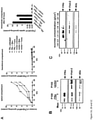
Figure 1 . FRa-mediated transport of 5MTHF in Z310 cells is stimulated by monensin and transferrin.- (A) Filter-grown Z310 cells stably transfected with FRa or vector were loaded with [3H]5MTHF for 1 hour. After three washing steps, cells were incubated with fresh RPMI medium containing either 7 µM monensin (Mon) or 0.5 mg/ml rat transferrin (Trans) or contained no supplementation. Samples from the apical and basolateral compartment were removed at the indicated time points. Subsequently, cells were lysed and radioactivity was measured in medium samples and cell lysates. Data are means ± s.e.m. of three independent experiments.
- (B) Z310 cells stably transfected with FRa or vector were treated with 7 µM monensin for 22 hours. The medium was collected and subjected to ultracentrifugation for exosome purification. The P100 pellets were subjected to western blots and probed with antibodies against FRα, Flot-2 and Alix.
- (C) To further characterize the vesicles derived from Z310 cells, the 100.000 x g pellet from monensin treated Z310 cells stably expressing FRa was subjected to sucrose gradient ultracentrifugation. FRa is mainly enriched in the same fractions that contained Flot-2 and Alix.
- (D) Apical transport of 5MTHF in primary PCPECs. Filter-grown PCPECs were loaded with 25 nM [3H]5MTHF for 24 hours. Cells were then incubated with 7 µM monensin or left untreated. Radioactivity from medium samples was measured at the indicated time points. The increase of the apical [3H]5MTHF concentration can be further enhanced by incubating the cells with monensin. Data are means ± s.e.m. of two independent experiments, each done in triplicate.
-
Figure 2 . Uptake of 5MTHF is not reduced by inhibition of organic anion transporters in Z310 cells stably expressing FRα.- Z310 cells stably expressing FRa were incubated with 12.5 nM [3H]5MTHF alone or together with TC, MTX, PAH, probenecid and digoxin for 1 hour at 37 °C. Subsequently, cells were lysed and intracellular radioactivity was measured. Only MTX at an inhibitory concentration for FRa of 10 µM significantly reduces 5MTHF uptake. Data are means ± s.e.m. of three independent experiments, each done in duplicate (***, p<0.001; Student's t-test).
- At the indicated concentrations TC inhibits transport by Oat3 and Oatp3 (Breen, C.M. et al., 2004, AM J Physiol Renal Physiol 287, F562-569), PAH inhibits transport by Oat1, Oat3 (Sweet, D.H. et al., 2002, J Biol Chem 277, 26934-26943) and p-glycoprotein (Martel, F. et al., 2006, Eur J Pharmacol 551, 19-26), Probenecid blocks Oat-mediated transport (Breen, C.M. et al., 2004, AM J Physiol Renal Physiol 287, F562-569), additionally probenecid is a known inhibitor of MRP1 (Olson, D.P. et al., 2002, AIDS 16, 1743-1747) and MRP4 (van Aubel, R.A. et al., 2002, J Am Soc Nephrol 13, 595-603). Digoxin is an inhibitor for Oatp2 (Breen, C.M. et al., 2004, AM J Physiol Renal Physiol 287, F562-569) and MTX at a concentration of 110 nM blocks RFC-mediated uptake of 5-MTHF.
-
Figure 3 . Contribution of PCFT and RFC on FRα-mediated 5MTHF uptake in Z310 cells.
Z310 cells stably transfected with FRα, PCFT and RFC either alone or in combination as indicated, were incubated with 12.5 nM [3H]5MTHF for 2 hours at 37 °C (gray bars). To block unspecific uptake, nonlabeled FA was added in an excess of 2.5 µM (black bars). Subsequently, cells were lysed and intracellular radioactivity was measured. PCFT and RFC do not contribute to FRα-mediated 5MTHF uptake in Z310 cells. Data are means ± s.e.m. of three independent experiments, each done in duplicate. -
Figure 4 . FRα containing exosomes are selectively internalized by astrocytes in vivo and in vitro.
Colocalization analysis of images from mouse brain sections reveal a colocalization of PKH26-stained FRa-positive exosomes and GFAP of 30% ± 4%. FRa negative exosomes show only a colocalization with GFAP of 4% ± 1.5%. Values represent means ± s.e.m. (***, p<0.001; Student's t-test). -
Figure 5 . Suggested model of cerebral folate transport
Plasma 5MTHF (F) is bound to FRalpha at the basolateral membrane of the choroid plexus. The 5MTHF-FRalpha complex is internalized by receptor-mediated endocytosis and translocated into GPI-anchored protein-enriched early endosomal compartments (GEEC). Multivesicular bodies (MVBs) are late endosomal compartments (LE) localized in the endocytotic route. Intralumenal vesicles of MVBs containing FRalpha are generated by inward budding of the limiting membrane. These intralumenal vesicles are released as exosomes into the CSF after fusion of the MCB with the apical membrane. Fusion of the limiting membrane of MVBs with the apical membrane explains the presence of FRalpha molecules on the apical surface of the choroid plexus cells too. FRalpha-containing exosomes circulate in the CSF, cross the ependymal cell layer and are distributed in the brain parenchyma. FRalpha-positive exosomes might initially be taken up by astrocytes and from these further delivered to neurons. FRalpha-negative exosomes hardly cross the ependymal cell layer and are targeted to other brain cells, such as microglia. - Antibodies against the following antigens were used: human FRa (mouse monoclonal, either Mov18/ZEL; Alexis Biochemicals or NCL-L-FRalpha Clone BN3.2; Novocastra), flotillin-2 (Flot-2, mouse monoclonal; BD Biosciences), transferrin (rabbit polyclonal; Dako, Denmark), ALG-2-interacting protein X (Alix, rabbit polyclonal; Covalab), glial fibrillary acidic protein (GFAP, rabbit polyclonal; Dako, Denmark), ionised calcium binding adaptor molecule 1 (iba1, rabbit polyclonal; WAKO Chemicals, Neuss, Germany), neuron specific nuclear protein (NeuN, mouse monoclonal; Chemicon, USA), neurite outgrowth inhibitor protein A (Nogo-A, mouse monoclonal; (Oertle et al., 2003 J. Neurosci 23, 5393-5406). A rabbit polyclonal antibody against human PCFT was kindly provided by I. David Goldman (Bronx, New York, USA). Secondary antibodies were purchased from Dianova (Hamburg, Germany). Folic acid conjugated to fluorescein isothiocyanate (FA-FITC) was kindly provided by Philip S. Low (West Lafayette, Indiana, USA). Phosphatidylinositol-specific phospholipase C from Bacillus cereus (PI-PLC) was from Sigma.
- The following inhibitors were used: taurocholate (TC), methotrexate (MTX), p-aminohippuric acid (PAH), probenecid, digoxin, brefeldin A (BFA) and N-ethylmaleimide (NEM). All were purchased from Sigma.
- Cloning of FRa cDNA has been described previously (Steinfeld et al., 2009: Am J Hum Genet 85, 354-363). For expression in immortalized rat choroid plexus epithelial cells Z310 FRa or PCFT cDNA was subcloned into the NheI/HindIII sites of pcDNA3.1 Hygro(+) (Invitrogen). For double mutant FRα/PCFT Z310, PCFT cDNA was subcloned into the NheI/HindIII sites of pcDNA3.1 Zeo(+) (Invitrogen). RFC cDNA was subcloned into the NheI/EcoRI sites of pEFIRES-P.
- Z310 cells were grown in DMEM supplemented with 10% fetal calf serum, 2 mM glutamine, 100 U/mL penicillin, 100 µg/mL streptomycin, 200 µg/mL G418 (Roth, Karlsruhe, Germany) and 10 ng/mL epidermal growth factor (Invitrogen).
- Transfections were done using Effectene transfection reagent (Qiagen) and stable Z310 cell clones were selected in the presence of 400 µg/mL hygromycin B (PAA Laboraories, Pasching, Austria), 250 µg/mL Zeocin, or 1 µg/mL Puromycin (Invitrogen), respectively. Stable transfectants of MDCK or HepG2 cells were selected in the presence of 800 µg/mL G418.
- Porcine choroid plexus epithelial cells (PCPECs) were prepared basically as described previously (Hakvoort et al., 1998: J Neurochem 71, 1141-1150). Isolated PCPECs were seeded in laminin-coated transwell culture plates (0.4 µm pore size, Costar, Cambridge, MA) with a diameter of 12 mm using a seeding density of 20 mg wet tissue/cm2. The cell culture medium (DMEM-Ham's F-12, Biochrom, Berlin, Germany) was supplemented with 10% fetal calf serum, 4 mM glutamine, 5 µg/ml insulin, 100 U/mL penicillin and 100 µg/mL streptomycin. Cytosine arabinoside (20 µM) was added to suppress the growth of contaminating cells. After reaching confluence at days in vitro (DIV) 8, growth medium was replaced by serum-free culture medium (SFM) to support cell differentiation and improve barrier function. At
DIV 10, the medium was replaced by folate-free RPMI medium (Invitrogen) without serum. The transport experiments were performed on cells at DIV 13-15 that exhibited high transepithelial electrical resistance (TEER) of about 1000 Ω × cm2. - Disassociated neonatal cortices from postnatal day 0-2 mice were cultured in DMEM supplemented with 10% fetal calf serum, 1% GlutaMAX (Invitrogen), 100 U/mL penicillin and 100 µg/mL streptomycin on laminin- and poly-L-lysin-coated coverslips at 37°C in 5% CO2. Medium was changed every 3-4 days. At DIV 7-14, mixed cortical cultures consisting of oligodendrocytes, cortical neurons and microglia on an astrocyte monolayer were obtained.
- Transcytosis assays were adapted from Polishchuk et al, 2004 (Polishchuk et al., 2004: Nat Cell Biol 6, 297-307). Before the experiments, cells were maintained in folate-free RPMI 1640 medium. Filter-grown Z310 cells stably expressing FRa were washed with HBS [20 mM HEPES, 140 mM NaCl, 5 mM KCI, 2 mM MgCl2, 5 mM glucose (pH 7.4)] and incubated on ice for 15 min with 100 nM FITC-labeled folic acid (FA-FITC) together with anti-human FRa antibody (Mov18/ZEL) added to the basolateral chamber of the transwell plate. Filters were washed three times with HBS (ice-cold) and some filters were fixed immediately after incubation on ice. The others were shifted to 37°C for the indicated time points, fixed and prepared for confocal microscopy. Biochemical evaluation of transcytosis was based on the idea that removal of protein from the basolateral surface will result in decrease of its amount at the apical domain of the plasma membrane. Therefore, filter-grown Z310 cells stable expressing FRa were incubated with PI-PLC added to the basolateral medium for the indicated time points to remove FRa from the basolateral surface of the cells. To detect the apical portion of FRα, cells were washed and PI-PLC was added to the apical medium 30 min on ice to harvest the receptor from the apical domain of the plasma membrane. Samples were than subjected to SDS-PAGE and western blotting with an anti-human FRa antibody (NCL-L-FRalpha).
- Exosomes were prepared from human CSF or from cell culture supernatant by performing sequential centrifugation. Therefore CSF or supernatant from Z310 cells was centrifuged for 15 min at 1500 x g to remove debris. The supernatant was then subjected to ultracentrifugation at 10.000 x g for 45 min and at 100.000 x g for 150 min. The collected pellets were finally resuspended in CellLytic™M Cell Lysis Reagent (Sigma) und subjected to SDS-PAGE and western blot analysis. For sucrose gradient analysis, a discontinuous sucrose density gradient was prepared by sequential layering of 2.0, 1.75, 1.5, 1.25, 1.0, 0.75, 0.5 and 0.25 M sucrose (w/w) in 20 mM HEPES, 2 mL each. The exosome samples were layered on top of the gradient and centrifuged for 16 hours at 100.000 x g. Subsequently, fractions were collected, diluted 1:4 in PBS and centrifuged at 100.000 x g for 70 min. Pellets were resuspended in Laemmli sample buffer and subjected to western blot analysis.
- For immunofluorescence analysis, the exosome pellet was resuspended in PBS and stained with PKH26 (Sigma). The staining reaction was stopped after 5 min with exosome-free FCS. Exosomes were then washed in PBS and pelleted by ultracentrifugation (100.000 x g, 70 min).
- For electron microscopy analysis, exosomes were prepared and the pellet was washed once in a large volume of PBS. Exosomes were then fixed with 4% paraformaldehyde and absorbed to glow discharged formvar-carbon coated copper grids. The samples were immunolabeled with anti-human FRa antibody (Mov18/ZEL) or anti-human Flotillin-2 antibody followed by a rabbit anti-mouse antibody. The antibodies were detected with Protein A coupled to 10 nm gold particles. After contrasting by neutral uranyl acetate and picking up with a wire loop from methylcellulose-uranyl acetate, samples were imaged on a Leo EM912AB-electron microscope (Zeiss) and images were acquired using on axis 2048X2048 charge-coupled device camera (Proscan, Schering).
- Immunoblot analysis was conducted using standard techniques. Briefly, the samples were denatured for 10 min at 95 °C in Laemmli sample buffer, subjected to SDS-PAGE (12% gel) and transferred to nitrocellulose membranes. The membranes were probed with a mouse monoclonal anti-human FRa antibody (NCL-L-FRalpha) and a rabbit polyclonal anti-human transferrin antibody. As antibodies against exosomal markers rabbit polyclonal antibody to Alix and mouse monoclonal Flot-2 were used. First antibodies were detected by using the appropriate secondary antibodies conjugated to horseradish peroxidise (HRP). The bands were visualized by chemiluminescence using Lumi Light Western blotting substrate (Roche Diagnostics, Mannheim, Germany).
- Confocal images were acquired on a spinning disk confocal imaging system (UltraView®, Perkin-Elmer). The system incorporated an Olympus IX81 inverted microscope using a 100X 1.4 NA lens. Excitation wavelengths and filter sets for FITC and Cy3 were as supplied by the manufacturer. Fixed cells labeled with antibodies and/or FA-FITC were optically sectioned into z-stacks. Image processing was performed using the Zeiss LSM image browser software and NIH ImageJ (http://rsbweb.nih.gov/ij; Open Source). Colocalization analysis for the internalization assay was done by using the "Colocalization Threshold" algorithm from ImageJ. Images from at least 20 slides per mouse brain were analyzed and data presented in the figure were from two independent experiments. Normal distribution of the data was analyzed with D'Agostino-Person normality test and significance was tested using Student's t-test.
- Exosomes were prepared and the pellet was washed once in a large volume of PBS. The exosomes were directly fixed on glass coverslips with 4% paraformaldehyde. Two-color STED microscopy was performed, using sheep anti-mouse polyclonal antibody labeled with Atto590 (for anti-human FRa as first antibody) and goat anti-rabbit polyclonal antibody conjugated to Atto647N (for Alix as marker for exosomes). The colocalization of FRa and Alix was analyzed by using the "Colocalization Threshold" algorithm from ImageJ.
- For 5MTHFuptake and transport measurements in Z310 cells, 4.2 * 104 cells were seeded in laminin coated transwell culture plates (0.3 cm2 growth area, 0.4 µm pore size) in folate-free RPMI medium. To test the barrier function, dye permeability assays with lucifer yellow were performed. Cells were used for experiments when the permeability was below 1% per hour. 72 hours post-confluent, cells were loaded with 12.5 nM [3H]5MTHF in pre-warmed folate-free RPMI medium containing 1% BSA added either to the apical (350 µL) or basolateral (700 µL) chamber of the transwell plates. [3',5',7,9-3H](6S)-5MTHF was purchased from Moravek Biochemicals (Brea, CA). Cells were then incubated at 37°C for 1 hour and were subsequently washed three times with folate-free RPMI medium. Fresh RPMI medium was added containing either 7 µM monensin (Sigma) or 0.5 mg/mL rat transferrin (Rockland, Gilbertsville, PA) and cells were then incubated at 37°C in 5% CO2. Samples were removed from the apical and basolateral medium at the indicated time points. To stop the transport experiment, the transwell plates were placed on ice and the cells were washed three times with ice-cold HBS. Cells were than dissolved in 0.2 M NaOH and protein determination was done with the BCA protein assay (Pierce, Rockford, IL). Radioactivity in medium samples and cell lysates was measured in a liquid scintillation spectrometer.
- Paraffin sections of human choroid plexus from post-mortem autopsy blocks were retrieved from the department of Neuropathology, University of Göttingen, Germany. Immunohistochemistry was carried out as previously described (Letiembre, M. et al., 2009, ). In brief, after deparaffinization, slides were pre-treated by heating in a microwave in citrate buffer (10 mM, pH 6.0) followed by incubation in guanidinthiocyanate (4 M) for 25 min. For staining with HRP-conjugated secondary antibody, endogenous peroxidase was blocked by incubation in 3% H2O2 for 20 min. staining on human sections was performed using mouse monoclonal FRa antibody (NCL-L-FRalpha) or rabbit polyclonal PCFT antibody followed by HRP-conjugated anti-mouse antibody. After development, the slides were counterstained with hematoxylin. For immunofluorescence, the sections were treated as described above except the use of secondary Alexa 488 donkey anti-mouse or Alexa 546 donkey anti rabbit antibodies.
- Mixed cortical cultures were obtained form neonatal mice and immediately plated on coverslips and incubated in the respective growth medium. Before the internalization assay, exosomes were purified from Z310 cells, stably transfected with FRa or vector and labelled with PKH26 and FA-FITC as described above. The uptake was performed by incubating the cell cultures with exosome dilutions for 2 hours at 37°C and 5% CO2. Cells were then fixed, permeabilized and stained with the respective antibodies for immunofluorescence analysis.
- In vivo experiments were carried out with C57BL/6 mice at the ages between 80 days and 100 days. The mice were anesthetized with ketamine (100-125 mg/kg) and xylazine (10-12,5 mg/mL) and the intraventricular cannulas were stereotaxically implanted into the lateral ventricle as described previously (Hung et al., 1984: Eur J Pharmacol 106, 645-648). 10 µL of labelled exosome solution were injected into the intraventricular cannula 4 hours before the mice were euthanized. Intact brains were then dissected, immediately frozen in liquid nitrogen pre-cooled isopentane and stored at -20°C. For fluorescence analysis, the frozen brain tissues were cryosectioned to a thickness of 10 µm. The frozen sections were fixed in methanol for 5 min at -20°C. After washing in PBS, sections were blocked for 1 hour in PBS containing 10% donkey serum (Sigma), followed by overnight incubation with primary antibodies in block buffer at 4°C. Sections were than washed in PBS, incubated with secondary Alexa 488 donkey anti-rabbit antibody and mounted using Slowfade Gold antifade reagent (Invitrogen).
- To elucidate the transcellular transport of 5MTHF across the choroid plexus we investigated the directed movements of radioactively labelled 5MTHF in Z310 cells as well as in primary porcine choroid plexus cells.
- FRa-transfected or vector-transfected Z310 cells were grown in a two chamber transwell system and [3H]5MTHF was either given into the basolateral or apical compartment. After a 1 h incubation with [3H]5MTHF the medium was replaced by fresh medium and the time-dependent emergence of [3H]5MTHF in both compartments was detected. Notably, 5MTHF was only released into the apical compartment of FRa-transfected cells but not of vector-transfected cells and the apical shedding of [3H]5MTHF was increased by the addition of either monensin (70% increase) or transferrin (36% increase) (
Fig 1A ). The monensin- or transferrin-dependent increase in apical release occurred at the expense of the intracellular [3H]5MTHF uptake. Whether Z310 cells were loaded with [3H]MTHF from the apical or basolateral site did not significantly alter the rate of apical shedding. But no significant basolateral release of [3H]5MTHF could be detected under any condition. These data suggest an exosomal type of FRα-mediated transport of 5MTHF into the apical compartment since it is known that monensin and transferrin induce exosome release from cells. The vesicular nature of the directed FRα-dependent transport was confirmed by fractionation of the cell culture supernatant. FR was enriched in the P100 (100.000 x g pellet) fraction and was co-purified with the exosomal markers Flot-2 and Alix (Fig. 1B ). The monensin-induced increase in FRa signal was proportional to the increase in Flot-2 and Alix release. Flot-2 and Alix release was also induced by monensin in vector-transfected cells in the absence of FRα. The vesicle-containing P100 fraction of FRa-transfected and monensin treated Z310 cells was subjected to sucrose gradient ultracentrifugation. Very similar to human CSF, FRa was mainly recovered from fractions possessing the characteristic density of exosomes (between 1.11 and 1.16 g/cm3) and containing the exosomal markers Flot-2 and Alix (Fig. 1C ). - In addition, the vesicles showed the typical size of exosomes and showed colocalization of FRa and Alix in two-color STED microscopy. Quantitative analysis revealed a colocalization of 63% ± 8% of FRa and Alix. These data demonstrate that exosomes derived from the supernatant of FRa-transfected Z310 cells share major characteristics with exosomes derived from human CSF. The fact that quantitative analysis of STED images achieved a higher percentage of FRa colocalization with the exosomal marker Alix can be explained by the FRa overexpression in Z310 cells.
- We also compared the protein composition of exosome preparations derived from Z310 cells with the one purified from human CSF by mass spectrometry (Table 1).Both preparations contained 22 of 25 commonly accepted exosomal markers (http://exocarta.org/exosome_markers) and showed an overall similar protein profile.
Table 1: Mass spectrometry analysis of exosomes purified from human CSF and Z310 cells human CSF Z310 vector-transfected Z310 FRa-transfected glyceraldehyde-3-phosphate dehydrogenase (GAPDH) glyceraldehyde-3-phosphate dehydrogenase (GAPDH) glyceraldehyde-3-phosphate dehydrogenase (GAPDH) ALG-2 interacting protein X (Alix) ALG-2 interacting protein X (Alix) ALG-2 interacting protein X (Alix) flotillin-1 flotillin-1 flotillin-1 flotillin-2 flotillin-2 flotillin-2 folate receptor α - folate receptor α (human) tumor suspectibilityl gene 101 (TSG101) tumor suspectibility gene 101 (TSG101) tumor suspectibility gene 101 (TSG101) CD63 CD63 CD63 transferrin receptor protein (CD71) transferrin receptor protein (CD71) transferrin receptor protein (CD71) - Exosome preparations from human CSF and Z310 cells were analyzed by mass spectrometry. Each preparation contained a couple of hundred proteins with a high probability of over 95%. Among these we could indentify FRa in the sample derived from human CSF as well as in FRα-tranfected Z310 cells. Although expression of endogenous rat FRa in Z310 cells has previously been indicated by RT-PCR (Halwachs, S. et al. 2011, Mol Pharmacol 80, 621-629) the rat FRa expression might be under the mass spectrometric detection level. Exosomal markers like ALG-2 interacting protein X (Alix), flotillin-1 (Flot-1), flotillin-2 (Flot-2), CD63, tumor suspectibility gene 101 (TSG101) and transferrin receptor (CD71) could consistently be detected in all exosome preparations indicating a similar composition of exosomes derived from Z310 cells when compared with exosomes purified from human CSF. The order in Table 1 represents semiquantitative incidence of proteins determined by the ranking of unweighted spectrum counts in the program Scaffold 3. Compared with the 25 most frequently identified proteins in exosomes (http://exocarta.org/exosome_markers) we found 22 (88%) (human CSF) and 21 (84%) (Z310 cells) identity, respectively.
- To validate our findings in Z310 cells, we studied the directional 5MTHF transport in primary porcine choroid plexus epithelial cells (PCPEC). Filter-grown PCPEC were loaded with 25 nM [3H]5MTHF for 24 hours. Subsequently, medium was changed and cells were treated with 7 µM monensin or left untreated. A significant increase of radioactivity could only be observed in the apical compartment (
Fig. 1D ). Treatment of cells with monensin further enhanced the apical accumulation of [3H]5MTHF by 33%. The rate of transport into the apical compartment corresponds to the one obtained for FRα-transfected Z310 cells. And equivalent to Z310 cells, no significant [3H]5MTHF release into the basolateral compartment could be detected. - We performed additional experiments to evaluate FRα-independent transport processes in Z310 cells at physiological MTHF concentration (12.5 nM). A couple of organic anion transporters are known to be expressed in Z310 cells (Halwachs et al., 2011). Contribution of Oat1, Oat3, Oatp2, Oatp3, Mrp1, Mrp4, p-glycoprotein (Mdr1) and RFC to 5MTHF uptake into FRa-transfected Z310 cells was assessed by the incubation of cells with the following inhibitors of these transporters: taurocholate (TC), methotrexate (MTX), p-aminohippuric acid (PAH), probenecid and digoxin. As illustrated in
Fig. 2 , none of the inhibitors reduced the uptake of [3H]5MTHF with the exception of MTX that significantly reduced 5MTHF uptake at a concentration of 10 µM. At this concentration MTX is known to block FR-mediated MTHF binding as well. - The contribution of the PCFT and the RFC to FRα-mediated 5MTHF uptake into Z310 cells was studied by stable cotransfection of FR with PCFT or RFC or a combination of both (
Fig. 3 ). Transfection of FRa increased the 5MTHF uptake 248-fold when compared to the vector-transfected control cells. Transfection of PCFT or RFC or the combination of both did not significantly alter 5MTHFuptake into Z310 cells when compared with the control cells. In case of the double transfectants FRα/PCFT and FRα/RFC or for the triple transfectant FRα/PCFT/RFC no further increase of 5MTHF uptake was observed when compared to the FRa-transfected cells (Fig. 3 ). These data indicate that at least at the physiological 5MTHF concentrations in plasma (4-20 nM) the uptake of 5MTHF into the choroid plexus is primarily mediated by FRα. - We have unexpectedly disclosed FRa as exosomal cargo in the CSF. To further understand the biological function of this transport mechanism we injected exosomes derived from FRa-transfected or vector-transfected Z310 cells into the lateral ventricle of C57BL/6 mice. Purified exosomes were labelled with the dye PKH26 before they were applied via stereotactically implanted intraventricular cannulas. 4 h post injection the mice were euthanized and their brains immediately frozen. Immunohistochemistry of the mouse brain sections revealed that FRa-positive exosomes penetrated into the brain parenchyma and colocalized with GFAP-positve astrocytes. In contrast, FRa-negative exosomes were mainly detected in the periventricular ependyma and did not colocalize with GFAP positive cells. Quantitative analysis revealed a 30% colocalization of PKH26-labelled FRa-positive exosomes with GFAP-positive cells but less than 4% colocalization in case of FRa-negative exosomes (
Fig. 4 ). To validate our in vivo findings we incubated primary mixed cortical cultures obtained from neonatal mice with either PKH26-labelled exosomes, with FA-FITC or a combination of both. After incubating the cells for 2 hours at 37 °C, cells were fixed and analyzed by immunofluorescence microscopy. FRa-positive exosomes were selectively taken up by GFAP-postive astrocytes and no significant colocalization was obtained with cells stained with markers for microglia, neurons and oligodendrocytes. FRa-negative exosomes did not colocalized with astrocytes but were internalized by Iba1-positive microglia. FA-FITC was also delivered to astrocytes when combined with FRa-positive exosomes. However, none of the cell types internalized free FA-FITC. - Several characteristics of FRa argue for its pivotal role for the 5MTHF transport across the blood-CSF barrier. Within the brain FRa is almost selectively expressed in the choroid plexus and its high binding affinity for folates (Kb 1-10 nM) perfectly matches the physiological 5MTHF concentration in human plasma (4-20 nM). Further, a hereditary FRa defect is associated with extremely low 5MTHF concentrations in the CSF (equal or below 5 nM) and causes a childhood onset neurodegenerative disease (Steinfeld et al., 2009: Am J Hum Genet 85, 354-363; Grapp et al., 2012:Brain 135, 2022-2031). Our experiments with polarized cells (Z310 cells) demonstrate a basolateral to apical sorting of FRa that provides for receptor-mediated endocytosis and transcytosis of folates. It is known that internalized GPI-anchored proteins such as FRa enter the endosomal compartment by a clathrin-independent pathway to form tubular early endocytic compartments called GPI-anchored protein enriched endosomal compartments (GEECs). During the subsequent endosomal maturation process multivesicular bodies are formed that contain intralumenal vesicles. Multivesicular bodies are known to be sites for intracellular sorting of proteins between recycling, degradation and secretion pathways. We have provided several lines of evidence that in choroid plexus cells FRα-containing multivesicular bodies fuse with the apical membrane and secrete their intralumenal vesicles as exosomes (
Fig. 5 ). We further suggest that FRa recycles between the apical membrane and the late endosomal compartment and thus offer 5MTHF uptake from the CSF as well. And we speculate that the third key folate transporter the PCFT may be responsible for the cytoplasmic 5MTHF transport out of acidified endosomal compartments. The lacking alterations in folate transport after transfection of the PCFT and the RFC or after addition of various inhibitors of OATs support the unique role of FRa during the transcellular 5MTHF transport in Z310 choroid plexus cells. These results are consistent with other studies on primary rat choroids plexus epithelial cells that showed two separate folate uptake mechanisms (Wollack et. Al., J. Neurochem, 2008, 104(6), 1494-1503). One FRα-dependent process mediates efficient uptake of 5MTHF at low extracellular folate concentrations (Km ~ 9.5 nM). The other way of internalization is independent of the FRa but likely depends on OAT such as the RFC and accounts for folate uptake at higher concentrations (Km ~ 766 nM). It is likely that any unbound 5MTHF released into the CSF will be picked up by membrane-bound FRa to recycle within the choroid plexus cells and will ultimately be secreted into CSF while bound to exosome-attached FRα. This unusual transport mechanism might serve as a stabilizing and buffering system for fluctuations in folate supply and demand of the brain. Most excitingly, our results indicate that the FRα-exosome transport vehicle is crucial for 5MTHF targeting to astrocytes. Interestingly, exosomes from both human CSF as well as Z310 cells contained several enzymes of folate metabolism, notable C-1-tetrahydrofolate synthase, 10-formyltetrahydrofolate dehydrogenase, and dihydrofolate reductase (Table 1). This implies that 5MTHF carrying exosome additionally provide enzymes essential for folate metabolism to their target cells. - Our studies disclosed the mechanism of cerebral folate transport deficiency and extended this entity to other disorders, such as Kearns-Sayre syndrome that affect the choroid plexus. The functional loss of PCFT may also compromise the choroid plexus function providing an explanation why patients with hereditary malabsorption show low 5MTHF concentration in the CSF even if they are supplemented with oral folate.
- The unexpected role of exosomes as transport medium for micronutritions and the impact of FRa expression on exosomal targeting stimulate further therapeutic applications for exosomes.
-
- <110> Georg-August-Universitaet Goettingen, Stiftung Oeffentlichen Rechts, Universitaetsmedizin
- <120> Composition and Delivery Vehicle for Active Agents and Methods therefor
- <130> 3294-059 PCT
- <150>
US13/650,193
<151> 2012-10-12 - <160> 2
- <170> PatentIn version 3.5
- <210> 1
<211> 257
<212> PRT
<213> homo sapiens - <400> 1

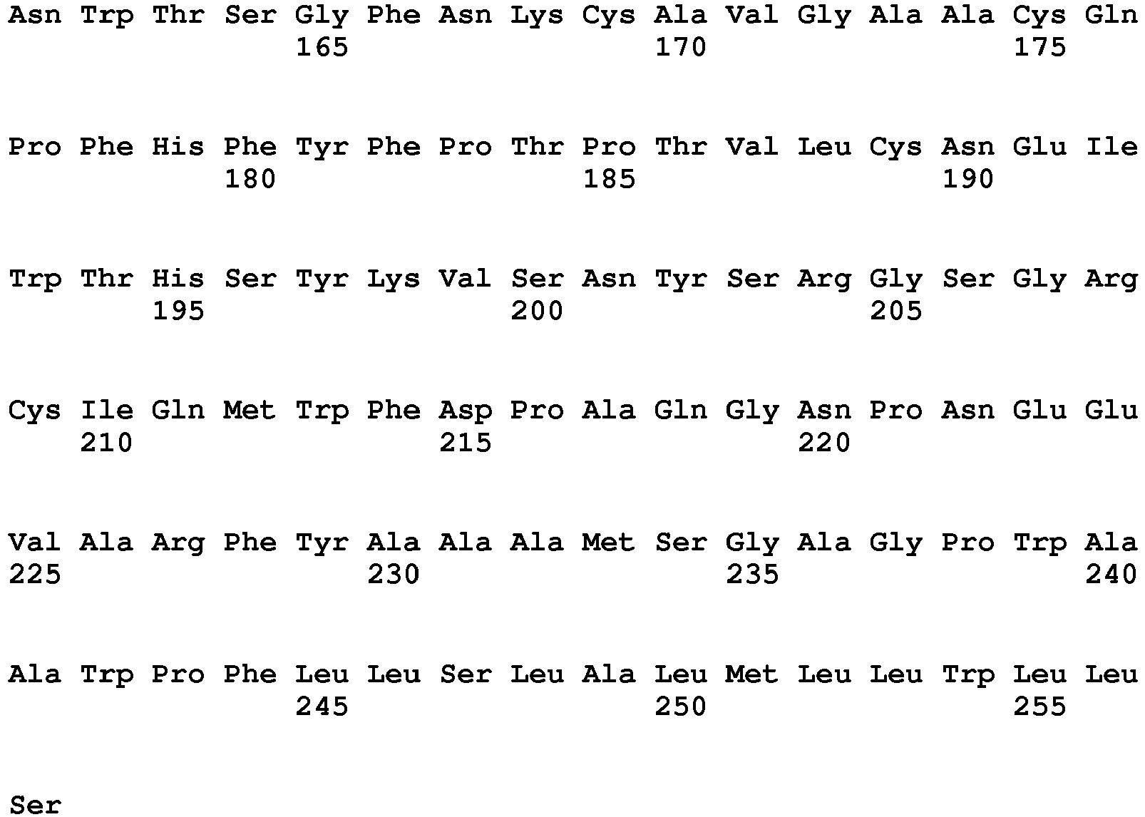
- <210> 2
<211> 1147
<212> DNA
<213> homo sapiens - <400> 2


Claims (18)
- Vehicle composition, comprising- vesicle forming components or particles;- an active agent associated with the vesicle forming components or particles, and- folate receptor alpha, for the targeting of vesicles into the CSF, the brain, the central nervous system and the spinal cord.
- The vehicle composition for use according to claim 1 wherein the vesicle forming components are lipids, in particular, vesicles forming membranes.
- The vehicle composition for use according to claim 1 or 2 wherein the vesicle formed by the vesicle forming components are exosomes, liposomes, nanostructured vesicles, micelles, erythrocyte ghosts, organic particle, inorganic particle, colloidal particle, nanoparticle, composite particle.
- The vehicle composition for use according to any one of claims 1 to 3 wherein the folate receptor alpha is located on the outer surface of the vesicle or particle.
- The vehicle composition for use according to any one of claims 1 to 4 wherein the folate receptor alpha is human folate receptor alpha, in particular, of SEQ ID No. 1.
- The vehicle composition according to any one of the preceding claims wherein active agents are drugs, in particular, an agent for treating or preventing neurological or neurodegenerative diseases, disorders or conditions or the active agents are marker molecules, magnetic agents or radioactive agents.
- The vehicle composition according to claim 1 for use in treating cancer.
- The vehicle composition according to any one of the preceding claims being a vesicular composition or particular composition.
- The vehicle composition according to any one of the proceeding claims for use for delivery of said active agent into astrocytes and/or neurons.
- A delivery system for use in the transport of components into the cerebrospinal fluid (CSF) and/or brain and/or central nervous system and/or spinal cord comprising vesicles or particles and folate receptor alpha located on the outer surface of said vesicles or particles, in particular, a vesicular composition, comprising- vesicle forming components;- an active agent associated with the vesicle forming components, and- folate receptor alpha .
- The delivery system according to claim 10 for use in treating or preventing neurological or neurodegenerative diseases, disorders or conditions or treating cancer by using an active ingredient.
- The delivery system according to any one of claims 10 1 for use for the transport of diagnostics into the CFS and/or brain and/or central nervous system and/or spinal cord comprising vesicles or particles and folate receptor alpha located on the outer surface of said vesicles or particles.
- The delivery system according to any one of claims 10 to 12 wherein the folate receptor alpha is a human folate receptor alpha, in particular, of SEQ ID No. 1.
- Vesicles or particles and folate receptor alpha located on the outer surface of said vesicles or particles as defined in claim 1 for the delivery of components, in particular, active agents into the CSF and/or brain and/or central nervous system and/or spinal cord by administering a delivery system comprising vesicles or particles, folate receptor alpha on the outer surface of said vesicles or particles, and said components associated with the vesicles or particles into the blood or CSF for use in the treatment of CSF or brain or CNS pertaining diseases, disorders or conditions.
- Vesicles or particles and folate receptor alpha located on the outer surface of said vesicles or particles as defined in claim 1 for the delivery of components, in particular, active agents into the CSF and/or brain and/or central nervous system and/or spinal cord by administering a delivery system comprising vesicles or particles, folate receptor alpha on the outer surface of said vesicles or particles, and said components associated with the vesicles or particles into the blood or CSF for use in the diagnosis, wherein the active agents are marker molecules, magnetic agents or radioactive agents.
- A delivery system for the transport of components into the CSF and/or brain and/or central nervous system and/or spinal cord comprising vesicles or particles, an active agent associated with the vesicle forming components or the particles, and folate receptor alpha located on the outer surface of said vesicles or particles, or a vesicular composition or particular 15 composition comprising vesicle forming components or particles; an active agent associated with the vesicle forming components or the particles, and folate receptor alpha to a subject in need thereof, for use in preventing or treating neurological or neurodegenerative diseases, disorders or conditions.
- Pharmaceutical composition or diagnostic composition containing a vehicle composition according to any one of claims 1 to 8 or a delivery system according to any one of claims 10 to 13 and a pharmaceutically acceptable carrier, diluent or excipient.
- An in vitro method for diagnosing folate deficiency in a subject comprising the step of obtaining CSF from said subject and determining the level or amount of folate receptor alpha or free folate receptor binding sites on exosomes present in the CSF.
Applications Claiming Priority (2)
| Application Number | Priority Date | Filing Date | Title |
|---|---|---|---|
| US13/650,193 US9950074B2 (en) | 2012-10-12 | 2012-10-12 | Composition and delivery vehicle for active agents and methods therefor |
| PCT/EP2013/071178 WO2014057043A1 (en) | 2012-10-12 | 2013-10-10 | Composition and delivery vehicle for active agents and methods therefor |
Publications (3)
| Publication Number | Publication Date |
|---|---|
| EP2908800A1 EP2908800A1 (en) | 2015-08-26 |
| EP2908800B1 true EP2908800B1 (en) | 2022-02-16 |
| EP2908800B8 EP2908800B8 (en) | 2022-04-20 |
Family
ID=49382404
Family Applications (1)
| Application Number | Title | Priority Date | Filing Date |
|---|---|---|---|
| EP13777258.8A Active EP2908800B8 (en) | 2012-10-12 | 2013-10-10 | Composition and delivery vehicle for active agents and methods therefor |
Country Status (3)
| Country | Link |
|---|---|
| US (1) | US9950074B2 (en) |
| EP (1) | EP2908800B8 (en) |
| WO (1) | WO2014057043A1 (en) |
Families Citing this family (1)
| Publication number | Priority date | Publication date | Assignee | Title |
|---|---|---|---|---|
| WO2021198954A1 (en) | 2020-03-31 | 2021-10-07 | Universidade Do Minho | Exosome-enriched extracellualr vesicles isolation, method and uses thereof |
Family Cites Families (6)
| Publication number | Priority date | Publication date | Assignee | Title |
|---|---|---|---|---|
| WO2000035422A2 (en) * | 1998-12-18 | 2000-06-22 | Hadasit Medical Research Services & Development Ltd. | Method of administering a compound to multi-drug resistant cells |
| KR100818462B1 (en) * | 2006-09-09 | 2008-04-01 | 재단법인서울대학교산학협력재단 | Functional silica nanoparticles combined with polyethylene glycol and preparation method thereof |
| WO2010143193A1 (en) | 2009-06-11 | 2010-12-16 | Yissum Research Development Company Of The Hebrew University Of Jerusalem, Ltd. | Targeted liposomes comprising n-containing bisphosphonates and uses thereof |
| AU2010268367B2 (en) | 2009-07-02 | 2016-10-20 | Ith Immune Therapy Holdings Ab | Exosome based treatment of cancer |
| US20120315324A1 (en) | 2010-02-05 | 2012-12-13 | University Of Louisville Research Foundation, Inc. | Exosomal compositions and methods for the treatment of disease |
| EP2425817A1 (en) | 2010-08-09 | 2012-03-07 | Commissariat à l'Énergie Atomique et aux Énergies Alternatives | Polymerized micelles for diagnosis |
-
2012
- 2012-10-12 US US13/650,193 patent/US9950074B2/en active Active
-
2013
- 2013-10-10 WO PCT/EP2013/071178 patent/WO2014057043A1/en not_active Ceased
- 2013-10-10 EP EP13777258.8A patent/EP2908800B8/en active Active
Also Published As
| Publication number | Publication date |
|---|---|
| US9950074B2 (en) | 2018-04-24 |
| EP2908800B8 (en) | 2022-04-20 |
| US20140105817A1 (en) | 2014-04-17 |
| WO2014057043A1 (en) | 2014-04-17 |
| EP2908800A1 (en) | 2015-08-26 |
Similar Documents
| Publication | Publication Date | Title |
|---|---|---|
| Grapp et al. | Choroid plexus transcytosis and exosome shuttling deliver folate into brain parenchyma | |
| Iyaswamy et al. | Fe65-engineered neuronal exosomes encapsulating corynoxine-B ameliorate cognition and pathology of Alzheimer’s disease | |
| Picone et al. | Synaptosomes: new vesicles for neuronal mitochondrial transplantation | |
| Luo et al. | A self-destructive nanosweeper that captures and clears amyloid β-peptides | |
| Delenclos et al. | Investigation of endocytic pathways for the internalization of exosome-associated oligomeric alpha-synuclein | |
| Leszek et al. | Nanotechnology for Alzheimer disease | |
| Huang et al. | Nanoreceptors promote mutant p53 protein degradation by mimicking selective autophagy receptors | |
| Muzio et al. | Retromer stabilization results in neuroprotection in a model of Amyotrophic Lateral Sclerosis | |
| Urwin et al. | Disruption of endocytic trafficking in frontotemporal dementia with CHMP2B mutations | |
| Li et al. | Autophagy is involved in oligodendroglial precursor-mediated clearance of amyloid peptide | |
| Formicola et al. | Nanomedicine for the treatment of Alzheimer's disease | |
| Zhang et al. | Aβ42 and ROS dual-targeted multifunctional nanocomposite for combination therapy of Alzheimer’s disease | |
| Sengupta et al. | Polymorphic α-synuclein strains modified by dopamine and docosahexaenoic acid interact differentially with tau protein | |
| US20150064115A1 (en) | Neurophilic nanoparticles | |
| Del Grosso et al. | Dysregulated autophagy as a new aspect of the molecular pathogenesis of Krabbe disease | |
| Kumar et al. | Correlation between exosomes and neuro-inflammation in various brain disorders | |
| Lee et al. | A guanidine-appended scyllo-inositol derivative AAD-66 enhances brain delivery and ameliorates Alzheimer’s phenotypes | |
| Morfill et al. | Nanostars carrying multifunctional neurotrophic dendrimers protect neurons in preclinical in vitro models of neurodegenerative disorders | |
| AU2011340796A1 (en) | Multifunctional nanoparticles | |
| Ren et al. | Flotillin-1 mediates PrPC endocytosis in the cultured cells during Cu2+ stimulation through molecular interaction | |
| Zhang et al. | Selective neuronal targeting, protection and signaling network analysis via dopamine-mediated mesoporous silica nanoparticles | |
| Ghosh et al. | Exosome: the “off-the-shelf” cellular nanocomponent as a potential pathogenic agent, a disease biomarker, and neurotherapeutics | |
| Sun et al. | A dual target molecular magnetic resonance imaging probe for noninvasive profiling of pathologic alpha-synuclein and microgliosis in a mouse model of Parkinson’s disease | |
| Wu et al. | Programmable cellular retention of nanoparticles by replacing the synergistic anion of transferrin | |
| Jiang et al. | Nanoparticles with affinity for α-Synuclein sequester α-Synuclein to form toxic aggregates in neurons with endolysosomal impairment |
Legal Events
| Date | Code | Title | Description |
|---|---|---|---|
| PUAI | Public reference made under article 153(3) epc to a published international application that has entered the european phase |
Free format text: ORIGINAL CODE: 0009012 |
|
| 17P | Request for examination filed |
Effective date: 20150511 |
|
| AK | Designated contracting states |
Kind code of ref document: A1 Designated state(s): AL AT BE BG CH CY CZ DE DK EE ES FI FR GB GR HR HU IE IS IT LI LT LU LV MC MK MT NL NO PL PT RO RS SE SI SK SM TR |
|
| AX | Request for extension of the european patent |
Extension state: BA ME |
|
| DAX | Request for extension of the european patent (deleted) | ||
| STAA | Information on the status of an ep patent application or granted ep patent |
Free format text: STATUS: EXAMINATION IS IN PROGRESS |
|
| 17Q | First examination report despatched |
Effective date: 20161206 |
|
| RIC1 | Information provided on ipc code assigned before grant |
Ipc: A61K 47/69 20170101ALI20210519BHEP Ipc: A61K 47/64 20170101ALI20210519BHEP Ipc: A61K 9/00 20060101AFI20210519BHEP |
|
| GRAP | Despatch of communication of intention to grant a patent |
Free format text: ORIGINAL CODE: EPIDOSNIGR1 |
|
| STAA | Information on the status of an ep patent application or granted ep patent |
Free format text: STATUS: GRANT OF PATENT IS INTENDED |
|
| INTG | Intention to grant announced |
Effective date: 20210830 |
|
| GRAS | Grant fee paid |
Free format text: ORIGINAL CODE: EPIDOSNIGR3 |
|
| GRAA | (expected) grant |
Free format text: ORIGINAL CODE: 0009210 |
|
| STAA | Information on the status of an ep patent application or granted ep patent |
Free format text: STATUS: THE PATENT HAS BEEN GRANTED |
|
| AK | Designated contracting states |
Kind code of ref document: B1 Designated state(s): AL AT BE BG CH CY CZ DE DK EE ES FI FR GB GR HR HU IE IS IT LI LT LU LV MC MK MT NL NO PL PT RO RS SE SI SK SM TR |
|
| REG | Reference to a national code |
Ref country code: GB Ref legal event code: FG4D |
|
| REG | Reference to a national code |
Ref country code: CH Ref legal event code: EP |
|
| REG | Reference to a national code |
Ref country code: DE Ref legal event code: R096 Ref document number: 602013080910 Country of ref document: DE |
|
| REG | Reference to a national code |
Ref country code: AT Ref legal event code: REF Ref document number: 1468455 Country of ref document: AT Kind code of ref document: T Effective date: 20220315 |
|
| REG | Reference to a national code |
Ref country code: IE Ref legal event code: FG4D |
|
| REG | Reference to a national code |
Ref country code: DE Ref legal event code: R081 Ref document number: 602013080910 Country of ref document: DE Owner name: STEINFELD, ROBERT, CH Free format text: FORMER OWNERS: ALLORPHAN UG, 37075 GOETTINGEN, DE; GEORG-AUGUST-UNIVERSITAET GOETTINGEN STIFTUNG OEFFENTLICHEN RECHTS UNIVERSITAETSMEDIZIN, 37075 GOETTINGEN, DE Ref country code: DE Ref legal event code: R081 Ref document number: 602013080910 Country of ref document: DE Owner name: ALLORPHAN UG, DE Free format text: FORMER OWNERS: ALLORPHAN UG, 37075 GOETTINGEN, DE; GEORG-AUGUST-UNIVERSITAET GOETTINGEN STIFTUNG OEFFENTLICHEN RECHTS UNIVERSITAETSMEDIZIN, 37075 GOETTINGEN, DE |
|
| REG | Reference to a national code |
Ref country code: CH Ref legal event code: PK Free format text: BERICHTIGUNG B8 |
|
| RAP2 | Party data changed (patent owner data changed or rights of a patent transferred) |
Owner name: ALLORPHAN UG Owner name: STEINFELD, ROBERT |
|
| REG | Reference to a national code |
Ref country code: LT Ref legal event code: MG9D |
|
| REG | Reference to a national code |
Ref country code: NL Ref legal event code: MP Effective date: 20220216 |
|
| REG | Reference to a national code |
Ref country code: AT Ref legal event code: MK05 Ref document number: 1468455 Country of ref document: AT Kind code of ref document: T Effective date: 20220216 |
|
| PG25 | Lapsed in a contracting state [announced via postgrant information from national office to epo] |
Ref country code: SE Free format text: LAPSE BECAUSE OF FAILURE TO SUBMIT A TRANSLATION OF THE DESCRIPTION OR TO PAY THE FEE WITHIN THE PRESCRIBED TIME-LIMIT Effective date: 20220216 Ref country code: RS Free format text: LAPSE BECAUSE OF FAILURE TO SUBMIT A TRANSLATION OF THE DESCRIPTION OR TO PAY THE FEE WITHIN THE PRESCRIBED TIME-LIMIT Effective date: 20220216 Ref country code: PT Free format text: LAPSE BECAUSE OF FAILURE TO SUBMIT A TRANSLATION OF THE DESCRIPTION OR TO PAY THE FEE WITHIN THE PRESCRIBED TIME-LIMIT Effective date: 20220616 Ref country code: NO Free format text: LAPSE BECAUSE OF FAILURE TO SUBMIT A TRANSLATION OF THE DESCRIPTION OR TO PAY THE FEE WITHIN THE PRESCRIBED TIME-LIMIT Effective date: 20220516 Ref country code: NL Free format text: LAPSE BECAUSE OF FAILURE TO SUBMIT A TRANSLATION OF THE DESCRIPTION OR TO PAY THE FEE WITHIN THE PRESCRIBED TIME-LIMIT Effective date: 20220216 Ref country code: LT Free format text: LAPSE BECAUSE OF FAILURE TO SUBMIT A TRANSLATION OF THE DESCRIPTION OR TO PAY THE FEE WITHIN THE PRESCRIBED TIME-LIMIT Effective date: 20220216 Ref country code: HR Free format text: LAPSE BECAUSE OF FAILURE TO SUBMIT A TRANSLATION OF THE DESCRIPTION OR TO PAY THE FEE WITHIN THE PRESCRIBED TIME-LIMIT Effective date: 20220216 Ref country code: ES Free format text: LAPSE BECAUSE OF FAILURE TO SUBMIT A TRANSLATION OF THE DESCRIPTION OR TO PAY THE FEE WITHIN THE PRESCRIBED TIME-LIMIT Effective date: 20220216 Ref country code: BG Free format text: LAPSE BECAUSE OF FAILURE TO SUBMIT A TRANSLATION OF THE DESCRIPTION OR TO PAY THE FEE WITHIN THE PRESCRIBED TIME-LIMIT Effective date: 20220516 |
|
| PG25 | Lapsed in a contracting state [announced via postgrant information from national office to epo] |
Ref country code: PL Free format text: LAPSE BECAUSE OF FAILURE TO SUBMIT A TRANSLATION OF THE DESCRIPTION OR TO PAY THE FEE WITHIN THE PRESCRIBED TIME-LIMIT Effective date: 20220216 Ref country code: LV Free format text: LAPSE BECAUSE OF FAILURE TO SUBMIT A TRANSLATION OF THE DESCRIPTION OR TO PAY THE FEE WITHIN THE PRESCRIBED TIME-LIMIT Effective date: 20220216 Ref country code: GR Free format text: LAPSE BECAUSE OF FAILURE TO SUBMIT A TRANSLATION OF THE DESCRIPTION OR TO PAY THE FEE WITHIN THE PRESCRIBED TIME-LIMIT Effective date: 20220517 Ref country code: FI Free format text: LAPSE BECAUSE OF FAILURE TO SUBMIT A TRANSLATION OF THE DESCRIPTION OR TO PAY THE FEE WITHIN THE PRESCRIBED TIME-LIMIT Effective date: 20220216 Ref country code: AT Free format text: LAPSE BECAUSE OF FAILURE TO SUBMIT A TRANSLATION OF THE DESCRIPTION OR TO PAY THE FEE WITHIN THE PRESCRIBED TIME-LIMIT Effective date: 20220216 |
|
| PG25 | Lapsed in a contracting state [announced via postgrant information from national office to epo] |
Ref country code: IS Free format text: LAPSE BECAUSE OF FAILURE TO SUBMIT A TRANSLATION OF THE DESCRIPTION OR TO PAY THE FEE WITHIN THE PRESCRIBED TIME-LIMIT Effective date: 20220616 |
|
| PG25 | Lapsed in a contracting state [announced via postgrant information from national office to epo] |
Ref country code: SM Free format text: LAPSE BECAUSE OF FAILURE TO SUBMIT A TRANSLATION OF THE DESCRIPTION OR TO PAY THE FEE WITHIN THE PRESCRIBED TIME-LIMIT Effective date: 20220216 Ref country code: SK Free format text: LAPSE BECAUSE OF FAILURE TO SUBMIT A TRANSLATION OF THE DESCRIPTION OR TO PAY THE FEE WITHIN THE PRESCRIBED TIME-LIMIT Effective date: 20220216 Ref country code: RO Free format text: LAPSE BECAUSE OF FAILURE TO SUBMIT A TRANSLATION OF THE DESCRIPTION OR TO PAY THE FEE WITHIN THE PRESCRIBED TIME-LIMIT Effective date: 20220216 Ref country code: EE Free format text: LAPSE BECAUSE OF FAILURE TO SUBMIT A TRANSLATION OF THE DESCRIPTION OR TO PAY THE FEE WITHIN THE PRESCRIBED TIME-LIMIT Effective date: 20220216 Ref country code: DK Free format text: LAPSE BECAUSE OF FAILURE TO SUBMIT A TRANSLATION OF THE DESCRIPTION OR TO PAY THE FEE WITHIN THE PRESCRIBED TIME-LIMIT Effective date: 20220216 Ref country code: CZ Free format text: LAPSE BECAUSE OF FAILURE TO SUBMIT A TRANSLATION OF THE DESCRIPTION OR TO PAY THE FEE WITHIN THE PRESCRIBED TIME-LIMIT Effective date: 20220216 |
|
| REG | Reference to a national code |
Ref country code: DE Ref legal event code: R097 Ref document number: 602013080910 Country of ref document: DE |
|
| PG25 | Lapsed in a contracting state [announced via postgrant information from national office to epo] |
Ref country code: AL Free format text: LAPSE BECAUSE OF FAILURE TO SUBMIT A TRANSLATION OF THE DESCRIPTION OR TO PAY THE FEE WITHIN THE PRESCRIBED TIME-LIMIT Effective date: 20220216 |
|
| PLBE | No opposition filed within time limit |
Free format text: ORIGINAL CODE: 0009261 |
|
| STAA | Information on the status of an ep patent application or granted ep patent |
Free format text: STATUS: NO OPPOSITION FILED WITHIN TIME LIMIT |
|
| 26N | No opposition filed |
Effective date: 20221117 |
|
| PG25 | Lapsed in a contracting state [announced via postgrant information from national office to epo] |
Ref country code: SI Free format text: LAPSE BECAUSE OF FAILURE TO SUBMIT A TRANSLATION OF THE DESCRIPTION OR TO PAY THE FEE WITHIN THE PRESCRIBED TIME-LIMIT Effective date: 20220216 |
|
| REG | Reference to a national code |
Ref country code: DE Ref legal event code: R082 Ref document number: 602013080910 Country of ref document: DE Representative=s name: MEISSNER BOLTE PATENTANWAELTE RECHTSANWAELTE P, DE |
|
| PG25 | Lapsed in a contracting state [announced via postgrant information from national office to epo] |
Ref country code: MC Free format text: LAPSE BECAUSE OF FAILURE TO SUBMIT A TRANSLATION OF THE DESCRIPTION OR TO PAY THE FEE WITHIN THE PRESCRIBED TIME-LIMIT Effective date: 20220216 |
|
| P01 | Opt-out of the competence of the unified patent court (upc) registered |
Effective date: 20230516 |
|
| REG | Reference to a national code |
Ref country code: BE Ref legal event code: MM Effective date: 20221031 |
|
| PG25 | Lapsed in a contracting state [announced via postgrant information from national office to epo] |
Ref country code: LU Free format text: LAPSE BECAUSE OF NON-PAYMENT OF DUE FEES Effective date: 20221010 |
|
| PG25 | Lapsed in a contracting state [announced via postgrant information from national office to epo] |
Ref country code: IT Free format text: LAPSE BECAUSE OF FAILURE TO SUBMIT A TRANSLATION OF THE DESCRIPTION OR TO PAY THE FEE WITHIN THE PRESCRIBED TIME-LIMIT Effective date: 20220216 |
|
| PG25 | Lapsed in a contracting state [announced via postgrant information from national office to epo] |
Ref country code: BE Free format text: LAPSE BECAUSE OF NON-PAYMENT OF DUE FEES Effective date: 20221031 |
|
| PG25 | Lapsed in a contracting state [announced via postgrant information from national office to epo] |
Ref country code: IE Free format text: LAPSE BECAUSE OF NON-PAYMENT OF DUE FEES Effective date: 20221010 |
|
| PG25 | Lapsed in a contracting state [announced via postgrant information from national office to epo] |
Ref country code: HU Free format text: LAPSE BECAUSE OF FAILURE TO SUBMIT A TRANSLATION OF THE DESCRIPTION OR TO PAY THE FEE WITHIN THE PRESCRIBED TIME-LIMIT; INVALID AB INITIO Effective date: 20131010 |
|
| PG25 | Lapsed in a contracting state [announced via postgrant information from national office to epo] |
Ref country code: CY Free format text: LAPSE BECAUSE OF FAILURE TO SUBMIT A TRANSLATION OF THE DESCRIPTION OR TO PAY THE FEE WITHIN THE PRESCRIBED TIME-LIMIT Effective date: 20220216 |
|
| PG25 | Lapsed in a contracting state [announced via postgrant information from national office to epo] |
Ref country code: MK Free format text: LAPSE BECAUSE OF FAILURE TO SUBMIT A TRANSLATION OF THE DESCRIPTION OR TO PAY THE FEE WITHIN THE PRESCRIBED TIME-LIMIT Effective date: 20220216 |
|
| PG25 | Lapsed in a contracting state [announced via postgrant information from national office to epo] |
Ref country code: TR Free format text: LAPSE BECAUSE OF FAILURE TO SUBMIT A TRANSLATION OF THE DESCRIPTION OR TO PAY THE FEE WITHIN THE PRESCRIBED TIME-LIMIT Effective date: 20220216 |
|
| PG25 | Lapsed in a contracting state [announced via postgrant information from national office to epo] |
Ref country code: MT Free format text: LAPSE BECAUSE OF FAILURE TO SUBMIT A TRANSLATION OF THE DESCRIPTION OR TO PAY THE FEE WITHIN THE PRESCRIBED TIME-LIMIT Effective date: 20220216 |
|
| REG | Reference to a national code |
Ref country code: CH Ref legal event code: U11 Free format text: ST27 STATUS EVENT CODE: U-0-0-U10-U11 (AS PROVIDED BY THE NATIONAL OFFICE) Effective date: 20251101 |
|
| PGFP | Annual fee paid to national office [announced via postgrant information from national office to epo] |
Ref country code: DE Payment date: 20251029 Year of fee payment: 13 |
|
| PGFP | Annual fee paid to national office [announced via postgrant information from national office to epo] |
Ref country code: GB Payment date: 20251024 Year of fee payment: 13 |
|
| PGFP | Annual fee paid to national office [announced via postgrant information from national office to epo] |
Ref country code: FR Payment date: 20251024 Year of fee payment: 13 |
|
| PGFP | Annual fee paid to national office [announced via postgrant information from national office to epo] |
Ref country code: CH Payment date: 20251101 Year of fee payment: 13 |