EP2908761B1 - Monitoring of hearing preservation during cochlear implant surgery using stapedial reflex measurement - Google Patents
Monitoring of hearing preservation during cochlear implant surgery using stapedial reflex measurement Download PDFInfo
- Publication number
- EP2908761B1 EP2908761B1 EP13847628.8A EP13847628A EP2908761B1 EP 2908761 B1 EP2908761 B1 EP 2908761B1 EP 13847628 A EP13847628 A EP 13847628A EP 2908761 B1 EP2908761 B1 EP 2908761B1
- Authority
- EP
- European Patent Office
- Prior art keywords
- response
- stapedial
- surgical
- reflex response
- intraoperative
- Prior art date
- Legal status (The legal status is an assumption and is not a legal conclusion. Google has not performed a legal analysis and makes no representation as to the accuracy of the status listed.)
- Active
Links
Images

Classifications
-
- A—HUMAN NECESSITIES
- A61—MEDICAL OR VETERINARY SCIENCE; HYGIENE
- A61B—DIAGNOSIS; SURGERY; IDENTIFICATION
- A61B5/00—Measuring for diagnostic purposes; Identification of persons
- A61B5/12—Audiometering
- A61B5/121—Audiometering evaluating hearing capacity
- A61B5/125—Audiometering evaluating hearing capacity objective methods
-
- A—HUMAN NECESSITIES
- A61—MEDICAL OR VETERINARY SCIENCE; HYGIENE
- A61N—ELECTROTHERAPY; MAGNETOTHERAPY; RADIATION THERAPY; ULTRASOUND THERAPY
- A61N1/00—Electrotherapy; Circuits therefor
- A61N1/18—Applying electric currents by contact electrodes
- A61N1/32—Applying electric currents by contact electrodes alternating or intermittent currents
- A61N1/36—Applying electric currents by contact electrodes alternating or intermittent currents for stimulation
- A61N1/36036—Applying electric currents by contact electrodes alternating or intermittent currents for stimulation of the outer, middle or inner ear
- A61N1/36038—Cochlear stimulation
- A61N1/36039—Cochlear stimulation fitting procedures
-
- A—HUMAN NECESSITIES
- A61—MEDICAL OR VETERINARY SCIENCE; HYGIENE
- A61B—DIAGNOSIS; SURGERY; IDENTIFICATION
- A61B5/00—Measuring for diagnostic purposes; Identification of persons
- A61B5/02—Detecting, measuring or recording for evaluating the cardiovascular system, e.g. pulse, heart rate, blood pressure or blood flow
- A61B5/026—Measuring blood flow
- A61B5/0265—Measuring blood flow using electromagnetic means, e.g. electromagnetic flowmeter
-
- A—HUMAN NECESSITIES
- A61—MEDICAL OR VETERINARY SCIENCE; HYGIENE
- A61B—DIAGNOSIS; SURGERY; IDENTIFICATION
- A61B5/00—Measuring for diagnostic purposes; Identification of persons
- A61B5/145—Measuring characteristics of blood in vivo, e.g. gas concentration or pH-value ; Measuring characteristics of body fluids or tissues, e.g. interstitial fluid or cerebral tissue
- A61B5/14542—Measuring characteristics of blood in vivo, e.g. gas concentration or pH-value ; Measuring characteristics of body fluids or tissues, e.g. interstitial fluid or cerebral tissue for measuring blood gases
-
- A—HUMAN NECESSITIES
- A61—MEDICAL OR VETERINARY SCIENCE; HYGIENE
- A61N—ELECTROTHERAPY; MAGNETOTHERAPY; RADIATION THERAPY; ULTRASOUND THERAPY
- A61N1/00—Electrotherapy; Circuits therefor
- A61N1/02—Details
- A61N1/04—Electrodes
- A61N1/05—Electrodes for implantation or insertion into the body, e.g. heart electrode
- A61N1/0526—Head electrodes
- A61N1/0541—Cochlear electrodes
Definitions
- the present invention relates to surgical techniques for cochlear implant systems.
- a normal ear transmits sounds as shown in Figure 1 through the outer ear 101 to the tympanic membrane (eardrum) 102, which moves the bones of the middle ear 103, which in turn vibrate the oval window and round window openings of the cochlea 104.
- the cochlea 104 is a long narrow duct wound spirally about its axis for approximately two and a half turns.
- the cochlea 104 includes an upper channel known as the scala vestibuli and a lower channel known as the scala tympani, which are connected by the cochlear duct.
- the scala tympani forms an upright spiraling cone with a center called the modiolar where the spiral ganglion cells of the acoustic nerve 113 reside.
- the fluid filled cochlea 104 functions as a transducer to generate electric pulses that are transmitted to the cochlear nerve 113, and ultimately to the brain. Hearing is impaired when there are problems in the ability to transduce external sounds into meaningful action potentials along the neural substrate of the cochlea 104.
- FIG. 1 shows some components of a typical cochlear implant system where an external microphone provides an audio signal input to an external signal processing stage 111 which implements one of various known signal processing schemes. The processed signal is converted by the external signal processing stage 111 into a digital data format, such as a sequence of data frames, for transmission into a receiver processor in an implant housing 108.
- the receiver processor in the implant housing 108 may perform additional signal processing such as error correction, pulse formation, etc., and produces a stimulation pattern (based on the extracted audio information) that is sent through wires in an electrode lead 109 to an implanted electrode array 110.
- the electrode array 110 includes multiple electrodes on its surface that provide selective stimulation of the cochlea 104.
- the electrode array 110 penetrates into the cochlea 104 through a surgical opening called a cochleostomy.
- the electrode array 110 has multiple electrode contacts on or slightly recessed below its outer surface for applying one or more electrical stimulation signals to target audio neural tissue within the cochlea 104.
- the extra-cochlear electrode lad 109 that goes from the implant housing 108 to the cochleostomy opening usually has no electrical contacts except perhaps a ground electrode and it encloses connecting wires that deliver electrical stimulation signals to the electrode contacts on the electrode array 110.
- Insertion and placement and insertion of the electrode array 110 into the cochlea 104 causes trauma to the cochlear tissue due to the rigidity, friction, and impact of moving the electrode array 110 through the cochlea 104.
- insertion of the electrode array 110 may damage soft tissues, membranes, thin bony shelves, blood vessels, neural elements, etc. In the case of multiple insertions, the damage can accumulate.
- removal and replacement of the electrode array 110 due to device failure or aging is also a serious problem. For example, patients with some residual hearing now receive hybrid implant systems that also include acoustic-mechanical stimulation components, and further hearing loss could occur when the electrode array 110 is removed or replaced.
- therapeutic drugs to regrow neural tissue around an inserted electrode array 110 which could suffer catastrophic consequences when the electrode is removed since any new neural tissue growth that might reach the electrode could be disrupted or destroyed.
- the stapedius is the smallest skeletal muscle in the human body. At just over one millimeter in length, its purpose is to stabilize the smallest bone in the body, the stapes.
- the stapedial reflex refers to the involuntary contraction of the stapedius and tensor tympani muscles of the ossicles that occurs in response to a loud sound.
- the stapedius muscle pulls the stapes (stirrup) of the middle ear away from the oval window of the cochlea and the tensor tympani muscle pulls the malleus (hammer) away from the ear drum.
- This reflex decreases the transmission of vibrational energy to the cochlea where it is converted into electrical impulses to be processed by the brain for perception as sound.
- the present invention provides a surgical arrangement for implantation of a cochlear implant according to claims 1 to 7.
- the stapedial reflex response may continue to be monitored after stopping the given step, and the given step may be continued if the response change returns to less than the safe change threshold value.
- the stapedial reflex response may be determined again after completing the given step, and compared to the baseline value response to evaluate hearing trauma resulting from the given step.
- the safe change threshold value may be, for example, a 30% change from the intraoperative baseline response.
- the stapedial reflex response may be determined based on measuring change of stapedial muscle blood flow or oxygen.
- the stapedial reflex response may include response amplitude growth and response latency. Either an acoustic stimulus or a bone conduction stimulus may be used to produce the stapedial reflex response. Examples of the given step include insertion of a stimulation electrode into the cochlea of the patient, performing a cochleostomy, and drilling a bone well for an implantable processor.
- Embodiments of the present invention provide useful real time intraoperative feedback to minimize mechanical trauma during insertion of the cochlear implant electrode. This helps evaluate each surgical step and maneuver in terms of surgical trauma that is measured as a level of frequency specific hearing preservation.
- the real time feedback also can provide the basis for a surgical navigation tool for use during the electrode insertion. After gentle electrode maneuvers, if the responses get smaller, the electrode can be drawn back slightly and after waiting a brief period for the response to recover, the surgeon can continue trying different maneuvers with the electrode.
- stapedial reflex response can be relatively quick and the measured responses are very robust, thus stapedial reflex response can be used as a real-time measurement with no need to determine average response.
- stapedial reflex responses are measureable at low frequencies and thus are very suitable for measurement of surgical-related hearing trauma, and specific information can be obtained about hearing preservation.
- existing clinical methods of measuring the stapedial reflex response cannot be used for intraoperative monitoring of hearing trauma.
- the described intraoperative measurement depends on the measurement of amplitude growth and stapedial reflex latencies, using different specific stimuli and comparing the obtained information with a baseline intraoperative stapedial reflex measurement.
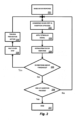
- FIG. 2 shows various functional blocks in a method for intraoperative measurement of stapedial reflex responses not part of the present invention.
- an intraoperative baseline value stapedial reflex response is determined for a cochlear implant patient, step 201.
- the multi-step surgical process commences to implant a cochlear implant system in the patient.
- a stimulus signal is applied, step 203, and the stapedial reflex response is monitored, step 204.
- the stapedial reflex response changes from the baseline value response more than a safe change threshold value (e.g., more than 30% change) step 205, then the given step is stopped, (e.g., electrode insertion is stopped) step 208 and trauma preventive action can be performed, step 209.
- the stapedial reflex response may continue to be monitored after stopping the given step, and the given step may be continued if the response change returns to less than the safe change threshold value. Otherwise, if the stapedial reflex response does not change from the baseline value response more than a safe change threshold value, step 205, then the given step in the surgical process continues until completed, and if the insertion process continues, step 206, the process repeats until all the steps have completed and the process ends, step 207.
- the stapedial reflex response may be determined again after completing the given step, and compared to the baseline value response to evaluate hearing trauma resulting from the given step.
- the surgeon is immediately notified if some substantial change occurs in the response (e.g., in the response amplitude). Based on the monitoring information, the surgeon can decide whether to continue or stop the current surgical step. For example, if the stapedial reflex amplitude and latency changed by 30%, the surgeon may be asked to stop performing the current surgical step (e.g., stop drilling, stop opening the cochlea, etc.) In the case of electrode insertion, the surgeon can stop inserting and gently withdraw the electrode array a small amount. After some time has elapsed, when the stapedial reflex amplitude returns to baseline or becomes stable and reliable again, the surgeon may continue. By giving this information to the surgeon quickly, surgically related hearing trauma can be expected to be reduced.
- the current surgical step e.g., stop drilling, stop opening the cochlea, etc.
- a full battery test that gives frequency dependent information about hearing trauma may not be necessary. Rather, one stimulus (e.g., broadband noise and/or tone bursts) can be tested. For simplification, only broadband noise at a few different amplitudes need be tested.
- One advantage of using broadband noise stimulus signals is high sensitivity to stapedial reflex amplitude and stapedial reflex threshold change.
- a final measurement of the stapedial reflex can be performed and compared with the baseline measurement to evaluate the total hearing trauma due to the surgery. That is, an approximate frequency dependent hearing loss for each specific frequency can be stated based on subtracting from the baseline and final stapedial reflex responses to determine an approximate total hearing loss.
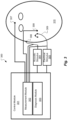
- FIG. 3 shows an arrangement for intraoperative measurement of stapedial reflex responses in a cochlear implant patient 310.
- a control module 301 can be a regular audiometer module configured for acoustic stimulation and recordings (e.g., based on the Med-El Maestro diagnostic module).
- the control module 301 directs acoustic stimulation module 304 to deliver acoustic stimulus signals to the ear canal of the patient 310 to evoke a stapedius reflex response.
- the acoustic stimulation can be performed on the ipsilateral side ear canal with an inserted ear phone 306.
- Electrical stimulation module 305 represents the components of the cochlear implant system being implanted along with its electrode array 309.
- the stimulus signals of the acoustic stimulation module 304 and the electrical stimulation module 305 also are measured as an input to a stapedius reflex response measurement module 302 of the control module 301.
- the stapedius reflex response measurements are made by one or more recording electrodes 308 (e.g., bipolar microwire electrodes) and in combination with a reference electrode 307.
- the recording electrodes 308 (+ and -) can be positioned around the stapedius tendon.
- Sensor or bipolar electrodes also can be attached to the cochlear implant electrode 309.
- the reference electrode 307 can be the housing of the cochlear implant processor or a distant electrode placed, for example, in the muscle temporalis 310.
- the reference electrode also can be a surface electrode, i.e. placed on the lower forehead of the patient.
- stapedial reflexes There are several different ways to evoke stapedial reflexes. Most typically, the stimulation of stapedial muscle can be evoked acoustically. Tympanometry records changes in middle ear immittance while air pressure is varied in the ear canal and stapedial reflexes are recorded at a single air pressure setting from either the ipsilateral or contralateral side. Alternatively, bone conduction may be sued to apply the stimulus signals. Such a measurement may provide more insight into the origin of the hearing loss (i.e. conductive or sensorineural hearing loss).
- the hearing loss i.e. conductive or sensorineural hearing loss
- the acoustic stimulus signals may be broadband noise signals and/or various tone bursts (e.g., 0.5 kHz, 1 kHz, 2 kHz, 4 kHz) at various amplitudes. For n different frequencies, at least two different pure tone frequencies maybe applied, and in addition, a broadband noise and/or chirp stimulus also may be used. Typical duration of the acoustic stimuli is 300-500 msec. Tone bursts are typically in the range of 250-2000 Hz and can be selected based on the preoperative baseline audiogram for the patient.
- Measurement of EMG potentials can be detected by a stapedius sensor or bipolar recording electrodes inserted in the stapedius tendon in the middle ear.
- visual observation of the stapedial muscle may be used.
- the amplitude of the stapedius reflex response, response latency, and timing can be quantified.
- Typical reflex latencies in normal subjects range from 40-180 milliseconds, typically around 107 milliseconds, and normally the reflex response does not decay.
- the critical percentage change of level of stapedial reflex amplitude and stapedial reflex latency may vary from patient to patient according to individual hearing sensitivity and retrocochlear function.
- Stapedius reflex thresholds are lowest for broadband stimuli; comparing normal hearing patients and patients with sensorineural hearing loss of 30-70 dB, the difference of average stapedial reflex threshold is 18dB.
- stapedius reflex responses is achievable in a large population-approximately 80% of patients with sensorineural hearing loss of less than 80dB have an elicitable stapedial reflex threshold, and approximately 95% of patients with sensorineural hearing loss of less than 60dB have and elicitable stapedial reflex threshold (requiring stimulation up to 125 dB).
- stapedial reflex thresholds do not increase linearly with the amount of sensorineural hearing loss.
- tone burst stimuli of both normal hearing patients and patients with sensorineural hearing loss of 30-70 dB the average amplitude growth does not significantly change (use of tone bursts may not be sensitive enough to judge the level of hearing trauma). And when using only broadband stimuli, frequency-specific information may not be obtainable.
- the middle ear structure needs to be undisturbed and fully functioning. Using recordings of EMG can achieve more sensitive and precise outcomes than measuring middle ear air pressure.
- ipsilateral recordings as opposed to the widely used eSRT method
- the responses may be recorded using bipolar microwire electrodes that are positioned around the stapedial tendon. This leads to a favorable S/N ratio.
- the blood flow probe may specifically be a laser doppler probe that be made in a very small diameter (e.g., 0.8 mm), which is easy to use during the surgical intervention.
- the blood flow probe needs to be kept stable and secure at a controlled fixed position in the vicinity of the stapedial muscle or stapedial tendon. Since the blood flow change occurs at the initial state of the stapedial muscle contraction, the surgeon can be informed of the stapedial response at a stimulus level before the stapedial reflex conventionally can be observed by pressure change from a probe placed in the ear canal.
- Embodiments of the present invention can be based on measuring such changes in oxygen level in the blood from the stapedius muscle; specifically, the decreased concentration of oxygen in the stapedial tissue that occurs when the muscle contracts. When the stapedius muscle is at rest, oxygen concentration in the tissues is at some "normal" baseline level. As soon as the stapedial reflex muscle contraction starts, the oxygen consumption by the muscle increases by a factor of ten or more, which can easily be measured.
- an oxygen probe being placed in the vicinity of the stapedial muscle in a controlled manner.
- There are several specific ways to measure the oxygen level such as by using an oxygen sensor.
- the oxygen sensor needs to be kept stable and secure at a controlled fixed position in the vicinity of the stapedial muscle or stapedial tendon. Since the oxygen level change occurs at the initial state of the stapedial muscle contraction, the surgeon can be informed of the stapedial response at a stimulus level before the stapedial reflex conventionally can be observed by pressure change from a probe placed in the ear canal.
Landscapes
- Health & Medical Sciences (AREA)
- Life Sciences & Earth Sciences (AREA)
- Engineering & Computer Science (AREA)
- Otolaryngology (AREA)
- Physics & Mathematics (AREA)
- Veterinary Medicine (AREA)
- Public Health (AREA)
- General Health & Medical Sciences (AREA)
- Animal Behavior & Ethology (AREA)
- Biomedical Technology (AREA)
- Audiology, Speech & Language Pathology (AREA)
- Heart & Thoracic Surgery (AREA)
- Medical Informatics (AREA)
- Molecular Biology (AREA)
- Surgery (AREA)
- Pathology (AREA)
- Biophysics (AREA)
- Multimedia (AREA)
- Acoustics & Sound (AREA)
- Nuclear Medicine, Radiotherapy & Molecular Imaging (AREA)
- Radiology & Medical Imaging (AREA)
- Prostheses (AREA)
- Cardiology (AREA)
- Hematology (AREA)
- Physiology (AREA)
Description
- The present invention relates to surgical techniques for cochlear implant systems.
- A normal ear transmits sounds as shown in
Figure 1 through theouter ear 101 to the tympanic membrane (eardrum) 102, which moves the bones of themiddle ear 103, which in turn vibrate the oval window and round window openings of thecochlea 104. Thecochlea 104 is a long narrow duct wound spirally about its axis for approximately two and a half turns. Thecochlea 104 includes an upper channel known as the scala vestibuli and a lower channel known as the scala tympani, which are connected by the cochlear duct. The scala tympani forms an upright spiraling cone with a center called the modiolar where the spiral ganglion cells of theacoustic nerve 113 reside. In response to received sounds transmitted by themiddle ear 103, the fluid filledcochlea 104 functions as a transducer to generate electric pulses that are transmitted to thecochlear nerve 113, and ultimately to the brain. Hearing is impaired when there are problems in the ability to transduce external sounds into meaningful action potentials along the neural substrate of thecochlea 104. - In some cases, hearing impairment can be addressed by a cochlear implant that electrically stimulates auditory nerve tissue with small currents delivered by multiple electrode contacts distributed along an implant electrode.
Figure 1 shows some components of a typical cochlear implant system where an external microphone provides an audio signal input to an externalsignal processing stage 111 which implements one of various known signal processing schemes. The processed signal is converted by the externalsignal processing stage 111 into a digital data format, such as a sequence of data frames, for transmission into a receiver processor in animplant housing 108. Besides extracting the audio information, the receiver processor in theimplant housing 108 may perform additional signal processing such as error correction, pulse formation, etc., and produces a stimulation pattern (based on the extracted audio information) that is sent through wires in anelectrode lead 109 to an implantedelectrode array 110. Typically, theelectrode array 110 includes multiple electrodes on its surface that provide selective stimulation of thecochlea 104. - The
electrode array 110 penetrates into thecochlea 104 through a surgical opening called a cochleostomy. Theelectrode array 110 has multiple electrode contacts on or slightly recessed below its outer surface for applying one or more electrical stimulation signals to target audio neural tissue within thecochlea 104. Theextra-cochlear electrode lad 109 that goes from theimplant housing 108 to the cochleostomy opening usually has no electrical contacts except perhaps a ground electrode and it encloses connecting wires that deliver electrical stimulation signals to the electrode contacts on theelectrode array 110. - Insertion and placement and insertion of the
electrode array 110 into thecochlea 104 causes trauma to the cochlear tissue due to the rigidity, friction, and impact of moving theelectrode array 110 through thecochlea 104. For example, insertion of theelectrode array 110 may damage soft tissues, membranes, thin bony shelves, blood vessels, neural elements, etc. In the case of multiple insertions, the damage can accumulate. In addition, removal and replacement of theelectrode array 110 due to device failure or aging is also a serious problem. For example, patients with some residual hearing now receive hybrid implant systems that also include acoustic-mechanical stimulation components, and further hearing loss could occur when theelectrode array 110 is removed or replaced. In addition, there are efforts to use therapeutic drugs to regrow neural tissue around an insertedelectrode array 110 which could suffer catastrophic consequences when the electrode is removed since any new neural tissue growth that might reach the electrode could be disrupted or destroyed. - It has been shown that patients with preserved low frequency hearing have significantly better outcomes than those without such hearing preservation. And it is generally accepted that the amount of electrode insertion trauma correlates significantly with the level of hearing loss caused during the surgery. Thus the extent of hearing preservation preoperatively or postoperatively is believed to serve as a good indicator of the magnitude of electrode insertion trauma. See Skarzynski et al., Atraumatic Round Window Deep Insertion Of Cochlear Electrodes, Acta Otolaryngol. 2011 Jul;131(7):740-9. Epub 2011 Apr 15.
US 2009/0018616 describes systems and methods for determining a threshold current level to evoke a postoperative stapedial muscle reflex. - Currently there is no existing developed objective method to evaluate intraoperative hearing trauma. There have been some initial proposals of intraoperative methods to detect possible intraoperative trauma, but all of these approaches still are under development. And all of the existing proposals are based on measuring evoked potentials (e.g., ECAPs) either using acoustic stimuli or some combination of electric and acoustic stimuli, and then recording near-field and/or far-field sensor measurements.
- Besides evoked potentials such as ECAPs, measurement of the stapedius reflex response also has been widely used in a clinical practice to evaluate hearing and fit hearing prosthesis systems. The stapedius is the smallest skeletal muscle in the human body. At just over one millimeter in length, its purpose is to stabilize the smallest bone in the body, the stapes. The stapedial reflex refers to the involuntary contraction of the stapedius and tensor tympani muscles of the ossicles that occurs in response to a loud sound. The stapedius muscle pulls the stapes (stirrup) of the middle ear away from the oval window of the cochlea and the tensor tympani muscle pulls the malleus (hammer) away from the ear drum. This reflex decreases the transmission of vibrational energy to the cochlea where it is converted into electrical impulses to be processed by the brain for perception as sound.
- Several methods to measure the stapedial reflex have been described including:
- Recording a pressure change from a probe placed in the ear canal,
- Recoding a myogenic response in the vicinity of the stapedial muscle or stapedial tendon, and
- Intraoperative surgical observation of the facial nerve during cochlear implant surgery.
- The present invention provides a surgical arrangement for implantation of a cochlear implant according to claims 1 to 7.
- The stapedial reflex response may continue to be monitored after stopping the given step, and the given step may be continued if the response change returns to less than the safe change threshold value. The stapedial reflex response may be determined again after completing the given step, and compared to the baseline value response to evaluate hearing trauma resulting from the given step.
- The safe change threshold value may be, for example, a 30% change from the intraoperative baseline response. The stapedial reflex response may be determined based on measuring change of stapedial muscle blood flow or oxygen. The stapedial reflex response may include response amplitude growth and response latency. Either an acoustic stimulus or a bone conduction stimulus may be used to produce the stapedial reflex response. Examples of the given step include insertion of a stimulation electrode into the cochlea of the patient, performing a cochleostomy, and drilling a bone well for an implantable processor.
-
-
Figure 1 shows various anatomical structures of the human ear and components of a typical cochlear implant system in relation thereto. -
Figure 2 shows various functional blocks in a method for intraoperative measurement of stapedius reflex responses according to an embodiment of the present invention. -
Figure 3 shows an arrangement for intraoperative measurement of stapedial reflex responses. - Embodiments of the present invention provide useful real time intraoperative feedback to minimize mechanical trauma during insertion of the cochlear implant electrode. This helps evaluate each surgical step and maneuver in terms of surgical trauma that is measured as a level of frequency specific hearing preservation. The real time feedback also can provide the basis for a surgical navigation tool for use during the electrode insertion. After gentle electrode maneuvers, if the responses get smaller, the electrode can be drawn back slightly and after waiting a brief period for the response to recover, the surgeon can continue trying different maneuvers with the electrode.
- Various ways are described for using a modified stapedial reflex response measurement to monitor hearing preservation during cochlear implant surgery. Measurement of stapedial reflexes can be relatively quick and the measured responses are very robust, thus stapedial reflex response can be used as a real-time measurement with no need to determine average response. Unlike other measurement strategies, stapedial reflex responses are measureable at low frequencies and thus are very suitable for measurement of surgical-related hearing trauma, and specific information can be obtained about hearing preservation. But existing clinical methods of measuring the stapedial reflex response cannot be used for intraoperative monitoring of hearing trauma. The described intraoperative measurement depends on the measurement of amplitude growth and stapedial reflex latencies, using different specific stimuli and comparing the obtained information with a baseline intraoperative stapedial reflex measurement.
-
Figure 2 shows various functional blocks in a method for intraoperative measurement of stapedial reflex responses not part of the present invention. Initially, an intraoperative baseline value stapedial reflex response is determined for a cochlear implant patient,step 201. Then the multi-step surgical process commences to implant a cochlear implant system in the patient. At one or more of the specific steps in the process (e.g., insertion of a stimulation electrode into the cochlea of the patient, performing a cochleostomy, and drilling a bone well for an implantable processor)step 202, a stimulus signal is applied,step 203, and the stapedial reflex response is monitored,step 204. If the stapedial reflex response changes from the baseline value response more than a safe change threshold value (e.g., more than 30% change)step 205, then the given step is stopped, (e.g., electrode insertion is stopped)step 208 and trauma preventive action can be performed, step 209. For example, the stapedial reflex response may continue to be monitored after stopping the given step, and the given step may be continued if the response change returns to less than the safe change threshold value. Otherwise, if the stapedial reflex response does not change from the baseline value response more than a safe change threshold value,step 205, then the given step in the surgical process continues until completed, and if the insertion process continues,step 206, the process repeats until all the steps have completed and the process ends,step 207. The stapedial reflex response may be determined again after completing the given step, and compared to the baseline value response to evaluate hearing trauma resulting from the given step. - During such real time monitoring of the stapedial reflex response, the surgeon is immediately notified if some substantial change occurs in the response (e.g., in the response amplitude). Based on the monitoring information, the surgeon can decide whether to continue or stop the current surgical step. For example, if the stapedial reflex amplitude and latency changed by 30%, the surgeon may be asked to stop performing the current surgical step (e.g., stop drilling, stop opening the cochlea, etc.) In the case of electrode insertion, the surgeon can stop inserting and gently withdraw the electrode array a small amount. After some time has elapsed, when the stapedial reflex amplitude returns to baseline or becomes stable and reliable again, the surgeon may continue. By giving this information to the surgeon quickly, surgically related hearing trauma can be expected to be reduced.
- During some or all of the specific surgical steps (such as cochleostomy or drilling), a full battery test that gives frequency dependent information about hearing trauma may not be necessary. Rather, one stimulus (e.g., broadband noise and/or tone bursts) can be tested. For simplification, only broadband noise at a few different amplitudes need be tested. One advantage of using broadband noise stimulus signals is high sensitivity to stapedial reflex amplitude and stapedial reflex threshold change.
- After the surgical process is complete (i.e., after the electrode is in place), a final measurement of the stapedial reflex can be performed and compared with the baseline measurement to evaluate the total hearing trauma due to the surgery. That is, an approximate frequency dependent hearing loss for each specific frequency can be stated based on subtracting from the baseline and final stapedial reflex responses to determine an approximate total hearing loss.
-
Figure 3 shows an arrangement for intraoperative measurement of stapedial reflex responses in acochlear implant patient 310. Acontrol module 301 can be a regular audiometer module configured for acoustic stimulation and recordings (e.g., based on the Med-El Maestro diagnostic module). Thecontrol module 301 directsacoustic stimulation module 304 to deliver acoustic stimulus signals to the ear canal of thepatient 310 to evoke a stapedius reflex response. For example, the acoustic stimulation can be performed on the ipsilateral side ear canal with an insertedear phone 306.Electrical stimulation module 305 represents the components of the cochlear implant system being implanted along with itselectrode array 309. The stimulus signals of theacoustic stimulation module 304 and theelectrical stimulation module 305 also are measured as an input to a stapedius reflexresponse measurement module 302 of thecontrol module 301. The stapedius reflex response measurements are made by one or more recording electrodes 308 (e.g., bipolar microwire electrodes) and in combination with areference electrode 307. The recording electrodes 308 (+ and -) can be positioned around the stapedius tendon. Sensor or bipolar electrodes also can be attached to thecochlear implant electrode 309. Thereference electrode 307 can be the housing of the cochlear implant processor or a distant electrode placed, for example, in themuscle temporalis 310. The reference electrode also can be a surface electrode, i.e. placed on the lower forehead of the patient. - There are several different ways to evoke stapedial reflexes. Most typically, the stimulation of stapedial muscle can be evoked acoustically. Tympanometry records changes in middle ear immittance while air pressure is varied in the ear canal and stapedial reflexes are recorded at a single air pressure setting from either the ipsilateral or contralateral side. Alternatively, bone conduction may be sued to apply the stimulus signals. Such a measurement may provide more insight into the origin of the hearing loss (i.e. conductive or sensorineural hearing loss).
- The acoustic stimulus signals may be broadband noise signals and/or various tone bursts (e.g., 0.5 kHz, 1 kHz, 2 kHz, 4 kHz) at various amplitudes. For n different frequencies, at least two different pure tone frequencies maybe applied, and in addition, a broadband noise and/or chirp stimulus also may be used. Typical duration of the acoustic stimuli is 300-500 msec. Tone bursts are typically in the range of 250-2000 Hz and can be selected based on the preoperative baseline audiogram for the patient.
- Measurement of EMG potentials can be detected by a stapedius sensor or bipolar recording electrodes inserted in the stapedius tendon in the middle ear. In addition or alternatively, visual observation of the stapedial muscle may be used. The amplitude of the stapedius reflex response, response latency, and timing (sustained or rapidly decaying) can be quantified. Typical reflex latencies in normal subjects range from 40-180 milliseconds, typically around 107 milliseconds, and normally the reflex response does not decay.
- The critical percentage change of level of stapedial reflex amplitude and stapedial reflex latency (i.e. 30%) may vary from patient to patient according to individual hearing sensitivity and retrocochlear function. The range for acoustic reflexes in individuals with normal hearing averages 70-100 decibel (dB) sound pressure level (SPL). The greater the hearing loss, the higher the acoustic reflex threshold for conductive hearing loss. For sensorineural hearing loss, acoustic reflex thresholds may be within the normal range, particularly for mild-to-moderate hearing losses with recruitment. Elevated or absent acoustic reflex thresholds (i.e., >100 dB SPL) for any given frequency may suggest sensorineural or conductive hearing loss, facial nerve disorder, or middle ear disorder. Reflexes usually are absent or cannot be recorded if the patient has type B tympanograms and so acoustic reflexes generally are not tested in such patients.
- Amplitude growth up to 20dB above the stapedius reflex threshold will be approximately linear both for tone bursts (500 Hz, 1 kHz, 2 kHz, etc.) and for broad band stimuli. Recordings of myogenic potential may be more sensitive than commercially available measurement via change of the air pressure on the eardrum. Stapedius reflex thresholds are lowest for broadband stimuli; comparing normal hearing patients and patients with sensorineural hearing loss of 30-70 dB, the difference of average stapedial reflex threshold is 18dB. Moreover, effective measurement of stapedius reflex responses is achievable in a large population-approximately 80% of patients with sensorineural hearing loss of less than 80dB have an elicitable stapedial reflex threshold, and approximately 95% of patients with sensorineural hearing loss of less than 60dB have and elicitable stapedial reflex threshold (requiring stimulation up to 125 dB).
- But stapedial reflex thresholds do not increase linearly with the amount of sensorineural hearing loss. For tone burst stimuli of both normal hearing patients and patients with sensorineural hearing loss of 30-70 dB, the average amplitude growth does not significantly change (use of tone bursts may not be sensitive enough to judge the level of hearing trauma). And when using only broadband stimuli, frequency-specific information may not be obtainable. During the intraoperative measurement, the middle ear structure needs to be undisturbed and fully functioning. Using recordings of EMG can achieve more sensitive and precise outcomes than measuring middle ear air pressure. For bilateral patients it may be easier to use ipsilateral recordings (as opposed to the widely used eSRT method) and thereby avoid influence from the contralateral ear. The responses may be recorded using bipolar microwire electrodes that are positioned around the stapedial tendon. This leads to a favorable S/N ratio.
- There have been reports describing attempts to measure cochlear blood flow. But there is no report of using such a blood flow measurement to measure the stapedial reflex response to sound stimulus (acoustic, mechanical or electric). Embodiments of the present invention do just that. When the stapedius muscle is at rest, blood flow through it is at some "normal" baseline level. As soon as the stapedial reflex muscle contraction starts, the blood flow through the muscle increases by a factor of ten or more. Stapedial muscle blood flow remains high for several hundred milliseconds as the reflex contractions end, but then returns back toward the normal baseline flow during the next few hundred milliseconds.
- Placing a blood flow probe in the vicinity of the stapedial muscle or tendon, this change in blood flow can easily be measured and recorded. The blood flow probe may specifically be a laser doppler probe that be made in a very small diameter (e.g., 0.8 mm), which is easy to use during the surgical intervention. For reliable measurements, the blood flow probe needs to be kept stable and secure at a controlled fixed position in the vicinity of the stapedial muscle or stapedial tendon. Since the blood flow change occurs at the initial state of the stapedial muscle contraction, the surgeon can be informed of the stapedial response at a stimulus level before the stapedial reflex conventionally can be observed by pressure change from a probe placed in the ear canal.
- In addition to the forgoing discussion of changes in blood flow signalling occurrence of the stapedius reflex, there also is a good correlation between increased blood flow increases and an increase in the amount of oxygen consumed during the muscle contraction. Because oxygen is consumed by a muscle when it contracts, the oxygen concentration in the tissue fluids decreases. This causes local arteriolar vasodilation both because arteriolar walls cannot maintain contraction in the absence of oxygen, and because oxygen deficiency causes release of vasodilator substances (e.g., Adenosine, K+ ions, ATP, lactic acid, carbon dioxide). That process can be effectively recorded by a device measuring the oxygen change.
- There have been reports attempting to measure such changes in oxygen level, but not to measure the stapedial reflex response to sound stimulus (acoustic, mechanical or electric). Embodiments of the present invention can be based on measuring such changes in oxygen level in the blood from the stapedius muscle; specifically, the decreased concentration of oxygen in the stapedial tissue that occurs when the muscle contracts. When the stapedius muscle is at rest, oxygen concentration in the tissues is at some "normal" baseline level. As soon as the stapedial reflex muscle contraction starts, the oxygen consumption by the muscle increases by a factor of ten or more, which can easily be measured.
- This is achieved by using an oxygen probe being placed in the vicinity of the stapedial muscle in a controlled manner. There are several specific ways to measure the oxygen level such as by using an oxygen sensor. For reliable measurements, the oxygen sensor needs to be kept stable and secure at a controlled fixed position in the vicinity of the stapedial muscle or stapedial tendon. Since the oxygen level change occurs at the initial state of the stapedial muscle contraction, the surgeon can be informed of the stapedial response at a stimulus level before the stapedial reflex conventionally can be observed by pressure change from a probe placed in the ear canal.
- Although various exemplary embodiments of the invention have been disclosed, it should be apparent to those skilled in the art that various changes and modifications can be made which will achieve some of the advantages of the invention.
Claims (7)
- A surgical arrangement for use in a multi-step surgical process to implant a cochlear implant system in a patient (310), the arrangement comprising:a control module (301) configured to control the multi-step surgical process including directing:i. an acoustic stimulation module (304) to deliver acoustic stimulus signals to the ear canal of the patient, andii. an electrical stimulation module (305) representing the components of the cochlear implant system including an electrode array (309) configured to deliver electrical stimulation signals to the cochlea being implanted;iii. one or more recording electrodes (308) configured to obtain stapedius reflex response measurements from the stimulus signals;iv. a stapedius reflex response measurement module (302) within the control module (301) and configured to determine from the stapedius reflex response measurements an intraoperative baseline value stapedial reflex response of the patient for the multi-step surgical process; andwherein the stapedius reflex response measurement module (302) is configured to monitor the intraoperative baseline value stapedial reflex response during the multi-step surgical process, and the control module (301) is further configured to
notify a surgeon to stop at a given step (208) if the intraoperative baseline value stapedial reflex response changes more than a safe change threshold value. - The surgical arrangement according to claim 1, wherein the control module (301) is further configured to :continue to monitor the intraoperative baseline value stapedial reflex response after stopping the given step (208), andthen continue the given step if the response change returns to less than the safe change threshold value.
- The surgical arrangement according to claim 1, wherein the control module (301) is further configured to:continue to monitor the intraoperative baseline stapedial reflex response after stopping the given step (208); andthen compare the post-stopping response to the baseline value response to evaluate hearing trauma resulting from the given step.
- The surgical arrangement according to claim 1, wherein the safe change threshold value is a 30% change from the intraoperative baseline response.
- The surgical arrangement according to claim 1, wherein the a stapedius reflex response measurement module (302) is configured to determine the intraoperative baseline stapedial reflex response based on measuring change of stapedial muscle blood flow and/or change of stapedial muscle oxygen.
- The surgical arrangement according to claim 1, wherein the intraoperative baseline stapedial reflex response includes response amplitude growth and response latency.
- The surgical arrangement according to claim 1, wherein the given step is one of insertion of a stimulation electrode into the cochlea of the patient, performing a cochleostomy, and drilling a bone well for an implantable processor.
Applications Claiming Priority (3)
| Application Number | Priority Date | Filing Date | Title |
|---|---|---|---|
| US201261714263P | 2012-10-16 | 2012-10-16 | |
| US201361834983P | 2013-06-14 | 2013-06-14 | |
| PCT/US2013/065210 WO2014062783A1 (en) | 2012-10-16 | 2013-10-16 | Monitoring of hearing preservation during cochlear implant surgery using stapedial reflex measurement |
Publications (3)
| Publication Number | Publication Date |
|---|---|
| EP2908761A1 EP2908761A1 (en) | 2015-08-26 |
| EP2908761A4 EP2908761A4 (en) | 2016-06-01 |
| EP2908761B1 true EP2908761B1 (en) | 2024-12-04 |
Family
ID=50475953
Family Applications (1)
| Application Number | Title | Priority Date | Filing Date |
|---|---|---|---|
| EP13847628.8A Active EP2908761B1 (en) | 2012-10-16 | 2013-10-16 | Monitoring of hearing preservation during cochlear implant surgery using stapedial reflex measurement |
Country Status (5)
| Country | Link |
|---|---|
| US (1) | US9107621B2 (en) |
| EP (1) | EP2908761B1 (en) |
| CN (1) | CN104736096B (en) |
| AU (1) | AU2013331333B2 (en) |
| WO (1) | WO2014062783A1 (en) |
Families Citing this family (11)
| Publication number | Priority date | Publication date | Assignee | Title |
|---|---|---|---|---|
| US10660604B2 (en) * | 2015-07-13 | 2020-05-26 | Otonexus Medical Technologies, Inc. | Apparatus and method for characterization of acute otitis media |
| WO2017065809A1 (en) | 2015-10-13 | 2017-04-20 | Advanced Bionics Ag | Systems and methods for intra-surgical monitoring of evoked responses that occur during an electrode lead insertion procedure |
| WO2017131675A1 (en) | 2016-01-27 | 2017-08-03 | Advanced Bionics Ag | Systems and methods for intra-surgical monitoring of cochlear trauma during an electrode lead insertion procedure |
| CN108697382B (en) * | 2016-02-26 | 2022-01-25 | Med-El电气医疗器械有限公司 | Detection of electrically evoked pusher muscle reflexes using acoustic transfer function changes in the middle or inner ear |
| US20170360364A1 (en) * | 2016-06-21 | 2017-12-21 | John Michael Heasman | Cochlea health monitoring |
| US10357656B2 (en) * | 2016-07-12 | 2019-07-23 | Cochlear Limited | Hearing prosthesis programming |
| US10500392B2 (en) * | 2016-08-24 | 2019-12-10 | Cochlear Limited | Monitoring stimulating assembly insertion |
| CN108703761B (en) * | 2018-06-11 | 2021-01-22 | 佛山博智医疗科技有限公司 | Method for testing hearing recognition sensitivity |
| CN112386793A (en) | 2019-08-12 | 2021-02-23 | 科利耳有限公司 | Real-time estimation of electrode array pose during intra-cochlear insertion |
| US20240081691A1 (en) * | 2021-01-25 | 2024-03-14 | Med-El Elektromedizinische Geraete Gmbh | Estimation of audiogram based on in-vivo acoustic chirp |
| WO2025158079A1 (en) | 2024-01-26 | 2025-07-31 | Med-El Elektromedizinische Geräte Ges.m.b.H. | System and method for detection of disturbed basilar membrane wave propagation |
Family Cites Families (5)
| Publication number | Priority date | Publication date | Assignee | Title |
|---|---|---|---|---|
| CA2258008A1 (en) * | 1996-06-20 | 1997-12-24 | Advanced Bionics Corporation | Self-adjusting cochlear implant system and method for fitting same |
| US7117038B1 (en) * | 2002-09-20 | 2006-10-03 | Advanced Bionics Corporation | Method and system for obtaining stapedial reflexes in cochlear implant users using multiband stimuli |
| US7206640B1 (en) | 2002-11-08 | 2007-04-17 | Advanced Bionics Corporation | Method and system for generating a cochlear implant program using multi-electrode stimulation to elicit the electrically-evoked compound action potential |
| US8103354B2 (en) * | 2006-07-17 | 2012-01-24 | Advanced Bionics, Llc | Systems and methods for determining a threshold current level required to evoke a stapedial muscle reflex |
| US20110066160A1 (en) * | 2008-04-03 | 2011-03-17 | The Trustees Of Columbia University In The City Of New York | Systems and methods for inserting steerable arrays into anatomical structures |
-
2013
- 2013-10-16 AU AU2013331333A patent/AU2013331333B2/en active Active
- 2013-10-16 US US14/054,898 patent/US9107621B2/en active Active
- 2013-10-16 EP EP13847628.8A patent/EP2908761B1/en active Active
- 2013-10-16 WO PCT/US2013/065210 patent/WO2014062783A1/en not_active Ceased
- 2013-10-16 CN CN201380054079.XA patent/CN104736096B/en active Active
Also Published As
| Publication number | Publication date |
|---|---|
| CN104736096A (en) | 2015-06-24 |
| WO2014062783A1 (en) | 2014-04-24 |
| US9107621B2 (en) | 2015-08-18 |
| EP2908761A4 (en) | 2016-06-01 |
| CN104736096B (en) | 2017-04-26 |
| EP2908761A1 (en) | 2015-08-26 |
| AU2013331333B2 (en) | 2016-07-07 |
| AU2013331333A1 (en) | 2015-04-09 |
| US20140107441A1 (en) | 2014-04-17 |
Similar Documents
| Publication | Publication Date | Title |
|---|---|---|
| US9107621B2 (en) | Monitoring of hearing preservation during cochlear implant surgery using stapedial reflex measurement | |
| US9072468B2 (en) | Apparatus and methods for acoustically or mechanically stimulating a cochlea and intracochlear recording of mechanically or acoustically evoked auditory potentials in the cochlea | |
| EP3419523B1 (en) | Detection of electrically evoked stapedius reflex | |
| Brackmann et al. | Auditory brainstem implant: I. Issues in surgical implantation | |
| US8768477B2 (en) | Post-auricular muscle response based hearing prosthesis fitting | |
| CA2230603C (en) | Apparatus and method for automatically determining stimulation parameters | |
| US20010014818A1 (en) | Implantable hearing assistance system with calibration and auditory response testing | |
| EP3539610B1 (en) | Cochlear implant fitting via efferent nerve fibers | |
| US11642524B2 (en) | Methods and systems for treating tinnitus with transtympanic electrical stimulation | |
| Wazen | Intraoperative monitoring of auditory function: experimental observations and new applications | |
| EP4403208A1 (en) | Device for diagnosis and/oder supporting of hearing capabilities | |
| WO2025230847A1 (en) | Cochlear implant calibration and health indications based on middle ear pressure | |
| AU2022210720B2 (en) | Estimation of audiogram based on in-vivo acoustic chirp | |
| Skarżyński | Intraoperative monitoring of hearing preservation with ECochG in treatment of partial deafness | |
| AU2014280878B2 (en) | Post-auricular muscle response based hearing prosthesis fitting | |
| US20160325095A1 (en) | Novel Recording Approach of Stapedius Muscle Activity | |
| Gonzalez et al. | Analysis and Characterization of Stapedius Muscle Electromyograms Elicited by Cochlear Implants in Rats. |
Legal Events
| Date | Code | Title | Description |
|---|---|---|---|
| PUAI | Public reference made under article 153(3) epc to a published international application that has entered the european phase |
Free format text: ORIGINAL CODE: 0009012 |
|
| 17P | Request for examination filed |
Effective date: 20150320 |
|
| AK | Designated contracting states |
Kind code of ref document: A1 Designated state(s): AL AT BE BG CH CY CZ DE DK EE ES FI FR GB GR HR HU IE IS IT LI LT LU LV MC MK MT NL NO PL PT RO RS SE SI SK SM TR |
|
| AX | Request for extension of the european patent |
Extension state: BA ME |
|
| RIN1 | Information on inventor provided before grant (corrected) |
Inventor name: POLAK, MAREK Inventor name: GRASSO, PETER |
|
| DAX | Request for extension of the european patent (deleted) | ||
| RA4 | Supplementary search report drawn up and despatched (corrected) |
Effective date: 20160429 |
|
| RIC1 | Information provided on ipc code assigned before grant |
Ipc: A61B 5/145 20060101ALN20160422BHEP Ipc: A61B 5/12 20060101AFI20160422BHEP Ipc: A61B 5/0265 20060101ALN20160422BHEP Ipc: A61N 1/36 20060101ALN20160422BHEP Ipc: A61N 1/05 20060101ALN20160422BHEP |
|
| STAA | Information on the status of an ep patent application or granted ep patent |
Free format text: STATUS: EXAMINATION IS IN PROGRESS |
|
| 17Q | First examination report despatched |
Effective date: 20190805 |
|
| REG | Reference to a national code |
Ref country code: DE Ref legal event code: R079 Free format text: PREVIOUS MAIN CLASS: A61B0019000000 Ipc: A61B0005120000 Ref country code: DE Ref legal event code: R079 Ref document number: 602013086341 Country of ref document: DE Free format text: PREVIOUS MAIN CLASS: A61B0019000000 Ipc: A61B0005120000 |
|
| GRAP | Despatch of communication of intention to grant a patent |
Free format text: ORIGINAL CODE: EPIDOSNIGR1 |
|
| STAA | Information on the status of an ep patent application or granted ep patent |
Free format text: STATUS: GRANT OF PATENT IS INTENDED |
|
| RIC1 | Information provided on ipc code assigned before grant |
Ipc: A61B 5/145 20060101ALN20240529BHEP Ipc: A61B 5/0265 20060101ALN20240529BHEP Ipc: A61N 1/05 20060101ALN20240529BHEP Ipc: A61N 1/36 20060101ALN20240529BHEP Ipc: A61B 5/12 20060101AFI20240529BHEP |
|
| INTG | Intention to grant announced |
Effective date: 20240625 |
|
| GRAS | Grant fee paid |
Free format text: ORIGINAL CODE: EPIDOSNIGR3 |
|
| GRAA | (expected) grant |
Free format text: ORIGINAL CODE: 0009210 |
|
| STAA | Information on the status of an ep patent application or granted ep patent |
Free format text: STATUS: THE PATENT HAS BEEN GRANTED |
|
| AK | Designated contracting states |
Kind code of ref document: B1 Designated state(s): AL AT BE BG CH CY CZ DE DK EE ES FI FR GB GR HR HU IE IS IT LI LT LU LV MC MK MT NL NO PL PT RO RS SE SI SK SM TR |
|
| REG | Reference to a national code |
Ref country code: GB Ref legal event code: FG4D |
|
| REG | Reference to a national code |
Ref country code: CH Ref legal event code: EP |
|
| REG | Reference to a national code |
Ref country code: DE Ref legal event code: R096 Ref document number: 602013086341 Country of ref document: DE |
|
| REG | Reference to a national code |
Ref country code: IE Ref legal event code: FG4D |
|
| P01 | Opt-out of the competence of the unified patent court (upc) registered |
Free format text: CASE NUMBER: APP_67780/2024 Effective date: 20241223 |
|
| REG | Reference to a national code |
Ref country code: LT Ref legal event code: MG9D |
|
| REG | Reference to a national code |
Ref country code: NL Ref legal event code: MP Effective date: 20241204 |
|
| PG25 | Lapsed in a contracting state [announced via postgrant information from national office to epo] |
Ref country code: HR Free format text: LAPSE BECAUSE OF FAILURE TO SUBMIT A TRANSLATION OF THE DESCRIPTION OR TO PAY THE FEE WITHIN THE PRESCRIBED TIME-LIMIT Effective date: 20241204 |
|
| PG25 | Lapsed in a contracting state [announced via postgrant information from national office to epo] |
Ref country code: FI Free format text: LAPSE BECAUSE OF FAILURE TO SUBMIT A TRANSLATION OF THE DESCRIPTION OR TO PAY THE FEE WITHIN THE PRESCRIBED TIME-LIMIT Effective date: 20241204 |
|
| PG25 | Lapsed in a contracting state [announced via postgrant information from national office to epo] |
Ref country code: BG Free format text: LAPSE BECAUSE OF FAILURE TO SUBMIT A TRANSLATION OF THE DESCRIPTION OR TO PAY THE FEE WITHIN THE PRESCRIBED TIME-LIMIT Effective date: 20241204 |
|
| PG25 | Lapsed in a contracting state [announced via postgrant information from national office to epo] |
Ref country code: ES Free format text: LAPSE BECAUSE OF FAILURE TO SUBMIT A TRANSLATION OF THE DESCRIPTION OR TO PAY THE FEE WITHIN THE PRESCRIBED TIME-LIMIT Effective date: 20241204 |
|
| PG25 | Lapsed in a contracting state [announced via postgrant information from national office to epo] |
Ref country code: NO Free format text: LAPSE BECAUSE OF FAILURE TO SUBMIT A TRANSLATION OF THE DESCRIPTION OR TO PAY THE FEE WITHIN THE PRESCRIBED TIME-LIMIT Effective date: 20250304 |
|
| PG25 | Lapsed in a contracting state [announced via postgrant information from national office to epo] |
Ref country code: LV Free format text: LAPSE BECAUSE OF FAILURE TO SUBMIT A TRANSLATION OF THE DESCRIPTION OR TO PAY THE FEE WITHIN THE PRESCRIBED TIME-LIMIT Effective date: 20241204 Ref country code: GR Free format text: LAPSE BECAUSE OF FAILURE TO SUBMIT A TRANSLATION OF THE DESCRIPTION OR TO PAY THE FEE WITHIN THE PRESCRIBED TIME-LIMIT Effective date: 20250305 |
|
| PG25 | Lapsed in a contracting state [announced via postgrant information from national office to epo] |
Ref country code: RS Free format text: LAPSE BECAUSE OF FAILURE TO SUBMIT A TRANSLATION OF THE DESCRIPTION OR TO PAY THE FEE WITHIN THE PRESCRIBED TIME-LIMIT Effective date: 20250304 |
|
| PG25 | Lapsed in a contracting state [announced via postgrant information from national office to epo] |
Ref country code: NL Free format text: LAPSE BECAUSE OF FAILURE TO SUBMIT A TRANSLATION OF THE DESCRIPTION OR TO PAY THE FEE WITHIN THE PRESCRIBED TIME-LIMIT Effective date: 20241204 |
|
| REG | Reference to a national code |
Ref country code: AT Ref legal event code: MK05 Ref document number: 1747242 Country of ref document: AT Kind code of ref document: T Effective date: 20241204 |
|
| PG25 | Lapsed in a contracting state [announced via postgrant information from national office to epo] |
Ref country code: SM Free format text: LAPSE BECAUSE OF FAILURE TO SUBMIT A TRANSLATION OF THE DESCRIPTION OR TO PAY THE FEE WITHIN THE PRESCRIBED TIME-LIMIT Effective date: 20241204 |
|
| PG25 | Lapsed in a contracting state [announced via postgrant information from national office to epo] |
Ref country code: PL Free format text: LAPSE BECAUSE OF FAILURE TO SUBMIT A TRANSLATION OF THE DESCRIPTION OR TO PAY THE FEE WITHIN THE PRESCRIBED TIME-LIMIT Effective date: 20241204 |
|
| PG25 | Lapsed in a contracting state [announced via postgrant information from national office to epo] |
Ref country code: IS Free format text: LAPSE BECAUSE OF FAILURE TO SUBMIT A TRANSLATION OF THE DESCRIPTION OR TO PAY THE FEE WITHIN THE PRESCRIBED TIME-LIMIT Effective date: 20250404 |
|
| PG25 | Lapsed in a contracting state [announced via postgrant information from national office to epo] |
Ref country code: PT Free format text: LAPSE BECAUSE OF FAILURE TO SUBMIT A TRANSLATION OF THE DESCRIPTION OR TO PAY THE FEE WITHIN THE PRESCRIBED TIME-LIMIT Effective date: 20250404 |
|
| PG25 | Lapsed in a contracting state [announced via postgrant information from national office to epo] |
Ref country code: EE Free format text: LAPSE BECAUSE OF FAILURE TO SUBMIT A TRANSLATION OF THE DESCRIPTION OR TO PAY THE FEE WITHIN THE PRESCRIBED TIME-LIMIT Effective date: 20241204 |
|
| PG25 | Lapsed in a contracting state [announced via postgrant information from national office to epo] |
Ref country code: RO Free format text: LAPSE BECAUSE OF FAILURE TO SUBMIT A TRANSLATION OF THE DESCRIPTION OR TO PAY THE FEE WITHIN THE PRESCRIBED TIME-LIMIT Effective date: 20241204 Ref country code: AT Free format text: LAPSE BECAUSE OF FAILURE TO SUBMIT A TRANSLATION OF THE DESCRIPTION OR TO PAY THE FEE WITHIN THE PRESCRIBED TIME-LIMIT Effective date: 20241204 |
|
| PG25 | Lapsed in a contracting state [announced via postgrant information from national office to epo] |
Ref country code: SK Free format text: LAPSE BECAUSE OF FAILURE TO SUBMIT A TRANSLATION OF THE DESCRIPTION OR TO PAY THE FEE WITHIN THE PRESCRIBED TIME-LIMIT Effective date: 20241204 |
|
| PG25 | Lapsed in a contracting state [announced via postgrant information from national office to epo] |
Ref country code: CZ Free format text: LAPSE BECAUSE OF FAILURE TO SUBMIT A TRANSLATION OF THE DESCRIPTION OR TO PAY THE FEE WITHIN THE PRESCRIBED TIME-LIMIT Effective date: 20241204 |
|
| PG25 | Lapsed in a contracting state [announced via postgrant information from national office to epo] |
Ref country code: IT Free format text: LAPSE BECAUSE OF FAILURE TO SUBMIT A TRANSLATION OF THE DESCRIPTION OR TO PAY THE FEE WITHIN THE PRESCRIBED TIME-LIMIT Effective date: 20241204 |
|
| REG | Reference to a national code |
Ref country code: DE Ref legal event code: R097 Ref document number: 602013086341 Country of ref document: DE |
|
| PG25 | Lapsed in a contracting state [announced via postgrant information from national office to epo] |
Ref country code: SE Free format text: LAPSE BECAUSE OF FAILURE TO SUBMIT A TRANSLATION OF THE DESCRIPTION OR TO PAY THE FEE WITHIN THE PRESCRIBED TIME-LIMIT Effective date: 20241204 |
|
| PG25 | Lapsed in a contracting state [announced via postgrant information from national office to epo] |
Ref country code: DK Free format text: LAPSE BECAUSE OF FAILURE TO SUBMIT A TRANSLATION OF THE DESCRIPTION OR TO PAY THE FEE WITHIN THE PRESCRIBED TIME-LIMIT Effective date: 20241204 |
|
| PLBE | No opposition filed within time limit |
Free format text: ORIGINAL CODE: 0009261 |
|
| STAA | Information on the status of an ep patent application or granted ep patent |
Free format text: STATUS: NO OPPOSITION FILED WITHIN TIME LIMIT |
|
| 26N | No opposition filed |
Effective date: 20250905 |
|
| PGFP | Annual fee paid to national office [announced via postgrant information from national office to epo] |
Ref country code: DE Payment date: 20251028 Year of fee payment: 13 |
|
| PGFP | Annual fee paid to national office [announced via postgrant information from national office to epo] |
Ref country code: GB Payment date: 20251023 Year of fee payment: 13 |
|
| PGFP | Annual fee paid to national office [announced via postgrant information from national office to epo] |
Ref country code: FR Payment date: 20251027 Year of fee payment: 13 |