EP2905330A1 - Cell culturing member, and cell culturing method - Google Patents
Cell culturing member, and cell culturing method Download PDFInfo
- Publication number
- EP2905330A1 EP2905330A1 EP13843761.1A EP13843761A EP2905330A1 EP 2905330 A1 EP2905330 A1 EP 2905330A1 EP 13843761 A EP13843761 A EP 13843761A EP 2905330 A1 EP2905330 A1 EP 2905330A1
- Authority
- EP
- European Patent Office
- Prior art keywords
- cell
- cell culture
- hydrogel layer
- species
- layer
- Prior art date
- Legal status (The legal status is an assumption and is not a legal conclusion. Google has not performed a legal analysis and makes no representation as to the accuracy of the status listed.)
- Withdrawn
Links
Images


Classifications
-
- C—CHEMISTRY; METALLURGY
- C12—BIOCHEMISTRY; BEER; SPIRITS; WINE; VINEGAR; MICROBIOLOGY; ENZYMOLOGY; MUTATION OR GENETIC ENGINEERING
- C12M—APPARATUS FOR ENZYMOLOGY OR MICROBIOLOGY; APPARATUS FOR CULTURING MICROORGANISMS FOR PRODUCING BIOMASS, FOR GROWING CELLS OR FOR OBTAINING FERMENTATION OR METABOLIC PRODUCTS, i.e. BIOREACTORS OR FERMENTERS
- C12M25/00—Means for supporting, enclosing or fixing the microorganisms, e.g. immunocoatings
- C12M25/06—Plates; Walls; Drawers; Multilayer plates
-
- C—CHEMISTRY; METALLURGY
- C12—BIOCHEMISTRY; BEER; SPIRITS; WINE; VINEGAR; MICROBIOLOGY; ENZYMOLOGY; MUTATION OR GENETIC ENGINEERING
- C12N—MICROORGANISMS OR ENZYMES; COMPOSITIONS THEREOF; PROPAGATING, PRESERVING, OR MAINTAINING MICROORGANISMS; MUTATION OR GENETIC ENGINEERING; CULTURE MEDIA
- C12N5/00—Undifferentiated human, animal or plant cells, e.g. cell lines; Tissues; Cultivation or maintenance thereof; Culture media therefor
- C12N5/0068—General culture methods using substrates
-
- C—CHEMISTRY; METALLURGY
- C12—BIOCHEMISTRY; BEER; SPIRITS; WINE; VINEGAR; MICROBIOLOGY; ENZYMOLOGY; MUTATION OR GENETIC ENGINEERING
- C12N—MICROORGANISMS OR ENZYMES; COMPOSITIONS THEREOF; PROPAGATING, PRESERVING, OR MAINTAINING MICROORGANISMS; MUTATION OR GENETIC ENGINEERING; CULTURE MEDIA
- C12N5/00—Undifferentiated human, animal or plant cells, e.g. cell lines; Tissues; Cultivation or maintenance thereof; Culture media therefor
- C12N5/06—Animal cells or tissues; Human cells or tissues
- C12N5/0602—Vertebrate cells
- C12N5/069—Vascular Endothelial cells
-
- C—CHEMISTRY; METALLURGY
- C12—BIOCHEMISTRY; BEER; SPIRITS; WINE; VINEGAR; MICROBIOLOGY; ENZYMOLOGY; MUTATION OR GENETIC ENGINEERING
- C12N—MICROORGANISMS OR ENZYMES; COMPOSITIONS THEREOF; PROPAGATING, PRESERVING, OR MAINTAINING MICROORGANISMS; MUTATION OR GENETIC ENGINEERING; CULTURE MEDIA
- C12N2501/00—Active agents used in cell culture processes, e.g. differentation
- C12N2501/10—Growth factors
- C12N2501/165—Vascular endothelial growth factor [VEGF]
-
- C—CHEMISTRY; METALLURGY
- C12—BIOCHEMISTRY; BEER; SPIRITS; WINE; VINEGAR; MICROBIOLOGY; ENZYMOLOGY; MUTATION OR GENETIC ENGINEERING
- C12N—MICROORGANISMS OR ENZYMES; COMPOSITIONS THEREOF; PROPAGATING, PRESERVING, OR MAINTAINING MICROORGANISMS; MUTATION OR GENETIC ENGINEERING; CULTURE MEDIA
- C12N2533/00—Supports or coatings for cell culture, characterised by material
- C12N2533/10—Mineral substrates
-
- C—CHEMISTRY; METALLURGY
- C12—BIOCHEMISTRY; BEER; SPIRITS; WINE; VINEGAR; MICROBIOLOGY; ENZYMOLOGY; MUTATION OR GENETIC ENGINEERING
- C12N—MICROORGANISMS OR ENZYMES; COMPOSITIONS THEREOF; PROPAGATING, PRESERVING, OR MAINTAINING MICROORGANISMS; MUTATION OR GENETIC ENGINEERING; CULTURE MEDIA
- C12N2533/00—Supports or coatings for cell culture, characterised by material
- C12N2533/10—Mineral substrates
- C12N2533/12—Glass
-
- C—CHEMISTRY; METALLURGY
- C12—BIOCHEMISTRY; BEER; SPIRITS; WINE; VINEGAR; MICROBIOLOGY; ENZYMOLOGY; MUTATION OR GENETIC ENGINEERING
- C12N—MICROORGANISMS OR ENZYMES; COMPOSITIONS THEREOF; PROPAGATING, PRESERVING, OR MAINTAINING MICROORGANISMS; MUTATION OR GENETIC ENGINEERING; CULTURE MEDIA
- C12N2533/00—Supports or coatings for cell culture, characterised by material
- C12N2533/30—Synthetic polymers
-
- C—CHEMISTRY; METALLURGY
- C12—BIOCHEMISTRY; BEER; SPIRITS; WINE; VINEGAR; MICROBIOLOGY; ENZYMOLOGY; MUTATION OR GENETIC ENGINEERING
- C12N—MICROORGANISMS OR ENZYMES; COMPOSITIONS THEREOF; PROPAGATING, PRESERVING, OR MAINTAINING MICROORGANISMS; MUTATION OR GENETIC ENGINEERING; CULTURE MEDIA
- C12N2533/00—Supports or coatings for cell culture, characterised by material
- C12N2533/50—Proteins
- C12N2533/54—Collagen; Gelatin
-
- C—CHEMISTRY; METALLURGY
- C12—BIOCHEMISTRY; BEER; SPIRITS; WINE; VINEGAR; MICROBIOLOGY; ENZYMOLOGY; MUTATION OR GENETIC ENGINEERING
- C12N—MICROORGANISMS OR ENZYMES; COMPOSITIONS THEREOF; PROPAGATING, PRESERVING, OR MAINTAINING MICROORGANISMS; MUTATION OR GENETIC ENGINEERING; CULTURE MEDIA
- C12N2537/00—Supports and/or coatings for cell culture characterised by physical or chemical treatment
- C12N2537/10—Cross-linking
Definitions
- the present invention relates to a cell culture device for in vitro culture of human and animal cells.
- the present invention also relates to a cell culture method to conduct an in vitro culture of human and animal cells.
- Cells in a living organism are arranged and disposed regularly and three-dimensionally.
- only a limited scope of technique is available for such arrangement and disposition of cultured cells.
- a cell culture device is prepared by patterning fine lines of a temperature-responsive polymer on a substrate, and cells are cultured on the cell culture device (as disclosed in PTL 1).
- the cell culture device is able to control the structure of cultured cells two-dimensionally (along membrane surface), because the cell culture part of the cell culture device is patterned with fine lines of capillary width.
- a three-dimensional or steric cell culture (culturing a massive form of cells), cells in the inner part of a massive form of cells are often fed with insufficient oxygen and nutrition, and the cells undergo functional decline or die. Contrary to the cultured cells, cells of three-dimensional aggregates in a living body do not face such problem, because blood vessels exist in the three-dimensional cell aggregates to supply sufficient oxygen and nutrition to cells inside the aggregates of a steric form.
- vascular network is the key factor for cells to survive and function in three-dimensional cell aggregates.
- the vascular network is a product of three-dimensional arrangement of vascular cells.
- it is indispensable to develop a technique which controls cell arrangement in a form similar to tissue structure of a living body.
- the problem to be solved by the present invention is to provide a cell culture device which can control the three-dimensional structure of cultured cells (aggregates of cells).
- the cell culture device of the present invention controls the three-dimensional arrangement of cells by utilizing the property of cells responsive to the concentration gradient of a cell growth factor.
- a hydrogel layer which gradually releases a cell growth factor is formed into a two-dimensional pattern on a substrate.
- a cell culture part for example, a Matrigel layer
- cells are seeded on the cell culture part.
- the cell growth factor is gradually released from the hydrogel layer into the cell culture part to make a three-dimensional concentration gradient. Then the cells migrate along the three-dimensional concentration gradient of the cell growth factor, and consequently the cells are arranged in the cell culture part according to the three-dimensional concentration gradient.
- the method of the present invention enables three-dimensional cell arrangement in a cell culture part by means of the combination of a two-dimensionally patterned hydrogel and the property of the hydrogel gradually releasing a cell growth factor.
- a cell culture device has a hydrogel layer being formed into a pattern on a substrate and a layer of cell culture part being formed on the substrate to cover the hydrogel layer, and the cell culture device is characterized in that the surface of the cell culture part functions as the plane for seeding a first cell species, the hydrogel layer is impregnated with a cell growth factor, and the cell growth factor is gradually released from the hydrogel layer into the cell culture part.
- the hydrogel layer may have a thickness which enables continuous release of the cell growth factor from the hydrogel layer into the cell culture part for a period ranging from 2 days to 21 days, and the thickness may range from 1 ⁇ m to 30 mm.
- the hydrogel layer may be composed of a thermally cross-linked gelatin, and the water content of a hydrogel constituting the hydrogel layer may range from 70 % to 99 %.
- the cell culture method of the present invention includes the following steps of:
- the first cell species used in the seeding of the step d) may be starved before the seeding.
- the water content of a hydrogel constituting the hydrogel layer ranges from 70 % to 99 %, and may be controlled to adjust the period of gradual release of the cell growth factor from the hydrogel layer into the cell culture part.
- the steps shown below follow the culturing of the first cell species of the step e):
- the cell culture device of the present invention includes a hydrogel layer or a certain thickness in which a cell growth factor is impregnated.
- the hydrogel layer gradually releases the cell growth factor which makes the cultured first cell species migrate from the surface of the cell culture part toward the hydrogel layer and grow with the aid of the other matters specifying the invention.
- Such function of the hydrogel layer make it possible to control the two-dimensional structure of the cultured first cells (aggregate) along the pattern of the hydrogel layer (along the surface of the substrate), and also to control the structure of the cultured first cell species (aggregate) along the thickness of the cell culture part, in other words, the three-dimensional structure of the cultured first cell species.
- the cell culture method of the present invention also make it possible to control the three-dimensional structure of cultured cells (aggregate) as well as the cell culture device mentioned above.
- the cell culture device and cell culture method of the present invention according to the embodiments of the present invention are further described referring to the figures.
- Some of the figures are pattern diagrams containing magnification of some elements for easy understanding of the present invention. Thus some of the dimensions or dimensional ratio between the elements may be different from that of an actual cell culture device and its parts.
- the dimensions, materials, forms, and relative positions of the members and parts described in the working examples of the present invention merely explain the present invention and do not restrict the scope of the present invention unless otherwise specified.
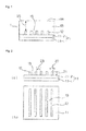
- Fig. 1 is the cross-sectional view of a cell culture device 1
- Fig. 2 is the illustrative diagram of the cell culture device 1 without a cell culture part
- Fig.2 (a) is the cross-sectional view
- Fig.2 (b) is the plan view.
- the cell culture device 1 is prepared by forming a hydrogel layer 12 on a substrate 11 and forming a cell culture part 13 covering the hydrogel layer 12.
- the hydrogel layer 12 is the carrier for gradual release of a cell growth factor.
- the hydrogel layer 12 also functions as a part to which growing first cell species adhere.
- the substrate 11 is usually a flat plate, thin flat plate, or film.
- the hydrogel layer 12 is formed into a pattern on the substrate 11, and non-cell-adhesive part 15 is exposed at the area of the substrate on which the hydrogel layer 12 is not formed.
- the substrate 11 may consist of only a base 11-1, if the base 11-1 is made of a non-cell-adhesive material (for example, polydimethylsiloxane, hereinafter referred to as PDMS).
- PDMS polydimethylsiloxane
- a non-cell-adhesive layer 11-2 is formed by coating the whole surface of the base 11-1 with a non-cell-adhesive material.
- the non-cell-adhesive layer 11-2 is required or not required according to the material and surface characteristics of the base 11-1.
- the material for the base 11-1 is selected from PDMS, PET, glass, silicone, metals (aluminum, stainless steel such as SUS 304, gold, silver, etc.), metal oxides (alumina, titanium oxide, ITO, and tin oxide), etc.
- the material for the non-cell-adhesive layer 11-2 is selected from agarose, polyethylene glycol, PDMS, etc.
- the hydrogel layer 12 is formed into a pattern for the purpose of controlling the two-dimensional form (along the surface of the substrate 11) of the aggregates of cultured first cell species.
- the hydrogel layer 12 in the embodiment is patterned into five straight lines arranged parallel.
- the line width (indicated by the arrow 22) of the patterned hydrogel layer is not specifically restricted, and preferably ranges from 10 ⁇ m to 500 ⁇ m.
- the space (indicated by the arrow 23) between the patterns of the hydrogel layer is not specifically restricted, and preferably ranges from 50 ⁇ m to 1000 ⁇ m.
- the hydrogel layer 12 has a thickness which makes a volume of the hydrogel layer 12.
- the hydrogel layer 12 can be impregnated with a cell growth factor which is stored in the layer and gradually released.
- the hydrogel layer 12 is composed of a hydrogel in order to store and gradually release a cell growth factor.
- the thickness (indicated by the arrow 21) of the hydrogel layer 12 is not specifically restricted. It is enough for the hydrogel layer 12 to have a thickness which allows the layer to contain an amount of a cell growth factor to be released sufficiently for a sufficient period for making seeded cell species migrate and grow toward the hydrogel layer 12.
- the desirable period of releasing a cell growth factor from the hydrogel layer preferably ranges from 2 days to 21 days.
- the thickness of the hydrogel layer preferably ranges from 1 ⁇ m to 3000 ⁇ m, more preferably from 20 ⁇ m to 70 ⁇ m. Such thickness enables the release of a cell growth factor in an amount sufficient for leading the direction of the growth of a first cell species throughout the culture period for the first cell species.
- the preferable material for the hydrogel layer 12 includes gelatin, collagen, hyaluronic acid, fibronectin, laminin, fibrin, etc. Of those materials, gelatin is more preferable, because it can be thermally cross-linked by heating at relatively low temperature.
- the hydrogel layer 12 may be formed optionally in several methods such as, 1) forming a thin hydrogel layer of a certain thickness on a glass plate, cutting the layer into a piece, and placing the piece on the substrate 11; 2) placing a gel material on the_substrate 11 with a dispenser: and 3) overprinting a hydrogel layer on the substrate 11 by screen printing.
- the cell culture part 13 is formed into a layer covering the hydrogel layer 12, and has a surface parallel to the plane of the substrate.
- the hydrogel layer 12 is surrounded by the non-cell-adhesive part 15 on the surface of the substrate 11.
- the cell culture part 13 formed into a layer covers the hydrogel layer 12 and the non-cell-adhesive part 15.
- the upper surface of the cell culture part 13 functions as the plane 14 for seeding a first cell species.
- the first cell species to be cultured is seeded on the plane 14 for seeding a first cell species of the cell culture device 1.
- the upper surface of the cell culture part 13 is one of the two surfaces of the layer of the cell culture part 13, and locates farther away from the substrate 11.
- the first cell species grows in the cell culture part 13, where the cell growth factor is released from the hydrogel layer 12.
- a second cell species also grows in the cell culture part 13.
- the thickness of the cell culture part 13, in other words, the distance (indicated by the arrow 26) between the surface of the hydrogel layer 12 and the plane 14 for seeding a first cell species should preferably range from 10 ⁇ m to 30 mm, more preferably from 100 ⁇ m to 5 mm.
- a thickness of the cell culture part 13 within the range effectively makes a first cell species grow toward the released cell growth factor, and consequently make the cell species form three-dimensional structure (extending along the thickness of the cell culture part).
- a preferable material for the cell culture part 13 is Matrigel or collagen, and the collagen includes collagen gel and collage sponge.
- the cell culture method for a first cell species is explained.
- a cell culture device is prepared prior to culturing the cell species.
- the cell culture device is completed through the steps of 1) forming a hydrogel layer 12 into a pattern on a substrate 11, 2) impregnating the hydrogel layer 12 with a cell growth factor, and 3) forming the layer of cell culture part 13 covering the hydrogel layer 12.
- Fig. 3 is the conceptual diagram of a cell culture method for a first cell species.
- Fig. 3(a) shows the state just after seeding a first cell species
- Fig.3 (b) shows an initial stage of the cell culture
- Fig.3 (c) shows the final stage of the cell culture.
- a first cell species is seeded on the plane 14 for seeding a first cell species, which is the surface of the cell culture part, as shown in Fig. 3(a) .
- the image of the first cell species seeded on the plane is indicated by the sign 31.
- the first cell species used in a preferable embodiment of the cell culture of the present invention include human umbilical vein endothelial cell (hereinafter referred to as HUVEC), fibroblast cell endothelial cell, epithelial cell, vascular endothelial cell, vascular smooth muscle cell, lymphatic endothelial cell, bile duct epithelial cell, and epidermal cell.
- the cell growth factor used for the cell culture method and cell culture device of the present invention is not specifically restricted, and any of cell growth factors facilitating the growth and migration of the first cell species can be used.
- VEGF vascular endothelial growth factor
- KGF keratinocyte growth factor
- the cell growth factor is released from the hydrogel layer 12 into the cell culture part 13 as shown in Fig. 3(b) .
- the image of the released cell growth factor is indicated by the arrow 34.
- the seeded cells 31 migrate and grow toward the released cell growth factor.
- the image of the aggregates of the cultured cells at an initial stage of the cell culture is indicated by the sign 32 in Fig. 3(b) .
- the first cell species repeatedly migrate and grow toward the released cell growth factor during the cell culture.
- the aggregates of the cultured first cell species bridge the plane 14 for seeding a first cell species and the hydrogel layer 12.
- the image of the aggregates of the cultured cells at the final stage of the cell culture is indicated by the sign 33 in Fig. 3(c) .
- the three-dimensional structure (along the thickness of the cell culture part 13) of the aggregates of cultured cells is controlled.
- the hydrogel layer 12 on the substrate 11 is surrounded by the non -cell- adhesive part 15 where the cultured cells do not extend.
- Such arrangement controls the two-dimensional structure (along the surface of the cell culture part 13) of the cultured cell aggregates.
- HUVECs are cultured into aggregates of cultured cells 33 having tubular structure.
- the second cell species is seeded.
- the second cell species is seeded on the cell culture part, and the cell culture device is kept under a cell culture condition to grow the second cell species.
- the cell culture condition varies depending on cell species to be cultured, and is selected from the conditions for known cell culture methods.
- Examples of the second cell species include hepatic parenchymal cell, Ito cell, pancreatic parenchymal cell, cardiac muscle cell, nerve cell, mesenchymal cell, and tumor cell. Any combination of the first and second cell species may be employed.
- Teijin® Tetoron® Film G274 placed on a hot plate (at 50 °C) was bar-coated with a 5-% aqueous solution of agarose (at 80°C), and dried in an oven at 40 °C for 30 minutes.
- a glass plate was heated on a hot plate (at 50 °C) and coated with a 30-% aqueous solution of gelatin. After cooling, a piece of the gelatin was cut out and placed on the agarose-coated film described above. Then the film was cut into a gelatin-patterned substrate,
- the gelatin-patterned substrate was vacuum-dried at 140°C for 48 hours to cross-link and sterilize the gelatin.
- the gelatin layer i.e., the hydrogel layer, had a thickness of 50 ⁇ m, and was patterned into a 70- ⁇ m wide straight line.
- HumanVEGF165 was employed as the cell growth factor.
- a piece of the cross-linked-gelatin-patterned substrate was immersed in an aqueous solution containing 40 ng/mL of human VEGF165 to impregnate the substrate with the solution.
- a control substrate was prepared by immersing a piece of the cross-linked-gelatin-patterned substrate in pure water to impregnate the substrate with the pure water.
- HUVECs were employed as the first cell species to be seeded.
- the HUVECs were cultured in a prescribed culture fluid, and then cultured in the mixture of Medium 199, 0.5 % of FCS (fetal calf serum), and 1 % of P/S (penicillin/streptomycin solution) for 1 day before seeding the cells.
- FCS fetal calf serum
- P/S penicillin/streptomycin solution
- the substrate with the pattern of the human VEGF165-impregnated gelatin was placed in each well of a 24-well plate, and a Matrigel layer, i.e., a cell culture part, was formed in each of cell culture inserts for the 24-well plate.
- the concentration of the Matrigel in the layer was 10 mg/mL.
- Two types of cell culture devices were prepared respectively with a Matrigel layer formed of 20 ⁇ L of the Matrigel solution and a Matrigel layer formed of 50 ⁇ L of the Matrigel solution.
- the Matrigel layer of 20 ⁇ L of the Matrigel solution was about 0.7 mm thick while the layer of 50 ⁇ L of the Matrigel solution was about 1.7 mm thick.
- a control cell culture device was prepared by placing the substrate with the pattern of the pure-water-impregnated gelatin in each well of a 24-well plate, and by forming a Matrigel layer, i.e., a cell culture part, in each of cell culture inserts for the 24-well plate.
- the starved HUVECs fluorescently labelled with PKH26 were seeded on the plane for seeding cell species, i.e., the surface of the Matrigel layer, and cultured in a CO 2 -purged incubator for 21 hours.
- the number of HUVECs migrating to the bottom of the cell culture part was determined by measuring the fluorescence intensity at the bottom of the cell culture inserts with a plate reader.
- the result is shown in Fig. 4 .
- the fluorescence intensity at the bottom of the cell culture inserts placed on the substrate patterned with human VEGF165-impregnated gelatin is stronger than the fluorescence intensity at the bottom of the cell culture inserts placed on the control substrate patterned with gelatin which was not impregnated with human VEGF165.
- the result implies that the HUVECs migrated through the cell culture part (the Matrigel layer) toward the human VGEF 165 which was released from the human VEGF165-impregnated gelatin patterned on the substrate.
Landscapes
- Health & Medical Sciences (AREA)
- Engineering & Computer Science (AREA)
- Life Sciences & Earth Sciences (AREA)
- Wood Science & Technology (AREA)
- Organic Chemistry (AREA)
- Biomedical Technology (AREA)
- Chemical & Material Sciences (AREA)
- Bioinformatics & Cheminformatics (AREA)
- Zoology (AREA)
- Biotechnology (AREA)
- Genetics & Genomics (AREA)
- Microbiology (AREA)
- Biochemistry (AREA)
- General Engineering & Computer Science (AREA)
- General Health & Medical Sciences (AREA)
- Cell Biology (AREA)
- Vascular Medicine (AREA)
- Immunology (AREA)
- Sustainable Development (AREA)
- Apparatus Associated With Microorganisms And Enzymes (AREA)
- Micro-Organisms Or Cultivation Processes Thereof (AREA)
Abstract
Description
- The present invention relates to a cell culture device for in vitro culture of human and animal cells. The present invention also relates to a cell culture method to conduct an in vitro culture of human and animal cells.
- Cells in a living organism are arranged and disposed regularly and three-dimensionally. In the current cell culture technology, only a limited scope of technique is available for such arrangement and disposition of cultured cells.
- There have been reported some techniques for two-dimensional cell arrangement. An example of the techniques provides a method in which cell-adhesive surface and non-cell adhesive surface are formed into patterns and used to control two-dimensional adhesion and subsequent growth of cells. In one of such known methods, a cell culture device is prepared by patterning fine lines of a temperature-responsive polymer on a substrate, and cells are cultured on the cell culture device (as disclosed in PTL 1). The cell culture device is able to control the structure of cultured cells two-dimensionally (along membrane surface), because the cell culture part of the cell culture device is patterned with fine lines of capillary width.
- On the contrary, the technology and methodology for three-dimensional cell arrangement are almost unavailable. Although a technique of layering cell sheets to make three-dimensional cell arrangement has been reported (as disclosed in PTL 2), the technique fails to achieve intentional control of the inner structure of the multilayered cells. A prior art proposed a cell culture method in which a massive form of cells is made by growing cells three-dimensionally or sterically, though the method also fails to intentionally control three-dimensional cell arrangement.
- In a three-dimensional or steric cell culture (culturing a massive form of cells), cells in the inner part of a massive form of cells are often fed with insufficient oxygen and nutrition, and the cells undergo functional decline or die. Contrary to the cultured cells, cells of three-dimensional aggregates in a living body do not face such problem, because blood vessels exist in the three-dimensional cell aggregates to supply sufficient oxygen and nutrition to cells inside the aggregates of a steric form.
- As mentioned above, a vascular network is the key factor for cells to survive and function in three-dimensional cell aggregates. The vascular network is a product of three-dimensional arrangement of vascular cells. For the future advancement of cell biology, it is indispensable to develop a technique which controls cell arrangement in a form similar to tissue structure of a living body.
- PTL 1: Japanese Patent Publication Laid open
2010-98973
PTL 1: Japanese Patent Publication Laid open2005-342112 - The problem to be solved by the present invention is to provide a cell culture device which can control the three-dimensional structure of cultured cells (aggregates of cells).
- Another problem to be solved by the present invention is to provide a cell culture method which can control the three-dimensional structure of cultured cells (aggregates of cells). Yet another problem to be solved by the present invention is to provide a cell culture method which can control the three-dimensional structure of a cultured first cell species (aggregates of cells) and also co-culture a second cell species.
- The cell culture device of the present invention controls the three-dimensional arrangement of cells by utilizing the property of cells responsive to the concentration gradient of a cell growth factor. At first, a hydrogel layer which gradually releases a cell growth factor is formed into a two-dimensional pattern on a substrate. Then a cell culture part (for example, a Matrigel layer) is placed on the patterned substrate, and cells are seeded on the cell culture part.
- The cell growth factor is gradually released from the hydrogel layer into the cell culture part to make a three-dimensional concentration gradient. Then the cells migrate along the three-dimensional concentration gradient of the cell growth factor, and consequently the cells are arranged in the cell culture part according to the three-dimensional concentration gradient.
- The method of the present invention enables three-dimensional cell arrangement in a cell culture part by means of the combination of a two-dimensionally patterned hydrogel and the property of the hydrogel gradually releasing a cell growth factor.
- A cell culture device according to one embodiment of the present invention has a hydrogel layer being formed into a pattern on a substrate and a layer of cell culture part being formed on the substrate to cover the hydrogel layer, and the cell culture device is characterized in that
the surface of the cell culture part functions as the plane for seeding a first cell species,
the hydrogel layer is impregnated with a cell growth factor, and the cell growth factor is gradually released from the hydrogel layer into the cell culture part. - In a preferable embodiment of the cell culture device of the present invention, the hydrogel layer may have a thickness which enables continuous release of the cell growth factor from the hydrogel layer into the cell culture part for a period ranging from 2 days to 21 days, and the thickness may range from 1 µm to 30 mm.
- In another preferable embodiment of the cell culture device of the present invention, the hydrogel layer may be composed of a thermally cross-linked gelatin, and the water content of a hydrogel constituting the hydrogel layer may range from 70 % to 99 %.
- The cell culture method of the present invention includes the following steps of:
- a) forming a hydrogel layer into a pattern on a substrate;
- b) impregnating the hydrogel layer with a cell growth factor;
- c) forming a layer of cell culture part on the substrate to cover the hydrogel layer and complete a cell culture device;
- d) seeding a first cell species on the surface of the cell culture part; and
- e) culturing the first cell species in the cell culture device under a culture condition.
- In a preferable embodiment of the cell culture method of the present invention, the first cell species used in the seeding of the step d) may be starved before the seeding. The water content of a hydrogel constituting the hydrogel layer ranges from 70 % to 99 %, and may be controlled to adjust the period of gradual release of the cell growth factor from the hydrogel layer into the cell culture part.
- In another preferable embodiment of the cell culture method of the present invention, the steps shown below follow the culturing of the first cell species of the step e):
- f) seeding a second cell species on the surface of the cell culture part; and
- g) culturing the second cell species in the cell culture device under a culture condition.
- The present invention, preferable embodiments of the present invention and the elements contained therein can be combined as far as possible to work the invention.
- The cell culture device of the present invention includes a hydrogel layer or a certain thickness in which a cell growth factor is impregnated. The hydrogel layer gradually releases the cell growth factor which makes the cultured first cell species migrate from the surface of the cell culture part toward the hydrogel layer and grow with the aid of the other matters specifying the invention. Such function of the hydrogel layer make it possible to control the two-dimensional structure of the cultured first cells (aggregate) along the pattern of the hydrogel layer (along the surface of the substrate), and also to control the structure of the cultured first cell species (aggregate) along the thickness of the cell culture part, in other words, the three-dimensional structure of the cultured first cell species.
- The cell culture method of the present invention also make it possible to control the three-dimensional structure of cultured cells (aggregate) as well as the cell culture device mentioned above.
-
- [
Fig. 1] Fig. 1 is the cross-sectional view of a cell culture device. - [
Fig. 2] Fig. 2 is the illustrative diagram of the cell culture device without a cell culture part.Fig.2 (a) is the cross-sectional view andFig.2 (b) is the plan view. - [
Fig. 3] Fig. 3 is the conceptual diagram of a cell culture method for a first cell species.Fig.3 (a), Fig.3 (b), and Fig.3 (c) are the conceptual diagrams respectively showing the state just after seeding a first cell species, an initial stage of the cell culture, and the final stage of the cell culture. - [
Fig. 4] Fig. 4 is a graph showing the result from the determination of the example. - The cell culture device and cell culture method of the present invention according to the embodiments of the present invention are further described referring to the figures. Some of the figures are pattern diagrams containing magnification of some elements for easy understanding of the present invention. Thus some of the dimensions or dimensional ratio between the elements may be different from that of an actual cell culture device and its parts. The dimensions, materials, forms, and relative positions of the members and parts described in the working examples of the present invention merely explain the present invention and do not restrict the scope of the present invention unless otherwise specified.
-
Fig. 1 is the cross-sectional view of a cell culture device 1,Fig. 2 is the illustrative diagram of the cell culture device 1 without a cell culture part,Fig.2 (a) is the cross-sectional view, andFig.2 (b) is the plan view. - The cell culture device 1 is prepared by forming a
hydrogel layer 12 on asubstrate 11 and forming acell culture part 13 covering thehydrogel layer 12. Thehydrogel layer 12 is the carrier for gradual release of a cell growth factor. Thehydrogel layer 12 also functions as a part to which growing first cell species adhere. - The
substrate 11 is usually a flat plate, thin flat plate, or film. Thehydrogel layer 12 is formed into a pattern on thesubstrate 11, and non-cell-adhesive part 15 is exposed at the area of the substrate on which thehydrogel layer 12 is not formed. Thesubstrate 11 may consist of only a base 11-1, if the base 11-1 is made of a non-cell-adhesive material (for example, polydimethylsiloxane, hereinafter referred to as PDMS). - If the base 11-1 is made of a cell-adhesive material (for example, polyethylene terephthalate, hereinafter referred to as PET), a non-cell-adhesive layer 11-2 is formed by coating the whole surface of the base 11-1 with a non-cell-adhesive material. In the present invention, the non-cell-adhesive layer 11-2 is required or not required according to the material and surface characteristics of the base 11-1.
- The material for the base 11-1 is selected from PDMS, PET, glass, silicone, metals (aluminum, stainless steel such as SUS 304, gold, silver, etc.), metal oxides (alumina, titanium oxide, ITO, and tin oxide), etc. The material for the non-cell-adhesive layer 11-2 is selected from agarose, polyethylene glycol, PDMS, etc.
- The
hydrogel layer 12 is formed into a pattern for the purpose of controlling the two-dimensional form (along the surface of the substrate 11) of the aggregates of cultured first cell species. As shown inFig. 2(b) , thehydrogel layer 12 in the embodiment is patterned into five straight lines arranged parallel. The line width (indicated by the arrow 22) of the patterned hydrogel layer is not specifically restricted, and preferably ranges from 10 µm to 500 µm. The space (indicated by the arrow 23) between the patterns of the hydrogel layer is not specifically restricted, and preferably ranges from 50 µm to 1000 µm. - The
hydrogel layer 12 has a thickness which makes a volume of thehydrogel layer 12. Thus thehydrogel layer 12 can be impregnated with a cell growth factor which is stored in the layer and gradually released. - The
hydrogel layer 12 is composed of a hydrogel in order to store and gradually release a cell growth factor. - The thickness (indicated by the arrow 21) of the
hydrogel layer 12 is not specifically restricted. It is enough for thehydrogel layer 12 to have a thickness which allows the layer to contain an amount of a cell growth factor to be released sufficiently for a sufficient period for making seeded cell species migrate and grow toward thehydrogel layer 12. - The period of releasing a cell growth factor from the hydrogel layer can be adjusted by controlling the degree of cross-linking of the hydrogel constituting the layer. The water content of the hydrogel indicates the degree of cross-linking of the hydrogel. The preferable degree of cross-linking of the hydrogel is represented by the water content ranging usually from 70 % to 99 %, more preferably from 90 % to 99 %. A hydrogel which contains water within the above range will continue to release a cell growth factor in an amount sufficient for leading the direction of the growth of a first cell species throughout the culture period for the first cell species.
- The desirable period of releasing a cell growth factor from the hydrogel layer preferably ranges from 2 days to 21 days. The thickness of the hydrogel layer preferably ranges from 1 µm to 3000 µm, more preferably from 20 µm to 70 µm. Such thickness enables the release of a cell growth factor in an amount sufficient for leading the direction of the growth of a first cell species throughout the culture period for the first cell species.
- The preferable material for the
hydrogel layer 12 includes gelatin, collagen, hyaluronic acid, fibronectin, laminin, fibrin, etc. Of those materials, gelatin is more preferable, because it can be thermally cross-linked by heating at relatively low temperature. - The
hydrogel layer 12 may be formed optionally in several methods such as, 1) forming a thin hydrogel layer of a certain thickness on a glass plate, cutting the layer into a piece, and placing the piece on thesubstrate 11; 2) placing a gel material onthe_substrate 11 with a dispenser: and 3) overprinting a hydrogel layer on thesubstrate 11 by screen printing. - The
cell culture part 13 is formed into a layer covering thehydrogel layer 12, and has a surface parallel to the plane of the substrate. Thehydrogel layer 12 is surrounded by the non-cell-adhesive part 15 on the surface of thesubstrate 11. Thecell culture part 13 formed into a layer covers thehydrogel layer 12 and the non-cell-adhesive part 15. - The upper surface of the
cell culture part 13 functions as theplane 14 for seeding a first cell species. The first cell species to be cultured is seeded on theplane 14 for seeding a first cell species of the cell culture device 1. The upper surface of thecell culture part 13 is one of the two surfaces of the layer of thecell culture part 13, and locates farther away from thesubstrate 11. - The first cell species grows in the
cell culture part 13, where the cell growth factor is released from thehydrogel layer 12. A second cell species also grows in thecell culture part 13. - The thickness of the
cell culture part 13, in other words, the distance (indicated by the arrow 26) between the surface of thehydrogel layer 12 and theplane 14 for seeding a first cell species should preferably range from 10 µm to 30 mm, more preferably from 100 µm to 5 mm. A thickness of thecell culture part 13 within the range effectively makes a first cell species grow toward the released cell growth factor, and consequently make the cell species form three-dimensional structure (extending along the thickness of the cell culture part). - A preferable material for the
cell culture part 13 is Matrigel or collagen, and the collagen includes collagen gel and collage sponge. - The cell culture method for a first cell species is explained. A cell culture device is prepared prior to culturing the cell species.
- The cell culture device is completed through the steps of 1) forming a
hydrogel layer 12 into a pattern on asubstrate 11, 2) impregnating thehydrogel layer 12 with a cell growth factor, and 3) forming the layer ofcell culture part 13 covering thehydrogel layer 12. -
Fig. 3 is the conceptual diagram of a cell culture method for a first cell species.Fig. 3(a) shows the state just after seeding a first cell species,Fig.3 (b) shows an initial stage of the cell culture, andFig.3 (c) shows the final stage of the cell culture. - A first cell species is seeded on the
plane 14 for seeding a first cell species, which is the surface of the cell culture part, as shown inFig. 3(a) . The image of the first cell species seeded on the plane is indicated by thesign 31. Examples of the first cell species used in a preferable embodiment of the cell culture of the present invention include human umbilical vein endothelial cell (hereinafter referred to as HUVEC), fibroblast cell endothelial cell, epithelial cell, vascular endothelial cell, vascular smooth muscle cell, lymphatic endothelial cell, bile duct epithelial cell, and epidermal cell. - The cell growth factor used for the cell culture method and cell culture device of the present invention is not specifically restricted, and any of cell growth factors facilitating the growth and migration of the first cell species can be used. For example, VEGF (vascular endothelial growth factor) is employed for culturing HUVEC and lymphatic endothelial cells, and KGF (keratinocyte growth factor) is employed for culturing epidermal cells.
- It is preferable to starve a
first cell species 31 prior to seeding, because the starvation treatment accelerates the migration and growth of the first cell species toward the released cell growth factor. - The cell growth factor is released from the
hydrogel layer 12 into thecell culture part 13 as shown inFig. 3(b) . The image of the released cell growth factor is indicated by the arrow 34. Theseeded cells 31 migrate and grow toward the released cell growth factor. The image of the aggregates of the cultured cells at an initial stage of the cell culture is indicated by thesign 32 inFig. 3(b) . - The first cell species repeatedly migrate and grow toward the released cell growth factor during the cell culture. At the final stage of the cell culture, the aggregates of the cultured first cell species bridge the
plane 14 for seeding a first cell species and thehydrogel layer 12. The image of the aggregates of the cultured cells at the final stage of the cell culture is indicated by thesign 33 inFig. 3(c) . Thus the three-dimensional structure (along the thickness of the cell culture part 13) of the aggregates of cultured cells is controlled. - The
hydrogel layer 12 on thesubstrate 11 is surrounded by the non -cell-adhesive part 15 where the cultured cells do not extend. Such arrangement controls the two-dimensional structure (along the surface of the cell culture part 13) of the cultured cell aggregates. - HUVECs are cultured into aggregates of
cultured cells 33 having tubular structure. - Then a preferable embodiment of the cell culture method of the present invention is described. In the cell culture device containing the aggregates of the cultured first cell species prepared as mentioned above, the second cell species is seeded. The second cell species is seeded on the cell culture part, and the cell culture device is kept under a cell culture condition to grow the second cell species. The cell culture condition varies depending on cell species to be cultured, and is selected from the conditions for known cell culture methods.
- Thus two species of cells are co-cultured in an environment similar to that in a living body.
- Examples of the second cell species include hepatic parenchymal cell, Ito cell, pancreatic parenchymal cell, cardiac muscle cell, nerve cell, mesenchymal cell, and tumor cell. Any combination of the first and second cell species may be employed.
- A sheet of easily-adhesive PET film (Teijin® Tetoron® Film G274) placed on a hot plate (at 50 °C) was bar-coated with a 5-% aqueous solution of agarose (at 80°C), and dried in an oven at 40 °C for 30 minutes.
- A glass plate was heated on a hot plate (at 50 °C) and coated with a 30-% aqueous solution of gelatin. After cooling, a piece of the gelatin was cut out and placed on the agarose-coated film described above. Then the film was cut into a gelatin-patterned substrate,
- The gelatin-patterned substrate was vacuum-dried at 140°C for 48 hours to cross-link and sterilize the gelatin. The gelatin layer, i.e., the hydrogel layer, had a thickness of 50 µm, and was patterned into a 70-µm wide straight line.
- HumanVEGF165 was employed as the cell growth factor. A piece of the cross-linked-gelatin-patterned substrate was immersed in an aqueous solution containing 40 ng/mL of human VEGF165 to impregnate the substrate with the solution. A control substrate was prepared by immersing a piece of the cross-linked-gelatin-patterned substrate in pure water to impregnate the substrate with the pure water.
- HUVECs were employed as the first cell species to be seeded. The HUVECs were cultured in a prescribed culture fluid, and then cultured in the mixture of Medium 199, 0.5 % of FCS (fetal calf serum), and 1 % of P/S (penicillin/streptomycin solution) for 1 day before seeding the cells. The cell culture in the mixture was conducted to starve the cells. The HUVECs were scraped after collagenase treatment just before the cell culture test, and fluorescently labeled with PKH26.
- The substrate with the pattern of the human VEGF165-impregnated gelatin was placed in each well of a 24-well plate, and a Matrigel layer, i.e., a cell culture part, was formed in each of cell culture inserts for the 24-well plate. The concentration of the Matrigel in the layer was 10 mg/mL. Two types of cell culture devices were prepared respectively with a Matrigel layer formed of 20 µL of the Matrigel solution and a Matrigel layer formed of 50 µL of the Matrigel solution. The Matrigel layer of 20 µL of the Matrigel solution was about 0.7 mm thick while the layer of 50 µL of the Matrigel solution was about 1.7 mm thick. A control cell culture device was prepared by placing the substrate with the pattern of the pure-water-impregnated gelatin in each well of a 24-well plate, and by forming a Matrigel layer, i.e., a cell culture part, in each of cell culture inserts for the 24-well plate.
- The starved HUVECs fluorescently labelled with PKH26 were seeded on the plane for seeding cell species, i.e., the surface of the Matrigel layer, and cultured in a CO2-purged incubator for 21 hours.
- After the cell culture, the number of HUVECs migrating to the bottom of the cell culture part (the Matrigel layer) was determined by measuring the fluorescence intensity at the bottom of the cell culture inserts with a plate reader.
- The result is shown in
Fig. 4 . The fluorescence intensity at the bottom of the cell culture inserts placed on the substrate patterned with human VEGF165-impregnated gelatin is stronger than the fluorescence intensity at the bottom of the cell culture inserts placed on the control substrate patterned with gelatin which was not impregnated with human VEGF165. The result implies that the HUVECs migrated through the cell culture part (the Matrigel layer) toward the human VGEF 165 which was released from the human VEGF165-impregnated gelatin patterned on the substrate. -
- 1
- Cell culture device
- 11
- Substrate
- 11-1
- Base
- 11-2
- Non-cell-adhesive layer
- 12
- Hydrogel layer
- 13
- Cell culture part
- 14
- Plane for seeding a first cell species
- 15
- Non-cell-adhesive part
- 31
- First cell species seeded (image)
- 32
- Aggregates of the cultured first cell species at an initial stage (image)
- 33
- Aggregates of the cultured first cell species at the final stage (image)
- 34
- Released cell growth factor (image)
Claims (9)
- A cell culture device having
a hydrogel layer being formed into a pattern on a substrate, and
a layer of cell culture part being formed on the substrate to cover the hydrogel layer; the cell culture device being characterized in that
the surface of the cell culture part functions as the plane for seeding a first cell species,
the hydrogel layer is impregnated with a cell growth factor, and
the cell growth factor is gradually released from the hydrogel layer into the cell culture part. - A cell culture device according to Claim 1, wherein the hydrogel layer has a
thickness which enables continuous release of the cell growth factor from the hydrogel layer into the cell culture part for a period ranging from 2 days to 21 days. - A cell culture device according to Claim 1, wherein the hydrogel layer has a
thickness ranging from 1 µm to 30 mm. - A cell culture device according to any one of Claims 1 to 3, wherein the
hydrogel layer is composed of a thermally cross-linked gelatin. - A cell culture device according to any one of Claims 1 to 4, wherein the water
content of a hydrogel constituting the hydrogel layer ranges from 70 % to 99 %. - A cell culture method comprising the following steps off:a) forming a hydrogel layer into a pattern on a substrate;b) impregnating the hydrogel layer with a cell growth factor;c) forming a layer of cell culture part on the substrate to cover the hydrogel layer and complete a cell culture devise:d) seeding a first cell species on the surface of the cell culture part; ande) culturing the first cell species in the cell culture device under a culture condition.
- A cell culture method according to Claim 6, wherein the first cell species used
in the seeding of the step d) are starved before the seeding. - A cell culture method according to Claim 6 or 7, wherein the water content of a
hydrogel constituting the hydrogel layer ranges from 70 % to 99 %, and is controlled to adjust the period of gradual release of the cell growth factor from the hydrogel layer into the cell culture part. - A cell culture method according to Claim 6, wherein the steps shown below
follow the culturing of the first cell species of the step e):f) seeding a second cell species on the surface of the cell culture part; andg) culturing the second cell species in the cell culture device under a culture condition.
Applications Claiming Priority (2)
| Application Number | Priority Date | Filing Date | Title |
|---|---|---|---|
| JP2012222744A JP5917357B2 (en) | 2012-10-05 | 2012-10-05 | Cell culture member and cell culture method |
| PCT/JP2013/076334 WO2014054538A1 (en) | 2012-10-05 | 2013-09-27 | Cell culturing member, and cell culturing method |
Publications (2)
| Publication Number | Publication Date |
|---|---|
| EP2905330A1 true EP2905330A1 (en) | 2015-08-12 |
| EP2905330A4 EP2905330A4 (en) | 2016-03-02 |
Family
ID=50434866
Family Applications (1)
| Application Number | Title | Priority Date | Filing Date |
|---|---|---|---|
| EP13843761.1A Withdrawn EP2905330A4 (en) | 2012-10-05 | 2013-09-27 | Cell culturing member, and cell culturing method |
Country Status (6)
| Country | Link |
|---|---|
| US (1) | US20150275172A1 (en) |
| EP (1) | EP2905330A4 (en) |
| JP (1) | JP5917357B2 (en) |
| KR (1) | KR20150063316A (en) |
| CN (1) | CN104704108A (en) |
| WO (1) | WO2014054538A1 (en) |
Families Citing this family (7)
| Publication number | Priority date | Publication date | Assignee | Title |
|---|---|---|---|---|
| KR102078602B1 (en) | 2015-07-23 | 2020-02-19 | 한국화학연구원 | Method for 3-dimensional co-culture of adipocytes with macrophages |
| US10000732B2 (en) | 2015-11-20 | 2018-06-19 | National Health Research Institutes | Microfluidic dual-well device for highthroughput single-cell capture and culture |
| JP6678520B2 (en) * | 2016-06-01 | 2020-04-08 | 大日本印刷株式会社 | Method for producing cell aggregate |
| WO2019161048A1 (en) * | 2018-02-14 | 2019-08-22 | The Trustees Of Columbia University In The City Of New York | Hierarchic neural microphysiological system for brain function and disorders |
| CN109628390A (en) * | 2018-12-30 | 2019-04-16 | 深圳光彩生命工程技术有限公司 | A kind of gelatinous cell culture formula of water-setting |
| EP4194547A4 (en) * | 2020-08-04 | 2024-07-31 | Nanjing Livingchip Biotechnology Co., Ltd. | METHOD FOR SCREENING TARGET CELLS OR CELLS AND BIOLOGICAL CULTURE CHIP |
| KR102522974B1 (en) * | 2020-11-26 | 2023-04-19 | 성균관대학교산학협력단 | 2d cell and 3d cell implant patch |
Family Cites Families (12)
| Publication number | Priority date | Publication date | Assignee | Title |
|---|---|---|---|---|
| US7727550B2 (en) * | 2003-02-21 | 2010-06-01 | The Uab Research Foundation | Biologically active native biomatrix composition |
| WO2005030476A1 (en) * | 2003-09-23 | 2005-04-07 | Trustees Of Boston University | Three-dimensional gels that have microscale features |
| US20070218554A1 (en) * | 2004-04-26 | 2007-09-20 | Dai Nippon Printing Co., Ltd. | Cell Culture Patterning Substrate And Method For Manufacturing The Same |
| JP4303643B2 (en) | 2004-06-01 | 2009-07-29 | 育男 森田 | Artificial tissue body and method for producing the same |
| US20060153894A1 (en) * | 2004-06-30 | 2006-07-13 | Ragae Ghabrial | Multi-compartment delivery system |
| WO2006090777A1 (en) * | 2005-02-23 | 2006-08-31 | Hi-Lex Corporation | Medical material, artificial tooth root and method of producing material for clinical use |
| US8129188B2 (en) * | 2005-09-29 | 2012-03-06 | Industrial Technology Research Institute | Cell culture apparatus and method of fabricating the apparatus |
| DE102007028423A1 (en) * | 2007-06-20 | 2008-12-24 | Fraunhofer-Gesellschaft zur Förderung der angewandten Forschung e.V. | Method and device for forming aggregates of biological cells |
| JP5656158B2 (en) * | 2008-03-05 | 2015-01-21 | 国立大学法人金沢大学 | Intracellular calcium ion monitoring device for regenerative tissue |
| JP2010098973A (en) | 2008-10-22 | 2010-05-06 | Konica Minolta Holdings Inc | Cell-culturing and supporting body and cell-culturing method |
| US9506029B2 (en) * | 2010-12-30 | 2016-11-29 | University Of Massachusetts Lowell | Responsive cell culture hydrogel |
| CN102188759A (en) * | 2011-03-11 | 2011-09-21 | 西安交通大学 | Liver tissue engineering scaffold for cell composite culture and preparation method thereof |
-
2012
- 2012-10-05 JP JP2012222744A patent/JP5917357B2/en active Active
-
2013
- 2013-09-27 CN CN201380052207.7A patent/CN104704108A/en active Pending
- 2013-09-27 EP EP13843761.1A patent/EP2905330A4/en not_active Withdrawn
- 2013-09-27 US US14/433,483 patent/US20150275172A1/en not_active Abandoned
- 2013-09-27 KR KR1020147036444A patent/KR20150063316A/en not_active Withdrawn
- 2013-09-27 WO PCT/JP2013/076334 patent/WO2014054538A1/en not_active Ceased
Also Published As
| Publication number | Publication date |
|---|---|
| US20150275172A1 (en) | 2015-10-01 |
| EP2905330A4 (en) | 2016-03-02 |
| WO2014054538A1 (en) | 2014-04-10 |
| JP2014073107A (en) | 2014-04-24 |
| KR20150063316A (en) | 2015-06-09 |
| JP5917357B2 (en) | 2016-05-11 |
| CN104704108A (en) | 2015-06-10 |
Similar Documents
| Publication | Publication Date | Title |
|---|---|---|
| US20150275172A1 (en) | Cell culture device and cell culture method | |
| Tanaka et al. | Evaluation of effects of shear stress on hepatocytes by a microchip-based system | |
| EP2335370B1 (en) | Organ-on-a-chip-device | |
| Ahn et al. | Engineering of uniform epidermal layers via sacrificial gelatin bioink‐assisted 3D extrusion bioprinting of skin | |
| US7919319B2 (en) | Cultured cell and method and apparatus for cell culture | |
| Rossi et al. | Generation of a three-dimensional full thickness skin equivalent and automated wounding | |
| JP5725560B2 (en) | Cell evaluation system using cell sheet and method of using the same | |
| WO2012133629A1 (en) | Method for producing artificial skin model, and artificial skin model | |
| Kaji et al. | Controlled cocultures of HeLa cells and human umbilical vein endothelial cells on detachable substrates | |
| EP1921450A1 (en) | Test method using cells and test kit therefor | |
| Mohamadian Namaqi et al. | Dynamic cell culture modulates colon cancer cell migration in a novel 3D cell culture system | |
| Chin et al. | A microfabricated porous collagen-based scaffold as prototype for skin substitutes | |
| Hughes et al. | Direct cell-cell communication with three-dimensional cell morphology on wrinkled microposts | |
| KR101919930B1 (en) | In vitro 3D cell culture model, method for preparing thereof and method for screening using the same | |
| Huang et al. | Microfluidic channel with embedded monolayer nanofibers for cell culture and co-culture | |
| Sugibayashi et al. | A molded hyaluronic acid gel as a micro-template for blood capillaries | |
| Li et al. | Probing tumor microtissue formation and epithelial-mesenchymal transition on a well-mesh microchip | |
| McDuffie et al. | Acrylic-based culture plate format perfusion device to establish liver endothelial–epithelial interface | |
| WO2010036913A2 (en) | Quantifying cell migration in wound healing assays | |
| Romo‐Morales et al. | Alvetex®, a highly porous polystyrene scaffold for routine three‐dimensional cell culture | |
| Polak et al. | Radial matrix constraint influences tissue contraction and promotes maturation of bi-layered skin equivalents | |
| JP5920749B2 (en) | Method for manufacturing artificial skin model and artificial skin model | |
| JP6315604B2 (en) | Method for manufacturing artificial skin model and artificial skin model | |
| Murakami et al. | Observation of a tight junction structure generated in LbL‐3D skin reconstructed by layer‐by‐layer cell coating technique | |
| Varone et al. | A novel Organ-Chip system emulates three-dimensional architecture of the human epithelia and allows fine control of mechanical forces acting on it. |
Legal Events
| Date | Code | Title | Description |
|---|---|---|---|
| PUAI | Public reference made under article 153(3) epc to a published international application that has entered the european phase |
Free format text: ORIGINAL CODE: 0009012 |
|
| 17P | Request for examination filed |
Effective date: 20150425 |
|
| AK | Designated contracting states |
Kind code of ref document: A1 Designated state(s): AL AT BE BG CH CY CZ DE DK EE ES FI FR GB GR HR HU IE IS IT LI LT LU LV MC MK MT NL NO PL PT RO RS SE SI SK SM TR |
|
| AX | Request for extension of the european patent |
Extension state: BA ME |
|
| DAX | Request for extension of the european patent (deleted) | ||
| RA4 | Supplementary search report drawn up and despatched (corrected) |
Effective date: 20160128 |
|
| RIC1 | Information provided on ipc code assigned before grant |
Ipc: C12N 5/071 20100101ALI20160122BHEP Ipc: C12M 3/04 20060101AFI20160122BHEP Ipc: C12N 1/00 20060101ALI20160122BHEP Ipc: C12M 1/12 20060101ALI20160122BHEP |
|
| STAA | Information on the status of an ep patent application or granted ep patent |
Free format text: STATUS: EXAMINATION IS IN PROGRESS |
|
| 17Q | First examination report despatched |
Effective date: 20170410 |
|
| STAA | Information on the status of an ep patent application or granted ep patent |
Free format text: STATUS: THE APPLICATION HAS BEEN WITHDRAWN |
|
| 18W | Application withdrawn |
Effective date: 20170808 |