EP2886045A1 - Biological signal measuring system - Google Patents
Biological signal measuring system Download PDFInfo
- Publication number
- EP2886045A1 EP2886045A1 EP14196031.0A EP14196031A EP2886045A1 EP 2886045 A1 EP2886045 A1 EP 2886045A1 EP 14196031 A EP14196031 A EP 14196031A EP 2886045 A1 EP2886045 A1 EP 2886045A1
- Authority
- EP
- European Patent Office
- Prior art keywords
- oxygen saturation
- average value
- section
- acquiring section
- heart rate
- Prior art date
- Legal status (The legal status is an assumption and is not a legal conclusion. Google has not performed a legal analysis and makes no representation as to the accuracy of the status listed.)
- Withdrawn
Links
Images



Classifications
-
- A—HUMAN NECESSITIES
- A61—MEDICAL OR VETERINARY SCIENCE; HYGIENE
- A61B—DIAGNOSIS; SURGERY; IDENTIFICATION
- A61B5/00—Measuring for diagnostic purposes; Identification of persons
- A61B5/72—Signal processing specially adapted for physiological signals or for diagnostic purposes
- A61B5/7235—Details of waveform analysis
- A61B5/7246—Details of waveform analysis using correlation, e.g. template matching or determination of similarity
-
- A—HUMAN NECESSITIES
- A61—MEDICAL OR VETERINARY SCIENCE; HYGIENE
- A61B—DIAGNOSIS; SURGERY; IDENTIFICATION
- A61B5/00—Measuring for diagnostic purposes; Identification of persons
- A61B5/74—Details of notification to user or communication with user or patient; User input means
- A61B5/746—Alarms related to a physiological condition, e.g. details of setting alarm thresholds or avoiding false alarms
-
- A—HUMAN NECESSITIES
- A61—MEDICAL OR VETERINARY SCIENCE; HYGIENE
- A61B—DIAGNOSIS; SURGERY; IDENTIFICATION
- A61B5/00—Measuring for diagnostic purposes; Identification of persons
- A61B5/02—Detecting, measuring or recording for evaluating the cardiovascular system, e.g. pulse, heart rate, blood pressure or blood flow
- A61B5/0205—Simultaneously evaluating both cardiovascular conditions and different types of body conditions, e.g. heart and respiratory condition
-
- A—HUMAN NECESSITIES
- A61—MEDICAL OR VETERINARY SCIENCE; HYGIENE
- A61B—DIAGNOSIS; SURGERY; IDENTIFICATION
- A61B5/00—Measuring for diagnostic purposes; Identification of persons
- A61B5/02—Detecting, measuring or recording for evaluating the cardiovascular system, e.g. pulse, heart rate, blood pressure or blood flow
- A61B5/024—Measuring pulse rate or heart rate
- A61B5/02416—Measuring pulse rate or heart rate using photoplethysmograph signals, e.g. generated by infrared radiation
-
- A—HUMAN NECESSITIES
- A61—MEDICAL OR VETERINARY SCIENCE; HYGIENE
- A61B—DIAGNOSIS; SURGERY; IDENTIFICATION
- A61B5/00—Measuring for diagnostic purposes; Identification of persons
- A61B5/145—Measuring characteristics of blood in vivo, e.g. gas concentration or pH-value ; Measuring characteristics of body fluids or tissues, e.g. interstitial fluid or cerebral tissue
- A61B5/1455—Measuring characteristics of blood in vivo, e.g. gas concentration or pH-value ; Measuring characteristics of body fluids or tissues, e.g. interstitial fluid or cerebral tissue using optical sensors, e.g. spectral photometrical oximeters
- A61B5/14551—Measuring characteristics of blood in vivo, e.g. gas concentration or pH-value ; Measuring characteristics of body fluids or tissues, e.g. interstitial fluid or cerebral tissue using optical sensors, e.g. spectral photometrical oximeters for measuring blood gases
-
- A—HUMAN NECESSITIES
- A61—MEDICAL OR VETERINARY SCIENCE; HYGIENE
- A61B—DIAGNOSIS; SURGERY; IDENTIFICATION
- A61B5/00—Measuring for diagnostic purposes; Identification of persons
- A61B5/145—Measuring characteristics of blood in vivo, e.g. gas concentration or pH-value ; Measuring characteristics of body fluids or tissues, e.g. interstitial fluid or cerebral tissue
- A61B5/1455—Measuring characteristics of blood in vivo, e.g. gas concentration or pH-value ; Measuring characteristics of body fluids or tissues, e.g. interstitial fluid or cerebral tissue using optical sensors, e.g. spectral photometrical oximeters
- A61B5/14551—Measuring characteristics of blood in vivo, e.g. gas concentration or pH-value ; Measuring characteristics of body fluids or tissues, e.g. interstitial fluid or cerebral tissue using optical sensors, e.g. spectral photometrical oximeters for measuring blood gases
- A61B5/14552—Details of sensors specially adapted therefor
-
- A—HUMAN NECESSITIES
- A61—MEDICAL OR VETERINARY SCIENCE; HYGIENE
- A61B—DIAGNOSIS; SURGERY; IDENTIFICATION
- A61B5/00—Measuring for diagnostic purposes; Identification of persons
- A61B5/72—Signal processing specially adapted for physiological signals or for diagnostic purposes
- A61B5/7203—Signal processing specially adapted for physiological signals or for diagnostic purposes for noise prevention, reduction or removal
-
- A—HUMAN NECESSITIES
- A61—MEDICAL OR VETERINARY SCIENCE; HYGIENE
- A61B—DIAGNOSIS; SURGERY; IDENTIFICATION
- A61B5/00—Measuring for diagnostic purposes; Identification of persons
- A61B5/72—Signal processing specially adapted for physiological signals or for diagnostic purposes
- A61B5/7271—Specific aspects of physiological measurement analysis
- A61B5/7278—Artificial waveform generation or derivation, e.g. synthesizing signals from measured signals
-
- A—HUMAN NECESSITIES
- A61—MEDICAL OR VETERINARY SCIENCE; HYGIENE
- A61B—DIAGNOSIS; SURGERY; IDENTIFICATION
- A61B5/00—Measuring for diagnostic purposes; Identification of persons
- A61B5/72—Signal processing specially adapted for physiological signals or for diagnostic purposes
- A61B5/7271—Specific aspects of physiological measurement analysis
- A61B5/7282—Event detection, e.g. detecting unique waveforms indicative of a medical condition
-
- A—HUMAN NECESSITIES
- A61—MEDICAL OR VETERINARY SCIENCE; HYGIENE
- A61B—DIAGNOSIS; SURGERY; IDENTIFICATION
- A61B5/00—Measuring for diagnostic purposes; Identification of persons
- A61B5/74—Details of notification to user or communication with user or patient; User input means
- A61B5/742—Details of notification to user or communication with user or patient; User input means using visual displays
-
- A—HUMAN NECESSITIES
- A61—MEDICAL OR VETERINARY SCIENCE; HYGIENE
- A61B—DIAGNOSIS; SURGERY; IDENTIFICATION
- A61B2503/00—Evaluating a particular growth phase or type of persons or animals
- A61B2503/04—Babies, e.g. for SIDS detection
-
- A—HUMAN NECESSITIES
- A61—MEDICAL OR VETERINARY SCIENCE; HYGIENE
- A61B—DIAGNOSIS; SURGERY; IDENTIFICATION
- A61B5/00—Measuring for diagnostic purposes; Identification of persons
- A61B5/02—Detecting, measuring or recording for evaluating the cardiovascular system, e.g. pulse, heart rate, blood pressure or blood flow
- A61B5/024—Measuring pulse rate or heart rate
- A61B5/02416—Measuring pulse rate or heart rate using photoplethysmograph signals, e.g. generated by infrared radiation
- A61B5/02427—Details of sensor
-
- A—HUMAN NECESSITIES
- A61—MEDICAL OR VETERINARY SCIENCE; HYGIENE
- A61B—DIAGNOSIS; SURGERY; IDENTIFICATION
- A61B5/00—Measuring for diagnostic purposes; Identification of persons
- A61B5/68—Arrangements of detecting, measuring or recording means, e.g. sensors, in relation to patient
- A61B5/6801—Arrangements of detecting, measuring or recording means, e.g. sensors, in relation to patient specially adapted to be attached to or worn on the body surface
- A61B5/6813—Specially adapted to be attached to a specific body part
- A61B5/6825—Hand
- A61B5/6826—Finger
-
- A—HUMAN NECESSITIES
- A61—MEDICAL OR VETERINARY SCIENCE; HYGIENE
- A61B—DIAGNOSIS; SURGERY; IDENTIFICATION
- A61B5/00—Measuring for diagnostic purposes; Identification of persons
- A61B5/68—Arrangements of detecting, measuring or recording means, e.g. sensors, in relation to patient
- A61B5/6801—Arrangements of detecting, measuring or recording means, e.g. sensors, in relation to patient specially adapted to be attached to or worn on the body surface
- A61B5/6813—Specially adapted to be attached to a specific body part
- A61B5/6829—Foot or ankle
Definitions
- the second optical sensor 4 includes a fastening portion 4a and a cable 4b.
- the fastening portion 4a is to be attached to a second body portion (for example, the fingertip of the foot) of the subject.
- the fastening portion 4a includes a light emitter and a light receiver.
- the light emitter emits a red light beam and an infrared light beam.
- the light receiver outputs a second signal corresponding to the intensities of the red and infrared light beams which are transmitted through or reflected from the second body portion.
- the second signal is input to the biological signal measuring apparatus 2 through the cable 4b.
- the biological signal measuring apparatus 2 may further include a first average value outputting section 43 and a second average value outputting section 44.
- the first average value outputting section 43 outputs a signal indicating the first average value A1 acquired by the first average oxygen saturation acquiring section 25.
- the second average value outputting section 44 outputs a signal indicating the second average value A2 acquired by the second average oxygen saturation acquiring section 26.
- the medical person can check visually and individually the oxygen saturation in the first body portion, and that in the second body portion. A reduction of the oxygen saturation indicates that the condition of the subject is not normal, and a countermeasure is required to be taken in advance of the determination of congenital heart disease in which the difference value is used.
- the medical person can comprehensively know the abnormality of the subject through the difference value displaying section 31, the first average value displaying section 32, and the second average value displaying section 33, and take correctly and rapidly a countermeasure. In congenital heart disease screening or the like, consequently, the burden of the medical person can be reduced, and correct determination can be supported.
- a reduction of the oxygen saturation indicates that the condition of the subject is not normal, and a countermeasure is required to be taken in advance of the determination of congenital heart disease in which the difference value is used.
- the medical person can comprehensively know the abnormality of the subject through the notifying section, and take correctly and rapidly a countermeasure. In congenital heart disease screening or the like, consequently, the burden of the medical person can be reduced, and correct determination can be supported.
- the notifying section is single, and therefore the component cost can be suppressed.
Landscapes
- Health & Medical Sciences (AREA)
- Life Sciences & Earth Sciences (AREA)
- Engineering & Computer Science (AREA)
- Physics & Mathematics (AREA)
- Public Health (AREA)
- Surgery (AREA)
- Veterinary Medicine (AREA)
- General Health & Medical Sciences (AREA)
- Animal Behavior & Ethology (AREA)
- Biophysics (AREA)
- Pathology (AREA)
- Biomedical Technology (AREA)
- Heart & Thoracic Surgery (AREA)
- Medical Informatics (AREA)
- Molecular Biology (AREA)
- Physiology (AREA)
- Signal Processing (AREA)
- Cardiology (AREA)
- Psychiatry (AREA)
- Computer Vision & Pattern Recognition (AREA)
- Artificial Intelligence (AREA)
- Spectroscopy & Molecular Physics (AREA)
- Optics & Photonics (AREA)
- Pulmonology (AREA)
- Measurement Of The Respiration, Hearing Ability, Form, And Blood Characteristics Of Living Organisms (AREA)
- Measuring Pulse, Heart Rate, Blood Pressure Or Blood Flow (AREA)
Abstract
Description
- The presently disclosed subject matter relates to a biological signal measuring system which is used in congenital heart disease screening in neonates.
- In order to perform congenital heart disease screening, a probe of a first pulse oximeter is attached to a first body portion (for example, the fingertip of the hand) of the subject, and that of a second pulse oximeter is attached to a second body portion (for example, the fingertip of the foot) of the subject (for example, see Patent Literature 1). In the case where the subject has congenital heart disease, a difference occurs between the values of arterial oxygen saturations (SpO2) which are detected in the first and second body portions. When the values of arterial oxygen saturations which are detected by the two pulse oximeters are compared to each other, therefore, presence or absence of congenital heart disease can be determined (for example, see Non-Patent
Literatures 1 and 2).
(Patent Literature 1) Japanese Patent No.4,704,037
(Non-patent Literature 1) P D Mcdonald, V Y Yu (1992), Simultaneous measurement of preductal and postductal oxygen saturation by pulse oximetry in hyaline membrane disease, Arch Dis Child 67: 1166-1168
(Non-patent Literature 2) P. Meier-Stauss, H. U. Bucher, R. Hurlimann, V. Konig, and R. Huch (1989), Pulse oximetry used for documenting oxygen saturation and right-to-left shunting immediately after birth, Eur J Pediatr 149: 851-855 - The value of arterial oxygen saturation varies every moment. It is difficult for the medical person to visually compare numerical values displayed on two pulse oximeters at certain timing, and correctly identify the difference between the numerical values.
- The presently disclosed subject matter may provide a technique for reducing the burden of a medical person in, for example, congenital heart disease screening, and supporting correct determination.
- According to the presently disclosed subject matter, there may be provided a biological signal measuring system comprising: a first optical sensor which is to be attached to a first body portion of a subject; a first attenuation ratio acquiring section which is configured to acquire a temporal change of a first attenuation ratio corresponding to an attenuation ratio of a plurality of wavelengths in the first body portion, based on a first signal output from the first optical sensor; a first oxygen saturation acquiring section which is configured to acquire a temporal change of a first oxygen saturation corresponding to an arterial oxygen saturation in the first body portion, based on the temporal change of the first attenuation ratio; a second optical sensor which is to be attached to a second body portion of the subject; a second attenuation ratio acquiring section which is configured to acquire a temporal change of a second attenuation ratio corresponding to an attenuation ratio of a plurality of wavelengths in the second body portion, based on a second signal output from the second optical sensor; a second oxygen saturation acquiring section which is configured to acquire a temporal change of a second oxygen saturation corresponding to an arterial oxygen saturation in the second body portion, based on the temporal change of the second attenuation ratio; a first average oxygen saturation acquiring section which is configured to acquire a first average value corresponding to an average value of the first oxygen saturation in a predetermined time period starting at a first time; a second average oxygen saturation acquiring section which is configured to acquire a second average value corresponding to an average value of the second oxygen saturation in the predetermined time period starting at a second time that is different from the first time; a difference value acquiring section which is configured to acquire a difference value between the first average value and the second average value; and a difference value displaying section which is configured to display the difference value.
- The biological signal measuring system may further comprise: a cross-correlation function acquiring section which is configured to acquire a cross-correlation function of the temporal change of the first attenuation ratio and the temporal change of the second attenuation ratio, or a cross-correlation function of the temporal change of the first oxygen saturation and the temporal change of the second oxygen saturation, and the first time and the second time may be determined based on the cross-correlation function.
- The biological signal measuring system may further comprise: a notifying section which, when the difference value is equal to or larger than a threshold, is configured to take a given notifying state.
- When the first average value is smaller than a threshold, or when the second average value is smaller than the threshold, the notifying section may be configured to take the given notifying state irrespective of the difference value.
- The biological signal measuring system may further comprise: a first average value displaying section which is configured to display the first average value; and a second average value displaying section which is configured to display the second average value.
- The biological signal measuring system may further comprise: a first notifying section which, when the first average value is smaller than a threshold, is configured to take a given notifying state; a second notifying section which, when the second average value is smaller than a threshold, is configured to take a given notifying state; and a third notifying section which, when the difference value is equal to or larger than a threshold, is configured to take a given notifying state.
- The biological signal measuring system may further comprise: a heart rate acquiring section which is configured to acquire a heart rate of the subject based on at least one of the first signal and the second signal; and a single heart rate displaying section which is configured to display the heart rate.
- The heart rate acquiring section may be configured to select one of the first signal and the second signal which contains a smaller amount of noises, and acquire the heart rate based on the selected one of the first signal and the second signal.
- The heart rate acquiring section may be configured to acquire, as the heart rate, an average value of: the heart rate based on the first signal; and the heart rate based on the second signal.
-
-
Fig. 1 is a view showing a biological signal measuring system of a first embodiment of the presently disclosed subject matter. -
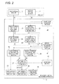
Fig. 2 is a functional block diagram showing the configuration of a biological signal measuring apparatus of the biological signal measuring system ofFig. 1 . -
Figs. 3A and 3B are views illustrating the operation of biological signal measuring apparatus ofFig. 2 . -
Fig. 4 is a view showing a biological signal measuring system of a second embodiment of the presently disclosed subject matter. -
Fig. 5 is a functional block diagram showing the configuration of a biological signal measuring apparatus of the biological signal measuring system ofFig. 4 . - Embodiments of the presently disclosed subject matter will be described in detail with reference to the accompanying drawings. In the drawings which will be used in the following description, the scale is adequately changed in order to draw components in a recognizable size.
-
Fig. 1 shows a biological signal measuringsystem 1 of a first embodiment of the presently disclosed subject matter. The biological signal measuringsystem 1 includes a biologicalsignal measuring apparatus 2, a firstoptical sensor 3, and a secondoptical sensor 4. The biologicalsignal measuring apparatus 2 is a so-called pulse oximeter, and the firstoptical sensor 3 and the secondoptical sensor 4 function as probes of the pulse oximeter. - The first
optical sensor 3 includes afastening portion 3a and acable 3b. Thefastening portion 3a is to be attached to a first body portion (for example, the fingertip of the hand) of the subject. Thefastening portion 3a includes a light emitter and a light receiver. The light emitter emits a red light beam and an infrared light beam. The light receiver outputs a first signal corresponding to the intensities of the red and infrared light beams which are transmitted through or reflected from the first body portion. The first signal is input to the biologicalsignal measuring apparatus 2 through thecable 3b. - The second
optical sensor 4 includes afastening portion 4a and acable 4b. Thefastening portion 4a is to be attached to a second body portion (for example, the fingertip of the foot) of the subject. Thefastening portion 4a includes a light emitter and a light receiver. The light emitter emits a red light beam and an infrared light beam. The light receiver outputs a second signal corresponding to the intensities of the red and infrared light beams which are transmitted through or reflected from the second body portion. The second signal is input to the biologicalsignal measuring apparatus 2 through thecable 4b. -
Fig. 2 is a functional block diagram showing the configuration of the biologicalsignal measuring apparatus 2. The biologicalsignal measuring apparatus 2 includes a first attenuationratio acquiring section 21, a second attenuationratio acquiring section 22, a first oxygensaturation acquiring section 23, a second oxygensaturation acquiring section 24, a first average oxygensaturation acquiring section 25, a second average oxygensaturation acquiring section 26, a differencevalue acquiring section 27, and a differencevalue outputting section 28. - The first attenuation
ratio acquiring section 21 acquires a temporal change of a first attenuation ratio φ1 corresponding to an attenuation ratio in the first body portion, based on the first signal G1 output from the firstoptical sensor 3. Specifically, attenuations of the red and infrared light beams due to transmission or reflection in the first body portion are acquired, and the ratio of the attenuations is set as the first attenuation ratio φ1. The first attenuation ratio φ1 is temporally changed in accordance with the blood pulsation in the first body portion. - The second attenuation
ratio acquiring section 22 acquires a temporal change of a second attenuation ratio φ2 corresponding to an attenuation ratio in the second body portion, based on the second signal G2 output from the secondoptical sensor 4. Specifically, attenuations of the red and infrared light beams due to transmission or reflection in the second body portion are acquired, and the ratio of the attenuations is set as the second attenuation ratio φ2. The second attenuation ratio φ2 is temporally changed in accordance with blood pulsation in the second body portion. - The first oxygen
saturation acquiring section 23 acquires a first oxygen saturation S1 corresponding to the arterial oxygen saturation in the first body portion, based on the first attenuation ratio φ1 acquired by the first attenuationratio acquiring section 21. The method of calculating an arterial oxygen saturation from an attenuation ratio may be a related-art method. Therefore, description of the method is omitted. In accordance with the temporal change of the first attenuation ratio φ1, also the first oxygen saturation S1 is temporally changed. - The second oxygen
saturation acquiring section 24 acquires a second oxygen saturation S2 corresponding to the arterial oxygen saturation in the second body portion, based on the second attenuation ratio φ2 acquired by the second attenuationratio acquiring section 22. The method of calculating an arterial oxygen saturation from an attenuation ratio may be a related-art method. Therefore, description of the method is omitted. In accordance with the temporal change of the second attenuation ratio φ2, also the second oxygen saturation S2 is temporally changed. - The first average oxygen
saturation acquiring section 25 acquires a first average value A1 corresponding to an average value of the first oxygen saturation S1 in a predetermined time period starting at a first time. The second average oxygensaturation acquiring section 26 acquires a second average value A2 corresponding to an average value of the second oxygen saturation S2 in a predetermined time period starting at a second time that is different from the first time. The differencevalue acquiring section 27 acquires the difference value |A1 - A2| between the first average value A1 and the second average value A2. The reason for the processes of the first average oxygensaturation acquiring section 25, the second average oxygensaturation acquiring section 26, and the differencevalue acquiring section 27 will be described with reference toFigs. 3A and 3B . -
Fig. 3A shows the temporal changes of the first oxygen saturation S1 acquired by the first oxygensaturation acquiring section 23, and the second oxygen saturation S2 acquired by the second oxygensaturation acquiring section 24. As described above, the difference value |S1 - S2 between the first oxygen saturation S1 and the second oxygen saturation S2 at certain timing can be used in determination of presence or absence of congenital heart disease in a neonate. When the values of S1 and S2 are changed every moment as shown in, for example, the middle portion of the graph, however, also the difference value |S1 - S2| is changed every moment. Therefore, the medical person is puzzled to determine which value is employed. - Consequently, the first average oxygen
saturation acquiring section 25 and the second average oxygensaturation acquiring section 26 average the values of the first oxygen saturation S1 and the second oxygen saturation S2 over a predetermined time period T to acquire the first average value A1 and the second average value A2. The temporal changes of the first average value A1 and the second average value A2 are moderate as compared to those of the first oxygen saturation S1 and the second oxygen saturation S2. Therefore, also the temporal change of the difference value |A1 - A2| acquired by the differencevalue acquiring section 27 is moderate, and the medical person easily knows the value. The predetermined time period T is adequately determined so as to contain a plurality of periods when the first oxygen saturation S1 and the second oxygen saturation S2 are updated. - In a configuration where the first oxygen saturation S1 and the second oxygen saturation S2 are simply averaged and the difference value of the average values is acquired, however, another cause of a measurement error is produced.
Fig. 3B shows the temporal changes of the first attenuation ratio φ1 acquired by the first attenuationratio acquiring section 21, and the second attenuation ratio φ2 acquired by the second attenuationratio acquiring section 22. The time period of arrival of blood from the heart to the first body portion is not equal to that of arrival of blood from the heart to the second body portion. Therefore, a time difference D is produced in the temporal changes of the first attenuation ratio φ1 and the second attenuation ratio φ2. In a significant comparison between the first oxygen saturation S1 and the second oxygen saturation S2, namely, it is required to consider the time difference. - Therefore, the first average oxygen
saturation acquiring section 25 and the second average oxygensaturation acquiring section 26 acquire the first average value A1 and the second average value A2 of predetermined time periods T starting at different times t1 and t2, respectively. For example, the time t1 and the time t2 are determined so that the difference between the times corresponds to the time difference D. Therefore, the difference value |A1 - A2| can be acquired based on the first oxygen saturation S1 and second oxygen saturation S2 of blood having the same meaning. - The difference
value outputting section 28 outputs a signal indicating the difference value |A1 - A2| acquired by the differencevalue acquiring section 27. As shown inFig. 1 , the biologicalsignal measuring apparatus 2 includes a differencevalue displaying section 31. The a differencevalue displaying section 31 displays the difference value |A1 - A2| (in the illustrated example, 7%) indicated by the signal which is output from the differencevalue outputting section 28. - According to the thus configured biological
signal measuring system 1, the difference value between the arterial oxygen saturation in the first body portion of the subject, and that in the second body portion is displayed, and hence the medical person is not required to determine the difference value while visually comparing a plurality of arterial oxygen saturations with each other. The difference value is acquired based on the values of the arterial oxygen saturations which are averaged over the predetermined time periods T, respectively. Therefore, the temporal change can be moderated. Since the predetermined time periods start at the different times, it is possible to suppress a measurement error of the arterial oxygen saturation due to a difference between the time periods of arrival of blood from the heart to the first and second body portions. In congenital heart disease screening or the like, consequently, the burden of the medical person can be reduced, and correct determination can be supported. - As shown in
Fig. 2 , the biologicalsignal measuring apparatus 2 may further include a cross-correlationfunction acquiring section 41. The cross-correlationfunction acquiring section 41 acquires a cross-correlation function of the temporal change of the first attenuation ratio φ1 acquired by the first attenuationratio acquiring section 21, and that of the second attenuation ratio φ2 acquired by the second attenuationratio acquiring section 22. The time t1 when the predetermined time period T for acquiring the first average value A1 by the first average oxygensaturation acquiring section 25 is started, and the time t2 when the predetermined time period T for acquiring the second average value A2 by the second average oxygensaturation acquiring section 26 is started are determined based on the cross-correlation function acquired by the cross-correlationfunction acquiring section 41. Specifically, when the times t1 and t2 are determined so that the cross-correlation function has the maximum value, the interval between the times t1 and t2 shown inFig. 3B can be made coincident with the difference D between the time periods of arrival of blood from the heart to the first and second body portions. - The cross-correlation
function acquiring section 41 may be configured so as to acquire a cross-correlation function of the temporal change of the first oxygen saturation S1 acquired by the first oxygensaturation acquiring section 23, and that of the second oxygen saturation S2 acquired by the second oxygensaturation acquiring section 24. Also in this case, when the times t1 and t2 are determined so that the cross-correlation function has the maximum value, the interval between the times t1 and t2 shown inFig. 3B can be made coincident with the difference D between the time periods of arrival of blood from the heart to the first and second body portions. - According to the configuration, the predetermined time periods can be set in a portion in which the values related to the first and second body portions are similarly changed. Therefore, a measurement error of the arterial oxygen saturation due to a difference between the times of arrival of blood from the heart to the first and second body portions can be minimized. In congenital heart disease screening or the like, consequently, the burden of the medical person can be reduced, and correct determination can be supported.
- As shown in
Figs. 1 and2 , the biologicalsignal measuring apparatus 2 may further include a notifyingsection 42. The notifyingsection 42 is configured so as to, when the difference value |A1 - A2| acquired by the differencevalue acquiring section 27 is equal to or larger than a predetermined threshold, take a given notifying state. For example, the threshold is set to 3% that is a value at which there is suspicion of congenital heart disease. In the case where the difference value |A1 - A2| is equal to or larger than 3%, the notifyingsection 42 emits, for example, red light. In the case where the difference value is smaller than 3%, the notifyingsection 42 may emit light of a color other than red, or may be set to a non-light-emission state. - According to the configuration, the medical person can easily determine the condition of the subject by referring the state of the notifying
section 42. In congenital heart disease screening or the like, consequently, the burden of the medical person can be reduced, and correct determination can be supported. - The notifying
section 42 may be configured in the following manner. When the first average value A1 acquired by the first average oxygensaturation acquiring section 25 is smaller than a predetermined threshold, or when the second average value A2 acquired by the second average oxygensaturation acquiring section 26 is smaller than the threshold, the notifying section takes the given notifying state irrespective of the difference value |A1 - A2| acquired by the differencevalue acquiring section 27. In this case, although not illustrated, the first average value A1 acquired by the first average oxygensaturation acquiring section 25, and the second average value A2 acquired by the second average oxygensaturation acquiring section 26 are directly input to the notifyingsection 42. For example, the threshold is set to 95% that is a value at which the condition of the subject is suspicious of not being normal. In the case where at least one of the first average value A1 and the second average value A2 is smaller than 95%, even when the difference value |A1 - A2| is smaller than 3%, the notifyingsection 42 emits, for example, red light. In the case where both the first average value A1 and the second average value A2 are equal to or larger than 95%, and the difference value |A1 - A2| is smaller than 3%, the notifyingsection 42 may emit light of a color other than red, or may be set to a non-light-emission state. - A reduction of the oxygen saturation indicates that the condition of the subject is not normal, and a countermeasure is required to be taken in advance of the determination of congenital heart disease in which the difference value is used. According to the configuration, the medical person can comprehensively know the abnormality of the subject through the notifying
section 42, and take correctly and rapidly a countermeasure. In congenital heart disease screening or the like, consequently, the burden of the medical person can be reduced, and correct determination can be supported. Moreover, the notifyingsection 42 is single, and therefore the component cost can be suppressed. - As shown in
Fig. 2 , the biologicalsignal measuring apparatus 2 may further include a first averagevalue outputting section 43 and a second averagevalue outputting section 44. The first averagevalue outputting section 43 outputs a signal indicating the first average value A1 acquired by the first average oxygensaturation acquiring section 25. The second averagevalue outputting section 44 outputs a signal indicating the second average value A2 acquired by the second average oxygensaturation acquiring section 26. - In this case, as shown in
Fig. 1 , the biologicalsignal measuring apparatus 2 further includes a first averagevalue displaying section 32 and a second averagevalue displaying section 33. The first averagevalue displaying section 32 displays the first average value A1 (in the illustrated example, 97%) indicated by the signal which is output from the first averagevalue outputting section 43. The second averagevalue displaying section 33 displays the second average value A2 (in the illustrated example, 90%) indicated by the signal which is output from the second averagevalue outputting section 44. - According to the configuration, the medical person can check visually and individually the oxygen saturation in the first body portion, and that in the second body portion. A reduction of the oxygen saturation indicates that the condition of the subject is not normal, and a countermeasure is required to be taken in advance of the determination of congenital heart disease in which the difference value is used. According to the configuration, the medical person can comprehensively know the abnormality of the subject through the difference
value displaying section 31, the first averagevalue displaying section 32, and the second averagevalue displaying section 33, and take correctly and rapidly a countermeasure. In congenital heart disease screening or the like, consequently, the burden of the medical person can be reduced, and correct determination can be supported. - As shown in
Fig. 2 , the biologicalsignal measuring apparatus 2 may further include a heartrate acquiring section 45. The heartrate acquiring section 45 acquires the heart rate of the subject based on at least one of the first signal G1 which is output from the firstoptical sensor 3, and the second signal G2 which is output from the secondoptical sensor 4. Specifically, periodical changes of the first and second signals G1, G2 due to the blood pulsation owing to the heartbeat are detects, and determined as the heart rate of the subject. - In this case, as shown in
Fig. 1 , the biologicalsignal measuring apparatus 2 further includes a single heartrate displaying section 34. The heartrate displaying section 34 displays the heart rate of the subject which is acquired by the heartrate acquiring section 45. - Unlike of the arterial oxygen saturation which is acquired in a plurality of body portions, the heart rate does not have a plurality of values at the same time. Although the heart rate can be acquired based on each of the first and second signals G1, G2, heart rates which are to be simultaneously displayed are unified into a single heart rate. Therefore, the medical person can know the heart rate without feeling cumbersome. In congenital heart disease screening or the like, consequently, the burden of the medical person can be reduced, and correct determination can be supported. Moreover, the heart
rate displaying section 34 is single, and therefore the component cost can be suppressed. - For example, the heart
rate acquiring section 45 selects one of the first signal G1 and the second signal G2 which contains a smaller amount of noises, and acquires the heart rate based on the selected one of the first signal G1 and the second signal G1. In this case, a more correct value can be provided as the heart rate of the subject. - Alternatively, the heart
rate acquiring section 45 may be configured so as to acquire the average value of the heart rate based on the first signal G1, and the heart rate based on the second signal G2, as the heart rate. In this case, a more stable value can be provided as the heart rate of the subject. - Next, a biological
signal measuring system 1A of a second embodiment of the presently disclosed subject matter will be described with reference toFigs. 4 and5 . The components which are identical or similar to those of the biologicalsignal measuring system 1 of the first embodiment are denoted by the same reference numerals, and repeated description is omitted. - The biological
signal measuring system 1A includes a biologicalsignal measuring apparatus 2A. The biologicalsignal measuring apparatus 2A is different from the biologicalsignal measuring apparatus 2 of the first embodiment in that the apparatus includes a first notifyingsection 46, a second notifyingsection 47, and a third notifyingsection 48, in place of the notifyingsection 42. - The first notifying
section 46 is configured so as to, when the first average value A1 acquired by the first average oxygensaturation acquiring section 25 is smaller than a predetermined threshold, take a given notifying state. For example, the threshold is set to 95% that is a value at which the condition of the subject is suspicious of not being normal. In the case where the first average value A1 is smaller than 95%, the first notifyingsection 46 emits, for example, red light. In the case where the first average value A1 is equal to or larger than 95%, the first notifyingsection 46 may emit light of a color other than red, or may be set to a non-light-emission state. - The second notifying
section 47 is configured so as to, when the second average value A2 acquired by the second average oxygensaturation acquiring section 26 is smaller than a predetermined threshold, take a given notifying state. For example, the threshold is set to 95% that is a value at which the condition of the subject is suspicious of not being normal. In the case where the second average value A2 is smaller than 95%, the second notifyingsection 47 emits, for example, red light. In the case where the second average value A2 is equal to or larger than 95%, the second notifyingsection 47 may emit light of a color other than red, or may be set to a non-light-emission state. - The third notifying
section 48 is configured so as to, when the difference value |A1 - A2| acquired by the differencevalue acquiring section 27 is equal to or larger than a predetermined threshold, take a given notifying state. For example, the threshold is set to 3% that is a value at which there is suspicion of congenital heart disease. In the case where the difference value |A1 - A2 is equal to or larger than 3%, the third notifyingsection 48 emits, for example, red light. In the case where the difference value is smaller than 3%, the third notifyingsection 48 may emit light of a color other than red, or may be set to a non-light-emission state. - According to the configuration, it is possible to easily know whether each of the arterial oxygen saturation in the first body portion, that in the second body portion, and the difference value of the two arterial oxygen saturations is within the normal range or not. In the case where an abnormality occurs in the condition of the subject, furthermore, it is possible to easily identify which one of the measurement values causes the abnormality. In congenital heart disease screening or the like, consequently, the burden of the medical person can be reduced, and correct determination can be supported.
- The embodiments have been described in order to facilitate understanding of the invention, and are not intended to limit the invention. It is a matter of course that the invention may be changed or improved without departing the spirit thereof, and includes equivalent of the embodiments.
- In the embodiments, the signal which is output from the difference
value outputting section 28, and which indicates the difference value |A1 - A2| is used for displaying the difference value on the differencevalue displaying section 31. Alternatively, the signal output from the differencevalue outputting section 28 may be used for indicating the difference value by means of sounds, symbols, characters, or colors. This is applicable also to the first averagevalue outputting section 43 and the second averagevalue outputting section 44. - The difference
value displaying section 31 is not always required to be disposed integrally with the biologicalsignal measuring apparatus 2. The signal output from the differencevalue outputting section 28 may be transmitted by wired or wireless transmission, and the difference value may be displayed on a displaying device which is separately disposed. This is applicable also to the first averagevalue displaying section 32 and the second averagevalue displaying section 33. - In the embodiments, the difference value acquired by the difference
value acquiring section 27 is used in determination of presence or absence of congenital heart disease in a neonate. The biological signal measuring system 1 (1A) of the presently disclosed subject matter can be used in an appropriate object in which the difference value can be set as a determination index. In this case, the threshold at which the notifying section 42 (third notifying section 48) takes the given notifying state can be adequately changed in accordance with the object. - According to an aspect of the presently disclosed subject matter, there is provided A biological signal measuring system comprising: a first optical sensor which is to be attached to a first body portion of a subject; a first attenuation ratio acquiring section which is configured to acquire a temporal change of a first attenuation ratio corresponding to an attenuation ratio of a plurality of wavelengths in the first body portion, based on a first signal output from the first optical sensor; a first oxygen saturation acquiring section which is configured to acquire a temporal change of a first oxygen saturation corresponding to an arterial oxygen saturation in the first body portion, based on the temporal change of the first attenuation ratio; a second optical sensor which is to be attached to a second body portion of the subject; a second attenuation ratio acquiring section which is configured to acquire a temporal change of a second attenuation ratio corresponding to an attenuation ratio of a plurality of wavelengths in the second body portion, based on a second signal output from the second optical sensor; a second oxygen saturation acquiring section which is configured to acquire a temporal change of a second oxygen saturation corresponding to an arterial oxygen saturation in the second body portion, based on the temporal change of the second attenuation ratio; a first average oxygen saturation acquiring section which is configured to acquire a first average value corresponding to an average value of the first oxygen saturation in a predetermined time period starting at a first time; a second average oxygen saturation acquiring section which is configured to acquire a second average value corresponding to an average value of the second oxygen saturation in the predetermined time period starting at a second time that is different from the first time; a difference value acquiring section which is configured to acquire a difference value between the first average value and the second average value; and a difference value displaying section which is configured to display the difference value.
- According to the above configuration, a difference value of the arterial oxygen saturation in the first body portion of the subject, and that in the second body portion is displayed, and hence the medical person is not required to determine the difference value while visually comparing a plurality of arterial oxygen saturations with each other. The difference value is acquired based on the values of the arterial oxygen saturations which are averaged over the predetermined time periods T, respectively. Therefore, the temporal change can be moderated. Since the predetermined time periods start at the different times, it is possible to suppress a measurement error of the arterial oxygen saturation due to a difference between the time periods of arrival of blood from the heart to the first and second body portions. In congenital heart disease screening or the like, consequently, the burden of the medical person can be reduced, and correct determination can be supported.
- The biological signal measuring system may further comprise: a cross-correlation function acquiring section which is configured to acquire a cross-correlation function of the temporal change of the first attenuation ratio and the temporal change of the second attenuation ratio, or a cross-correlation function of the temporal change of the first oxygen saturation and the temporal change of the second oxygen saturation, and the first time and the second time may be determined based on the cross-correlation function.
- According to the above configuration, the predetermined time periods can be set in a portion in which the values related to the first and second body portions are similarly changed. Therefore, a measurement error of the arterial oxygen saturation due to a difference between the times of arrival of blood from the heart to the first and second body portions can be minimized. In congenital heart disease screening or the like, consequently, the burden of the medical person can be reduced, and correct determination can be supported.
- The biological signal measuring system may further comprise: a notifying section which, when the difference value is equal to or larger than a threshold, is configured to take a given notifying state.
- According to the above configuration, the medical person can easily determine the condition of the subject by referring the state of the notifying section. In congenital heart disease screening or the like, consequently, the burden of the medical person can be reduced, and correct determination can be supported.
- When the first average value is smaller than a threshold, or when the second average value is smaller than the threshold, the notifying section may be configured to take the given notifying state irrespective of the difference value.
- A reduction of the oxygen saturation indicates that the condition of the subject is not normal, and a countermeasure is required to be taken in advance of the determination of congenital heart disease in which the difference value is used. According to the above configuration, the medical person can comprehensively know the abnormality of the subject through the notifying section, and take correctly and rapidly a countermeasure. In congenital heart disease screening or the like, consequently, the burden of the medical person can be reduced, and correct determination can be supported. Moreover, the notifying section is single, and therefore the component cost can be suppressed.
- The biological signal measuring system may further comprise: a first average value displaying section which is configured to display the first average value; and a second average value displaying section which is configured to display the second average value.
- According to the above configuration, the medical person can check visually and individually the oxygen saturation in the first body portion, and that in the second body portion. A reduction of the oxygen saturation indicates that the condition of the subject is not normal, and a countermeasure is required to be taken in advance of the determination of congenital heart disease in which the difference value is used. According to the configuration, the medical person can comprehensively know the abnormality of the subject through the difference value displaying section, the first average value displaying section, and the second average value displaying section, and take correctly and rapidly a countermeasure. In congenital heart disease screening or the like, consequently, the burden of the medical person can be reduced, and correct determination can be supported.
- The biological signal measuring system may further comprise: a first notifying section which, when the first average value is smaller than a threshold, is configured to take a given notifying state; a second notifying section which, when the second average value is smaller than a threshold, is configured to take a given notifying state; and a third notifying section which, when the difference value is equal to or larger than a threshold, is configured to take a given notifying state.
- According to the above configuration, it is possible to easily know whether the arterial oxygen saturation in the first body portion, that in the second body portion, and the difference value of the two arterial oxygen saturations are in respective normal ranges or not. In the case where an abnormality occurs in the condition of the subject, furthermore, it is possible to easily identify which one of the measurement values causes the abnormality. In congenital heart disease screening or the like, consequently, the burden of the medical person can be reduced, and correct determination can be supported.
- The biological signal measuring system may further comprise: a heart rate acquiring section which is configured to acquire a heart rate of the subject based on at least one of the first signal and the second signal; and a single heart rate displaying section which is configured to display the heart rate.
- Unlike of the arterial oxygen saturation which is acquired in a plurality of body portions, the heart rate does not have a plurality of values at the same time. Although the heart rate can be acquired based on each of the first and second signals, heart rates which are to be simultaneously displayed are unified into a single heart rate. Therefore, the medical person can know the heart rate without feeling cumbersome. In congenital heart disease screening or the like, consequently, the burden of the medical person can be reduced, and correct determination can be supported. Moreover, the heart rate displaying section is single, and therefore the component cost can be suppressed.
- The heart rate acquiring section may be configured to select one of the first signal and the second signal which contains a smaller amount of noises, and acquire the heart rate based on the selected one of the first signal and the second signal.
- In this case, a more correct value can be provided as the heart rate of the subject.
- The heart rate acquiring section may be configured to acquire, as the heart rate, an average value of: the heart rate based on the first signal; and the heart rate based on the second signal.
- In this case, a more stable value can be provided as the heart rate of the subject.
Claims (9)
- A biological signal measuring system comprising:a first optical sensor which is to be attached to a first body portion of a subject;a first attenuation ratio acquiring section which is configured to acquire a temporal change of a first attenuation ratio corresponding to an attenuation ratio of a plurality of wavelengths in the first body portion, based on a first signal output from the first optical sensor;a first oxygen saturation acquiring section which is configured to acquire a temporal change of a first oxygen saturation corresponding to an arterial oxygen saturation in the first body portion, based on the temporal change of the first attenuation ratio;a second optical sensor which is to be attached to a second body portion of the subject;a second attenuation ratio acquiring section which is configured to acquire a temporal change of a second attenuation ratio corresponding to an attenuation ratio of a plurality of wavelengths in the second body portion, based on a second signal output from the second optical sensor;a second oxygen saturation acquiring section which is configured to acquire a temporal change of a second oxygen saturation corresponding to an arterial oxygen saturation in the second body portion, based on the temporal change of the second attenuation ratio;a first average oxygen saturation acquiring section which is configured to acquire a first average value corresponding to an average value of the first oxygen saturation in a predetermined time period starting at a first time;a second average oxygen saturation acquiring section which is configured to acquire a second average value corresponding to an average value of the second oxygen saturation in the predetermined time period starting at a second time that is different from the first time;a difference value acquiring section which is configured to acquire a difference value between the first average value and the second average value; anda difference value displaying section which is configured to display the difference value.
- The biological signal measuring system according to claim 1, further comprising: a cross-correlation function acquiring section which is configured to acquire a cross-correlation function of the temporal change of the first attenuation ratio and the temporal change of the second attenuation ratio, or a cross-correlation function of the temporal change of the first oxygen saturation and the temporal change of the second oxygen saturation, wherein
the first time and the second time are determined based on the cross-correlation function. - The biological signal measuring system according to claim 1 or 2, further comprising: a notifying section which, when the difference value is equal to or larger than a threshold, is configured to take a given notifying state.
- The biological signal measuring system according to claim 3, wherein, when the first average value is smaller than a threshold, or when the second average value is smaller than the threshold, the notifying section is configured to take the given notifying state irrespective of the difference value.
- The biological signal measuring system according to claim 1 or 2, further comprising:a first average value displaying section which is configured to display the first average value; anda second average value displaying section which is configured to display the second average value.
- The biological signal measuring system according to claim 5, further comprising:a first notifying section which, when the first average value is smaller than a threshold, is configured to take a given notifying state;a second notifying section which, when the second average value is smaller than a threshold, is configured to take a given notifying state; anda third notifying section which, when the difference value is equal to or larger than a threshold, is configured to take a given notifying state.
- The biological signal measuring system according to any one of claims 1 to 6, further comprising:a heart rate acquiring section which is configured to acquire a heart rate of the subject based on at least one of the first signal and the second signal; anda single heart rate displaying section which is configured to display the heart rate.
- The biological signal measuring system according to claim 7, wherein the heart rate acquiring section is configured to select one of the first signal and the second signal which contains a smaller amount of noises, and acquire the heart rate based on the selected one of the first signal and the second signal.
- The biological signal measuring system according to claim 7, wherein the heart rate acquiring section is configured to acquire, as the heart rate, an average value of: the heart rate based on the first signal; and the heart rate based on the second signal.
Applications Claiming Priority (1)
| Application Number | Priority Date | Filing Date | Title |
|---|---|---|---|
| JP2013251445A JP6298278B2 (en) | 2013-12-04 | 2013-12-04 | Biological signal measurement system |
Publications (1)
| Publication Number | Publication Date |
|---|---|
| EP2886045A1 true EP2886045A1 (en) | 2015-06-24 |
Family
ID=52101036
Family Applications (1)
| Application Number | Title | Priority Date | Filing Date |
|---|---|---|---|
| EP14196031.0A Withdrawn EP2886045A1 (en) | 2013-12-04 | 2014-12-03 | Biological signal measuring system |
Country Status (4)
| Country | Link |
|---|---|
| US (1) | US10349897B2 (en) |
| EP (1) | EP2886045A1 (en) |
| JP (1) | JP6298278B2 (en) |
| CN (1) | CN104706362A (en) |
Families Citing this family (14)
| Publication number | Priority date | Publication date | Assignee | Title |
|---|---|---|---|---|
| USD756817S1 (en) * | 2015-01-06 | 2016-05-24 | Covidien Lp | Module connectable to a sensor |
| JP6622544B2 (en) | 2015-10-07 | 2019-12-18 | 日本光電工業株式会社 | Biological signal measuring apparatus and biological signal measuring system |
| KR102427034B1 (en) | 2016-04-22 | 2022-07-29 | 바이압틱스 인코포레이티드 | Determination of absolute and relative tissue oxygen saturation |
| KR102378607B1 (en) * | 2016-04-29 | 2022-03-24 | 배비스 인코포레이티드 | Birth point system and device, biochemical cartridge, and methods for newborn screening |
| JP7020820B2 (en) | 2017-08-29 | 2022-02-16 | 日本光電工業株式会社 | Pulse oximeter |
| JP7166085B2 (en) | 2018-06-22 | 2022-11-07 | 日本光電工業株式会社 | Biological information display device and biological information output method |
| JP2020014665A (en) | 2018-07-25 | 2020-01-30 | 日本光電工業株式会社 | Biological information display device and method for outputting data for displaying biological information |
| CN110833422A (en) * | 2018-08-16 | 2020-02-25 | 康泰医学系统(秦皇岛)股份有限公司 | Blood oxygen instrument |
| CN110840430B (en) * | 2018-08-21 | 2022-09-13 | 北京万生人和科技有限公司 | Intra-abdominal pressure data screening method, computer-readable storage medium, and intra-abdominal pressure data screening device |
| CN109222994A (en) * | 2018-10-12 | 2019-01-18 | 深圳迈瑞生物医疗电子股份有限公司 | Oxygen saturation monitor display methods and custodial care facility |
| CN111358426A (en) * | 2018-12-25 | 2020-07-03 | 北京小米移动软件有限公司 | Electronic equipment, physiological detection method and device |
| CN109998557A (en) * | 2019-04-15 | 2019-07-12 | 复旦大学附属儿科医院 | A kind of real-time dynamic screening monitor system of wireless newborn's congenital heart disease |
| CN109998558B (en) * | 2019-04-15 | 2021-12-03 | 复旦大学附属儿科医院 | Double-part oxyhemoglobin saturation detection system and method |
| US11903701B1 (en) * | 2023-03-22 | 2024-02-20 | Know Labs, Inc. | Enhanced SPO2 measuring device |
Citations (3)
| Publication number | Priority date | Publication date | Assignee | Title |
|---|---|---|---|---|
| US20030236452A1 (en) * | 2002-06-20 | 2003-12-25 | Richard Melker | Novel non-invasive perfusion monitor and system, specially configured oximeter probes, methods of using same, and covers for probes |
| US20080071155A1 (en) * | 2006-09-20 | 2008-03-20 | Kiani Massi E | Congenital heart disease monitor |
| US7376452B2 (en) * | 2005-01-28 | 2008-05-20 | Nihon Kohden Corporation | Apparatus and method for measuring transit time of oxygen in blood |
Family Cites Families (5)
| Publication number | Priority date | Publication date | Assignee | Title |
|---|---|---|---|---|
| EP2319398B1 (en) | 1998-06-03 | 2019-01-16 | Masimo Corporation | Stereo pulse oximeter |
| DE69800355T2 (en) * | 1998-06-05 | 2001-03-01 | Hewlett-Packard Co., Palo Alto | Pulse rate and heart rate matching detection for pulse oximetry |
| JP2003225214A (en) * | 2002-02-04 | 2003-08-12 | Mitsuba Corp | Device for detecting bioinformation |
| EP2481352B1 (en) | 2002-06-20 | 2014-01-15 | The University of Florida | Perfusion monitor and system, including specifically configured oximeter probes and covers for oximeter probes |
| US9042973B2 (en) * | 2011-03-24 | 2015-05-26 | Draeger Medical Systems, Inc. | Apparatus and method for measuring physiological signal quality |
-
2013
- 2013-12-04 JP JP2013251445A patent/JP6298278B2/en active Active
-
2014
- 2014-12-02 US US14/557,806 patent/US10349897B2/en active Active
- 2014-12-03 EP EP14196031.0A patent/EP2886045A1/en not_active Withdrawn
- 2014-12-04 CN CN201410734721.5A patent/CN104706362A/en not_active Withdrawn
Patent Citations (4)
| Publication number | Priority date | Publication date | Assignee | Title |
|---|---|---|---|---|
| US20030236452A1 (en) * | 2002-06-20 | 2003-12-25 | Richard Melker | Novel non-invasive perfusion monitor and system, specially configured oximeter probes, methods of using same, and covers for probes |
| JP4704037B2 (en) | 2002-06-20 | 2011-06-15 | フロリダ大学 | Using a novel non-invasive perfusion monitor and system, specially configured oximeter probes, methods of their use, and probe cover, and integrated nasal pulse oximeter probe / cannula device, novel pulse oximetry sensor Closed loop oxygen controller and integrated pulse oximeter probe / breathing apparatus |
| US7376452B2 (en) * | 2005-01-28 | 2008-05-20 | Nihon Kohden Corporation | Apparatus and method for measuring transit time of oxygen in blood |
| US20080071155A1 (en) * | 2006-09-20 | 2008-03-20 | Kiani Massi E | Congenital heart disease monitor |
Non-Patent Citations (2)
| Title |
|---|
| P D MCDONALD; V Y YU: "Simultaneous measurement of preductal and postductal oxygen saturation by pulse oximetry in hyaline membrane disease", ARCH DIS CHILD, vol. 67, 1992, pages 1166 - 1168 |
| P. MEIER-STAUSS; H. U. BUCHER; R. HURLIMANN; V. KONIG; R. HUCH: "Pulse oximetry used for documenting oxygen saturation and right-to-left shunting immediately after birth", EUR J PEDIATR, vol. 149, 1989, pages 851 - 855 |
Also Published As
| Publication number | Publication date |
|---|---|
| US10349897B2 (en) | 2019-07-16 |
| JP2015107229A (en) | 2015-06-11 |
| CN104706362A (en) | 2015-06-17 |
| JP6298278B2 (en) | 2018-03-20 |
| US20150150513A1 (en) | 2015-06-04 |
Similar Documents
| Publication | Publication Date | Title |
|---|---|---|
| US10349897B2 (en) | Biological signal measuring system | |
| US8792950B2 (en) | Oximetry with remote display | |
| US7470235B2 (en) | Pulse wave detecting device and method therefor | |
| EP3104767B1 (en) | Device, system and method for determining vital signs of a subject based on reflected and transmitted light | |
| US20150223700A1 (en) | Device, system and method for determining vital signs of a subject based on reflected and transmitted light | |
| EP0897692A2 (en) | Fast-turnoff photodiodes with switched-gain preamplifiers in photoplethysmographic measurement instruments | |
| EP2636370B1 (en) | Biological information acquiring system with airway adaptor | |
| CN104507396A (en) | Optical device for living body measurement and method of analysis using same | |
| US20190261890A1 (en) | Optical respiration rate detection device | |
| JP7370539B2 (en) | Bilirubin concentration measurement system | |
| JP2010240298A (en) | Biological light measurement device and biological light measurement method | |
| US20210298622A1 (en) | Biological information measurement device | |
| US20180220968A1 (en) | Biological light measurement device | |
| EP3028630B1 (en) | Measuring system for evaluating condition of patient | |
| EP2570078B1 (en) | Biological signal measuring apparatus | |
| US9910960B2 (en) | Methods and systems for providing the proximity of a process requirement metric to a system process requirement | |
| US20180344219A1 (en) | Multi color pulse oximeter | |
| EP3357411A1 (en) | Optical sensing apparatus and corresponding optical sensing method | |
| JPWO2019181268A1 (en) | Biological information measuring device | |
| EP4079219A1 (en) | Biological information measurement apparatus and biological information measurement method | |
| JP2018161512A (en) | Measurement system and patient condition evaluation method | |
| EP2813179B1 (en) | Biological information monitor | |
| EP3598936A1 (en) | Physiological information measurement device and method for outputting data for displaying physiological information | |
| JP6309659B1 (en) | Biological signal analyzer and control method thereof |
Legal Events
| Date | Code | Title | Description |
|---|---|---|---|
| PUAI | Public reference made under article 153(3) epc to a published international application that has entered the european phase |
Free format text: ORIGINAL CODE: 0009012 |
|
| 17P | Request for examination filed |
Effective date: 20141203 |
|
| AK | Designated contracting states |
Kind code of ref document: A1 Designated state(s): AL AT BE BG CH CY CZ DE DK EE ES FI FR GB GR HR HU IE IS IT LI LT LU LV MC MK MT NL NO PL PT RO RS SE SI SK SM TR |
|
| AX | Request for extension of the european patent |
Extension state: BA ME |
|
| R17P | Request for examination filed (corrected) |
Effective date: 20151218 |
|
| RBV | Designated contracting states (corrected) |
Designated state(s): AL AT BE BG CH CY CZ DE DK EE ES FI FR GB GR HR HU IE IS IT LI LT LU LV MC MK MT NL NO PL PT RO RS SE SI SK SM TR |
|
| 17Q | First examination report despatched |
Effective date: 20160510 |
|
| RIC1 | Information provided on ipc code assigned before grant |
Ipc: A61B 5/024 20060101ALI20180723BHEP Ipc: A61B 5/1455 20060101ALI20180723BHEP Ipc: A61B 5/00 20060101ALI20180723BHEP Ipc: A61B 5/0205 20060101AFI20180723BHEP |
|
| GRAP | Despatch of communication of intention to grant a patent |
Free format text: ORIGINAL CODE: EPIDOSNIGR1 |
|
| INTG | Intention to grant announced |
Effective date: 20180910 |
|
| STAA | Information on the status of an ep patent application or granted ep patent |
Free format text: STATUS: THE APPLICATION IS DEEMED TO BE WITHDRAWN |
|
| 18D | Application deemed to be withdrawn |
Effective date: 20190122 |