EP2880177B1 - Hypoxia inducible factor (hif) activity reporter cell line - Google Patents
Hypoxia inducible factor (hif) activity reporter cell line Download PDFInfo
- Publication number
- EP2880177B1 EP2880177B1 EP13713255.1A EP13713255A EP2880177B1 EP 2880177 B1 EP2880177 B1 EP 2880177B1 EP 13713255 A EP13713255 A EP 13713255A EP 2880177 B1 EP2880177 B1 EP 2880177B1
- Authority
- EP
- European Patent Office
- Prior art keywords
- hif
- cell
- cells
- hypoxia
- reporter gene
- Prior art date
- Legal status (The legal status is an assumption and is not a legal conclusion. Google has not performed a legal analysis and makes no representation as to the accuracy of the status listed.)
- Not-in-force
Links
- 206010021143 Hypoxia Diseases 0.000 title claims description 36
- 230000007954 hypoxia Effects 0.000 title claims description 28
- 230000001939 inductive effect Effects 0.000 title claims description 4
- 230000000694 effects Effects 0.000 title description 19
- 108090000623 proteins and genes Proteins 0.000 claims description 24
- 108700008625 Reporter Genes Proteins 0.000 claims description 21
- 238000000034 method Methods 0.000 claims description 15
- 230000004044 response Effects 0.000 claims description 15
- 108060001084 Luciferase Proteins 0.000 claims description 14
- 239000005089 Luciferase Substances 0.000 claims description 13
- 230000014509 gene expression Effects 0.000 claims description 11
- 150000001875 compounds Chemical class 0.000 claims description 9
- 230000007959 normoxia Effects 0.000 claims description 8
- 238000011144 upstream manufacturing Methods 0.000 claims description 7
- 102000003951 Erythropoietin Human genes 0.000 claims description 6
- 108090000394 Erythropoietin Proteins 0.000 claims description 6
- 229940105423 erythropoietin Drugs 0.000 claims description 6
- OXCMYAYHXIHQOA-UHFFFAOYSA-N potassium;[2-butyl-5-chloro-3-[[4-[2-(1,2,4-triaza-3-azanidacyclopenta-1,4-dien-5-yl)phenyl]phenyl]methyl]imidazol-4-yl]methanol Chemical compound [K+].CCCCC1=NC(Cl)=C(CO)N1CC1=CC=C(C=2C(=CC=CC=2)C2=N[N-]N=N2)C=C1 OXCMYAYHXIHQOA-UHFFFAOYSA-N 0.000 claims description 6
- 238000012216 screening Methods 0.000 claims description 5
- 101000987586 Homo sapiens Eosinophil peroxidase Proteins 0.000 claims description 4
- 102000044890 human EPO Human genes 0.000 claims description 4
- 108091027981 Response element Proteins 0.000 claims description 3
- 238000006243 chemical reaction Methods 0.000 claims description 3
- 108090000790 Enzymes Proteins 0.000 claims description 2
- 102000004190 Enzymes Human genes 0.000 claims description 2
- 108090000331 Firefly luciferases Proteins 0.000 claims description 2
- 101000920686 Homo sapiens Erythropoietin Proteins 0.000 claims description 2
- 239000000556 agonist Substances 0.000 claims description 2
- 239000005557 antagonist Substances 0.000 claims description 2
- 230000007423 decrease Effects 0.000 claims description 2
- 108091006047 fluorescent proteins Proteins 0.000 claims description 2
- 102000034287 fluorescent proteins Human genes 0.000 claims description 2
- 230000011664 signaling Effects 0.000 claims description 2
- 210000004027 cell Anatomy 0.000 description 102
- 239000006144 Dulbecco’s modified Eagle's medium Substances 0.000 description 16
- 238000003556 assay Methods 0.000 description 13
- 239000013612 plasmid Substances 0.000 description 11
- GXJABQQUPOEUTA-RDJZCZTQSA-N bortezomib Chemical compound C([C@@H](C(=O)N[C@@H](CC(C)C)B(O)O)NC(=O)C=1N=CC=NC=1)C1=CC=CC=C1 GXJABQQUPOEUTA-RDJZCZTQSA-N 0.000 description 9
- DQLATGHUWYMOKM-UHFFFAOYSA-L cisplatin Chemical compound N[Pt](N)(Cl)Cl DQLATGHUWYMOKM-UHFFFAOYSA-L 0.000 description 9
- 229960004316 cisplatin Drugs 0.000 description 9
- 239000003814 drug Substances 0.000 description 9
- 229940079593 drug Drugs 0.000 description 9
- 238000011534 incubation Methods 0.000 description 9
- 239000000203 mixture Substances 0.000 description 9
- QVGXLLKOCUKJST-UHFFFAOYSA-N atomic oxygen Chemical compound [O] QVGXLLKOCUKJST-UHFFFAOYSA-N 0.000 description 8
- 239000003112 inhibitor Substances 0.000 description 8
- 230000005764 inhibitory process Effects 0.000 description 8
- 150000007523 nucleic acids Chemical class 0.000 description 8
- 229910052760 oxygen Inorganic materials 0.000 description 8
- 239000001301 oxygen Substances 0.000 description 8
- 229960001467 bortezomib Drugs 0.000 description 7
- 230000001146 hypoxic effect Effects 0.000 description 7
- 230000006698 induction Effects 0.000 description 7
- 229930193140 Neomycin Natural products 0.000 description 6
- 229960004927 neomycin Drugs 0.000 description 6
- 108020004707 nucleic acids Proteins 0.000 description 6
- 102000039446 nucleic acids Human genes 0.000 description 6
- 230000035945 sensitivity Effects 0.000 description 6
- 239000013598 vector Substances 0.000 description 6
- 230000003115 biocidal effect Effects 0.000 description 5
- 239000006285 cell suspension Substances 0.000 description 5
- 238000011161 development Methods 0.000 description 5
- 230000018109 developmental process Effects 0.000 description 5
- 238000005415 bioluminescence Methods 0.000 description 4
- 230000029918 bioluminescence Effects 0.000 description 4
- 238000000423 cell based assay Methods 0.000 description 4
- 239000003153 chemical reaction reagent Substances 0.000 description 4
- 238000007877 drug screening Methods 0.000 description 4
- 230000002401 inhibitory effect Effects 0.000 description 4
- 238000003670 luciferase enzyme activity assay Methods 0.000 description 4
- 201000008968 osteosarcoma Diseases 0.000 description 4
- 102000004169 proteins and genes Human genes 0.000 description 4
- 230000002829 reductive effect Effects 0.000 description 4
- 108091032973 (ribonucleotides)n+m Proteins 0.000 description 3
- 241000894006 Bacteria Species 0.000 description 3
- IAZDPXIOMUYVGZ-UHFFFAOYSA-N Dimethylsulphoxide Chemical compound CS(C)=O IAZDPXIOMUYVGZ-UHFFFAOYSA-N 0.000 description 3
- 239000012097 Lipofectamine 2000 Substances 0.000 description 3
- 206010028980 Neoplasm Diseases 0.000 description 3
- 108700026226 TATA Box Proteins 0.000 description 3
- 201000011510 cancer Diseases 0.000 description 3
- 238000010367 cloning Methods 0.000 description 3
- 238000012761 co-transfection Methods 0.000 description 3
- 238000011109 contamination Methods 0.000 description 3
- 238000011160 research Methods 0.000 description 3
- 238000013207 serial dilution Methods 0.000 description 3
- 238000012360 testing method Methods 0.000 description 3
- 238000013518 transcription Methods 0.000 description 3
- 230000035897 transcription Effects 0.000 description 3
- 102000040650 (ribonucleotides)n+m Human genes 0.000 description 2
- 108010035563 Chloramphenicol O-acetyltransferase Proteins 0.000 description 2
- 108010043121 Green Fluorescent Proteins Proteins 0.000 description 2
- 102000004144 Green Fluorescent Proteins Human genes 0.000 description 2
- 241000204031 Mycoplasma Species 0.000 description 2
- 108091028043 Nucleic acid sequence Proteins 0.000 description 2
- 108091081024 Start codon Proteins 0.000 description 2
- 208000007536 Thrombosis Diseases 0.000 description 2
- 241000700605 Viruses Species 0.000 description 2
- 230000004913 activation Effects 0.000 description 2
- 239000003242 anti bacterial agent Substances 0.000 description 2
- 230000006907 apoptotic process Effects 0.000 description 2
- 230000017531 blood circulation Effects 0.000 description 2
- 239000006143 cell culture medium Substances 0.000 description 2
- 230000030833 cell death Effects 0.000 description 2
- 230000003833 cell viability Effects 0.000 description 2
- 230000001413 cellular effect Effects 0.000 description 2
- 208000019065 cervical carcinoma Diseases 0.000 description 2
- 239000005090 green fluorescent protein Substances 0.000 description 2
- 230000000670 limiting effect Effects 0.000 description 2
- 230000004660 morphological change Effects 0.000 description 2
- 230000008569 process Effects 0.000 description 2
- 230000000644 propagated effect Effects 0.000 description 2
- 230000009467 reduction Effects 0.000 description 2
- 230000002103 transcriptional effect Effects 0.000 description 2
- 230000035899 viability Effects 0.000 description 2
- HLYSGKVCRRETQL-UHFFFAOYSA-N 2-(2,5-dimethyl-1h-tetrazol-1-ium-3-yl)-4,5-dimethyl-1,3-thiazole;bromide Chemical compound [Br-].CN1[NH2+]C(C)=NN1C1=NC(C)=C(C)S1 HLYSGKVCRRETQL-UHFFFAOYSA-N 0.000 description 1
- FWBHETKCLVMNFS-UHFFFAOYSA-N 4',6-Diamino-2-phenylindol Chemical compound C1=CC(C(=N)N)=CC=C1C1=CC2=CC=C(C(N)=N)C=C2N1 FWBHETKCLVMNFS-UHFFFAOYSA-N 0.000 description 1
- OPIFSICVWOWJMJ-AEOCFKNESA-N 5-bromo-4-chloro-3-indolyl beta-D-galactoside Chemical compound O[C@@H]1[C@@H](O)[C@@H](O)[C@@H](CO)O[C@H]1OC1=CNC2=CC=C(Br)C(Cl)=C12 OPIFSICVWOWJMJ-AEOCFKNESA-N 0.000 description 1
- 240000005369 Alstonia scholaris Species 0.000 description 1
- 108091093088 Amplicon Proteins 0.000 description 1
- 206010059245 Angiopathy Diseases 0.000 description 1
- 108020005544 Antisense RNA Proteins 0.000 description 1
- 201000001320 Atherosclerosis Diseases 0.000 description 1
- 206010006187 Breast cancer Diseases 0.000 description 1
- 108091026890 Coding region Proteins 0.000 description 1
- IGXWBGJHJZYPQS-SSDOTTSWSA-N D-Luciferin Chemical compound OC(=O)[C@H]1CSC(C=2SC3=CC=C(O)C=C3N=2)=N1 IGXWBGJHJZYPQS-SSDOTTSWSA-N 0.000 description 1
- 238000000116 DAPI staining Methods 0.000 description 1
- 108020004414 DNA Proteins 0.000 description 1
- CYCGRDQQIOGCKX-UHFFFAOYSA-N Dehydro-luciferin Natural products OC(=O)C1=CSC(C=2SC3=CC(O)=CC=C3N=2)=N1 CYCGRDQQIOGCKX-UHFFFAOYSA-N 0.000 description 1
- 108010024212 E-Selectin Proteins 0.000 description 1
- 102100023471 E-selectin Human genes 0.000 description 1
- 101150002621 EPO gene Proteins 0.000 description 1
- 208000005189 Embolism Diseases 0.000 description 1
- 241000588724 Escherichia coli Species 0.000 description 1
- BJGNCJDXODQBOB-UHFFFAOYSA-N Fivefly Luciferin Natural products OC(=O)C1CSC(C=2SC3=CC(O)=CC=C3N=2)=N1 BJGNCJDXODQBOB-UHFFFAOYSA-N 0.000 description 1
- 108700039691 Genetic Promoter Regions Proteins 0.000 description 1
- 208000032612 Glial tumor Diseases 0.000 description 1
- 206010018338 Glioma Diseases 0.000 description 1
- 102100032742 Histone-lysine N-methyltransferase SETD2 Human genes 0.000 description 1
- 101000654725 Homo sapiens Histone-lysine N-methyltransferase SETD2 Proteins 0.000 description 1
- DDWFXDSYGUXRAY-UHFFFAOYSA-N Luciferin Natural products CCc1c(C)c(CC2NC(=O)C(=C2C=C)C)[nH]c1Cc3[nH]c4C(=C5/NC(CC(=O)O)C(C)C5CC(=O)O)CC(=O)c4c3C DDWFXDSYGUXRAY-UHFFFAOYSA-N 0.000 description 1
- 108010047357 Luminescent Proteins Proteins 0.000 description 1
- 102000006830 Luminescent Proteins Human genes 0.000 description 1
- 238000000134 MTT assay Methods 0.000 description 1
- 231100000002 MTT assay Toxicity 0.000 description 1
- 241000699666 Mus <mouse, genus> Species 0.000 description 1
- 206010038389 Renal cancer Diseases 0.000 description 1
- 208000006265 Renal cell carcinoma Diseases 0.000 description 1
- 241000242583 Scyphozoa Species 0.000 description 1
- 108020004459 Small interfering RNA Proteins 0.000 description 1
- 102000005789 Vascular Endothelial Growth Factors Human genes 0.000 description 1
- 108010019530 Vascular Endothelial Growth Factors Proteins 0.000 description 1
- 241000251539 Vertebrata <Metazoa> Species 0.000 description 1
- 238000002835 absorbance Methods 0.000 description 1
- 238000004458 analytical method Methods 0.000 description 1
- 229940088710 antibiotic agent Drugs 0.000 description 1
- 238000013459 approach Methods 0.000 description 1
- 210000001367 artery Anatomy 0.000 description 1
- 238000003149 assay kit Methods 0.000 description 1
- 102000005936 beta-Galactosidase Human genes 0.000 description 1
- 108010005774 beta-Galactosidase Proteins 0.000 description 1
- 201000008274 breast adenocarcinoma Diseases 0.000 description 1
- 210000004899 c-terminal region Anatomy 0.000 description 1
- 238000010370 cell cloning Methods 0.000 description 1
- 238000004113 cell culture Methods 0.000 description 1
- 239000013592 cell lysate Substances 0.000 description 1
- 230000006037 cell lysis Effects 0.000 description 1
- 230000009134 cell regulation Effects 0.000 description 1
- 229960005091 chloramphenicol Drugs 0.000 description 1
- WIIZWVCIJKGZOK-RKDXNWHRSA-N chloramphenicol Chemical compound ClC(Cl)C(=O)N[C@H](CO)[C@H](O)C1=CC=C([N+]([O-])=O)C=C1 WIIZWVCIJKGZOK-RKDXNWHRSA-N 0.000 description 1
- 230000001010 compromised effect Effects 0.000 description 1
- 230000001143 conditioned effect Effects 0.000 description 1
- 238000012790 confirmation Methods 0.000 description 1
- 230000009089 cytolysis Effects 0.000 description 1
- 231100000433 cytotoxic Toxicity 0.000 description 1
- 230000001472 cytotoxic effect Effects 0.000 description 1
- 230000004069 differentiation Effects 0.000 description 1
- 238000010790 dilution Methods 0.000 description 1
- 239000012895 dilution Substances 0.000 description 1
- 238000009826 distribution Methods 0.000 description 1
- 238000007876 drug discovery Methods 0.000 description 1
- 239000003596 drug target Substances 0.000 description 1
- 238000005516 engineering process Methods 0.000 description 1
- 239000003623 enhancer Substances 0.000 description 1
- 238000011156 evaluation Methods 0.000 description 1
- 210000002950 fibroblast Anatomy 0.000 description 1
- 239000012737 fresh medium Substances 0.000 description 1
- 239000011521 glass Substances 0.000 description 1
- 239000001963 growth medium Substances 0.000 description 1
- 238000000338 in vitro Methods 0.000 description 1
- 208000028867 ischemia Diseases 0.000 description 1
- 101150066555 lacZ gene Proteins 0.000 description 1
- 230000007774 longterm Effects 0.000 description 1
- 238000004020 luminiscence type Methods 0.000 description 1
- 210000004072 lung Anatomy 0.000 description 1
- 239000012139 lysis buffer Substances 0.000 description 1
- 210000001161 mammalian embryo Anatomy 0.000 description 1
- 239000003550 marker Substances 0.000 description 1
- 230000007246 mechanism Effects 0.000 description 1
- 239000002609 medium Substances 0.000 description 1
- 230000000877 morphologic effect Effects 0.000 description 1
- VMGAPWLDMVPYIA-HIDZBRGKSA-N n'-amino-n-iminomethanimidamide Chemical compound N\N=C\N=N VMGAPWLDMVPYIA-HIDZBRGKSA-N 0.000 description 1
- 238000005457 optimization Methods 0.000 description 1
- 239000002245 particle Substances 0.000 description 1
- 230000000144 pharmacologic effect Effects 0.000 description 1
- 239000002244 precipitate Substances 0.000 description 1
- 102000004196 processed proteins & peptides Human genes 0.000 description 1
- 108090000765 processed proteins & peptides Proteins 0.000 description 1
- 230000035755 proliferation Effects 0.000 description 1
- 230000005855 radiation Effects 0.000 description 1
- 108010054624 red fluorescent protein Proteins 0.000 description 1
- 201000010174 renal carcinoma Diseases 0.000 description 1
- BOLDJAUMGUJJKM-LSDHHAIUSA-N renifolin D Natural products CC(=C)[C@@H]1Cc2c(O)c(O)ccc2[C@H]1CC(=O)c3ccc(O)cc3O BOLDJAUMGUJJKM-LSDHHAIUSA-N 0.000 description 1
- 230000020874 response to hypoxia Effects 0.000 description 1
- 230000000717 retained effect Effects 0.000 description 1
- 238000007423 screening assay Methods 0.000 description 1
- 210000002966 serum Anatomy 0.000 description 1
- 150000003384 small molecules Chemical class 0.000 description 1
- 238000010186 staining Methods 0.000 description 1
- 210000000130 stem cell Anatomy 0.000 description 1
- 230000024642 stem cell division Effects 0.000 description 1
- 230000000638 stimulation Effects 0.000 description 1
- 238000003860 storage Methods 0.000 description 1
- 239000000758 substrate Substances 0.000 description 1
- 108091006106 transcriptional activators Proteins 0.000 description 1
- 238000001890 transfection Methods 0.000 description 1
- 239000012096 transfection reagent Substances 0.000 description 1
- 238000012546 transfer Methods 0.000 description 1
- 238000010200 validation analysis Methods 0.000 description 1
- 208000019553 vascular disease Diseases 0.000 description 1
- 210000003462 vein Anatomy 0.000 description 1
- 238000012800 visualization Methods 0.000 description 1
Images



Classifications
-
- G—PHYSICS
- G01—MEASURING; TESTING
- G01N—INVESTIGATING OR ANALYSING MATERIALS BY DETERMINING THEIR CHEMICAL OR PHYSICAL PROPERTIES
- G01N33/00—Investigating or analysing materials by specific methods not covered by groups G01N1/00 - G01N31/00
- G01N33/48—Biological material, e.g. blood, urine; Haemocytometers
- G01N33/50—Chemical analysis of biological material, e.g. blood, urine; Testing involving biospecific ligand binding methods; Immunological testing
- G01N33/5005—Chemical analysis of biological material, e.g. blood, urine; Testing involving biospecific ligand binding methods; Immunological testing involving human or animal cells
- G01N33/5008—Chemical analysis of biological material, e.g. blood, urine; Testing involving biospecific ligand binding methods; Immunological testing involving human or animal cells for testing or evaluating the effect of chemical or biological compounds, e.g. drugs, cosmetics
- G01N33/5011—Chemical analysis of biological material, e.g. blood, urine; Testing involving biospecific ligand binding methods; Immunological testing involving human or animal cells for testing or evaluating the effect of chemical or biological compounds, e.g. drugs, cosmetics for testing antineoplastic activity
-
- C—CHEMISTRY; METALLURGY
- C12—BIOCHEMISTRY; BEER; SPIRITS; WINE; VINEGAR; MICROBIOLOGY; ENZYMOLOGY; MUTATION OR GENETIC ENGINEERING
- C12Q—MEASURING OR TESTING PROCESSES INVOLVING ENZYMES, NUCLEIC ACIDS OR MICROORGANISMS; COMPOSITIONS OR TEST PAPERS THEREFOR; PROCESSES OF PREPARING SUCH COMPOSITIONS; CONDITION-RESPONSIVE CONTROL IN MICROBIOLOGICAL OR ENZYMOLOGICAL PROCESSES
- C12Q1/00—Measuring or testing processes involving enzymes, nucleic acids or microorganisms; Compositions therefor; Processes of preparing such compositions
- C12Q1/66—Measuring or testing processes involving enzymes, nucleic acids or microorganisms; Compositions therefor; Processes of preparing such compositions involving luciferase
-
- G—PHYSICS
- G01—MEASURING; TESTING
- G01N—INVESTIGATING OR ANALYSING MATERIALS BY DETERMINING THEIR CHEMICAL OR PHYSICAL PROPERTIES
- G01N33/00—Investigating or analysing materials by specific methods not covered by groups G01N1/00 - G01N31/00
- G01N33/48—Biological material, e.g. blood, urine; Haemocytometers
- G01N33/50—Chemical analysis of biological material, e.g. blood, urine; Testing involving biospecific ligand binding methods; Immunological testing
- G01N33/5005—Chemical analysis of biological material, e.g. blood, urine; Testing involving biospecific ligand binding methods; Immunological testing involving human or animal cells
- G01N33/5008—Chemical analysis of biological material, e.g. blood, urine; Testing involving biospecific ligand binding methods; Immunological testing involving human or animal cells for testing or evaluating the effect of chemical or biological compounds, e.g. drugs, cosmetics
- G01N33/502—Chemical analysis of biological material, e.g. blood, urine; Testing involving biospecific ligand binding methods; Immunological testing involving human or animal cells for testing or evaluating the effect of chemical or biological compounds, e.g. drugs, cosmetics for testing non-proliferative effects
- G01N33/5023—Chemical analysis of biological material, e.g. blood, urine; Testing involving biospecific ligand binding methods; Immunological testing involving human or animal cells for testing or evaluating the effect of chemical or biological compounds, e.g. drugs, cosmetics for testing non-proliferative effects on expression patterns
-
- G—PHYSICS
- G01—MEASURING; TESTING
- G01N—INVESTIGATING OR ANALYSING MATERIALS BY DETERMINING THEIR CHEMICAL OR PHYSICAL PROPERTIES
- G01N2333/00—Assays involving biological materials from specific organisms or of a specific nature
- G01N2333/435—Assays involving biological materials from specific organisms or of a specific nature from animals; from humans
- G01N2333/475—Assays involving growth factors
- G01N2333/505—Erythropoietin [EPO]
-
- G—PHYSICS
- G01—MEASURING; TESTING
- G01N—INVESTIGATING OR ANALYSING MATERIALS BY DETERMINING THEIR CHEMICAL OR PHYSICAL PROPERTIES
- G01N2500/00—Screening for compounds of potential therapeutic value
- G01N2500/04—Screening involving studying the effect of compounds C directly on molecule A (e.g. C are potential ligands for a receptor A, or potential substrates for an enzyme A)
-
- G—PHYSICS
- G01—MEASURING; TESTING
- G01N—INVESTIGATING OR ANALYSING MATERIALS BY DETERMINING THEIR CHEMICAL OR PHYSICAL PROPERTIES
- G01N2500/00—Screening for compounds of potential therapeutic value
- G01N2500/10—Screening for compounds of potential therapeutic value involving cells
-
- G—PHYSICS
- G01—MEASURING; TESTING
- G01N—INVESTIGATING OR ANALYSING MATERIALS BY DETERMINING THEIR CHEMICAL OR PHYSICAL PROPERTIES
- G01N2800/00—Detection or diagnosis of diseases
- G01N2800/70—Mechanisms involved in disease identification
- G01N2800/7038—Hypoxia
Definitions
- hypoxia inducible factor a basic helix-loop-helix transcriptional activator that mediates changes in gene expression in response to changes in cellular oxygen concentration.
- Hypoxia a state of reduced oxygen, can occur when the lungs are compromised or blood flow is reduced.
- Ischemia reduction in blood flow, can be caused by the obstruction of an artery or vein by a blood clot (thrombus) or by any foreign circulating matter (embolus), or by a vascular disorder such as atherosclerosis.
- HIF assay kits One is from Invitrogen, named 'CellSensor® HRE-bla ME-180 Cell Line' (cat# K1644). The other is from Panomics, under the name 'Stable Cell Line: NIH3T3/HIF-luc' (cat # RC0017). SABiosciences, a Qiagen company, also sells HIF assay kits, however, their kits are in the form of ready-to-transduce lentiviral particles or ready-to-transfect plasmids. Recently, The Emory University and The University of Rochester have also reported developments of reporter cell lines for HIF activity assay in their technology transfer catalogs.
- US 2005/037390 discloses the reporter HSV-TK under HIF control with HRE from EPO.
- a further object of the present invention is to provide novel means for drug screening of modulators of HIF response, for example systems that are suitable to be applied in a high throughput format.
- a Saos-2 cell line which comprises a gene construct which has a reporter gene stably integrated under the control of a hypoxia inducible factor (HIF) responsive promoter, wherein the HIF responsive promoter comprises four hypoxia-response elements (HRE) of the HRE sequence found in the human erythropoietin (EPO) gene, and wherein the HIF responsive promoter is upstream of the reporter gene and comprises the four HRE in a tandem repeat.
- HIF hypoxia inducible factor
- a gene construct shall refer to a nucleic acid molecule composed of several functional elements to allow for the expression of a reporter gene.
- the nucleic acid used in the context of the present invention can be single or double stranded; can be DNA or other nucleic acid molecules known in the art.
- a promoter is a nucleic acid sequence that directs the transcription of a structural gene.
- a promoter is located in the 5' region of a gene, proximal to the transcriptional start site of a structural gene.
- the promoter of the invention comprises 4 HRE element(s). Additionally, the promoter may include upstream elements. Such elements include UARs and optionally, other nucleic acid sequences that affect transcription of a structural gene such as a synthetic upstream element or an enhancer.
- a core promoter or minimal promoter contains the essential nucleotide sequences for expression of the operable linked coding sequence, including the TATA box and start of transcription.
- a core promoter may or may not have detectable activity in the absence of specific sequences that may enhance the activity or confer tissue specific activity.
- Upstream of the reporter gene shall mean that the promoter sequence is located 5' of the start codon of the reporter gene, even more preferably wherein the HRE of the invention are located 5' of a TATA box upstream of the start codon of the reporter gene.
- the construct of the present invention is depicted schematically in Figure 2 .
- the HRE is derived from the human EPO promoter region.
- any reporter gene known to the person of skill in the art may be used and operably linked to the above described HRE element.
- Commonly used reporter genes that induce visually identifiable characteristics usually involve fluorescent and luminescent proteins. Examples include the gene that encodes jellyfish green fluorescent protein (GFP), which causes cells that express it to glow green under blue light, the enzyme luciferase, which catalyzes a reaction with luciferin to produce light, and the red fluorescent protein from the gene dsRed.
- GFP jellyfish green fluorescent protein
- luciferase which catalyzes a reaction with luciferin to produce light
- red fluorescent protein from the gene dsRed dsRed.
- a common reporter in bacteria is the E. coli lacZ gene, which encodes the protein beta- galactosidase. This enzyme causes bacteria expressing the gene to appear blue when grown on a medium that contains the substrate analog X-gal.
- CAT chloramphenicol acetyltransferase
- Similar markers are known to work for vertebrate cell lines.
- Most preferred in the context of the present invention is the use of a luciferase, most preferably wherein the reporter gene is a firefly luciferase.
- the invention involves a gene construct comprising the exact 4 times the sequence of the human EPO HRE upstream of a TATA-box and the reporter gene.
- a vector is a nucleic acid that is capable of being introduced into a cell. It is preferred that the proteins encoded by the introduced nucleic acids are expressed within the cell upon introduction of the vector.
- the vector of the present invention comprises recombinant vectors, plasmids, phagemids, phages, cosmids, viruses, in particular but not limited to virus-derived amplicon vectors.
- the inventors used the human osteosarcoma cell line, Saos-2, while others have used the ME-180 cervical carcinoma cell line (Invitrogen) and the NIH-3T3 mouse embryo fibroblast cell line (Panomics). A rat glioma cell line was also used (Research Tools catalog, Emory University).
- several human cancer cell lines were screened including breast adenocarcinoma MCF7, cervical carcinoma HeLa, renal carcinoma 786-O and osteosarcoma Saos-2 cell lines. Luciferase signals that were produced by our reporter construct upon hypoxia induction were highest in the Saos-2 cells. Based on these novel findings, the development of a stable hypoxia reporter construct cell line, using the Saos-2, was conducted.
- the problem posed is furthermore solved by a method for assaying HIF response in a cell, comprising the steps of
- hypoxia refers to an oxygen (O 2 ) concentration in the atmosphere that is between 0 and 5% O 2 , preferably between 0.01 and 1% O 2 , more preferably between 0.01 and 0.3% O 2 .
- hypoxia refers to a concentration of oxygen of about 0.3% O 2 .
- the term “normoxia” shall denote a concentration of oxygen of between 5 and 21% O 2 , preferably between 10 and 21% O 2 , more preferably between 10 and 20% O 2 .
- the “normoxia” refers to an normal atmospheric oxygen concentration.
- the problem of the present invention is furthermore solved by a screening method for identifying modulators of HIF response comprising, performing the above method of the invention, and contacting said cells with a compound which is suspected to be a modulator of HIF signaling, wherein an increase in reporter gene expression compared to an untreated control indicates that the compound is an agonist of HIF response, and wherein a decrease of reporter gene expression compared to an untreated control indicates that the compound is an antagonist of HIF response.
- test compounds for use in the context of the present invention are small molecules comprised in compound libraries.
- proteins or peptides can be used.
- nucleic acid molecules can be used as candidate compounds for the herein described screening methods.
- inhibitory RNAs are of particular interest. This may be antagomirs, siRNAs, dsRNAs or antisense RNAs.
- Inhibitors RNA molecules are commercially available in libraries suitable for a screening approach in high throughput format.
- HIF cell-based screening assay was achieved by an initial co-transfection of the 4XEPO-HRE-FLuc plasmid and the pcDNA3 plasmid (Invitrogen) carrying an antibiotic resistant gene, into the Saos-2 osteosarcoma cell line (ATCC). A subpopulation of cells stably transfected with the plasmids was later obtained by the G418 antibiotic selection. Single cell clones were isolated from the mass population of stable transfectants using a limited dilution cloning strategy. A single clone with the highest reporter activity was used for a proof-of-concept study using known inhibitors of HIF. These inhibitors are the commercially available drugs, Bortezomib (Velcade) and Cisplatin.
- the 4XEPO-HRE-FLuc plasmid construct contained four copies of the HRE of the EPO gene while the pcDNA3 vector carries a neomycin-resistant gene.
- Co-transfection of these plasmids into Saos-2 cells was performed at a 10:1 ratio (4XEPO-HRE-FLuc: pcDNA3) using the Lipofectamine 2000 (Invitrogen) fusogen. Initially, 8.3 X 10 5 Saos-2 cells were seeded in an antibiotic-free DMEM with 10% FBS in a T-25 flask for 24 hours.
- a plasmid mixture was prepared by adding 11.4 ⁇ g of 4XEPO-HRE-FLuc and 1.14 ⁇ g of pcDNA3 in 625 ⁇ l serum-free and antibiotic-free DMEM.
- a transfection reagent mixture was prepared by adding 18.75 ⁇ l of Lipofectamine 2000 into 606.25 ⁇ l serum-free and antibiotic-free DMEM. The reagent mixture was gently mixed and incubated at room temperature for 5 minutes. After the incubation, the plasmid mixture and the reagent mixture were combined, vortexed and incubated at room temperature for 20 minutes.
- the surviving cells which constitute the mass population of stably-transfected, neomycin-resistant parental cells, were propagated further. This parental population was then tested for its ability to upregulate the luciferase reporter protein upon hypoxia induction.
- a single luciferase assay was conducted. Initially, 2.5 X 10 4 of the cells were plated in wells of a 48-well plates in antibiotic-free DMEM with 10% FBS for 16 hours. One of the plates was then incubated for 24 hours in a normoxic (21% 02) gaseous atmosphere in a humidified CO2 incubator while the other was incubated in a hypoxic (0.5% 02) gaseous environment in a hypoxic chamber (BioSpherix, USA) controlled by an oxygen regulator (ProOx model 110, BioSpherix, USA.
- Bright-GloTM Luciferase Assay was conducted to analyse the HIF reporter activity. Initially, growth media in each well was discarded and the cells were rinsed once with 300 ⁇ l of 1XPBS. Room temperature-equilibrated Glo Lysis Buffer (GLB; 60 ⁇ l) was then added into the wells. Plates were then gently rocked on a shaker for 5 min at room temperature. Cell lysis was confirmed visually by observing the wells of the 48-well plate under a light microscope. Thirty microliters of the resulting cell lysates were then transferred into individual glass vials. Bright-GloTM Assay Reagent was then added into each tube at a 1:1 (v/v) ratio. Luminescence intensity was immediately read and recorded by a Luminometer (Sirius-2, Titertek- Berthold).
- single cell cloning was perfomed using a limiting serial dilution cloning strategy.
- the parental cells were seeded in the first row wells (A1-A8) of a 96-well plate at 1.5 X10 4 cells for 24 hours. After an overnight incubation, spent media in the wells was pippeted out carefully and the cells were washed with 50 ⁇ l 1 X PBS. Trypsinization of cells was done by adding 50 ⁇ l of 1X Trypsin-EDTA in each well for 1 minute at 37°C.
- each well was topped up to 100 ⁇ l with DMEM supplemented with G 418 and 10% FBS. One hundred microliters of the same media was pipetted into the remaining 9 rows of empty wells. Cell suspension from the first well (100 ⁇ l) was then transferred into the adjacent well and the mixture was pipetted up and down. The same volume of cell suspension was then transferred from this well to the next. This process was repeated to obtain a two-fold serial dilution of the cell suspension. All of the cell suspension from the last well (200 ⁇ l) was diluted in 10 ml of conditioned DMEM supplemented with G 418 and 10% FBS. The mixture was then inverted up and down to ensure even distribution of cells.
- This cell suspension (100 ⁇ l) was alliquoted into wells of a 96-well plate. The plate was then incubated in a 45° slanted position for 5 hours at 37°C. This was done to ensure attachment of single cells at the edge of each respective well. After the incubation each well was examined for the presence of a single attached cell. Wells containing single cells were properly labeled. The plate was subjected to further incubation at 37 °C for 7 to 14 days. Individual cells in the wells were visualized only every 4 days to reduce disturbance of the cell culture media. During visualization, images of the cells were captured.
- 2.7 X10 4 , 3.3 X10 4 and 4.0 X10 4 cells were seeded into wells of a 48-well plate for 24 hours. The seeded cells were then exposed to either normoxic or hypoxic conditions for 24 hours, as described above. Following lysis and mixture with the Bright-Glo Assay Reagent, the bioluminescence intensity was analysed as described in the earlier methods.
- HIF inhibitors are Bortezomib (Velcade) and Cisplatin. Treatment of these drugs in certain cancer cells leads to cell death.
- HIF inhibition rather than this cytotoxic effect, we simultaneously determined the viability of cells under various concentrations of each drug used for the inhibition assay.
- Cells were seeded at 1.5 X 10 4 cells per well in 96-well plates for 24 hours followed by treatment with selected concentrations of Bortezomib and cisplatin. The cells were then incubated in either normoxic or hypoxic conditions for 24 hours. After the treatment, cell culture media was replaced with fresh serum-free DMEM containing 0.5 mg/ml MTT. After 4 hours of incubation in a humidified CO2 incubator at 37 °C, formazan precipitates were dissolved with 100 ⁇ l of DMSO. The resulting reaction was read using a microplate reader at 570 nm absorbance and 630 nm as the reference wavelength.
- IC50 half maximal inhibitory concentration
- Saos-2 osteosarcoma cells that showed a typical epitheloid morphology ( Figure 1 ), were co-transfected with the recombinant pLUC-MCS carrying the 4XEPO-HRE (4XEPO-HRE-FLuc) and pCDNA3 plasmids, harboring the Neomycin resistant gene ( Figure 2 ).
- the resulting transfectants were then treated with G418 antibiotics.
- changes in the cell culture were monitored under a light microscope. Representative images of the observed morphological changes at selected times during the treatment are shown in Figure 3 . By day 3, most of the cells in the culture were rounded up and floating suggesting their sensitivity to the G418 treatment.
- the mass population of cells was evaluated for their ability to respond to hypoxia stimulation by measuring their luciferase expression level.
- the hypoxic cells were found to be able to produce 90-fold higher luciferase signal compared to the cells in normoxia ( Figure 4 ). Therefore, we conclude that this mass population retained their 4XEPO-HRE gene construct, and they are responsive to hypoxia. A limiting serial dilution cloning strategy was then performed on these cells.
Landscapes
- Health & Medical Sciences (AREA)
- Life Sciences & Earth Sciences (AREA)
- Engineering & Computer Science (AREA)
- Chemical & Material Sciences (AREA)
- Immunology (AREA)
- Biomedical Technology (AREA)
- Molecular Biology (AREA)
- Urology & Nephrology (AREA)
- Hematology (AREA)
- Organic Chemistry (AREA)
- Biotechnology (AREA)
- Physics & Mathematics (AREA)
- Biochemistry (AREA)
- Bioinformatics & Cheminformatics (AREA)
- Analytical Chemistry (AREA)
- General Health & Medical Sciences (AREA)
- Microbiology (AREA)
- Proteomics, Peptides & Aminoacids (AREA)
- Wood Science & Technology (AREA)
- Zoology (AREA)
- Toxicology (AREA)
- Tropical Medicine & Parasitology (AREA)
- Cell Biology (AREA)
- Food Science & Technology (AREA)
- Medicinal Chemistry (AREA)
- General Physics & Mathematics (AREA)
- Pathology (AREA)
- Genetics & Genomics (AREA)
- Biophysics (AREA)
- General Engineering & Computer Science (AREA)
- Micro-Organisms Or Cultivation Processes Thereof (AREA)
- Measuring Or Testing Involving Enzymes Or Micro-Organisms (AREA)
Description
- The present invention is defined by the appended set of claims.
- An early response to tissue hypoxia is induction of hypoxia inducible factor (HIF), a basic helix-loop-helix transcriptional activator that mediates changes in gene expression in response to changes in cellular oxygen concentration. Hypoxia, a state of reduced oxygen, can occur when the lungs are compromised or blood flow is reduced. Ischemia, reduction in blood flow, can be caused by the obstruction of an artery or vein by a blood clot (thrombus) or by any foreign circulating matter (embolus), or by a vascular disorder such as atherosclerosis.
- Modern drug discovery has been accelerated by the use of cell-based assays. Currently, these assays are utilized for lead identification, drug dose optimization as well as target validation. The use of small-scale yet high-throughput techniques makes these cell-based assays more efficient and cheap for drug screening. These assays are preferred over other in-vitro methods since they give direct cellular functional responses to the drug target of interest. This allows for clearer understanding of physiological and pharmacological responses to the target.
- Currently, demand for assays to quantitate HIF activities is tremendous. This is due to the fact that besides being involved in various cellular regulations from proliferation, apoptosis to cancer development, HIF has also been shown to be involved in stem cell renewal and differentiation (Keith and Simon, 2007). The boost of stem cell research in the last decade has raised the need for a more robust cell-based assay system to measure HIF activities particularly in HIF-related drug screening. Unfortunately, the systems that are currently available are very inefficient, costly, time consuming and most importantly provide limited sensitivity. The choice of cell lines is also restrictive.
- Development of cell-based HIF assay systems have been previously reported (Woldemichael et al., 2006; Ji et al., 2008). However, up to now only two HIF assay cell lines are commercially available.
- One is from Invitrogen, named 'CellSensor® HRE-bla ME-180 Cell Line' (cat# K1644). The other is from Panomics, under the name 'Stable Cell Line: NIH3T3/HIF-luc' (cat # RC0017). SABiosciences, a Qiagen company, also sells HIF assay kits, however, their kits are in the form of ready-to-transduce lentiviral particles or ready-to-transfect plasmids. Recently, The Emory University and The University of Rochester have also reported developments of reporter cell lines for HIF activity assay in their technology transfer catalogs. The signal ratio of hypoxia versus normoxia for the commercially-available cellSensor HRE-bla (Invitrogen) is just 13 fold. While another system developed by Li et al., (2008), only gave 14-fold difference. In the case of an unfavourable signal to noise ratio in cell based assays, such low inducibilities might render the application of these systems in a high throughput format for drug screening impossible.
- ZHENG K Y Z ET AL, JOURNAL OF ETHNOPHARMACOLOGY, ELSEVIER SCIENTIFIC PUBLISHERS LTD, IE, vol. 132, no. 1, ISSN 0378-8741, (20101028), pages 259 - 267, (20100817) discloses gene constructs with reporter luciferase under control of the HIF response elements. S. M. WOOD, JOURNAL OF BIOLOGICAL CHEMISTRY, (19980403), vol. 273, discloses the reporter E-selectin under the control of 5 HRE derived from EPO.
-
US 2005/037390 discloses the reporter HSV-TK under HIF control with HRE from EPO. - SHIBATA T ET AL, INTERNATIONAL JOURNAL OF RADIATION: ONCOLOGY BIOLOGY PHYSICS, PERGAMON PRESS, USA, (19981101), vol. 42, no. 4, doi:10.1016/S0360-3016(98)00298-3, ISSN 0360-3016, pages 913 - 916 disclose a reporter luciferase under control of a HIF promoter comprising HRE from EPO.
- KALUZ S ET AL, BIOCHEMICAL AND BIOPHYSICAL RESEARCH COMMUNICATIONS, ACADEMIC PRESS INC. ORLANDO, FL, US, vol. 370, no. 4, doi:10.1016/J.BBRC.2008.03.147, ISSN 0006-291X, (20080613), pages 613 - 618, (20080408), disclose a reporter under HIF control, where the HIF comprises 4 HRE from EPO.
- In view of the above known reporter constructs of HIF activity it is an object of the present invention to provide an HIF responsive system which shows higher inducibility and specificity to HIF activation, specifically in response to hypoxia. Therefore, a further object of the present invention is to provide novel means for drug screening of modulators of HIF response, for example systems that are suitable to be applied in a high throughput format.
- The above problem is solved in a first aspect by a Saos-2 cell line which comprises a gene construct which has a reporter gene stably integrated under the control of a hypoxia inducible factor (HIF) responsive promoter, wherein the HIF responsive promoter comprises four hypoxia-response elements (HRE) of the HRE sequence found in the human erythropoietin (EPO) gene, and wherein the HIF responsive promoter is upstream of the reporter gene and comprises the four HRE in a tandem repeat.
- A gene construct shall refer to a nucleic acid molecule composed of several functional elements to allow for the expression of a reporter gene. The nucleic acid used in the context of the present invention can be single or double stranded; can be DNA or other nucleic acid molecules known in the art.
- A promoter is a nucleic acid sequence that directs the transcription of a structural gene. Typically a promoter is located in the 5' region of a gene, proximal to the transcriptional start site of a structural gene. The promoter of the invention comprises 4 HRE element(s). Additionally, the promoter may include upstream elements. Such elements include UARs and optionally, other nucleic acid sequences that affect transcription of a structural gene such as a synthetic upstream element or an enhancer.
- A core promoter or minimal promoter contains the essential nucleotide sequences for expression of the operable linked coding sequence, including the TATA box and start of transcription. By this definition, a core promoter may or may not have detectable activity in the absence of specific sequences that may enhance the activity or confer tissue specific activity.
- Upstream of the reporter gene shall mean that the promoter sequence is located 5' of the start codon of the reporter gene, even more preferably wherein the HRE of the invention are located 5' of a TATA box upstream of the start codon of the reporter gene. In a preferred embodiment the construct of the present invention is depicted schematically in
Figure 2 . - The HRE is derived from the human EPO promoter region.
- In the context of the present invention any reporter gene known to the person of skill in the art may be used and operably linked to the above described HRE element. Commonly used reporter genes that induce visually identifiable characteristics usually involve fluorescent and luminescent proteins. Examples include the gene that encodes jellyfish green fluorescent protein (GFP), which causes cells that express it to glow green under blue light, the enzyme luciferase, which catalyzes a reaction with luciferin to produce light, and the red fluorescent protein from the gene dsRed. A common reporter in bacteria is the E. coli lacZ gene, which encodes the protein beta- galactosidase. This enzyme causes bacteria expressing the gene to appear blue when grown on a medium that contains the substrate analog X-gal. An example of a selectable-marker which is also a reporter in bacteria is the chloramphenicol acetyltransferase (CAT) gene, which confers resistance to the antibiotic chloramphenicol. Similar markers are known to work for vertebrate cell lines. Most preferred in the context of the present invention is the use of a luciferase, most preferably wherein the reporter gene is a firefly luciferase. The invention involves a gene construct comprising the exact 4 times the sequence of the human EPO HRE upstream of a TATA-box and the reporter gene.
- A vector is a nucleic acid that is capable of being introduced into a cell. It is preferred that the proteins encoded by the introduced nucleic acids are expressed within the cell upon introduction of the vector. In a preferred embodiment, the vector of the present invention comprises recombinant vectors, plasmids, phagemids, phages, cosmids, viruses, in particular but not limited to virus-derived amplicon vectors.
- In the present invention, the inventors used the human osteosarcoma cell line, Saos-2, while others have used the ME-180 cervical carcinoma cell line (Invitrogen) and the NIH-3T3 mouse embryo fibroblast cell line (Panomics). A rat glioma cell line was also used (Research Tools catalog, Emory University). In the present study several human cancer cell lines were screened including breast adenocarcinoma MCF7, cervical carcinoma HeLa, renal carcinoma 786-O and osteosarcoma Saos-2 cell lines. Luciferase signals that were produced by our reporter construct upon hypoxia induction were highest in the Saos-2 cells. Based on these novel findings, the development of a stable hypoxia reporter construct cell line, using the Saos-2, was conducted.
- The problem posed is furthermore solved by a method for assaying HIF response in a cell, comprising the steps of
- a. providing a cell according to appended
claims - b. incubating said cell in hypoxic conditions,
- c. optionally, incubating a control cell in normoxic conditions,
- d. ensuring the reporter gene expression corresponds to the HIF response in said cell.
- In the context of the present invention, the term "hypoxia" shall refer to an oxygen (O2) concentration in the atmosphere that is between 0 and 5% O2, preferably between 0.01 and 1% O2, more preferably between 0.01 and 0.3% O2. In a most preferred embodiment hypoxia refers to a concentration of oxygen of about 0.3% O2.
- On the other hand, in the context of the present invention the term "normoxia" shall denote a concentration of oxygen of between 5 and 21% O2, preferably between 10 and 21% O2, more preferably between 10 and 20% O2. In a most preferred embodiment the "normoxia" refers to an normal atmospheric oxygen concentration.
- The problem of the present invention is furthermore solved by a screening method for identifying modulators of HIF response comprising, performing the above method of the invention, and contacting said cells with a compound which is suspected to be a modulator of HIF signaling, wherein an increase in reporter gene expression compared to an untreated control indicates that the compound is an agonist of HIF response, and wherein a decrease of reporter gene expression compared to an untreated control indicates that the compound is an antagonist of HIF response.
- Suitable test compounds for use in the context of the present invention are small molecules comprised in compound libraries. Alternatively, proteins or peptides can be used.
- Also nucleic acid molecules can be used as candidate compounds for the herein described screening methods. Of these inhibitory RNAs are of particular interest. This may be antagomirs, siRNAs, dsRNAs or antisense RNAs. Inhibitors RNA molecules are commercially available in libraries suitable for a screening approach in high throughput format.
-
- Figure 1:
- Saos-2 cells obtained from the ATCC displayed a typical epithelioid morphology.
- Figure 2:
- Map of the plasmids that were co-transfected into Saos-2 cells.
- Figure 3:
- Representative images of morphological changes in Saos-2 at different days following co-transfection with pLUC-MCS carrying the 4XEPO-HRE and pCDNA3 harboring the Neomycin resistant gene, and subsequent antibiotic treatment.
- Figure 4:
- Intensity of bioluminescence produced by the mass population of the stable transfectants upon hypoxia induction.
- Figure 5:
- Representative images for the development of a cell population from a single isolated cell.
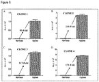
- Figure 6:
- Fold difference in bioluminescence intensity in each of the four clones upon hypoxia induction.
- Figure 7:
-
Clone 3 cells displayed similar morphology to the parental Saos-2 cells following culture in DMEM media with 10% FBS in a humidified CO2 incubator at 37°C. - Figure 8:
- Different cells numbers gave the similar fold difference in bioluminescense intensity following hypoxia induction.
- Figure 9:
- Commercially available inhibitors of HIF activity, Bortezomib and cisplatin led to the reduction of reporter signal intensities.
- Establishment of this HIF cell-based screening assay was achieved by an initial co-transfection of the 4XEPO-HRE-FLuc plasmid and the pcDNA3 plasmid (Invitrogen) carrying an antibiotic resistant gene, into the Saos-2 osteosarcoma cell line (ATCC). A subpopulation of cells stably transfected with the plasmids was later obtained by the G418 antibiotic selection. Single cell clones were isolated from the mass population of stable transfectants using a limited dilution cloning strategy. A single clone with the highest reporter activity was used for a proof-of-concept study using known inhibitors of HIF. These inhibitors are the commercially available drugs, Bortezomib (Velcade) and Cisplatin.
- The 4XEPO-HRE-FLuc plasmid construct contained four copies of the HRE of the EPO gene while the pcDNA3 vector carries a neomycin-resistant gene. Co-transfection of these plasmids into Saos-2 cells was performed at a 10:1 ratio (4XEPO-HRE-FLuc: pcDNA3) using the Lipofectamine 2000 (Invitrogen) fusogen. Initially, 8.3 X 10⁵ Saos-2 cells were seeded in an antibiotic-free DMEM with 10% FBS in a T-25 flask for 24 hours. On the following day, a plasmid mixture was prepared by adding 11.4 µg of 4XEPO-HRE-FLuc and 1.14 µg of pcDNA3 in 625 µl serum-free and antibiotic-free DMEM. At the same time a transfection reagent mixture was prepared by adding 18.75 µl of Lipofectamine 2000 into 606.25 µl serum-free and antibiotic-free DMEM. The reagent mixture was gently mixed and incubated at room temperature for 5 minutes. After the incubation, the plasmid mixture and the reagent mixture were combined, vortexed and incubated at room temperature for 20 minutes. While the complexes were incubating, cultured media from the overnight T-25 flask Saos-2 cells was discarded, and the cells were washed once with 5 ml of serum- and antibiotic-free DMEM. At the end of the 20 minutes incubation, the 1250 µl of plasmid-Lipofectamine 2000 complexes was added into the cells. The flask was then swirled followed by an addition of 3750 □l of serum- and antibiotic-free DMEM. The cells were incubated for 6 hours in a humidified incubator supplied with 5% CO2 at 37°C. Five ml of DMEM with 10% serum and antibiotic-free was added into the flask after 6 hours in order to make a final concentration of 5% reduced-serum DMEM. Five ml of antibiotic-free DMEM with 10% FBS was then added into the in order to make final concentration of 5% reduced-serum DMEM.
- Selection of a stably-transfected population of Saos-2 cells from the transfected cells above was performed using the G418 antibiotic. After 48 hours of incubation, the transfection media mixture was removed from the cells. They were then washed twice with 1X PBS followed by the addition of DMEM containing 1 mg/ml G418 and 10% FBS. The cells were continued to be incubated in a humidified incubator supplied with 5% CO2 at 37°C for 3 more days. After the incubation period, spent media was removed and replaced with fresh media containing 1mg/ml G418 and 10% FBS was added. This media replacement was done at every 3 days for a period of 3 weeks. After 3 weeks, the surviving cells, which constitute the mass population of stably-transfected, neomycin-resistant parental cells, were propagated further. This parental population was then tested for its ability to upregulate the luciferase reporter protein upon hypoxia induction.
- To confirm the HIF reporter activity of neomycin-resistant parental population, a single luciferase assay was conducted. Initially, 2.5 X 10⁴ of the cells were plated in wells of a 48-well plates in antibiotic-free DMEM with 10% FBS for 16 hours. One of the plates was then incubated for 24 hours in a normoxic (21% 02) gaseous atmosphere in a humidified CO2 incubator while the other was incubated in a hypoxic (0.5% 02) gaseous environment in a hypoxic chamber (BioSpherix, USA) controlled by an oxygen regulator (ProOx model 110, BioSpherix, USA. After 24 hours of exposure to different oxygen concentrations, Bright-Glo™ Luciferase Assay (Promega, USA) was conducted to analyse the HIF reporter activity. Initially, growth media in each well was discarded and the cells were rinsed once with 300 µl of 1XPBS. Room temperature-equilibrated Glo Lysis Buffer (GLB; 60 µl) was then added into the wells. Plates were then gently rocked on a shaker for 5 min at room temperature. Cell lysis was confirmed visually by observing the wells of the 48-well plate under a light microscope. Thirty microliters of the resulting cell lysates were then transferred into individual glass vials. Bright-Glo™ Assay Reagent was then added into each tube at a 1:1 (v/v) ratio. Luminescence intensity was immediately read and recorded by a Luminometer (Sirius-2, Titertek- Berthold).
- After confirmation of the HIF reporter activity in the mass population of the stable parental neomycin-resistant cells, single cell cloning was perfomed using a limiting serial dilution cloning strategy. The parental cells were seeded in the first row wells (A1-A8) of a 96-well plate at 1.5 X10⁴ cells for 24 hours. After an overnight incubation, spent media in the wells was pippeted out carefully and the cells were washed with 50 µl 1 X PBS. Trypsinization of cells was done by adding 50 µl of 1X Trypsin-EDTA in each well for 1 minute at 37°C. After cells became detached, the content of each well was topped up to 100 µl with DMEM supplemented with
G 418 and 10% FBS. One hundred microliters of the same media was pipetted into the remaining 9 rows of empty wells. Cell suspension from the first well (100 µl) was then transferred into the adjacent well and the mixture was pipetted up and down. The same volume of cell suspension was then transferred from this well to the next. This process was repeated to obtain a two-fold serial dilution of the cell suspension. All of the cell suspension from the last well (200 µl) was diluted in 10 ml of conditioned DMEM supplemented withG 418 and 10% FBS. The mixture was then inverted up and down to ensure even distribution of cells. This cell suspension (100 µl) was alliquoted into wells of a 96-well plate. The plate was then incubated in a 45° slanted position for 5 hours at 37°C. This was done to ensure attachment of single cells at the edge of each respective well. After the incubation each well was examined for the presence of a single attached cell. Wells containing single cells were properly labeled. The plate was subjected to further incubation at 37 °C for 7 to 14 days. Individual cells in the wells were visualized only every 4 days to reduce disturbance of the cell culture media. During visualization, images of the cells were captured. - Single cells which survived and grew into a colony after the 7 to 14 days incubation were trypsinized and transferred into wells of a 48-well plate. As the number of cells gradually increased the subculturing was performed into bigger tissue culture containers. Parameters for the propagation and upscaling of the cells are summarized in Table 1. Each cell clone population was then cryopreserved for long term storage.
Table 1: Parameters for single cell clone upscaling. Type of Seed Vessel Surface Area (cm2) Volume (ml) 1XPBS 1XTrypsin-EDTA DMEM with G 418 and 10% FBS96-well plate 0.37 0.05 0.03 0.1 48-well plate 0.95 0.2 0.05 0.3 24-well plate 1.65 0.5 0.1 0.5 6-well plate 9.46 1 0.25 1 T-25 flask 25 3 0.5 5 T-75 flask 75 5 1 10 - After pure stable cell clones were obtained, their reporter activity towards HIF induction was retested using the single luciferase assay as described above. The cells were also confirmed to be free from mycoplasma contamination using a 4'-6-Diamidino-2-phenylindole (DAPI) staining.
- To determine the optimum cell density to be used in the HIF activity assay, 2.7 X10⁴, 3.3 X10⁴ and 4.0 X10⁴ cells were seeded into wells of a 48-well plate for 24 hours. The seeded cells were then exposed to either normoxic or hypoxic conditions for 24 hours, as described above. Following lysis and mixture with the Bright-Glo Assay Reagent, the bioluminescence intensity was analysed as described in the earlier methods.
- To verify the property of the stable single cell population as an assay system to measure HIF activity, a proof of concept study was performed with commercially available and known HIF inhibitors. These inhibitors are Bortezomib (Velcade) and Cisplatin. Treatment of these drugs in certain cancer cells leads to cell death. To confirm that the inhibition of bioluminescence in our cells assay system is due to HIF inhibition rather than this cytotoxic effect, we simultaneously determined the viability of cells under various concentrations of each drug used for the inhibition assay. The 3-(4,5-dimethylthiazol-2-yl)-2,5-dimethyltetrazolium bromide MTT assay was used. Cells were seeded at 1.5 X 10⁴ cells per well in 96-well plates for 24 hours followed by treatment with selected concentrations of Bortezomib and cisplatin. The cells were then incubated in either normoxic or hypoxic conditions for 24 hours. After the treatment, cell culture media was replaced with fresh serum-free DMEM containing 0.5 mg/ml MTT. After 4 hours of incubation in a humidified CO2 incubator at 37 °C, formazan precipitates were dissolved with 100 µl of DMSO. The resulting reaction was read using a microplate reader at 570 nm absorbance and 630 nm as the reference wavelength. The half maximal inhibitory concentration (IC50) of each drug towards the cells was determined by plotting graphs using the Graphpad Prism 5 software. HIF inhibitory effects at the various concentrations of drugs tested were also measured using the single luciferase assay system. A correlation between the drugs' effects on cell viability and HIF inhibition was then evaluated.
- Saos-2 osteosarcoma cells, that showed a typical epitheloid morphology (
Figure 1 ), were co-transfected with the recombinant pLUC-MCS carrying the 4XEPO-HRE (4XEPO-HRE-FLuc) and pCDNA3 plasmids, harboring the Neomycin resistant gene (Figure 2 ). The resulting transfectants were then treated with G418 antibiotics. Throughout the treatment, changes in the cell culture were monitored under a light microscope. Representative images of the observed morphological changes at selected times during the treatment are shown inFigure 3 . Byday 3, most of the cells in the culture were rounded up and floating suggesting their sensitivity to the G418 treatment. Cells that appear to be resistant to the drug, on the other hand, continued to proliferate. After 20 days of treatment, this resistant population of cells became confluent in the tissue culture flasks. These cells, designated as the 'mass population', were propagated for further selection and stored for future use. - The mass population of cells was evaluated for their ability to respond to hypoxia stimulation by measuring their luciferase expression level. The hypoxic cells were found to be able to produce 90-fold higher luciferase signal compared to the cells in normoxia (
Figure 4 ). Therefore, we conclude that this mass population retained their 4XEPO-HRE gene construct, and they are responsive to hypoxia. A limiting serial dilution cloning strategy was then performed on these cells. - A number of single cells were obtained. Four of them proliferated to form cell colonies. Representative images of
clone # 3, at different times of the culture, are shown inFigure 5 . Byday 21 each clone became a confluent population in the culture flask. To ensure that no mycoplasma contamination was introduced during the process, the cells were stained using DAPI staining method. Results obtained showed that they are free from contamination (Figure 6 ). - To test the level of hypoxia-responsive luciferase signal of each cell clone, they were subjected to hypoxia exposure. All comparisons were done against their normoxia culture controls. Out of the four clones,
clone 3 gave the highest luciferase signal, that was 517-fold higher than the normoxia control (Figure 7 ). To the best of our knowledge, this is the highest hypoxia-specific signal that has been obtained thus far. Previously, using a VEGF promoter construct, Woldemichael et al., (2006) obtained a 40-fold signal intensity. Using a similar promoter, Bo et al., (2008) however only managed to obtain a 14-fold increase in signal. Even using a novel HIF reporter construct containing tandem repeats of minimum HIF binding sequences, Zhou et al., (2011) obtained just a 100-fold greater luciferase signal under hypoxia. Ourclone 1 gave a fold difference comparable to their findings. However, ourclones - Microscopic observation of all of the four stable Saos-2 clones showed no morphological differences compared to their parental cells (
Figure 7 ). High cell density is known to be a factor in increased HIF activity in certain cell lines (Kaluz et al., 2006). To test whether cell density affects the signal sensitivity of ourclone 3, cells were cultured at different cell numbers and then exposed to hypoxia. Results showed that at densities of 2.7 x 10⁴ to 4.0 x 10⁴, no statistically significant variation was observed in the signals (Figure 9 ). Hence, we conclude that the hypoxia-responsive signal produced by this stable Saos-2 clone remain consistent at different cell densities. We have also tested signal stability of theclone 3 as the passage numbers increased. Thus far, our findings showed that the signal intensity is still stable up to 30 passages (data not shown). We will monitor the signal further as the passage numbers increase over time. - A proof of concept study was conducted using known inhibitors of HIF which are Bortezomib and cisplatin. Bortezomib, a proteasomal inhibitor, was shown to inhibit transcriptional activity of HIF-1 via specific effect on HIF1-1α C-terminal activation domain (Kaluz et al., 2006). Treatment of bortezomib, at concentrations of up to 500 nM, significantly reduced the hypoxia-induced luciferase signal in our
clone 3 cells without affecting cell viability (Figure 10). At higher concentrations, the inhibition was associated with cell death. In addition to Bortezomib, cisplatin was also shown to inhibit HIF. The mechanism of inhibition, however, was achieved via a repression of HIF1-1α protein expression (Duyndam et al., 2007). In the present study, addition of cisplatin at 25 µM concentration led to almost 60% inhibition of luciferase signals inhypoxic clone 3. Viability of the cells was also reduced. Previous studies have shown that cisplatin treatment resulted in HIF inhibition and also apoptotic cell death (Tanaka et al., 2005). In summary, our proof of concept using Bortezomib and cisplatin showed that the hypoxia-inducible signal in ourclone 3 population is responsive to the inhibitory effects of these drugs suggesting the reliability and sensitivity of the assay system. -
- Duyndam MC, van Berkel MP, Dorsman JC, Rockx DA, Pinedo HM, Boven E. Cisplatin and doxorubicin repress Vascular Endothelial Growth Factor expression and differentially down-regulate Hypoxia-inducible Factor I activity in human ovarian cancer cells. Biochem Pharmacol. 2007 74:191-201.
- Ek ET, Dass CR, Choong PF. Commonly used mouse models of osteosarcoma. Crit Rev Oncol Hematol. 2006 60:1-8.
- Fogh J, Fogh JM, Orfeo T. One hundred and twenty-seven cultured human tumor cell lines producing tumors in nude mice. J Natl Cancer Inst 1977 59:221-6.
- Ji DB, Zhu HB, Ye J, Li CL. Establishment of a cell-based sssay to screen regulators of the hypoxia-Inducible factor-1-dependent vascular endothelial growth factor promoter. Biol Pharm Bull 2008, 31: 2255-2259.
- Rankin EB, Wu C, Khatri R, Wilson TL, Andersen R, Araldi E, Rankin AL, Yuan J, Kuo CJ, Schipani E, Giaccia AJ. The HIF Signaling Pathway in Osteoblasts Directly Modulates Erythropoiesis through the Production of EPO. Cell. 2012 149:63-74.
- Kaluz, S., M. Kaluzova, and E. J. Stanbridge. The role of extracellular signal regulated protein kinase in transcriptional regulation of the hypoxia marker carbonic anhydrase IX. J Cell Biochem 2006, 97:207-16.
- Keith B, Simon MC. Hypoxia-inducible factors, stem cells, and cancer. Cell. 2007 129:465-72.
- Tanaka T, Kojima I, Ohse T, Inagi R, Miyata T, Ingelfinger JR, Fujita T, Nangaku M. Hypoxia-inducible factor modulates tubular cell survival in cisplatin nephrotoxicity. Am J Physiol Renal Physiol. 2005 289:F1123-33.
- Woldemichael GM, Vasselli JR, Gardella RS, Mckee TC, Linehan WM, McMahon JB. Development of a cell-based reporter assay for screening of inhibitors of hypoxia-inducible factor 2-induced gene expression. J Biomol Screen 2006, 11: 678.
- Zhou W, Dosey TL, Biechele T, Moon RT, Horwitz MS, Ruohola-Baker H. Assessment of hypoxia inducible factor levels in cancer cell lines upon hypoxic induction using a novel reporter construct. Plos One 2011, 6: e27460.
Claims (6)
- A Saos-2 cell line which comprises a gene construct which has a reporter gene stably integrated under the control of a hypoxia inducible factor (HIF) responsive promoter, wherein the HIF responsive promoter comprises four hypoxia-response elements (HRE) of the HRE sequence found in the human erythropoietin (EPO) gene, and wherein the HIF responsive promoter is upstream of the reporter gene and comprises the four HRE in a tandem repeat.
- The Saos-2 cell line according to claim 1, wherein the reporter gene is selected from a luciferase, fluorescent protein or an enzyme which catalyzes a chemical reaction, preferably wherein the reporter gene is a firefly luciferase.
- A method for assaying HIF response in a cell, comprising the steps ofa. providing a cell according to claim 1 or 2,b. incubating said cell at hypoxia,c. optionally, incubating a control cell at normoxiad. wherein the reporter gene expression corresponds to the HIF response in said cell.
- The method according to claim 3, wherein hypoxia is between 0 and 5% O2, preferably between 0.01 and 1% O2, more preferably between 0.01 and 0.3% O2.
- The method according to claims 3 or 4, wherein normoxia is between 5 and 21% O2, preferably between 10 and 21% O2, more preferably between 10 and 20% O2.
- A screening method for identifying modulators of HIF response comprising, performing the method according to any one of claims 3 to 5, and contacting said cells with a compound which is suspected to be a modulator of HIF signalling, wherein an increase in reporter gene expression compared to an untreated control indicates that the compound is an agonist of HIF response, and wherein a decrease of reporter gene expression compared to an untreated control indicates that the compound is an antagonist of HIF response.
Applications Claiming Priority (2)
| Application Number | Priority Date | Filing Date | Title |
|---|---|---|---|
| MYPI2012003492A MY172311A (en) | 2012-08-02 | 2012-08-02 | Hypoxia inducible factor (hif) activity reporter cell line |
| PCT/MY2013/000013 WO2014021705A1 (en) | 2012-08-02 | 2013-01-29 | Hypoxia inducible factor (hif) activity reporter cell line |
Publications (2)
| Publication Number | Publication Date |
|---|---|
| EP2880177A1 EP2880177A1 (en) | 2015-06-10 |
| EP2880177B1 true EP2880177B1 (en) | 2017-07-19 |
Family
ID=48040394
Family Applications (1)
| Application Number | Title | Priority Date | Filing Date |
|---|---|---|---|
| EP13713255.1A Not-in-force EP2880177B1 (en) | 2012-08-02 | 2013-01-29 | Hypoxia inducible factor (hif) activity reporter cell line |
Country Status (7)
| Country | Link |
|---|---|
| US (1) | US9995730B2 (en) |
| EP (1) | EP2880177B1 (en) |
| JP (1) | JP6309520B2 (en) |
| CN (1) | CN104662164A (en) |
| IN (1) | IN2015DN00205A (en) |
| MY (1) | MY172311A (en) |
| WO (1) | WO2014021705A1 (en) |
Families Citing this family (2)
| Publication number | Priority date | Publication date | Assignee | Title |
|---|---|---|---|---|
| KR101849964B1 (en) * | 2016-07-26 | 2018-04-19 | 울산대학교 산학협력단 | IL-7 expression reporter cell lines and methods for screening therapeutic agents for immunodeficiency diseases using the cell lines |
| US20230374526A1 (en) * | 2020-03-26 | 2023-11-23 | Asklepios Biopharmaceutical, Inc. | Forskolin-inducible promoters and hypoxia-inducible promoters |
Family Cites Families (2)
| Publication number | Priority date | Publication date | Assignee | Title |
|---|---|---|---|---|
| US5834306A (en) * | 1994-12-23 | 1998-11-10 | Sri International | Tissue specific hypoxia regulated therapeutic constructs |
| WO2005001136A1 (en) * | 2003-06-04 | 2005-01-06 | Irm Llc | Methods and compositions for modulating erythropoietin expression |
-
2012
- 2012-08-02 MY MYPI2012003492A patent/MY172311A/en unknown
-
2013
- 2013-01-29 WO PCT/MY2013/000013 patent/WO2014021705A1/en not_active Ceased
- 2013-01-29 JP JP2015525393A patent/JP6309520B2/en not_active Expired - Fee Related
- 2013-01-29 EP EP13713255.1A patent/EP2880177B1/en not_active Not-in-force
- 2013-01-29 CN CN201380040870.5A patent/CN104662164A/en active Pending
- 2013-01-29 IN IN205DEN2015 patent/IN2015DN00205A/en unknown
- 2013-01-29 US US14/415,064 patent/US9995730B2/en not_active Expired - Fee Related
Non-Patent Citations (1)
| Title |
|---|
| None * |
Also Published As
| Publication number | Publication date |
|---|---|
| CN104662164A (en) | 2015-05-27 |
| IN2015DN00205A (en) | 2015-06-12 |
| WO2014021705A1 (en) | 2014-02-06 |
| MY172311A (en) | 2019-11-21 |
| US20150168376A1 (en) | 2015-06-18 |
| US9995730B2 (en) | 2018-06-12 |
| EP2880177A1 (en) | 2015-06-10 |
| JP2015524272A (en) | 2015-08-24 |
| JP6309520B2 (en) | 2018-04-11 |
Similar Documents
| Publication | Publication Date | Title |
|---|---|---|
| Zhao et al. | Foxp1/2/4 regulate endochondral ossification as a suppresser complex | |
| JP2003525032A (en) | Reagents and methods for identifying and modulating expression of genes regulated by CDK inhibitors | |
| Machado-Neto et al. | ANKHD1 silencing inhibits Stathmin 1 activity, cell proliferation and migration of leukemia cells | |
| Borisova et al. | Protein translation rate determines neocortical neuron fate | |
| Lin et al. | SH3GL3 functions as a potent tumor suppressor in lung cancer in a SH3 domain dependent manner | |
| EP2985347B1 (en) | Method for detecting protein stability and uses thereof | |
| EP2946207B1 (en) | Methods and systems for identifying patient specific driver mutations | |
| US9995730B2 (en) | Hypoxia Inducible Factor (HIF) Activity Reporter Cell Line | |
| CN102395685A (en) | Method for identifying a subpopulation of mammalian cells with distinctive ribosome translation profiles | |
| Singh et al. | Mitochondrial import of human and yeast fumarase in live mammalian cells: retrograde translocation of the yeast enzyme is mainly caused by its poor targeting sequence | |
| US20030069256A1 (en) | Use of isogenic human cancer cells for high-throughput screening and drug discovery | |
| Pereverzev et al. | Differences in nonsense-mediated mRNA decay activity in mammalian cell lines revealed by a fluorescence reporter | |
| Lin et al. | Interaction between Nm23 and the tumor suppressor VHL | |
| CN113215168A (en) | Marker for cancer cell drug resistance, preparation combination for reversing cancer cell drug resistance and application thereof | |
| Hayat et al. | Inhibitors of eIF1A-ribosome interaction unveil uORF-dependent regulation of translation initiation and antitumor and antiviral effects | |
| US20030211523A1 (en) | Two-hybrid double screening system and method | |
| Hevér et al. | Ets2 is not required for Ras or Neu/ErbB-2 mediated cellular transformation in vitro | |
| WO2005049868A1 (en) | Methods and agents for screening for compounds capable of modulating her2 expression | |
| KR100791859B1 (en) | Transformation cell line expressing fusion protein of p53 tumor suppressor protein with fluorescent protein and method of screening for substance related to 활성 53 tumor suppressor protein activity | |
| US20010046704A1 (en) | Methods and compositions to activate genes in cells | |
| JP2023121261A (en) | Modified osteoblast-like cells for evaluating implant materials | |
| Chen | The Interactions Between JAK/STAT Signaling Ligands in Drosophila Melanogaster | |
| WO2024180248A1 (en) | R-loop sensor | |
| CN119841897A (en) | Tumor targeting therapeutic polypeptide based on Smac/GPX4 interaction and application thereof | |
| KR101275777B1 (en) | Cell line which is transfected with Wnt active measurement vector and High-Throughput Screening method of selective ligands by using it |
Legal Events
| Date | Code | Title | Description |
|---|---|---|---|
| PUAI | Public reference made under article 153(3) epc to a published international application that has entered the european phase |
Free format text: ORIGINAL CODE: 0009012 |
|
| 17P | Request for examination filed |
Effective date: 20150127 |
|
| AK | Designated contracting states |
Kind code of ref document: A1 Designated state(s): AL AT BE BG CH CY CZ DE DK EE ES FI FR GB GR HR HU IE IS IT LI LT LU LV MC MK MT NL NO PL PT RO RS SE SI SK SM TR |
|
| AX | Request for extension of the european patent |
Extension state: BA ME |
|
| DAX | Request for extension of the european patent (deleted) | ||
| 17Q | First examination report despatched |
Effective date: 20151214 |
|
| GRAP | Despatch of communication of intention to grant a patent |
Free format text: ORIGINAL CODE: EPIDOSNIGR1 |
|
| INTG | Intention to grant announced |
Effective date: 20170314 |
|
| GRAS | Grant fee paid |
Free format text: ORIGINAL CODE: EPIDOSNIGR3 |
|
| GRAA | (expected) grant |
Free format text: ORIGINAL CODE: 0009210 |
|
| AK | Designated contracting states |
Kind code of ref document: B1 Designated state(s): AL AT BE BG CH CY CZ DE DK EE ES FI FR GB GR HR HU IE IS IT LI LT LU LV MC MK MT NL NO PL PT RO RS SE SI SK SM TR |
|
| REG | Reference to a national code |
Ref country code: GB Ref legal event code: FG4D |
|
| REG | Reference to a national code |
Ref country code: CH Ref legal event code: EP |
|
| REG | Reference to a national code |
Ref country code: IE Ref legal event code: FG4D |
|
| REG | Reference to a national code |
Ref country code: AT Ref legal event code: REF Ref document number: 910449 Country of ref document: AT Kind code of ref document: T Effective date: 20170815 |
|
| REG | Reference to a national code |
Ref country code: DE Ref legal event code: R096 Ref document number: 602013023704 Country of ref document: DE |
|
| REG | Reference to a national code |
Ref country code: NL Ref legal event code: MP Effective date: 20170719 |
|
| REG | Reference to a national code |
Ref country code: LT Ref legal event code: MG4D |
|
| REG | Reference to a national code |
Ref country code: AT Ref legal event code: MK05 Ref document number: 910449 Country of ref document: AT Kind code of ref document: T Effective date: 20170719 |
|
| REG | Reference to a national code |
Ref country code: FR Ref legal event code: PLFP Year of fee payment: 6 |
|
| PG25 | Lapsed in a contracting state [announced via postgrant information from national office to epo] |
Ref country code: FI Free format text: LAPSE BECAUSE OF FAILURE TO SUBMIT A TRANSLATION OF THE DESCRIPTION OR TO PAY THE FEE WITHIN THE PRESCRIBED TIME-LIMIT Effective date: 20170719 Ref country code: NL Free format text: LAPSE BECAUSE OF FAILURE TO SUBMIT A TRANSLATION OF THE DESCRIPTION OR TO PAY THE FEE WITHIN THE PRESCRIBED TIME-LIMIT Effective date: 20170719 Ref country code: NO Free format text: LAPSE BECAUSE OF FAILURE TO SUBMIT A TRANSLATION OF THE DESCRIPTION OR TO PAY THE FEE WITHIN THE PRESCRIBED TIME-LIMIT Effective date: 20171019 Ref country code: SE Free format text: LAPSE BECAUSE OF FAILURE TO SUBMIT A TRANSLATION OF THE DESCRIPTION OR TO PAY THE FEE WITHIN THE PRESCRIBED TIME-LIMIT Effective date: 20170719 Ref country code: AT Free format text: LAPSE BECAUSE OF FAILURE TO SUBMIT A TRANSLATION OF THE DESCRIPTION OR TO PAY THE FEE WITHIN THE PRESCRIBED TIME-LIMIT Effective date: 20170719 Ref country code: LT Free format text: LAPSE BECAUSE OF FAILURE TO SUBMIT A TRANSLATION OF THE DESCRIPTION OR TO PAY THE FEE WITHIN THE PRESCRIBED TIME-LIMIT Effective date: 20170719 Ref country code: HR Free format text: LAPSE BECAUSE OF FAILURE TO SUBMIT A TRANSLATION OF THE DESCRIPTION OR TO PAY THE FEE WITHIN THE PRESCRIBED TIME-LIMIT Effective date: 20170719 |
|
| PG25 | Lapsed in a contracting state [announced via postgrant information from national office to epo] |
Ref country code: RS Free format text: LAPSE BECAUSE OF FAILURE TO SUBMIT A TRANSLATION OF THE DESCRIPTION OR TO PAY THE FEE WITHIN THE PRESCRIBED TIME-LIMIT Effective date: 20170719 Ref country code: GR Free format text: LAPSE BECAUSE OF FAILURE TO SUBMIT A TRANSLATION OF THE DESCRIPTION OR TO PAY THE FEE WITHIN THE PRESCRIBED TIME-LIMIT Effective date: 20171020 Ref country code: ES Free format text: LAPSE BECAUSE OF FAILURE TO SUBMIT A TRANSLATION OF THE DESCRIPTION OR TO PAY THE FEE WITHIN THE PRESCRIBED TIME-LIMIT Effective date: 20170719 Ref country code: IS Free format text: LAPSE BECAUSE OF FAILURE TO SUBMIT A TRANSLATION OF THE DESCRIPTION OR TO PAY THE FEE WITHIN THE PRESCRIBED TIME-LIMIT Effective date: 20171119 Ref country code: PL Free format text: LAPSE BECAUSE OF FAILURE TO SUBMIT A TRANSLATION OF THE DESCRIPTION OR TO PAY THE FEE WITHIN THE PRESCRIBED TIME-LIMIT Effective date: 20170719 Ref country code: BG Free format text: LAPSE BECAUSE OF FAILURE TO SUBMIT A TRANSLATION OF THE DESCRIPTION OR TO PAY THE FEE WITHIN THE PRESCRIBED TIME-LIMIT Effective date: 20171019 Ref country code: LV Free format text: LAPSE BECAUSE OF FAILURE TO SUBMIT A TRANSLATION OF THE DESCRIPTION OR TO PAY THE FEE WITHIN THE PRESCRIBED TIME-LIMIT Effective date: 20170719 |
|
| REG | Reference to a national code |
Ref country code: DE Ref legal event code: R097 Ref document number: 602013023704 Country of ref document: DE |
|
| PG25 | Lapsed in a contracting state [announced via postgrant information from national office to epo] |
Ref country code: CZ Free format text: LAPSE BECAUSE OF FAILURE TO SUBMIT A TRANSLATION OF THE DESCRIPTION OR TO PAY THE FEE WITHIN THE PRESCRIBED TIME-LIMIT Effective date: 20170719 Ref country code: RO Free format text: LAPSE BECAUSE OF FAILURE TO SUBMIT A TRANSLATION OF THE DESCRIPTION OR TO PAY THE FEE WITHIN THE PRESCRIBED TIME-LIMIT Effective date: 20170719 Ref country code: DK Free format text: LAPSE BECAUSE OF FAILURE TO SUBMIT A TRANSLATION OF THE DESCRIPTION OR TO PAY THE FEE WITHIN THE PRESCRIBED TIME-LIMIT Effective date: 20170719 |
|
| PGFP | Annual fee paid to national office [announced via postgrant information from national office to epo] |
Ref country code: GB Payment date: 20180124 Year of fee payment: 6 Ref country code: DE Payment date: 20180206 Year of fee payment: 6 |
|
| PLBE | No opposition filed within time limit |
Free format text: ORIGINAL CODE: 0009261 |
|
| STAA | Information on the status of an ep patent application or granted ep patent |
Free format text: STATUS: NO OPPOSITION FILED WITHIN TIME LIMIT |
|
| PG25 | Lapsed in a contracting state [announced via postgrant information from national office to epo] |
Ref country code: SM Free format text: LAPSE BECAUSE OF FAILURE TO SUBMIT A TRANSLATION OF THE DESCRIPTION OR TO PAY THE FEE WITHIN THE PRESCRIBED TIME-LIMIT Effective date: 20170719 Ref country code: SK Free format text: LAPSE BECAUSE OF FAILURE TO SUBMIT A TRANSLATION OF THE DESCRIPTION OR TO PAY THE FEE WITHIN THE PRESCRIBED TIME-LIMIT Effective date: 20170719 Ref country code: IT Free format text: LAPSE BECAUSE OF FAILURE TO SUBMIT A TRANSLATION OF THE DESCRIPTION OR TO PAY THE FEE WITHIN THE PRESCRIBED TIME-LIMIT Effective date: 20170719 Ref country code: EE Free format text: LAPSE BECAUSE OF FAILURE TO SUBMIT A TRANSLATION OF THE DESCRIPTION OR TO PAY THE FEE WITHIN THE PRESCRIBED TIME-LIMIT Effective date: 20170719 |
|
| PGFP | Annual fee paid to national office [announced via postgrant information from national office to epo] |
Ref country code: FR Payment date: 20180111 Year of fee payment: 6 |
|
| 26N | No opposition filed |
Effective date: 20180420 |
|
| PG25 | Lapsed in a contracting state [announced via postgrant information from national office to epo] |
Ref country code: SI Free format text: LAPSE BECAUSE OF FAILURE TO SUBMIT A TRANSLATION OF THE DESCRIPTION OR TO PAY THE FEE WITHIN THE PRESCRIBED TIME-LIMIT Effective date: 20170719 |
|
| REG | Reference to a national code |
Ref country code: CH Ref legal event code: PL |
|
| PG25 | Lapsed in a contracting state [announced via postgrant information from national office to epo] |
Ref country code: LU Free format text: LAPSE BECAUSE OF NON-PAYMENT OF DUE FEES Effective date: 20180129 |
|
| REG | Reference to a national code |
Ref country code: IE Ref legal event code: MM4A |
|
| REG | Reference to a national code |
Ref country code: BE Ref legal event code: MM Effective date: 20180131 |
|
| PG25 | Lapsed in a contracting state [announced via postgrant information from national office to epo] |
Ref country code: BE Free format text: LAPSE BECAUSE OF NON-PAYMENT OF DUE FEES Effective date: 20180131 Ref country code: CH Free format text: LAPSE BECAUSE OF NON-PAYMENT OF DUE FEES Effective date: 20180131 Ref country code: LI Free format text: LAPSE BECAUSE OF NON-PAYMENT OF DUE FEES Effective date: 20180131 |
|
| PG25 | Lapsed in a contracting state [announced via postgrant information from national office to epo] |
Ref country code: IE Free format text: LAPSE BECAUSE OF NON-PAYMENT OF DUE FEES Effective date: 20180129 |
|
| PG25 | Lapsed in a contracting state [announced via postgrant information from national office to epo] |
Ref country code: MC Free format text: LAPSE BECAUSE OF FAILURE TO SUBMIT A TRANSLATION OF THE DESCRIPTION OR TO PAY THE FEE WITHIN THE PRESCRIBED TIME-LIMIT Effective date: 20170719 |
|
| REG | Reference to a national code |
Ref country code: DE Ref legal event code: R119 Ref document number: 602013023704 Country of ref document: DE |
|
| GBPC | Gb: european patent ceased through non-payment of renewal fee |
Effective date: 20190129 |
|
| PG25 | Lapsed in a contracting state [announced via postgrant information from national office to epo] |
Ref country code: DE Free format text: LAPSE BECAUSE OF NON-PAYMENT OF DUE FEES Effective date: 20190801 Ref country code: FR Free format text: LAPSE BECAUSE OF NON-PAYMENT OF DUE FEES Effective date: 20190131 |
|
| PG25 | Lapsed in a contracting state [announced via postgrant information from national office to epo] |
Ref country code: GB Free format text: LAPSE BECAUSE OF NON-PAYMENT OF DUE FEES Effective date: 20190129 |
|
| PG25 | Lapsed in a contracting state [announced via postgrant information from national office to epo] |
Ref country code: MT Free format text: LAPSE BECAUSE OF NON-PAYMENT OF DUE FEES Effective date: 20180129 |
|
| PG25 | Lapsed in a contracting state [announced via postgrant information from national office to epo] |
Ref country code: TR Free format text: LAPSE BECAUSE OF FAILURE TO SUBMIT A TRANSLATION OF THE DESCRIPTION OR TO PAY THE FEE WITHIN THE PRESCRIBED TIME-LIMIT Effective date: 20170719 |
|
| PG25 | Lapsed in a contracting state [announced via postgrant information from national office to epo] |
Ref country code: PT Free format text: LAPSE BECAUSE OF FAILURE TO SUBMIT A TRANSLATION OF THE DESCRIPTION OR TO PAY THE FEE WITHIN THE PRESCRIBED TIME-LIMIT Effective date: 20170719 |
|
| PG25 | Lapsed in a contracting state [announced via postgrant information from national office to epo] |
Ref country code: MK Free format text: LAPSE BECAUSE OF NON-PAYMENT OF DUE FEES Effective date: 20170719 Ref country code: HU Free format text: LAPSE BECAUSE OF FAILURE TO SUBMIT A TRANSLATION OF THE DESCRIPTION OR TO PAY THE FEE WITHIN THE PRESCRIBED TIME-LIMIT; INVALID AB INITIO Effective date: 20130129 Ref country code: CY Free format text: LAPSE BECAUSE OF FAILURE TO SUBMIT A TRANSLATION OF THE DESCRIPTION OR TO PAY THE FEE WITHIN THE PRESCRIBED TIME-LIMIT Effective date: 20170719 |
|
| PG25 | Lapsed in a contracting state [announced via postgrant information from national office to epo] |
Ref country code: AL Free format text: LAPSE BECAUSE OF FAILURE TO SUBMIT A TRANSLATION OF THE DESCRIPTION OR TO PAY THE FEE WITHIN THE PRESCRIBED TIME-LIMIT Effective date: 20170719 |