EP2857520A1 - Method for in vitro investigating mitochondrial dna levels in a biological sample, kits and uses thereof - Google Patents
Method for in vitro investigating mitochondrial dna levels in a biological sample, kits and uses thereof Download PDFInfo
- Publication number
- EP2857520A1 EP2857520A1 EP20130306378 EP13306378A EP2857520A1 EP 2857520 A1 EP2857520 A1 EP 2857520A1 EP 20130306378 EP20130306378 EP 20130306378 EP 13306378 A EP13306378 A EP 13306378A EP 2857520 A1 EP2857520 A1 EP 2857520A1
- Authority
- EP
- European Patent Office
- Prior art keywords
- mtdna
- level
- patient
- gastric cancer
- susceptible
- Prior art date
- Legal status (The legal status is an assumption and is not a legal conclusion. Google has not performed a legal analysis and makes no representation as to the accuracy of the status listed.)
- Withdrawn
Links
- 108020005196 Mitochondrial DNA Proteins 0.000 title claims abstract description 203
- 238000000034 method Methods 0.000 title claims abstract description 104
- 239000012472 biological sample Substances 0.000 title claims abstract description 55
- 238000000338 in vitro Methods 0.000 title claims abstract description 10
- 208000005718 Stomach Neoplasms Diseases 0.000 claims abstract description 156
- 206010017758 gastric cancer Diseases 0.000 claims abstract description 156
- 201000011549 stomach cancer Diseases 0.000 claims abstract description 156
- 108090001007 Interleukin-8 Proteins 0.000 claims abstract description 41
- 230000003862 health status Effects 0.000 claims abstract description 27
- 238000012544 monitoring process Methods 0.000 claims abstract description 22
- 108090001005 Interleukin-6 Proteins 0.000 claims abstract description 21
- 102000013264 Interleukin-23 Human genes 0.000 claims abstract description 20
- 108010065637 Interleukin-23 Proteins 0.000 claims abstract description 20
- 238000011835 investigation Methods 0.000 claims abstract description 14
- 230000002757 inflammatory effect Effects 0.000 claims abstract description 8
- 238000011176 pooling Methods 0.000 claims abstract description 7
- 230000002496 gastric effect Effects 0.000 claims description 50
- 231100000504 carcinogenesis Toxicity 0.000 claims description 33
- 239000000523 sample Substances 0.000 claims description 33
- 208000005623 Carcinogenesis Diseases 0.000 claims description 32
- 230000036952 cancer formation Effects 0.000 claims description 32
- 210000004369 blood Anatomy 0.000 claims description 28
- 239000008280 blood Substances 0.000 claims description 28
- 108091093105 Nuclear DNA Proteins 0.000 claims description 25
- 238000001514 detection method Methods 0.000 claims description 20
- 108020004707 nucleic acids Proteins 0.000 claims description 20
- 102000039446 nucleic acids Human genes 0.000 claims description 20
- 150000007523 nucleic acids Chemical class 0.000 claims description 20
- 108020004414 DNA Proteins 0.000 claims description 19
- 238000003753 real-time PCR Methods 0.000 claims description 18
- 108090000623 proteins and genes Proteins 0.000 claims description 17
- 238000011282 treatment Methods 0.000 claims description 16
- 208000007882 Gastritis Diseases 0.000 claims description 14
- 238000012360 testing method Methods 0.000 claims description 14
- 210000000265 leukocyte Anatomy 0.000 claims description 13
- 239000013615 primer Substances 0.000 claims description 13
- 230000003321 amplification Effects 0.000 claims description 12
- 238000001574 biopsy Methods 0.000 claims description 12
- 208000015181 infectious disease Diseases 0.000 claims description 12
- 238000003199 nucleic acid amplification method Methods 0.000 claims description 12
- 239000000427 antigen Substances 0.000 claims description 11
- 108091007433 antigens Proteins 0.000 claims description 11
- 102000036639 antigens Human genes 0.000 claims description 11
- 239000003155 DNA primer Substances 0.000 claims description 9
- 206010019375 Helicobacter infections Diseases 0.000 claims description 9
- 239000003153 chemical reaction reagent Substances 0.000 claims description 9
- 210000001156 gastric mucosa Anatomy 0.000 claims description 9
- 210000004027 cell Anatomy 0.000 claims description 8
- 230000002914 neoplasic effect Effects 0.000 claims description 7
- 239000002773 nucleotide Substances 0.000 claims description 6
- 206010000087 Abdominal pain upper Diseases 0.000 claims description 5
- 108010014303 DNA-directed DNA polymerase Proteins 0.000 claims description 5
- 102000016928 DNA-directed DNA polymerase Human genes 0.000 claims description 5
- 238000003556 assay Methods 0.000 claims description 5
- 239000007853 buffer solution Substances 0.000 claims description 5
- 230000002438 mitochondrial effect Effects 0.000 claims description 5
- -1 - optionally Substances 0.000 claims description 4
- 101100476480 Mus musculus S100a8 gene Proteins 0.000 claims description 4
- 238000009396 hybridization Methods 0.000 claims description 4
- 102000004169 proteins and genes Human genes 0.000 claims description 4
- AHCYMLUZIRLXAA-SHYZEUOFSA-N Deoxyuridine 5'-triphosphate Chemical compound O1[C@H](COP(O)(=O)OP(O)(=O)OP(O)(O)=O)[C@@H](O)C[C@@H]1N1C(=O)NC(=O)C=C1 AHCYMLUZIRLXAA-SHYZEUOFSA-N 0.000 claims description 3
- SUYVUBYJARFZHO-RRKCRQDMSA-N dATP Chemical compound C1=NC=2C(N)=NC=NC=2N1[C@H]1C[C@H](O)[C@@H](COP(O)(=O)OP(O)(=O)OP(O)(O)=O)O1 SUYVUBYJARFZHO-RRKCRQDMSA-N 0.000 claims description 3
- SUYVUBYJARFZHO-UHFFFAOYSA-N dATP Natural products C1=NC=2C(N)=NC=NC=2N1C1CC(O)C(COP(O)(=O)OP(O)(=O)OP(O)(O)=O)O1 SUYVUBYJARFZHO-UHFFFAOYSA-N 0.000 claims description 3
- RGWHQCVHVJXOKC-SHYZEUOFSA-J dCTP(4-) Chemical compound O=C1N=C(N)C=CN1[C@@H]1O[C@H](COP([O-])(=O)OP([O-])(=O)OP([O-])([O-])=O)[C@@H](O)C1 RGWHQCVHVJXOKC-SHYZEUOFSA-J 0.000 claims description 3
- HAAZLUGHYHWQIW-KVQBGUIXSA-N dGTP Chemical compound C1=NC=2C(=O)NC(N)=NC=2N1[C@H]1C[C@H](O)[C@@H](COP(O)(=O)OP(O)(=O)OP(O)(O)=O)O1 HAAZLUGHYHWQIW-KVQBGUIXSA-N 0.000 claims description 3
- 230000003247 decreasing effect Effects 0.000 claims description 3
- 238000010186 staining Methods 0.000 claims description 3
- 108010006785 Taq Polymerase Proteins 0.000 claims description 2
- XSQUKJJJFZCRTK-UHFFFAOYSA-N Urea Chemical compound NC(N)=O XSQUKJJJFZCRTK-UHFFFAOYSA-N 0.000 claims description 2
- 239000004202 carbamide Substances 0.000 claims description 2
- 230000008685 targeting Effects 0.000 claims description 2
- 230000004544 DNA amplification Effects 0.000 claims 1
- 102000004890 Interleukin-8 Human genes 0.000 description 34
- 206010028980 Neoplasm Diseases 0.000 description 34
- 229940096397 interleukin-8 Drugs 0.000 description 25
- XKTZWUACRZHVAN-VADRZIEHSA-N interleukin-8 Chemical compound C([C@H](NC(=O)[C@H](CC(O)=O)NC(=O)[C@H](CC=1C2=CC=CC=C2NC=1)NC(=O)[C@@H](NC(C)=O)CCSC)C(=O)N[C@@H](CC(O)=O)C(=O)N[C@@H](CC(O)=O)C(=O)N[C@@H](CC(C)C)C(=O)N[C@@H](CC(N)=O)C(=O)N[C@@H](CC=1C=CC=CC=1)C(=O)N[C@@H]([C@@H](C)O)C(=O)NCC(=O)N[C@@H](CCSC)C(=O)N1[C@H](CCC1)C(=O)N1[C@H](CCC1)C(=O)N[C@@H](C)C(=O)N[C@H](CC(O)=O)C(=O)N[C@H](CCC(O)=O)C(=O)N[C@H](CC(O)=O)C(=O)N[C@H](CC=1C=CC(O)=CC=1)C(=O)N[C@H](CO)C(=O)N1[C@H](CCC1)C(N)=O)C1=CC=CC=C1 XKTZWUACRZHVAN-VADRZIEHSA-N 0.000 description 25
- 206010054949 Metaplasia Diseases 0.000 description 23
- 230000003902 lesion Effects 0.000 description 23
- 201000011510 cancer Diseases 0.000 description 22
- 102000004889 Interleukin-6 Human genes 0.000 description 16
- 238000011002 quantification Methods 0.000 description 16
- 208000037265 diseases, disorders, signs and symptoms Diseases 0.000 description 14
- 201000010099 disease Diseases 0.000 description 13
- 208000016644 chronic atrophic gastritis Diseases 0.000 description 12
- 230000002093 peripheral effect Effects 0.000 description 11
- 230000008569 process Effects 0.000 description 10
- 208000004300 Atrophic Gastritis Diseases 0.000 description 9
- 208000036495 Gastritis atrophic Diseases 0.000 description 9
- 230000008859 change Effects 0.000 description 8
- 230000000968 intestinal effect Effects 0.000 description 8
- 230000000875 corresponding effect Effects 0.000 description 7
- 201000006585 gastric adenocarcinoma Diseases 0.000 description 7
- 230000014509 gene expression Effects 0.000 description 7
- 238000003752 polymerase chain reaction Methods 0.000 description 7
- 210000002966 serum Anatomy 0.000 description 7
- 238000002965 ELISA Methods 0.000 description 6
- 206010027476 Metastases Diseases 0.000 description 6
- 238000002512 chemotherapy Methods 0.000 description 6
- 238000011161 development Methods 0.000 description 6
- 230000002441 reversible effect Effects 0.000 description 6
- 210000001519 tissue Anatomy 0.000 description 6
- 206010058314 Dysplasia Diseases 0.000 description 5
- 238000004458 analytical method Methods 0.000 description 5
- 238000003745 diagnosis Methods 0.000 description 5
- 238000005259 measurement Methods 0.000 description 5
- 230000015689 metaplastic ossification Effects 0.000 description 5
- 230000009401 metastasis Effects 0.000 description 5
- 230000035772 mutation Effects 0.000 description 5
- 125000003729 nucleotide group Chemical group 0.000 description 5
- 210000005259 peripheral blood Anatomy 0.000 description 5
- 239000011886 peripheral blood Substances 0.000 description 5
- 102000004127 Cytokines Human genes 0.000 description 4
- 108090000695 Cytokines Proteins 0.000 description 4
- WSFSSNUMVMOOMR-UHFFFAOYSA-N Formaldehyde Chemical compound O=C WSFSSNUMVMOOMR-UHFFFAOYSA-N 0.000 description 4
- 201000003791 MALT lymphoma Diseases 0.000 description 4
- 108091028043 Nucleic acid sequence Proteins 0.000 description 4
- 108091034117 Oligonucleotide Proteins 0.000 description 4
- 208000037976 chronic inflammation Diseases 0.000 description 4
- 230000006020 chronic inflammation Effects 0.000 description 4
- 239000000975 dye Substances 0.000 description 4
- 230000008029 eradication Effects 0.000 description 4
- 238000002474 experimental method Methods 0.000 description 4
- 230000004054 inflammatory process Effects 0.000 description 4
- 239000000463 material Substances 0.000 description 4
- 230000001855 preneoplastic effect Effects 0.000 description 4
- 230000005855 radiation Effects 0.000 description 4
- 238000005070 sampling Methods 0.000 description 4
- 238000001356 surgical procedure Methods 0.000 description 4
- 208000024891 symptom Diseases 0.000 description 4
- 238000007400 DNA extraction Methods 0.000 description 3
- 238000008157 ELISA kit Methods 0.000 description 3
- 241000282412 Homo Species 0.000 description 3
- 101001055222 Homo sapiens Interleukin-8 Proteins 0.000 description 3
- 206010061218 Inflammation Diseases 0.000 description 3
- 238000011529 RT qPCR Methods 0.000 description 3
- JLCPHMBAVCMARE-UHFFFAOYSA-N [3-[[3-[[3-[[3-[[3-[[3-[[3-[[3-[[3-[[3-[[3-[[5-(2-amino-6-oxo-1H-purin-9-yl)-3-[[3-[[3-[[3-[[3-[[3-[[5-(2-amino-6-oxo-1H-purin-9-yl)-3-[[5-(2-amino-6-oxo-1H-purin-9-yl)-3-hydroxyoxolan-2-yl]methoxy-hydroxyphosphoryl]oxyoxolan-2-yl]methoxy-hydroxyphosphoryl]oxy-5-(5-methyl-2,4-dioxopyrimidin-1-yl)oxolan-2-yl]methoxy-hydroxyphosphoryl]oxy-5-(6-aminopurin-9-yl)oxolan-2-yl]methoxy-hydroxyphosphoryl]oxy-5-(6-aminopurin-9-yl)oxolan-2-yl]methoxy-hydroxyphosphoryl]oxy-5-(6-aminopurin-9-yl)oxolan-2-yl]methoxy-hydroxyphosphoryl]oxy-5-(6-aminopurin-9-yl)oxolan-2-yl]methoxy-hydroxyphosphoryl]oxyoxolan-2-yl]methoxy-hydroxyphosphoryl]oxy-5-(5-methyl-2,4-dioxopyrimidin-1-yl)oxolan-2-yl]methoxy-hydroxyphosphoryl]oxy-5-(4-amino-2-oxopyrimidin-1-yl)oxolan-2-yl]methoxy-hydroxyphosphoryl]oxy-5-(5-methyl-2,4-dioxopyrimidin-1-yl)oxolan-2-yl]methoxy-hydroxyphosphoryl]oxy-5-(5-methyl-2,4-dioxopyrimidin-1-yl)oxolan-2-yl]methoxy-hydroxyphosphoryl]oxy-5-(6-aminopurin-9-yl)oxolan-2-yl]methoxy-hydroxyphosphoryl]oxy-5-(6-aminopurin-9-yl)oxolan-2-yl]methoxy-hydroxyphosphoryl]oxy-5-(4-amino-2-oxopyrimidin-1-yl)oxolan-2-yl]methoxy-hydroxyphosphoryl]oxy-5-(4-amino-2-oxopyrimidin-1-yl)oxolan-2-yl]methoxy-hydroxyphosphoryl]oxy-5-(4-amino-2-oxopyrimidin-1-yl)oxolan-2-yl]methoxy-hydroxyphosphoryl]oxy-5-(6-aminopurin-9-yl)oxolan-2-yl]methoxy-hydroxyphosphoryl]oxy-5-(4-amino-2-oxopyrimidin-1-yl)oxolan-2-yl]methyl [5-(6-aminopurin-9-yl)-2-(hydroxymethyl)oxolan-3-yl] hydrogen phosphate Polymers Cc1cn(C2CC(OP(O)(=O)OCC3OC(CC3OP(O)(=O)OCC3OC(CC3O)n3cnc4c3nc(N)[nH]c4=O)n3cnc4c3nc(N)[nH]c4=O)C(COP(O)(=O)OC3CC(OC3COP(O)(=O)OC3CC(OC3COP(O)(=O)OC3CC(OC3COP(O)(=O)OC3CC(OC3COP(O)(=O)OC3CC(OC3COP(O)(=O)OC3CC(OC3COP(O)(=O)OC3CC(OC3COP(O)(=O)OC3CC(OC3COP(O)(=O)OC3CC(OC3COP(O)(=O)OC3CC(OC3COP(O)(=O)OC3CC(OC3COP(O)(=O)OC3CC(OC3COP(O)(=O)OC3CC(OC3COP(O)(=O)OC3CC(OC3COP(O)(=O)OC3CC(OC3COP(O)(=O)OC3CC(OC3COP(O)(=O)OC3CC(OC3CO)n3cnc4c(N)ncnc34)n3ccc(N)nc3=O)n3cnc4c(N)ncnc34)n3ccc(N)nc3=O)n3ccc(N)nc3=O)n3ccc(N)nc3=O)n3cnc4c(N)ncnc34)n3cnc4c(N)ncnc34)n3cc(C)c(=O)[nH]c3=O)n3cc(C)c(=O)[nH]c3=O)n3ccc(N)nc3=O)n3cc(C)c(=O)[nH]c3=O)n3cnc4c3nc(N)[nH]c4=O)n3cnc4c(N)ncnc34)n3cnc4c(N)ncnc34)n3cnc4c(N)ncnc34)n3cnc4c(N)ncnc34)O2)c(=O)[nH]c1=O JLCPHMBAVCMARE-UHFFFAOYSA-N 0.000 description 3
- 239000000090 biomarker Substances 0.000 description 3
- 238000004820 blood count Methods 0.000 description 3
- 239000003795 chemical substances by application Substances 0.000 description 3
- 230000000112 colonic effect Effects 0.000 description 3
- 238000002591 computed tomography Methods 0.000 description 3
- 206010020718 hyperplasia Diseases 0.000 description 3
- 201000011591 microinvasive gastric cancer Diseases 0.000 description 3
- 210000005087 mononuclear cell Anatomy 0.000 description 3
- 230000009826 neoplastic cell growth Effects 0.000 description 3
- 229940021182 non-steroidal anti-inflammatory drug Drugs 0.000 description 3
- 230000007170 pathology Effects 0.000 description 3
- 108091033319 polynucleotide Proteins 0.000 description 3
- 102000040430 polynucleotide Human genes 0.000 description 3
- 239000002157 polynucleotide Substances 0.000 description 3
- 238000010837 poor prognosis Methods 0.000 description 3
- 230000000770 proinflammatory effect Effects 0.000 description 3
- 230000000405 serological effect Effects 0.000 description 3
- 208000036170 B-Cell Marginal Zone Lymphoma Diseases 0.000 description 2
- 206010061850 Extranodal marginal zone B-cell lymphoma (MALT type) Diseases 0.000 description 2
- 206010061968 Gastric neoplasm Diseases 0.000 description 2
- 241000590002 Helicobacter pylori Species 0.000 description 2
- 102000015696 Interleukins Human genes 0.000 description 2
- 108010063738 Interleukins Proteins 0.000 description 2
- 206010058467 Lung neoplasm malignant Diseases 0.000 description 2
- 206010025323 Lymphomas Diseases 0.000 description 2
- 241001465754 Metazoa Species 0.000 description 2
- 210000001015 abdomen Anatomy 0.000 description 2
- 208000009956 adenocarcinoma Diseases 0.000 description 2
- 208000007502 anemia Diseases 0.000 description 2
- 238000009534 blood test Methods 0.000 description 2
- 230000001684 chronic effect Effects 0.000 description 2
- 239000010432 diamond Substances 0.000 description 2
- 239000003814 drug Substances 0.000 description 2
- 208000017215 gastric mucosa-associated lymphoid tissue lymphoma Diseases 0.000 description 2
- 208000021302 gastroesophageal reflux disease Diseases 0.000 description 2
- 239000003102 growth factor Substances 0.000 description 2
- 229940037467 helicobacter pylori Drugs 0.000 description 2
- 238000010562 histological examination Methods 0.000 description 2
- 238000002955 isolation Methods 0.000 description 2
- 210000004072 lung Anatomy 0.000 description 2
- 201000005202 lung cancer Diseases 0.000 description 2
- 208000020816 lung neoplasm Diseases 0.000 description 2
- 210000001165 lymph node Anatomy 0.000 description 2
- 210000003563 lymphoid tissue Anatomy 0.000 description 2
- 238000004519 manufacturing process Methods 0.000 description 2
- 239000003550 marker Substances 0.000 description 2
- 210000004877 mucosa Anatomy 0.000 description 2
- 230000001613 neoplastic effect Effects 0.000 description 2
- 239000000041 non-steroidal anti-inflammatory agent Substances 0.000 description 2
- 238000010606 normalization Methods 0.000 description 2
- 230000003287 optical effect Effects 0.000 description 2
- 108091032973 (ribonucleotides)n+m Proteins 0.000 description 1
- AOJJSUZBOXZQNB-VTZDEGQISA-N 4'-epidoxorubicin Chemical compound O([C@H]1C[C@@](O)(CC=2C(O)=C3C(=O)C=4C=CC=C(C=4C(=O)C3=C(O)C=21)OC)C(=O)CO)[C@H]1C[C@H](N)[C@@H](O)[C@H](C)O1 AOJJSUZBOXZQNB-VTZDEGQISA-N 0.000 description 1
- 101150044182 8 gene Proteins 0.000 description 1
- 108091006112 ATPases Proteins 0.000 description 1
- 102000011767 Acute-Phase Proteins Human genes 0.000 description 1
- 108010062271 Acute-Phase Proteins Proteins 0.000 description 1
- 102000057290 Adenosine Triphosphatases Human genes 0.000 description 1
- 108010088751 Albumins Proteins 0.000 description 1
- 102000009027 Albumins Human genes 0.000 description 1
- 241000894006 Bacteria Species 0.000 description 1
- 208000026310 Breast neoplasm Diseases 0.000 description 1
- GAGWJHPBXLXJQN-UORFTKCHSA-N Capecitabine Chemical compound C1=C(F)C(NC(=O)OCCCCC)=NC(=O)N1[C@H]1[C@H](O)[C@H](O)[C@@H](C)O1 GAGWJHPBXLXJQN-UORFTKCHSA-N 0.000 description 1
- GAGWJHPBXLXJQN-UHFFFAOYSA-N Capecitabine Natural products C1=C(F)C(NC(=O)OCCCCC)=NC(=O)N1C1C(O)C(O)C(C)O1 GAGWJHPBXLXJQN-UHFFFAOYSA-N 0.000 description 1
- 201000009030 Carcinoma Diseases 0.000 description 1
- 102000019034 Chemokines Human genes 0.000 description 1
- 108010012236 Chemokines Proteins 0.000 description 1
- 108020004638 Circular DNA Proteins 0.000 description 1
- 206010009944 Colon cancer Diseases 0.000 description 1
- 208000001333 Colorectal Neoplasms Diseases 0.000 description 1
- 108010068682 Cyclophilins Proteins 0.000 description 1
- 102000001493 Cyclophilins Human genes 0.000 description 1
- 102000053602 DNA Human genes 0.000 description 1
- 239000003298 DNA probe Substances 0.000 description 1
- 102000004190 Enzymes Human genes 0.000 description 1
- 108090000790 Enzymes Proteins 0.000 description 1
- HTIJFSOGRVMCQR-UHFFFAOYSA-N Epirubicin Natural products COc1cccc2C(=O)c3c(O)c4CC(O)(CC(OC5CC(N)C(=O)C(C)O5)c4c(O)c3C(=O)c12)C(=O)CO HTIJFSOGRVMCQR-UHFFFAOYSA-N 0.000 description 1
- GHASVSINZRGABV-UHFFFAOYSA-N Fluorouracil Chemical compound FC1=CNC(=O)NC1=O GHASVSINZRGABV-UHFFFAOYSA-N 0.000 description 1
- 208000012895 Gastric disease Diseases 0.000 description 1
- 238000002738 Giemsa staining Methods 0.000 description 1
- 101001012157 Homo sapiens Receptor tyrosine-protein kinase erbB-2 Proteins 0.000 description 1
- 102100037850 Interferon gamma Human genes 0.000 description 1
- 108010074328 Interferon-gamma Proteins 0.000 description 1
- 208000008839 Kidney Neoplasms Diseases 0.000 description 1
- 238000000585 Mann–Whitney U test Methods 0.000 description 1
- 241000699670 Mus sp. Species 0.000 description 1
- ZDZOTLJHXYCWBA-VCVYQWHSSA-N N-debenzoyl-N-(tert-butoxycarbonyl)-10-deacetyltaxol Chemical compound O([C@H]1[C@H]2[C@@](C([C@H](O)C3=C(C)[C@@H](OC(=O)[C@H](O)[C@@H](NC(=O)OC(C)(C)C)C=4C=CC=CC=4)C[C@]1(O)C3(C)C)=O)(C)[C@@H](O)C[C@H]1OC[C@]12OC(=O)C)C(=O)C1=CC=CC=C1 ZDZOTLJHXYCWBA-VCVYQWHSSA-N 0.000 description 1
- 208000008469 Peptic Ulcer Diseases 0.000 description 1
- 102100030086 Receptor tyrosine-protein kinase erbB-2 Human genes 0.000 description 1
- 206010038389 Renal cancer Diseases 0.000 description 1
- 102000004243 Tubulin Human genes 0.000 description 1
- 108090000704 Tubulin Proteins 0.000 description 1
- 108010046334 Urease Proteins 0.000 description 1
- 230000033115 angiogenesis Effects 0.000 description 1
- 238000010171 animal model Methods 0.000 description 1
- 239000003242 anti bacterial agent Substances 0.000 description 1
- 238000013196 antibiotherapy Methods 0.000 description 1
- 229940088710 antibiotic agent Drugs 0.000 description 1
- 230000006907 apoptotic process Effects 0.000 description 1
- 238000003491 array Methods 0.000 description 1
- 210000003719 b-lymphocyte Anatomy 0.000 description 1
- 239000012620 biological material Substances 0.000 description 1
- 150000001622 bismuth compounds Chemical class 0.000 description 1
- 210000004556 brain Anatomy 0.000 description 1
- 210000000481 breast Anatomy 0.000 description 1
- 238000004364 calculation method Methods 0.000 description 1
- 238000011088 calibration curve Methods 0.000 description 1
- 230000005773 cancer-related death Effects 0.000 description 1
- 229960004117 capecitabine Drugs 0.000 description 1
- 230000001413 cellular effect Effects 0.000 description 1
- 230000006364 cellular survival Effects 0.000 description 1
- 238000005119 centrifugation Methods 0.000 description 1
- 238000012512 characterization method Methods 0.000 description 1
- 238000006243 chemical reaction Methods 0.000 description 1
- 208000023652 chronic gastritis Diseases 0.000 description 1
- DQLATGHUWYMOKM-UHFFFAOYSA-L cisplatin Chemical compound N[Pt](N)(Cl)Cl DQLATGHUWYMOKM-UHFFFAOYSA-L 0.000 description 1
- 229960004316 cisplatin Drugs 0.000 description 1
- 230000000295 complement effect Effects 0.000 description 1
- 230000002596 correlated effect Effects 0.000 description 1
- 238000007405 data analysis Methods 0.000 description 1
- 230000034994 death Effects 0.000 description 1
- 231100000517 death Toxicity 0.000 description 1
- 230000007547 defect Effects 0.000 description 1
- 229910003460 diamond Inorganic materials 0.000 description 1
- 230000004069 differentiation Effects 0.000 description 1
- BFMYDTVEBKDAKJ-UHFFFAOYSA-L disodium;(2',7'-dibromo-3',6'-dioxido-3-oxospiro[2-benzofuran-1,9'-xanthene]-4'-yl)mercury;hydrate Chemical compound O.[Na+].[Na+].O1C(=O)C2=CC=CC=C2C21C1=CC(Br)=C([O-])C([Hg])=C1OC1=C2C=C(Br)C([O-])=C1 BFMYDTVEBKDAKJ-UHFFFAOYSA-L 0.000 description 1
- 229960003668 docetaxel Drugs 0.000 description 1
- 229940079593 drug Drugs 0.000 description 1
- 230000000694 effects Effects 0.000 description 1
- 238000001839 endoscopy Methods 0.000 description 1
- 230000007613 environmental effect Effects 0.000 description 1
- 229960001904 epirubicin Drugs 0.000 description 1
- 210000002919 epithelial cell Anatomy 0.000 description 1
- 210000000981 epithelium Anatomy 0.000 description 1
- 229960005420 etoposide Drugs 0.000 description 1
- VJJPUSNTGOMMGY-MRVIYFEKSA-N etoposide Chemical compound COC1=C(O)C(OC)=CC([C@@H]2C3=CC=4OCOC=4C=C3[C@@H](O[C@H]3[C@@H]([C@@H](O)[C@@H]4O[C@H](C)OC[C@H]4O3)O)[C@@H]3[C@@H]2C(OC3)=O)=C1 VJJPUSNTGOMMGY-MRVIYFEKSA-N 0.000 description 1
- 210000003527 eukaryotic cell Anatomy 0.000 description 1
- 238000011156 evaluation Methods 0.000 description 1
- 239000007850 fluorescent dye Substances 0.000 description 1
- 229960002949 fluorouracil Drugs 0.000 description 1
- 239000012634 fragment Substances 0.000 description 1
- 230000006870 function Effects 0.000 description 1
- 210000001035 gastrointestinal tract Anatomy 0.000 description 1
- 238000002575 gastroscopy Methods 0.000 description 1
- 230000002068 genetic effect Effects 0.000 description 1
- 230000000762 glandular Effects 0.000 description 1
- 102000006602 glyceraldehyde-3-phosphate dehydrogenase Human genes 0.000 description 1
- 108020004445 glyceraldehyde-3-phosphate dehydrogenase Proteins 0.000 description 1
- 230000036541 health Effects 0.000 description 1
- 230000002324 hematogenic effect Effects 0.000 description 1
- 238000007490 hematoxylin and eosin (H&E) staining Methods 0.000 description 1
- 229940022353 herceptin Drugs 0.000 description 1
- 230000028993 immune response Effects 0.000 description 1
- 238000003018 immunoassay Methods 0.000 description 1
- 230000006698 induction Effects 0.000 description 1
- 230000002401 inhibitory effect Effects 0.000 description 1
- 230000003993 interaction Effects 0.000 description 1
- 229960004768 irinotecan Drugs 0.000 description 1
- UWKQSNNFCGGAFS-XIFFEERXSA-N irinotecan Chemical compound C1=C2C(CC)=C3CN(C(C4=C([C@@](C(=O)OC4)(O)CC)C=4)=O)C=4C3=NC2=CC=C1OC(=O)N(CC1)CCC1N1CCCCC1 UWKQSNNFCGGAFS-XIFFEERXSA-N 0.000 description 1
- 201000010982 kidney cancer Diseases 0.000 description 1
- 238000002032 lab-on-a-chip Methods 0.000 description 1
- 238000002430 laser surgery Methods 0.000 description 1
- 238000013532 laser treatment Methods 0.000 description 1
- 210000004185 liver Anatomy 0.000 description 1
- 230000036210 malignancy Effects 0.000 description 1
- 230000003211 malignant effect Effects 0.000 description 1
- 210000003470 mitochondria Anatomy 0.000 description 1
- 230000004898 mitochondrial function Effects 0.000 description 1
- 239000000203 mixture Substances 0.000 description 1
- 210000002433 mononuclear leukocyte Anatomy 0.000 description 1
- 210000000440 neutrophil Anatomy 0.000 description 1
- 210000000056 organ Anatomy 0.000 description 1
- 210000003463 organelle Anatomy 0.000 description 1
- DWAFYCQODLXJNR-BNTLRKBRSA-L oxaliplatin Chemical compound O1C(=O)C(=O)O[Pt]11N[C@@H]2CCCC[C@H]2N1 DWAFYCQODLXJNR-BNTLRKBRSA-L 0.000 description 1
- 229960001756 oxaliplatin Drugs 0.000 description 1
- 230000008506 pathogenesis Effects 0.000 description 1
- 208000011906 peptic ulcer disease Diseases 0.000 description 1
- 230000036470 plasma concentration Effects 0.000 description 1
- 230000004983 pleiotropic effect Effects 0.000 description 1
- 239000002243 precursor Substances 0.000 description 1
- 230000002265 prevention Effects 0.000 description 1
- 230000003449 preventive effect Effects 0.000 description 1
- 230000037452 priming Effects 0.000 description 1
- 230000000750 progressive effect Effects 0.000 description 1
- 229940126409 proton pump inhibitor Drugs 0.000 description 1
- 239000000612 proton pump inhibitor Substances 0.000 description 1
- 230000005180 public health Effects 0.000 description 1
- 238000011160 research Methods 0.000 description 1
- 230000000241 respiratory effect Effects 0.000 description 1
- 108020004418 ribosomal RNA Proteins 0.000 description 1
- 238000005185 salting out Methods 0.000 description 1
- 230000028327 secretion Effects 0.000 description 1
- 230000035945 sensitivity Effects 0.000 description 1
- 210000002356 skeleton Anatomy 0.000 description 1
- 239000000243 solution Substances 0.000 description 1
- 238000002798 spectrophotometry method Methods 0.000 description 1
- 230000004936 stimulating effect Effects 0.000 description 1
- 210000002784 stomach Anatomy 0.000 description 1
- 239000000126 substance Substances 0.000 description 1
- 230000004083 survival effect Effects 0.000 description 1
- 238000002560 therapeutic procedure Methods 0.000 description 1
- 229960000575 trastuzumab Drugs 0.000 description 1
- 210000004881 tumor cell Anatomy 0.000 description 1
- 230000004614 tumor growth Effects 0.000 description 1
- 239000000439 tumor marker Substances 0.000 description 1
- 238000010200 validation analysis Methods 0.000 description 1
Images




Classifications
-
- C—CHEMISTRY; METALLURGY
- C12—BIOCHEMISTRY; BEER; SPIRITS; WINE; VINEGAR; MICROBIOLOGY; ENZYMOLOGY; MUTATION OR GENETIC ENGINEERING
- C12Q—MEASURING OR TESTING PROCESSES INVOLVING ENZYMES, NUCLEIC ACIDS OR MICROORGANISMS; COMPOSITIONS OR TEST PAPERS THEREFOR; PROCESSES OF PREPARING SUCH COMPOSITIONS; CONDITION-RESPONSIVE CONTROL IN MICROBIOLOGICAL OR ENZYMOLOGICAL PROCESSES
- C12Q1/00—Measuring or testing processes involving enzymes, nucleic acids or microorganisms; Compositions therefor; Processes of preparing such compositions
- C12Q1/68—Measuring or testing processes involving enzymes, nucleic acids or microorganisms; Compositions therefor; Processes of preparing such compositions involving nucleic acids
- C12Q1/6876—Nucleic acid products used in the analysis of nucleic acids, e.g. primers or probes
- C12Q1/6883—Nucleic acid products used in the analysis of nucleic acids, e.g. primers or probes for diseases caused by alterations of genetic material
- C12Q1/6886—Nucleic acid products used in the analysis of nucleic acids, e.g. primers or probes for diseases caused by alterations of genetic material for cancer
-
- C—CHEMISTRY; METALLURGY
- C12—BIOCHEMISTRY; BEER; SPIRITS; WINE; VINEGAR; MICROBIOLOGY; ENZYMOLOGY; MUTATION OR GENETIC ENGINEERING
- C12Q—MEASURING OR TESTING PROCESSES INVOLVING ENZYMES, NUCLEIC ACIDS OR MICROORGANISMS; COMPOSITIONS OR TEST PAPERS THEREFOR; PROCESSES OF PREPARING SUCH COMPOSITIONS; CONDITION-RESPONSIVE CONTROL IN MICROBIOLOGICAL OR ENZYMOLOGICAL PROCESSES
- C12Q2600/00—Oligonucleotides characterized by their use
- C12Q2600/112—Disease subtyping, staging or classification
-
- C—CHEMISTRY; METALLURGY
- C12—BIOCHEMISTRY; BEER; SPIRITS; WINE; VINEGAR; MICROBIOLOGY; ENZYMOLOGY; MUTATION OR GENETIC ENGINEERING
- C12Q—MEASURING OR TESTING PROCESSES INVOLVING ENZYMES, NUCLEIC ACIDS OR MICROORGANISMS; COMPOSITIONS OR TEST PAPERS THEREFOR; PROCESSES OF PREPARING SUCH COMPOSITIONS; CONDITION-RESPONSIVE CONTROL IN MICROBIOLOGICAL OR ENZYMOLOGICAL PROCESSES
- C12Q2600/00—Oligonucleotides characterized by their use
- C12Q2600/158—Expression markers
Definitions
- the present invention relates to the field of in vitro testing methods based on the investigation of the level of mitochondrial DNA (mtDNA) in biological samples collected from individuals, animals or humans, and in particular relates to methods that can be applied to the monitoring and/or diagnosing of the health status of a patient susceptible of suffering from a gastric cancer or susceptible of suffering from condition(s) that may evolve in a gastric cancer, especially methods suitable for the monitoring or the detection of a risk of gastric carcinogenesis.
- the invention also relates to kits for performing the methods of the invention and their uses.
- GC Gastric cancer
- GC is often asymptomatic or causes only nonspecific symptoms in its early stages. By the time heavy symptoms occur, the cancer has often reached an advanced stage and may have also metastasized.
- GC is often diagnosed at an advanced stage, e.g at stage IV for 79% of the tumors. These tumors generate early hematogenic metastasis to the liver, lung, brain and skeleton. Consequently, gastric cancer carries a poor prognosis with an overall 5-years survival rate around 15%. 2 However if it is diagnosed at an early stage, it can be a curable disease. 3 These data highlight the importance and the need for characterization and validation of early GC biomarkers to reduce the morbidity and mortality associated to gastric adenocarcinoma.
- GC arises from the complex interplay of various factors including environmental factors and host genetic factors. 4, 5
- the major risk factor occurring for 60 to 80% of gastric cancer cases is Helicobacter pylori infection.
- the prevalence of H. pylori infection is high, with 80 to 95% of the population infected in developing countries and up to 30% to 40% of adults in industrialized countries.
- 6 The infection mostly persists life-long if not treated. All infected individuals develop a gastritis which can evolve to peptic ulcer diseases in 10% of the cases, while gastric adenocarcinoma and mucosa-associated lymphoid tissue (MALT) lymphoma will develop for 3% and 0.3% of infected subjects, respectively. 6, 7
- Gastric adenocarcinoma is a malignant epithelial tumor, originating from glandular epithelium of the gastric mucosa.
- a major proportion of GC i.e. more than 90%, are adenocarcinomas.
- intestinal type or diffuse type there are two major types of gastric adenocarcinoma: intestinal type or diffuse type. Adenocarcinomas tend to aggressively invade the gastric wall, infiltrating the gastric mucosa, and present several degrees of differentiation, from well to poorly differentiated.
- MALT lymphoma (or MALToma) is a form of lymphoma involving the mucosa-associated lymphoid tissue (MALT), frequently found in the stomach. This disease originates from B cells in the marginal zone of the MALT, and is also called extranodal marginal zone B cell lymphoma. Gastric MALT lymphoma is frequently associated (but not in all cases) with a chronic inflammation resulting from the presence of H. pylori, or linked with the presence of H. pylori.
- GC encompasses gastric adenocarcinoma and MALT lymphoma.
- GC of intestinal-type is mostly induced by H. pylori infection. It develops through a sequence of precursor lesions from atrophic gastritis to intestinal metaplasia (IM) then dysplasia and cancer. 8 IM are recognized as pre-neoplastic lesions. It is important to notice that the eradication of the infection at an early stage (IM) can reverse and more importantly prevent the development of precancerous lesions. 5 Accordingly, in humans several studies showed regression of precancerous lesions after H. pylori eradication, 9 10 also confirmed in animal models. 11, 12
- the sequential steps in the precancerous process can be considered as (in evolving order): gastritis (superficial or, at a later stage, chronic atrophic gastritis, for example), metaplasia (small intestinal or colonic metaplasia), and finally, dysplasia.
- the cancerous (or neoplasic) stage may then be entered, depending on the evolution of the disease.
- the clinical stages of GC can be classified using known classifications systems, e.g. the TNM Classification of Malignant Tumours staging system that describes the extent of a patient's cancer. Using this type of classification, one can distinguish between Stages 0, I, II, III or IV. Other types of staging systems can be used.
- the gastric cancer is limited to the inner lining of the gastric mucosa and may be treatable by surgery when found very early, without need for chemotherapy or radiation treatments.
- the disease has penetrated the deeper layers of the gastric mucosa, and may be treated by surgery, sometimes associated with chemotherapy and/or radiation treatments.
- Stage III the disease may have penetrated other nearby tissues distant lymph nodes. Treated as for Stage II, a cure is still possible in some cases.
- Stage IV the disease has spread to nearby tissues and more distant lymph nodes, or has metastasized to other organs. A cure is very rarely possible at this stage. Some techniques are used to prolong life or improve symptoms, including laser treatment, surgery, and/or stents to keep the digestive tract open, and chemotherapy.
- Mitochondria are essential organelles of eukaryotic cells and possess their own genome. Defects in mitochondrial functions are associated with various human diseases including cancer. 13 In humans, mitochondrial DNA (mtDNA) is a 16.6Kb circular DNA molecule present at hundreds to thousands copies per cell. 14 Mutations in mtDNA have been reported in all cancer examined to date. As example in colorectal cancer, presence of mtDNA mutation is associated with a poor prognosis. 15 The presence of mtDNA mutations has also been described at early stages of gastric carcinogenesis. 16, 17 In H. pylori chronic gastritis patients, mtDNA mutations are significantly more frequent in gastric cancer patients than in cancer free patients.
- mtDNA mutations are induced in vitro in H. pylori infected gastric epithelial cell 19 and in the gastric mucosa of mice chronically infected.
- Tumor specific changes in mtDNA copy number have also been reported in a broad range of primary human cancers. 21, 22 .
- Xia et al reported mtDNA depletion in the peripheral blood of patients strongly associated with stage I breast tumors. 28
- a decrease in mtDNA content has been described in most tumor tissues of advanced GC, compared with nearby non-tumorous control tissue.
- variation of mtDNA levels in circulating blood can therefore be considered as a potential marker for the development of serological diagnosis test for a detection of the presence of gastric preneoplasia or cancer lesions.
- the present invention proposes a solution to this uncertainty, and further provides tools for investigating the level of mtDNA in a biological sample, in particular in a blood sample, said sample being obtained from a patient susceptible of suffering from a gastric cancer condition or condition(s) susceptible to evolve in a gastric cancer, in particular for monitoring or detecting a risk of an ongoing gastric carcinogenesis process.
- gastric carcinogenesis encompasses both the precancerous manifestations described above including gastritis, pre-neoplastic lesions such as intestinal metaplasia (small intestinal or colonic metaplasia), neoplastic lesions such as dysplasia, that may evolve in gastric cancer, and the different cancerous stages of gastric cancer, in particular, use of the expression “ gastric carcinogenesis " covers the development of gastric cancer since detection of the presence of a gastritis or associated lesions in a patient.
- the method of the invention can suitably be used for monitoring or diagnosing the health status of a such a patient.
- the present invention relies on studies in which the peripheral leucocytes mtDNA levels have been measured in patients at various steps of the gastric carcinogenesis process. Relevant variations of mtDNA levels have been surprisingly found during the progression from gastric inflammatory lesions in gastritis patients until the development of malignant lesions, supporting the notion that circulating mtDNA level can be considered as a potential biomarker for detection of gastric carcinogenesis, in particular at early steps during gastric carcinogenesis.
- the present invention relates to an in vitro method for investigating the level of mtDNA in a biological sample removed from a patient susceptible of suffering from a gastric cancer condition or susceptible of suffering of condition(s) susceptible to evolve in a gastric cancer condition, by pooling said biological sample removed from such a patient into categories, said method comprising the steps of:
- a level of mtDNA as determined in step a. which enables the assignment of the tested biological sample to Group I or Group III, is an indicator for the presence, or indicates the possibility of presence, of gastritis lesions (in Group I) or gastric neoplasia (in Group III), leading to further clinical investigation for the subject(s) assigned to these two groups, including endoscopy and anatomopathology analysis.
- patient susceptible of suffering from a gastric cancer condition or susceptible of suffering of condition(s) susceptible to evolve in a gastric cancer condition it is meant a patient presenting a suspicion for developing gastric cancer, for example because of physical clues, family history or complaint(s) indicating to the practitioner that an etiology of gastric cancer may be present or may become present. It also includes patients with gastroesophageal reflux, with chronic gastric pain as well as H. pylori seropositive subjects or H. pylori seronegative subjects, which have been beforehand eradicated for H. pylori infection. This definition also encompasses individuals having a declared gastric cancer condition or condition(s) susceptible to evolve in a gastric cancer condition, under treatment or not, which should be monitored.
- a particular group of patients eligible for the performance of the method of the invention is a group of patients with chronic inflammation associated with gastritis, or patient(s) with gastroesophageal reflux, with chronic gastric pain as well as H. pylori seropositive subjects.
- a "patient susceptible of suffering from a gastric cancer condition or susceptible of suffering of condition(s) susceptible to evolve in a gastric cancer condition may also be H. pylori negative at the time of the sampling and/or testing because of a successful eradication of the infection.
- the above definition also includes patients previously diagnosed for chronic atrophic gastritis or other gastric lesion that need a clinical follow-up.
- the method of the invention may be carried out on a sample isolated (collected, removed) from an individual who has been previously diagnosed for a gastric cancer or for lesions that may lead to a gastric cancer (pre-neoplasic condition) and who, optionally, may have been subjected to a treatment, such as surgery and/or chemotherapy and/or radiations treatment, in particular an individual pertaining to the above-mentioned particular group(s) of patient(s).
- a treatment such as surgery and/or chemotherapy and/or radiations treatment
- gastric cancer are encompassed: gastric adenocarcinoma, both diffuse or intestinal types, and MALT lymphoma.
- a level of mtDNA e.g. a quantity of mtDNA in an assayed biological sample collected from an individual, animal or human, with respect to a quantity considered as a standard value, said level reflecting the abundance of mtDNA copies in said sample.
- level of mtDNA it is meant a result qualifying the change in mtDNA with respect to a standard or normal level and in particular a value aimed at quantifying or representing the amount, in particular the number of copies of mtDNA, in particular the absolute number of copies of mtDNA, in the tested biological sample, or a relative amount of mtDNA in the tested biological sample, in particular when normalized with respect to a quantity of nuclear DNA (nDNA) or another suitable reference also present in said biological sample, or with respect to normalizing genes or DNA sequences pertaining to nDNA.
- nDNA nuclear DNA
- the investigation according to the invention may thus be achieved without determining the absolute quantity of mtDNA in the sample but by evaluating the variation (increase of the level or the decrease of the level) of said mtDNA and assigning said variation to a range as defined above.
- nucleic acids quantification from a biological sample.
- Such technique enables the determination of the average concentration (or amount) of nucleic acids, i.e., DNA or RNA, especially mtDNA within the context of the present invention, present in a sample.
- concentrations or amounts
- methods can be used to establish such concentrations (or amounts), including (1) spectrophotometric analysis of nucleic acids and their further quantification and (2) quantification using the measurement of the fluorescence intensity of dyes that bind to nucleic acids and selectively fluoresce when bound, as well as (3) quantification after specific nucleic acids amplification, such as in the real time PCR technique, which also relies on the detection of a fluorescent dye bound to said nucleic acids to be detected and quantified. All these techniques may be used with their corresponding appropriate containers for the nucleic acids to quantify, such as cuvettes or microplates or arrays, as well as combined in more integrated systems such as chips, lab-on-a-chip, microfluidic supports (including microfluidic cartridge or chip). Depending on the technique used, prior isolation of mtDNA to be quantified may be required, according to the common knowledge in the art of nucleic acids analysis.
- a normal threshold value determined for healthy subjects it is meant a value found by assaying one biological sample from an healthy subject or alternatively found by assaying several biological samples from several distinct healthy subjects, the resulting normal threshold value being then determined as the mathematical mean of the levels values of all the assayed healthy subjects biological samples, or alternatively found by assaying a pool of biological samples from several distinct healthy subjects.
- such a normal threshold value is determined for a pool of healthy subjects.
- healthy subject(s) it is meant subjects that would have no symptoms of gastric disorders or patients referred to for gastroscopy with gastric biopsies corresponding to a normal phenotype, and in both cases not infected with H. pylori or that received an eradication therapy followed by a confirmed negative H. pylori serology.
- the biological sample removed from such healthy subject(s) is a blood sample.
- normal threshold value determined for healthy subjects or for a pool of healthy subjects it is meant a value, corresponding to a level of mtDNA, which may be predetermined according to the knowledge of the skilled in the art because it is known to correspond to a normal mtDNA value, or, according to a preferred embodiment, a value determined from assays made on subjects, in particular individuals, known to be healthy, i.e., not presenting a gastric cancer condition or suffering of condition(s) susceptible to evolve in a gastric cancer, or as explained above, said patients providing samples which either are individually tested and the results obtained from said samples treated to provide a mean value or a median value or said samples are tested as pooled samples to provide a so-called normal value. The thus obtained value provides a threshold that determines the reference value for normal samples.
- fold it is meant the expression of a change, in particular a number, describing how much a given quantity changes from a normal to a tested value, the normal value being in particular the "normal threshold” and the tested value being the mtDNA copy number or a representative value thereof, in the tested sample.
- a normal value of 30 and a tested value of 60 correspond to a fold change of 2, or in common terms, a two-fold increase.
- a normal value of 60 and a tested value of 30 correspond to a fold change of 0.5, or in common terms, a 0.5 fold decrease, also referred to as a "minus" two-folds decrease (expressed in negative terms, with a "minus” sign before the number).
- Fold changes therefore correspond to a ratio of the tested value to the normal value.
- the fold change results from the determination of a ratio of the tested value against the normal value.
- the invention accordingly also relates to a method of determining a "normal threshold" value for the mtDNA which value is associated with an absence of gastric cancer condition or an absence of a condition susceptible to evolve in a gastric cancer condition as disclosed herein.
- normal threshold value may be regarded as involving a pooling method to the extent that it results either from pooling the results or from pooling the samples said samples being obtained from healthy subjects.
- Healthy subjects refer to a subject with no diagnosed chronic inflammation either gastric or other and with a negative H. pylori serology. It can also be a subject that is not treated by antibiotherapy or Non-Steroidal Anti-Inflammatory Drug(s) (NSAID), with a normal blood count and in the absence of any medical treatment at the time of the sampling and/or testing.
- NSAID Non-Steroidal Anti-Inflammatory Drug
- the normal values providing the threshold to determine the level of change in mtDNA of tested biological sample are the result of the above disclosed pooling method when performed on a pre-determined relevant population, as also detailed above.
- the method of the invention can subsequently be used for monitoring or diagnosing the health status of a patient susceptible of suffering from a gastric cancer condition or susceptible of suffering of condition(s) susceptible to evolve in a gastric cancer condition, such a method comprising steps a., b. and c. as disclosed above and further comprising the following step:
- assignment of a mtDNA level value in Group I or III in step c. as described above provides the information that the patient from which the tested biological sample has been removed is susceptible of suffering from gastric lesions, which also suggests performance of further clinical investigations.
- other investigations methods aimed at confirming or excluding the presence of as gastric carcinogenesis process may be optical gastroscopic examination, computed tomography (or CT) scanning of the abdomen, biopsies for histological examination, various blood tests, e.g., Complete Blood Count (CBC) to check for anemia.
- CBC Complete Blood Count
- a risk of gastric carcinogenesis it is meant a risk of presenting a gastric cancer condition or a risk of presenting condition(s) susceptible to evolve in a gastric cancer condition, which encompass:
- the method of the invention enables concluding to the existence of a risk of gastric carcinogenesis, in particular at an early stage, when the tested biological sample pertains to Group I and the mtDNA level change measured with respect to normal value is less than 0.5 fold.
- early stage of gastric carcinogenesis it is meant a pre-neoplasic stage, as defined above, for example a gastritis stage.
- the method of the invention enables to conclude to the existence of a risk of a gastric cancer, when the tested biological sample pertains to Group III and the mtDNA level change measured with respect to normal value is more than 30 folds.
- the gastric carcinogenesis referred to herein is associated with an Helicobacter pylori infection or is an Helicobacter pylori- induced gastric carcinogenesis.
- the invention also comprises a method of investigating the level of mtDNA in a sample removed from a patient, where the patient has been previously or is simultaneously or in parallel tested for infection by Helicobacter pylori.
- the detection of H. pylori may be performed on a fraction of the sample removed from the patient, in particular on the serum fraction of a blood sample by carrying out a step of detection of antigens specific for H.pylori infection.
- People infected by H. pylori have specific IgA and IgG antibodies that can be easily detectable.
- the search for the presence of CagA antigens can also confirm the presence of H. pylori. Another method to detect H.
- H. pylori is the 13 C urea breath test, a non-invasive test with high sensitivity widely used in human medicine ( Graham et al, 1987, Lancet, 1 : 1174-1177 ). This respiratory test allows an indirect measure of the H . pylori -associated urease activity. Presence of H. pylori can also be detected in stools by immunoassay indicating the presence of H. pylori antigens or by amplification of H. pylori DNA in particular by polymerase chain reaction (PCR) using specific primers for H. pylori genes sequences, which are available in the literature to one skilled in the art, and detection of the amplified DNA.
- PCR polymerase chain reaction
- the blood sample may be prepared on the one hand to purify the cellular fraction of the blood sample, in particular the mononuclear cells or leukocytes containing the mtDNA to be assayed and on the other hand to collect the serum enabling the detection of H.pylori infection.
- the tested biological sample is obtained from a patient diagnosed with gastric carcinogenesis and under treatment for this condition or not, and/or a patient having an ongoing, treated or not, Helicobacter pylori infection, and/or a patient having antecedents of Helicobacter pylori infection(s), eradicated by prior or ongoing treatment or not, and/or an individual having gastric pain and/or a family history of gastric cancer.
- the method for investigating the level of mtDNA in a biological sample according to the invention is performed using any relevant process for the quantification of nucleic acids, as in particular introduced above. It especially involves treating the sample to gain access to mononuclear cells, in particular leukocytes, and furthermore to provide access to nucleic acid, especially by extracting the nucleic acid, and optionally by separating the mtDNA from nuclear DNA.
- the method also involves using polynucleotides, in particular probes and/or oligonucleotides when they are complementary to a region of interest in the mtDNA, said polynucleotides optionally including a detectable label or marker (such as a fluorochrome or a radioelement) in order to allow quantitative detection of the mtDNA.
- a detectable label or marker such as a fluorochrome or a radioelement
- the method for investigating the level of mtDNA in a biological sample and optionally the method of determining a "normal threshold" value involve(s) a step of amplification of the mtDNA, possibly a step of differential amplification of the mtDNA with respect to the nuclear DNA.
- the "level of mtDNA” is determined by quantitative polymerase chain reaction (q-PCR). Primers specific for the 12sRNA mitochondrial gene may be used, although one skilled in the art can suitably choose other genes or sequences of the mitochondrial genome for implementing such a technique, following guidance available in the literature of this field with respect to this technique.
- PCR polymerase chain reaction
- a DNA template a DNA template, at least one pair of specific oligonucleotide primers, nucleotides (dATP, dCTP, dGTP, dUTP), a suitable buffer solution and a thermo stable DNA polymerase are required.
- a substance marked with a fluorophore is generally added to one reagent of this mixture in a thermal cycler that contains sensors for measuring the fluorescence of the fluorophore after it has been excited at the required wavelength allowing the generation rate to be measured for one or more specific products.
- Real-time PCR is generally applied to the detection and quantification of DNA in samples to determine the presence and/or abundance of a particular DNA sequence in these samples. A measurement is made after each amplification cycle, which enables the quantification of the amplified product in real time.
- Real-time PCR is performed by using a real-time PCR apparatus, and after each cycle, the levels of fluorescence are measured with a detector. Used dyed generally only fluoresce when bound to the DNA amplified through PCR, and the increase of fluorescence is detected, corresponding to increasing presence of the amplified products, at each amplification cycle.
- Real-time PCR can be used to quantify nucleic acids by either relative quantification or absolute quantification. Absolute quantification gives the exact number of target DNA molecules by comparison with DNA standards using a calibration curve. By this method, it is possible to determine the number of mtDNA copies in patients suspected for the presence of gastric pre-neoplasia or neoplasia and compare this number to the number of mtDNA copies defined in healthy subjects. (Ref: von Wurmb-Schwark et al, 2002, Forensic Science International, 126: 34-39 ). Relative quantification enables determining fold-differences between a target sequence, the quantity of which is to be determined, and a "housekeeping sequence".
- RNAs sequences are those found in genes coding for the following proteins.
- tubulin glyceraldehyde-3-phosphate dehydrogenase
- albumin glyceraldehyde-3-phosphate dehydrogenase
- ribosomal RNAs sequences can be used.
- Real time PCR allows quantification of the desired product at any point in the amplification process by measuring fluorescence. Measurement is expressed using a Cycle Threshold (C T ) value (C T ; PCR cycle at which the fluorescence of the sequence of interest is detected; the lowest is the C T value, the more abundant is the target sequence).
- C T Cycle Threshold
- a normalization procedure such as the ⁇ C T -method can be used, said ⁇ C T -method being used for analyzing a relative gene expression.
- the primers used for amplification can be chosen by one skilled in the art according to the common knowledge in the field of this technique, as indicated in particular in the above-mentioned reference publications.
- the invention thus concerns a method wherein the level of mtDNA is determined by quantitative polymerase chain reaction (q-PCR) following the steps of:
- the invention thus concerns a method for investigating the level of mtDNA in a biological sample wherein the biological sample is from a patient diagnosed with gastric carcinogenesis and under treatment for this condition or not, and/or a patient having an ongoing, treated or not, Helicobacter pylori infection, and/or a patient having antecedents of Helicobacter pylori infection(s), eradicated or not, and/or an individual having gastric pain and/or a family history of gastric cancer.
- the mtDNA of the tested sample is detected or quantified by using a region in the 12sRNA gene of the mitochondrial genome
- the nDNA of the tested sample is detected or quantified by using a region in the 18sRNA gene of the nuclear genome.
- Other couples of mtDNA/nDNA primers can be chosen and used by the person skilled in the art, as reminded above, for example mtDNA encoded ATPase 8 gene and nDNA glyceraldehyde-3-phosphodehydrogenase (GAPDH) or Beta 2 microglobuline ( ⁇ 2M).
- oligonucleotide primers and/or DNA probes suitable for carrying out the invention may advantageously be selected for their capacity to hybridize to a strand of the 12sRNA gene or to a strand of the 18sRNA strand.
- the oligonucleotide (forward and reverse) primers have a length adapted to specifically priming the targeted mtDNA or nDNA and in particular have a length within a range of 10 to 30 nucleotides.
- Examples of oligonucleotide primers are polynucleotides comprising or consisting of the sequences disclosed in the experiments provided herein.
- the probes may have a length of 50 to several thousand of nucleotides, in particular may have a length of 50 to 500 nucleotides.
- Oligonucleotides and probes to carry out the detection of mtDNA are selected for their ability to hybridize specifically with the targeted mtDNA and accordingly would not hybridize significantly in PCR operating conditions with nuclear DNA.
- the chosen mtDNA sequences are highly specific to mitochondrial genome and nonexistent in nuclear genome.
- the invention thus also relates to the use of the pairs of oligonucleotides either as unique pair or combined primer pairs having the following sequences hybridizing to the 12SRNA: 12S (forward): 5'- GCT CGC CAG AAC ACT ACG AG; (reverse): 5'- CAG GGT TTG CTG AAG ATG GCG for the detection of mtDNA. It may further involve primers as the following hybridizing with a nuclear encoded 18SRNA: 18S (forward): 5'- GAG AAA CGG CTA CCA CAT CC; (reverse): 5'- GCC TCG AAA GAG TCC TGT AT.
- the method of the invention is performed using a biological sample removed from a patient, that is a blood sample or, in particular when the patient already present gastric mucosa lesions, a biopsy sample, in particular a biopsy sample of gastric mucosa. All layers of the gastric mucosa may be used for sampling. mtDNA and/or DNA extraction and isolation can be performed according to commonly known methods.
- the level of mtDNA is determined by testing circulating blood mtDNA, in particular is determined by testing the mtDNA of leukocyte(s), in particular leukocyte(s) isolated from a blood sample, especially when circulating blood samples are used for performing the method of the invention.
- the investigation of the level of the mtDNA in a patient is associated with the determination of a different biological parameter measured in order to improve the significance or the interpretation of the results obtained in relation with the determination of mtDNA level with a view to ascertain the patient's condition.
- Other biological parameters in this regard include the parameters which are known to provide indication as to a condition susceptible to evolve toward gastric carcinogenesis, such as measurement of cytokine, especially interleukin expression in particular pro-inflammatory interleukin, including IL-8 expression.
- Another object of the invention is accordingly a method for monitoring or diagnosing the health status of a patient susceptible of suffering from a gastric cancer condition or susceptible of suffering of condition(s) susceptible to evolve in a gastric cancer condition, said method comprising the following steps:
- measuring in parallel to the investigation of the level of mtDNA
- the measurement of the level of cytokine IL-8 is made by using samplings of biological material(s) in a same individual, collected within a close interval of time, i.e. avoiding an interval of time that may render both parameters not representative of the physiological status of the individual at a given time, in particular for evaluating both parameters at a same given time t.
- the inventors have indeed assessed the pertinence of measuring the level of cytokine IL-8 for a same tested individual, in particular for providing a better evaluation of the health status of said tested individual.
- a strong association between cancer and inflammation has long been observed.
- Association between circulating cytokine level(s) and gastric cancer risk has also been reported, highlighting in particular that an high H. pylori prevalence and increased circulating IL-8 level may be associated with increased risk of gastric cancer ( Epplein et al, 2013, Cancer Causes Control , DOI: 10.107/s10552-013-0284-z).
- cytokine IL-8 can be measured using commonly known methods in the art such as, as a non limitative example, through enzyme-linked immunosorbent assay(s) (ELISA) ( Engvall E and Perlman P, 1971, lmmunochemistry, 8: 871-874 ).
- ELISA enzyme-linked immunosorbent assay
- levels of cytokine IL-8 can be measured using the ELISA kit commercialized by BD Biosciences: BDOptEIA Set Human IL-8. Cat. No. 555244.
- the invention also relates to a method for monitoring or diagnosing the health status of a patient susceptible of suffering from a gastric cancer condition or suffering from a gastric cancer condition or susceptible of suffering of condition(s) susceptible to evolve in a gastric cancer condition, said method comprising the following steps:
- IL-6 IL-6 and IL-23 are actively involved in the pathogenesis and development of tumors. They facilitate tumor growth by inhibiting apoptosis of tumor cells and by angiogenesis induction within the tumor.
- IL-6 is a pleiotropic cytokine involved in immune responses, inflammatory reactions, and haematopoiesis.
- IL-6 strongly induces the secretion of IFN- ⁇ , affects the haematopoiesis by stimulating thrombocytopoiesis, and the production of neutrophils, and stimulates the production of acute phase proteins.
- Platelets take part in inflammation and in cancerous diseases.They play an active role in the inflammatory process due to secreted proinflammatory factors, chemokines, and growth factors.
- An excessive number of platelets is the cause of a significant increase in the risk of metastases in each stage of cancer and an indicator of poor prognosis, for example, in gastric, lung, and kidney cancer.
- LPLT large platelets
- MPV mean platelet volume
- Plate count can be expressed as platelet count per unit volume of blood, generally ranging between 150.10 9 and 350.10 9 per liter of blood.
- MMV Mean platelet volume
- distribution volume generally ranging from 7,8 to 11,5 femtoliter (fl).
- Large platelets are defined as platelets having a volume above 20 femtoliter (fl).
- PLT, MPV and LPLT parameters can be collected through an hematology analyzer instrument, available in the art.
- Levels (or concentrations) of IL-6 and/or IL-23 parameters can be measured using commonly known methods in the art, in particular through ELISA testing.
- the above-mentioned parameters may also enable distinguishing between different gastric cancer stages.
- high levels of IL-23 and IL-6 have been observed in patients with early gastric cancer.
- a control level of IL-23 in a group of normal subjects was found to be 5.21 ⁇ 3.65 pg/ml compared to 20.42 ⁇ 1.44 pg/ml in a group of early gastric cancer patients.
- Control level for IL-6 was found to be 2.45 ⁇ 1.44 pg/ml in a group of normal subjects compared to 10.80 ⁇ 8.24 pg/ml in a group of early gastric cancer patients.
- the method for monitoring or diagnosing the health status of a patient susceptible of suffering from a gastric cancer condition or suffering from a gastric cancer condition or susceptible of suffering of condition(s) susceptible to evolve in a gastric cancer condition of the invention encompasses a determination of any combination of the above-mentioned parameters, in particular a combination of determination of the level of mtDNA and the level of at least one of the parameters selected amongst: IL-8, PLT, MVP, LPLT, IL-6, IL-23, or a combination of determination of the level of mtDNA and the level of IL-8 and the level of at least one of the parameters selected amongst: PLT, MVP, LPLT, IL-6, IL-23, in particular a combination of determination of the level of mtDNA and the level of IL-8 and the level of any one of either IL-6 or IL-23.
- the invention therefore also relates to a method for in vitro monitoring or diagnosing the health status of a patient susceptible of suffering from a gastric cancer condition or suffering from a gastric cancer condition or susceptible of suffering of condition(s) susceptible to evolve in a gastric cancer condition, said method comprising the following steps:
- such a step c. of conclusion about the health status of a patient takes into account the value(s) of a level of IL-23 and/or of a level of IL-6 for detecting a risk of presence of a gastric cancer condition or the presence of a gastric cancer condition, in particular a gastric cancer condition at an early stage, in a tested patient, or takes into account a relevant increase of any one or all of such value(s) with respect to a normal threshold value determined for healthy subjects, as defined herein and according to the data provided herein and/or by Matowicka-Karna et al. An increase may be calculated in fold(s) for quantifying said increase.
- conclusion is made of a risk of presence of a gastric cancer condition or conclusion is made of the presence of a gastric cancer condition, in particular a gastric cancer condition at an early stage, when the IL-23 level found in the tested sample(s) is superior to about 18 or about 19 or about 20 pg/ml, in particular superior to 20,5 pg/ml, and/or when the IL-6 level is superior to about 8 or about 9 or about 10 pg/ml, in particular superior to 10,8 pg/ml.
- such a conclusion is made in combination with deduction(s) made with respect to another parameter, as described herein.
- Another object of the invention is to provide a kit suitable for carrying out a method of the invention as defined herein, comprising:
- a kit of the invention comprises primers suitable for amplifying 12S and optionally 18S gene sequences or fragments thereof, such as 5'- GCT CGC CAG AAC ACT ACG AG and 5'-CAG GGT TTG CTG AAG ATG GCG nucleotide sequences (12S), and/or 5'- GAG AAA CGG CTA CCA CAT CC and 5'- GCC TCG AAA GAG TCC TGT AT sequences (18S).
- primers suitable for amplifying 12S and optionally 18S gene sequences or fragments thereof such as 5'- GCT CGC CAG AAC ACT ACG AG and 5'-CAG GGT TTG CTG AAG ATG GCG nucleotide sequences (12S), and/or 5'- GAG AAA CGG CTA CCA CAT CC and 5'- GCC TCG AAA GAG TCC TGT AT sequences (18S).
- primers may be used within such a kit, as detailed hereabove.
- Another object of the invention is to provide a kit suitable for carrying out a method of the invention as defined herein, comprising:
- the invention also relates to the use of kit(s) according to the invention for investigating the level of mtDNA or of the other parameter(s) described herein in a biological sample, and/or monitoring or diagnosing the health status of a patient susceptible of suffering from a gastric cancer condition or susceptible of suffering of condition(s) susceptible to evolve in a gastric cancer condition and/or monitoring or detecting a risk of gastric carcinogenesis, or suffering from a gastric cancer condition, including the health status of said patient with respect to an H. pylori infection, according to the description provided herein, in all its detailed or encompassed embodiments.
- the present invention is a basis for a non-invasive test for measuring mtDNA levels, in particular in circulating blood.
- mtDNA levels may be used as an efficient biomarker for an early detection of gastric carcinogenesis, in particular an early detection of the presence of gastric lesions involved or at the basis of a gastric carcinogenesis process.
- the present invention may be especially suitable for prevention purposes, but also for monitoring the progression of a gastric carcinogenesis process, subject to an on-going treatment or not, monitoring a shift from a pre-neoplasic condition to a neoplasic condition, or as a follow-up after a cure to screen for a recurrence of the disease.
- the method of the invention may be used on patients under treatment, such as chemotherapy treatment and/or radiations treatment, as an indicator of treatment efficiency, disease stage, and disease development.
- the method of the invention may be used on patients presenting a HER2 overexpressing gastric adenocarcinoma, treated or not with trastuzumab (Herceptin®, Genentech).
- trastuzumab Herceptin®, Genentech.
- Other known drugs used in gastric cancer chemotherapy are (non-limitative examples): 5-fluorouracil, cisplatin, epirubicin, etoposide, docetaxel, oxaliplatin, capecitabine, or irinotecan.
- Cohort 1 included 48 healthy blood-donors as the reference group, 28 patients with non-atrophic gastritis (NAG), and 78 with gastric cancer (GC) for a total of 154 adults recruited during the period 2009-2011.
- the healthy individuals were blood donors recruited at the blood bank of the Instituto Mexicano del Seguro Social (IMSS), Medical Center SXXI.
- Cohort 2 included 48 patients with NAG, 34 patients with intestinal metaplasia (IM), and 49 with gastric cancer (GC) for a total of 131 patients recruited during the period 1999-2002.
- IMSS Instituto Mexicano del Seguro Social
- the inventors selected patients who were not under treatment for cancer and who had not been treated with antibiotics, bismuth compounds, proton pump inhibitors and non-steroidal anti-inflammatory drugs for at least two weeks prior to the study. Diagnosis was based on endoscopic examination and histopathology studies. 30 All patients and controls were informed about the study and asked to sign a consent letter. The study was approved by the ethical committee from the National Council for Research on Health, IMSS.
- gastric biopsies collected from both the antrum and the corpus were taken. Biopsies were processed either for histology or immediately frozen at -70 °C. For histology, biopsies were immersed in formalin and processed for H&E staining for diagnosis of the lesions. Presence of non-atrophic gastritis, atrophic gastritis, intestinal metaplasia, or dysplasia was documented in each biopsy. The presence of H. pylori bacteria was confirmed by Giemsa staining. For patients with gastric cancer one fraction from the tumor lesion and one from the adjacent tissue were collected during surgery and divided for either histology or DNA extraction. For histology, tissues were immersed in formalin and processed for staining with H&E and analysed for H. pylori and for type of gastric tumor.
- Circulating DNA was prepared from a sample of 10 ml of peripheral blood from each patient, and mononuclear cells were purified by centrifugation through a Ficoll-Hypaque density gradient. DNA was isolated from these cells using the salting-out microtechnique and frozen at -70°C until tested for mtDNA. The serum fraction was frozen at -20°C until tested for serology to H. pylori antigens.
- IgG antibodies against H. pylori whole-cell antigens and against CagA H . pylori protein were tested in sera, using enzyme-linked immunoabsorbent assays which were previously validated by the inventors. 31
- mtDNA was quantified as previously described 33, 35 , on total DNA isolated from circulating leucocytes using the StepOne TM Plus Real-Time PCR system and FastStart Universal SYBR Green Master (Applied Biosystems) following the manufacturer's instruction. MtDNA was quantified using a region in the 12SRNA gene. The nuclear encoded 18SRNA gene was used as an endogenous reference.
- the qPCR was carried out in 20 ⁇ l of total reaction volume containing 5 ⁇ l of DNA (200 pg), 10 ⁇ l of FastStart Universal SYBR Green Master, 0,2 ⁇ L of primers (10 ⁇ M) using an initial denature step at 95°C for 10 minutes and 40 cycles of 15 seconds at 95°C and 1 minute at 60°C. All samples were analyzed in triplicate.
- the average threshold cycle number values (Ct) of the nDNA and mtDNA were obtained from each case.
- IL-8 levels were quantified in the serum of patients by enzyme-linked immunosorbent assay (ELISA) by using the ELISA kit BDOptEIA Set Human IL-8. Cat. No. 555244 commercialized by BD Biosciences according to the recommendations of the laminate.
- ELISA enzyme-linked immunosorbent assay
- the Mann-Whitney U test was used to compare mtDNA level in peripheral blood between healthy subjects and patients at various stages of the gastric pathologies, as well as to compare mtDNA level isolated from gastric biopsies. Differences were considered significant when P ⁇ 0.05.
- Odd Ratio (OR) determination the 95% confidence intervals were determined according to the Woolf's method ( Woolf B, Ann Hum Genet, 1955, 19: 251-253 ).
- Table 1 describes the general characteristics of the patients included in this study.
- NAG non-atrophic gastritis
- GC gastric cancer
- samples are from blood donors, all H. pylori negative.
- GC patients 28 were diagnosed as diffuse type with 9 cases inoperable and 6 with metastasis.
- Twenty GC were of intestinal type with 8 cases inoperable, 7 with hyperplasia and 7 with metastasis.
- 69% of the NAG patients are H. pylori positive compared to 75% and 65% in IM and GC groups.
- IM and GC patients were older with a mean age of 61 and 62 respectively, compared to 50 for NAG patients as also observed for cohort 1.
- 31 and 20 were diffuse and intestinal type, respectively.
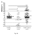
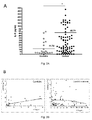
- mtDNA level was comprised between 1 and more than 30, compared to NAG samples found between 0 and less than 30 ( Figure 1B ). Also when comparing GC samples of all types, according to the presence of hyperplasia, metastasis or judged as inoperable by surgeons, no significant differences were observed on the variations of mtDNA level between the different GC groups. As reported in the table 1, the H. pylori serology was positive for half of the NAG and 61% of GC patients. No significant difference was observed comparing circulating blood white cells mtDNA level in H. pylori -positive and negative patients either both groups of patients ( Figure 4 ).
- samples can be classified in 3 main groups corresponding to mtDNA ⁇ 2 (Group I), 2 ⁇ mtDNA ⁇ 20 (Group II) and mtDNA>20 (Group III) ( Figure 1C ).
- the distribution of samples among these groups was not influenced by age or gender of the studied-population (data not shown), and showed important differences according to the gastric pathologies.
- Group I with mtDNA ⁇ 2 no healthy subjects were detected compared to 46% NAG and 14% GC samples. Indeed, for all healthy subjects mtDNA levels were found in Group II comprised between a value of 2 to 20.
- IL-8 In GC patients, two groups of samples according to IL-8 levels can be distinguished with IL-8 ⁇ 50 and IL-8>100 pg/ml corresponding to 79% and 21% of patients respectively. It is to be noticed that for 93% of GC patients, IL-8 higher than 100pg/ml is associated with mtDNA level of Group I or II. No significant correlation was observed between IL-8 and peripheral leucocytes mtDNA levels in GC patients. In addition, there were no significant differences in IL-8 according to the different groups defined for mtDNA level (data not shown).
- peripheral leucocytes mtDNA level shifted to higher values between gastric pre-neoplastic (NAG and IM) and neoplastic stages (GC), establishing a signature assessing the evolution of the disease, that may be used when monitoring the health status of monitored patients or individuals.
- NAG and IM gastric pre-neoplastic
- GC neoplastic stages
Landscapes
- Chemical & Material Sciences (AREA)
- Life Sciences & Earth Sciences (AREA)
- Health & Medical Sciences (AREA)
- Organic Chemistry (AREA)
- Proteomics, Peptides & Aminoacids (AREA)
- Engineering & Computer Science (AREA)
- Immunology (AREA)
- Pathology (AREA)
- Analytical Chemistry (AREA)
- Zoology (AREA)
- Genetics & Genomics (AREA)
- Wood Science & Technology (AREA)
- Physics & Mathematics (AREA)
- Biotechnology (AREA)
- Microbiology (AREA)
- Molecular Biology (AREA)
- Hospice & Palliative Care (AREA)
- Biophysics (AREA)
- Oncology (AREA)
- Biochemistry (AREA)
- Bioinformatics & Cheminformatics (AREA)
- General Engineering & Computer Science (AREA)
- General Health & Medical Sciences (AREA)
- Measuring Or Testing Involving Enzymes Or Micro-Organisms (AREA)
Abstract
The invention relates to an in vitro method for investigating the level of mtDNA in a biological sample removed from a patient susceptible of suffering from a gastric cancer condition or susceptible of suffering of condition(s) susceptible to evolve in a gastric cancer condition by pooling said biological sample into categories, upon determination of the level of mtDNA in said biological sample. The invention also relates to a method for in vitro monitoring or diagnosing the health status of a patient susceptible of suffering from a gastric cancer condition or suffering from a gastric cancer condition or susceptible of suffering of condition(s) susceptible to evolve in a gastric cancer condition, comprising a step of measuring, in parallel to the investigation of the level of mtDNA, the level of any one of the following parameters: cytokine IL-8, platelet count (PLT) and/or mean platelet volume (MPV) and/or percentage of large platelets (LPLT), inflammatory markers such as IL-6 and/or IL-23, or any combination of these parameters, and concluding about the health status of the tested patient. The invention also relates to kit(s) suitable for carrying out the method(s) of the invention and their use.
Description
- The present invention relates to the field of in vitro testing methods based on the investigation of the level of mitochondrial DNA (mtDNA) in biological samples collected from individuals, animals or humans, and in particular relates to methods that can be applied to the monitoring and/or diagnosing of the health status of a patient susceptible of suffering from a gastric cancer or susceptible of suffering from condition(s) that may evolve in a gastric cancer, especially methods suitable for the monitoring or the detection of a risk of gastric carcinogenesis. The invention also relates to kits for performing the methods of the invention and their uses.
- Gastric cancer (GC) is the second leading cause of cancer-related death in developing countries and the fourth in industrialized countries. It affects about one-million people per year.1 Although the incidence of GC and mortality rates have gradually decreased in the last decades, it remains an important public health problem worldwide with approximately 800 000 deaths per year.
- GC is often asymptomatic or causes only nonspecific symptoms in its early stages. By the time heavy symptoms occur, the cancer has often reached an advanced stage and may have also metastasized.
- Actually and as a consequence, GC is often diagnosed at an advanced stage, e.g at stage IV for 79% of the tumors. These tumors generate early hematogenic metastasis to the liver, lung, brain and skeleton. Consequently, gastric cancer carries a poor prognosis with an overall 5-years survival rate around 15%.2 However if it is diagnosed at an early stage, it can be a curable disease.3 These data highlight the importance and the need for characterization and validation of early GC biomarkers to reduce the morbidity and mortality associated to gastric adenocarcinoma.
- GC arises from the complex interplay of various factors including environmental factors and host genetic factors.4, 5 However, the major risk factor occurring for 60 to 80% of gastric cancer cases is Helicobacter pylori infection. The prevalence of H. pylori infection is high, with 80 to 95% of the population infected in developing countries and up to 30% to 40% of adults in industrialized countries.6 The infection mostly persists life-long if not treated. All infected individuals develop a gastritis which can evolve to peptic ulcer diseases in 10% of the cases, while gastric adenocarcinoma and mucosa-associated lymphoid tissue (MALT) lymphoma will develop for 3% and 0.3% of infected subjects, respectively.6, 7
- Gastric adenocarcinoma is a malignant epithelial tumor, originating from glandular epithelium of the gastric mucosa. A major proportion of GC, i.e. more than 90%, are adenocarcinomas. Based on an histological distinction, there are two major types of gastric adenocarcinoma: intestinal type or diffuse type. Adenocarcinomas tend to aggressively invade the gastric wall, infiltrating the gastric mucosa, and present several degrees of differentiation, from well to poorly differentiated.
- MALT lymphoma (or MALToma) is a form of lymphoma involving the mucosa-associated lymphoid tissue (MALT), frequently found in the stomach. This disease originates from B cells in the marginal zone of the MALT, and is also called extranodal marginal zone B cell lymphoma. Gastric MALT lymphoma is frequently associated (but not in all cases) with a chronic inflammation resulting from the presence of H. pylori, or linked with the presence of H. pylori.
- In the context of the invention, GC encompasses gastric adenocarcinoma and MALT lymphoma.
- GC of intestinal-type is mostly induced by H. pylori infection. It develops through a sequence of precursor lesions from atrophic gastritis to intestinal metaplasia (IM) then dysplasia and cancer.8 IM are recognized as pre-neoplastic lesions. It is important to notice that the eradication of the infection at an early stage (IM) can reverse and more importantly prevent the development of precancerous lesions.5 Accordingly, in humans several studies showed regression of precancerous lesions after H. pylori eradication,9 10 also confirmed in animal models. 11, 12
- In intestinal-type of GC, the sequential steps in the precancerous process can be considered as (in evolving order): gastritis (superficial or, at a later stage, chronic atrophic gastritis, for example), metaplasia (small intestinal or colonic metaplasia), and finally, dysplasia.
- The cancerous (or neoplasic) stage may then be entered, depending on the evolution of the disease. The clinical stages of GC can be classified using known classifications systems, e.g. the TNM Classification of Malignant Tumours staging system that describes the extent of a patient's cancer. Using this type of classification, one can distinguish between
Stages 0, I, II, III or IV. Other types of staging systems can be used. - In
Stage 0, the gastric cancer is limited to the inner lining of the gastric mucosa and may be treatable by surgery when found very early, without need for chemotherapy or radiation treatments. - In Stages I and II, the disease has penetrated the deeper layers of the gastric mucosa, and may be treated by surgery, sometimes associated with chemotherapy and/or radiation treatments.
- In Stage III, the disease may have penetrated other nearby tissues distant lymph nodes. Treated as for Stage II, a cure is still possible in some cases.
- In Stage IV, the disease has spread to nearby tissues and more distant lymph nodes, or has metastasized to other organs. A cure is very rarely possible at this stage. Some techniques are used to prolong life or improve symptoms, including laser treatment, surgery, and/or stents to keep the digestive tract open, and chemotherapy.
- Mitochondria are essential organelles of eukaryotic cells and possess their own genome. Defects in mitochondrial functions are associated with various human diseases including cancer.13 In humans, mitochondrial DNA (mtDNA) is a 16.6Kb circular DNA molecule present at hundreds to thousands copies per cell.14 Mutations in mtDNA have been reported in all cancer examined to date. As example in colorectal cancer, presence of mtDNA mutation is associated with a poor prognosis.15 The presence of mtDNA mutations has also been described at early stages of gastric carcinogenesis.16, 17 In H. pylori chronic gastritis patients, mtDNA mutations are significantly more frequent in gastric cancer patients than in cancer free patients.18 According to the inventors previous studies, mtDNA mutations are induced in vitro in H. pylori infected gastric epithelial cell19 and in the gastric mucosa of mice chronically infected.20 Tumor specific changes in mtDNA copy number have also been reported in a broad range of primary human cancers.21, 22. Xia et al, reported mtDNA depletion in the peripheral blood of patients strongly associated with stage I breast tumors.28 A decrease in mtDNA content has been described in most tumor tissues of advanced GC, compared with nearby non-tumorous control tissue.23 Variation of mtDNA level in cancer patients compared to healthy subjects has also been described for circulating cell-free mtDNA in the plasma and serum, leading to postulate that level of circulating mtDNA can constitute a potential preventive/diagnosis cancer marker detectable by a non-invasive method.19 Indeed, changes in mtDNA content in circulating blood samples have been described in patients with various types of tumors. As example, levels of circulating mtDNA were significantly higher in patients with urologic malignancies compared to healthy volunteers,24 as also reported in several case-control studies in breast,25 colorectal26 and lung cancer.27 These studies indicate that increased mtDNA content in peripheral blood should be associated with elevated cancer risk. In a recent case-control study conducted within a large prospective cohort of women residing in Shangai, no association was shown between leukocyte mtDNA copy number and presence of gastric tumor.29 However a positive association between low mtDNA copy number in blood drawn within the two years prior to cancer diagnosis and risk of developing GC was observed, suggesting that circulating blood mtDNA levels could be an indicator of the presence of GC lesions at an early stage.
- So far, variation of mtDNA levels in circulating blood can therefore be considered as a potential marker for the development of serological diagnosis test for a detection of the presence of gastric preneoplasia or cancer lesions.
- However, the literature is not consistent when it comes to determining what variation(s) in mtDNA levels may be correlated with the presence of gastric preneoplasia or cancer lesions associated with a risk of gastric cancer, let alone what particular mtDNA levels may be associated with such a risk.
- The present invention proposes a solution to this uncertainty, and further provides tools for investigating the level of mtDNA in a biological sample, in particular in a blood sample, said sample being obtained from a patient susceptible of suffering from a gastric cancer condition or condition(s) susceptible to evolve in a gastric cancer, in particular for monitoring or detecting a risk of an ongoing gastric carcinogenesis process.
- As used herein, the term "gastric carcinogenesis" encompasses both the precancerous manifestations described above including gastritis, pre-neoplastic lesions such as intestinal metaplasia (small intestinal or colonic metaplasia), neoplastic lesions such as dysplasia, that may evolve in gastric cancer, and the different cancerous stages of gastric cancer, in particular, use of the expression "gastric carcinogenesis" covers the development of gastric cancer since detection of the presence of a gastritis or associated lesions in a patient.
- The method of the invention can suitably be used for monitoring or diagnosing the health status of a such a patient.
- The present invention relies on studies in which the peripheral leucocytes mtDNA levels have been measured in patients at various steps of the gastric carcinogenesis process. Relevant variations of mtDNA levels have been surprisingly found during the progression from gastric inflammatory lesions in gastritis patients until the development of malignant lesions, supporting the notion that circulating mtDNA level can be considered as a potential biomarker for detection of gastric carcinogenesis, in particular at early steps during gastric carcinogenesis.
- Accordingly, the present invention relates to an in vitro method for investigating the level of mtDNA in a biological sample removed from a patient susceptible of suffering from a gastric cancer condition or susceptible of suffering of condition(s) susceptible to evolve in a gastric cancer condition, by pooling said biological sample removed from such a patient into categories, said method comprising the steps of:
- a. Determining the level of mtDNA in said biological sample, and
- b. Comparing the level of mtDNA determined in step a. with a normal threshold value determined for healthy subjects, and
- c. From the comparison made in step b., assigning the tested biological sample to one of the following categories:
- Group I: if the level of mtDNA determined in step a. is decreased with respect to the normal threshold value introduced in step b. by less than 2 folds, i.e., the ratio of the level of mtDNA determined in step a. over the normal threshold value determined for healthy subjects of step b. is inferior to 2,
- Group II: if the level of mtDNA determined in step a. is from 2 and 20 folds with respect to the normal threshold value introduced in step b., i.e., the ratio of the level of mtDNA determined in step a. over the normal threshold value determined for healthy subjects of step b. is equal or superior to 2, but equal or inferior to 20,
- Group III: if the level of mtDNA determined in step a. is increased with respect to the normal threshold value introduced in step b. by more than 20 folds, i.e., the ratio of the level of mtDNA determined in step a. over the normal threshold value determined for a pool of healthy subjects of step b. is superior to 20.
- According to a particular embodiment, a level of mtDNA as determined in step a., which enables the assignment of the tested biological sample to Group I or Group III, is an indicator for the presence, or indicates the possibility of presence, of gastritis lesions (in Group I) or gastric neoplasia (in Group III), leading to further clinical investigation for the subject(s) assigned to these two groups, including endoscopy and anatomopathology analysis.
- By "patient susceptible of suffering from a gastric cancer condition or susceptible of suffering of condition(s) susceptible to evolve in a gastric cancer condition", it is meant a patient presenting a suspicion for developing gastric cancer, for example because of physical clues, family history or complaint(s) indicating to the practitioner that an etiology of gastric cancer may be present or may become present. It also includes patients with gastroesophageal reflux, with chronic gastric pain as well as H. pylori seropositive subjects or H. pylori seronegative subjects, which have been beforehand eradicated for H. pylori infection. This definition also encompasses individuals having a declared gastric cancer condition or condition(s) susceptible to evolve in a gastric cancer condition, under treatment or not, which should be monitored. A particular group of patients eligible for the performance of the method of the invention is a group of patients with chronic inflammation associated with gastritis, or patient(s) with gastroesophageal reflux, with chronic gastric pain as well as H. pylori seropositive subjects. As mentioned above, a "patient susceptible of suffering from a gastric cancer condition or susceptible of suffering of condition(s) susceptible to evolve in a gastric cancer condition" may also be H. pylori negative at the time of the sampling and/or testing because of a successful eradication of the infection. The above definition also includes patients previously diagnosed for chronic atrophic gastritis or other gastric lesion that need a clinical follow-up.
- In a particular embodiment, the method of the invention may be carried out on a sample isolated (collected, removed) from an individual who has been previously diagnosed for a gastric cancer or for lesions that may lead to a gastric cancer (pre-neoplasic condition) and who, optionally, may have been subjected to a treatment, such as surgery and/or chemotherapy and/or radiations treatment, in particular an individual pertaining to the above-mentioned particular group(s) of patient(s).
- Under "gastric cancer" are encompassed: gastric adenocarcinoma, both diffuse or intestinal types, and MALT lymphoma.
- By "investigating the level of mtDNA" it is meant detecting, in particular quantifying a "level of mtDNA", e.g. a quantity of mtDNA in an assayed biological sample collected from an individual, animal or human, with respect to a quantity considered as a standard value, said level reflecting the abundance of mtDNA copies in said sample.
- By "level of mtDNA", it is meant a result qualifying the change in mtDNA with respect to a standard or normal level and in particular a value aimed at quantifying or representing the amount, in particular the number of copies of mtDNA, in particular the absolute number of copies of mtDNA, in the tested biological sample, or a relative amount of mtDNA in the tested biological sample, in particular when normalized with respect to a quantity of nuclear DNA (nDNA) or another suitable reference also present in said biological sample, or with respect to normalizing genes or DNA sequences pertaining to nDNA. In a particular embodiment, the investigation according to the invention may thus be achieved without determining the absolute quantity of mtDNA in the sample but by evaluating the variation (increase of the level or the decrease of the level) of said mtDNA and assigning said variation to a range as defined above.
- To obtain a "level of mtDNA" according to the present invention, use is made of techniques enabling nucleic acids quantification from a biological sample. Such technique enables the determination of the average concentration (or amount) of nucleic acids, i.e., DNA or RNA, especially mtDNA within the context of the present invention, present in a sample. Several methods can be used to establish such concentrations (or amounts), including (1) spectrophotometric analysis of nucleic acids and their further quantification and (2) quantification using the measurement of the fluorescence intensity of dyes that bind to nucleic acids and selectively fluoresce when bound, as well as (3) quantification after specific nucleic acids amplification, such as in the real time PCR technique, which also relies on the detection of a fluorescent dye bound to said nucleic acids to be detected and quantified. All these techniques may be used with their corresponding appropriate containers for the nucleic acids to quantify, such as cuvettes or microplates or arrays, as well as combined in more integrated systems such as chips, lab-on-a-chip, microfluidic supports (including microfluidic cartridge or chip). Depending on the technique used, prior isolation of mtDNA to be quantified may be required, according to the common knowledge in the art of nucleic acids analysis.
- By "a normal threshold value determined for healthy subjects", it is meant a value found by assaying one biological sample from an healthy subject or alternatively found by assaying several biological samples from several distinct healthy subjects, the resulting normal threshold value being then determined as the mathematical mean of the levels values of all the assayed healthy subjects biological samples, or alternatively found by assaying a pool of biological samples from several distinct healthy subjects.
- According to a particular embodiment, such a normal threshold value is determined for a pool of healthy subjects.
- By "healthy subject(s)" it is meant subjects that would have no symptoms of gastric disorders or patients referred to for gastroscopy with gastric biopsies corresponding to a normal phenotype, and in both cases not infected with H. pylori or that received an eradication therapy followed by a confirmed negative H. pylori serology. In a particular embodiment, the biological sample removed from such healthy subject(s) is a blood sample.
- More particularly, by "normal threshold value determined for healthy subjects or for a pool of healthy subjects", it is meant a value, corresponding to a level of mtDNA, which may be predetermined according to the knowledge of the skilled in the art because it is known to correspond to a normal mtDNA value, or, according to a preferred embodiment, a value determined from assays made on subjects, in particular individuals, known to be healthy, i.e., not presenting a gastric cancer condition or suffering of condition(s) susceptible to evolve in a gastric cancer, or as explained above, said patients providing samples which either are individually tested and the results obtained from said samples treated to provide a mean value or a median value or said samples are tested as pooled samples to provide a so-called normal value. The thus obtained value provides a threshold that determines the reference value for normal samples.
- By "fold", it is meant the expression of a change, in particular a number, describing how much a given quantity changes from a normal to a tested value, the normal value being in particular the "normal threshold" and the tested value being the mtDNA copy number or a representative value thereof, in the tested sample. For example, a normal value of 30 and a tested value of 60 correspond to a fold change of 2, or in common terms, a two-fold increase. To the contrary, a normal value of 60 and a tested value of 30 correspond to a fold change of 0.5, or in common terms, a 0.5 fold decrease, also referred to as a "minus" two-folds decrease (expressed in negative terms, with a "minus" sign before the number). Fold changes therefore correspond to a ratio of the tested value to the normal value. In other words, the fold change results from the determination of a ratio of the tested value against the normal value.
- The invention accordingly also relates to a method of determining a "normal threshold" value for the mtDNA which value is associated with an absence of gastric cancer condition or an absence of a condition susceptible to evolve in a gastric cancer condition as disclosed herein.
- The determination of "normal threshold" value may be regarded as involving a pooling method to the extent that it results either from pooling the results or from pooling the samples said samples being obtained from healthy subjects. Healthy subjects refer to a subject with no diagnosed chronic inflammation either gastric or other and with a negative H. pylori serology. It can also be a subject that is not treated by antibiotherapy or Non-Steroidal Anti-Inflammatory Drug(s) (NSAID), with a normal blood count and in the absence of any medical treatment at the time of the sampling and/or testing. According to another particular embodiment the normal values providing the threshold to determine the level of change in mtDNA of tested biological sample are the result of the above disclosed pooling method when performed on a pre-determined relevant population, as also detailed above.
- The method of the invention can subsequently be used for monitoring or diagnosing the health status of a patient susceptible of suffering from a gastric cancer condition or susceptible of suffering of condition(s) susceptible to evolve in a gastric cancer condition, such a method comprising steps a., b. and c. as disclosed above and further comprising the following step:
- d. from the assignment to a Group I, II or III made in step c. as described above, concluding about the health status of a patient from which the tested biological sample has been removed.
- By "concluding about the health status" it is meant concluding that the individual, the biological sample of which is tested, may present a risk of suffering of a gastric cancer or from condition(s) that may evolve in a gastric cancer, i.e., condition(s) indicating a risk that a gastric carcinogenesis is present in the tested individual.
- In particular and according to a finding related to the present invention, assignment of a mtDNA level value in Group I or III in step c. as described above, provides the information that the patient from which the tested biological sample has been removed is susceptible of suffering from gastric lesions, which also suggests performance of further clinical investigations. As non-limitative examples, other investigations methods aimed at confirming or excluding the presence of as gastric carcinogenesis process may be optical gastroscopic examination, computed tomography (or CT) scanning of the abdomen, biopsies for histological examination, various blood tests, e.g., Complete Blood Count (CBC) to check for anemia.
- Accordingly, it is another object of the invention to provide a method for monitoring or detecting a risk of gastric carcinogenesis in a patient, wherein said method comprises identifying whether the level of mtDNA of the assayed sample has a value that pertains to Group I or Group III, a risk of gastric carcinogenesis being present if the biological sample removed from said patient has such a value.
- By "a risk of gastric carcinogenesis" it is meant a risk of presenting a gastric cancer condition or a risk of presenting condition(s) susceptible to evolve in a gastric cancer condition, which encompass:
- precancerous manifestations of gastric cancer, that may further evolve in gastric cancer, such as: gastritis or gastritis lesions (superficial or, at a later stage, chronic atrophic gastritis, for example), metaplasia (small intestinal or colonic metaplasia), and finally, dysplasia, and
- the different cancerous (or neoplasic) stages of gastric cancer, as defined above.
- According to a particular finding, the method of the invention enables concluding to the existence of a risk of gastric carcinogenesis, in particular at an early stage, when the tested biological sample pertains to Group I and the mtDNA level change measured with respect to normal value is less than 0.5 fold.
- In a particular embodiment, by "early stage of gastric carcinogenesis" it is meant a pre-neoplasic stage, as defined above, for example a gastritis stage.
- According to another particular finding, the method of the invention enables to conclude to the existence of a risk of a gastric cancer, when the tested biological sample pertains to Group III and the mtDNA level change measured with respect to normal value is more than 30 folds.
- According to a particular embodiment, the gastric carcinogenesis referred to herein is associated with an Helicobacter pylori infection or is an Helicobacter pylori-induced gastric carcinogenesis.
- Accordingly, the invention also comprises a method of investigating the level of mtDNA in a sample removed from a patient, where the patient has been previously or is simultaneously or in parallel tested for infection by Helicobacter pylori. The detection of H. pylori may be performed on a fraction of the sample removed from the patient, in particular on the serum fraction of a blood sample by carrying out a step of detection of antigens specific for H.pylori infection. People infected by H. pylori have specific IgA and IgG antibodies that can be easily detectable. In addition, the search for the presence of CagA antigens can also confirm the presence of H. pylori. Another method to detect H. pylori is the 13C urea breath test, a non-invasive test with high sensitivity widely used in human medicine (Graham et al, 1987, Lancet, 1 : 1174-1177). This respiratory test allows an indirect measure of the H. pylori-associated urease activity. Presence of H. pylori can also be detected in stools by immunoassay indicating the presence of H. pylori antigens or by amplification of H. pylori DNA in particular by polymerase chain reaction (PCR) using specific primers for H. pylori genes sequences, which are available in the literature to one skilled in the art, and detection of the amplified DNA.
- For detection of H. pylori, the blood sample may be prepared on the one hand to purify the cellular fraction of the blood sample, in particular the mononuclear cells or leukocytes containing the mtDNA to be assayed and on the other hand to collect the serum enabling the detection of H.pylori infection.
- In a particular embodiment, the tested biological sample is obtained from a patient diagnosed with gastric carcinogenesis and under treatment for this condition or not, and/or a patient having an ongoing, treated or not, Helicobacter pylori infection, and/or a patient having antecedents of Helicobacter pylori infection(s), eradicated by prior or ongoing treatment or not, and/or an individual having gastric pain and/or a family history of gastric cancer.
- The method for investigating the level of mtDNA in a biological sample according to the invention is performed using any relevant process for the quantification of nucleic acids, as in particular introduced above. It especially involves treating the sample to gain access to mononuclear cells, in particular leukocytes, and furthermore to provide access to nucleic acid, especially by extracting the nucleic acid, and optionally by separating the mtDNA from nuclear DNA. The method also involves using polynucleotides, in particular probes and/or oligonucleotides when they are complementary to a region of interest in the mtDNA, said polynucleotides optionally including a detectable label or marker (such as a fluorochrome or a radioelement) in order to allow quantitative detection of the mtDNA.
- According to an embodiment of the invention, the method for investigating the level of mtDNA in a biological sample and optionally the method of determining a "normal threshold" value involve(s) a step of amplification of the mtDNA, possibly a step of differential amplification of the mtDNA with respect to the nuclear DNA.
- According to a specific embodiment, the "level of mtDNA" is determined by quantitative polymerase chain reaction (q-PCR). Primers specific for the 12sRNA mitochondrial gene may be used, although one skilled in the art can suitably choose other genes or sequences of the mitochondrial genome for implementing such a technique, following guidance available in the literature of this field with respect to this technique.
- PCR (polymerase chain reaction) is a common method for amplifying DNA. In order to amplify small amounts of DNA, a DNA template, at least one pair of specific oligonucleotide primers, nucleotides (dATP, dCTP, dGTP, dUTP), a suitable buffer solution and a thermo stable DNA polymerase are required. A substance marked with a fluorophore is generally added to one reagent of this mixture in a thermal cycler that contains sensors for measuring the fluorescence of the fluorophore after it has been excited at the required wavelength allowing the generation rate to be measured for one or more specific products. Real-time PCR (q-PCR) is generally applied to the detection and quantification of DNA in samples to determine the presence and/or abundance of a particular DNA sequence in these samples. A measurement is made after each amplification cycle, which enables the quantification of the amplified product in real time.
- Real-time PCR is performed by using a real-time PCR apparatus, and after each cycle, the levels of fluorescence are measured with a detector. Used dyed generally only fluoresce when bound to the DNA amplified through PCR, and the increase of fluorescence is detected, corresponding to increasing presence of the amplified products, at each amplification cycle.
- Real-time PCR can be used to quantify nucleic acids by either relative quantification or absolute quantification. Absolute quantification gives the exact number of target DNA molecules by comparison with DNA standards using a calibration curve. By this method, it is possible to determine the number of mtDNA copies in patients suspected for the presence of gastric pre-neoplasia or neoplasia and compare this number to the number of mtDNA copies defined in healthy subjects. (Ref: von Wurmb-Schwark et al, 2002, Forensic Science International, 126: 34-39). Relative quantification enables determining fold-differences between a target sequence, the quantity of which is to be determined, and a "housekeeping sequence".
- In order to quantify the presence of a specific DNA target sequence, representative of the copy number of the mtDNA, it is indeed convenient to express its relative level in relation to another DNA sequence called a "normalizing sequence" or "housekeeping sequence", which is selected for its almost constant rate of expression. Housekeeping sequences are usually found in genes involved in the functions related to basic cellular survival, which normally implies constitutive gene expression. This enables the provision of a ratio expressing the presence of the amplified sequence of interest over the presence of the amplified selected normalizer. This method allows obtaining a value evaluating the relative presence of the amplified sequence of interest actually knowing its absolute quantity within the tested sample.
- Commonly used normalizing sequences are those found in genes coding for the following proteins. As a non-limitative list, tubulin, glyceraldehyde-3-phosphate dehydrogenase, albumin, cyclophilin, ribosomal RNAs sequences can be used.
- Real time PCR allows quantification of the desired product at any point in the amplification process by measuring fluorescence. Measurement is expressed using a Cycle Threshold (CT) value (CT; PCR cycle at which the fluorescence of the sequence of interest is detected; the lowest is the CT value, the more abundant is the target sequence). To quantify the presence of the target sequence when a normalization sequence is used, a normalization procedure such as the ΔΔCT-method can be used, said ΔΔCT-method being used for analyzing a relative gene expression.
- According to a more specific embodiment of the invention, the "level of mtDNA"i is determined by quantitative polymerase chain reaction (q-PCR) through reference to a selected normalizer gene or nDNA sequence, the level of mtDNA being calculated according to the
formula 2ΔCt, wherein ΔCt = CtnDNA - CtmtDNA, as described in reference publications Fan et al, 2009 , J Cancer Res Clin Oncol, 135 ; 983-989 and/or Chatre and Richetti, 2013, J of Cell Sciences, 126 : 914-926. With this calculation method, the level of mtDNA can be calculated using the ΔCT of average CT of mtDNA and nDNA (ΔCT = CtnDNA - CtmtDNA,) as 2ΔCt. The primers used for amplification can be chosen by one skilled in the art according to the common knowledge in the field of this technique, as indicated in particular in the above-mentioned reference publications. - The invention thus concerns a method wherein the level of mtDNA is determined by quantitative polymerase chain reaction (q-PCR) following the steps of:
- preparing the biological sample to provide access to the nucleic acid, especially mitochondrial nucleic acid of cells;
- contacting the prepared sample with oligonucleotide primers targeting the mtDNA;
- performing amplification cycles,
- simultaneously running amplification of a normalizer nDNA,
- quantitatively detecting the mtDNA and the normalizer nDNA.
- determining the level of mtDNA through reference to a selected normalizer nDNA sequence, the level of mtDNA being calculated according to the
formula 2ΔCt, wherein ΔCt = CtnDNA - CtmtDNA. - The invention thus concerns a method for investigating the level of mtDNA in a biological sample wherein the biological sample is from a patient diagnosed with gastric carcinogenesis and under treatment for this condition or not, and/or a patient having an ongoing, treated or not, Helicobacter pylori infection, and/or a patient having antecedents of Helicobacter pylori infection(s), eradicated or not, and/or an individual having gastric pain and/or a family history of gastric cancer.
- According to a particular embodiment, the mtDNA of the tested sample is detected or quantified by using a region in the 12sRNA gene of the mitochondrial genome, and the nDNA of the tested sample is detected or quantified by using a region in the 18sRNA gene of the nuclear genome. Other couples of mtDNA/nDNA primers (probes) can be chosen and used by the person skilled in the art, as reminded above, for example mtDNA encoded
ATPase 8 gene and nDNA glyceraldehyde-3-phosphodehydrogenase (GAPDH) orBeta 2 microglobuline (β2M). - Accordingly, in a particular embodiment, oligonucleotide primers and/or DNA probes suitable for carrying out the invention may advantageously be selected for their capacity to hybridize to a strand of the 12sRNA gene or to a strand of the 18sRNA strand. The oligonucleotide (forward and reverse) primers have a length adapted to specifically priming the targeted mtDNA or nDNA and in particular have a length within a range of 10 to 30 nucleotides. Examples of oligonucleotide primers are polynucleotides comprising or consisting of the sequences disclosed in the experiments provided herein. The probes may have a length of 50 to several thousand of nucleotides, in particular may have a length of 50 to 500 nucleotides.
- Oligonucleotides and probes to carry out the detection of mtDNA are selected for their ability to hybridize specifically with the targeted mtDNA and accordingly would not hybridize significantly in PCR operating conditions with nuclear DNA. Preferably, the chosen mtDNA sequences are highly specific to mitochondrial genome and nonexistent in nuclear genome.
- The invention thus also relates to the use of the pairs of oligonucleotides either as unique pair or combined primer pairs having the following sequences hybridizing to the 12SRNA: 12S (forward): 5'- GCT CGC CAG AAC ACT ACG AG; (reverse): 5'- CAG GGT TTG CTG AAG ATG GCG for the detection of mtDNA. It may further involve primers as the following hybridizing with a nuclear encoded 18SRNA: 18S (forward): 5'- GAG AAA CGG CTA CCA CAT CC; (reverse): 5'- GCC TCG AAA GAG TCC TGT AT.
- In a particular embodiment, the method of the invention is performed using a biological sample removed from a patient, that is a blood sample or, in particular when the patient already present gastric mucosa lesions, a biopsy sample, in particular a biopsy sample of gastric mucosa. All layers of the gastric mucosa may be used for sampling. mtDNA and/or DNA extraction and isolation can be performed according to commonly known methods.
- According to another particular embodiment, the level of mtDNA is determined by testing circulating blood mtDNA, in particular is determined by testing the mtDNA of leukocyte(s), in particular leukocyte(s) isolated from a blood sample, especially when circulating blood samples are used for performing the method of the invention.
- In a particular embodiment of the invention, the investigation of the level of the mtDNA in a patient is associated with the determination of a different biological parameter measured in order to improve the significance or the interpretation of the results obtained in relation with the determination of mtDNA level with a view to ascertain the patient's condition. Other biological parameters in this regard include the parameters which are known to provide indication as to a condition susceptible to evolve toward gastric carcinogenesis, such as measurement of cytokine, especially interleukin expression in particular pro-inflammatory interleukin, including IL-8 expression.
- Another object of the invention is accordingly a method for monitoring or diagnosing the health status of a patient susceptible of suffering from a gastric cancer condition or susceptible of suffering of condition(s) susceptible to evolve in a gastric cancer condition, said method comprising the following steps:
- a. Performing a method of the invention for investigating the level of mtDNA in the tested patient as described above, and
- b. Measuring, in parallel to the investigation of the level of mtDNA of step a., the level of cytokine IL-8 in a biological sample removed from the patient tested in step a., and
- c. Concluding about the health status of a patient on the basis of the results obtained from step a. and b.
- By "measuring, in parallel to the investigation of the level of mtDNA", it is meant that the measurement of the level of cytokine IL-8 is made by using samplings of biological material(s) in a same individual, collected within a close interval of time, i.e. avoiding an interval of time that may render both parameters not representative of the physiological status of the individual at a given time, in particular for evaluating both parameters at a same given time t.
- The inventors have indeed assessed the pertinence of measuring the level of cytokine IL-8 for a same tested individual, in particular for providing a better evaluation of the health status of said tested individual. A strong association between cancer and inflammation has long been observed. Association between circulating cytokine level(s) and gastric cancer risk has also been reported, highlighting in particular that an high H. pylori prevalence and increased circulating IL-8 level may be associated with increased risk of gastric cancer (Epplein et al, 2013, Cancer Causes Control, DOI: 10.107/s10552-013-0284-z).
- Levels of cytokine IL-8 can be measured using commonly known methods in the art such as, as a non limitative example, through enzyme-linked immunosorbent assay(s) (ELISA) (Engvall E and Perlman P, 1971, lmmunochemistry, 8: 871-874). For example, levels of cytokine IL-8 can be measured using the ELISA kit commercialized by BD Biosciences: BDOptEIA Set Human IL-8. Cat. No. 555244.
- According to a particular embodiment, if one determines that the level of mtDNA pertains to Group I or Group II and the level of cytokine IL-8 is above 100 pg/mL, conclusion can be made of a risk of presence of a gastric cancer.
- In another aspect, the invention also relates to a method for monitoring or diagnosing the health status of a patient susceptible of suffering from a gastric cancer condition or suffering from a gastric cancer condition or susceptible of suffering of condition(s) susceptible to evolve in a gastric cancer condition, said method comprising the following steps:
- a. Performing a method of the invention for investigating the level of mtDNA in the tested patient as described above, and
- b. Measuring, in parallel to the investigation of the level of mtDNA of step a., the level of any one of the following parameters: cytokine IL-8, platelet count (PLT) and/or mean platelet volume (MPV) and/or percentage of large platelets (LPLT), inflammatory markers such as IL-6 and/or IL-23, or any combination of these parameters in a biological sample removed from the patient tested in step a., and
- c. Concluding about the health status of a patient on the basis of the results obtained from step a. and b.
- Matowicka-Karna J et al (Volume 2013, article ID 401623, Clinical and Developmental Immunology) have evidenced the contribution of platelets and inflammatory markers in gastric cancer. Generally cancer cells are a source of inflammatory cytokines and growth factors such as IL-6 or IL-23. IL-6 and IL-23 are actively involved in the pathogenesis and development of tumors. They facilitate tumor growth by inhibiting apoptosis of tumor cells and by angiogenesis induction within the tumor. IL-6 is a pleiotropic cytokine involved in immune responses, inflammatory reactions, and haematopoiesis. A direct association between increasing plasma levels of IL-6 and gastric cancer risk has been reported in Wong et al, 2011, Cancer Sciences, 102: 1911-1915. IL-23 strongly induces the secretion of IFN-γ, affects the haematopoiesis by stimulating thrombocytopoiesis, and the production of neutrophils, and stimulates the production of acute phase proteins. Platelets take part in inflammation and in cancerous diseases.They play an active role in the inflammatory process due to secreted proinflammatory factors, chemokines, and growth factors. An excessive number of platelets is the cause of a significant increase in the risk of metastases in each stage of cancer and an indicator of poor prognosis, for example, in gastric, lung, and kidney cancer. In cancer disease, an increase in the percentage of large platelets (LPLT) is observed, and because young, metabolically active platelets appear in the circulation, this may lead to an increase in MPV (mean platelet volume).
- "Platelet count" (PLT) can be expressed as platelet count per unit volume of blood, generally ranging between 150.109 and 350.109 per liter of blood.
- "Mean platelet volume" (MPV) can be calculated on the basis of distribution volume, generally ranging from 7,8 to 11,5 femtoliter (fl).
- "Large platelets" are defined as platelets having a volume above 20 femtoliter (fl).
- PLT, MPV and LPLT parameters can be collected through an hematology analyzer instrument, available in the art.
- Levels (or concentrations) of IL-6 and/or IL-23 parameters can be measured using commonly known methods in the art, in particular through ELISA testing.
- The above-mentioned parameters may also enable distinguishing between different gastric cancer stages. Reference is made to Matowicka-Karna J et al (Volume 2013, article ID 401623, Clinical and Developmental Immunology). In particular, high levels of IL-23 and IL-6 have been observed in patients with early gastric cancer. In the experiments described by Matowicka-Karna et al, a control level of IL-23 in a group of normal subjects was found to be 5.21±3.65 pg/ml compared to 20.42±1.44 pg/ml in a group of early gastric cancer patients. Control level for IL-6 was found to be 2.45±1.44 pg/ml in a group of normal subjects compared to 10.80±8.24 pg/ml in a group of early gastric cancer patients.
- According to a particular embodiment, the method for monitoring or diagnosing the health status of a patient susceptible of suffering from a gastric cancer condition or suffering from a gastric cancer condition or susceptible of suffering of condition(s) susceptible to evolve in a gastric cancer condition of the invention encompasses a determination of any combination of the above-mentioned parameters, in particular a combination of determination of the level of mtDNA and the level of at least one of the parameters selected amongst: IL-8, PLT, MVP, LPLT, IL-6, IL-23, or a combination of determination of the level of mtDNA and the level of IL-8 and the level of at least one of the parameters selected amongst: PLT, MVP, LPLT, IL-6, IL-23, in particular a combination of determination of the level of mtDNA and the level of IL-8 and the level of any one of either IL-6 or IL-23.
- The invention therefore also relates to a method for in vitro monitoring or diagnosing the health status of a patient susceptible of suffering from a gastric cancer condition or suffering from a gastric cancer condition or susceptible of suffering of condition(s) susceptible to evolve in a gastric cancer condition, said method comprising the following steps:
- a. Performing a method of the invention as described herein for investigating the level of mtDNA in the tested patient, and
- b. Measuring, in parallel to the investigation of the level of mtDNA of step a., the level of any one of the following parameters: cytokine IL-8, platelet count (PLT) and/or mean platelet volume (MPV) and/or percentage of large platelets (LPLT), inflammatory markers such as IL-6 and/or IL-23, or any combination of these parameters in a biological sample removed from the patient tested in step a., and
- c. Concluding about the health status of a patient on the basis of the results obtained from step a. and b.
- According to a particular embodiment, such a step c. of conclusion about the health status of a patient takes into account the value(s) of a level of IL-23 and/or of a level of IL-6 for detecting a risk of presence of a gastric cancer condition or the presence of a gastric cancer condition, in particular a gastric cancer condition at an early stage, in a tested patient, or takes into account a relevant increase of any one or all of such value(s) with respect to a normal threshold value determined for healthy subjects, as defined herein and according to the data provided herein and/or by Matowicka-Karna et al. An increase may be calculated in fold(s) for quantifying said increase.
- According to a specific embodiment, conclusion is made of a risk of presence of a gastric cancer condition or conclusion is made of the presence of a gastric cancer condition, in particular a gastric cancer condition at an early stage, when the IL-23 level found in the tested sample(s) is superior to about 18 or about 19 or about 20 pg/ml, in particular superior to 20,5 pg/ml, and/or when the IL-6 level is superior to about 8 or about 9 or about 10 pg/ml, in particular superior to 10,8 pg/ml.
- According to another specific embodiment, such a conclusion is made in combination with deduction(s) made with respect to another parameter, as described herein.
- Another object of the invention is to provide a kit suitable for carrying out a method of the invention as defined herein, comprising:
- At least one pair of specific oligonucleotide primers specific for hybridization with mtDNA and, optionally, at least one pair of specific oligonucleotide primers specific for hybridization with H. pylori nucleic acid(s) sequence(s), and, optionally, one or several of the following reagents,
- nucleotides (e.g. dATP, dCTP, dGTP, dUTP),
- a DNA polymerase, in particular a thermostable DNA polymerase, such as a Taq DNA Polymerase,
- at least one dye for staining nucleic acids, in particular a dye detectable in a real-time PCT equipment,
- optionally, a buffer solution,
- optionally, reagents necessary for the hybridation of the primers to their targets,
- optionally, a reference dye and,
- optionally a notice providing instructions for use and expected values for interpretation of results.
- According to a particular embodiment, a kit of the invention comprises primers suitable for amplifying 12S and optionally 18S gene sequences or fragments thereof, such as 5'- GCT CGC CAG AAC ACT ACG AG and 5'-CAG GGT TTG CTG AAG ATG GCG nucleotide sequences (12S), and/or 5'- GAG AAA CGG CTA CCA CAT CC and 5'- GCC TCG AAA GAG TCC TGT AT sequences (18S).
- Other primers may be used within such a kit, as detailed hereabove.
- Another object of the invention is to provide a kit suitable for carrying out a method of the invention as defined herein, comprising:
- at least one antibody specific for a protein selected amongst: IL-8, IL-6, IL-23 or a combination of several antibodies specific for IL-8, IL-6, IL-23, and, optionally, at least one antibody specific for H. pylori antigen(s), such as CagA antigens, and, optionally, one or several of the following reagents,
- a secondary antibody or reagent to reveal a complex between specific antibody(ies) recited above and its(their) target,
- optionally, a buffer solution,
- optionally, an assay plate, and
- optionally a notice providing instructions for use and expected values for interpretation of results.
- The invention also relates to the use of kit(s) according to the invention for investigating the level of mtDNA or of the other parameter(s) described herein in a biological sample, and/or monitoring or diagnosing the health status of a patient susceptible of suffering from a gastric cancer condition or susceptible of suffering of condition(s) susceptible to evolve in a gastric cancer condition and/or monitoring or detecting a risk of gastric carcinogenesis, or suffering from a gastric cancer condition, including the health status of said patient with respect to an H. pylori infection, according to the description provided herein, in all its detailed or encompassed embodiments.
- The present invention is a basis for a non-invasive test for measuring mtDNA levels, in particular in circulating blood. According to the invention mtDNA levels may be used as an efficient biomarker for an early detection of gastric carcinogenesis, in particular an early detection of the presence of gastric lesions involved or at the basis of a gastric carcinogenesis process. The present invention may be especially suitable for prevention purposes, but also for monitoring the progression of a gastric carcinogenesis process, subject to an on-going treatment or not, monitoring a shift from a pre-neoplasic condition to a neoplasic condition, or as a follow-up after a cure to screen for a recurrence of the disease.
- The method of the invention may be used on patients under treatment, such as chemotherapy treatment and/or radiations treatment, as an indicator of treatment efficiency, disease stage, and disease development.
- The method of the invention may be used on patients presenting a HER2 overexpressing gastric adenocarcinoma, treated or not with trastuzumab (Herceptin®, Genentech). Other known drugs used in gastric cancer chemotherapy are (non-limitative examples): 5-fluorouracil, cisplatin, epirubicin, etoposide, docetaxel, oxaliplatin, capecitabine, or irinotecan.
- As a result of performing the methods of the invention on patients, other investigations methods aimed at confirming or excluding the presence of as gastric carcinogenesis process may be indicated, such as an optical gastroscopic examination, computed tomography or CT scanning of the abdomen, biopsies for histological examination, various blood tests may also be done, such as Complete Blood Count (CBC) to check for anemia.
- The invention will be further described herein, referring to the following figures and experimental section.
-
-
Figure 1 . Circulating mtDNA content in white blood cells of NAG and GC patients compared to healthy blood donors. A) MtDNA was quantified by quantitative real-time PCR from DNA isolated from peripheral leukocytes isolated from healthy blood donors H. pylori-negative (open triangles) and from non atrophic gastritis (NAG) (open circles) and GC patients (closed triangle) fromcohort 1. MtDNA relative values was measured as previously described according to nuclear DNA (nDNA) (see Material and methods).35 Each symbol corresponds to a single patient. Bars and numbers indicate geometric means. Data are obtained from measures done in triplicate in three independent experiments. *P<0.05 ; **P<0.001. B) Peripheral leukocytes mtDNA level in NAG and GC patients according to the type of cancer either diffuse (n=31) or intestinal (n=20) or the presence of hyperplasia (n=19), metastasis (n=19) or late-stage cancer judged inoperable by surgeons (n=17) C) Distribution of the samples according to their mtDNA peripheral blood content in three different groups, Group I : mtDNA<2, Group II : 2<mtDNA<20 and Group III : mtDNA>20. No healthy samples are found in Group I. Samples with mtDNA level > 30 include only H. pylori infected gastric cancer patients. D) Samples with mtDNA<1 can subdivided in 2 sub-groups : Group la : mtDNA<0.5 corresponding exclusively to NAG samples and Group Ib : mtDNA<1 including 94% of NAG samples (16/17) and 6% of GC samples (1/17). -
Figure 2 . Comparison of IL-8 levels in sera of NAG and GC patients. A) IL-8 was quantified by using the ELISA kit BDOptEIA Set Human IL-8. Cat. No. 555244 commercialized by BD Biosciences according to the recommendations of the fabricant, in the sera from NAG and GC patients fromcohort 1. The mean IL-8 level is higher in GC (median= 3.95) than in NAG (median= 16.65) patients. B) Analysis of a correlation between IL-8 serological levels and peripheral leukocytes mtDNA amount in NAG and GC patients fromcohort 1. A positive significant correlation between IL-8 and mtDNA was detected for NAG samples (r=0.586 ; p=0.0041) but not in the case of GC patients. -
Figure 3 . Comparison of mtDNA level at various steps of the gastric carcinogenesis process. Quantification of peripheral leukocytes mtDNA level in NAG, IM and GC patients fromcohort 2. MtDNA was measured as previously reported35 and described in material and methods. -
Figure 4 : Circulating mtDNA content in white blood cells of NAG and GC patients compared to healthy blood donors according to their H. pylori serology. MtDNA was quantified by quantitative real-time PCR from DNA isolated from peripheral leukocytes isolated from healthy blood donors H. pylori-negative (open circles) and from non atrophic gastritis (NAG) H. pylori negative (grey circles) and H. pylori positive (closed circles) and GC patients H. pylori negative (grey diamond) and H. pylori positive (closed diamonds) fromcohort 1. MtDNA relative values was measured as previously described according to nuclear DNA (nDNA) (see Material and methods).35 Each symbol corresponds to a single patient. Bars and numbers indicate geometric means. Data are obtained from measures done in triplicate in three independent experiments. *P<0.05 ; **P<0.001. - Two cohorts of Mexican adult patients were studied.
Cohort 1 included 48 healthy blood-donors as the reference group, 28 patients with non-atrophic gastritis (NAG), and 78 with gastric cancer (GC) for a total of 154 adults recruited during the period 2009-2011. The healthy individuals were blood donors recruited at the blood bank of the Instituto Mexicano del Seguro Social (IMSS), Medical Center SXXI.Cohort 2 included 48 patients with NAG, 34 patients with intestinal metaplasia (IM), and 49 with gastric cancer (GC) for a total of 131 patients recruited during the period 1999-2002. Patients from both cohorts were adults who were attended for gastroduodenal diseases at the Instituto Mexicano del Seguro Social (IMSS) Medical Center SXXI, in Mexico City. The inventors selected patients who were not under treatment for cancer and who had not been treated with antibiotics, bismuth compounds, proton pump inhibitors and non-steroidal anti-inflammatory drugs for at least two weeks prior to the study. Diagnosis was based on endoscopic examination and histopathology studies. 30 All patients and controls were informed about the study and asked to sign a consent letter. The study was approved by the ethical committee from the National Council for Research on Health, IMSS. - For patients with gastritis or precancerous lesions, gastric biopsies collected from both the antrum and the corpus were taken. Biopsies were processed either for histology or immediately frozen at -70 °C. For histology, biopsies were immersed in formalin and processed for H&E staining for diagnosis of the lesions. Presence of non-atrophic gastritis, atrophic gastritis, intestinal metaplasia, or dysplasia was documented in each biopsy. The presence of H. pylori bacteria was confirmed by Giemsa staining. For patients with gastric cancer one fraction from the tumor lesion and one from the adjacent tissue were collected during surgery and divided for either histology or DNA extraction. For histology, tissues were immersed in formalin and processed for staining with H&E and analysed for H. pylori and for type of gastric tumor.
- Circulating DNA was prepared from a sample of 10 ml of peripheral blood from each patient, and mononuclear cells were purified by centrifugation through a Ficoll-Hypaque density gradient. DNA was isolated from these cells using the salting-out microtechnique and frozen at -70°C until tested for mtDNA. The serum fraction was frozen at -20°C until tested for serology to H. pylori antigens.
- IgG antibodies against H. pylori whole-cell antigens and against CagA H. pylori protein were tested in sera, using enzyme-linked immunoabsorbent assays which were previously validated by the inventors. 31
- The quantification of mtDNA was performed as previously described 33, 35, on total DNA isolated from circulating leucocytes using the StepOne™ Plus Real-Time PCR system and FastStart Universal SYBR Green Master (Applied Biosystems) following the manufacturer's instruction. MtDNA was quantified using a region in the 12SRNA gene. The nuclear encoded 18SRNA gene was used as an endogenous reference. Primers used to amplify the 12S and 18S gene sequences were: 12S (forward): 5'- GCT CGC CAG AAC ACT ACG AG; (reverse): 5'- CAG GGT TTG CTG AAG ATG GCG and 18S (forward): 5'- GAG AAA CGG CTA CCA CAT CC; (reverse): 5'- GCC TCG AAA GAG TCC TGT AT.34 The qPCR was carried out in 20µl of total reaction volume containing 5µl of DNA (200 pg), 10µl of FastStart Universal SYBR Green Master, 0,2µL of primers (10µM) using an initial denature step at 95°C for 10 minutes and 40 cycles of 15 seconds at 95°C and 1 minute at 60°C. All samples were analyzed in triplicate. To determine the quantities of mtDNA and nDNA present in blood samples, the average threshold cycle number values (Ct) of the nDNA and mtDNA were obtained from each case. The level of mtDNA was calculated using the delta Ct (ΔCt) of average Ct of nDNA and mtDNA (ΔCt = CtnDNA-CtmtDNA) as 2ΔCt as previously described. 35
- IL-8 levels were quantified in the serum of patients by enzyme-linked immunosorbent assay (ELISA) by using the ELISA kit BDOptEIA Set Human IL-8. Cat. No. 555244 commercialized by BD Biosciences according to the recommendations of the fabricant.
- The Mann-Whitney U test was used to compare mtDNA level in peripheral blood between healthy subjects and patients at various stages of the gastric pathologies, as well as to compare mtDNA level isolated from gastric biopsies. Differences were considered significant when P<0.05. For Odd Ratio (OR) determination the 95% confidence intervals were determined according to the Woolf's method (Woolf B, Ann Hum Genet, 1955, 19: 251-253).
-

- Table 1 describes the general characteristics of the patients included in this study. In the
cohort 1, half of the non-atrophic gastritis (NAG) patients were H. pylori-positive compared to 61% in the gastric cancer (GC) group. In the control group, samples are from blood donors, all H. pylori negative. Among GC patients, 28 were diagnosed as diffuse type with 9 cases inoperable and 6 with metastasis. Twenty GC were of intestinal type with 8 cases inoperable, 7 with hyperplasia and 7 with metastasis. In thecohort 2, 69% of the NAG patients are H. pylori positive compared to 75% and 65% in IM and GC groups. Overall, IM and GC patients were older with a mean age of 61 and 62 respectively, compared to 50 for NAG patients as also observed forcohort 1. - In
cohort 1, mtDNA was quantified in DNA isolated from leucocytes of patients with non-atrophic gastritis (NAG) (n=28) and gastric cancer (n=78) and compared with healthy H. pylori negative subjects (n=48). The mean mtDNA value in the circulating blood white cells was 3-fold lower in patients with NAG (p=0.02) and slighlty higher 1.4-fold (p=0.0013) in GC patients compared to healthy subjects (Figure 1A ). The comparison between NAG and GC patients showed a mean mtDNA level 4-fold higher in GC samples (P=0.0009). Among GC samples, 31 and 20 were diffuse and intestinal type, respectively. In both cases, mtDNA level was comprised between 1 and more than 30, compared to NAG samples found between 0 and less than 30 (Figure 1B ). Also when comparing GC samples of all types, according to the presence of hyperplasia, metastasis or judged as inoperable by surgeons, no significant differences were observed on the variations of mtDNA level between the different GC groups. As reported in the table 1, the H. pylori serology was positive for half of the NAG and 61% of GC patients. No significant difference was observed comparing circulating blood white cells mtDNA level in H. pylori-positive and negative patients either both groups of patients (Figure 4 ). According to their mtDNA level, samples can be classified in 3 main groups corresponding to mtDNA<2 (Group I), 2<mtDNA<20 (Group II) and mtDNA>20 (Group III) (Figure 1C ). The distribution of samples among these groups was not influenced by age or gender of the studied-population (data not shown), and showed important differences according to the gastric pathologies. In the Group I with mtDNA<2 no healthy subjects were detected compared to 46% NAG and 14% GC samples. Indeed, for all healthy subjects mtDNA levels were found in Group II comprised between a value of 2 to 20. Whereas for NAG and GC patients, the distribution of mtDNA level was broader with samples in the Groups I and II for NAG and I to III for GC. Furthermore, samples with mtDNA<1 corresponded mainly to NAG 39% (OR= 4.28 ; 95% CI=2.11-8.69) with only 2% GC (OR= 0.14; 95% CI=0.03-0.64) (Figure 1 D) . MtDNA values lowest than 0.5 concerned only NAG samples (Figure 1 D) . In addition, mtDNA levels >10 were more associated with NAG (OR=2.33 ; 95% CI=0.69-7.81) and GC (OR=5.13; 95% CI=1.95-13.48) than healthy subjects. Highest levels of mtDNA>30 were only observed in GC patients (Figure 1A ). - Chronic inflammation is associated with atrophic gastritis and plays an important role at early steps during the promotion of carcinogenesis36. In order to further improve the significance of the measure of circulating mtDNA regarding the inflammatory background of the patient, the level of the pro-inflammatory cytokine IL-8 in the serum was measured in 22 NAG and 77 GC patients from
cohort 1. As reported inFigure 2A , the mean IL-8 value was 4-fold lower in NAG compared to GC patients. All NAG patients showed IL-8 levels lower than 50 pg/ml. A significant correlation was observed between progressive increase of IL-8 and peripheral leucocytes mtDNA levels for NAG patients (r=0.586 ; P=0.0041). However this group concerned only 22 samples (Figure 2B ). In GC patients, two groups of samples according to IL-8 levels can be distinguished with IL-8<50 and IL-8>100 pg/ml corresponding to 79% and 21% of patients respectively. It is to be noticed that for 93% of GC patients, IL-8 higher than 100pg/ml is associated with mtDNA level of Group I or II. No significant correlation was observed between IL-8 and peripheral leucocytes mtDNA levels in GC patients. In addition, there were no significant differences in IL-8 according to the different groups defined for mtDNA level (data not shown). - In order to analyse if the levels of mtDNA varied according to the suggested clinical history of GC, mtDNA was quantified in NAG (n=46), IM (n=31) and GC (n=49) patients in a second series of samples (Cohort 2) (Table 1). Despite the fact that overall the mean mtDNA levels measured for samples from
cohort 2 were lower than forcohort 1 was more recent, no significant differences were observed when comparing circulating mtDNA level between patients with NAG and patients with IM (Figure 3A ). As observed in thecohort 1, levels of mtDNA in GC patients were 3-fold higher that those observed in patients with NAG (P=0.02) and IM (P=0.002). When distribution of mtDNA levels were analyzed according to the gastric pathology, the inventors found that values higher than 2 were observed in 35% of GC patients, compared to 4% in patients with NAG (OR=11.69; 95% CI=2.52-54.22) and 10% in patients with IM samples (OR=4.96; 95% CI=1.31-18.72). Thus, peripheral leucocytes mtDNA levels were higher in GC than IM patients. According to the data obtained from the two cohorts, inventors' results showed that peripheral leucocytes mtDNA level shifted to higher values between gastric pre-neoplastic (NAG and IM) and neoplastic stages (GC), establishing a signature assessing the evolution of the disease, that may be used when monitoring the health status of monitored patients or individuals. -
- 1. Parkin DM, Bray F, Ferlay J, Pisani P. Global cancer statistics, 2002. CA CAncer J. Clin 2005;55:74-108.
- 2. Kamangar F, Dawsey SM, Blaser MJ, Perez-Perez GI, Pietinen P, Newschaffer CJ, Abnet CC, Albanes D, Virtamo J, Taylor PR. Opposing risks of gastric cardia and noncardia gastric adenocarcinomas associated with Helicobacter pylori seropositivity. J Natl Cancer Inst 2006;98:1445-52.
- 3. Roukos DH. Early-stage gastric cancer: a highly treatable disease. Annals of Surgical Oncology 2003;11:127-9.
- 4. Resende C, Thiel A, Machado JC, Rlstmäki A. Gastric cancer: Basic aspects. Helicobacter 2011;16:38-44.
- 5. Bornschein J, Rokkas T, Selgrad M, Malfertheimer P. Gastric cancer: Clinical aspects, Epidemiology and Molecular Background. Helicobacter 2011 ;16:45-52.
- 6. Blaser MJ, Atherton JC. Helicobacter pylori persistence: biology and disease. J. Clin Invest 2004;113:321-33.
- 7. Atherton JC. The pathogenesis of Helicobacter pylori-induced gastro-duodenal diseases. Annual Rev. Pathol. Mech. Dis. 2006;1:63-96.
- 8. Correa P. Human gastric carcinogenesis : a multistep and multifactorial process. First American Cancer Society Award Lecture on Cancer Epidemiology and Prevention. Cancer Research 1992;52:6735-40.
- 9. Zhou L, Sung, J.J., Lin. S.. A five-year follow-up study on the pathological changes of gastric mucosa after H. pylori eradication. Chinese Med. J. 2003;116:11-4.
- 10. Ley C, Mohar, A., Guarner, J.. Helicobacter pylori eradication and gastric preneoplastic conditions: a randomized, double-blind, placebo-controlled trial. Cancer Epidemiol Biomarkers 2004;13:4-10.
- 11. Nozaki K, Shimizu N, Ikehara Y, Inoue M, Tsukamoto T, Inada K, Tanaka H, Kumagai T, Kaminishi M, Tatematsu M. Effect of early eradication on Helicobacter pylori-related gastric carcinogenesis in Mongolian gerbils. Cancer Sci 2003;94:235-9.
- 12. Lee CW, Rickman B, Rogers AB, Ge Z, Wang TC, Fox JG. Helicobacter pylori eradication prevents progression of gastric cancer in hypergastrinemic INS-GAS mice. Cancer Res 2008;68:3540-8.
- 13. Czarnecka. AM, Gammazza. AM, Di Felice. V, Zummo. G, Cappello.F. Cancer as a mitochondriopathy. J. Can Mol. 2007;3:71-9.
- 14. Clay Montier LL, Deng JJ, Bai Y. Number matters: control of mammalian mitochondrial DNA copy number J. Genet. Genomics 2009;36:125-31.
- 15. Lievre A., Chapusot C., Bouvier A, Zinzindohoue F, Piard Fea. J. Clin. Oncol 2005;23:3517-25.
- 16. Kamalidehghan B, Houshmand M, Shafa Shariat Panahi M, Rezza Abbaszadegan M, Ismail P, Bagher Shiroudi M. Tumoral cell mtDNA approximately 8.9kb deletion is more common than other deletions in gastric cancer. Arch. Med. Res. 2006;37:848-53.
- 17. Massimo V, et al. Microsatellite instability, mitochondrial DNA large deletions and mitochondrial DNA mutations in gastric carcinoma. Genes, Chromosomes and Cancer 2001;32:136-43.
- 18. Hiyama T, Tanaka S, Shima H, Kose K, Kitadai Y, Ito M, Sumii M, Yoshihara M, Shimamoto F, Haruma K, Chayama K. Somatic mutation of mitochondrial DNA in Helicobacter pylori-associated chronic gastritis in patients with and without gastric cancer. Int J Mol Med 2003;12:169-74.
- 19. Machado A, Figuereido C, Touati E, Maximo V, Sousa S, Michel V, Carneiro F, Nielsen FC, Seruca R, Rasmussen LJ. Helicobacter pylori infection downregulates nuclear and mitochondrial DNA repair in gastric cells. Clinical Cancer Res 2009;15:2995-3002.
- 20. Touati E. When bacteria become mutagenic and carcinogenic: lessons from H. pylori. Mutation Research/ Genetic Toxicology and Environmental Mutagenesis 2010;703:66-70.
- 21. Mambo. E, Chatterjee. A, Xing J, Tallini G, Haugen BR, Yeung S-C, Sukumar S, Sidransky D. Tumor-specific changes in mtDNA content in human cancer. Int J Cancer 2005;116:920-4.
- 22. Yu M. Generation, function and diagnostic value of mitochondrial DNA copy number alterations in human cancers. Life Sciences 2011;89:65-71.
- 23. Lee HC, Yin PH, Lin JC, Wu CC, Chen CY, Wu CW, al e. Mitochondrial genome instability and mtDNA depletion in human cancers. Ann N Y Acad Sci 2005;1042:109-22.
- 24. Ellinger J, Muller DC, Muller SC, Hauser S, Heukamp LC, von Ruecker A, Bastian PJ, Walgenbach-Brunagel G. Circulating mitochondrial DNA in serum: a universal diagnostic biomarker for patients with urological malignancies. Urol. Oncol. 2011.
- 25. Shen J, Platek M, Mahasneh A, Ambrosone CB, Zhao H. Mitochondrial copy number and risk of breast cancer: a pilot study. Mitochondrion 2010;10:62-8.
- 26. Qu F, Liu X, Zhou F, Yang H, Bao G, He X, Xing J. Association between mitochondrial DNA content in leukocytes and colorectal cancer risk: a case-control analysis. Cancer 2011;117:3148-55.
- 27. Bonner MR, Shen M, Liu CS, Divita M, He X, Lan Q. Mitochondrial DNA content and lung cancer risk in Xuan Wei, China. Lung Cancer 2009;63:331-4.
- 28. Xia P, An H, Dang C, Radpour R, Kohler C, Fokas E, Engenhart-Cabillic R, Holzgreve W, Zhong X. Decreased mitochondrial DNA content in blood samples of patients with stage I breast cancer. BMC Cancer 2009;21:454.
- 29. Liao LM, Baccarelli A, Shu XO, Gao YT, Ji BT, Yang GZ, Li HL, Hoxha M, Dioni L, Rothman N, Zheng W, Chow WH. Mitochondrial DNA copy number and risk of gastric cancer: a report from the Shangai women's health study. Cancer Epidemiol Biomarkers Prev 2011;20:1944-9.
- 30. Camorlinga-Ponce M, Flores-Luna L, Lazcano-Ponce E, Herrero R, Bernal-Sahagun F, Abdo-Francis JM, Aguirre-Garcia J, Munoz N, Torres J. Age and severity of mucosal lesions influence the performance of serologic markers in Helicobacter pylori-associated gastroduodenal pathologies. Cancer Epidemiol Biomarkers Prev 2008;17:2498-504.
- 31. Camorlinga-Ponce M, Torres J, Perez-Perez G, Leal-Herrera Y, Gonzalez-Ortiz B, Madrazo de la Garza A, Gomez A, Munoz O. Validation of a serologic test for the diagnosis of Helicobacter pylori infection and the immune response to urease and CagA in children. Am J Gastroenterol 1998;93:1264-70.
- 32. Zuchner S, Mersiyanova IV, Muglia M, Bissar-Tadmouri N, Rochelle J, Dadali E.L., Zappia M, Nelis E, Patitucci A., Senderek Jea. Mutations in the mitochondrial GTPase mitofusin2 cause Charcot-Marie-Tooth neuropathy type 2A. Nat Genet 2004;36:449-51.
- 33. Chatre L, Ricchetti M. Large heterogeneity of mitochondrial DNA transcription and initiation of replication exposed by single-cell imaging. J Cell Sci 2012.
- 34. Parone PA, Da Cruz. S, Tondera. D, Mattenberger. Y., James. D.I, Maechler P, Barja F, Martinou JC. Preventing mitchondrial fission impairs mitochondrial function and leads to loss of mitochondrial DNA. PLoS One 2008;3:s.
- 35. Fan AX, Radpour R, Haghighi MM, Kohler C, Xia P, Hahn S, Holzgreve W, Zhong XY. Mitochondrial DNA content in paired normal and cancerous breast tissue samples from patients with breast cancer. J Cancer Res Clin Oncol 2009;135:983-9.
- 36. Correa P. Human gastric carcinogenesis : a multistep and multifactorial process. First American Cancer Society Award Lecture on Cancer Epidemiology and Prevention. Cancer Res 1992;52:6735-40.


Claims (16)
- An in vitro method for investigating the level of mtDNA in a biological sample removed from a patient susceptible of suffering from a gastric cancer condition or susceptible of suffering of condition(s) susceptible to evolve in a gastric cancer condition by pooling said biological sample into categories, said method comprising the steps of:a. determining the level of mtDNA in said biological sample, andb. comparing the level of mtDNA determined in step a) with a normal threshold value determined for healthy subject(s), andc. from the comparison made in step b), assigning the tested biological sample to one of the following categories:- Group I: if the level of mtDNA determined in step a) is decreased with respect to the normal threshold value introduced in step b) by less than 2 folds,- Group II: if the level of mtDNA determined in step a) is increased from 2 to 20 folds with respect to the normal threshold value introduced in step b),- Group III: if the level of mtDNA determined in step a) is increased with respect to the normal threshold value introduced in step b) by more than 20 folds.
- The method of claim 1, for monitoring or diagnosing the health status of a patient susceptible of suffering from a gastric cancer condition or susceptible of suffering of condition(s) susceptible to evolve in a gastric cancer condition, said method further comprising the following step:d. From the assignment to a Group I, II or III, made in step c), concluding about the health status of a patient from which the tested biological sample has been removed.
- The method of any one of claim 1 or 2, for monitoring or detecting a risk of gastric carcinogenesis in a patient, wherein a risk of gastric carcinogenesis exists if the biological sample removed from said patient pertains to Group I or Group III.
- The method of claims 2 or 3, wherein if the tested biological sample pertains to Group I determining whether the mtDNA level is less than 0.5 fold with respect to normal threshold value, and, if so, concluding about the existence of a risk of gastric carcinogenesis at an early stage, in particular a pre-neoplasic stage, for example at a gastritis stage.
- The method of claims 2 or 3, wherein if the tested biological sample pertains to Group III determining whether and the mtDNA level is more than 30 folds and, if so, concluding about the existence of a gastric cancer.
- The method of any one of claims 2 to 5, which comprises a previous, simultaneous or parallel step a detection of an Helicobacter pylori infection, in particular through detection of antigen(s) specific for H. pylori infection, or through an assay involving DNA amplification and subsequent detection of said DNA, or detection of the presence of specific H. pylori IgA and IgG antibodies in a biological sample removed from the tested patient, or through an 13C urea breath test performed on the tested patient
- The method of any one of claims 1 to 6, wherein the biological sample is from a patient diagnosed with gastric carcinogenesis and under treatment for this condition or not, and/or a patient having an ongoing, treated or not, Helicobacter pylori infection, and/or a patient having antecedents of Helicobacter pylori infection(s), eradicated or not, and/or an individual having gastric pain and/or a family history of gastric cancer.
- The method of any one of claims 1 to 7, wherein the level of mtDNA is determined by quantitative polymerase chain reaction (q-PCR) following the steps of:- preparing the biological sample to provide access to the nucleic acid, especially mitochondrial nucleic acid of cells;- contacting the prepared sample with oligonucleotide primers targeting the mtDNA;- performing amplification cycles,- simultaneously running amplification of a normalizer nDNA,- quantitatively detecting the mtDNA and the normalizer nDNA.- determining the level of mtDNA through reference to a selected normalizer nDNA sequence, the level of mtDNA being calculated according to the formula 2ΔCt, wherein ΔCt = CtnDNA - CtmtDNA.
- The method of any one of claims 1 to 8, wherein the biological sample removed from the patient is a blood sample or a biopsy sample, in particular a biopsy sample of gastric mucosa.
- The method of any one of claims 1 to 9, wherein the level of mtDNA is determined by testing circulating blood mtDNA, in particular is determined by testing the mtDNA of leukocyte(s).
- A method for in vitro monitoring or diagnosing the health status of a patient susceptible of suffering from a gastric cancer condition or susceptible of suffering of condition(s) susceptible to evolve in a gastric cancer condition, said method comprising the following steps on a biological sample removed from said patient:a. Performing a method according to any one of claims 1 to 10 for investigating the level of mtDNA in the tested patient, andb. Measuring, in parallel to the investigation of the level of mtDNA of step a., the level of cytokine IL-8 in a biological sample removed from the patient tested in step a, andc. Concluding about the health status of a patient on the basis of the results obtained from step a. and b.
- The method of claim 11, for detecting a risk of presence of a gastric cancer in a tested patient comprising determining whether the level of mtDNA pertains to Group I or Group II and determining whether the level of cytokine IL-8 is above 100 pg/mL.
- A method for in vitro monitoring or diagnosing the health status of a patient susceptible of suffering from a gastric cancer condition or suffering from a gastric cancer condition or susceptible of suffering of condition(s) susceptible to evolve in a gastric cancer condition, said method comprising the following steps:a. Performing a method according to any one of claims 1 to 10 for investigating the level of mtDNA in the tested patient, andb. Measuring, in parallel to the investigation of the level of mtDNA of step a., the level of any one of the following parameters: cytokine IL-8, platelet count (PLT) and/or mean platelet volume (MPV) and/or percentage of large platelets (LPLT), inflammatory markers such as IL-6 and/or IL-23, or any combination of these parameters in a biological sample removed from the patient tested in step a., andc. Concluding about the health status of a patient on the basis of the results obtained from step a. and b.
- Kit suitable for carrying out a method as defined in any one of claims 1 to 13, comprising:- At least one pair of specific oligonucleotide primers specific for hybridization with mtDNA and, optionally, at least one pair of specific oligonucleotide primers specific for hybridization with H. pylori nucleic acid(s) sequence(s), and, optionally, one or several of the following reagents,- nucleotides (e.g. dATP, dCTP, dGTP, dUTP),- a DNA polymerase, in particular a thermostable DNA polymerase, such as a Taq DNA Polymerase,- at least one dye for staining nucleic acids, in particular a dye detectable in a real-time PCT equipment,- optionally, a buffer solution,- optionally, reagents necessary for the hybridation of the primers to their targets,- optionally, a reference dye and,- a notice providing instructions for use and expected values for interpretation of results.
- Kit suitable for carrying out a method as defined in any one of claims 1 to 13, comprising:- at least one antibody specific for a protein selected amongst: IL-8, IL-6, IL-23 or a combination of several antibodies specific for IL-8, IL-6, IL-23, and, optionally, at least one antibody specific for H. pylori antigen(s), such as CagA antigens, and, optionally one or several of the following reagents,- a secondary antibody or reagent to reveal a complex between specific antibody(ies) recited above and its(their) target,- optionally, a buffer solution,- optionally, an assay plate, and- optionally a notice providing instructions for use and expected values for interpretation of results.
- Use of a kit according to any one of claims 14 or 15 for investigating the level of mtDNA in a biological sample and/or of the level(s) of any one of the following parameters: IL-8, IL-6, IL-23 or combination(s) thereof, and/or monitoring or diagnosing the health status of a patient susceptible of suffering from a gastric cancer condition or susceptible of suffering of condition(s) susceptible to evolve in a gastric cancer condition and/or monitoring or detecting a risk of gastric carcinogenesis, or suffering from a gastric cancer condition, including the health status of said patient with respect to an H. pylori infection.
Priority Applications (2)
| Application Number | Priority Date | Filing Date | Title |
|---|---|---|---|
| EP20130306378 EP2857520A1 (en) | 2013-10-03 | 2013-10-03 | Method for in vitro investigating mitochondrial dna levels in a biological sample, kits and uses thereof |
| PCT/EP2014/071239 WO2015049372A1 (en) | 2013-10-03 | 2014-10-03 | Method for in vitro investigating mitochondrial dna levels in a biological sample, kits and uses thereof |
Applications Claiming Priority (1)
| Application Number | Priority Date | Filing Date | Title |
|---|---|---|---|
| EP20130306378 EP2857520A1 (en) | 2013-10-03 | 2013-10-03 | Method for in vitro investigating mitochondrial dna levels in a biological sample, kits and uses thereof |
Publications (1)
| Publication Number | Publication Date |
|---|---|
| EP2857520A1 true EP2857520A1 (en) | 2015-04-08 |
Family
ID=49448074
Family Applications (1)
| Application Number | Title | Priority Date | Filing Date |
|---|---|---|---|
| EP20130306378 Withdrawn EP2857520A1 (en) | 2013-10-03 | 2013-10-03 | Method for in vitro investigating mitochondrial dna levels in a biological sample, kits and uses thereof |
Country Status (2)
| Country | Link |
|---|---|
| EP (1) | EP2857520A1 (en) |
| WO (1) | WO2015049372A1 (en) |
Families Citing this family (1)
| Publication number | Priority date | Publication date | Assignee | Title |
|---|---|---|---|---|
| CA3202252A1 (en) | 2020-12-21 | 2022-06-30 | Eliette Camille TOUATI | Biomarkers signature(s) for the prevention and early detection of gastric cancer |
-
2013
- 2013-10-03 EP EP20130306378 patent/EP2857520A1/en not_active Withdrawn
-
2014
- 2014-10-03 WO PCT/EP2014/071239 patent/WO2015049372A1/en not_active Ceased
Non-Patent Citations (52)
| Title |
|---|
| A. M. D. MACHADO ET AL: "Helicobacter pylori Infection Induces Genetic Instability of Nuclear and Mitochondrial DNA in Gastric Cells", CLINICAL CANCER RESEARCH, vol. 15, no. 9, 21 April 2009 (2009-04-21), pages 2995 - 3002, XP055089028, ISSN: 1078-0432, DOI: 10.1158/1078-0432.CCR-08-2686 * |
| ATHERTON JC: "The pathogenesis of Helicobacter pylori-induced gastro-duodenal diseases", ANNUAL REV. PATHOL. MECH. DIS., vol. 1, 2006, pages 63 - 96 |
| BLASER MJ; ATHERTON JC: "Helicobacter pylori persistence: biology and disease", J. CLIN INVEST, vol. 113, 2004, pages 321 - 33 |
| BONNER MR; SHEN M; LIU CS; DIVITA M; HE X; LAN Q: "Mitochondrial DNA content and lung cancer risk in Xuan Wei, China", LUNG CANCER, vol. 63, 2009, pages 331 - 4 |
| BORNSCHEIN J; ROKKAS T; SELGRAD M; MALFERTHEIMER P: "Gastric cancer: Clinical aspects, Epidemiology and Molecular Background", HELICOBACTER, vol. 16, 2011, pages 45 - 52 |
| CAMORLINGA-PONCE M; FLORES-LUNA L; LAZCANO-PONCE E; HERRERO R; BERNAL-SAHAGUN F; ABDO-FRANCIS JM; AGUIRRE-GARCIA J; MUNOZ N, TORRE: "Age and severity of mucosal lesions influence the performance of serologic markers in Helicobacter pylori-associated gastroduodenal pathologies", CANCER EPIDEMIOL BIOMARKERS PREV, vol. 17, 2008, pages 2498 - 504 |
| CAMORLINGA-PONCE M; TORRES J; PEREZ-PEREZ G, LEAL-HERRERA Y; GONZALEZ-ORTIZ B; MADRAZO DE LA GARZA A; GOMEZ A; MUNOZ O: "Validation of a serologic test for the diagnosis of Helicobacter pylori infection and the immune response to urease and CagA in children", AM J GASTROENTEROL, vol. 93, 1998, pages 1264 - 70 |
| CHATRE L; RICCHETTI M: "Large heterogeneity of mitochondrial DNA transcription and initiation of replication exposed by single-cell imaging", J CELL SCI, 2012 |
| CHATRE; RICHETTI, J OF CELL SCIENCES, vol. 126, 2013, pages 914 - 926 |
| CHENG-BO HAN ET AL: "Quantitative analysis of tumor mitochondrial RNA using microarray", WORLD J GASTROENTEROL, vol. 11, no. 1, 7 January 2005 (2005-01-07), pages 36 - 40, XP055089032 * |
| CHEW-WUN WU ET AL: "Mitochondrial DNA mutations and mitochondrial DNA depletion in gastric cancer", GENES, CHROMOSOMES AND CANCER, vol. 44, no. 1, September 2005 (2005-09-01), pages 19 - 28, XP055088662, ISSN: 1045-2257, DOI: 10.1002/gcc.20213 * |
| CLAY MONTIER LL; DENG JJ; BAI Y: "Number matters: control of mammalian mitochondrial DNA copy number J", GENET. GENOMICS, vol. 36, 2009, pages 125 - 31 |
| CORREA P: "Human gastric carcinogenesis : a multistep and multifactorial process. First American Cancer Society Award Lecture on Cancer Epidemiology and Prevention", CANCER RES, vol. 52, 1992, pages 6735 - 40 |
| CORREA P: "Human gastric carcinogenesis : a multistep and multifactorial process. First American Cancer Society Award Lecture on Cancer Epidemiology and Prevention", CANCER RESEARCH, vol. 52, 1992, pages 6735 - 40 |
| CZARNECKA. AM; GAMMAZZA. AM; DI FELICE. V; ZUMMO. G; CAPPELLO.F: "Cancer as a mitochondriopathy", J. CAN MOL., vol. 3, 2007, pages 71 - 9 |
| ELLINGER J; MULLER DC; MULLER SC; HAUSER S; HEUKAMP LC; VON RUECKER A; BASTIAN PJ; WALGENBACH-BRUNAGEL G: "Circulating mitochondrial DNA in serum: a universal diagnostic biomarker for patients with urological malignancies", UROL. ONCOL., 2011 |
| ENGVALL E; PERLMAN P, IMMUNOCHEMISTRY, vol. 8, 1971, pages 871 - 874 |
| FAN AX; RADPOUR R; HAGHIGHI MM; KOHLER C; XIA P; HAHN S; HOLZGREVE W; ZHONG XY: "Mitochondrial DNA content in paired normal and cancerous breast tissue samples from patients with breast cancer", J CANCER RES CLIN ONCOL, vol. 135, 2009, pages 983 - 9 |
| FAN ET AL., J CANCER RES CLIN ONCOL, vol. 135, 2009, pages 983 - 989 |
| GRAHAM ET AL., LANCET, vol. 1, 1987, pages 1174 - 1177 |
| HIYAMA T; TANAKA S; SHIMA H; KOSE K; KITADAI Y; ITO M; SUMII M; YOSHIHARA M; SHIMAMOTO F; HARUMA K: "Somatic mutation of mitochondrial DNA in Helicobacter pylori-associated chronic gastritis in patients with and without gastric cancer", INT J MOL MED, vol. 12, 2003, pages 169 - 74 |
| KAMALIDEHGHAN B; HOUSHMAND M; SHAFA SHARIAT PANAHI M; REZZA ABBASZADEGAN M; ISMAIL P; BAGHER SHIROUDI M: "Tumoral cell mtDNA approximately 8.9kb deletion is more common than other deletions in gastric cancer", ARCH. MED. RES., vol. 37, 2006, pages 848 - 53 |
| KAMANGAR F; DAWSEY SM; BLASER MJ; PEREZ-PEREZ GI; PIETINEN P; NEWSCHAFFER CJ; ABNET CC; ALBANES D; VIRTAMO J; TAYLOR PR: "Opposing risks of gastric cardia and noncardia gastric adenocarcinomas associated with Helicobacter pylori seropositivity", J NATL CANCER INST, vol. 98, 2006, pages 1445 - 52 |
| KARNA J ET AL., CLINICAL AND DEVELOPMENTAL IMMUNOLOGY, 2013 |
| L. M. LIAO ET AL: "Mitochondrial DNA Copy Number and Risk of Gastric Cancer: a Report from the Shanghai Women's Health Study", CANCER EPIDEMIOLOGY, BIOMARKERS & PREVENTION, vol. 20, no. 9, 22 July 2011 (2011-07-22), pages 1944 - 1949, XP055089004, ISSN: 1055-9965, DOI: 10.1158/1055-9965.EPI-11-0379 * |
| LEE CW; RICKMAN B; ROGERS AB; GE Z; WANG TC; FOX JG: "Helicobacter pylori eradication prevents progression of gastric cancer in hypergastrinemic INS-GAS mice", CANCER RES, vol. 68, 2008, pages 3540 - 8 |
| LEE HC; YIN PH; LIN JC; WU CC; CHEN CY; WU CW: "Mitochondrial genome instability and mtDNA depletion in human cancers", ANN N Y ACAD SCI, vol. 1042, 2005, pages 109 - 22 |
| LEY C; MOHAR, A.; GUARNER, J.: "Helicobacter pylori eradication and gastric preneoplastic conditions: a randomized, double-blind, placebo- controlled trial", CANCER EPIDEMIOL BIOMARKERS, vol. 13, 2004, pages 4 - 10 |
| LIAO LM; BACCARELLI A; SHU XO; GAO YT; JI BT; YANG GZ; LI HL; HOXHA M; DIONI L; ROTHMAN N: "Mitochondrial DNA copy number and risk of gastric cancer: a report from the Shangai women's health study", CANCER EPIDEMIOL BIOMARKERS PREV, vol. 20, 2011, pages 1944 - 9 |
| LIEVRE A.; CHAPUSOT C.; BOUVIER A; ZINZINDOHOUE F; PIARD FEA. J, CLIN. ONCOL, vol. 23, 2005, pages 3517 - 25 |
| MACHADO A; FIGUEREIDO C; TOUATI E; MAXIMO V; SOUSA S; MICHEL V; CARNEIRO F; NIELSEN FC; SERUCA R; RASMUSSEN LJ: "Helicobacter pylori infection downregulates nuclear and mitochondrial DNA repair in gastric cells", CLINICAL CANCER RES, vol. 15, 2009, pages 2995 - 3002 |
| MAMBO E ET AL: "TUMOR-SPECIFIC CHANGES IN MTDNA CONTENT IN HUMAN CANCER", INTERNATIONAL JOURNAL OF CANCER, JOHN WILEY & SONS, INC, US, vol. 116, no. 6, 10 October 2005 (2005-10-10), pages 920 - 924, XP009067717, ISSN: 0020-7136, DOI: 10.1002/IJC.21110 * |
| MAMBO. E; CHATTERJEE. A; XING J; TALLINI G; HAUGEN BR; YEUNG S-C; SUKUMAR S; SIDRANSKY D: "Tumor-specific changes in mtDNA content in human cancer", INT J CANCER, vol. 116, 2005, pages 920 - 4 |
| MAN YU: "Generation, function and diagnostic value of mitochondrial DNA copy number alterations in human cancers", LIFE SCIENCES, vol. 89, no. 3-4, 13 June 2011 (2011-06-13), pages 65 - 71, XP055088651, ISSN: 0024-3205, DOI: 10.1016/j.lfs.2011.05.010 * |
| MASSIMO V ET AL.: "Microsatellite instability, mitochondrial DNA large deletions and mitochondrial DNA mutations in gastric carcinoma. Genes", CHROMOSOMES AND CANCER, vol. 32, 2001, pages 136 - 43 |
| MATOWICKA-KARNA J ET AL., CLINICAL AND DEVELOPMENTAL IMMUNOLOGY, 2013 |
| MEIRA EPPLEIN ET AL: "Circulating cytokines and gastric cancer risk", CANCER CAUSES & CONTROL, vol. 24, no. 12, 20 September 2013 (2013-09-20), pages 2245 - 2250, XP055088728, ISSN: 0957-5243, DOI: 10.1007/s10552-013-0284-z * |
| NOZAKI K; SHIMIZU N; IKEHARA Y; INOUE M; TSUKAMOTO T; INADA K; TANAKA H; KUMAGAI T; KAMINISHI M; TATEMATSU M: "Effect of early eradication on Helicobacter pylori-related gastric carcinogenesis in Mongolian gerbils", CANCER SCI, vol. 94, 2003, pages 235 - 9 |
| PARKIN DM; BRAY F; FERLAY J; PISANI P: "Global cancer statistics", CA CANCER J. CLIN 2005, vol. 55, 2002, pages 74 - 108 |
| PARONE PA; DA CRUZ. S; TONDERA. D; MATTENBERGER. Y.; JAMES. D.I; MAECHLER P; BARJA F; MARTINOU JC: "Preventing mitchondrial fission impairs mitochondrial function and leads to loss of mitochondrial DNA", PLOS ONE, 2008, pages 3 |
| QU F; LIU X; ZHOU F; YANG H; BAO G; HE X; XING J: "Association between mitochondrial DNA content in leukocytes and colorectal cancer risk: a case-control analysis", CANCER, vol. 117, 2011, pages 3148 - 55 |
| RESENDE C; THIEL A; MACHADO JC; R!STMAKI A: "Gastric cancer: Basic aspects", HELICOBACTER, vol. 16, 2011, pages 38 - 44 |
| ROUKOS DH: "Early-stage gastric cancer: a highly treatable disease", ANNALS OF SURGICAL ONCOLOGY, vol. 11, 2003, pages 127 - 9 |
| SHEN J; PLATEK M; MAHASNEH A; AMBROSONE CB; ZHAO H: "Mitochondrial copy number and risk of breast cancer: a pilot study", MITOCHONDRION, vol. 10, 2010, pages 62 - 8 |
| TOUATI E: "When bacteria become mutagenic and carcinogenic: lessons from H. pylori", MUTATION RESEARCH/ GENETIC TOXICOLOGY AND ENVIRONMENTAL MUTAGENESIS, vol. 703, 2010, pages 66 - 70 |
| WONG ET AL., CANCER SCIENCES, vol. 102, 2011, pages 1911 - 1915 |
| WOOLF B, ANN HUM GENET, vol. 19, 1955, pages 251 - 253 |
| WURMB-SCHWARK ET AL., FORENSIC SCIENCE INTERNATIONAL, vol. 126, 2002, pages 34 - 39 |
| XIA P; AN H; DANG C; RADPOUR R; KOHLER C; FOKAS E; ENGENHART- CABILLIC R; HOLZGREVE W; ZHONG X: "Decreased mitochondrial DNA content in blood samples of patients with stage I breast cancer", BMC CANCER, vol. 21, 2009, pages 454 |
| YU M: "Generation, function and diagnostic value of mitochondrial DNA copy number alterations in human cancers", LIFE SCIENCES, vol. 89, 2011, pages 65 - 71 |
| ZHOU L; SUNG, J.J.; LIN. S.: "A five-year follow-up study on the pathological changes of gastric mucosa after H. pylori eradication", CHINESE MED. J., vol. 116, 2003, pages 11 - 4 |
| ZUCHNER S; MERSIYANOVA IV; MUGLIA M; BISSAR-TADMOURI N; ROCHELLE J; DADALI E.L.; ZAPPIA M; NELIS E; PATITUCCI A.; SENDEREK JEA: "Mutations in the mitochondrial GTPase mitofusin2 cause Charcot-Marie- Tooth neuropathy type 2A", NAT GENET, vol. 36, 2004, pages 449 - 51 |
Also Published As
| Publication number | Publication date |
|---|---|
| WO2015049372A1 (en) | 2015-04-09 |
Similar Documents
| Publication | Publication Date | Title |
|---|---|---|
| Tay et al. | Liquid biopsy in breast cancer: a focused review | |
| US20240344140A1 (en) | Methods and materials for detecting colorectal neoplasm | |
| KR102930361B1 (en) | Cell-free DNA for the evaluation and/or treatment of cancer | |
| Eshleman et al. | KRAS and guanine nucleotide-binding protein mutations in pancreatic juice collected from the duodenum of patients at high risk for neoplasia undergoing endoscopic ultrasound | |
| Majumder et al. | Methylated DNA in pancreatic juice distinguishes patients with pancreatic cancer from controls | |
| US20200370127A1 (en) | Biomarkers in Peripheral Blood Mononuclear Cells for Diagnosing or Detecting Lung Cancers | |
| JP6897970B2 (en) | How to check for colorectal tumors | |
| Chang et al. | Mutation spectra of RAS gene family in colorectal cancer | |
| ES2652600T3 (en) | Diagnosis and treatment of breast cancer | |
| CN110484624B (en) | A peripheral blood-based gastric cancer biomarker and its detection method and application | |
| Tamura et al. | Postoperative recurrence detection using individualized circulating tumor DNA in upper tract urothelial carcinoma | |
| Li et al. | LUNX mRNA-positive cells at different time points predict prognosis in patients with surgically resected nonsmall cell lung cancer | |
| AU2019263297A1 (en) | Methods of diagnosing and treating patients with cutaneous squamous cell carcinoma | |
| EP4263872A1 (en) | Biomarkers signature(s) for the prevention and early detection of gastric cancer | |
| Danese et al. | Real-time polymerase chain reaction quantification of free DNA in serum of patients with polyps and colorectal cancers | |
| Jafri et al. | Detection of kras gene in colorectal cancer patients through liquid biopsy: A cost-effective method | |
| US20230257824A1 (en) | Biomarkers for diagnosing and monitoring lung cancer | |
| AU2015246009A1 (en) | Methods and kits for identifying pre-cancerous colorectal polyps and colorectal cancer | |
| WO2015049372A1 (en) | Method for in vitro investigating mitochondrial dna levels in a biological sample, kits and uses thereof | |
| Pu et al. | Cancer stage-dependent alterations in cell-free DNA in patients with colorectal cancer | |
| Łochowska et al. | Cell-free tumour DNA as a diagnostic and prognostic biomarker in non-small cell lung carcinoma | |
| JP2024508300A (en) | Non-small cell lung cancer monitoring method and kit | |
| Mare | The role of Liquid Biopsy in resected Stage II and III colon Cancer | |
| Zhou et al. | 1479P Clinicopathological and genomic characteristics of DNA Mismatch Repair-Deficient (dMMR) and microsatellite instability-high (MSI-H) resected gastric adenocarcinoma | |
| Yoshinaga | S1945 The Expression of Peroxisome Proliferator-Activated Receptor Delta Accompanying Cyclooxygenase-2 in Tissues is Associated With Poor Prognosis in Colorectal Cancer Patients |
Legal Events
| Date | Code | Title | Description |
|---|---|---|---|
| PUAI | Public reference made under article 153(3) epc to a published international application that has entered the european phase |
Free format text: ORIGINAL CODE: 0009012 |
|
| 17P | Request for examination filed |
Effective date: 20131003 |
|
| AK | Designated contracting states |
Kind code of ref document: A1 Designated state(s): AL AT BE BG CH CY CZ DE DK EE ES FI FR GB GR HR HU IE IS IT LI LT LU LV MC MK MT NL NO PL PT RO RS SE SI SK SM TR |
|
| AX | Request for extension of the european patent |
Extension state: BA ME |
|
| STAA | Information on the status of an ep patent application or granted ep patent |
Free format text: STATUS: THE APPLICATION IS DEEMED TO BE WITHDRAWN |
|
| 18D | Application deemed to be withdrawn |
Effective date: 20151009 |