EP2857514A1 - Nucleic acid molecule binding to influenza virus and use therefor - Google Patents
Nucleic acid molecule binding to influenza virus and use therefor Download PDFInfo
- Publication number
- EP2857514A1 EP2857514A1 EP13800261.3A EP13800261A EP2857514A1 EP 2857514 A1 EP2857514 A1 EP 2857514A1 EP 13800261 A EP13800261 A EP 13800261A EP 2857514 A1 EP2857514 A1 EP 2857514A1
- Authority
- EP
- European Patent Office
- Prior art keywords
- seq
- nucleic acid
- acid molecule
- aptamer
- polynucleotide
- Prior art date
- Legal status (The legal status is an assumption and is not a legal conclusion. Google has not performed a legal analysis and makes no representation as to the accuracy of the status listed.)
- Withdrawn
Links
- 102000039446 nucleic acids Human genes 0.000 title claims abstract description 129
- 108020004707 nucleic acids Proteins 0.000 title claims abstract description 129
- 150000007523 nucleic acids Chemical class 0.000 title claims abstract description 129
- 241000712461 unidentified influenza virus Species 0.000 title claims abstract description 125
- 230000009149 molecular binding Effects 0.000 title 1
- 108091033319 polynucleotide Proteins 0.000 claims abstract description 123
- 239000002157 polynucleotide Substances 0.000 claims abstract description 123
- 102000040430 polynucleotide Human genes 0.000 claims abstract description 123
- 230000000295 complement effect Effects 0.000 claims abstract description 7
- 238000012217 deletion Methods 0.000 claims abstract description 4
- 230000037430 deletion Effects 0.000 claims abstract description 4
- 238000003780 insertion Methods 0.000 claims abstract description 4
- 230000037431 insertion Effects 0.000 claims abstract description 4
- 238000006467 substitution reaction Methods 0.000 claims abstract description 4
- 239000011230 binding agent Substances 0.000 claims description 10
- 238000001514 detection method Methods 0.000 abstract description 41
- 108091023037 Aptamer Proteins 0.000 description 188
- 230000027455 binding Effects 0.000 description 127
- 238000009739 binding Methods 0.000 description 126
- 239000000185 hemagglutinin Substances 0.000 description 121
- 101710154606 Hemagglutinin Proteins 0.000 description 119
- 101710093908 Outer capsid protein VP4 Proteins 0.000 description 119
- 101710135467 Outer capsid protein sigma-1 Proteins 0.000 description 119
- 101710176177 Protein A56 Proteins 0.000 description 119
- 108090000623 proteins and genes Proteins 0.000 description 68
- 102000004169 proteins and genes Human genes 0.000 description 68
- 241000700605 Viruses Species 0.000 description 47
- 238000002835 absorbance Methods 0.000 description 30
- 238000000034 method Methods 0.000 description 28
- 125000003729 nucleotide group Chemical group 0.000 description 27
- 238000002198 surface plasmon resonance spectroscopy Methods 0.000 description 26
- IUVCFHHAEHNCFT-INIZCTEOSA-N 2-[(1s)-1-[4-amino-3-(3-fluoro-4-propan-2-yloxyphenyl)pyrazolo[3,4-d]pyrimidin-1-yl]ethyl]-6-fluoro-3-(3-fluorophenyl)chromen-4-one Chemical compound C1=C(F)C(OC(C)C)=CC=C1C(C1=C(N)N=CN=C11)=NN1[C@@H](C)C1=C(C=2C=C(F)C=CC=2)C(=O)C2=CC(F)=CC=C2O1 IUVCFHHAEHNCFT-INIZCTEOSA-N 0.000 description 25
- 239000000872 buffer Substances 0.000 description 22
- 239000000523 sample Substances 0.000 description 20
- 230000000903 blocking effect Effects 0.000 description 17
- 238000004458 analytical method Methods 0.000 description 13
- RWQNBRDOKXIBIV-UHFFFAOYSA-N thymine Chemical group CC1=CNC(=O)NC1=O RWQNBRDOKXIBIV-UHFFFAOYSA-N 0.000 description 13
- 108020004414 DNA Proteins 0.000 description 12
- 230000000692 anti-sense effect Effects 0.000 description 12
- 210000004027 cell Anatomy 0.000 description 12
- 125000002637 deoxyribonucleotide group Chemical group 0.000 description 12
- OPTASPLRGRRNAP-UHFFFAOYSA-N cytosine Chemical group NC=1C=CNC(=O)N=1 OPTASPLRGRRNAP-UHFFFAOYSA-N 0.000 description 11
- 125000002652 ribonucleotide group Chemical group 0.000 description 11
- 241000283973 Oryctolagus cuniculus Species 0.000 description 10
- 230000000052 comparative effect Effects 0.000 description 10
- 238000012797 qualification Methods 0.000 description 10
- 238000011002 quantification Methods 0.000 description 10
- ISAKRJDGNUQOIC-UHFFFAOYSA-N Uracil Chemical group O=C1C=CNC(=O)N1 ISAKRJDGNUQOIC-UHFFFAOYSA-N 0.000 description 9
- 238000006243 chemical reaction Methods 0.000 description 9
- UYTPUPDQBNUYGX-UHFFFAOYSA-N guanine Chemical compound O=C1NC(N)=NC2=C1N=CN2 UYTPUPDQBNUYGX-UHFFFAOYSA-N 0.000 description 9
- 208000015181 infectious disease Diseases 0.000 description 9
- FAPWRFPIFSIZLT-UHFFFAOYSA-M Sodium chloride Chemical compound [Na+].[Cl-] FAPWRFPIFSIZLT-UHFFFAOYSA-M 0.000 description 8
- 239000000243 solution Substances 0.000 description 8
- 108060003951 Immunoglobulin Proteins 0.000 description 7
- 239000007983 Tris buffer Substances 0.000 description 7
- 102000018358 immunoglobulin Human genes 0.000 description 7
- LENZDBCJOHFCAS-UHFFFAOYSA-N tris Chemical compound OCC(N)(CO)CO LENZDBCJOHFCAS-UHFFFAOYSA-N 0.000 description 7
- ZHNUHDYFZUAESO-UHFFFAOYSA-N Formamide Chemical compound NC=O ZHNUHDYFZUAESO-UHFFFAOYSA-N 0.000 description 6
- IQFYYKKMVGJFEH-XLPZGREQSA-N Thymidine Chemical compound O=C1NC(=O)C(C)=CN1[C@@H]1O[C@H](CO)[C@@H](O)C1 IQFYYKKMVGJFEH-XLPZGREQSA-N 0.000 description 6
- 238000005406 washing Methods 0.000 description 6
- GFFGJBXGBJISGV-UHFFFAOYSA-N Adenine Chemical compound NC1=NC=NC2=C1N=CN2 GFFGJBXGBJISGV-UHFFFAOYSA-N 0.000 description 5
- 229930024421 Adenine Natural products 0.000 description 5
- 241001504426 Zosterops japonicus Species 0.000 description 5
- 229960000643 adenine Drugs 0.000 description 5
- 238000003556 assay Methods 0.000 description 5
- 239000003795 chemical substances by application Substances 0.000 description 5
- 230000037029 cross reaction Effects 0.000 description 5
- 229940104302 cytosine Drugs 0.000 description 5
- 239000002609 medium Substances 0.000 description 5
- 239000007758 minimum essential medium Substances 0.000 description 5
- 239000000203 mixture Substances 0.000 description 5
- 239000000178 monomer Substances 0.000 description 5
- 229940113082 thymine Drugs 0.000 description 5
- YBJHBAHKTGYVGT-ZKWXMUAHSA-N (+)-Biotin Chemical group N1C(=O)N[C@@H]2[C@H](CCCCC(=O)O)SC[C@@H]21 YBJHBAHKTGYVGT-ZKWXMUAHSA-N 0.000 description 4
- KDCGOANMDULRCW-UHFFFAOYSA-N 7H-purine Chemical compound N1=CNC2=NC=NC2=C1 KDCGOANMDULRCW-UHFFFAOYSA-N 0.000 description 4
- 108091003079 Bovine Serum Albumin Proteins 0.000 description 4
- 241000283707 Capra Species 0.000 description 4
- 241000287828 Gallus gallus Species 0.000 description 4
- TWRXJAOTZQYOKJ-UHFFFAOYSA-L Magnesium chloride Chemical compound [Mg+2].[Cl-].[Cl-] TWRXJAOTZQYOKJ-UHFFFAOYSA-L 0.000 description 4
- CZPWVGJYEJSRLH-UHFFFAOYSA-N Pyrimidine Chemical compound C1=CN=CN=C1 CZPWVGJYEJSRLH-UHFFFAOYSA-N 0.000 description 4
- UIIMBOGNXHQVGW-UHFFFAOYSA-M Sodium bicarbonate Chemical compound [Na+].OC([O-])=O UIIMBOGNXHQVGW-UHFFFAOYSA-M 0.000 description 4
- 102000004142 Trypsin Human genes 0.000 description 4
- 108090000631 Trypsin Proteins 0.000 description 4
- 238000010494 dissociation reaction Methods 0.000 description 4
- 230000005593 dissociations Effects 0.000 description 4
- 235000013601 eggs Nutrition 0.000 description 4
- 238000002347 injection Methods 0.000 description 4
- 239000007924 injection Substances 0.000 description 4
- 239000003446 ligand Substances 0.000 description 4
- 238000012986 modification Methods 0.000 description 4
- 230000004048 modification Effects 0.000 description 4
- 229920000136 polysorbate Polymers 0.000 description 4
- 238000002360 preparation method Methods 0.000 description 4
- 239000011780 sodium chloride Substances 0.000 description 4
- UCSJYZPVAKXKNQ-HZYVHMACSA-N streptomycin Chemical compound CN[C@H]1[C@H](O)[C@@H](O)[C@H](CO)O[C@H]1O[C@@H]1[C@](C=O)(O)[C@H](C)O[C@H]1O[C@@H]1[C@@H](NC(N)=N)[C@H](O)[C@@H](NC(N)=N)[C@H](O)[C@H]1O UCSJYZPVAKXKNQ-HZYVHMACSA-N 0.000 description 4
- 239000012588 trypsin Substances 0.000 description 4
- 229940035893 uracil Drugs 0.000 description 4
- 241000713196 Influenza B virus Species 0.000 description 3
- 102000005348 Neuraminidase Human genes 0.000 description 3
- 108010006232 Neuraminidase Proteins 0.000 description 3
- 101710163270 Nuclease Proteins 0.000 description 3
- 241000710960 Sindbis virus Species 0.000 description 3
- 108010090804 Streptavidin Proteins 0.000 description 3
- 238000011481 absorbance measurement Methods 0.000 description 3
- 125000000539 amino acid group Chemical group 0.000 description 3
- 239000000427 antigen Substances 0.000 description 3
- 108091007433 antigens Proteins 0.000 description 3
- 102000036639 antigens Human genes 0.000 description 3
- 238000005119 centrifugation Methods 0.000 description 3
- 230000007423 decrease Effects 0.000 description 3
- 239000012091 fetal bovine serum Substances 0.000 description 3
- 239000001963 growth medium Substances 0.000 description 3
- 238000009396 hybridization Methods 0.000 description 3
- 230000003389 potentiating effect Effects 0.000 description 3
- 125000002924 primary amino group Chemical group [H]N([H])* 0.000 description 3
- 239000000047 product Substances 0.000 description 3
- 230000004544 DNA amplification Effects 0.000 description 2
- 102000004190 Enzymes Human genes 0.000 description 2
- 108090000790 Enzymes Proteins 0.000 description 2
- 229930182566 Gentamicin Natural products 0.000 description 2
- CEAZRRDELHUEMR-URQXQFDESA-N Gentamicin Chemical compound O1[C@H](C(C)NC)CC[C@@H](N)[C@H]1O[C@H]1[C@H](O)[C@@H](O[C@@H]2[C@@H]([C@@H](NC)[C@@](C)(O)CO2)O)[C@H](N)C[C@@H]1N CEAZRRDELHUEMR-URQXQFDESA-N 0.000 description 2
- 241000712431 Influenza A virus Species 0.000 description 2
- ZDXPYRJPNDTMRX-VKHMYHEASA-N L-glutamine Chemical compound OC(=O)[C@@H](N)CCC(N)=O ZDXPYRJPNDTMRX-VKHMYHEASA-N 0.000 description 2
- 229930182816 L-glutamine Natural products 0.000 description 2
- 102000009073 Macrophage Migration-Inhibitory Factors Human genes 0.000 description 2
- 108010048043 Macrophage Migration-Inhibitory Factors Proteins 0.000 description 2
- 241001465754 Metazoa Species 0.000 description 2
- 108091034117 Oligonucleotide Proteins 0.000 description 2
- 229930182555 Penicillin Natural products 0.000 description 2
- JGSARLDLIJGVTE-MBNYWOFBSA-N Penicillin G Chemical compound N([C@H]1[C@H]2SC([C@@H](N2C1=O)C(O)=O)(C)C)C(=O)CC1=CC=CC=C1 JGSARLDLIJGVTE-MBNYWOFBSA-N 0.000 description 2
- 108091093037 Peptide nucleic acid Proteins 0.000 description 2
- 239000002202 Polyethylene glycol Substances 0.000 description 2
- QAOWNCQODCNURD-UHFFFAOYSA-N Sulfuric acid Chemical compound OS(O)(=O)=O QAOWNCQODCNURD-UHFFFAOYSA-N 0.000 description 2
- 150000001413 amino acids Chemical class 0.000 description 2
- 125000003277 amino group Chemical group 0.000 description 2
- 230000003321 amplification Effects 0.000 description 2
- 230000015572 biosynthetic process Effects 0.000 description 2
- 229960002685 biotin Drugs 0.000 description 2
- 235000020958 biotin Nutrition 0.000 description 2
- 239000011616 biotin Substances 0.000 description 2
- 239000007853 buffer solution Substances 0.000 description 2
- BVKZGUZCCUSVTD-UHFFFAOYSA-N carbonic acid Chemical compound OC(O)=O BVKZGUZCCUSVTD-UHFFFAOYSA-N 0.000 description 2
- 239000012141 concentrate Substances 0.000 description 2
- 230000000120 cytopathologic effect Effects 0.000 description 2
- 230000001419 dependent effect Effects 0.000 description 2
- 238000011161 development Methods 0.000 description 2
- 239000003085 diluting agent Substances 0.000 description 2
- 230000001747 exhibiting effect Effects 0.000 description 2
- 238000002474 experimental method Methods 0.000 description 2
- 229960002518 gentamicin Drugs 0.000 description 2
- 210000003734 kidney Anatomy 0.000 description 2
- 239000007788 liquid Substances 0.000 description 2
- 229910001629 magnesium chloride Inorganic materials 0.000 description 2
- 239000012528 membrane Substances 0.000 description 2
- 238000010369 molecular cloning Methods 0.000 description 2
- 238000003199 nucleic acid amplification method Methods 0.000 description 2
- 229940049954 penicillin Drugs 0.000 description 2
- 229920001223 polyethylene glycol Polymers 0.000 description 2
- 150000003212 purines Chemical group 0.000 description 2
- 150000003230 pyrimidines Chemical group 0.000 description 2
- 125000000714 pyrimidinyl group Chemical group 0.000 description 2
- 125000000548 ribosyl group Chemical group C1([C@H](O)[C@H](O)[C@H](O1)CO)* 0.000 description 2
- 229910000030 sodium bicarbonate Inorganic materials 0.000 description 2
- 229960005322 streptomycin Drugs 0.000 description 2
- 239000000758 substrate Substances 0.000 description 2
- 239000006228 supernatant Substances 0.000 description 2
- 229910021642 ultra pure water Inorganic materials 0.000 description 2
- 239000012498 ultrapure water Substances 0.000 description 2
- 239000011782 vitamin Substances 0.000 description 2
- 229940088594 vitamin Drugs 0.000 description 2
- 229930003231 vitamin Natural products 0.000 description 2
- 235000013343 vitamin Nutrition 0.000 description 2
- 239000011534 wash buffer Substances 0.000 description 2
- ASJSAQIRZKANQN-CRCLSJGQSA-N 2-deoxy-D-ribose Chemical group OC[C@@H](O)[C@@H](O)CC=O ASJSAQIRZKANQN-CRCLSJGQSA-N 0.000 description 1
- PAHVFZSVIQBSNV-UHFFFAOYSA-N 3,5-dichloroheptan-4-one Chemical compound CCC(Cl)C(=O)C(Cl)CC PAHVFZSVIQBSNV-UHFFFAOYSA-N 0.000 description 1
- FWMNVWWHGCHHJJ-SKKKGAJSSA-N 4-amino-1-[(2r)-6-amino-2-[[(2r)-2-[[(2r)-2-[[(2r)-2-amino-3-phenylpropanoyl]amino]-3-phenylpropanoyl]amino]-4-methylpentanoyl]amino]hexanoyl]piperidine-4-carboxylic acid Chemical compound C([C@H](C(=O)N[C@H](CC(C)C)C(=O)N[C@H](CCCCN)C(=O)N1CCC(N)(CC1)C(O)=O)NC(=O)[C@H](N)CC=1C=CC=CC=1)C1=CC=CC=C1 FWMNVWWHGCHHJJ-SKKKGAJSSA-N 0.000 description 1
- 108090001008 Avidin Proteins 0.000 description 1
- OKTJSMMVPCPJKN-UHFFFAOYSA-N Carbon Chemical compound [C] OKTJSMMVPCPJKN-UHFFFAOYSA-N 0.000 description 1
- CURLTUGMZLYLDI-UHFFFAOYSA-N Carbon dioxide Chemical compound O=C=O CURLTUGMZLYLDI-UHFFFAOYSA-N 0.000 description 1
- 108091008102 DNA aptamers Proteins 0.000 description 1
- 241000713297 Influenza C virus Species 0.000 description 1
- 208000007696 Multicystic Dysplastic Kidney Diseases 0.000 description 1
- 238000005576 amination reaction Methods 0.000 description 1
- 238000000137 annealing Methods 0.000 description 1
- 230000002238 attenuated effect Effects 0.000 description 1
- 239000011324 bead Substances 0.000 description 1
- 229940098773 bovine serum albumin Drugs 0.000 description 1
- 238000011088 calibration curve Methods 0.000 description 1
- 229910052799 carbon Inorganic materials 0.000 description 1
- 239000003153 chemical reaction reagent Substances 0.000 description 1
- 239000012228 culture supernatant Substances 0.000 description 1
- 238000004925 denaturation Methods 0.000 description 1
- 230000036425 denaturation Effects 0.000 description 1
- 230000000694 effects Effects 0.000 description 1
- 229940096118 ella Drugs 0.000 description 1
- 238000005516 engineering process Methods 0.000 description 1
- 239000012530 fluid Substances 0.000 description 1
- 238000003682 fluorination reaction Methods 0.000 description 1
- 229960002949 fluorouracil Drugs 0.000 description 1
- 238000010353 genetic engineering Methods 0.000 description 1
- HNDVDQJCIGZPNO-UHFFFAOYSA-N histidine Natural products OC(=O)C(N)CC1=CN=CN1 HNDVDQJCIGZPNO-UHFFFAOYSA-N 0.000 description 1
- 230000003100 immobilizing effect Effects 0.000 description 1
- 238000011534 incubation Methods 0.000 description 1
- 206010022000 influenza Diseases 0.000 description 1
- 230000009545 invasion Effects 0.000 description 1
- 238000004519 manufacturing process Methods 0.000 description 1
- 238000005259 measurement Methods 0.000 description 1
- 230000011987 methylation Effects 0.000 description 1
- 238000007069 methylation reaction Methods 0.000 description 1
- 210000000214 mouth Anatomy 0.000 description 1
- 230000009871 nonspecific binding Effects 0.000 description 1
- 238000001668 nucleic acid synthesis Methods 0.000 description 1
- 239000002245 particle Substances 0.000 description 1
- 102000013415 peroxidase activity proteins Human genes 0.000 description 1
- 108040007629 peroxidase activity proteins Proteins 0.000 description 1
- 210000003800 pharynx Anatomy 0.000 description 1
- 230000007505 plaque formation Effects 0.000 description 1
- 238000000746 purification Methods 0.000 description 1
- 239000002213 purine nucleotide Substances 0.000 description 1
- 125000000561 purinyl group Chemical group N1=C(N=C2N=CNC2=C1)* 0.000 description 1
- 239000002719 pyrimidine nucleotide Substances 0.000 description 1
- 230000003252 repetitive effect Effects 0.000 description 1
- 238000011160 research Methods 0.000 description 1
- 239000011369 resultant mixture Substances 0.000 description 1
- 150000003839 salts Chemical class 0.000 description 1
- 230000001932 seasonal effect Effects 0.000 description 1
- 238000004088 simulation Methods 0.000 description 1
- 238000003786 synthesis reaction Methods 0.000 description 1
- 230000002123 temporal effect Effects 0.000 description 1
- 229950000329 thiouracil Drugs 0.000 description 1
- OOLLAFOLCSJHRE-ZHAKMVSLSA-N ulipristal acetate Chemical compound C1=CC(N(C)C)=CC=C1[C@@H]1C2=C3CCC(=O)C=C3CC[C@H]2[C@H](CC[C@]2(OC(C)=O)C(C)=O)[C@]2(C)C1 OOLLAFOLCSJHRE-ZHAKMVSLSA-N 0.000 description 1
- 238000009281 ultraviolet germicidal irradiation Methods 0.000 description 1
- 230000003612 virological effect Effects 0.000 description 1
Images









Classifications
-
- C—CHEMISTRY; METALLURGY
- C12—BIOCHEMISTRY; BEER; SPIRITS; WINE; VINEGAR; MICROBIOLOGY; ENZYMOLOGY; MUTATION OR GENETIC ENGINEERING
- C12Q—MEASURING OR TESTING PROCESSES INVOLVING ENZYMES, NUCLEIC ACIDS OR MICROORGANISMS; COMPOSITIONS OR TEST PAPERS THEREFOR; PROCESSES OF PREPARING SUCH COMPOSITIONS; CONDITION-RESPONSIVE CONTROL IN MICROBIOLOGICAL OR ENZYMOLOGICAL PROCESSES
- C12Q1/00—Measuring or testing processes involving enzymes, nucleic acids or microorganisms; Compositions therefor; Processes of preparing such compositions
- C12Q1/70—Measuring or testing processes involving enzymes, nucleic acids or microorganisms; Compositions therefor; Processes of preparing such compositions involving virus or bacteriophage
- C12Q1/701—Specific hybridization probes
-
- C—CHEMISTRY; METALLURGY
- C12—BIOCHEMISTRY; BEER; SPIRITS; WINE; VINEGAR; MICROBIOLOGY; ENZYMOLOGY; MUTATION OR GENETIC ENGINEERING
- C12N—MICROORGANISMS OR ENZYMES; COMPOSITIONS THEREOF; PROPAGATING, PRESERVING, OR MAINTAINING MICROORGANISMS; MUTATION OR GENETIC ENGINEERING; CULTURE MEDIA
- C12N15/00—Mutation or genetic engineering; DNA or RNA concerning genetic engineering, vectors, e.g. plasmids, or their isolation, preparation or purification; Use of hosts therefor
- C12N15/09—Recombinant DNA-technology
- C12N15/11—DNA or RNA fragments; Modified forms thereof; Non-coding nucleic acids having a biological activity
- C12N15/115—Aptamers, i.e. nucleic acids binding a target molecule specifically and with high affinity without hybridising therewith ; Nucleic acids binding to non-nucleic acids, e.g. aptamers
-
- C—CHEMISTRY; METALLURGY
- C12—BIOCHEMISTRY; BEER; SPIRITS; WINE; VINEGAR; MICROBIOLOGY; ENZYMOLOGY; MUTATION OR GENETIC ENGINEERING
- C12N—MICROORGANISMS OR ENZYMES; COMPOSITIONS THEREOF; PROPAGATING, PRESERVING, OR MAINTAINING MICROORGANISMS; MUTATION OR GENETIC ENGINEERING; CULTURE MEDIA
- C12N2310/00—Structure or type of the nucleic acid
- C12N2310/10—Type of nucleic acid
- C12N2310/16—Aptamers
-
- C—CHEMISTRY; METALLURGY
- C12—BIOCHEMISTRY; BEER; SPIRITS; WINE; VINEGAR; MICROBIOLOGY; ENZYMOLOGY; MUTATION OR GENETIC ENGINEERING
- C12Q—MEASURING OR TESTING PROCESSES INVOLVING ENZYMES, NUCLEIC ACIDS OR MICROORGANISMS; COMPOSITIONS OR TEST PAPERS THEREFOR; PROCESSES OF PREPARING SUCH COMPOSITIONS; CONDITION-RESPONSIVE CONTROL IN MICROBIOLOGICAL OR ENZYMOLOGICAL PROCESSES
- C12Q2600/00—Oligonucleotides characterized by their use
- C12Q2600/112—Disease subtyping, staging or classification
Definitions
- the present invention relates to a nucleic acid molecule that binds to influenza viruses and use thereof.
- influenza virus detection a method utilizing gene amplification or a method using an antibody has been used, for example.
- the former is a method in which nucleic acids in a sample are subjected to PCR or the like so as to amplify a base sequence unique to an influenza virus and the presence or absence of infection with the influenza virus is determined depending on whether or not the amplification has occurred.
- it is necessary to, for example, pretreat a sample collected from a human body, which takes time and effort. Also, there is a problem of false positive errors due to the amplification of similar sequences etc.
- an antibody has a problem in that preparation thereof itself is a very complicated operation and requires high cost, for example.
- the antibody also has a problem concerning the detection accuracy, because of the possibility of non-specific binding to antigens other than a target antigen.
- a nucleic acid molecule that specifically binds to an antigen is attracting attention as a substitute for the antibody (Non-Patent Documents 1 and 2).
- Non-Patent Documents 1 and 2 previously reported nucleic acid molecules that bind to influenza viruses have insufficient binding ability.
- a novel nucleic acid molecule is demanded.
- nucleic acid molecule that can be used in detection and the like of influenza viruses.
- the present invention provides a nucleic acid molecule that binds to an influenza virus, including at least one polynucleotide selected from the group consisting of the following polynucleotides (a) to (d):
- the present invention also provides a binding agent that binds to an influenza virus, containing the nucleic acid molecule according to the present invention.
- the nucleic acid molecule according to the present invention can bind to influenza viruses, and thus can detect influenza viruses, for example. Therefore, the nucleic acid molecule according to the present invention can be a very useful tool for the detection of influenza viruses in the fields of clinical practice, animal husbandry, and the like, for example. Brief Description of Drawings
- the nucleic acid molecule of the present invention is a nucleic acid molecule that binds to an influenza virus, including at least one polynucleotide selected from the group consisting of the following polynucleotides (a) to (d):
- binding to an influenza virus also is referred to as, for example, “having binding ability to an influenza virus” or “having binding activity to an influenza virus”.
- the binding between the nucleic acid molecule of the present invention and an influenza virus can be determined by, for example, surface plasmon resonance (SPR) analysis and the like. The analysis can be carried out using a ProteON (trade name, BioRad), for example.
- influenza virus to which the present invention is applicable is by no means limited.
- influenza virus examples include influenza A virus, influenza B virus, and influenza C virus.
- Influenza A virus has 144 subtypes resulting from the combinations of 16 types (H1 to H16) of hemagglutinin (HA) and 9 types (N1 to N9) of neuraminidase (NA), and the subtype to which the present invention is applicable is not particularly limited. In particular, it maybe H5N1, H1N1, or H3N2, for example.
- examples of H5N1 include A/Anhui/1/2005 and A/goose_Guiyang/337/2006, A/Japanese white eye/Hong Kong/1038/2006; examples of H1N1 include A/California/04/2009, A/Brisbane/59/2007, and A/Georgia/20/2006; and examples of H3N2 include A/Aichi/2/1968, A/Wisconsin/67/2005, A/Brisbane/59/2007, and A/Moscow/10/1999.
- influenza B virus only the combination of 1 type of HA and 1 type of NA is present. Examples of the influenza B virus include B/Florida/4/2006 and B/Malaysia/2506/2004.
- the nucleic acid molecule of the present invention can bind to influenza virus-derived hemagglutinin (HA), for example. More specifically, the nucleic acid molecule of the present invention can bind to the HA, HA1 as a subunit of the HA, and a complex of HA1 and H2, for example.
- HA hemagglutinin
- "binding to influenza virus-derived HA (and grammatical variations thereof)" also is referred to as, for example, “having binding ability to the HA” or “having binding activity to the HA”.
- the binding between the nucleic acid molecule of the present invention and the HA can be determined by the same analyses as those described above.
- influenza virus from which the HA is derived is not particularly limited, and examples thereof includes the above-described influenza viruses.
- binding to an influenza virus (and grammatical variations thereof) should be interpreted as interchangeable with “binding to influenza virus-derived HA", for example.
- the building blocks of the polynucleotides (a) to (d) are, for example, nucleotide residues, examples of which include deoxyribonucleotide residues and ribonucleotide residues.
- the polynucleotide is, for example, DNA consisting of deoxyribonucleotide residues or DNA including a ribonucleotide residue(s), which both may further include a non-nucleotide residue(s), for example, as described below.
- the nucleic acid according to the present invention hereinafter also is referred to as "DNA aptamer", for example.
- the nucleic acid molecule according to the present invention may consist of any of the polynucleotides (a) to (d) or may include any of the polynucleotides (a) to (d), for example.
- the nucleic acid molecule of the present invention may include, for example, two or more polynucleotides selected from the polynucleotides (a) to (d), as described below.
- the two or more polynucleotides may have either the same sequence or different sequences.
- the nucleic acid molecule of the present invention further may include a linker(s) and/or an additional sequence(s), for example.
- the polynucleotide (a) has any of base sequences of SEQ ID NOs: 1 to 30.
- RHA0002_s16 (SEQ ID NO: 13) GGTTTGGTCTGGTTGG RHA0002_s20 (SEQ ID NO: 14) GTGGTTTGGTCTGGTTGGAC
- RHA0002_s26 (SEQ ID NO: 15) CGTTTGGTTTGGTCTGGTTGGCAACG
- RHA0002_s28 (SEQ ID NO: 16) CGTTATGGTTTGGTCTGGTTGGCTAACG RHA0002_s33 (SEQ ID NO: 17) GTGTTTGATGGTTTGGTCTGGTTGGCTTAACAC
- RHA0006_s19 (SEQ ID NO: 19) GGGTTTGGGTTGGGTTGGG RHA0385_s28 (SEQ ID NO: 25) TTGGGGTTATTTTGGGAGGGCGGGGGTT RHA0471_s51 (SEQ ID NO: 27) TCCTCGTTGGGGGTG
- the term "one or more” is not limited as long as, for example, it is in the range where the polynucleotide (b) binds to the influenza virus or HA.
- the "one or more" bases in any of the base sequences of the polynucleotide (a) are, for example, 1 to 60 bases, preferably 1 to 30 bases, more preferably 1 to 15 bases, still more preferably 1 to 5 bases, and particularly preferably 1 or 2 bases.
- the "identity” is not limited as long as, for example, it is in the range where the polynucleotide (c) binds to the influenza virus or HA.
- the identity is, for example, at least 80% or at least 85%, preferably at least 90%, more preferably at least 95%, at least 96%, or at least 97%, still more preferably at least 98%, and particularly preferably at least 99%.
- the identity can be calculated with analysis software such as BLAST or FASTA using default parameters, for example (the same applies hereinafter).
- the "polynucleotide hybridizing to” is, for example, a polynucleotide perfectly or partially complementary to the polynucleotide (a).
- the hybridization can be detected by various kinds of hybridization assay, for example.
- the hybridization assay is not particularly limited, and for example, a method described in " Molecular Cloning: A Laboratory Manual 2nd Ed.” edited by Sambrook et al. (Cold Spring Harbor Laboratory Press (1989 )) or the like can be employed.
- the "stringent conditions” may be, for example, any of low stringency conditions, medium stringency conditions, and high stringency conditions.
- the "low stringency conditions” are, for example, 5 ⁇ SSC, 5 ⁇ Denhardt's solution, 0.5% SDS, 50% formamide, and 32°C.
- the "medium stringency conditions” are, for example, 5 ⁇ SSC, 5 ⁇ Denhardt's solution, 0.5% SDS, 50% formamide, and 42°C.
- the "high stringency conditions” are, for example, 5 ⁇ SSC, 5 ⁇ Denhardt's solution, 0.5% SDS, and 50% formamide, and 50°C.
- Those skilled in the art can set the degree of stringency by, for example, setting the conditions such as the temperature, the salt concentration, the concentration and the length of a probe, the ionic strength, the time, etc. as appropriate.
- the "stringent conditions” conditions described in " Molecular Cloning: A Laboratory Manual 2nd Ed.” edited by Sambrook et al. (Cold Spring Harbor Laboratory Press (1989 )) or the like described above can be employed, for example.
- the polynucleotides (b) to (d) are not particularly limited.
- the polynucleotide (a) has the base sequence of SEQ ID NO: 12 (RHA0002)
- the polynucleotides (b) to (d) may be, for example, polynucleotides having base sequences of SEQ ID NOs: 13 to 17.
- the polynucleotide (a) has the base sequence of SEQ ID NO: 17 (RHA0002_s33)
- the polynucleotides (b) to (d) may be, for example, the polynucleotides having base sequences of SEQ ID NOs: 13 to 16.
- RHA0002_s16 (SEQ ID NO: 13) GGTTTGGTCTGGTTGG RHA0002_s20 (SEQ ID NO: 14) GTGGTTTGGTCTGGTTGGAC RHA0002_s26 (SEQ ID NO: 15) CGTTTGGTTTGGTCTGGTTGGCAACG RHA0002_s28 (SEQ ID NO: 16) CGTTATGGTTTGGTCTGGTTGGCTAACG RHA0002_s33 (SEQ ID NO: 17) GTGTTTGATGGTTTGGTCTGGTTGGCTTAACAC
- the nucleic acid molecule according to the present invention may include, for example, one sequence selected from the polynucleotides (a) to (d) or a plurality of sequences selected from the polynucleotides (a) to (d).
- the plurality of polynucleotide sequences preferably are linked to each other to form a single-stranded polynucleotide.
- the plurality of polynucleotide sequences may be linked to each other directly, or may be linked to each other indirectly with a linker being present between each pair of adjacent polynucleotide sequences, for example.
- the polynucleotide sequences preferably are linked to each other directly or indirectly at their ends.
- the plurality of polynucleotide sequences may be the same or different from each other, for example. Preferably, the plurality of polynucleotide sequences are the same, for example.
- the number of the sequences is not particularly limited, and is, for example, 2 or more, preferably 2 to 20, more preferably 2 to 10, and still more preferably 2 or 3.
- the linker is not particularly limited.
- the length of the linker is not particularly limited, and is, for example, 1- to 200-mer, preferably 1- to 20-mer, more preferably 3- to 12-mer, and still more preferably 5- to 9-mer.
- the building blocks of the linker are, for example, nucleotide residues, examples of which include deoxyribonucleotide residues and ribonucleotide residues.
- the linker is not particularly limited, and examples thereof include polynucleotides such as DNA consisting of deoxyribonucleotide residues and DNA including a ribonucleotide residue(s).
- linker examples include polydeoxythymine (poly(dT)), polydeoxyadenine (poly(dA)), and poly(dA-dT) having a repetitive sequence composed of A and T.
- the linker is poly(dT) or poly(dA-dT).
- Examples of a single-stranded polynucleotide including two sequences selected from the polynucleotides (a) to (d) include a polynucleotide including two base sequences of either SEQ ID NO: 19 (RHA0006_s19) or SEQ ID NO: 17 (RHA0002_s33) linked to each other via a linker.
- Examples of the sequence of the single-stranded polynucleotide are shown below as SEQ ID NO: 31 and SEQ ID NO: 45. In the following sequences, the underlined parts each correspond to the sequence of SEQ ID NO: 19 or SEQ ID NO: 33, and the sequence -(N)n-present between the sequences corresponds to a linker.
- N indicates a nucleotide residue.
- the nucleotide residue N is not particularly limited.
- the nucleotide residue N is, for example, a ribonucleotide residue or a deoxyribonucleotide residue, and preferably is a deoxyribonucleotide residue.
- the base in the nucleotide residue is not particularly limited, and is, for example, A, G, C, T, and/or U.
- n is an integer that indicates the number of bases in the nucleotide residues.
- n is a positive integer, and is, for example, 1 to 200 bases, preferably 5 to 20 bases.
- RHA0006_s19_d (SEQ ID NO: 31) GGGTTTGGGTTGGGTTGGG -(N)n- GGGTTTGGGTTGGGTTGGG
- RHA0006_s19_d9 SEQ ID NO: 32) GGGTTTGGGTTGGGTTGGG TTTTTTTTT GGGTTTGGGTTGGGTTGGG
- RHA0006_s19_d5 SEQ ID NO: 33
- RHA0006_s19_d9_AT SEQ ID NO: 46
- Examples of a single-stranded polynucleotide including three sequences selected from the polynucleotides (a) to (d) include a polynucleotide including three base sequences of SEQ ID NO: 19 (RHA0006_s19) linked to each other via linkers.
- An example of the sequence of the single-stranded polynucleotide is shown below as SEQ ID NO: 50.
- the underlined parts each correspond to the sequence of SEQ ID NO: 19, and the sequences -(N)n- between the sequences each correspond to a linker. N and n are as described above.
- the sequences of the two linkers may be the same or different from each other, for example.
- the polynucleotide preferably is a single-stranded polynucleotide. It is preferable that the single-stranded polynucleotide can form a stem structure and a loop structure by self-annealing, for example. It is preferable that the polynucleotide can form a stem-loop structure, an internal loop structure, and/or a bulge structure, for example.
- the nucleic acid molecule of the present invention may be a double strand, for example.
- one of single-stranded polynucleotides includes any of the polynucleotides (a) to (d), and the other single-stranded polynucleotide is not limited.
- the other single-stranded polynucleotide may be, for example, a polynucleotide including a base sequence complementary to any of the polynucleotides (a) to (d).
- the nucleic acid molecule of the present invention is a double strand
- the dissociated single-stranded polynucleotide including any of the polynucleotides (a) to (d) is forming a stem structure and a loop structure as described above, for example.
- the expression “can form a stem structure and a loop structure (and grammatical variations thereof)” encompasses that, for example, a stem structure and a loop structure are formed actually, and also, even if a stem structure and a loop structure are not formed, they can be formed depending on conditions.
- the expression “can form a stem structure and a loop structure (and grammatical variations thereof)” encompasses, for example, both the case where the formation thereof has been confirmed through an experiment and the case where the formation thereof is predicted through simulation using a computer or the like.
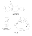
- FIG. 1 shows predicted secondary structures of RHA0002 (SEQ ID NO: 12), RHA0002_s16 (SEQ ID NO: 13), RHA0002_s20 (SEQ ID NO: 14), RHA0002_s26 (SEQ ID NO: 15), RHA0002_s28 (SEQ ID NO: 16), and RHA0002_s33 (SEQ ID NO: 17).
- FIG 2 shows predicted secondary structures of RHA0006 (SEQ ID NO: 18), RHA0006_s19 (SEQ ID NO: 19), and RHA0006_s19_d9 (SEQ ID NO: 32) in which the linker is poly(dT) consisting of 9 bases.
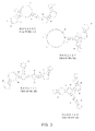
- FIG. 3 shows predicted secondary structures of RHA0020 (SEQ ID NO: 11), RHA0111 (SEQ ID NO: 28), RHA0127 (SEQ ID NO: 29), and RHA0124 (SEQ ID NO: 30).
- FIG. 19 shows predicted secondary structures of RHA0023 (SEQ ID NO: 1), RHA0030 (SEQ ID NO: 2), RHA0013 (SEQ ID NO: 3), and RHA0009 (SEQ ID NO: 4).
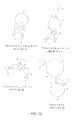
- FIG. 20 shows predicted secondary structures of RHA0058 (SEQ ID NO: 5), RHA0099 (SEQ ID NO: 6), RHA0110 (SEQ ID NO: 7), and RHA0098 (SEQ ID NO: 8).
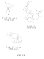
- FIG 21 shows predicted secondary structures of RHA0144 (SEQ ID NO: 9), RHA0133 (SEQ ID NO: 10), RHA0002_s33_d9 (SEQ ID NO: 48), and RHA0006_s19_d9_AT(SEQ ID NO: 46).
- FIG. 22 shows predicted secondary structures of RHA0006_s19_d_R20 (SEQ ID NO: 47), RHA0006_s19_t9 (SEQ ID NO: 51), RHA0006_s19_d5 (SEQ ID NO: 33), and RHA1634 (SEQ ID NO:20).
- FIG 23 shows predicted secondary structures of RHA1634_s60 (SEQ ID NO: 21), RHA1635 (SEQ ID NO: 22), RHA1635_s62 (SEQ ID NO: 23), and RHA0385 (SEQ ID NO: 24).
- FIG. 24 shows predicted secondary structures of RHA0385_s28 (SEQ ID NO: 25), RHA0471 (SEQ ID NO: 26), and RHA0471_s51 (SEQ ID NO: 27).
- the building blocks of the nucleic acid molecule of the present invention are, for example, nucleotide residues.
- nucleotide residues include deoxyribonucleotide residues and ribonucleotide residues.
- nucleic acid molecule of the present invention include DNA consisting of deoxyribonucleotide residues only and DNA including one or more ribonucleotide residues. In the latter case, "one or more" is not particularly limited.
- the number of the ribonucleotide residues in the polynucleotide is, for example, 1 to 91, preferably 1 to 30, more preferably 1 to 15, still more preferably 1 to 7, particularly preferably 1 to 3, and most preferably 1 or 2.
- the nucleic acid molecule of the present invention may include one or more modified nucleotide residues, for example.
- the term "one or more” is not particularly limited.
- the number of the modified nucleotide residues in the polynucleotide is, for example, 1 to 91, preferably 1 to 30, more preferably 1 to 15, still more preferably 1 to 7, particularly preferably 1 to 3, and most preferably 1 or 2.
- modified nucleotide residue examples include modified deoxyribonucleotide residues and modified ribonucleotide residues.
- the modified nucleotide residue may be the above-described nucleotide residue with a modified sugar residue, for example.
- sugar residue examples include ribose residues and deoxyribose residues.
- the modified site in the nucleotide residue is not particularly limited, and may be, for example, the 2'-position and/or the 4'-position in the sugar residue.
- the modification include methylation, fluorination, amination, and thiation.
- the modified nucleotide residue may be, for example, the one obtained by modification of a nucleotide residue having a pyrimidine base (pyrimidine nucleus) as the base or the one obtained by modification of a nucleotide residue having a purine base (purine nucleus) as the base. Among them, the former is preferable.
- nucleotide residue having a pyrimidine base is referred to as a "pyrimidine nucleotide residue”
- a pyrimidine nucleotide residue that has been modified is referred to as a "modified pyrimidine nucleotide residue”
- a nucleotide residue having a purine base is referred to as a "purine nucleotide residue”
- a purine nucleotide residue that has been modified is referred to as a "modified purine nucleotide residue”.
- pyrimidine nucleotide residues examples include: uracil nucleotide residues having uracil; cytosine nucleotide residues having cytosine; and thymine nucleotide residues having thymine.
- the base in the modified nucleotide residue is a pyrimidine base, for example, it is preferable that the 2'-position and/or carbon in the 4'-position in the sugar residue is modified.
- modified nucleotide residues include modified nucleotide residues with the 2'-position in the ribose residue being modified, such as a 2'-methylated-uracil nucleotide residue, a 2'-methylated-cytosine nucleotide residue, a 2'-fluorinated-uracil nucleotide residue, a 2'-fluorinated-cytosine nucleotide residue, a 2'-aminated-uracil nucleotide residue, a 2'-aminated-cytosine nucleotide residue, a 2'-thiated-uracil nucleotide residue, and a 2'-thiated-cytosine nucleotide residue.
- modified nucleotide residues with the 2'-position in the ribose residue being modified such as a 2'-methylated-uracil nucleotide residue, a 2'-methylated-cytosine nucleotide residue, a 2
- the base in the nucleotide residue may be, for example, a natural base (non-artificial base) such as adenine (a), cytosine (c), guanine (g), thymine (t), or uracil (u), or an unnatural base (artificial base).
- a natural base such as adenine (a), cytosine (c), guanine (g), thymine (t), or uracil (u)
- an unnatural base artificial base
- the artificial base include a modified base and an altered base, which both preferably has a similar function to the natural base (a, c, g, t, or u).
- Examples of the artificial base having the similar function include: an artificial base that can bind to cytosine (c), as a substitute for guanine (g); an artificial base that can bind to guanine (g), as a substitute for cytosine (c); an artificial base that can bind to thymine (t) or uracil (u), as a substitute for adenine (a); an artificial base that can bind to adenine (a), as a substitute for thymine (t); and an artificial base that can bind to adenine (a), as a substitute for uracil (u).
- Examples of the modified base include methylated bases, fluorinated bases, aminated bases, and thiated bases.
- modified base examples include 2'-methyluracil, 2'-methylcytosine, 2'-fluorouracil, 2'-fluorocytosine, 2'-aminouracil, 2'-aminocytosine, 2'-thiouracil, and 2'-thiocytosine.
- bases represented by a, g, c, t, and u encompass not only the natural bases but also the artificial bases having similar functions to the natural bases.
- the nucleic acid molecule of the present invention may include one or more artificial nucleic acid monomer residues, for example.
- the term "one or more" is not particularly limited.
- the number of the artificial nucleic acid monomer residues in the polynucleotide is, for example, 1 to 91, preferably 1 to 30, more preferably 1 to 15, still more preferably 1 to 7, particularly preferably 1 to 3, and most preferably 1 or 2.
- the artificial nucleic acid monomer residue include PNA (peptide nucleic acid), LNA (Locked Nucleic Acid), and ENA (2'-O, 4'-C-Ethylenebridged Nucleic Acids).
- the nucleic acid in the monomer residue is as described above, for example.
- the nucleic acid molecule of the present invention is resistant to nuclease, for example.
- the nucleic acid molecule of the present invention includes the modified nucleotide residue(s) and/or the artificial nucleic acid monomer residue(s), for example.
- the nucleic acid molecule of the present invention may have PEG (polyethylene glycol) of several tens of kDa, deoxythymidine, or the like bound to, e.g., the 5' end or the 3' end thereof.
- the nucleic acid molecule of the present invention may further include an additional sequence, for example.
- the additional sequence is bound to at least one of the 5' end and the 3' end, more preferably to the 3' end of the nucleic acid molecule, for example.
- the additional sequence is not particularly limited.
- the length of the additional sequence is not particularly limited, and is, for example, 1- to 200-mer, preferably 1- to 50-mer, more preferably 1- to 25-mer, and still more preferably 18- to 24-mer.
- the building blocks of the additional sequence are, for example, nucleotide residues, examples of which include deoxyribonucleotide residues and ribonucleotide residues.
- the additional sequence is not particularly limited, and examples thereof include polynucleotides such as DNA consisting of deoxyribonucleotide residues and DNA including a ribonucleotide residue(s).
- Specific examples of the additional sequence include poly(dT) and poly(dA), and the additional sequence preferably is poly(dT) or poly(dA).
- the nucleic acid molecule of the present invention can be used in the state where it is immobilized on a carrier, for example. It is preferable to immobilize either the 5' end or the 3' end, more preferably the 3' end of the nucleic acid molecule of the present invention, for example.
- the nucleic acid molecule of the present invention may be immobilized either directly or indirectly to the carrier, for example. In the latter case, it is preferable to immobilize the nucleic acid molecule via the additional sequence, for example.
- the method for immobilizing the nucleic acid molecule is not particularly limited.
- the carrier is not particularly limited, and may be, for example, a base such as a plate, a sheet, a film, a filter, a tube, or beads.
- the method for producing the nucleic acid molecule of the present invention is not particularly limited.
- the nucleic acid molecule of the present invention can be synthesized by known methods such as nucleic acid synthesis methods etc. utilizing chemical synthesis and methods utilizing genetic engineering.
- the dissociation constant of the nucleic acid molecule of the present invention against an influenza virus or the HA is, for example, 30 nmol/L or less, preferably 10 nmol/L or less, more preferably 1 nmol/L or less, still more preferably 0.1 nmol/L or less, and particularly preferably 0.01 nmol/L or less.
- the nucleic acid molecule of the present invention exhibits binding properties to influenza viruses or influenza virus-derived HA, as described above.
- use of the nucleic acid molecule of the present invention is not particularly limited as long as it is the use utilizing the binding properties to the influenza viruses and the HA.
- the nucleic acid molecule of the present invention can be used in various methods, for example, as a substitute for an antibody against the influenza viruses and the HA.
- a method for analyzing an influenza virus according to the present invention is characterized in that it includes the steps of: bringing the nucleic acid molecule of the present invention into contact with a sample; and detecting the binding between the nucleic acid molecule and an influenza virus.
- the presence or absence of the influenza virus in the sample can be analyzed by detecting the presence or absence of the binding between the nucleic acid molecule and the influenza virus in the detection step, or alternatively, the amount of the influenza virus in the sample can be analyzed by measuring the amount of the binding between the nucleic acid molecule and the influenza virus in the sample.
- an influenza virus in a sample can be analyzed without subjecting the sample to a pretreatment for, e.g., destroying the virus.
- a method for analyzing influenza virus-derived HA according to the present invention is characterized in that it includes the steps of: bringing the nucleic acid molecule of the present invention into contact with a sample; and detecting the binding between the nucleic acid molecule and the HA.
- the presence or absence of the HA in the sample can be analyzed by detecting the presence or absence of the binding between the nucleic acid molecule and the HA in the detection step, or alternatively, the amount of the HA in the sample can be analyzed by measuring the amount of the binding between the nucleic acid molecule and the HA in the sample.
- influenza virus detection method includes, for example, the above-described HA analysis method, and it analyzes an influenza virus in the sample by detecting the binding between the nucleic acid molecule and the HA.
- the presence or absence of the influenza virus in the sample can be analyzed by detecting the presence or absence of the binding between the nucleic acid molecule and the HA in the detection step, or alternatively, the amount of the influenza virus in the sample can be analyzed by measuring the amount of the binding between the nucleic acid molecule and the HA in the sample.
- the amount of HA and the amount of an influenza virus generally are in a relative relationship.
- the amount of the influenza virus can be analyzed from the amount of the HA on the basis of the thus-determined relative relationship.
- the relative relationship can be indicated with a calibration curve or the like, for example.
- the nucleic acid molecule may be brought into contact with the sample after the sample has been subjected to a pretreatment of destroying the virus, or the nucleic acid molecule may be brought into contact with the sample without conducting the pretreatment.
- the nucleic acid molecule of the present invention can bind to influenza viruses, as described above.
- the nucleic acid molecule of the present invention can be used widely in, for example, not only the above-described influenza virus detection and HA detection but also various uses that utilize their binding. Specific examples of such uses include products containing the nucleic acid molecule, such as a filter, a mask, a wiping agent, a gargling agent, and a troche.
- the filter include an air filter and a liquid filter.
- the nucleic acid molecule in the filter binds to influenza viruses.
- the filter can remove the influenza viruses from the air and liquid and can prevent the spread of the influenza viruses, for example.
- the mask can prevent infection with influenza viruses, because the nucleic acid molecule binds to the influenza viruses to inhibit the invasion of the influenza viruses into the body.
- the wiping agent can remove influenza viruses from an object to be wiped using the wiping agent, because the nucleic acid molecule binds to the influenza viruses, for example.
- the gargling agent can remove influenza viruses attached to the oral cavity and the pharynx, because the nucleic acid molecule binds to the influenza viruses, for example.
- the binding agent according to the present invention is a binding agent that binds to an influenza virus, characterized in that it contains the nucleic acid molecule of the present invention. It is only necessary that the binding agent according to the present invention contains the nucleic acid molecule according to the present invention, and other configurations are by no means limited.
- detection and the like of influenza viruses are possible, for example, as described above.
- the binding agent of the present invention also can be referred to as, for example, a binding agent that binds to influenza virus-derived HA, and it is possible to detect an influenza virus by detecting the HA, for example.
- Aptamers capable of binding to influenza viruses were prepared, and the binding ability of each aptamer to influenza virus-derived HA was examined.
- aptamers of the present example the following polynucleotides were synthesized.
- N denotes deoxyribonucleotide residues, and nucleic acids contained therein are adenine, guanine, cytosine, and/or thymine.
- aptamer A10 As an aptamer A10 according to a comparative example, a polynucleotides having the following sequence described in Non-Patent Document 2 (BBRC) was synthesized.
- BBRC Non-Patent Document 2
- A/Anhui/1/2005-derived HA1 and A/goose_Guiyang/337/2006-derived HA1 are composed of 437 amino acid residues, and 24 amino acid residues out of the 437 amino acid residues are different from each other (5.49%).
- SEQ ID NOs: 36 and 37 different amino acids are underlined.
- SEQ ID NO: 38 of A/Anhui/1/2005-derived HA amino acids different from those in A/goose_Guiyang/337/2006-derived HA are underlined.
- a streptavidin-immobilized chip (trade name: ProteOn NLC Sensor Chip, BioRad) was set in the ProteON XPR36.
- a ligand at 5000 nmol/L was injected to a flow cell of the sensor chip using ultrapure water (DDW), and the binding was allowed to proceed until the signal intensity (RU: Resonance Unit) reached about 1000 RU.
- RU Resonance Unit
- biotinylated poly(dT) obtained by biotinylating the 5' end of 24-mer deoxythymidine was used.
- the poly(dA)-added aptamer at 400 nmol/L was injected to the flow cell of the chip using an SPR buffer at a flow rate of 25 ⁇ L/min for 80 seconds, and the binding was allowed to proceed until the signal intensity reached about 700 RU.
- the target protein at 500 nmol/L was injected using the SPR buffer at a flow rate of 50 ⁇ L/min for 120 seconds, followed by washing performed by flowing the SPR buffer under the same conditions. Signal intensity measurement was performed concurrently with the injection of the target protein and the washing with the SPR buffer.
- a selection buffer had the following composition: 50 mmol/L Tris, 0.1 mol/L NaCl, 0.01 % Tween®, 5 mmol/L K + , and 1 mmol/L Mg 2+ .
- the pH of the selection buffer was 7.4.
- the SPR buffer had the following composition: 50 mmol/L Tris, 0.1 mol/L NaCl, 0.01 % Tween®, and 10 mmol/L K + .
- the pH of the SPR buffer was 7.4.
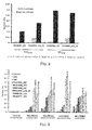
- FIG. 4 is a graph showing the binding ability of each aptamer to each target protein.
- the vertical axis indicates the relative value of the signal intensity (RU) measured using the Prote ON® XRP36. The relative value was determined by calculating a value obtained by dividing the signal intensity at the start of the washing (at the end of the target protein injection) with the signal intensity before the start of the target protein injection (at the end of the poly(dA)-added aptamer injection).
- "0-pool" indicates the result obtained regarding the N30.
- the aptamers of the comparative examples both did not exhibit binding ability to HA1 Anhui , HA1 Guiyang , and HA Anhui .
- the aptamers of the present example all exhibited binding ability to HA1 Anhui , HA1 Guiyang , and HA Anhui , whereas they did not bind to either BSA or His-MIF.
- FIG. 5 is a graph showing the binding ability of each aptamer to each antibody.
- the vertical axis indicates the relative value of the signal intensity (RU), as in FIG. 4 .
- the relative value was determined in the same manner as in the item (3) above.
- "0-pool" indicates the result obtained regarding the N30.
- the aptamers of the present example exhibited potent binding ability to HA1 Anhui , whereas they did not exhibit binding ability to any of the antibodies.
- the present example examined the binding ability of an aptamer obtained by truncating RHA0002 (SEQ ID NO: 12) to HA.
- RHA0002 (SEQ ID NO: 12) and a truncated aptamer thereof shown below were synthesized.
- RHA0002_s33 (SEQ ID NO: 17) GTGTTTGATGGTTTGGTCTGGTTGGCTTAACAC
- Example 1 the binding ability to target proteins was analyzed in the same manner as in Example A1. The results thereof are shown in Table 1 below. In Table 1, the relative value was determined in the same manner as in the item (3) in Example A1. [Table 1] Aptamer Relative Value HA1 Anhui HA1 Guiyang HA Anhui RHA0002 0.5039 0.4935 0.2936 RHA0002_s33 0.9958 0.9477 0.9847 N30 0.0286 0.0091 0.0045
- the truncated aptamer exhibited binding ability to each target protein.
- RHA0002_s33 SEQ ID NO: 17
- improved binding ability by truncation As can be seen from Table 1, the truncated aptamer exhibited binding ability to each target protein.
- RHA0002_s33 SEQ ID NO: 17
- improved binding ability by truncation As can be seen from Table 1, the truncated aptamer exhibited binding ability to each target protein.
- RHA0002_s33 SEQ ID NO: 17
- the present example examined the binding ability to HA of: RHA0006 (SEQ ID NO: 18); a truncated aptamer obtained by truncating RHA0006, and an aptamer including two truncated sequences of the truncated aptamer.
- RHA0006_s19 (SEQ ID NO: 19) GGGTTTGGGTTGGGTTGGG
- RHA0006_s19_d9 SEQ ID NO: 32
- the present example examined the binding ability of each aptamer to HA.
- RHA0002_s33 (SEQ ID NO: 17) GTGTTTGATGGTTTGGTCTGGTTGGCTTAACAC
- RHA0006_s19 (SEQ ID NO: 19) GGGTTTGGGTTGGGTTGGG
- RHA0006_s19_d9 (SEQ ID NO: 32) GGGTTTGGGTTGGGTTGGG TTTTTTTTT GGGTTTGGGTTGGGTTGGG
- aptamers of comparative examples the aptamer A10 used as the aptamer of the comparative example in Example A1 and an aptamer A05 having the following sequence described in Non-Patent Document 2 (BBRC) were used.
- FIG. 6 is a graph showing the binding ability of each aptamer to each target protein.
- the vertical axis indicates the relative value of the signal intensity (RU) measured using the ProteON® XRP36. The relative value was determined in the same manner as in the item (3) in Example A1.
- "0-tool" indicates the result obtained regarding the N30.
- the aptamers of the present example all exhibited binding ability to HA1 Anhui , HA1 Guiyang , and HA Anhui , whereas they did not bind to either BSA or His-MIF.
- RHA0002_s33 SEQ ID NO: 17
- RHA0006_s19_d9 SEQ ID NO: 32
- FIG. 7 is a graph showing the binding ability of each aptamer to each antibody.
- the vertical axis indicates the relative value of the signal intensity (RU), as in FIG 6 .
- the aptamers of the present example exhibited potent binding ability to the HA1 Anhui , whereas they did not exhibit binding ability to any of the antibodies.
- the present example examined the dissociation constant of each of aptamers RHA0002_s33 (SEQ ID NO: 17) and RHA0006_s19_d9 (SEQ ID NO: 32) against HA.
- Example A1 SPR was carried out in the same manner as in Example A1, except that the concentration of the biotinylated poly T was set to 100 nmol/L and the concentration of each of HA1 Anhui , HA1 Guiyang , and HA Anhui was set to a predetermined value (15.6, 31.3, 62.5, 125, or 250 nmol/L). Then, from the results of the SPR, the dissociation constant of each aptamer was calculated. The results thereof are shown in Table 3 below.
- RHA0002_s33 SEQ ID NO: 17
- RHA0006_s19_d9 SEQ ID NO: 32
- the aptamer of the present invention, HA1 or HA, and an antibody were subjected to a binding reaction in this temporal order, and the binding ability thereof was analyzed by SPR.
- the aptamers used in the present example were RHA0002_s33 (SEQ ID NO: 17) and RHA0006_s19_d9 (SEQ ID NO:32).
- HA1 Anhui was used as HA1
- HA Anhui was used as HA.
- the antibodies used in the present example were Control Ab (rabbit immunoglobulin, Beckman Coulter, Inc., #731642) as a control antibody and Anti HA Ab (rabbit anti-HA antibody, Sino Biological Inc., #11048-RP02).
- SPR was carried out in the same manner as in the item (3) in Example A1, except that the conditions of the SPR were set as follows. First, a ligand at 5000 nmol/L was injected to a flow cell of the sensor chip using ultrapure water (DDW), and the binding was allowed to proceed until the signal intensity (RU: Resonance Unit) reached about 1000 RU. As the ligand, biotinylated poly(dT) obtained by biotinylating the 5' end of 24-mer deoxythymidine was used.
- the poly(dA)-added aptamer at 400 nmol/L was injected to the flow cell of the chip using an SPR buffer at a flow rate of 25 ⁇ L/min for 80 seconds, and the binding was allowed to proceed until the signal intensity reached about 700 RU.
- the target protein at 500 nmol/L was injected at a flow rate of 25 ⁇ L/min for 80 seconds and a detection antibody was then injected at 1 ⁇ mol/L at a flow rate of 50 ⁇ L/min for 120 seconds. Finally, washing was performed by flowing the SPR buffer.
- FIG. 8 is a graph showing the binding ability between each aptamer, each target protein, and each antibody.
- the vertical axis indicates the relative value of the signal intensity (RU) measured using the Prote ON® XRP36. The relative value was determined in the same manner as in the item (3) in Example A1.
- the present example examined the binding ability of each aptamer to HA by SPR.
- RHA0002_s33 (SEQ ID NO: 17) GTGTTTGATGGTTTGGTCTGGTTGGCTTAACAC
- RHA0006_s19_d5 (SEQ ID NO: 33) GGGTTTGGGTTGGGTTGGGTTTTTGGGTTTGGGTTGGGTTGGG RHA0385_s28 (SEQ ID NO: 25) TTGGGGTTATTTTGGGAGGGCGGGGGTT RHA0471_s51 (SEQ ID NO: 27) TCCTCGTTGGGGGTGGTGGTGGGTTTCGGTTCATGGGTGGGACGGAGAGGA
- Control nucleic acid molecule that does not exhibit binding properties to HA, an aptamer A22 of Non-Patent Document 1, which has been reported not to exhibit binding properties to HA, and the above-described aptamer A05 of Non-Patent Document 2 were synthesized.
- Control SEQ ID NO: 39
- Target proteins used in the present example were A/Anhui/1/2005-derived HA1 (HA1 Anhui : SEQ ID NO: 36) in Example A1, and A/California/04/2009-derived HA1, A/Brisbane/59/2007-derived HA1, and A/Aichi/2/1968-derived HA1, which are shown below.
- FIG. 9 is a graph showing the binding ability of each aptamer to each target protein.
- the vertical axis indicates the relative value of the signal intensity (RU) measured using the ProteON® XRP36. The relative value was determined in the same manner as in the item (3) in Example A1.
- the aptamers according to the present example all exhibited higher binding ability to each HA1 than the aptamers according to the comparative examples.
- HA was detected by ELAA (Enzyme-linked Aptamer Assay).
- Aptamers used in the present example were RHA0006_s19_d5 (SEQ ID NO: 33), RHA1634_s60 (SEQ ID NO: 21), RHA1635_s62 (SEQ ID NO: 23), RHA0385_28 (SEQ ID NO: 25), and RHA0471_s51 (SEQ ID NO: 27), which were synthesized in Example B1. Also, the same control nucleic acid molecule and aptamers of the comparative examples as in Example B1 were used. The 3' end of each of these aptamers was biotinylated, and the thus-obtained biotinylated aptamers (detection aptamers) were used in ELAA to be described below.
- Target proteins used in the present example were A/Anhui/1/2005-derived HA (HA/H5N1) and A/California/04/2009-derived HA (HA/H1N1), which were used in Examples A1 and B1, respectively.
- the target was immobilized in wells, and the detection aptamer was brought into contact with the immobilization target. By detecting the detection aptamer bound to the immobilized target, the immobilized target was detected.
- This method hereinafter is referred to as a direct method of ELAA.
- the HA was diluted with a carbonic acid buffer (pH 9.6), and 100 ⁇ L of the thus-obtained 10 ⁇ g/mL HA was added to each well of a 96-well plate (trade name: Immuno 96 Microwell Plates MaxiSorp: Nunc). The HA was allowed to adsorb to the wells at 4°C overnight. The wells were then washed with 200 ⁇ L of a washing buffer (50 mmol/L Tris, 100 mmol/L NaCl, 5 mmol/L KCl, 1 mmol/L MgCl2, 0.01 % Tween® 20, pH 7.4).
- a washing buffer 50 mmol/L Tris, 100 mmol/L NaCl, 5 mmol/L KCl, 1 mmol/L MgCl2, 0.01 % Tween® 20, pH 7.4
- a blocking buffer Protein-Free (TBS) blocking buffer, #37570, PIERCE
- TBS Protein-Free (TBS) blocking buffer, #37570, PIERCE
- the biotinylated aptamer was diluted to 1 ⁇ mol/L. Thereafter, 100 ⁇ L of the diluted biotinylated aptamer was added to each well, and the plate was incubated at room temperature for 1 hour. Subsequently, the wells were washed with the washing solution.
- a reaction solution 50 mmol/L Tris, 100 mmol/L NaCl, 5 mmol/L KCl, 1 mmol/L MgCl 2 , 0.01% Tween® 20, 0.05% BSA, pH 7.4
- FIG. 10 is a graph showing the amount of each HA bound to each aptamer.
- the vertical axis indicates the absorbance at a wavelength of 450 nm (reference wavelength: 620 nm), which indicates the binding ability.
- the aptamers according to the present example all exhibited higher binding ability to each HA than the aptamers according to the comparative examples.
- the absorbance was below the detection limit. From these results, it was found that the aptamers of the present example exhibited the binding properties not to the plate but to the immobilized HA1. According to such a method, the measured absorbance indicates the amount of an aptamer bound to immobilized HA, and hence, indirect qualification and quantification of the HA are possible. Moreover, because qualification and quantification of HA are possible, qualification and quantification of influenza virus also are possible.
- HA captured by an immobilized antibody was detected by ELAA (Enzyme-linked Aptamer Assay).
- Aptamers used in the present example were RHA0002_s33 (SEQ ID NO: 17), RHA0006_s19_d5 (SEQ ID NO: 33), RHA1634_s60 (SEQ ID NO: 21), and RHA1635_s62 (SEQ ID NO: 23), which were synthesized in Example B1. Also, the same control nucleic acid molecule as in Example B1 was used. The 3' end of each of these aptamers was biotinylated, and the thus-obtained biotinylated aptamers were used in ELAA to be described below.
- Example B1 As a target protein, A/Anhui/1/2005-derived HA (HA1/H5N1) used in Example B1 was used.
- H5N1 rabbit anti-hemagglutinin (H5N1) pAb (Sino Biological Inc., #11048-RP2) was used.
- ⁇ g of the antibody was added to each well of a 96-well plate (trade name: Immuno 96 Microwell Plates MaxiSorp, Nunc). The plate was incubated at 4°C overnight to immobilize the antibody.
- a blocking buffer a Protein-Free (TBS) blocking buffer (trade name, #37570, PIERCE) was used, and the blocking treatment was carried out at room temperature for 1 hour. Then, 1 ⁇ g of the HA was added to each well of the plate. The plate was incubated at room temperature for 2 hours, thereby causing the HA to bind to the immobilized antibody on the plate.
- TBS Protein-Free
- the ELAA in the present example was carried out under the same conditions as those for the ELAA in Example B2.
- the absorbance measurement also was performed with respect to a system not containing the HA and a system in which the antibody had not been immobilized.
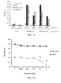
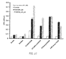
- FIG. 11 is a graph showing the amount of the HA bound to each aptamer.
- the vertical axis indicates the absorbance at a wavelength of 450 nm (reference wavelength: 620 nm), which indicates the binding ability.
- the aptamers according to the present example all exhibited higher absorbances than the control nucleic acid molecule.
- RHA0006_s19_d5 (SEQ ID NO: 33) exhibited a very high absorbance.
- the absorbance was below the detection limit. From these results, it was found that the aptamers of the present example exhibited binding properties not to the antibody or the plate but to the HA bound to the plate via the antibody. According to such a method (Sandwich-ELAA), the measured absorbance indicates the amount of an antibody bound to HA captured by an aptamer, and hence, indirect qualification and quantification of the HA are possible.
- HA was captured by an immobilized aptamer, and the HA was detected using a primary antibody and a secondary antibody.
- RHA0002_s33 (SEQ ID NO: 17) synthesized in Example B1 was used. Also, the same control nucleic acid molecule as in Example B1 was used. To the 3' end each of these aptamers, an amino group (-NH 2 ) was introduced. The thus-obtained aminated aptamers were used.
- Example B1 As a target protein, A/Anhui/1/2005-derived HA (HA1/H5N1) used in Example B1 was used.
- a primary antibody used in the present example was rabbit anti-hemagglutinin (H5N1) pAb (Sino Biological Inc., #11048-RP2), which is a polyclonal antibody exhibiting binding properties to HA/H5N1.
- a secondary antibody used in the present example was HRP-labeled anti-rabbit IgG antibody ( ⁇ -Rabit IgG-HRP, #NA934-1ML, GE), which is a polyclonal antibody capable of binding to the primary antibody.
- the aminated aptamer diluted with a carbonic acid buffer (pH 9.6) was added. The plate was incubated at 4°C overnight to immobilize the aminated aptamer. The concentration of the aminated aptamer in each well of the plate was set to 0, 0.002, 0.02, 0.2, or 2 ⁇ M.
- the first blocking treatment was performed at room temperature for 1 hour using 1 mol/L Tris (pH 7.4).
- the second blocking treatment was performed at room temperature for 1 hour using a Protein-Free (TBS) blocking buffer (trade name, #37570, PIERCE) as a blocking buffer.
- TBS Protein-Free
- the ELAA in the present example was carried out under the same conditions as those for the ELAA in Example B2.
- the absorbance measurement also was performed with respect to a system in which, instead of the primary antibody, rabbit immunoglobulin (Beckman Coulter, Inc., #731642) was used as a control antibody.
- FIG. 12 is a graph showing the amount of the HA bound to the aptamer.
- the vertical axis indicates the absorbance at a wavelength of 450 nm (reference wavelength: 620 nm), which indicates the binding ability.
- the absorbance increased in a manner dependent on the concentration of the immobilized RHA0002_s33 (SEQ ID NO: 17) immobilized on the plate. From this result, it was found that the aptamer of the present example can capture HA in a concentration-dependent manner. According to such a method (Sandwich-ELAA), the measured absorbance indicates the amount of an antibody bound to captured HA, and hence, indirect qualification and quantification of the HA are possible.
- HA was captured by an immobilized capturing aptamer, and the HA was detected using a detection aptamer.
- Aptamers used in the present example were RHA0002_s33 (SEQ ID NO: 17), RHA0006_s19_d5 (SEQ ID NO: 33), RHA0385_s28 (SEQ ID NO: 25), and RHA0471_s51 (SEQ ID NO: 27), which were synthesized in Example B1.
- the capturing aptamer to be immobilized on a plate was configured so that an amino group (-NH 2 ) was introduced to the 3' end thereof.
- the detection aptamer was configured so that the 3' end thereof was biotinylated.
- the same control nucleic acid molecule as in Example B1 was used. The combinations of the capturing aptamer and the detection aptamer are shown below.
- Example B1 As a target protein, A/California/04/2009-derived HA (HA1/H1N1) used in Example B1 was used.
- the capturing aptamer was immobilized on each well, and a target was brought into contact with the capturing aptamer. Further, the detection aptamer was brought into contact with the target bound to the capturing aptamer, thereby sandwiching the target between the capturing aptamer and the detection aptamer. Then, the target was detected by detecting the detection aptamer in this complex.
- This method hereinafter is referred to as a sandwich method of ELAA.
- the plate was incubated at room temperature for 2 hours, thereby causing the HA to bind to the capturing aptamer on the plate. Subsequently, 100 ⁇ L of the detection aptamer at 1 ⁇ mol/L was added to each well of the plate, and the plate was incubated at room temperature for 1 hour. Thereafter, a color-developing reaction was caused using streptavidin-HRP ( ⁇ 1000), and the absorbance was measured. Unless otherwise stated, the ELAA in the present example was carried out under the same conditions as those for the ELAA in Example B2.
- FIG. 13 is a graph showing the amount of the HA bound to the detection aptamer bound to the HA captured by the capturing aptamer.
- the aptamers of the present example were used as the capturing aptamer and the detection aptamer, higher absorbances were exhibited as compared with the case where the control was used.
- very high absorbances were exhibited when the combination of the capturing aptamer and the detection aptamer was as follows: the combination of RHA0385_s28 (SEQ ID NO: 25) and RHA0006_s19_d5 (SEQ ID NO: 33); and the combination of RHA0006_s19_d5 (SEQ ID NO: 33) and RHA0006_s19_d5 (SEQ ID NO: 33) or RHA0385_s28 (SEQ ID NO: 25).
- the measured absorbance indicates the amount of the detection aptamer bound to the HA captured by the capturing aptamer, and hence, indirect qualification and quantification of the HA are possible.
- FIG. 14 is a graph showing the amount of the HA bound to the detection aptamer bound to the HA captured by the capturing aptamer. As can be seen from FIG.
- RHA0006_s19_d5 (SEQ ID NO: 33) synthesized in Example B1 was used.
- 24-mer polydeoxyadenine (poly(dA)) was added to the 3' end of the aptamer, and further, the 3' end of the poly(dA) was biotinylated.
- the thus-obtained biotinylated poly(dA)-added aptamer was used.
- An antisense (SEQ ID NO: 44) having a sequence complementary to the aptamer was synthesized, and the 5' end thereof was aminated.
- Antisense (SEQ ID NO: 44) CCCAAACCCAACCCAACCCAACCCAAAAACCCAAACCCAACCCAACCCTTTTT
- Example B1 As a target protein, A/California/04/2009-derived HA (HA/H5N1) used in Example B1 was used.
- the HA at a predetermined concentration (0, 0.0002, 0.002, 0.02, 0.2, or 2 ⁇ mol/L) was mixed with the biotinylated poly(dA)-added aptamer at 2 nmol/L, and the resultant mixture was incubated at 4°C overnight, thereby causing the binding between the HA and the aptamer. Then, this mixture was added to the well to achieve a concentration of 1 nmol/L. The plate was incubated at room temperature for 1 hour, and then washed. Thereafter, a color-developing reaction was caused by streptavidin (SA)-HRP ( ⁇ 1000), and the absorbance was measured. Unless otherwise stated, the ELAA in the present example was carried out under the same conditions as those for the ELAA in Example B2.
- the absorbance measurement was performed in the same manner, except that, instead of the aminated antisense, 24-mer poly(dT) with the aminated 5' end was used.
- FIG. 15 is a graph showing the amount of the aptamer bound to the immobilized antisense.
- the vertical axis indicates the absorbance at a wavelength of 450 nm (reference wavelength: 620 nm), which indicates the binding ability.
- the absorbance decreases in correlation with the increase in the concentration of the HA.
- the aptamer can bind to the antisense immobilized on the plate.
- biotin contained in the aptamer captured on the plate via the antisense binds to the SA-HRP to cause color development.
- the aptamer binds to the HA and thus cannot bind to the antisense immobilized on the plate. Accordingly, the aptamer is not captured on the plate. Thus, a color-developing reaction by SA-HRP does not occur.
- the absorbance decreases in correlation with the increase in the concentration of the HA.
- the poly(dT) when used, the poly(dT) can bind to the poly(dA) added to the aptamer.
- the decrease in absorbance accompanying the increase in HA was not observed.
- the measured absorbance indicates the amount of the aptamer that is captured by the antisense and is not bound to HA, and hence, indirect qualification and quantification of HA are possible.
- the present example examined a cross-reaction of an aptamer with an antibody. Unless otherwise stated, the examination was carried out in the same manner as in the item (4) in Example A1.
- A/Anhui/1/2005-derived HA1 (HA1 Anhui ), BSA, and His-MIF were used instead of the A/California/04/2009-derived HA1 (HA1 California ).
- Antibodies used in the present example were Rabbit IgG, Mouse IgG, Chicken IgG, Rat IgG, and Goat IgG described in the item (4) in Example A1.
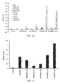
- FIG. 16 is a graph showing the binding ability of each aptamer to each antibody.
- the vertical axis indicates the relative value of the signal intensity (RU), as in FIG. 4 .
- the nine kinds of bars shown for each aptamer indicate, from the left, the results obtained regarding BSA, Rabbit IgG, Mouse IgG, Chicken IgG, Rat IgG, Goat IgG. His-MIF, HA1/H1N1, and HA1/H5N1.
- the aptamers of the present example exhibited potent binding ability to HA, whereas they exhibited little binding ability to any of the antibodies.
- the present example examined the binding ability of each of the following aptamers to a target protein. Unless otherwise stated, the examination was carried out in the same manner as in the item (3) in Example A1.
- RHA0002_s16 (SEQ ID NO: 13) GGTTTGGTCTGGTTGG RHA0002_s20 (SEQ ID NO: 14) GTGGTTTGGTCTGGTTGGAC RHA0002_s26 (SEQ ID NO: 15) CGTTTGGTTTGGTCTGGTTGGCAACG RHA0002_s28 (SEQ ID NO: 16) CGTTATGGTTTGGTCTGGTTGGCTAACG RHA0002_s33 (SEQ ID NO: 17) GTGTTTGATGGTTTGGTCTGGTTGGCTTAACAC
- the HA1 Anhui was used as the target protein.
- FIG. 17 is a graph showing the binding ability of each aptamer to the target protein.
- the vertical axis indicates the relative value of the signal intensity (RU). The relative value was determined in the same manner as in the item (3) in Example A1.
- "0-pool" indicates the result obtained regarding the N30.
- the aptamers of the present example all exhibited binding ability to HA1 Anhui .
- the present example examined the binding ability of each of the following aptamers to a target protein and an antibody. Unless otherwise stated, the examination was carried out in the same manner as in the item (4) in Example A1.
- RHA0002_s33 (SEQ ID NO: 17) GTGTTTGATGGTTTGGTCTGGTTGGCTTAACAC RHA0006_s19 (SEQ ID NO: 19) GGGTTTGGGTTGGGTTGGG RHA0006_s19_d9 (SEQ ID NO: 32) GGGTTTGGGTTGGGTTGGG TTTTTTTTT GGGTTTGGGTTGGGTTGGG RHA0006_s19_d9_AT(SEQ ID NO: 46) GGGTTTGGGTTGGGTTGGG ATATATATA GGGTTTGGGTTGGGTTGGG RHA0006_s19_d5 (SEQ ID NO: 33) GGGTTTGGGTTGGGTTGGG TTTTTGGTTGGGTTGGG
- the HA1 Anhui and the HA Anhui were used.
- Rabbit IgG described in Example A1 (4) was used.
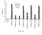
- FIG. 18 is a graph showing the binding ability of each aptamer to the respective target proteins and the antibody.
- the vertical axis indicates the relative value of the signal intensity (RU). The relative value was determined in the same manner as in the item (3) in Example A1.
- the "Control" indicates the result obtained regarding the N30.
- the aptamers of the present example all exhibited binding ability to HA1 Anhui and HA Anhui , whereas they exhibited little binding ability to the antibody. Also, as a result of changing the length of the linker in the aptamer, RHA0006_s19_d5 (SEQ ID NO: 33) in which the linker length was 5-mer exhibited superior binding ability to RHA0006_s19_d9 (SEQ ID NO: 32) in which the linker length was 9-mer.
- an immobilized influenza virus was detected by ELAA.
- Influenza viruses used in the present example are shown below.
- As a control virus the following non-influenza virus was used.
- Virus stocks of the H1N1, the H3N2, and the non-influenza viruses were prepared in the following manner.
- a growth medium had the following composition (final concentration).
- the culture conditions were set as follows: 37°C ⁇ 2°C, with humidity, 5% CO 2 , overnight. The culture plate was examined before use, and it was confirmed that the culture density was 80%.
- the MCDK cells confluent in a flask (about 9.6 ⁇ 10 6 cells/mL) were infected with each type of virus.
- the cells were cultured using an infection medium having the following composition (final concentration).
- As the components of the infection medium products commercially available from Life Technologies were used, unless otherwise stated.
- the multiplicity of infection (MOI) was set to 0.01 PFU/cell.
- the multiplicity of infection was set to 0.05 PFU/cell.
- PFU is a plaque formation unit.
- modified trypsin was added to the flask to achieve a concentration of 2 ⁇ g/mL.
- trypsin treated with L-(tosylamide-2-phenyl)ethyl chloromethyl ketone (TPCK) (trade name, TPCK treated trypsin, USB) was used.
- the culture supernatant containing the virus was collected by centrifugation (200 ⁇ G, 10 minutes, 4°C).
- the remaining supernatant was subjected to centrifugation using ultrafilter membranes to remove proteins of 100 kDa or less and to concentrate the virus.
- ultrafilter membranes Amicon Ultra centrifugal filter devices 100,000 NMWL (trade name, Millipore) were used, and the conditions for the centrifugation were 3000 ⁇ G, 4°C, and 30 minutes. This operation was repeated until the entire supernatant was concentrated.
- the thus-obtained concentrate was used as a semipurified virus stock. Part of the virus stock was collected. The virus contained therein was inactivated by UV irradiation, and the concentration of the virus was determined using a BCA assay (trade name, Pierce). The virus stock was stored at or below -65°C.
- a virus stock of the H5N1, which is an avian influenza virus was prepared in the following manner.
- Attenuated rg A/Japanese White Eye/Hong Kong/38/06 was inoculated into specific pathogen-free (SPF) hen eggs.
- the hen eggs were grown and infected with the virus.
- a chorioallantoic fluid fraction containing the virus was collected.
- the collected fraction was then diluted with a diluent.
- As the diluent 1 ⁇ PBS (pH 7.4) containing 100 U/mL penicillin, 100 ⁇ g/mL streptomycin, and 50 ⁇ g/mL gentamicin was used.
- the diluted fraction was used as a virus stock, without being concentrated as described in the item (2) above.
- the virus stock was stored at or below -65°C.
- the H5N1 was amplified in the hen eggs as described above, it contained a large amount of hen egg-derived proteins. Thus, the purification step using a column described in the item "(2) Virus stock preparation 1" above was not performed. On this account, regarding the virus stock of the H5N1, the amount thereof to be used was indicated as the protein concentration, instead of the virus concentration.
- the H5N1 was used in such a manner that the protein concentration thereof would be 100 times greater (100 ⁇ V ⁇ g/well) than the virus concentration (V ⁇ g/well) of the viruses other than the H5N1, in order to set the number of immobilized virus particles of the H5N1 to be comparable to those of the other viruses.
- Aptamers used in the present example were: RHA0006_s19 (SEQ ID NO: 19) shown in Example A3; and RHA1635_s62 (SEQ ID NO: 23), RHA0385_s28 (SEQ ID NO: 25), and RHA0006_s19_d5 (SEQ ID NO: 33) shown in Example B1. Then, ELAA was carried out in the same manner as in Example B2, except that the virus was immobilized instead of the target protein. Furthermore, as a control, the same control nucleic acid molecule (SEQ ID NO: 39) as in Example B1 was used.
- FIG. 25 is a graph showing the amount of each aptamer bound to the virus.
- the vertical axis indicates the absorbance at a wavelength of 450 nm (reference wavelength: 620 nm), which indicates the binding ability, and the horizontal axis indicates the amount of the protein ( ⁇ g/well) corresponding to the amount of the immobilized virus.
- control nucleic acid molecule exhibited little binding properties to the influenza virus H5N1.
- each of the aptamers exhibited high binding properties to the influenza virus H5N1.
- the absorbance (not shown) indicating the binding properties to H1N1 was comparable to the absorbance indicating the binding properties to the H5N1 (protein: 100 ⁇ g/well) shown in FIG 25 .
- the H5N1 was immobilized so that the protein concentration thereof would be 100 times greater (100 ⁇ V ⁇ g/well) than the virus concentration (V ⁇ g/well) of the viruses other than the H5N1, as described above.
- FIG. 26 is a graph showing the amount of each aptamer bound to each type of virus.
- the vertical axis indicates the absorbance at a wavelength of 450 nm (reference wavelength: 620 nm), which indicates the binding ability.
- the control nucleic acid molecule exhibited little binding properties to both the non-influenza virus and the influenza viruses.
- each of the aptamers exhibited no binding properties to the non-influenza virus and exhibited high binding properties only to the influenza viruses. From these results, it was found that the aptamer of the present invention also is applicable to a direct method in influenza virus detection.
- influenza virus was captured by an immobilized capturing aptamer, and then the influenza virus was detected by a detection aptamer.
- Viruses used in the present example were the following viruses provided in Example D1.
- RHA0385_s28 SEQ ID NO: 25
- RHA0006_s19_d5 SEQ ID NO: 33
- RHA0006_s19_d5 SEQ ID NO: 33
- ELAA was carried out in the same manner as in Example B3, except that: the virus was used instead of the target protein; for the H5N1, the amount of the protein was set to 1 ⁇ g/well; for the viruses other than the H5N1, the amount of the virus was set to 1 ⁇ g/well; the concentration of the capturing aptamer was set to 0.1 ⁇ mol/L; and the concentration of the biotinylated aptamer was set to 0.1 ⁇ mol/L. Furthermore, as a blank, ELAA was carried out in the same manner as in the above with respect to a system containing a buffer instead of the influenza virus. Also, as a control, ELAA was carried out in the same manner as in the above with respect to a system in which, instead of the aptamers, the control nucleic acid molecules (SEQ ID NO: 39) used in Example B1 were used.
- FIG. 27 is a graph showing the amount of each influenza virus bound to the aptamer.
- the vertical axis indicates the absorbance at a wavelength of 450 nm (reference wavelength: 620 nm), which indicates the binding ability.
- the control nucleic acid molecule exhibited little binding properties to both the non-influenza virus and the influenza viruses.
- each of the aptamers exhibited no binding properties to the non-influenza virus and exhibited high binding properties only to the influenza viruses. From these results, it was found that the aptamer of the present invention also is applicable to a sandwich method in influenza virus detection.
- the nucleic acid molecule of the present invention can bind to influenza viruses, and thus can detect influenza viruses, for example. Therefore, the nucleic acid molecule of the present invention can be a very useful tool for the detection of influenza viruses in the fields of clinical practice, animal husbandry, and the like, for example.
Landscapes
- Life Sciences & Earth Sciences (AREA)
- Health & Medical Sciences (AREA)
- Engineering & Computer Science (AREA)
- Genetics & Genomics (AREA)
- Chemical & Material Sciences (AREA)
- Organic Chemistry (AREA)
- Wood Science & Technology (AREA)
- Zoology (AREA)
- Biomedical Technology (AREA)
- General Engineering & Computer Science (AREA)
- Biotechnology (AREA)
- Bioinformatics & Cheminformatics (AREA)
- Molecular Biology (AREA)
- Physics & Mathematics (AREA)
- Microbiology (AREA)
- Biochemistry (AREA)
- General Health & Medical Sciences (AREA)
- Biophysics (AREA)
- Immunology (AREA)
- Proteomics, Peptides & Aminoacids (AREA)
- Plant Pathology (AREA)
- Virology (AREA)
- Analytical Chemistry (AREA)
- Measuring Or Testing Involving Enzymes Or Micro-Organisms (AREA)
- Medicines Containing Material From Animals Or Micro-Organisms (AREA)
- Micro-Organisms Or Cultivation Processes Thereof (AREA)
Abstract
(a) a polynucleotide that has any of base sequences of SEQ ID NOs: 1 to 30;
(b) a polynucleotide that has a base sequence obtained by deletion, substitution, insertion, and/or addition of one or more bases in any of the base sequences of the polynucleotide (a) and binds to the influenza virus;
(c) a polynucleotide that has a base sequence with an identity of at least 80% to any of the base sequences of the polynucleotide (a) and binds to the influenza virus; and
(d) a polynucleotide that has a base sequence complementary to a polynucleotide hybridizing to any of the base sequences of the polynucleotide (a) under stringent conditions and binds to the influenza virus.
Description
- The present invention relates to a nucleic acid molecule that binds to influenza viruses and use thereof.
- Spread of infection with seasonal influenza viruses and novel influenza viruses have been seen in recent years, and influenza virus detection is becoming more and more important.
- In influenza virus detection, a method utilizing gene amplification or a method using an antibody has been used, for example. The former is a method in which nucleic acids in a sample are subjected to PCR or the like so as to amplify a base sequence unique to an influenza virus and the presence or absence of infection with the influenza virus is determined depending on whether or not the amplification has occurred. However, in order to carry out gene amplification, it is necessary to, for example, pretreat a sample collected from a human body, which takes time and effort. Also, there is a problem of false positive errors due to the amplification of similar sequences etc.
- On the other hand, an antibody has a problem in that preparation thereof itself is a very complicated operation and requires high cost, for example. The antibody also has a problem concerning the detection accuracy, because of the possibility of non-specific binding to antigens other than a target antigen. On account of these problems, in recent years, a nucleic acid molecule that specifically binds to an antigen is attracting attention as a substitute for the antibody (Non-Patent
Documents 1 and 2). However, previously reported nucleic acid molecules that bind to influenza viruses have insufficient binding ability. Hence, a novel nucleic acid molecule is demanded. -
- [Non-Patent Document 1] Jeon et al., J. Biol Chem. 2004 279 (46), 48410-48419
- [Non-Patent Document 2] Cheng et al., Biochem Biophys Res Commun. 2008 366 (3), 670-674
- With the foregoing in mind, it is an object of the present invention to provide a nucleic acid molecule that can be used in detection and the like of influenza viruses. Means for Solving Problems
- The present invention provides a nucleic acid molecule that binds to an influenza virus, including at least one polynucleotide selected from the group consisting of the following polynucleotides (a) to (d):
- (a) a polynucleotide that has any of base sequences of SEQ ID NOs: 1 to 30;
- (b) a polynucleotide that has a base sequence obtained by deletion, substitution, insertion, and/or addition of one or more bases in any of the base sequences of the polynucleotide (a) and binds to the influenza virus;
- (c) a polynucleotide that has a base sequence with an identity of at least 80% to any of the base sequences of the polynucleotide (a) and binds to the influenza virus; and
- (d) a polynucleotide that has a base sequence complementary to a polynucleotide hybridizing to any of the base sequences of the polynucleotide (a) under stringent conditions and binds to the influenza virus.
- The present invention also provides a binding agent that binds to an influenza virus, containing the nucleic acid molecule according to the present invention.
- The nucleic acid molecule according to the present invention can bind to influenza viruses, and thus can detect influenza viruses, for example. Therefore, the nucleic acid molecule according to the present invention can be a very useful tool for the detection of influenza viruses in the fields of clinical practice, animal husbandry, and the like, for example. Brief Description of Drawings
- [
FIG. 1] FIG 1 is a schematic view showing predicted secondary structures of polynucleotides in the present invention. - [
FIG. 2] FIG. 2 is a schematic view showing predicted secondary structures of other polynucleotides in the present invention. - [
FIG. 3] FIG. 3 is a schematic view showing predicted secondary structures of still other polynucleotides in the present invention. - [
FIG. 4] FIG. 4 is a graph showing the binding ability of nucleic acid molecules to target proteins in Example A1 of the present invention. - [
FIG 5] FIG. 5 is a graph showing the binding ability of the nucleic acid molecules to antibodies in Example A1 of the present invention. - [
FIG. 6] FIG. 6 is a graph showing the binding ability of nucleic acid molecules to target proteins in Example A4 of the present invention. - [
FIG. 7] FIG 7 is a graph showing the binding ability of the nucleic acid molecules to antibodies in Example A4 of the present invention. - [
FIG. 8] FIG. 8 is a graph showing the binding ability of each nucleic acid molecule, each HA, and each antibody in Example A6 of the present invention. - [
FIG 9] FIG. 9 is a graph showing the binding ability of nucleic acid molecules to target proteins in Example B1 of the present invention. - [
FIG. 10] FIG. 10 is a graph showing the binding ability of nucleic acid molecules to target proteins in Example B2 of the present invention. - [
FIG. 11] FIG. 11 is a graph showing the binding ability of nucleic acid molecules to a target protein in Example B3 of the present invention. - [
FIG 12] FIG. 12 is a graph showing the binding ability of nucleic acid molecules to a target protein in Example B4 of the present invention. - [
FIG. 13] FIG 13 is a graph showing the binding ability of nucleic acid molecules to a target protein in Example B5 of the present invention. - [
FIG. 14] FIG 14 is a graph showing the binding ability of nucleic acid molecules to target proteins in Example B5 of the present invention. - [
FIG. 15] FIG. 15 is a graph showing the binding ability of a nucleic acid molecule to a target protein in Example B6 of the present invention. - [
FIG. 16] FIG 16 is a graph showing the binding ability of nucleic acid molecules to target proteins in Example B7 of the present invention. - [
FIG. 17] FIG. 17 is a graph showing the binding ability of nucleic acid molecules to a target protein in Example C1 of the present invention. - [
FIG. 18] FIG. 18 is a graph showing the binding ability of nucleic acid molecules to target proteins in Example C2 of the present invention. - [
FIG. 19] FIG 19 is a schematic view showing predicted secondary structures of still other polynucleotides in the present invention. - [
FIG 20] FIG. 20 is a schematic view showing predicted secondary structures of still other polynucleotides in the present invention. - [
FIG. 21] FIG. 21 is a schematic view showing predicted secondary structures of still other polynucleotides in the present invention. - [
FIG. 22] FIG. 22 is a schematic view showing predicted secondary structures of still other polynucleotides in the present invention. - [
FIG. 23] FIG. 23 is a schematic view showing predicted secondary structures of still other polynucleotides in the present invention. - [
FIG. 24] FIG 24 is a schematic view showing predicted secondary structures of still other polynucleotides in the present invention. - [
FIG. 25] FIG. 25 is a graph showing the binding ability of nucleic acid molecules to influenza viruses in Example D1 of the present invention. - [
FIG. 26] FIG. 26 is a graph showing the binding ability of nucleic acid molecules to influenza viruses in Example D1 of the present invention. - [
FIG. 27] FIG. 27 is a graph showing the binding ability of nucleic acid molecules to influenza viruses in Example D2 of the present invention. - Mode for Carrying out the Invention
- As described above, the nucleic acid molecule of the present invention is a nucleic acid molecule that binds to an influenza virus, including at least one polynucleotide selected from the group consisting of the following polynucleotides (a) to (d):
- (a) a polynucleotide that has any of base sequences of SEQ ID NOs: 1 to 30;
- (b) a polynucleotide that has a base sequence obtained by deletion, substitution, insertion, and/or addition of one or more bases in any of the base sequences of the polynucleotide (a) and binds to the influenza virus;
- (c) a polynucleotide that has a base sequence with an identity of at least 80% to any of the base sequences of the polynucleotide (a) and binds to the influenza virus; and
- (d) a polynucleotide that has a base sequence complementary to a polynucleotide hybridizing to any of the base sequences of the polynucleotide (a) under stringent conditions and binds to the influenza virus.
- In the present invention, "binding to an influenza virus (and grammatical variations thereof)" also is referred to as, for example, "having binding ability to an influenza virus" or "having binding activity to an influenza virus". The binding between the nucleic acid molecule of the present invention and an influenza virus can be determined by, for example, surface plasmon resonance (SPR) analysis and the like. The analysis can be carried out using a ProteON (trade name, BioRad), for example.
- The type of influenza virus to which the present invention is applicable is by no means limited. Examples of the influenza virus include influenza A virus, influenza B virus, and influenza C virus. Influenza A virus has 144 subtypes resulting from the combinations of 16 types (H1 to H16) of hemagglutinin (HA) and 9 types (N1 to N9) of neuraminidase (NA), and the subtype to which the present invention is applicable is not particularly limited. In particular, it maybe H5N1, H1N1, or H3N2, for example. More specifically, examples of H5N1 include A/Anhui/1/2005 and A/goose_Guiyang/337/2006, A/Japanese white eye/Hong Kong/1038/2006; examples of H1N1 include A/California/04/2009, A/Brisbane/59/2007, and A/Georgia/20/2006; and examples of H3N2 include A/Aichi/2/1968, A/Wisconsin/67/2005, A/Brisbane/59/2007, and A/Moscow/10/1999. In influenza B virus, only the combination of 1 type of HA and 1 type of NA is present. Examples of the influenza B virus include B/Florida/4/2006 and B/Malaysia/2506/2004.
- The nucleic acid molecule of the present invention can bind to influenza virus-derived hemagglutinin (HA), for example. More specifically, the nucleic acid molecule of the present invention can bind to the HA, HA1 as a subunit of the HA, and a complex of HA1 and H2, for example. In the present invention, "binding to influenza virus-derived HA (and grammatical variations thereof)" also is referred to as, for example, "having binding ability to the HA" or "having binding activity to the HA". The binding between the nucleic acid molecule of the present invention and the HA can be determined by the same analyses as those described above. The influenza virus from which the HA is derived is not particularly limited, and examples thereof includes the above-described influenza viruses. In the present invention, hereinafter, "binding to an influenza virus (and grammatical variations thereof)" should be interpreted as interchangeable with "binding to influenza virus-derived HA", for example.
- In the nucleic acid molecule of the present invention, the building blocks of the polynucleotides (a) to (d) are, for example, nucleotide residues, examples of which include deoxyribonucleotide residues and ribonucleotide residues. The polynucleotide is, for example, DNA consisting of deoxyribonucleotide residues or DNA including a ribonucleotide residue(s), which both may further include a non-nucleotide residue(s), for example, as described below. The nucleic acid according to the present invention hereinafter also is referred to as "DNA aptamer", for example.
- The nucleic acid molecule according to the present invention may consist of any of the polynucleotides (a) to (d) or may include any of the polynucleotides (a) to (d), for example. In the latter case, the nucleic acid molecule of the present invention may include, for example, two or more polynucleotides selected from the polynucleotides (a) to (d), as described below. The two or more polynucleotides may have either the same sequence or different sequences. Also, in the latter case, the nucleic acid molecule of the present invention further may include a linker(s) and/or an additional sequence(s), for example.
- The polynucleotide (a) has any of base sequences of SEQ ID NOs: 1 to 30.





 RHA0002_s16 (SEQ ID NO: 13)
GGTTTGGTCTGGTTGG
RHA0002_s20 (SEQ ID NO: 14)
GTGGTTTGGTCTGGTTGGAC
RHA0002_s26 (SEQ ID NO: 15)
CGTTTGGTTTGGTCTGGTTGGCAACG
RHA0002_s28 (SEQ ID NO: 16)
CGTTATGGTTTGGTCTGGTTGGCTAACG
RHA0002_s33 (SEQ ID NO: 17)
GTGTTTGATGGTTTGGTCTGGTTGGCTTAACACRHA0006_s19 (SEQ ID NO: 19)
GGGTTTGGGTTGGGTTGGG


 RHA0385_s28 (SEQ ID NO: 25)
TTGGGGTTATTTTGGGAGGGCGGGGGTT RHA0471_s51 (SEQ ID NO: 27)
TCCTCGTTGGGGGTGGTGGTGGGTTTCGGTTCATGGGTGGGACGGAGAGGA


- Regarding the polynucleotide (b), the term "one or more" is not limited as long as, for example, it is in the range where the polynucleotide (b) binds to the influenza virus or HA. The "one or more" bases in any of the base sequences of the polynucleotide (a) are, for example, 1 to 60 bases, preferably 1 to 30 bases, more preferably 1 to 15 bases, still more preferably 1 to 5 bases, and particularly preferably 1 or 2 bases.
- Regarding the polynucleotide (c), the "identity" is not limited as long as, for example, it is in the range where the polynucleotide (c) binds to the influenza virus or HA. The identity is, for example, at least 80% or at least 85%, preferably at least 90%, more preferably at least 95%, at least 96%, or at least 97%, still more preferably at least 98%, and particularly preferably at least 99%. The identity can be calculated with analysis software such as BLAST or FASTA using default parameters, for example (the same applies hereinafter).
- Regarding the polynucleotide (d), the "polynucleotide hybridizing to" is, for example, a polynucleotide perfectly or partially complementary to the polynucleotide (a). The hybridization can be detected by various kinds of hybridization assay, for example. The hybridization assay is not particularly limited, and for example, a method described in "Molecular Cloning: A Laboratory Manual 2nd Ed." edited by Sambrook et al. (Cold Spring Harbor Laboratory Press (1989)) or the like can be employed.
- Regarding the polynucleotide (d), the "stringent conditions" may be, for example, any of low stringency conditions, medium stringency conditions, and high stringency conditions. The "low stringency conditions" are, for example, 5 × SSC, 5× Denhardt's solution, 0.5% SDS, 50% formamide, and 32°C. The "medium stringency conditions" are, for example, 5× SSC, 5 × Denhardt's solution, 0.5% SDS, 50% formamide, and 42°C. The "high stringency conditions" are, for example, 5× SSC, 5× Denhardt's solution, 0.5% SDS, and 50% formamide, and 50°C. Those skilled in the art can set the degree of stringency by, for example, setting the conditions such as the temperature, the salt concentration, the concentration and the length of a probe, the ionic strength, the time, etc. as appropriate. As the "stringent conditions", conditions described in "Molecular Cloning: A Laboratory Manual 2nd Ed." edited by Sambrook et al. (Cold Spring Harbor Laboratory Press (1989)) or the like described above can be employed, for example.
- The polynucleotides (b) to (d) are not particularly limited. When the polynucleotide (a) has the base sequence of SEQ ID NO: 12 (RHA0002), the polynucleotides (b) to (d) may be, for example, polynucleotides having base sequences of SEQ ID NOs: 13 to 17. When the polynucleotide (a) has the base sequence of SEQ ID NO: 17 (RHA0002_s33), the polynucleotides (b) to (d) may be, for example, the polynucleotides having base sequences of SEQ ID NOs: 13 to 16.
RHA0002_s16 (SEQ ID NO: 13)
GGTTTGGTCTGGTTGG
RHA0002_s20 (SEQ ID NO: 14)
GTGGTTTGGTCTGGTTGGAC
RHA0002_s26 (SEQ ID NO: 15)
CGTTTGGTTTGGTCTGGTTGGCAACG
RHA0002_s28 (SEQ ID NO: 16)
CGTTATGGTTTGGTCTGGTTGGCTAACG
RHA0002_s33 (SEQ ID NO: 17)
GTGTTTGATGGTTTGGTCTGGTTGGCTTAACAC - The nucleic acid molecule according to the present invention may include, for example, one sequence selected from the polynucleotides (a) to (d) or a plurality of sequences selected from the polynucleotides (a) to (d). In the latter case, the plurality of polynucleotide sequences preferably are linked to each other to form a single-stranded polynucleotide. The plurality of polynucleotide sequences may be linked to each other directly, or may be linked to each other indirectly with a linker being present between each pair of adjacent polynucleotide sequences, for example. The polynucleotide sequences preferably are linked to each other directly or indirectly at their ends. The plurality of polynucleotide sequences may be the same or different from each other, for example. Preferably, the plurality of polynucleotide sequences are the same, for example. When the nucleic acid molecule of the present invention includes the plurality of polynucleotide sequences, the number of the sequences is not particularly limited, and is, for example, 2 or more, preferably 2 to 20, more preferably 2 to 10, and still more preferably 2 or 3.
- The linker is not particularly limited. The length of the linker is not particularly limited, and is, for example, 1- to 200-mer, preferably 1- to 20-mer, more preferably 3- to 12-mer, and still more preferably 5- to 9-mer. The building blocks of the linker are, for example, nucleotide residues, examples of which include deoxyribonucleotide residues and ribonucleotide residues. The linker is not particularly limited, and examples thereof include polynucleotides such as DNA consisting of deoxyribonucleotide residues and DNA including a ribonucleotide residue(s). Specific examples of the linker include polydeoxythymine (poly(dT)), polydeoxyadenine (poly(dA)), and poly(dA-dT) having a repetitive sequence composed of A and T. Preferably, the linker is poly(dT) or poly(dA-dT).
- Examples of a single-stranded polynucleotide including two sequences selected from the polynucleotides (a) to (d) include a polynucleotide including two base sequences of either SEQ ID NO: 19 (RHA0006_s19) or SEQ ID NO: 17 (RHA0002_s33) linked to each other via a linker. Examples of the sequence of the single-stranded polynucleotide are shown below as SEQ ID NO: 31 and SEQ ID NO: 45. In the following sequences, the underlined parts each correspond to the sequence of SEQ ID NO: 19 or SEQ ID NO: 33, and the sequence -(N)n-present between the sequences corresponds to a linker. N indicates a nucleotide residue. The nucleotide residue N is not particularly limited. The nucleotide residue N is, for example, a ribonucleotide residue or a deoxyribonucleotide residue, and preferably is a deoxyribonucleotide residue. The base in the nucleotide residue is not particularly limited, and is, for example, A, G, C, T, and/or U. In the following sequences, n is an integer that indicates the number of bases in the nucleotide residues. Preferably, n is a positive integer, and is, for example, 1 to 200 bases, preferably 5 to 20 bases.
RHA0006_s19_d (SEQ ID NO: 31)
GGGTTTGGGTTGGGTTGGG-(N)n-GGGTTTGGGTTGGGTTGGG
- Specific examples of SEQ ID NO: 31 include the following sequences in which, for example, n = 5, n = 9, and n = 20.
RHA0006_s19_d9 (SEQ ID NO: 32)
GGGTTTGGGTTGGGTTGGGTTTTTTTTTGGGTTTGGGTTGGGTTGGG
RHA0006_s19_d5 (SEQ ID NO: 33)
GGGTTTGGGTTGGGTTGGGTTTTTGGGTTTGGGTTGGGTTGGG
RHA0006_s19_d9_AT (SEQ ID NO: 46)
GGGTTTGGGTTGGGTTGGGATATATATAGGGTTTGGGTTGGGTTGGG
- Specific examples of SEQ ID NO: 45 include the following sequences in which, for example, n = 9 and n = 5.


- Examples of a single-stranded polynucleotide including three sequences selected from the polynucleotides (a) to (d) include a polynucleotide including three base sequences of SEQ ID NO: 19 (RHA0006_s19) linked to each other via linkers. An example of the sequence of the single-stranded polynucleotide is shown below as SEQ ID NO: 50. In the following sequence, the underlined parts each correspond to the sequence of SEQ ID NO: 19, and the sequences -(N)n- between the sequences each correspond to a linker. N and n are as described above. In the following sequence, the sequences of the two linkers may be the same or different from each other, for example.

- Specific examples of SEQ ID NO: 50 include the following sequence in which, for example, n = 9.

- In the nucleic acid molecule of the present invention, the polynucleotide preferably is a single-stranded polynucleotide. It is preferable that the single-stranded polynucleotide can form a stem structure and a loop structure by self-annealing, for example. It is preferable that the polynucleotide can form a stem-loop structure, an internal loop structure, and/or a bulge structure, for example.
- The nucleic acid molecule of the present invention may be a double strand, for example. When the nucleic acid molecule is a double strand, for example, one of single-stranded polynucleotides includes any of the polynucleotides (a) to (d), and the other single-stranded polynucleotide is not limited. The other single-stranded polynucleotide may be, for example, a polynucleotide including a base sequence complementary to any of the polynucleotides (a) to (d). When the nucleic acid molecule of the present invention is a double strand, it is preferable to dissociate the double strand to single-stranded polynucleotides by denaturation or the like before use, for example. Also, it is preferable that the dissociated single-stranded polynucleotide including any of the polynucleotides (a) to (d) is forming a stem structure and a loop structure as described above, for example.
- In the present invention, the expression "can form a stem structure and a loop structure (and grammatical variations thereof)" encompasses that, for example, a stem structure and a loop structure are formed actually, and also, even if a stem structure and a loop structure are not formed, they can be formed depending on conditions. The expression "can form a stem structure and a loop structure (and grammatical variations thereof)" encompasses, for example, both the case where the formation thereof has been confirmed through an experiment and the case where the formation thereof is predicted through simulation using a computer or the like.
- Predicted secondary structures of the polynucleotides are illustrated in
FIGs. 1 to 3 andFIGs. 19 to 24 . It is to be noted, however, that the present invention is not limited thereto.FIG. 1 shows predicted secondary structures of RHA0002 (SEQ ID NO: 12), RHA0002_s16 (SEQ ID NO: 13), RHA0002_s20 (SEQ ID NO: 14), RHA0002_s26 (SEQ ID NO: 15), RHA0002_s28 (SEQ ID NO: 16), and RHA0002_s33 (SEQ ID NO: 17).FIG 2 shows predicted secondary structures of RHA0006 (SEQ ID NO: 18), RHA0006_s19 (SEQ ID NO: 19), and RHA0006_s19_d9 (SEQ ID NO: 32) in which the linker is poly(dT) consisting of 9 bases.FIG. 3 shows predicted secondary structures of RHA0020 (SEQ ID NO: 11), RHA0111 (SEQ ID NO: 28), RHA0127 (SEQ ID NO: 29), and RHA0124 (SEQ ID NO: 30). -
FIG. 19 shows predicted secondary structures of RHA0023 (SEQ ID NO: 1), RHA0030 (SEQ ID NO: 2), RHA0013 (SEQ ID NO: 3), and RHA0009 (SEQ ID NO: 4).FIG. 20 shows predicted secondary structures of RHA0058 (SEQ ID NO: 5), RHA0099 (SEQ ID NO: 6), RHA0110 (SEQ ID NO: 7), and RHA0098 (SEQ ID NO: 8).FIG 21 shows predicted secondary structures of RHA0144 (SEQ ID NO: 9), RHA0133 (SEQ ID NO: 10), RHA0002_s33_d9 (SEQ ID NO: 48), and RHA0006_s19_d9_AT(SEQ ID NO: 46).FIG. 22 shows predicted secondary structures of RHA0006_s19_d_R20 (SEQ ID NO: 47), RHA0006_s19_t9 (SEQ ID NO: 51), RHA0006_s19_d5 (SEQ ID NO: 33), and RHA1634 (SEQ ID NO:20).FIG 23 shows predicted secondary structures of RHA1634_s60 (SEQ ID NO: 21), RHA1635 (SEQ ID NO: 22), RHA1635_s62 (SEQ ID NO: 23), and RHA0385 (SEQ ID NO: 24).FIG. 24 shows predicted secondary structures of RHA0385_s28 (SEQ ID NO: 25), RHA0471 (SEQ ID NO: 26), and RHA0471_s51 (SEQ ID NO: 27). - The building blocks of the nucleic acid molecule of the present invention are, for example, nucleotide residues. Examples of the nucleotide residues include deoxyribonucleotide residues and ribonucleotide residues. Examples of the nucleic acid molecule of the present invention include DNA consisting of deoxyribonucleotide residues only and DNA including one or more ribonucleotide residues. In the latter case, "one or more" is not particularly limited. For example, the number of the ribonucleotide residues in the polynucleotide is, for example, 1 to 91, preferably 1 to 30, more preferably 1 to 15, still more preferably 1 to 7, particularly preferably 1 to 3, and most preferably 1 or 2.
- The nucleic acid molecule of the present invention may include one or more modified nucleotide residues, for example. The term "one or more" is not particularly limited. For example, the number of the modified nucleotide residues in the polynucleotide is, for example, 1 to 91, preferably 1 to 30, more preferably 1 to 15, still more preferably 1 to 7, particularly preferably 1 to 3, and most preferably 1 or 2.
- Examples of the modified nucleotide residue include modified deoxyribonucleotide residues and modified ribonucleotide residues. The modified nucleotide residue may be the above-described nucleotide residue with a modified sugar residue, for example. Examples of the sugar residue include ribose residues and deoxyribose residues. The modified site in the nucleotide residue is not particularly limited, and may be, for example, the 2'-position and/or the 4'-position in the sugar residue. Examples of the modification include methylation, fluorination, amination, and thiation. The modified nucleotide residue may be, for example, the one obtained by modification of a nucleotide residue having a pyrimidine base (pyrimidine nucleus) as the base or the one obtained by modification of a nucleotide residue having a purine base (purine nucleus) as the base. Among them, the former is preferable. Hereinafter, the nucleotide residue having a pyrimidine base is referred to as a "pyrimidine nucleotide residue"; a pyrimidine nucleotide residue that has been modified is referred to as a "modified pyrimidine nucleotide residue"; a nucleotide residue having a purine base is referred to as a "purine nucleotide residue"; and a purine nucleotide residue that has been modified is referred to as a "modified purine nucleotide residue". Examples of the pyrimidine nucleotide residue include: uracil nucleotide residues having uracil; cytosine nucleotide residues having cytosine; and thymine nucleotide residues having thymine. In the case where the base in the modified nucleotide residue is a pyrimidine base, for example, it is preferable that the 2'-position and/or carbon in the 4'-position in the sugar residue is modified. Specific examples of the modified nucleotide residue include modified nucleotide residues with the 2'-position in the ribose residue being modified, such as a 2'-methylated-uracil nucleotide residue, a 2'-methylated-cytosine nucleotide residue, a 2'-fluorinated-uracil nucleotide residue, a 2'-fluorinated-cytosine nucleotide residue, a 2'-aminated-uracil nucleotide residue, a 2'-aminated-cytosine nucleotide residue, a 2'-thiated-uracil nucleotide residue, and a 2'-thiated-cytosine nucleotide residue.
- The base in the nucleotide residue may be, for example, a natural base (non-artificial base) such as adenine (a), cytosine (c), guanine (g), thymine (t), or uracil (u), or an unnatural base (artificial base). Examples of the artificial base include a modified base and an altered base, which both preferably has a similar function to the natural base (a, c, g, t, or u). Examples of the artificial base having the similar function include: an artificial base that can bind to cytosine (c), as a substitute for guanine (g); an artificial base that can bind to guanine (g), as a substitute for cytosine (c); an artificial base that can bind to thymine (t) or uracil (u), as a substitute for adenine (a); an artificial base that can bind to adenine (a), as a substitute for thymine (t); and an artificial base that can bind to adenine (a), as a substitute for uracil (u). Examples of the modified base include methylated bases, fluorinated bases, aminated bases, and thiated bases. Specific examples of the modified base include 2'-methyluracil, 2'-methylcytosine, 2'-fluorouracil, 2'-fluorocytosine, 2'-aminouracil, 2'-aminocytosine, 2'-thiouracil, and 2'-thiocytosine. In the present invention, for example, bases represented by a, g, c, t, and u encompass not only the natural bases but also the artificial bases having similar functions to the natural bases.
- The nucleic acid molecule of the present invention may include one or more artificial nucleic acid monomer residues, for example. The term "one or more" is not particularly limited. For example, the number of the artificial nucleic acid monomer residues in the polynucleotide is, for example, 1 to 91, preferably 1 to 30, more preferably 1 to 15, still more preferably 1 to 7, particularly preferably 1 to 3, and most preferably 1 or 2. Examples of the artificial nucleic acid monomer residue include PNA (peptide nucleic acid), LNA (Locked Nucleic Acid), and ENA (2'-O, 4'-C-Ethylenebridged Nucleic Acids). The nucleic acid in the monomer residue is as described above, for example.
- It is preferable that the nucleic acid molecule of the present invention is resistant to nuclease, for example. In order to allow the nucleic acid molecule to have nuclease resistance, it is preferable that the nucleic acid molecule of the present invention includes the modified nucleotide residue(s) and/or the artificial nucleic acid monomer residue(s), for example. Also, in order to allow the nucleic acid molecule to have nuclease resistance, the nucleic acid molecule of the present invention may have PEG (polyethylene glycol) of several tens of kDa, deoxythymidine, or the like bound to, e.g., the 5' end or the 3' end thereof.
- The nucleic acid molecule of the present invention may further include an additional sequence, for example. Preferably, the additional sequence is bound to at least one of the 5' end and the 3' end, more preferably to the 3' end of the nucleic acid molecule, for example. The additional sequence is not particularly limited. The length of the additional sequence is not particularly limited, and is, for example, 1- to 200-mer, preferably 1- to 50-mer, more preferably 1- to 25-mer, and still more preferably 18- to 24-mer. The building blocks of the additional sequence are, for example, nucleotide residues, examples of which include deoxyribonucleotide residues and ribonucleotide residues. The additional sequence is not particularly limited, and examples thereof include polynucleotides such as DNA consisting of deoxyribonucleotide residues and DNA including a ribonucleotide residue(s). Specific examples of the additional sequence include poly(dT) and poly(dA), and the additional sequence preferably is poly(dT) or poly(dA).
- The nucleic acid molecule of the present invention can be used in the state where it is immobilized on a carrier, for example. It is preferable to immobilize either the 5' end or the 3' end, more preferably the 3' end of the nucleic acid molecule of the present invention, for example. When the nucleic acid molecule of the present invention is immobilized, the nucleic acid molecule may be immobilized either directly or indirectly to the carrier, for example. In the latter case, it is preferable to immobilize the nucleic acid molecule via the additional sequence, for example. The method for immobilizing the nucleic acid molecule is not particularly limited. For example, streptavidin or avidin is bound to either one of the carrier and the nucleic acid molecule/the additional sequence, and biotin is bound to the other. By utilizing the binding of between the former and the latter, immobilization can be achieved. The carrier is not particularly limited, and may be, for example, a base such as a plate, a sheet, a film, a filter, a tube, or beads.
- The method for producing the nucleic acid molecule of the present invention is not particularly limited. For example, the nucleic acid molecule of the present invention can be synthesized by known methods such as nucleic acid synthesis methods etc. utilizing chemical synthesis and methods utilizing genetic engineering.
- The dissociation constant of the nucleic acid molecule of the present invention against an influenza virus or the HA is, for example, 30 nmol/L or less, preferably 10 nmol/L or less, more preferably 1 nmol/L or less, still more preferably 0.1 nmol/L or less, and particularly preferably 0.01 nmol/L or less.
- The nucleic acid molecule of the present invention exhibits binding properties to influenza viruses or influenza virus-derived HA, as described above. Thus, use of the nucleic acid molecule of the present invention is not particularly limited as long as it is the use utilizing the binding properties to the influenza viruses and the HA. The nucleic acid molecule of the present invention can be used in various methods, for example, as a substitute for an antibody against the influenza viruses and the HA.
- According to the nucleic acid molecule of the present invention, for example, by detecting the binding between the nucleic acid molecule and an influenza virus, it is possible to conduct analysis such as qualification or quantification with respect to the influenza virus. That is, a method for analyzing an influenza virus according to the present invention is characterized in that it includes the steps of: bringing the nucleic acid molecule of the present invention into contact with a sample; and detecting the binding between the nucleic acid molecule and an influenza virus. More specifically, for example, the presence or absence of the influenza virus in the sample can be analyzed by detecting the presence or absence of the binding between the nucleic acid molecule and the influenza virus in the detection step, or alternatively, the amount of the influenza virus in the sample can be analyzed by measuring the amount of the binding between the nucleic acid molecule and the influenza virus in the sample. According to this method, for example, because the nucleic acid molecule of the present invention can bind to influenza viruses, an influenza virus in a sample can be analyzed without subjecting the sample to a pretreatment for, e.g., destroying the virus.
- Also, according to the nucleic acid molecule of the present invention, for example, by detecting the binding between the nucleic acid molecule and HA derived from an influenza virus, it is possible to conduct analysis such as qualification or quantification with respect to the HA or indirectly with respect to the influenza virus. That is, a method for analyzing influenza virus-derived HA according to the present invention is characterized in that it includes the steps of: bringing the nucleic acid molecule of the present invention into contact with a sample; and detecting the binding between the nucleic acid molecule and the HA. More specifically, in this analysis method, the presence or absence of the HA in the sample can be analyzed by detecting the presence or absence of the binding between the nucleic acid molecule and the HA in the detection step, or alternatively, the amount of the HA in the sample can be analyzed by measuring the amount of the binding between the nucleic acid molecule and the HA in the sample. Furthermore, it is also possible to carry out influenza virus detection by utilizing this HA detection method. An influenza virus detection method according to the present invention includes, for example, the above-described HA analysis method, and it analyzes an influenza virus in the sample by detecting the binding between the nucleic acid molecule and the HA. More specifically, the presence or absence of the influenza virus in the sample can be analyzed by detecting the presence or absence of the binding between the nucleic acid molecule and the HA in the detection step, or alternatively, the amount of the influenza virus in the sample can be analyzed by measuring the amount of the binding between the nucleic acid molecule and the HA in the sample. The amount of HA and the amount of an influenza virus generally are in a relative relationship. Thus, for example, by determining the relative relationship between the amount of the influenza virus and the amount of the HA in advance, the amount of the influenza virus can be analyzed from the amount of the HA on the basis of the thus-determined relative relationship. The relative relationship can be indicated with a calibration curve or the like, for example. In the case of this method, for example, the nucleic acid molecule may be brought into contact with the sample after the sample has been subjected to a pretreatment of destroying the virus, or the nucleic acid molecule may be brought into contact with the sample without conducting the pretreatment.
- The nucleic acid molecule of the present invention can bind to influenza viruses, as described above. Thus, the nucleic acid molecule of the present invention can be used widely in, for example, not only the above-described influenza virus detection and HA detection but also various uses that utilize their binding. Specific examples of such uses include products containing the nucleic acid molecule, such as a filter, a mask, a wiping agent, a gargling agent, and a troche. Examples of the filter include an air filter and a liquid filter. The nucleic acid molecule in the filter binds to influenza viruses. Thus, the filter can remove the influenza viruses from the air and liquid and can prevent the spread of the influenza viruses, for example. The mask can prevent infection with influenza viruses, because the nucleic acid molecule binds to the influenza viruses to inhibit the invasion of the influenza viruses into the body. The wiping agent can remove influenza viruses from an object to be wiped using the wiping agent, because the nucleic acid molecule binds to the influenza viruses, for example. The gargling agent can remove influenza viruses attached to the oral cavity and the pharynx, because the nucleic acid molecule binds to the influenza viruses, for example.
- As described above, the binding agent according to the present invention is a binding agent that binds to an influenza virus, characterized in that it contains the nucleic acid molecule of the present invention. It is only necessary that the binding agent according to the present invention contains the nucleic acid molecule according to the present invention, and other configurations are by no means limited. By using the binding agent of the present invention, detection and the like of influenza viruses are possible, for example, as described above. Also, as described above, the binding agent of the present invention also can be referred to as, for example, a binding agent that binds to influenza virus-derived HA, and it is possible to detect an influenza virus by detecting the HA, for example.
- Next, examples of the present invention will be described. It is to be noted, however, that the present invention is by no means limited by the following examples. Commercially available reagents in the examples were used in accordance with their protocols, unless otherwise stated.
- Aptamers capable of binding to influenza viruses were prepared, and the binding ability of each aptamer to influenza virus-derived HA was examined.
- As aptamers of the present example, the following polynucleotides were synthesized.


- A DNA library containing a plurality of DNAs each consisting of an oligonucleotide represented by SEQ ID NO: 34, which includes a 30-mer random sequence (N)30, was prepared as Comparative Example N30. Also, a DNA library containing a plurality of DNAs each consisting of an oligonucleotide represented by SEQ ID NO: 35, which includes a 40-mer random sequence (N)40, was provided as Comparative Example N40. In the following sequences, "N" denotes deoxyribonucleotide residues, and nucleic acids contained therein are adenine, guanine, cytosine, and/or thymine.
N30 (SEQ ID NO: 34)
GGTTAGCCCGTCCCGAGATAAC-(N)30-CTTAACACACGGCGGCTGTAG
N40 (SEQ ID NO: 35)
ACCCAGTGTCCC-(N)40-GACGGAGAGGAGGACGG - As an aptamer A10 according to a comparative example, a polynucleotides having the following sequence described in Non-Patent Document 2 (BBRC) was synthesized.

- To the 3' end of each of the above-described aptamers, 24-mer polydeoxyadenine (poly(dA)) was added. The thus-obtained poly(dA)-added aptamers were used in SPR to be described below.
- As target proteins, the following proteins were used. A/Anhui/1/2005-derived HA1 and A/goose_Guiyang/337/2006-derived HA1 are composed of 437 amino acid residues, and 24 amino acid residues out of the 437 amino acid residues are different from each other (5.49%). In SEQ ID NOs: 36 and 37, different amino acids are underlined. In SEQ ID NO: 38 of A/Anhui/1/2005-derived HA, amino acids different from those in A/goose_Guiyang/337/2006-derived HA are underlined.
-
- bovine serum albumin
- Nippon Gene Co., Ltd.
-
- MIF (macrophage migration inhibitory factor) with a histidine tag added thereto
- ATGen Co., Ltd. Gyeonggi-do
-
- A/Anhui/1/2005-derived HA1
- Sino Biological Inc. (#11048-V08H2)


-
- A/goose_Guiyang/337/2006-derived HA1
- Sino Biological Inc. (#11690-V08H1)

-
- A/Anhui/1/2005-derived HA
- Sino Biological Inc. (#11048-V08H1)

- The analysis of the binding ability was carried out using a ProteON XPR36 (BioRad) in accordance with its instructions for use.
- First, as a sensor chip designed specifically for the ProteON, a streptavidin-immobilized chip (trade name: ProteOn NLC Sensor Chip, BioRad) was set in the ProteON XPR36. A ligand at 5000 nmol/L was injected to a flow cell of the sensor chip using ultrapure water (DDW), and the binding was allowed to proceed until the signal intensity (RU: Resonance Unit) reached about 1000 RU. As the ligand, biotinylated poly(dT) obtained by biotinylating the 5' end of 24-mer deoxythymidine was used. Then, the poly(dA)-added aptamer at 400 nmol/L was injected to the flow cell of the chip using an SPR buffer at a flow rate of 25 µL/min for 80 seconds, and the binding was allowed to proceed until the signal intensity reached about 700 RU. Subsequently, the target protein at 500 nmol/L was injected using the SPR buffer at a flow rate of 50 µL/min for 120 seconds, followed by washing performed by flowing the SPR buffer under the same conditions. Signal intensity measurement was performed concurrently with the injection of the target protein and the washing with the SPR buffer.
- A selection buffer had the following composition: 50 mmol/L Tris, 0.1 mol/L NaCl, 0.01 % Tween®, 5 mmol/L K+, and 1 mmol/L Mg2+. The pH of the selection buffer was 7.4. The SPR buffer had the following composition: 50 mmol/L Tris, 0.1 mol/L NaCl, 0.01 % Tween®, and 10 mmol/L K+. The pH of the SPR buffer was 7.4.
- The results thereof are shown in
FIG. 4. FIG. 4 is a graph showing the binding ability of each aptamer to each target protein. In the graph ofFIG 4 , the vertical axis indicates the relative value of the signal intensity (RU) measured using the Prote ON® XRP36. The relative value was determined by calculating a value obtained by dividing the signal intensity at the start of the washing (at the end of the target protein injection) with the signal intensity before the start of the target protein injection (at the end of the poly(dA)-added aptamer injection). InFIG. 4 , "0-pool" indicates the result obtained regarding the N30. - As can be seen from
FIG. 4 , the aptamers of the comparative examples both did not exhibit binding ability to HA1Anhui, HA1Guiyang, and HAAnhui. In contrast, the aptamers of the present example all exhibited binding ability to HA1Anhui, HA1Guiyang, and HAAnhui, whereas they did not bind to either BSA or His-MIF. - Next, cross-reactions of each of the aptamers with antibodies were examined. SPR was carried out in the same manner as in the above item (3), except that HA1Anhui and the following antibodies were used as the target proteins.
- Beckman Coulter, Inc. (#731642)
-
- Mouse immunoglobulin
- Beckman Coulter, Inc. (#731620)
-
- Rat immunoglobulin
- Beckman Coulter, Inc. (#731628)
-
- Goat immunoglobulin
- Beckman Coulter, Inc. (#731635)
-
- Chicken immunoglobulin
- Immunology Consultants Laboratory (ICL), Inc. (#CGHL-10A)
- The results thereof are shown in
FIG. 5. FIG. 5 is a graph showing the binding ability of each aptamer to each antibody. In the graph ofFIG. 5 , the vertical axis indicates the relative value of the signal intensity (RU), as inFIG. 4 . The relative value was determined in the same manner as in the item (3) above. InFIG. 5 , "0-pool" indicates the result obtained regarding the N30. - As can be seen from
FIG. 5 , the aptamers of the present example exhibited potent binding ability to HA1Anhui, whereas they did not exhibit binding ability to any of the antibodies. These results demonstrate that, for example, at the time of detecting an influenza virus or HA using the aptamer of the present invention, an antibody also can be used in combination. - The present example examined the binding ability of an aptamer obtained by truncating RHA0002 (SEQ ID NO: 12) to HA.
- RHA0002 (SEQ ID NO: 12) and a truncated aptamer thereof shown below were synthesized.RHA0002_s33 (SEQ ID NO: 17)

GTGTTTGATGGTTTGGTCTGGTTGGCTTAACAC - Except that the above aptamers were used, the binding ability to target proteins was analyzed in the same manner as in Example A1. The results thereof are shown in Table 1 below. In Table 1, the relative value was determined in the same manner as in the item (3) in Example A1.
[Table 1] Aptamer Relative Value HA1Anhui HA1Guiyang HAAnhui RHA0002 0.5039 0.4935 0.2936 RHA0002_s33 0.9958 0.9477 0.9847 N30 0.0286 0.0091 0.0045 - As can be seen from Table 1, the truncated aptamer exhibited binding ability to each target protein. In particular, RHA0002_s33 (SEQ ID NO: 17) exhibited improved binding ability by truncation.
- The present example examined the binding ability to HA of: RHA0006 (SEQ ID NO: 18); a truncated aptamer obtained by truncating RHA0006, and an aptamer including two truncated sequences of the truncated aptamer.
- RHA0006_s19 (SEQ ID NO: 19)

GGGTTTGGGTTGGGTTGGG
RHA0006_s19_d9 (SEQ ID NO: 32)
GGGTTTGGGTTGGGTTGGGTTTTTTTTTGGGTTTGGGTTGGGTTGGG - Except that the above aptamers were used, the binding ability to target proteins was analyzed in the same manner as in Example A1. The results thereof are shown in Table 2 below. In Table 2, the relative value was determined in the same manner as in the item (3) in Example A1.
[Table 2] Aptamer Relative Value HA1Anhui HA1Guiyang HAAnhui RHA0006 0.2970 0.2432 0.1122 RHA0006_s19 0.4240 0.2297 0.1662 RHA0006_s19_d 0.9374 0.9096 0.7082 N30 0.0286 0.0091 0.0045 - As can be seen from Table 2, the truncated aptamer and the aptamers having the two truncated sequences each exhibited binding ability to the respective target proteins. In particular, RHA0006_s19_d9 (SEQ ID NO: 32), which is the aptamer having the two truncated sequences, exhibited further improved binding ability.
- [Example A4]
- The present example examined the binding ability of each aptamer to HA.
- As aptamers of the present example, the following polynucleotides were synthesized.RHA0002_s33 (SEQ ID NO: 17)

GTGTTTGATGGTTTGGTCTGGTTGGCTTAACACRHA0006_s19 (SEQ ID NO: 19)
GGGTTTGGGTTGGGTTGGG
RHA0006_s19_d9 (SEQ ID NO: 32)
GGGTTTGGGTTGGGTTGGGTTTTTTTTTGGGTTTGGGTTGGGTTGGG - As aptamers of comparative examples, the aptamer A10 used as the aptamer of the comparative example in Example A1 and an aptamer A05 having the following sequence described in Non-Patent Document 2 (BBRC) were used.

- Except that the above aptamers were used, the binding ability to target proteins was analyzed in the same manner as in Example A1. The results thereof are shown in
FIG. 6. FIG. 6 is a graph showing the binding ability of each aptamer to each target protein. In the graph ofFIG 6 , the vertical axis indicates the relative value of the signal intensity (RU) measured using the ProteON® XRP36. The relative value was determined in the same manner as in the item (3) in Example A1. InFIG. 6 , "0-tool" indicates the result obtained regarding the N30. - As can be seen from
FIG. 6 , the aptamers of the present example all exhibited binding ability to HA1Anhui, HA1Guiyang, and HAAnhui, whereas they did not bind to either BSA or His-MIF. In particular, RHA0002_s33 (SEQ ID NO: 17) and RHA0006_s19_d9 (SEQ ID NO: 32) exhibited high binding ability. - Cross-reactions of the above aptamers were examined in the same manner as in Example A1. The results thereof are shown in
FIG. 7. FIG. 7 is a graph showing the binding ability of each aptamer to each antibody. In the graph ofFIG. 7 , the vertical axis indicates the relative value of the signal intensity (RU), as inFIG 6 . - As can be seen from
FIG. 7 , the aptamers of the present example exhibited potent binding ability to the HA1Anhui, whereas they did not exhibit binding ability to any of the antibodies. These results demonstrate that, for example, at the time of detecting HA using the aptamer of the present invention, an antibody also can be used in combination. - The present example examined the dissociation constant of each of aptamers RHA0002_s33 (SEQ ID NO: 17) and RHA0006_s19_d9 (SEQ ID NO: 32) against HA.
- SPR was carried out in the same manner as in Example A1, except that the concentration of the biotinylated poly T was set to 100 nmol/L and the concentration of each of HA1Anhui, HA1Guiyang, and HAAnhui was set to a predetermined value (15.6, 31.3, 62.5, 125, or 250 nmol/L). Then, from the results of the SPR, the dissociation constant of each aptamer was calculated. The results thereof are shown in Table 3 below.
[Table 3] RHA0002_s33 Ka(1/Ms) Kd(1/s) Kd(M) Rmax (RU) HA1Anhui 3.07E+04 5.30E-04 1.72E+08 4.75E+02 HA1Guiyang 5.77E+04 1.31E-03 2.26E-08 2.35E+02 HAAnhui 1.93E+04 5.16E-04 2.67E-08 1.04E+03 RHA0006_s19_d9 Ka (1/Ms) Kd(1/s) KD(M) Rmax (RU) HA1Anhui 3.62E+04 5.54E-04 1.53E-08 3.70E+02 HA1Guiyang 5.17E+04 1.07E-03 2.08E-08 3.61E+02 HAAnhui 1.98E+04 4.90E-04 2.47E-08 9.81E+02 - As can be seen from Table 3, RHA0002_s33 (SEQ ID NO: 17) and RHA0006_s19_d9 (SEQ ID NO: 32) each exhibited a very low dissociation constant against each target protein, which means that they have very high binding ability to each target protein.
- The aptamer of the present invention, HA1 or HA, and an antibody were subjected to a binding reaction in this temporal order, and the binding ability thereof was analyzed by SPR.
- The aptamers used in the present example were RHA0002_s33 (SEQ ID NO: 17) and RHA0006_s19_d9 (SEQ ID NO:32). HA1Anhui was used as HA1, and HAAnhui was used as HA. The antibodies used in the present example were Control Ab (rabbit immunoglobulin, Beckman Coulter, Inc., #731642) as a control antibody and Anti HA Ab (rabbit anti-HA antibody, Sino Biological Inc., #11048-RP02).
- SPR was carried out in the same manner as in the item (3) in Example A1, except that the conditions of the SPR were set as follows. First, a ligand at 5000 nmol/L was injected to a flow cell of the sensor chip using ultrapure water (DDW), and the binding was allowed to proceed until the signal intensity (RU: Resonance Unit) reached about 1000 RU. As the ligand, biotinylated poly(dT) obtained by biotinylating the 5' end of 24-mer deoxythymidine was used. Then, the poly(dA)-added aptamer at 400 nmol/L was injected to the flow cell of the chip using an SPR buffer at a flow rate of 25 µL/min for 80 seconds, and the binding was allowed to proceed until the signal intensity reached about 700 RU. Next, using the SPR buffer, the target protein at 500 nmol/L was injected at a flow rate of 25 µL/min for 80 seconds and a detection antibody was then injected at 1 µmol/L at a flow rate of 50 µL/min for 120 seconds. Finally, washing was performed by flowing the SPR buffer.
- The results thereof are shown in
FIG. 8. FIG. 8 is a graph showing the binding ability between each aptamer, each target protein, and each antibody. In the graph ofFIG. 8 , the vertical axis indicates the relative value of the signal intensity (RU) measured using the Prote ON® XRP36. The relative value was determined in the same manner as in the item (3) in Example A1. - As can be seen from
FIG. 8 , when either aptamer was used, the binding between the aptamer, HA or HA1, and the control antibody was not observed, and the binding between the aptamer, HA or HA1, and the anti-HA antibody was observed. These results demonstrate that the aptamer of the present invention can be used in combination with an antibody. - The present example examined the binding ability of each aptamer to HA by SPR.
- As aptamers of the present example, the following polynucleotides were synthesized.
RHA0002_s33 (SEQ ID NO: 17)
GTGTTTGATGGTTTGGTCTGGTTGGCTTAACAC
RHA0006_s19_d5 (SEQ ID NO: 33)
GGGTTTGGGTTGGGTTGGGTTTTTGGGTTTGGGTTGGGTTGGG RHA0385_s28 (SEQ ID NO: 25)
TTGGGGTTATTTTGGGAGGGCGGGGGTT
RHA0471_s51 (SEQ ID NO: 27)
TCCTCGTTGGGGGTGGTGGTGGGTTTCGGTTCATGGGTGGGACGGAGAGGA - A control nucleic acid molecule that does not exhibit binding properties to HA, an aptamer A22 of
Non-Patent Document 1, which has been reported not to exhibit binding properties to HA, and the above-described aptamer A05 ofNon-Patent Document 2 were synthesized.
Control (SEQ ID NO: 39)
CGGCGTTTGGTTGGCGTGAACCATTTTTAAGTT


- To the 3' end of each of the above-described aptamers, 24-mer polydeoxyadenine (poly(dA)) was added. The thus-obtained poly(dA)-added aptamers were used in SPR to be described below.
- Target proteins used in the present example were A/Anhui/1/2005-derived HA1 (HA1Anhui: SEQ ID NO: 36) in Example A1, and A/California/04/2009-derived HA1, A/Brisbane/59/2007-derived HA1, and A/Aichi/2/1968-derived HA1, which are shown below.
-
- A/California/04/2009-derived HA1
- Sino Biological Inc. #1055-V08H4

-
- A/Brisbane/59/2007-derived HA1
- Sino Biological Inc. #11052-V08H1

-
- A/Aichi/2/1968-derived HA1
- Sino Biological Inc. #11707-V08H1

- SPR was carried out in the same manner as in the item (3) in Example A1, except that the above aptamers and target proteins were used. The results thereof are shown in
FIG. 9. FIG. 9 is a graph showing the binding ability of each aptamer to each target protein. In the graph ofFIG. 9 , the vertical axis indicates the relative value of the signal intensity (RU) measured using the ProteON® XRP36. The relative value was determined in the same manner as in the item (3) in Example A1. - As can be seen from
FIG. 9 , the aptamers according to the present example all exhibited higher binding ability to each HA1 than the aptamers according to the comparative examples. - Using an aptamer, immobilized HA was detected by ELAA (Enzyme-linked Aptamer Assay).
- Aptamers used in the present example were RHA0006_s19_d5 (SEQ ID NO: 33), RHA1634_s60 (SEQ ID NO: 21), RHA1635_s62 (SEQ ID NO: 23), RHA0385_28 (SEQ ID NO: 25), and RHA0471_s51 (SEQ ID NO: 27), which were synthesized in Example B1. Also, the same control nucleic acid molecule and aptamers of the comparative examples as in Example B1 were used. The 3' end of each of these aptamers was biotinylated, and the thus-obtained biotinylated aptamers (detection aptamers) were used in ELAA to be described below.
- Target proteins used in the present example were A/Anhui/1/2005-derived HA (HA/H5N1) and A/California/04/2009-derived HA (HA/H1N1), which were used in Examples A1 and B1, respectively.
- The target was immobilized in wells, and the detection aptamer was brought into contact with the immobilization target. By detecting the detection aptamer bound to the immobilized target, the immobilized target was detected. This method hereinafter is referred to as a direct method of ELAA.
- First, the HA was diluted with a carbonic acid buffer (pH 9.6), and 100 µL of the thus-obtained 10 µg/mL HA was added to each well of a 96-well plate (trade name: Immuno 96 Microwell Plates MaxiSorp: Nunc). The HA was allowed to adsorb to the wells at 4°C overnight. The wells were then washed with 200 µL of a washing buffer (50 mmol/L Tris, 100 mmol/L NaCl, 5 mmol/L KCl, 1 mmol/L MgCl2, 0.01
% Tween® 20, pH 7.4). Thereafter, 200 µL of a blocking buffer (Protein-Free (TBS) blocking buffer, #37570, PIERCE) was added, and the plate was incubated at room temperature for 1 hour. After the incubation, the wells were washed three timed with 200 µL of the washing buffer. Thus, the plate having the HA immobilized thereon was prepared. - Then, with a reaction solution (50 mmol/L Tris, 100 mmol/L NaCl, 5 mmol/L KCl, 1 mmol/L MgCl2, 0.01
% Tween® 20, 0.05% BSA, pH 7.4), the biotinylated aptamer was diluted to 1 µmol/L. Thereafter, 100 µL of the diluted biotinylated aptamer was added to each well, and the plate was incubated at room temperature for 1 hour. Subsequently, the wells were washed with the washing solution. Thereafter, 100 µL of 1000-fold diluted streptavidin-horseradish peroxidase (SA-HRP: GE, #RPN1231_2ML) was added, and the reaction was allowed to proceed at room temperature for 30 minutes. The wells were then washed with the washing solution. Thereafter, 100 µL of a TMB-E substrate (Moss Inc.) was added, and the reaction (color development) was allowed to proceed at room temperature for 20 minutes. Then, 100 µL of 0.5 mol/L sulfuric acid was added to the wells to terminate the reaction. Thereafter, the absorbance at a wavelength of 450 nm (reference wavelength: 620 nm) was measured using a plate reader. - As a blank, a 96-well plate without HA immobilized thereon was subjected to ELAA in the same manner as in the above.
- The results thereof are shown in
FIG. 10. FIG 10 is a graph showing the amount of each HA bound to each aptamer. InFIG. 10 , the vertical axis indicates the absorbance at a wavelength of 450 nm (reference wavelength: 620 nm), which indicates the binding ability. - As can be seen from
FIG. 10 , the aptamers according to the present example all exhibited higher binding ability to each HA than the aptamers according to the comparative examples. Regarding the blank, the absorbance was below the detection limit. From these results, it was found that the aptamers of the present example exhibited the binding properties not to the plate but to the immobilized HA1. According to such a method, the measured absorbance indicates the amount of an aptamer bound to immobilized HA, and hence, indirect qualification and quantification of the HA are possible. Moreover, because qualification and quantification of HA are possible, qualification and quantification of influenza virus also are possible. - Using an aptamer, HA captured by an immobilized antibody was detected by ELAA (Enzyme-linked Aptamer Assay).
- Aptamers used in the present example were RHA0002_s33 (SEQ ID NO: 17), RHA0006_s19_d5 (SEQ ID NO: 33), RHA1634_s60 (SEQ ID NO: 21), and RHA1635_s62 (SEQ ID NO: 23), which were synthesized in Example B1. Also, the same control nucleic acid molecule as in Example B1 was used. The 3' end of each of these aptamers was biotinylated, and the thus-obtained biotinylated aptamers were used in ELAA to be described below.
- As a target protein, A/Anhui/1/2005-derived HA (HA1/H5N1) used in Example B1 was used.
- As a polyclonal antibody exhibiting binding properties to HA/H5N1, a rabbit anti-hemagglutinin (H5N1) pAb (Sino Biological Inc., #11048-RP2) was used.
- 1 µg of the antibody was added to each well of a 96-well plate (trade name: Immuno 96 Microwell Plates MaxiSorp, Nunc). The plate was incubated at 4°C overnight to immobilize the antibody. As a blocking buffer, a Protein-Free (TBS) blocking buffer (trade name, #37570, PIERCE) was used, and the blocking treatment was carried out at room temperature for 1 hour. Then, 1 µg of the HA was added to each well of the plate. The plate was incubated at room temperature for 2 hours, thereby causing the HA to bind to the immobilized antibody on the plate. Subsequently, 100 µL of the biotinylated aptamer at 1 µmol/L was added to each well of the plate, and the plate was incubated at room temperature for 1 hour. Thereafter, a color-developing reaction was caused using streptavidin-HRP (×1000), and the absorbance was measured. Unless otherwise stated, the ELAA in the present example was carried out under the same conditions as those for the ELAA in Example B2.
- As blanks, the absorbance measurement also was performed with respect to a system not containing the HA and a system in which the antibody had not been immobilized.
- The results thereof are shown in
FIG 11. FIG. 11 is a graph showing the amount of the HA bound to each aptamer. InFIG. 11 , the vertical axis indicates the absorbance at a wavelength of 450 nm (reference wavelength: 620 nm), which indicates the binding ability. - As can be seen from
FIG. 11 , regarding each HA, the aptamers according to the present example all exhibited higher absorbances than the control nucleic acid molecule. In particular, RHA0006_s19_d5 (SEQ ID NO: 33) exhibited a very high absorbance. Regarding the blanks, in each of the system not containing the HA and the system in which the antibody had not been immobilized, the absorbance was below the detection limit. From these results, it was found that the aptamers of the present example exhibited binding properties not to the antibody or the plate but to the HA bound to the plate via the antibody. According to such a method (Sandwich-ELAA), the measured absorbance indicates the amount of an antibody bound to HA captured by an aptamer, and hence, indirect qualification and quantification of the HA are possible. - HA was captured by an immobilized aptamer, and the HA was detected using a primary antibody and a secondary antibody.
- RHA0002_s33 (SEQ ID NO: 17) synthesized in Example B1 was used. Also, the same control nucleic acid molecule as in Example B1 was used. To the 3' end each of these aptamers, an amino group (-NH2) was introduced. The thus-obtained aminated aptamers were used.
- As a target protein, A/Anhui/1/2005-derived HA (HA1/H5N1) used in Example B1 was used.
- A primary antibody used in the present example was rabbit anti-hemagglutinin (H5N1) pAb (Sino Biological Inc., #11048-RP2), which is a polyclonal antibody exhibiting binding properties to HA/H5N1. A secondary antibody used in the present example was HRP-labeled anti-rabbit IgG antibody (α-Rabit IgG-HRP, #NA934-1ML, GE), which is a polyclonal antibody capable of binding to the primary antibody.
- To a 96-well plate (trade name: immobilized Amino plate, #436006, Nunc), the aminated aptamer diluted with a carbonic acid buffer (pH 9.6) was added. The plate was incubated at 4°C overnight to immobilize the aminated aptamer. The concentration of the aminated aptamer in each well of the plate was set to 0, 0.002, 0.02, 0.2, or 2 µM. The first blocking treatment was performed at room temperature for 1 hour using 1 mol/L Tris (pH 7.4). The second blocking treatment was performed at room temperature for 1 hour using a Protein-Free (TBS) blocking buffer (trade name, #37570, PIERCE) as a blocking buffer. Then, 1 µg of the Ha was added to each well of the plate. The plate was incubated at room temperature for 2 hours, thereby causing the HA to bind to the immobilized aptamer on the plate. Subsequently, 100 µL of the primary antibody at 100 nµM was added to each well of the plate, and the plate was incubated at room temperature for 1 hour. Thereafter, the plate was washed. The secondary antibody (×5000) further was added thereto, and the plate was incubated at room temperature for 30 minutes. Thereafter, a color-developing reaction was caused using a TMB-E substrate, and the absorbance was measured. Unless otherwise stated, the ELAA in the present example was carried out under the same conditions as those for the ELAA in Example B2.
- As a blank, the absorbance measurement also was performed with respect to a system in which, instead of the primary antibody, rabbit immunoglobulin (Beckman Coulter, Inc., #731642) was used as a control antibody.
- The results thereof are shown in
FIG. 12. FIG. 12 is a graph showing the amount of the HA bound to the aptamer. InFIG. 12 , the vertical axis indicates the absorbance at a wavelength of 450 nm (reference wavelength: 620 nm), which indicates the binding ability. - As can be seen from
FIG. 12 , the absorbance increased in a manner dependent on the concentration of the immobilized RHA0002_s33 (SEQ ID NO: 17) immobilized on the plate. From this result, it was found that the aptamer of the present example can capture HA in a concentration-dependent manner. According to such a method (Sandwich-ELAA), the measured absorbance indicates the amount of an antibody bound to captured HA, and hence, indirect qualification and quantification of the HA are possible. - HA was captured by an immobilized capturing aptamer, and the HA was detected using a detection aptamer.
- Aptamers used in the present example were RHA0002_s33 (SEQ ID NO: 17), RHA0006_s19_d5 (SEQ ID NO: 33), RHA0385_s28 (SEQ ID NO: 25), and RHA0471_s51 (SEQ ID NO: 27), which were synthesized in Example B1. The capturing aptamer to be immobilized on a plate was configured so that an amino group (-NH2) was introduced to the 3' end thereof. The detection aptamer was configured so that the 3' end thereof was biotinylated. Also, the same control nucleic acid molecule as in Example B1 was used. The combinations of the capturing aptamer and the detection aptamer are shown below.
[Table 4] Capturing aptamer Detection aptamer Control RHA0006_s19_d5 RHA0385_s28 RHA0471_s51 RHA0385_s28 Control RHA0006_s19_d5 RHA0471_s51 RHA0006_s19_d5 Control RHA0006_s19_d5 RHA0385_s28 RHA0471_s51 RHA0002_s33 RHA0385_s28 - As a target protein, A/California/04/2009-derived HA (HA1/H1N1) used in Example B1 was used.
- The capturing aptamer was immobilized on each well, and a target was brought into contact with the capturing aptamer. Further, the detection aptamer was brought into contact with the target bound to the capturing aptamer, thereby sandwiching the target between the capturing aptamer and the detection aptamer. Then, the target was detected by detecting the detection aptamer in this complex. This method hereinafter is referred to as a sandwich method of ELAA.
- First, 100 µL of the capturing aptamer at 0.2 µmol/L was added to each well of a 96-well plate (trade name: immobilized Amino plate, #436006, Nunc). The plate was then was incubated at room temperature for 2 hours to immobilize the aminated aptamer. The first blocking treatment was performed at room temperature for 1 hour using 1 mol/L Tris (pH 7.4). The second blocking treatment was performed at room temperature for 1 hour using a Protein-Free (TBS) blocking buffer (trade name, #37570, PIERCE) as a blocking buffer. Then, 1 µg of the Ha was added to each well of the plate. The plate was incubated at room temperature for 2 hours, thereby causing the HA to bind to the capturing aptamer on the plate. Subsequently, 100 µL of the detection aptamer at 1 µmol/L was added to each well of the plate, and the plate was incubated at room temperature for 1 hour. Thereafter, a color-developing reaction was caused using streptavidin-HRP (×1000), and the absorbance was measured. Unless otherwise stated, the ELAA in the present example was carried out under the same conditions as those for the ELAA in Example B2.
- The results thereof are shown in
FIG. 13. FIG 13 is a graph showing the amount of the HA bound to the detection aptamer bound to the HA captured by the capturing aptamer. - As can be seen from
FIG. 13 , when the aptamers of the present example were used as the capturing aptamer and the detection aptamer, higher absorbances were exhibited as compared with the case where the control was used. In particular, very high absorbances were exhibited when the combination of the capturing aptamer and the detection aptamer was as follows: the combination of RHA0385_s28 (SEQ ID NO: 25) and RHA0006_s19_d5 (SEQ ID NO: 33); and the combination of RHA0006_s19_d5 (SEQ ID NO: 33) and RHA0006_s19_d5 (SEQ ID NO: 33) or RHA0385_s28 (SEQ ID NO: 25). According to such a method (Sandwich-ELAA), the measured absorbance indicates the amount of the detection aptamer bound to the HA captured by the capturing aptamer, and hence, indirect qualification and quantification of the HA are possible. - ELLA was carried out in the same manner as described above, except that: A/Anhui/1/2005-derived HA (HAAnhui) also was used as a target protein in addition to the A/California/04/2009-derived HA (HACalifornia); and the combination of RHA0006_s19_d5 (SEQ ID NO: 33) and RHA0385_s28 (SEQ ID NO: 25) was employed as the combination of the capturing aptamer and the detection aptamer. The results thereof are shown in
FIG. 14. FIG 14 is a graph showing the amount of the HA bound to the detection aptamer bound to the HA captured by the capturing aptamer. As can be seen fromFIG. 14 , high absorbances were observed not only when the target protein was HACalifornia but also when the target protein was HAAnhui. From these results, it was found that, by using a capturing aptamer and a detection aptamer, indirect qualification and quantification of HA are possible. - RHA0006_s19_d5 (SEQ ID NO: 33) synthesized in Example B1 was used. 24-mer polydeoxyadenine (poly(dA)) was added to the 3' end of the aptamer, and further, the 3' end of the poly(dA) was biotinylated. The thus-obtained biotinylated poly(dA)-added aptamer was used.
- An antisense (SEQ ID NO: 44) having a sequence complementary to the aptamer was synthesized, and the 5' end thereof was aminated.
Antisense (SEQ ID NO: 44)
CCCAAACCCAACCCAACCCAAAAACCCAAACCCAACCCAACCCTTTTT - As a target protein, A/California/04/2009-derived HA (HA/H5N1) used in Example B1 was used.
- 100 µL of the aminated antisense at 0.02 µmol/L was added to each well of a 96-well plate (trade name: immobilized Amino plate, #436006, Nunc). The plate was then incubated at room temperature for 2 hours to immobilize the aminated antisense. The first blocking treatment was performed at room temperature for 1 hour using 1 mol/L Tris (pH 7.4). The second blocking treatment was performed at room temperature for 1 hour using a Protein-Free (TBS) blocking buffer (trade name, #37570, PIERCE) as a blocking buffer.
- On the other hand, the HA at a predetermined concentration (0, 0.0002, 0.002, 0.02, 0.2, or 2 µmol/L) was mixed with the biotinylated poly(dA)-added aptamer at 2 nmol/L, and the resultant mixture was incubated at 4°C overnight, thereby causing the binding between the HA and the aptamer. Then, this mixture was added to the well to achieve a concentration of 1 nmol/L. The plate was incubated at room temperature for 1 hour, and then washed. Thereafter, a color-developing reaction was caused by streptavidin (SA)-HRP (×1000), and the absorbance was measured. Unless otherwise stated, the ELAA in the present example was carried out under the same conditions as those for the ELAA in Example B2.
- As a comparative example, the absorbance measurement was performed in the same manner, except that, instead of the aminated antisense, 24-mer poly(dT) with the aminated 5' end was used.
- The results thereof are shown in
FIG. 15. FIG. 15 is a graph showing the amount of the aptamer bound to the immobilized antisense. InFIG. 15 , the vertical axis indicates the absorbance at a wavelength of 450 nm (reference wavelength: 620 nm), which indicates the binding ability. - As can be seen from
FIG. 15 , when the antisense and the aptamer were used in combination, the absorbance decreases in correlation with the increase in the concentration of the HA. The reason for this is as follows. When the HA is not present, the aptamer can bind to the antisense immobilized on the plate. Thus, biotin contained in the aptamer captured on the plate via the antisense binds to the SA-HRP to cause color development. However, when the HA is present, the aptamer binds to the HA and thus cannot bind to the antisense immobilized on the plate. Accordingly, the aptamer is not captured on the plate. Thus, a color-developing reaction by SA-HRP does not occur. Therefore, the absorbance decreases in correlation with the increase in the concentration of the HA. On the other hand, when the poly(dT) is used, the poly(dT) can bind to the poly(dA) added to the aptamer. Thus, the decrease in absorbance accompanying the increase in HA was not observed. According to such a method (Competitive-ELAA), the measured absorbance indicates the amount of the aptamer that is captured by the antisense and is not bound to HA, and hence, indirect qualification and quantification of HA are possible. - The present example examined a cross-reaction of an aptamer with an antibody. Unless otherwise stated, the examination was carried out in the same manner as in the item (4) in Example A1.
- As target proteins, A/Anhui/1/2005-derived HA1 (HA1Anhui), BSA, and His-MIF were used instead of the A/California/04/2009-derived HA1 (HA1California). Antibodies used in the present example were Rabbit IgG, Mouse IgG, Chicken IgG, Rat IgG, and Goat IgG described in the item (4) in Example A1.
- The results thereof are shown in
FIG. 16. FIG. 16 is a graph showing the binding ability of each aptamer to each antibody. In the graph ofFIG. 16 , the vertical axis indicates the relative value of the signal intensity (RU), as inFIG. 4 . The nine kinds of bars shown for each aptamer indicate, from the left, the results obtained regarding BSA, Rabbit IgG, Mouse IgG, Chicken IgG, Rat IgG, Goat IgG. His-MIF, HA1/H1N1, and HA1/H5N1. - As can be seen from
FIG. 16 , the aptamers of the present example exhibited potent binding ability to HA, whereas they exhibited little binding ability to any of the antibodies. These results demonstrate that, for example, at the time of detecting an influenza virus or HA using the aptamer of the present invention, an antibody also can be used in combination. - The present example examined the binding ability of each of the following aptamers to a target protein. Unless otherwise stated, the examination was carried out in the same manner as in the item (3) in Example A1.
- RHA0002_s16 (SEQ ID NO: 13)

GGTTTGGTCTGGTTGG
RHA0002_s20 (SEQ ID NO: 14)
GTGGTTTGGTCTGGTTGGAC
RHA0002_s26 (SEQ ID NO: 15)
CGTTTGGTTTGGTCTGGTTGGCAACG
RHA0002_s28 (SEQ ID NO: 16)
CGTTATGGTTTGGTCTGGTTGGCTAACG
RHA0002_s33 (SEQ ID NO: 17)
GTGTTTGATGGTTTGGTCTGGTTGGCTTAACAC - As the target protein, the HA1Anhui was used.
- The results thereof are shown in
FIG. 17. FIG. 17 is a graph showing the binding ability of each aptamer to the target protein. In the graph ofFIG. 17 , the vertical axis indicates the relative value of the signal intensity (RU). The relative value was determined in the same manner as in the item (3) in Example A1. InFIG. 17 , "0-pool" indicates the result obtained regarding the N30. As can be seen fromFIG 17 , the aptamers of the present example all exhibited binding ability to HA1Anhui. - The present example examined the binding ability of each of the following aptamers to a target protein and an antibody. Unless otherwise stated, the examination was carried out in the same manner as in the item (4) in Example A1.
- RHA0002_s33 (SEQ ID NO: 17)
GTGTTTGATGGTTTGGTCTGGTTGGCTTAACACRHA0006_s19 (SEQ ID NO: 19)
GGGTTTGGGTTGGGTTGGG
RHA0006_s19_d9 (SEQ ID NO: 32)
GGGTTTGGGTTGGGTTGGGTTTTTTTTTGGGTTTGGGTTGGGTTGGG
RHA0006_s19_d9_AT(SEQ ID NO: 46)
GGGTTTGGGTTGGGTTGGGATATATATAGGGTTTGGGTTGGGTTGGG
RHA0006_s19_d5 (SEQ ID NO: 33)
GGGTTTGGGTTGGGTTGGGTTTTTGGGTTTGGGTTGGGTTGGG

- As the target proteins, the HA1Anhui and the HAAnhui were used. As the antibody, Rabbit IgG described in Example A1 (4) was used.
- The results thereof are shown in
FIG 18. FIG. 18 is a graph showing the binding ability of each aptamer to the respective target proteins and the antibody. In the graph ofFIG 18 , the vertical axis indicates the relative value of the signal intensity (RU). The relative value was determined in the same manner as in the item (3) in Example A1. InFIG 18 , the "Control" indicates the result obtained regarding the N30. - As can be seen from
FIG. 18 , the aptamers of the present example all exhibited binding ability to HA1Anhui and HAAnhui, whereas they exhibited little binding ability to the antibody. Also, as a result of changing the length of the linker in the aptamer, RHA0006_s19_d5 (SEQ ID NO: 33) in which the linker length was 5-mer exhibited superior binding ability to RHA0006_s19_d9 (SEQ ID NO: 32) in which the linker length was 9-mer. - Using an aptamer, an immobilized influenza virus was detected by ELAA.
- Influenza viruses used in the present example are shown below. As a control virus, the following non-influenza virus was used.
-
- A/Brisbane/10/07
- A/California/04/09
- A/Georgia/20/06
-
- A/Wisconsin/67/05
- A/Brisbane/59/07
- A/Moscow/10/99
- A/Japanese white eye/Hong Kong/38/06
- Sindbis virus
- Virus stocks of the H1N1, the H3N2, and the non-influenza viruses were prepared in the following manner.
- An MDCK (Madin Darby Canine Kidney) cell line (ATCC® CCL-34 (trademark)) established from a dog's kidney was used to provide cells for viral research. A growth medium had the following composition (final concentration). Fetal bovine serum (FBS) as one of the components of the growth medium was DFBS (trade name, Mediatech), and the remaining components were products commercially available from Invitrogen. The culture conditions were set as follows: 37°C ± 2°C, with humidity, 5% CO2, overnight. The culture plate was examined before use, and it was confirmed that the culture density was 80%.
-
- 1 × minimum essential medium (MEM)
- 5% FBS
- 2mmol/L L-glutamine
- 3% NaHCO3
- 1 × MEM Vitamins Solution
- The MCDK cells confluent in a flask (about 9.6 × 10⁶ cells/mL) were infected with each type of virus. The cells were cultured using an infection medium having the following composition (final concentration). As the components of the infection medium, products commercially available from Life Technologies were used, unless otherwise stated. For the A/Brisbane/59/07 and the A/California/04/09, the multiplicity of infection (MOI) was set to 0.01 PFU/cell. For the A/Brisbane/10107, the A/Wisconsin/67/05, the A/Moscow/10/99, the A/Georgia/20/06, and the Sinbis, the multiplicity of infection (MOI) was set to 0.05 PFU/cell. PFU is a plaque formation unit.
-
- 1 × MEM
- 3% NaHCO3
- 1 × MEM Vitamins solution (Sigma)
- 1 × L-glutamine
- 100 U/mL penicillin
- 100 µg/mL streptomycin
- 50 µg/mL gentamicin
- The cells were infected with the virus for 1 hour. Thereafter, modified trypsin was added to the flask to achieve a concentration of 2 µg/mL. As the modified trypsin, trypsin treated with L-(tosylamide-2-phenyl)ethyl chloromethyl ketone (TPCK) (trade name, TPCK treated trypsin, USB) was used.
- Then, when the cytopathic effect (CPE) reached about 100%, the culture supernatant containing the virus was collected by centrifugation (200 × G, 10 minutes, 4°C). The remaining supernatant was subjected to centrifugation using ultrafilter membranes to remove proteins of 100 kDa or less and to concentrate the virus. As the ultrafilter membranes, Amicon Ultra centrifugal filter devices 100,000 NMWL (trade name, Millipore) were used, and the conditions for the centrifugation were 3000 × G, 4°C, and 30 minutes. This operation was repeated until the entire supernatant was concentrated. The thus-obtained concentrate was used as a semipurified virus stock. Part of the virus stock was collected. The virus contained therein was inactivated by UV irradiation, and the concentration of the virus was determined using a BCA assay (trade name, Pierce). The virus stock was stored at or below -65°C.
- A virus stock of the H5N1, which is an avian influenza virus, was prepared in the following manner.
- Attenuated rg A/Japanese White Eye/Hong Kong/38/06 was inoculated into specific pathogen-free (SPF) hen eggs. The hen eggs were grown and infected with the virus. Then, 48 hours after the infection, a chorioallantoic fluid fraction containing the virus was collected. The collected fraction was then diluted with a diluent. As the diluent, 1 × PBS (pH 7.4) containing 100 U/mL penicillin, 100 µg/mL streptomycin, and 50 µg/mL gentamicin was used. The diluted fraction was used as a virus stock, without being concentrated as described in the item (2) above. The virus stock was stored at or below -65°C.
- Because the H5N1 was amplified in the hen eggs as described above, it contained a large amount of hen egg-derived proteins. Thus, the purification step using a column described in the item "(2)
Virus stock preparation 1" above was not performed. On this account, regarding the virus stock of the H5N1, the amount thereof to be used was indicated as the protein concentration, instead of the virus concentration. The H5N1 was used in such a manner that the protein concentration thereof would be 100 times greater (100×V µg/well) than the virus concentration (V µg/well) of the viruses other than the H5N1, in order to set the number of immobilized virus particles of the H5N1 to be comparable to those of the other viruses. - Aptamers used in the present example were: RHA0006_s19 (SEQ ID NO: 19) shown in Example A3; and RHA1635_s62 (SEQ ID NO: 23), RHA0385_s28 (SEQ ID NO: 25), and RHA0006_s19_d5 (SEQ ID NO: 33) shown in Example B1. Then, ELAA was carried out in the same manner as in Example B2, except that the virus was immobilized instead of the target protein. Furthermore, as a control, the same control nucleic acid molecule (SEQ ID NO: 39) as in Example B1 was used.
- The binding ability of each aptamer to viruses was examined, and the results of the examination are shown below.
- Regarding the RHA1635_s62 (SEQ ID NO: 23), the RHA0006_s19_d5 (SEQ ID NO: 33), and the RHA0385_s28 (SEQ ID NO: 25), their binding ability to A/Japanese white eye/Hong Kong/38/06 as H5N1 was examined. The concentration of each aptamer was set to 0.2 µmol/L. The results thereof are shown in
FIG. 25. FIG 25 is a graph showing the amount of each aptamer bound to the virus. InFIG. 25 , the vertical axis indicates the absorbance at a wavelength of 450 nm (reference wavelength: 620 nm), which indicates the binding ability, and the horizontal axis indicates the amount of the protein (µg/well) corresponding to the amount of the immobilized virus. - As can be seen from
FIG. 25 , the control nucleic acid molecule exhibited little binding properties to the influenza virus H5N1. In contrast, each of the aptamers exhibited high binding properties to the influenza virus H5N1. - Regarding RHA0006_s19_d5, the absorbance (not shown) indicating the binding properties to H1N1 (virus: 1 µg/well) was comparable to the absorbance indicating the binding properties to the H5N1 (protein: 100 µg/well) shown in
FIG 25 . On the basis of this result, in the following experiments, the H5N1 was immobilized so that the protein concentration thereof would be 100 times greater (100×V µg/well) than the virus concentration (V µg/well) of the viruses other than the H5N1, as described above. - The binding ability of the RHA1635_s62 (SEQ ID NO: 23) and the RHA0006_s19_d5 (SEQ ID NO: 33) to the influenza viruses H1N1, H3N2, and H5N1 and to Sindbis virus as the non-influenza virus were examined. The concentration of each aptamer was set to 0.2 µmol/L. For the viruses other than the H5N1, the amount of the immobilized virus was set to 1 µg/well. For the H5N1, instead of the amount of the immobilized virus, the amount of the protein corresponding thereto was set to 100 µg/well. The results thereof are shown in
FIG. 26. FIG 26 is a graph showing the amount of each aptamer bound to each type of virus. InFIG. 26 , the vertical axis indicates the absorbance at a wavelength of 450 nm (reference wavelength: 620 nm), which indicates the binding ability. - As can be seen from
FIG. 26 , the control nucleic acid molecule exhibited little binding properties to both the non-influenza virus and the influenza viruses. In contrast, each of the aptamers exhibited no binding properties to the non-influenza virus and exhibited high binding properties only to the influenza viruses. From these results, it was found that the aptamer of the present invention also is applicable to a direct method in influenza virus detection. - An influenza virus was captured by an immobilized capturing aptamer, and then the influenza virus was detected by a detection aptamer.
- Viruses used in the present example were the following viruses provided in Example D1.
-
- A/California/04/09
- A/Georgia/20/06
- A/Moscow/10/99
- A/Japanese white eye/Hong Kong/38/06
- Sindbis virus
- As capturing aptamers, RHA0385_s28 (SEQ ID NO: 25) and RHA0006_s19_d5 (SEQ ID NO: 33) were used. As a biotinylated detection aptamer, RHA0006_s19_d5 (SEQ ID NO: 33) was used. ELAA was carried out in the same manner as in Example B3, except that: the virus was used instead of the target protein; for the H5N1, the amount of the protein was set to 1 µg/well; for the viruses other than the H5N1, the amount of the virus was set to 1 µg/well; the concentration of the capturing aptamer was set to 0.1 µmol/L; and the concentration of the biotinylated aptamer was set to 0.1 µmol/L. Furthermore, as a blank, ELAA was carried out in the same manner as in the above with respect to a system containing a buffer instead of the influenza virus. Also, as a control, ELAA was carried out in the same manner as in the above with respect to a system in which, instead of the aptamers, the control nucleic acid molecules (SEQ ID NO: 39) used in Example B1 were used.
- The results thereof are shown in
FIG. 27. FIG 27 is a graph showing the amount of each influenza virus bound to the aptamer. InFIG. 27 , the vertical axis indicates the absorbance at a wavelength of 450 nm (reference wavelength: 620 nm), which indicates the binding ability. - As can be seen from
FIG. 27 , the control nucleic acid molecule exhibited little binding properties to both the non-influenza virus and the influenza viruses. In contrast, each of the aptamers exhibited no binding properties to the non-influenza virus and exhibited high binding properties only to the influenza viruses. From these results, it was found that the aptamer of the present invention also is applicable to a sandwich method in influenza virus detection. - While the present invention has been described above with reference to embodiments and examples, the present invention is by no means limited thereto. Various changes and modifications that may become apparent to those skilled in the art may be made in the configuration and specifics of the present invention without departing from the scope of the present invention.
- This application claims priority from Japanese Patent Application No.
2012-126861 filed on June 4, 2012 - The nucleic acid molecule of the present invention can bind to influenza viruses, and thus can detect influenza viruses, for example. Therefore, the nucleic acid molecule of the present invention can be a very useful tool for the detection of influenza viruses in the fields of clinical practice, animal husbandry, and the like, for example.
- TF12067WO sequence list_ST25.txt


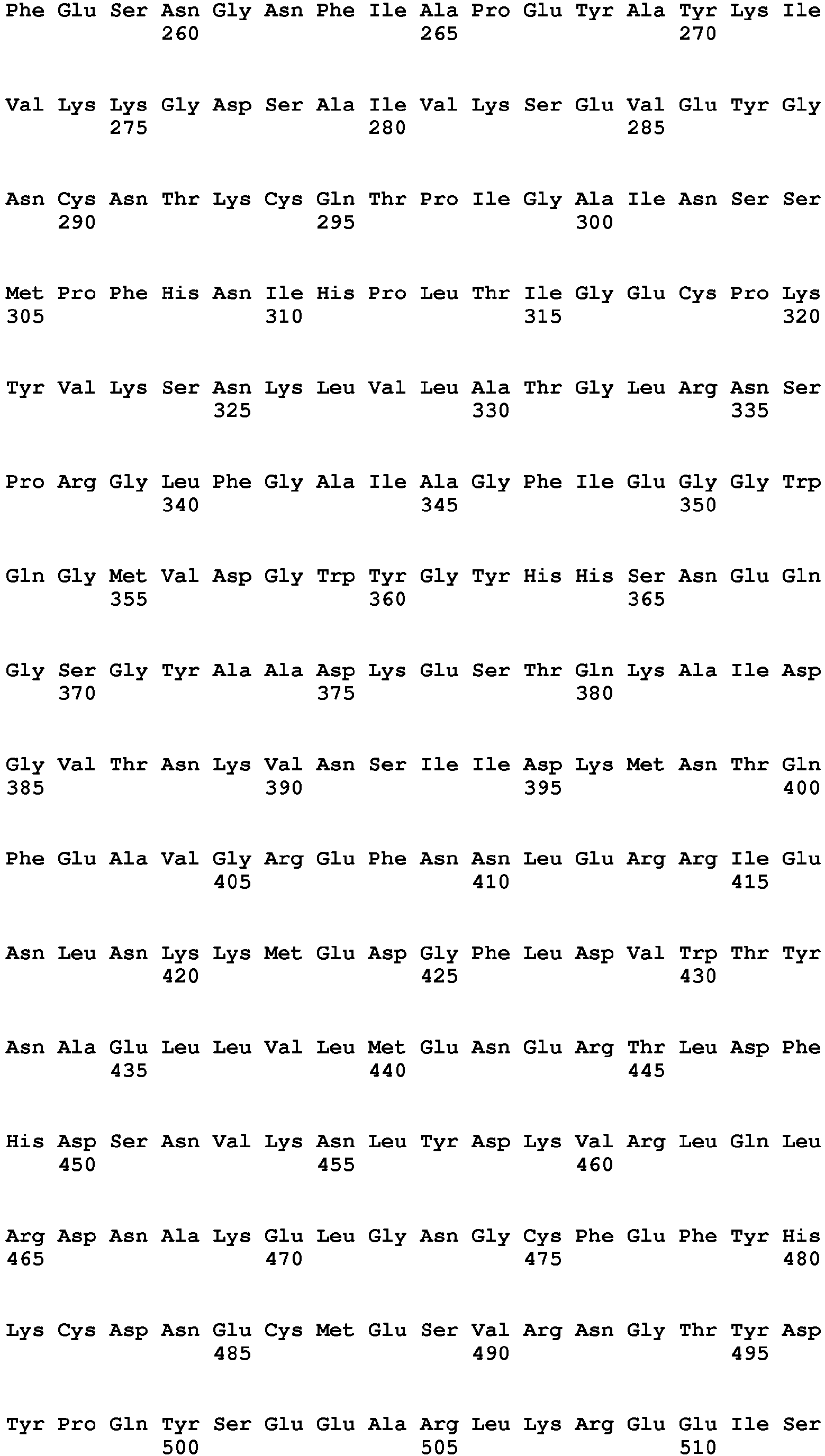


Claims (7)
- A nucleic acid molecule that binds to an influenza virus, the nucleic acid molecule comprising at least one polynucleotide selected from the group consisting of the following polynucleotides (a) to (d):(a) a polynucleotide that has any of base sequences of SEQ ID NOs: 1 to 30;(b) a polynucleotide that has a base sequence obtained by deletion, substitution, insertion, and/or addition of one or more bases in any of the base sequences of the polynucleotide (a) and binds to the influenza virus;(c) a polynucleotide that has a base sequence with an identity of at least 80% to any of the base sequences of the polynucleotide (a) and binds to the influenza virus; and(d) a polynucleotide that has a base sequence complementary to a polynucleotide hybridizing to any of the base sequences of the polynucleotide (a) under stringent conditions and binds to the influenza virus.
- The nucleic acid molecule according to claim 1, comprising two or more polynucleotides selected from the polynucleotides (a) to (d).
- The nucleic acid molecule according to claim 2, wherein the two or more polynucleotides are linked to each other via a linker.
- The nucleic acid molecule according to claim 2 or 3, comprising two polynucleotides (RHA0006_s19) each having a base sequence of SEQ ID NO: 19 or two polynucleotides (RHA0002_s33) each having a base sequence of SEQ ID NO: 17, with the two polynucleotides being linked to each other via a linker.
- The nucleic acid molecule according to claim 4, wherein the two polynucleotides linked to each other via the linker as a whole have a base sequence of SEQ ID NO: 31 or 45.
- The nucleic acid molecule according to any one of claims 1 to 5, wherein the polynucleotide is DNA.
- A binding agent that binds to an influenza virus, the binding agent comprising the nucleic acid molecule according to any one of claims 1 to 6.
Applications Claiming Priority (2)
| Application Number | Priority Date | Filing Date | Title |
|---|---|---|---|
| JP2012126861 | 2012-06-04 | ||
| PCT/JP2013/062256 WO2013183383A1 (en) | 2012-06-04 | 2013-04-25 | Nucleic acid molecule binding to influenza virus and use therefor |
Publications (2)
| Publication Number | Publication Date |
|---|---|
| EP2857514A1 true EP2857514A1 (en) | 2015-04-08 |
| EP2857514A4 EP2857514A4 (en) | 2016-02-24 |
Family
ID=49711784
Family Applications (1)
| Application Number | Title | Priority Date | Filing Date |
|---|---|---|---|
| EP13800261.3A Withdrawn EP2857514A4 (en) | 2012-06-04 | 2013-04-25 | Nucleic acid molecule binding to influenza virus and use therefor |
Country Status (8)
| Country | Link |
|---|---|
| US (1) | US20150167106A1 (en) |
| EP (1) | EP2857514A4 (en) |
| JP (1) | JP5960814B2 (en) |
| CN (1) | CN104364375A (en) |
| CA (1) | CA2873844A1 (en) |
| HK (1) | HK1206783A1 (en) |
| SG (1) | SG11201407274SA (en) |
| WO (1) | WO2013183383A1 (en) |
Families Citing this family (5)
| Publication number | Priority date | Publication date | Assignee | Title |
|---|---|---|---|---|
| CN109456974A (en) * | 2018-07-02 | 2019-03-12 | 广西医科大学 | A kind of Endoglin aptamer and its application |
| CN109576273A (en) * | 2018-07-03 | 2019-04-05 | 广西医科大学 | A set of tumor-marker molecular nucleic acid aptamers and application based on CRISPR/Cas9 |
| LU101073B1 (en) | 2018-12-21 | 2020-06-24 | Luxembourg Inst Science & Tech List | Dna aptamers specific of adenovirus types |
| KR20230040359A (en) | 2020-07-17 | 2023-03-22 | 트르스티스 오브 보스톤 유니버시티 | Virus detection system and its use |
| EP4006154A3 (en) * | 2020-10-30 | 2022-06-29 | ARKRAY, Inc. | Nucleic acid aptamer for influenza virus and detection of influenza virus |
Family Cites Families (6)
| Publication number | Priority date | Publication date | Assignee | Title |
|---|---|---|---|---|
| US7834146B2 (en) * | 2000-05-08 | 2010-11-16 | Monsanto Technology Llc | Recombinant polypeptides associated with plants |
| US7612194B2 (en) * | 2001-07-24 | 2009-11-03 | Monsanto Technology Llc | Nucleic acid sequences from Diabrotica virgifera virgifera LeConte and uses thereof |
| US7786279B2 (en) * | 2003-02-27 | 2010-08-31 | Yeda Research And Development Co. Ltd. | Nucleic acid molecules, polypeptides, antibodies and compositions for treating and detecting influenza virus infection |
| JP4441618B2 (en) * | 2003-10-06 | 2010-03-31 | 独立行政法人産業技術総合研究所 | How to detect influenza virus |
| JP5783590B2 (en) * | 2008-12-09 | 2015-09-24 | 国立大学法人東京農工大学 | Method for producing thyroglobulin-binding aptamer and aptamer multimer |
| WO2011146825A2 (en) * | 2010-05-20 | 2011-11-24 | The Board Of Trustees Of The University Of Arkansas | Avian influenza h5n1 specific aptamers and their use |
-
2013
- 2013-04-25 HK HK15107382.4A patent/HK1206783A1/en unknown
- 2013-04-25 EP EP13800261.3A patent/EP2857514A4/en not_active Withdrawn
- 2013-04-25 US US14/405,026 patent/US20150167106A1/en not_active Abandoned
- 2013-04-25 CN CN201380029160.2A patent/CN104364375A/en active Pending
- 2013-04-25 SG SG11201407274SA patent/SG11201407274SA/en unknown
- 2013-04-25 CA CA2873844A patent/CA2873844A1/en not_active Abandoned
- 2013-04-25 JP JP2014519882A patent/JP5960814B2/en active Active
- 2013-04-25 WO PCT/JP2013/062256 patent/WO2013183383A1/en not_active Ceased
Also Published As
| Publication number | Publication date |
|---|---|
| JP5960814B2 (en) | 2016-08-02 |
| EP2857514A4 (en) | 2016-02-24 |
| US20150167106A1 (en) | 2015-06-18 |
| WO2013183383A1 (en) | 2013-12-12 |
| CN104364375A (en) | 2015-02-18 |
| CA2873844A1 (en) | 2013-12-12 |
| JPWO2013183383A1 (en) | 2016-01-28 |
| HK1206783A1 (en) | 2016-01-15 |
| SG11201407274SA (en) | 2014-12-30 |
Similar Documents
| Publication | Publication Date | Title |
|---|---|---|
| Kirkpatrick et al. | The influenza virus hemagglutinin head evolves faster than the stalk domain | |
| Mellits et al. | Interaction of adenovirus VA RNAI with the protein kinase DAI: nonequivalence of binding and function | |
| Moon et al. | A noncoding RNA produced by arthropod-borne flaviviruses inhibits the cellular exoribonuclease XRN1 and alters host mRNA stability | |
| Shiratori et al. | Selection of DNA aptamers that bind to influenza A viruses with high affinity and broad subtype specificity | |
| AU2004215133B2 (en) | Nucleic acid molecules, polypeptides, antibodies and compositions containing same useful for treating and detecting influenza virus infection | |
| Nobusawa et al. | Comparison of complete amino acid sequences and receptor-binding properties among 13 serotypes of hemagglutinins of influenza A viruses | |
| US20150167106A1 (en) | Nucleic acid molecule that binds to influenza viruses and use thereof | |
| CN112063765B (en) | A kind of kit for double detection of virus by nucleic acid antibody and preparation method thereof | |
| WO2021041541A1 (en) | Modified circular rnas and methods of use thereof | |
| Palese et al. | Genetics of influenza viruses | |
| de Haan et al. | Cooperative involvement of the S1 and S2 subunits of the murine coronavirus spike protein in receptor binding and extended host range | |
| Inomata et al. | Alkaline-tolerant RNA aptamers useful to purify acid-sensitive antibodies in neutral conditions | |
| CN107058329A (en) | A kind of aptamer for specifically binding NDV and its screening technique and application | |
| CN112280901B (en) | Application of improved nucleic acid detection technology in preparation of virus detection kit | |
| EP2489734A1 (en) | NUCLEIC ACID MOLECULE HAVING AFFINITY WITH IgG ANTIBODY DERIVED FROM RODENT, BINDING AGENT, DETECTION REAGENT, AND DETECTION KIT | |
| Cryer et al. | Making Sense from Structure: What the Immune System Sees in Viral RNA | |
| US20170137824A1 (en) | New treatment against influenza virus | |
| EP3878860A1 (en) | Monoclonal antibody 2g1 for broad-spectrum neutralization of ebola viruses and application thereof | |
| KR20160019573A (en) | Aptamer specific to influenza virus h1 protein, sensing method using the aptamer, vector for the aptamer, and composition for diagnosis and activity inhibition comprising the aptamer | |
| KR20230154328A (en) | Method for manufacturing hotspot peptide-nucleic acids hybrid based on in vitro selection | |
| Simmons et al. | A protective and broadly binding antibody class engages the influenza virus hemagglutinin head at its stem interface | |
| Shubsda et al. | Binding of human immunodeficiency virus type 1 nucleocapsid protein to Ψ-RNA-SL3 | |
| KR20170042077A (en) | Nucleic Acid Aptamer Capable of Specifically Binding to AI Virus and Uses Thereof | |
| US20240002860A1 (en) | SARS-CoV-2 SPIKE GLYCOPROTEIN-BINDING NUCLEIC ACID MOLECULE, SARS-CoV-2 DETECTION SENSOR, SARS-CoV-2 DETECTION REAGENT, SARS-CoV-2 DETECTION METHOD, AND SARS-CoV-2 VIRUS DEACTIVATOR | |
| Wurm | Manufacturing of biopharmaceuticals and implications for biosimilars |
Legal Events
| Date | Code | Title | Description |
|---|---|---|---|
| PUAI | Public reference made under article 153(3) epc to a published international application that has entered the european phase |
Free format text: ORIGINAL CODE: 0009012 |
|
| 17P | Request for examination filed |
Effective date: 20150105 |
|
| AK | Designated contracting states |
Kind code of ref document: A1 Designated state(s): AL AT BE BG CH CY CZ DE DK EE ES FI FR GB GR HR HU IE IS IT LI LT LU LV MC MK MT NL NO PL PT RO RS SE SI SK SM TR |
|
| AX | Request for extension of the european patent |
Extension state: BA ME |
|
| DAX | Request for extension of the european patent (deleted) | ||
| RIC1 | Information provided on ipc code assigned before grant |
Ipc: C12N 15/115 20100101AFI20160114BHEP |
|
| RA4 | Supplementary search report drawn up and despatched (corrected) |
Effective date: 20160125 |
|
| STAA | Information on the status of an ep patent application or granted ep patent |
Free format text: STATUS: THE APPLICATION HAS BEEN WITHDRAWN |
|
| 18W | Application withdrawn |
Effective date: 20160419 |