EP2855524B1 - Novel il-3 antibodies and their use in diagnosis and treatment of diseases or malfunctions associated with elevated levels of il-3 - Google Patents
Novel il-3 antibodies and their use in diagnosis and treatment of diseases or malfunctions associated with elevated levels of il-3 Download PDFInfo
- Publication number
- EP2855524B1 EP2855524B1 EP13726501.3A EP13726501A EP2855524B1 EP 2855524 B1 EP2855524 B1 EP 2855524B1 EP 13726501 A EP13726501 A EP 13726501A EP 2855524 B1 EP2855524 B1 EP 2855524B1
- Authority
- EP
- European Patent Office
- Prior art keywords
- antibody
- antibodies
- clone
- cells
- patients
- Prior art date
- Legal status (The legal status is an assumption and is not a legal conclusion. Google has not performed a legal analysis and makes no representation as to the accuracy of the status listed.)
- Active
Links
Images

















Classifications
-
- C—CHEMISTRY; METALLURGY
- C07—ORGANIC CHEMISTRY
- C07K—PEPTIDES
- C07K16/00—Immunoglobulins [IG], e.g. monoclonal or polyclonal antibodies
- C07K16/18—Immunoglobulins [IG], e.g. monoclonal or polyclonal antibodies against material from animals or humans
- C07K16/24—Immunoglobulins [IG], e.g. monoclonal or polyclonal antibodies against material from animals or humans against cytokines, lymphokines or interferons
- C07K16/244—Interleukins [IL]
-
- G—PHYSICS
- G01—MEASURING; TESTING
- G01N—INVESTIGATING OR ANALYSING MATERIALS BY DETERMINING THEIR CHEMICAL OR PHYSICAL PROPERTIES
- G01N33/00—Investigating or analysing materials by specific methods not covered by groups G01N1/00 - G01N31/00
- G01N33/48—Biological material, e.g. blood, urine; Haemocytometers
- G01N33/50—Chemical analysis of biological material, e.g. blood, urine; Testing involving biospecific ligand binding methods; Immunological testing
- G01N33/68—Chemical analysis of biological material, e.g. blood, urine; Testing involving biospecific ligand binding methods; Immunological testing involving proteins, peptides or amino acids
- G01N33/6863—Cytokines, i.e. immune system proteins modifying a biological response such as cell growth proliferation or differentiation, e.g. TNF, CNF, GM-CSF, lymphotoxin, MIF or their receptors
- G01N33/6869—Interleukin
-
- C—CHEMISTRY; METALLURGY
- C07—ORGANIC CHEMISTRY
- C07K—PEPTIDES
- C07K2317/00—Immunoglobulins specific features
- C07K2317/20—Immunoglobulins specific features characterized by taxonomic origin
- C07K2317/21—Immunoglobulins specific features characterized by taxonomic origin from primates, e.g. man
-
- C—CHEMISTRY; METALLURGY
- C07—ORGANIC CHEMISTRY
- C07K—PEPTIDES
- C07K2317/00—Immunoglobulins specific features
- C07K2317/20—Immunoglobulins specific features characterized by taxonomic origin
- C07K2317/24—Immunoglobulins specific features characterized by taxonomic origin containing regions, domains or residues from different species, e.g. chimeric, humanized or veneered
-
- C—CHEMISTRY; METALLURGY
- C07—ORGANIC CHEMISTRY
- C07K—PEPTIDES
- C07K2317/00—Immunoglobulins specific features
- C07K2317/30—Immunoglobulins specific features characterized by aspects of specificity or valency
- C07K2317/33—Crossreactivity, e.g. for species or epitope, or lack of said crossreactivity
-
- C—CHEMISTRY; METALLURGY
- C07—ORGANIC CHEMISTRY
- C07K—PEPTIDES
- C07K2317/00—Immunoglobulins specific features
- C07K2317/70—Immunoglobulins specific features characterized by effect upon binding to a cell or to an antigen
- C07K2317/73—Inducing cell death, e.g. apoptosis, necrosis or inhibition of cell proliferation
-
- C—CHEMISTRY; METALLURGY
- C07—ORGANIC CHEMISTRY
- C07K—PEPTIDES
- C07K2317/00—Immunoglobulins specific features
- C07K2317/70—Immunoglobulins specific features characterized by effect upon binding to a cell or to an antigen
- C07K2317/76—Antagonist effect on antigen, e.g. neutralization or inhibition of binding
-
- G—PHYSICS
- G01—MEASURING; TESTING
- G01N—INVESTIGATING OR ANALYSING MATERIALS BY DETERMINING THEIR CHEMICAL OR PHYSICAL PROPERTIES
- G01N2800/00—Detection or diagnosis of diseases
Definitions
- the present invention relates to novel anti-interleukin 3 (IL-3) antibodies as defined in the claims, nucleic acid sequences encoding such antibodies and hybridoma cell lines producing the antibodies according to the present invention.
- the invention further relates to pharmaceutical compositions containing the novel IL-3 antibodies, which are useful for the prevention or treatment of diseases or malfunctions which are associated with elevated levels of IL-3 in a human patient as well as to diagnostic kits and methods for reliably determining the IL-3 level in the blood, plasma, serum or other body fluid (e.g. urine, synovial fluid) of a patient.
- body fluid e.g. urine, synovial fluid
- Cytokines are polypeptides that influence the function of certain cells upon binding to specific cellular receptors and are divided in subclasses, i.e., interleukins, interferons, colony-stimulating factors (CSFs), lymphokines, growth factors and monokines. It is well known that cytokines play a major role in cell proliferation and, e.g., also inflammatory diseases.
- Cell proliferation is a complex process wherein growth factors bind to specific receptors on the cell surface, whereupon endocytosis occurs and the complexes of cytokine and receptor are internalized causing a cellular response.
- Such cellular responses include specific gene transcription activities as DNA synthesis and cell replication.
- most of the cytokines When tested in relatively high concentrations, most of the cytokines have several differing biological effects. Because of these effects of cytokines, there is a high interest in investigations for possible therapeutic uses of these proteins.
- Interleukins are mediators of the immune system which are produced in low concentration mostly in leukocytes. They influence the growth, differentiation and activity of cells of the immune system and thus belong to the immune modulators. They also take effect by binding to receptors on the surface of target cells and thus change the transcription rate of certain genes. They play an important role in the triggering of a multiplicity of cellular responses.
- Interleukins are, e.g., involved in the immunological cell activation cascade and subsequent inflammatory changes. Irregular and/or abnormal inflammation is a major component and factor of a wide range of human diseases, one of which is the immunological disorder rheumatoid arthritis (RA). But also other immunological diseases are influenced by interleukins.
- RA immunological disorder rheumatoid arthritis
- IL-3 also designated as Multi-CSF, is a well-known member of the interleukin family. It has a growth stimulating and differentiating effect on various hematopoietic precursor cells and acts as a growth factor for mast cells. Together with IL-5 and GM-CSF, IL-3 belongs to the family of hematopoietic cytokines with four short alpha-helical bundles. GM-CSF and IL-3 stimulate the formation of neutrophilic and eosinophilic granulocyte colonies as well as macrophages. It further stimulates the formation of mast, megakaryocyte and pure and mixed erythroid colonies ( D. Metcalf, "The hematopoietic colony-stimulating factors", 1984, Elsevier, Amsterd am).
- IL-3 consists of 133 amino acids and is known for its stimulation of colony formation by human hematopoietic progenitor cells and the stimulation of DNA synthesis by human acute myelogenous leukemia (AML) blasts.
- IL-3 binds to a unique receptor also known as CD123 antigen.
- the receptor belongs to the type I cytokine receptor family and is a heterodimer with a unique ⁇ -chain paired with a common ⁇ -subunit ( ⁇ C or CDW 131).
- IL-3 binds to the unique ⁇ -receptor subunit. Signal transduction is mediated, however, by the common ⁇ -receptor subunit ( ⁇ C) by the JAK2-STAT5 pathway.
- IL-3 is mainly produced by activated CD4+ T-cells and contributes especially to growth, differentiation and survival of CD34+ hematopoietic progenitor cells.
- IL-3 has been observed to promote the differentiation of basophiles and mast cells from bone marrow cells. It has further been observed to induce IL-6 release by murine basophils and to up-regulate MHC-II expression and IL-1 secretion in monocyte/macrophages. Further, IL-3 supports the differentiation of monocytes into dendritic cells and osteoclasts.
- RA rheumatoid arthritis
- NSAIDs non-steroidal anti-inflammatory drugs
- NSAIDs act as anti-inflammatory and analgetic agents and often only achieve an alleviation of pain.
- the drugs further interfere with a certain step in the inflammatory cascade, where prostaglandine is generated by cyclooxygenases.
- NSAIDs do not influence the underlying inflammatory process and are thus not able to retard the joint destruction, which is the most deleterious effect of RA.
- DMARDs disease-modifying anti-rheumatic drugs
- methotrexate the most commonly used anti-rheumatic, the effect of which is based on a reversible inhibition of the enzyme dihydrofolate reductase.
- leflunomide Another commonly used substance for treating RA. Both pharmaceuticals are long-acting and thus have to be administered over a longer period of time (usually 12 - 16 weeks) to show the desired effects. To bridge the time until DMARDs improve the disease, most patients are administered steroids.
- a further approach for treating RA are "biologicals" that block cytokines like TNF, IL-6, IL-1 or costimulatory molecules like B7 or that deplete leukocyte subsets (e.g. B cells).
- Biologicals e.g. the TNF antibody Infliximab
- TNF antibody Infliximab are mostly used for severe disease processes and after DMARDs have failed to sufficiently control disease activity.
- Biologicals influence a plurality of signal systems in the immune system and have a variety of serious side effects including bacterial and viral infections and a higher risk for development of neoplasia.
- IL-3 inhibitors can be used in treatment of early stages of rheumatoid arthritis.
- a genetic analysis found an association between a single nucleotide polymorphism in the IL-3 promoter gene and RA.
- WO 2010/063488 proposes such use of inhibitors, mainly antibodies or antibody fragments, antibody variants or antibody multimers in prophylactic RA treatment, therapeutic treatment in early stages of the disease or in maintenance treatment.
- Such anti-IL-3 antibodies preferably should also exhibit none or very low cross reactivity with IL-3 from other species.
- the antibodies of the present invention show also surprisingly low levels of cross-reactivity with IL-3 molecules of other mammalian origin.
- Amino acid identities of a human protein and the mouse protein is 29% which seems quite low.
- amino acid identities between 72 and 99% there are amino acid identities between 72 and 99% ( figure 1 ).
- IL-5 and GM-CSF are particularly important.
- a high cross-reactivity of an anti-IL-3 antibody with such cytokines in an immunoassay can lead to incorrect results regarding the fact whether an IL-3 overexpression has an important influence in the manifestation and progression of the autoimmune disease.
- Such results have an important impact on the decision whether the application of an anti-IL-3 antibody can be considered a promising therapeutic approach.
- inventive antibodies it is thus preferred for inventive antibodies to show the lowest possible cross-reactivity with human IL-5 and GM-CSF. Particularly preferred, it is a characterizing feature of an inventive antibody that it binds to IL-5 or GM-CSF to an extent of below 5%, more preferred below 2% and particularly preferred below 1% as compared to the amount of IL-3 bound by the antibody.
- ELISA enzyme-linked immunosorbent assay
- ELISA plates are coated with any anti-human-IL-3 antibody which can also be a commercially available antibody (e.g., a goat IgG anti-human-IL-3 antibody).
- any anti-human-IL-3 antibody which can also be a commercially available antibody (e.g., a goat IgG anti-human-IL-3 antibody).
- 1 ug/ml of the antibody is incubated with the ELISA plates over night in a refrigerator whereupon a washing step and a blocking with human IL-3 (250 ng/ml in PBS buffer) is performed, thus fixing human IL-3 on the solid phase.
- candidate antibodies are then added in different concentrations and detected by means of a secondary HRP- (horseradish peroxidase) labelled polyclonal antibody.
- Clone 11 is a mouse anti-human-IL-3 antibody and shows a very high specificity and affinity to IL-3. Clone 11 specifically binds to the epitope with the amino acids SWVN according to SEQ ID NO: 2. This particularly preferred antibody was deposited at DSMZ (Braunschweig, Germany) under number DSM ACC3163.
- clone 11 has proven very superior characteristics with regard to specificity, lack of cross-reactivity but also with regard to affinity and avidity. This preferred antibody therefore is considered especially suitable for use in diagnostics as well as therapeutic measures.
- a particularly preferred inventive anti-IL-3 antibody is a humanized version of clone 11.
- clone 11 is humanized in any manner known in the art.
- at least one of the CDRs of clone 11 is identically maintained/preserved, other parts of the mouse-antibody clone 11 can entirely or partially be replaced by sequences of human antibody origin.
- it is essential that the obtained humanized antibody still shows the same specificity which can easily be determined using known methods.
- such a humanized antibody clone 11 also shows just as little cross-reactivity with IL-3 of species other than the human and with other human cytokines as the deposited mouse clone 11.
- a further subject-matter of the present invention is a nucleic acid which encodes an antibody, according to the present invention.
- the nucleic acid codes for the antibody clone 11.
- a further, especially preferred nucleic acid codes for a humanized clone 11, whereby it comprises at least those nucleotides coding for the regions in antibody clone 11, which entail the binding to the specific human IL-3-epitope according to the present invention.
- the hybridoma cell line according to the present invention is cell line 11.14.6 (DSM ACC 3163) producing antibody clone 11.
- a further subject matter of the invention is a diagnostic method for determining the IL-3 level in body fluids of a patient, especially in blood, plasma or serum.
- the method comprises adding an anti-IL3 antibody, according to the present invention to a sample comprising a body fluid sample under conditions which allow for the binding of the antibody to IL-3 and detecting the amount of antibody-bound IL-3 in the sample.
- the accurate determination of the presence of IL-3 in body fluids can be improved to a major extent and such methods and test kits, therefore, are a further subject matter of the present invention. It has surprisingly been found that using the antibodies and methods of the present invention, IL-3 could be specifically detected even if present in only picogram / ml concentrations in a sample.
- the relative affinity of the antibodies of the present invention towards IL-3 is thus about at least a factor 100 higher than the affinity of known antibodies.
- the diagnostic method is conducted as an ELISA assay.
- the general test regime used for ELISA assays is well known to the skilled person.
- At least two antibodies which bind to the target molecule are used.
- One of these antibodies is bound to a solid phase allowing for the separation of the antigen to be determined from the test sample.
- a second antigen-specific and labeled antibody is added and after further removal of excess labeled antibody and optionally further washing steps the amount of label bound via the antibody-antigen-antibody complex is determined and correlated to the amount of antigen present.
- both antibodies used in such an ELISA assay are specific to the antigen to a high extent, an especially high degree of specificity is needed mainly in one of the two antibodies involved.
- a very specific antibody can be used to be able to separate only the desired antigen from the liquid sample without any cross-reacting background. If only the desired antigen is coupled via the antibody to the solid surface, the specificity of the second antibody is not so decisive anymore since unspecific binding to other antigens is not an issue in such a case. Accordingly, it is possible to use a less specific antibody as the second antibody in such a context.
- the first solid phase-bound antibody it is possible to use a high affinity and high avidity antibody as the first solid phase-bound antibody and to use a highly specific antibody as the second and labeled antibody.
- a highly specific antibody such an embodiment, unspecific binding of other antigens like IL-5 or GM-CSF might take place to some extent due to a certain cross-reactivity of the first antibody, however, the actual detection by the labeled antibody is then restricted to IL-3 by using a highly specific second antibody.
- an ELISA assay method and kit are used wherein as the highly specific antibody in the test an antibody according to the present invention is used.
- the highly specific antibody used in the ELISA assay is antibody clone 11.
- first antibody / second antibody preferred combinations of antibodies (first antibody / second antibody) are clone 13 / 11 (most preferred), clone 11 / 13 (preferred). as well as combinations of clone 11 with clone 47. Clone 13 and clone 47 are considered to bind to a 3-D epitope of IL-3 in its native conformation.
- the present diagnostic method is especially suited and useful for the detection of IL-3 in a plasma or serum sample.
- detection of IL-3 in plasma or serum did not lead to sufficiently meaningful results especially due to cross-reactivities of known antibodies and insufficient affinities of such antibodies with regard to the low levels of IL-3 in present blood, plasma or serum in healthy and in RA patients. Therefore, often synovial fluids were used as test sample. However, obtaining such a test sample is much more difficult and cumbersome to the patient.
- the present invention via its antibodies and their use in diagnosis is a big step forward to relating IL-3 presence to the severity of RA and RA incidents in patients and thus to also determine the supposition of a patient to treatment with IL-3 antibodies.
- Test kits which contain antibodies according to the present invention together with other substances for conducting the diagnostic methods of this invention are also a further embodiment of the invention.
- IL-3 in body fluids of a patient
- Such kits can be provided for easy and effective use in laboratories of every size.
- Test reagents for the various applicable test formats are well known in the art.
- the inventive contribution to the test kits lies in the presence of antibodies according to the present invention or combinations of antibodies according to the present invention with other IL-3 specific antibodies, most preferably a combination of antibody clones 11 and 13.
- WO2010/063488 is directed to use of IL-3 inhibitors for the treatment of early stage RA patients.
- IL-3 inhibitors are especially well suited for a therapeutic application. It seems vital that antibodies used in therapy should not entail drawbacks brought on e.g. by cross-reactivity with other unrelated cytokines or severe immunological responses experienced with known antibodies.
- a further subject and embodiment of the present invention therefore is the use of the novel and advantageous antibodies of the invention in a pharmaceutical composition.
- Such pharmaceutical compositions according to the invention are characterized by the presence of a pharmaceutically effective amount of an antibody as described herein as an active ingredient.
- Usual adjuvants and/or carrier substances for pharmaceutical preparations can be included as desired and deemed appropriate.
- compositions according to the present invention can be used for the treatment or for the prevention and prophylaxis of diseases or malfunctions which are associated with elevated levels or IL-3.
- IL-3 has a significant growth stimulating and differentiating effect on various hematopoietic precursor cells and is also a growth factor for mast cells.
- the signal transduction caused by IL-3 has major impact on the immune system.
- Any disease or medical condition in which IL-3 plays a direct or indirect role in development or progression is a candidate for the treatment by administering the antibodies according to the present invention.
- IL-3 is related to the immune system, mostly an autoimmune disease and especially RA or acute or chronic graft-versus-host disease and multiple sclerosis.
- composition according to the invention is a use in suppressing the activity of human basophils in persons suffering from an allergic reaction and for the stratification of patients having increased IL-3 levels in serum or plasma.
- IL-3 in the respective disease or malfunction elevated levels of IL-3 as compared to healthy persons have to be present in any body fluid including synovial fluids but also blood, plasma and serum.
- the antibodies of the present invention which are contained in the pharmaceutical composition bind specifically to IL-3 and thereby inhibit the activity of IL-3.
- compositions of the present invention are especially useful in treating patients with active episodes of autoimmune diseases, as RA, and for the prophylactic treatment to avoid the occurrence of active episodes of the disease.
- IL-3 targeted treatment should exhibit less severe side effects than currently used pharmaceuticals, especially regarding to infection or neoplasia. In certain cases, it could be desirable to combine treatment with the antibodies and pharmaceutical compositions of the present invention with other medicines like methotrexate or leflunomide.
- An individualized treatment strategy according to IL-3 levels in plasma, serum or other body fluids presents an advantage compared to available biologicals, since currently it is not possible to predict reliably, which patient will respond to a specific therapy (including biologicals). Further an individualized approach improves the safety of treatment by reducing the risk of side effects of an ineffective therapy and reduces the costs for treatment of RA.
- the treatment with the antibodies of the present invention would preferentially be started as soon as elevated IL-3 levels in blood, plasma or serum are detected.
- early-on treatment can be applied in patients where RA activity is correlated with elevated IL-3 levels and long term joint damage can be avoided or kept to a minimum.
- treatment with anti-IL-3 antibodies would preferentially be started, if patients failed to a previous treatment with DMARDs or biologicals.
- the use of the antibodies of the invention can reduce cell infiltration of synovial tissue which can be a further negative factor in the disease pathology.
- An effective dose of IL-3 antibody contained in the pharmaceutical composition of the present invention can easily be determined by a physician as generally known in the art.
- An effective dose is an amount that alleviates the symptoms of the disease or prevents any further progress of the disease or deterioration of the condition of the patient.
- Progress of disease and condition of the patient can be monitored by determining the IL-3 level and other markers for inflammation or autoimmunity in body fluids, preferably blood, plasma or serum, and by determining other disease scores or using known diagnostic methods.
- Effectiveness of a therapy can be measured as the percentage of patients that achieve a certain (e.g. 30% , 50% or 70%) reduction in disease activity. Any decrease of IL-3 or other markers for inflammation in body fluids is also an indication of a successful treatment.
- Effective doses of the pharmaceutical composition can be determined using dose-response-curves as is well-known to the skilled artisan.
- the amount of IL-3 present in the body fluids of a patient as determined using the diagnostic methods according to the present invention can also be a basis for determining the effective dose of antibody in the pharmaceutical composition for each patient and the severity of the disease and malfunction.
- dosage further depends on the activity, avidity and the half-life of the antibody.
- the dosage is preferably in ta range of 1 to 1000 mg and more preferably 10 to 100 mg per application.
- the pharmaceutical composition is preferably applied once a day to once a month, again depending on the half-life of the antibody.
- the antibody of the invention is antibody clone 11.
- Antibody clone 11 does not show detectable cross-reaction with GM-CSF and IL-5 and therefore does not influence the activity of these cytokines in the patient (see example 4b). Further it has been shown that clone 11 is very effective in inhibiting the IL-3 dependent growth of human TF-1 cells (see example 5a and 5b), proving an efficient inhibition of the IL-3 activity in patients by this antibody.
- the growth of TF-1 cells is a well known test system for IL-3 activity and the inhibition thereof.
- clone 11 shows excellent results in view of inhibition of IL-3 activity (see example 5c and 5d). Clone 11 also shows a very high affinity and avidity and already very small amounts of this antibody are sufficient for its inhibitory activity (example 5).
- Clone 11 is an excellent inhibitor of IL-3 and therefore a promising pharmaceutical with regard to treatment of IL-3 related malfunctions and diseases.
- Anti-IL-3 antibodies were produced by immunizing Balb/c mice using at least 6 i.p. injections of human eukaryotic glycosylated IL-3 in alumn at four week intervals. Two days before cell fusion, IL-3 in PBS was injected intraperitoneally. Antibody-producing splenocytes obtained from the immunized mice (HGPRT positive, able to grow on HAT medium) were fused with the myeloma cell line X63Ag8.6.5.3 in the presence of polyethylene glycol (PEG) and a selection of hybridomas performed in an HAT-selection medium.
- PEG polyethylene glycol
- Hybridomas were cultivated in RPMI-1640 medium supplemented by 10% FCS (neat inactivated, HIA), P/S and glutamine (1:100). Obtained cells are able to grow in suspension and are splitted every three days in a ratio of 1:4.
- hybridoma cells are transferred from a cell culture bottle into 50 ml or 15 ml cell culture flasks (BD FalconTM). After centrifugation at 1400 rpm for 5 minutes at room temperature, the supernatant is completely removed. Cells are resuspended in a freezing medium (90% FCS (HIA) + 10% DMSO) and 1.5 ml aliquots are filled into vials. The cells are prefrozen in a freezing container in a freezer at -80°C and after 1-2 days transferred to a liquid nitrogen storage tank.
- a freezing medium 90% FCS (HIA) + 10% DMSO
- Cloning and recloning of the obtained hybridoma cell lines are performed using limited dilution to provide long-term stable sources for monoclonal antibodies.
- 96-well-plates are coated overnight at room temperature with anti-mouse IgG (1:100 in PBS) in a concentration of 100 ⁇ l/well. Blocking is performed by adding 100 ⁇ l per well of 2 % BSA in PBS and incubation at room temperature for two hours. After the blocking reaction, the plates are washed twice.
- the plate is washed three times and then incubated with biotinylated anti-mouse IgG1 (diluted by 1:250 in 2 % BSA in PBS) for one hour at room temperature with 100 ⁇ l per well. After washing the plate a further three times, streptavidin-HRP (1:1000 in 2 % BSA in PBS) is added for one hour at room temperature and in the dark. The concentration of the antibodies is determined after adding ABTS and incubating for further 30 minutes and measuring the signal at 405 and 490 nanometers on a spectrophotrometer. Based on this determination, a desired amount of the antibodies tested is applied for the further tests.
- Samples of IL-3 are prepared by mixing of recombinant human IL-3 1:1 with Laemmli buffer and heating the samples at 60°C for 5 min. An amount of 1 ⁇ g per lane of IL-3 as well as a usual standard for determining molecule sizes is loaded onto the gel. The gel is then mounted in a SDS-PAGE gel electrophoresis apparatus which already contains a running buffer. The inserted gel is then cautiously overlayed with additional running buffer and electrophoresis performed at 20 to 25 mA with voltage adjusted to infinite for approximately 1,5 hours. When the run is completed, the gel is retrieved from the apparatus and the stacking gel is removed.
- Antibody clones are incubated at a concentration of 5 ⁇ g/ml in blocking solution for 2 hours at room temperature under agitation on the shaking apparatus. After three washing steps, HRP labelled anti-mouse immunoglobulin (1:1000 in blocking solution) is added and incubation is conducted for 1 hour at room temperature while shaking. After three further wahing steps, a detection solution (1:1 mixture of solutions A and B of the Westernblotting Luminal Reagent obtained from NALGENE) is added and incubated for 1 min at room temperature. Films are then adjusted on the membranes with different times of expositions and developed in the dark room.
- Fig. 2 shows the results of binding of antibody clones 2, 3, 5, 6, 7, 8, 10, 11 and 13. Binding to IL-3 at the given concentration was detected for clones 8, 11 and, to a lesser extent, for clone 13.
- the affinity of the obtained antibodies for IL-3 was measured in an ELISA assay.
- ELISA plates were coated overnight with different concentrations (2 ⁇ g/ml, 0.66 ⁇ g/ml, 0.22 ⁇ g/ml, 0 ⁇ g/ml) of anti-human IL-3 antibody (RD, goat IgG anti-human IL-3 AF-203-NA).
- RD anti-human IL-3 antibody
- duplicates were used (2 x 12 wells).
- the first concentration (2 ⁇ g/ml) is diluted in PBS, further dilutions are made in PBS containing 2 ⁇ g/ml control goat IgG to keep the total concentration of IgG constant. Blocking with 2% BSA is performed for 2 hours at room temperature, followed by 5 washing steps using PBS.
- hIL-3 (0.25 ⁇ g/ml in PBS)
- hIL-3 (0.25 ⁇ g/ml in PBS)
- no hIL- 3 is added.
- the wells are incubated overnight at 4°C with serial (1:3) dilutions of antibodies clone 8 and 11 obtained in example 1, the antibodies being used in PBS buffer containing 2% BSA and with a starting concentration of 20 ⁇ g/ml.
- bound antibody is detected using goat-anti-mouse-HRP antibody (1:500 in PBS with 2% BSA) and incubation for 1 hour at room temperature.
- ABTS ROCHE, 1 mg/ml
- Results are shown in Figs. 3 and 4 for antibodies clone 8 and 11, respectively. Both antibodies show a high affinity for IL-3 in the assay.
- Fig. 5 shows the results of further tests including other antibodies. The tests were performed in the same manner as described, however, coating of the solid phase was performed using 1 ⁇ g/ml goat IgG anti-human IL 3 (see above) and different concentrations/dilutions of antibodies as shown in the figure.
- the cytokine coated plates were washed three times and blocking performed for 2 hours at room temperature using 2% BSA in PBS. After three further washing steps, antibodies clone 3.47.20, 8.36.38, 10.12.4, 11.14.6, 13.4.4 and just medium (RPMI1640 containing 10 % FCS) as control were added at a concentration of 40 ⁇ g/ml and 1:5 and 1:25 dilutions thereof in a volume of 100 ⁇ l/well and inicubated for 1 hour at room temperature. On each plate a negative control is used.
- a secondary HRP-labelled rabbit anti-mouse IgG (DAKO-Cytomation P260 (1:2000 in 2% BSA in PBS, 100 ⁇ l/well) was added and the plates incubated at room temperature for 1 hour in the dark.
- ABTS (ROCHE, 1 mg/ml) was added and spectrometry performed at 405 and 490 nm after 30 min.
- the IL-3 coated plates were washed three times and blocking performed for 2 hours at room temperature with 2% BSA in PBS. After three washing steps, antibody clones 3.47.20, 8.36.38, 10.12.4, 11.14.6, 13.4.4 in certain concentrations as indicated in Figs 7 and 8 , and 1:5, 1:25 and 1:125 dilutions thereof were added at volumes of 100 ⁇ l/well.
- R&D monoclonal anti-IL-3 antibody clone 4806 R&D Systems, Inc., catalogue No.
- MAB203 was used (100 ⁇ l/well) in concentrations of 40 ⁇ g/ml, 20 ⁇ g/ml, 10 ⁇ g/ml, 5 ⁇ g/ml and 2.5 ⁇ g/ml and, as negative control, medium (100 ⁇ l/well) without antibody (RPMI 1640 containing 10 % FCS) was used. On each plate a negative control was used.
- a secondary HRP-labelled rabbit anti-mouse IgG (DAKO-Cytomation P260 (1:2000 in 2% BSA in PBS, 100 ⁇ l/well) was added and the plates incubated at room temperature for 1 hour in the dark.
- ABTS (ROCHE, 1 mg/ml) was added and spectrometry performed at 405 and 490 nm after 30 min.
- TF1 cells are human erythroblasts and the cell line has been established by T. Kitamura in 1987 from bone marrow of a 35 year old male Japanese suffering from severe pancytopenia. Growth of TF1 cells is completely dependent on the presence of IL-3 or GM-CSF. Thus, a test based on the cell proliferation of TF1 cells can be used to determine blocking of the IL-3 acitvity which in turn leads to a decrease or even a complete inhibition of the growth of TF1 cells. For such a test, a MTT-cell-proliferation assay is performed to determine the viability of cells based on the activity of the mitochondrial dehydrogenase.
- the dehydrogenase's substrate MTT (3-(4,5-dimethylthiazol-2-yl)-2,5-diphenyl tetrazolium bromide) which shows a yellow color in solution, is cleaved at the tetrazolium ring by the enzymatic activity leading to formation of purple MTT formazane crystals.
- Such crystals can be dissolved in isopropanol, the purple solution measured in a spectrometer and the results correlated to the amount of viable TF1 cells.
- TF1 cells were grown in suspension in a culture medium (RPMI-1640 containing 10% FCS (HIA), P/S and Glutamin (1:100) and supplemented with either 5ng/ml of IL-3 or 5 ng/ml of GM-CSF) and splitted 1:4 every third day.
- a culture medium RPMI-1640 containing 10% FCS (HIA), P/S and Glutamin (1:100) and supplemented with either 5ng/ml of IL-3 or 5 ng/ml of GM-CSF
- a culture medium RPMI-1640 containing 10% FCS (HIA), P/S and Glutamin (1:100) and supplemented with either 5ng/ml of IL-3 or 5 ng/ml of GM-CSF
- cells were transferred from a cell culture bottle to 50 ml or 15 ml cell culture flasks (BD FalconTM). After centrifugation at 1400 rpm for 5 minutes at room temperature, the supernatant is completely removed.
- Cells are resuspended in culture medium (RPMI-1640 containing 10% FCS (HIA) +P/S + Glutamin + 5 ng/ml IL-3) and 5% DMSO and 1.5 ml aliquots are filled into vials.
- the cells are prefrozen in a freezing container in a freezer at -80°C and after 1-2 days transferred to a liquid nitrogen storage tank.
- TF-1 cells that had been splitted every third day according to the protocol described above are splitted 1:2 in culture medium containing 5 ng/ml human IL-3 on the day before the experiment is performed.
- cells are cetrifuged for 5 minutes at 1600 rpm at room temperature.
- the culture medium is removed and the cells washed twice in RPMI medium before cells are resuspended in 1 ml RPMI-1640 + 10% FCS (HIA) + P/S + Glutamin (1:100), counted and supplemented with buffer to a final concentration of 1 x 10 5 cells/ml.
- IL-3 The general influence of IL-3 on the growth of TF1 cells is shown in Fig. 9 .
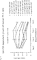
- Results for different amounts of IL-3 with and without different concentrations of mAbs are shown in Figs. 10 to 13 .
- IL-3 binds to the IL-3 receptor which is comprised of an alpha chain of 70kDa and a beta chain of about 130kDa. The same beta chain is also present on receptors for IL-5 and GM-CSF. Therefore, in another experiment it was tested whether anti-IL-3 antibodies also influence the growth of TF1 cells in the presence of GM-CSF. For this purpose, the experiment described above was repeated adding GM-CSF, IL-3 and mixtures thereof preincubated with the anti-IL-3 antibodies to the culture medium of TF1 cells. A control without GM-CSF and IL-3 was included.
- GM-CSF The general influence of GM-CSF on the growth of TF1 cells is shown in Fig. 14 .
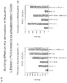
- the results of the tests in view of the blocking of this influence and amounts of GM-CSF and antibodies, respectively, used in this example are shown in Fig. 15 indicating that none of the tested antibodies had a blocking effect on GM-CSF and its growth induction toward TF1 cells.
- PBMC peripheral blood mononuclear cells
- human PBMC cells were isolated from human blood using the Ficoll-gradient centrifugation method. Cells were resuspended at a concentration of 10 Mio. cells/ml (in PBS) and 10 ⁇ l aliquots incubated with purified polyclonal mouse IgG (SIGMA-Aldrich) for 15 min at room temperature, to block Fc mediated interactions.
- biotinylated hIL-3 was preincubated with dilutions of antibodies.
- 10 ⁇ l of biotinylated hIL-3 (1.25 ⁇ g/ml) were preincubated with 10 ⁇ l of anti-IL-3 antibody at 100 ⁇ g/ml or dilutions thereof at 1/3, 1/9, 1/27, 1/81 and 1/243 , or with 10 ⁇ l PBS, 10 ⁇ l isotype antibody (50 ⁇ g/ml) or 20 ⁇ l blocking Ab (R&D Systems Cat No NF300) and incubated for 30 min at room temperature.
- a negative control reagent (R&D Systems Cat. No. NF300) were preincubated with 15 ⁇ l of anti-IL-3 antibody (50 ⁇ g/ml), 10 ⁇ l PBS, 10 ⁇ l isotype antibody (50 ⁇ g/ml) or 20 ⁇ l blocking AB (R&D Systems Cat. No. NF300).
- Human basophilic granulocytes show an IL-3 induced upregulation of CD203c and concurrent release of IL-4 and IL-13.
- PBMC peripheral blood mononuclear cells
- Stimulation of basophils was effected by adding hIL-3, anti-lgE (1 ug/ml clone G7-26 BD-Pharmingen) and C5a and combinations thereof to 200,000 basophil cells per well in a total of 220 ⁇ l culture medium (RPMI + 10% FCS + P/S + Glutamin). The samples were incubated for 5 h or overnight (if not otherwise indicated). The supernatant was recovered and analyzed for presence of IL-13 and IL-4 by ELISA using a commercially available kit from R&D Systems.
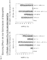
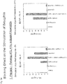
- the basophils were analyzed by flowcytometry for upregulation of CD203c by simultaneous staining with anti-human CD123 PE-Cy5 (1:10), anti-HLA-DR II APC (1:50) and anti-human CD 203c PE (1:10) antibodies (all BD Bioscience). Basophils were identified by high expression of CD123 and absence of HLA-DR. Results are shown in Figs. 22 to 24 ..
- peptides collectively representing the complete h-IL3 sequence were synthesized with an overlap of 6 amino acids on each side of the peptides ( Fig. 28 ).
- ELISA plates were coated with human IL-3 (1 ⁇ g/ml) and peptides 1 to 6 (1 ⁇ g/ml) in PBS, respectively, and as a negative control PBS was used. Each well contained a 100 ⁇ l sample and the plates were kept overnight in the refrigerator.
- a common ELISA plate was used to test binding to IL3 as well as the peptides 1 to 6.
- the prepared plates were washed three times and a blocking performed for 2 hours at room temperature with 2% BSA in PBS. After a further three washing steps, antibody clones 8.36.38, 11.14.6, 13.4.4, R&D anti-IL-3 antibody clone 4806 (R&D Systems, Inc., catalogue No. MAB203) and the negative control were added to the wells at 100 ⁇ l/well and incubated for 1 hour at room temperature.
- dilutions were used containing 40 ⁇ g/ml, 4 ⁇ g/ml and 0.4 ⁇ g/ml.
- the plates were washed three times and a secondary HRP labeled rabbit anti-mouse IgG antibody (DAKO-Cytomation, P260) added (1:2000 in 2% BSA in PBS, 100 ⁇ l/well) and incubated for 1 hour at room temperature and in the dark. After further three washing steps, ABTS reagent was added and the plates incubated for 30 minutes whereupon spectrometry was performed at 405 and 490 nm. Results are shown in Figs.
- clone 13 only binds to the complete IL-3 protein
- the R&D antibody binds to IL-3 and weakly to peptide 1
- clone 11 shows strong binding to IL-3 and peptide 1
- clone 8 shows strong binding to IL-3 and peptide 6.
- clone 11 shows very specific and strong binding to an epitope within peptide 1.
- Anti-IL-3 antibody clones 8, 11, 13 and further antibody clones 44 (44.16.16, DSM ACC3166) and 47 (47.28.15, DSM ACC3167) were analysed for their use in the development of a highly sensitive and specific ELISA assay for the determination of IL-3, especially for diagnostic purposes in blood, plasma or serum, as well as other body fluids.
- ELISA plates were incubated with 5 ⁇ g/ml of anti-IL-3 antibody overnight at room temperature to coat the plates. After three washing steps, blocking is performed using 1% BSA in PBS at 100 ⁇ l/well for 1 hour at room temperature. After further three washing steps, samples are incubated with 60 ⁇ l/well of IL-3 in various concentrations in PBS, plasma and serum.
- detection of solid-phase bound IL-3 is performed by adding 60 ⁇ l/well of a different and HRP labelled anti-IL-3 antibody at a concentration of 400 ng/ml and incubation for 2 hours at room temperature, followed by three washing steps and addition of TMB buffer (10 ml TMB buffer, 1 tablet of TMB, 3 ⁇ l H 2 O 2 ) (0,1 mg/ml, SIGMA-ALDRICH).
- TMB buffer 10 ml TMB buffer, 1 tablet of TMB, 3 ⁇ l H 2 O 2
- the reaction is stopped by adding 100 ⁇ l/well of H 2 SO 4 (12,5 % in H 2 O).
- the results are obtained by spectrometry at 450 nm and shown in Figs. 33 to 36 .
- Labelling of the anti-IL3 antibody clones was performed using the Lightning-Link HRP Conjugation Kit (Innova Biosciences) using the following protocol: For each of the purified antibody clones 8, 11, 13, 44 and 47 100 ⁇ l solutions with a concentration of 1 ⁇ g/ ⁇ l (in PBS) were produced. To each antibody solution, 10 ⁇ l of LL-modifying reagent were added and the obtained solution mixed carefully. For each antibody solution a Lightning Link mix bottle (100 ⁇ g reagent) was opened and the antibody solution including the LL-modifying agent added directly onto the reagent powder. Mixing was performed very cautiously by up- and down-pipetting of the solution.
- the lid was readjusted on the bottle of the Lightning Link mix and the bottles incubated for 3 hours at room temperature whereupon 10 ⁇ l LL-quencher reagent were added and incubated for a further 30 min at room temperature. After this treatment the antibodies were stored at -20°C for further use.
- analogue tests were performed using the Quantikine Human IL-3 ELISA test kit provided by R&D Systems, Inc., Catalogue No. Dy 203.
- An inventive test kit with clones 13/11 retained the same sensitivity as when using PBS or PBS/BSA samples also for plasma and serum (comparison shown in Fig. 36 ). Tests performed with different plasma samples (Plasma EDTA, Plasma Citrat) and serum could also be shown to be stable for at least 24 hours at room temperature ( Fig. 37 ) and there was also no detectable signal loss after freezing and thawing of the samples ( Figs. 38 to 40 ).
- the obtained data clearly demonstrate that IL-3 but not IL-6 or TNF- ⁇ can separate between RA and non-RA types of arthritis.
- Fig. 45 > 60% of the patients not responding to DMARDs/biologicals express high IL-3 levels. Among those patients that did not respond to current therapies patients with high IL-3 levels were more frequent. These patients would qualify for treatment with anti-IL-3 antibodies since the data indicate that patients with high IL-3 levels obviously do not respond to other kinds of therapies like, e.g., DMARDs or biologicals.
Landscapes
- Health & Medical Sciences (AREA)
- Life Sciences & Earth Sciences (AREA)
- Chemical & Material Sciences (AREA)
- Immunology (AREA)
- Molecular Biology (AREA)
- Organic Chemistry (AREA)
- Engineering & Computer Science (AREA)
- Biochemistry (AREA)
- General Health & Medical Sciences (AREA)
- Medicinal Chemistry (AREA)
- Proteomics, Peptides & Aminoacids (AREA)
- Cell Biology (AREA)
- Urology & Nephrology (AREA)
- Biomedical Technology (AREA)
- Hematology (AREA)
- Biophysics (AREA)
- Genetics & Genomics (AREA)
- Biotechnology (AREA)
- Microbiology (AREA)
- Food Science & Technology (AREA)
- Physics & Mathematics (AREA)
- Analytical Chemistry (AREA)
- General Physics & Mathematics (AREA)
- Pathology (AREA)
- Peptides Or Proteins (AREA)
- Medicines Containing Antibodies Or Antigens For Use As Internal Diagnostic Agents (AREA)
Description
- The present invention relates to novel anti-interleukin 3 (IL-3) antibodies as defined in the claims, nucleic acid sequences encoding such antibodies and hybridoma cell lines producing the antibodies according to the present invention. The invention further relates to pharmaceutical compositions containing the novel IL-3 antibodies, which are useful for the prevention or treatment of diseases or malfunctions which are associated with elevated levels of IL-3 in a human patient as well as to diagnostic kits and methods for reliably determining the IL-3 level in the blood, plasma, serum or other body fluid (e.g. urine, synovial fluid) of a patient.
- Interleukins belong to the large family of proteins called cytokines. Cytokines are polypeptides that influence the function of certain cells upon binding to specific cellular receptors and are divided in subclasses, i.e., interleukins, interferons, colony-stimulating factors (CSFs), lymphokines, growth factors and monokines. It is well known that cytokines play a major role in cell proliferation and, e.g., also inflammatory diseases.
- Cell proliferation is a complex process wherein growth factors bind to specific receptors on the cell surface, whereupon endocytosis occurs and the complexes of cytokine and receptor are internalized causing a cellular response. Such cellular responses include specific gene transcription activities as DNA synthesis and cell replication. When tested in relatively high concentrations, most of the cytokines have several differing biological effects. Because of these effects of cytokines, there is a high interest in investigations for possible therapeutic uses of these proteins.
- Interleukins are mediators of the immune system which are produced in low concentration mostly in leukocytes. They influence the growth, differentiation and activity of cells of the immune system and thus belong to the immune modulators. They also take effect by binding to receptors on the surface of target cells and thus change the transcription rate of certain genes. They play an important role in the triggering of a multiplicity of cellular responses.
- Interleukins are, e.g., involved in the immunological cell activation cascade and subsequent inflammatory changes. Irregular and/or abnormal inflammation is a major component and factor of a wide range of human diseases, one of which is the immunological disorder rheumatoid arthritis (RA). But also other immunological diseases are influenced by interleukins.
- IL-3, also designated as Multi-CSF, is a well-known member of the interleukin family. It has a growth stimulating and differentiating effect on various hematopoietic precursor cells and acts as a growth factor for mast cells. Together with IL-5 and GM-CSF, IL-3 belongs to the family of hematopoietic cytokines with four short alpha-helical bundles. GM-CSF and IL-3 stimulate the formation of neutrophilic and eosinophilic granulocyte colonies as well as macrophages. It further stimulates the formation of mast, megakaryocyte and pure and mixed erythroid colonies (D. Metcalf, "The hematopoietic colony-stimulating factors", 1984, Elsevier, Amsterdam).
- IL-3 consists of 133 amino acids and is known for its stimulation of colony formation by human hematopoietic progenitor cells and the stimulation of DNA synthesis by human acute myelogenous leukemia (AML) blasts. IL-3 binds to a unique receptor also known as CD123 antigen. The receptor belongs to the type I cytokine receptor family and is a heterodimer with a unique α-chain paired with a common β-subunit (βC or CDW 131). IL-3 binds to the unique α-receptor subunit. Signal transduction is mediated, however, by the common β-receptor subunit (βC) by the JAK2-STAT5 pathway.
- IL-3 is mainly produced by activated CD4+ T-cells and contributes especially to growth, differentiation and survival of CD34+ hematopoietic progenitor cells. In vitro, IL-3 has been observed to promote the differentiation of basophiles and mast cells from bone marrow cells. It has further been observed to induce IL-6 release by murine basophils and to up-regulate MHC-II expression and IL-1 secretion in monocyte/macrophages. Further, IL-3 supports the differentiation of monocytes into dendritic cells and osteoclasts.
- Since the first detection of IL-3 in a human genomic library, it has been a focus of investigations to determine its role in healthy humans as well as its possible role in the occurrence of diseases. The ability of cytokines to initiate or regulate hematopoieses is of interest, especially as far as malfunctions or diseases of the immune system are concerned. Such disorders seem to be connected to disturbances of the hematopoietic system and it was assumed that such diseases could be treated by providing viable progenitor cells to the hematopoietic system. Triggering such progenitor cells to differentiate was considered as a means to treat the respective diseases.
- Until several years ago, little was known about the role of IL-3 in auto-immune diseases and especially rheumatoid arthritis (RA). RA is the most prevalent inflammatory disease of the joints. The initial disease stages often develop gradually but can also manifest themselves with an instantaneous outburst. While pain occurs predominantly in joints of the fingers or toes, also other joints can be affected. The affected joints show swelling and usually are hyperthermic. Mostly, the disease proceeds in episodes, an episode usually lasting between several weeks to months. In between episodes, generally, there is an improvement of symptoms.
- The etiology of RA is not yet known. An autoimmune cause is strongly suspected with viral and bacterial causes being also discussed. A genetic influence has been reported by several authors (Hemminki K. et al., Arthritis Rheum. 2009; 60(3): 661-8, Padyukov L. et al., Arthritis Rheum. 2004; 50(10) 3085-92). It is assumed that misdirected immune cells invade the affected joints and cause the production of pro-inflammatory cytokines. According to one theory, the balance between cytokines is disturbed in RA. It has been reported that IL-1, IL-6 and TNFα are present in excess in RA and are assumed to be responsible for the deleterious inflammatory processes in cartilage tissue and for the activation of osteoclasts.
- The treatment of rheumatoid arthritis is still considered difficult and burdensome to the patients since medications with a high risk of adverse side effects have to be used. One way of treating the disease is to perform a symptomatic treatment, mostly using non-steroidal anti-inflammatory drugs (NSAIDs). These drugs act as anti-inflammatory and analgetic agents and often only achieve an alleviation of pain. The drugs further interfere with a certain step in the inflammatory cascade, where prostaglandine is generated by cyclooxygenases. NSAIDs, however, do not influence the underlying inflammatory process and are thus not able to retard the joint destruction, which is the most deleterious effect of RA.
- To prevent joint destruction and disease activity, a further current approach for treating RA is the use of disease-modifying anti-rheumatic drugs (DMARDs). These pharmaceuticals actually modify the disease process. Examples of DMARDs are methotrexate, the most commonly used anti-rheumatic, the effect of which is based on a reversible inhibition of the enzyme dihydrofolate reductase. Another commonly used substance for treating RA is leflunomide, which provides an effect by intervening with the pyrimidine metabolism. Both pharmaceuticals are long-acting and thus have to be administered over a longer period of time (usually 12 - 16 weeks) to show the desired effects. To bridge the time until DMARDs improve the disease, most patients are administered steroids.
- A further approach for treating RA are "biologicals" that block cytokines like TNF, IL-6, IL-1 or costimulatory molecules like B7 or that deplete leukocyte subsets (e.g. B cells). Biologicals (e.g. the TNF antibody Infliximab) are mostly used for severe disease processes and after DMARDs have failed to sufficiently control disease activity. Biologicals influence a plurality of signal systems in the immune system and have a variety of serious side effects including bacterial and viral infections and a higher risk for development of neoplasia.
- All known treatments have severe disadvantages and side effects and, therefore, it was an object to develop new drugs for the treatment of RA which are effective, are more selectively expressed than other cytokines in patients with autoimmune disease especially RA and have less side effects than the currently used treatment regimes.
- More recently, an involvement of IL-3 in autoimmune diseases and especially in RA has been described.
WO 2010/063488 describes that IL-3 inhibitors can be used in treatment of early stages of rheumatoid arthritis. Although the patent application mentions that no IL-3 mRNA was detected in the synovium of patients with RA and no effect of IL-3 was observed on cultured fibroblasts, a genetic analysis found an association between a single nucleotide polymorphism in the IL-3 promoter gene and RA. Based on this finding and also further studies which show the presence of considerably elevated levels of IL-3 in RA patients,WO 2010/063488 proposes such use of inhibitors, mainly antibodies or antibody fragments, antibody variants or antibody multimers in prophylactic RA treatment, therapeutic treatment in early stages of the disease or in maintenance treatment. - However, there is still a need for effective antibodies with high specificity towards IL-3 which also show a high affinity and avidity. Such antibodies are desirable for an envisioned therapeutic use but also for a meaningful and reliable diagnosis. Test results that have been produced within the research framework that led to the present invention indicate that not in all RA patients symptoms and systemic inflammation correlate with an elevated IL-3 level. Obviously there are different groups of RA patients as far as involvement of IL-3 in the propagation of the disease is concerned. Accordingly, patients that do not show elevated levels of IL-3 in acute phases of the disease, most probably would not benefit from a treatment with anti-IL-3 antibodies. Unprofitable expenses to the health system can be avoided by a reliable diagnostic test with regard to the presence of IL-3 in the blood or serum of patients. Currently available commercial diagnostic test kits for IL-3 determination have not proven reliable when conducted directly on blood, plasma or serum. Therefore, it was a further object of the present invention to provide antibodies that do not substantially cross-react with other human interleukins or cytokines like IL-5 or GM-CSF and thus are able to reliably detect elevated IL-3 levels in a diagnostic assay.
- Such anti-IL-3 antibodies preferably should also exhibit none or very low cross reactivity with IL-3 from other species. Within this object of the present invention, it is desirable to provide antibodies which are able to inhibit the activity of IL-3 efficiently and specifically, thus making them useful agents for treating the disease in patients having been diagnosed for elevated levels of IL-3.
- These objects of the present invention are solved by novel anti-IL-3 antibodies according to the present invention as specified in the appended claims and the following description.
- Apart from a very high specificity for only IL-3 but not other cytokines, the antibodies of the present invention show also surprisingly low levels of cross-reactivity with IL-3 molecules of other mammalian origin. Amino acid identities of a human protein and the mouse protein is 29% which seems quite low. However for marmoset, rhesus or a chimpanzee proteins there are amino acid identities between 72 and 99% (
figure 1 ). - As regards the further cytokines, which may also be present at an elevated level in autoimmune diseases, IL-5 and GM-CSF are particularly important. A high cross-reactivity of an anti-IL-3 antibody with such cytokines in an immunoassay can lead to incorrect results regarding the fact whether an IL-3 overexpression has an important influence in the manifestation and progression of the autoimmune disease. Such results, however, have an important impact on the decision whether the application of an anti-IL-3 antibody can be considered a promising therapeutic approach.
- It is thus preferred for inventive antibodies to show the lowest possible cross-reactivity with human IL-5 and GM-CSF. Particularly preferred, it is a characterizing feature of an inventive antibody that it binds to IL-5 or GM-CSF to an extent of below 5%, more preferred below 2% and particularly preferred below 1% as compared to the amount of IL-3 bound by the antibody.
- The examples enclosed with this specification show the superior characteristics with regard to specificity and lack of cross-reactivity with IL-3 of other species and with other human cytokines for antibodies according to the present invention.
- By means of an enzyme-linked immunosorbent assay (ELISA), the specifity and cross-reactivity of the produced antibodies according to the invention can be easily determined. In this context, recombinantly produced cytokines (e.g. human IL-3, IL-5 and GM-CSF, or IL-3 from other species, respectively) are coated onto a suitable surface in the test arrangement, the produced anti-human-IL-3-antibodies are added and their binding is detected on the coated surface by means of a corresponding detection reagent, such as a labelled anti-lgG-antibody. In such a test regimen, antibodies according to the present invention bind nearly exclusively to IL-3 while binding to IL-5 and GM-CSF occurs only to a very minor extent, if at all. The same is true for antibodies according to the invention as far as IL-3 from other species is concerned. Especially the preferred antibodies binding specifically to the epitope SWVN do not cross-react with protein from other species to a detectable extent (see Example 4).
- For the selection of particularly suitable antibodies, the affinity of same is examined by further ELISA assaying. In this context, ELISA plates are coated with any anti-human-IL-3 antibody which can also be a commercially available antibody (e.g., a goat IgG anti-human-IL-3 antibody). In this respect, for instance, 1 ug/ml of the antibody is incubated with the ELISA plates over night in a refrigerator whereupon a washing step and a blocking with human IL-3 (250 ng/ml in PBS buffer) is performed, thus fixing human IL-3 on the solid phase. During such a test, candidate antibodies are then added in different concentrations and detected by means of a secondary HRP- (horseradish peroxidase) labelled polyclonal antibody.
- The antibody according to the present invention was designated
clone 11.Clone 11 is a mouse anti-human-IL-3 antibody and shows a very high specificity and affinity to IL-3.Clone 11 specifically binds to the epitope with the amino acids SWVN according to SEQ ID NO: 2. This particularly preferred antibody was deposited at DSMZ (Braunschweig, Germany) under number DSM ACC3163. - In many experiments and also in the examples enclosed to the present specification,
clone 11 has proven very superior characteristics with regard to specificity, lack of cross-reactivity but also with regard to affinity and avidity. This preferred antibody therefore is considered especially suitable for use in diagnostics as well as therapeutic measures. - To make this remarkable antibody even more useful for therapeutic uses, a particularly preferred inventive anti-IL-3 antibody is a humanized version of
clone 11. For this purpose, clone 11 is humanized in any manner known in the art. In this context, at least one of the CDRs ofclone 11 is identically maintained/preserved, other parts of the mouse-antibody clone 11 can entirely or partially be replaced by sequences of human antibody origin. In this context, it is essential that the obtained humanized antibody still shows the same specificity which can easily be determined using known methods. Preferably, such ahumanized antibody clone 11 also shows just as little cross-reactivity with IL-3 of species other than the human and with other human cytokines as the depositedmouse clone 11. - A further subject-matter of the present invention is a nucleic acid which encodes an antibody, according to the present invention.
- The nucleic acid codes for the
antibody clone 11. A further, especially preferred nucleic acid codes for ahumanized clone 11, whereby it comprises at least those nucleotides coding for the regions inantibody clone 11, which entail the binding to the specific human IL-3-epitope according to the present invention. - The hybridoma cell line according to the present invention is cell line 11.14.6 (DSM ACC 3163) producing
antibody clone 11. - A further subject matter of the invention is a diagnostic method for determining the IL-3 level in body fluids of a patient, especially in blood, plasma or serum. The method comprises adding an anti-IL3 antibody, according to the present invention to a sample comprising a body fluid sample under conditions which allow for the binding of the antibody to IL-3 and detecting the amount of antibody-bound IL-3 in the sample.
- As already mentioned above, it has been noted that presently available antibodies and test kits for detecting and measuring IL-3 in a sample do not deliver accurate enough results to base a therapeutic approach thereon. Especially for whole blood, plasma and serum, results of commercially available test kits have proven to be unreliable because of high background levels and unspecific cross-reaction being indistinguishable from actual IL-3 binding. Also the binding affinity of known antibodies to IL-3 present in samples of patient blood, plasma or serum has proven insufficient for a correct analysis of a possible influence of IL-3 in an autoimmune disease, especially in RA.
- By using the antibodies according to the present invention in diagnostic methods and corresponding test kits, the accurate determination of the presence of IL-3 in body fluids can be improved to a major extent and such methods and test kits, therefore, are a further subject matter of the present invention. It has surprisingly been found that using the antibodies and methods of the present invention, IL-3 could be specifically detected even if present in only picogram / ml concentrations in a sample. The relative affinity of the antibodies of the present invention towards IL-3 is thus about at least a
factor 100 higher than the affinity of known antibodies. - In a preferred embodiment of the present invention, the diagnostic method is conducted as an ELISA assay. The general test regime used for ELISA assays is well known to the skilled person. At least two antibodies which bind to the target molecule are used. One of these antibodies is bound to a solid phase allowing for the separation of the antigen to be determined from the test sample. Upon removal of the test sample from the solid phase and washing steps as considered appropriate, a second antigen-specific and labeled antibody is added and after further removal of excess labeled antibody and optionally further washing steps the amount of label bound via the antibody-antigen-antibody complex is determined and correlated to the amount of antigen present.
- Although it is necessary that both antibodies used in such an ELISA assay are specific to the antigen to a high extent, an especially high degree of specificity is needed mainly in one of the two antibodies involved. E.g., as a first solid phase-bound antibody a very specific antibody can be used to be able to separate only the desired antigen from the liquid sample without any cross-reacting background. If only the desired antigen is coupled via the antibody to the solid surface, the specificity of the second antibody is not so decisive anymore since unspecific binding to other antigens is not an issue in such a case. Accordingly, it is possible to use a less specific antibody as the second antibody in such a context.
- Alternatively, it is possible to use a high affinity and high avidity antibody as the first solid phase-bound antibody and to use a highly specific antibody as the second and labeled antibody. In such an embodiment, unspecific binding of other antigens like IL-5 or GM-CSF might take place to some extent due to a certain cross-reactivity of the first antibody, however, the actual detection by the labeled antibody is then restricted to IL-3 by using a highly specific second antibody.
- In the present invention, an ELISA assay method and kit are used wherein as the highly specific antibody in the test an antibody according to the present invention is used. The highly specific antibody used in the ELISA assay is
antibody clone 11. - To avoid possible steric hindrance issues for the ELISA performance, it is preferable to use a second antibody that binds to an epitope of IL-3 at another part of the protein. It is well within the ambit of the present invention to use a known and commercially available antibody as the second antibody. However, according to the present invention preferred combinations of antibodies (first antibody / second antibody) are
clone 13 / 11 (most preferred),clone 11 / 13 (preferred). as well as combinations ofclone 11 withclone 47.Clone 13 andclone 47 are considered to bind to a 3-D epitope of IL-3 in its native conformation. The above mentioned clones were deposited at DSMZ in Braunschweig/Germany: clone 11 (11.14.6) = DSM ACC3163; Clone 13 (13.4.4) = DSM ACC3164; Clone 44 (44.16.16) = DSM ACC3166; clone 47 (47.28.15) = DSM ACC3167. - Although it might be useful for diagnostic purposes to be able to detect IL-3 in any body fluid or also cell preparation, the present diagnostic method is especially suited and useful for the detection of IL-3 in a plasma or serum sample. With the heretofore known methods and antibodies, detection of IL-3 in plasma or serum did not lead to sufficiently meaningful results especially due to cross-reactivities of known antibodies and insufficient affinities of such antibodies with regard to the low levels of IL-3 in present blood, plasma or serum in healthy and in RA patients. Therefore, often synovial fluids were used as test sample. However, obtaining such a test sample is much more difficult and cumbersome to the patient. Use of the antibodies according to the present invention surprisingly opens up the possibility to reliably detect and draw conclusions from the presence of IL-3 in blood, plasma and serum,. Thus, the present invention via its antibodies and their use in diagnosis is a big step forward to relating IL-3 presence to the severity of RA and RA incidents in patients and thus to also determine the supposition of a patient to treatment with IL-3 antibodies.
- Test kits which contain antibodies according to the present invention together with other substances for conducting the diagnostic methods of this invention are also a further embodiment of the invention. For determining the presence and amount of IL-3 in body fluids of a patient, such kits can be provided for easy and effective use in laboratories of every size. Test reagents for the various applicable test formats are well known in the art. The inventive contribution to the test kits lies in the presence of antibodies according to the present invention or combinations of antibodies according to the present invention with other IL-3 specific antibodies, most preferably a combination of
antibody clones - As discussed above, especially
WO2010/063488 is directed to use of IL-3 inhibitors for the treatment of early stage RA patients. However, also in this context it was not yet clear which kind of inhibitors and which kind of IL-3 antibodies would be especially well suited for a therapeutic application. It seems vital that antibodies used in therapy should not entail drawbacks brought on e.g. by cross-reactivity with other unrelated cytokines or severe immunological responses experienced with known antibodies. - A further subject and embodiment of the present invention therefore is the use of the novel and advantageous antibodies of the invention in a pharmaceutical composition. Such pharmaceutical compositions according to the invention are characterized by the presence of a pharmaceutically effective amount of an antibody as described herein as an active ingredient. Usual adjuvants and/or carrier substances for pharmaceutical preparations can be included as desired and deemed appropriate.
- Pharmaceutical compositions according to the present invention can be used for the treatment or for the prevention and prophylaxis of diseases or malfunctions which are associated with elevated levels or IL-3. As discussed in the introductory part of this specification, IL-3 has a significant growth stimulating and differentiating effect on various hematopoietic precursor cells and is also a growth factor for mast cells. The signal transduction caused by IL-3 has major impact on the immune system. Any disease or medical condition in which IL-3 plays a direct or indirect role in development or progression is a candidate for the treatment by administering the antibodies according to the present invention. Preferably such disease or malfunction connected with elevated levels of IL-3 is related to the immune system, mostly an autoimmune disease and especially RA or acute or chronic graft-versus-host disease and multiple sclerosis.
- Further circumstances which are preferably treated by administering the pharmaceutical composition according to the invention is a use in suppressing the activity of human basophils in persons suffering from an allergic reaction and for the stratification of patients having increased IL-3 levels in serum or plasma.
- To be eligible for treatment with the pharmaceutical composition of the present invention, in the respective disease or malfunction elevated levels of IL-3 as compared to healthy persons have to be present in any body fluid including synovial fluids but also blood, plasma and serum. The antibodies of the present invention which are contained in the pharmaceutical composition bind specifically to IL-3 and thereby inhibit the activity of IL-3.
- Especially in view of RA it has been found that for a large group of patients elevated levels of IL-3 are correlated with the aggravation caused in the patient and the progression of the disease. IL-3 is detected mainly in active RA, whereas patients with a non-active stage of RA usually do not show elevated levels of IL-3. Thus the pharmaceutical compositions of the present invention are especially useful in treating patients with active episodes of autoimmune diseases, as RA, and for the prophylactic treatment to avoid the occurrence of active episodes of the disease.
- Since available therapies are only effective in about 50% of treated patients, providing the pharmaceutical compositions according to the invention is a major step to a new and gentle treatment of auto-immune disease in patients. Based on the lack of an overt phenotype of IL-3 deficient mice (Nature. 1998; 392(6671):90-3) and no obvious side effects of mice treated with antibodies against IL-3 (Arthritis Rheum. 2009 ;60(5):1352-61) IL-3 targeted treatment should exhibit less severe side effects than currently used pharmaceuticals, especially regarding to infection or neoplasia. In certain cases, it could be desirable to combine treatment with the antibodies and pharmaceutical compositions of the present invention with other medicines like methotrexate or leflunomide. An individualized treatment strategy according to IL-3 levels in plasma, serum or other body fluids, presents an advantage compared to available biologicals, since currently it is not possible to predict reliably, which patient will respond to a specific therapy (including biologicals). Further an individualized approach improves the safety of treatment by reducing the risk of side effects of an ineffective therapy and reduces the costs for treatment of RA.
- Also the treatment with the antibodies of the present invention would preferentially be started as soon as elevated IL-3 levels in blood, plasma or serum are detected. Thus, early-on treatment can be applied in patients where RA activity is correlated with elevated IL-3 levels and long term joint damage can be avoided or kept to a minimum. In addition, treatment with anti-IL-3 antibodies would preferentially be started, if patients failed to a previous treatment with DMARDs or biologicals. Further it is expected that the use of the antibodies of the invention can reduce cell infiltration of synovial tissue which can be a further negative factor in the disease pathology.
- An effective dose of IL-3 antibody contained in the pharmaceutical composition of the present invention can easily be determined by a physician as generally known in the art. An effective dose is an amount that alleviates the symptoms of the disease or prevents any further progress of the disease or deterioration of the condition of the patient. Progress of disease and condition of the patient can be monitored by determining the IL-3 level and other markers for inflammation or autoimmunity in body fluids, preferably blood, plasma or serum, and by determining other disease scores or using known diagnostic methods. For RA, progress of the disease and the status of the patient can be determined using e.g. the DAS28 activity score. To prevent joint damage a DAS28 <= 2,6 should be achieved. Effectiveness of a therapy can be measured as the percentage of patients that achieve a certain (e.g. 30% , 50% or 70%) reduction in disease activity. Any decrease of IL-3 or other markers for inflammation in body fluids is also an indication of a successful treatment. Effective doses of the pharmaceutical composition can be determined using dose-response-curves as is well-known to the skilled artisan. The amount of IL-3 present in the body fluids of a patient as determined using the diagnostic methods according to the present invention can also be a basis for determining the effective dose of antibody in the pharmaceutical composition for each patient and the severity of the disease and malfunction.
- For the pharmaceutical composition according to the invention and the amount of IL-3 antibody contained therein, dosage further depends on the activity, avidity and the half-life of the antibody. For antibodies having a half-life of about one to two weeks, the dosage is preferably in ta range of 1 to 1000 mg and more preferably 10 to 100 mg per application. The pharmaceutical composition is preferably applied once a day to once a month, again depending on the half-life of the antibody.
- Also with regard to a pharmaceutical composition and its use for the treatment of diseases which are correlated with increased IL-3 levels in body fluids, the antibody of the invention is
antibody clone 11.Antibody clone 11 does not show detectable cross-reaction with GM-CSF and IL-5 and therefore does not influence the activity of these cytokines in the patient (see example 4b). Further it has been shown thatclone 11 is very effective in inhibiting the IL-3 dependent growth of human TF-1 cells (see example 5a and 5b), proving an efficient inhibition of the IL-3 activity in patients by this antibody. The growth of TF-1 cells is a well known test system for IL-3 activity and the inhibition thereof. Further, the blocking of the stimulation of basophils is in-vitro evidence for an inhibition of IL-3 activity. Again, clone 11 shows excellent results in view of inhibition of IL-3 activity (see example 5c and 5d).Clone 11 also shows a very high affinity and avidity and already very small amounts of this antibody are sufficient for its inhibitory activity (example 5). -
Clone 11 is an excellent inhibitor of IL-3 and therefore a promising pharmaceutical with regard to treatment of IL-3 related malfunctions and diseases. - The following examples and figures further illustrate and describe the present invention but are not intended to limit the scope thereof.
-
Fig. 1 shows the amino acid sequence homology of IL-3 of various species; -
Fig. 2 shows in a Western blot the ability of monoclonal antibodies to bind to IL-3. -
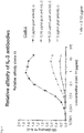
Figs. 3 and4 show the relative affinity ofantibodies clone 8 and clone 11 for IL-3 as determined by using varying amounts of the antibodies in ELISA assays for which different amounts of IL-3 were bound via goat anti-human-IL-3 antibodies to a solid surface via increasing amounts of coated goat anti-hIL-3 antibody. -
Fig. 5 shows the relative IL-3 affinity of antibodies determined using varying concentrations of antibodies at a constant amount of IL-3 which was bound to the solid phase in an ELISA assay. -
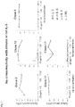
Fig. 6 shows the results of tests performed to detect a possible cross-reactivity of anti-IL-3 antibodies with other human cytokines. In the tests, binding of the antibodies to IL-3, GM-CSF and IL-5 was compared. -
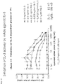
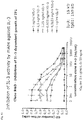
Fig. 7 and8 show the results of tests performed to detect possible cross-reactivity of the various anti-IL-3 antibodies with IL-3 from other species. In the test depicted inFig. 8 , also a commercially available anti-IL-3 antibody was included. -
Fig. 9 shows the IL-3 dependent growth of TF1 cells wherein viable cells do not propagate in the absence of IL-3 and cell growth can be shown to be dependent on the amount of IL-3 in the growth medium. -
Figs. 10 to 13 show the ability of various monoclonal antibodies - including a commercially available anti-IL-3 antibody - to inhibit the IL-3 dependent growth of TF1 cells. For differing concentrations of hIL-3 present in the cell growth medium, the effect of the antibodies was tested showing distinct inhibition of cell growth forantibodies clone 11,clone 13 and the commercially available R&D anti-IL- 3 antibody, whereasclone 8 had only a minor effect. -
Fig. 14 shows the GM-CSF dependent growth of TF1 cells wherein viable cells do not propagate in the absence of GM-CSF and IL-3. Either one of these cytokines is necessary for growth and propagation of TF1 cells. -
Fig. 15 shows the effect of various antibodies (clone 8,clone 11,clone 13 and R&D) and different concentrations thereof in the medium on the growth of TF1 cells in the presence of IL-3, GM-CSF or a combination thereof, or in the absence of these cytokines. None of the antibodies showed a marked inhibitory effect on the growth of TF1 cells in the presence of GM-CSF whereas, again, clone 11 and at a higher concentration also clone 13 and the R&D antibody clearly inhibited the IL-3 effect on the growth of TF1 cells. -
Fig. 16 shows the binding of biotinylated IL-3 to peripheral mononuclear blood cells as well as to monocytes and lymphocytes as compared to a negative control. Basophils were identified by high expression of CD123 and absence of HLA-DR. Plasmacytoid dendritic cells (pDC) were identified by high expression of CD123 and HLA-DR. Monocytes and B cells were identified by light scatter properties and expression of HLA-DR. -
Fig. 17 shows the inhibiting effect ofantibodies clone 8,clone 11,clone 13 as compared to a negative control and isotype antibody as a positive control with regard to binding of IL-3 to IL-3 receptors on Basophils, pDC, monocytes and B cells . It can be seen thatclones clone 8 had nearly no effect at all. -
Fig. 18 to 21 show the inhibitory effect of different amounts of antibody (clones Fig. 18 ), plasmocytoid dendritic cells pDC (Fig. 19 ), monocytes (Fig. 20 ) and B-cells (Fig. 21 ). Clear and strong inhibition of the binding of IL-3 to all cell types was effected byclones clone 8 had a much weaker effect, if any. -
Fig. 22 shows the results of a FACS analysis for a detection of the stimulation marker CD203c after stimulation of basophils at different time intervals. -
Fig. 23 shows the results of an ELISA assay for detecting IL-13,Fig. 24 shows the results of an ELISA assay for detecting hIL-4 both of which are present in response to a stimulation of basophils over different time intervals. -
Fig. 25 shows the results of a FACS analysis for detection of the stimulation marker CD203c upon addition of IL-3 and IL-3 preincubated withantibody clones clones 11 and for the R&D antibody, only a much smaller inhibiting effect was observed forclone 13. -
Fig. 26 shows the results of an ELISA assay for detecting hIL-4 after stimulation of basophils with IL-3 and IL-3 preincubated with antibodies. In the test, again,antibody clone 11 and the R&D antibody had a remarkable inhibitory effect on the IL-4 release in basophils, whereasclone 13 did not show any effect. -
Fig. 27 shows the results of an ELISA assay for detecting hIL-13 after stimulation of basophils with IL-3 and IL-3 preincubated with antibodies. In this test, all threeantibodies clone 11,clone 13 and R&D showed strong inhibition of IL-13 formation in the cells. -
Fig. 28 shows six peptides which were used for an epitope mapping with regard to highly specific and affine hIL-3 antibodies. To this end, overlapping peptides were created that together cover the complete human IL-3 sequence. The peptides were coated onto solid surfaces and used in ELISA assays to detect specifically binding monoclonal antibodies. -
Figs. 29 and30 show the results of such ELISA assays indicating that while allantibodies clone 8,clone 11,clone 13 and R&D strongly bound hIL-3,clones 13 and R&D did not have a strong specificity for one of thepeptides 1 to 6, whereasclone 11 andclone 8 bound specifically topeptides -
Fig. 31 shows a set of mutated forms ofpeptide 1 which were used for a further fine mapping of species-specific epitopes in this part of the IL-3 protein sequence. The peptides were used in ELISA assays for testingantibody clone 11 and the R&D anti-IL-3 antibody with regard to their specificity. The results of the assays are shown inFig. 32 . Among this set of peptides, the R&D antibody does not show a strong specificity for any one of them but binds strongly only to the complete IL-3.Clone 11 on the other hand, shows a strong binding to peptide 1B only, but does not bind to other peptides which are related to rhesus or marmoset IL-3 proteins. -
Fig. 33 to 36 show the results of ELISA assays which were performed using differing combinations of coating and detection antibodies selected from theantibody clones clones -
Fig. 37 to 40 show the results of ELISA assays performed to investigate the stability of tests performed inter alia with plasma and serum and using as the coating (solid-phase bound)antibody clone 13 and as detection antibody HRP-labelledclone 11. Plasma and serum samples containing IL-3 were stored for the given periods of time at various temperatures. The test results confirmed very good stability and performance for the test formats. -
Figs. 41 and42 show clinical data regarding the plasma IL-3 levels of patients with or without active RA, as well as data showing an analysis of plasma IL-3 levels of randomly picked patients presenting at the University Hospital Regensburg. The results indicate that in general only a small percentage of randomly picked patients show IL-3 levels above 20 pg/ml, while more than 50% of patients with active RA have IL-3 levels above 20 pg/ml. Among confirmed active RA patients, there are two subgroups only one of which shows high plasma levels of IL-3. -
Figs. 43 and44 _show clinical data regarding the plasma IL-3, IL-6 and TNF-α levels of patients with or without active RA (Fig. 43 ), as well as data showing an analysis of cytokine levels in patients with diagnosed RA (Fig. 44 ). The results indicate that IL-3 but not IL-6 or TNF- α can separate between RA and non-RA types of arthritis (Fig. 43 ). Furthermore, what can be concluded from the data presented inFig. 43 is that IL-3 and IL-6 but not TNF-α correlate with disease activity in patients with RA. -
Fig. 45 shows clinical data regarding the plasma IL-3, IL-6 and TNF-α levels of RA-patients treated with DMARD and/or biologicals. The data indicate that 63% of RA patients not responding to DMARDs/biologicals express high IL-3 levels. Patients with high IL-3 levels are more frequent among those patients that did not respond to current therapies. These patients would qualify for treatment with anti-IL-3-antibodies. - Anti-IL-3 antibodies were produced by immunizing Balb/c mice using at least 6 i.p. injections of human eukaryotic glycosylated IL-3 in alumn at four week intervals. Two days before cell fusion, IL-3 in PBS was injected intraperitoneally. Antibody-producing splenocytes obtained from the immunized mice (HGPRT positive, able to grow on HAT medium) were fused with the myeloma cell line X63Ag8.6.5.3 in the presence of polyethylene glycol (PEG) and a selection of hybridomas performed in an HAT-selection medium. Hybridomas were cultivated in RPMI-1640 medium supplemented by 10% FCS (neat inactivated, HIA), P/S and glutamine (1:100). Obtained cells are able to grow in suspension and are splitted every three days in a ratio of 1:4.
- For storage purposes hybridoma cells are transferred from a cell culture bottle into 50 ml or 15 ml cell culture flasks (BD Falcon™). After centrifugation at 1400 rpm for 5 minutes at room temperature, the supernatant is completely removed. Cells are resuspended in a freezing medium (90% FCS (HIA) + 10% DMSO) and 1.5 ml aliquots are filled into vials. The cells are prefrozen in a freezing container in a freezer at -80°C and after 1-2 days transferred to a liquid nitrogen storage tank.
- Cloning and recloning of the obtained hybridoma cell lines are performed using limited dilution to provide long-term stable sources for monoclonal antibodies.
- Obtained antibodies are shown in table 1.
- For determining the isotypes of the antibodies, ELISA assays were performed using hIL-3 coated plates to which the antibodies were added. Bound antibodies were detected using isotype specific secondary antibodies. For further analyses, only antibodies of isotype IgG were used.
Table 1 - Overwiew of mAbs against human IL-3 Original clone First cloning Second cloning Isotype Clone 2 2.28 2.28.11 IgM, kappa Clone 3 3.47 3.47.20 IgG1, kappa Clone 5 5.3 5.3.2 IgM, kappa Clone 6 6.38 6.38.14 IgG1, kappa Clone 7 7.42 7.42.45 IgM, kappa Clone 8 8.36 8.36.38 IgG1, kappa Clone 10 10.12 10.12.4 IgG1, kappa Clone 11 11.14 11.14.6 IgG1, kappa Clone 13 13.47 13.4.4 IgG1, kappa Clone 36 36.26 36.26.10 IgG1, kappa Clone 38 38.18 38.18.5 IgG1, lambda Clone 41 41.28 41.28.4 IgG1, kappa Clone 42 42.47 42.47.36 IgG1, kappa Clone 43 43.14 43.14.28 IgG1, kappa Clone 44 44.16 44.16.16 IgG1, kappa Clone 45 45.14 45.14.27 IgG1, kappa Clone 46 46.21 46.21.1 IgG1, kappa Clone 47 47.28 47.28.15 IgG1, kappa - Several of the obtained antibodies of the type IgG1 were isolated from hybridoma clones and their concentration determined. The determination of the concentration was performed according to following method: 96-well-plates are coated overnight at room temperature with anti-mouse IgG (1:100 in PBS) in a concentration of 100 µl/well. Blocking is performed by adding 100 µl per well of 2 % BSA in PBS and incubation at room temperature for two hours. After the blocking reaction, the plates are washed twice. Two samples and blanks, respectively, of supernatants of clones 3.47.20, 6.38.14, 8.36.38, 10.12.4, 11.14.6 are incubated undiluted, as well as with dilutions of 1:3, 1:9, 1:27, 1:81, 1:243, 1:729 and 1:2187 (100 µl per well, dilution in 2 % BSA in PBS) at room temperature. Mouse IgG1 in a starting concentration of 1 mg/ml is used as standard, whereas a concentration of 20 ng/ml is applied in dilutions of 1:2, 1:4, 1:8, 1:16, 1:32, 1:64 and 1:128.
- The plate is washed three times and then incubated with biotinylated anti-mouse IgG1 (diluted by 1:250 in 2 % BSA in PBS) for one hour at room temperature with 100 µl per well. After washing the plate a further three times, streptavidin-HRP (1:1000 in 2 % BSA in PBS) is added for one hour at room temperature and in the dark. The concentration of the antibodies is determined after adding ABTS and incubating for further 30 minutes and measuring the signal at 405 and 490 nanometers on a spectrophotrometer. Based on this determination, a desired amount of the antibodies tested is applied for the further tests.
- For preparing the gel and performing the western-blot analysis, standard methods are used. A 12% PAA resolving gel is poured, overlayed with about 1-2 ml of water and polymerisation conducted for 30 to 45 min until a recognizable "line" is formed. The water is removed, a stacking gel poured onto the resolving gel and a Teflon comb is inserted. Polymerisation is performed for 30 min, then the comb is carefully removed.
- Samples of IL-3 are prepared by mixing of recombinant human IL-3 1:1 with Laemmli buffer and heating the samples at 60°C for 5 min. An amount of 1µg per lane of IL-3 as well as a usual standard for determining molecule sizes is loaded onto the gel. The gel is then mounted in a SDS-PAGE gel electrophoresis apparatus which already contains a running buffer. The inserted gel is then cautiously overlayed with additional running buffer and electrophoresis performed at 20 to 25 mA with voltage adjusted to infinite for approximately 1,5 hours. When the run is completed, the gel is retrieved from the apparatus and the stacking gel is removed.
- Six layers of Whatman paper that has been presoaked in transfer buffer, and a PVDF membrane are cut to fit the size of the gel. The transfer stack is adjusted in the usual way and transfer effected by semi-dry blotting for 40 min at 20-25 mA and voltage adjusted to infinite. The membrane is then incubated overnight at 4°C on a shaking apparatus with a blocking solution (5 % powdered skim milk in PBS) and the membrane washed three times for 5 min each with PBS at room temperature.
- Antibody clones are incubated at a concentration of 5 µg/ml in blocking solution for 2 hours at room temperature under agitation on the shaking apparatus. After three washing steps, HRP labelled anti-mouse immunoglobulin (1:1000 in blocking solution) is added and incubation is conducted for 1 hour at room temperature while shaking. After three further wahing steps, a detection solution (1:1 mixture of solutions A and B of the Westernblotting Luminal Reagent obtained from NALGENE) is added and incubated for 1 min at room temperature. Films are then adjusted on the membranes with different times of expositions and developed in the dark room.
-
Fig. 2 shows the results of binding ofantibody clones clones clone 13. - The affinity of the obtained antibodies for IL-3 was measured in an ELISA assay. ELISA plates were coated overnight with different concentrations (2 µg/ml, 0.66µg/ml, 0.22 µg/ml, 0 µg/ml) of anti-human IL-3 antibody (RD, goat IgG anti-human IL-3 AF-203-NA). For each concentration, duplicates were used (2 x 12 wells). For this purpose, the first concentration (2µg/ml) is diluted in PBS, further dilutions are made in PBS containing 2 µg/ml control goat IgG to keep the total concentration of IgG constant. Blocking with 2% BSA is performed for 2 hours at room temperature, followed by 5 washing steps using PBS.
- The wells are then incubated with hIL-3 (0.25 µg/ml in PBS) for 2 hours at room temperature, for the control group no hIL- 3 is added. After five further washing steps with PBS, the wells are incubated overnight at 4°C with serial (1:3) dilutions of
antibodies clone - After five further washing steps, bound antibody is detected using goat-anti-mouse-HRP antibody (1:500 in PBS with 2% BSA) and incubation for 1 hour at room temperature. After five further washing steps, ABTS (ROCHE, 1 mg/ml) is added as substrate and the optical density measured in a spectrometer at 405 nm.
- Results are shown in
Figs. 3 and4 forantibodies clone Fig. 5 shows the results of further tests including other antibodies. The tests were performed in the same manner as described, however, coating of the solid phase was performed using 1 µg/ml goat IgG anti-human IL 3 (see above) and different concentrations/dilutions of antibodies as shown in the figure. - To determine the usefulness of the obtained monoclonal antibodies for diagnostic assays, it is important to be able to exclude cross-reactivities with closely related cytokines which are also present in blood, plasma, serum or other body fluids of patients. To this end, wells of ELISA plates were coated by adding 100 µl/well of human IL-3 (1 µg/ml), GM-CSF (1 µg/ml) or IL-5 (1 µg/ml) in PBS. As negative control PBS was used (100 µl/well). For each tested antibody, different dilutions were tested mandatorily on a common plate with hIL-3, hGM-CSF, hIL-5 and PBS.
- The cytokine coated plates were washed three times and blocking performed for 2 hours at room temperature using 2% BSA in PBS. After three further washing steps, antibodies clone 3.47.20, 8.36.38, 10.12.4, 11.14.6, 13.4.4 and just medium (RPMI1640 containing 10 % FCS) as control were added at a concentration of 40 µg/ml and 1:5 and 1:25 dilutions thereof in a volume of 100 µl/well and inicubated for 1 hour at room temperature. On each plate a negative control is used.
- After three washing steps, a secondary HRP-labelled rabbit anti-mouse IgG (DAKO-Cytomation P260 (1:2000 in 2% BSA in PBS, 100 µl/well) was added and the plates incubated at room temperature for 1 hour in the dark. After another three washing steps, ABTS (ROCHE, 1 mg/ml) was added and spectrometry performed at 405 and 490 nm after 30 min.
- The results are shown in
Fig. 6 indicating some weak cross-reactivity forclones clones - As a further property of the monoclonal antibodies, their cross reactivity with IL-3 from other species was determined. For a respective assay, the wells of ELISA plates were coated with human, murine, rat and rhesus IL-3 (1 µg/ml) in PBS as well as with PBS as background with 100 µl/well and incubated overnight in a refrigerator. For each antibody, different dilutions were tested mandatorily on a common plate with hIL-3, murine IL-3, rat IL-3, rhesus IL-3 and PBS negative control.
- The IL-3 coated plates were washed three times and blocking performed for 2 hours at room temperature with 2% BSA in PBS. After three washing steps, antibody clones 3.47.20, 8.36.38, 10.12.4, 11.14.6, 13.4.4 in certain concentrations as indicated in
Figs 7 and8 , and 1:5, 1:25 and 1:125 dilutions thereof were added at volumes of 100 µl/well. R&D monoclonal anti-IL-3 antibody clone 4806 (R&D Systems, Inc., catalogue No. MAB203) was used (100µl/well) in concentrations of 40 µg/ml, 20 µg/ml, 10 µg/ml, 5 µg/ml and 2.5 µg/ml and, as negative control, medium (100 µl/well) without antibody (RPMI 1640 containing 10 % FCS) was used. On each plate a negative control was used. - After three washing steps, a secondary HRP-labelled rabbit anti-mouse IgG (DAKO-Cytomation P260 (1:2000 in 2% BSA in PBS, 100 µl/well) was added and the plates incubated at room temperature for 1 hour in the dark. After another three washing steps, ABTS (ROCHE, 1 mg/ml) was added and spectrometry performed at 405 and 490 nm after 30 min.
- Results are shown in
Figs. 7 and8 , as mentioned above, indicating that albeit a faint cross reactivitiy ofclone 10, none of the antibodies of example 1 showed detectable cross-reactivity. The R&D antibody on the other hand, showed some cross-reaction with rhesus IL-3. - To analyse the ability of antibodies obtained according to example 1 to block IL-3 activity, several different experiments were performed.
- TF1 cells are human erythroblasts and the cell line has been established by T. Kitamura in 1987 from bone marrow of a 35 year old male Japanese suffering from severe pancytopenia. Growth of TF1 cells is completely dependent on the presence of IL-3 or GM-CSF. Thus, a test based on the cell proliferation of TF1 cells can be used to determine blocking of the IL-3 acitvity which in turn leads to a decrease or even a complete inhibition of the growth of TF1 cells. For such a test, a MTT-cell-proliferation assay is performed to determine the viability of cells based on the activity of the mitochondrial dehydrogenase. The dehydrogenase's substrate MTT (3-(4,5-dimethylthiazol-2-yl)-2,5-diphenyl tetrazolium bromide) which shows a yellow color in solution, is cleaved at the tetrazolium ring by the enzymatic activity leading to formation of purple MTT formazane crystals. Such crystals can be dissolved in isopropanol, the purple solution measured in a spectrometer and the results correlated to the amount of viable TF1 cells.
- TF1 cells were grown in suspension in a culture medium (RPMI-1640 containing 10% FCS (HIA), P/S and Glutamin (1:100) and supplemented with either 5ng/ml of IL-3 or 5 ng/ml of GM-CSF) and splitted 1:4 every third day. For storage, cells were transferred from a cell culture bottle to 50 ml or 15 ml cell culture flasks (BD Falcon™). After centrifugation at 1400 rpm for 5 minutes at room temperature, the supernatant is completely removed. Cells are resuspended in culture medium (RPMI-1640 containing 10% FCS (HIA) +P/S + Glutamin + 5 ng/ml IL-3) and 5% DMSO and 1.5 ml aliquots are filled into vials. The cells are prefrozen in a freezing container in a freezer at -80°C and after 1-2 days transferred to a liquid nitrogen storage tank.
- TF-1 cells that had been splitted every third day according to the protocol described above are splitted 1:2 in culture medium containing 5 ng/ml human IL-3 on the day before the experiment is performed.
For the experiment, cells are cetrifuged for 5 minutes at 1600 rpm at room temperature. The culture medium is removed and the cells washed twice in RPMI medium before cells are resuspended in 1 ml RPMI-1640 + 10% FCS (HIA) + P/S + Glutamin (1:100), counted and supplemented with buffer to a final concentration of 1 x 10⁵ cells/ml. - In a 96-well-plate, 10,000 cells in 100 µl medium (RPMI + 10% FCS + P/S + Glutamin) are provided to each well. 100 µl of IL-3 which has been preincubated with monoclonal antibody of example 1 for 60 min at 37°C. For the preincubation different final concentrations of antibody and IL-3 are used. To obtain such final concentrations, the concentration of the antibody and IL-3 solutions needs to be twice the amount of the end concentration. After 5 days of incubation at 37°C and addition of 5% CO2, 100 µl of medium are removed from each well and 10 µl MTT solution (LCG Standard-ATCC) are added to each well and the plates incubated for another 4 hours in an incubator at 37°C and 5% CO2. After this further incubation, 100µl MTT solvent are added and the contents of the wells mixed carefully. After an overnight incubation, optical density is determined at 570 and 690 nm and the number of viable cells calculated therefrom.
- Experiments were performed for antibody clones 8.36.38 (clone 8), 11.14.6 (clone 11) 13.4.4 (clone 13), a commercially available anti-hIL-3 antibody clone 4806 (RD catalogue No. MAB203) and a mouse
IgG1 kappa MOPC 21 antibody (without azide) as isotype control (Sigma-Aldrich). - The general influence of IL-3 on the growth of TF1 cells is shown in
Fig. 9 . Results for different amounts of IL-3 with and without different concentrations of mAbs are shown inFigs. 10 to 13 . - As mentioned above, growth of TF1 cells is dependent on the presence of IL-3 or GM-CSF. As shown in example 5a), anti-IL-3 antibodies have a negative effect on the growth of TF1 cells. IL-3 binds to the IL-3 receptor which is comprised of an alpha chain of 70kDa and a beta chain of about 130kDa. The same beta chain is also present on receptors for IL-5 and GM-CSF. Therefore, in another experiment it was tested whether anti-IL-3 antibodies also influence the growth of TF1 cells in the presence of GM-CSF. For this purpose, the experiment described above was repeated adding GM-CSF, IL-3 and mixtures thereof preincubated with the anti-IL-3 antibodies to the culture medium of TF1 cells. A control without GM-CSF and IL-3 was included.
- The general influence of GM-CSF on the growth of TF1 cells is shown in
Fig. 14 . The results of the tests in view of the blocking of this influence and amounts of GM-CSF and antibodies, respectively, used in this example are shown inFig. 15 indicating that none of the tested antibodies had a blocking effect on GM-CSF and its growth induction toward TF1 cells. - Binding of IL-3 to human peripheral blood mononuclear cells (PBMC) was analysed using commerically available biotinylated IL-3. For the assay, biotinylated IL-3 was incubated with PBMC and binding detected via Avidin-Fluorescein (biotinylated hIL-3 and other reagents obtained from R&D Systems, Inc., Cat. No. NF300).
- For this purpose, human PBMC cells were isolated from human blood using the Ficoll-gradient centrifugation method. Cells were resuspended at a concentration of 10 Mio. cells/ml (in PBS) and 10 µl aliquots incubated with purified polyclonal mouse IgG (SIGMA-Aldrich) for 15 min at room temperature, to block Fc mediated interactions.
- In separate reactions, biotinylated hIL-3 was preincubated with dilutions of antibodies. For this purpose, 10 µl of biotinylated hIL-3 (1.25 µg/ml) were preincubated with 10 µl of anti-IL-3 antibody at 100 µg/ml or dilutions thereof at 1/3, 1/9, 1/27, 1/81 and 1/243 , or with 10 µl PBS, 10 µl isotype antibody (50 µg/ml) or 20 µl blocking Ab (R&D Systems Cat No NF300) and incubated for 30 min at room temperature.
- For the negative control, 10 µl of a negative control reagent (R&D Systems Cat. No. NF300) were preincubated with 15 µl of anti-IL-3 antibody (50 µg/ml), 10 µl PBS, 10 µl isotype antibody (50 µg/ml) or 20 µl blocking AB (R&D Systems Cat. No. NF300).
- 10 µl of the isolated and incubated PBMC cell preparation (10 Mio. cells/ml) were added to each of the preincubated IL-3/antibody or negative control samples and further incubated for 1 hour at 4°C. For the detection of IL-3 bound to PBMC, 10 µl avidin-FITC reagent together with anti-human CD123 PE-Cy5 (1:10) and anti-HLA-DR II APC (1:50) were added and the samples incubated for a further 30 min at 4°C. Cells were washed twice with 2 ml 1xRDF1 buffer and resuspended in 200 µl of this buffer per sample and FACS analysis performed. Basophils were identified by high expression of CD123 and absence of HLA-DR. Plasmacytoid dendritic cells (pDC) were identified by high expression of CD123 and HLA-DR. Monocytes and B cells were identified by light scatter properties and expression of HLA-DR.
- This analysis was performed for the anti-IL-3
antibodies clone Figs. 16 to 21 , indicating binding of IL-3 to the strongly CD123 positive basophilic granulocytes and plasmozytoid dendritic cells and to a lesser extent to the weakly CD123 positive monocytes and B-cells.Antibody clones antibody clone 8 showed only weak inhibition. - Human basophilic granulocytes show an IL-3 induced upregulation of CD203c and concurrent release of IL-4 and IL-13. In this example, it was determined whether anti-IL-3 antibodies are also able to inhibit the effect of IL-3 in this regard. For this purpose, PBMC were obtained as described in Example 5c) and basophils purified therefrom using magnetic beads (Basophil Isolation Kit II, Miltenyi Biotech).
- Stimulation of basophils was effected by adding hIL-3, anti-lgE (1 ug/ml clone G7-26 BD-Pharmingen) and C5a and combinations thereof to 200,000 basophil cells per well in a total of 220 µl culture medium (RPMI + 10% FCS + P/S + Glutamin). The samples were incubated for 5 h or overnight (if not otherwise indicated). The supernatant was recovered and analyzed for presence of IL-13 and IL-4 by ELISA using a commercially available kit from R&D Systems. The basophils were analyzed by flowcytometry for upregulation of CD203c by simultaneous staining with anti-human CD123 PE-Cy5 (1:10), anti-HLA-DR II APC (1:50) and anti-human CD 203c PE (1:10) antibodies (all BD Bioscience). Basophils were identified by high expression of CD123 and absence of HLA-DR. Results are shown in
Figs. 22 to 24 .. - To determine the inhibiting effect of anti-IL-3 antibodies, tests were performed using human IL-3 in the assay regime as described above but wherein the IL-3 was preincubated with 25 µg/ml of
antibody clones Figs. 25 to 27 indicating a strong inhibiting effect ofantibodies clone 11 and R&D with regard to upregulation of CD203c and release of hIL-4 and hIL-13, whereasantibody clone 13 showed a strong inhibiting effect on the hIL-13 release only. - For an epitope mapping, 6 peptides collectively representing the complete h-IL3 sequence were synthesized with an overlap of 6 amino acids on each side of the peptides (
Fig. 28 ). ELISA plates were coated with human IL-3 (1 µg/ml) andpeptides 1 to 6 (1 µg/ml) in PBS, respectively, and as a negative control PBS was used. Each well contained a 100 µl sample and the plates were kept overnight in the refrigerator. - For each of the antibodies and the dilutions thereof, a common ELISA plate was used to test binding to IL3 as well as the
peptides 1 to 6. The prepared plates were washed three times and a blocking performed for 2 hours at room temperature with 2% BSA in PBS. After a further three washing steps, antibody clones 8.36.38, 11.14.6, 13.4.4, R&D anti-IL-3 antibody clone 4806 (R&D Systems, Inc., catalogue No. MAB203) and the negative control were added to the wells at 100 µl/well and incubated for 1 hour at room temperature. For the antibodies, dilutions were used containing 40 µg/ml, 4 µg/ml and 0.4 µg/ml. - The plates were washed three times and a secondary HRP labeled rabbit anti-mouse IgG antibody (DAKO-Cytomation, P260) added (1:2000 in 2% BSA in PBS, 100 µl/well) and incubated for 1 hour at room temperature and in the dark. After further three washing steps, ABTS reagent was added and the plates incubated for 30 minutes whereupon spectrometry was performed at 405 and 490 nm. Results are shown in
Figs. 29 and30 indicating thatclone 13 only binds to the complete IL-3 protein, the R&D antibody binds to IL-3 and weakly topeptide 1, whereasclone 11 shows strong binding to IL-3 andpeptide 1 andclone 8 shows strong binding to IL-3 andpeptide 6. Thus, clone 11 shows very specific and strong binding to an epitope withinpeptide 1. - For a further determination of possible cross-reactivity of
clone 11 with IL-3 from other species and especially the very homologuous proteins from rhesus or marmoset, point mutations were introduced into the sequence ofpeptide 1. The mutated sequences used for this assay are shown inFig. 31 . - The same experiment as described above was carried out using these mutated peptides (
Fig. 32 ). The test results indicated a strong binding ofantibody clone 11 to peptides IL3-1 and IL3-1B, whereas no binding occurred to IL3-Rhesus, IL3-1A and IL3-MA, indicating a high species specificity ofclone 11 for the human protein and highly affine binding ofclone 11 to the epitope SWVN. - Anti-IL-3
antibody clones - To this end, ELISA plates were incubated with 5 µg/ml of anti-IL-3 antibody overnight at room temperature to coat the plates. After three washing steps, blocking is performed using 1% BSA in PBS at 100 µl/well for 1 hour at room temperature. After further three washing steps, samples are incubated with 60 µl/well of IL-3 in various concentrations in PBS, plasma and serum. After another three washing steps, detection of solid-phase bound IL-3 is performed by adding 60 µl/well of a different and HRP labelled anti-IL-3 antibody at a concentration of 400 ng/ml and incubation for 2 hours at room temperature, followed by three washing steps and addition of TMB buffer (10 ml TMB buffer, 1 tablet of TMB, 3 µl H2O2) (0,1 mg/ml, SIGMA-ALDRICH). The reaction is stopped by adding 100 µl/well of H2SO4 (12,5 % in H2O). The results are obtained by spectrometry at 450 nm and shown in
Figs. 33 to 36 . - Labelling of the anti-IL3 antibody clones was performed using the Lightning-Link HRP Conjugation Kit (Innova Biosciences) using the following protocol: For each of the purified
antibody clones - As a comparative assay, analogue tests were performed using the Quantikine Human IL-3 ELISA test kit provided by R&D Systems, Inc., Catalogue No. Dy 203.
- The results of this experiment are shown in
Fig. 33 to 36 and indicate thatantibody clone 8 is not suitable as either coating or detection antibody, whereasantibody clones clone 13 as coating and clone 11-HRP as detection antibody. It is furthermore observed that the commercially available IL-3 ELISA test kit obtainable from R&D Systems shows a remarkably lower sensitivity as an ELISA test kit according to the present invention withantibody clones 13 and 11 (Fig. 33 ) . It was also observed that the IL-3 ELISA test kit of R&D Systems showed high background signals and therefore was not sufficiently reliable and sensitive when plasma or serum samples are used. An inventive test kit withclones 13/11, on the other hand, retained the same sensitivity as when using PBS or PBS/BSA samples also for plasma and serum (comparison shown inFig. 36 ). Tests performed with different plasma samples (Plasma EDTA, Plasma Citrat) and serum could also be shown to be stable for at least 24 hours at room temperature (Fig. 37 ) and there was also no detectable signal loss after freezing and thawing of the samples (Figs. 38 to 40 ). - An ELISA assay with
clone 13 as coating and clone 11-HRP as detection antibody was performed for patients with inflammatory joint diseases. Test conditions and reagents were as described in Example 7. The results are shown inFig. 41 . - It was observed that in patients with non-active RA (DAS28 <2.6, N=9) the mean plasma level of IL-3 was significantly lower (12 pg/ml) than for patients with active RA (DAS28 ≥2.6, N=45; IL-3 = 73 pg/ml). For patients suffering from a different form of arthritis (non-rheumatoid arthritis, n=10), also significantly lower mean IL-3 plasma levels (IL-3 = 1 pg/ml) were observed than for patients with active RA.
- Remarkably, there were two groups of patients with active RA (DAS ≥2.6). About half of the patients (N=21) showed very low IL-3 plasma levels (mean value IL-3 = 1.3 pg/ml, SEM (standard error of the mean) = 0.27 pg/ml), whereas the second group (N = 24) showed very high IL-3 levels (mean value IL-3 = 136 pg/ml, SEM = 35 pg/ml). The ability to perform the test according to the present invention and to gain reliable and specific information about the IL-3 levels in the patients allows for a stratification of active-RA patients for a therapeutic IL-3 blocking treatment into subgroups with high and with low IL-3 levels. Patients with high IL-3 levels can be considered as a target group that will greatly benefit from such treatment.
- For a further experiment, random plasma samples from patients treated at the University Hospital Regensburg were analyzed. The diagnosis of these patients was not known as the samples were analyzed in an anonymous way. The data obtained are shown in
Fig. 42 and indicate that only a very low percentage of patients (4.7%) treated at the University Hospital express high levels of IL-3 while most of the patients express no IL- 3 or very low levels thereof. - In further experiments plasma IL-3, IL-6 and TNF-α levels have been analysed in patients suffering from arthritis/arthralgia (no rheumatoid arthritis; n = 87) or from rheumatoid arthritis (n = 108) (
Fig. 43 ). The obtained data clearly demonstrate that IL-3 but not IL-6 or TNF-α can separate between RA and non-RA types of arthritis. - Within the group of RA patients it was found that IL-3 and IL-6 but not TNF-α levels were strongly increased in patients with active RA (DAS28 > 2,6; n = 93) compared to patients with non-active RA (DAS28 ≤2,6; n = 15). Still, the IL-6 levels were clearly decreased in comparison to the IL-3 levels (
Fig. 44 ). - As shown in
Fig. 45 > 60% of the patients not responding to DMARDs/biologicals express high IL-3 levels. Among those patients that did not respond to current therapies patients with high IL-3 levels were more frequent. These patients would qualify for treatment with anti-IL-3 antibodies since the data indicate that patients with high IL-3 levels obviously do not respond to other kinds of therapies like, e.g., DMARDs or biologicals. -
- <110> Universitätsklinikum Regensburg
- <120> Novel IL-3 antibodies and their use in diagnosis and treatment of diseases or malfunctions associated with elevated levels of IL-3.
- <130> 52958P EP
- <160> 2
- <170> PatentIn version 3.3
- <210> 1
<211> 133
<212> PRT
<213> Homo sapiens - <220>
<221> Human IL-3 (without signal peptide)
<222> (1)..(133) - <400> 1

- <210> 2
<211> 4
<212> PRT
<213> Homo sapiens - <220>
<221> Human IL-3 epitope
<222> (1)..(4) - <400> 2

Claims (9)
- Anti-IL-3 antibody produced by hybridoma cell line DSM ACC3163.
- Nucleic acid, characterized in that it encodes the anti-IL-3 antibody of claim 1.
- Hybridoma cell line, characterized in that it produces the anti-IL-3 antibody of claim 1.
- Diagnostic method for determining the IL-3 level in a body fluid, preferably in blood, plasma or serum of a patient, wherein the method comprises adding the anti-IL-3 antibody of claim 1 to a sample comprising said body fluid, preferably blood, plasma or serum, under conditions which allow for binding of said antibody to IL-3 and detecting the amount of antibody-bound IL-3 in said sample.
- Diagnostic method according to claim 4, wherein the method is performed as an ELISA assay using two anti-IL-3 antibodies, wherein one of the antibodies is fixed to a solid phase and the other antibody carries a detectable label, and one of the antibodies is the anti-IL-3 antibody of claim 1.
- Diagnostic method according to claim 4 or 5, wherein the antibody carrying a detectable label is the antibody of claim 1.
- Diagnostic method according to any one of claims 4 to 6, wherein the solid-phase bound antibody is the antibody produced by hybridoma cell line DSM ACC3164.
- Pharmaceutical composition which contains a pharmaceutically effective amount of the anti-IL-3 antibody of claim 1 and optionally pharmaceutically acceptable adjuvants, excipients and/or carriers.
- Pharmaceutical composition according to claim 8 for use in the treatment or prevention of diseases associated with elevated levels of IL-3 in a patient, wherein the disease or malfunction is an autoimmune disease, preferably rheumatoid arthritis or chronic graft-versus-host disease and multiple sclerosis.
Priority Applications (1)
| Application Number | Priority Date | Filing Date | Title |
|---|---|---|---|
| EP13726501.3A EP2855524B1 (en) | 2012-05-29 | 2013-05-29 | Novel il-3 antibodies and their use in diagnosis and treatment of diseases or malfunctions associated with elevated levels of il-3 |
Applications Claiming Priority (3)
| Application Number | Priority Date | Filing Date | Title |
|---|---|---|---|
| EP12169799.9A EP2669294A1 (en) | 2012-05-29 | 2012-05-29 | Novel IL-3 antibodies and their use in diagnosis and treatment of diseases or malfunctions associated with elevated levels of IL-3 |
| PCT/EP2013/061121 WO2013178706A1 (en) | 2012-05-29 | 2013-05-29 | Novel il-3 antibodies and their use in diagnosis and treatment of diseases or malfunctions associated with elevated levels of il-3 |
| EP13726501.3A EP2855524B1 (en) | 2012-05-29 | 2013-05-29 | Novel il-3 antibodies and their use in diagnosis and treatment of diseases or malfunctions associated with elevated levels of il-3 |
Publications (2)
| Publication Number | Publication Date |
|---|---|
| EP2855524A1 EP2855524A1 (en) | 2015-04-08 |
| EP2855524B1 true EP2855524B1 (en) | 2018-10-31 |
Family
ID=48570121
Family Applications (2)
| Application Number | Title | Priority Date | Filing Date |
|---|---|---|---|
| EP12169799.9A Withdrawn EP2669294A1 (en) | 2012-05-29 | 2012-05-29 | Novel IL-3 antibodies and their use in diagnosis and treatment of diseases or malfunctions associated with elevated levels of IL-3 |
| EP13726501.3A Active EP2855524B1 (en) | 2012-05-29 | 2013-05-29 | Novel il-3 antibodies and their use in diagnosis and treatment of diseases or malfunctions associated with elevated levels of il-3 |
Family Applications Before (1)
| Application Number | Title | Priority Date | Filing Date |
|---|---|---|---|
| EP12169799.9A Withdrawn EP2669294A1 (en) | 2012-05-29 | 2012-05-29 | Novel IL-3 antibodies and their use in diagnosis and treatment of diseases or malfunctions associated with elevated levels of IL-3 |
Country Status (3)
| Country | Link |
|---|---|
| US (1) | US9822176B2 (en) |
| EP (2) | EP2669294A1 (en) |
| WO (1) | WO2013178706A1 (en) |
Cited By (1)
| Publication number | Priority date | Publication date | Assignee | Title |
|---|---|---|---|---|
| EP4663657A1 (en) | 2024-06-14 | 2025-12-17 | Universität Regensburg | Il-3 signaling inhibitor for use in a method of treatment of long-covid and/or post-covid syndrome |
Families Citing this family (6)
| Publication number | Priority date | Publication date | Assignee | Title |
|---|---|---|---|---|
| EP2669295A1 (en) | 2012-05-29 | 2013-12-04 | Universitätsklinikum Regensburg | Diagnostic method for determining the presence and amount of human interleukin-3 in a sample using novel IL-3 antibodies |
| KR101510743B1 (en) | 2013-07-23 | 2015-04-10 | (주)케어젠 | Peptides Having an Inhibitory Activity on Osteoclast Differentiation and Uses Thereof |
| EP2868328A1 (en) * | 2013-10-31 | 2015-05-06 | Universitätsklinikum Regensburg | IL-3 blockade in systemic lupus erythematosus and multiple sclerosis |
| JP6845237B2 (en) * | 2015-11-11 | 2021-03-17 | ウニベルシテートシュクリニクム レーゲンスブルク | Anti-human IL-3 antibodies, their use in diseases or dysfunctions associated with elevated expression or levels of IL-3, and their use in methods of detecting human IL-3. |
| IL318918A (en) | 2022-08-11 | 2025-04-01 | Universit?Tsklinikum Regensburg | Antibodies targeting il3 |
| CN119751672B (en) * | 2024-11-13 | 2025-09-05 | 武汉伊莱瑞特生物科技股份有限公司 | An anti-human IL-3 composition and its application, detection kit and detection method |
Family Cites Families (3)
| Publication number | Priority date | Publication date | Assignee | Title |
|---|---|---|---|---|
| ES2371468T3 (en) | 2003-11-26 | 2012-01-03 | MAX-PLANCK-Gesellschaft zur Förderung der Wissenschaften e.V. | SUBSTANCE THAT JOINS THE HUMAN RECEIVER llb THROUGH Fc IgG (Fc? Rllb). |
| EP2193790A1 (en) | 2008-12-04 | 2010-06-09 | Klinikum der Universität Regensburg | IL-3 Inhibitors in use for treatment of rheumatoid arthritis in an early stage |
| US20100209341A1 (en) * | 2009-02-18 | 2010-08-19 | Csl Limited | Treatment of chronic inflammatory conditions |
-
2012
- 2012-05-29 EP EP12169799.9A patent/EP2669294A1/en not_active Withdrawn
-
2013
- 2013-05-29 US US14/400,954 patent/US9822176B2/en active Active
- 2013-05-29 WO PCT/EP2013/061121 patent/WO2013178706A1/en not_active Ceased
- 2013-05-29 EP EP13726501.3A patent/EP2855524B1/en active Active
Non-Patent Citations (1)
| Title |
|---|
| R&D SYSTEMS: "Human IL-3 antibody", 1 January 2012 (2012-01-01), XP055401260, Retrieved from the Internet <URL:https://resources.rndsystems.com/pdfs/datasheets/mab203.pdf> [retrieved on 20170825] * |
Cited By (1)
| Publication number | Priority date | Publication date | Assignee | Title |
|---|---|---|---|---|
| EP4663657A1 (en) | 2024-06-14 | 2025-12-17 | Universität Regensburg | Il-3 signaling inhibitor for use in a method of treatment of long-covid and/or post-covid syndrome |
Also Published As
| Publication number | Publication date |
|---|---|
| EP2669294A1 (en) | 2013-12-04 |
| WO2013178706A1 (en) | 2013-12-05 |
| EP2855524A1 (en) | 2015-04-08 |
| US9822176B2 (en) | 2017-11-21 |
| US20150175693A1 (en) | 2015-06-25 |
Similar Documents
| Publication | Publication Date | Title |
|---|---|---|
| Glatt et al. | Dual IL-17A and IL-17F neutralisation by bimekizumab in psoriatic arthritis: evidence from preclinical experiments and a randomised placebo-controlled clinical trial that IL-17F contributes to human chronic tissue inflammation | |
| US11053298B2 (en) | Epitope for switching to TH2 cell and use thereof | |
| EP4186926A1 (en) | Ccr8 antibody and application thereof | |
| JP7066614B2 (en) | New anti-PD-L1 antibody | |
| US9822176B2 (en) | IL-3 antibodies and their use in diagnosis and treatment of diseases or malfunctions associated with elevated levels of IL-3 | |
| JP2024522360A (en) | Anti-CCR8 antibodies and uses thereof | |
| JP2023532234A (en) | Anti-CD73 antibody and its use | |
| AU2006214473A1 (en) | Interleukin-17F antibodies and other IL-17F signaling antagonists and uses therefor | |
| US9938343B2 (en) | Diagnostic method for determining the presence and amount of human interleukin-3 in a sample using novel IL-3 antibodies | |
| JP2019534710A (en) | Antibody binding to interleukin 2 and use thereof | |
| US20250304703A1 (en) | Anti-human il-4ra antibody and application thereof | |
| JP2021003111A (en) | Anti-human il-3 antibodies, their use in treatment of disease or malfunction associated with elevated expression or levels of il-3, and their use in method to detect human il-3 | |
| KR20240099435A (en) | human tumor necrosis factor alpha antibody | |
| WO2023051656A1 (en) | Bispecific antibody and application thereof | |
| CN116685677A (en) | Cell surface antigen for activating immune cells and its application | |
| US20250298032A1 (en) | Antibodies against chemokines, method for identifying said antibodies and uses thereof | |
| CN113698484B (en) | Anti-IL-23R antibody and use thereof | |
| WO2024078381A1 (en) | Antibody or antigen-binding fragment thereof binding to human cd39, and preparation method therefor and use thereof | |
| JP2025534787A (en) | Anti-human interleukin-36 receptor monoclonal antibody and uses thereof | |
| EP4663657A1 (en) | Il-3 signaling inhibitor for use in a method of treatment of long-covid and/or post-covid syndrome | |
| CN116023475B (en) | Fully Human Monoclonal Antibody Against SARS-CoV-2, Its Preparation and Application | |
| JP2021513357A (en) | Antibodies to cat McDonough sarcoma (FMS) -like tyrosine kinase 3 receptor ligand (FLT3L) and their use for treating autoimmune and inflammatory diseases | |
| CN117858899A (en) | Anti-CCR8 antibodies and uses thereof | |
| 안준형 | Immunomodulatory Effect of Monoclonal Antibodies against Human Immune-checkpoint Molecule, VISTA | |
| CN110922483A (en) | Anti-CCR5 antibody and its application in the treatment of tumor |
Legal Events
| Date | Code | Title | Description |
|---|---|---|---|
| PUAI | Public reference made under article 153(3) epc to a published international application that has entered the european phase |
Free format text: ORIGINAL CODE: 0009012 |
|
| 17P | Request for examination filed |
Effective date: 20141208 |
|
| AK | Designated contracting states |
Kind code of ref document: A1 Designated state(s): AL AT BE BG CH CY CZ DE DK EE ES FI FR GB GR HR HU IE IS IT LI LT LU LV MC MK MT NL NO PL PT RO RS SE SI SK SM TR |
|
| AX | Request for extension of the european patent |
Extension state: BA ME |
|
| RIN1 | Information on inventor provided before grant (corrected) |
Inventor name: BRUEHL, HILKE Inventor name: MACK, MATTHIAS Inventor name: RENNER, KERSTIN |
|
| DAX | Request for extension of the european patent (deleted) | ||
| STAA | Information on the status of an ep patent application or granted ep patent |
Free format text: STATUS: EXAMINATION IS IN PROGRESS |
|
| 17Q | First examination report despatched |
Effective date: 20161205 |
|
| GRAP | Despatch of communication of intention to grant a patent |
Free format text: ORIGINAL CODE: EPIDOSNIGR1 |
|
| STAA | Information on the status of an ep patent application or granted ep patent |
Free format text: STATUS: GRANT OF PATENT IS INTENDED |
|
| INTG | Intention to grant announced |
Effective date: 20180606 |
|
| GRAS | Grant fee paid |
Free format text: ORIGINAL CODE: EPIDOSNIGR3 |
|
| GRAA | (expected) grant |
Free format text: ORIGINAL CODE: 0009210 |
|
| STAA | Information on the status of an ep patent application or granted ep patent |
Free format text: STATUS: THE PATENT HAS BEEN GRANTED |
|
| AK | Designated contracting states |
Kind code of ref document: B1 Designated state(s): AL AT BE BG CH CY CZ DE DK EE ES FI FR GB GR HR HU IE IS IT LI LT LU LV MC MK MT NL NO PL PT RO RS SE SI SK SM TR |
|
| REG | Reference to a national code |
Ref country code: CH Ref legal event code: EP Ref country code: GB Ref legal event code: FG4D |
|
| REG | Reference to a national code |
Ref country code: AT Ref legal event code: REF Ref document number: 1059261 Country of ref document: AT Kind code of ref document: T Effective date: 20181115 |
|
| REG | Reference to a national code |
Ref country code: DE Ref legal event code: R096 Ref document number: 602013045926 Country of ref document: DE |
|
| REG | Reference to a national code |
Ref country code: IE Ref legal event code: FG4D |
|
| REG | Reference to a national code |
Ref country code: CH Ref legal event code: NV Representative=s name: VALIPAT S.A. GEVERS SA, CH |
|
| REG | Reference to a national code |
Ref country code: NL Ref legal event code: MP Effective date: 20181031 |
|
| REG | Reference to a national code |
Ref country code: LT Ref legal event code: MG4D |
|
| REG | Reference to a national code |
Ref country code: CH Ref legal event code: PCAR Free format text: NEW ADDRESS: RUE DES NOYERS 11, 2000 NEUCHATEL (CH) |
|
| REG | Reference to a national code |
Ref country code: AT Ref legal event code: MK05 Ref document number: 1059261 Country of ref document: AT Kind code of ref document: T Effective date: 20181031 |
|
| PG25 | Lapsed in a contracting state [announced via postgrant information from national office to epo] |
Ref country code: BG Free format text: LAPSE BECAUSE OF FAILURE TO SUBMIT A TRANSLATION OF THE DESCRIPTION OR TO PAY THE FEE WITHIN THE PRESCRIBED TIME-LIMIT Effective date: 20190131 Ref country code: PL Free format text: LAPSE BECAUSE OF FAILURE TO SUBMIT A TRANSLATION OF THE DESCRIPTION OR TO PAY THE FEE WITHIN THE PRESCRIBED TIME-LIMIT Effective date: 20181031 Ref country code: HR Free format text: LAPSE BECAUSE OF FAILURE TO SUBMIT A TRANSLATION OF THE DESCRIPTION OR TO PAY THE FEE WITHIN THE PRESCRIBED TIME-LIMIT Effective date: 20181031 Ref country code: AT Free format text: LAPSE BECAUSE OF FAILURE TO SUBMIT A TRANSLATION OF THE DESCRIPTION OR TO PAY THE FEE WITHIN THE PRESCRIBED TIME-LIMIT Effective date: 20181031 Ref country code: LT Free format text: LAPSE BECAUSE OF FAILURE TO SUBMIT A TRANSLATION OF THE DESCRIPTION OR TO PAY THE FEE WITHIN THE PRESCRIBED TIME-LIMIT Effective date: 20181031 Ref country code: NO Free format text: LAPSE BECAUSE OF FAILURE TO SUBMIT A TRANSLATION OF THE DESCRIPTION OR TO PAY THE FEE WITHIN THE PRESCRIBED TIME-LIMIT Effective date: 20190131 Ref country code: ES Free format text: LAPSE BECAUSE OF FAILURE TO SUBMIT A TRANSLATION OF THE DESCRIPTION OR TO PAY THE FEE WITHIN THE PRESCRIBED TIME-LIMIT Effective date: 20181031 Ref country code: IS Free format text: LAPSE BECAUSE OF FAILURE TO SUBMIT A TRANSLATION OF THE DESCRIPTION OR TO PAY THE FEE WITHIN THE PRESCRIBED TIME-LIMIT Effective date: 20190228 Ref country code: LV Free format text: LAPSE BECAUSE OF FAILURE TO SUBMIT A TRANSLATION OF THE DESCRIPTION OR TO PAY THE FEE WITHIN THE PRESCRIBED TIME-LIMIT Effective date: 20181031 Ref country code: FI Free format text: LAPSE BECAUSE OF FAILURE TO SUBMIT A TRANSLATION OF THE DESCRIPTION OR TO PAY THE FEE WITHIN THE PRESCRIBED TIME-LIMIT Effective date: 20181031 |
|
| PG25 | Lapsed in a contracting state [announced via postgrant information from national office to epo] |
Ref country code: AL Free format text: LAPSE BECAUSE OF FAILURE TO SUBMIT A TRANSLATION OF THE DESCRIPTION OR TO PAY THE FEE WITHIN THE PRESCRIBED TIME-LIMIT Effective date: 20181031 Ref country code: GR Free format text: LAPSE BECAUSE OF FAILURE TO SUBMIT A TRANSLATION OF THE DESCRIPTION OR TO PAY THE FEE WITHIN THE PRESCRIBED TIME-LIMIT Effective date: 20190201 Ref country code: NL Free format text: LAPSE BECAUSE OF FAILURE TO SUBMIT A TRANSLATION OF THE DESCRIPTION OR TO PAY THE FEE WITHIN THE PRESCRIBED TIME-LIMIT Effective date: 20181031 Ref country code: SE Free format text: LAPSE BECAUSE OF FAILURE TO SUBMIT A TRANSLATION OF THE DESCRIPTION OR TO PAY THE FEE WITHIN THE PRESCRIBED TIME-LIMIT Effective date: 20181031 Ref country code: RS Free format text: LAPSE BECAUSE OF FAILURE TO SUBMIT A TRANSLATION OF THE DESCRIPTION OR TO PAY THE FEE WITHIN THE PRESCRIBED TIME-LIMIT Effective date: 20181031 Ref country code: PT Free format text: LAPSE BECAUSE OF FAILURE TO SUBMIT A TRANSLATION OF THE DESCRIPTION OR TO PAY THE FEE WITHIN THE PRESCRIBED TIME-LIMIT Effective date: 20190301 |
|
| PG25 | Lapsed in a contracting state [announced via postgrant information from national office to epo] |
Ref country code: IT Free format text: LAPSE BECAUSE OF FAILURE TO SUBMIT A TRANSLATION OF THE DESCRIPTION OR TO PAY THE FEE WITHIN THE PRESCRIBED TIME-LIMIT Effective date: 20181031 Ref country code: DK Free format text: LAPSE BECAUSE OF FAILURE TO SUBMIT A TRANSLATION OF THE DESCRIPTION OR TO PAY THE FEE WITHIN THE PRESCRIBED TIME-LIMIT Effective date: 20181031 Ref country code: CZ Free format text: LAPSE BECAUSE OF FAILURE TO SUBMIT A TRANSLATION OF THE DESCRIPTION OR TO PAY THE FEE WITHIN THE PRESCRIBED TIME-LIMIT Effective date: 20181031 |
|
| REG | Reference to a national code |
Ref country code: DE Ref legal event code: R097 Ref document number: 602013045926 Country of ref document: DE |
|
| PG25 | Lapsed in a contracting state [announced via postgrant information from national office to epo] |
Ref country code: RO Free format text: LAPSE BECAUSE OF FAILURE TO SUBMIT A TRANSLATION OF THE DESCRIPTION OR TO PAY THE FEE WITHIN THE PRESCRIBED TIME-LIMIT Effective date: 20181031 Ref country code: SK Free format text: LAPSE BECAUSE OF FAILURE TO SUBMIT A TRANSLATION OF THE DESCRIPTION OR TO PAY THE FEE WITHIN THE PRESCRIBED TIME-LIMIT Effective date: 20181031 Ref country code: SM Free format text: LAPSE BECAUSE OF FAILURE TO SUBMIT A TRANSLATION OF THE DESCRIPTION OR TO PAY THE FEE WITHIN THE PRESCRIBED TIME-LIMIT Effective date: 20181031 Ref country code: EE Free format text: LAPSE BECAUSE OF FAILURE TO SUBMIT A TRANSLATION OF THE DESCRIPTION OR TO PAY THE FEE WITHIN THE PRESCRIBED TIME-LIMIT Effective date: 20181031 |
|
| PLBE | No opposition filed within time limit |
Free format text: ORIGINAL CODE: 0009261 |
|
| STAA | Information on the status of an ep patent application or granted ep patent |
Free format text: STATUS: NO OPPOSITION FILED WITHIN TIME LIMIT |
|
| 26N | No opposition filed |
Effective date: 20190801 |
|
| PG25 | Lapsed in a contracting state [announced via postgrant information from national office to epo] |
Ref country code: SI Free format text: LAPSE BECAUSE OF FAILURE TO SUBMIT A TRANSLATION OF THE DESCRIPTION OR TO PAY THE FEE WITHIN THE PRESCRIBED TIME-LIMIT Effective date: 20181031 |
|
| PG25 | Lapsed in a contracting state [announced via postgrant information from national office to epo] |
Ref country code: MC Free format text: LAPSE BECAUSE OF FAILURE TO SUBMIT A TRANSLATION OF THE DESCRIPTION OR TO PAY THE FEE WITHIN THE PRESCRIBED TIME-LIMIT Effective date: 20181031 |
|
| REG | Reference to a national code |
Ref country code: BE Ref legal event code: MM Effective date: 20190531 |
|
| PG25 | Lapsed in a contracting state [announced via postgrant information from national office to epo] |
Ref country code: LU Free format text: LAPSE BECAUSE OF NON-PAYMENT OF DUE FEES Effective date: 20190529 |
|
| PG25 | Lapsed in a contracting state [announced via postgrant information from national office to epo] |
Ref country code: TR Free format text: LAPSE BECAUSE OF FAILURE TO SUBMIT A TRANSLATION OF THE DESCRIPTION OR TO PAY THE FEE WITHIN THE PRESCRIBED TIME-LIMIT Effective date: 20181031 |
|
| PG25 | Lapsed in a contracting state [announced via postgrant information from national office to epo] |
Ref country code: IE Free format text: LAPSE BECAUSE OF NON-PAYMENT OF DUE FEES Effective date: 20190529 |
|
| PG25 | Lapsed in a contracting state [announced via postgrant information from national office to epo] |
Ref country code: BE Free format text: LAPSE BECAUSE OF NON-PAYMENT OF DUE FEES Effective date: 20190531 |
|
| PG25 | Lapsed in a contracting state [announced via postgrant information from national office to epo] |
Ref country code: CY Free format text: LAPSE BECAUSE OF FAILURE TO SUBMIT A TRANSLATION OF THE DESCRIPTION OR TO PAY THE FEE WITHIN THE PRESCRIBED TIME-LIMIT Effective date: 20181031 |
|
| PG25 | Lapsed in a contracting state [announced via postgrant information from national office to epo] |
Ref country code: HU Free format text: LAPSE BECAUSE OF FAILURE TO SUBMIT A TRANSLATION OF THE DESCRIPTION OR TO PAY THE FEE WITHIN THE PRESCRIBED TIME-LIMIT; INVALID AB INITIO Effective date: 20130529 Ref country code: MT Free format text: LAPSE BECAUSE OF FAILURE TO SUBMIT A TRANSLATION OF THE DESCRIPTION OR TO PAY THE FEE WITHIN THE PRESCRIBED TIME-LIMIT Effective date: 20181031 |
|
| PG25 | Lapsed in a contracting state [announced via postgrant information from national office to epo] |
Ref country code: MK Free format text: LAPSE BECAUSE OF FAILURE TO SUBMIT A TRANSLATION OF THE DESCRIPTION OR TO PAY THE FEE WITHIN THE PRESCRIBED TIME-LIMIT Effective date: 20181031 |
|
| REG | Reference to a national code |
Ref country code: FR Ref legal event code: PLFP Year of fee payment: 11 |
|
| P01 | Opt-out of the competence of the unified patent court (upc) registered |
Effective date: 20230522 |
|
| REG | Reference to a national code |
Ref country code: CH Ref legal event code: U11 Free format text: ST27 STATUS EVENT CODE: U-0-0-U10-U11 (AS PROVIDED BY THE NATIONAL OFFICE) Effective date: 20260601 |
|
| PGFP | Annual fee paid to national office [announced via postgrant information from national office to epo] |
Ref country code: GB Payment date: 20260409 Year of fee payment: 14 |
|
| PGFP | Annual fee paid to national office [announced via postgrant information from national office to epo] |
Ref country code: DE Payment date: 20260402 Year of fee payment: 14 |
|
| PGFP | Annual fee paid to national office [announced via postgrant information from national office to epo] |
Ref country code: CH Payment date: 20260601 Year of fee payment: 14 |
|
| PGFP | Annual fee paid to national office [announced via postgrant information from national office to epo] |
Ref country code: FR Payment date: 20260401 Year of fee payment: 14 |