EP2851425A1 - Rna detection method and detection kit - Google Patents
Rna detection method and detection kit Download PDFInfo
- Publication number
- EP2851425A1 EP2851425A1 EP13790416.5A EP13790416A EP2851425A1 EP 2851425 A1 EP2851425 A1 EP 2851425A1 EP 13790416 A EP13790416 A EP 13790416A EP 2851425 A1 EP2851425 A1 EP 2851425A1
- Authority
- EP
- European Patent Office
- Prior art keywords
- probe
- nucleic acid
- region
- probes
- signal amplification
- Prior art date
- Legal status (The legal status is an assumption and is not a legal conclusion. Google has not performed a legal analysis and makes no representation as to the accuracy of the status listed.)
- Withdrawn
Links
- 238000001514 detection method Methods 0.000 title claims abstract description 83
- 239000000523 sample Substances 0.000 claims abstract description 594
- 230000003321 amplification Effects 0.000 claims abstract description 169
- 238000003199 nucleic acid amplification method Methods 0.000 claims abstract description 169
- 108700011259 MicroRNAs Proteins 0.000 claims abstract description 93
- 238000000034 method Methods 0.000 claims abstract description 86
- 239000002679 microRNA Substances 0.000 claims abstract description 72
- 229920000642 polymer Polymers 0.000 claims abstract description 64
- 108020004999 messenger RNA Proteins 0.000 claims abstract description 60
- 239000000126 substance Substances 0.000 claims abstract description 50
- 238000006243 chemical reaction Methods 0.000 claims abstract description 43
- 108091032973 (ribonucleotides)n+m Proteins 0.000 claims abstract description 33
- 230000000295 complement effect Effects 0.000 claims description 178
- 150000007523 nucleic acids Chemical group 0.000 claims description 135
- 102000039446 nucleic acids Human genes 0.000 claims description 131
- 108020004707 nucleic acids Proteins 0.000 claims description 131
- 108020004711 Nucleic Acid Probes Proteins 0.000 claims description 51
- 239000002853 nucleic acid probe Substances 0.000 claims description 51
- 108091034057 RNA (poly(A)) Proteins 0.000 claims description 40
- 238000002372 labelling Methods 0.000 claims description 11
- 230000035945 sensitivity Effects 0.000 abstract description 14
- 239000000539 dimer Substances 0.000 description 247
- 230000015572 biosynthetic process Effects 0.000 description 168
- 238000013519 translation Methods 0.000 description 102
- 238000004132 cross linking Methods 0.000 description 93
- 238000010586 diagram Methods 0.000 description 33
- 239000011324 bead Substances 0.000 description 22
- 239000012295 chemical reaction liquid Substances 0.000 description 22
- 210000004027 cell Anatomy 0.000 description 21
- YBJHBAHKTGYVGT-ZKWXMUAHSA-N (+)-Biotin Chemical compound N1C(=O)N[C@@H]2[C@H](CCCCC(=O)O)SC[C@@H]21 YBJHBAHKTGYVGT-ZKWXMUAHSA-N 0.000 description 20
- JCLFHZLOKITRCE-UHFFFAOYSA-N 4-pentoxyphenol Chemical compound CCCCCOC1=CC=C(O)C=C1 JCLFHZLOKITRCE-UHFFFAOYSA-N 0.000 description 18
- 239000000203 mixture Substances 0.000 description 18
- 239000011534 wash buffer Substances 0.000 description 15
- OKIZCWYLBDKLSU-UHFFFAOYSA-M N,N,N-Trimethylmethanaminium chloride Chemical compound [Cl-].C[N+](C)(C)C OKIZCWYLBDKLSU-UHFFFAOYSA-M 0.000 description 13
- 239000011859 microparticle Substances 0.000 description 11
- 108090000623 proteins and genes Proteins 0.000 description 11
- 229960002685 biotin Drugs 0.000 description 10
- 235000020958 biotin Nutrition 0.000 description 10
- 239000011616 biotin Substances 0.000 description 10
- 238000009396 hybridization Methods 0.000 description 10
- 102100023344 Centromere protein F Human genes 0.000 description 9
- 101001032016 Clostridium acetobutylicum (strain ATCC 824 / DSM 792 / JCM 1419 / LMG 5710 / VKM B-1787) Hydroxylamine reductase 1 Proteins 0.000 description 9
- 101001032022 Clostridium acetobutylicum (strain ATCC 824 / DSM 792 / JCM 1419 / LMG 5710 / VKM B-1787) Hydroxylamine reductase 2 Proteins 0.000 description 9
- 239000003153 chemical reaction reagent Substances 0.000 description 9
- 230000000052 comparative effect Effects 0.000 description 9
- 230000014509 gene expression Effects 0.000 description 9
- 238000001338 self-assembly Methods 0.000 description 9
- 239000013589 supplement Substances 0.000 description 9
- 108020004414 DNA Proteins 0.000 description 8
- 102000004169 proteins and genes Human genes 0.000 description 8
- 102000040650 (ribonucleotides)n+m Human genes 0.000 description 7
- 238000000684 flow cytometry Methods 0.000 description 7
- 230000006870 function Effects 0.000 description 7
- 238000003753 real-time PCR Methods 0.000 description 7
- 238000010839 reverse transcription Methods 0.000 description 7
- 206010028980 Neoplasm Diseases 0.000 description 6
- 108091036407 Polyadenylation Proteins 0.000 description 6
- PXIPVTKHYLBLMZ-UHFFFAOYSA-N Sodium azide Chemical compound [Na+].[N-]=[N+]=[N-] PXIPVTKHYLBLMZ-UHFFFAOYSA-N 0.000 description 6
- 239000000090 biomarker Substances 0.000 description 6
- QKNYBSVHEMOAJP-UHFFFAOYSA-N 2-amino-2-(hydroxymethyl)propane-1,3-diol;hydron;chloride Chemical compound Cl.OCC(N)(CO)CO QKNYBSVHEMOAJP-UHFFFAOYSA-N 0.000 description 5
- KCXVZYZYPLLWCC-UHFFFAOYSA-N EDTA Chemical compound OC(=O)CN(CC(O)=O)CCN(CC(O)=O)CC(O)=O KCXVZYZYPLLWCC-UHFFFAOYSA-N 0.000 description 5
- 102100031181 Glyceraldehyde-3-phosphate dehydrogenase Human genes 0.000 description 5
- 229920001213 Polysorbate 20 Polymers 0.000 description 5
- 201000011510 cancer Diseases 0.000 description 5
- 238000002474 experimental method Methods 0.000 description 5
- 108020004445 glyceraldehyde-3-phosphate dehydrogenase Proteins 0.000 description 5
- 235000010486 polyoxyethylene sorbitan monolaurate Nutrition 0.000 description 5
- 239000000256 polyoxyethylene sorbitan monolaurate Substances 0.000 description 5
- 229940016590 sarkosyl Drugs 0.000 description 5
- 108700004121 sarkosyl Proteins 0.000 description 5
- KSAVQLQVUXSOCR-UHFFFAOYSA-M sodium lauroyl sarcosinate Chemical compound [Na+].CCCCCCCCCCCC(=O)N(C)CC([O-])=O KSAVQLQVUXSOCR-UHFFFAOYSA-M 0.000 description 5
- 108091007773 MIR100 Proteins 0.000 description 4
- 108091028043 Nucleic acid sequence Proteins 0.000 description 4
- OPTASPLRGRRNAP-UHFFFAOYSA-N cytosine Chemical compound NC=1C=CNC(=O)N=1 OPTASPLRGRRNAP-UHFFFAOYSA-N 0.000 description 4
- UYTPUPDQBNUYGX-UHFFFAOYSA-N guanine Chemical compound O=C1NC(N)=NC2=C1N=CN2 UYTPUPDQBNUYGX-UHFFFAOYSA-N 0.000 description 4
- -1 i.e. Proteins 0.000 description 4
- 108091062762 miR-21 stem-loop Proteins 0.000 description 4
- 108091041631 miR-21-1 stem-loop Proteins 0.000 description 4
- 108091044442 miR-21-2 stem-loop Proteins 0.000 description 4
- 102000007469 Actins Human genes 0.000 description 3
- 108010085238 Actins Proteins 0.000 description 3
- 108091070493 Homo sapiens miR-21 stem-loop Proteins 0.000 description 3
- 108091008065 MIR21 Proteins 0.000 description 3
- GFFGJBXGBJISGV-UHFFFAOYSA-N adenyl group Chemical group N1=CN=C2N=CNC2=C1N GFFGJBXGBJISGV-UHFFFAOYSA-N 0.000 description 3
- 238000011161 development Methods 0.000 description 3
- 230000018109 developmental process Effects 0.000 description 3
- 201000010099 disease Diseases 0.000 description 3
- 208000037265 diseases, disorders, signs and symptoms Diseases 0.000 description 3
- 238000002493 microarray Methods 0.000 description 3
- 238000011160 research Methods 0.000 description 3
- 210000001519 tissue Anatomy 0.000 description 3
- 108090000994 Catalytic RNA Proteins 0.000 description 2
- 102000053642 Catalytic RNA Human genes 0.000 description 2
- 108020003215 DNA Probes Proteins 0.000 description 2
- 239000003298 DNA probe Substances 0.000 description 2
- 241000206602 Eukaryota Species 0.000 description 2
- 108091068853 Homo sapiens miR-100 stem-loop Proteins 0.000 description 2
- 108091007774 MIR107 Proteins 0.000 description 2
- 108091033773 MiR-155 Proteins 0.000 description 2
- 108091028049 Mir-221 microRNA Proteins 0.000 description 2
- 238000000636 Northern blotting Methods 0.000 description 2
- 108091034117 Oligonucleotide Proteins 0.000 description 2
- 108020005187 Oligonucleotide Probes Proteins 0.000 description 2
- 108020004566 Transfer RNA Proteins 0.000 description 2
- 229940104302 cytosine Drugs 0.000 description 2
- 238000003745 diagnosis Methods 0.000 description 2
- 238000010195 expression analysis Methods 0.000 description 2
- 108091033753 let-7d stem-loop Proteins 0.000 description 2
- 108091031326 miR-15b stem-loop Proteins 0.000 description 2
- 108091027943 miR-16 stem-loop Proteins 0.000 description 2
- 108091031103 miR-181a stem-loop Proteins 0.000 description 2
- 108091046591 miR-181a-4 stem-loop Proteins 0.000 description 2
- 108091049627 miR-181a-5 stem-loop Proteins 0.000 description 2
- 108091064825 miR-181c stem-loop Proteins 0.000 description 2
- 108091044400 miR-181c-1 stem-loop Proteins 0.000 description 2
- 108091048818 miR-181c-2 stem-loop Proteins 0.000 description 2
- 108091032779 miR-181c-3 stem-loop Proteins 0.000 description 2
- 108091061917 miR-221 stem-loop Proteins 0.000 description 2
- 108091063489 miR-221-1 stem-loop Proteins 0.000 description 2
- 108091055391 miR-221-2 stem-loop Proteins 0.000 description 2
- 108091031076 miR-221-3 stem-loop Proteins 0.000 description 2
- 108091035591 miR-23a stem-loop Proteins 0.000 description 2
- 108091034121 miR-92a stem-loop Proteins 0.000 description 2
- 108091041519 miR-92a-3 stem-loop Proteins 0.000 description 2
- 238000012986 modification Methods 0.000 description 2
- 230000004048 modification Effects 0.000 description 2
- 239000002751 oligonucleotide probe Substances 0.000 description 2
- 239000002243 precursor Substances 0.000 description 2
- 238000003757 reverse transcription PCR Methods 0.000 description 2
- 108020004418 ribosomal RNA Proteins 0.000 description 2
- 108091092562 ribozyme Proteins 0.000 description 2
- 239000000758 substrate Substances 0.000 description 2
- YLZOPXRUQYQQID-UHFFFAOYSA-N 3-(2,4,6,7-tetrahydrotriazolo[4,5-c]pyridin-5-yl)-1-[4-[2-[[3-(trifluoromethoxy)phenyl]methylamino]pyrimidin-5-yl]piperazin-1-yl]propan-1-one Chemical compound N1N=NC=2CN(CCC=21)CCC(=O)N1CCN(CC1)C=1C=NC(=NC=1)NCC1=CC(=CC=C1)OC(F)(F)F YLZOPXRUQYQQID-UHFFFAOYSA-N 0.000 description 1
- FWMNVWWHGCHHJJ-SKKKGAJSSA-N 4-amino-1-[(2r)-6-amino-2-[[(2r)-2-[[(2r)-2-[[(2r)-2-amino-3-phenylpropanoyl]amino]-3-phenylpropanoyl]amino]-4-methylpentanoyl]amino]hexanoyl]piperidine-4-carboxylic acid Chemical compound C([C@H](C(=O)N[C@H](CC(C)C)C(=O)N[C@H](CCCCN)C(=O)N1CCC(N)(CC1)C(O)=O)NC(=O)[C@H](N)CC=1C=CC=CC=1)C1=CC=CC=C1 FWMNVWWHGCHHJJ-SKKKGAJSSA-N 0.000 description 1
- QYYMDNHUJFIDDQ-UHFFFAOYSA-N 5-chloro-2-methyl-1,2-thiazol-3-one;2-methyl-1,2-thiazol-3-one Chemical compound CN1SC=CC1=O.CN1SC(Cl)=CC1=O QYYMDNHUJFIDDQ-UHFFFAOYSA-N 0.000 description 1
- 108091032955 Bacterial small RNA Proteins 0.000 description 1
- 102000004163 DNA-directed RNA polymerases Human genes 0.000 description 1
- 108090000626 DNA-directed RNA polymerases Proteins 0.000 description 1
- SHIBSTMRCDJXLN-UHFFFAOYSA-N Digoxigenin Natural products C1CC(C2C(C3(C)CCC(O)CC3CC2)CC2O)(O)C2(C)C1C1=CC(=O)OC1 SHIBSTMRCDJXLN-UHFFFAOYSA-N 0.000 description 1
- 102000004190 Enzymes Human genes 0.000 description 1
- 108090000790 Enzymes Proteins 0.000 description 1
- 108700039887 Essential Genes Proteins 0.000 description 1
- 241000282412 Homo Species 0.000 description 1
- 108091065981 Homo sapiens miR-155 stem-loop Proteins 0.000 description 1
- 241001465754 Metazoa Species 0.000 description 1
- 238000002944 PCR assay Methods 0.000 description 1
- 206010061902 Pancreatic neoplasm Diseases 0.000 description 1
- 108091093037 Peptide nucleic acid Proteins 0.000 description 1
- 108020004518 RNA Probes Proteins 0.000 description 1
- 239000003391 RNA probe Substances 0.000 description 1
- 102000006382 Ribonucleases Human genes 0.000 description 1
- 108010083644 Ribonucleases Proteins 0.000 description 1
- 108020004459 Small interfering RNA Proteins 0.000 description 1
- 229910000147 aluminium phosphate Inorganic materials 0.000 description 1
- 238000003556 assay Methods 0.000 description 1
- 230000008901 benefit Effects 0.000 description 1
- 238000002306 biochemical method Methods 0.000 description 1
- 230000010261 cell growth Effects 0.000 description 1
- 230000009918 complex formation Effects 0.000 description 1
- 230000001276 controlling effect Effects 0.000 description 1
- 238000000354 decomposition reaction Methods 0.000 description 1
- 230000003247 decreasing effect Effects 0.000 description 1
- 230000004069 differentiation Effects 0.000 description 1
- QONQRTHLHBTMGP-UHFFFAOYSA-N digitoxigenin Natural products CC12CCC(C3(CCC(O)CC3CC3)C)C3C11OC1CC2C1=CC(=O)OC1 QONQRTHLHBTMGP-UHFFFAOYSA-N 0.000 description 1
- SHIBSTMRCDJXLN-KCZCNTNESA-N digoxigenin Chemical compound C1([C@@H]2[C@@]3([C@@](CC2)(O)[C@H]2[C@@H]([C@@]4(C)CC[C@H](O)C[C@H]4CC2)C[C@H]3O)C)=CC(=O)OC1 SHIBSTMRCDJXLN-KCZCNTNESA-N 0.000 description 1
- 229940079593 drug Drugs 0.000 description 1
- 239000003814 drug Substances 0.000 description 1
- 230000000694 effects Effects 0.000 description 1
- 210000001671 embryonic stem cell Anatomy 0.000 description 1
- 238000005516 engineering process Methods 0.000 description 1
- 230000009368 gene silencing by RNA Effects 0.000 description 1
- 230000002068 genetic effect Effects 0.000 description 1
- 239000011521 glass Substances 0.000 description 1
- PCHJSUWPFVWCPO-UHFFFAOYSA-N gold Chemical compound [Au] PCHJSUWPFVWCPO-UHFFFAOYSA-N 0.000 description 1
- 239000010931 gold Substances 0.000 description 1
- 229910052737 gold Inorganic materials 0.000 description 1
- 210000005260 human cell Anatomy 0.000 description 1
- 230000003100 immobilizing effect Effects 0.000 description 1
- 238000000338 in vitro Methods 0.000 description 1
- 238000011835 investigation Methods 0.000 description 1
- 238000004020 luminiscence type Methods 0.000 description 1
- 239000006249 magnetic particle Substances 0.000 description 1
- 208000015486 malignant pancreatic neoplasm Diseases 0.000 description 1
- 230000007246 mechanism Effects 0.000 description 1
- 108091070501 miRNA Proteins 0.000 description 1
- 238000003253 miRNA assay Methods 0.000 description 1
- 238000012544 monitoring process Methods 0.000 description 1
- 208000025113 myeloid leukemia Diseases 0.000 description 1
- 239000005416 organic matter Substances 0.000 description 1
- 230000008212 organismal development Effects 0.000 description 1
- 201000002528 pancreatic cancer Diseases 0.000 description 1
- 208000008443 pancreatic carcinoma Diseases 0.000 description 1
- 239000002245 particle Substances 0.000 description 1
- 150000008300 phosphoramidites Chemical class 0.000 description 1
- NBIIXXVUZAFLBC-UHFFFAOYSA-N phosphoric acid Substances OP(O)(O)=O NBIIXXVUZAFLBC-UHFFFAOYSA-N 0.000 description 1
- 239000000049 pigment Substances 0.000 description 1
- 108091007428 primary miRNA Proteins 0.000 description 1
- 230000008569 process Effects 0.000 description 1
- 238000012545 processing Methods 0.000 description 1
- 238000011002 quantification Methods 0.000 description 1
- 239000002096 quantum dot Substances 0.000 description 1
- 230000035484 reaction time Effects 0.000 description 1
- 230000001105 regulatory effect Effects 0.000 description 1
- 230000009711 regulatory function Effects 0.000 description 1
- 238000012552 review Methods 0.000 description 1
- 238000011895 specific detection Methods 0.000 description 1
- 230000006641 stabilisation Effects 0.000 description 1
- 238000011105 stabilization Methods 0.000 description 1
- 230000001629 suppression Effects 0.000 description 1
- RWQNBRDOKXIBIV-UHFFFAOYSA-N thymine Chemical class CC1=CNC(=O)NC1=O RWQNBRDOKXIBIV-UHFFFAOYSA-N 0.000 description 1
- 238000013518 transcription Methods 0.000 description 1
- 230000035897 transcription Effects 0.000 description 1
Images






Classifications
-
- C—CHEMISTRY; METALLURGY
- C12—BIOCHEMISTRY; BEER; SPIRITS; WINE; VINEGAR; MICROBIOLOGY; ENZYMOLOGY; MUTATION OR GENETIC ENGINEERING
- C12Q—MEASURING OR TESTING PROCESSES INVOLVING ENZYMES, NUCLEIC ACIDS OR MICROORGANISMS; COMPOSITIONS OR TEST PAPERS THEREFOR; PROCESSES OF PREPARING SUCH COMPOSITIONS; CONDITION-RESPONSIVE CONTROL IN MICROBIOLOGICAL OR ENZYMOLOGICAL PROCESSES
- C12Q1/00—Measuring or testing processes involving enzymes, nucleic acids or microorganisms; Compositions therefor; Processes of preparing such compositions
- C12Q1/68—Measuring or testing processes involving enzymes, nucleic acids or microorganisms; Compositions therefor; Processes of preparing such compositions involving nucleic acids
- C12Q1/6876—Nucleic acid products used in the analysis of nucleic acids, e.g. primers or probes
-
- C—CHEMISTRY; METALLURGY
- C12—BIOCHEMISTRY; BEER; SPIRITS; WINE; VINEGAR; MICROBIOLOGY; ENZYMOLOGY; MUTATION OR GENETIC ENGINEERING
- C12Q—MEASURING OR TESTING PROCESSES INVOLVING ENZYMES, NUCLEIC ACIDS OR MICROORGANISMS; COMPOSITIONS OR TEST PAPERS THEREFOR; PROCESSES OF PREPARING SUCH COMPOSITIONS; CONDITION-RESPONSIVE CONTROL IN MICROBIOLOGICAL OR ENZYMOLOGICAL PROCESSES
- C12Q1/00—Measuring or testing processes involving enzymes, nucleic acids or microorganisms; Compositions therefor; Processes of preparing such compositions
- C12Q1/68—Measuring or testing processes involving enzymes, nucleic acids or microorganisms; Compositions therefor; Processes of preparing such compositions involving nucleic acids
- C12Q1/6813—Hybridisation assays
- C12Q1/6816—Hybridisation assays characterised by the detection means
- C12Q1/682—Signal amplification
-
- C—CHEMISTRY; METALLURGY
- C12—BIOCHEMISTRY; BEER; SPIRITS; WINE; VINEGAR; MICROBIOLOGY; ENZYMOLOGY; MUTATION OR GENETIC ENGINEERING
- C12Q—MEASURING OR TESTING PROCESSES INVOLVING ENZYMES, NUCLEIC ACIDS OR MICROORGANISMS; COMPOSITIONS OR TEST PAPERS THEREFOR; PROCESSES OF PREPARING SUCH COMPOSITIONS; CONDITION-RESPONSIVE CONTROL IN MICROBIOLOGICAL OR ENZYMOLOGICAL PROCESSES
- C12Q2600/00—Oligonucleotides characterized by their use
- C12Q2600/178—Oligonucleotides characterized by their use miRNA, siRNA or ncRNA
Definitions
- the present invention relates to a method and kit for detecting RNA such as microRNA (miRNA) and messenger RNA (mRNA), and signal amplification probes to be used in the method, and more particularly, to a method and kit for detecting RNA, which allow RNA to be detected with high sensitivity and simply, and signal amplification probes.
- RNA such as microRNA (miRNA) and messenger RNA (mRNA)
- mRNA messenger RNA
- MicroRNA is a kind of small RNA to be transcribed in various eukaryotes including humans.
- a primary transcript (pri-miRNA) transcribed from a non-gene region or intron portion on a genome is processed into precursor microRNA (pre-miRNA) having a hairpin structure and then into mature microRNA (mature-miRNA) having about 22 bases (Non Patent Document 1).
- pre-miRNA precursor microRNA
- mature-miRNA mature microRNA
- the microRNA functions as a regulatory factor in a process of translation from transcribed mRNA to a protein (Non Patent Document 2).
- 800 or more kinds of microRNAs are expected to be present on a human genome (Non Patent Document 3), but many of the microRNAs have not been identified for a messenger (mRNA) subject to their regulatory functions.
- Non Patent Document 4 has revealed that many microRNAs regulate expression of genes involved in multicellular organism development, differentiation, cell growth, and the like.
- Non Patent Document 5 reports on microRNAs considered to function in canceration or cancer suppression mechanism of cells, and many of the microRNAs are expressed in increased or decreased amounts in cancer cells as compared to normal cells.
- an investigation on an expression amount of microRNA enables profiling of canceration of a cell or tissue of interest to be performed in the future, and it is predicted that identification or quantitative determination of microRNA in a cell or a tissue serves as one of the important items for diagnosis of cancer in the future.
- Non Patent Document 6 As methods of detecting microRNA, there have been reported a northern blotting method (Non Patent Document 6), a primer elongation method (Non Patent Documents 7 and 8), an invader method (Non Patent Document 9), a ribozyme signal amplification method (Non Patent Document 10), a mirMASA bead-based technology (Non Patent Document 11), bead-based flow cytometry (Non Patent Document 12), a real-time RT-PCR (Non Patent Documents 13 and 14), a microarray (Non Patent Document 15), and the like, and there has been attempted a labeling method involving using gold particles (Non Patent Document 16).
- a kit using an RNase protection assay is available as a reagent for research (Non Patent Document 17).
- the above-mentioned methods have problems such as insufficient sensitivity (northern blotting method), necessity of amplifying precursor microRNA, which serves as a target, by a PCR (e.g., real-time RT-PCR), detection requiring much time and complicated operations (bead-based flow cytometry, invader method), and difficulty in simultaneously analyzing many kinds of target nucleic acids (primer elongation method, invader method).
- messenger RNA is a nucleic acid responsible for part of gene expression and is synthesized by a function of RNA polymerase using DNA as a template (transcription). After that, a protein of interest is synthesized by a ribosome using genetic information included in the messenger RNA (translation).
- the messenger RNA can be said to be a very important element for expression of a protein from DNA.
- Messenger RNA from eukaryotes has a CAP structure at the 5' end and poly(A) at the 3' end unlike other RNAs, i.e., transfer RNA (tRNA) and ribosomal RNA (rRNA). The structures also have a function of controlling decomposition of the messenger RNA itself.
- a protein has been used as a biomarker mainly in a biochemical approach.
- research has been made on whether or not expression of messenger RNA from which a protein originates can be used as a biomarker associated with a disease or the like.
- Studies on expression of several thousands of or several tens of thousands of messenger RNAs using a messenger array have revealed presence of an expression profile of messenger RNA specific for a certain disease.
- not only expression profiles obtained from many messenger RNAs but also single messenger RNA is known to serve as a biomarker.
- messenger RNA of a certain gene is used as a biomarker, and is actually covered by insurance as an in vitro diagnostic in Japan.
- usefulness of messenger RNA as a next-generation biomarker serving as an alternative to a protein is considered to increase continuously in the future.
- RNA detection methods there have been reported methods involving using an RT-PCR method, a real-time PCR method including TaqMan, a messenger array, and a branched-DNA method.
- the above-mentioned methods have problems such as necessity of a step of converting target messenger RNA from RNA to DNA by reverse transcription or necessity of amplifying the target messenger RNA by a PCR (e.g., real-time RT-PCR), detection requiring a time of 1 day or more and complicated operations (messenger array), and difficulty in simultaneously analyzing many kinds of target nucleic acids (real-time
- Patent Documents 1 and 2 there is a disclosure of a novel isothermal signal amplification method without using any enzyme.
- This method is a method involving subjecting a pair of oligonucleotide probes having base sequence regions complementary to each other to a self-assembly reaction to form an assembly of the probes (probe polymer), and is applied to detection of a target nucleic acid in a sample by quantitative determination of the polymer.
- This method is, for example, a method of effectively detecting a target nucleic acid, involving using a probe (assist probe) having both of a base sequence complementary to a target nucleic acid and a sequence complementary to a probe to be used in probe polymer formation, or using, as one site of a complementary base sequence region of a probe to be used, a base sequence complementary to a target nucleic acid, or allowing the probe and the target nucleic acid to directly or indirectly bind to each other to form a probe polymer, and is called a PALSAR method.
- Patent Document 3 there is a disclosure of a method of detecting a small nucleic acid including microRNA.
- this method has a problem in that its sensitivity is at a level of several hundred amol and hence limitations are imposed on sensitivity and a reaction temperature, although the method has an advantage such as no modification of microRNA.
- Patent Document 4 Although detection of messenger RNA is performed using a PALSAR method, it is essential to perform a reverse transcription reaction in this method.
- An object of the present invention is to provide a method and kit for detecting RNA, which allow RNA such as microRNA and messenger RNA to be detected with high sensitivity and simply, and signal amplification probes to be used in the method.
- RNA can be detected with high sensitivity by: using in signal amplification probes for a hybridization reaction to be used in a PALSAR method, signal amplification probes in which one region of one of the signal amplification probes is a region including a poly(T) sequence, which is a nucleic acid sequence of consecutive thymines (T); adding poly(A), which is a nucleic acid sequence of consecutive adenines (A), to the 3' end of target RNA as necessary; then subjecting RNA captured by a capture probe and the signal amplification probes to a reaction to form a complex for detection including a probe polymer; and detecting the probe polymer.
- a method of detecting RNA of the present invention is a method of detecting target RNA, the method including: (A) a capturing step of capturing RNA capable of including a poly(A) sequence at the 3' end by a capture substance for capturing the RNA, i.e., a capturing step of capturing RNA by a capture substance for capturing the RNA; (B) a complex-for-detection forming step of subjecting the captured RNA and one kind or a plurality of kinds of signal amplification probes capable of self-assembling to a reaction to form a complex for detection including the RNA, the capture substance, and a probe polymer formed from the signal amplification probes; and (C) a detecting step of detecting the probe polymer bound in the complex for detection to detect the RNA, one of the signal amplification probes having a poly(T) sequence.
- the poly(T) sequence have a length of from 11 to 26 bases.
- the signal amplification probe having a poly(T) sequence have the poly(T) sequence at the 3' end.
- At least one of the signal amplification probes be labeled with a labeling substance.
- the capture substance include a base sequence complementary to the RNA, and be immobilized onto a support or have introduced therein a moiety capable of being immobilized onto the support.
- the hybridization reaction is not particularly limited, but more suitably includes a self-assembly reaction in which nucleic acids self-assemble to form a double-stranded self-assembly through hybridization using a plurality of kinds of nucleic acid probes having complementary base sequence regions capable of hybridizing with each other.
- a first example of the method of detecting RNA of the present invention is a method of detecting target RNA, the method including: (A) a capturing step of capturing RNA by a capture substance for capturing the RNA; (B) a step of subjecting the captured RNA, a first signal amplification probe, and a second signal amplification probe to a reaction to form a complex for detection including the RNA, the capture substance, and a probe polymer formed from the first signal amplification probe and the second signal amplification probe; and (C) a step of detecting the probe polymer bound in the complex for detection to detect the RNA, the first signal amplification probe including a nucleic acid probe that includes at least a nucleic acid region X, a nucleic acid region Y, and a nucleic acid region Z including a poly(T) sequence in the stated order from the 5' end side, the second signal amplification probe including a nucleic acid probe that includes at least a nucleic acid region
- the poly(T) sequence have a length of from 11 to 26 bases.
- the nucleic acid region X have a length of from 11 to 20 bases
- the nucleic acid region Y have a length of from 11 to 20 bases
- the nucleic acid region Z have a length of from 11 to 26 bases.
- first signal amplification probe and/or the second signal amplification probe be labeled with a labeling substance.
- any of RNA having a poly(A) sequence which is a nucleic acid sequence of consecutive adenines (A), such as mRNA, and RNA having no poly(A) sequence may be detected as the target RNA.
- the RNA having a poly(A) sequence is detected as the target RNA
- the poly(A) sequence of the target RNA and the poly(T) sequence of the signal amplification probe hybridize with each other to form a complex for detection, and the complex for detection can be detected to detect the target RNA.
- RNA having no poly(A) sequence for example, microRNA having no poly(A) sequence is detected as the target RNA
- the poly(A) sequence of the poly(A)-added target RNA and the poly(T) sequence of the signal amplification probe hybridize with each other to form a complex for detection, and the complex for detection can be detected to detect the target RNA.
- a kit for detecting RNA of the present invention is a kit for detecting RNA, the kit including: a capture substance for capturing target RNA; and one kind or a plurality of kinds of signal amplification probes, in which the plurality of kinds of signal amplification probes are a plurality of kinds of signal amplification probes that have complementary base sequence regions capable of hybridizing with each other and can form a probe polymer through a self-assembly reaction, one of the plurality of kinds of signal amplification probes having a poly(T) sequence, and in which the one kind of signal amplification probe is one kind of signal amplification probe having a poly(T) sequence and including three or more nucleic acid regions, each of the nucleic acid regions in the one kind of signal amplification probe including a first region and a second region complementary to the first region so that the regions are adjacent to each other.
- the plurality of kinds of signal amplification probes include a first signal amplification probe and a second signal amplification probe
- the first signal amplification probe be a nucleic acid probe that includes at least a nucleic acid region X, a nucleic acid region Y, and a nucleic acid region Z including a poly(T) sequence in the stated order from the 5' end side
- the second signal amplification probe be a nucleic acid probe that includes at least a nucleic acid region X' complementary to the nucleic acid region X, a nucleic acid region Y' complementary to the nucleic acid region Y, and a nucleic acid region Z' complementary to the nucleic acid region Z in the stated order from the 5' end side.
- Signal amplification probes of the present invention are one kind or a plurality of kinds of signal amplification probes to be used in the method of detecting RNA of the present invention, in which the plurality of kinds of signal amplification probes are a plurality of kinds of signal amplification probes that have complementary base sequence regions capable of hybridizing with each other and can form a probe polymer through a self-assembly reaction, one of the plurality of kinds of signal amplification probes having a poly(T) sequence, and in which the one kind of signal amplification probe is one kind of signal amplification probe having a poly(T) sequence and including three or more nucleic acid regions, each of the nucleic acid regions in the one kind of signal amplification probe including a first region and a second region complementary to the first region so that the regions are adjacent to each other.
- the poly(T) sequence have a length of from 11 to 26 bases.
- the signal amplification probe having a poly(T) sequence have the poly(T) sequence at the 3' end.
- At least one of the signal amplification probes be labeled with a labeling substance.
- the plurality of kinds of signal amplification probes are a pair of signal amplification probes, including a first signal amplification probe and a second signal amplification probe, in which the first signal amplification probe is a nucleic acid probe that includes at least a nucleic acid region X, a nucleic acid region Y, and a nucleic acid region Z including a poly(T) sequence in the stated order from the 5' end side, and in which the second signal amplification probe is a nucleic acid probe that includes at least a nucleic acid region X' complementary to the nucleic acid region X, a nucleic acid region Y' complementary to the nucleic acid region Y, and a nucleic acid region Z' complementary to the nucleic acid region Z in the stated order from the 5' end side.
- the first signal amplification probe is a nucleic acid probe that includes at least a nucleic acid region X, a nucleic acid region Y, and a nucleic acid
- the plurality of kinds of signal amplification probes are a plurality of dimer probes or dimer formation probes for forming the dimer probes, in which each of the plurality of dimer probes is a dimer formed from two kinds of dimer formation probes, in which each of the dimer formation probes includes three regions, i.e., a 5' side region, a mid-region, and a 3' side region, in which in the two kinds of dimer formation probes, the mid-regions are complementary to each other and the 3' side regions and the 5' side regions are non-complementary to each other, and in which each of the 5' side regions of each of the plurality of dimer probes is complementary to any one of the 5' side regions of the other dimer probes and each of the 3' side regions of each of the dimer probes is complementary to any one of the 3' side regions of the other dimer probes.
- the 5' side region have a length of from 11 to 20 bases
- the mid-region have a length of from 11 to 20 bases
- the 3' side region have a length of from 11 to 26 bases.
- the plurality of kinds of signal amplification probes include one or more pairs of dimer formation probes or one or more dimer probes to be formed from the pairs of dimer formation probes, and one or more cross-linking probes, in which each of the dimer formation probes includes three regions, i.e., a 5' side region, a mid-region, and a 3' side region, the mid-regions of each of the pairs of dimer formation probes are complementary to each other, and all of the 3' side regions and 5' side regions of each of the pairs of dimer formation probes are non-complementary to each other, and in which each of the cross-linking probes includes two regions, i.e., a 5' side region and a 3' side region, the 3' side region of each of the cross-linking probes is complementary to any one of the 3' side regions of the dimer formation probes, and the 5' side region of each of the cross-linking probes is complementary to any one of the 5
- the 5' side region have a length of from 11 to 20 bases
- the mid-region have a length of from 11 to 20 bases
- the 3' side region have a length of from 11 to 26 bases.
- the method and kit for detecting RNA which allow target RNA to be detected with high sensitivity and simply, and the signal amplification probes for a hybridization reaction to be used in the method can be provided.
- the present invention does not require a reverse transcription reaction and does not require an assist probe for linking a probe polymer and target RNA, and thus allows target RNA to be detected simply.
- a plurality of kinds of target RNAs such as microRNA and mRNA can be simultaneously detected.
- a method of detecting RNA of the present invention is a method of detecting target RNA, the method including: (A) a capturing step of capturing RNA by a capture substance for capturing the RNA; (B) a complex-for-detection forming step of subjecting the captured RNA and one kind or a plurality of kinds of signal amplification probes capable of self-assembling to a reaction to form a complex for detection including the RNA, the capture substance, and a probe polymer formed from the signal amplification probes; and (C) a detecting step of detecting the probe polymer bound in the complex for detection to detect the RNA, one of the signal amplification probes having a poly(T) sequence.
- any of RNA having a poly(A) sequence which is a nucleic acid sequence of consecutive adenines (A), such as mRNA, and RNA having no poly(A) sequence may be detected as the target RNA.
- the RNA having a poly(A) sequence is detected as the target RNA
- the poly(A) sequence of the target RNA and the poly(T) sequence of the signal amplification probe hybridize with each other to form a complex for detection, and the complex for detection can be detected to detect the target RNA.
- RNA having no poly(A) sequence for example, microRNA having no poly(A) sequence is detected as the target RNA
- the poly(A) sequence of the poly(A)-added target RNA and the poly(T) sequence of the signal amplification probe hybridize with each other to form a complex for detection, and the complex for detection can be detected to detect the target RNA.
- RNA is messenger RNA having a poly(A) tail.
- the method of detecting RNA according to the first aspect of the present invention is a method of detecting messenger RNA, the method including: (A) a capturing step of subjecting messenger RNA having a poly(A) tail and a capture substance for capturing the messenger RNA to a reaction to capture the messenger RNA; (B) a complex-for-detection forming step of subjecting the captured messenger RNA and one kind or a plurality of kinds of signal amplification probes to a reaction to form a complex for detection including the messenger RNA, the capture substance, and a probe polymer formed from the signal amplification probes; and (C) a detecting step of detecting the probe polymer bound in the complex for detection to detect the messenger RNA, the plurality of kinds of signal amplification probes being a plurality of kinds of signal amplification probes that have complementary base sequence regions capable of hybridizing with each other and can form a probe polymer through a self-
- RNA is detected using a pair of signal amplification probes including a first signal amplification probe and a second signal amplification probe.
- FIGS. 1 are schematic explanatory diagrams illustrating an example of the method of detecting RNA according to the first aspect of the present invention
- FIG. 1(a) illustrates messenger RNA captured by a capture substance after a capturing step
- FIG. 1(b) illustrates a complex for detection after a complex-for-detection forming step
- FIG. 2 is a schematic explanatory diagram illustrating an example of a pair of signal amplification probes to be used in probe polymer formation
- FIG. 3 is a schematic explanatory diagram illustrating a probe polymer to be formed using the pair of signal amplification probes illustrated in FIG. 2 .
- reference symbol 10a denotes messenger RNA.
- the present invention is suitably used for the detection of messenger RNA having poly(A) at the 3' end, is suitably used for the detection of messenger RNA having from 300 to 7,000 bases, and is particularly suitably used for the detection of messenger RNA having from about 300 to 3,000 bases.
- many kinds of nucleic acid molecules can be simultaneously detected.
- a capturing step of subjecting messenger RNA 10a having poly(A) and a capture substance 14 for capturing the target messenger RNA 10a to a reaction to capture the messenger RNA 10a is performed [step (A)].
- the capture substance 14 may be any substance capable of specifically binding to the target messenger RNA without any particular limitation. However, it is suitable that the capture substance 14 include a base sequence complementary to the target messenger RNA 10a, and be immobilized onto a support 16 or have introduced therein a moiety capable of being immobilized onto the support 16.
- the support 16 for example, a fluorescent microparticle, a magnetic particle, a microplate, a microarray, a slide glass, or a substrate such as an electrically conductive substrate.
- the support 16 is not particularly limited.
- the support 16 is directly bound to a nucleic acid probe for capture including a base sequence complementary to the target messenger RNA 10a, but the support 16 may be bound to the nucleic acid probe for capture via any other nucleic acid probe, organic matter, or the like.
- it may be possible to configure the nucleic acid probe for capture so as to further have a capture region K, and use the support 16 having immobilized thereonto a second capture probe having a region K' complementary to the capture region K.
- a complex-for-detection forming step of subjecting the captured messenger RNA 10a, a first signal amplification probe 18a, and a second signal amplification probe 18b to a reaction to form a complex for detection 30 including the messenger RNA 10a, the capture substance 14, and a probe polymer 22 formed from the first signal amplification probe 18a and the second signal amplification probe 18b is performed [step (B)], and then a detecting step of detecting the probe polymer 22 bound in the complex for detection 30 to detect the messenger RNA is performed [step (C)] to detect the messenger RNA.
- the first signal amplification probe 18a is a nucleic acid probe that includes at least a nucleic acid region X, a nucleic acid region Y, and a nucleic acid region Z including a poly(T) sequence in the stated order from the 5' end side
- the second signal amplification probe 18b is a nucleic acid probe that includes at least a nucleic acid region X' complementary to the nucleic acid region X, a nucleic acid region Y' complementary to the nucleic acid region Y, and a nucleic acid region Z' complementary to the nucleic acid region Z in the stated order from the 5' end side.
- the first signal amplification probe 18a and the second signal amplification probe 18b when the first signal amplification probe 18a and the second signal amplification probe 18b are subjected to a reaction, the first signal amplification probe 18a and the second signal amplification probe 18b self-assemble to form an assembly (probe polymer 22).
- the first signal amplification probe 18a to be used in the present invention has the nucleic acid region Z including a poly(T) sequence, and hence can bind to the messenger RNA 10a having a poly(A) tail.
- the captured messenger RNA 10a having a poly(A) tail, the first signal amplification probe 18a, and the second signal amplification probe 18b are subjected to a reaction to form the complex for detection 30 including the messenger RNA 10a, the capture substance 14, and the probe polymer 22 formed from the first signal amplification probe 18a and the second signal amplification probe 18b, and the probe polymer 22 can be detected to detect the messenger RNA with high sensitivity and simply.
- a method of detecting the probe polymer is not particularly limited, and the probe polymer is suitably detected by means of, for example, fluorescence, luminescence, color development, or the like.
- FIGS. 1 there is illustrated an example using the capture substance 14 immobilized onto the support 16 in advance, but the timing of immobilizing the capture substance 14 onto the support 16 is not particularly limited, and the capture substance 14 and the support 16 may be allowed to directly or indirectly bind to each other during or after complex formation.
- each nucleic acid region in the first signal amplification probe 18a and the second signal amplification probe 18b is not particularly limited, but is preferably 5 bases or more, more preferably 8 bases or more, and it is particularly preferred that the nucleic acid regions X and X' each have a length of from 11 to 20 bases, the nucleic acid regions Y and Y' each have a length of from 11 to 20 bases, and the nucleic acid regions Z and Z' each have a length of from 11 to 26 bases.
- the lengths of the nucleic acid regions in the respective probes may be the same as or different from each other, but are desirably the same.
- FIGS. 1 to FIG. 3 there is illustrated an example using a pair of signal amplification probes in which the number of complementary nucleic acid regions is 3, but a pair of signal amplification probes in which the number of complementary nucleic acid regions is 4 or more may be used.
- a plurality of dimer probes capable of self-assembling or dimer formation probes for forming the dimer probes.
- the plurality of dimer probes are configured so that: each of the 5' side regions of one of the plurality of dimer probes is complementary to any one of the 5' side regions of the other dimer probes; and each of the 3' side regions of one of the plurality of dimer probes is complementary to any one of the 3' side regions of the other dimer probes.
- the plurality of dimer probes can self-assemble in themselves to form an assembly (probe polymer).
- a nucleic acid probe disclosed in Patent Document 5 is used.
- FIGS. 4 and FIG. 5 there is illustrated an example of a case of using two dimer probes (a first dimer probe 40a and a second dimer probe 40b).
- the first dimer probe 40a is formed by allowing two kinds of single-stranded nucleic acid probes (a first dimer formation probe 41a and a second dimer formation probe 41b) to hybridize with each other.
- the first dimer formation probe 41a includes three regions, i.e., a 5' side region (region A), a mid-region (region B), and a 3' side region (region C)
- the second dimer formation probe 41b includes three regions, i.e., a 5' side region (region D), a mid-region (region B'), and a 3' side region (region F).
- the mid-regions (regions B and B') are complementary to each other, and the 3' side regions (regions C and F) and the 5' side regions (regions A and D) are non-complementary to each other.
- the second dimer probe 40b is formed by allowing two kinds of single-stranded nucleic acid probes (a third dimer formation probe 41c and a fourth dimer formation probe 41d) to hybridize with each other.
- the third dimer formation probe 41c includes three regions, i.e., a 5' side region (region A'), a mid-region (region E), and a 3' side region (region C')
- the fourth dimer formation probe 41d includes three regions, i.e., a 5' side region (region D'), a mid-region (region E'), and a 3' side region (region F').
- the mid-regions (regions E and E') are complementary to each other, and the 3' side regions (regions C' and F') and the 5' side regions (regions A' and D') are non-complementary to each other.
- the region A' means a region having a base sequence complementary to the region A
- the region C' means a region having a base sequence complementary to the region C
- the region D' means a region having a base sequence complementary to the region D
- the region F' means a region having a base sequence complementary to the region F.
- the 5' side regions (regions A and D) of the first dimer probe 40a are complementary to the 5' side regions (regions A' and D') of the second dimer probe 40b, and the 3' side regions (regions C and F) of the first dimer probe 40a are complementary to the 3' side regions (regions C' and F') of the second dimer probe 40b.
- the first and second dimer probes 40a and 40b can be allowed to hybridize with each other to form a signal probe polymer 50 ( FIG. 5 ).
- FIGS. 4 there is illustrated an example in which: the 5' side region and 3' side region of the first dimer formation probe 41a are complementary to the 5' side region and 3' side region of the third dimer formation probe 41c, respectively; and the 5' side region and 3' side region of the second dimer formation probe 41b are complementary to the 5' side region and 3' side region of the fourth dimer formation probe 41d, respectively.
- the dimer probes only need to be configured so that: the 5' side region of one dimer probe is complementary to the 5' side region of another dimer probe; and the 3' side region of one dimer probe is complementary to the 3' side region of another dimer probe.
- FIG. 6 is a schematic explanatory diagram illustrating another example (second example) of the second dimer probe to be used together with the first dimer probe 40a illustrated in FIGS. 4 .
- a dimer probe 40c formed by allowing a dimer formation probe 41e, whose 5' side region is a region (region A') complementary to the 5' side region of the first dimer formation probe 41a illustrated in FIGS. 4 and whose 3' side region is a region (region F') complementary to the 3' side region of the second dimer formation probe 41b illustrated in FIGS. 4 , and a dimer formation probe 41f, whose 5' side region is a region (region D') complementary to the 5' side region of the second dimer formation probe 41b illustrated in FIGS. 4 and whose 3' side region is a region (region C') complementary to the 3' side region of the first dimer formation probe 41a illustrated in FIGS. 4 , to hybridize with each other may also be used as the second dimer probe.
- FIGS. 4 there is illustrated an example of using two kinds of dimer probes, but more kinds of dimer probes may also be used by devising a positional relationship between complementary regions (Patent Document 5).
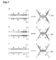
- FIGS. 7 are schematic explanatory diagrams illustrating an example (third example) of a case of using three dimer probes (the first dimer probe 40a, a second dimer probe 40d, a third dimer probe 40e).
- the first dimer probe 40a is configured in the same manner as in FIG. 4(a) .
- the second dimer probe 40d is formed by allowing two kinds of single-stranded nucleic acid probes (dimer formation probes 41g, 41h) to hybridize with each other.
- the dimer formation probe 41g includes three regions, i.e., a 5' side region (region G), a mid-region (region E), and a 3' side region (region C'), in which the 3' side region (region C') is complementary to the 3' side region (region C) of the first dimer probe 40a.
- the dimer formation probe 41h includes three regions, i.e., a 5' side region (region D'), a mid-region (region E'), and a 3' side region (region H), in which the 5' side region (region D') is complementary to the 5' side region (region D) of the first dimer probe 40a.
- the mid-region E of the dimer formation probe 41g is complementary to the mid-region E' of the dimer formation probe 41h.
- the third dimer probe 40e is formed by allowing two kinds of single-stranded nucleic acid probes (dimer formation probes 41j, 41k) to hybridize with each other.
- the dimer formation probe 41j includes three regions, i.e., a 5' side region (region A'), a mid-region (region J), and a 3' side region (region H'), in which: the 5' side region (region A') is complementary to the 5' side region (region A) of the first dimer probe 40a; and the 3' side region (region H') is complementary to the 3' side region (region H) of the second dimer probe 40d.
- the dimer formation probe 41k includes three regions, i.e., a 5' side region (region G'), a mid-region (region J'), and a 3' side region (region F'), in which: the 5' side region (region G') is complementary to the 5' side region (region G) of the second dimer probe 40d; and the 3' side region (region F') is complementary to the 3' side region (region F) of the first dimer probe 40a.
- the region J' is a region complementary to the region J.
- FIGS. 7 there is adopted such a configuration that: one 5' side region and one 3' side region of the first dimer probe are complementary to one 5' side region and one 3' side region of the second dimer probe, respectively; the other 5' side region and the other 3' side region of the first dimer probe are complementary to one 5' side region and one 3' side region of the third dimer probe, respectively; and the other 5' side region and the other 3' side region of the second dimer probe are complementary to the other 5' side region and the other 3' side region of the third dimer probe, respectively.
- a signal probe polymer as an assembly of dimer probes is formed by: using a plurality of kinds of dimer probes configured so that each of the 3' side regions of each of the dimer probes is complementary to any one of the 3' side regions of the other dimer probes, and each of the 5' side regions of each of the dimer probes is complementary to any one of the 5' side regions of the other dimer probes; and allowing these plurality of kinds of dimer probes to hybridize with each other.
- a combination of dimer probes having a complementary relationship is not particularly limited, but as illustrated in FIGS. 7 , it is preferred to adopt such a configuration that one 3' side region and one 5' side region in each of the dimer probes are complementary to one 3' side region and one 5' side region in any one of the other dimer probes, respectively.
- each complementary region of the dimer formation probes is at least 5 bases, preferably at least 8 bases, more preferably from 10 bases to 100 bases, still more preferably from 12 to 30 bases in terms of the number of bases.
- the lengths of the complementary regions in the respective probes are desirably the same.
- the dimer formation probes before dimer probe formation may be used in place of the dimer probes, but the dimer probes are preferably used.
- any one of the 3' regions (e.g., regions C, F, H) of the signal amplification probes according to the second exemplary embodiment includes a poly(T) sequence, and a region complementary thereto (e.g., regions C', F', H') includes a poly(A) sequence.
- signal amplification probes As signal amplification probes according to a third exemplary embodiment to be used in the present invention, there are given a plurality of kinds of signal amplification probes including one or more pairs of dimer formation probes, or one or more dimer probes to be formed from the pairs of dimer formation probes, and one or more kinds of cross-linking probes.
- each of the dimer formation probes includes three regions, i.e., a 5' side region, a mid-region, and a 3' side region, the mid-regions of each of the pairs of dimer formation probes are complementary to each other, and all of the 3' side regions and 5' side regions of each of the pairs of dimer formation probes are non-complementary to each other;
- each of the cross-linking probes includes two regions, i.e., a 5' side region and a 3' side region, each of the 5' side regions of the dimer formation probes is complementary to any one of the 5' side regions of the cross-linking probes, and each of the 3' side regions of the dimer formation probes is complementary to any one of the 3' side regions of the cross-linking probes; and the cross-linking probes can cross-link dimers to be formed from the dimer formation probes.
- the signal amplification probes can self-assemble
- FIGS. 8 there is illustrated a first example of a case of using a pair of dimer formation probes and a pair of cross-linking probes.
- FIG. 8(a) there are illustrated a pair of dimer formation probes (a first dimer formation probe 61a and a second dimer formation probe 61b), and a dimer probe 60a formed from the pair of dimer formation probes 61a, 61b.
- the pair of dimer formation probes is formed of two kinds of single-stranded nucleic acid probes (the first dimer formation probe 61a and the second dimer formation probe 61b), and each of the dimer formation probes 61a, 61b includes at least three regions, i.e., a mid-region, a 5' side region located closer to the 5' side with respect to the mid-region, and a 3' side region located closer to the 3' side with respect to the mid-region.
- regions i.e., a mid-region, a 5' side region located closer to the 5' side with respect to the mid-region, and a 3' side region located closer to the 3' side with respect to the mid-region.
- the 5' side region, mid-region, and 3' side region of the first dimer formation probe 61a are represented by a region A, a region B, and a region C, respectively
- the 5' side region, mid-region, and 3' side region of the second dimer formation probe 61b are represented by a region D, a region B', and a region F, respectively.
- the mid-regions (regions B and B') of the first dimer formation probe 61a and the second dimer formation probe 61b are complementary to each other, and all of the four regions, i.e., the 5' side regions (regions A and D) and 3' side regions (regions C and F) of the first dimer formation probe 61a and the second dimer formation probe 61b are non-complementary to each other. Both the probes are allowed to hybridize with each other to form the dimer probe 60a.
- a dimer probe formed by allowing a pair of dimer formation probes to hybridize with each other in advance may be used, or the dimer formation probes may be used as they are, but the dimer probe is preferably used.
- each of the pairs of dimer formation probes is configured in the same manner as in the case of using one pair of dimer formation probes described above.
- the plurality of pairs of dimer formation probes are used to form the same number of dimer probes.
- FIGS. 14(a) and 14(b) are schematic explanatory diagrams illustrating an example of two pairs of dimer formation probes, and two dimer probes to be formed from the pairs of dimer formation probes.
- FIG. 14(a) illustrates a first pair of dimer formation probes (a first dimer formation probe 71a and a second dimer formation probe 71b), and a dimer probe 70a to be formed from the pair of dimer formation probes 71a, 71b
- FIG. 14(b) illustrates a second pair of dimer formation probes (a first dimer formation probe 71c and a second dimer formation probe 71d), and a dimer probe 70b to be formed from the pair of dimer formation probes 71c, 71d.
- the 5' side region, mid-region, and 3' side region of the first dimer formation probe 71a in the first pair are represented by a region A, a region B, and a region C, respectively
- the 5' side region, mid-region, and 3' side region of the second dimer formation probe 71b in the first pair are represented by a region D, a region B', and a region F, respectively
- the 5' side region, mid-region, and 3' side region of the first dimer formation probe 71c in the second pair are represented by a region G, a region E, and a region H, respectively
- the 5' side region, mid-region, and 3' side region of the second dimer formation probe 71d in the second pair are represented by a region I, a region E', and a region J, respectively.
- the mid-regions (regions B and B', or regions E and E') of the first and second dimer formation probes in each of the pairs are complementary to each other, all of the four regions (regions A, C, D, and F, and regions G, H, I, and J), i.e., the 5' side regions and 3' side regions of the first and second dimer formation probes in the respective pairs are non-complementary to each other, and the dimer formation probes 71a, 71b, 71c, 71d in each of the pairs are allowed to hybridize with each other to form the two dimer probes 70a, 70b.
- the cross-linking probes are one or more kinds of single-stranded nucleic acid probes that can cross-link the dimer probes to be formed from the dimer formation probes, and include at least two regions. Of the two regions, the region located on the 5' side is referred to as 5' side region, and the region located on the 3' side is referred to as 3' side region.
- each of the 5' side regions of the dimer formation probes is complementary to any one of the 5' side regions of the cross-linking probes, and each of the 3' side regions of the dimer formation probes is complementary to any one of the 3' side regions of the cross-linking probes.
- the cross-linking probes can bind to the dimer formation probes so as to cross-link one or more kinds of plurality of dimer probes to be formed from the dimer formation probes, to thereby form an assembly of probes (probe polymer).
- the pair of dimer formation probes (a first dimer formation probe and a second dimer formation probe) and the pair of cross-linking probes (a first cross-linking probe and a second cross-linking probe) are configured so that: each of the 5' side regions of the pair of dimer formation probes is complementary to any one of the 5' side regions of the pair of cross-linking probes; each of the 5' side regions of the pair of cross-linking probes is complementary to any one of the 5' side regions of the pair of dimer formation probes; each of the 3' side regions of the pair of dimer formation probes is complementary to any one of the 3' side regions of the pair of cross-linking probes; and each of the 3' side regions of the pair of cross-linking probes is complementary to any one of the 3' side regions of the pair of dimer formation probes.
- FIG. 8(b) is a schematic explanatory diagram illustrating an example of a pair of cross-linking probes (a first cross-linking probe 62a and a second cross-linking probe 62b) to be used together with the pair of dimer formation probes 61a, 61b illustrated in FIG. 8(a) .
- Suitable examples of the cross-linking probes to be used together with the pair of dimer formation probes 61a, 61b illustrated in FIG. 8(a) include, as illustrated in FIG. 8(b) , such a pair of cross-linking probes that: the 5' side region of the first cross-linking probe 62a is complementary to the 5' side region (region A) of the first dimer formation probe 61a; the 3' side region of the first cross-linking probe 62a is complementary to the 3' side region (region C) of the first dimer formation probe 61a; the 5' side region of the second cross-linking probe 62b is complementary to the 5' side region (region D) of the second dimer formation probe 61b; and the 3' side region of the second cross-linking probe 62b is complementary to the 3' side region (region F) of the second dimer formation probe 61b.
- the dimer formation probes 61a, 61b illustrated in FIG. 8(a) and the cross-linking probes 62a, 62b illustrated in FIG. 8(b) can be allowed to hybridize with each other to form a signal probe polymer 68 ( FIG. 9 ).
- FIGS. 8 there is illustrated an example in which: the 5' side region and 3' side region of the first dimer formation probe 61a are complementary to the 5' side region and 3' side region of the first cross-linking probe 62a, respectively; and the 5' side region and 3' side region of the second dimer formation probe 61b are complementary to the 5' side region and 3' side region of the second cross-linking probe 62b, respectively.
- such a configuration that the 5' side region of one dimer formation probe is complementary to the 5' side region of one cross-linking probe and the 3' side region of one dimer formation probe is complementary to the 3' side region of one cross-linking probe only needs to be adopted.
- FIG. 10 is a schematic explanatory diagram illustrating another example (second example) of the pair of cross-linking probes (a first cross-linking probe 62c and a second cross-linking probe 62d) to be used together with the pair of dimer formation probes 61a, 61b illustrated in FIGS. 8 .
- the pair of cross-linking probes in which: the 5' side region of the first cross-linking probe 62c is complementary to the 5' side region (region A) of the first dimer formation probe 61a; the 3' side region of the first cross-linking probe 62c is complementary to the 3' side region (region F) of the second dimer formation probe 61b; the 5' side region of the second cross-linking probe 62d is complementary to the 5' side region (region D) of the second dimer formation probe 61b; and the 3' side region of the second cross-linking probe 62d is complementary to the 3' side region (region C) of the first dimer formation probe 61a.
- the dimer formation probes 61a, 61b illustrated in FIG. 8(a) and the cross-linking probes 62c, 62d illustrated in FIG. 10 can be allowed to hybridize with each other to form the signal probe polymer 68 ( FIG. 11 ).
- n represents an integer of 2 or more pairs of dimer formation probes (i.e., 2n dimer formation probes) and n pairs of cross-linking probes (i.e., 2n cross-linking probes) are used, and there is suitably adopted such a configuration that: each of the 5' side regions of the dimer formation probes is complementary to any one of the 5' side regions of the cross-linking probes; each of the 5' side regions of the cross-linking probes is complementary to any one of the other 5' side regions of the dimer formation probes; each of the 3' side regions of the dimer formation probes is complementary to any one of the 3' side regions of the cross-linking probes; and each of the 3' side regions of the cross-linking probes is complementary to any one of the 3' side regions of the dimer formation probes.
- FIGS. 14(c) and 14(d) are schematic explanatory diagrams illustrating an example of two pairs of cross-linking probes 72a to 72d to be used together with the two pairs of dimer formation probes 71a to 71d illustrated in FIGS. 14(a) and 14(b).
- FIG. 14(c) illustrates a first pair of cross-linking probes (the first cross-linking probe 72a and the second cross-linking probe 72b)
- FIG. 14(d) illustrates a second pair of cross-linking probes (the first cross-linking probe 72c and the second cross-linking probe 72d).
- each of the 5' side regions of the dimer formation probes is complementary to any one of the 5' side regions of the cross-linking probes; each of the 5' side regions of the cross-linking probes is complementary to any one of the 5' side regions of the dimer formation probes; each of the 3' side regions of the dimer formation probes is complementary to any one of the 3' side regions of the cross-linking probes; and each of the 3' side regions of the cross-linking probes is complementary to any one of the 3' side regions of the dimer formation probes.
- the 5' side region of the first cross-linking probe 72a in the first pair is complementary to the 5' side region (region A) of the first dimer formation probe 71a in the first pair;
- the 5' side region of the second cross-linking probe 72b in the first pair is complementary to the 5' side region (region D) of the second dimer formation probe 71b in the first pair;
- the 5' side region of the first cross-linking probe 72c in the second pair is complementary to the 5' side region (region G) of the first dimer formation probe 71c in the second pair;
- the 5' side region of the second cross-linking probe 72d in the second pair is complementary to the 5' side region (region I) of the second dimer formation probe 71d in the second pair;
- the 3' side region of the first cross-linking probe 72a in the first pair is complementary to any one of the 3' side regions of the four kinds of dimer formation probes 71
- the 3' side region of the second cross-linking probe 72b in the first pair is complementary to any one of the 3' side regions of the four kinds of dimer formation probes 71a to 71d, except for the one selected for the first cross-linking probe 72a in the first pair (in FIGS. 14 , there is illustrated a case of being complementary to the region J); the 3' side region of the first cross-linking probe 72c in the second pair is complementary to any one of the 3' side regions of the four kinds of dimer formation probes 71a to 71d, except for the ones selected for the first and second cross-linking probes 72a, 72b in the first pair (in FIGS.
- FIGS. 14 there is illustrated a case of being complementary to the region C); and the 3' side region of the second cross-linking probe 72d in the second pair is complementary to one of the 3' side regions of the four kinds of dimer formation probes 71a to 71d, except for the ones selected for the first and second cross-linking probes 72a, 72b in the first pair and the first cross-linking probe 72c in the second pair (in FIGS. 14 , there is illustrated a case of being complementary to the region F).
- the dimer formation probes 71a to 71d and cross-linking probes 72a to 72d illustrated in FIGS. 14 can be allowed to hybridize with each other to form the signal probe polymer 68.
- FIGS. 14 there is illustrated a case where: the 3' side region of the first cross-linking probe 72a in the first pair is complementary to the 3' side region of the first dimer formation probe 71c in the second pair; the 3' side region of the second cross-linking probe 72b in the first pair is complementary to the 3' side region of the second dimer formation probe 71b in the second pair; the 3' side region of the first cross-linking probe 72c in the second pair is complementary to the 3' side region of the first dimer formation probe 71a in the first pair; and the 3' side region of the second cross-linking probe 72d in the second pair is complementary to the 3' side region of the second dimer formation probe 71b in the first pair, but no limitation is imposed on a combination of the 3' side regions of the dimer formation probes to be complementary to each of the 3' side regions of the cross-linking probes.
- each complementary region of the dimer formation probes and the cross-linking probes is at least 5 bases, preferably at least 8 bases, more preferably from 10 bases to 100 bases, still more preferably from 12 to 30 bases in terms of the number of bases.
- the lengths of the complementary regions in the respective probes are desirably the same. It should be noted that in the signal amplification probes according to the third exemplary embodiment, any one of the 3' side regions (e.g., regions C, F, H, J) includes a poly(T) sequence, and a region complementary thereto (e.g., regions C', F', H', J') includes a poly(A) sequence.
- non-complementary base sequences may be any base sequences that do not hybridize with each other.
- the non-complementary base sequences also encompass base sequences that are identical to each other.
- FIGS. 12 are schematic explanatory diagrams illustrating a third example of a pair of dimer formation probes and one kind of pair of cross-linking probes to be used in the present invention.
- reference symbol 60b denotes a dimer probe, and there is illustrated an example of dimer formation using two kinds of dimer formation probes 61c, 61d in which the 3' side regions and the 5' side regions each have identical base sequences. That is, there is adopted such a configuration that in the dimer probe 60a of FIGS.
- the region A and the region D have identical base sequences
- the region C and the region F have identical base sequences.
- the pair of cross-linking probes to be used together with the dimer probe 60b are identical to each other, and one kind of cross-linking probe 62c is used.
- the dimer formation probes 61c, 61d and cross-linking probe 62c illustrated in FIGS. 12 can be allowed to hybridize with each other to form the signal probe polymer 68 ( FIG. 13 ).
- the nucleic acid probe is a nucleic acid probe that includes three or more nucleic acid regions, and there is adopted such a configuration that each of the nucleic acid regions in the nucleic acid probe includes a first region and a second region complementary to the first region so that the regions are adjacent to each other.
- one kind of nucleic acid probe alone can form a polymer of the nucleic acid probe.
- a nucleic acid probe disclosed in Patent Document 7 is used.
- the nucleic acid region in the nucleic acid probe means a region including the first region and the second region.
- each of the first region and second region in each nucleic acid region is referred to as complementary region.
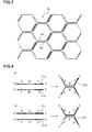
- FIG. 15 is a schematic explanatory diagram illustrating an example of one kind of nucleic acid probe capable of self-assembling to be used in the present invention.
- FIGS. 16 are schematic explanatory diagrams illustrating the binding manner of the nucleic acid probes of FIG. 15 .
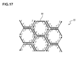
- FIG. 17 is a schematic explanatory diagram illustrating a self-assembly (probe polymer) formed from the nucleic acid probes of FIG. 15 .
- FIG. 15 is a schematic explanatory diagram illustrating an example of the signal amplification probe according to the fourth exemplary embodiment to be used in the present invention, and there is illustrated an example including three nucleic acid regions.
- one kind of nucleic acid probe 80 capable of self-assembling has a nucleic acid region 80a, a nucleic acid region 80b, and a nucleic acid region 80c in the stated order from the 5' end portion, and each of the nucleic acid regions 80a, 80b and 80c includes two regions complementary to each other so that the regions are adjacent to each other.
- the nucleic acid region 80a includes a complementary region X and a complementary region X' having a base sequence complementary to the complementary region X so that the regions are adjacent to each other
- the nucleic acid region 80b includes a complementary region Y and a complementary region Y' having a base sequence complementary to the complementary region Y so that the regions are adjacent to each other
- the nucleic acid region 80c includes a complementary region Z and a complementary region Z' having a base sequence complementary to the complementary region Z so that the regions are adjacent to each other.
- the complementary region X and the complementary region X', the complementary region Y and the complementary region Y', and the complementary region Z and the complementary region Z' are complementary nucleic acid regions capable of hybridizing with each other, respectively.
- the complementary region X and the complementary region X' bind to each other [ FIG. 16(a) ]
- the complementary region Y and the complementary region Y' bind to each other [ FIG. 16(b)
- the complementary region Z and the complementary region Z' bind to each other [ FIG. 16(c) ].
- FIGS. 16 as a suitable example, there is illustrated a nucleic acid probe having three nucleic acid regions (80a, 80b, and 80c).
- the number of nucleic acid regions is not particularly limited as long as the number is 3 or more.
- the nucleic acid probes 80 When the nucleic acid probes 80 are subjected to a hybridization reaction, as illustrated in FIG. 17 , the nucleic acid probes 80 can self-assemble to form a probe polymer 90 as a self-assembly of the nucleic acid probes 80.
- the 3' region (e.g., region Z') of the signal amplification probe according to the fourth exemplary embodiment includes a poly(T) sequence, and a region complementary thereto (e.g., region Z) includes a poly(A) sequence.
- the length of each complementary region is preferably 2 bases or more, more preferably from 3 bases to 50 bases, still more preferably from 4 to 15 bases in terms of the number of bases.
- the lengths of the respective complementary regions in the nucleic acid probes are desirably the same.
- the poly(T) sequence have a length of from 11 to 26 bases. It should be noted that in FIGS. 1 , there is illustrated an example in which the entirety of the nucleic acid region Z is a poly(T) sequence. However, the first signal amplification probe 18a having the nucleic acid region Z including a poly(T) sequence and any other sequence may also be used.
- each nucleic acid region of each signal amplification probe except for the nucleic acid region including a poly(T) sequence, is not particularly limited as long as predetermined regions are configured to have complementary base sequences so as to form a probe polymer, but bases at both ends of each region are each preferably guanine or cytosine.
- bases at both ends of each region are each guanine or cytosine, a reaction time can be shortened, a stable probe polymer can be formed at additionally low reaction temperature, and workability and detection sensitivity can be improved.
- the signal amplification probes are generally constructed of DNA or RNA, but may be constructed of a nucleic acid analog.
- the nucleic acid analog include a peptide nucleic acid (PNA, see, for example, WO 92/20702 A1 ) and a locked nucleic acid (LNA, see, for example, Koshkin AA et al. Tetrahedron 1998. 54, 3607-3630 ., Koshkin AA et al. J. Am. Chem. Soc. 1998. 120, 13252-13253 ., Wahlestedt C et al. PNAS. 2000. 97, 5633-5638 .).
- PNA peptide nucleic acid
- LNA locked nucleic acid
- the signal amplification probes of a pair are generally constructed of nucleic acids of the same kind, but for example, a DNA probe and an RNA probe may form a pair. That is, the kind of a nucleic acid of a probe may be selected from DNA, RNA and a nucleic acid analog (e.g., PNA or LNA).
- the probe does not need to be constructed of one kind of nucleic acid, for example, DNA alone, and for example, an oligonucleotide probe (chimeric probe) constructed of DNA and RNA may also be used as necessary, which is also included in the present invention.
- Such probe may be synthesized by a known method.
- the DNA probe may be synthesized by a phosphoramidite method using a DNA synthesizer Model 394 manufactured by Applied Biosystem Inc.
- a phosphoric acid triester method, an H-phosphonate method, a thiophosphonate method, or the like is available as an alternative method, and the probe may be synthesized by any method.
- one or both of the respective signal amplification probes to be used are preferably labeled with a labeling substance 20.
- a labeling substance 20 include a radioisotope, biotin, digoxigenin, a fluorophore, a luminophore and a pigment.
- RNA having no poly(A) tail for example, microRNA is described below.
- the method of detecting RNA according to the second aspect of the present invention is a method of detecting microRNA, the method including: (D) a poly(A) adding step of adding poly(A) to the 3' end of microRNA; (A) a capturing step of subjecting the microRNA and a capture substance for capturing the microRNA to a reaction to capture the microRNA; (B) a complex-for-detection forming step of subjecting the captured microRNA and one kind or a plurality of kinds of signal amplification probes to a reaction to form a complex for detection including the microRNA, the capture substance, and a probe polymer formed from the signal amplification probes; and (C) a detecting step of detecting the probe polymer bound in the complex for detection to detect the microRNA, the plurality of kinds of signal amplification probes being a plurality of kinds of signal amplification probes that have complementary base sequence regions capable of hybridizing with each other and can form a probe polymer through
- FIGS. 18 are schematic explanatory diagrams illustrating an example of the method of detecting RNA according to the second aspect of the present invention
- FIG. 18(a) illustrates microRNA
- FIG. 18(b) illustrates poly(A)-added microRNA after a poly(A) adding step
- FIG. 18(c) illustrates microRNA captured by a capture substance after a capturing step
- FIG. 18(d) illustrates a complex for detection after a complex-for-detection forming step.
- reference symbol 10b denotes microRNA.
- the present invention is suitably used for the detection of a short target nucleic acid, is suitably used for the detection of microRNA having from 17 to 63 bases, and is particularly suitably used for the detection of microRNA having from about 17 to 24 bases.
- many kinds of nucleic acid molecules can be simultaneously detected.
- a poly(A) adding step of adding poly(A) to the 3' end of microRNA 10b is performed to form poly(A)-added microRNA 12 [step (D)].
- a capturing step of subjecting the poly(A)-added microRNA 12 and the capture substance 14 for capturing the target microRNA 10b to a reaction to capture the poly(A)-added microRNA 12 is performed [step (A)]. It should be noted that in FIGS.
- step (A) is performed after the step (D), but it may be possible to perform the step (D) after the step (A), that is, to perform a capturing step of subjecting the microRNA 10b and the capture substance 14 to a reaction to capture the microRNA 10b [step (A)], and then perform a poly(A) adding step of adding poly(A) to the 3' end of the microRNA 10b [step (D)] to capture the poly(A)-added microRNA 12 by the capture substance 14.
- the steps (A) to (C) may be performed in the same manner as in the above-mentioned first aspect.
- a plurality of kinds of target nucleic acids can be simultaneously detected by using a plurality of the capture substances 14 in combination.
- the respective signal amplification probes to be used may be the same, and the support 16 and the capture substance 14 only need to be prepared in a plurality of kinds, leading to excellent workability and low cost.
- oligoRNAs Six kinds of synthetic oligoRNAs (hsa-miR-100, 155, 15b, 16, 21 and 23a/b) were used as target microRNAs.
- the six kinds of synthetic RNAs were mixed in equal amounts and used for an experiment with the amounts adjusted to 0, 0.316 amol, 1 amol, 3.16 amol, 10 amol or 31.6 amol.
- Base sequence of hsa-miR-100 5'-AACCCGUAGAUCCGAACUUGUG-3' (SEQ ID NO: 1) Base sequence of hsa-miR-155 5'-UUAAUGCUAAUCGUGAUAGGGG-3' (SEQ ID NO: 2) Base sequence of hsa-miR-15b 5'-UAGCAGCACAUCAUGGUUUACA-3' (SEQ ID NO: 3) Base sequence of hsa-miR-16 5'-UAGCAGCACGUAAAUAUUGGCG-3' (SEQ ID NO: 4) Base sequence of hsa-miR-21 5'-UAGCUUAUCAGACUGAUGUUGA-3' (SEQ ID NO: 5) Base sequence of hsa-miR-23a/b 5'-AUCACAUUGCCAGGGAUUUCC-3' (SEQ ID NO: 6)
- 12 kinds of spherical beads fluorescent microparticles (Luminex bead, manufactured by Luminex Corporation.)] to which 12 kinds of capture probes (CP-hsa-let7d, CP-hsa-miR-100, 107, 155, 15b, 16, 181a, 181c, 21, 221, 23a/b and 92a) were bound, respectively, were used as capture substances.
- 12 kinds of spherical beads fluorescent microparticles (Luminex bead, manufactured by Luminex Corporation.)] to which 12 kinds of capture probes (CP-hsa-let7d, CP-hsa-miR-100, 107, 155, 15b, 16, 181a, 181c, 21, 221, 23a/b and 92a) were bound, respectively, were used as capture substances.
- Base sequence of CP-hsa-let7d 5'-AACTATGCAACCTACTACCTCT-3' Base sequence of CP-hsa-miR-100 5'-CACAAGTTCGGATCTACGGGTT-3' (SEQ ID NO: 8) Base sequence of CP-hsa-miR-107 5'-TGATAGCCCTGTACAATGCTGCT-3' (SEQ ID NO: 9) Base sequence of CP-hsa-miR-155 5'-CCCCTATCACGATTAGCATTAA-3' (SEQ ID NO: 10) Base sequence of CP-hsa-miR-15b 5'-TGTAAACCATGATGTGCTGCTA-3' (SEQ ID NO: 11) Base sequence of CP-hsa-miR-16 5'-CGCCAATATTTACGTGCTGCTA-3' (SEQ ID NO: 12) Base sequence of CP-hsa-miR-181a 5'-ACTCACCGACA
- HCP-1 and HCP-2 Two kinds of nucleic acid probes labeled with biotin at the 5' end were used as signal amplification probes.
- Base sequence of HCP-1 (5'-X-Y-Z-3') 5'-CAACAATCAGGAC GATACCGATGAAG TTTTTTTTTTTTTTTTTTTTTTTTTTTTTTTTTTTTTTTTTTTTTTTTTTTTTTTTTTTTTTTTTTTTTTTTTTTTTTTTTTTTTTTTTTTTTTTTTTTTTTTTTTTTTTTTTTTTTTTTTTTTTTTTTTTTTTTTTTTTTTTTTTTTTTTTTTTTTTTTTTTTTTTTTTTTTTTTTTTTTTTTTTTTTTTTTTTTTTTTTTTTTTTTTTTTTTTTTTTTTTTTTTTTTTTTTTTTTTTTTTTTTTTTTTTTTTTTTTTTTTTTTTTTTTTTTTTTTTTTTTTTTTTTTTTTTTTTTTTTTTTTTTTTTTTTTTTTTTTTTTTTTTTTTTTTTTTTTTTTTTTTTTTTTTTTTTTTTTTT
- a first hybridization reaction liquid [0.1 ⁇ L each of the 12 kinds of spherical beads (1,000 beads for each) (1.2 ⁇ L in total), 7.5 ⁇ L of 5 M tetra-methyl ammonium chloride (TMAC), 3.75 ⁇ L of 10 ⁇ Supplement [500 mM Tris-HCl (pH 8.0), 40 mM EDTA (pH 8.0), 1% sarkosyl], and 2.55 ⁇ L of D.W.] so that the total volume was 25 ⁇ L, and the mixture was subjected to a reaction at 50°C for 60 minutes (capturing step).
- TMAC tetra-methyl ammonium chloride
- reaction liquid after the capturing step 25 ⁇ L of a PALSAR reaction liquid [7.5 ⁇ L of 5 M TMAC, 2.5 ⁇ L of 10 ⁇ Supplement, 2.5 ⁇ L of 20 pmol/ ⁇ L HCP-1, 2.0 ⁇ L of 20 pmol/ ⁇ L HCP-2 and 10.5 ⁇ L of D.W.] so that the total volume was 50 ⁇ L, and the mixture was subjected to a reaction at 44°C for 60 minutes (complex-for-detection forming step).
- a PALSAR reaction liquid 7.5 ⁇ L of 5 M TMAC, 2.5 ⁇ L of 10 ⁇ Supplement, 2.5 ⁇ L of 20 pmol/ ⁇ L HCP-1, 2.0 ⁇ L of 20 pmol/ ⁇ L HCP-2 and 10.5 ⁇ L of D.W.
- the reaction liquid after the complex-for-detection forming step was washed once with Wash Buffer [1 ⁇ PBS with 0.02% tween 20 and 1.5 ppm Proclin 300]. After that, 50 ⁇ L of a detection reagent [SA-PE (manufactured by ProZyme, Inc.): 5 ng/ ⁇ L] were added thereto, and the mixture was left to stand at room temperature for 10 minutes and then washed once with Wash Buffer. After that, 100 ⁇ L of Wash Buffer were added thereto, and fluorescence from the biotin and the fluorescent microparticles was measured by flow cytometry (Luminex System manufactured by Luminex Corporation) to detect signals from the 12 kinds of microRNAs (detecting step). Table 1 and FIGS.
- Target miRNA concentration (amol) Detection signal from target miRNA (MFI) let-7d miR-100 miR-107 miR-155 miR-15b miR-16 miR-181a miR-181c miR-21 miR-221 miR-23a/b miR-92a 0 15.7 12.2 25.7 28.7 20.8 12.0 27.0 39.0 33.3 22.2 14.2 19.5 0.316 16.2 68.5 20.8 63.8 65.2 33.0 27.0 42.3 40.7 24.0 40.7 19.7 1 18.3 220.8 25.7 140.2 144.5 77.5 30.2 42.7 46.3 22.7 112.3 21.8 3.16 16.7 706.3 26.0 389.8 380.8 282.3 28.7 40.3 84.2 19.3 366.7 17.7 10 16.3 2154.8 25.3 1274.3 1292.3 881.3 26.2 43.8 252.3 18.7 1252.2 22.2 31.6 16.0 7783.0 23.8 4353.7 4542.0 2996.8 25.0 39.7 810.2 21.8 3842.3 2
- MicroRNA detection was performed using a commercially available miRNA detection kit and the same target microRNAs as in Example 1.
- the microRNA detection was performed using Vantage microRNA Labeling Kit (manufactured by Marligen) as a labeling reagent, Vantage miRNA Multiplex Detection Kit Pancreatic Cancer Panel 1 (manufactured by Marligen) as a detection reagent, and the same six kinds of target microRNAs as in Example 1.
- the composition of the reagents and a reaction method were based on the protocols included with the kits. Table 2 and FIGS. 20 show the results.
- Target miRNA concentration (amol) Detection signal from target miRNA (MFI) let-7d miR-100 miR-107 miR-155 miR-15b miR-16 miR-181a miR-181c miR-21 miR-221 miR-23a/b miR-92a 5.8S control 0 34 55 43 67 33 23 95 70 35 37 46 61 52 0.316 39 70 56 76 45 29 111 85 39 39 62 56 55 1 31 69 51 69 40 26 95 73 28 32 54 62 56 3.16 59 110 97 97 86 52 129 101 60 66 110 88 118 10 36 92 57 78 57 37 104 73 36 37 59 66 53 31.6 40 178 63 104 66 83 113 85 39 47 79 69 74
- the synthetic oligoRNA (hsa-miR-21) used in Example 1 was used as target microRNA.
- the synthetic oligoRNA was used for an experiment with the amount adjusted to 0, 0.1 amol, 0.316 amol, 1 amol, 3.16 amol, 10 amol, 31.6 amol, 100 amol, or 316 amol (Example 2-1).
- RNAs 10 ng each of 10 kinds of cell-derived total RNAs (NCI-H650, MCF-7, CFPAC-1, LC-1F, A549, Hep-2, PC-14, HeLa, MDA-MB-435s and BT549) were used as target microRNAs (Examples 2-2 to 2-11).
- Spherical beads to which the capture probe (CP-hsa-miR-21) used in Example 1 was bound were used as a capture substance.
- the poly(A) adding step was performed by the same method as in Example 1 using the above-mentioned target microRNA.
- a first hybridization reaction liquid [0.4 ⁇ L of spherical beads (1,000 beads), 7.5 ⁇ L of 5 M TMAC, 3.75 ⁇ L of 10 ⁇ Supplement [500 mM Tris-HCl (pH 8.0), 40 mM EDTA (pH 8.0), 1% sarkosyl] and 3.35 ⁇ L of D.W.] so that the total volume was 25 ⁇ L, and the mixture was subjected to a reaction at 50°C for 60 minutes (capturing step).
- a first hybridization reaction liquid [0.4 ⁇ L of spherical beads (1,000 beads), 7.5 ⁇ L of 5 M TMAC, 3.75 ⁇ L of 10 ⁇ Supplement [500 mM Tris-HCl (pH 8.0), 40 mM EDTA (pH 8.0), 1% sarkosyl] and 3.35 ⁇ L of D.W.] so that the total volume was 25 ⁇ L, and the mixture was subjected to a reaction at 50°C for
- the complex-for-detection forming step was performed by the same method as in Example 1 using 25 ⁇ L of the reaction liquid after the capturing step.
- the reaction liquid after the complex-for-detection forming step was washed once with Wash Buffer [1 ⁇ PBS with 0.02% tween 20, 0.1% BSA and 0.05% NaN 3 ]. After that, 50 ⁇ L of a detection reagent [SA-PE (manufactured by Invitrogen): 10 ng/ ⁇ L] were added thereto, and the mixture was left to stand at room temperature for 10 minutes and then washed once with Wash Buffer. After that, 100 ⁇ L of Wash Buffer were added thereto, and fluorescence from the biotin and the fluorescent microparticles was measured by flow cytometry (Luminex System manufactured by Luminex Corporation) to detect signals from the microRNA (detecting step).
- FIG. 21 shows the results of Example 2-1.
- FIG. 23 is a graph showing a correlation between the results of Example 2 and Comparative Example 2.
- Comparative Example 2 Example 2 A549 5.27 3.59 BT-549 2.57 1.34 CFPAC 19.75 15.69 HeLa 2.09 2.86 HEp2 4.25 3.18 LC-1F 7.44 6.62 MCF-7 29.26 26.52 MDA 2.27 2.45 NCI-H650 21.44 24.27 PC-14 4.38 2.59 (amol)
- MicroRNA (hsa-miR-21 and hsa-miR-16) detection was performed by a real-time PCR method using TaqManProbe and the same target microRNA as in Example 2.
- a reverse transcription reaction was performed using TaqMan MicroRNA Primer (manufactured by ABI, for miR-21 and 16) and TaqMan MicroRNA Reverse Transcription Kit (manufactured by ABI) in accordance with the accompanying protocols.
- a real-time PCR reaction was performed using TaqMan MicroRNA Assays (manufactured by ABI, for miR-21 and 16) and Universal PCR Master Mix, No AmpErase UNG (manufactured by ABI) and a reverse transcription reaction liquid in accordance with the accompanying protocols.
- FIG. 22 and Table 3 show the results.
- FIG. 23 is a graph showing a correlation between the results of Example 2 and Comparative Example 2.
- the invention of the present application allowed the microRNAs to be detected at a level of up to several amol, and allowed the microRNAs to be detected with high sensitivity and simply. Further, as shown in the results of Example 2 and Comparative Example 2, it was demonstrated that the invention of the present application exhibited a high correlation with the real-time PCR method.
- Cell-derived total RNA (cell line RERF-LC-AI) including Beta-Actin (BA) and GAPDH (G3P) as target messenger RNAs was used.
- the total RNA was used for an experiment with the amount adjusted to 0, 0.1 ⁇ g, 0.5 ⁇ g, 1 ⁇ g or 2.5 ⁇ g.
- the two kinds of nucleic acid probes (HCP-1 and HCP-2) labeled with biotin at the 5' end used in Example 1 were used as signal amplification probes.
- RNA was added 15 ⁇ L of a first hybridization reaction liquid [0.2 ⁇ L each of the two kinds of spherical beads (2,000 beads for each) (0.4 ⁇ L in total), 7.5 ⁇ L of 5 M tetra-methyl ammonium chloride (TMAC), 2.5 ⁇ L of 10 ⁇ Supplement [500 mM Tris-HCl (pH 8.0), 40 mM EDTA (pH 8.0), 1% sarkosyl] and 4.6 ⁇ L of D.W.] so that the total volume was 25 ⁇ L, and the mixture was subjected to a reaction at 55°C for 60 minutes (capturing step).
- TMAC tetra-methyl ammonium chloride
- 10 ⁇ Supplement 500 mM Tris-HCl (pH 8.0), 40 mM EDTA (pH 8.0), 1% sarkosyl] and 4.6 ⁇ L of D.W.
- reaction liquid after the capturing step 25 ⁇ L of a PALSAR reaction liquid [7.5 ⁇ L of 5 M TMAC, 2.5 ⁇ L of 10 ⁇ Supplement, 2.5 ⁇ L of 20 pmol/ ⁇ L HCP-1, 2.0 ⁇ L of 20 pmol/ ⁇ L HCP-2 and 10.5 ⁇ L of D.W.] so that the total volume was 50 ⁇ L, and the mixture was subjected to a reaction at 44°C for 60 minutes (complex-for-detection forming step).
- a PALSAR reaction liquid 7.5 ⁇ L of 5 M TMAC, 2.5 ⁇ L of 10 ⁇ Supplement, 2.5 ⁇ L of 20 pmol/ ⁇ L HCP-1, 2.0 ⁇ L of 20 pmol/ ⁇ L HCP-2 and 10.5 ⁇ L of D.W.
- the reaction liquid after the complex-for-detection forming step was washed once with Wash Buffer [1 ⁇ PBS with 0.02% tween 20 and 0.5% Sodium Azide]. After that, 50 ⁇ L of a detection reagent [SA-PE (manufactured by ProZyme, Inc.): 5 ng/ ⁇ L] were added thereto, and the mixture was left to stand at room temperature for 10 minutes and then washed once with Wash Buffer. After that, 100 ⁇ L of Wash Buffer were added thereto, and fluorescence from the biotin and the fluorescent microparticles was measured by flow cytometry (Luminex System manufactured by Luminex Corporation) to detect signals from the two kinds of messenger RNAs (detecting step).
- FIG. 24 shows the results.
- Cell-derived total RNAs (cell lines RERF-LC-AI, A549, HeLa, PC-14 and Hep-2) including Beta-Actin and GAPDH as target messenger RNAs were used. The total RNAs were each used for an experiment with the amount adjusted to 1 ⁇ g or 5 ⁇ g.
- spherical beads Fluorescent microparticles (Luminex bead, manufactured by Luminex Corporation)] to which the same two kinds of capture probes (CP-BA-1 and CP-G3P-1) as in Example 3 were bound, respectively, were used as capture substances.
- HCP-1 and HCP-2 The same two kinds of nucleic acid probes (HCP-1 and HCP-2) labeled with biotin at the 5' end as in Example 1 were used as signal amplification probes.
- RNA was added 15 ⁇ L of a first hybridization reaction liquid [0.2 ⁇ L each of the two kinds of spherical beads (2,000 beads for each) (0.4 ⁇ L in total), 7.5 ⁇ L of 5 M tetra-methyl ammonium chloride (TMAC), 2.5 ⁇ L of 10 ⁇ Supplement [500 mM Tris-HCl (pH 8.0), 40 mM EDTA (pH 8.0), 1% sarkosyl] and 4.6 ⁇ L of D.W.] so that the total volume was 25 ⁇ L, and the mixture was subjected to a reaction at 55°C for 60 minutes (capturing step).
- TMAC tetra-methyl ammonium chloride
- 10 ⁇ Supplement 500 mM Tris-HCl (pH 8.0), 40 mM EDTA (pH 8.0), 1% sarkosyl] and 4.6 ⁇ L of D.W.
- reaction liquid after the capturing step 25 ⁇ L of a PALSAR reaction liquid [7.5 ⁇ L of 5 M TMAC, 2.5 ⁇ L of 10 ⁇ Supplement, 2.5 ⁇ L of 20 pmol/ ⁇ L HCP-1, 2.0 ⁇ L of 20 pmol/ ⁇ L HCP-2 and 10.5 ⁇ L of D.W.] so that the total volume was 50 ⁇ L, and the mixture was subjected to a reaction at 44°C for 60 minutes (complex-for-detection forming step).
- a PALSAR reaction liquid 7.5 ⁇ L of 5 M TMAC, 2.5 ⁇ L of 10 ⁇ Supplement, 2.5 ⁇ L of 20 pmol/ ⁇ L HCP-1, 2.0 ⁇ L of 20 pmol/ ⁇ L HCP-2 and 10.5 ⁇ L of D.W.
- the reaction liquid after the complex-for-detection forming step was washed once with Wash Buffer [1 ⁇ PBS with 0.1% BSA, 0.02% tween 20 and 0.5% Sodium Azide]. After that, 50 ⁇ L of a detection reagent [SA-PE (manufactured by Invitrogen): 10 ng/ ⁇ L] were added thereto, and the mixture was left to stand at room temperature for 10 minutes and then washed once with Wash Buffer. After that, 100 ⁇ L of Wash Buffer were added thereto, and fluorescence from the biotin and the fluorescent microparticles was measured by flow cytometry (Luminex System manufactured by Luminex Corporation) to detect signals from the two kinds of messenger RNAs (detecting step).
- FIG. 25 shows the results.
- Cell-derived total RNA (cell line CCRF-CEM) including GAPDH as target messenger RNA was used.
- the total RNA extracted from 10 7 cells was diluted and used for an experiment with the amount adjusted to one corresponding to 10 3 , 10 4 , 10 5 or 10 6 cells.
- Spherical beads [fluorescent microparticles (Luminex bead, manufactured by Luminex Corporation)] to which a capture probe (CP-G3P-2) was bound were used as a capture substance.
- a capture probe CP-G3P-2
- the two kinds of nucleic acid probes (HCP-1 and HCP-2) labeled with biotin at the 5' end used in Example 1 were used as signal amplification probes.
- RNA was added 15 ⁇ L of a first hybridization reaction liquid [0.1 ⁇ L of the spherical beads (1,000 beads for each), 7.5 ⁇ L of 5 M tetra-methyl ammonium chloride (TMAC), 2.5 ⁇ L of 10 ⁇ Supplement [500 mM Tris-HCl (pH 8.0), 40 mM EDTA (pH 8.0), 1% sarkosyl] and D.W. in such an amount that the total amount was adjusted to 15 ⁇ L] so that the total volume was 25 ⁇ L, and the mixture was subjected to a reaction at 55°C for 4 hours (capturing step).
- TMAC tetra-methyl ammonium chloride
- 10 ⁇ Supplement 500 mM Tris-HCl (pH 8.0), 40 mM EDTA (pH 8.0), 1% sarkosyl] and D.W. in such an amount that the total amount was adjusted to 15 ⁇ L] so that the total volume was 25 ⁇ L, and
- reaction liquid after the capturing step 25 ⁇ L of a PALSAR reaction liquid [7.5 ⁇ L of 5 M TMAC, 2.5 ⁇ L of 10 ⁇ Supplement, 2.5 ⁇ L of 20 pmol/ ⁇ L HCP-1, 1.75 ⁇ L of 20 pmol/ ⁇ L HCP-2 and 10.75 ⁇ L of D.W.] so that the total volume was 50 ⁇ L, and the mixture was subjected to a reaction at 44°C for 60 minutes (complex-for-detection forming step).
- a PALSAR reaction liquid 7.5 ⁇ L of 5 M TMAC, 2.5 ⁇ L of 10 ⁇ Supplement, 2.5 ⁇ L of 20 pmol/ ⁇ L HCP-1, 1.75 ⁇ L of 20 pmol/ ⁇ L HCP-2 and 10.75 ⁇ L of D.W.
- the reaction liquid after the complex-for-detection forming step was washed once with Wash Buffer [1 ⁇ PBS with 0.02% tween 20 and 1.5 ppm ProClin 300]. After that, 50 ⁇ L of a detection reagent [SA-PE (manufactured by ProZyme, Inc.): 5 ng/ ⁇ L] were added thereto, and the mixture was left to stand at room temperature for 10 minutes and then washed once with Wash Buffer. After that, 100 ⁇ L of Wash Buffer were added thereto, and fluorescence from the biotin and the fluorescent microparticles was measured by flow cytometry (Luminex System manufactured by Luminex Corporation) to detect signals from the messenger RNA (detecting step).
- FIG. 26 shows the results.
- the messenger RNA was successfully detected in an intact and non-labeled state of the RNA without performing a reverse transcription reaction or a PCR.
- a housekeeping gene present at a certain ratio in each cell was able to be detected.
- the messenger RNA of GAPDH was able to be detected with linearity and high sensitivity from 10 3 cells.
- 10a messenger RNA
- 10b microRNA
- 12 poly(A)-added microRNA
- 14 capture substance
- 16 support
- 18a first signal amplification probe
- 18b second signal amplification probe
- 20 labeling substance
- 22, 50, 68, 90 probe polymer
- 30 complex for detection
- 40a first dimer probe
- 40b, 40c 40d: second dimer probe
- 40e third dimer probe
- 41a to 41h, 41j, 41k dimer formation probe
- 60a, 60b dimer probe
- 61a to 61d dimer formation probe
- 62a to 62d cross-linking probe
- 70a, 70b dimer probe
- 71a to 71d dimer formation probe
- 72a to 72d cross-linking probe
- 80 nucleic acid probe
- 80a, 80b, 80c nucleic acid region.
Landscapes
- Chemical & Material Sciences (AREA)
- Life Sciences & Earth Sciences (AREA)
- Organic Chemistry (AREA)
- Proteomics, Peptides & Aminoacids (AREA)
- Zoology (AREA)
- Health & Medical Sciences (AREA)
- Engineering & Computer Science (AREA)
- Wood Science & Technology (AREA)
- Analytical Chemistry (AREA)
- Microbiology (AREA)
- Physics & Mathematics (AREA)
- Molecular Biology (AREA)
- Immunology (AREA)
- Biotechnology (AREA)
- Biophysics (AREA)
- Biochemistry (AREA)
- Bioinformatics & Cheminformatics (AREA)
- General Engineering & Computer Science (AREA)
- General Health & Medical Sciences (AREA)
- Genetics & Genomics (AREA)
- Measuring Or Testing Involving Enzymes Or Micro-Organisms (AREA)
Abstract
Provided are a method and kit for detecting RNA, which allow RNA such as microRNA and messenger RNA to be detected with high sensitivity and simply, and signal amplification probes to be used in the method. Specifically, provided is a method of detecting target RNA, the method including: (A) a capturing step of capturing RNA by a capture substance for capturing the RNA; (B) a complex-for-detection forming step of subjecting the captured RNA and signal amplification probes capable of self-assembling to a reaction to form a complex for detection including the RNA, the capture substance, and a probe polymer formed from the signal amplification probes; and (C) a detecting step of detecting the probe polymer bound in the complex for detection to detect the RNA, one of the signal amplification probes including a poly(T) sequence.
Description
- The present invention relates to a method and kit for detecting RNA such as microRNA (miRNA) and messenger RNA (mRNA), and signal amplification probes to be used in the method, and more particularly, to a method and kit for detecting RNA, which allow RNA to be detected with high sensitivity and simply, and signal amplification probes.
- MicroRNA is a kind of small RNA to be transcribed in various eukaryotes including humans. A primary transcript (pri-miRNA) transcribed from a non-gene region or intron portion on a genome is processed into precursor microRNA (pre-miRNA) having a hairpin structure and then into mature microRNA (mature-miRNA) having about 22 bases (Non Patent Document 1). It has been revealed that the microRNA functions as a regulatory factor in a process of translation from transcribed mRNA to a protein (Non Patent Document 2). At present, 800 or more kinds of microRNAs are expected to be present on a human genome (Non Patent Document 3), but many of the microRNAs have not been identified for a messenger (mRNA) subject to their regulatory functions.
- However, previous research has revealed that many microRNAs regulate expression of genes involved in multicellular organism development, differentiation, cell growth, and the like (Non Patent Document 4). In addition, there are reports on microRNAs considered to function in canceration or cancer suppression mechanism of cells, and many of the microRNAs are expressed in increased or decreased amounts in cancer cells as compared to normal cells (Non Patent Document 5). Thus, it is considered that an investigation on an expression amount of microRNA enables profiling of canceration of a cell or tissue of interest to be performed in the future, and it is predicted that identification or quantitative determination of microRNA in a cell or a tissue serves as one of the important items for diagnosis of cancer in the future.
- Heretofore, as methods of detecting microRNA, there have been reported a northern blotting method (Non Patent Document 6), a primer elongation method (
Non Patent Documents 7 and 8), an invader method (Non Patent Document 9), a ribozyme signal amplification method (Non Patent Document 10), a mirMASA bead-based technology (Non Patent Document 11), bead-based flow cytometry (Non Patent Document 12), a real-time RT-PCR (Non Patent Documents 13 and 14), a microarray (Non Patent Document 15), and the like, and there has been attempted a labeling method involving using gold particles (Non Patent Document 16). In addition, a kit using an RNase protection assay is available as a reagent for research (Non Patent Document 17). - However, the above-mentioned methods have problems such as insufficient sensitivity (northern blotting method), necessity of amplifying precursor microRNA, which serves as a target, by a PCR (e.g., real-time RT-PCR), detection requiring much time and complicated operations (bead-based flow cytometry, invader method), and difficulty in simultaneously analyzing many kinds of target nucleic acids (primer elongation method, invader method).
- Meanwhile, messenger RNA is a nucleic acid responsible for part of gene expression and is synthesized by a function of RNA polymerase using DNA as a template (transcription). After that, a protein of interest is synthesized by a ribosome using genetic information included in the messenger RNA (translation). The messenger RNA can be said to be a very important element for expression of a protein from DNA. Messenger RNA from eukaryotes has a CAP structure at the 5' end and poly(A) at the 3' end unlike other RNAs, i.e., transfer RNA (tRNA) and ribosomal RNA (rRNA). The structures also have a function of controlling decomposition of the messenger RNA itself.
- Heretofore, in the field of diagnosis, a protein has been used as a biomarker mainly in a biochemical approach. However, it is difficult to prepare an antibody for detecting the protein in some cases, and the protein alone is not necessarily sufficient as the biomarker in terms of sensitivity and specificity. Thus, research has been made on whether or not expression of messenger RNA from which a protein originates can be used as a biomarker associated with a disease or the like. Studies on expression of several thousands of or several tens of thousands of messenger RNAs using a messenger array have revealed presence of an expression profile of messenger RNA specific for a certain disease. In addition, not only expression profiles obtained from many messenger RNAs but also single messenger RNA is known to serve as a biomarker. For example, as a myeloid leukemia-related diagnostic drug, messenger RNA of a certain gene is used as a biomarker, and is actually covered by insurance as an in vitro diagnostic in Japan. As can be seen from those facts, usefulness of messenger RNA as a next-generation biomarker serving as an alternative to a protein is considered to increase continuously in the future.
- Heretofore, as methods of detecting messenger RNA, there have been reported methods involving using an RT-PCR method, a real-time PCR method including TaqMan, a messenger array, and a branched-DNA method.
- However, the above-mentioned methods have problems such as necessity of a step of converting target messenger RNA from RNA to DNA by reverse transcription or necessity of amplifying the target messenger RNA by a PCR (e.g., real-time RT-PCR), detection requiring a time of 1 day or more and complicated operations (messenger array), and difficulty in simultaneously analyzing many kinds of target nucleic acids (real-time
- PCR).
- Meanwhile, in
Patent Documents - In addition, in
Patent Document 3, there is a disclosure of a method of detecting a small nucleic acid including microRNA. However, this method has a problem in that its sensitivity is at a level of several hundred amol and hence limitations are imposed on sensitivity and a reaction temperature, although the method has an advantage such as no modification of microRNA. - Further, in
Patent Document 4, although detection of messenger RNA is performed using a PALSAR method, it is essential to perform a reverse transcription reaction in this method. -
- Patent Document 1:
JP 3267576 B - Patent Document 2:
JP 3310662 B - Patent Document 3:
WO 2010/087409 A1 - Patent Document 4:
WO 2004/072302 A1 - Patent Document 5:
WO 02/031192 A1 - Patent Document 6:
JP 2002-355081 A - Patent Document 7:
WO 2009/54320 A1 -
- Non Patent Document 1: Kim VN, Nam JW. Genomics of microRNA. Trends Genet. 2006 Mar; 22(3): 165-73.
- Non Patent Document 2: Sontheimer, E., J. Assembly and function of RNA silencing complex. Nature Reviews Molecular Cell Biology. 2005 Feb; 6 127-138
- Non Patent Document 3: Bentwich I, Avniel A, Karov Y, Aharonov R, Gilad S, Barad O, Barzilai A, Einat P, Einav U, Meiri E, Sharon E, Spector Y, Bentwich Z. Identification of hundreds of conserved and nonconserved human microRNAs. Nat Genet. 2005 Jul; 37(7): 766-70
- Non Patent Document 4: Alvarez-Garcia I, Miska EA. MicroRNA functions in animal development and human disease. Development. 2005 Nov; 132(21): 4653-62.
- Non Patent Document 5: Esquela-Kerscher A, Slack FJ. Oncomirs - microRNAs with a role in cancer. Nat Rev Cancer. 2006 Apr; 6(4): 259-269
- Non Patent Document 6: Lagos-Quintana, M., Rauhut, R., Meyer, J., Borkhardt, A., and Tuschl, T. New microRNAs from mouse and human. RNA. 2003 Feb; 9(2): 175-179.
- Non Patent Document 7: Zeng, Y. and Cullen, B.R. Sequence requirements for micro RNA processing and function in human cells. RNA. 2003 Jan; 9(1): 112-123.
- Non Patent Document 8: Raymond CK, Roberts BS, Garrett-Engele P, Lim LP, Johnson JM. Simple, quantitative primer-extension PCR assay for direct monitoring of microRNAs and short-interfering RNAs. RNA. 2005 Nov; 11: 1737-1744
- Non Patent Document 9: Allawi, H.T., Dahlberg, J.E., Olson, S., Lund, E., Olson, M., Ma, W.P., Takova, T., Neri, B.P., and Lyamichev, V.I. Quantitation of microRNAs using a modified Invader assay. RNA. 2004 Jul; 10(7): 1153-1161.
- Non Patent Document 10: Hartig, J.S., Grune, I., Najafi-Shoushtari, S.H., and Famulok, M. Sequence-specific detection of MicroRNAs by signal-amplifying ribozymes. J. Am. Chem. Soc. 2004 .
- Non Patent Document 11: Barad O, Meiri E, Avniel A, Aharonov R, Barzilai A, Bentwich I, Einav U, Gilad S, Hurban P, Karov Y, Lobenhofer EK, Sharon E, Shiboleth YM, Shtutman M, Bentwich Z, Einat P. MicroRNA expression detected by oligonucleotide microarrays: system establishment and expression profiling in human tissues. Genome Res. 2004 Dec; 14(12): 2486-94.
- Non Patent Document 12: Lu J, Getz G, Miska EA, Alvarez-Saavedra E, Lamb J, Peck D, Sweet-Cordero A, Ebert BL, Mak RH, Ferrando AA, Downing JR, Jacks T, Horvitz HR, Golub TR. MicroRNA expression profiles classify human cancers. Nature. 2005
- Non Patent Document 13: Chen C, Ridzon DA, Broomer AJ, Zhou Z, Lee DH, Nguyen JT, Barbisin M, Xu NL, Mahuvakar VR, Andersen MR, Lao KQ, Livak KJ, Guegler KJ. Real-time quantification of microRNAs by stem-loop RT-PCR. Nucleic Acids Res. 2005 Nov 27; 33(20): e179.
- Non Patent Document 14: Tang F, Hajkova P, Barton SC, Lao K, Surani MA. MicroRNA expression profiling of single whole embryonic stem cells. Nucleic Acids Res. 2006 Jan 24; 34(2): e9.
- Non Patent Document 15: Liu CG, Calin GA, Meloon B, Gamliel N, Sevignani C, Ferracin M, Dumitru CD, Shimizu M, Zupo S, Dono M, Alder H, Bullrich F, Negrini M, Croce CM. An oligonucleotide microchip for genome-wide microRNA profiling in human and mouse tissues. Proc Natl Acad Sci USA. 2004 Jan 29; 101(26): 9740-4.
- Non Patent Document 16: Liang RQ, Li W, Li Y, Tan CY, Li JX, Jin YX, Ruan KC. An oligonucleotide microarray for microRNA expression analysis based on labeling RNA with quantum dot and nanogold probe. Nucleic Acids Res. 2005 Jan 31; 33(2): e17.
- Non Patent Document 17: mirVana miRNA Detection kit (Ambion Inc.)
- An object of the present invention is to provide a method and kit for detecting RNA, which allow RNA such as microRNA and messenger RNA to be detected with high sensitivity and simply, and signal amplification probes to be used in the method.
- In order to achieve the above-mentioned object, the inventors of the present invention have made intensive studies. As a result, the inventors have surprisingly found that RNA can be detected with high sensitivity by: using in signal amplification probes for a hybridization reaction to be used in a PALSAR method, signal amplification probes in which one region of one of the signal amplification probes is a region including a poly(T) sequence, which is a nucleic acid sequence of consecutive thymines (T); adding poly(A), which is a nucleic acid sequence of consecutive adenines (A), to the 3' end of target RNA as necessary; then subjecting RNA captured by a capture probe and the signal amplification probes to a reaction to form a complex for detection including a probe polymer; and detecting the probe polymer.
- That is, a method of detecting RNA of the present invention is a method of detecting target RNA, the method including: (A) a capturing step of capturing RNA capable of including a poly(A) sequence at the 3' end by a capture substance for capturing the RNA, i.e., a capturing step of capturing RNA by a capture substance for capturing the RNA; (B) a complex-for-detection forming step of subjecting the captured RNA and one kind or a plurality of kinds of signal amplification probes capable of self-assembling to a reaction to form a complex for detection including the RNA, the capture substance, and a probe polymer formed from the signal amplification probes; and (C) a detecting step of detecting the probe polymer bound in the complex for detection to detect the RNA, one of the signal amplification probes having a poly(T) sequence.
- It is preferred that in the signal amplification probe, the poly(T) sequence have a length of from 11 to 26 bases.
- It is suitable that the signal amplification probe having a poly(T) sequence have the poly(T) sequence at the 3' end.
- It is preferred that at least one of the signal amplification probes be labeled with a labeling substance.
- It is suitable that the capture substance include a base sequence complementary to the RNA, and be immobilized onto a support or have introduced therein a moiety capable of being immobilized onto the support.
- In the present invention, the hybridization reaction is not particularly limited, but more suitably includes a self-assembly reaction in which nucleic acids self-assemble to form a double-stranded self-assembly through hybridization using a plurality of kinds of nucleic acid probes having complementary base sequence regions capable of hybridizing with each other.
- That is, a first example of the method of detecting RNA of the present invention is a method of detecting target RNA, the method including: (A) a capturing step of capturing RNA by a capture substance for capturing the RNA; (B) a step of subjecting the captured RNA, a first signal amplification probe, and a second signal amplification probe to a reaction to form a complex for detection including the RNA, the capture substance, and a probe polymer formed from the first signal amplification probe and the second signal amplification probe; and (C) a step of detecting the probe polymer bound in the complex for detection to detect the RNA, the first signal amplification probe including a nucleic acid probe that includes at least a nucleic acid region X, a nucleic acid region Y, and a nucleic acid region Z including a poly(T) sequence in the stated order from the 5' end side, the second signal amplification probe including a nucleic acid probe that includes at least a nucleic acid region X' complementary to the nucleic acid region X, a nucleic acid region Y' complementary to the nucleic acid region Y, and a nucleic acid region Z' complementary to the nucleic acid region Z in the stated order from the 5' end side.
- It is preferred that in the first signal amplification probe, the poly(T) sequence have a length of from 11 to 26 bases.
- It is suitable that in the first signal amplification probe, the nucleic acid region X have a length of from 11 to 20 bases, the nucleic acid region Y have a length of from 11 to 20 bases, and the nucleic acid region Z have a length of from 11 to 26 bases.
- It is preferred that the first signal amplification probe and/or the second signal amplification probe be labeled with a labeling substance.
- In the present invention, any of RNA having a poly(A) sequence, which is a nucleic acid sequence of consecutive adenines (A), such as mRNA, and RNA having no poly(A) sequence may be detected as the target RNA. When the RNA having a poly(A) sequence is detected as the target RNA, the poly(A) sequence of the target RNA and the poly(T) sequence of the signal amplification probe hybridize with each other to form a complex for detection, and the complex for detection can be detected to detect the target RNA. In addition, when the RNA having no poly(A) sequence, for example, microRNA having no poly(A) sequence is detected as the target RNA, by performing (D) a poly(A) adding step of adding poly(A) to the 3' end of the microRNA before or after the step (A) (preferably before the step (A)), the poly(A) sequence of the poly(A)-added target RNA and the poly(T) sequence of the signal amplification probe hybridize with each other to form a complex for detection, and the complex for detection can be detected to detect the target RNA.
- A kit for detecting RNA of the present invention is a kit for detecting RNA, the kit including: a capture substance for capturing target RNA; and one kind or a plurality of kinds of signal amplification probes, in which the plurality of kinds of signal amplification probes are a plurality of kinds of signal amplification probes that have complementary base sequence regions capable of hybridizing with each other and can form a probe polymer through a self-assembly reaction, one of the plurality of kinds of signal amplification probes having a poly(T) sequence, and in which the one kind of signal amplification probe is one kind of signal amplification probe having a poly(T) sequence and including three or more nucleic acid regions, each of the nucleic acid regions in the one kind of signal amplification probe including a first region and a second region complementary to the first region so that the regions are adjacent to each other.
- It is suitable that the plurality of kinds of signal amplification probes include a first signal amplification probe and a second signal amplification probe, the first signal amplification probe be a nucleic acid probe that includes at least a nucleic acid region X, a nucleic acid region Y, and a nucleic acid region Z including a poly(T) sequence in the stated order from the 5' end side, and the second signal amplification probe be a nucleic acid probe that includes at least a nucleic acid region X' complementary to the nucleic acid region X, a nucleic acid region Y' complementary to the nucleic acid region Y, and a nucleic acid region Z' complementary to the nucleic acid region Z in the stated order from the 5' end side.
- Signal amplification probes of the present invention are one kind or a plurality of kinds of signal amplification probes to be used in the method of detecting RNA of the present invention, in which the plurality of kinds of signal amplification probes are a plurality of kinds of signal amplification probes that have complementary base sequence regions capable of hybridizing with each other and can form a probe polymer through a self-assembly reaction, one of the plurality of kinds of signal amplification probes having a poly(T) sequence, and in which the one kind of signal amplification probe is one kind of signal amplification probe having a poly(T) sequence and including three or more nucleic acid regions, each of the nucleic acid regions in the one kind of signal amplification probe including a first region and a second region complementary to the first region so that the regions are adjacent to each other.
- It is preferred that in the signal amplification probe, the poly(T) sequence have a length of from 11 to 26 bases.
- It is suitable that the signal amplification probe having a poly(T) sequence have the poly(T) sequence at the 3' end.
- It is preferred that at least one of the signal amplification probes be labeled with a labeling substance.
- According to a first exemplary embodiment, the plurality of kinds of signal amplification probes are a pair of signal amplification probes, including a first signal amplification probe and a second signal amplification probe, in which the first signal amplification probe is a nucleic acid probe that includes at least a nucleic acid region X, a nucleic acid region Y, and a nucleic acid region Z including a poly(T) sequence in the stated order from the 5' end side, and in which the second signal amplification probe is a nucleic acid probe that includes at least a nucleic acid region X' complementary to the nucleic acid region X, a nucleic acid region Y' complementary to the nucleic acid region Y, and a nucleic acid region Z' complementary to the nucleic acid region Z in the stated order from the 5' end side.
- According to a second exemplary embodiment, the plurality of kinds of signal amplification probes are a plurality of dimer probes or dimer formation probes for forming the dimer probes, in which each of the plurality of dimer probes is a dimer formed from two kinds of dimer formation probes, in which each of the dimer formation probes includes three regions, i.e., a 5' side region, a mid-region, and a 3' side region, in which in the two kinds of dimer formation probes, the mid-regions are complementary to each other and the 3' side regions and the 5' side regions are non-complementary to each other, and in which each of the 5' side regions of each of the plurality of dimer probes is complementary to any one of the 5' side regions of the other dimer probes and each of the 3' side regions of each of the dimer probes is complementary to any one of the 3' side regions of the other dimer probes.
- It is suitable that in the signal amplification probes, the 5' side region have a length of from 11 to 20 bases, the mid-region have a length of from 11 to 20 bases, and the 3' side region have a length of from 11 to 26 bases.
- According to a third exemplary embodiment, the plurality of kinds of signal amplification probes include one or more pairs of dimer formation probes or one or more dimer probes to be formed from the pairs of dimer formation probes, and one or more cross-linking probes, in which each of the dimer formation probes includes three regions, i.e., a 5' side region, a mid-region, and a 3' side region, the mid-regions of each of the pairs of dimer formation probes are complementary to each other, and all of the 3' side regions and 5' side regions of each of the pairs of dimer formation probes are non-complementary to each other, and in which each of the cross-linking probes includes two regions, i.e., a 5' side region and a 3' side region, the 3' side region of each of the cross-linking probes is complementary to any one of the 3' side regions of the dimer formation probes, and the 5' side region of each of the cross-linking probes is complementary to any one of the 5' side regions of the dimer formation probes.
- It is suitable that in the signal amplification probes, the 5' side region have a length of from 11 to 20 bases, the mid-region have a length of from 11 to 20 bases, and the 3' side region have a length of from 11 to 26 bases.
- According to one embodiment of the present invention, the method and kit for detecting RNA, which allow target RNA to be detected with high sensitivity and simply, and the signal amplification probes for a hybridization reaction to be used in the method can be provided. The present invention does not require a reverse transcription reaction and does not require an assist probe for linking a probe polymer and target RNA, and thus allows target RNA to be detected simply. According to another embodiment of the present invention, a plurality of kinds of target RNAs such as microRNA and mRNA can be simultaneously detected.
-
-
FIGS. 1 are schematic explanatory diagrams illustrating an example of a method of detecting RNA according to a first aspect of the present invention,FIG. 1(a) illustrates messenger RNA captured by a capture substance after a capturing step, andFIG. 1(b) illustrates a complex for detection after a complex-for-detection forming step. -
FIG. 2 is a schematic explanatory diagram illustrating an example of signal amplification probes according to a first exemplary embodiment to be used in probe polymer formation. -
FIG. 3 is a schematic explanatory diagram illustrating a probe polymer to be formed using the signal amplification probes illustrated inFIG. 2 . -
FIGS. 4 are schematic explanatory diagrams illustrating a first example of signal amplification probes according to a second exemplary embodiment to be used in probe polymer formation. -
FIG. 5 is a schematic explanatory diagram illustrating a probe polymer to be formed using the signal amplification probes illustrated inFIGS. 4 . -
FIG. 6 is a schematic explanatory diagram illustrating a second example of the signal amplification probes according to the second exemplary embodiment to be used in probe polymer formation. -
FIGS. 7 are schematic explanatory diagrams illustrating a third example of the signal amplification probes according to the second exemplary embodiment to be used in probe polymer formation. -
FIGS. 8 are schematic explanatory diagrams illustrating a first example of signal amplification probes according to a third exemplary embodiment to be used in probe polymer formation. -
FIG. 9 is a schematic explanatory diagram illustrating a probe polymer to be formed using the signal amplification probes illustrated inFIGS. 8 . -
FIG. 10 is a schematic explanatory diagram illustrating a second example of the signal amplification probes according to the third exemplary embodiment to be used in probe polymer formation. -
FIG. 11 is a schematic explanatory diagram illustrating a probe polymer to be formed using the signal amplification probes illustrated inFIG. 10 . -
FIGS. 12 are schematic explanatory diagrams illustrating a third example of the signal amplification probes according to the third exemplary embodiment to be used in probe polymer formation. -
FIG. 13 is a schematic explanatory diagram illustrating a probe polymer to be formed using the signal amplification probes illustrated inFIGS. 12 . -
FIGS. 14 are schematic explanatory diagrams illustrating a fourth example of the signal amplification probes according to the third exemplary embodiment to be used in probe polymer formation. -
FIG. 15 is a schematic explanatory diagram illustrating an example of a signal amplification probe according to a fourth exemplary embodiment to be used in probe polymer formation. -
FIGS. 16 are schematic explanatory diagrams illustrating the binding manner of the signal amplification probes illustrated inFIG. 15 . -
FIG. 17 is a schematic explanatory diagram illustrating a probe polymer to be formed using the signal amplification probes illustrated inFIG. 15 . -
FIGS. 18 are schematic explanatory diagrams illustrating an example of a method of detecting RNA according to a second aspect of the present invention,FIG. 18(a) illustrates microRNA,FIG. 18(b) illustrates poly(A)-added microRNA after a poly(A) adding step,FIG. 18(c) illustrates microRNA captured by a capture substance after a capturing step, andFIG. 18(d) illustrates a complex for detection after a complex-for-detection forming step. -
FIGS. 19 are graphs showing the results of Example 1. -
FIGS. 20 are graphs showing the results of Comparative Example 1. -
FIG. 21 is a graph showing the results of Example 2. -
FIG. 22 is a graph showing the results of Comparative Example 2. -
FIG. 23 is a graph showing a correlation between the results of Example 2 and Comparative Example 2. -
FIG. 24 is a graph showing the results of Example 3. -
FIG. 25 is a graph showing the results of Example 4. -
FIG. 26 is a graph showing the results of Example 5. - Embodiments of the present invention are described below with reference to the accompanying drawings. It should be appreciated that the illustrated examples are given for illustrative purposes only and various modifications may be made without departing from the technical concept of the present invention.
- A method of detecting RNA of the present invention is a method of detecting target RNA, the method including: (A) a capturing step of capturing RNA by a capture substance for capturing the RNA; (B) a complex-for-detection forming step of subjecting the captured RNA and one kind or a plurality of kinds of signal amplification probes capable of self-assembling to a reaction to form a complex for detection including the RNA, the capture substance, and a probe polymer formed from the signal amplification probes; and (C) a detecting step of detecting the probe polymer bound in the complex for detection to detect the RNA, one of the signal amplification probes having a poly(T) sequence.
- In the present invention, any of RNA having a poly(A) sequence, which is a nucleic acid sequence of consecutive adenines (A), such as mRNA, and RNA having no poly(A) sequence may be detected as the target RNA. When the RNA having a poly(A) sequence is detected as the target RNA, the poly(A) sequence of the target RNA and the poly(T) sequence of the signal amplification probe hybridize with each other to form a complex for detection, and the complex for detection can be detected to detect the target RNA. In addition, when the RNA having no poly(A) sequence, for example, microRNA having no poly(A) sequence is detected as the target RNA, by performing (D) a poly(A) adding step of adding poly(A) to the 3' end of the microRNA before or after the step (A) (preferably before the step (A)), the poly(A) sequence of the poly(A)-added target RNA and the poly(T) sequence of the signal amplification probe hybridize with each other to form a complex for detection, and the complex for detection can be detected to detect the target RNA.
- As a method of detecting RNA according to a first aspect of the present invention, a case where the target RNA is messenger RNA having a poly(A) tail is described below.
- The method of detecting RNA according to the first aspect of the present invention, in which the target RNA is messenger RNA having a poly(A) tail, is a method of detecting messenger RNA, the method including: (A) a capturing step of subjecting messenger RNA having a poly(A) tail and a capture substance for capturing the messenger RNA to a reaction to capture the messenger RNA; (B) a complex-for-detection forming step of subjecting the captured messenger RNA and one kind or a plurality of kinds of signal amplification probes to a reaction to form a complex for detection including the messenger RNA, the capture substance, and a probe polymer formed from the signal amplification probes; and (C) a detecting step of detecting the probe polymer bound in the complex for detection to detect the messenger RNA, the plurality of kinds of signal amplification probes being a plurality of kinds of signal amplification probes that have complementary base sequence regions capable of hybridizing with each other and can form a probe polymer through a self-assembly reaction, one of the plurality of kinds of signal amplification probes having a poly(T) sequence, the one kind of signal amplification probe being one kind of signal amplification probe having a poly(T) sequence and including three or more nucleic acid regions, each of the nucleic acid regions in the one kind of signal amplification probe including a first region and a second region complementary to the first region so that the regions are adjacent each other.
- According to a first exemplary embodiment of the present invention, RNA is detected using a pair of signal amplification probes including a first signal amplification probe and a second signal amplification probe.
-
FIGS. 1 are schematic explanatory diagrams illustrating an example of the method of detecting RNA according to the first aspect of the present invention,FIG. 1(a) illustrates messenger RNA captured by a capture substance after a capturing step, andFIG. 1(b) illustrates a complex for detection after a complex-for-detection forming step.FIG. 2 is a schematic explanatory diagram illustrating an example of a pair of signal amplification probes to be used in probe polymer formation.FIG. 3 is a schematic explanatory diagram illustrating a probe polymer to be formed using the pair of signal amplification probes illustrated inFIG. 2 . - In
FIGS. 1 ,reference symbol 10a denotes messenger RNA. The present invention is suitably used for the detection of messenger RNA having poly(A) at the 3' end, is suitably used for the detection of messenger RNA having from 300 to 7,000 bases, and is particularly suitably used for the detection of messenger RNA having from about 300 to 3,000 bases. In addition, many kinds of nucleic acid molecules can be simultaneously detected. - As illustrated in
FIG. 1(a) , a capturing step of subjectingmessenger RNA 10a having poly(A) and acapture substance 14 for capturing thetarget messenger RNA 10a to a reaction to capture themessenger RNA 10a is performed [step (A)]. - The
capture substance 14 may be any substance capable of specifically binding to the target messenger RNA without any particular limitation. However, it is suitable that thecapture substance 14 include a base sequence complementary to thetarget messenger RNA 10a, and be immobilized onto asupport 16 or have introduced therein a moiety capable of being immobilized onto thesupport 16. - It is preferred to use, as the
support 16, for example, a fluorescent microparticle, a magnetic particle, a microplate, a microarray, a slide glass, or a substrate such as an electrically conductive substrate. It should be noted that inFIGS. 1 , there is illustrated an example in the case of using a microparticle, but in the present invention, thesupport 16 is not particularly limited. It should be noted that inFIGS. 1 , there is illustrated an example in which thesupport 16 is directly bound to a nucleic acid probe for capture including a base sequence complementary to thetarget messenger RNA 10a, but thesupport 16 may be bound to the nucleic acid probe for capture via any other nucleic acid probe, organic matter, or the like. For example, it may be possible to configure the nucleic acid probe for capture so as to further have a capture region K, and use thesupport 16 having immobilized thereonto a second capture probe having a region K' complementary to the capture region K. - Next, as illustrated in
FIG. 1(b) , a complex-for-detection forming step of subjecting the capturedmessenger RNA 10a, a firstsignal amplification probe 18a, and a secondsignal amplification probe 18b to a reaction to form a complex fordetection 30 including themessenger RNA 10a, thecapture substance 14, and aprobe polymer 22 formed from the firstsignal amplification probe 18a and the secondsignal amplification probe 18b is performed [step (B)], and then a detecting step of detecting theprobe polymer 22 bound in the complex fordetection 30 to detect the messenger RNA is performed [step (C)] to detect the messenger RNA. - As illustrated in
FIG. 2 and FIG. 1(b) , the firstsignal amplification probe 18a is a nucleic acid probe that includes at least a nucleic acid region X, a nucleic acid region Y, and a nucleic acid region Z including a poly(T) sequence in the stated order from the 5' end side, and the secondsignal amplification probe 18b is a nucleic acid probe that includes at least a nucleic acid region X' complementary to the nucleic acid region X, a nucleic acid region Y' complementary to the nucleic acid region Y, and a nucleic acid region Z' complementary to the nucleic acid region Z in the stated order from the 5' end side. As illustrated inFIG. 3 , when the firstsignal amplification probe 18a and the secondsignal amplification probe 18b are subjected to a reaction, the firstsignal amplification probe 18a and the secondsignal amplification probe 18b self-assemble to form an assembly (probe polymer 22). - The first
signal amplification probe 18a to be used in the present invention has the nucleic acid region Z including a poly(T) sequence, and hence can bind to themessenger RNA 10a having a poly(A) tail. Thus, as illustrated inFIG. 1(b) , the capturedmessenger RNA 10a having a poly(A) tail, the firstsignal amplification probe 18a, and the secondsignal amplification probe 18b are subjected to a reaction to form the complex fordetection 30 including themessenger RNA 10a, thecapture substance 14, and theprobe polymer 22 formed from the firstsignal amplification probe 18a and the secondsignal amplification probe 18b, and theprobe polymer 22 can be detected to detect the messenger RNA with high sensitivity and simply. A method of detecting the probe polymer is not particularly limited, and the probe polymer is suitably detected by means of, for example, fluorescence, luminescence, color development, or the like. - It should be noted that in
FIGS. 1 , there is illustrated an example using thecapture substance 14 immobilized onto thesupport 16 in advance, but the timing of immobilizing thecapture substance 14 onto thesupport 16 is not particularly limited, and thecapture substance 14 and thesupport 16 may be allowed to directly or indirectly bind to each other during or after complex formation. - The length of each nucleic acid region in the first
signal amplification probe 18a and the secondsignal amplification probe 18b is not particularly limited, but is preferably 5 bases or more, more preferably 8 bases or more, and it is particularly preferred that the nucleic acid regions X and X' each have a length of from 11 to 20 bases, the nucleic acid regions Y and Y' each have a length of from 11 to 20 bases, and the nucleic acid regions Z and Z' each have a length of from 11 to 26 bases. The lengths of the nucleic acid regions in the respective probes may be the same as or different from each other, but are desirably the same. In addition, inFIGS. 1 to FIG. 3 , there is illustrated an example using a pair of signal amplification probes in which the number of complementary nucleic acid regions is 3, but a pair of signal amplification probes in which the number of complementary nucleic acid regions is 4 or more may be used. - As signal amplification probes according to a second exemplary embodiment to be used in the present invention, there are given a plurality of dimer probes capable of self-assembling or dimer formation probes for forming the dimer probes. The plurality of dimer probes are configured so that: each of the 5' side regions of one of the plurality of dimer probes is complementary to any one of the 5' side regions of the other dimer probes; and each of the 3' side regions of one of the plurality of dimer probes is complementary to any one of the 3' side regions of the other dimer probes. Thus, the plurality of dimer probes can self-assemble in themselves to form an assembly (probe polymer). For example, a nucleic acid probe disclosed in
Patent Document 5 is used. - In
FIGS. 4 andFIG. 5 , there is illustrated an example of a case of using two dimer probes (afirst dimer probe 40a and asecond dimer probe 40b). - As illustrated in
FIG. 4(a) , thefirst dimer probe 40a is formed by allowing two kinds of single-stranded nucleic acid probes (a firstdimer formation probe 41a and a seconddimer formation probe 41b) to hybridize with each other. The firstdimer formation probe 41a includes three regions, i.e., a 5' side region (region A), a mid-region (region B), and a 3' side region (region C), and the seconddimer formation probe 41b includes three regions, i.e., a 5' side region (region D), a mid-region (region B'), and a 3' side region (region F). In the firstdimer formation probe 41a and the seconddimer formation probe 41b, the mid-regions (regions B and B') are complementary to each other, and the 3' side regions (regions C and F) and the 5' side regions (regions A and D) are non-complementary to each other. - As illustrated in
FIG. 4(b) , thesecond dimer probe 40b is formed by allowing two kinds of single-stranded nucleic acid probes (a thirddimer formation probe 41c and a fourthdimer formation probe 41d) to hybridize with each other. The thirddimer formation probe 41c includes three regions, i.e., a 5' side region (region A'), a mid-region (region E), and a 3' side region (region C'), and the fourthdimer formation probe 41d includes three regions, i.e., a 5' side region (region D'), a mid-region (region E'), and a 3' side region (region F'). In the thirddimer formation probe 41c and the fourthdimer formation probe 41d, the mid-regions (regions E and E') are complementary to each other, and the 3' side regions (regions C' and F') and the 5' side regions (regions A' and D') are non-complementary to each other. - It should be noted that in the present invention, the region A' means a region having a base sequence complementary to the region A, the region C' means a region having a base sequence complementary to the region C, the region D' means a region having a base sequence complementary to the region D, and the region F' means a region having a base sequence complementary to the region F.
- The 5' side regions (regions A and D) of the
first dimer probe 40a are complementary to the 5' side regions (regions A' and D') of thesecond dimer probe 40b, and the 3' side regions (regions C and F) of thefirst dimer probe 40a are complementary to the 3' side regions (regions C' and F') of thesecond dimer probe 40b. Thus, the first and second dimer probes 40a and 40b can be allowed to hybridize with each other to form a signal probe polymer 50 (FIG. 5 ). - In
FIGS. 4 , there is illustrated an example in which: the 5' side region and 3' side region of the firstdimer formation probe 41a are complementary to the 5' side region and 3' side region of the thirddimer formation probe 41c, respectively; and the 5' side region and 3' side region of the seconddimer formation probe 41b are complementary to the 5' side region and 3' side region of the fourthdimer formation probe 41d, respectively. In the present invention, however, the dimer probes only need to be configured so that: the 5' side region of one dimer probe is complementary to the 5' side region of another dimer probe; and the 3' side region of one dimer probe is complementary to the 3' side region of another dimer probe. -
FIG. 6 is a schematic explanatory diagram illustrating another example (second example) of the second dimer probe to be used together with thefirst dimer probe 40a illustrated inFIGS. 4 . - As illustrated in
FIG. 6 , adimer probe 40c formed by allowing adimer formation probe 41e, whose 5' side region is a region (region A') complementary to the 5' side region of the firstdimer formation probe 41a illustrated inFIGS. 4 and whose 3' side region is a region (region F') complementary to the 3' side region of the seconddimer formation probe 41b illustrated inFIGS. 4 , and adimer formation probe 41f, whose 5' side region is a region (region D') complementary to the 5' side region of the seconddimer formation probe 41b illustrated inFIGS. 4 and whose 3' side region is a region (region C') complementary to the 3' side region of the firstdimer formation probe 41a illustrated inFIGS. 4 , to hybridize with each other may also be used as the second dimer probe. - In addition, in
FIGS. 4 , there is illustrated an example of using two kinds of dimer probes, but more kinds of dimer probes may also be used by devising a positional relationship between complementary regions (Patent Document 5). -
FIGS. 7 are schematic explanatory diagrams illustrating an example (third example) of a case of using three dimer probes (thefirst dimer probe 40a, asecond dimer probe 40d, athird dimer probe 40e). - In
FIG. 7(a) , thefirst dimer probe 40a is configured in the same manner as inFIG. 4(a) . - In
FIG. 7(b) , thesecond dimer probe 40d is formed by allowing two kinds of single-stranded nucleic acid probes (dimer formation probes 41g, 41h) to hybridize with each other. Thedimer formation probe 41g includes three regions, i.e., a 5' side region (region G), a mid-region (region E), and a 3' side region (region C'), in which the 3' side region (region C') is complementary to the 3' side region (region C) of thefirst dimer probe 40a. Thedimer formation probe 41h includes three regions, i.e., a 5' side region (region D'), a mid-region (region E'), and a 3' side region (region H), in which the 5' side region (region D') is complementary to the 5' side region (region D) of thefirst dimer probe 40a. The mid-region E of thedimer formation probe 41g is complementary to the mid-region E' of thedimer formation probe 41h. - In
FIG. 7(c) , thethird dimer probe 40e is formed by allowing two kinds of single-stranded nucleic acid probes (dimer formation probes 41j, 41k) to hybridize with each other. The dimer formation probe 41j includes three regions, i.e., a 5' side region (region A'), a mid-region (region J), and a 3' side region (region H'), in which: the 5' side region (region A') is complementary to the 5' side region (region A) of thefirst dimer probe 40a; and the 3' side region (region H') is complementary to the 3' side region (region H) of thesecond dimer probe 40d. Thedimer formation probe 41k includes three regions, i.e., a 5' side region (region G'), a mid-region (region J'), and a 3' side region (region F'), in which: the 5' side region (region G') is complementary to the 5' side region (region G) of thesecond dimer probe 40d; and the 3' side region (region F') is complementary to the 3' side region (region F) of thefirst dimer probe 40a. It should be noted that inFIG. 7(c) , the region J' is a region complementary to the region J. - That is, in
FIGS. 7 , there is adopted such a configuration that: one 5' side region and one 3' side region of the first dimer probe are complementary to one 5' side region and one 3' side region of the second dimer probe, respectively; the other 5' side region and the other 3' side region of the first dimer probe are complementary to one 5' side region and one 3' side region of the third dimer probe, respectively; and the other 5' side region and the other 3' side region of the second dimer probe are complementary to the other 5' side region and the other 3' side region of the third dimer probe, respectively. - As illustrated in
FIGS. 7 , a signal probe polymer as an assembly of dimer probes is formed by: using a plurality of kinds of dimer probes configured so that each of the 3' side regions of each of the dimer probes is complementary to any one of the 3' side regions of the other dimer probes, and each of the 5' side regions of each of the dimer probes is complementary to any one of the 5' side regions of the other dimer probes; and allowing these plurality of kinds of dimer probes to hybridize with each other. - It should be noted that in the present invention, a combination of dimer probes having a complementary relationship is not particularly limited, but as illustrated in
FIGS. 7 , it is preferred to adopt such a configuration that one 3' side region and one 5' side region in each of the dimer probes are complementary to one 3' side region and one 5' side region in any one of the other dimer probes, respectively. - The length of each complementary region of the dimer formation probes is at least 5 bases, preferably at least 8 bases, more preferably from 10 bases to 100 bases, still more preferably from 12 to 30 bases in terms of the number of bases. In addition, the lengths of the complementary regions in the respective probes are desirably the same.
- In the present invention, the dimer formation probes before dimer probe formation may be used in place of the dimer probes, but the dimer probes are preferably used. It should be noted that any one of the 3' regions (e.g., regions C, F, H) of the signal amplification probes according to the second exemplary embodiment includes a poly(T) sequence, and a region complementary thereto (e.g., regions C', F', H') includes a poly(A) sequence.
- As signal amplification probes according to a third exemplary embodiment to be used in the present invention, there are given a plurality of kinds of signal amplification probes including one or more pairs of dimer formation probes, or one or more dimer probes to be formed from the pairs of dimer formation probes, and one or more kinds of cross-linking probes. The signal amplification probes according to a third exemplary embodiment are configured so that: each of the dimer formation probes includes three regions, i.e., a 5' side region, a mid-region, and a 3' side region, the mid-regions of each of the pairs of dimer formation probes are complementary to each other, and all of the 3' side regions and 5' side regions of each of the pairs of dimer formation probes are non-complementary to each other; each of the cross-linking probes includes two regions, i.e., a 5' side region and a 3' side region, each of the 5' side regions of the dimer formation probes is complementary to any one of the 5' side regions of the cross-linking probes, and each of the 3' side regions of the dimer formation probes is complementary to any one of the 3' side regions of the cross-linking probes; and the cross-linking probes can cross-link dimers to be formed from the dimer formation probes. Thus, the signal amplification probes can self-assemble in themselves to form an assembly (probe polymer). For example, a nucleic acid probe disclosed in Patent Document 6 is used.
- In
FIGS. 8 , there is illustrated a first example of a case of using a pair of dimer formation probes and a pair of cross-linking probes. - In
FIG. 8(a) , there are illustrated a pair of dimer formation probes (a firstdimer formation probe 61a and a seconddimer formation probe 61b), and adimer probe 60a formed from the pair of dimer formation probes 61a, 61b. - As illustrated in
FIG. 8(a) , the pair of dimer formation probes is formed of two kinds of single-stranded nucleic acid probes (the firstdimer formation probe 61a and the seconddimer formation probe 61b), and each of the dimer formation probes 61a, 61b includes at least three regions, i.e., a mid-region, a 5' side region located closer to the 5' side with respect to the mid-region, and a 3' side region located closer to the 3' side with respect to the mid-region. InFIGS. 8 , the 5' side region, mid-region, and 3' side region of the firstdimer formation probe 61a are represented by a region A, a region B, and a region C, respectively, and the 5' side region, mid-region, and 3' side region of the seconddimer formation probe 61b are represented by a region D, a region B', and a region F, respectively. The mid-regions (regions B and B') of the firstdimer formation probe 61a and the seconddimer formation probe 61b are complementary to each other, and all of the four regions, i.e., the 5' side regions (regions A and D) and 3' side regions (regions C and F) of the firstdimer formation probe 61a and the seconddimer formation probe 61b are non-complementary to each other. Both the probes are allowed to hybridize with each other to form thedimer probe 60a. - In the present invention, a dimer probe formed by allowing a pair of dimer formation probes to hybridize with each other in advance may be used, or the dimer formation probes may be used as they are, but the dimer probe is preferably used.
- In the case of using a plurality of pairs of dimer formation probes, each of the pairs of dimer formation probes is configured in the same manner as in the case of using one pair of dimer formation probes described above. The plurality of pairs of dimer formation probes are used to form the same number of dimer probes.
-
FIGS. 14(a) and 14(b) are schematic explanatory diagrams illustrating an example of two pairs of dimer formation probes, and two dimer probes to be formed from the pairs of dimer formation probes.FIG. 14(a) illustrates a first pair of dimer formation probes (a firstdimer formation probe 71a and a seconddimer formation probe 71b), and adimer probe 70a to be formed from the pair of dimer formation probes 71a, 71b, andFIG. 14(b) illustrates a second pair of dimer formation probes (a firstdimer formation probe 71c and a seconddimer formation probe 71d), and a dimer probe 70b to be formed from the pair of dimer formation probes 71c, 71d. - In
FIGS. 14 , the 5' side region, mid-region, and 3' side region of the firstdimer formation probe 71a in the first pair are represented by a region A, a region B, and a region C, respectively, the 5' side region, mid-region, and 3' side region of the seconddimer formation probe 71b in the first pair are represented by a region D, a region B', and a region F, respectively, the 5' side region, mid-region, and 3' side region of the firstdimer formation probe 71c in the second pair are represented by a region G, a region E, and a region H, respectively, and the 5' side region, mid-region, and 3' side region of the seconddimer formation probe 71d in the second pair are represented by a region I, a region E', and a region J, respectively. The mid-regions (regions B and B', or regions E and E') of the first and second dimer formation probes in each of the pairs are complementary to each other, all of the four regions (regions A, C, D, and F, and regions G, H, I, and J), i.e., the 5' side regions and 3' side regions of the first and second dimer formation probes in the respective pairs are non-complementary to each other, and the dimer formation probes 71a, 71b, 71c, 71d in each of the pairs are allowed to hybridize with each other to form the twodimer probes 70a, 70b. In the present invention, it is preferred that all of the 3' side regions and 5' side regions of the dimer formation probes to be used be non-complementary to each other. - In the present invention, the cross-linking probes are one or more kinds of single-stranded nucleic acid probes that can cross-link the dimer probes to be formed from the dimer formation probes, and include at least two regions. Of the two regions, the region located on the 5' side is referred to as 5' side region, and the region located on the 3' side is referred to as 3' side region. In the present invention, each of the 5' side regions of the dimer formation probes is complementary to any one of the 5' side regions of the cross-linking probes, and each of the 3' side regions of the dimer formation probes is complementary to any one of the 3' side regions of the cross-linking probes. With such configuration, the cross-linking probes can bind to the dimer formation probes so as to cross-link one or more kinds of plurality of dimer probes to be formed from the dimer formation probes, to thereby form an assembly of probes (probe polymer).
- In the case of using a pair of dimer formation probes, it is preferred to use a pair of cross-linking probes. The pair of dimer formation probes (a first dimer formation probe and a second dimer formation probe) and the pair of cross-linking probes (a first cross-linking probe and a second cross-linking probe) are configured so that: each of the 5' side regions of the pair of dimer formation probes is complementary to any one of the 5' side regions of the pair of cross-linking probes; each of the 5' side regions of the pair of cross-linking probes is complementary to any one of the 5' side regions of the pair of dimer formation probes; each of the 3' side regions of the pair of dimer formation probes is complementary to any one of the 3' side regions of the pair of cross-linking probes; and each of the 3' side regions of the pair of cross-linking probes is complementary to any one of the 3' side regions of the pair of dimer formation probes.
-
FIG. 8(b) is a schematic explanatory diagram illustrating an example of a pair of cross-linking probes (afirst cross-linking probe 62a and asecond cross-linking probe 62b) to be used together with the pair of dimer formation probes 61a, 61b illustrated inFIG. 8(a) . - Suitable examples of the cross-linking probes to be used together with the pair of dimer formation probes 61a, 61b illustrated in
FIG. 8(a) include, as illustrated inFIG. 8(b) , such a pair of cross-linking probes that: the 5' side region of thefirst cross-linking probe 62a is complementary to the 5' side region (region A) of the firstdimer formation probe 61a; the 3' side region of thefirst cross-linking probe 62a is complementary to the 3' side region (region C) of the firstdimer formation probe 61a; the 5' side region of thesecond cross-linking probe 62b is complementary to the 5' side region (region D) of the seconddimer formation probe 61b; and the 3' side region of thesecond cross-linking probe 62b is complementary to the 3' side region (region F) of the seconddimer formation probe 61b. The dimer formation probes 61a, 61b illustrated inFIG. 8(a) and the cross-linking probes 62a, 62b illustrated inFIG. 8(b) can be allowed to hybridize with each other to form a signal probe polymer 68 (FIG. 9 ). - In
FIGS. 8 , there is illustrated an example in which: the 5' side region and 3' side region of the firstdimer formation probe 61a are complementary to the 5' side region and 3' side region of thefirst cross-linking probe 62a, respectively; and the 5' side region and 3' side region of the seconddimer formation probe 61b are complementary to the 5' side region and 3' side region of thesecond cross-linking probe 62b, respectively. In the present invention, such a configuration that the 5' side region of one dimer formation probe is complementary to the 5' side region of one cross-linking probe and the 3' side region of one dimer formation probe is complementary to the 3' side region of one cross-linking probe only needs to be adopted. -
FIG. 10 is a schematic explanatory diagram illustrating another example (second example) of the pair of cross-linking probes (afirst cross-linking probe 62c and asecond cross-linking probe 62d) to be used together with the pair of dimer formation probes 61a, 61b illustrated inFIGS. 8 . - As illustrated in
FIG. 10 , as another example of the pair of cross-linking probes, there is given a pair of cross-linking probes in which: the 5' side region of thefirst cross-linking probe 62c is complementary to the 5' side region (region A) of the firstdimer formation probe 61a; the 3' side region of thefirst cross-linking probe 62c is complementary to the 3' side region (region F) of the seconddimer formation probe 61b; the 5' side region of thesecond cross-linking probe 62d is complementary to the 5' side region (region D) of the seconddimer formation probe 61b; and the 3' side region of thesecond cross-linking probe 62d is complementary to the 3' side region (region C) of the firstdimer formation probe 61a. The dimer formation probes 61a, 61b illustrated inFIG. 8(a) and the cross-linking probes 62c, 62d illustrated inFIG. 10 can be allowed to hybridize with each other to form the signal probe polymer 68 (FIG. 11 ). - In the case of using a plurality of pairs of dimer formation probes, the same number of pairs of cross-linking probes are preferably used. Specifically, n (n represents an integer of 2 or more) pairs of dimer formation probes (i.e., 2n dimer formation probes) and n pairs of cross-linking probes (i.e., 2n cross-linking probes) are used, and there is suitably adopted such a configuration that: each of the 5' side regions of the dimer formation probes is complementary to any one of the 5' side regions of the cross-linking probes; each of the 5' side regions of the cross-linking probes is complementary to any one of the other 5' side regions of the dimer formation probes; each of the 3' side regions of the dimer formation probes is complementary to any one of the 3' side regions of the cross-linking probes; and each of the 3' side regions of the cross-linking probes is complementary to any one of the 3' side regions of the dimer formation probes.
-
FIGS. 14(c) and 14(d) are schematic explanatory diagrams illustrating an example of two pairs ofcross-linking probes 72a to 72d to be used together with the two pairs of dimer formation probes 71a to 71d illustrated inFIGS. 14(a) and 14(b). FIG. 14(c) illustrates a first pair of cross-linking probes (thefirst cross-linking probe 72a and thesecond cross-linking probe 72b), andFIG. 14(d) illustrates a second pair of cross-linking probes (thefirst cross-linking probe 72c and thesecond cross-linking probe 72d). - As illustrated in
FIGS. 14 , two pairs of cross-linking probes (i.e., four kinds of cross-linking probes) are used as cross-linking probes to be used together with the two pairs of dimer formation probes 71a to 71d, and there is suitably adopted such a configuration that: each of the 5' side regions of the dimer formation probes is complementary to any one of the 5' side regions of the cross-linking probes; each of the 5' side regions of the cross-linking probes is complementary to any one of the 5' side regions of the dimer formation probes; each of the 3' side regions of the dimer formation probes is complementary to any one of the 3' side regions of the cross-linking probes; and each of the 3' side regions of the cross-linking probes is complementary to any one of the 3' side regions of the dimer formation probes. - Specifically, as illustrated in
FIGS. 14 , there are suitably used two pairs of cross-linking probes in which: the 5' side region of the first cross-linking probe 72a in the first pair is complementary to the 5' side region (region A) of the first dimer formation probe 71a in the first pair; the 5' side region of the second cross-linking probe 72b in the first pair is complementary to the 5' side region (region D) of the second dimer formation probe 71b in the first pair; the 5' side region of the first cross-linking probe 72c in the second pair is complementary to the 5' side region (region G) of the first dimer formation probe 71c in the second pair; the 5' side region of the second cross-linking probe 72d in the second pair is complementary to the 5' side region (region I) of the second dimer formation probe 71d in the second pair; the 3' side region of the first cross-linking probe 72a in the first pair is complementary to any one of the 3' side regions of the four kinds of dimer formation probes 71a to 71d (inFIGS. 14 , there is illustrated a case of being complementary to the region H); the 3' side region of the second cross-linking probe 72b in the first pair is complementary to any one of the 3' side regions of the four kinds of dimer formation probes 71a to 71d, except for the one selected for the first cross-linking probe 72a in the first pair (inFIGS. 14 , there is illustrated a case of being complementary to the region J); the 3' side region of the first cross-linking probe 72c in the second pair is complementary to any one of the 3' side regions of the four kinds of dimer formation probes 71a to 71d, except for the ones selected for the first and second cross-linking probes 72a, 72b in the first pair (inFIGS. 14 , there is illustrated a case of being complementary to the region C); and the 3' side region of the second cross-linking probe 72d in the second pair is complementary to one of the 3' side regions of the four kinds of dimer formation probes 71a to 71d, except for the ones selected for the first and second cross-linking probes 72a, 72b in the first pair and the first cross-linking probe 72c in the second pair (inFIGS. 14 , there is illustrated a case of being complementary to the region F). The dimer formation probes 71a to 71d andcross-linking probes 72a to 72d illustrated inFIGS. 14 can be allowed to hybridize with each other to form thesignal probe polymer 68. - It should be noted that in
FIGS. 14 , there is illustrated a case where: the 3' side region of thefirst cross-linking probe 72a in the first pair is complementary to the 3' side region of the firstdimer formation probe 71c in the second pair; the 3' side region of thesecond cross-linking probe 72b in the first pair is complementary to the 3' side region of the seconddimer formation probe 71b in the second pair; the 3' side region of thefirst cross-linking probe 72c in the second pair is complementary to the 3' side region of the firstdimer formation probe 71a in the first pair; and the 3' side region of thesecond cross-linking probe 72d in the second pair is complementary to the 3' side region of the seconddimer formation probe 71b in the first pair, but no limitation is imposed on a combination of the 3' side regions of the dimer formation probes to be complementary to each of the 3' side regions of the cross-linking probes. - The length of each complementary region of the dimer formation probes and the cross-linking probes is at least 5 bases, preferably at least 8 bases, more preferably from 10 bases to 100 bases, still more preferably from 12 to 30 bases in terms of the number of bases. In addition, the lengths of the complementary regions in the respective probes are desirably the same. It should be noted that in the signal amplification probes according to the third exemplary embodiment, any one of the 3' side regions (e.g., regions C, F, H, J) includes a poly(T) sequence, and a region complementary thereto (e.g., regions C', F', H', J') includes a poly(A) sequence.
- In the present invention, non-complementary base sequences may be any base sequences that do not hybridize with each other. The non-complementary base sequences also encompass base sequences that are identical to each other.
FIGS. 12 are schematic explanatory diagrams illustrating a third example of a pair of dimer formation probes and one kind of pair of cross-linking probes to be used in the present invention. InFIG. 12(a) ,reference symbol 60b denotes a dimer probe, and there is illustrated an example of dimer formation using two kinds of dimer formation probes 61c, 61d in which the 3' side regions and the 5' side regions each have identical base sequences. That is, there is adopted such a configuration that in thedimer probe 60a ofFIGS. 8 , the region A and the region D have identical base sequences, and the region C and the region F have identical base sequences. With this configuration, as illustrated inFIG. 12(b) , the pair of cross-linking probes to be used together with thedimer probe 60b are identical to each other, and one kind ofcross-linking probe 62c is used. The dimer formation probes 61c, 61d andcross-linking probe 62c illustrated inFIGS. 12 can be allowed to hybridize with each other to form the signal probe polymer 68 (FIG. 13 ). - As a signal amplification probe according to a fourth exemplary embodiment to be used in the present invention, there is given one kind of nucleic acid probe capable of self-assembling. The nucleic acid probe is a nucleic acid probe that includes three or more nucleic acid regions, and there is adopted such a configuration that each of the nucleic acid regions in the nucleic acid probe includes a first region and a second region complementary to the first region so that the regions are adjacent to each other. With this configuration, one kind of nucleic acid probe alone can form a polymer of the nucleic acid probe. For example, a nucleic acid probe disclosed in
Patent Document 7 is used. In the description of the present application, the nucleic acid region in the nucleic acid probe means a region including the first region and the second region. In addition, each of the first region and second region in each nucleic acid region is referred to as complementary region. -
FIG. 15 is a schematic explanatory diagram illustrating an example of one kind of nucleic acid probe capable of self-assembling to be used in the present invention.FIGS. 16 are schematic explanatory diagrams illustrating the binding manner of the nucleic acid probes ofFIG. 15 .FIG. 17 is a schematic explanatory diagram illustrating a self-assembly (probe polymer) formed from the nucleic acid probes ofFIG. 15 . -
FIG. 15 is a schematic explanatory diagram illustrating an example of the signal amplification probe according to the fourth exemplary embodiment to be used in the present invention, and there is illustrated an example including three nucleic acid regions. As illustrated inFIG. 15 , one kind ofnucleic acid probe 80 capable of self-assembling has anucleic acid region 80a, anucleic acid region 80b, and anucleic acid region 80c in the stated order from the 5' end portion, and each of thenucleic acid regions nucleic acid region 80a includes a complementary region X and a complementary region X' having a base sequence complementary to the complementary region X so that the regions are adjacent to each other, thenucleic acid region 80b includes a complementary region Y and a complementary region Y' having a base sequence complementary to the complementary region Y so that the regions are adjacent to each other, and thenucleic acid region 80c includes a complementary region Z and a complementary region Z' having a base sequence complementary to the complementary region Z so that the regions are adjacent to each other. - In the
nucleic acid probe 80, the complementary region X and the complementary region X', the complementary region Y and the complementary region Y', and the complementary region Z and the complementary region Z' are complementary nucleic acid regions capable of hybridizing with each other, respectively. As illustrated inFIGS. 16 , the complementary region X and the complementary region X' bind to each other [FIG. 16(a) ], the complementary region Y and the complementary region Y' bind to each other [FIG. 16(b) ], and the complementary region Z and the complementary region Z' bind to each other [FIG. 16(c) ]. It should be noted that inFIGS. 16 , as a suitable example, there is illustrated a nucleic acid probe having three nucleic acid regions (80a, 80b, and 80c). However, the number of nucleic acid regions is not particularly limited as long as the number is 3 or more. - When the nucleic acid probes 80 are subjected to a hybridization reaction, as illustrated in
FIG. 17 , the nucleic acid probes 80 can self-assemble to form aprobe polymer 90 as a self-assembly of the nucleic acid probes 80. It should be noted that the 3' region (e.g., region Z') of the signal amplification probe according to the fourth exemplary embodiment includes a poly(T) sequence, and a region complementary thereto (e.g., region Z) includes a poly(A) sequence. - In the nucleic acid probes 80, the length of each complementary region is preferably 2 bases or more, more preferably from 3 bases to 50 bases, still more preferably from 4 to 15 bases in terms of the number of bases. In addition, the lengths of the respective complementary regions in the nucleic acid probes are desirably the same.
- In the present invention, it is suitable that in the respective signal amplification probes, the poly(T) sequence have a length of from 11 to 26 bases. It should be noted that in
FIGS. 1 , there is illustrated an example in which the entirety of the nucleic acid region Z is a poly(T) sequence. However, the firstsignal amplification probe 18a having the nucleic acid region Z including a poly(T) sequence and any other sequence may also be used. - The base sequence of each nucleic acid region of each signal amplification probe, except for the nucleic acid region including a poly(T) sequence, is not particularly limited as long as predetermined regions are configured to have complementary base sequences so as to form a probe polymer, but bases at both ends of each region are each preferably guanine or cytosine. When the bases at both ends of each region are each guanine or cytosine, a reaction time can be shortened, a stable probe polymer can be formed at additionally low reaction temperature, and workability and detection sensitivity can be improved.
- The signal amplification probes are generally constructed of DNA or RNA, but may be constructed of a nucleic acid analog. Examples of the nucleic acid analog include a peptide nucleic acid (PNA, see, for example,
WO 92/20702 A1 - Such probe may be synthesized by a known method. For example, the DNA probe may be synthesized by a phosphoramidite method using a DNA synthesizer Model 394 manufactured by Applied Biosystem Inc. In addition, a phosphoric acid triester method, an H-phosphonate method, a thiophosphonate method, or the like is available as an alternative method, and the probe may be synthesized by any method.
- In the present invention, one or both of the respective signal amplification probes to be used are preferably labeled with a
labeling substance 20. Suitable examples of thelabeling substance 20 include a radioisotope, biotin, digoxigenin, a fluorophore, a luminophore and a pigment. - Next, as a method of detecting RNA according to a second aspect of the present invention, a case where the target RNA is RNA having no poly(A) tail, for example, microRNA is described below.
- The method of detecting RNA according to the second aspect of the present invention, in which the target RNA is microRNA, is a method of detecting microRNA, the method including: (D) a poly(A) adding step of adding poly(A) to the 3' end of microRNA; (A) a capturing step of subjecting the microRNA and a capture substance for capturing the microRNA to a reaction to capture the microRNA; (B) a complex-for-detection forming step of subjecting the captured microRNA and one kind or a plurality of kinds of signal amplification probes to a reaction to form a complex for detection including the microRNA, the capture substance, and a probe polymer formed from the signal amplification probes; and (C) a detecting step of detecting the probe polymer bound in the complex for detection to detect the microRNA, the plurality of kinds of signal amplification probes being a plurality of kinds of signal amplification probes that have complementary base sequence regions capable of hybridizing with each other and can form a probe polymer through a self-assembly reaction, one of the plurality of kinds of signal amplification probes having a poly(T) sequence, the one kind of signal amplification probe being one kind of signal amplification probe having a poly(T) sequence and including three or more nucleic acid regions, each of the nucleic acid regions in the one kind of signal amplification probe including a first region and a second region complementary to the first region so that the regions are adjacent to each other.
-
FIGS. 18 are schematic explanatory diagrams illustrating an example of the method of detecting RNA according to the second aspect of the present invention,FIG. 18(a) illustrates microRNA,FIG. 18(b) illustrates poly(A)-added microRNA after a poly(A) adding step,FIG. 18(c) illustrates microRNA captured by a capture substance after a capturing step, andFIG. 18(d) illustrates a complex for detection after a complex-for-detection forming step. - In
FIGS. 18 ,reference symbol 10b denotes microRNA. The present invention is suitably used for the detection of a short target nucleic acid, is suitably used for the detection of microRNA having from 17 to 63 bases, and is particularly suitably used for the detection of microRNA having from about 17 to 24 bases. In addition, many kinds of nucleic acid molecules can be simultaneously detected. - As illustrated in
FIG. 18(b) , a poly(A) adding step of adding poly(A) to the 3' end ofmicroRNA 10b is performed to form poly(A)-added microRNA 12 [step (D)]. Next, as illustrated inFIG. 18(c) , a capturing step of subjecting the poly(A)-addedmicroRNA 12 and thecapture substance 14 for capturing thetarget microRNA 10b to a reaction to capture the poly(A)-addedmicroRNA 12 is performed [step (A)]. It should be noted that inFIGS. 18 , there is illustrated a case where the step (A) is performed after the step (D), but it may be possible to perform the step (D) after the step (A), that is, to perform a capturing step of subjecting themicroRNA 10b and thecapture substance 14 to a reaction to capture themicroRNA 10b [step (A)], and then perform a poly(A) adding step of adding poly(A) to the 3' end of themicroRNA 10b [step (D)] to capture the poly(A)-addedmicroRNA 12 by thecapture substance 14. - In the method of detecting RNA according to the second aspect of the present invention, the steps (A) to (C) may be performed in the same manner as in the above-mentioned first aspect.
- In the present invention, a plurality of kinds of target nucleic acids can be simultaneously detected by using a plurality of the
capture substances 14 in combination. In the simultaneous detection of the plurality of kinds of target nucleic acids, the respective signal amplification probes to be used may be the same, and thesupport 16 and thecapture substance 14 only need to be prepared in a plurality of kinds, leading to excellent workability and low cost. - The present invention is more specifically described below by way of Examples. However, it should be appreciated that Examples shown below are given for illustrative purposes and should not be interpreted as limiting the present invention.
- Six kinds of synthetic oligoRNAs (hsa-miR-100, 155, 15b, 16, 21 and 23a/b) were used as target microRNAs. The six kinds of synthetic RNAs were mixed in equal amounts and used for an experiment with the amounts adjusted to 0, 0.316 amol, 1 amol, 3.16 amol, 10 amol or 31.6 amol.
Base sequence of hsa-miR-100
5'-AACCCGUAGAUCCGAACUUGUG-3' (SEQ ID NO: 1)
Base sequence of hsa-miR-155
5'-UUAAUGCUAAUCGUGAUAGGGG-3' (SEQ ID NO: 2)
Base sequence of hsa-miR-15b
5'-UAGCAGCACAUCAUGGUUUACA-3' (SEQ ID NO: 3)
Base sequence of hsa-miR-16
5'-UAGCAGCACGUAAAUAUUGGCG-3' (SEQ ID NO: 4)
Base sequence of hsa-miR-21
5'-UAGCUUAUCAGACUGAUGUUGA-3' (SEQ ID NO: 5)
Base sequence of hsa-miR-23a/b
5'-AUCACAUUGCCAGGGAUUUCC-3' (SEQ ID NO: 6) - 12 kinds of spherical beads [fluorescent microparticles (Luminex bead, manufactured by Luminex Corporation.)] to which 12 kinds of capture probes (CP-hsa-let7d, CP-hsa-miR-100, 107, 155, 15b, 16, 181a, 181c, 21, 221, 23a/b and 92a) were bound, respectively, were used as capture substances.
Base sequence of CP-hsa-let7d
5'-AACTATGCAACCTACTACCTCT-3' (SEQ ID NO: 7)
Base sequence of CP-hsa-miR-100
5'-CACAAGTTCGGATCTACGGGTT-3' (SEQ ID NO: 8)
Base sequence of CP-hsa-miR-107
5'-TGATAGCCCTGTACAATGCTGCT-3' (SEQ ID NO: 9)
Base sequence of CP-hsa-miR-155
5'-CCCCTATCACGATTAGCATTAA-3' (SEQ ID NO: 10)
Base sequence of CP-hsa-miR-15b
5'-TGTAAACCATGATGTGCTGCTA-3' (SEQ ID NO: 11)
Base sequence of CP-hsa-miR-16
5'-CGCCAATATTTACGTGCTGCTA-3' (SEQ ID NO: 12)
Base sequence of CP-hsa-miR-181a
5'-ACTCACCGACAGCGTTGAATGTT-3' (SEQ ID NO: 13)
Base sequence of CP-hsa-miR-181c
5'-ACTCACCGACAGGTTGAATGTT-3' (SEQ ID NO: 14)
Base sequence of CP-hsa-miR-21
5'-TCAACATCAGTCTGATAAGCTA-3' (SEQ ID NO: 15)
Base sequence of CP-hsa-miR-221
5'-GAAACCCAGCAGACAATGTAGCT-3' (SEQ ID NO: 16)
Base sequence of CP-hsa-miR-23a/b
5'-GGAAATCCCTGGCAATGTGAT-3' (SEQ ID NO: 17)
Base sequence of CP-hsa-miR-92a
5'-ACAGGCCGGGACAAGTGCAATA-3' (SEQ ID NO: 18) - Two kinds of nucleic acid probes (HCP-1 and HCP-2) labeled with biotin at the 5' end were used as signal amplification probes.
Base sequence of HCP-1 (5'-X-Y-Z-3')
5'-CAACAATCAGGAC GATACCGATGAAG TTTTTTTTTTTTTTTTTTTT-3' (SEQ ID NO: 19)
Base sequence of HCP-2 (5'-X'-Y'-Z'-3')
- 5 µL of the target microRNA, 1 µL of 10×PAP Buffer (manufactured by NEB), 1 µL of 10 mM ATP (manufactured by NEB), 0.2 µL of PolyA polymerase (manufactured by NEB), and 2.8 µL of D.W. were added so that the total volume was 10 µL, and the mixture was subjected to a reaction at 37°C for 15 minutes (poly(A) adding step).
- To 10 µL of the reaction liquid after the poly(A) adding step were added 15 µL of a first hybridization reaction liquid [0.1 µL each of the 12 kinds of spherical beads (1,000 beads for each) (1.2 µL in total), 7.5 µL of 5 M tetra-methyl ammonium chloride (TMAC), 3.75 µL of 10×Supplement [500 mM Tris-HCl (pH 8.0), 40 mM EDTA (pH 8.0), 1% sarkosyl], and 2.55 µL of D.W.] so that the total volume was 25 µL, and the mixture was subjected to a reaction at 50°C for 60 minutes (capturing step).
- To 25 µL of the reaction liquid after the capturing step were added 25 µL of a PALSAR reaction liquid [7.5 µL of 5 M TMAC, 2.5 µL of 10×Supplement, 2.5 µL of 20 pmol/µL HCP-1, 2.0 µL of 20 pmol/µL HCP-2 and 10.5 µL of D.W.] so that the total volume was 50 µL, and the mixture was subjected to a reaction at 44°C for 60 minutes (complex-for-detection forming step).
- The reaction liquid after the complex-for-detection forming step was washed once with Wash Buffer [1×PBS with 0.02
% tween 20 and 1.5 ppm Proclin 300]. After that, 50 µL of a detection reagent [SA-PE (manufactured by ProZyme, Inc.): 5 ng/µL] were added thereto, and the mixture was left to stand at room temperature for 10 minutes and then washed once with Wash Buffer. After that, 100 µL of Wash Buffer were added thereto, and fluorescence from the biotin and the fluorescent microparticles was measured by flow cytometry (Luminex System manufactured by Luminex Corporation) to detect signals from the 12 kinds of microRNAs (detecting step). Table 1 andFIGS. 19 show the results.[Table 1] Target miRNA concentration (amol) Detection signal from target miRNA (MFI) let-7d miR-100 miR-107 miR-155 miR-15b miR-16 miR-181a miR-181c miR-21 miR-221 miR-23a/b miR- 92a 0 15.7 12.2 25.7 28.7 20.8 12.0 27.0 39.0 33.3 22.2 14.2 19.5 0.316 16.2 68.5 20.8 63.8 65.2 33.0 27.0 42.3 40.7 24.0 40.7 19.7 1 18.3 220.8 25.7 140.2 144.5 77.5 30.2 42.7 46.3 22.7 112.3 21.8 3.16 16.7 706.3 26.0 389.8 380.8 282.3 28.7 40.3 84.2 19.3 366.7 17.7 10 16.3 2154.8 25.3 1274.3 1292.3 881.3 26.2 43.8 252.3 18.7 1252.2 22.2 31.6 16.0 7783.0 23.8 4353.7 4542.0 2996.8 25.0 39.7 810.2 21.8 3842.3 20.0 - MicroRNA detection was performed using a commercially available miRNA detection kit and the same target microRNAs as in Example 1.
- The microRNA detection was performed using Vantage microRNA Labeling Kit (manufactured by Marligen) as a labeling reagent, Vantage miRNA Multiplex Detection Kit Pancreatic Cancer Panel 1 (manufactured by Marligen) as a detection reagent, and the same six kinds of target microRNAs as in Example 1. The composition of the reagents and a reaction method were based on the protocols included with the kits. Table 2 and
FIGS. 20 show the results.[Table 2] Target miRNA concentration (amol) Detection signal from target miRNA (MFI) let-7d miR-100 miR-107 miR-155 miR-15b miR-16 miR-181a miR-181c miR-21 miR-221 miR-23a/b miR-92a 5.8 S control 0 34 55 43 67 33 23 95 70 35 37 46 61 52 0.316 39 70 56 76 45 29 111 85 39 39 62 56 55 1 31 69 51 69 40 26 95 73 28 32 54 62 56 3.16 59 110 97 97 86 52 129 101 60 66 110 88 118 10 36 92 57 78 57 37 104 73 36 37 59 66 53 31.6 40 178 63 104 66 83 113 85 39 47 79 69 74 - The synthetic oligoRNA (hsa-miR-21) used in Example 1 was used as target microRNA. The synthetic oligoRNA was used for an experiment with the amount adjusted to 0, 0.1 amol, 0.316 amol, 1 amol, 3.16 amol, 10 amol, 31.6 amol, 100 amol, or 316 amol (Example 2-1).
- In addition, 10 ng each of 10 kinds of cell-derived total RNAs (NCI-H650, MCF-7, CFPAC-1, LC-1F, A549, Hep-2, PC-14, HeLa, MDA-MB-435s and BT549) were used as target microRNAs (Examples 2-2 to 2-11).
- Spherical beads to which the capture probe (CP-hsa-miR-21) used in Example 1 was bound were used as a capture substance.
- The poly(A) adding step was performed by the same method as in Example 1 using the above-mentioned target microRNA.
- To 10 µL of the reaction liquid after the poly(A) adding step were added 15 µL of a first hybridization reaction liquid [0.4 µL of spherical beads (1,000 beads), 7.5 µL of 5 M TMAC, 3.75 µL of 10×Supplement [500 mM Tris-HCl (pH 8.0), 40 mM EDTA (pH 8.0), 1% sarkosyl] and 3.35 µL of D.W.] so that the total volume was 25 µL, and the mixture was subjected to a reaction at 50°C for 60 minutes (capturing step).
- The complex-for-detection forming step was performed by the same method as in Example 1 using 25 µL of the reaction liquid after the capturing step.
- The reaction liquid after the complex-for-detection forming step was washed once with Wash Buffer [1×PBS with 0.02
% tween 20, 0.1% BSA and 0.05% NaN3]. After that, 50 µL of a detection reagent [SA-PE (manufactured by Invitrogen): 10 ng/µL] were added thereto, and the mixture was left to stand at room temperature for 10 minutes and then washed once with Wash Buffer. After that, 100 µL of Wash Buffer were added thereto, and fluorescence from the biotin and the fluorescent microparticles was measured by flow cytometry (Luminex System manufactured by Luminex Corporation) to detect signals from the microRNA (detecting step).FIG. 21 shows the results of Example 2-1. In addition, Table 3 shows the results of quantitative determination values of Example 2-2 to Example 2-11 calculated from the graph ofFIG. 21 . Further,FIG. 23 is a graph showing a correlation between the results of Example 2 and Comparative Example 2.[Table 3] Comparative Example 2 Example 2 A549 5.27 3.59 BT-549 2.57 1.34 CFPAC 19.75 15.69 HeLa 2.09 2.86 HEp2 4.25 3.18 LC-1F 7.44 6.62 MCF-7 29.26 26.52 MDA 2.27 2.45 NCI-H650 21.44 24.27 PC-14 4.38 2.59 (amol) - MicroRNA (hsa-miR-21 and hsa-miR-16) detection was performed by a real-time PCR method using TaqManProbe and the same target microRNA as in Example 2.
- A reverse transcription reaction was performed using TaqMan MicroRNA Primer (manufactured by ABI, for miR-21 and 16) and TaqMan MicroRNA Reverse Transcription Kit (manufactured by ABI) in accordance with the accompanying protocols. After that, a real-time PCR reaction was performed using TaqMan MicroRNA Assays (manufactured by ABI, for miR-21 and 16) and Universal PCR Master Mix, No AmpErase UNG (manufactured by ABI) and a reverse transcription reaction liquid in accordance with the accompanying protocols.
FIG. 22 and Table 3 show the results. In addition,FIG. 23 is a graph showing a correlation between the results of Example 2 and Comparative Example 2. - As shown in the results of Examples 1 and 2, the invention of the present application allowed the microRNAs to be detected at a level of up to several amol, and allowed the microRNAs to be detected with high sensitivity and simply. Further, as shown in the results of Example 2 and Comparative Example 2, it was demonstrated that the invention of the present application exhibited a high correlation with the real-time PCR method.
- Cell-derived total RNA (cell line RERF-LC-AI) including Beta-Actin (BA) and GAPDH (G3P) as target messenger RNAs was used. The total RNA was used for an experiment with the amount adjusted to 0, 0.1 µg, 0.5 µg, 1 µg or 2.5 µg.
- Two kinds of spherical beads [fluorescent microparticles (Luminex bead, manufactured by Luminex Corporation)] to which two kinds of capture probes (CP-BA-1 and CP-G3P-1) were bound, respectively, were used as capture substances.
Base sequence of CP-BA-1Base sequence of CP-G3P-1

- The two kinds of nucleic acid probes (HCP-1 and HCP-2) labeled with biotin at the 5' end used in Example 1 were used as signal amplification probes.
- To 10 µL of the total RNA were added 15 µL of a first hybridization reaction liquid [0.2 µL each of the two kinds of spherical beads (2,000 beads for each) (0.4 µL in total), 7.5 µL of 5 M tetra-methyl ammonium chloride (TMAC), 2.5 µL of 10×Supplement [500 mM Tris-HCl (pH 8.0), 40 mM EDTA (pH 8.0), 1% sarkosyl] and 4.6 µL of D.W.] so that the total volume was 25 µL, and the mixture was subjected to a reaction at 55°C for 60 minutes (capturing step).
- To 25 µL of the reaction liquid after the capturing step were added 25 µL of a PALSAR reaction liquid [7.5 µL of 5 M TMAC, 2.5 µL of 10×Supplement, 2.5 µL of 20 pmol/µL HCP-1, 2.0 µL of 20 pmol/µL HCP-2 and 10.5 µL of D.W.] so that the total volume was 50 µL, and the mixture was subjected to a reaction at 44°C for 60 minutes (complex-for-detection forming step).
- The reaction liquid after the complex-for-detection forming step was washed once with Wash Buffer [1×PBS with 0.02
% tween 20 and 0.5% Sodium Azide]. After that, 50 µL of a detection reagent [SA-PE (manufactured by ProZyme, Inc.): 5 ng/µL] were added thereto, and the mixture was left to stand at room temperature for 10 minutes and then washed once with Wash Buffer. After that, 100 µL of Wash Buffer were added thereto, and fluorescence from the biotin and the fluorescent microparticles was measured by flow cytometry (Luminex System manufactured by Luminex Corporation) to detect signals from the two kinds of messenger RNAs (detecting step).FIG. 24 shows the results. - Cell-derived total RNAs (cell lines RERF-LC-AI, A549, HeLa, PC-14 and Hep-2) including Beta-Actin and GAPDH as target messenger RNAs were used. The total RNAs were each used for an experiment with the amount adjusted to 1 µg or 5 µg.
- Two kinds of spherical beads [fluorescent microparticles (Luminex bead, manufactured by Luminex Corporation)] to which the same two kinds of capture probes (CP-BA-1 and CP-G3P-1) as in Example 3 were bound, respectively, were used as capture substances.
- The same two kinds of nucleic acid probes (HCP-1 and HCP-2) labeled with biotin at the 5' end as in Example 1 were used as signal amplification probes.
- To 10 µL of the total RNA were added 15 µL of a first hybridization reaction liquid [0.2 µL each of the two kinds of spherical beads (2,000 beads for each) (0.4 µL in total), 7.5 µL of 5 M tetra-methyl ammonium chloride (TMAC), 2.5 µL of 10×Supplement [500 mM Tris-HCl (pH 8.0), 40 mM EDTA (pH 8.0), 1% sarkosyl] and 4.6 µL of D.W.] so that the total volume was 25 µL, and the mixture was subjected to a reaction at 55°C for 60 minutes (capturing step).
- To 25 µL of the reaction liquid after the capturing step were added 25 µL of a PALSAR reaction liquid [7.5 µL of 5 M TMAC, 2.5 µL of 10×Supplement, 2.5 µL of 20 pmol/µL HCP-1, 2.0 µL of 20 pmol/µL HCP-2 and 10.5 µL of D.W.] so that the total volume was 50 µL, and the mixture was subjected to a reaction at 44°C for 60 minutes (complex-for-detection forming step).
- The reaction liquid after the complex-for-detection forming step was washed once with Wash Buffer [1×PBS with 0.1% BSA, 0.02
% tween 20 and 0.5% Sodium Azide]. After that, 50 µL of a detection reagent [SA-PE (manufactured by Invitrogen): 10 ng/µL] were added thereto, and the mixture was left to stand at room temperature for 10 minutes and then washed once with Wash Buffer. After that, 100 µL of Wash Buffer were added thereto, and fluorescence from the biotin and the fluorescent microparticles was measured by flow cytometry (Luminex System manufactured by Luminex Corporation) to detect signals from the two kinds of messenger RNAs (detecting step).FIG. 25 shows the results. - Cell-derived total RNA (cell line CCRF-CEM) including GAPDH as target messenger RNA was used. The total RNA extracted from 107 cells was diluted and used for an experiment with the amount adjusted to one corresponding to 103, 104, 105 or 106 cells.
- Spherical beads [fluorescent microparticles (Luminex bead, manufactured by Luminex Corporation)] to which a capture probe (CP-G3P-2) was bound were used as a capture substance.
Base sequence of CP-G3P-2
- In addition, the following two kinds of probes were used for the capture stabilization of RNA.
BL-1BL-2

- The two kinds of nucleic acid probes (HCP-1 and HCP-2) labeled with biotin at the 5' end used in Example 1 were used as signal amplification probes.
- To 10 µL of the total RNA were added 15 µL of a first hybridization reaction liquid [0.1 µL of the spherical beads (1,000 beads for each), 7.5 µL of 5 M tetra-methyl ammonium chloride (TMAC), 2.5 µL of 10×Supplement [500 mM Tris-HCl (pH 8.0), 40 mM EDTA (pH 8.0), 1% sarkosyl] and D.W. in such an amount that the total amount was adjusted to 15 µL] so that the total volume was 25 µL, and the mixture was subjected to a reaction at 55°C for 4 hours (capturing step).
- To 25 µL of the reaction liquid after the capturing step were added 25 µL of a PALSAR reaction liquid [7.5 µL of 5 M TMAC, 2.5 µL of 10×Supplement, 2.5 µL of 20 pmol/µL HCP-1, 1.75 µL of 20 pmol/µL HCP-2 and 10.75 µL of D.W.] so that the total volume was 50 µL, and the mixture was subjected to a reaction at 44°C for 60 minutes (complex-for-detection forming step).
- The reaction liquid after the complex-for-detection forming step was washed once with Wash Buffer [1×PBS with 0.02
% tween 20 and 1.5 ppm ProClin 300]. After that, 50 µL of a detection reagent [SA-PE (manufactured by ProZyme, Inc.): 5 ng/µL] were added thereto, and the mixture was left to stand at room temperature for 10 minutes and then washed once with Wash Buffer. After that, 100 µL of Wash Buffer were added thereto, and fluorescence from the biotin and the fluorescent microparticles was measured by flow cytometry (Luminex System manufactured by Luminex Corporation) to detect signals from the messenger RNA (detecting step).FIG. 26 shows the results. - As shown in the results of Examples 3 and 4, in the invention of the present application, the messenger RNA was successfully detected in an intact and non-labeled state of the RNA without performing a reverse transcription reaction or a PCR. In addition, irrespective of the amounts of a target to be used, a housekeeping gene present at a certain ratio in each cell was able to be detected. Further, as shown in the results of Example 5, it was demonstrated that the messenger RNA of GAPDH was able to be detected with linearity and high sensitivity from 103 cells.
- 10a: messenger RNA, 10b: microRNA, 12: poly(A)-added microRNA, 14: capture substance, 16: support, 18a: first signal amplification probe , 18b: second signal amplification probe, 20: labeling substance, 22, 50, 68, 90: probe polymer, 30: complex for detection, 40a: first dimer probe, 40b, 40c, 40d: second dimer probe, 40e: third dimer probe, 41a to 41h, 41j, 41k: dimer formation probe, 60a, 60b: dimer probe, 61a to 61d: dimer formation probe, 62a to 62d: cross-linking probe, 70a, 70b: dimer probe, 71a to 71d: dimer formation probe, 72a to 72d: cross-linking probe, 80: nucleic acid probe, 80a, 80b, 80c: nucleic acid region.


Claims (9)
- A method of detecting RNA, including detecting target RNA,
the method comprising:(A) a capturing step of capturing RNA by a capture substance for capturing the RNA;(B) a step of subjecting the captured RNA, a first signal amplification probe, and a second signal amplification probe to a reaction to form a complex for detection comprising the RNA, the capture substance, and a probe polymer formed from the first signal amplification probe and the second signal amplification probe; and(C) a step of detecting the probe polymer bound in the complex for detection to detect the RNA,the first signal amplification probe comprising a nucleic acid probe that comprises at least a nucleic acid region X, a nucleic acid region Y, and a nucleic acid region Z comprising a poly(T) sequence in the stated order from a 5' end side,
the second signal amplification probe comprising a nucleic acid probe that comprises at least a nucleic acid region X' complementary to the nucleic acid region X, a nucleic acid region Y' complementary to the nucleic acid region Y, and a nucleic acid region Z' complementary to the nucleic acid region Z in the stated order from a 5' end side. - The method of detecting RNA according to claim 1, wherein in the first signal amplification probe, the poly(T) sequence has a length of from 11 to 26 bases.
- The method of detecting RNA according to claim 1 or 2, wherein in the first signal amplification probe, the nucleic acid region X has a length of from 11 to 20 bases, the nucleic acid region Y has a length of from 11 to 20 bases, and the nucleic acid region Z has a length of from 11 to 26 bases.
- The method of detecting RNA according to any one of claims 1 to 3, wherein the first signal amplification probe and/or the second signal amplification probe is labeled with a labeling substance.
- The method of detecting RNA according to any one of claims 1 to 4, wherein the capture substance comprises a base sequence complementary to the RNA, and is immobilized onto a support or has introduced therein a moiety capable of being immobilized onto the support.
- The method of detecting RNA according to any one of claims 1 to 5, wherein the target RNA comprises messenger RNA.
- The method of detecting RNA according to any one of claims 1 to 5, wherein the target RNA comprises microRNA, and the method further comprises (D) a poly(A) adding step of adding poly(A) to a 3' end of the microRNA.
- A kit for detecting RNA,
the kit comprising:a capture substance for capturing target RNA;a first signal amplification probe; anda second signal amplification probe,wherein the first signal amplification probe comprises a nucleic acid probe that comprises at least a nucleic acid region X, a nucleic acid region Y, and a nucleic acid region Z comprising a poly(T) sequence in the stated order from a 5' end side, andwherein the second signal amplification probe comprises a nucleic acid probe that comprises at least a nucleic acid region X' complementary to the nucleic acid region X, a nucleic acid region Y' complementary to the nucleic acid region Y, and a nucleic acid region Z' complementary to the nucleic acid region Z in the stated order from a 5' end side. - A pair of signal amplification probes, comprising:a first signal amplification probe; anda second signal amplification probe,wherein the first signal amplification probe comprises a nucleic acid probe that comprises at least a nucleic acid region X, a nucleic acid region Y, and a nucleic acid region Z comprising a poly(T) sequence in the stated order from a 5' end side,wherein the second signal amplification probe comprises a nucleic acid probe that comprises at least a nucleic acid region X' complementary to the nucleic acid region X, a nucleic acid region Y' complementary to the nucleic acid region Y, and a nucleic acid region Z' complementary to the nucleic acid region Z in the stated order from a 5' end side, andwherein the pair of signal amplification probes is used in the method according to any one of claims 1 to 7.
Applications Claiming Priority (3)
| Application Number | Priority Date | Filing Date | Title |
|---|---|---|---|
| JP2012111645 | 2012-05-15 | ||
| JP2012111644 | 2012-05-15 | ||
| PCT/JP2013/063297 WO2013172305A1 (en) | 2012-05-15 | 2013-05-13 | Rna detection method and detection kit |
Publications (1)
| Publication Number | Publication Date |
|---|---|
| EP2851425A1 true EP2851425A1 (en) | 2015-03-25 |
Family
ID=49583710
Family Applications (1)
| Application Number | Title | Priority Date | Filing Date |
|---|---|---|---|
| EP13790416.5A Withdrawn EP2851425A1 (en) | 2012-05-15 | 2013-05-13 | Rna detection method and detection kit |
Country Status (4)
| Country | Link |
|---|---|
| US (1) | US20150299788A1 (en) |
| EP (1) | EP2851425A1 (en) |
| JP (1) | JPWO2013172305A1 (en) |
| WO (1) | WO2013172305A1 (en) |
Cited By (1)
| Publication number | Priority date | Publication date | Assignee | Title |
|---|---|---|---|---|
| US11993806B2 (en) | 2018-09-11 | 2024-05-28 | Sekisui Medical Co., Ltd. | Method of detecting or method of quantifying oligonucleotides |
Families Citing this family (4)
| Publication number | Priority date | Publication date | Assignee | Title |
|---|---|---|---|---|
| JP7161173B2 (en) * | 2018-06-29 | 2022-10-26 | 積水メディカル株式会社 | Method for quantifying siRNA |
| EP4060047A4 (en) * | 2019-11-14 | 2023-12-13 | Sekisui Medical Co., Ltd. | METHOD FOR DETECTING OR QUANTIFYING OLIGONUCLEOTIDES |
| EP4098744A4 (en) | 2020-01-31 | 2024-03-13 | Sekisui Medical Co., Ltd. | METHOD FOR DETECTING OR QUANTIFYING A NUCLEIC ACID |
| JP7062803B1 (en) | 2021-03-31 | 2022-05-06 | 積水メディカル株式会社 | Anti-drug antibody measurement method |
Family Cites Families (10)
| Publication number | Priority date | Publication date | Assignee | Title |
|---|---|---|---|---|
| DK51092D0 (en) | 1991-05-24 | 1992-04-15 | Ole Buchardt | OLIGONUCLEOTIDE ANALOGUE DESCRIBED BY PEN, MONOMERIC SYNTHONES AND PROCEDURES FOR PREPARING THEREOF, AND APPLICATIONS THEREOF |
| JP3267576B2 (en) | 1998-11-09 | 2002-03-18 | 三光純薬株式会社 | Method for forming probe polymer |
| ES2317893T3 (en) | 2000-03-31 | 2009-05-01 | Sanko Junyaku Co., Ltd. | PROBES TO BUILD PROBE POLYMERS, PROCEDURE TO BUILD A PROBE POLYMER AND USE THE SAME. |
| WO2002031192A1 (en) | 2000-10-11 | 2002-04-18 | Sanko Junyaku Co., Ltd. | Method of constructing self-assembly of probes and method of detecting the same |
| JP4121757B2 (en) | 2001-03-21 | 2008-07-23 | 三光純薬株式会社 | Method for producing self-assembly by oligonucleotide and method for detecting gene |
| CA2515734C (en) | 2003-02-14 | 2013-04-09 | Eisai Co., Ltd. | Signal amplification method for detecting expressed gene |
| EP1854883B1 (en) * | 2005-02-28 | 2011-09-28 | Eisai R&D Management Co., Ltd. | Assist probes and method of using the same |
| SG185315A1 (en) | 2007-10-24 | 2012-11-29 | Eisai R&D Man Co Ltd | Nucleic acid probe, and method of forming probe -polymer |
| EP2213751A1 (en) * | 2007-10-31 | 2010-08-04 | Eisai R&D Management Co., Ltd. | Polymer for detection of target substance, and method for detection of target substance |
| JP2012070635A (en) * | 2009-01-29 | 2012-04-12 | Eisai R & D Management Co Ltd | Method for detecting nucleic acid |
-
2013
- 2013-05-13 JP JP2014515616A patent/JPWO2013172305A1/en active Pending
- 2013-05-13 EP EP13790416.5A patent/EP2851425A1/en not_active Withdrawn
- 2013-05-13 US US14/397,693 patent/US20150299788A1/en not_active Abandoned
- 2013-05-13 WO PCT/JP2013/063297 patent/WO2013172305A1/en not_active Ceased
Non-Patent Citations (1)
| Title |
|---|
| See references of WO2013172305A1 * |
Cited By (1)
| Publication number | Priority date | Publication date | Assignee | Title |
|---|---|---|---|---|
| US11993806B2 (en) | 2018-09-11 | 2024-05-28 | Sekisui Medical Co., Ltd. | Method of detecting or method of quantifying oligonucleotides |
Also Published As
| Publication number | Publication date |
|---|---|
| WO2013172305A1 (en) | 2013-11-21 |
| US20150299788A1 (en) | 2015-10-22 |
| JPWO2013172305A1 (en) | 2016-01-12 |
Similar Documents
| Publication | Publication Date | Title |
|---|---|---|
| US10041107B2 (en) | Methods and compositions for detection of small RNAs | |
| EP2038432B1 (en) | Method for detecting and quantifying a target nucleic acid generated by rt-pcr of mirna | |
| US20070065844A1 (en) | Solution-based methods for RNA expression profiling | |
| Chen et al. | Highly sensitive and specific microRNA expression profiling using BeadArray technology | |
| US9493832B2 (en) | Methods of detecting sepsis | |
| US20080076674A1 (en) | Novel oligonucleotide compositions and probe sequences useful for detection and analysis of non coding RNAs associated with cancer | |
| Krichevsky | MicroRNA profiling: from dark matter to white matter, or identifying new players in neurobiology | |
| US20150299788A1 (en) | Rna detection method and detection kit | |
| US10801056B2 (en) | Kit and method for detection of microRNA | |
| US20160215328A1 (en) | Methods, compositions and systems for the analysis of nucleic acid molecules | |
| JP2012070635A (en) | Method for detecting nucleic acid | |
| Biscontin et al. | New miRNA labeling method for bead-based quantification | |
| EP3093342B1 (en) | Rna micro-array for detecting interaction between protein and rna containing a higher order structure | |
| Zhang et al. | In situ hybridization-based detection of microRNAs in human diseases | |
| EP1854883B1 (en) | Assist probes and method of using the same | |
| CN101289691A (en) | Application of MiRNA in the diagnosis of central nervous system leukemia and its kit | |
| KR101874089B1 (en) | A kit and method for detecting RNA molecules | |
| WO2006084201A2 (en) | Method of isolating, labeling and profiling small rna and whole-genome transcripts | |
| Class et al. | Patent application title: SOLUTION-BASED METHODS FOR RNA EXPRESSION PROFILING Inventors: Massachusetts Institute Of Technology Todd R. Golub (Newton, MA, US) Justin Lamb (Cambridge, MA, US) Dana-Farber Cancer Institute, Inc. David Peck (Framingham, MA, US) Jun Lu (Medford, MA, US) Eric Alexander Miska (Cambridge, GB) Assignees: DANA-FARBER CANCER INSTITUTE, INC. Massachusetts Institute of Technology | |
| Thangamani Muthukumar | MicroRNAs and | |
| Castoldi et al. | 12 How to Assay |
Legal Events
| Date | Code | Title | Description |
|---|---|---|---|
| PUAI | Public reference made under article 153(3) epc to a published international application that has entered the european phase |
Free format text: ORIGINAL CODE: 0009012 |
|
| 17P | Request for examination filed |
Effective date: 20141107 |
|
| AK | Designated contracting states |
Kind code of ref document: A1 Designated state(s): AL AT BE BG CH CY CZ DE DK EE ES FI FR GB GR HR HU IE IS IT LI LT LU LV MC MK MT NL NO PL PT RO RS SE SI SK SM TR |
|
| AX | Request for extension of the european patent |
Extension state: BA ME |
|
| DAX | Request for extension of the european patent (deleted) | ||
| STAA | Information on the status of an ep patent application or granted ep patent |
Free format text: STATUS: THE APPLICATION HAS BEEN WITHDRAWN |
|
| 18W | Application withdrawn |
Effective date: 20151001 |