EP2850596B1 - Method and system for the three-dimensional reconstruction of structures - Google Patents
Method and system for the three-dimensional reconstruction of structures Download PDFInfo
- Publication number
- EP2850596B1 EP2850596B1 EP13791513.8A EP13791513A EP2850596B1 EP 2850596 B1 EP2850596 B1 EP 2850596B1 EP 13791513 A EP13791513 A EP 13791513A EP 2850596 B1 EP2850596 B1 EP 2850596B1
- Authority
- EP
- European Patent Office
- Prior art keywords
- model
- representative
- vectors
- reduced dimension
- shape
- Prior art date
- Legal status (The legal status is an assumption and is not a legal conclusion. Google has not performed a legal analysis and makes no representation as to the accuracy of the status listed.)
- Active
Links
Images
Classifications
-
- A—HUMAN NECESSITIES
- A61—MEDICAL OR VETERINARY SCIENCE; HYGIENE
- A61B—DIAGNOSIS; SURGERY; IDENTIFICATION
- A61B6/00—Apparatus or devices for radiation diagnosis; Apparatus or devices for radiation diagnosis combined with radiation therapy equipment
- A61B6/52—Devices using data or image processing specially adapted for radiation diagnosis
- A61B6/5205—Devices using data or image processing specially adapted for radiation diagnosis involving processing of raw data to produce diagnostic data
-
- G—PHYSICS
- G06—COMPUTING OR CALCULATING; COUNTING
- G06T—IMAGE DATA PROCESSING OR GENERATION, IN GENERAL
- G06T7/00—Image analysis
- G06T7/50—Depth or shape recovery
- G06T7/55—Depth or shape recovery from multiple images
-
- F—MECHANICAL ENGINEERING; LIGHTING; HEATING; WEAPONS; BLASTING
- F04—POSITIVE - DISPLACEMENT MACHINES FOR LIQUIDS; PUMPS FOR LIQUIDS OR ELASTIC FLUIDS
- F04C—ROTARY-PISTON, OR OSCILLATING-PISTON, POSITIVE-DISPLACEMENT MACHINES FOR LIQUIDS; ROTARY-PISTON, OR OSCILLATING-PISTON, POSITIVE-DISPLACEMENT PUMPS
- F04C2270/00—Control; Monitoring or safety arrangements
- F04C2270/04—Force
- F04C2270/041—Controlled or regulated
-
- G—PHYSICS
- G06—COMPUTING OR CALCULATING; COUNTING
- G06T—IMAGE DATA PROCESSING OR GENERATION, IN GENERAL
- G06T2207/00—Indexing scheme for image analysis or image enhancement
- G06T2207/10—Image acquisition modality
- G06T2207/10004—Still image; Photographic image
- G06T2207/10012—Stereo images
-
- G—PHYSICS
- G06—COMPUTING OR CALCULATING; COUNTING
- G06T—IMAGE DATA PROCESSING OR GENERATION, IN GENERAL
- G06T2207/00—Indexing scheme for image analysis or image enhancement
- G06T2207/10—Image acquisition modality
- G06T2207/10116—X-ray image
-
- G—PHYSICS
- G06—COMPUTING OR CALCULATING; COUNTING
- G06T—IMAGE DATA PROCESSING OR GENERATION, IN GENERAL
- G06T2207/00—Indexing scheme for image analysis or image enhancement
- G06T2207/20—Special algorithmic details
- G06T2207/20076—Probabilistic image processing
-
- G—PHYSICS
- G06—COMPUTING OR CALCULATING; COUNTING
- G06T—IMAGE DATA PROCESSING OR GENERATION, IN GENERAL
- G06T2207/00—Indexing scheme for image analysis or image enhancement
- G06T2207/30—Subject of image; Context of image processing
- G06T2207/30004—Biomedical image processing
- G06T2207/30008—Bone
Definitions
- the present disclosure relates to a method and system for the three-dimensional reconstruction of structures. More specifically, the present disclosure relates to a method and system for the three-dimensional reconstruction of anatomical structures such as bones.
- CT-scan systems submit the patient to high radiation levels.
- a large volume of information needs to be processed and the patient is required to lie down for an extended period of time during examination, which does not allow for the observations of deformations when the patient is standing and under the influence of gravity.
- the MRI systems do not submit the patient to high radiation levels, they possess the same last two disadvantages of CT-scan systems.
- Another imaging technique, completely harmless to the patient is ultrasound imaging. The latter non-invasive technique, however, is not very suitable for the analysis of 3D geometry of the bone structure because they generate too much echo and measurement noise (parasitic echoes to the geometric surface shape of dense structures).
- MDS Multi-Dimensional Scaling
- 3D multi-planar reconstruction which consists of reconstructing a 3D anatomical structure from multiple radiographic views, belongs to the class of ill-posed problems in the sense of Hadamard. For example, the acquisition of two bi-planar radiographic images provides a set of incomplete data that does not ensure the uniqueness of the solution.
- the present disclosure provides a method for the 3D reconstruction of a structure according to claim 1, and methods according to claims 2 and 3.
- the present disclosure also provides a system for the 3D reconstruction of a structure according to claim 4, and systems according to claims 5 to 8.
- the present disclosure further provides a processor executable product stored on a data storage medium according to claim 9 and configured to cause the processor to execute the steps of the method for the 3D reconstruction of a structure according to the present disclosure.
- the non-limitative illustrative embodiment of the present disclosure provides a method and system for the three dimensional (3D) reconstruction of structures, more specifically anatomical structures such as bones.
- anatomical structures such as bones.
- the example of anatomical structures will be used, more precisely bones of the human body, but it is to be understood that the system and method can be used for other types of structures.
- the 3D reconstruction is achieved using calibrated bi-planar images of the anatomical structure from a two dimensional (2D) imaging system and a priori knowledge of the overall geometrical structure of each structure, e.g. anatomical structure or bone.
- This knowledge comes from a surface model learning set of considerable size, allowing modeling to take into account the anatomical variability and those related to diseases of the anatomical structures, to which is applied non-linear statistical dimension reduction, for example using the Multi-Dimensional Scaling (MDS) algorithm.
- MDS Multi-Dimensional Scaling
- This step allows the defining of a mesh deformation in a reduced dimensional space in which each point of the grid corresponds to a surface model of the learning set.
- This network in turn allows the defining of a concise 3D reconstruction parametric model in which all statistically admissible deformations from the learning set will be the values of a reduced parameter vector.
- the 3D reconstruction method then consists in adjusting the projections of an anatomical structure surface model with the contours and region (homogeneous zones) of the segmented images, comprising the corresponding anatomical structure, in the bi-planar images.
- the 3D reconstruction problem can thus be viewed as a problem of estimation of deformation parameters of a surface model or as a problem of minimizing a cost functions of dimension d . This minimization can be achieved by a stochastic algorithm.
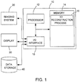
- the 3D reconstruction system 10 includes a processor 12 with an associated memory 14 having stored therein processor executable instructions 16 for configuring the processor 12 to perform various processes, namely the 3D reconstruction process, which process will be further described below.
- the image restoration system 10 further includes an input/output (I/O) interface 18 for communication with a 2D imaging system 20 and a display 30.
- I/O input/output
- the 3D reconstruction system 10 obtains at least two bi-planar images of the anatomical structure from the 2D imaging system 20 and executes the 3D reconstruction process on the acquired images.
- the resulting 3D reconstruction is then displayed on the display 30 and may be saved to the memory 14, to other data storage devices or medium 40, or provided to a further system via the I/O interface 18.
- PCA Principal Component Analysis
- a mixture of probabilistic PCA can be viewed as a problem of linear or locally linear dimensionality reduction. Specifically, it consists of an orthogonal projection onto a subspace of the original space.
- the observations can be transformed into data-centric and reduced by using the similarity between observations.
- the goal is to find a configuration of points y i with i ⁇ ⁇ 1, ⁇ , l ⁇ in a space of smaller dimension which retains the distances between initial points x i .
- the classical MDS algorithm can be achieved either by classical optimization methods (e.g. gradient descent or equivalent) or by algebraic methods, which can be briefly recalled as:
- the best strategies to achieve the MDS of data are to first use an algebraic method, imprecise but fast, and then use the resulting solution as initialization of an optimization method based on a gradient descent procedure or stochastic local exploration. This combination of analytical and algebraic methods of optimization provides an excellent compromise between computational speed and accuracy of the proposed solution.
- the MDS is widely used for data visualization, for example, in the field of the analysis of functional Magnetic Resonance Imaging (MRI), in molecular modeling or hyper-spectral imaging.
- MRI Magnetic Resonance Imaging
- MDS has variants as Stochastic Proximity Embedding (SPE), Curvilinear Component Analysis (CCA), SNE and FastMap.
- SPE Stochastic Proximity Embedding
- CCA Curvilinear Component Analysis
- SNE FastMap
- the 3D reconstruction process uses a segmentation method that takes into account both the notion of contour and region (detection of homogeneous zones). Cooperation between segmentation by region and contours contributes to an improved consideration of the characteristics of the anatomical structures in the images and, therefore, better segmentation thanks to the complementary nature of these two types of information. Thus, segmentation by region-contour cooperation can be expressed as a support between these two concepts in order to improve the final segmentation result.
- the size and more specifically the texture (i.e. distribution or statistical distribution) of the image gradient of each region present in the image is a discriminant information to distinguish bones from muscles.
- the bones have a gradient distribution with uniform orientation and muscles, a distribution gradient with a longitudinal orientation (corresponding to the direction of muscle fibers) and smaller amplitude.
- a requantized histogram of the amplitude and direction of the gradient is a discriminant information to segment the image (i.e. differentiate the different regions), which is then used for localization and 3D reconstruction.
- the segmentation method used by the 3D reconstruction process is based on the distribution of gradient texture and a non-linear MDS dimensionality reduction.
- a vector of dimension D having characteristics including parameters characterizing the gradient distribution of texture and orientation (considered in a small window centered about the pixel).
- the use of the non-linear MDS dimensionality reduction statistical method will non-linearly reduce the LxH textural (gradient) vectors of dimension D associated with each pixel of the images in a space of dimension d (with d ⁇ D ) maintaining "as best as can be” the considered distances (Euclidean or Bhattacharya).
- This procedure can be done using a multi-resolution approach similar to that proposed in M. Mignotte, "MDS-based multiresolution nonlinear dimensionality reduction model for color image segmentation", IEEE Transactions on Neural Networks, 22 (3) :447-460, March 2011 .
- This dimensionality reduction will result in the optimization of a cost function where will be introduced, in addition, to reflect the contours, a weighting factor to encourage the pairing of pixels which are not separated by a contour and thus promote consistency between pixels in a region.
- This non-linear dimensionality step allows the defining of a deformation mesh in a reduced dimensional space in which each point of the grid will actually represent a surface model of the learning set. This in turn allows the defining of a concise 3D reconstruction parametric model in which all eligible and statistically learned deformations from the learning set will be the values of a reduced vector of d parameters.
- the 3D reconstruction process consist then in the adjustment of the projections of an anatomical structure surface model (contained in this deformation mesh and by interpolation) with the contours of the segmented image, comprising the corresponding anatomical structure, by the method proposed in F. Destrempes, M. Mignotte, J.-F. Angers, "Localization of shapes using statistical models and stochastic optimization", IEEE Transactions on Pattern Analysis and Machine Intelligence, 29 (9):1603-1615, September 2007 .
- This 3D reconstruction process is based on a likelihood using contour pre-detection as well on a global constraint using the concept of specificity. This property is based on the fact that labels (of classes), provided by a textural pre-segmentation of the image, inside and outside of the form are different (or specific to a neighborhood of the object). This feature allows the basing of the likelihood of the Bayesian model on attributes that have the property to be very robust to noise.
- the problem of 3D reconstruction is then seen as a simple problem of deformation parameters estimation of the surface model or, equivalently, as a problem of minimizing a cost function of dimension d. This minimization can be achieved by a stochastic algorithm.
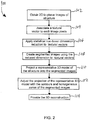
- FIG. 2 there is shown a flow diagram of an illustrative example of the 3D reconstruction process 100 executed by the processor 12 (see FIG. 1 ). Steps of the process 100 are indicated by blocks 102 to 114.
- the process 100 starts at block 102 where at least two 2D bi-planar images of a structure, for example an anatomical structure of a patient, are obtained from the imaging system 20.
- a textural vector is assigned to each pixel of each of the at least two bi-planar images.
- procedures that may be used include Isomap, Multi-Dimensional Scaling, Locally Linear Embedding, Semi-Definite Embedding and t-Distributed Stochastic Neighbor Embedding.
- segmented images of the at least two calibrated 2D bi-planar images are created using the reduced dimension textural vectors.
- the representative 3D model of the structure is projected onto the segmented images and, at block 112, the projection of the representative 3D model is adjusted with the contours and homogeneous zones of the segmented images using the optimization of a cost function to obtain the 3D reconstruction of the structure.
- the representative 3D model of the structure can be obtained using a learning set.
- the learning set contains, for each type of structure, a plurality of associated structure surface models. Associated with each structure surface model is a shape vector characterizing the shape of the structure surface model in 3D.
- a non-linear statistical dimensionality reduction procedure is then applied to the shape vectors to obtain reduced dimension shape vectors.
- a point in the reduced dimension space is then selected and then its corresponding reduced dimension shape vector is varied by a predefined interval.
- a predefined number of closest neighboring points to the selected point are then selected and the representative 3D model is then provided by interpolation using the shape vectors associated with the closest neighboring points to the selected point. It is to be understood that the predefined number of neighboring points to the selected point is a function of the degree of the interpolation.
Landscapes
- Engineering & Computer Science (AREA)
- Health & Medical Sciences (AREA)
- Life Sciences & Earth Sciences (AREA)
- Medical Informatics (AREA)
- Computer Vision & Pattern Recognition (AREA)
- Physics & Mathematics (AREA)
- Biomedical Technology (AREA)
- Surgery (AREA)
- Nuclear Medicine, Radiotherapy & Molecular Imaging (AREA)
- Optics & Photonics (AREA)
- Pathology (AREA)
- Radiology & Medical Imaging (AREA)
- Biophysics (AREA)
- Heart & Thoracic Surgery (AREA)
- Molecular Biology (AREA)
- High Energy & Nuclear Physics (AREA)
- Animal Behavior & Ethology (AREA)
- General Health & Medical Sciences (AREA)
- Public Health (AREA)
- Veterinary Medicine (AREA)
- General Physics & Mathematics (AREA)
- Theoretical Computer Science (AREA)
- Image Analysis (AREA)
- Image Processing (AREA)
Description
- The present disclosure relates to a method and system for the three-dimensional reconstruction of structures. More specifically, the present disclosure relates to a method and system for the three-dimensional reconstruction of anatomical structures such as bones.
- Methods of three-dimensional (3D) reconstruction associated with Computerized Tomography (CT) scan or Magnetic Resonance Imaging (MRI) systems provide accurate 3D reconstructions of human bone structures. However, CT-scan systems submit the patient to high radiation levels. In addition, a large volume of information needs to be processed and the patient is required to lie down for an extended period of time during examination, which does not allow for the observations of deformations when the patient is standing and under the influence of gravity. Although the MRI systems do not submit the patient to high radiation levels, they possess the same last two disadvantages of CT-scan systems. Another imaging technique, completely harmless to the patient is ultrasound imaging. The latter non-invasive technique, however, is not very suitable for the analysis of 3D geometry of the bone structure because they generate too much echo and measurement noise (parasitic echoes to the geometric surface shape of dense structures).
- The article of C. Koehler and T. Wischgoll "Knowledge-Assisted Reconstruction of the Human Rib Cage and Lungs", IEEE Computer Graphics and Applications, vol. 30, no. 1,2010, pp. 17-29, describes reconstruction algorithms to facilitate early detection of diseases, based on approximate volumetric reconstructions by using the inverse radon transform on X-ray image pairs.
- The article of M. Mignotte, "MDS-Based Multiresolution Nonlinear Dimensionality Reduction Model for Color Image Segmentation", IEEE Trans. Neural Netw., vol. 22, no. 3, 2011, pp. 447-460, describes Multi-Dimensional Scaling (MDS) for the segmentation of 2D images.
- Another 3-D reconstruction technique is presented in the article of Benameur et al., "A Hierarchical Statistical Modeling Approach for the Unsupervised 3-D Biplanar Reconstruction of Scoliotic Spine", IEEE Trans. Biomed. Eng., vol. 52, no. 12, 2005, pp. 2014-2057.
- The problem of 3D multi-planar reconstruction, which consists of reconstructing a 3D anatomical structure from multiple radiographic views, belongs to the class of ill-posed problems in the sense of Hadamard. For example, the acquisition of two bi-planar radiographic images provides a set of incomplete data that does not ensure the uniqueness of the solution.
- Accordingly, there is a need for to a method and system for the 3D reconstruction of structures and more specifically anatomical structures that addresses the above-described shortcomings.
- The present disclosure provides a method for the 3D reconstruction of a structure according to claim 1, and methods according to claims 2 and 3.
- The present disclosure also provides a system for the 3D reconstruction of a structure according to claim 4, and systems according to claims 5 to 8.
- The present disclosure further provides a processor executable product stored on a data storage medium according to claim 9 and configured to cause the processor to execute the steps of the method for the 3D reconstruction of a structure according to the present disclosure.
- Embodiments of the disclosure will be described by way of examples only with reference to the accompanying drawing, in which:
-
FIG. 1 is a schematic representation of a three-dimensional reconstruction system in accordance with an illustrative embodiment of the present disclosure; and -
FIG. 2 is a flow diagram of a three-dimensional reconstruction process in accordance with an illustrative embodiment of the present disclosure. - Similar references used in different Figures denote similar components.
- Generally stated, the non-limitative illustrative embodiment of the present disclosure provides a method and system for the three dimensional (3D) reconstruction of structures, more specifically anatomical structures such as bones. In the present disclosure, for illustrative purposes, the example of anatomical structures will be used, more precisely bones of the human body, but it is to be understood that the system and method can be used for other types of structures.
- The 3D reconstruction is achieved using calibrated bi-planar images of the anatomical structure from a two dimensional (2D) imaging system and a priori knowledge of the overall geometrical structure of each structure, e.g. anatomical structure or bone. This knowledge comes from a surface model learning set of considerable size, allowing modeling to take into account the anatomical variability and those related to diseases of the anatomical structures, to which is applied non-linear statistical dimension reduction, for example using the Multi-Dimensional Scaling (MDS) algorithm. The MDS algorithm allows the finding of a subspace of small dimension which preserves the metric chosen in the original space. This step allows the defining of a mesh deformation in a reduced dimensional space in which each point of the grid corresponds to a surface model of the learning set. This network in turn allows the defining of a concise 3D reconstruction parametric model in which all statistically admissible deformations from the learning set will be the values of a reduced parameter vector.
- The 3D reconstruction method then consists in adjusting the projections of an anatomical structure surface model with the contours and region (homogeneous zones) of the segmented images, comprising the corresponding anatomical structure, in the bi-planar images. The 3D reconstruction problem can thus be viewed as a problem of estimation of deformation parameters of a surface model or as a problem of minimizing a cost functions of dimension d. This minimization can be achieved by a stochastic algorithm.
- Referring to
FIG. 1 , the3D reconstruction system 10 includes aprocessor 12 with an associatedmemory 14 having stored thereinprocessor executable instructions 16 for configuring theprocessor 12 to perform various processes, namely the 3D reconstruction process, which process will be further described below. Theimage restoration system 10 further includes an input/output (I/O)interface 18 for communication with a2D imaging system 20 and adisplay 30. - The
3D reconstruction system 10 obtains at least two bi-planar images of the anatomical structure from the2D imaging system 20 and executes the 3D reconstruction process on the acquired images. The resulting 3D reconstruction is then displayed on thedisplay 30 and may be saved to thememory 14, to other data storage devices ormedium 40, or provided to a further system via the I/O interface 18. - The application of Principal Component Analysis (PCA) and/or a mixture of probabilistic PCA can be viewed as a problem of linear or locally linear dimensionality reduction. Specifically, it consists of an orthogonal projection onto a subspace of the original space.
- Briefly recalling some basic ideas of the PCA, let (xi ∈ R d}i=1,···,l be a set of observations described by d attributes. The PCA performs "optimal" graphic presentation of the observations minimizing the deformation of the cloud of points in a subspace of dimension q < d. Let x be the matrix of observations. For centric data, the variance of the principal component ck = Xpk where pk is a unit vector of the variance matrix

- Linear methods are not effective when the data are not distributed in a linear sub-space of the original space. To overcome this problem, many algorithms for non-linear dimensionality reduction have been developed. These algorithms are generally based on the idea that there is a non-linear sub-space that contains the data distribution. Among the algorithms proposed in the literature, the most popular are the Isomap, the Multi-Dimensional Scaling (MDS), Locally Linear Embedding (LLE), Semi-Definite Embedding (SDE) and t-Distributed Stochastic Neighbor Embedding (t-SNE). In the illustrative embodiment of the present disclosure, the approach used is the MDS but it is to be understood that in alternative embodiments other non-linear dimensionality reduction approaches may also be used.
- In the MDS statistical approach used in the illustrative embodiment, let there be l observations described by d attributes. Let X be the matrix of the observations (xi ∈ Rd ):

- The observations can be transformed into data-centric and reduced by using the similarity between observations. The distance matrix Δ defined by (dij ) where d(xi,xj ) = ||xi - xj || l
2 . The goal is to find a configuration of points yi with i ∈ {1,···,l} in a space of smaller dimension which retains the distances between initial points xi . In other words, we look for yi ∈ Rq, q < d such that d(yi ,yj ) ≈ dij with dij = d(xi,xj ). This results in an optimization function J defined by:
- The classical MDS algorithm can be achieved either by classical optimization methods (e.g. gradient descent or equivalent) or by algebraic methods, which can be briefly recalled as:
- From the distance matrix Δ, build the matrix M where M = HAH,

- Perform the spectral decomposition of de M ∈ Rlxl where M = WΛWt, W = [u 1 ··· ul ] ∈ Rlxl et Λ = diag(λ 1 ··· λl ). The matrix M has at most $d$ zero eigenvalues (assuming of course that l > d).
- For a configuration of points in a space of reduced dimension q, simply then consider

λ uj where j ∈ {1, ···, q}. - The best strategies to achieve the MDS of data are to first use an algebraic method, imprecise but fast, and then use the resulting solution as initialization of an optimization method based on a gradient descent procedure or stochastic local exploration. This combination of analytical and algebraic methods of optimization provides an excellent compromise between computational speed and accuracy of the proposed solution.
- The MDS is widely used for data visualization, for example, in the field of the analysis of functional Magnetic Resonance Imaging (MRI), in molecular modeling or hyper-spectral imaging. The popularity of MDS gave variants as Stochastic Proximity Embedding (SPE), Curvilinear Component Analysis (CCA), SNE and FastMap.
- The 3D reconstruction process uses a segmentation method that takes into account both the notion of contour and region (detection of homogeneous zones). Cooperation between segmentation by region and contours contributes to an improved consideration of the characteristics of the anatomical structures in the images and, therefore, better segmentation thanks to the complementary nature of these two types of information. Thus, segmentation by region-contour cooperation can be expressed as a support between these two concepts in order to improve the final segmentation result.
- In an anatomical structure image, only the size and more specifically the texture (i.e. distribution or statistical distribution) of the image gradient of each region present in the image is a discriminant information to distinguish bones from muscles. Indeed, the bones have a gradient distribution with uniform orientation and muscles, a distribution gradient with a longitudinal orientation (corresponding to the direction of muscle fibers) and smaller amplitude. A requantized histogram of the amplitude and direction of the gradient is a discriminant information to segment the image (i.e. differentiate the different regions), which is then used for localization and 3D reconstruction.
- Accordingly, the segmentation method used by the 3D reconstruction process is based on the distribution of gradient texture and a non-linear MDS dimensionality reduction. For each pixel (i,j) of each of the bi-planar images of size LxH will be associated a vector of dimension D, having characteristics including parameters characterizing the gradient distribution of texture and orientation (considered in a small window centered about the pixel). In this context, the use of the non-linear MDS dimensionality reduction statistical method will non-linearly reduce the LxH textural (gradient) vectors of dimension D associated with each pixel of the images in a space of dimension d (with d ≪ D) maintaining "as best as can be" the considered distances (Euclidean or Bhattacharya). This procedure can be done using a multi-resolution approach similar to that proposed in M. Mignotte, "MDS-based multiresolution nonlinear dimensionality reduction model for color image segmentation", IEEE Transactions on Neural Networks, 22 (3) :447-460, March 2011. This dimensionality reduction will result in the optimization of a cost function where will be introduced, in addition, to reflect the contours, a weighting factor to encourage the pairing of pixels which are not separated by a contour and thus promote consistency between pixels in a region. This dimensionality reduction with, for example, d = 1 will lead to a viewable grayscale image where different regions appear with reduced values and very different levels of gray for which a simple K-means clustering will reliably segment (and quickly) the image into two classes.
- This non-linear dimensionality step allows the defining of a deformation mesh in a reduced dimensional space in which each point of the grid will actually represent a surface model of the learning set. This in turn allows the defining of a concise 3D reconstruction parametric model in which all eligible and statistically learned deformations from the learning set will be the values of a reduced vector of d parameters.
- From this deformation non-linear mesh summarizing all statistically eligible surface structures, the 3D reconstruction process consist then in the adjustment of the projections of an anatomical structure surface model (contained in this deformation mesh and by interpolation) with the contours of the segmented image, comprising the corresponding anatomical structure, by the method proposed in F. Destrempes, M. Mignotte, J.-F. Angers, "Localization of shapes using statistical models and stochastic optimization", IEEE Transactions on Pattern Analysis and Machine Intelligence, 29 (9):1603-1615, September 2007.
- This 3D reconstruction process is based on a likelihood using contour pre-detection as well on a global constraint using the concept of specificity. This property is based on the fact that labels (of classes), provided by a textural pre-segmentation of the image, inside and outside of the form are different (or specific to a neighborhood of the object). This feature allows the basing of the likelihood of the Bayesian model on attributes that have the property to be very robust to noise.
- The problem of 3D reconstruction is then seen as a simple problem of deformation parameters estimation of the surface model or, equivalently, as a problem of minimizing a cost function of dimension d. This minimization can be achieved by a stochastic algorithm.
- Referring to
FIG. 2 , there is shown a flow diagram of an illustrative example of the3D reconstruction process 100 executed by the processor 12 (seeFIG. 1 ). Steps of theprocess 100 are indicated byblocks 102 to 114. - The
process 100 starts atblock 102 where at least two 2D bi-planar images of a structure, for example an anatomical structure of a patient, are obtained from theimaging system 20. - Then, at
block 104, a textural vector is assigned to each pixel of each of the at least two bi-planar images. The dimension D of the textural vectors on the quantization used for the gradient and orientation parameters. For example, using four (4) quantization intervals for the gradient (i.e. [0, 63], ]63, 128], ]128, 184], ]184, 255]) and four (4) quantization intervals for the orientation (i.e. [0, π/4],] π/4, π/2],] π/2, 3π/2], ]3π/2, π]), each vector with be of a dimension D=16 (each combination of gradient and orientation will be assigned a value representing the number of neighboring pixels having that specific combination). - At
block 106, a non-linear statistical dimensionality reduction procedure is applied to the textural vectors to reduce their dimension to d for example d = 2. Examples of procedures that may be used include Isomap, Multi-Dimensional Scaling, Locally Linear Embedding, Semi-Definite Embedding and t-Distributed Stochastic Neighbor Embedding. - At
block 108, segmented images of the at least two calibrated 2D bi-planar images are created using the reduced dimension textural vectors. - Then, at block 110, the representative 3D model of the structure is projected onto the segmented images and, at block 112, the projection of the representative 3D model is adjusted with the contours and homogeneous zones of the segmented images using the optimization of a cost function to obtain the 3D reconstruction of the structure.
- Finally, at
block 114, the 3D reconstruction of the structure is provided. - The representative 3D model of the structure can be obtained using a learning set. The learning set contains, for each type of structure, a plurality of associated structure surface models. Associated with each structure surface model is a shape vector characterizing the shape of the structure surface model in 3D. A non-linear statistical dimensionality reduction procedure is then applied to the shape vectors to obtain reduced dimension shape vectors. A point in the reduced dimension space is then selected and then its corresponding reduced dimension shape vector is varied by a predefined interval. A predefined number of closest neighboring points to the selected point are then selected and the representative 3D model is then provided by interpolation using the shape vectors associated with the closest neighboring points to the selected point. It is to be understood that the predefined number of neighboring points to the selected point is a function of the degree of the interpolation.
Claims (9)
- A method (100) for the 3D reconstruction of an anatomical structure, the method comprising:- obtaining (102) at least two calibrated 2D bi-planar images of the structure;- associating (104) with each pixel of each of the at least two calibrated 2D bi-planar images a textural vector characterizing the distribution of the gradient texture and orientation in a window centered around the pixel;- applying (106) a non-linear statistical dimensionality reduction procedure to the textural vectors to obtain reduced dimension textural vectors;- creating (108) segmented images of the at least two calibrated 2D bi-planar images using the reduced dimension textural vectors;- selecting a representative 3D model of the anatomical structure from a learning set containing a plurality of structure surface models, wherein the selection of the representative 3D model of the structure is performed by:- associating with each structure surface model a shape vector characterizing the shape of the structure surface model in 3D;- applying a non-linear statistical dimensionality reduction procedure to the shape vectors to obtain reduced dimension shape vectors;- selecting a point in the reduced dimension space;- varying the reduced dimension shape vector corresponding to the selected point by a predefined interval;- selecting a predefined number of closest neighboring points to the selected point; and- providing the representative 3D model by interpolation using the shape vectors associated with the closest neighboring points to the selected point;the predefined number of closest neighboring points being a function of the degree of the interpolation.- projecting (110) the representative 3D model onto the segmented images; and- adjusting (112) the projection with the contours and homogeneous zones of the segmented images by minimizing a cost function of the deformation parameters of the representative 3D model to obtain the 3D reconstruction of the anatomical structure.
- The method of claim 1, wherein the representative 3D model is selected from a plurality of proposed representative 3D models associated with respective points in the reduced dimension space.
- The method of claim 1 or 2, wherein the non-linear statistical dimensionality reduction procedure is selected from a group consisting of Isomap, Multi-Dimensional Scaling, Locally Linear Embedding, Semi-Definite Embedding and t-Distributed Stochastic Neighbor Embedding.
- A system (10) for the 3D reconstruction of an anatomical structure, comprising:an input/output interface (18) configured to receive at least two calibrated 2D bi-planar images;a database (14, 40) having stored therein a learning set containing a plurality of structure surface models; anda processor (12) in communication with the input/output interface (18) and the database (14, 40), wherein the processor is configured for:- associating with each pixel of each of the at least two calibrated 2D bi-planar images a textural vector characterizing the distribution of the gradient texture and orientation in a window centered around the pixel;- applying a non-linear statistical dimensionality reduction procedure to the textural vectors to obtain reduced dimension textural vectors;- creating segmented images of the at least two calibrated 2D bi-planar images using the reduced dimension textural vectors;- selecting a representative 3D model of the anatomical structure from the learning set, wherein the selection of the representative 3D model of the structure is performed by:- associating with each structure surface model a shape vector characterizing the shape of the structure surface model in 3D;- applying a non-linear statistical dimensionality reduction procedure to the shape vectors to obtain reduced dimension shape vectors;- selecting a point in the reduced dimension space;- varying the reduced dimension shape vector corresponding to the selected point by a predefined interval;- selecting a predefined number of closest neighboring points to the selected point; and- providing the representative 3D model by interpolation using the shape vectors associated with the closest neighboring points to the selected point;the predefined number of closest neighboring points being a function of the degree of the interpolation.- projecting the representative 3D model onto the segmented images; and- adjusting the projection with the contours and homogeneous zones of the segmented images by minimizing a cost function of the deformation parameters of the representative 3D model to obtain the 3D reconstruction of the anatomical structure.
- The system of claim 4, further comprising a display (30) in communication with the input/output interface (18), the input/output interface (18) being further configured to provide the 3D reconstruction of the structure to the display (30).
- The system of claims 4 or 5, further comprising a 2D imaging system (20) in communication with the input/output interface (18), the 2D imaging system (20) providing the at least two calibrated 2D bi-planar images to the input/output interface (18).
- The system of any of claims 4 to 6, wherein the non-linear statistical dimensionality reduction procedure is selected from a group consisting of Isomap, Multi-Dimensional Scaling, Locally Linear Embedding, Semi-Definite Embedding and t-Distributed Stochastic Neighbor Embedding.
- The system of any of claims 4 to 7, wherein the representative 3D model is selected from a plurality of proposed representative 3D models associated with respective points in the reduced dimension space.
- A processor executable product stored on a data storage medium and configured to cause the processor to perform operations comprising:- obtaining at least two calibrated 2D bi-planar images of an anatomical structure;- associating with each pixel of each of the at least two calibrated 2D bi-planar images a textural vector characterizing the distribution of the gradient texture and orientation in a window centered around the pixel;- applying a non-linear statistical dimensionality reduction procedure to the textural vectors to obtain reduced dimension textural vectors;- creating segmented images of the at least two calibrated 2D bi-planar images using the reduced dimension textural vectors;- selecting a representative 3D model of the anatomical structure from a learning set containing a plurality of structure surface models, wherein the selection of the representative 3D model of the structure is performed by:- associating with each structure surface model a shape vector characterizing the shape of the structure surface model in 3D;- applying a non-linear statistical dimensionality reduction procedure to the shape vectors to obtain reduced dimension shape vectors;- selecting a point in the reduced dimension space;- varying the reduced dimension shape vector corresponding to the selected point by a predefined interval;- selecting a predefined number of closest neighboring points to the selected point; and- providing the representative 3D model by interpolation using the shape vectors associated with the closest neighboring points to the selected point;the predefined number of closest neighboring points being a function of the degree of the interpolation.- projecting the representative 3D model onto the segmented images; and- adjusting the projection with the contours and homogeneous zones of the segmented images by minimizing a cost function of the deformation parameters of the representative 3D model to obtain the 3D reconstruction of the anatomical structure.
Applications Claiming Priority (3)
| Application Number | Priority Date | Filing Date | Title |
|---|---|---|---|
| CA2778347A CA2778347A1 (en) | 2012-05-17 | 2012-05-17 | Method and apparatus for non-linear statistical modeling |
| US201261688629P | 2012-05-18 | 2012-05-18 | |
| PCT/CA2013/000481 WO2013170360A1 (en) | 2012-05-17 | 2013-05-17 | Method and system for the three-dimensional reconstruction of structures |
Publications (3)
| Publication Number | Publication Date |
|---|---|
| EP2850596A1 EP2850596A1 (en) | 2015-03-25 |
| EP2850596A4 EP2850596A4 (en) | 2015-07-08 |
| EP2850596B1 true EP2850596B1 (en) | 2018-12-05 |
Family
ID=49582932
Family Applications (1)
| Application Number | Title | Priority Date | Filing Date |
|---|---|---|---|
| EP13791513.8A Active EP2850596B1 (en) | 2012-05-17 | 2013-05-17 | Method and system for the three-dimensional reconstruction of structures |
Country Status (3)
| Country | Link |
|---|---|
| EP (1) | EP2850596B1 (en) |
| CA (2) | CA2778347A1 (en) |
| WO (1) | WO2013170360A1 (en) |
Families Citing this family (5)
| Publication number | Priority date | Publication date | Assignee | Title |
|---|---|---|---|---|
| CN107817527B (en) * | 2017-10-31 | 2019-02-12 | 吉林大学 | A Random Noise Suppression Method for Desert Seismic Exploration Based on Block Sparse Compressed Sensing |
| CN108537750A (en) * | 2018-03-29 | 2018-09-14 | 哈尔滨理工大学 | A kind of quick spine CT 3-dimensional reconstruction method |
| CN113143243B (en) * | 2021-02-25 | 2023-08-22 | 厦门大学 | Deep learning magnetic resonance spectrum reconstruction method based on spectrum decomposition |
| CN117635875B (en) * | 2024-01-25 | 2024-05-14 | 深圳市其域创新科技有限公司 | Three-dimensional reconstruction method, device and terminal |
| CN119810125B (en) * | 2024-11-22 | 2025-06-24 | 中国人民解放军总医院第三医学中心 | Intelligent kidney wound surface segmentation system |
Family Cites Families (1)
| Publication number | Priority date | Publication date | Assignee | Title |
|---|---|---|---|---|
| WO2009058915A1 (en) * | 2007-10-29 | 2009-05-07 | The Trustees Of The University Of Pennsylvania | Computer assisted diagnosis (cad) of cancer using multi-functional, multi-modal in-vivo magnetic resonance spectroscopy (mrs) and imaging (mri) |
-
2012
- 2012-05-17 CA CA2778347A patent/CA2778347A1/en not_active Abandoned
-
2013
- 2013-05-17 EP EP13791513.8A patent/EP2850596B1/en active Active
- 2013-05-17 CA CA2873918A patent/CA2873918C/en active Active
- 2013-05-17 WO PCT/CA2013/000481 patent/WO2013170360A1/en not_active Ceased
Non-Patent Citations (1)
| Title |
|---|
| None * |
Also Published As
| Publication number | Publication date |
|---|---|
| CA2873918A1 (en) | 2013-11-21 |
| CA2778347A1 (en) | 2013-11-17 |
| WO2013170360A1 (en) | 2013-11-21 |
| EP2850596A4 (en) | 2015-07-08 |
| EP2850596A1 (en) | 2015-03-25 |
| CA2873918C (en) | 2020-09-15 |
Similar Documents
| Publication | Publication Date | Title |
|---|---|---|
| US11250601B2 (en) | Learning-assisted multi-modality dielectric imaging | |
| Azam et al. | Multimodal medical image registration and fusion for quality enhancement | |
| US11756160B2 (en) | ML-based methods for pseudo-CT and HR MR image estimation | |
| Staib et al. | Model-based deformable surface finding for medical images | |
| Milchenko et al. | Obscuring surface anatomy in volumetric imaging data | |
| US8135189B2 (en) | System and method for organ segmentation using surface patch classification in 2D and 3D images | |
| Ballester et al. | Segmentation and measurement of brain structures in MRI including confidence bounds | |
| Salehi et al. | Deep learning-based non-rigid image registration for high-dose rate brachytherapy in inter-fraction cervical cancer | |
| Lu et al. | Precise segmentation of multiple organs in CT volumes using learning-based approach and information theory | |
| US8929636B2 (en) | Method and system for image segmentation | |
| CA2873918C (en) | Method and system for the three-dimensional reconstruction of structures | |
| US10410355B2 (en) | Methods and systems for image analysis using non-euclidean deformed graphs | |
| Zanaty et al. | A kernelized fuzzy c-means algorithm for automatic magnetic resonance image segmentation | |
| Ciurte et al. | A semi-supervised patch-based approach for segmentation of fetal ultrasound imaging | |
| Pradhan et al. | Machine learning model for multi-view visualization of medical images | |
| US9235931B2 (en) | Method and system for the three-dimensional reconstruction of structures | |
| CN108961256B (en) | Image segmentation method, surgical navigation device, electronic device, and storage medium | |
| TW201124944A (en) | Medical image analysis system for displaying anatomical images subject to deformation and related methods | |
| Nain et al. | Shape-driven 3D segmentation using spherical wavelets | |
| Mazaheri et al. | Hybrid Pixel‐Based Method for Cardiac Ultrasound Fusion Based on Integration of PCA and DWT | |
| Athawale et al. | A statistical framework for quantification and visualisation of positional uncertainty in deep brain stimulation electrodes | |
| Han et al. | A momentum-based diffeomorphic demons framework for deformable MR-CT image registration | |
| Stough et al. | Regional appearance in deformable model segmentation | |
| Fang et al. | Superpixel/voxel medical image segmentation algorithm based on the regional interlinked value | |
| Lee et al. | Super-resolution multi-contrast unbiased eye atlases with deep probabilistic refinement |
Legal Events
| Date | Code | Title | Description |
|---|---|---|---|
| PUAI | Public reference made under article 153(3) epc to a published international application that has entered the european phase |
Free format text: ORIGINAL CODE: 0009012 |
|
| 17P | Request for examination filed |
Effective date: 20141212 |
|
| AK | Designated contracting states |
Kind code of ref document: A1 Designated state(s): AL AT BE BG CH CY CZ DE DK EE ES FI FR GB GR HR HU IE IS IT LI LT LU LV MC MK MT NL NO PL PT RO RS SE SI SK SM TR |
|
| AX | Request for extension of the european patent |
Extension state: BA ME |
|
| RA4 | Supplementary search report drawn up and despatched (corrected) |
Effective date: 20150609 |
|
| RIC1 | Information provided on ipc code assigned before grant |
Ipc: A61B 6/02 20060101ALI20150602BHEP Ipc: G06T 17/00 20060101AFI20150602BHEP |
|
| DAX | Request for extension of the european patent (deleted) | ||
| STAA | Information on the status of an ep patent application or granted ep patent |
Free format text: STATUS: EXAMINATION IS IN PROGRESS |
|
| 17Q | First examination report despatched |
Effective date: 20170609 |
|
| GRAP | Despatch of communication of intention to grant a patent |
Free format text: ORIGINAL CODE: EPIDOSNIGR1 |
|
| STAA | Information on the status of an ep patent application or granted ep patent |
Free format text: STATUS: GRANT OF PATENT IS INTENDED |
|
| INTG | Intention to grant announced |
Effective date: 20180629 |
|
| RAP1 | Party data changed (applicant data changed or rights of an application transferred) |
Owner name: EIFFEL MEDTECH INC. |
|
| GRAS | Grant fee paid |
Free format text: ORIGINAL CODE: EPIDOSNIGR3 |
|
| GRAA | (expected) grant |
Free format text: ORIGINAL CODE: 0009210 |
|
| STAA | Information on the status of an ep patent application or granted ep patent |
Free format text: STATUS: THE PATENT HAS BEEN GRANTED |
|
| AK | Designated contracting states |
Kind code of ref document: B1 Designated state(s): AL AT BE BG CH CY CZ DE DK EE ES FI FR GB GR HR HU IE IS IT LI LT LU LV MC MK MT NL NO PL PT RO RS SE SI SK SM TR |
|
| REG | Reference to a national code |
Ref country code: GB Ref legal event code: FG4D |
|
| REG | Reference to a national code |
Ref country code: CH Ref legal event code: EP |
|
| REG | Reference to a national code |
Ref country code: AT Ref legal event code: REF Ref document number: 1074019 Country of ref document: AT Kind code of ref document: T Effective date: 20181215 |
|
| REG | Reference to a national code |
Ref country code: IE Ref legal event code: FG4D |
|
| REG | Reference to a national code |
Ref country code: DE Ref legal event code: R096 Ref document number: 602013047872 Country of ref document: DE |
|
| REG | Reference to a national code |
Ref country code: NL Ref legal event code: MP Effective date: 20181205 |
|
| REG | Reference to a national code |
Ref country code: AT Ref legal event code: MK05 Ref document number: 1074019 Country of ref document: AT Kind code of ref document: T Effective date: 20181205 |
|
| REG | Reference to a national code |
Ref country code: LT Ref legal event code: MG4D |
|
| PG25 | Lapsed in a contracting state [announced via postgrant information from national office to epo] |
Ref country code: FI Free format text: LAPSE BECAUSE OF FAILURE TO SUBMIT A TRANSLATION OF THE DESCRIPTION OR TO PAY THE FEE WITHIN THE PRESCRIBED TIME-LIMIT Effective date: 20181205 Ref country code: NO Free format text: LAPSE BECAUSE OF FAILURE TO SUBMIT A TRANSLATION OF THE DESCRIPTION OR TO PAY THE FEE WITHIN THE PRESCRIBED TIME-LIMIT Effective date: 20190305 Ref country code: AT Free format text: LAPSE BECAUSE OF FAILURE TO SUBMIT A TRANSLATION OF THE DESCRIPTION OR TO PAY THE FEE WITHIN THE PRESCRIBED TIME-LIMIT Effective date: 20181205 Ref country code: ES Free format text: LAPSE BECAUSE OF FAILURE TO SUBMIT A TRANSLATION OF THE DESCRIPTION OR TO PAY THE FEE WITHIN THE PRESCRIBED TIME-LIMIT Effective date: 20181205 Ref country code: LV Free format text: LAPSE BECAUSE OF FAILURE TO SUBMIT A TRANSLATION OF THE DESCRIPTION OR TO PAY THE FEE WITHIN THE PRESCRIBED TIME-LIMIT Effective date: 20181205 Ref country code: LT Free format text: LAPSE BECAUSE OF FAILURE TO SUBMIT A TRANSLATION OF THE DESCRIPTION OR TO PAY THE FEE WITHIN THE PRESCRIBED TIME-LIMIT Effective date: 20181205 Ref country code: HR Free format text: LAPSE BECAUSE OF FAILURE TO SUBMIT A TRANSLATION OF THE DESCRIPTION OR TO PAY THE FEE WITHIN THE PRESCRIBED TIME-LIMIT Effective date: 20181205 Ref country code: BG Free format text: LAPSE BECAUSE OF FAILURE TO SUBMIT A TRANSLATION OF THE DESCRIPTION OR TO PAY THE FEE WITHIN THE PRESCRIBED TIME-LIMIT Effective date: 20190305 |
|
| PG25 | Lapsed in a contracting state [announced via postgrant information from national office to epo] |
Ref country code: AL Free format text: LAPSE BECAUSE OF FAILURE TO SUBMIT A TRANSLATION OF THE DESCRIPTION OR TO PAY THE FEE WITHIN THE PRESCRIBED TIME-LIMIT Effective date: 20181205 Ref country code: GR Free format text: LAPSE BECAUSE OF FAILURE TO SUBMIT A TRANSLATION OF THE DESCRIPTION OR TO PAY THE FEE WITHIN THE PRESCRIBED TIME-LIMIT Effective date: 20190306 Ref country code: SE Free format text: LAPSE BECAUSE OF FAILURE TO SUBMIT A TRANSLATION OF THE DESCRIPTION OR TO PAY THE FEE WITHIN THE PRESCRIBED TIME-LIMIT Effective date: 20181205 Ref country code: RS Free format text: LAPSE BECAUSE OF FAILURE TO SUBMIT A TRANSLATION OF THE DESCRIPTION OR TO PAY THE FEE WITHIN THE PRESCRIBED TIME-LIMIT Effective date: 20181205 |
|
| PG25 | Lapsed in a contracting state [announced via postgrant information from national office to epo] |
Ref country code: NL Free format text: LAPSE BECAUSE OF FAILURE TO SUBMIT A TRANSLATION OF THE DESCRIPTION OR TO PAY THE FEE WITHIN THE PRESCRIBED TIME-LIMIT Effective date: 20181205 |
|
| PG25 | Lapsed in a contracting state [announced via postgrant information from national office to epo] |
Ref country code: IT Free format text: LAPSE BECAUSE OF FAILURE TO SUBMIT A TRANSLATION OF THE DESCRIPTION OR TO PAY THE FEE WITHIN THE PRESCRIBED TIME-LIMIT Effective date: 20181205 Ref country code: CZ Free format text: LAPSE BECAUSE OF FAILURE TO SUBMIT A TRANSLATION OF THE DESCRIPTION OR TO PAY THE FEE WITHIN THE PRESCRIBED TIME-LIMIT Effective date: 20181205 Ref country code: PT Free format text: LAPSE BECAUSE OF FAILURE TO SUBMIT A TRANSLATION OF THE DESCRIPTION OR TO PAY THE FEE WITHIN THE PRESCRIBED TIME-LIMIT Effective date: 20190405 Ref country code: PL Free format text: LAPSE BECAUSE OF FAILURE TO SUBMIT A TRANSLATION OF THE DESCRIPTION OR TO PAY THE FEE WITHIN THE PRESCRIBED TIME-LIMIT Effective date: 20181205 |
|
| PG25 | Lapsed in a contracting state [announced via postgrant information from national office to epo] |
Ref country code: EE Free format text: LAPSE BECAUSE OF FAILURE TO SUBMIT A TRANSLATION OF THE DESCRIPTION OR TO PAY THE FEE WITHIN THE PRESCRIBED TIME-LIMIT Effective date: 20181205 Ref country code: SM Free format text: LAPSE BECAUSE OF FAILURE TO SUBMIT A TRANSLATION OF THE DESCRIPTION OR TO PAY THE FEE WITHIN THE PRESCRIBED TIME-LIMIT Effective date: 20181205 Ref country code: SK Free format text: LAPSE BECAUSE OF FAILURE TO SUBMIT A TRANSLATION OF THE DESCRIPTION OR TO PAY THE FEE WITHIN THE PRESCRIBED TIME-LIMIT Effective date: 20181205 Ref country code: RO Free format text: LAPSE BECAUSE OF FAILURE TO SUBMIT A TRANSLATION OF THE DESCRIPTION OR TO PAY THE FEE WITHIN THE PRESCRIBED TIME-LIMIT Effective date: 20181205 Ref country code: IS Free format text: LAPSE BECAUSE OF FAILURE TO SUBMIT A TRANSLATION OF THE DESCRIPTION OR TO PAY THE FEE WITHIN THE PRESCRIBED TIME-LIMIT Effective date: 20190405 |
|
| REG | Reference to a national code |
Ref country code: DE Ref legal event code: R097 Ref document number: 602013047872 Country of ref document: DE |
|
| PLBE | No opposition filed within time limit |
Free format text: ORIGINAL CODE: 0009261 |
|
| STAA | Information on the status of an ep patent application or granted ep patent |
Free format text: STATUS: NO OPPOSITION FILED WITHIN TIME LIMIT |
|
| PG25 | Lapsed in a contracting state [announced via postgrant information from national office to epo] |
Ref country code: DK Free format text: LAPSE BECAUSE OF FAILURE TO SUBMIT A TRANSLATION OF THE DESCRIPTION OR TO PAY THE FEE WITHIN THE PRESCRIBED TIME-LIMIT Effective date: 20181205 Ref country code: SI Free format text: LAPSE BECAUSE OF FAILURE TO SUBMIT A TRANSLATION OF THE DESCRIPTION OR TO PAY THE FEE WITHIN THE PRESCRIBED TIME-LIMIT Effective date: 20181205 |
|
| 26N | No opposition filed |
Effective date: 20190906 |
|
| REG | Reference to a national code |
Ref country code: CH Ref legal event code: PL |
|
| PG25 | Lapsed in a contracting state [announced via postgrant information from national office to epo] |
Ref country code: LI Free format text: LAPSE BECAUSE OF NON-PAYMENT OF DUE FEES Effective date: 20190531 Ref country code: CH Free format text: LAPSE BECAUSE OF NON-PAYMENT OF DUE FEES Effective date: 20190531 Ref country code: MC Free format text: LAPSE BECAUSE OF FAILURE TO SUBMIT A TRANSLATION OF THE DESCRIPTION OR TO PAY THE FEE WITHIN THE PRESCRIBED TIME-LIMIT Effective date: 20181205 |
|
| REG | Reference to a national code |
Ref country code: BE Ref legal event code: MM Effective date: 20190531 |
|
| PG25 | Lapsed in a contracting state [announced via postgrant information from national office to epo] |
Ref country code: LU Free format text: LAPSE BECAUSE OF NON-PAYMENT OF DUE FEES Effective date: 20190517 |
|
| PG25 | Lapsed in a contracting state [announced via postgrant information from national office to epo] |
Ref country code: TR Free format text: LAPSE BECAUSE OF FAILURE TO SUBMIT A TRANSLATION OF THE DESCRIPTION OR TO PAY THE FEE WITHIN THE PRESCRIBED TIME-LIMIT Effective date: 20181205 |
|
| PG25 | Lapsed in a contracting state [announced via postgrant information from national office to epo] |
Ref country code: IE Free format text: LAPSE BECAUSE OF NON-PAYMENT OF DUE FEES Effective date: 20190517 |
|
| PG25 | Lapsed in a contracting state [announced via postgrant information from national office to epo] |
Ref country code: BE Free format text: LAPSE BECAUSE OF NON-PAYMENT OF DUE FEES Effective date: 20190531 |
|
| PG25 | Lapsed in a contracting state [announced via postgrant information from national office to epo] |
Ref country code: CY Free format text: LAPSE BECAUSE OF FAILURE TO SUBMIT A TRANSLATION OF THE DESCRIPTION OR TO PAY THE FEE WITHIN THE PRESCRIBED TIME-LIMIT Effective date: 20181205 |
|
| PG25 | Lapsed in a contracting state [announced via postgrant information from national office to epo] |
Ref country code: HU Free format text: LAPSE BECAUSE OF FAILURE TO SUBMIT A TRANSLATION OF THE DESCRIPTION OR TO PAY THE FEE WITHIN THE PRESCRIBED TIME-LIMIT; INVALID AB INITIO Effective date: 20130517 Ref country code: MT Free format text: LAPSE BECAUSE OF FAILURE TO SUBMIT A TRANSLATION OF THE DESCRIPTION OR TO PAY THE FEE WITHIN THE PRESCRIBED TIME-LIMIT Effective date: 20181205 |
|
| PG25 | Lapsed in a contracting state [announced via postgrant information from national office to epo] |
Ref country code: MK Free format text: LAPSE BECAUSE OF FAILURE TO SUBMIT A TRANSLATION OF THE DESCRIPTION OR TO PAY THE FEE WITHIN THE PRESCRIBED TIME-LIMIT Effective date: 20181205 |
|
| PGFP | Annual fee paid to national office [announced via postgrant information from national office to epo] |
Ref country code: DE Payment date: 20250408 Year of fee payment: 13 |
|
| PGFP | Annual fee paid to national office [announced via postgrant information from national office to epo] |
Ref country code: GB Payment date: 20250407 Year of fee payment: 13 |
|
| PGFP | Annual fee paid to national office [announced via postgrant information from national office to epo] |
Ref country code: FR Payment date: 20250407 Year of fee payment: 13 |