EP2837389A1 - Medicinal composition for treating infarction - Google Patents
Medicinal composition for treating infarction Download PDFInfo
- Publication number
- EP2837389A1 EP2837389A1 EP13776214.2A EP13776214A EP2837389A1 EP 2837389 A1 EP2837389 A1 EP 2837389A1 EP 13776214 A EP13776214 A EP 13776214A EP 2837389 A1 EP2837389 A1 EP 2837389A1
- Authority
- EP
- European Patent Office
- Prior art keywords
- infarction
- hsa
- mir
- autophagy
- treating
- Prior art date
- Legal status (The legal status is an assumption and is not a legal conclusion. Google has not performed a legal analysis and makes no representation as to the accuracy of the status listed.)
- Withdrawn
Links
- 206010061216 Infarction Diseases 0.000 title claims abstract description 71
- 230000007574 infarction Effects 0.000 title claims abstract description 71
- 239000000203 mixture Substances 0.000 title description 6
- 230000004900 autophagic degradation Effects 0.000 claims abstract description 80
- 108091069002 Homo sapiens miR-145 stem-loop Proteins 0.000 claims abstract description 67
- 239000003623 enhancer Substances 0.000 claims abstract description 66
- 239000008194 pharmaceutical composition Substances 0.000 claims abstract description 42
- 208000010125 myocardial infarction Diseases 0.000 claims abstract description 31
- 238000000034 method Methods 0.000 claims abstract description 24
- 239000004480 active ingredient Substances 0.000 claims abstract description 10
- 206010008118 cerebral infarction Diseases 0.000 claims abstract description 10
- 208000026106 cerebrovascular disease Diseases 0.000 claims abstract description 10
- 206010058571 Spinal cord infarction Diseases 0.000 claims description 7
- 238000001990 intravenous administration Methods 0.000 claims description 7
- 238000004519 manufacturing process Methods 0.000 claims description 5
- 238000002560 therapeutic procedure Methods 0.000 abstract description 9
- 239000003527 fibrinolytic agent Substances 0.000 abstract description 4
- 229960000103 thrombolytic agent Drugs 0.000 abstract description 4
- 239000002679 microRNA Substances 0.000 description 27
- 239000002502 liposome Substances 0.000 description 26
- 108091070501 miRNA Proteins 0.000 description 18
- 210000004027 cell Anatomy 0.000 description 17
- 238000002360 preparation method Methods 0.000 description 16
- 108090000623 proteins and genes Proteins 0.000 description 14
- 239000002773 nucleotide Substances 0.000 description 11
- 125000003729 nucleotide group Chemical group 0.000 description 11
- 102000004169 proteins and genes Human genes 0.000 description 11
- 230000009471 action Effects 0.000 description 9
- 210000001519 tissue Anatomy 0.000 description 9
- FAPWRFPIFSIZLT-UHFFFAOYSA-M Sodium chloride Chemical compound [Na+].[Cl-] FAPWRFPIFSIZLT-UHFFFAOYSA-M 0.000 description 8
- 238000010171 animal model Methods 0.000 description 7
- 239000000654 additive Substances 0.000 description 6
- 238000002347 injection Methods 0.000 description 6
- 239000007924 injection Substances 0.000 description 6
- 230000000302 ischemic effect Effects 0.000 description 6
- 108091007428 primary miRNA Proteins 0.000 description 6
- 239000000243 solution Substances 0.000 description 6
- XLYOFNOQVPJJNP-UHFFFAOYSA-N water Substances O XLYOFNOQVPJJNP-UHFFFAOYSA-N 0.000 description 6
- HDTRYLNUVZCQOY-UHFFFAOYSA-N α-D-glucopyranosyl-α-D-glucopyranoside Natural products OC1C(O)C(O)C(CO)OC1OC1C(O)C(O)C(O)C(CO)O1 HDTRYLNUVZCQOY-UHFFFAOYSA-N 0.000 description 5
- FFHPXOJTVQDVMO-DSYKOEDSSA-N (2r,3s,5r)-5-(6-aminopurin-9-yl)-2-methyloxolan-3-ol Chemical compound C1[C@H](O)[C@@H](C)O[C@H]1N1C2=NC=NC(N)=C2N=C1 FFHPXOJTVQDVMO-DSYKOEDSSA-N 0.000 description 5
- SVJMLYUFVDMUHP-XIFFEERXSA-N (4S)-2,6-dimethyl-4-(3-nitrophenyl)-1,4-dihydropyridine-3,5-dicarboxylic acid O5-[3-(4,4-diphenyl-1-piperidinyl)propyl] ester O3-methyl ester Chemical compound C1([C@@H]2C(=C(C)NC(C)=C2C(=O)OC)C(=O)OCCCN2CCC(CC2)(C=2C=CC=CC=2)C=2C=CC=CC=2)=CC=CC([N+]([O-])=O)=C1 SVJMLYUFVDMUHP-XIFFEERXSA-N 0.000 description 5
- PVHUJELLJLJGLN-INIZCTEOSA-N (S)-nitrendipine Chemical compound CCOC(=O)C1=C(C)NC(C)=C(C(=O)OC)[C@@H]1C1=CC=CC([N+]([O-])=O)=C1 PVHUJELLJLJGLN-INIZCTEOSA-N 0.000 description 5
- SGTNSNPWRIOYBX-UHFFFAOYSA-N 2-(3,4-dimethoxyphenyl)-5-{[2-(3,4-dimethoxyphenyl)ethyl](methyl)amino}-2-(propan-2-yl)pentanenitrile Chemical compound C1=C(OC)C(OC)=CC=C1CCN(C)CCCC(C#N)(C(C)C)C1=CC=C(OC)C(OC)=C1 SGTNSNPWRIOYBX-UHFFFAOYSA-N 0.000 description 5
- UIAGMCDKSXEBJQ-IBGZPJMESA-N 3-o-(2-methoxyethyl) 5-o-propan-2-yl (4s)-2,6-dimethyl-4-(3-nitrophenyl)-1,4-dihydropyridine-3,5-dicarboxylate Chemical compound COCCOC(=O)C1=C(C)NC(C)=C(C(=O)OC(C)C)[C@H]1C1=CC=CC([N+]([O-])=O)=C1 UIAGMCDKSXEBJQ-IBGZPJMESA-N 0.000 description 5
- QOYHHIBFXOOADH-UHFFFAOYSA-N 8-[4,4-bis(4-fluorophenyl)butyl]-1-phenyl-1,3,8-triazaspiro[4.5]decan-4-one Chemical compound C1=CC(F)=CC=C1C(C=1C=CC(F)=CC=1)CCCN1CCC2(C(NCN2C=2C=CC=CC=2)=O)CC1 QOYHHIBFXOOADH-UHFFFAOYSA-N 0.000 description 5
- ITPDYQOUSLNIHG-UHFFFAOYSA-N Amiodarone hydrochloride Chemical compound [Cl-].CCCCC=1OC2=CC=CC=C2C=1C(=O)C1=CC(I)=C(OCC[NH+](CC)CC)C(I)=C1 ITPDYQOUSLNIHG-UHFFFAOYSA-N 0.000 description 5
- 102100035037 Calpastatin Human genes 0.000 description 5
- PGGUOGKHUUUWAF-ROUUACIJSA-N Calpeptin Chemical compound CCCC[C@@H](C=O)NC(=O)[C@H](CC(C)C)NC(=O)OCC1=CC=CC=C1 PGGUOGKHUUUWAF-ROUUACIJSA-N 0.000 description 5
- GJSURZIOUXUGAL-UHFFFAOYSA-N Clonidine Chemical compound ClC1=CC=CC(Cl)=C1NC1=NCCN1 GJSURZIOUXUGAL-UHFFFAOYSA-N 0.000 description 5
- ZFMITUMMTDLWHR-UHFFFAOYSA-N Minoxidil Chemical compound NC1=[N+]([O-])C(N)=CC(N2CCCCC2)=N1 ZFMITUMMTDLWHR-UHFFFAOYSA-N 0.000 description 5
- JDUWHZOLEDOQSR-JKPSMKLGSA-N Penitrem A Chemical compound O([C@H]1[C@H]2[C@@](C(N3)=C11)(C)[C@@]4(C)CC[C@@H]5O[C@@H]([C@@H]([C@H]6O[C@@]56[C@]4(O)CC2)O)C(=C)C)C(C)(C)[C@H]2C[C@H]4[C@]2(O)C2=C1C3=CC(Cl)=C2CC4=C JDUWHZOLEDOQSR-JKPSMKLGSA-N 0.000 description 5
- HDTRYLNUVZCQOY-WSWWMNSNSA-N Trehalose Natural products O[C@@H]1[C@@H](O)[C@@H](O)[C@@H](CO)O[C@@H]1O[C@@H]1[C@H](O)[C@@H](O)[C@@H](O)[C@@H](CO)O1 HDTRYLNUVZCQOY-WSWWMNSNSA-N 0.000 description 5
- HDTRYLNUVZCQOY-LIZSDCNHSA-N alpha,alpha-trehalose Chemical compound O[C@@H]1[C@@H](O)[C@H](O)[C@@H](CO)O[C@@H]1O[C@@H]1[C@H](O)[C@@H](O)[C@H](O)[C@@H](CO)O1 HDTRYLNUVZCQOY-LIZSDCNHSA-N 0.000 description 5
- 229960005260 amiodarone Drugs 0.000 description 5
- 239000007864 aqueous solution Substances 0.000 description 5
- 108010044208 calpastatin Proteins 0.000 description 5
- ZXJCOYBPXOBJMU-HSQGJUDPSA-N calpastatin peptide Ac 184-210 Chemical compound C([C@@H](C(=O)N[C@@H]([C@@H](C)CC)C(=O)N[C@@H](CCC(O)=O)C(=O)N[C@@H](CCC(O)=O)C(=O)N[C@@H](CC(C)C)C(=O)NCC(=O)N[C@@H](CCCCN)C(=O)N[C@@H](CCCNC(N)=N)C(=O)N[C@@H](CCC(O)=O)C(=O)N[C@@H](C(C)C)C(=O)N[C@@H]([C@@H](C)O)C(=O)N[C@@H]([C@@H](C)CC)C(=O)N1[C@@H](CCC1)C(=O)N1[C@@H](CCC1)C(=O)N[C@@H](CCCCN)C(=O)N[C@@H](CC=1C=CC(O)=CC=1)C(=O)N[C@@H](CCCNC(N)=N)C(=O)N[C@@H](CCC(O)=O)C(=O)N[C@@H](CC(C)C)C(=O)N[C@@H](CC(C)C)C(=O)N[C@@H](C)C(N)=O)NC(=O)[C@@H](NC(=O)[C@H](CO)NC(=O)[C@H](CO)NC(=O)[C@H](CCSC)NC(=O)[C@H]1N(CCC1)C(=O)[C@H](CC(O)=O)NC(C)=O)[C@@H](C)O)C1=CC=C(O)C=C1 ZXJCOYBPXOBJMU-HSQGJUDPSA-N 0.000 description 5
- 108010082989 calpeptin Proteins 0.000 description 5
- FFGPTBGBLSHEPO-UHFFFAOYSA-N carbamazepine Chemical compound C1=CC2=CC=CC=C2N(C(=O)N)C2=CC=CC=C21 FFGPTBGBLSHEPO-UHFFFAOYSA-N 0.000 description 5
- 229960000623 carbamazepine Drugs 0.000 description 5
- 229960002896 clonidine Drugs 0.000 description 5
- NIJJYAXOARWZEE-UHFFFAOYSA-N di-n-propyl-acetic acid Natural products CCCC(C(O)=O)CCC NIJJYAXOARWZEE-UHFFFAOYSA-N 0.000 description 5
- 238000002474 experimental method Methods 0.000 description 5
- 229960003532 fluspirilene Drugs 0.000 description 5
- 210000005003 heart tissue Anatomy 0.000 description 5
- 238000001802 infusion Methods 0.000 description 5
- RDOIQAHITMMDAJ-UHFFFAOYSA-N loperamide Chemical compound C=1C=CC=CC=1C(C=1C=CC=CC=1)(C(=O)N(C)C)CCN(CC1)CCC1(O)C1=CC=C(Cl)C=C1 RDOIQAHITMMDAJ-UHFFFAOYSA-N 0.000 description 5
- 229960001571 loperamide Drugs 0.000 description 5
- 229960003632 minoxidil Drugs 0.000 description 5
- 229950010800 niguldipine Drugs 0.000 description 5
- 229960000715 nimodipine Drugs 0.000 description 5
- 229960005425 nitrendipine Drugs 0.000 description 5
- JDUWHZOLEDOQSR-UHFFFAOYSA-N penitrem A Natural products C1CC2(O)C34OC4C(O)C(C(=C)C)OC3CCC2(C)C(C(N2)=C34)(C)C1C3OC(C)(C)C1CC3C1(O)C1=C4C2=CC(Cl)=C1CC3=C JDUWHZOLEDOQSR-UHFFFAOYSA-N 0.000 description 5
- YVUQSNJEYSNKRX-UHFFFAOYSA-N pimozide Chemical compound C1=CC(F)=CC=C1C(C=1C=CC(F)=CC=1)CCCN1CCC(N2C(NC3=CC=CC=C32)=O)CC1 YVUQSNJEYSNKRX-UHFFFAOYSA-N 0.000 description 5
- 229960003634 pimozide Drugs 0.000 description 5
- ZAHRKKWIAAJSAO-UHFFFAOYSA-N rapamycin Natural products COCC(O)C(=C/C(C)C(=O)CC(OC(=O)C1CCCCN1C(=O)C(=O)C2(O)OC(CC(OC)C(=CC=CC=CC(C)CC(C)C(=O)C)C)CCC2C)C(C)CC3CCC(O)C(C3)OC)C ZAHRKKWIAAJSAO-UHFFFAOYSA-N 0.000 description 5
- QFJCIRLUMZQUOT-HPLJOQBZSA-N sirolimus Chemical compound C1C[C@@H](O)[C@H](OC)C[C@@H]1C[C@@H](C)[C@H]1OC(=O)[C@@H]2CCCCN2C(=O)C(=O)[C@](O)(O2)[C@H](C)CC[C@H]2C[C@H](OC)/C(C)=C/C=C/C=C/[C@@H](C)C[C@@H](C)C(=O)[C@H](OC)[C@H](O)/C(C)=C/[C@@H](C)C(=O)C1 QFJCIRLUMZQUOT-HPLJOQBZSA-N 0.000 description 5
- 229960002930 sirolimus Drugs 0.000 description 5
- AEQFSUDEHCCHBT-UHFFFAOYSA-M sodium valproate Chemical compound [Na+].CCCC(C([O-])=O)CCC AEQFSUDEHCCHBT-UHFFFAOYSA-M 0.000 description 5
- 229940084026 sodium valproate Drugs 0.000 description 5
- 229940074410 trehalose Drugs 0.000 description 5
- ZEWQUBUPAILYHI-UHFFFAOYSA-N trifluoperazine Chemical compound C1CN(C)CCN1CCCN1C2=CC(C(F)(F)F)=CC=C2SC2=CC=CC=C21 ZEWQUBUPAILYHI-UHFFFAOYSA-N 0.000 description 5
- 229960002324 trifluoperazine Drugs 0.000 description 5
- 229960001722 verapamil Drugs 0.000 description 5
- 102100021066 Fibroblast growth factor receptor substrate 2 Human genes 0.000 description 4
- 101710126950 Fibroblast growth factor receptor substrate 2 Proteins 0.000 description 4
- DHMQDGOQFOQNFH-UHFFFAOYSA-N Glycine Chemical compound NCC(O)=O DHMQDGOQFOQNFH-UHFFFAOYSA-N 0.000 description 4
- 230000037396 body weight Effects 0.000 description 4
- 239000003795 chemical substances by application Substances 0.000 description 4
- 230000014509 gene expression Effects 0.000 description 4
- 239000002245 particle Substances 0.000 description 4
- 230000001575 pathological effect Effects 0.000 description 4
- -1 pilmenidine Chemical compound 0.000 description 4
- 239000011780 sodium chloride Substances 0.000 description 4
- 108091032973 (ribonucleotides)n+m Proteins 0.000 description 3
- 102000007469 Actins Human genes 0.000 description 3
- 108010085238 Actins Proteins 0.000 description 3
- WQZGKKKJIJFFOK-GASJEMHNSA-N Glucose Natural products OC[C@H]1OC(O)[C@H](O)[C@@H](O)[C@@H]1O WQZGKKKJIJFFOK-GASJEMHNSA-N 0.000 description 3
- PEDCQBHIVMGVHV-UHFFFAOYSA-N Glycerine Chemical compound OCC(O)CO PEDCQBHIVMGVHV-UHFFFAOYSA-N 0.000 description 3
- 101001139130 Homo sapiens Krueppel-like factor 5 Proteins 0.000 description 3
- 102100020680 Krueppel-like factor 5 Human genes 0.000 description 3
- OKKJLVBELUTLKV-UHFFFAOYSA-N Methanol Chemical compound OC OKKJLVBELUTLKV-UHFFFAOYSA-N 0.000 description 3
- 241000283973 Oryctolagus cuniculus Species 0.000 description 3
- 210000004957 autophagosome Anatomy 0.000 description 3
- 210000004204 blood vessel Anatomy 0.000 description 3
- 150000001720 carbohydrates Chemical class 0.000 description 3
- 239000000969 carrier Substances 0.000 description 3
- 230000000052 comparative effect Effects 0.000 description 3
- 210000004351 coronary vessel Anatomy 0.000 description 3
- 238000001514 detection method Methods 0.000 description 3
- 239000003937 drug carrier Substances 0.000 description 3
- 230000006870 function Effects 0.000 description 3
- 239000008103 glucose Substances 0.000 description 3
- 239000012528 membrane Substances 0.000 description 3
- 230000009467 reduction Effects 0.000 description 3
- 239000000523 sample Substances 0.000 description 3
- 239000007787 solid Substances 0.000 description 3
- 239000000126 substance Substances 0.000 description 3
- 238000001262 western blot Methods 0.000 description 3
- QKNYBSVHEMOAJP-UHFFFAOYSA-N 2-amino-2-(hydroxymethyl)propane-1,3-diol;hydron;chloride Chemical compound Cl.OCC(N)(CO)CO QKNYBSVHEMOAJP-UHFFFAOYSA-N 0.000 description 2
- FWMNVWWHGCHHJJ-SKKKGAJSSA-N 4-amino-1-[(2r)-6-amino-2-[[(2r)-2-[[(2r)-2-[[(2r)-2-amino-3-phenylpropanoyl]amino]-3-phenylpropanoyl]amino]-4-methylpentanoyl]amino]hexanoyl]piperidine-4-carboxylic acid Chemical compound C([C@H](C(=O)N[C@H](CC(C)C)C(=O)N[C@H](CCCCN)C(=O)N1CCC(N)(CC1)C(O)=O)NC(=O)[C@H](N)CC=1C=CC=CC=1)C1=CC=CC=C1 FWMNVWWHGCHHJJ-SKKKGAJSSA-N 0.000 description 2
- 206010002091 Anaesthesia Diseases 0.000 description 2
- 229920002307 Dextran Polymers 0.000 description 2
- 239000004471 Glycine Substances 0.000 description 2
- 102000019149 MAP kinase activity proteins Human genes 0.000 description 2
- 108040008097 MAP kinase activity proteins Proteins 0.000 description 2
- 102100030217 Myocardin Human genes 0.000 description 2
- 108010081823 Myocardin Proteins 0.000 description 2
- 239000012124 Opti-MEM Substances 0.000 description 2
- 239000002033 PVDF binder Substances 0.000 description 2
- 229920001213 Polysorbate 20 Polymers 0.000 description 2
- 239000007983 Tris buffer Substances 0.000 description 2
- 230000001133 acceleration Effects 0.000 description 2
- 230000037005 anaesthesia Effects 0.000 description 2
- 239000003963 antioxidant agent Substances 0.000 description 2
- 235000006708 antioxidants Nutrition 0.000 description 2
- 210000004961 autolysosome Anatomy 0.000 description 2
- 230000017531 blood circulation Effects 0.000 description 2
- 230000024245 cell differentiation Effects 0.000 description 2
- 230000005754 cellular signaling Effects 0.000 description 2
- 210000000805 cytoplasm Anatomy 0.000 description 2
- 230000007423 decrease Effects 0.000 description 2
- 239000003814 drug Substances 0.000 description 2
- 230000000694 effects Effects 0.000 description 2
- 238000001415 gene therapy Methods 0.000 description 2
- 230000013632 homeostatic process Effects 0.000 description 2
- 230000003834 intracellular effect Effects 0.000 description 2
- 208000028867 ischemia Diseases 0.000 description 2
- 230000007246 mechanism Effects 0.000 description 2
- 238000002156 mixing Methods 0.000 description 2
- 210000004165 myocardium Anatomy 0.000 description 2
- 108020004707 nucleic acids Proteins 0.000 description 2
- 102000039446 nucleic acids Human genes 0.000 description 2
- 150000007523 nucleic acids Chemical class 0.000 description 2
- 239000002953 phosphate buffered saline Substances 0.000 description 2
- 239000002504 physiological saline solution Substances 0.000 description 2
- 239000000256 polyoxyethylene sorbitan monolaurate Substances 0.000 description 2
- 229920002981 polyvinylidene fluoride Polymers 0.000 description 2
- 239000002243 precursor Substances 0.000 description 2
- 239000003755 preservative agent Substances 0.000 description 2
- 238000004393 prognosis Methods 0.000 description 2
- 238000000746 purification Methods 0.000 description 2
- 208000037803 restenosis Diseases 0.000 description 2
- 235000020183 skimmed milk Nutrition 0.000 description 2
- 239000006228 supernatant Substances 0.000 description 2
- 238000010998 test method Methods 0.000 description 2
- 238000012360 testing method Methods 0.000 description 2
- 230000001225 therapeutic effect Effects 0.000 description 2
- LENZDBCJOHFCAS-UHFFFAOYSA-N tris Chemical compound OCC(N)(CO)CO LENZDBCJOHFCAS-UHFFFAOYSA-N 0.000 description 2
- 102000040650 (ribonucleotides)n+m Human genes 0.000 description 1
- 102000010400 1-phosphatidylinositol-3-kinase activity proteins Human genes 0.000 description 1
- GUBGYTABKSRVRQ-XLOQQCSPSA-N Alpha-Lactose Chemical compound O[C@@H]1[C@@H](O)[C@@H](O)[C@@H](CO)O[C@H]1O[C@@H]1[C@@H](CO)O[C@H](O)[C@H](O)[C@H]1O GUBGYTABKSRVRQ-XLOQQCSPSA-N 0.000 description 1
- 239000004475 Arginine Substances 0.000 description 1
- 235000011330 Armoracia rusticana Nutrition 0.000 description 1
- 240000003291 Armoracia rusticana Species 0.000 description 1
- 206010003210 Arteriosclerosis Diseases 0.000 description 1
- 108091032955 Bacterial small RNA Proteins 0.000 description 1
- 229920001342 Bakelite® Polymers 0.000 description 1
- 108091003079 Bovine Serum Albumin Proteins 0.000 description 1
- FBPFZTCFMRRESA-KVTDHHQDSA-N D-Mannitol Chemical compound OC[C@@H](O)[C@@H](O)[C@H](O)[C@H](O)CO FBPFZTCFMRRESA-KVTDHHQDSA-N 0.000 description 1
- KCXVZYZYPLLWCC-UHFFFAOYSA-N EDTA Chemical compound OC(=O)CN(CC(O)=O)CCN(CC(O)=O)CC(O)=O KCXVZYZYPLLWCC-UHFFFAOYSA-N 0.000 description 1
- 102000004190 Enzymes Human genes 0.000 description 1
- 108090000790 Enzymes Proteins 0.000 description 1
- JZNWSCPGTDBMEW-UHFFFAOYSA-N Glycerophosphorylethanolamin Natural products NCCOP(O)(=O)OCC(O)CO JZNWSCPGTDBMEW-UHFFFAOYSA-N 0.000 description 1
- 229930186217 Glycolipid Natural products 0.000 description 1
- 206010019280 Heart failures Diseases 0.000 description 1
- DGAQECJNVWCQMB-PUAWFVPOSA-M Ilexoside XXIX Chemical compound C[C@@H]1CC[C@@]2(CC[C@@]3(C(=CC[C@H]4[C@]3(CC[C@@H]5[C@@]4(CC[C@@H](C5(C)C)OS(=O)(=O)[O-])C)C)[C@@H]2[C@]1(C)O)C)C(=O)O[C@H]6[C@@H]([C@H]([C@@H]([C@H](O6)CO)O)O)O.[Na+] DGAQECJNVWCQMB-PUAWFVPOSA-M 0.000 description 1
- 206010061218 Inflammation Diseases 0.000 description 1
- ODKSFYDXXFIFQN-BYPYZUCNSA-P L-argininium(2+) Chemical compound NC(=[NH2+])NCCC[C@H]([NH3+])C(O)=O ODKSFYDXXFIFQN-BYPYZUCNSA-P 0.000 description 1
- GUBGYTABKSRVRQ-QKKXKWKRSA-N Lactose Natural products OC[C@H]1O[C@@H](O[C@H]2[C@H](O)[C@@H](O)C(O)O[C@@H]2CO)[C@H](O)[C@@H](O)[C@H]1O GUBGYTABKSRVRQ-QKKXKWKRSA-N 0.000 description 1
- 239000012098 Lipofectamine RNAiMAX Substances 0.000 description 1
- 229930195725 Mannitol Natural products 0.000 description 1
- 108700011259 MicroRNAs Proteins 0.000 description 1
- 108091092724 Noncoding DNA Proteins 0.000 description 1
- 108091034117 Oligonucleotide Proteins 0.000 description 1
- 241000283977 Oryctolagus Species 0.000 description 1
- 108091007960 PI3Ks Proteins 0.000 description 1
- 229910019142 PO4 Inorganic materials 0.000 description 1
- 208000031481 Pathologic Constriction Diseases 0.000 description 1
- 239000002202 Polyethylene glycol Substances 0.000 description 1
- 206010038563 Reocclusion Diseases 0.000 description 1
- 208000007107 Stomach Ulcer Diseases 0.000 description 1
- 208000007536 Thrombosis Diseases 0.000 description 1
- 208000033774 Ventricular Remodeling Diseases 0.000 description 1
- HMNZFMSWFCAGGW-XPWSMXQVSA-N [3-[hydroxy(2-hydroxyethoxy)phosphoryl]oxy-2-[(e)-octadec-9-enoyl]oxypropyl] (e)-octadec-9-enoate Chemical compound CCCCCCCC\C=C\CCCCCCCC(=O)OCC(COP(O)(=O)OCCO)OC(=O)CCCCCCC\C=C\CCCCCCCC HMNZFMSWFCAGGW-XPWSMXQVSA-N 0.000 description 1
- 230000002159 abnormal effect Effects 0.000 description 1
- 230000000996 additive effect Effects 0.000 description 1
- 230000002411 adverse Effects 0.000 description 1
- 150000001413 amino acids Chemical class 0.000 description 1
- 125000000129 anionic group Chemical group 0.000 description 1
- 230000001093 anti-cancer Effects 0.000 description 1
- 230000003078 antioxidant effect Effects 0.000 description 1
- 230000006907 apoptotic process Effects 0.000 description 1
- ODKSFYDXXFIFQN-UHFFFAOYSA-N arginine Natural products OC(=O)C(N)CCCNC(N)=N ODKSFYDXXFIFQN-UHFFFAOYSA-N 0.000 description 1
- 208000011775 arteriosclerosis disease Diseases 0.000 description 1
- 239000012752 auxiliary agent Substances 0.000 description 1
- WQZGKKKJIJFFOK-VFUOTHLCSA-N beta-D-glucose Chemical compound OC[C@H]1O[C@@H](O)[C@H](O)[C@@H](O)[C@@H]1O WQZGKKKJIJFFOK-VFUOTHLCSA-N 0.000 description 1
- 239000011230 binding agent Substances 0.000 description 1
- 230000000903 blocking effect Effects 0.000 description 1
- 229940098773 bovine serum albumin Drugs 0.000 description 1
- UDSAIICHUKSCKT-UHFFFAOYSA-N bromophenol blue Chemical compound C1=C(Br)C(O)=C(Br)C=C1C1(C=2C=C(Br)C(O)=C(Br)C=2)C2=CC=CC=C2S(=O)(=O)O1 UDSAIICHUKSCKT-UHFFFAOYSA-N 0.000 description 1
- 239000000872 buffer Substances 0.000 description 1
- 239000001506 calcium phosphate Substances 0.000 description 1
- 229910000389 calcium phosphate Inorganic materials 0.000 description 1
- 235000011010 calcium phosphates Nutrition 0.000 description 1
- 239000002775 capsule Substances 0.000 description 1
- 230000000747 cardiac effect Effects 0.000 description 1
- 230000030833 cell death Effects 0.000 description 1
- 230000001413 cellular effect Effects 0.000 description 1
- 238000005119 centrifugation Methods 0.000 description 1
- 238000006243 chemical reaction Methods 0.000 description 1
- 239000003153 chemical reaction reagent Substances 0.000 description 1
- 235000012000 cholesterol Nutrition 0.000 description 1
- 150000001841 cholesterols Chemical class 0.000 description 1
- 239000003086 colorant Substances 0.000 description 1
- 238000012258 culturing Methods 0.000 description 1
- 230000001186 cumulative effect Effects 0.000 description 1
- 229960003964 deoxycholic acid Drugs 0.000 description 1
- KXGVEGMKQFWNSR-LLQZFEROSA-N deoxycholic acid Chemical compound C([C@H]1CC2)[C@H](O)CC[C@]1(C)[C@@H]1[C@@H]2[C@@H]2CC[C@H]([C@@H](CCC(O)=O)C)[C@@]2(C)[C@@H](O)C1 KXGVEGMKQFWNSR-LLQZFEROSA-N 0.000 description 1
- KXGVEGMKQFWNSR-UHFFFAOYSA-N deoxycholic acid Natural products C1CC2CC(O)CCC2(C)C2C1C1CCC(C(CCC(O)=O)C)C1(C)C(O)C2 KXGVEGMKQFWNSR-UHFFFAOYSA-N 0.000 description 1
- 238000011161 development Methods 0.000 description 1
- 230000018109 developmental process Effects 0.000 description 1
- 239000003085 diluting agent Substances 0.000 description 1
- LOKCTEFSRHRXRJ-UHFFFAOYSA-I dipotassium trisodium dihydrogen phosphate hydrogen phosphate dichloride Chemical compound P(=O)(O)(O)[O-].[K+].P(=O)(O)([O-])[O-].[Na+].[Na+].[Cl-].[K+].[Cl-].[Na+] LOKCTEFSRHRXRJ-UHFFFAOYSA-I 0.000 description 1
- 201000010099 disease Diseases 0.000 description 1
- 208000037265 diseases, disorders, signs and symptoms Diseases 0.000 description 1
- 239000012153 distilled water Substances 0.000 description 1
- 238000009826 distribution Methods 0.000 description 1
- 229940079593 drug Drugs 0.000 description 1
- 238000001962 electrophoresis Methods 0.000 description 1
- 239000000839 emulsion Substances 0.000 description 1
- 239000000796 flavoring agent Substances 0.000 description 1
- 235000013355 food flavoring agent Nutrition 0.000 description 1
- 238000004108 freeze drying Methods 0.000 description 1
- 150000002270 gangliosides Chemical class 0.000 description 1
- 201000005917 gastric ulcer Diseases 0.000 description 1
- 239000008187 granular material Substances 0.000 description 1
- 238000000227 grinding Methods 0.000 description 1
- 230000004217 heart function Effects 0.000 description 1
- 208000031169 hemorrhagic disease Diseases 0.000 description 1
- 235000003642 hunger Nutrition 0.000 description 1
- 230000004054 inflammatory process Effects 0.000 description 1
- 229910017053 inorganic salt Inorganic materials 0.000 description 1
- 230000007154 intracellular accumulation Effects 0.000 description 1
- 238000007918 intramuscular administration Methods 0.000 description 1
- 238000010253 intravenous injection Methods 0.000 description 1
- FZWBNHMXJMCXLU-BLAUPYHCSA-N isomaltotriose Chemical compound O[C@@H]1[C@@H](O)[C@H](O)[C@@H](CO)O[C@@H]1OC[C@@H]1[C@@H](O)[C@H](O)[C@@H](O)[C@@H](OC[C@@H](O)[C@@H](O)[C@H](O)[C@@H](O)C=O)O1 FZWBNHMXJMCXLU-BLAUPYHCSA-N 0.000 description 1
- 239000008101 lactose Substances 0.000 description 1
- 150000002632 lipids Chemical class 0.000 description 1
- 238000011068 loading method Methods 0.000 description 1
- 239000000314 lubricant Substances 0.000 description 1
- 239000012139 lysis buffer Substances 0.000 description 1
- 230000002132 lysosomal effect Effects 0.000 description 1
- 238000012423 maintenance Methods 0.000 description 1
- 239000000594 mannitol Substances 0.000 description 1
- 235000010355 mannitol Nutrition 0.000 description 1
- 238000005259 measurement Methods 0.000 description 1
- 230000010534 mechanism of action Effects 0.000 description 1
- 239000002609 medium Substances 0.000 description 1
- 230000004060 metabolic process Effects 0.000 description 1
- 244000000010 microbial pathogen Species 0.000 description 1
- 229940126619 mouse monoclonal antibody Drugs 0.000 description 1
- 208000031225 myocardial ischemia Diseases 0.000 description 1
- 235000015097 nutrients Nutrition 0.000 description 1
- 150000007524 organic acids Chemical class 0.000 description 1
- 239000002357 osmotic agent Substances 0.000 description 1
- 239000003002 pH adjusting agent Substances 0.000 description 1
- 239000000137 peptide hydrolase inhibitor Substances 0.000 description 1
- 239000000546 pharmaceutical excipient Substances 0.000 description 1
- 235000021317 phosphate Nutrition 0.000 description 1
- 150000008103 phosphatidic acids Chemical class 0.000 description 1
- 150000008105 phosphatidylcholines Chemical class 0.000 description 1
- 150000008104 phosphatidylethanolamines Chemical class 0.000 description 1
- 229920001223 polyethylene glycol Polymers 0.000 description 1
- 235000010486 polyoxyethylene sorbitan monolaurate Nutrition 0.000 description 1
- 239000000843 powder Substances 0.000 description 1
- 239000002244 precipitate Substances 0.000 description 1
- 238000004321 preservation Methods 0.000 description 1
- 230000002335 preservative effect Effects 0.000 description 1
- 230000008569 process Effects 0.000 description 1
- 238000012545 processing Methods 0.000 description 1
- 230000035755 proliferation Effects 0.000 description 1
- 238000002731 protein assay Methods 0.000 description 1
- 238000003753 real-time PCR Methods 0.000 description 1
- 230000001172 regenerating effect Effects 0.000 description 1
- 230000009703 regulation of cell differentiation Effects 0.000 description 1
- 230000025053 regulation of cell proliferation Effects 0.000 description 1
- 230000014493 regulation of gene expression Effects 0.000 description 1
- 230000008439 repair process Effects 0.000 description 1
- 238000011160 research Methods 0.000 description 1
- 238000010839 reverse transcription Methods 0.000 description 1
- 238000003757 reverse transcription PCR Methods 0.000 description 1
- 150000003839 salts Chemical class 0.000 description 1
- 238000011012 sanitization Methods 0.000 description 1
- 238000000790 scattering method Methods 0.000 description 1
- 210000002966 serum Anatomy 0.000 description 1
- 230000011664 signaling Effects 0.000 description 1
- 210000002460 smooth muscle Anatomy 0.000 description 1
- 239000011734 sodium Substances 0.000 description 1
- 229910052708 sodium Inorganic materials 0.000 description 1
- 239000002904 solvent Substances 0.000 description 1
- 238000001694 spray drying Methods 0.000 description 1
- 230000037351 starvation Effects 0.000 description 1
- 230000002966 stenotic effect Effects 0.000 description 1
- 238000007920 subcutaneous administration Methods 0.000 description 1
- 239000004094 surface-active agent Substances 0.000 description 1
- 239000000725 suspension Substances 0.000 description 1
- 239000003826 tablet Substances 0.000 description 1
- 238000013518 transcription Methods 0.000 description 1
- 230000035897 transcription Effects 0.000 description 1
- 239000012096 transfection reagent Substances 0.000 description 1
- 238000012546 transfer Methods 0.000 description 1
- QORWJWZARLRLPR-UHFFFAOYSA-H tricalcium bis(phosphate) Chemical compound [Ca+2].[Ca+2].[Ca+2].[O-]P([O-])([O-])=O.[O-]P([O-])([O-])=O QORWJWZARLRLPR-UHFFFAOYSA-H 0.000 description 1
- 235000015112 vegetable and seed oil Nutrition 0.000 description 1
- 239000008158 vegetable oil Substances 0.000 description 1
- 238000012795 verification Methods 0.000 description 1
- 238000005406 washing Methods 0.000 description 1
Images

Classifications
-
- C—CHEMISTRY; METALLURGY
- C12—BIOCHEMISTRY; BEER; SPIRITS; WINE; VINEGAR; MICROBIOLOGY; ENZYMOLOGY; MUTATION OR GENETIC ENGINEERING
- C12N—MICROORGANISMS OR ENZYMES; COMPOSITIONS THEREOF; PROPAGATING, PRESERVING, OR MAINTAINING MICROORGANISMS; MUTATION OR GENETIC ENGINEERING; CULTURE MEDIA
- C12N15/00—Mutation or genetic engineering; DNA or RNA concerning genetic engineering, vectors, e.g. plasmids, or their isolation, preparation or purification; Use of hosts therefor
- C12N15/09—Recombinant DNA-technology
- C12N15/11—DNA or RNA fragments; Modified forms thereof; Non-coding nucleic acids having a biological activity
- C12N15/113—Non-coding nucleic acids modulating the expression of genes, e.g. antisense oligonucleotides; Antisense DNA or RNA; Triplex- forming oligonucleotides; Catalytic nucleic acids, e.g. ribozymes; Nucleic acids used in co-suppression or gene silencing
-
- A—HUMAN NECESSITIES
- A61—MEDICAL OR VETERINARY SCIENCE; HYGIENE
- A61K—PREPARATIONS FOR MEDICAL, DENTAL OR TOILETRY PURPOSES
- A61K31/00—Medicinal preparations containing organic active ingredients
- A61K31/70—Carbohydrates; Sugars; Derivatives thereof
- A61K31/7088—Compounds having three or more nucleosides or nucleotides
- A61K31/713—Double-stranded nucleic acids or oligonucleotides
-
- A—HUMAN NECESSITIES
- A61—MEDICAL OR VETERINARY SCIENCE; HYGIENE
- A61K—PREPARATIONS FOR MEDICAL, DENTAL OR TOILETRY PURPOSES
- A61K9/00—Medicinal preparations characterised by special physical form
- A61K9/0012—Galenical forms characterised by the site of application
- A61K9/0019—Injectable compositions; Intramuscular, intravenous, arterial, subcutaneous administration; Compositions to be administered through the skin in an invasive manner
-
- A—HUMAN NECESSITIES
- A61—MEDICAL OR VETERINARY SCIENCE; HYGIENE
- A61K—PREPARATIONS FOR MEDICAL, DENTAL OR TOILETRY PURPOSES
- A61K9/00—Medicinal preparations characterised by special physical form
- A61K9/10—Dispersions; Emulsions
- A61K9/127—Synthetic bilayered vehicles, e.g. liposomes or liposomes with cholesterol as the only non-phosphatidyl surfactant
-
- A—HUMAN NECESSITIES
- A61—MEDICAL OR VETERINARY SCIENCE; HYGIENE
- A61P—SPECIFIC THERAPEUTIC ACTIVITY OF CHEMICAL COMPOUNDS OR MEDICINAL PREPARATIONS
- A61P9/00—Drugs for disorders of the cardiovascular system
- A61P9/10—Drugs for disorders of the cardiovascular system for treating ischaemic or atherosclerotic diseases, e.g. antianginal drugs, coronary vasodilators, drugs for myocardial infarction, retinopathy, cerebrovascula insufficiency, renal arteriosclerosis
-
- C—CHEMISTRY; METALLURGY
- C12—BIOCHEMISTRY; BEER; SPIRITS; WINE; VINEGAR; MICROBIOLOGY; ENZYMOLOGY; MUTATION OR GENETIC ENGINEERING
- C12N—MICROORGANISMS OR ENZYMES; COMPOSITIONS THEREOF; PROPAGATING, PRESERVING, OR MAINTAINING MICROORGANISMS; MUTATION OR GENETIC ENGINEERING; CULTURE MEDIA
- C12N2310/00—Structure or type of the nucleic acid
- C12N2310/10—Type of nucleic acid
- C12N2310/14—Type of nucleic acid interfering nucleic acids [NA]
- C12N2310/141—MicroRNAs, miRNAs
-
- C—CHEMISTRY; METALLURGY
- C12—BIOCHEMISTRY; BEER; SPIRITS; WINE; VINEGAR; MICROBIOLOGY; ENZYMOLOGY; MUTATION OR GENETIC ENGINEERING
- C12N—MICROORGANISMS OR ENZYMES; COMPOSITIONS THEREOF; PROPAGATING, PRESERVING, OR MAINTAINING MICROORGANISMS; MUTATION OR GENETIC ENGINEERING; CULTURE MEDIA
- C12N2320/00—Applications; Uses
- C12N2320/30—Special therapeutic applications
Definitions
- the present invention relates to a pharmaceutical composition for treating infarction. More specifically, the present invention relates to the use of an "autophagy enhancer" that accelerates autophagy as an agent for treating infarction.
- Myocardial infarction is a type of ischemic heart disease, and refers to a state in which the amount of blood flow in the coronary arteries of the heart decreases, the heart muscle becomes ischemic, and then the heart muscle dies. Decrease of blood flow in the coronary artery is mainly due to stenoses caused by a factor such as arteriosclerosis.
- Examples of therapeutic methods for treating myocardial infarction include dissolving the blood clots with a thrombolytic agent (e.g., tPA), and balloon therapy, in which a catheter is inserted in the blood vessel to expand the blood vessel with a balloon.
- a thrombolytic agent e.g., tPA
- balloon therapy in which a catheter is inserted in the blood vessel to expand the blood vessel with a balloon.
- Balloon therapy can achieve a high success rate in recanalization, and it can prevent restenosis by keeping a stent in the blood vessel after it is performed. However, restenosis may still occur.
- a balloon therapy can be conducted only in medical facilities that have a room for cardiac catheterization.
- An miRNA is a single-stranded RNA consisting of about 21 to 25 nucleotides, and functions in regulation of gene expression.
- the miRNA is a small RNA derived from non-coding region encoded in the genome.
- an miRNA is produced from the gene transferred from a single or clustered miRNA precursor. More specifically, a pri-miRNA, which is a primary transcription from a gene, is transferred. Subsequently, in the stepwise processing from a pri-miRNA to a mature type of miRNA, a pre-miRNA consisting of about 70 to 80 nucleotides having a specific hairpin structure is produced from a pri-mRNA. Further, a mature type of miRNA is produced from pre-miRNA through the intervention of Dicer. It is believed that the miRNA is deeply involved in biological phenomenon, such as cell differentiation, proliferation, apoptosis, etc., that are essential for living things.
- hsa-miR-145 is an miRNA that consists of 23 nucleotides and is believed to be involved in anti-cancer action, as well as the regulation of cell proliferation and differentiation of smooth muscle (Patent Literature 1, Non-patent Literature 1 and 2).
- An object of the present invention is to provide a pharmaceutical composition for use in treating infarction, such as myocardial infarction or cerebral infarction, by a method that is different from a conventional therapeutic method using a thrombolytic agent and balloon therapy.
- the present invention aims to provide a pharmaceutical composition for treating infarcted tissue, wherein the pharmaceutical composition can selectively and noninvasively reach and treat the local region where a myocardial infarction or cerebral infarction occurs.
- the present inventors conducted intensive research to achieve the above objects and found that, by administering hsa-miR-145, which is an miRNA, to the site of infarcted tissue, the size of the site of infarction (infarct size) can be reduced, and that the miRNA has an action of accelerating autophagy. More specifically, they found that the miRNA can function as an "autophagy enhancer.” Based on these findings, the present inventors believed that an autophagy enhancer, in particular, hsa-miR-145, is effective in the repair and amelioration of infarcted tissue, and useful as an active ingredient of gene therapy for treating infarcted tissue. The present invention has been thus accomplished.
- the present invention includes the following embodiments.
- autophagy enhancer in the inventions related to the method for treating infarction according to (II-1) to (II-6), the term “autophagy enhancer” can be replaced with "the pharmaceutical composition for treating infarction” defined by any one of (I-1) to (I-6) described above.
- the pharmaceutical composition for treating infarction of the present invention is characterized in that it comprises an autophagy enhancer, in particular, hsa-miR-145, as an active ingredient.
- the pharmaceutical composition for treating infarction of the present invention can reduce the infarct size by administering an autophagy enhancer, in particular, hsa-miR-145 (i.e., an active ingredient), to the infarcted site; therefore, it can be effectively used for treating infarction.
- an autophagy enhancer in particular, hsa-miR-145 (i.e., an active ingredient)
- the pharmaceutical composition of the present invention has a remarkable effect in reduction of the infarct size in myocardial infarction, in particular, in severe, large myocardial infarction.
- Severe, large myocardial infarction is a disease with an unfavorable life prognosis, since the progression of left ventricular remodeling results in heart failure.
- the pharmaceutical composition for treating infarction of the present invention can favorably improve the life prognosis after the treatment of the severe, large myocardial infarction.
- the pharmaceutical composition for treating infarction of the present invention is characterized in that it comprises an autophagy enhancer as an active ingredient.
- Autophagy is a self-purification action that cells inherently possess. It is a mechanism in which cellular components that become old and unnecessary are wrapped in a vesicle-like structure (an autophagosome) together with cytoplasms, and degraded via lysosomal enzymes.
- autophagy There are two types of autophagy: one is basic autophagy, which is necessary to maintain intracellular metabolism, homeostasis, etc.; and the other is stress-induced autophagy.
- autophagy By the action of autophagy, intracellular accumulation of abnormal proteins can be suppressed.
- autophagy functions to remove or purify them.
- autophagy is involved in the maintenance of homeostasis in a living body. Autophagy is also induced when the cells fall into starvation, or pathological conditions such as ischemia or inflammation.
- autophagy enhancer indicates a substance that can accelerate the aforesaid "autophagy," which is the self-purification action of cell.
- specific examples thereof include hsa-miR-145, a type of miRNA whose ability to accelerate autophagy is demonstrated in the Examples below; and substances conventionally known to have an action of accelerating autophagy, such as rapamycin, carbamazepine, sodium valproate, verapamil, loperamide, amiodarone, nimodipine, nitrendipine, niguldipine, primozide, calpastatin, calpeptin, clonidine, pilmenidine, 2',5'-dideoxyadenosine, minoxidil, penitrem A, fluspiriline, trifluoperazine, and trehalose ( S.
- autophagy enhancers may be used as an active ingredient of the pharmaceutical composition for treating infarction of the present invention singly or in any combination of two or more, as long as they do not adversely affect the action of autophagy acceleration.
- hsa-miR-145 or a combination of hsa-miR-145 with one, or two or more other autophagy enhancers is used.
- Hsa-miR-145 is an oligonucleotide known as a human-derived micro RNA, and is registered as Accession No. MI0000461 in the microRNA database (miRBase) (http://www.mirbase.org/) (Entrez Gene: 406937, HGNC: 31532).
- SEQ ID NO: 1 shows the nucleotide sequence of hsa-miR-145.
- SEQ ID NO: 2 shows the nucleotide sequence of a primary miRNA (pri-miRNA) having a hairpin loop structure, which is a molecule before it is processed into hsa-miR-145.
- Hsa-miR-145 can be synthesized by a known nucleic acid synthetic process based on this nucleotide sequence (SEQ ID NO: 1); hsa-miR-145 is also commercially available from Applied Biosystems, etc.
- the hsa-miR-145 may be artificially chemosynthesized, biochemically synthesized, synthesized in a living body, or produced by double-stranded RNA (pri-miRNA) having 80 or more nucleotides being decomposed in or outside of the body.
- the pharmaceutical composition of the present invention can be produced by combining the aforesaid autophagy enhancer as an active ingredient with, depending on the administration route, pharmaceutically acceptable carriers or additives ordinarily used for medicaments.
- a nucleic acid such as, in particular, hsa-miR-145
- the pharmaceutical composition of the present invention can be generally produced by combining it with pharmaceutically acceptable carriers or additives used for the production of gene therapy agents.
- the administration route is not limited as long as the autophagy enhancer can be delivered to the affected site (infarcted tissue), and may be administered orally or non-orally (e.g., intravenous administration, direct administration to the affected site, intramuscular administration, subcutaneous administration, intradermal administration, mucosal administration, rectal administration, and transpulmonary administration).
- intravenous administration by injection or infusion, and direct administration to the affected site are preferable.
- carriers or additives that can be used with the autophagy enhancer include various carriers or additives ordinarily used for injection preparations or infusion preparations.
- the carriers include distillated water; an aqueous solution containing sodium chloride or a mixture of sodium chloride with an inorganic salt; an aqueous solution containing a saccharide such as mannitol, lactose, dextran, or glucose; an aqueous solution containing an amino acid such as glycine or arginine; an aqueous solution containing an organic acid and a saccharide such as glucose; and an aqueous solution containing a salt and a saccharide such as glucose.
- preparations may be formed into a solution, suspension, or emulsion by using, if necessary, auxiliary agents such as an osmotic agent, a pH adjuster, vegetable oil, and/or a surfactant. Furthermore, a pharmaceutically acceptable anti-oxidant or preservative may be used as an additive. Note that the injection preparation and infusion preparation may be formed, by powdering (spray-drying) or freeze-drying, into a solid preparation that is dissolved before use.
- the pharmaceutical composition of the present invention may be in a form that contains an unmodified autophagy enhancer dissolved in a carrier for injection preparation such as distillated water; or the pharmaceutical composition of the present invention may be in a form that contains an autophagy enhancer in a liposomal state.
- liposomal encompasses both the state in which the autophagy enhancer is enclosed in a liposome (encapsulated in a liposome), and the state in which the autophagy enhancer is attached on the surface of liposome.
- the liposome is not limited as long as it can enclose (encapsulate) the autophagy enhancer of the present invention therein, or as long as the autophagy enhancer can be attached on its surface.
- the liposome there is no particular limitation to the liposome; however, when hsa-miR-145 is used as the autophagy enhancer, because the hsa-miR-145 is anionic, examples of the usable liposomes include those having a positive electric charge such as Lipofectamine, Lipofectin, Cellfectin, and other positively charged liposomes; and liposomes comprising components such as calcium phosphate, dextran, and/or polyethylene glycol. These liposomes are also commercially available; for example, Lipofectamine can be purchased from Invitrogen Coporation, etc., and FuGENE 6 Transfection Reagent can be purchased from Roche Diagnostics.
- a method for encapsulating a drug in a liposome is known.
- the autophagy enhancer of the present invention can be encapsulated in a liposome according to such a known method.
- the preparation can be conducted according to the manufacturer's manual.
- a liposome encapsulating an autophagy enhancer therein can be obtained by, for example, forming a liposome using a solution containing an autophagy enhancer and lipids containing phosphatidylcholines, phosphatidylethanolamine, phosphatidic acids or long-chain alkyl phosphates, gangliosides, glycolipids or phosphatidylglycerols and cholesterols.
- the mean particle diameter of the liposome is preferably 150 nm or less, more preferably 140 nm or less, still more preferably 120 nm or less, and furthermore preferably 100 nm or less in consideration of passing it through the capillary.
- the mean particle diameter is preferably 30 nm or more, more preferably 50 nm or more, and further more preferably 70 nm or more.
- the mean particle diameter refers to the value of the particle diameter at 50% in the cumulative distribution determined by the laser diffraction scattering method.
- the pharmaceutical composition of the present invention may also take the form of oral administrations such as solid preparations, e.g., tablets, capsules, granules, and powder.
- solid preparations e.g., tablets, capsules, granules, and powder.
- pharmaceutically acceptable carriers or additives may be added. Examples thereof include, but are not limited to, excipients, binders, disintegrators, solubilizing agents, lubricants, anti-oxidants, preservatives, pH adjusters, coloring agents, and flavoring agents.
- the amount of the autophagy enhancer contained in the pharmaceutical composition of the present invention varies depending on the administration route of the pharmaceutical composition of the present invention, and the type of autophagy enhancer.
- the pharmaceutical composition of the present invention may contain the hsa-miR-145 in an amount of 10 to 1000 ⁇ g, preferably 50 to 160 ⁇ g, and more preferably 70 to 80 ⁇ g per dose.
- the amount thereof may be suitably selected based on the above.
- the pharmaceutical composition of the present invention can be used as an agent for treating infarction of patients having myocardial infarction, cerebral infarction, or spinal cord infarction.
- the pharmaceutical composition of the present invention is preferably used for treating myocardial infarction, and more preferably treating severe, large myocardial infarction.
- the dose of the pharmaceutical composition of the present invention may be suitably selected depending on the types or severity of the infarction such as myocardial infarction, cerebral infarction, or spinal cord infarction; and the age, body weight of the patient.
- the dose is preferably the amount suitable for regenerating or repairing at least some portion of infarcted tissue, and this amount is referred to "a pharmaceutically effective amount" in the present invention.
- the following is an approximation of the effective amount: as the dose of the autophagy enhancer per administration, for example, when hsa-miR-145 is used, the content of hsa-miR-145 is generally 10 to 1000 ⁇ g, preferably 50 to 160 ⁇ g, and more preferably 70 to 80 ⁇ g.
- the amount thereof can be suitably selected based on the above amount.
- the frequency of administration of the pharmaceutical composition of the present invention may be once or several times per day, and is suitably selected depending on the conditions of the infarction patient.
- Hsa-miR-145 (5 nM; obtained from Applied Biosystems) was dissolved in 100 ⁇ l of RNase-free distillated water. 400 ⁇ l of reduced serum medium (Opti-MEM (GIBCO)) was added thereto, followed by mixing by shaking with a vortex mixer for 30 seconds. Thereafter, 10 ⁇ l of Lipofectamine RNAiMax (Invitrogen) was added to the mixture, followed by mixing by shaking with a vortex mixer for another 30 seconds. Subsequently, the mixture thus obtained was incubated at room temperature for 10 minutes. 300 ⁇ l of Opti-MEM was added thereto to a total volume of 800 ⁇ l. The hsa-miR-145-containing liposome thus prepared was used in Example 2 described below.
- the hsa-miR-145-containing liposome prepared in Example 1 above was administered to the infarcted site of an animal model having a severe, large myocardial infarction so as to evaluate the therapeutic effect of hsa-miR-145 on infarction.
- a pathological animal model having a severe, large myocardial infarction was prepared using a Japanese white rabbit (male, body weight: about 2 kg), and subjecting its coronary artery to ischemia for 30 minutes.
- Each group of hearts (the hsa-miR-145 administered group, and the control group) thus extracted was stained with Evans Blue Dye so as to determine the ischemic region and the non-ischemic region.
- the hearts were stained with TTC so as to determine the infarct area.
- the size of the infarct area was calculated as the proportion (%) to the area at risk 100% (potential ischemic area) of the entire heart.
- Fig. 1 shows the stained tissue in a stenotic site in the heart (obstructed site in the heart) in each group (hsa-miR-145 administered group and the control group).
- (A) shows the stained image of the heart tissue of the control group
- (B) shows the stained image of the heart tissue of the hsa-miR-145 administered group.
- the region stained blue with Evans Blue is the non-ischemic region; the region that was not stained blue is the ischemic region; and the region stained gray with TTC is the infarct area.
- the infarct size in the heart in the hsa-miR-145 administered group was significantly smaller than the infarct size in the heart of the control group to which hsa-miR-145 was not administered.
- the measurement of the expression amount of hsa-miR-145 can be performed with a known method.
- a specific method is as follows:
- the forward primer, reverse primer, and probe used in the RT-PCR can be suitably selected based on a known method.
- Example 2 it was confirmed that hsa-miR-145 was effective for the reduction of the myocardial infarction foci, and preservation of the cardiac function. Therefore, the mechanisms of the action of hsa-miR-145 were verified.
- H9c2 cells which are derived from rat heart, were transfected with the hsa-miR-145-containing liposome prepared in Example 1 above. Specifically, the hsa-miR-145-containing liposome was prepared to have a final hsa-miR-145 concentration of 20 nM or 40 nM, after which H9c2 cells were cultured in the presence of the hsa-miR-145-containing liposome at 37°C for 72 hours.
- H9c2 cells total protein was extracted from the H9c2 cells thus obtained. Specifically, the cultured H9c2 cells were scraped off from the culture dish using a cell scraper (Sumitomo Bakelite Co., Ltd., Japan), and then suspended in Phosphate Buffered Saline (PBS(-); Takara, Japan). The result was placed in a 15-ml centrifuge tube, and then centrifuged at 2000 rpm for 3 minutes.
- a cell scraper Suditomo Bakelite Co., Ltd., Japan
- PBS(-) Phosphate Buffered Saline
- Lysis Buffer 1% NP-40, 0.1% deoxycholic acid, 0.1% SDS, 150 mM NaCl, 1 mM EDTA
- proteinase inhibitor cocktail Sigma-Aldrich Co.
- the mixture was stirred with a vortex mixer to completely dissolve the cells. After being allowed to stand still on ice for 20 minutes, the result was centrifuged at 4°C at 12000 rpm for 20 minutes, and the supernatant thereof was collected to be used as a protein extract.
- the protein concentration of the protein extract was measured with a DC Protein Assay Kit (Bio-Rad; Hercules, CA, USA) according to the protocol of the kit using a spectrophotometer (BioPhotometer Plus, Eppendorf AG; Hamburg, Germany).
- the protein extract (total protein amount: 100 ⁇ g) prepared above and 20 ⁇ l of 5 ⁇ SDS solution (0.25 M Tris-HCl (pH 6.8), 10% SDS, 0.5% Bromophenol Blue, 0.5% M DTT, 50% glycerol) were added and mixed in a microtube. Sterile distilled water was added thereto to a total amount of 100 ⁇ l. Subsequently, the mixture was heated at 98°C for 5 minutes with a block incubator (Astec; Fukuoka, Japan), and then quenched on ice for 5 minutes.
- 5 ⁇ SDS solution 0.25 M Tris-HCl (pH 6.8), 10% SDS, 0.5% Bromophenol Blue, 0.5% M DTT, 50% glycerol
- the above-described protein treated with SDS was electrophoresed using an XV Pantera System (DRC Co., Ltd.; Tokyo, Japan) or Easy Separator® (Wako Pure Chemical Industries, Ltd.) After electrophoresis, the sample was blotted on a PVDF membrane (PerkinElmer Life Science; Boston, MA, USA) using a Trans-Blot SD Semi-Dry Transfer Cell (Bio-Rad).
- the PVDF membrane was hydrophilized with methanol beforehand, immersed in a blotting buffer (48 mM Tris, 40 mM glycine), and then shaken before use. Blotting was performed at 15 V, 370 mA, for 40 minutes.
- TBS-T Tris-buffered saline-Tween 20
- 5% skim milk Cell Signaling; Danvers, MA, USA
- blocking was performed at room temperature while shaking for 1 hour.
- TBS-T the resultant was immersed in a primary antibody solution diluted with a primary antibody diluent (2% bovine serum albumin, 0.01% sodium azide-added TBS-T) at 4°C overnight.
- Primary antibody diluent 2% bovine serum albumin, 0.01% sodium azide-added TBS-T
- detection was performed using the same membrane and using an anti-human ⁇ -actin mouse monoclonal antibody (AC-15, Sigma-Aldrich) (1:4000) as a primary antibody. After sanitizing at room temperature for 1 hour, the detection was performed according to the protocol described above.
- Example 3(1-1) and Example 3(1-2) above An experiment was conducted in the same manner as Example 3(1-1) and Example 3(1-2) above, except that miRNA having a nucleotide sequence of 5'-GUAGGAGUAGUGAAAGGCC-3' (Dharmacon, Inc.) (SEQ ID NO: 3) was used instead of hsa-miR-145.
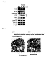
- Fig. 3 shows the electrophoretic profile of the Western blotting performed in the experiment above.
- control in the electrophoretic profile indicates the results of the Comparative Experiment above.
- Fig. 4 shows H9c2 cells treated with hsa-miR-145 having a final concentration of 40 nM, observed with an electron microscope. As shown in Fig. 4 , the shapes of autophagosomes ( Fig. 4 (A) ) and autolysosomes ( Fig. 4 (B) ) were observed in the cells.
- SEQ ID NO: 1 shows the nucleotide sequence of hsa-miR-145.
- SEQ ID NO: 2 shows the nucleotide sequence of the primary miRNA having a hairpin loop structure, which is a precursor of hsa-miR-145.
- SEQ ID NO: 3 shows the nucleotide sequence of the miRNA used in the Comparative Experiment of Example 3.
Landscapes
- Health & Medical Sciences (AREA)
- Life Sciences & Earth Sciences (AREA)
- Chemical & Material Sciences (AREA)
- Engineering & Computer Science (AREA)
- General Health & Medical Sciences (AREA)
- Genetics & Genomics (AREA)
- Pharmacology & Pharmacy (AREA)
- Animal Behavior & Ethology (AREA)
- Medicinal Chemistry (AREA)
- Public Health (AREA)
- Veterinary Medicine (AREA)
- Biomedical Technology (AREA)
- Epidemiology (AREA)
- Bioinformatics & Cheminformatics (AREA)
- Molecular Biology (AREA)
- Organic Chemistry (AREA)
- Biotechnology (AREA)
- General Engineering & Computer Science (AREA)
- Wood Science & Technology (AREA)
- Zoology (AREA)
- Biochemistry (AREA)
- Biophysics (AREA)
- Physics & Mathematics (AREA)
- Plant Pathology (AREA)
- Dermatology (AREA)
- Microbiology (AREA)
- Dispersion Chemistry (AREA)
- Urology & Nephrology (AREA)
- Vascular Medicine (AREA)
- Cardiology (AREA)
- Heart & Thoracic Surgery (AREA)
- Chemical Kinetics & Catalysis (AREA)
- General Chemical & Material Sciences (AREA)
- Nuclear Medicine, Radiotherapy & Molecular Imaging (AREA)
- Pharmaceuticals Containing Other Organic And Inorganic Compounds (AREA)
- Medicines That Contain Protein Lipid Enzymes And Other Medicines (AREA)
- Medicinal Preparation (AREA)
Abstract
The present invention provides a pharmaceutical composition for treating infarction, such as myocardial infarction or cerebral infarction, with a method that is different from conventional therapeutic methods that use a thrombolytic agent or balloon therapy. An autophagy enhancer such as hsa-miR-145 is used as the active ingredient of the pharmaceutical composition for treating infarction. In particular, an autophagy enhancer such as hsa-miR-145 is used in a liposomal state as an active ingredient of the pharmaceutical composition for treating infarction.
Description
- The present invention relates to a pharmaceutical composition for treating infarction. More specifically, the present invention relates to the use of an "autophagy enhancer" that accelerates autophagy as an agent for treating infarction.
- Myocardial infarction is a type of ischemic heart disease, and refers to a state in which the amount of blood flow in the coronary arteries of the heart decreases, the heart muscle becomes ischemic, and then the heart muscle dies. Decrease of blood flow in the coronary artery is mainly due to stenoses caused by a factor such as arteriosclerosis.
- Examples of therapeutic methods for treating myocardial infarction include dissolving the blood clots with a thrombolytic agent (e.g., tPA), and balloon therapy, in which a catheter is inserted in the blood vessel to expand the blood vessel with a balloon. It is known that a therapeutic method in which a thrombolytic agent is administered via intravenous injection is simple, but reocclusion easily occurs. Furthermore, it is necessary to pay attention to patients with bleeding disorders such as gastric ulcer. Balloon therapy can achieve a high success rate in recanalization, and it can prevent restenosis by keeping a stent in the blood vessel after it is performed. However, restenosis may still occur. Furthermore, there is a limitation such that a balloon therapy can be conducted only in medical facilities that have a room for cardiac catheterization.
- An miRNA is a single-stranded RNA consisting of about 21 to 25 nucleotides, and functions in regulation of gene expression. The miRNA is a small RNA derived from non-coding region encoded in the genome. Generally, an miRNA is produced from the gene transferred from a single or clustered miRNA precursor. More specifically, a pri-miRNA, which is a primary transcription from a gene, is transferred. Subsequently, in the stepwise processing from a pri-miRNA to a mature type of miRNA, a pre-miRNA consisting of about 70 to 80 nucleotides having a specific hairpin structure is produced from a pri-mRNA. Further, a mature type of miRNA is produced from pre-miRNA through the intervention of Dicer. It is believed that the miRNA is deeply involved in biological phenomenon, such as cell differentiation, proliferation, apoptosis, etc., that are essential for living things.
- Among miRNAs, hsa-miR-145 is an miRNA that consists of 23 nucleotides and is believed to be involved in anti-cancer action, as well as the regulation of cell proliferation and differentiation of smooth muscle (Patent Literature 1, Non-patent Literature 1 and 2).
- PTL 1:
WO2009/105759 -
- NPL 1: S. Noguchi et al., J. Vet. Med. Sci., 74(1), 1-8, 2012
- NPL 2: Eric M. Small et al., Nature, 469, 336-342, 2011
- An object of the present invention is to provide a pharmaceutical composition for use in treating infarction, such as myocardial infarction or cerebral infarction, by a method that is different from a conventional therapeutic method using a thrombolytic agent and balloon therapy. In particular, the present invention aims to provide a pharmaceutical composition for treating infarcted tissue, wherein the pharmaceutical composition can selectively and noninvasively reach and treat the local region where a myocardial infarction or cerebral infarction occurs.
- The present inventors conducted intensive research to achieve the above objects and found that, by administering hsa-miR-145, which is an miRNA, to the site of infarcted tissue, the size of the site of infarction (infarct size) can be reduced, and that the miRNA has an action of accelerating autophagy. More specifically, they found that the miRNA can function as an "autophagy enhancer." Based on these findings, the present inventors believed that an autophagy enhancer, in particular, hsa-miR-145, is effective in the repair and amelioration of infarcted tissue, and useful as an active ingredient of gene therapy for treating infarcted tissue. The present invention has been thus accomplished.
- More specifically, the present invention includes the following embodiments.
-
- (I-1) A pharmaceutical composition for treating infarction comprising an autophagy enhancer as an active ingredient.
- (I-2) The pharmaceutical composition for treating infarction according to (I-1), wherein the autophagy enhancer is at least one member selected from the group consisting of hsa-miR-145, rapamycin, carbamazepine, sodium valproate, verapamil, loperamide, amiodarone, nimodipine, nitrendipine, niguldipine, primozide, calpastatin, calpeptin, clonidine, pilmenidine, 2',5'-dideoxyadenosine, minoxidil, penitrem A, fluspiriline, trifluoperazine, and trehalose.
- (I-3) The pharmaceutical composition for treating infarction according to (1-1) or (I-2), comprising the autophagy enhancer in a liposomal state.
- (I-4) The pharmaceutical composition for treating infarction according to any one of (I-1) to (I-3), which is in an intravenous administration.
- (I-5) The pharmaceutical composition for treating infarction according to any one of (I-1) to (I-4), wherein the infarction is myocardial infarction, cerebral infarction, or spinal cord infarction.
- (I-6) The pharmaceutical composition for treating infarction according to (1-5), wherein the myocardial infarction is severe.
-
- (II-1) A method for treating infarction, the method comprising administering an autophagy enhancer to a patient with infarction.
- (II-2) The method for treating infarction according to (II-1), wherein the autophagy enhancer is at least one member selected from the group consisting of hsa-miR-145, rapamycin, carbamazepine, sodium valproate, verapamil, loperamide, amiodarone, nimodipine, nitrendipine, niguldipine, primozide, calpastatin, calpeptin, clonidine, pilmenidine, 2',5'-dideoxyadenosine, minoxidil, penitrem A, fluspiriline, trifluoperazine, and trehalose.
- (II-3) The method for treating infarction according to (II-1) or (II-2), wherein the autophagy enhancer is administered in a liposomized state.
- (II-4) The method for treating infarction according to any one of (II-1) to (II-3), comprising intravenously administering the autophagy enhancer.
- (II-5) The method for treating infarction according to any one of (II-1) to (II-4), wherein the infarction patient has myocardial infarction, cerebral infarction, or spinal cord infarction.
- (II-6) The method for treating infarction according to (II-5), wherein the myocardial infarction patient has a severe, large myocardial infarction.
- In the inventions related to the method for treating infarction according to (II-1) to (II-6), the term "autophagy enhancer" can be replaced with "the pharmaceutical composition for treating infarction" defined by any one of (I-1) to (I-6) described above.
-
- (III-1-1) An autophagy enhancer for use in treating infarction.
- (III-1-2) The autophagy enhancer according to (III-1-1), wherein the autophagy enhancer is at least one member selected from the group consisting of hsa-miR-145, rapamycin, carbamazepine, sodium valproate, verapamil, loperamide, amiodarone, nimodipine, nitrendipine, niguldipine, primozide, calpastatin, calpeptin, clonidine, pilmenidine, 2',5'-dideoxyadenosine, minoxidil, penitrem A, fluspiriline, trifluoperazine, and trehalose.
- (III-1-3) The autophagy enhancer according to (III-1-1) or (III-1-2), wherein the autophagy enhancer is in a liposomal state.
- (III-1-4) The method for treating infarction according to any one of (III-1-1) to (III-1-3), wherein the autophagy enhancer is intravenous administration.
- (III-1-5) The autophagy enhancer according to any one of (III-1-1) to (III-1-4), wherein the infarction is myocardial infarction, cerebral infarction, or spinal cord infarction.
- (III-1-6) The autophagy enhancer according to (III-1-5), wherein the myocardial infarction is severe.
-
- (III-2-1) Use of an autophagy enhancer for the manufacture of a pharmaceutical composition for treating infarction.
- (III-2-2) Use of the autophagy enhancer according to (III-2-1), wherein the autophagy enhancer is at least one member selected from the group consisting of hsa-miR-145, rapamycin, carbamazepine, sodium valproate, verapamil, loperamide, amiodarone, nimodipine, nitrendipine, niguldipine, primozide, calpastatin, calpeptin, clonidine, pilmenidine, 2',5'-dideoxyadenosine, minoxidil, penitrem A, fluspiriline, trifluoperazine, and trehalose.
- (III-2-3) The use according to (III-2-1) or (III-2-2), wherein the autophagy enhancer is in a liposomal state.
- (III-2-4) The use according to any one of (III-2-1) to (III-2-3), wherein the pharmaceutical composition for treating infarction is in an intravenous administration.
- (III-2-5) The use according to any one of (III-2-1) to (III-2-4), wherein the infarction is myocardial infarction, cerebral infarction, or spinal cord infarction.
- (III-2-6) The use according to (III-2-5), wherein the myocardial infarction is severe.
- The pharmaceutical composition for treating infarction of the present invention is characterized in that it comprises an autophagy enhancer, in particular, hsa-miR-145, as an active ingredient. The pharmaceutical composition for treating infarction of the present invention can reduce the infarct size by administering an autophagy enhancer, in particular, hsa-miR-145 (i.e., an active ingredient), to the infarcted site; therefore, it can be effectively used for treating infarction. As is clear from Example 2 described later, the pharmaceutical composition of the present invention has a remarkable effect in reduction of the infarct size in myocardial infarction, in particular, in severe, large myocardial infarction.
- Severe, large myocardial infarction is a disease with an unfavorable life prognosis, since the progression of left ventricular remodeling results in heart failure. The pharmaceutical composition for treating infarction of the present invention can favorably improve the life prognosis after the treatment of the severe, large myocardial infarction.
-
-
Fig. 1 shows stained images of the heart tissue of a severe, large myocardial infarction animal model (rabbit) stained with Evans Blue and TTC (Example 2).Fig. 1(A) shows the stained image of the heart tissue of a severe, large myocardial infarction animal model (control group) to which physiological saline was intravenously administered instead of an hsa-miR-145-containing liposome.Fig. 1(B) shows the stained image of the heart tissue of a severe, large myocardial infarction animal model (hsa-miR-145 administered group) to which an hsa-miR-145-containing liposome was intravenously administered. -
Fig. 2 is a graph that indicates the infarct size in the hearts of the hsa-miR-145 administered group (n=5) and the control group (n=5) as a proportion (%) of the infarct area of the heart relative to the area at risk of the entire heart. -
Fig. 3 shows an electrophoretic profile of the Western blotting performed in Example 3.Fig. 3 shows expressions of each protein (FRS2, KLF5, Myocardin, LC3B, p-Akt, Akt, p-ERK1/2, ERK1/2, and β-actin) in H9c2 cells treated with hsa-miR-145. -
Fig. 4 shows images of H9c2 cells treated with hsa-miR-145 observed with an electron microscope.Fig. 4(A) clearly indicates that autophagosomes are formed in the cell, andFig. 4(B) clearly indicates that autolysosomes are formed in the cell (Example 3). - The pharmaceutical composition for treating infarction of the present invention is characterized in that it comprises an autophagy enhancer as an active ingredient.
- Autophagy is a self-purification action that cells inherently possess. It is a mechanism in which cellular components that become old and unnecessary are wrapped in a vesicle-like structure (an autophagosome) together with cytoplasms, and degraded via lysosomal enzymes. There are two types of autophagy: one is basic autophagy, which is necessary to maintain intracellular metabolism, homeostasis, etc.; and the other is stress-induced autophagy. By the action of autophagy, intracellular accumulation of abnormal proteins can be suppressed. Furthermore, when an excessive amount of protein is produced, the intracellular nutrient environment is deteriorated, or pathogenic microorganisms enter the cytoplasm, etc., autophagy functions to remove or purify them. As described above, autophagy is involved in the maintenance of homeostasis in a living body. Autophagy is also induced when the cells fall into starvation, or pathological conditions such as ischemia or inflammation.
- In the present invention, "autophagy enhancer" indicates a substance that can accelerate the aforesaid "autophagy," which is the self-purification action of cell. Specific examples thereof include hsa-miR-145, a type of miRNA whose ability to accelerate autophagy is demonstrated in the Examples below; and substances conventionally known to have an action of accelerating autophagy, such as rapamycin, carbamazepine, sodium valproate, verapamil, loperamide, amiodarone, nimodipine, nitrendipine, niguldipine, primozide, calpastatin, calpeptin, clonidine, pilmenidine, 2',5'-dideoxyadenosine, minoxidil, penitrem A, fluspiriline, trifluoperazine, and trehalose (S. Sarkar et al., Cell Death and Differentiation 16, 46-56, 2009). Note that these autophagy enhancers may be used as an active ingredient of the pharmaceutical composition for treating infarction of the present invention singly or in any combination of two or more, as long as they do not adversely affect the action of autophagy acceleration. Preferably, hsa-miR-145, or a combination of hsa-miR-145 with one, or two or more other autophagy enhancers is used.
- Hsa-miR-145 is an oligonucleotide known as a human-derived micro RNA, and is registered as Accession No. MI0000461 in the microRNA database (miRBase) (http://www.mirbase.org/) (Entrez Gene: 406937, HGNC: 31532).
- SEQ ID NO: 1 shows the nucleotide sequence of hsa-miR-145. SEQ ID NO: 2 shows the nucleotide sequence of a primary miRNA (pri-miRNA) having a hairpin loop structure, which is a molecule before it is processed into hsa-miR-145.
- Hsa-miR-145 can be synthesized by a known nucleic acid synthetic process based on this nucleotide sequence (SEQ ID NO: 1); hsa-miR-145 is also commercially available from Applied Biosystems, etc. When used for the pharmaceutical composition for treating infarction, regardless of the production method of the hsa-miR-145, the hsa-miR-145 may be artificially chemosynthesized, biochemically synthesized, synthesized in a living body, or produced by double-stranded RNA (pri-miRNA) having 80 or more nucleotides being decomposed in or outside of the body.
- The pharmaceutical composition of the present invention can be produced by combining the aforesaid autophagy enhancer as an active ingredient with, depending on the administration route, pharmaceutically acceptable carriers or additives ordinarily used for medicaments. When a nucleic acid such as, in particular, hsa-miR-145, is used as the autophagy enhancer, the pharmaceutical composition of the present invention can be generally produced by combining it with pharmaceutically acceptable carriers or additives used for the production of gene therapy agents.
- The administration route is not limited as long as the autophagy enhancer can be delivered to the affected site (infarcted tissue), and may be administered orally or non-orally (e.g., intravenous administration, direct administration to the affected site, intramuscular administration, subcutaneous administration, intradermal administration, mucosal administration, rectal administration, and transpulmonary administration). Among these, intravenous administration by injection or infusion, and direct administration to the affected site are preferable.
- In the case of an injection preparation or infusion preparation, carriers or additives that can be used with the autophagy enhancer include various carriers or additives ordinarily used for injection preparations or infusion preparations. Examples of the carriers include distillated water; an aqueous solution containing sodium chloride or a mixture of sodium chloride with an inorganic salt; an aqueous solution containing a saccharide such as mannitol, lactose, dextran, or glucose; an aqueous solution containing an amino acid such as glycine or arginine; an aqueous solution containing an organic acid and a saccharide such as glucose; and an aqueous solution containing a salt and a saccharide such as glucose. These preparations may be formed into a solution, suspension, or emulsion by using, if necessary, auxiliary agents such as an osmotic agent, a pH adjuster, vegetable oil, and/or a surfactant. Furthermore, a pharmaceutically acceptable anti-oxidant or preservative may be used as an additive. Note that the injection preparation and infusion preparation may be formed, by powdering (spray-drying) or freeze-drying, into a solid preparation that is dissolved before use.
- The pharmaceutical composition of the present invention may be in a form that contains an unmodified autophagy enhancer dissolved in a carrier for injection preparation such as distillated water; or the pharmaceutical composition of the present invention may be in a form that contains an autophagy enhancer in a liposomal state. Here, the term liposomal encompasses both the state in which the autophagy enhancer is enclosed in a liposome (encapsulated in a liposome), and the state in which the autophagy enhancer is attached on the surface of liposome.
- The liposome is not limited as long as it can enclose (encapsulate) the autophagy enhancer of the present invention therein, or as long as the autophagy enhancer can be attached on its surface. There is no particular limitation to the liposome; however, when hsa-miR-145 is used as the autophagy enhancer, because the hsa-miR-145 is anionic, examples of the usable liposomes include those having a positive electric charge such as Lipofectamine, Lipofectin, Cellfectin, and other positively charged liposomes; and liposomes comprising components such as calcium phosphate, dextran, and/or polyethylene glycol. These liposomes are also commercially available; for example, Lipofectamine can be purchased from Invitrogen Coporation, etc., and FuGENE 6 Transfection Reagent can be purchased from Roche Diagnostics.
- A method for encapsulating a drug in a liposome is known. The autophagy enhancer of the present invention can be encapsulated in a liposome according to such a known method. For example, when the commercially available liposomes mentioned above are used, the preparation can be conducted according to the manufacturer's manual. If commercially available liposomes are not used, a liposome encapsulating an autophagy enhancer therein can be obtained by, for example, forming a liposome using a solution containing an autophagy enhancer and lipids containing phosphatidylcholines, phosphatidylethanolamine, phosphatidic acids or long-chain alkyl phosphates, gangliosides, glycolipids or phosphatidylglycerols and cholesterols.
- The mean particle diameter of the liposome is preferably 150 nm or less, more preferably 140 nm or less, still more preferably 120 nm or less, and furthermore preferably 100 nm or less in consideration of passing it through the capillary. The mean particle diameter is preferably 30 nm or more, more preferably 50 nm or more, and further more preferably 70 nm or more. Here, the mean particle diameter refers to the value of the particle diameter at 50% in the cumulative distribution determined by the laser diffraction scattering method.
- In addition to injection preparations or infusion preparations, the pharmaceutical composition of the present invention may also take the form of oral administrations such as solid preparations, e.g., tablets, capsules, granules, and powder. In the production of such solid preparations, in accordance with an ordinary method, pharmaceutically acceptable carriers or additives may be added. Examples thereof include, but are not limited to, excipients, binders, disintegrators, solubilizing agents, lubricants, anti-oxidants, preservatives, pH adjusters, coloring agents, and flavoring agents.
- The amount of the autophagy enhancer contained in the pharmaceutical composition of the present invention varies depending on the administration route of the pharmaceutical composition of the present invention, and the type of autophagy enhancer. However, when hsa-miR-145 is used as an autophagy enhancer, the pharmaceutical composition of the present invention may contain the hsa-miR-145 in an amount of 10 to 1000 µg, preferably 50 to 160 µg, and more preferably 70 to 80 µg per dose. When another autophagy enhancer is used, the amount thereof may be suitably selected based on the above.
- The pharmaceutical composition of the present invention can be used as an agent for treating infarction of patients having myocardial infarction, cerebral infarction, or spinal cord infarction. The pharmaceutical composition of the present invention is preferably used for treating myocardial infarction, and more preferably treating severe, large myocardial infarction.
- The dose of the pharmaceutical composition of the present invention may be suitably selected depending on the types or severity of the infarction such as myocardial infarction, cerebral infarction, or spinal cord infarction; and the age, body weight of the patient. The dose is preferably the amount suitable for regenerating or repairing at least some portion of infarcted tissue, and this amount is referred to "a pharmaceutically effective amount" in the present invention. The following is an approximation of the effective amount: as the dose of the autophagy enhancer per administration, for example, when hsa-miR-145 is used, the content of hsa-miR-145 is generally 10 to 1000 µg, preferably 50 to 160 µg, and more preferably 70 to 80 µg. When other autophagy enhancers are used, the amount thereof can be suitably selected based on the above amount.
- The frequency of administration of the pharmaceutical composition of the present invention may be once or several times per day, and is suitably selected depending on the conditions of the infarction patient.
- The present invention is described in more detail below with reference to Examples and the like. However, the present invention is not limited to these Examples.
- Hsa-miR-145 (5 nM; obtained from Applied Biosystems) was dissolved in 100 µl of RNase-free distillated water. 400 µl of reduced serum medium (Opti-MEM (GIBCO)) was added thereto, followed by mixing by shaking with a vortex mixer for 30 seconds. Thereafter, 10 µl of Lipofectamine RNAiMax (Invitrogen) was added to the mixture, followed by mixing by shaking with a vortex mixer for another 30 seconds. Subsequently, the mixture thus obtained was incubated at room temperature for 10 minutes. 300 µl of Opti-MEM was added thereto to a total volume of 800 µl. The hsa-miR-145-containing liposome thus prepared was used in Example 2 described below.
- The hsa-miR-145-containing liposome prepared in Example 1 above was administered to the infarcted site of an animal model having a severe, large myocardial infarction so as to evaluate the therapeutic effect of hsa-miR-145 on infarction.
- A pathological animal model having a severe, large myocardial infarction was prepared using a Japanese white rabbit (male, body weight: about 2 kg), and subjecting its coronary artery to ischemia for 30 minutes.
- The hsa-miR-145-containing liposome prepared in Example 1 was intravenously administered to each of the pathological animal models (n=5) in an amount of 0.035 mg per kg of body weight. 14 days after the administration, the rabbits were sacrificed under anesthesia, and their hearts were extracted (hsa-miR-145 administered group). For the control group, instead of the hsa-miR-145-containing liposome, 0.5 ml of physiological saline per kg of body weight was intravenously administered to pathological animal models (n=5) having severe, large myocardial infarction. 14 days after the administration, the rabbits were sacrificed under anesthesia, and their hearts were extracted.
- Each group of hearts (the hsa-miR-145 administered group, and the control group) thus extracted was stained with Evans Blue Dye so as to determine the ischemic region and the non-ischemic region. The hearts were stained with TTC so as to determine the infarct area.
- Using these stains as indexes, the size of the infarct area was calculated as the proportion (%) to the area at
risk 100% (potential ischemic area) of the entire heart. -
Fig. 1 shows the stained tissue in a stenotic site in the heart (obstructed site in the heart) in each group (hsa-miR-145 administered group and the control group). InFig. 1 , (A) shows the stained image of the heart tissue of the control group, and (B) shows the stained image of the heart tissue of the hsa-miR-145 administered group. The region stained blue with Evans Blue is the non-ischemic region; the region that was not stained blue is the ischemic region; and the region stained gray with TTC is the infarct area. -
Fig. 2 shows a comparison in the infarct size in each heart between the hsa-miR-145 administered group (n=5) and the control group (n=5). As is clear fromFig. 2 , the infarct size in the heart in the hsa-miR-145 administered group was significantly smaller than the infarct size in the heart of the control group to which hsa-miR-145 was not administered. These results confirmed that hsa-miR-145 has an action that reduces infarct size. - The measurement of the expression amount of hsa-miR-145 can be performed with a known method. A specific method is as follows:
- The tissues are separately collected from the infarct site, boundary site, and normal site in an extracted heart. The collected tissues are homogenized and dissolved using TRIzol® Reagent (Applied Biosystems; AB) to extract RNA. Thereafter, RT is conducted with a TaqMan miRNA Reverse Transcription Kit (AB), and the expression of hsa-miR-145 is detected by real-time PCR using a TaqMan miRNA primer (AB).
- The forward primer, reverse primer, and probe used in the RT-PCR can be suitably selected based on a known method.
- In Example 2 described above, it was confirmed that hsa-miR-145 was effective for the reduction of the myocardial infarction foci, and preservation of the cardiac function. Therefore, the mechanisms of the action of hsa-miR-145 were verified.
- H9c2 cells, which are derived from rat heart, were transfected with the hsa-miR-145-containing liposome prepared in Example 1 above. Specifically, the hsa-miR-145-containing liposome was prepared to have a final hsa-miR-145 concentration of 20 nM or 40 nM, after which H9c2 cells were cultured in the presence of the hsa-miR-145-containing liposome at 37°C for 72 hours.
- After culturing, total protein was extracted from the H9c2 cells thus obtained. Specifically, the cultured H9c2 cells were scraped off from the culture dish using a cell scraper (Sumitomo Bakelite Co., Ltd., Japan), and then suspended in Phosphate Buffered Saline (PBS(-); Takara, Japan). The result was placed in a 15-ml centrifuge tube, and then centrifuged at 2000 rpm for 3 minutes. After the centrifugation, the supernatant was removed, and Lysis Buffer (1% NP-40, 0.1% deoxycholic acid, 0.1% SDS, 150 mM NaCl, 1 mM EDTA) and 1% proteinase inhibitor cocktail (Sigma-Aldrich Co.) were added to the precipitate. The mixture was stirred with a vortex mixer to completely dissolve the cells. After being allowed to stand still on ice for 20 minutes, the result was centrifuged at 4°C at 12000 rpm for 20 minutes, and the supernatant thereof was collected to be used as a protein extract. The protein concentration of the protein extract was measured with a DC Protein Assay Kit (Bio-Rad; Hercules, CA, USA) according to the protocol of the kit using a spectrophotometer (BioPhotometer Plus, Eppendorf AG; Hamburg, Germany).
- The protein extract (total protein amount: 100 µg) prepared above and 20 µl of 5×SDS solution (0.25 M Tris-HCl (pH 6.8), 10% SDS, 0.5% Bromophenol Blue, 0.5% M DTT, 50% glycerol) were added and mixed in a microtube. Sterile distilled water was added thereto to a total amount of 100 µl. Subsequently, the mixture was heated at 98°C for 5 minutes with a block incubator (Astec; Fukuoka, Japan), and then quenched on ice for 5 minutes.
- The above-described protein treated with SDS was electrophoresed using an XV Pantera System (DRC Co., Ltd.; Tokyo, Japan) or Easy Separator® (Wako Pure Chemical Industries, Ltd.) After electrophoresis, the sample was blotted on a PVDF membrane (PerkinElmer Life Science; Boston, MA, USA) using a Trans-Blot SD Semi-Dry Transfer Cell (Bio-Rad). The PVDF membrane was hydrophilized with methanol beforehand, immersed in a blotting buffer (48 mM Tris, 40 mM glycine), and then shaken before use. Blotting was performed at 15 V, 370 mA, for 40 minutes.
- After the completion of blotting, the result was immersed in Tris-buffered saline-Tween 20 (TBS-T) (20 mM tris-HCl (pH 7.5), 150 mM NaCl, 0.1% Tween 20) to which 5% skim milk (Cell Signaling; Danvers, MA, USA) had been added. Then, blocking was performed at room temperature while shaking for 1 hour. After washing with TBS-T, the resultant was immersed in a primary antibody solution diluted with a primary antibody diluent (2% bovine serum albumin, 0.01% sodium azide-added TBS-T) at 4°C overnight. Antibodies of each of the proteins shown in
Fig. 3 (FRS2, KLF5, Myocardin, LC3B, p-Akt, Akt, p-ERK1/2, ERK1/2, and β-actin) were used as the primary antibody. The next day, the resultant was washed with TBS-T, immersed in a secondary antibody solution diluted with 5% skim milk, shaken for 1 hour, and then washed with TBS-T. A monoclonal anti-mouse or anti-rabbit horseradish-labeled antibody (#7076 or #7074, Cell Signaling Technology, Inc.) (1:4000) was used as the secondary antibody. - Subsequently, color development was performed using Western Lightning® ECL Pro (PerkinElmer), and detection was performed by chemiluminescence using an ImageQuant LAS 4000 Mini (GE Healthcare Japan; Tokyo, Japan).
- As a control for loading, detection was performed using the same membrane and using an anti-human β-actin mouse monoclonal antibody (AC-15, Sigma-Aldrich) (1:4000) as a primary antibody. After sanitizing at room temperature for 1 hour, the detection was performed according to the protocol described above.
- An experiment was conducted in the same manner as Example 3(1-1) and Example 3(1-2) above, except that miRNA having a nucleotide sequence of 5'-GUAGGAGUAGUGAAAGGCC-3' (Dharmacon, Inc.) (SEQ ID NO: 3) was used instead of hsa-miR-145.
-
Fig. 3 shows the electrophoretic profile of the Western blotting performed in the experiment above. InFig. 3 , control in the electrophoretic profile indicates the results of the Comparative Experiment above. - As is clear from these results, a remarkable conversion from LC3-BI to LC3-BII and acceleration of PI3K/Akt (p-Akt) signaling were observed in a cell into which hsa-miR-145 was introduced. In contrast, reduction in expression was observed in both KLF5 and FRS2 (fibroblast growth factor receptor substrate 2).
-
Fig. 4 shows H9c2 cells treated with hsa-miR-145 having a final concentration of 40 nM, observed with an electron microscope. As shown inFig. 4 , the shapes of autophagosomes (Fig. 4 (A) ) and autolysosomes (Fig. 4 (B) ) were observed in the cells. - SEQ ID NO: 1 shows the nucleotide sequence of hsa-miR-145. SEQ ID NO: 2 shows the nucleotide sequence of the primary miRNA having a hairpin loop structure, which is a precursor of hsa-miR-145. SEQ ID NO: 3 shows the nucleotide sequence of the miRNA used in the Comparative Experiment of Example 3.

Claims (13)
- A pharmaceutical composition for treating infarction comprising an autophagy enhancer as an active ingredient.
- The pharmaceutical composition for treating infarction according to claim 1, wherein the autophagy enhancer is hsa-miR-145.
- The pharmaceutical composition for treating infarction according to claim 1 or 2, comprising the autophagy enhancer in a liposomal state.
- The pharmaceutical composition for treating infarction according to any one of claims 1 to 3, which is in an intravenous administration.
- The pharmaceutical composition for treating infarction according to any one of claims 1 to 4, wherein the infarction is myocardial infarction, cerebral infarction, or spinal cord infarction.
- A method for treating infarction, the method comprising administering an autophagy enhancer to a patient with infarction.
- The method for treating infarction according to claim 6, wherein the autophagy enhancer is hsa-miR-145.
- The method for treating infarction according to claim 6 or 7, wherein the autophagy enhancer is administered in a liposomal state.
- The method for treating infarction according to any one of claims 6 to 8, wherein the autophagy enhancer is intravenous administration.
- An autophagy enhancer for use in treating infarction.
- The autophagy enhancer according to claim 10, wherein the autophagy enhancer is hsa-miR-145.
- Use of an autophagy enhancer for the manufacture of a pharmaceutical composition for treating infarction.
- The use according to claim 12, wherein the autophagy enhancer is hsa-miR-145.
Applications Claiming Priority (2)
| Application Number | Priority Date | Filing Date | Title |
|---|---|---|---|
| JP2012090873 | 2012-04-12 | ||
| PCT/JP2013/061118 WO2013154192A1 (en) | 2012-04-12 | 2013-04-12 | Medicinal composition for treating infarction |
Publications (1)
| Publication Number | Publication Date |
|---|---|
| EP2837389A1 true EP2837389A1 (en) | 2015-02-18 |
Family
ID=49327750
Family Applications (1)
| Application Number | Title | Priority Date | Filing Date |
|---|---|---|---|
| EP13776214.2A Withdrawn EP2837389A1 (en) | 2012-04-12 | 2013-04-12 | Medicinal composition for treating infarction |
Country Status (8)
| Country | Link |
|---|---|
| US (1) | US20150064237A1 (en) |
| EP (1) | EP2837389A1 (en) |
| JP (1) | JPWO2013154192A1 (en) |
| KR (1) | KR20150002781A (en) |
| CN (1) | CN104470543A (en) |
| CA (1) | CA2870248A1 (en) |
| IN (1) | IN2014DN09092A (en) |
| WO (1) | WO2013154192A1 (en) |
Families Citing this family (1)
| Publication number | Priority date | Publication date | Assignee | Title |
|---|---|---|---|---|
| WO2021250969A1 (en) | 2020-06-08 | 2021-12-16 | 国立大学法人東海国立大学機構 | Use of microrna in chronic-phase cardiac function improving treatment |
Family Cites Families (3)
| Publication number | Priority date | Publication date | Assignee | Title |
|---|---|---|---|---|
| JP2008539276A (en) * | 2005-04-27 | 2008-11-13 | ユニバーシティ オブ フロリダ リサーチ ファウンデーション,インコーポレイティド | Materials and methods for improving the resolution of mutant proteins associated with human disease |
| CN102083980A (en) * | 2008-02-21 | 2011-06-01 | 得克萨斯系统大学董事会 | Micro-RNAs that modulate smooth muscle proliferation and differentiation and uses thereof |
| DK2310020T3 (en) * | 2008-07-11 | 2017-06-26 | Univ Texas | PRESENT UNKNOWN ACETATES OF 2-DEOXY MONOSACCHARIDES WITH ANTICANCER ACTIVITY |
-
2013
- 2013-04-12 CA CA 2870248 patent/CA2870248A1/en not_active Abandoned
- 2013-04-12 US US14/391,302 patent/US20150064237A1/en not_active Abandoned
- 2013-04-12 WO PCT/JP2013/061118 patent/WO2013154192A1/en not_active Ceased
- 2013-04-12 CN CN201380019601.0A patent/CN104470543A/en active Pending
- 2013-04-12 JP JP2014510217A patent/JPWO2013154192A1/en active Pending
- 2013-04-12 KR KR20147031372A patent/KR20150002781A/en not_active Withdrawn
- 2013-04-12 EP EP13776214.2A patent/EP2837389A1/en not_active Withdrawn
- 2013-04-12 IN IN9092DEN2014 patent/IN2014DN09092A/en unknown
Non-Patent Citations (1)
| Title |
|---|
| See references of WO2013154192A1 * |
Also Published As
| Publication number | Publication date |
|---|---|
| JPWO2013154192A1 (en) | 2015-12-21 |
| IN2014DN09092A (en) | 2015-05-22 |
| KR20150002781A (en) | 2015-01-07 |
| CN104470543A (en) | 2015-03-25 |
| CA2870248A1 (en) | 2013-10-17 |
| WO2013154192A1 (en) | 2013-10-17 |
| US20150064237A1 (en) | 2015-03-05 |
Similar Documents
| Publication | Publication Date | Title |
|---|---|---|
| JP7746319B2 (en) | Cardiosphere-derived cells and exosomes secreted by such cells in the treatment of muscular dystrophy | |
| JP7635131B2 (en) | Method for preparing lipid nanoparticles | |
| EP3668522B1 (en) | Efficacious mrna vaccines | |
| AU2010294197C1 (en) | Method for the preparation of micro-RNA and its therapeutic application | |
| EP3661526B1 (en) | Cardiosphere-derived cells and their extracellular vesicles for treatment and prevention of cancer | |
| EP4520349A1 (en) | Interfering rna for inhibiting b7-h3 gene expression and use thereof | |
| WO2024026482A1 (en) | Lipid nanoparticle compositions comprising surface lipid derivatives and related uses | |
| KR20250029877A (en) | Noncoding RNA agents for treating immune diseases (BCYRN1 and its derivatives) | |
| WO2024026475A1 (en) | Compositions for delivery to hematopoietic stem and progenitor cells (hspcs) and related uses | |
| JP2025507571A (en) | mRNA encoding a checkpoint cancer vaccine and uses thereof | |
| US20150064237A1 (en) | Medicinal composition for treating infarction | |
| JP2012502007A (en) | Treatment of scleroderma | |
| WO2024026487A1 (en) | Lipid nanoparticle compositions comprising phospholipid derivatives and related uses | |
| Wang et al. | CTLA-4 nanovesicles disrupt dendritic cell-driven CD8 T cell priming for myocardial infarction therapy | |
| WO2006137377A1 (en) | Neuronal regeneration promoter | |
| HK40120941A (en) | Cardiosphere-derived cells and exosomes secreted by such cells in the treatment of muscular dystrophy | |
| CN120168430A (en) | Lipid nanoparticles for improving drug liver targeting and preparation method and application thereof |
Legal Events
| Date | Code | Title | Description |
|---|---|---|---|
| PUAI | Public reference made under article 153(3) epc to a published international application that has entered the european phase |
Free format text: ORIGINAL CODE: 0009012 |
|
| 17P | Request for examination filed |
Effective date: 20141103 |
|
| AK | Designated contracting states |
Kind code of ref document: A1 Designated state(s): AL AT BE BG CH CY CZ DE DK EE ES FI FR GB GR HR HU IE IS IT LI LT LU LV MC MK MT NL NO PL PT RO RS SE SI SK SM TR |
|
| AX | Request for extension of the european patent |
Extension state: BA ME |
|
| DAX | Request for extension of the european patent (deleted) | ||
| STAA | Information on the status of an ep patent application or granted ep patent |
Free format text: STATUS: THE APPLICATION HAS BEEN WITHDRAWN |
|
| 18W | Application withdrawn |
Effective date: 20151023 |