EP2828377B1 - Method for manufacturing a three-dimensional biomimetic scaffold and uses thereof - Google Patents
Method for manufacturing a three-dimensional biomimetic scaffold and uses thereof Download PDFInfo
- Publication number
- EP2828377B1 EP2828377B1 EP13702949.2A EP13702949A EP2828377B1 EP 2828377 B1 EP2828377 B1 EP 2828377B1 EP 13702949 A EP13702949 A EP 13702949A EP 2828377 B1 EP2828377 B1 EP 2828377B1
- Authority
- EP
- European Patent Office
- Prior art keywords
- mobile phase
- phase
- hydrogel
- immobile
- insulating material
- Prior art date
- Legal status (The legal status is an assumption and is not a legal conclusion. Google has not performed a legal analysis and makes no representation as to the accuracy of the status listed.)
- Not-in-force
Links
- 238000000034 method Methods 0.000 title claims description 154
- 230000003592 biomimetic effect Effects 0.000 title claims description 101
- 238000004519 manufacturing process Methods 0.000 title claims description 25
- 239000000017 hydrogel Substances 0.000 claims description 115
- 239000012528 membrane Substances 0.000 claims description 77
- 230000005684 electric field Effects 0.000 claims description 55
- 239000011810 insulating material Substances 0.000 claims description 55
- 239000000872 buffer Substances 0.000 claims description 43
- 102000004169 proteins and genes Human genes 0.000 claims description 42
- 108090000623 proteins and genes Proteins 0.000 claims description 42
- 150000001875 compounds Chemical class 0.000 claims description 36
- 239000012634 fragment Substances 0.000 claims description 32
- 239000003814 drug Substances 0.000 claims description 30
- 239000000412 dendrimer Substances 0.000 claims description 28
- 229920000736 dendritic polymer Polymers 0.000 claims description 28
- 229920000936 Agarose Polymers 0.000 claims description 26
- 108090000765 processed proteins & peptides Proteins 0.000 claims description 23
- 239000004205 dimethyl polysiloxane Substances 0.000 claims description 20
- 229920000435 poly(dimethylsiloxane) Polymers 0.000 claims description 20
- -1 polydimethylsiloxane Polymers 0.000 claims description 16
- 229940079593 drug Drugs 0.000 claims description 15
- 150000007523 nucleic acids Chemical class 0.000 claims description 15
- 239000003102 growth factor Substances 0.000 claims description 14
- 102000039446 nucleic acids Human genes 0.000 claims description 14
- 108020004707 nucleic acids Proteins 0.000 claims description 14
- 238000000059 patterning Methods 0.000 claims description 14
- 230000010261 cell growth Effects 0.000 claims description 10
- 102000004190 Enzymes Human genes 0.000 claims description 9
- 108090000790 Enzymes Proteins 0.000 claims description 9
- 238000012258 culturing Methods 0.000 claims description 9
- 230000001575 pathological effect Effects 0.000 claims description 9
- 229920002401 polyacrylamide Polymers 0.000 claims description 9
- 150000001793 charged compounds Chemical class 0.000 claims description 8
- 230000008929 regeneration Effects 0.000 claims description 7
- 238000011069 regeneration method Methods 0.000 claims description 7
- 229940088597 hormone Drugs 0.000 claims description 6
- 239000005556 hormone Substances 0.000 claims description 6
- 239000011859 microparticle Substances 0.000 claims description 6
- 239000002105 nanoparticle Substances 0.000 claims description 6
- 238000000338 in vitro Methods 0.000 claims description 5
- 238000007877 drug screening Methods 0.000 claims description 4
- 238000001727 in vivo Methods 0.000 claims description 2
- 210000004027 cell Anatomy 0.000 description 103
- 210000004379 membrane Anatomy 0.000 description 67
- 210000001519 tissue Anatomy 0.000 description 54
- 239000000463 material Substances 0.000 description 35
- 239000000243 solution Substances 0.000 description 26
- 239000000499 gel Substances 0.000 description 22
- 102000010834 Extracellular Matrix Proteins Human genes 0.000 description 20
- 108010037362 Extracellular Matrix Proteins Proteins 0.000 description 20
- 210000002744 extracellular matrix Anatomy 0.000 description 20
- 239000008363 phosphate buffer Substances 0.000 description 19
- 229920000642 polymer Polymers 0.000 description 18
- 238000001962 electrophoresis Methods 0.000 description 15
- 102000004196 processed proteins & peptides Human genes 0.000 description 13
- 230000006870 function Effects 0.000 description 12
- 239000002609 medium Substances 0.000 description 12
- 238000004113 cell culture Methods 0.000 description 11
- 239000000178 monomer Substances 0.000 description 11
- 230000000975 bioactive effect Effects 0.000 description 10
- 239000002245 particle Substances 0.000 description 10
- PEDCQBHIVMGVHV-UHFFFAOYSA-N glycerol group Chemical group OCC(O)CO PEDCQBHIVMGVHV-UHFFFAOYSA-N 0.000 description 9
- 239000000203 mixture Substances 0.000 description 9
- 229920003023 plastic Polymers 0.000 description 9
- 239000004033 plastic Substances 0.000 description 9
- 230000001172 regenerating effect Effects 0.000 description 9
- 230000006378 damage Effects 0.000 description 8
- 238000010586 diagram Methods 0.000 description 8
- 239000012620 biological material Substances 0.000 description 7
- 238000004720 dielectrophoresis Methods 0.000 description 7
- 238000006116 polymerization reaction Methods 0.000 description 7
- YMWUJEATGCHHMB-UHFFFAOYSA-N Dichloromethane Chemical compound ClCCl YMWUJEATGCHHMB-UHFFFAOYSA-N 0.000 description 6
- ZMXDDKWLCZADIW-UHFFFAOYSA-N N,N-Dimethylformamide Chemical compound CN(C)C=O ZMXDDKWLCZADIW-UHFFFAOYSA-N 0.000 description 6
- 238000013461 design Methods 0.000 description 6
- 201000010099 disease Diseases 0.000 description 6
- 208000037265 diseases, disorders, signs and symptoms Diseases 0.000 description 6
- 239000008194 pharmaceutical composition Substances 0.000 description 6
- 238000004088 simulation Methods 0.000 description 6
- 238000010186 staining Methods 0.000 description 6
- 239000000126 substance Substances 0.000 description 6
- XLYOFNOQVPJJNP-UHFFFAOYSA-N water Substances O XLYOFNOQVPJJNP-UHFFFAOYSA-N 0.000 description 6
- QFVHZQCOUORWEI-UHFFFAOYSA-N 4-[(4-anilino-5-sulfonaphthalen-1-yl)diazenyl]-5-hydroxynaphthalene-2,7-disulfonic acid Chemical compound C=12C(O)=CC(S(O)(=O)=O)=CC2=CC(S(O)(=O)=O)=CC=1N=NC(C1=CC=CC(=C11)S(O)(=O)=O)=CC=C1NC1=CC=CC=C1 QFVHZQCOUORWEI-UHFFFAOYSA-N 0.000 description 5
- 229920001661 Chitosan Polymers 0.000 description 5
- 108010010803 Gelatin Proteins 0.000 description 5
- 108090000723 Insulin-Like Growth Factor I Proteins 0.000 description 5
- 206010028980 Neoplasm Diseases 0.000 description 5
- 102000013275 Somatomedins Human genes 0.000 description 5
- 150000001413 amino acids Chemical class 0.000 description 5
- 210000000988 bone and bone Anatomy 0.000 description 5
- 238000004132 cross linking Methods 0.000 description 5
- 239000013078 crystal Substances 0.000 description 5
- 238000002474 experimental method Methods 0.000 description 5
- 229920000159 gelatin Polymers 0.000 description 5
- 239000008273 gelatin Substances 0.000 description 5
- 235000019322 gelatine Nutrition 0.000 description 5
- 235000011852 gelatine desserts Nutrition 0.000 description 5
- 239000007788 liquid Substances 0.000 description 5
- 210000002901 mesenchymal stem cell Anatomy 0.000 description 5
- 230000005012 migration Effects 0.000 description 5
- 238000013508 migration Methods 0.000 description 5
- 230000004048 modification Effects 0.000 description 5
- 238000012986 modification Methods 0.000 description 5
- 229910000402 monopotassium phosphate Inorganic materials 0.000 description 5
- 235000019796 monopotassium phosphate Nutrition 0.000 description 5
- 239000011148 porous material Substances 0.000 description 5
- GNSKLFRGEWLPPA-UHFFFAOYSA-M potassium dihydrogen phosphate Chemical compound [K+].OP(O)([O-])=O GNSKLFRGEWLPPA-UHFFFAOYSA-M 0.000 description 5
- 230000008569 process Effects 0.000 description 5
- 210000001082 somatic cell Anatomy 0.000 description 5
- NNRFRJQMBSBXGO-CIUDSAMLSA-N (3s)-3-[[2-[[(2s)-2-amino-5-(diaminomethylideneamino)pentanoyl]amino]acetyl]amino]-4-[[(1s)-1-carboxy-2-hydroxyethyl]amino]-4-oxobutanoic acid Chemical compound NC(N)=NCCC[C@H](N)C(=O)NCC(=O)N[C@@H](CC(O)=O)C(=O)N[C@@H](CO)C(O)=O NNRFRJQMBSBXGO-CIUDSAMLSA-N 0.000 description 4
- PQMFVUNERGGBPG-UHFFFAOYSA-N (6-bromopyridin-2-yl)hydrazine Chemical compound NNC1=CC=CC(Br)=N1 PQMFVUNERGGBPG-UHFFFAOYSA-N 0.000 description 4
- FHVDTGUDJYJELY-UHFFFAOYSA-N 6-{[2-carboxy-4,5-dihydroxy-6-(phosphanyloxy)oxan-3-yl]oxy}-4,5-dihydroxy-3-phosphanyloxane-2-carboxylic acid Chemical compound O1C(C(O)=O)C(P)C(O)C(O)C1OC1C(C(O)=O)OC(OP)C(O)C1O FHVDTGUDJYJELY-UHFFFAOYSA-N 0.000 description 4
- 241000894006 Bacteria Species 0.000 description 4
- 101800000414 Corticotropin Proteins 0.000 description 4
- IAZDPXIOMUYVGZ-UHFFFAOYSA-N Dimethylsulphoxide Chemical compound CS(C)=O IAZDPXIOMUYVGZ-UHFFFAOYSA-N 0.000 description 4
- KCXVZYZYPLLWCC-UHFFFAOYSA-N EDTA Chemical compound OC(=O)CN(CC(O)=O)CCN(CC(O)=O)CC(O)=O KCXVZYZYPLLWCC-UHFFFAOYSA-N 0.000 description 4
- 102000003951 Erythropoietin Human genes 0.000 description 4
- 108090000394 Erythropoietin Proteins 0.000 description 4
- 102100037362 Fibronectin Human genes 0.000 description 4
- 101000829980 Homo sapiens Ral guanine nucleotide dissociation stimulator Proteins 0.000 description 4
- 206010027476 Metastases Diseases 0.000 description 4
- 102100023320 Ral guanine nucleotide dissociation stimulator Human genes 0.000 description 4
- 102000036693 Thrombopoietin Human genes 0.000 description 4
- 108010041111 Thrombopoietin Proteins 0.000 description 4
- 239000007983 Tris buffer Substances 0.000 description 4
- 239000002253 acid Substances 0.000 description 4
- 239000004480 active ingredient Substances 0.000 description 4
- 239000011543 agarose gel Substances 0.000 description 4
- 229940072056 alginate Drugs 0.000 description 4
- 229920000615 alginic acid Polymers 0.000 description 4
- 235000010443 alginic acid Nutrition 0.000 description 4
- 238000013459 approach Methods 0.000 description 4
- 239000012736 aqueous medium Substances 0.000 description 4
- 230000008901 benefit Effects 0.000 description 4
- 230000015572 biosynthetic process Effects 0.000 description 4
- 239000004327 boric acid Substances 0.000 description 4
- 201000011510 cancer Diseases 0.000 description 4
- 230000024245 cell differentiation Effects 0.000 description 4
- 238000002659 cell therapy Methods 0.000 description 4
- 230000001276 controlling effect Effects 0.000 description 4
- IDLFZVILOHSSID-OVLDLUHVSA-N corticotropin Chemical compound C([C@@H](C(=O)N[C@@H](CO)C(=O)N[C@@H](CCSC)C(=O)N[C@@H](CCC(O)=O)C(=O)N[C@@H](CC=1NC=NC=1)C(=O)N[C@@H](CC=1C=CC=CC=1)C(=O)N[C@@H](CCCNC(N)=N)C(=O)N[C@@H](CC=1C2=CC=CC=C2NC=1)C(=O)NCC(=O)N[C@@H](CCCCN)C(=O)N1[C@@H](CCC1)C(=O)N[C@@H](C(C)C)C(=O)NCC(=O)N[C@@H](CCCCN)C(=O)N[C@@H](CCCCN)C(=O)N[C@@H](CCCNC(N)=N)C(=O)N[C@@H](CCCNC(N)=N)C(=O)N1[C@@H](CCC1)C(=O)N[C@@H](C(C)C)C(=O)N[C@@H](CCCCN)C(=O)N[C@@H](C(C)C)C(=O)N[C@@H](CC=1C=CC(O)=CC=1)C(=O)N1[C@@H](CCC1)C(=O)N[C@@H](CC(N)=O)C(=O)NCC(=O)N[C@@H](C)C(=O)N[C@@H](CCC(O)=O)C(=O)N[C@@H](CC(O)=O)C(=O)N[C@@H](CCC(O)=O)C(=O)N[C@@H](CO)C(=O)N[C@@H](C)C(=O)N[C@@H](CCC(O)=O)C(=O)N[C@@H](C)C(=O)N[C@@H](CC=1C=CC=CC=1)C(=O)N1[C@@H](CCC1)C(=O)N[C@@H](CC(C)C)C(=O)N[C@@H](CCC(O)=O)C(=O)N[C@@H](CC=1C=CC=CC=1)C(O)=O)NC(=O)[C@@H](N)CO)C1=CC=C(O)C=C1 IDLFZVILOHSSID-OVLDLUHVSA-N 0.000 description 4
- 229960000258 corticotropin Drugs 0.000 description 4
- 230000018109 developmental process Effects 0.000 description 4
- LOKCTEFSRHRXRJ-UHFFFAOYSA-I dipotassium trisodium dihydrogen phosphate hydrogen phosphate dichloride Chemical compound P(=O)(O)(O)[O-].[K+].P(=O)(O)([O-])[O-].[Na+].[Na+].[Cl-].[K+].[Cl-].[Na+] LOKCTEFSRHRXRJ-UHFFFAOYSA-I 0.000 description 4
- 229940076247 disodium hydrogen phosphate dehydrate Drugs 0.000 description 4
- 229940105423 erythropoietin Drugs 0.000 description 4
- 102000034287 fluorescent proteins Human genes 0.000 description 4
- 108091006047 fluorescent proteins Proteins 0.000 description 4
- 239000012212 insulator Substances 0.000 description 4
- NOESYZHRGYRDHS-UHFFFAOYSA-N insulin Chemical compound N1C(=O)C(NC(=O)C(CCC(N)=O)NC(=O)C(CCC(O)=O)NC(=O)C(C(C)C)NC(=O)C(NC(=O)CN)C(C)CC)CSSCC(C(NC(CO)C(=O)NC(CC(C)C)C(=O)NC(CC=2C=CC(O)=CC=2)C(=O)NC(CCC(N)=O)C(=O)NC(CC(C)C)C(=O)NC(CCC(O)=O)C(=O)NC(CC(N)=O)C(=O)NC(CC=2C=CC(O)=CC=2)C(=O)NC(CSSCC(NC(=O)C(C(C)C)NC(=O)C(CC(C)C)NC(=O)C(CC=2C=CC(O)=CC=2)NC(=O)C(CC(C)C)NC(=O)C(C)NC(=O)C(CCC(O)=O)NC(=O)C(C(C)C)NC(=O)C(CC(C)C)NC(=O)C(CC=2NC=NC=2)NC(=O)C(CO)NC(=O)CNC2=O)C(=O)NCC(=O)NC(CCC(O)=O)C(=O)NC(CCCNC(N)=N)C(=O)NCC(=O)NC(CC=3C=CC=CC=3)C(=O)NC(CC=3C=CC=CC=3)C(=O)NC(CC=3C=CC(O)=CC=3)C(=O)NC(C(C)O)C(=O)N3C(CCC3)C(=O)NC(CCCCN)C(=O)NC(C)C(O)=O)C(=O)NC(CC(N)=O)C(O)=O)=O)NC(=O)C(C(C)CC)NC(=O)C(CO)NC(=O)C(C(C)O)NC(=O)C1CSSCC2NC(=O)C(CC(C)C)NC(=O)C(NC(=O)C(CCC(N)=O)NC(=O)C(CC(N)=O)NC(=O)C(NC(=O)C(N)CC=1C=CC=CC=1)C(C)C)CC1=CN=CN1 NOESYZHRGYRDHS-UHFFFAOYSA-N 0.000 description 4
- 230000009401 metastasis Effects 0.000 description 4
- 239000002953 phosphate buffered saline Substances 0.000 description 4
- OXCMYAYHXIHQOA-UHFFFAOYSA-N potassium;[2-butyl-5-chloro-3-[[4-[2-(1,2,4-triaza-3-azanidacyclopenta-1,4-dien-5-yl)phenyl]phenyl]methyl]imidazol-4-yl]methanol Chemical compound [K+].CCCCC1=NC(Cl)=C(CO)N1CC1=CC=C(C=2C(=CC=CC=2)C2=N[N-]N=N2)C=C1 OXCMYAYHXIHQOA-UHFFFAOYSA-N 0.000 description 4
- 239000000047 product Substances 0.000 description 4
- 230000008439 repair process Effects 0.000 description 4
- QZAYGJVTTNCVMB-UHFFFAOYSA-N serotonin Chemical compound C1=C(O)C=C2C(CCN)=CNC2=C1 QZAYGJVTTNCVMB-UHFFFAOYSA-N 0.000 description 4
- SFLSHLFXELFNJZ-QMMMGPOBSA-N (-)-norepinephrine Chemical compound NC[C@H](O)C1=CC=C(O)C(O)=C1 SFLSHLFXELFNJZ-QMMMGPOBSA-N 0.000 description 3
- QKNYBSVHEMOAJP-UHFFFAOYSA-N 2-amino-2-(hydroxymethyl)propane-1,3-diol;hydron;chloride Chemical compound Cl.OCC(N)(CO)CO QKNYBSVHEMOAJP-UHFFFAOYSA-N 0.000 description 3
- CSCPPACGZOOCGX-UHFFFAOYSA-N Acetone Chemical compound CC(C)=O CSCPPACGZOOCGX-UHFFFAOYSA-N 0.000 description 3
- 239000000275 Adrenocorticotropic Hormone Substances 0.000 description 3
- 101800001288 Atrial natriuretic factor Proteins 0.000 description 3
- 102000055006 Calcitonin Human genes 0.000 description 3
- 108060001064 Calcitonin Proteins 0.000 description 3
- LFQSCWFLJHTTHZ-UHFFFAOYSA-N Ethanol Chemical compound CCO LFQSCWFLJHTTHZ-UHFFFAOYSA-N 0.000 description 3
- 102000018233 Fibroblast Growth Factor Human genes 0.000 description 3
- 108050007372 Fibroblast Growth Factor Proteins 0.000 description 3
- 108010067306 Fibronectins Proteins 0.000 description 3
- 241001465754 Metazoa Species 0.000 description 3
- OKKJLVBELUTLKV-UHFFFAOYSA-N Methanol Chemical compound OC OKKJLVBELUTLKV-UHFFFAOYSA-N 0.000 description 3
- 108010025020 Nerve Growth Factor Proteins 0.000 description 3
- 102000009087 Prolactin-Releasing Hormone Human genes 0.000 description 3
- 108010087786 Prolactin-Releasing Hormone Proteins 0.000 description 3
- VYPSYNLAJGMNEJ-UHFFFAOYSA-N Silicium dioxide Chemical compound O=[Si]=O VYPSYNLAJGMNEJ-UHFFFAOYSA-N 0.000 description 3
- YXFVVABEGXRONW-UHFFFAOYSA-N Toluene Chemical compound CC1=CC=CC=C1 YXFVVABEGXRONW-UHFFFAOYSA-N 0.000 description 3
- 125000000539 amino acid group Chemical group 0.000 description 3
- KBZOIRJILGZLEJ-LGYYRGKSSA-N argipressin Chemical compound C([C@H]1C(=O)N[C@@H](CCC(N)=O)C(=O)N[C@@H](CC(N)=O)C(=O)N[C@@H](CSSC[C@@H](C(N[C@@H](CC=2C=CC(O)=CC=2)C(=O)N1)=O)N)C(=O)N1[C@@H](CCC1)C(=O)N[C@@H](CCCN=C(N)N)C(=O)NCC(N)=O)C1=CC=CC=C1 KBZOIRJILGZLEJ-LGYYRGKSSA-N 0.000 description 3
- 229960004015 calcitonin Drugs 0.000 description 3
- BBBFJLBPOGFECG-VJVYQDLKSA-N calcitonin Chemical compound N([C@H](C(=O)N[C@@H](CC(C)C)C(=O)NCC(=O)N[C@@H](CCCCN)C(=O)N[C@@H](CC(C)C)C(=O)N[C@@H](CO)C(=O)N[C@@H](CCC(N)=O)C(=O)N[C@@H](CCC(O)=O)C(=O)N[C@@H](CC(C)C)C(=O)N[C@@H](CC=1NC=NC=1)C(=O)N[C@@H](CCCCN)C(=O)N[C@@H](CC(C)C)C(=O)N[C@@H](CCC(N)=O)C(=O)N[C@@H]([C@@H](C)O)C(=O)N[C@@H](CC=1C=CC(O)=CC=1)C(=O)N1[C@@H](CCC1)C(=O)N[C@@H](CCCNC(N)=N)C(=O)N[C@@H]([C@@H](C)O)C(=O)N[C@@H](CC(N)=O)C(=O)N[C@@H]([C@@H](C)O)C(=O)NCC(=O)N[C@@H](CO)C(=O)NCC(=O)N[C@@H]([C@@H](C)O)C(=O)N1[C@@H](CCC1)C(N)=O)C(C)C)C(=O)[C@@H]1CSSC[C@H](N)C(=O)N[C@@H](CO)C(=O)N[C@@H](CC(N)=O)C(=O)N[C@@H](CC(C)C)C(=O)N[C@@H](CO)C(=O)N[C@@H]([C@@H](C)O)C(=O)N1 BBBFJLBPOGFECG-VJVYQDLKSA-N 0.000 description 3
- 210000000170 cell membrane Anatomy 0.000 description 3
- 238000011161 development Methods 0.000 description 3
- 230000000694 effects Effects 0.000 description 3
- 230000004907 flux Effects 0.000 description 3
- 239000011521 glass Substances 0.000 description 3
- 239000001963 growth medium Substances 0.000 description 3
- 208000015181 infectious disease Diseases 0.000 description 3
- 238000002844 melting Methods 0.000 description 3
- 230000008018 melting Effects 0.000 description 3
- 238000002156 mixing Methods 0.000 description 3
- VLKZOEOYAKHREP-UHFFFAOYSA-N n-Hexane Chemical compound CCCCCC VLKZOEOYAKHREP-UHFFFAOYSA-N 0.000 description 3
- 229960002748 norepinephrine Drugs 0.000 description 3
- SFLSHLFXELFNJZ-UHFFFAOYSA-N norepinephrine Natural products NCC(O)C1=CC=C(O)C(O)=C1 SFLSHLFXELFNJZ-UHFFFAOYSA-N 0.000 description 3
- 210000000056 organ Anatomy 0.000 description 3
- 239000003960 organic solvent Substances 0.000 description 3
- 239000002877 prolactin releasing hormone Substances 0.000 description 3
- 210000003491 skin Anatomy 0.000 description 3
- 238000002174 soft lithography Methods 0.000 description 3
- 210000000278 spinal cord Anatomy 0.000 description 3
- 230000007480 spreading Effects 0.000 description 3
- 238000003892 spreading Methods 0.000 description 3
- 230000003068 static effect Effects 0.000 description 3
- 238000012360 testing method Methods 0.000 description 3
- 230000001225 therapeutic effect Effects 0.000 description 3
- KIUKXJAPPMFGSW-DNGZLQJQSA-N (2S,3S,4S,5R,6R)-6-[(2S,3R,4R,5S,6R)-3-Acetamido-2-[(2S,3S,4R,5R,6R)-6-[(2R,3R,4R,5S,6R)-3-acetamido-2,5-dihydroxy-6-(hydroxymethyl)oxan-4-yl]oxy-2-carboxy-4,5-dihydroxyoxan-3-yl]oxy-5-hydroxy-6-(hydroxymethyl)oxan-4-yl]oxy-3,4,5-trihydroxyoxane-2-carboxylic acid Chemical compound CC(=O)N[C@H]1[C@H](O)O[C@H](CO)[C@@H](O)[C@@H]1O[C@H]1[C@H](O)[C@@H](O)[C@H](O[C@H]2[C@@H]([C@@H](O[C@H]3[C@@H]([C@@H](O)[C@H](O)[C@H](O3)C(O)=O)O)[C@H](O)[C@@H](CO)O2)NC(C)=O)[C@@H](C(O)=O)O1 KIUKXJAPPMFGSW-DNGZLQJQSA-N 0.000 description 2
- 108091032973 (ribonucleotides)n+m Proteins 0.000 description 2
- VOXZDWNPVJITMN-ZBRFXRBCSA-N 17β-estradiol Chemical compound OC1=CC=C2[C@H]3CC[C@](C)([C@H](CC4)O)[C@@H]4[C@@H]3CCC2=C1 VOXZDWNPVJITMN-ZBRFXRBCSA-N 0.000 description 2
- NIXOWILDQLNWCW-UHFFFAOYSA-N 2-Propenoic acid Natural products OC(=O)C=C NIXOWILDQLNWCW-UHFFFAOYSA-N 0.000 description 2
- LFTRJWKKLPVMNE-RCBQFDQVSA-N 2-[[(2s)-2-[[2-[[(2s)-1-[(2s)-2-amino-3-methylbutanoyl]pyrrolidine-2-carbonyl]amino]acetyl]amino]-3-methylbutanoyl]amino]acetic acid Chemical compound CC(C)[C@H](N)C(=O)N1CCC[C@H]1C(=O)NCC(=O)N[C@@H](C(C)C)C(=O)NCC(O)=O LFTRJWKKLPVMNE-RCBQFDQVSA-N 0.000 description 2
- 102400001318 Adrenomedullin Human genes 0.000 description 2
- 101800004616 Adrenomedullin Proteins 0.000 description 2
- 102000015427 Angiotensins Human genes 0.000 description 2
- 108010064733 Angiotensins Proteins 0.000 description 2
- 108010005853 Anti-Mullerian Hormone Proteins 0.000 description 2
- 102400001282 Atrial natriuretic peptide Human genes 0.000 description 2
- 101800001890 Atrial natriuretic peptide Proteins 0.000 description 2
- 108010006654 Bleomycin Proteins 0.000 description 2
- 102000007350 Bone Morphogenetic Proteins Human genes 0.000 description 2
- 108010007726 Bone Morphogenetic Proteins Proteins 0.000 description 2
- 102400000667 Brain natriuretic peptide 32 Human genes 0.000 description 2
- 101800000407 Brain natriuretic peptide 32 Proteins 0.000 description 2
- 101800002247 Brain natriuretic peptide 45 Proteins 0.000 description 2
- 102000004219 Brain-derived neurotrophic factor Human genes 0.000 description 2
- 108090000715 Brain-derived neurotrophic factor Proteins 0.000 description 2
- DLGOEMSEDOSKAD-UHFFFAOYSA-N Carmustine Chemical compound ClCCNC(=O)N(N=O)CCCl DLGOEMSEDOSKAD-UHFFFAOYSA-N 0.000 description 2
- HEDRZPFGACZZDS-UHFFFAOYSA-N Chloroform Chemical compound ClC(Cl)Cl HEDRZPFGACZZDS-UHFFFAOYSA-N 0.000 description 2
- 101800001982 Cholecystokinin Proteins 0.000 description 2
- 102100025841 Cholecystokinin Human genes 0.000 description 2
- 102000008186 Collagen Human genes 0.000 description 2
- 108010035532 Collagen Proteins 0.000 description 2
- 208000035473 Communicable disease Diseases 0.000 description 2
- 102400000739 Corticotropin Human genes 0.000 description 2
- 241000186227 Corynebacterium diphtheriae Species 0.000 description 2
- CMSMOCZEIVJLDB-UHFFFAOYSA-N Cyclophosphamide Chemical compound ClCCN(CCCl)P1(=O)NCCCO1 CMSMOCZEIVJLDB-UHFFFAOYSA-N 0.000 description 2
- 108020004414 DNA Proteins 0.000 description 2
- 241000252212 Danio rerio Species 0.000 description 2
- AOJJSUZBOXZQNB-TZSSRYMLSA-N Doxorubicin Chemical compound O([C@H]1C[C@@](O)(CC=2C(O)=C3C(=O)C=4C=CC=C(C=4C(=O)C3=C(O)C=21)OC)C(=O)CO)[C@H]1C[C@H](N)[C@H](O)[C@H](C)O1 AOJJSUZBOXZQNB-TZSSRYMLSA-N 0.000 description 2
- 239000006144 Dulbecco’s modified Eagle's medium Substances 0.000 description 2
- 102000012673 Follicle Stimulating Hormone Human genes 0.000 description 2
- 108010079345 Follicle Stimulating Hormone Proteins 0.000 description 2
- 108700012941 GNRH1 Proteins 0.000 description 2
- 102000034615 Glial cell line-derived neurotrophic factor Human genes 0.000 description 2
- 108091010837 Glial cell line-derived neurotrophic factor Proteins 0.000 description 2
- 102400000321 Glucagon Human genes 0.000 description 2
- 108060003199 Glucagon Proteins 0.000 description 2
- 108010017080 Granulocyte Colony-Stimulating Factor Proteins 0.000 description 2
- 102000004269 Granulocyte Colony-Stimulating Factor Human genes 0.000 description 2
- 108010017213 Granulocyte-Macrophage Colony-Stimulating Factor Proteins 0.000 description 2
- 102100039620 Granulocyte-macrophage colony-stimulating factor Human genes 0.000 description 2
- 239000000095 Growth Hormone-Releasing Hormone Substances 0.000 description 2
- 102000004858 Growth differentiation factor-9 Human genes 0.000 description 2
- 108090001086 Growth differentiation factor-9 Proteins 0.000 description 2
- 102100039939 Growth/differentiation factor 8 Human genes 0.000 description 2
- 241000590002 Helicobacter pylori Species 0.000 description 2
- 108090000100 Hepatocyte Growth Factor Proteins 0.000 description 2
- 102100021866 Hepatocyte growth factor Human genes 0.000 description 2
- 102100031000 Hepatoma-derived growth factor Human genes 0.000 description 2
- NTYJJOPFIAHURM-UHFFFAOYSA-N Histamine Chemical compound NCCC1=CN=CN1 NTYJJOPFIAHURM-UHFFFAOYSA-N 0.000 description 2
- 101000595923 Homo sapiens Placenta growth factor Proteins 0.000 description 2
- 208000026350 Inborn Genetic disease Diseases 0.000 description 2
- 102000004877 Insulin Human genes 0.000 description 2
- 108090001061 Insulin Proteins 0.000 description 2
- FBOZXECLQNJBKD-ZDUSSCGKSA-N L-methotrexate Chemical compound C=1N=C2N=C(N)N=C(N)C2=NC=1CN(C)C1=CC=C(C(=O)N[C@@H](CCC(O)=O)C(O)=O)C=C1 FBOZXECLQNJBKD-ZDUSSCGKSA-N 0.000 description 2
- XNSAINXGIQZQOO-UHFFFAOYSA-N L-pyroglutamyl-L-histidyl-L-proline amide Natural products NC(=O)C1CCCN1C(=O)C(NC(=O)C1NC(=O)CC1)CC1=CN=CN1 XNSAINXGIQZQOO-UHFFFAOYSA-N 0.000 description 2
- 102000016267 Leptin Human genes 0.000 description 2
- 108010092277 Leptin Proteins 0.000 description 2
- 102000009151 Luteinizing Hormone Human genes 0.000 description 2
- 108010073521 Luteinizing Hormone Proteins 0.000 description 2
- 102000002274 Matrix Metalloproteinases Human genes 0.000 description 2
- 108010000684 Matrix Metalloproteinases Proteins 0.000 description 2
- 108010007013 Melanocyte-Stimulating Hormones Proteins 0.000 description 2
- YJPIGAIKUZMOQA-UHFFFAOYSA-N Melatonin Natural products COC1=CC=C2N(C(C)=O)C=C(CCN)C2=C1 YJPIGAIKUZMOQA-UHFFFAOYSA-N 0.000 description 2
- CERQOIWHTDAKMF-UHFFFAOYSA-M Methacrylate Chemical compound CC(=C)C([O-])=O CERQOIWHTDAKMF-UHFFFAOYSA-M 0.000 description 2
- 102100030173 Muellerian-inhibiting factor Human genes 0.000 description 2
- 108010056852 Myostatin Proteins 0.000 description 2
- NWIBSHFKIJFRCO-WUDYKRTCSA-N Mytomycin Chemical compound C1N2C(C(C(C)=C(N)C3=O)=O)=C3[C@@H](COC(N)=O)[C@@]2(OC)[C@@H]2[C@H]1N2 NWIBSHFKIJFRCO-WUDYKRTCSA-N 0.000 description 2
- IMNFDUFMRHMDMM-UHFFFAOYSA-N N-Heptane Chemical compound CCCCCCC IMNFDUFMRHMDMM-UHFFFAOYSA-N 0.000 description 2
- 102000015336 Nerve Growth Factor Human genes 0.000 description 2
- 102400000050 Oxytocin Human genes 0.000 description 2
- 101800000989 Oxytocin Proteins 0.000 description 2
- XNOPRXBHLZRZKH-UHFFFAOYSA-N Oxytocin Natural products N1C(=O)C(N)CSSCC(C(=O)N2C(CCC2)C(=O)NC(CC(C)C)C(=O)NCC(N)=O)NC(=O)C(CC(N)=O)NC(=O)C(CCC(N)=O)NC(=O)C(C(C)CC)NC(=O)C1CC1=CC=C(O)C=C1 XNOPRXBHLZRZKH-UHFFFAOYSA-N 0.000 description 2
- 102000003982 Parathyroid hormone Human genes 0.000 description 2
- 108090000445 Parathyroid hormone Proteins 0.000 description 2
- OFBQJSOFQDEBGM-UHFFFAOYSA-N Pentane Chemical compound CCCCC OFBQJSOFQDEBGM-UHFFFAOYSA-N 0.000 description 2
- 102100035194 Placenta growth factor Human genes 0.000 description 2
- 102000004576 Placental Lactogen Human genes 0.000 description 2
- 108010003044 Placental Lactogen Proteins 0.000 description 2
- 239000000381 Placental Lactogen Substances 0.000 description 2
- 108010038512 Platelet-Derived Growth Factor Proteins 0.000 description 2
- 102000010780 Platelet-Derived Growth Factor Human genes 0.000 description 2
- 239000002202 Polyethylene glycol Substances 0.000 description 2
- RJKFOVLPORLFTN-LEKSSAKUSA-N Progesterone Chemical compound C1CC2=CC(=O)CC[C@]2(C)[C@@H]2[C@@H]1[C@@H]1CC[C@H](C(=O)C)[C@@]1(C)CC2 RJKFOVLPORLFTN-LEKSSAKUSA-N 0.000 description 2
- 102100024819 Prolactin Human genes 0.000 description 2
- 108010057464 Prolactin Proteins 0.000 description 2
- 102000003743 Relaxin Human genes 0.000 description 2
- 108090000103 Relaxin Proteins 0.000 description 2
- 108010086019 Secretin Proteins 0.000 description 2
- 102100037505 Secretin Human genes 0.000 description 2
- 102000012479 Serine Proteases Human genes 0.000 description 2
- 108010022999 Serine Proteases Proteins 0.000 description 2
- BQCADISMDOOEFD-UHFFFAOYSA-N Silver Chemical compound [Ag] BQCADISMDOOEFD-UHFFFAOYSA-N 0.000 description 2
- 102100022831 Somatoliberin Human genes 0.000 description 2
- 101710142969 Somatoliberin Proteins 0.000 description 2
- NKANXQFJJICGDU-QPLCGJKRSA-N Tamoxifen Chemical compound C=1C=CC=CC=1C(/CC)=C(C=1C=CC(OCCN(C)C)=CC=1)/C1=CC=CC=C1 NKANXQFJJICGDU-QPLCGJKRSA-N 0.000 description 2
- MUMGGOZAMZWBJJ-DYKIIFRCSA-N Testostosterone Chemical compound O=C1CC[C@]2(C)[C@H]3CC[C@](C)([C@H](CC4)O)[C@@H]4[C@@H]3CCC2=C1 MUMGGOZAMZWBJJ-DYKIIFRCSA-N 0.000 description 2
- 102000035100 Threonine proteases Human genes 0.000 description 2
- 108091005501 Threonine proteases Proteins 0.000 description 2
- IQFYYKKMVGJFEH-XLPZGREQSA-N Thymidine Chemical compound O=C1NC(=O)C(C)=CN1[C@@H]1O[C@H](CO)[C@@H](O)C1 IQFYYKKMVGJFEH-XLPZGREQSA-N 0.000 description 2
- 102000011923 Thyrotropin Human genes 0.000 description 2
- 108010061174 Thyrotropin Proteins 0.000 description 2
- 239000000627 Thyrotropin-Releasing Hormone Substances 0.000 description 2
- 102400000336 Thyrotropin-releasing hormone Human genes 0.000 description 2
- 101800004623 Thyrotropin-releasing hormone Proteins 0.000 description 2
- 102000004887 Transforming Growth Factor beta Human genes 0.000 description 2
- 108090001012 Transforming Growth Factor beta Proteins 0.000 description 2
- 102400001320 Transforming growth factor alpha Human genes 0.000 description 2
- 101800004564 Transforming growth factor alpha Proteins 0.000 description 2
- 108060008682 Tumor Necrosis Factor Proteins 0.000 description 2
- 102000000852 Tumor Necrosis Factor-alpha Human genes 0.000 description 2
- 108010004977 Vasopressins Proteins 0.000 description 2
- 102000002852 Vasopressins Human genes 0.000 description 2
- JXLYSJRDGCGARV-WWYNWVTFSA-N Vinblastine Natural products O=C(O[C@H]1[C@](O)(C(=O)OC)[C@@H]2N(C)c3c(cc(c(OC)c3)[C@]3(C(=O)OC)c4[nH]c5c(c4CCN4C[C@](O)(CC)C[C@H](C3)C4)cccc5)[C@@]32[C@H]2[C@@]1(CC)C=CCN2CC3)C JXLYSJRDGCGARV-WWYNWVTFSA-N 0.000 description 2
- 208000027418 Wounds and injury Diseases 0.000 description 2
- RJURFGZVJUQBHK-UHFFFAOYSA-N actinomycin D Natural products CC1OC(=O)C(C(C)C)N(C)C(=O)CN(C)C(=O)C2CCCN2C(=O)C(C(C)C)NC(=O)C1NC(=O)C1=C(N)C(=O)C(C)=C2OC(C(C)=CC=C3C(=O)NC4C(=O)NC(C(N5CCCC5C(=O)N(C)CC(=O)N(C)C(C(C)C)C(=O)OC4C)=O)C(C)C)=C3N=C21 RJURFGZVJUQBHK-UHFFFAOYSA-N 0.000 description 2
- 239000013543 active substance Substances 0.000 description 2
- 239000000654 additive Substances 0.000 description 2
- 230000000996 additive effect Effects 0.000 description 2
- OIRDTQYFTABQOQ-KQYNXXCUSA-N adenosine Chemical compound C1=NC=2C(N)=NC=NC=2N1[C@@H]1O[C@H](CO)[C@@H](O)[C@H]1O OIRDTQYFTABQOQ-KQYNXXCUSA-N 0.000 description 2
- 239000000853 adhesive Substances 0.000 description 2
- 230000001070 adhesive effect Effects 0.000 description 2
- UCTWMZQNUQWSLP-UHFFFAOYSA-N adrenaline Chemical compound CNCC(O)C1=CC=C(O)C(O)=C1 UCTWMZQNUQWSLP-UHFFFAOYSA-N 0.000 description 2
- ULCUCJFASIJEOE-NPECTJMMSA-N adrenomedullin Chemical compound C([C@@H](C(=O)N[C@@H](CCC(N)=O)C(=O)NCC(=O)N[C@@H]([C@@H](C)CC)C(=O)N[C@@H](CCCNC(N)=N)C(=O)N[C@@H](CO)C(=O)N[C@@H](CC=1C=CC=CC=1)C(=O)NCC(=O)N[C@@H]1C(N[C@@H](CCCNC(N)=N)C(=O)N[C@@H](CC=2C=CC=CC=2)C(=O)NCC(=O)N[C@H](C(=O)N[C@@H](CSSC1)C(=O)N[C@@H]([C@@H](C)O)C(=O)N[C@@H](C(C)C)C(=O)N[C@@H](CCC(N)=O)C(=O)N[C@@H](CCCCN)C(=O)N[C@@H](CC(C)C)C(=O)N[C@@H](C)C(=O)N[C@@H](CC=1NC=NC=1)C(=O)N[C@@H](CCC(N)=O)C(=O)N[C@@H]([C@@H](C)CC)C(=O)N[C@@H](CC=1C=CC(O)=CC=1)C(=O)N[C@@H](CCC(N)=O)C(=O)N[C@@H](CC=1C=CC=CC=1)C(=O)N[C@@H]([C@@H](C)O)C(=O)N[C@@H](CC(O)=O)C(=O)N[C@@H](CCCCN)C(=O)N[C@@H](CC(O)=O)C(=O)N[C@@H](CCCCN)C(=O)N[C@@H](CC(O)=O)C(=O)N[C@@H](CC(N)=O)C(=O)N[C@@H](C(C)C)C(=O)N[C@@H](C)C(=O)N1[C@@H](CCC1)C(=O)N[C@@H](CCCNC(N)=N)C(=O)N[C@@H](CO)C(=O)N[C@@H](CCCCN)C(=O)N[C@@H]([C@@H](C)CC)C(=O)N[C@@H](CO)C(=O)N1[C@@H](CCC1)C(=O)N[C@@H](CCC(N)=O)C(=O)NCC(=O)N[C@@H](CC=1C=CC(O)=CC=1)C(N)=O)[C@@H](C)O)=O)NC(=O)[C@H](CC(N)=O)NC(=O)[C@H](CC(N)=O)NC(=O)[C@H](CCSC)NC(=O)[C@H](CO)NC(=O)[C@H](CCC(N)=O)NC(=O)[C@H](CCCNC(N)=N)NC(=O)[C@@H](N)CC=1C=CC(O)=CC=1)C1=CC=CC=C1 ULCUCJFASIJEOE-NPECTJMMSA-N 0.000 description 2
- 239000000868 anti-mullerian hormone Substances 0.000 description 2
- VSRXQHXAPYXROS-UHFFFAOYSA-N azanide;cyclobutane-1,1-dicarboxylic acid;platinum(2+) Chemical compound [NH2-].[NH2-].[Pt+2].OC(=O)C1(C(O)=O)CCC1 VSRXQHXAPYXROS-UHFFFAOYSA-N 0.000 description 2
- 230000017531 blood circulation Effects 0.000 description 2
- 229940112869 bone morphogenetic protein Drugs 0.000 description 2
- 229940077737 brain-derived neurotrophic factor Drugs 0.000 description 2
- 239000007853 buffer solution Substances 0.000 description 2
- 239000003990 capacitor Substances 0.000 description 2
- 229960004562 carboplatin Drugs 0.000 description 2
- 230000000747 cardiac effect Effects 0.000 description 2
- 229960005243 carmustine Drugs 0.000 description 2
- NSQLIUXCMFBZME-MPVJKSABSA-N carperitide Chemical compound C([C@H]1C(=O)NCC(=O)NCC(=O)N[C@@H](CCCNC(N)=N)C(=O)N[C@@H](CCSC)C(=O)N[C@@H](CC(O)=O)C(=O)N[C@@H](CCCNC(N)=N)C(=O)N[C@H](C(NCC(=O)N[C@@H](C)C(=O)N[C@@H](CCC(N)=O)C(=O)N[C@@H](CO)C(=O)NCC(=O)N[C@@H](CC(C)C)C(=O)NCC(=O)N[C@@H](CSSC[C@@H](C(=O)N1)NC(=O)[C@H](CO)NC(=O)[C@H](CO)NC(=O)[C@H](CCCNC(N)=N)NC(=O)[C@H](CCCNC(N)=N)NC(=O)[C@H](CC(C)C)NC(=O)[C@@H](N)CO)C(=O)N[C@@H](CC(N)=O)C(=O)N[C@@H](CO)C(=O)N[C@@H](CC=1C=CC=CC=1)C(=O)N[C@@H](CCCNC(N)=N)C(=O)N[C@@H](CC=1C=CC(O)=CC=1)C(O)=O)=O)[C@@H](C)CC)C1=CC=CC=C1 NSQLIUXCMFBZME-MPVJKSABSA-N 0.000 description 2
- 210000000845 cartilage Anatomy 0.000 description 2
- 238000006243 chemical reaction Methods 0.000 description 2
- 239000003795 chemical substances by application Substances 0.000 description 2
- 229940107137 cholecystokinin Drugs 0.000 description 2
- 210000001612 chondrocyte Anatomy 0.000 description 2
- 238000012650 click reaction Methods 0.000 description 2
- 229920001436 collagen Polymers 0.000 description 2
- 210000004087 cornea Anatomy 0.000 description 2
- 238000005520 cutting process Methods 0.000 description 2
- 229960004397 cyclophosphamide Drugs 0.000 description 2
- OPTASPLRGRRNAP-UHFFFAOYSA-N cytosine Chemical compound NC=1C=CNC(=O)N=1 OPTASPLRGRRNAP-UHFFFAOYSA-N 0.000 description 2
- 210000003298 dental enamel Anatomy 0.000 description 2
- 125000004386 diacrylate group Chemical group 0.000 description 2
- VYFYYTLLBUKUHU-UHFFFAOYSA-N dopamine Chemical compound NCCC1=CC=C(O)C(O)=C1 VYFYYTLLBUKUHU-UHFFFAOYSA-N 0.000 description 2
- 239000003937 drug carrier Substances 0.000 description 2
- 230000013020 embryo development Effects 0.000 description 2
- 238000005516 engineering process Methods 0.000 description 2
- 230000007613 environmental effect Effects 0.000 description 2
- 229960005309 estradiol Drugs 0.000 description 2
- 229930182833 estradiol Natural products 0.000 description 2
- 239000012091 fetal bovine serum Substances 0.000 description 2
- 238000011049 filling Methods 0.000 description 2
- 229940028334 follicle stimulating hormone Drugs 0.000 description 2
- 208000016361 genetic disease Diseases 0.000 description 2
- MASNOZXLGMXCHN-ZLPAWPGGSA-N glucagon Chemical compound C([C@@H](C(=O)N[C@H](C(=O)N[C@@H](CCC(N)=O)C(=O)N[C@@H](CC=1C2=CC=CC=C2NC=1)C(=O)N[C@@H](CC(C)C)C(=O)N[C@@H](CCSC)C(=O)N[C@@H](CC(N)=O)C(=O)N[C@@H]([C@@H](C)O)C(O)=O)C(C)C)NC(=O)[C@H](CC(O)=O)NC(=O)[C@H](CCC(N)=O)NC(=O)[C@H](C)NC(=O)[C@H](CCCNC(N)=N)NC(=O)[C@H](CCCNC(N)=N)NC(=O)[C@H](CO)NC(=O)[C@H](CC(O)=O)NC(=O)[C@H](CC(C)C)NC(=O)[C@H](CC=1C=CC(O)=CC=1)NC(=O)[C@H](CCCCN)NC(=O)[C@H](CO)NC(=O)[C@H](CC=1C=CC(O)=CC=1)NC(=O)[C@H](CC(O)=O)NC(=O)[C@H](CO)NC(=O)[C@@H](NC(=O)[C@H](CC=1C=CC=CC=1)NC(=O)[C@@H](NC(=O)CNC(=O)[C@H](CCC(N)=O)NC(=O)[C@H](CO)NC(=O)[C@@H](N)CC=1NC=NC=1)[C@@H](C)O)[C@@H](C)O)C1=CC=CC=C1 MASNOZXLGMXCHN-ZLPAWPGGSA-N 0.000 description 2
- 229960004666 glucagon Drugs 0.000 description 2
- UYTPUPDQBNUYGX-UHFFFAOYSA-N guanine Chemical compound O=C1NC(N)=NC2=C1N=CN2 UYTPUPDQBNUYGX-UHFFFAOYSA-N 0.000 description 2
- 230000002440 hepatic effect Effects 0.000 description 2
- 108010052188 hepatoma-derived growth factor Proteins 0.000 description 2
- 229920002674 hyaluronan Polymers 0.000 description 2
- 229960003160 hyaluronic acid Drugs 0.000 description 2
- JYGXADMDTFJGBT-VWUMJDOOSA-N hydrocortisone Chemical compound O=C1CC[C@]2(C)[C@H]3[C@@H](O)C[C@](C)([C@@](CC4)(O)C(=O)CO)[C@@H]4[C@@H]3CCC2=C1 JYGXADMDTFJGBT-VWUMJDOOSA-N 0.000 description 2
- 230000001900 immune effect Effects 0.000 description 2
- 208000027866 inflammatory disease Diseases 0.000 description 2
- 208000014674 injury Diseases 0.000 description 2
- 229940125396 insulin Drugs 0.000 description 2
- 229940039781 leptin Drugs 0.000 description 2
- NRYBAZVQPHGZNS-ZSOCWYAHSA-N leptin Chemical compound O=C([C@H](CO)NC(=O)[C@H](CC(C)C)NC(=O)[C@H](CC(O)=O)NC(=O)[C@H](CC(C)C)NC(=O)[C@H](CCC(N)=O)NC(=O)[C@H](CC=1C2=CC=CC=C2NC=1)NC(=O)[C@H](CC(C)C)NC(=O)[C@@H](NC(=O)[C@H](CC(O)=O)NC(=O)[C@H](CCC(N)=O)NC(=O)[C@H](CC(C)C)NC(=O)[C@H](CO)NC(=O)CNC(=O)[C@H](CCC(N)=O)NC(=O)[C@@H](N)CC(C)C)CCSC)N1CCC[C@H]1C(=O)NCC(=O)N[C@@H](CS)C(O)=O NRYBAZVQPHGZNS-ZSOCWYAHSA-N 0.000 description 2
- 150000002617 leukotrienes Chemical class 0.000 description 2
- 210000003041 ligament Anatomy 0.000 description 2
- 239000012160 loading buffer Substances 0.000 description 2
- 229940040129 luteinizing hormone Drugs 0.000 description 2
- 239000011159 matrix material Substances 0.000 description 2
- 201000001441 melanoma Diseases 0.000 description 2
- 229960003987 melatonin Drugs 0.000 description 2
- DRLFMBDRBRZALE-UHFFFAOYSA-N melatonin Chemical compound COC1=CC=C2NC=C(CCNC(C)=O)C2=C1 DRLFMBDRBRZALE-UHFFFAOYSA-N 0.000 description 2
- 230000002503 metabolic effect Effects 0.000 description 2
- 229960000485 methotrexate Drugs 0.000 description 2
- 229940053128 nerve growth factor Drugs 0.000 description 2
- HPNRHPKXQZSDFX-OAQDCNSJSA-N nesiritide Chemical compound C([C@H]1C(=O)NCC(=O)N[C@@H](CCCNC(N)=N)C(=O)N[C@@H](CCCCN)C(=O)N[C@@H](CCSC)C(=O)N[C@@H](CC(O)=O)C(=O)N[C@@H](CCCNC(N)=N)C(=O)N[C@H](C(N[C@@H](CO)C(=O)N[C@@H](CO)C(=O)N[C@@H](CO)C(=O)N[C@@H](CO)C(=O)NCC(=O)N[C@@H](CC(C)C)C(=O)NCC(=O)N[C@@H](CSSC[C@@H](C(=O)N1)NC(=O)CNC(=O)[C@H](CO)NC(=O)CNC(=O)[C@H](CCC(N)=O)NC(=O)[C@@H](NC(=O)[C@H](CCSC)NC(=O)[C@H](CCCCN)NC(=O)[C@H]1N(CCC1)C(=O)[C@@H](N)CO)C(C)C)C(=O)N[C@@H](CCCCN)C(=O)N[C@@H](C(C)C)C(=O)N[C@@H](CC(C)C)C(=O)N[C@@H](CCCNC(N)=N)C(=O)N[C@@H](CCCNC(N)=N)C(=O)N[C@@H](CC=1N=CNC=1)C(O)=O)=O)[C@@H](C)CC)C1=CC=CC=C1 HPNRHPKXQZSDFX-OAQDCNSJSA-N 0.000 description 2
- 208000015122 neurodegenerative disease Diseases 0.000 description 2
- 125000003729 nucleotide group Chemical group 0.000 description 2
- 210000000963 osteoblast Anatomy 0.000 description 2
- 229940094443 oxytocics prostaglandins Drugs 0.000 description 2
- XNOPRXBHLZRZKH-DSZYJQQASA-N oxytocin Chemical compound C([C@H]1C(=O)N[C@H](C(N[C@@H](CCC(N)=O)C(=O)N[C@@H](CC(N)=O)C(=O)N[C@@H](CSSC[C@H](N)C(=O)N1)C(=O)N1[C@@H](CCC1)C(=O)N[C@@H](CC(C)C)C(=O)NCC(N)=O)=O)[C@@H](C)CC)C1=CC=C(O)C=C1 XNOPRXBHLZRZKH-DSZYJQQASA-N 0.000 description 2
- 229960001723 oxytocin Drugs 0.000 description 2
- 239000000199 parathyroid hormone Substances 0.000 description 2
- 229960001319 parathyroid hormone Drugs 0.000 description 2
- 230000000144 pharmacologic effect Effects 0.000 description 2
- 229920001223 polyethylene glycol Polymers 0.000 description 2
- 230000000379 polymerizing effect Effects 0.000 description 2
- 229920001184 polypeptide Polymers 0.000 description 2
- 229940097325 prolactin Drugs 0.000 description 2
- KAQKFAOMNZTLHT-OZUDYXHBSA-N prostaglandin I2 Chemical compound O1\C(=C/CCCC(O)=O)C[C@@H]2[C@@H](/C=C/[C@@H](O)CCCCC)[C@H](O)C[C@@H]21 KAQKFAOMNZTLHT-OZUDYXHBSA-N 0.000 description 2
- 150000003180 prostaglandins Chemical class 0.000 description 2
- XNSAINXGIQZQOO-SRVKXCTJSA-N protirelin Chemical compound NC(=O)[C@@H]1CCCN1C(=O)[C@@H](NC(=O)[C@H]1NC(=O)CC1)CC1=CN=CN1 XNSAINXGIQZQOO-SRVKXCTJSA-N 0.000 description 2
- BBEAQIROQSPTKN-UHFFFAOYSA-N pyrene Chemical compound C1=CC=C2C=CC3=CC=CC4=CC=C1C2=C43 BBEAQIROQSPTKN-UHFFFAOYSA-N 0.000 description 2
- 230000005855 radiation Effects 0.000 description 2
- 230000009467 reduction Effects 0.000 description 2
- 238000012552 review Methods 0.000 description 2
- 150000003839 salts Chemical class 0.000 description 2
- 229960002101 secretin Drugs 0.000 description 2
- OWMZNFCDEHGFEP-NFBCVYDUSA-N secretin human Chemical compound C([C@@H](C(=O)N[C@H](C(=O)N[C@@H](CO)C(=O)N[C@@H](CCC(O)=O)C(=O)N[C@@H](CC(C)C)C(=O)N[C@@H](CO)C(=O)N[C@@H](CCCNC(N)=N)C(=O)N[C@@H](CC(C)C)C(=O)N[C@@H](CCCNC(N)=N)C(=O)N[C@@H](CCC(O)=O)C(=O)NCC(=O)N[C@@H](C)C(=O)N[C@@H](CCCNC(N)=N)C(=O)N[C@@H](CC(C)C)C(=O)N[C@@H](CCC(N)=O)C(=O)N[C@@H](CCCNC(N)=N)C(=O)N[C@@H](CC(C)C)C(=O)N[C@@H](CC(C)C)C(=O)N[C@@H](CCC(N)=O)C(=O)NCC(=O)N[C@@H](CC(C)C)C(=O)N[C@@H](C(C)C)C(N)=O)[C@@H](C)O)NC(=O)[C@@H](NC(=O)CNC(=O)[C@H](CC(O)=O)NC(=O)[C@H](CO)NC(=O)[C@@H](N)CC=1NC=NC=1)[C@@H](C)O)C1=CC=CC=C1 OWMZNFCDEHGFEP-NFBCVYDUSA-N 0.000 description 2
- 229910052709 silver Inorganic materials 0.000 description 2
- 239000004332 silver Substances 0.000 description 2
- IZTQOLKUZKXIRV-YRVFCXMDSA-N sincalide Chemical compound C([C@@H](C(=O)N[C@@H](CCSC)C(=O)NCC(=O)N[C@@H](CC=1C2=CC=CC=C2NC=1)C(=O)N[C@@H](CCSC)C(=O)N[C@@H](CC(O)=O)C(=O)N[C@@H](CC=1C=CC=CC=1)C(N)=O)NC(=O)[C@@H](N)CC(O)=O)C1=CC=C(OS(O)(=O)=O)C=C1 IZTQOLKUZKXIRV-YRVFCXMDSA-N 0.000 description 2
- RWVGQQGBQSJDQV-UHFFFAOYSA-M sodium;3-[[4-[(e)-[4-(4-ethoxyanilino)phenyl]-[4-[ethyl-[(3-sulfonatophenyl)methyl]azaniumylidene]-2-methylcyclohexa-2,5-dien-1-ylidene]methyl]-n-ethyl-3-methylanilino]methyl]benzenesulfonate Chemical compound [Na+].C1=CC(OCC)=CC=C1NC1=CC=C(C(=C2C(=CC(C=C2)=[N+](CC)CC=2C=C(C=CC=2)S([O-])(=O)=O)C)C=2C(=CC(=CC=2)N(CC)CC=2C=C(C=CC=2)S([O-])(=O)=O)C)C=C1 RWVGQQGBQSJDQV-UHFFFAOYSA-M 0.000 description 2
- 125000006850 spacer group Chemical group 0.000 description 2
- 210000000130 stem cell Anatomy 0.000 description 2
- UCSJYZPVAKXKNQ-HZYVHMACSA-N streptomycin Chemical compound CN[C@H]1[C@H](O)[C@@H](O)[C@H](CO)O[C@H]1O[C@@H]1[C@](C=O)(O)[C@H](C)O[C@H]1O[C@@H]1[C@@H](NC(N)=N)[C@H](O)[C@@H](NC(N)=N)[C@H](O)[C@H]1O UCSJYZPVAKXKNQ-HZYVHMACSA-N 0.000 description 2
- 239000000758 substrate Substances 0.000 description 2
- 239000000725 suspension Substances 0.000 description 2
- 210000002435 tendon Anatomy 0.000 description 2
- ZRKFYGHZFMAOKI-QMGMOQQFSA-N tgfbeta Chemical compound C([C@H](NC(=O)[C@H](C(C)C)NC(=O)CNC(=O)[C@H](CCC(O)=O)NC(=O)[C@H](CCCNC(N)=N)NC(=O)[C@H](CC(N)=O)NC(=O)[C@H](CC(C)C)NC(=O)[C@H]([C@@H](C)O)NC(=O)[C@H](CCC(O)=O)NC(=O)[C@H]([C@@H](C)O)NC(=O)[C@H](CC(C)C)NC(=O)CNC(=O)[C@H](C)NC(=O)[C@H](CO)NC(=O)[C@H](CCC(N)=O)NC(=O)[C@@H](NC(=O)[C@H](C)NC(=O)[C@H](C)NC(=O)[C@@H](NC(=O)[C@H](CC(C)C)NC(=O)[C@@H](N)CCSC)C(C)C)[C@@H](C)CC)C(=O)N[C@@H]([C@@H](C)O)C(=O)N[C@@H](C(C)C)C(=O)N[C@@H](CC=1C=CC=CC=1)C(=O)N[C@@H](C)C(=O)N1[C@@H](CCC1)C(=O)N[C@@H]([C@@H](C)O)C(=O)N[C@@H](CC(N)=O)C(=O)N[C@@H](CCC(O)=O)C(=O)N[C@@H](C)C(=O)N[C@@H](CC=1C=CC=CC=1)C(=O)N[C@@H](CCCNC(N)=N)C(=O)N[C@@H](C)C(=O)N[C@@H](CC(C)C)C(=O)N1[C@@H](CCC1)C(=O)N1[C@@H](CCC1)C(=O)N[C@@H](CCCNC(N)=N)C(=O)N[C@@H](CCC(O)=O)C(=O)N[C@@H](CCCNC(N)=N)C(=O)N[C@@H](CO)C(=O)N[C@@H](CCCNC(N)=N)C(=O)N[C@@H](CC(C)C)C(=O)N[C@@H](CC(C)C)C(O)=O)C1=CC=C(O)C=C1 ZRKFYGHZFMAOKI-QMGMOQQFSA-N 0.000 description 2
- 238000002560 therapeutic procedure Methods 0.000 description 2
- RZWIIPASKMUIAC-VQTJNVASSA-N thromboxane Chemical compound CCCCCCCC[C@H]1OCCC[C@@H]1CCCCCCC RZWIIPASKMUIAC-VQTJNVASSA-N 0.000 description 2
- 229940034199 thyrotropin-releasing hormone Drugs 0.000 description 2
- 230000017423 tissue regeneration Effects 0.000 description 2
- LENZDBCJOHFCAS-UHFFFAOYSA-N tris Chemical compound OCC(N)(CO)CO LENZDBCJOHFCAS-UHFFFAOYSA-N 0.000 description 2
- 108010054022 valyl-prolyl-glycyl-valyl-glycine Proteins 0.000 description 2
- 230000002792 vascular Effects 0.000 description 2
- 229960003726 vasopressin Drugs 0.000 description 2
- 229960003048 vinblastine Drugs 0.000 description 2
- JXLYSJRDGCGARV-XQKSVPLYSA-N vincaleukoblastine Chemical compound C([C@@H](C[C@]1(C(=O)OC)C=2C(=CC3=C([C@]45[C@H]([C@@]([C@H](OC(C)=O)[C@]6(CC)C=CCN([C@H]56)CC4)(O)C(=O)OC)N3C)C=2)OC)C[C@@](C2)(O)CC)N2CCC2=C1NC1=CC=CC=C21 JXLYSJRDGCGARV-XQKSVPLYSA-N 0.000 description 2
- DNXHEGUUPJUMQT-UHFFFAOYSA-N (+)-estrone Natural products OC1=CC=C2C3CCC(C)(C(CC4)=O)C4C3CCC2=C1 DNXHEGUUPJUMQT-UHFFFAOYSA-N 0.000 description 1
- PROQIPRRNZUXQM-UHFFFAOYSA-N (16alpha,17betaOH)-Estra-1,3,5(10)-triene-3,16,17-triol Natural products OC1=CC=C2C3CCC(C)(C(C(O)C4)O)C4C3CCC2=C1 PROQIPRRNZUXQM-UHFFFAOYSA-N 0.000 description 1
- YEYSQTFJKAWMNG-XMZRARIVSA-N (1r,2r)-2-[(dimethylamino)methyl]-1-(3-methoxyphenyl)cyclohexan-1-ol;n-(4-hydroxyphenyl)acetamide Chemical compound CC(=O)NC1=CC=C(O)C=C1.COC1=CC=CC([C@]2(O)[C@H](CCCC2)CN(C)C)=C1 YEYSQTFJKAWMNG-XMZRARIVSA-N 0.000 description 1
- MTCFGRXMJLQNBG-REOHCLBHSA-N (2S)-2-Amino-3-hydroxypropansäure Chemical compound OC[C@H](N)C(O)=O MTCFGRXMJLQNBG-REOHCLBHSA-N 0.000 description 1
- LJRDOKAZOAKLDU-UDXJMMFXSA-N (2s,3s,4r,5r,6r)-5-amino-2-(aminomethyl)-6-[(2r,3s,4r,5s)-5-[(1r,2r,3s,5r,6s)-3,5-diamino-2-[(2s,3r,4r,5s,6r)-3-amino-4,5-dihydroxy-6-(hydroxymethyl)oxan-2-yl]oxy-6-hydroxycyclohexyl]oxy-4-hydroxy-2-(hydroxymethyl)oxolan-3-yl]oxyoxane-3,4-diol;sulfuric ac Chemical compound OS(O)(=O)=O.N[C@@H]1[C@@H](O)[C@H](O)[C@H](CN)O[C@@H]1O[C@H]1[C@@H](O)[C@H](O[C@H]2[C@@H]([C@@H](N)C[C@@H](N)[C@@H]2O)O[C@@H]2[C@@H]([C@@H](O)[C@H](O)[C@@H](CO)O2)N)O[C@@H]1CO LJRDOKAZOAKLDU-UDXJMMFXSA-N 0.000 description 1
- MZOFCQQQCNRIBI-VMXHOPILSA-N (3s)-4-[[(2s)-1-[[(2s)-1-[[(1s)-1-carboxy-2-hydroxyethyl]amino]-4-methyl-1-oxopentan-2-yl]amino]-5-(diaminomethylideneamino)-1-oxopentan-2-yl]amino]-3-[[2-[[(2s)-2,6-diaminohexanoyl]amino]acetyl]amino]-4-oxobutanoic acid Chemical compound OC[C@@H](C(O)=O)NC(=O)[C@H](CC(C)C)NC(=O)[C@H](CCCN=C(N)N)NC(=O)[C@H](CC(O)=O)NC(=O)CNC(=O)[C@@H](N)CCCCN MZOFCQQQCNRIBI-VMXHOPILSA-N 0.000 description 1
- NMWKYTGJWUAZPZ-WWHBDHEGSA-N (4S)-4-[[(4R,7S,10S,16S,19S,25S,28S,31R)-31-[[(2S)-2-[[(1R,6R,9S,12S,18S,21S,24S,27S,30S,33S,36S,39S,42R,47R,53S,56S,59S,62S,65S,68S,71S,76S,79S,85S)-47-[[(2S)-2-[[(2S)-4-amino-2-[[(2S)-2-[[(2S)-2-[[(2S)-2-[[(2S)-2-[[(2S)-2-amino-3-methylbutanoyl]amino]-3-methylbutanoyl]amino]-3-hydroxypropanoyl]amino]-3-(1H-imidazol-4-yl)propanoyl]amino]-3-phenylpropanoyl]amino]-4-oxobutanoyl]amino]-3-carboxypropanoyl]amino]-18-(4-aminobutyl)-27,68-bis(3-amino-3-oxopropyl)-36,71,76-tribenzyl-39-(3-carbamimidamidopropyl)-24-(2-carboxyethyl)-21,56-bis(carboxymethyl)-65,85-bis[(1R)-1-hydroxyethyl]-59-(hydroxymethyl)-62,79-bis(1H-imidazol-4-ylmethyl)-9-methyl-33-(2-methylpropyl)-8,11,17,20,23,26,29,32,35,38,41,48,54,57,60,63,66,69,72,74,77,80,83,86-tetracosaoxo-30-propan-2-yl-3,4,44,45-tetrathia-7,10,16,19,22,25,28,31,34,37,40,49,55,58,61,64,67,70,73,75,78,81,84,87-tetracosazatetracyclo[40.31.14.012,16.049,53]heptaoctacontane-6-carbonyl]amino]-3-methylbutanoyl]amino]-7-(3-carbamimidamidopropyl)-25-(hydroxymethyl)-19-[(4-hydroxyphenyl)methyl]-28-(1H-imidazol-4-ylmethyl)-10-methyl-6,9,12,15,18,21,24,27,30-nonaoxo-16-propan-2-yl-1,2-dithia-5,8,11,14,17,20,23,26,29-nonazacyclodotriacontane-4-carbonyl]amino]-5-[[(2S)-1-[[(2S)-1-[[(2S)-3-carboxy-1-[[(2S)-1-[[(2S)-1-[[(1S)-1-carboxyethyl]amino]-4-methyl-1-oxopentan-2-yl]amino]-4-methyl-1-oxopentan-2-yl]amino]-1-oxopropan-2-yl]amino]-1-oxopropan-2-yl]amino]-3-(1H-imidazol-4-yl)-1-oxopropan-2-yl]amino]-5-oxopentanoic acid Chemical compound CC(C)C[C@H](NC(=O)[C@H](CC(C)C)NC(=O)[C@H](CC(O)=O)NC(=O)[C@H](C)NC(=O)[C@H](Cc1c[nH]cn1)NC(=O)[C@H](CCC(O)=O)NC(=O)[C@@H]1CSSC[C@H](NC(=O)[C@@H](NC(=O)[C@@H]2CSSC[C@@H]3NC(=O)[C@H](Cc4ccccc4)NC(=O)[C@H](CCC(N)=O)NC(=O)[C@@H](NC(=O)[C@H](Cc4c[nH]cn4)NC(=O)[C@H](CO)NC(=O)[C@H](CC(O)=O)NC(=O)[C@@H]4CCCN4C(=O)[C@H](CSSC[C@H](NC(=O)[C@@H](NC(=O)CNC(=O)[C@H](Cc4c[nH]cn4)NC(=O)[C@H](Cc4ccccc4)NC3=O)[C@@H](C)O)C(=O)N[C@@H](CCCNC(N)=N)C(=O)N[C@@H](Cc3ccccc3)C(=O)N[C@@H](CC(C)C)C(=O)N[C@@H](C(C)C)C(=O)N[C@@H](CCC(N)=O)C(=O)N[C@@H](CCC(O)=O)C(=O)N[C@@H](CC(O)=O)C(=O)N[C@@H](CCCCN)C(=O)N3CCC[C@H]3C(=O)N[C@@H](C)C(=O)N2)NC(=O)[C@H](CC(O)=O)NC(=O)[C@H](CC(N)=O)NC(=O)[C@H](Cc2ccccc2)NC(=O)[C@H](Cc2c[nH]cn2)NC(=O)[C@H](CO)NC(=O)[C@@H](NC(=O)[C@@H](N)C(C)C)C(C)C)[C@@H](C)O)C(C)C)C(=O)N[C@@H](Cc2c[nH]cn2)C(=O)N[C@@H](CO)C(=O)NCC(=O)N[C@@H](Cc2ccc(O)cc2)C(=O)N[C@@H](C(C)C)C(=O)NCC(=O)N[C@@H](C)C(=O)N[C@@H](CCCNC(N)=N)C(=O)N1)C(=O)N[C@@H](C)C(O)=O NMWKYTGJWUAZPZ-WWHBDHEGSA-N 0.000 description 1
- HMLGSIZOMSVISS-ONJSNURVSA-N (7r)-7-[[(2z)-2-(2-amino-1,3-thiazol-4-yl)-2-(2,2-dimethylpropanoyloxymethoxyimino)acetyl]amino]-3-ethenyl-8-oxo-5-thia-1-azabicyclo[4.2.0]oct-2-ene-2-carboxylic acid Chemical compound N([C@@H]1C(N2C(=C(C=C)CSC21)C(O)=O)=O)C(=O)\C(=N/OCOC(=O)C(C)(C)C)C1=CSC(N)=N1 HMLGSIZOMSVISS-ONJSNURVSA-N 0.000 description 1
- FDKXTQMXEQVLRF-ZHACJKMWSA-N (E)-dacarbazine Chemical compound CN(C)\N=N\c1[nH]cnc1C(N)=O FDKXTQMXEQVLRF-ZHACJKMWSA-N 0.000 description 1
- 229920002818 (Hydroxyethyl)methacrylate Polymers 0.000 description 1
- 229930182837 (R)-adrenaline Natural products 0.000 description 1
- UCTWMZQNUQWSLP-VIFPVBQESA-N (R)-adrenaline Chemical compound CNC[C@H](O)C1=CC=C(O)C(O)=C1 UCTWMZQNUQWSLP-VIFPVBQESA-N 0.000 description 1
- WHTVZRBIWZFKQO-AWEZNQCLSA-N (S)-chloroquine Chemical compound ClC1=CC=C2C(N[C@@H](C)CCCN(CC)CC)=CC=NC2=C1 WHTVZRBIWZFKQO-AWEZNQCLSA-N 0.000 description 1
- WURBVZBTWMNKQT-UHFFFAOYSA-N 1-(4-chlorophenoxy)-3,3-dimethyl-1-(1,2,4-triazol-1-yl)butan-2-one Chemical compound C1=NC=NN1C(C(=O)C(C)(C)C)OC1=CC=C(Cl)C=C1 WURBVZBTWMNKQT-UHFFFAOYSA-N 0.000 description 1
- NVKAWKQGWWIWPM-ABEVXSGRSA-N 17-β-hydroxy-5-α-Androstan-3-one Chemical compound C1C(=O)CC[C@]2(C)[C@H]3CC[C@](C)([C@H](CC4)O)[C@@H]4[C@@H]3CC[C@H]21 NVKAWKQGWWIWPM-ABEVXSGRSA-N 0.000 description 1
- IHPYMWDTONKSCO-UHFFFAOYSA-N 2,2'-piperazine-1,4-diylbisethanesulfonic acid Chemical compound OS(=O)(=O)CCN1CCN(CCS(O)(=O)=O)CC1 IHPYMWDTONKSCO-UHFFFAOYSA-N 0.000 description 1
- SMZOUWXMTYCWNB-UHFFFAOYSA-N 2-(2-methoxy-5-methylphenyl)ethanamine Chemical compound COC1=CC=C(C)C=C1CCN SMZOUWXMTYCWNB-UHFFFAOYSA-N 0.000 description 1
- JKMHFZQWWAIEOD-UHFFFAOYSA-N 2-[4-(2-hydroxyethyl)piperazin-1-yl]ethanesulfonic acid Chemical compound OCC[NH+]1CCN(CCS([O-])(=O)=O)CC1 JKMHFZQWWAIEOD-UHFFFAOYSA-N 0.000 description 1
- AXAVXPMQTGXXJZ-UHFFFAOYSA-N 2-aminoacetic acid;2-amino-2-(hydroxymethyl)propane-1,3-diol Chemical compound NCC(O)=O.OCC(N)(CO)CO AXAVXPMQTGXXJZ-UHFFFAOYSA-N 0.000 description 1
- ASJSAQIRZKANQN-UHFFFAOYSA-N 2-deoxypentose Chemical compound OCC(O)C(O)CC=O ASJSAQIRZKANQN-UHFFFAOYSA-N 0.000 description 1
- CQOQDQWUFQDJMK-SSTWWWIQSA-N 2-methoxy-17beta-estradiol Chemical compound C([C@@H]12)C[C@]3(C)[C@@H](O)CC[C@H]3[C@@H]1CCC1=C2C=C(OC)C(O)=C1 CQOQDQWUFQDJMK-SSTWWWIQSA-N 0.000 description 1
- WUIABRMSWOKTOF-OYALTWQYSA-N 3-[[2-[2-[2-[[(2s,3r)-2-[[(2s,3s,4r)-4-[[(2s,3r)-2-[[6-amino-2-[(1s)-3-amino-1-[[(2s)-2,3-diamino-3-oxopropyl]amino]-3-oxopropyl]-5-methylpyrimidine-4-carbonyl]amino]-3-[(2r,3s,4s,5s,6s)-3-[(2r,3s,4s,5r,6r)-4-carbamoyloxy-3,5-dihydroxy-6-(hydroxymethyl)ox Chemical compound OS([O-])(=O)=O.N([C@H](C(=O)N[C@H](C)[C@@H](O)[C@H](C)C(=O)N[C@@H]([C@H](O)C)C(=O)NCCC=1SC=C(N=1)C=1SC=C(N=1)C(=O)NCCC[S+](C)C)[C@@H](O[C@H]1[C@H]([C@@H](O)[C@H](O)[C@H](CO)O1)O[C@@H]1[C@H]([C@@H](OC(N)=O)[C@H](O)[C@@H](CO)O1)O)C=1NC=NC=1)C(=O)C1=NC([C@H](CC(N)=O)NC[C@H](N)C(N)=O)=NC(N)=C1C WUIABRMSWOKTOF-OYALTWQYSA-N 0.000 description 1
- NMUSYJAQQFHJEW-KVTDHHQDSA-N 5-azacytidine Chemical compound O=C1N=C(N)N=CN1[C@H]1[C@H](O)[C@H](O)[C@@H](CO)O1 NMUSYJAQQFHJEW-KVTDHHQDSA-N 0.000 description 1
- SQDAZGGFXASXDW-UHFFFAOYSA-N 5-bromo-2-(trifluoromethoxy)pyridine Chemical compound FC(F)(F)OC1=CC=C(Br)C=N1 SQDAZGGFXASXDW-UHFFFAOYSA-N 0.000 description 1
- WYWHKKSPHMUBEB-UHFFFAOYSA-N 6-Mercaptoguanine Natural products N1C(N)=NC(=S)C2=C1N=CN2 WYWHKKSPHMUBEB-UHFFFAOYSA-N 0.000 description 1
- VVIAGPKUTFNRDU-UHFFFAOYSA-N 6S-folinic acid Natural products C1NC=2NC(N)=NC(=O)C=2N(C=O)C1CNC1=CC=C(C(=O)NC(CCC(O)=O)C(O)=O)C=C1 VVIAGPKUTFNRDU-UHFFFAOYSA-N 0.000 description 1
- STQGQHZAVUOBTE-UHFFFAOYSA-N 7-Cyan-hept-2t-en-4,6-diinsaeure Natural products C1=2C(O)=C3C(=O)C=4C(OC)=CC=CC=4C(=O)C3=C(O)C=2CC(O)(C(C)=O)CC1OC1CC(N)C(O)C(C)O1 STQGQHZAVUOBTE-UHFFFAOYSA-N 0.000 description 1
- HFDKKNHCYWNNNQ-YOGANYHLSA-N 75976-10-2 Chemical compound C([C@@H](C(=O)N[C@@H]([C@@H](C)CC)C(=O)N[C@@H](CC(N)=O)C(=O)N[C@@H](CCSC)C(=O)N[C@@H](CC(C)C)C(=O)N[C@@H]([C@@H](C)O)C(=O)N[C@@H](CCCNC(N)=N)C(=O)N1[C@@H](CCC1)C(=O)N[C@@H](CCCNC(N)=N)C(=O)N[C@@H](CC=1C=CC(O)=CC=1)C(N)=O)NC(=O)[C@H](CCCNC(N)=N)NC(=O)[C@H](CCCNC(N)=N)NC(=O)[C@H](CC(C)C)NC(=O)[C@H](CC(O)=O)NC(=O)[C@H](C)NC(=O)[C@H](C)NC(=O)[C@H](CC=1C=CC(O)=CC=1)NC(=O)[C@H](CCC(N)=O)NC(=O)[C@H](C)NC(=O)[C@H](CCSC)NC(=O)[C@H](CCC(N)=O)NC(=O)[C@H](CCC(O)=O)NC(=O)[C@H]1N(CCC1)C(=O)[C@@H](NC(=O)[C@H](C)NC(=O)[C@H](CC(N)=O)NC(=O)[C@H](CC(O)=O)NC(=O)CNC(=O)[C@H]1N(CCC1)C(=O)[C@H](CC=1C=CC(O)=CC=1)NC(=O)[C@@H](NC(=O)[C@H]1N(CCC1)C(=O)[C@H](CCC(O)=O)NC(=O)[C@H](CC(C)C)NC(=O)[C@H]1N(CCC1)C(=O)[C@H](C)N)C(C)C)[C@@H](C)O)C1=CC=C(O)C=C1 HFDKKNHCYWNNNQ-YOGANYHLSA-N 0.000 description 1
- SHGAZHPCJJPHSC-ZVCIMWCZSA-N 9-cis-retinoic acid Chemical compound OC(=O)/C=C(\C)/C=C/C=C(/C)\C=C\C1=C(C)CCCC1(C)C SHGAZHPCJJPHSC-ZVCIMWCZSA-N 0.000 description 1
- 101150079978 AGRN gene Proteins 0.000 description 1
- 102000011690 Adiponectin Human genes 0.000 description 1
- 108010076365 Adiponectin Proteins 0.000 description 1
- 102100040026 Agrin Human genes 0.000 description 1
- 108700019743 Agrin Proteins 0.000 description 1
- PQSUYGKTWSAVDQ-ZVIOFETBSA-N Aldosterone Chemical compound C([C@@]1([C@@H](C(=O)CO)CC[C@H]1[C@@H]1CC2)C=O)[C@H](O)[C@@H]1[C@]1(C)C2=CC(=O)CC1 PQSUYGKTWSAVDQ-ZVIOFETBSA-N 0.000 description 1
- PQSUYGKTWSAVDQ-UHFFFAOYSA-N Aldosterone Natural products C1CC2C3CCC(C(=O)CO)C3(C=O)CC(O)C2C2(C)C1=CC(=O)CC2 PQSUYGKTWSAVDQ-UHFFFAOYSA-N 0.000 description 1
- OGSPWJRAVKPPFI-UHFFFAOYSA-N Alendronic Acid Chemical compound NCCCC(O)(P(O)(O)=O)P(O)(O)=O OGSPWJRAVKPPFI-UHFFFAOYSA-N 0.000 description 1
- 102000009840 Angiopoietins Human genes 0.000 description 1
- 108010009906 Angiopoietins Proteins 0.000 description 1
- 102000004881 Angiotensinogen Human genes 0.000 description 1
- 108090001067 Angiotensinogen Proteins 0.000 description 1
- 108020000948 Antisense Oligonucleotides Proteins 0.000 description 1
- 101001007348 Arachis hypogaea Galactose-binding lectin Proteins 0.000 description 1
- BFYIZQONLCFLEV-DAELLWKTSA-N Aromasine Chemical compound O=C1C=C[C@]2(C)[C@H]3CC[C@](C)(C(CC4)=O)[C@@H]4[C@@H]3CC(=C)C2=C1 BFYIZQONLCFLEV-DAELLWKTSA-N 0.000 description 1
- BSYNRYMUTXBXSQ-UHFFFAOYSA-N Aspirin Chemical compound CC(=O)OC1=CC=CC=C1C(O)=O BSYNRYMUTXBXSQ-UHFFFAOYSA-N 0.000 description 1
- 102000002723 Atrial Natriuretic Factor Human genes 0.000 description 1
- 241000193755 Bacillus cereus Species 0.000 description 1
- 102100036597 Basement membrane-specific heparan sulfate proteoglycan core protein Human genes 0.000 description 1
- DWRXFEITVBNRMK-UHFFFAOYSA-N Beta-D-1-Arabinofuranosylthymine Natural products O=C1NC(=O)C(C)=CN1C1C(O)C(O)C(CO)O1 DWRXFEITVBNRMK-UHFFFAOYSA-N 0.000 description 1
- 241000588832 Bordetella pertussis Species 0.000 description 1
- 241000589969 Borreliella burgdorferi Species 0.000 description 1
- 108091003079 Bovine Serum Albumin Proteins 0.000 description 1
- COVZYZSDYWQREU-UHFFFAOYSA-N Busulfan Chemical compound CS(=O)(=O)OCCCCOS(C)(=O)=O COVZYZSDYWQREU-UHFFFAOYSA-N 0.000 description 1
- 239000002126 C01EB10 - Adenosine Substances 0.000 description 1
- 235000021318 Calcifediol Nutrition 0.000 description 1
- GAGWJHPBXLXJQN-UORFTKCHSA-N Capecitabine Chemical compound C1=C(F)C(NC(=O)OCCCCC)=NC(=O)N1[C@H]1[C@H](O)[C@H](O)[C@@H](C)O1 GAGWJHPBXLXJQN-UORFTKCHSA-N 0.000 description 1
- GAGWJHPBXLXJQN-UHFFFAOYSA-N Capecitabine Natural products C1=C(F)C(NC(=O)OCCCCC)=NC(=O)N1C1C(O)C(O)C(C)O1 GAGWJHPBXLXJQN-UHFFFAOYSA-N 0.000 description 1
- 102000053642 Catalytic RNA Human genes 0.000 description 1
- 108090000994 Catalytic RNA Proteins 0.000 description 1
- 241000606153 Chlamydia trachomatis Species 0.000 description 1
- 229920001287 Chondroitin sulfate Polymers 0.000 description 1
- PTOAARAWEBMLNO-KVQBGUIXSA-N Cladribine Chemical compound C1=NC=2C(N)=NC(Cl)=NC=2N1[C@H]1C[C@H](O)[C@@H](CO)O1 PTOAARAWEBMLNO-KVQBGUIXSA-N 0.000 description 1
- 241000193163 Clostridioides difficile Species 0.000 description 1
- 241000193155 Clostridium botulinum Species 0.000 description 1
- 241000193468 Clostridium perfringens Species 0.000 description 1
- 241000193449 Clostridium tetani Species 0.000 description 1
- 102000047200 Collagen Type XVIII Human genes 0.000 description 1
- 108010001463 Collagen Type XVIII Proteins 0.000 description 1
- PMATZTZNYRCHOR-CGLBZJNRSA-N Cyclosporin A Chemical compound CC[C@@H]1NC(=O)[C@H]([C@H](O)[C@H](C)C\C=C\C)N(C)C(=O)[C@H](C(C)C)N(C)C(=O)[C@H](CC(C)C)N(C)C(=O)[C@H](CC(C)C)N(C)C(=O)[C@@H](C)NC(=O)[C@H](C)NC(=O)[C@H](CC(C)C)N(C)C(=O)[C@H](C(C)C)NC(=O)[C@H](CC(C)C)N(C)C(=O)CN(C)C1=O PMATZTZNYRCHOR-CGLBZJNRSA-N 0.000 description 1
- 108010036949 Cyclosporine Proteins 0.000 description 1
- UHDGCWIWMRVCDJ-CCXZUQQUSA-N Cytarabine Chemical compound O=C1N=C(N)C=CN1[C@H]1[C@@H](O)[C@H](O)[C@@H](CO)O1 UHDGCWIWMRVCDJ-CCXZUQQUSA-N 0.000 description 1
- 102000004127 Cytokines Human genes 0.000 description 1
- 108090000695 Cytokines Proteins 0.000 description 1
- IGXWBGJHJZYPQS-SSDOTTSWSA-N D-Luciferin Chemical compound OC(=O)[C@H]1CSC(C=2SC3=CC=C(O)C=C3N=2)=N1 IGXWBGJHJZYPQS-SSDOTTSWSA-N 0.000 description 1
- HMFHBZSHGGEWLO-SOOFDHNKSA-N D-ribofuranose Chemical compound OC[C@H]1OC(O)[C@H](O)[C@@H]1O HMFHBZSHGGEWLO-SOOFDHNKSA-N 0.000 description 1
- XUIIKFGFIJCVMT-GFCCVEGCSA-N D-thyroxine Chemical compound IC1=CC(C[C@@H](N)C(O)=O)=CC(I)=C1OC1=CC(I)=C(O)C(I)=C1 XUIIKFGFIJCVMT-GFCCVEGCSA-N 0.000 description 1
- 108010092160 Dactinomycin Proteins 0.000 description 1
- CYCGRDQQIOGCKX-UHFFFAOYSA-N Dehydro-luciferin Natural products OC(=O)C1=CSC(C=2SC3=CC(O)=CC=C3N=2)=N1 CYCGRDQQIOGCKX-UHFFFAOYSA-N 0.000 description 1
- 229920002307 Dextran Polymers 0.000 description 1
- 102000016942 Elastin Human genes 0.000 description 1
- 108010014258 Elastin Proteins 0.000 description 1
- 102000002045 Endothelin Human genes 0.000 description 1
- 108050009340 Endothelin Proteins 0.000 description 1
- 108010092674 Enkephalins Proteins 0.000 description 1
- 241000194032 Enterococcus faecalis Species 0.000 description 1
- 241000588724 Escherichia coli Species 0.000 description 1
- DNXHEGUUPJUMQT-CBZIJGRNSA-N Estrone Chemical compound OC1=CC=C2[C@H]3CC[C@](C)(C(CC4)=O)[C@@H]4[C@@H]3CCC2=C1 DNXHEGUUPJUMQT-CBZIJGRNSA-N 0.000 description 1
- 108010008165 Etanercept Proteins 0.000 description 1
- 229920000181 Ethylene propylene rubber Polymers 0.000 description 1
- HKVAMNSJSFKALM-GKUWKFKPSA-N Everolimus Chemical compound C1C[C@@H](OCCO)[C@H](OC)C[C@@H]1C[C@@H](C)[C@H]1OC(=O)[C@@H]2CCCCN2C(=O)C(=O)[C@](O)(O2)[C@H](C)CC[C@H]2C[C@H](OC)/C(C)=C/C=C/C=C/[C@@H](C)C[C@@H](C)C(=O)[C@H](OC)[C@H](O)/C(C)=C/[C@@H](C)C(=O)C1 HKVAMNSJSFKALM-GKUWKFKPSA-N 0.000 description 1
- 102000009123 Fibrin Human genes 0.000 description 1
- 108010073385 Fibrin Proteins 0.000 description 1
- BWGVNKXGVNDBDI-UHFFFAOYSA-N Fibrin monomer Chemical compound CNC(=O)CNC(=O)CN BWGVNKXGVNDBDI-UHFFFAOYSA-N 0.000 description 1
- 206010016654 Fibrosis Diseases 0.000 description 1
- BJGNCJDXODQBOB-UHFFFAOYSA-N Fivefly Luciferin Natural products OC(=O)C1CSC(C=2SC3=CC(O)=CC=C3N=2)=N1 BJGNCJDXODQBOB-UHFFFAOYSA-N 0.000 description 1
- GHASVSINZRGABV-UHFFFAOYSA-N Fluorouracil Chemical compound FC1=CNC(=O)NC1=O GHASVSINZRGABV-UHFFFAOYSA-N 0.000 description 1
- 102400000921 Gastrin Human genes 0.000 description 1
- 108010052343 Gastrins Proteins 0.000 description 1
- 101800001586 Ghrelin Proteins 0.000 description 1
- 102400000442 Ghrelin-28 Human genes 0.000 description 1
- 102100031132 Glucose-6-phosphate isomerase Human genes 0.000 description 1
- 108010070600 Glucose-6-phosphate isomerase Proteins 0.000 description 1
- 108010051696 Growth Hormone Proteins 0.000 description 1
- 239000007995 HEPES buffer Substances 0.000 description 1
- 241000606768 Haemophilus influenzae Species 0.000 description 1
- 229920002971 Heparan sulfate Polymers 0.000 description 1
- 101000775498 Homo sapiens Adenylate cyclase type 10 Proteins 0.000 description 1
- 101001027128 Homo sapiens Fibronectin Proteins 0.000 description 1
- 101000843809 Homo sapiens Hydroxycarboxylic acid receptor 2 Proteins 0.000 description 1
- 101001057159 Homo sapiens Melanoma-associated antigen C3 Proteins 0.000 description 1
- 101001074035 Homo sapiens Zinc finger protein GLI2 Proteins 0.000 description 1
- WOBHKFSMXKNTIM-UHFFFAOYSA-N Hydroxyethyl methacrylate Chemical compound CC(=C)C(=O)OCCO WOBHKFSMXKNTIM-UHFFFAOYSA-N 0.000 description 1
- MPBVHIBUJCELCL-UHFFFAOYSA-N Ibandronate Chemical compound CCCCCN(C)CCC(O)(P(O)(O)=O)P(O)(O)=O MPBVHIBUJCELCL-UHFFFAOYSA-N 0.000 description 1
- HEFNNWSXXWATRW-UHFFFAOYSA-N Ibuprofen Chemical compound CC(C)CC1=CC=C(C(C)C(O)=O)C=C1 HEFNNWSXXWATRW-UHFFFAOYSA-N 0.000 description 1
- XDXDZDZNSLXDNA-TZNDIEGXSA-N Idarubicin Chemical compound C1[C@H](N)[C@H](O)[C@H](C)O[C@H]1O[C@@H]1C2=C(O)C(C(=O)C3=CC=CC=C3C3=O)=C3C(O)=C2C[C@@](O)(C(C)=O)C1 XDXDZDZNSLXDNA-TZNDIEGXSA-N 0.000 description 1
- XDXDZDZNSLXDNA-UHFFFAOYSA-N Idarubicin Natural products C1C(N)C(O)C(C)OC1OC1C2=C(O)C(C(=O)C3=CC=CC=C3C3=O)=C3C(O)=C2CC(O)(C(C)=O)C1 XDXDZDZNSLXDNA-UHFFFAOYSA-N 0.000 description 1
- 102000006992 Interferon-alpha Human genes 0.000 description 1
- 108010047761 Interferon-alpha Proteins 0.000 description 1
- 108010002352 Interleukin-1 Proteins 0.000 description 1
- 102000000589 Interleukin-1 Human genes 0.000 description 1
- 102000051628 Interleukin-1 receptor antagonist Human genes 0.000 description 1
- 108700021006 Interleukin-1 receptor antagonist Proteins 0.000 description 1
- 108010002350 Interleukin-2 Proteins 0.000 description 1
- 108010002386 Interleukin-3 Proteins 0.000 description 1
- 108090000978 Interleukin-4 Proteins 0.000 description 1
- 108010002616 Interleukin-5 Proteins 0.000 description 1
- 108090001005 Interleukin-6 Proteins 0.000 description 1
- 108010002586 Interleukin-7 Proteins 0.000 description 1
- SHGAZHPCJJPHSC-NUEINMDLSA-N Isotretinoin Chemical compound OC(=O)C=C(C)/C=C/C=C(C)C=CC1=C(C)CCCC1(C)C SHGAZHPCJJPHSC-NUEINMDLSA-N 0.000 description 1
- UETNIIAIRMUTSM-UHFFFAOYSA-N Jacareubin Natural products CC1(C)OC2=CC3Oc4c(O)c(O)ccc4C(=O)C3C(=C2C=C1)O UETNIIAIRMUTSM-UHFFFAOYSA-N 0.000 description 1
- 229920000288 Keratan sulfate Polymers 0.000 description 1
- 241000588747 Klebsiella pneumoniae Species 0.000 description 1
- 240000001046 Lactobacillus acidophilus Species 0.000 description 1
- 235000013956 Lactobacillus acidophilus Nutrition 0.000 description 1
- 241000589242 Legionella pneumophila Species 0.000 description 1
- URLZCHNOLZSCCA-VABKMULXSA-N Leu-enkephalin Chemical compound C([C@@H](C(=O)N[C@@H](CC(C)C)C(O)=O)NC(=O)CNC(=O)CNC(=O)[C@@H](N)CC=1C=CC(O)=CC=1)C1=CC=CC=C1 URLZCHNOLZSCCA-VABKMULXSA-N 0.000 description 1
- 239000000232 Lipid Bilayer Substances 0.000 description 1
- 241000186779 Listeria monocytogenes Species 0.000 description 1
- DDWFXDSYGUXRAY-UHFFFAOYSA-N Luciferin Natural products CCc1c(C)c(CC2NC(=O)C(=C2C=C)C)[nH]c1Cc3[nH]c4C(=C5/NC(CC(=O)O)C(C)C5CC(=O)O)CC(=O)c4c3C DDWFXDSYGUXRAY-UHFFFAOYSA-N 0.000 description 1
- 239000000637 Melanocyte-Stimulating Hormone Substances 0.000 description 1
- 102400000740 Melanocyte-stimulating hormone alpha Human genes 0.000 description 1
- 102100027248 Melanoma-associated antigen C3 Human genes 0.000 description 1
- 101710200814 Melanotropin alpha Proteins 0.000 description 1
- CERQOIWHTDAKMF-UHFFFAOYSA-N Methacrylic acid Chemical compound CC(=C)C(O)=O CERQOIWHTDAKMF-UHFFFAOYSA-N 0.000 description 1
- 101000981253 Mus musculus GPI-linked NAD(P)(+)-arginine ADP-ribosyltransferase 1 Proteins 0.000 description 1
- 241000187479 Mycobacterium tuberculosis Species 0.000 description 1
- 241000202934 Mycoplasma pneumoniae Species 0.000 description 1
- WHNWPMSKXPGLAX-UHFFFAOYSA-N N-Vinyl-2-pyrrolidone Chemical compound C=CN1CCCC1=O WHNWPMSKXPGLAX-UHFFFAOYSA-N 0.000 description 1
- ZDZOTLJHXYCWBA-VCVYQWHSSA-N N-debenzoyl-N-(tert-butoxycarbonyl)-10-deacetyltaxol Chemical compound O([C@H]1[C@H]2[C@@](C([C@H](O)C3=C(C)[C@@H](OC(=O)[C@H](O)[C@@H](NC(=O)OC(C)(C)C)C=4C=CC=CC=4)C[C@]1(O)C3(C)C)=O)(C)[C@@H](O)C[C@H]1OC[C@]12OC(=O)C)C(=O)C1=CC=CC=C1 ZDZOTLJHXYCWBA-VCVYQWHSSA-N 0.000 description 1
- CMWTZPSULFXXJA-UHFFFAOYSA-N Naproxen Natural products C1=C(C(C)C(O)=O)C=CC2=CC(OC)=CC=C21 CMWTZPSULFXXJA-UHFFFAOYSA-N 0.000 description 1
- 241000588652 Neisseria gonorrhoeae Species 0.000 description 1
- 241000588650 Neisseria meningitidis Species 0.000 description 1
- 102000007072 Nerve Growth Factors Human genes 0.000 description 1
- 101100058191 Neurospora crassa (strain ATCC 24698 / 74-OR23-1A / CBS 708.71 / DSM 1257 / FGSC 987) bcp-1 gene Proteins 0.000 description 1
- 108091034117 Oligonucleotide Proteins 0.000 description 1
- 102000002512 Orexin Human genes 0.000 description 1
- 239000007990 PIPES buffer Substances 0.000 description 1
- 229910019142 PO4 Inorganic materials 0.000 description 1
- 229930012538 Paclitaxel Natural products 0.000 description 1
- 102000018886 Pancreatic Polypeptide Human genes 0.000 description 1
- 229930182555 Penicillin Natural products 0.000 description 1
- JGSARLDLIJGVTE-MBNYWOFBSA-N Penicillin G Chemical compound N([C@H]1[C@H]2SC([C@@H](N2C1=O)C(O)=O)(C)C)C(=O)CC1=CC=CC=C1 JGSARLDLIJGVTE-MBNYWOFBSA-N 0.000 description 1
- 102100027467 Pro-opiomelanocortin Human genes 0.000 description 1
- 108010067787 Proteoglycans Proteins 0.000 description 1
- 102000016611 Proteoglycans Human genes 0.000 description 1
- 241000589516 Pseudomonas Species 0.000 description 1
- 108091030071 RNAI Proteins 0.000 description 1
- 101000633010 Rattus norvegicus Somatostatin Proteins 0.000 description 1
- 108090000783 Renin Proteins 0.000 description 1
- 102100028255 Renin Human genes 0.000 description 1
- 108091028664 Ribonucleotide Proteins 0.000 description 1
- PYMYPHUHKUWMLA-LMVFSUKVSA-N Ribose Natural products OC[C@@H](O)[C@@H](O)[C@@H](O)C=O PYMYPHUHKUWMLA-LMVFSUKVSA-N 0.000 description 1
- IIDJRNMFWXDHID-UHFFFAOYSA-N Risedronic acid Chemical compound OP(=O)(O)C(P(O)(O)=O)(O)CC1=CC=CN=C1 IIDJRNMFWXDHID-UHFFFAOYSA-N 0.000 description 1
- 241000607142 Salmonella Species 0.000 description 1
- MTCFGRXMJLQNBG-UHFFFAOYSA-N Serine Natural products OCC(N)C(O)=O MTCFGRXMJLQNBG-UHFFFAOYSA-N 0.000 description 1
- 241000607768 Shigella Species 0.000 description 1
- 241000607764 Shigella dysenteriae Species 0.000 description 1
- 108091027967 Small hairpin RNA Proteins 0.000 description 1
- 108020004459 Small interfering RNA Proteins 0.000 description 1
- 102220497176 Small vasohibin-binding protein_T47D_mutation Human genes 0.000 description 1
- 102000005157 Somatostatin Human genes 0.000 description 1
- 108010056088 Somatostatin Proteins 0.000 description 1
- 102100038803 Somatotropin Human genes 0.000 description 1
- 241000191967 Staphylococcus aureus Species 0.000 description 1
- 241000194017 Streptococcus Species 0.000 description 1
- 101000983124 Sus scrofa Pancreatic prohormone precursor Proteins 0.000 description 1
- 239000008051 TBE buffer Substances 0.000 description 1
- JXAGDPXECXQWBC-LJQANCHMSA-N Tanomastat Chemical compound C([C@H](C(=O)O)CC(=O)C=1C=CC(=CC=1)C=1C=CC(Cl)=CC=1)SC1=CC=CC=C1 JXAGDPXECXQWBC-LJQANCHMSA-N 0.000 description 1
- 239000004809 Teflon Substances 0.000 description 1
- 229920006362 Teflon® Polymers 0.000 description 1
- BPEGJWRSRHCHSN-UHFFFAOYSA-N Temozolomide Chemical compound O=C1N(C)N=NC2=C(C(N)=O)N=CN21 BPEGJWRSRHCHSN-UHFFFAOYSA-N 0.000 description 1
- 108010049264 Teriparatide Proteins 0.000 description 1
- AUYYCJSJGJYCDS-LBPRGKRZSA-N Thyrolar Chemical compound IC1=CC(C[C@H](N)C(O)=O)=CC(I)=C1OC1=CC=C(O)C(I)=C1 AUYYCJSJGJYCDS-LBPRGKRZSA-N 0.000 description 1
- IVTVGDXNLFLDRM-HNNXBMFYSA-N Tomudex Chemical compound C=1C=C2NC(C)=NC(=O)C2=CC=1CN(C)C1=CC=C(C(=O)N[C@@H](CCC(O)=O)C(O)=O)S1 IVTVGDXNLFLDRM-HNNXBMFYSA-N 0.000 description 1
- 206010052779 Transplant rejections Diseases 0.000 description 1
- 206010064390 Tumour invasion Diseases 0.000 description 1
- 102000005789 Vascular Endothelial Growth Factors Human genes 0.000 description 1
- 108010019530 Vascular Endothelial Growth Factors Proteins 0.000 description 1
- GXBMIBRIOWHPDT-UHFFFAOYSA-N Vasopressin Natural products N1C(=O)C(CC=2C=C(O)C=CC=2)NC(=O)C(N)CSSCC(C(=O)N2C(CCC2)C(=O)NC(CCCN=C(N)N)C(=O)NCC(N)=O)NC(=O)C(CC(N)=O)NC(=O)C(CCC(N)=O)NC(=O)C1CC1=CC=CC=C1 GXBMIBRIOWHPDT-UHFFFAOYSA-N 0.000 description 1
- 102100026383 Vasopressin-neurophysin 2-copeptin Human genes 0.000 description 1
- 241000607626 Vibrio cholerae Species 0.000 description 1
- XTXRWKRVRITETP-UHFFFAOYSA-N Vinyl acetate Chemical compound CC(=O)OC=C XTXRWKRVRITETP-UHFFFAOYSA-N 0.000 description 1
- 241000269370 Xenopus <genus> Species 0.000 description 1
- 241000607479 Yersinia pestis Species 0.000 description 1
- 102100035558 Zinc finger protein GLI2 Human genes 0.000 description 1
- JLCPHMBAVCMARE-UHFFFAOYSA-N [3-[[3-[[3-[[3-[[3-[[3-[[3-[[3-[[3-[[3-[[3-[[5-(2-amino-6-oxo-1H-purin-9-yl)-3-[[3-[[3-[[3-[[3-[[3-[[5-(2-amino-6-oxo-1H-purin-9-yl)-3-[[5-(2-amino-6-oxo-1H-purin-9-yl)-3-hydroxyoxolan-2-yl]methoxy-hydroxyphosphoryl]oxyoxolan-2-yl]methoxy-hydroxyphosphoryl]oxy-5-(5-methyl-2,4-dioxopyrimidin-1-yl)oxolan-2-yl]methoxy-hydroxyphosphoryl]oxy-5-(6-aminopurin-9-yl)oxolan-2-yl]methoxy-hydroxyphosphoryl]oxy-5-(6-aminopurin-9-yl)oxolan-2-yl]methoxy-hydroxyphosphoryl]oxy-5-(6-aminopurin-9-yl)oxolan-2-yl]methoxy-hydroxyphosphoryl]oxy-5-(6-aminopurin-9-yl)oxolan-2-yl]methoxy-hydroxyphosphoryl]oxyoxolan-2-yl]methoxy-hydroxyphosphoryl]oxy-5-(5-methyl-2,4-dioxopyrimidin-1-yl)oxolan-2-yl]methoxy-hydroxyphosphoryl]oxy-5-(4-amino-2-oxopyrimidin-1-yl)oxolan-2-yl]methoxy-hydroxyphosphoryl]oxy-5-(5-methyl-2,4-dioxopyrimidin-1-yl)oxolan-2-yl]methoxy-hydroxyphosphoryl]oxy-5-(5-methyl-2,4-dioxopyrimidin-1-yl)oxolan-2-yl]methoxy-hydroxyphosphoryl]oxy-5-(6-aminopurin-9-yl)oxolan-2-yl]methoxy-hydroxyphosphoryl]oxy-5-(6-aminopurin-9-yl)oxolan-2-yl]methoxy-hydroxyphosphoryl]oxy-5-(4-amino-2-oxopyrimidin-1-yl)oxolan-2-yl]methoxy-hydroxyphosphoryl]oxy-5-(4-amino-2-oxopyrimidin-1-yl)oxolan-2-yl]methoxy-hydroxyphosphoryl]oxy-5-(4-amino-2-oxopyrimidin-1-yl)oxolan-2-yl]methoxy-hydroxyphosphoryl]oxy-5-(6-aminopurin-9-yl)oxolan-2-yl]methoxy-hydroxyphosphoryl]oxy-5-(4-amino-2-oxopyrimidin-1-yl)oxolan-2-yl]methyl [5-(6-aminopurin-9-yl)-2-(hydroxymethyl)oxolan-3-yl] hydrogen phosphate Polymers Cc1cn(C2CC(OP(O)(=O)OCC3OC(CC3OP(O)(=O)OCC3OC(CC3O)n3cnc4c3nc(N)[nH]c4=O)n3cnc4c3nc(N)[nH]c4=O)C(COP(O)(=O)OC3CC(OC3COP(O)(=O)OC3CC(OC3COP(O)(=O)OC3CC(OC3COP(O)(=O)OC3CC(OC3COP(O)(=O)OC3CC(OC3COP(O)(=O)OC3CC(OC3COP(O)(=O)OC3CC(OC3COP(O)(=O)OC3CC(OC3COP(O)(=O)OC3CC(OC3COP(O)(=O)OC3CC(OC3COP(O)(=O)OC3CC(OC3COP(O)(=O)OC3CC(OC3COP(O)(=O)OC3CC(OC3COP(O)(=O)OC3CC(OC3COP(O)(=O)OC3CC(OC3COP(O)(=O)OC3CC(OC3COP(O)(=O)OC3CC(OC3CO)n3cnc4c(N)ncnc34)n3ccc(N)nc3=O)n3cnc4c(N)ncnc34)n3ccc(N)nc3=O)n3ccc(N)nc3=O)n3ccc(N)nc3=O)n3cnc4c(N)ncnc34)n3cnc4c(N)ncnc34)n3cc(C)c(=O)[nH]c3=O)n3cc(C)c(=O)[nH]c3=O)n3ccc(N)nc3=O)n3cc(C)c(=O)[nH]c3=O)n3cnc4c3nc(N)[nH]c4=O)n3cnc4c(N)ncnc34)n3cnc4c(N)ncnc34)n3cnc4c(N)ncnc34)n3cnc4c(N)ncnc34)O2)c(=O)[nH]c1=O JLCPHMBAVCMARE-UHFFFAOYSA-N 0.000 description 1
- 229960003697 abatacept Drugs 0.000 description 1
- 210000003815 abdominal wall Anatomy 0.000 description 1
- 229960001138 acetylsalicylic acid Drugs 0.000 description 1
- RJURFGZVJUQBHK-IIXSONLDSA-N actinomycin D Chemical compound C[C@H]1OC(=O)[C@H](C(C)C)N(C)C(=O)CN(C)C(=O)[C@@H]2CCCN2C(=O)[C@@H](C(C)C)NC(=O)[C@H]1NC(=O)C1=C(N)C(=O)C(C)=C2OC(C(C)=CC=C3C(=O)N[C@@H]4C(=O)N[C@@H](C(N5CCC[C@H]5C(=O)N(C)CC(=O)N(C)[C@@H](C(C)C)C(=O)O[C@@H]4C)=O)C(C)C)=C3N=C21 RJURFGZVJUQBHK-IIXSONLDSA-N 0.000 description 1
- 230000009471 action Effects 0.000 description 1
- 229960002964 adalimumab Drugs 0.000 description 1
- 229960005305 adenosine Drugs 0.000 description 1
- 229960005310 aldesleukin Drugs 0.000 description 1
- 108700025316 aldesleukin Proteins 0.000 description 1
- 229960002478 aldosterone Drugs 0.000 description 1
- 229960000548 alemtuzumab Drugs 0.000 description 1
- 229940062527 alendronate Drugs 0.000 description 1
- 229960001445 alitretinoin Drugs 0.000 description 1
- SHGAZHPCJJPHSC-YCNIQYBTSA-N all-trans-retinoic acid Chemical compound OC(=O)\C=C(/C)\C=C\C=C(/C)\C=C\C1=C(C)CCCC1(C)C SHGAZHPCJJPHSC-YCNIQYBTSA-N 0.000 description 1
- HMFHBZSHGGEWLO-UHFFFAOYSA-N alpha-D-Furanose-Ribose Natural products OCC1OC(O)C(O)C1O HMFHBZSHGGEWLO-UHFFFAOYSA-N 0.000 description 1
- 230000004075 alteration Effects 0.000 description 1
- 229960000473 altretamine Drugs 0.000 description 1
- JKOQGQFVAUAYPM-UHFFFAOYSA-N amifostine Chemical compound NCCCNCCSP(O)(O)=O JKOQGQFVAUAYPM-UHFFFAOYSA-N 0.000 description 1
- 229960001097 amifostine Drugs 0.000 description 1
- 125000003277 amino group Chemical group 0.000 description 1
- ROBVIMPUHSLWNV-UHFFFAOYSA-N aminoglutethimide Chemical compound C=1C=C(N)C=CC=1C1(CC)CCC(=O)NC1=O ROBVIMPUHSLWNV-UHFFFAOYSA-N 0.000 description 1
- 229960003437 aminoglutethimide Drugs 0.000 description 1
- 229960001694 anagrelide Drugs 0.000 description 1
- OTBXOEAOVRKTNQ-UHFFFAOYSA-N anagrelide Chemical compound N1=C2NC(=O)CN2CC2=C(Cl)C(Cl)=CC=C21 OTBXOEAOVRKTNQ-UHFFFAOYSA-N 0.000 description 1
- 229960004238 anakinra Drugs 0.000 description 1
- 238000004458 analytical method Methods 0.000 description 1
- 229960002932 anastrozole Drugs 0.000 description 1
- YBBLVLTVTVSKRW-UHFFFAOYSA-N anastrozole Chemical compound N#CC(C)(C)C1=CC(C(C)(C#N)C)=CC(CN2N=CN=C2)=C1 YBBLVLTVTVSKRW-UHFFFAOYSA-N 0.000 description 1
- AEMFNILZOJDQLW-QAGGRKNESA-N androst-4-ene-3,17-dione Chemical compound O=C1CC[C@]2(C)[C@H]3CC[C@](C)(C(CC4)=O)[C@@H]4[C@@H]3CCC2=C1 AEMFNILZOJDQLW-QAGGRKNESA-N 0.000 description 1
- 229960005471 androstenedione Drugs 0.000 description 1
- AEMFNILZOJDQLW-UHFFFAOYSA-N androstenedione Natural products O=C1CCC2(C)C3CCC(C)(C(CC4)=O)C4C3CCC2=C1 AEMFNILZOJDQLW-UHFFFAOYSA-N 0.000 description 1
- 230000033115 angiogenesis Effects 0.000 description 1
- 210000004102 animal cell Anatomy 0.000 description 1
- 239000000074 antisense oligonucleotide Substances 0.000 description 1
- 238000012230 antisense oligonucleotides Methods 0.000 description 1
- 239000007864 aqueous solution Substances 0.000 description 1
- 206010003246 arthritis Diseases 0.000 description 1
- QVGXLLKOCUKJST-UHFFFAOYSA-N atomic oxygen Chemical compound [O] QVGXLLKOCUKJST-UHFFFAOYSA-N 0.000 description 1
- 210000003050 axon Anatomy 0.000 description 1
- 229960002756 azacitidine Drugs 0.000 description 1
- 229960002170 azathioprine Drugs 0.000 description 1
- LMEKQMALGUDUQG-UHFFFAOYSA-N azathioprine Chemical compound CN1C=NC([N+]([O-])=O)=C1SC1=NC=NC2=C1NC=N2 LMEKQMALGUDUQG-UHFFFAOYSA-N 0.000 description 1
- XFILPEOLDIKJHX-QYZOEREBSA-N batimastat Chemical compound C([C@@H](C(=O)NC)NC(=O)[C@H](CC(C)C)[C@H](CSC=1SC=CC=1)C(=O)NO)C1=CC=CC=C1 XFILPEOLDIKJHX-QYZOEREBSA-N 0.000 description 1
- 229950001858 batimastat Drugs 0.000 description 1
- IQFYYKKMVGJFEH-UHFFFAOYSA-N beta-L-thymidine Natural products O=C1NC(=O)C(C)=CN1C1OC(CO)C(O)C1 IQFYYKKMVGJFEH-UHFFFAOYSA-N 0.000 description 1
- 108010042362 beta-Lipotropin Proteins 0.000 description 1
- 230000008827 biological function Effects 0.000 description 1
- 229960001561 bleomycin Drugs 0.000 description 1
- OYVAGSVQBOHSSS-UAPAGMARSA-O bleomycin A2 Chemical compound N([C@H](C(=O)N[C@H](C)[C@@H](O)[C@H](C)C(=O)N[C@@H]([C@H](O)C)C(=O)NCCC=1SC=C(N=1)C=1SC=C(N=1)C(=O)NCCC[S+](C)C)[C@@H](O[C@H]1[C@H]([C@@H](O)[C@H](O)[C@H](CO)O1)O[C@@H]1[C@H]([C@@H](OC(N)=O)[C@H](O)[C@@H](CO)O1)O)C=1N=CNC=1)C(=O)C1=NC([C@H](CC(N)=O)NC[C@H](N)C(N)=O)=NC(N)=C1C OYVAGSVQBOHSSS-UAPAGMARSA-O 0.000 description 1
- 210000004204 blood vessel Anatomy 0.000 description 1
- 210000002449 bone cell Anatomy 0.000 description 1
- 210000001185 bone marrow Anatomy 0.000 description 1
- 210000004271 bone marrow stromal cell Anatomy 0.000 description 1
- 229960002092 busulfan Drugs 0.000 description 1
- DQXBYHZEEUGOBF-UHFFFAOYSA-N but-3-enoic acid;ethene Chemical compound C=C.OC(=O)CC=C DQXBYHZEEUGOBF-UHFFFAOYSA-N 0.000 description 1
- JWUBBDSIWDLEOM-DTOXIADCSA-N calcidiol Chemical compound C1(/[C@@H]2CC[C@@H]([C@]2(CCC1)C)[C@@H](CCCC(C)(C)O)C)=C\C=C1\C[C@@H](O)CCC1=C JWUBBDSIWDLEOM-DTOXIADCSA-N 0.000 description 1
- 229960004361 calcifediol Drugs 0.000 description 1
- 229960005084 calcitriol Drugs 0.000 description 1
- GMRQFYUYWCNGIN-NKMMMXOESA-N calcitriol Chemical compound C1(/[C@@H]2CC[C@@H]([C@]2(CCC1)C)[C@@H](CCCC(C)(C)O)C)=C\C=C1\C[C@@H](O)C[C@H](O)C1=C GMRQFYUYWCNGIN-NKMMMXOESA-N 0.000 description 1
- 235000020964 calcitriol Nutrition 0.000 description 1
- 239000011612 calcitriol Substances 0.000 description 1
- 239000003560 cancer drug Substances 0.000 description 1
- 230000009400 cancer invasion Effects 0.000 description 1
- 229960004117 capecitabine Drugs 0.000 description 1
- 125000003178 carboxy group Chemical group [H]OC(*)=O 0.000 description 1
- 229950008486 carperitide Drugs 0.000 description 1
- 230000003915 cell function Effects 0.000 description 1
- 230000004663 cell proliferation Effects 0.000 description 1
- 230000001413 cellular effect Effects 0.000 description 1
- AOXOCDRNSPFDPE-UKEONUMOSA-N chembl413654 Chemical compound C([C@H](C(=O)NCC(=O)N[C@H](CC=1C2=CC=CC=C2NC=1)C(=O)N[C@H](CCSC)C(=O)N[C@H](CC(O)=O)C(=O)N[C@H](CC=1C=CC=CC=1)C(N)=O)NC(=O)[C@@H](C)NC(=O)[C@@H](CCC(O)=O)NC(=O)[C@@H](CCC(O)=O)NC(=O)[C@@H](CCC(O)=O)NC(=O)[C@H](CCC(O)=O)NC(=O)[C@H](CCC(O)=O)NC(=O)[C@H](CC(C)C)NC(=O)[C@H](CC=1C2=CC=CC=C2NC=1)NC(=O)[C@H]1N(CCC1)C(=O)CNC(=O)[C@@H](N)CCC(O)=O)C1=CC=C(O)C=C1 AOXOCDRNSPFDPE-UKEONUMOSA-N 0.000 description 1
- 238000007385 chemical modification Methods 0.000 description 1
- 239000007795 chemical reaction product Substances 0.000 description 1
- 229940045110 chitosan Drugs 0.000 description 1
- 229940038705 chlamydia trachomatis Drugs 0.000 description 1
- JCKYGMPEJWAADB-UHFFFAOYSA-N chlorambucil Chemical compound OC(=O)CCCC1=CC=C(N(CCCl)CCCl)C=C1 JCKYGMPEJWAADB-UHFFFAOYSA-N 0.000 description 1
- 229960004630 chlorambucil Drugs 0.000 description 1
- YACLQRRMGMJLJV-UHFFFAOYSA-N chloroprene Chemical compound ClC(=C)C=C YACLQRRMGMJLJV-UHFFFAOYSA-N 0.000 description 1
- 229960003677 chloroquine Drugs 0.000 description 1
- WHTVZRBIWZFKQO-UHFFFAOYSA-N chloroquine Natural products ClC1=CC=C2C(NC(C)CCCN(CC)CC)=CC=NC2=C1 WHTVZRBIWZFKQO-UHFFFAOYSA-N 0.000 description 1
- 229940059329 chondroitin sulfate Drugs 0.000 description 1
- 229960004407 chorionic gonadotrophin Drugs 0.000 description 1
- 229960001265 ciclosporin Drugs 0.000 description 1
- DQLATGHUWYMOKM-UHFFFAOYSA-L cisplatin Chemical compound N[Pt](N)(Cl)Cl DQLATGHUWYMOKM-UHFFFAOYSA-L 0.000 description 1
- 229960004316 cisplatin Drugs 0.000 description 1
- 229960002436 cladribine Drugs 0.000 description 1
- 239000003086 colorant Substances 0.000 description 1
- 239000002299 complementary DNA Substances 0.000 description 1
- 238000004590 computer program Methods 0.000 description 1
- 239000012141 concentrate Substances 0.000 description 1
- 239000004020 conductor Substances 0.000 description 1
- 229940035811 conjugated estrogen Drugs 0.000 description 1
- 210000000795 conjunctiva Anatomy 0.000 description 1
- 238000007796 conventional method Methods 0.000 description 1
- 238000001816 cooling Methods 0.000 description 1
- 229930182912 cyclosporin Natural products 0.000 description 1
- 229960000684 cytarabine Drugs 0.000 description 1
- 229940104302 cytosine Drugs 0.000 description 1
- 229960003901 dacarbazine Drugs 0.000 description 1
- 229960000640 dactinomycin Drugs 0.000 description 1
- STQGQHZAVUOBTE-VGBVRHCVSA-N daunorubicin Chemical compound O([C@H]1C[C@@](O)(CC=2C(O)=C3C(=O)C=4C=CC=C(C=4C(=O)C3=C(O)C=21)OC)C(C)=O)[C@H]1C[C@H](N)[C@H](O)[C@H](C)O1 STQGQHZAVUOBTE-VGBVRHCVSA-N 0.000 description 1
- 229960000975 daunorubicin Drugs 0.000 description 1
- 230000007423 decrease Effects 0.000 description 1
- 230000007850 degeneration Effects 0.000 description 1
- FMGSKLZLMKYGDP-USOAJAOKSA-N dehydroepiandrosterone Chemical compound C1[C@@H](O)CC[C@]2(C)[C@H]3CC[C@](C)(C(CC4)=O)[C@@H]4[C@@H]3CC=C21 FMGSKLZLMKYGDP-USOAJAOKSA-N 0.000 description 1
- 229960001251 denosumab Drugs 0.000 description 1
- CFCUWKMKBJTWLW-UHFFFAOYSA-N deoliosyl-3C-alpha-L-digitoxosyl-MTM Natural products CC=1C(O)=C2C(O)=C3C(=O)C(OC4OC(C)C(O)C(OC5OC(C)C(O)C(OC6OC(C)C(O)C(C)(O)C6)C5)C4)C(C(OC)C(=O)C(O)C(C)O)CC3=CC2=CC=1OC(OC(C)C1O)CC1OC1CC(O)C(O)C(C)O1 CFCUWKMKBJTWLW-UHFFFAOYSA-N 0.000 description 1
- 239000005547 deoxyribonucleotide Substances 0.000 description 1
- 125000002637 deoxyribonucleotide group Chemical group 0.000 description 1
- 238000003745 diagnosis Methods 0.000 description 1
- 230000004069 differentiation Effects 0.000 description 1
- 238000000979 dip-pen nanolithography Methods 0.000 description 1
- 238000004754 direct current dielectrophoresis Methods 0.000 description 1
- BNIILDVGGAEEIG-UHFFFAOYSA-L disodium hydrogen phosphate Chemical compound [Na+].[Na+].OP([O-])([O-])=O BNIILDVGGAEEIG-UHFFFAOYSA-L 0.000 description 1
- 229960003668 docetaxel Drugs 0.000 description 1
- 229960003638 dopamine Drugs 0.000 description 1
- 229960004679 doxorubicin Drugs 0.000 description 1
- 238000012377 drug delivery Methods 0.000 description 1
- 238000007876 drug discovery Methods 0.000 description 1
- 230000009977 dual effect Effects 0.000 description 1
- 230000004064 dysfunction Effects 0.000 description 1
- 229920002549 elastin Polymers 0.000 description 1
- 239000003792 electrolyte Substances 0.000 description 1
- 210000002889 endothelial cell Anatomy 0.000 description 1
- ZUBDGKVDJUIMQQ-UBFCDGJISA-N endothelin-1 Chemical compound C([C@@H](C(=O)N[C@@H](CC(C)C)C(=O)N[C@@H](CC(O)=O)C(=O)N[C@@H]([C@@H](C)CC)C(=O)N[C@@H]([C@@H](C)CC)C(=O)N[C@@H](CC=1C2=CC=CC=C2NC=1)C(O)=O)NC(=O)[C@H]1NC(=O)[C@H](CC=2C=CC=CC=2)NC(=O)[C@@H](CC=2C=CC(O)=CC=2)NC(=O)[C@H](C(C)C)NC(=O)[C@H]2CSSC[C@@H](C(N[C@H](CO)C(=O)N[C@@H](CO)C(=O)N[C@H](CC(C)C)C(=O)N[C@@H](CCSC)C(=O)N[C@H](CC(O)=O)C(=O)N[C@@H](CCCCN)C(=O)N[C@@H](CCC(O)=O)C(=O)N2)=O)NC(=O)[C@@H](CO)NC(=O)[C@H](N)CSSC1)C1=CNC=N1 ZUBDGKVDJUIMQQ-UBFCDGJISA-N 0.000 description 1
- 229940032049 enterococcus faecalis Drugs 0.000 description 1
- 229960005139 epinephrine Drugs 0.000 description 1
- 210000000981 epithelium Anatomy 0.000 description 1
- 229960001123 epoprostenol Drugs 0.000 description 1
- PROQIPRRNZUXQM-ZXXIGWHRSA-N estriol Chemical compound OC1=CC=C2[C@H]3CC[C@](C)([C@H]([C@H](O)C4)O)[C@@H]4[C@@H]3CCC2=C1 PROQIPRRNZUXQM-ZXXIGWHRSA-N 0.000 description 1
- 229960001348 estriol Drugs 0.000 description 1
- 229960003399 estrone Drugs 0.000 description 1
- 229960000403 etanercept Drugs 0.000 description 1
- 239000005038 ethylene vinyl acetate Substances 0.000 description 1
- 229960005167 everolimus Drugs 0.000 description 1
- 229960000255 exemestane Drugs 0.000 description 1
- 230000028023 exocytosis Effects 0.000 description 1
- 229950003499 fibrin Drugs 0.000 description 1
- 210000002950 fibroblast Anatomy 0.000 description 1
- 230000004761 fibrosis Effects 0.000 description 1
- GVEPBJHOBDJJJI-UHFFFAOYSA-N fluoranthrene Natural products C1=CC(C2=CC=CC=C22)=C3C2=CC=CC3=C1 GVEPBJHOBDJJJI-UHFFFAOYSA-N 0.000 description 1
- GNBHRKFJIUUOQI-UHFFFAOYSA-N fluorescein Chemical compound O1C(=O)C2=CC=CC=C2C21C1=CC=C(O)C=C1OC1=CC(O)=CC=C21 GNBHRKFJIUUOQI-UHFFFAOYSA-N 0.000 description 1
- 229920005560 fluorosilicone rubber Polymers 0.000 description 1
- 229960002949 fluorouracil Drugs 0.000 description 1
- 239000004088 foaming agent Substances 0.000 description 1
- VVIAGPKUTFNRDU-ABLWVSNPSA-N folinic acid Chemical compound C1NC=2NC(N)=NC(=O)C=2N(C=O)C1CNC1=CC=C(C(=O)N[C@@H](CCC(O)=O)C(O)=O)C=C1 VVIAGPKUTFNRDU-ABLWVSNPSA-N 0.000 description 1
- 235000008191 folinic acid Nutrition 0.000 description 1
- 239000011672 folinic acid Substances 0.000 description 1
- 238000004108 freeze drying Methods 0.000 description 1
- 230000005714 functional activity Effects 0.000 description 1
- 239000007789 gas Substances 0.000 description 1
- 230000009368 gene silencing by RNA Effects 0.000 description 1
- 230000002068 genetic effect Effects 0.000 description 1
- GNKDKYIHGQKHHM-RJKLHVOGSA-N ghrelin Chemical compound C([C@H](NC(=O)[C@@H](NC(=O)[C@H](CO)NC(=O)CN)COC(=O)CCCCCCC)C(=O)N[C@@H](CC(C)C)C(=O)N[C@@H](CO)C(=O)N1[C@@H](CCC1)C(=O)N[C@@H](CCC(O)=O)C(=O)N[C@@H](CC=1N=CNC=1)C(=O)N[C@@H](CCC(N)=O)C(=O)N[C@@H](CCCNC(N)=N)C(=O)N[C@@H](C(C)C)C(=O)N[C@@H](CCC(N)=O)C(=O)N[C@@H](CCC(N)=O)C(=O)N[C@@H](CCCNC(N)=N)C(=O)N[C@@H](CCCCN)C(=O)N[C@@H](CCC(O)=O)C(=O)N[C@@H](CO)C(=O)N[C@@H](CCCCN)C(=O)N[C@@H](CCCCN)C(=O)N1[C@@H](CCC1)C(=O)N1[C@@H](CCC1)C(=O)N[C@@H](C)C(=O)N[C@@H](CCCCN)C(=O)N[C@@H](CC(C)C)C(=O)N[C@@H](CCC(N)=O)C(=O)N1[C@@H](CCC1)C(=O)N[C@@H](CCCNC(N)=N)C(O)=O)C1=CC=CC=C1 GNKDKYIHGQKHHM-RJKLHVOGSA-N 0.000 description 1
- 210000004907 gland Anatomy 0.000 description 1
- 230000000762 glandular Effects 0.000 description 1
- ZDXPYRJPNDTMRX-UHFFFAOYSA-N glutamine Natural products OC(=O)C(N)CCC(N)=O ZDXPYRJPNDTMRX-UHFFFAOYSA-N 0.000 description 1
- 150000004676 glycans Chemical class 0.000 description 1
- PCHJSUWPFVWCPO-UHFFFAOYSA-N gold Chemical compound [Au] PCHJSUWPFVWCPO-UHFFFAOYSA-N 0.000 description 1
- 229910052737 gold Inorganic materials 0.000 description 1
- 239000010931 gold Substances 0.000 description 1
- 230000012010 growth Effects 0.000 description 1
- 239000000122 growth hormone Substances 0.000 description 1
- 229940047650 haemophilus influenzae Drugs 0.000 description 1
- 238000010438 heat treatment Methods 0.000 description 1
- 229940037467 helicobacter pylori Drugs 0.000 description 1
- UUVWYPNAQBNQJQ-UHFFFAOYSA-N hexamethylmelamine Chemical compound CN(C)C1=NC(N(C)C)=NC(N(C)C)=N1 UUVWYPNAQBNQJQ-UHFFFAOYSA-N 0.000 description 1
- 238000013537 high throughput screening Methods 0.000 description 1
- 229960001340 histamine Drugs 0.000 description 1
- 229960000890 hydrocortisone Drugs 0.000 description 1
- XXSMGPRMXLTPCZ-UHFFFAOYSA-N hydroxychloroquine Chemical compound ClC1=CC=C2C(NC(C)CCCN(CCO)CC)=CC=NC2=C1 XXSMGPRMXLTPCZ-UHFFFAOYSA-N 0.000 description 1
- 229960004171 hydroxychloroquine Drugs 0.000 description 1
- 229920002681 hypalon Polymers 0.000 description 1
- 229940015872 ibandronate Drugs 0.000 description 1
- 229960001680 ibuprofen Drugs 0.000 description 1
- 229960000908 idarubicin Drugs 0.000 description 1
- HOMGKSMUEGBAAB-UHFFFAOYSA-N ifosfamide Chemical compound ClCCNP1(=O)OCCCN1CCCl HOMGKSMUEGBAAB-UHFFFAOYSA-N 0.000 description 1
- 229960001101 ifosfamide Drugs 0.000 description 1
- 230000001506 immunosuppresive effect Effects 0.000 description 1
- 238000011065 in-situ storage Methods 0.000 description 1
- 239000012678 infectious agent Substances 0.000 description 1
- 230000002458 infectious effect Effects 0.000 description 1
- 230000002757 inflammatory effect Effects 0.000 description 1
- 229960000598 infliximab Drugs 0.000 description 1
- 239000000893 inhibin Substances 0.000 description 1
- ZPNFWUPYTFPOJU-LPYSRVMUSA-N iniprol Chemical compound C([C@H]1C(=O)NCC(=O)NCC(=O)N[C@H]2CSSC[C@H]3C(=O)N[C@@H](CCCCN)C(=O)N[C@@H](C)C(=O)N[C@@H](CCCNC(N)=N)C(=O)N[C@H](C(N[C@H](C(=O)N[C@@H](CCCNC(N)=N)C(=O)N[C@@H](CC=4C=CC(O)=CC=4)C(=O)N[C@@H](CC=4C=CC=CC=4)C(=O)N[C@@H](CC=4C=CC(O)=CC=4)C(=O)N[C@@H](CC(N)=O)C(=O)N[C@@H](C)C(=O)N[C@@H](CCCCN)C(=O)N[C@@H](C)C(=O)NCC(=O)N[C@@H](CC(C)C)C(=O)N[C@@H](CSSC[C@H](NC(=O)[C@H](CC(O)=O)NC(=O)[C@H](CCC(O)=O)NC(=O)[C@H](C)NC(=O)[C@H](CO)NC(=O)[C@H](CCCCN)NC(=O)[C@H](CC=4C=CC=CC=4)NC(=O)[C@H](CC(N)=O)NC(=O)[C@H](CC(N)=O)NC(=O)[C@H](CCCNC(N)=N)NC(=O)[C@H](CCCCN)NC(=O)[C@H](C)NC(=O)[C@H](CCCNC(N)=N)NC2=O)C(=O)N[C@@H](CCSC)C(=O)N[C@@H](CCCNC(N)=N)C(=O)N[C@@H]([C@@H](C)O)C(=O)N[C@@H](CSSC[C@H](NC(=O)[C@H](CC=2C=CC=CC=2)NC(=O)[C@H](CC(O)=O)NC(=O)[C@H]2N(CCC2)C(=O)[C@@H](N)CCCNC(N)=N)C(=O)N[C@@H](CC(C)C)C(=O)N[C@@H](CCC(O)=O)C(=O)N2[C@@H](CCC2)C(=O)N2[C@@H](CCC2)C(=O)N[C@@H](CC=2C=CC(O)=CC=2)C(=O)N[C@@H]([C@@H](C)O)C(=O)NCC(=O)N2[C@@H](CCC2)C(=O)N3)C(=O)NCC(=O)NCC(=O)N[C@@H](C)C(O)=O)C(=O)N[C@@H](CCC(N)=O)C(=O)N[C@H](C(=O)N[C@@H](CC=2C=CC=CC=2)C(=O)N[C@H](C(=O)N1)C(C)C)[C@@H](C)O)[C@@H](C)CC)=O)[C@@H](C)CC)C1=CC=C(O)C=C1 ZPNFWUPYTFPOJU-LPYSRVMUSA-N 0.000 description 1
- 102000006495 integrins Human genes 0.000 description 1
- 108010044426 integrins Proteins 0.000 description 1
- 230000003993 interaction Effects 0.000 description 1
- 230000035992 intercellular communication Effects 0.000 description 1
- 229950000038 interferon alfa Drugs 0.000 description 1
- 210000000936 intestine Anatomy 0.000 description 1
- 229960004768 irinotecan Drugs 0.000 description 1
- UWKQSNNFCGGAFS-XIFFEERXSA-N irinotecan Chemical compound C1=C2C(CC)=C3CN(C(C4=C([C@@](C(=O)OC4)(O)CC)C=4)=O)C=4C3=NC2=CC=C1OC(=O)N(CC1)CCC1N1CCCCC1 UWKQSNNFCGGAFS-XIFFEERXSA-N 0.000 description 1
- FZWBNHMXJMCXLU-BLAUPYHCSA-N isomaltotriose Chemical compound O[C@@H]1[C@@H](O)[C@H](O)[C@@H](CO)O[C@@H]1OC[C@@H]1[C@@H](O)[C@H](O)[C@@H](O)[C@@H](OC[C@@H](O)[C@@H](O)[C@H](O)[C@@H](O)C=O)O1 FZWBNHMXJMCXLU-BLAUPYHCSA-N 0.000 description 1
- 229960005280 isotretinoin Drugs 0.000 description 1
- KXCLCNHUUKTANI-RBIYJLQWSA-N keratan Chemical compound CC(=O)N[C@@H]1[C@@H](O)C[C@@H](COS(O)(=O)=O)O[C@H]1O[C@@H]1[C@@H](O)[C@H](O[C@@H]2[C@H](O[C@@H](O[C@H]3[C@H]([C@@H](COS(O)(=O)=O)O[C@@H](O)[C@@H]3O)O)[C@H](NC(C)=O)[C@H]2O)COS(O)(=O)=O)O[C@H](COS(O)(=O)=O)[C@@H]1O KXCLCNHUUKTANI-RBIYJLQWSA-N 0.000 description 1
- 229940045505 klebsiella pneumoniae Drugs 0.000 description 1
- 229940039695 lactobacillus acidophilus Drugs 0.000 description 1
- 210000000867 larynx Anatomy 0.000 description 1
- VHOGYURTWQBHIL-UHFFFAOYSA-N leflunomide Chemical compound O1N=CC(C(=O)NC=2C=CC(=CC=2)C(F)(F)F)=C1C VHOGYURTWQBHIL-UHFFFAOYSA-N 0.000 description 1
- 229960000681 leflunomide Drugs 0.000 description 1
- 229940115932 legionella pneumophila Drugs 0.000 description 1
- 229960001691 leucovorin Drugs 0.000 description 1
- 229940115931 listeria monocytogenes Drugs 0.000 description 1
- 229920002521 macromolecule Polymers 0.000 description 1
- 229960004616 medroxyprogesterone Drugs 0.000 description 1
- FRQMUZJSZHZSGN-HBNHAYAOSA-N medroxyprogesterone Chemical compound C([C@@]12C)CC(=O)C=C1[C@@H](C)C[C@@H]1[C@@H]2CC[C@]2(C)[C@@](O)(C(C)=O)CC[C@H]21 FRQMUZJSZHZSGN-HBNHAYAOSA-N 0.000 description 1
- SGDBTWWWUNNDEQ-LBPRGKRZSA-N melphalan Chemical compound OC(=O)[C@@H](N)CC1=CC=C(N(CCCl)CCCl)C=C1 SGDBTWWWUNNDEQ-LBPRGKRZSA-N 0.000 description 1
- 229960001924 melphalan Drugs 0.000 description 1
- 230000003340 mental effect Effects 0.000 description 1
- GLVAUDGFNGKCSF-UHFFFAOYSA-N mercaptopurine Chemical compound S=C1NC=NC2=C1NC=N2 GLVAUDGFNGKCSF-UHFFFAOYSA-N 0.000 description 1
- 229960001428 mercaptopurine Drugs 0.000 description 1
- 108091070501 miRNA Proteins 0.000 description 1
- 239000002679 microRNA Substances 0.000 description 1
- 239000003094 microcapsule Substances 0.000 description 1
- 239000004005 microsphere Substances 0.000 description 1
- 230000003278 mimic effect Effects 0.000 description 1
- 229960004023 minocycline Drugs 0.000 description 1
- DYKFCLLONBREIL-KVUCHLLUSA-N minocycline Chemical compound C([C@H]1C2)C3=C(N(C)C)C=CC(O)=C3C(=O)C1=C(O)[C@@]1(O)[C@@H]2[C@H](N(C)C)C(O)=C(C(N)=O)C1=O DYKFCLLONBREIL-KVUCHLLUSA-N 0.000 description 1
- CFCUWKMKBJTWLW-BKHRDMLASA-N mithramycin Chemical compound O([C@@H]1C[C@@H](O[C@H](C)[C@H]1O)OC=1C=C2C=C3C[C@H]([C@@H](C(=O)C3=C(O)C2=C(O)C=1C)O[C@@H]1O[C@H](C)[C@@H](O)[C@H](O[C@@H]2O[C@H](C)[C@H](O)[C@H](O[C@@H]3O[C@H](C)[C@@H](O)[C@@](C)(O)C3)C2)C1)[C@H](OC)C(=O)[C@@H](O)[C@@H](C)O)[C@H]1C[C@@H](O)[C@H](O)[C@@H](C)O1 CFCUWKMKBJTWLW-BKHRDMLASA-N 0.000 description 1
- 230000000116 mitigating effect Effects 0.000 description 1
- 229960004857 mitomycin Drugs 0.000 description 1
- KKZJGLLVHKMTCM-UHFFFAOYSA-N mitoxantrone Chemical compound O=C1C2=C(O)C=CC(O)=C2C(=O)C2=C1C(NCCNCCO)=CC=C2NCCNCCO KKZJGLLVHKMTCM-UHFFFAOYSA-N 0.000 description 1
- 229960001156 mitoxantrone Drugs 0.000 description 1
- 230000000921 morphogenic effect Effects 0.000 description 1
- 238000000465 moulding Methods 0.000 description 1
- 210000004877 mucosa Anatomy 0.000 description 1
- 210000003205 muscle Anatomy 0.000 description 1
- 230000003387 muscular Effects 0.000 description 1
- QNILTEGFHQSKFF-UHFFFAOYSA-N n-propan-2-ylprop-2-enamide Chemical compound CC(C)NC(=O)C=C QNILTEGFHQSKFF-UHFFFAOYSA-N 0.000 description 1
- 239000002088 nanocapsule Substances 0.000 description 1
- 238000001127 nanoimprint lithography Methods 0.000 description 1
- 239000002077 nanosphere Substances 0.000 description 1
- 229960002009 naproxen Drugs 0.000 description 1
- CMWTZPSULFXXJA-VIFPVBQESA-N naproxen Chemical compound C1=C([C@H](C)C(O)=O)C=CC2=CC(OC)=CC=C21 CMWTZPSULFXXJA-VIFPVBQESA-N 0.000 description 1
- 229920005615 natural polymer Polymers 0.000 description 1
- IXOXBSCIXZEQEQ-UHTZMRCNSA-N nelarabine Chemical compound C1=NC=2C(OC)=NC(N)=NC=2N1[C@@H]1O[C@H](CO)[C@@H](O)[C@@H]1O IXOXBSCIXZEQEQ-UHTZMRCNSA-N 0.000 description 1
- 229960000801 nelarabine Drugs 0.000 description 1
- 210000005036 nerve Anatomy 0.000 description 1
- 230000001537 neural effect Effects 0.000 description 1
- 210000002569 neuron Anatomy 0.000 description 1
- BPGXUIVWLQTVLZ-OFGSCBOVSA-N neuropeptide y(npy) Chemical compound C([C@@H](C(=O)N[C@@H]([C@@H](C)CC)C(=O)N[C@@H](CC(N)=O)C(=O)N[C@@H](CC(C)C)C(=O)N[C@@H]([C@@H](C)CC)C(=O)N[C@@H]([C@@H](C)O)C(=O)N[C@@H](CCCN=C(N)N)C(=O)N[C@@H](CCC(N)=O)C(=O)N[C@@H](CCCN=C(N)N)C(=O)N[C@@H](CC=1C=CC(O)=CC=1)C(O)=O)NC(=O)[C@H](CC=1N=CNC=1)NC(=O)[C@H](CCCN=C(N)N)NC(=O)[C@H](CC(C)C)NC(=O)[C@H](C)NC(=O)[C@H](CO)NC(=O)[C@H](CC=1C=CC(O)=CC=1)NC(=O)[C@H](CC=1C=CC(O)=CC=1)NC(=O)[C@H](CCCN=C(N)N)NC(=O)[C@H](C)NC(=O)[C@H](CC(C)C)NC(=O)[C@H](CC(O)=O)NC(=O)[C@H](CCC(O)=O)NC(=O)[C@H](C)NC(=O)[C@H]1N(CCC1)C(=O)[C@H](C)NC(=O)[C@H](CC(O)=O)NC(=O)[C@H](CCC(O)=O)NC(=O)CNC(=O)[C@H]1N(CCC1)C(=O)[C@H](CC(N)=O)NC(=O)[C@H](CC(O)=O)NC(=O)[C@H]1N(CCC1)C(=O)[C@H](CCCCN)NC(=O)[C@H](CO)NC(=O)[C@H]1N(CCC1)C(=O)[C@@H](N)CC=1C=CC(O)=CC=1)C1=CC=C(O)C=C1 BPGXUIVWLQTVLZ-OFGSCBOVSA-N 0.000 description 1
- 239000000615 nonconductor Substances 0.000 description 1
- 239000002773 nucleotide Substances 0.000 description 1
- 235000015097 nutrients Nutrition 0.000 description 1
- 108060005714 orexin Proteins 0.000 description 1
- 230000001009 osteoporotic effect Effects 0.000 description 1
- 238000013021 overheating Methods 0.000 description 1
- DWAFYCQODLXJNR-BNTLRKBRSA-L oxaliplatin Chemical compound O1C(=O)C(=O)O[Pt]11N[C@@H]2CCCC[C@H]2N1 DWAFYCQODLXJNR-BNTLRKBRSA-L 0.000 description 1
- 229960001756 oxaliplatin Drugs 0.000 description 1
- 239000001301 oxygen Substances 0.000 description 1
- 229910052760 oxygen Inorganic materials 0.000 description 1
- 229960001592 paclitaxel Drugs 0.000 description 1
- 239000000123 paper Substances 0.000 description 1
- LCCNCVORNKJIRZ-UHFFFAOYSA-N parathion Chemical compound CCOP(=S)(OCC)OC1=CC=C([N+]([O-])=O)C=C1 LCCNCVORNKJIRZ-UHFFFAOYSA-N 0.000 description 1
- 229960005079 pemetrexed Drugs 0.000 description 1
- QOFFJEBXNKRSPX-ZDUSSCGKSA-N pemetrexed Chemical compound C1=N[C]2NC(N)=NC(=O)C2=C1CCC1=CC=C(C(=O)N[C@@H](CCC(O)=O)C(O)=O)C=C1 QOFFJEBXNKRSPX-ZDUSSCGKSA-N 0.000 description 1
- 229960001639 penicillamine Drugs 0.000 description 1
- 229940049954 penicillin Drugs 0.000 description 1
- 210000004303 peritoneum Anatomy 0.000 description 1
- 108010049224 perlecan Proteins 0.000 description 1
- 210000003800 pharynx Anatomy 0.000 description 1
- NBIIXXVUZAFLBC-UHFFFAOYSA-K phosphate Chemical compound [O-]P([O-])([O-])=O NBIIXXVUZAFLBC-UHFFFAOYSA-K 0.000 description 1
- 239000010452 phosphate Substances 0.000 description 1
- 238000000206 photolithography Methods 0.000 description 1
- BASFCYQUMIYNBI-UHFFFAOYSA-N platinum Chemical compound [Pt] BASFCYQUMIYNBI-UHFFFAOYSA-N 0.000 description 1
- 229960003171 plicamycin Drugs 0.000 description 1
- 229920001200 poly(ethylene-vinyl acetate) Polymers 0.000 description 1
- 229920005559 polyacrylic rubber Polymers 0.000 description 1
- 239000004417 polycarbonate Substances 0.000 description 1
- 229920000515 polycarbonate Polymers 0.000 description 1
- 239000002861 polymer material Substances 0.000 description 1
- 229920001282 polysaccharide Polymers 0.000 description 1
- 239000005017 polysaccharide Substances 0.000 description 1
- 229920002635 polyurethane Polymers 0.000 description 1
- 239000004814 polyurethane Substances 0.000 description 1
- 230000004481 post-translational protein modification Effects 0.000 description 1
- 229960004618 prednisone Drugs 0.000 description 1
- XOFYZVNMUHMLCC-ZPOLXVRWSA-N prednisone Chemical compound O=C1C=C[C@]2(C)[C@H]3C(=O)C[C@](C)([C@@](CC4)(O)C(=O)CO)[C@@H]4[C@@H]3CCC2=C1 XOFYZVNMUHMLCC-ZPOLXVRWSA-N 0.000 description 1
- 230000002265 prevention Effects 0.000 description 1
- 230000003449 preventive effect Effects 0.000 description 1
- YKPYIPVDTNNYCN-INIZCTEOSA-N prinomastat Chemical compound ONC(=O)[C@H]1C(C)(C)SCCN1S(=O)(=O)C(C=C1)=CC=C1OC1=CC=NC=C1 YKPYIPVDTNNYCN-INIZCTEOSA-N 0.000 description 1
- 229950003608 prinomastat Drugs 0.000 description 1
- 239000000186 progesterone Substances 0.000 description 1
- 229960003387 progesterone Drugs 0.000 description 1
- 230000017854 proteolysis Effects 0.000 description 1
- 239000010453 quartz Substances 0.000 description 1
- 229960004622 raloxifene Drugs 0.000 description 1
- GZUITABIAKMVPG-UHFFFAOYSA-N raloxifene Chemical compound C1=CC(O)=CC=C1C1=C(C(=O)C=2C=CC(OCCN3CCCCC3)=CC=2)C2=CC=C(O)C=C2S1 GZUITABIAKMVPG-UHFFFAOYSA-N 0.000 description 1
- 229960004432 raltitrexed Drugs 0.000 description 1
- 108020003175 receptors Proteins 0.000 description 1
- 102000005962 receptors Human genes 0.000 description 1
- 230000001105 regulatory effect Effects 0.000 description 1
- 239000003488 releasing hormone Substances 0.000 description 1
- 230000000284 resting effect Effects 0.000 description 1
- 230000002441 reversible effect Effects 0.000 description 1
- 239000002336 ribonucleotide Substances 0.000 description 1
- 125000002652 ribonucleotide group Chemical group 0.000 description 1
- 108091092562 ribozyme Proteins 0.000 description 1
- 229940089617 risedronate Drugs 0.000 description 1
- 229960004641 rituximab Drugs 0.000 description 1
- 238000007789 sealing Methods 0.000 description 1
- 229940076279 serotonin Drugs 0.000 description 1
- 210000002966 serum Anatomy 0.000 description 1
- 229940007046 shigella dysenteriae Drugs 0.000 description 1
- 238000007873 sieving Methods 0.000 description 1
- 230000011664 signaling Effects 0.000 description 1
- 239000002924 silencing RNA Substances 0.000 description 1
- 239000000377 silicon dioxide Substances 0.000 description 1
- 229920002379 silicone rubber Polymers 0.000 description 1
- 239000004945 silicone rubber Substances 0.000 description 1
- 239000004055 small Interfering RNA Substances 0.000 description 1
- 239000002904 solvent Substances 0.000 description 1
- NHXLMOGPVYXJNR-ATOGVRKGSA-N somatostatin Chemical compound C([C@H]1C(=O)N[C@H](C(N[C@@H](CO)C(=O)N[C@@H](CSSC[C@@H](C(=O)N[C@@H](CCCCN)C(=O)N[C@@H](CC(N)=O)C(=O)N[C@@H](CC=2C=CC=CC=2)C(=O)N[C@@H](CC=2C=CC=CC=2)C(=O)N[C@@H](CC=2C3=CC=CC=C3NC=2)C(=O)N[C@@H](CCCCN)C(=O)N[C@H](C(=O)N1)[C@@H](C)O)NC(=O)CNC(=O)[C@H](C)N)C(O)=O)=O)[C@H](O)C)C1=CC=CC=C1 NHXLMOGPVYXJNR-ATOGVRKGSA-N 0.000 description 1
- 229960000553 somatostatin Drugs 0.000 description 1
- NHXLMOGPVYXJNR-UHFFFAOYSA-N srif Chemical compound N1C(=O)C(C(C)O)NC(=O)C(CCCCN)NC(=O)C(CC=2C3=CC=CC=C3NC=2)NC(=O)C(CC=2C=CC=CC=2)NC(=O)C(CC=2C=CC=CC=2)NC(=O)C(CC(N)=O)NC(=O)C(CCCCN)NC(=O)C(NC(=O)CNC(=O)C(C)N)CSSCC(C(O)=O)NC(=O)C(CO)NC(=O)C(C(O)C)NC(=O)C1CC1=CC=CC=C1 NHXLMOGPVYXJNR-UHFFFAOYSA-N 0.000 description 1
- 238000007447 staining method Methods 0.000 description 1
- 230000001954 sterilising effect Effects 0.000 description 1
- 239000003270 steroid hormone Substances 0.000 description 1
- 230000004936 stimulating effect Effects 0.000 description 1
- 229960005322 streptomycin Drugs 0.000 description 1
- 229960001940 sulfasalazine Drugs 0.000 description 1
- NCEXYHBECQHGNR-QZQOTICOSA-N sulfasalazine Chemical compound C1=C(O)C(C(=O)O)=CC(\N=N\C=2C=CC(=CC=2)S(=O)(=O)NC=2N=CC=CC=2)=C1 NCEXYHBECQHGNR-QZQOTICOSA-N 0.000 description 1
- NCEXYHBECQHGNR-UHFFFAOYSA-N sulfasalazine Natural products C1=C(O)C(C(=O)O)=CC(N=NC=2C=CC(=CC=2)S(=O)(=O)NC=2N=CC=CC=2)=C1 NCEXYHBECQHGNR-UHFFFAOYSA-N 0.000 description 1
- 238000011477 surgical intervention Methods 0.000 description 1
- 238000003786 synthesis reaction Methods 0.000 description 1
- 229960001603 tamoxifen Drugs 0.000 description 1
- 239000008399 tap water Substances 0.000 description 1
- 235000020679 tap water Nutrition 0.000 description 1
- RCINICONZNJXQF-MZXODVADSA-N taxol Chemical compound O([C@@H]1[C@@]2(C[C@@H](C(C)=C(C2(C)C)[C@H](C([C@]2(C)[C@@H](O)C[C@H]3OC[C@]3([C@H]21)OC(C)=O)=O)OC(=O)C)OC(=O)[C@H](O)[C@@H](NC(=O)C=1C=CC=CC=1)C=1C=CC=CC=1)O)C(=O)C1=CC=CC=C1 RCINICONZNJXQF-MZXODVADSA-N 0.000 description 1
- 229960004964 temozolomide Drugs 0.000 description 1
- 229960005460 teriparatide Drugs 0.000 description 1
- OGBMKVWORPGQRR-UMXFMPSGSA-N teriparatide Chemical compound C([C@H](NC(=O)[C@H](CCSC)NC(=O)[C@H](CC(C)C)NC(=O)[C@H](CCC(N)=O)NC(=O)[C@@H](NC(=O)[C@H](CCC(O)=O)NC(=O)[C@H](CO)NC(=O)[C@@H](NC(=O)[C@@H](N)CO)C(C)C)[C@@H](C)CC)C(=O)N[C@@H](CC(N)=O)C(=O)N[C@@H](CC(C)C)C(=O)NCC(=O)N[C@@H](CCCCN)C(=O)N[C@@H](CC=1N=CNC=1)C(=O)N[C@@H](CC(C)C)C(=O)N[C@@H](CC(N)=O)C(=O)N[C@@H](CO)C(=O)N[C@@H](CCSC)C(=O)N[C@@H](CCC(O)=O)C(=O)N[C@@H](CCCNC(N)=N)C(=O)N[C@@H](C(C)C)C(=O)N[C@@H](CCC(O)=O)C(=O)N[C@@H](CC=1C2=CC=CC=C2NC=1)C(=O)N[C@@H](CC(C)C)C(=O)N[C@@H](CCCNC(N)=N)C(=O)N[C@@H](CCCCN)C(=O)N[C@@H](CCCCN)C(=O)N[C@@H](CC(C)C)C(=O)N[C@@H](CCC(N)=O)C(=O)N[C@@H](CC(O)=O)C(=O)N[C@@H](C(C)C)C(=O)N[C@@H](CC=1N=CNC=1)C(=O)N[C@@H](CC(N)=O)C(=O)N[C@@H](CC=1C=CC=CC=1)C(O)=O)C1=CNC=N1 OGBMKVWORPGQRR-UMXFMPSGSA-N 0.000 description 1
- 229960003604 testosterone Drugs 0.000 description 1
- 229920001169 thermoplastic Polymers 0.000 description 1
- 239000004416 thermosoftening plastic Substances 0.000 description 1
- 229940104230 thymidine Drugs 0.000 description 1
- 229960000874 thyrotropin Drugs 0.000 description 1
- 230000001748 thyrotropin Effects 0.000 description 1
- 229940034208 thyroxine Drugs 0.000 description 1
- XUIIKFGFIJCVMT-UHFFFAOYSA-N thyroxine-binding globulin Natural products IC1=CC(CC([NH3+])C([O-])=O)=CC(I)=C1OC1=CC(I)=C(O)C(I)=C1 XUIIKFGFIJCVMT-UHFFFAOYSA-N 0.000 description 1
- 210000002303 tibia Anatomy 0.000 description 1
- 229960003087 tioguanine Drugs 0.000 description 1
- MNRILEROXIRVNJ-UHFFFAOYSA-N tioguanine Chemical compound N1C(N)=NC(=S)C2=NC=N[C]21 MNRILEROXIRVNJ-UHFFFAOYSA-N 0.000 description 1
- 229960003989 tocilizumab Drugs 0.000 description 1
- 238000012876 topography Methods 0.000 description 1
- 229960001727 tretinoin Drugs 0.000 description 1
- PIEPQKCYPFFYMG-UHFFFAOYSA-N tris acetate Chemical compound CC(O)=O.OCC(N)(CO)CO PIEPQKCYPFFYMG-UHFFFAOYSA-N 0.000 description 1
- 210000003454 tympanic membrane Anatomy 0.000 description 1
- 210000000689 upper leg Anatomy 0.000 description 1
- 210000003708 urethra Anatomy 0.000 description 1
- 210000003932 urinary bladder Anatomy 0.000 description 1
- VBEQCZHXXJYVRD-GACYYNSASA-N uroanthelone Chemical compound C([C@@H](C(=O)N[C@H](C(=O)N[C@@H](CS)C(=O)N[C@@H](CC(N)=O)C(=O)N[C@@H](CS)C(=O)N[C@H](C(=O)N[C@@H]([C@@H](C)CC)C(=O)NCC(=O)N[C@@H](CC=1C=CC(O)=CC=1)C(=O)N[C@@H](CO)C(=O)NCC(=O)N[C@@H](CC(O)=O)C(=O)N[C@@H](CCCNC(N)=N)C(=O)N[C@@H](CS)C(=O)N[C@@H](CCC(N)=O)C(=O)N[C@@H]([C@@H](C)O)C(=O)N[C@@H](CCCNC(N)=N)C(=O)N[C@@H](CC(O)=O)C(=O)N[C@@H](CC(C)C)C(=O)N[C@@H](CCCNC(N)=N)C(=O)N[C@@H](CC=1C2=CC=CC=C2NC=1)C(=O)N[C@@H](CC=1C2=CC=CC=C2NC=1)C(=O)N[C@@H](CCC(O)=O)C(=O)N[C@@H](CC(C)C)C(=O)N[C@@H](CCCNC(N)=N)C(O)=O)C(C)C)[C@@H](C)O)NC(=O)[C@H](CO)NC(=O)[C@H](CC(O)=O)NC(=O)[C@H](CC(C)C)NC(=O)[C@H](CO)NC(=O)[C@H](CCC(O)=O)NC(=O)[C@@H](NC(=O)[C@H](CC=1NC=NC=1)NC(=O)[C@H](CCSC)NC(=O)[C@H](CS)NC(=O)[C@@H](NC(=O)CNC(=O)CNC(=O)[C@H](CC(N)=O)NC(=O)[C@H](CC(C)C)NC(=O)[C@H](CS)NC(=O)[C@H](CC=1C=CC(O)=CC=1)NC(=O)CNC(=O)[C@H](CC(O)=O)NC(=O)[C@H](CC=1C=CC(O)=CC=1)NC(=O)[C@H](CO)NC(=O)[C@H](CO)NC(=O)[C@H]1N(CCC1)C(=O)[C@H](CS)NC(=O)CNC(=O)[C@H]1N(CCC1)C(=O)[C@H](CC=1C=CC(O)=CC=1)NC(=O)[C@H](CO)NC(=O)[C@@H](N)CC(N)=O)C(C)C)[C@@H](C)CC)C1=CC=C(O)C=C1 VBEQCZHXXJYVRD-GACYYNSASA-N 0.000 description 1
- 210000001215 vagina Anatomy 0.000 description 1
- 229940118696 vibrio cholerae Drugs 0.000 description 1
- 229960004528 vincristine Drugs 0.000 description 1
- OGWKCGZFUXNPDA-XQKSVPLYSA-N vincristine Chemical compound C([N@]1C[C@@H](C[C@]2(C(=O)OC)C=3C(=CC4=C([C@]56[C@H]([C@@]([C@H](OC(C)=O)[C@]7(CC)C=CCN([C@H]67)CC5)(O)C(=O)OC)N4C=O)C=3)OC)C[C@@](C1)(O)CC)CC1=C2NC2=CC=CC=C12 OGWKCGZFUXNPDA-XQKSVPLYSA-N 0.000 description 1
- OGWKCGZFUXNPDA-UHFFFAOYSA-N vincristine Natural products C1C(CC)(O)CC(CC2(C(=O)OC)C=3C(=CC4=C(C56C(C(C(OC(C)=O)C7(CC)C=CCN(C67)CC5)(O)C(=O)OC)N4C=O)C=3)OC)CN1CCC1=C2NC2=CC=CC=C12 OGWKCGZFUXNPDA-UHFFFAOYSA-N 0.000 description 1
- 229920003176 water-insoluble polymer Polymers 0.000 description 1
- 230000036642 wellbeing Effects 0.000 description 1
- 230000029663 wound healing Effects 0.000 description 1
- 229960004276 zoledronic acid Drugs 0.000 description 1
- XRASPMIURGNCCH-UHFFFAOYSA-N zoledronic acid Chemical compound OP(=O)(O)C(P(O)(O)=O)(O)CN1C=CN=C1 XRASPMIURGNCCH-UHFFFAOYSA-N 0.000 description 1
- WHNFPRLDDSXQCL-UAZQEYIDSA-N α-msh Chemical compound C([C@@H](C(=O)N[C@@H](CO)C(=O)N[C@@H](CCSC)C(=O)N[C@@H](CCC(O)=O)C(=O)N[C@@H](CC=1NC=NC=1)C(=O)N[C@@H](CC=1C=CC=CC=1)C(=O)N[C@@H](CCCNC(N)=N)C(=O)N[C@@H](CC=1C2=CC=CC=C2NC=1)C(=O)NCC(=O)N[C@@H](CCCCN)C(=O)N1[C@@H](CCC1)C(=O)N[C@@H](C(C)C)C(N)=O)NC(=O)[C@H](CO)NC(C)=O)C1=CC=C(O)C=C1 WHNFPRLDDSXQCL-UAZQEYIDSA-N 0.000 description 1
Images

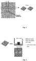
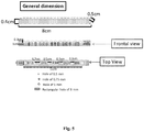
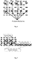


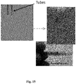
Classifications
-
- C—CHEMISTRY; METALLURGY
- C12—BIOCHEMISTRY; BEER; SPIRITS; WINE; VINEGAR; MICROBIOLOGY; ENZYMOLOGY; MUTATION OR GENETIC ENGINEERING
- C12N—MICROORGANISMS OR ENZYMES; COMPOSITIONS THEREOF; PROPAGATING, PRESERVING, OR MAINTAINING MICROORGANISMS; MUTATION OR GENETIC ENGINEERING; CULTURE MEDIA
- C12N5/00—Undifferentiated human, animal or plant cells, e.g. cell lines; Tissues; Cultivation or maintenance thereof; Culture media therefor
- C12N5/0068—General culture methods using substrates
-
- B—PERFORMING OPERATIONS; TRANSPORTING
- B03—SEPARATION OF SOLID MATERIALS USING LIQUIDS OR USING PNEUMATIC TABLES OR JIGS; MAGNETIC OR ELECTROSTATIC SEPARATION OF SOLID MATERIALS FROM SOLID MATERIALS OR FLUIDS; SEPARATION BY HIGH-VOLTAGE ELECTRIC FIELDS
- B03C—MAGNETIC OR ELECTROSTATIC SEPARATION OF SOLID MATERIALS FROM SOLID MATERIALS OR FLUIDS; SEPARATION BY HIGH-VOLTAGE ELECTRIC FIELDS
- B03C5/00—Separating dispersed particles from liquids by electrostatic effect
- B03C5/005—Dielectrophoresis, i.e. dielectric particles migrating towards the region of highest field strength
-
- C—CHEMISTRY; METALLURGY
- C25—ELECTROLYTIC OR ELECTROPHORETIC PROCESSES; APPARATUS THEREFOR
- C25D—PROCESSES FOR THE ELECTROLYTIC OR ELECTROPHORETIC PRODUCTION OF COATINGS; ELECTROFORMING; APPARATUS THEREFOR
- C25D13/00—Electrophoretic coating characterised by the process
-
- G—PHYSICS
- G01—MEASURING; TESTING
- G01N—INVESTIGATING OR ANALYSING MATERIALS BY DETERMINING THEIR CHEMICAL OR PHYSICAL PROPERTIES
- G01N27/00—Investigating or analysing materials by the use of electric, electrochemical, or magnetic means
- G01N27/26—Investigating or analysing materials by the use of electric, electrochemical, or magnetic means by investigating electrochemical variables; by using electrolysis or electrophoresis
- G01N27/416—Systems
- G01N27/447—Systems using electrophoresis
-
- G—PHYSICS
- G01—MEASURING; TESTING
- G01N—INVESTIGATING OR ANALYSING MATERIALS BY DETERMINING THEIR CHEMICAL OR PHYSICAL PROPERTIES
- G01N27/00—Investigating or analysing materials by the use of electric, electrochemical, or magnetic means
- G01N27/26—Investigating or analysing materials by the use of electric, electrochemical, or magnetic means by investigating electrochemical variables; by using electrolysis or electrophoresis
- G01N27/416—Systems
- G01N27/447—Systems using electrophoresis
- G01N27/453—Cells therefor
-
- A—HUMAN NECESSITIES
- A61—MEDICAL OR VETERINARY SCIENCE; HYGIENE
- A61L—METHODS OR APPARATUS FOR STERILISING MATERIALS OR OBJECTS IN GENERAL; DISINFECTION, STERILISATION OR DEODORISATION OF AIR; CHEMICAL ASPECTS OF BANDAGES, DRESSINGS, ABSORBENT PADS OR SURGICAL ARTICLES; MATERIALS FOR BANDAGES, DRESSINGS, ABSORBENT PADS OR SURGICAL ARTICLES
- A61L27/00—Materials for grafts or prostheses or for coating grafts or prostheses
- A61L27/36—Materials for grafts or prostheses or for coating grafts or prostheses containing ingredients of undetermined constitution or reaction products thereof, e.g. transplant tissue, natural bone, extracellular matrix
- A61L27/3604—Materials for grafts or prostheses or for coating grafts or prostheses containing ingredients of undetermined constitution or reaction products thereof, e.g. transplant tissue, natural bone, extracellular matrix characterised by the human or animal origin of the biological material, e.g. hair, fascia, fish scales, silk, shellac, pericardium, pleura, renal tissue, amniotic membrane, parenchymal tissue, fetal tissue, muscle tissue, fat tissue, enamel
- A61L27/3633—Extracellular matrix [ECM]
-
- C—CHEMISTRY; METALLURGY
- C12—BIOCHEMISTRY; BEER; SPIRITS; WINE; VINEGAR; MICROBIOLOGY; ENZYMOLOGY; MUTATION OR GENETIC ENGINEERING
- C12N—MICROORGANISMS OR ENZYMES; COMPOSITIONS THEREOF; PROPAGATING, PRESERVING, OR MAINTAINING MICROORGANISMS; MUTATION OR GENETIC ENGINEERING; CULTURE MEDIA
- C12N2533/00—Supports or coatings for cell culture, characterised by material
- C12N2533/30—Synthetic polymers
-
- C—CHEMISTRY; METALLURGY
- C12—BIOCHEMISTRY; BEER; SPIRITS; WINE; VINEGAR; MICROBIOLOGY; ENZYMOLOGY; MUTATION OR GENETIC ENGINEERING
- C12N—MICROORGANISMS OR ENZYMES; COMPOSITIONS THEREOF; PROPAGATING, PRESERVING, OR MAINTAINING MICROORGANISMS; MUTATION OR GENETIC ENGINEERING; CULTURE MEDIA
- C12N2533/00—Supports or coatings for cell culture, characterised by material
- C12N2533/50—Proteins
-
- C—CHEMISTRY; METALLURGY
- C12—BIOCHEMISTRY; BEER; SPIRITS; WINE; VINEGAR; MICROBIOLOGY; ENZYMOLOGY; MUTATION OR GENETIC ENGINEERING
- C12N—MICROORGANISMS OR ENZYMES; COMPOSITIONS THEREOF; PROPAGATING, PRESERVING, OR MAINTAINING MICROORGANISMS; MUTATION OR GENETIC ENGINEERING; CULTURE MEDIA
- C12N2533/00—Supports or coatings for cell culture, characterised by material
- C12N2533/70—Polysaccharides
- C12N2533/76—Agarose, agar-agar
-
- C—CHEMISTRY; METALLURGY
- C12—BIOCHEMISTRY; BEER; SPIRITS; WINE; VINEGAR; MICROBIOLOGY; ENZYMOLOGY; MUTATION OR GENETIC ENGINEERING
- C12N—MICROORGANISMS OR ENZYMES; COMPOSITIONS THEREOF; PROPAGATING, PRESERVING, OR MAINTAINING MICROORGANISMS; MUTATION OR GENETIC ENGINEERING; CULTURE MEDIA
- C12N2535/00—Supports or coatings for cell culture characterised by topography
- C12N2535/10—Patterned coating
-
- C—CHEMISTRY; METALLURGY
- C12—BIOCHEMISTRY; BEER; SPIRITS; WINE; VINEGAR; MICROBIOLOGY; ENZYMOLOGY; MUTATION OR GENETIC ENGINEERING
- C12N—MICROORGANISMS OR ENZYMES; COMPOSITIONS THEREOF; PROPAGATING, PRESERVING, OR MAINTAINING MICROORGANISMS; MUTATION OR GENETIC ENGINEERING; CULTURE MEDIA
- C12N2537/00—Supports and/or coatings for cell culture characterised by physical or chemical treatment
Definitions
- the invention relates to the field of tissue engineering and regenerative medicine, and particularly to a method for manufacturing a three-dimensional (3D) biomimetic scaffold that exploits the use of electric fields and electrical insulating materials to pattern previously polymerized hydrogels with different molecules and/or macromolecular entities.
- the invention also relates to the 3D-biomimetic scaffolds obtained and to the uses and applications thereof.
- the microenvironment of a cell comprises all the cues (stimuli) affecting the cell and includes attachment to neighboring cells, attachment to the structural molecules of the extracellular matrix (e.g., via integrins), molecules secreted by the cell itself or by other cells, nutrients, oxygen, mechanical stimuli (such as shear stress), and many others.
- the extracellular matrix ECM
- tissue engineering Within the biomedical field, areas such as tissue engineering, regenerative medicine, drug discovery, or cell biology would highly benefit from the development of 3D biomimetic environments that recreate the complexity of the natural extracellular matrix.
- the general strategy is usually to seed cells within a scaffold, a structural device that defines the geometry of the replacement tissue and provides environmental cues that promote tissue regeneration.
- tissue engineering products must be not only efficacious but also cost-effective, introducing a potential dichotomy between the need for sophistication and ease of production.
- the extracellular matrix found in living tissues is a complex 3D highly hydrated environment made from many elements such as soluble or surface bound molecules, proteins, enzymes, and physical cues like pores and topographies. The precise spatial location of these molecules plays a key role in the behaviour of cells, tissues, and ultimately organs ( Perez-Castillejos R. 2010. Materials Today, 13(1-2): 32-41 ).
- Albrecht D.R. et al. ( Albrecht D.R. et al. 2005. Lab Chip, 5(1): 111-118 ) disclosed a method for encapsulating live cells in 3D-hydrogels using dielectrophoresis.
- the dielectrophoretic forces to locate cells are applied over a prepolymer suspension and said prepolymer suspension is then polymerized by exposure to UV light.
- the same dielectrophoretic forces have been reported to be useful to locate microgels containing encapsulating bioactive materials such as proteins or cells ( Albrecht D.R. et al. 2007. Lab Chip, 7: 702-709 ).
- this method requires the use of special chambers with a plurality of micropatterned surface electrodes to design the position of the molecules or cells in the hydrogel.
- a great advantage of this technique is its versatility: since the technique relies on the application of an electric field, any type of charged molecule can be patterned within any type of hydrogel. This method is simple, easy to handle and affordable and does not need complex equipment. A chamber having only two electrodes as used for protein electrophoresis is enough. It serves as a micro/nano fabrication lithographic technique with potential use in any soft-matter material.
- Section 2.3 of the Examples show that mesenchymal cells cultured on the top of a slice of the 3D biomimetic scaffold obtained according to the method of the invention are capable of attaching to the adhesive peptides patterned on the scaffold and to spread on the region where said peptides are patterned.
- the resulting 3D biomimetic scaffolds show an extremely high level of bioactivity and anisotropy and are suitable for applications in tissue engineering, regenerative medicine, in vivo -like models for drug screening, or cell biology studies such as embryogenesis, spinal cord regeneration or cancer metastasis.
- the invention relates to a method for manufacturing a three-dimensional (3D) biomimetic scaffold comprising:
- the invention relates to a three-dimensional (3D) biomimetic scaffold obtainable by the method according to the invention or a fragment thereof.
- the invention relates to a three-dimensional (3D) biomimetic scaffold according to the invention or to a three-dimensional (3D) biomimetic scaffold obtained according to the method of the invention or a fragment thereof for use in medicine.
- the invention relates to a three-dimensional (3D) biomimetic scaffold according to the invention or to a three-dimensional (3D) biomimetic scaffold obtained according to the method of the invention or a fragment thereof for use in the replacement, reparation or regeneration of a damaged, dysfunctional or pathological tissue.
- the invention relates to the use of a three-dimensional (3D) biomimetic scaffold according to the invention or to a three-dimensional (3D) biomimetic scaffold obtained according to the method of the invention or a fragment thereof as an in vivo -like model for drug screening or as a model for molecular or cell biology studies.
- the invention relates to the use of a three-dimensional (3D) biomimetic scaffold according to the invention or to a three-dimensional (3D) biomimetic scaffold obtained according to the method of the invention or a fragment thereof to support cell growth.
- the present invention provides 3D biomimetic scaffolds, methods for producing said scaffolds, and applications thereof.
- the invention relates to a method, hereinafter referred to as the "method of the invention", for manufacturing a three-dimensional (3D) biomimetic scaffold comprising:
- the "method of the invention” is a lithographic technique that exploits electrical fields (dielectrophoresis) to pattern previously polymerized hydrogel materials with different charged compounds and/or macromolecular entities in three dimensions.
- lithographic technique refers to a method for creating micropatterns of bioactive molecules and/or macromolecular entities in a hydrogel material.
- 3D biomimetic scaffold refers to a three-dimensional artificial structure that partially or completely mimics (e.g. replicates, reproduces, imitates or is similar to) the natural extracellular matrix in its structure or function and exhibits a precise spatial location of bioactive molecules and/or macromolecular entities.
- the extracellular matrix is the extracellular part of animal tissue that usually provides structural support to the animal cells in addition to performing various other important functions such as segregating tissues from one another and regulating intercellular communication. In addition, it sequesters a wide range of cellular growth factors and acts as a local depot for them.
- the extracellular matrix is composed of proteins and polysaccharides that cells secrete into their environment.
- the nature, form and composition of the ECM is diverse and it depends on the types of cells it is associated with.
- the 3D biomimetic scaffold of the invention is capable of recreating the complexity of the natural extracellular matrix found in living tissues.
- the types of scaffolds manufactured will depend on the specific design and application.
- three-dimensional or “ 3D”
- the term “three-dimensional” or " 3D ", as used herein, means that the biomimetic scaffold of the invention has three dimensions, i.e. that three co-ordinates are needed to locate a molecule within it.
- the three-dimensional shape of the biomimetic scaffold may be any 3D shape, size, configuration, depth, width, or other geometry capable of being formed in a mold, and further capable of being reproduced by downstream steps (i.e. capable of being adopted by a polymerizable compound).
- the 3D shape formed is similar to or representative of a 3D geometry found in a biological system.
- the responsible of the 3D shape of the biomimetic scaffold is the immobile phase of the method of the invention.
- immobile phase refers to any previously polymerized hydrogel that is going to be patterned with the mobile phase.
- hydrogel refers to water-insoluble three-dimensional networks that are formed by the cross-linking of water-soluble monomers. The cross-linking of the water soluble monomers into a water insoluble polymer allows the hydrogel to "swell” and topologically trap other compounds or macromolecular structures, thereby forming an interpenetrating covalent network in and around the molecules trapped. Any hydrogel may be used in the present invention.
- the hydrogels used in the method of the invention are previously polymerized hydrogels.
- previously polymerized it is meant that the cross-linking of water-soluble monomers has been reached before starting to apply the method of the invention and that the immobile phase of the method of the invention is a network of interacting polymer chains that are highly hydrated, having a elasticity similar to that of natural tissues.
- the previously polymerized hydrogel may contain cells entrapped or encapsulated within the hydrogel during the polymerization step.
- at least a cell is contained in the previously polymerized hydrogel used in the method of the invention before starting to apply an electric field.
- a fragment of a cell is contained in the previously polymerized hydrogel.
- At least a compound selected from a peptide, a protein, a nucleic acid, a nanoparticle, a microparticle, a dendrimer, a dendrimer conjugate or a drug is contained in the previously polymerized hydrogel.
- the process of the invention allows the generation of precise patterns of molecules within a wide range of hydrogel materials, including traditional ones like agarose and polyacrylamide or cutting edge ones like those formed through self-assembling materials.
- Some known natural polymers and synthetic monomers used in hydrogel fabrication include, without limitation, chitosan, alginate, fibrin, fibronectin, collagen, gelatin, elastin, hyaluronic acid, heparan sulfate, chondroitin sulfate, keratan sulfate, dextran, hydroxyethyl methacrylate, N-(2-hydroxypropyl) methacrylate, N-vinyl-2-pyrrolidone, N-isopropyl acrylamide, vinyl acetate, acrylic acid, methacrylic acid, polyethylene glycol diacrylate/methacrylate, polyethylene glycol diacrylate/dimethylacrylate, polyacrylamide, agarose, just to name a few.
- the hydrogel useful in the present invention is selected from polyacrylamide, agarose, chitosan, alginate, gelatin and the commercially available BDTM PuraMatrixTM Peptide Hydrogel (BD Biosciences).
- Preferred hydrogels are polyacrylamide hydrogel and agarose hydrogel, more preferably agarose hydrogel.
- concentrations of the monomers in the hydrogel depend on the specific hydrogel used.
- Typical concentrations of the monomers in the hydrogel are between 4% and 16% for polyacrylamide, preferably 8%; between 1% and 5% for agarose, preferably 2%; between 0.5% and 2% for chitosan, preferably 1.5%; between 1% and 3% for alginate, preferably 2%; and between 5% and 10% for gelatin, preferably 5%. These concentrations permit the pattern conformation.
- agarose hydrogel is preferred.
- the hydrogel is agarose.
- Typical concentration of agarose in the hydrogel may be from 0.5 to 5, preferably 2%. These concentrations permit the conformation of patterns. It is possible to use a higher concentration of agarose, although it compromises the handelability due to the viscosity of the agarose melt solution.
- the hydrogel may also be a bioactive hydrogel with sites designed to bind passing molecules.
- the polymerized hydrogels may be prepared in situ by different methods well known by the skilled person depending on the monomer used or may be commercially available.
- the monomer to be polymerized is mixed with the same buffer used in the method for covering the mobile phase, the immobile phase and the electrical insulating material in step (b) of the method of the invention.
- This mixture forms a pre-gel solution.
- the pre-gel solution of this polymerizable material is poured into a mold and may be manufactured by a method such as that described in section 1.1.2 of the Examples.
- the electrical insulating material is going to be a porous membrane, a piece of the polymer used for manufacturing the membrane or a mold having the same shape than the membrane without holes is placed on top of the immobile phase to get the space where the membrane will be located.
- the piece or mold is substituted by the porous membrane that is going to be used in the method of the invention.
- the electrical insulating material is going to be a combination of a porous membrane and one or more tubes, or a tube or a series of tubes without the membrane, the single tube or a series of plugged tubes of plastic or crystal are inserted through the polymer used for manufacturing the membrane.
- the ensemble of the polymer for manufacturing the porous membrane and the tube or tubes are positioned on the top of the liquid before the pre-gel polymerizes into hydrogel.
- the electrical insulating material includes the membrane, the plugged tubes are substituted by hollow tubes filled with mobile phase.
- the membrane is removed and the holes made by the plugged tubes are filled by hollow tubes containing the mobile phase.
- the porous membrane is obtained by curing the polymer inside the mold in which the hydrogel polymerizes.
- curing is a term in polymer chemistry that refers to the toughening or hardening of a polymer material by cross-linking of polymer chains. Once this is prepared, the hydrogel is allowed to polymerize.
- the polymerization is achieved through the use of temperature, time, a polymerizing agent, and/or any other polymerization trigger.
- the polymerization requirements will depend upon the particular polymerizable monomer or the polymerizing compound chosen.
- Porous hydrogels suitable for the present invention may be obtained by freeze drying ( Madihally S.V. and Matthew H.W. 1999. Biomaterials, 20(12): 1133-42 ) or by using dense gas CO 2 as a foaming agent ( Ji C. et al. 2011. Acta Biomater, 7(4): 1653-64 ).
- the mold defines the shape of the immobile phase which will be the shape of the 3D biomimetic scaffold obtained by the method of the invention.
- the shape of the biomimetic scaffold may be any desired shape. Possible shapes are, without limitation, cylindrical, cubic, cuboidal, any kind of prism shape (triangular, rectangular, pentagonal, hexagonal, etc.).
- the mold in which the hydrogel polymerizes is the same mold that is going to be subjected to an electric field in step b) of the invention.
- the mold may be of any material capable of accepting a three-dimensional shape while being sufficiently rigid to maintain the shape in downstream steps of the method.
- the mold may be manufactured in one piece or may be formed by different pieces to be assambled to prepare the hydrogel. Preferred materials for the mold are PDMS, crystal, thermoplastic, and other materials that allow manufacturing any desired shape or form.
- the mold is a typical mold for protein electrophoresis.
- Typical vertical slab hydrogels are normally cast between a pair of glass plates for support. A space is constructed by separating the two plates with spacer strips down the edges of the plates, then sealing the edges and bottom to form a liquidtight box or sandwich.
- the hydrogels for the method of the invention may be of any desired size. Typical hydrogels for this invention range in size from 2 to 21 cm x 3 to 30 cm and from 0.2 to 7 cm thick; preferably 8.3 cm x 9.7 cm and from 0.2 to 0.5 cm thick.
- a gasket or form comprising the bottom of the molding cavity is removed after hydrogel polymerization in order to allow current to pass through two opposite edges of the hydrogel slab; one of these edges represents the open (top) surface of the hydrogel cavity, and the other is formed against its removable bottom.
- the term "mobile phase" refers to the phase that is displaced through the hydrogel when performing the method of the invention and that contains the bioactive molecules and/or macromolecular entities that are going to pattern the hydrogel.
- the mobile phase is an aqueous or organic medium comprising at least a charged compound and/or a charged macromolecular entity.
- aqueous or organic medium refers to a solution that contains the compound(s) and/or macromolecular entity and provides the mobile phase with the right density for running the molecules.
- aqueous medium refers to a liquid medium composed largely, but not necessarily exclusively, of water; preferably comprising at least 50 weight percent water. Other components may also be present, such as salts, co-solvents, buffers, colorants and the like.
- the medium is an aqueous medium.
- the aqueous medium is 2% low melting agarose in the same buffer used for step (b) of the method of the invention.
- the aqueous medium is glycerol in the same buffer used for step (b) of the method of the invention.
- organic medium refers to a liquid medium composed largely, but not necessarily exclusively, of one or more organic solvents, it being possible for said medium to contain water; preferably comprising at least 50 weight percent organic solvents.
- organic solvents for organic medium that may be used in the invention are, without limitation, dimethyl sulfoxide (DMSO), dimethylformamide (DMF), ethanol, methanol, dichloromethane (DCM), CHCl 3 , hexane, pentane, heptane, bencene, toluene and acetone.
- any type of hydrogel can be patterned using any charged molecule.
- charged refers to the electric charge that the compound or macromolecular entity has at the pH at which step (b) of the method of the invention is performed.
- the mobile phase contains the components usually present in the ECM.
- Components of the ECM are produced intracellularly by resident cells, and secreted into the ECM via exocytosis. Once secreted, they then aggregate with the existing matrix.
- compound as used herein referred to the mobile phase, relates to any kind of charged molecule capable of being displaced through a hydrogel when an electric field is applied and includes bioactive molecules and structural molecules, soluble and surface bound molecules.
- Compounds useful in the present invention are, without limitation, peptides, proteins, nucleic acids, nanoparticles, microparticles, fluorescent molecules, dendrimers, dendrimers conjugated with epitopes or drugs or fluorescent molecules, hormones, growth factors, enzymes, drugs, etc.
- the compound is selected from a peptide, a protein, a nucleic acid, a nanoparticle, a microparticle, a dendrimer or a peptide-dendrimer conjugate; preferably is selected from a peptide, a protein, a dendrimer and a peptide-dendrimer conjugate.
- peptide or "polypeptide”, as used herein, refers to a short polymer of amino acid monomers linked by peptide bonds, typically containing less than 50 monomer units. Any peptide may be used in the present invention. In a preferred embodiment the compound is a peptide.
- protein refers to one or more peptides (or polypeptides), i.e., polymer chains of amino acids bonded together by peptide bonds between the carboxyl and amino groups of adjacent amino acid residues, optionally including modifications, e.g., post-translational modifications, which alter the physical and chemical properties, folding, stability, activity, and ultimately, the function of the proteins. Proteins having non-peptide groups attached (i.e., prosthetic groups or cofactors) are also included within this definition.
- the number of amino acid residues in a protein can vary in a broad range, for example, the polymer chain of amino acid residues linked by peptide bonds may contain typically 50 or more amino acids residues.
- proteins useful in the present invention are, without limitation, perlecan, agrin, collagen XVIII.
- the compound is a protein.
- the compound is an elastin-like polymer or a mixture thereof. Elastin-like polymers consist of repeating pentapeptide domains of the amino acids VPGIG and VPGKG or VPGVG with the cell binding sequence RGDS.
- nucleic acid refers to a polymer of nucleotides having two or more deoxyribonucleotide, ribonucleotide or nucleotide analog molecules as well as molecules that are structurally similar to a native nucleic acid, but differ from the native nucleic acid (e.g. through chemical modification) at one or more of the nucleic acid backbone (e.g. phosphate in native nucleic acids), nucleic acid sugar (e.g. deoxyribose for native DNA and ribose in native RNA), and nucleic acid base (e.g. adenosine, cytosine, guanine or thymidine in native nucleic acids).
- nucleic acid backbone e.g. phosphate in native nucleic acids
- nucleic acid sugar e.g. deoxyribose for native DNA and ribose in native RNA
- nucleic acid base e.g. adenosine, cyto
- the nucleic acid can be a double stranded or single stranded nucleic acid including, without limitation, DNA, RNA, oligonucleotides, PNAs, cDNA, RNAi, shRNA, miRNA, siRNA, ribozymes, antisense oligonucleotides, as well as modified forms thereof.
- the compound is a nucleic acid.
- nanoparticle refers to a particle having a diameter below 1 ⁇ m and including, without limitation, nanospheres and nanocapsules.
- the compound is a nanoparticle.
- microparticle refers to a particle having a diameter between 1 and 250 ⁇ m and including, without limitation, microspheres and microcapsules. In a preferred embodiment the compound is a microparticle.
- dendrimer refers to a repetitively branched molecule typically symmetric around the core, and often adopting a spherical three-dimensional morphology. The person skilled in the art knows that any type of dendrimer may be used.
- the compound is a dendrimer.
- the compound is a peptide-dendrimer conjugate.
- peptide-dendrimer conjugate refers to a dendrimer conjugated to one or more epitopes to homogenize the size and the charge of said peptides.
- the compound may be a molecule capable of exerting a function, preferably a biological function.
- the compound is selected from a hormone, a growth factor, an enzyme, a fluorescent molecule, a drug, a fluorescent molecule-dendrimer conjugate or a drug-dendrimer conjugate.
- hormone refers to a chemical released by a cell or a gland in one part of the body that sends out messages that affect cells in other parts of the organism.
- examples of hormones that are useful in the present invention are, without limitation, melatonin (MT), serotonin (5-HT), thyroxine (T4), triiodothyronine (T3), epinephrine or adrenaline (EPI), norepinephrine or noradrenaline (NRE), dopamine (DPM or DA), antimullerian hormone or mullerian inhibiting hormone (AMH), adiponectin (Acrp30), adrenocorticotropic hormone or corticotrophin (ACTH), angiotensinogen and angiotensin (AGT), antidiuretic hormone or vasopressin (ADH), atrial natriuretic peptide or atriopeptin (ANP), calcitonin (CT
- growth factor refers to a naturally occurring substance capable of stimulating cellular growth, proliferation and cellular differentiation. Usually it is a protein or a steroid hormone. Growth factors typically act as signalling molecules between cells. Examples of growth factors are cytokines that bind to specific receptors on the surface of their target cells. Their function varies between growth factors. For example, bone morphogenic proteins stimulate bone cell differentiation, while fibroblast growth factors and vascular endothelial growth factors stimulate blood vessel differentiation (angiogenesis).
- growth factors are, without limitation, adrenomedullin (AM), angiopoietin (Ang), autocrine motility factor, bone morphogenetic proteins (BMPs), brain-derived neurotrophic factor (BDNF), epidermal growth factor (EGF), erythropoietin (EPO), fibroblast growth factors (FGFs), glial cell line-derived neurotrophic factor (GDNF), granulocyte colony-stimulating factor (G-CSF), granulocyte macrophage colony-stimulating factor (GM-CSF), growth differentiation factor-9 (GDF9), hepatocyte growth factor (HGF), hepatoma-derived growth factor (HDGF), insulin-like growth factor (IGF), migration-stimulating factor, myostatin (GDF-8), nerve growth factor (NGF), neurotrophins, platelet-derived growth factor (PDGF), thrombopoietin (TPO), transforming growth factor alpha (TGF- ⁇ ), transforming growth factor beta (TGF
- enzyme refers to a protein that catalyzes chemical reactions.
- examples of enzymes useful in the present invention are, without limitation, serine proteases, threonine proteases and matrix metalloproteinases.
- the compound is an enzyme.
- fluorescent molecule refers to a molecule capable of emitting fluorescence. Any fluorescent molecule may be used in the present invention. Examples of fluorescent molecules are, without limitation, pyrene, fluorescein and luciferin. In a preferred embodiment the compound is a fluorescent molecule. In a more preferred embodiment the compound is a fluorescent molecule-dendrimer conjugate, i.e. a dendrimer conjugated to one or more molecules that emit fluorescence.
- drug as used herein, relates to any chemical substance that, when absorbed into the body of a living organism, alters normal bodily function or that is used in the treatment, cure, prevention or diagnosis of a disease or enhances physical or mental well-being.
- drugs useful in this invention are, without limitation, cancer drugs such as isotretinoin, tretinoin, cladribine, azacitidine, fluorouracil, mercaptopurine, thioguanine, paclitaxel, actinomycin-D, doxorubicin, daunorubicin, everolimus, anagrelide, aldesleukin, alemtuzumab, pemetrexed, alitretinoin, vinblastine, melphalan, interferon alfa, altretamine, methotrexate, amifostine, aminoglutethimide, anastrozole, exemestane, nelarabine, bleomycin, busulfan, capecitabine, carboplatin, carmustine, chlorambucil, cisplatin, cyclophosphamide, ifosfamide, carmustine, cytarabine, dacarbazine, docetaxe
- the compound is a drug.
- the compound is a drug-dendrimer conjugate.
- drug-dendrimer conjugate refers to a conjugate of a dendrimer with one or more drugs wherein the dendrimer acts as a carrier for said drugs and is used for drug delivery.
- Macromolecular entity refers to a charged structure formed by more than one molecule and includes cells and fragments thereof as well as bacteria.
- Suitable cells for the invention are cell types involved in ECM formation such as fibroblasts, chondrocytes and osteoblasts.
- Other kind of cells useful are, without limitation, stem cells, mesenchymal stem cells, melanoma cell lines such as COLO-794, COLO-800, COLO-818, FM-28, WM-75, WM-793, WM-852, FR-M-170, FR-M-204, Ma-Mel-16, Ma-Mel-25, and other melanoma cell lines listed in the European Searchable Tumour Line Database (ESTDAB) (http://www.ebi.ac.uk/ipd/estdab/) .
- ESTDAB European Searchable Tumour Line Database
- the macromolecular entity is a cell. Fragments of cells included in the present invention may be cell membranes. By “cell membrane " it is understood a biological membrane that separates the interior of a cell from the outside environment. It consists of the lipid bilayer with embedded proteins. In another preferred embodiment the macromolecular entity is a fragment of a cell, preferably a cell membrane.
- the macromolecular entity is a bacterium.
- Any bacteria may be used in the present invention.
- bacteria are, without limitation, Escherichia coli, Shigella spp., Bacillus cereus, Yersinia pestis, Pseudomonas spp., Bordetella pertussis, Borrelia burgdorferi, Campylobacter pylori, Chlamydia trachomatis, Clostridium botulinum, Clostridium difficile, Clostridium perfringens, Clostridium tetani, Corynebacterium diphtheriae, Enterococcus faecalis, Haemophilus influenzae, Helicobacter pylori, Klebsiella pneumoniae, Lactobacillus acidophilus, Legionella pneumophila, Listeria monocytogenes, Mycobacterium diphtheriae, Mycobacterium tuberculosis, Mycoplasma pneumoniae
- the mobile phase is prepared by dissolving or suspending the compounds or the macromolecular entities in the aqueous or organic solution by methods well known in the art.
- Step a) of the method of the invention involves contacting the mobile phase with the immobile phase through an electrical insulating material containing the mobile phase.
- electrical insulating material refers to a material that has a relative static permittivity or dielectric constant greater than 1.
- relative permittivity refers to the extent to which a material concentrates electrostatic lines of flux.
- the relative permittivity is the ratio of the amount of electrical energy stored in a material by an applied voltage, relative to that stored in a vacuum. And it is static when it is calculated in a frequency of zero.
- the electric insulating material modifies the electric field applied in step (b) of the method of the invention.
- Said electrical insulating material is also named "mobile container” in the Examples of the present description, since it contains the mobile phase and guides the patterning of the molecules within the hydrogel.
- Examples of electrical insulating materials are glass, paper, teflon, rubber-like polymers and most plastics.
- the electrical insulating material useful in this invention is selected from:
- the dimension and position of the electrical insulating material is one of the factors involved in controlling the location of the components of the mobile phase through the immobile phase.
- porous membrane refers to a layer of electrical insulating material that has one or more holes that allow the mobile phase pass through said membrane.
- porous does not refer to the porosity of the insulating material.
- the membrane is "porous” because it has one or more holes through which the mobile phase passes and said holes are the "pores" of the membrane. Said holes or “pores” define the geometrical regions through which the mobile phase passes and may be open-holes or may be filled with tubes that contain the mobile phase. By “geometrical regions” it is understood the x-y shape through which the molecules will pass. The geometry of said holes depends on the particular pattern to be obtained in the hydrogel.
- Each hole may be a rectangular hole, a cylindrical hole, or any other shape. Each hole may be filed with a different mobile phase.
- the porous membrane as such can be easily modulated to exhibit almost any design.
- the shape of the membrane may be any desired shape.
- the porous membrane is rectangular.
- the porous membrane can be of various thickness, particularly preferred are from 200 ⁇ m to 0.5 cm, more preferably 0.3 cm.
- the porous membrane must be made from an electrical insulating material.
- Materials suitable for said porous membrane are, without limitation, polyurethane, polycarbonates, ethylene-vinyl acetate, silicone rubber such as polydimethylsiloxane, fluorosilicone rubber, polyacrylic rubber, chlorosulfonated polyethylene, ethylene propylene rubber and chloroprene; preferably polydimethylsiloxane (PDMS).
- PDMS polydimethylsiloxane
- the porous membrane is placed on the surface of the top of the immobile phase, i.e. in contact with the upper edge of the hydrogel.
- the geometrical regions of the porous membrane are defined by holes in said membrane.
- the geometrical regions of the porous membrane are defined by tubes inserted in the holes of said membrane.
- this membrane may not be necessary for less complex designs.
- small tubes containing the mobile phase may be sufficient to define the areas where the compounds or macromolecular entities will be patterned. Said small tubes are placed in the locations where the mobile phase will be applied.
- tubes suitable for the present invention may be tubes having triangular, square, rectangular, pentagonal, hexagonal or any other cross-section. Said tubes can have a different length depending on the hydrogel size. Preferred length is from 0.5 cm to 10 cm, more preferably of 3.5 cm.
- Suitable electrical insulating materials for tubes are, without limitation, silica, quartz, crystal, plastic; preferably crystal or plastic. The tube must be inserted into the top of the immobile phase, i.e. inserted into the upper edge of the hydrogel. When only tubes are used as electrical insulating materials, these tubes do not create a significant modification of the electrical field (see section 2.1 of the Examples).
- the electrical insulating material is a combination of a porous membrane and at least a tube.
- the electrical insulating material contains the mobile phase. This means that the holes of the porous membrane or the tubes are filled with the mobile phase.
- step (a) implies filling the holes of the porous membrane or the tubes with the mobile phase. This must be done by means known by the person skilled in the art just before to start step (b) of the method of the invention.
- Step (b) of the method of the invention consists of subjecting the ensemble formed by the mobile phase, the immobile phase and the electrical insulating material to a direct current (DC) electric field created inside a chamber and applied in closed circuit by a pair of electrodes in vertical or horizontal configuration, wherein the chamber contains the mobile phase, the immobile phase and the electrical insulating material covered by a buffer. Therefore, step (b) of the method of the invention is based on the dielectrophoretic principle.
- the ensemble formed by the mobile phase, the immobile phase and the electrical insulating material must be inside a chamber.
- the term "chamber” means a typical electrophoretic chamber or a chamber specially designed for the manufacturing of the biomimetic scaffold of the invention that has essentially the same features that a typical electrophoretic chamber.
- Said chamber is designed to perform the migration of the mobile phase by applying an electric field.
- These chambers typically consist of a nonconducting material (e.g., glass, acrylic) and include a pair of electrodes.
- electrode relates to an electrical conductor used to make contact with a nonmetallic part of a circuit, in this case with an electrolyte.
- air of electrodes it is meant that the chamber has only two electrodes: an anode and a cathode.
- the anode is the positive (+) electrode and the cathode is the negative (-).
- the electrons enter the device through the cathode and exit the device through the anode.
- Materials for electrodes may be, without limitation, platinum wire, silver and gold.
- a hydrogel is typically mounted on a suitable electrophoresis apparatus, so that one edge of the hydrogel contacts a first buffer reservoir containing an electrode and the opposite hydrogel edge contacts a second reservoir with a second electrode, steps being taken so that the current passing between the electrodes is confined to run mainly or exclusively through the hydrogel.
- the method of the invention may be performed in vertical configuration or in horizontal configuration, preferably in vertical configuration.
- vertical configuration means that the hydrogel's upper edge is in contact with an upper buffer reservoir and the lower edge is in contact with a lower reservoir.
- Vertical electrophoresis has an angle 90 degree to the bottom side.
- horizontal configuration means that the hydrogel is run in a horizontal orientation with the gel resting on a platform between the buffer reservoirs, submerged under a layer of a few millimeters of buffer. Horizontal electrophoresis has an angle 180 degree to the bottom side. Thus, the mobile phase runs through the immobile phase in a horizontal direction.
- the chamber contains the mobile phase, the immobile phase and the electrical insulating material covered by a buffer.
- the tube(s) or porous membrane used as electrical insulating material must be completely submerged in buffer.
- buffer relates to the buffer solution used for running the electrophoresis.
- Said “buffer” is an aqueous solution consisting of a mixture of a weak acid and its conjugate base or a weak base and its conjugate acid. It has the property that the pH of the solution changes very little when a small amount of strong acid or base is added to it.
- Preferred pH for the buffer of the invention is 7.4, which is the normal extracellular pH.
- the 3D biomimetic scaffold of the invention may be used as a model for diseases in which physiological pH is altered.
- buffer solutions useful in this invention are, without limitation, tris-HCl (range of pH 7.5 to 9.0), tris-glycine (range of pH 8.3 to 9.5), tris-borate (range of pH 7.0 to 8.5), tris-acetate (range of pH from 7.2 to 8.5), TBE (tris/boric acid/EDTA) (range of pH 8.3 to 8.6), HEPES (range of pH from 6.8 to 8.2), PIPES (range of pH from 6.1 to 7.5), TES (range of pH from 6.8 to 8.2), PBS (range of pH from 7.0 to 7.8), PB (range of pH from 7.2 to 8.2); preferably TBE (tris/boric acid/EDTA), PBS (phosphate buffered saline) and PB (potassium phosphate monobasic/disodium hydrogen phosphate dehydrate); more preferably TBE and PB, the most preferred PB.
- tris-HCl range of pH 7.5 to 9.0
- tris-glycine
- Buffer compositions are well known by the person skilled in the art.
- the pH of the buffer composition is chosen depending on the isoelectric point of the compounds of the mobile phase in order to obtain a charged compound. For example, if the isoelectric point of a compound of the mobile phase is 4, the pH of the buffer composition must be higher than 4 to have the compound negatively charged and must be lower than 4 to have the compound positively charged.
- Buffer most appropriate for culturing cells is PB.
- a suitable composition for PB is 2.6 mM potassium phosphate monobasic and 7.3 mM disodium hydrogen phosphate dehydrate having pH 7.4 and ionic strength of 24 mM.
- the components of the mobile phase are in native state.
- native as used herein, it is meant that the method of the invention is made in absence of any denaturing agent.
- proteins migrate as a function of their charge, size and shape and interactions between subunits are maintained.
- proteins preserve their function as is expected for a biomimetic scaffold.
- This technique uses a direct current electric field applied in a closed circuit to move and position bioactive molecules to create reproducible well organized patterns within hydrogel materials in 3D.
- direct current or " DC”
- DC direct current
- electrical field refers to the field that surrounds electrically charged particles and time-varying magnetic fields.
- closed circuit refers to an electric circuit providing an uninterrupted, endless path for the flow of current.
- the molecules and/or macromolecular entities contained in the mobile phase will move through the hydrogel toward the anode if negatively charged or toward the cathode if positively charged. Therefore, the method of the invention is useful for both positively and negatively charged molecules.
- the person skilled in the art can reverse the polarity of the electric field applied depending on the charge of the molecules and/or macromolecular entities contained in the mobile phase.
- a typical system for vertical or horizontal protein electrophoresis may be used.
- the method of the invention uses an electrical insulating material and thus relies in the dielectrophoretic principle, specifically in the insulator-based direct current dielectrophoresis (DC-iDEP), which uses insulating obstacles to create spatial field nonuniformities.
- DC-iDEP direct current dielectrophoresis
- Dielectrophoresis refers to the force that is used to move the molecules and/or macromolecular entities of the mobile phase through the hydrogel matrix when said entities are subjected to a nonuniform electric field.
- Power supplies used in the present invention hold one electrical parameter (current, voltage, or power) constant.
- the resistance of the dielectrophoresis circuit does not remain constant during a run.
- CM Clausius-Mossotti
- the method of the invention differs from the classical electrophoresis since molecules are not separated by their molecular weight and the electrical field is modified by using an electrical insulating material.
- step (c) of the method of the invention starts.
- This step consists of patterning the immobile phase with the components of the mobile phase, in which said components move by forming patterns within the immobile phase and are not separated by their molecular weight and in which the location of the components of the mobile phase is controlled as desired by modulating the electric field and the time.
- One of the characteristic features of this method is that the mobile phase is patterned within the immobile phase.
- Patterning or “move by forming patterns”, as used herein, relates to drawing a continuous path in the hydrogel with the molecules and/or macromolecular entities of the mobile phase.
- the molecules are homogeneously distributed along the path or distributed along the path forming a gradient and are not separated by their molecular weight or concentrated in a specific location.
- the molecules or macromolecular entities contained in the mobile phase are finally immobilized within the hydrogel.
- the homogeneous or gradient patterns may be obtained by conventional methods known by the skilled person in the art.
- the components of the mobile phase are positioned in specific locations within the hydrogel to create a complex in vivo -like environment.
- components of the mobile phase refers to the charged compounds and/or charged macromolecular entities that form part of the mobile phase.
- the expression “are not separated by their molecular weight”, as used herein, means that the resulting hydrogel is different from a hydrogel obtained in classical electrophoresis, when molecules are separated by their molecular weight, i.e. proteins are resolved.
- molecular weight refers to the sum of the atomic weights of all the atoms in a molecule. When the molecule is a peptide or a protein the molecular weight is expressed in Daltons.
- location of the components of the mobile phase refers to the specific pattern obtained in the hydrogel formed by the mobile phase. Different factors are involved in the final location of the components of the mobile phase through the hydrogel. Specifically, the location of the components depends on the molecular size and charge of the mobile phase, the porosity of the immobile phase, the dimension and position of the insulating material and the modulation of the electrical field and the time during which the mobile phase is patterned through the immobile phase. The person skilled in the art knows how to modify said parameters to locate the components of the mobile phase in the location desired.
- the velocity of migration of the components of the mobile phase through the gel is affected directly by the molecular size of said components.
- Components having a small molecular size move faster and migrate further than components with a big molecular size.
- the overall charge of a component also affects the speed at which it moves through the hydrogel.
- the charge of the mobile phase is due to the buffer pH. Therefore, small molecular size and strong charge increase the migration rate of the component through the hydrogel, whereas large size and weak charge decreases the migration rate.
- the porosity of the immobile phase also affects the final location of the components of the mobile phase.
- the pore size and sieving characteristics of a hydrogel can be controlled by adjusting the concentration of the polymer (agarose, chitosan, gelatin, etc.). The higher the concentration, the smaller the pore size.
- the dimension and position of the insulating material is another important parameter that allows controlling the final location of the molecules of the mobile phase.
- the expression " is controlled as desired ", as used herein, means that the operator that puts into practice the method of the invention can decide in which part of the immobile phase is to be patterned each mobile phase. To achieve this, the operator may modify some variables such as the electric field or the time that the electric field is applied. The use of electrical insulating materials, and the specific design of the porous membrane may be changed to obtain the desired pattern. The person skilled in the art may predict the pattern obtained by using simulations in computer programs such as COMSOL Multiphysics, as is disclosed in the Examples.
- modulating the electric field includes any modulation of the electric field that may be performed by the use of an electrical insulating material or by the modification of the level of constant current or constant voltage.
- the dimension and position of the electrical insulating material allows modulating the electrical field.
- voltage refers to the potential difference between two points. The voltage is denoted ⁇ V and is measured in volts or Joules per Coulomb. A voltimeter can be used to measure the voltage between two points in a system. Section 2.2 of the Examples show curved patterned lines obtained when the electrical field is affected.
- the electrical field is also modulated by the use of a cooling system.
- modulating the time means that the operator may decide the time during which the voltage is applied. Time may be measured in seconds, minutes or hours.
- the hydrogel is removed from the mold used for performing the method and is ready for use.
- the method of the invention may be performed under sterile conditions, for example when the mobile phase contains cells. In such conditions sterilized materials are required. Methods for sterilizing are well known by the person skilled in the art. Therefore, in a preferred embodiment, the method is performed under sterile conditions.
- hydrogel may be stained.
- Staining methods include dye-binding (e.g. Coomassie Brilliant Blue) or silver stains. Another possibility to confirm the pattern obtained is to use fluorescently labeled proteins in the mobile phase.
- the 3D-patterning method of the invention allows controlling with high precision and reproducibility the positioning and presentation of different molecules and/or macromolecular entities inside the scaffold allowing constructing scaffolds with programmed spatial features for tissue engineering or other applications.
- the 3D biomimetic scaffold of the invention may be used as such or may be cut into fragments for different applications.
- the 3D biomimetic scaffold of the invention may include cells, specifically live cells.
- Cells may be on the 3D biomimetic scaffold because they have been included before the polymerization of the hydrogel or because they have been patterned as forming part of the mobile phase. Said cells may be cultured directly on the biomimetic scaffold. In addition, cells may be cultured on the surface of the scaffold.
- the 3D biomimetic scaffold obtained by the method of the invention may be cut into pieces and each piece may be used as support for cell growth.
- the method further comprises culturing cells in or on the product resulting from step c) or a fragment thereof.
- culturing cells refers to submit cells to the conditions suitable for maintaining them viable while growing and spreading over the scaffold. Conditions for culturing cells are well known by the person skilled in the art and will vary depending on the specific type of cell cultured.
- the 3D structure is seeded with cells and cultured for a period of time with suitable culture media, preferably until the entire structure is coated with living cells. Said living cells are capable of migrating through the 3D scaffold following the pattern obtained by the method of the invention.
- tisssue or regenerative medicine cells may be seeded within a scaffold that defines the geometry of the replacement tissue and provides environmental cues that promote tissue regeneration.
- cell relates to any kind of cell.
- useful cells for this invention are, without limitation, primary cells such as endothelial cells, mesenchymal stem cells, osteoblasts, chondrocytes and neurons; cell lines such as HeLa, DU145, LNCaP, MCF-7, MDA-MB-438, PC3, T47D, THP-1, U87, SH-SY5Y, Saos-2, Vero, GH3, PC12, MC3T3, Tobacco BY-2, Zebrafish ZF4, Zebrafish AB9, MDCK, Xenopus A6, 293-T, 3T3, 721, 9L, A-549, A172, A20, A253, A2780, A2780ADR, A2780cis, A431, ALC, B16, B35, BCP-1, BEAS-2B, bEnd.3, BHK-21, BR 293, BxPC3, C2C12, C3H-10T1/2, C6/36
- product resulting from step c) refers to the whole 3D biomimetic scaffold as is obtained from the method of the invention.
- Cells cultured may be those cells being inside the biomimetic scaffold or those cells deposited on the surface of the biomimetic scaffold.
- a "fragment" of the biomimetic scaffold is a piece of the biomimetic scaffold, either longitudinal or transversal, preferably a transversal piece, that has been obtained after cutting the whole biomimetic scaffold obtained by the method of the invention ( Figure 3 ).
- cells are seeded on the surface of a fragment of the biomimetic scaffold and are cultured on top of it, as is showed on section 1.4.1 of the Examples.
- FIG. 1 schematically shows the components of the method of the invention for manufacturing 3D biomimetic scaffolds in accordance with different embodiments.
- a porous membrane (3) is used as electrical insulating material; in this embodiment, a mobile phase (1) comprising a solution of molecules passes through a porous membrane (3) in order to pattern an immobile phase (2) [ Figure 1 (a) , left]; then the device is submerged in a buffer (8) inside a chamber (5) having two electrodes (6) and (7) and patterns are formed according to the method of the invention - the arrows indicate the direction in which the mobile phase (1) runs [ Figure 1 (a) , right].
- tubes (4) filled with proteins as electrical insulating material are used; in this embodiment, tubes (4) are filled with mobile phase (1) in order to pattern an immobile phase (2) [ Figure 1 (b) , left]; then the device is submerged in a buffer (8) inside a chamber (5) having two electrodes (6) and (7) and patterns are formed according to the method of the invention.
- a second aspect of the present invention relates to a three-dimensional (3D) biomimetic scaffold obtainable by the method of the invention described above (hereinafter, 3D biomimetic scaffold of the invention) or a fragment thereof.
- the 3D biomimetic scaffold is a replacing tissue.
- This technique can be applied to studies or therapies to regenerate any kind of tissue such as bone, cartilage, skin, ligament, tendon, neural, cardiac, muscle, enamel, hepatic, epithelial and corneal tissue.
- the replacing structure is an intervertebral disc.
- the replacing structure is an orosteochondral segment.
- the 3D biomimetic scaffold of the invention can be cut into the desired size and/or can be provided in a suitable conformation for use.
- the extracellular matrix contains many macromolecules such as proteoglycans, collagens, laminins, fibronectin and sequestered growth factors, and it is primarily this molecular information that confers its bioactivity.
- the 3D biomimetic scaffold of the invention is useful in tissue engineering for facilitating recreation of complex 3D tissues or environments to guide cells and create tissues in vitro.
- the application of growth factors within biomaterials also represents a powerful tool for controlling cell differentiation and function.
- the patterning of growth factors within prefabricated scaffolds could aid the generation of heterogeneous tissues.
- biomimetic scaffolds can be directly implanted in patients or may be used to expand and guide cell populations that are subsequently delivered and implanted into areas that require large cell populations.
- biomimetic scaffolds of the invention are also useful in regenerative medicine since they can be implanted at the site of injury without including cells.
- suitable scaffolds for regenerative medicine may be intervertebral discs or orosteochondral segments or other kind of scaffolds for spinal cord regeneration.
- tissue engineering is to construct artificial biological tissues and to use them for medical purposes to restore, replace or increase the functional activities of diseased tissues.
- the therapeutic use of techniques of this type is virtually unlimited with applications in all fields.
- tissue engineering techniques allows reducing the waiting lists for tissues and organs, with the consequent reduction of the disease morbidity-mortality in recipient. As a consequence, it also logically reduces the morbidity-mortality in organ donors.
- tissue engineering techniques which include: (a) a significant reduction of the number of infections from the donor to the recipient by infectious agents; and (b) the absence of host immune graft rejection, therefore the patient does not need to undergo immunosuppressing treatment, side effects and problems associated with immunodepression being prevented.
- the method of the invention is particularly useful in tissue engineering and regenerative medicine.
- another aspect of the invention relates to a three-dimensional (3D) biomimetic scaffold of the invention or a three-dimensional (3D) biomimetic scaffold obtained according to the method of the invention or a fragment thereof for use in medicine.
- Another aspect of the invention relates to a three-dimensional (3D) biomimetic scaffold of the invention or a three-dimensional (3D) biomimetic scaffold obtained according to the method of the invention or a fragment thereof for use in the replacement, reparation or regeneration of a damaged, dysfunctional or pathological tissue.
- the invention relates to the use of a three-dimensional (3D) biomimetic scaffold of the invention or a three-dimensional (3D) biomimetic scaffold obtained according to the method of the invention or a fragment thereof to replace, repair or regenerate a damaged, dysfunctional or pathological tissue.
- the invention in another aspect, relates to a method of treatment for replacing, repairing or regenerating a damaged, dysfunctional or pathological tissue comprising the administration of a 3D biomimetic scaffold of the invention or a 3D biomimetic scaffold obtained according to the method of the invention or a fragment thereof.
- the biomimetic scaffold of the invention can be used to replace, repair or regenerate a damaged, dysfunctional or pathological tissue of a living organism.
- the tissue can be an internal tissue such as, for example, but not limited to, an intervertebral disc, an orosteochondral segment, cardiac, cartilage, connective, muscular, vascular, nerve, adipose, hepatic, pancreatic, bone, glandular or epithelial tissue; or external tissue such as, for example, but not limited to, cornea, skin, enamel or epithelial squamous tissue.
- the damaged tissue is selected from the list comprising: skin, bladder, urethra, cornea, mucosa, conjunctiva, abdominal wall, eardrum, pharynx, larynx, intestine, peritoneum, ligament, tendon, bone, meninx or vagina.
- the tissue can be diseased or damaged as a result of a dysfunction, an injury or a disease, for example, but not limited to, an infectious disease, an inflammatory disease, a genetic disease or a degenerative disease; physical damage such as a traumatism or a surgical intervention, a chemical damage or blood flow interruption.
- Another aspect of the present invention relates to the use of the 3D biomimetic scaffold of the invention for preparing a medicament.
- Said medicament is a medicament for somatic cell therapy.
- somatic cell therapy is understood as the use of living, autologous, allogenic or xenogenic somatic cells, the biological characteristic of which have been substantially altered as a result of their manipulation for obtaining a therapeutic, diagnostic or preventive effect through metabolic, pharmacological or immunological means.
- medicaments for somatic cell therapy are, for example, but not limited to: cells manipulated to modify their immunological, metabolic or other type of functional properties in qualitative and quantitative aspects; sorted, selected and manipulated cells which are subsequently subjected to a manufacturing process for the purpose of obtaining the end product; cells manipulated and combined with non-cellular components (for example, biological or inert matrices or medical devices) performing the principle intended action in the finished product; autologous cell derivatives expressed ex vivo (in vitro) under specific culture conditions; cells which are genetically modified or are subjected to another type of manipulation to express homologous or non-homologous functional properties not expressed before.
- non-cellular components for example, biological or inert matrices or medical devices
- the invention relates to the use of a three-dimensional (3D) biomimetic scaffold of the invention or a three-dimensional (3D) biomimetic scaffold obtained according to the method of the invention or a fragment thereof for manufacturing a medicament to replace, repair or regenerate a damaged, dysfunctional or pathological tissue.
- Another aspect of the invention relates to a pharmaceutical composition
- a pharmaceutical composition comprising the 3D biomimetic scaffold of the invention or a 3D biomimetic scaffold obtained according to the method of the invention or a fragment thereof.
- said pharmaceutical composition is for use in somatic cell therapy, more preferably to replace, repair or regenerate a damaged, dysfunctional or pathological tissue.
- the pharmaceutical composition comprises the 3D biomimetic scaffold of the invention, or a 3D biomimetic scaffold obtained according to the method of the invention or a fragment thereof and also a pharmaceutically acceptable carrier.
- the pharmaceutical composition comprises the 3D biomimetic scaffold of the invention or the 3D biomimetic scaffold obtained according to the method of the invention or a fragment thereof and also another active ingredient.
- the pharmaceutical composition comprises the 3D biomimetic scaffold of the invention or a 3D biomimetic scaffold obtained according to the method of the invention or a fragment thereof and also another active ingredient together with a pharmaceutically acceptable carrier.
- active ingredient means any component which potentially provides a pharmacological activity or another different effect in diagnosing, curing, mitigating, treating, or preventing a disease, or which affects the structure or function of the human body or body of other animals.
- compositions of the present invention can be used in a treatment method in an isolated manner or together with other pharmaceutical compounds.
- the 3D biomimetic scaffold of the invention may recreate the in vivo environment, it is useful as a model to screen for active drugs. This allows minimizing the use of animal testing since compounds may be assayed in vitro with higher reliability.
- Another aspect relates to the use of a three-dimensional (3D) biomimetic scaffold of the invention or of a three-dimensional (3D) biomimetic scaffold obtained according to the method of the invention or a fragment thereof as an in vivo -like model for drug screening.
- said technology will allow for the fabrication of devices that can be used to test drugs using high-throughput screening. For example, by using different biomimetic scaffolds designed to have different patterns.
- ECM structure and composition also helps in comprehending the complex dynamics of tumor invasion and metastasis in cancer biology as metastasis often involves the destruction of extracellular matrix by enzymes such as serine and threonine proteases and matrix metalloproteinases.
- another aspect relates to the use of a three-dimensional (3D) biomimetic scaffold of the invention or of a three-dimensional (3D) biomimetic scaffold obtained according to the method of the invention or a fragment thereof as a model for molecular or cell biology studies.
- model for molecular or cell biology studies means that the 3D biomimetic scaffold of the invention is useful to study in vitro the complex environment present in different biological tissues, developmental processes, or pathological conditions.
- said molecular or cell biology studies may be related to the study of cancer metastasis, embryogenesis, studies of binding, spinal cord regeneration, etc. For example, to study factors that favor the migration of a cancer cell, or factors that favor the regeneration of the axon of a neurone.
- the 3D biomimetic scaffold of the invention is capable of supporting cell growth.
- Cells may be cultured on the surface of a fragment of the hydrogel.
- cells are encapsulated in the polymerizable material prior to polymerization.
- cells may be patterned using the method of the invention. Therefore, another aspect of the invention is the use of a three-dimensional (3D) biomimetic scaffold of the invention or of a three-dimensional (3D) biomimetic scaffold obtained according to the method of the invention or a fragment thereof to support cell growth.
- the method of the invention is able to move and position bioactive molecules (such as peptides, proteins, dendrimers, etc.) within a three-dimensional (3D) space to create reproducible well organized patterns within hydrogel materials.
- bioactive molecules such as peptides, proteins, dendrimers, etc.
- a Hoefer Mighty Small II Mini Vertical Electrophoresis System (HOEFER-SE260-10A-1.5, Hoefer Inc, USA) was used.
- the buffer was prepared by mixing in 1,000 ml of miliQ water, 0.0026 mol of potassium phosphate monobasic as acid component (P5655, Sigma, Germany) and 0.0073 mol of di-sodium hydrogen phosphate dehydrate as basic component (1.06580.0500, Merck, USA) at room temperature (RT).
- This solution is called phosphate buffer (PB) and has pH 7.4 and ionic strength of 0.024 M.
- PB phosphate buffer
- TBE Tris/Boric Acid/EDTA
- the Dual Gel Caster (Hoefer Inc, USA) was used wherein a 2% melted agarose solution was added.
- the agarose solution was prepared by mixing 2% Agarose D1 Low EEO (8014, Pronadisa, Italy) to the either PB or TBE.
- the DGC was modified by, instead of using the regular spacers, using two plastic bars that were 5 mm wide, 100 mm long, and 5 mm high. To get the space where the mobile container was located on the IP, two different structures were used.
- the first one was a rectangular piece of polydimethylsiloxane (PDMS) (Sylgard 184, Dow Corning, USA) that was 1 cm high, 0.5 cm wide, and 9 cm long.
- the second structure was a series of plugged tubes. Two kinds of tubes were used: a) the first one was a crystal tube with an outside diameter of 0.8 mm, an inside diameter of 0.6 mm and 32 mm long (1605 BCRS, Vitrex Medical, Denmark) which was plugged by heating, and b) the second one was a plastic tube with an outside diameter of 1 mm and 15 mm long which was plugged by filling it with PDMS.
- PDMS polydimethylsiloxane
- IP was liquid the structure with the tubes was positioned on the top of the liquid IP placed into the DGC.
- IP could be any kind of hydrogel such as Polyacrylamide, Chitosan, PureMatrix, Alginate, Gelatin, etc.
- agarose was used because it is easy to handle and permits cell culture ( Mercey E. et al. 2010. Biomaterials, 31: 3156-3165 ).
- the mobile phase (MP) consists of two parts including: a) the molecules that will be patterned, and b) a loading buffer, which is the solution that contains the molecules and provides the MP with the right density for running the molecules.
- the ELPs consist of repeating pentapeptide domains of the amino acids VPGIG and VPGVG with the cell binding sequence RGDS ( Tejeda-Montes E. et al. 2012. Acta Biomaterialia, 8(3): 998-1009 ).
- This ELP solution was made by mixing 50 mg of ELP in 1 ml of PBS.
- a fluorescent protein (FP) (18772, Sigma-Aldrich, Germany) was also used to be able to visualize the ELPs running in real time. It is important to keep in mind that it is possible to use other molecules such as peptides, proteins, or dendrimers.
- FP fluorescent protein
- the loading buffer two different approaches were tested. In the first one, a 2% low melting agarose (LMA) (8053, Pronadisa, Italy) solution was prepared in 10 ml of either PB or TBE.
- LMA low melting agarose
- This solution was heated up to its melting point (about 65°C) in a microwave and then allowed to cool down to 37°C, after which 200 ⁇ l of this solution were added into a 200 ⁇ l solution of the ELP.
- the final solution was kept above the LMA gelling point (about 26°C) prior to be used.
- the use of LMA was avoided by using glycerol (G8773, Sigma-Aldrich, Germany) instead. For example, 100 ⁇ l of glycerol were added to 1 ml of buffer. In this case, the solution was prepared by adding 50% of the ELP solution into 1 ml of either PB or TBE.
- the first one is a membrane of PDMS. This was obtained curing the PDMS polymer inside of the DGC. To get an inside chamber in the MC, a piece of plastic plugged tube was pasted horizontally, with dimensions of 1 mm outside diameter and 5 cm long, to conserve its form inside of the MC. After curing the PDMS, the membrane was dimpled manually with sharp razors (Harris Uni-CoreTM, Sigma-Aldrich, USA) with different diameters [0.5 mm, 0.75 mm, 1 mm ( Figure 2 )]. This technique may be improved by soft lithography technique ( Xia Y. and Whitesides G.M.
- the second kind of mobile containers are the tubes described before but each one unplugged.
- Each MC was filled by the MP solution at about 28°C.
- 300 ⁇ l of said solution were deposited inside the chambers of the membrane, while the 1 mm outside diameter tubes were filled with 50 ⁇ l using a Hamilton syringe and the 0.6 mm inside diameter tubes were filled by capillarity with 9 ⁇ l of the solution. After the tubes were filled, they were allowed to cool down to room temperature.
- the MP was made, the tubes and the membrane were placed on the IP after it was gelled. Hence each tube was filled by a 20 ⁇ l Hamilton Syringe with the MP without LMA.
- each MC was placed on the hole already done with the "like-brush" structure, the electrophoresis chamber (EC) was connected to a cooler system and cold water around 1°C was bombed through the system to cooler out. Also the EC was put inside of an icebox to keep it at 4°C. The EC was feed with an (PS 304 II, Apelex, France) power supply setup to 90 mA and 200 V. The gel was run for 90 minutes, reaching at the end an inside temperature of 33°C, at 140 V and 90 mA.
- EC electrophoresis chamber
- Rat mesenchymal stem cells were extracted and isolated from rat tibia and femur bone marrow. The bones were provided by the Animal Research Center of the Parc Cientific Barcelona. Cells were expanded in Dubelcco's modified Eagle's medium (DMEM; Invitrogen, Spain), supplemented with 10% fetal bovine serum (FBS; Attendbio, Spain), 1% glutamine (Sigma-Aldrich, Spain) and 1% penicillin/streptomycin (Sigma-Aldrich, Spain). Rat MSCs of passage 5-6 were diluted in serum free DMEM and seeded at 95,000 cell/cm 2 onto the pieces of running agarose with the pattern. Each piece was set to keep the cell next to the patterned area. After 2 and 4 hours, each culture was observed for finding a circular pattern and cell spreading.
- DMEM Dubelcco's modified Eagle's medium
- FBS fetal bovine serum
- glutamine Sigma-Aldrich, Spain
- Cell culture was performed using 24 well plates containing the patterned samples.
- the IP was cut perpendicular to the patterns into multiple pieces as shown in Figure 3 . These thin slices of the IP comprising the molecular patterns were placed inside of each well for cell culture. Each slice was extracted from different levels of the cylindrical pattern and placed on each well of the 24 well plate.
- a piece of a soft plastic tube was inserted into the gel piece ( Figure 4 ) to provide "walls" so that the culture medium and cells could be inoculated and cultured on top of the gel slices. Finally, the cells were inoculated and cultured.
- the cell culture experiment demonstrated that the patterns are present during cell culture and that the molecule patterned is bioactive.
- the cells seemed to recognize the RGDS-containing ELP and attached as fast as they recognize an adhesive protein such as fibronectin on a tissue culture dish.
- Figure 13 shows a patterned 3D cylindrical-shaped hydrogel obtainable according to the method of the invention.
- Figure 14 shows macro patterns in a vertical configuration on native polyacrylamide hydrogel obtained by the method of the invention using tubes as mobile containers and Tris-HCl as a buffer.
- the diagram of the configuration is shown on the left side whereas on the right side it is shown a frontal view of the patterned gel (top) after staining with Coomassie Blue wherein vertical lines indicate the length of the protein pattern and horizontal lines indicate the width of the protein pattern.
- a transversal cut of the patterned gel is also shown (bottom) wherein vertical lines indicate the width of the protein pattern.
- Figure 15 shows macro patterns in a vertical configuration on native agarose hydrogel obtained by the method of the invention using tubes as mobile containers and TBE as buffer.
- the diagram of the configuration is shown on the left side whereas on the right side it is shown a frontal view (top) and a transversal cut (bottom) of the patterned gel after staining with Coomassie Blue.
Landscapes
- Life Sciences & Earth Sciences (AREA)
- Health & Medical Sciences (AREA)
- Chemical & Material Sciences (AREA)
- Engineering & Computer Science (AREA)
- Molecular Biology (AREA)
- General Health & Medical Sciences (AREA)
- Organic Chemistry (AREA)
- Electrochemistry (AREA)
- Chemical Kinetics & Catalysis (AREA)
- Biochemistry (AREA)
- Genetics & Genomics (AREA)
- Biotechnology (AREA)
- Wood Science & Technology (AREA)
- Zoology (AREA)
- Biomedical Technology (AREA)
- Bioinformatics & Cheminformatics (AREA)
- Physics & Mathematics (AREA)
- Analytical Chemistry (AREA)
- General Physics & Mathematics (AREA)
- Immunology (AREA)
- Pathology (AREA)
- Cell Biology (AREA)
- Microbiology (AREA)
- General Engineering & Computer Science (AREA)
- Materials Engineering (AREA)
- Metallurgy (AREA)
- Micro-Organisms Or Cultivation Processes Thereof (AREA)
- Apparatus Associated With Microorganisms And Enzymes (AREA)
Description
- The invention relates to the field of tissue engineering and regenerative medicine, and particularly to a method for manufacturing a three-dimensional (3D) biomimetic scaffold that exploits the use of electric fields and electrical insulating materials to pattern previously polymerized hydrogels with different molecules and/or macromolecular entities. The invention also relates to the 3D-biomimetic scaffolds obtained and to the uses and applications thereof.
- The microenvironment of a cell comprises all the cues (stimuli) affecting the cell and includes attachment to neighboring cells, attachment to the structural molecules of the extracellular matrix (e.g., via integrins), molecules secreted by the cell itself or by other cells, nutrients, oxygen, mechanical stimuli (such as shear stress), and many others. As well as requiring information from each other, cells derive a vast wealth of information from their environments, including the material that surrounds and separates them within tissues, the extracellular matrix (ECM).
- Within the biomedical field, areas such as tissue engineering, regenerative medicine, drug discovery, or cell biology would highly benefit from the development of 3D biomimetic environments that recreate the complexity of the natural extracellular matrix. The general strategy is usually to seed cells within a scaffold, a structural device that defines the geometry of the replacement tissue and provides environmental cues that promote tissue regeneration. Yet for commercial success tissue engineering products must be not only efficacious but also cost-effective, introducing a potential dichotomy between the need for sophistication and ease of production.
- Due to this necessity, the last twenty years have seen a rapid increase in the development of two-dimensional (2D) surface patterning techniques that allow to control with high precision and reproducibility the positioning and presentation of different molecules on surfaces. These studies have had a huge impact in areas like tissue engineering, biosensors, and cell and molecular biology studies in general.
- There are many fabrication processes available to generate functional patterns of molecules on 2D surfaces. Examples include soft lithography techniques, dip pen nano lithography, photolithography, nanoimprint lithography, or microfluidic devices (Chen C.S. et al. 1997. Science, 276 (5317): 1425-1428; Chiche A. et al. 2008. Soft Matter, 4(14):2360-2364). Nonetheless, 2D patterns are a weak representation of the real cell environment and therefore there is great need to create structures or scaffolds that exhibit similar spatial control of bioactive molecules as those of 2D surfaces but within 3D. The extracellular matrix found in living tissues is a complex 3D highly hydrated environment made from many elements such as soluble or surface bound molecules, proteins, enzymes, and physical cues like pores and topographies. The precise spatial location of these molecules plays a key role in the behaviour of cells, tissues, and ultimately organs (Perez-Castillejos R. 2010. Materials Today, 13(1-2): 32-41).
- The field of fabricating 3D hydrogels with patterns is much less developed and only recently have researchers begun to explore different approaches. Emerging techniques include: bioprinting multilayered structures (Moon S.J. et al. 2010. Tissue Engineering: Part C Methods. 16: 157-166; Fernandez J. G. and Khademhosseini A. 2010. Advanced Materials 22: 2538-2541; Aubin H. et al. 2010. Biomaterials 31: 6941-6951), additive photopatterning (Liu Tsang V. et al. 2007. FASEB Journal, 21: 790-801), stereolithography (Khetan S. et al. 2010. Methods in Bioengineering. Eds.: M.L. Yarmush and R.S. Langer. Artech House Publishing), microfluidics (Wong A.P. et al. 2008. Biomaterials 29: 1853-1861) and sequential click reactions with photoaddition (DeForest C.A. et al. 2010. Chemistry of Materials, 22: 4783-4790; Johnson L.M. et al. 2010. ACS Appl Mater Interfaces, 2(7): 1963-1972; DeForest C.A. et al. 2009. Nature Materials, 8: 659-664). However, none of these techniques uses an electric field to move the molecules that are going to pattern the 3D hydrogel.
- Albrecht D.R. et al. (Albrecht D.R. et al. 2005. Lab Chip, 5(1): 111-118) disclosed a method for encapsulating live cells in 3D-hydrogels using dielectrophoresis. However, the dielectrophoretic forces to locate cells are applied over a prepolymer suspension and said prepolymer suspension is then polymerized by exposure to UV light. The same dielectrophoretic forces have been reported to be useful to locate microgels containing encapsulating bioactive materials such as proteins or cells (Albrecht D.R. et al. 2007. Lab Chip, 7: 702-709). Nevertheless, this method requires the use of special chambers with a plurality of micropatterned surface electrodes to design the position of the molecules or cells in the hydrogel.
- Techniques such as bioprinting, additive photopatterning, stereolithography and microfluidics have the disadvantage that they do not allow fabricating scaffolds with inhomogeneous concentrations of soluble molecules. On the other hand, sequential click reactions and dielectrophoresis have the disadvantage that the material is exposed to UV radiation. Moreover, some of them are expensive and it is necessary sophisticated equipment and chemical reactions to get a controllable pattern.
- Furthermore, some approaches have been published that use 3D synthetic hydrogels to support cell growth (Cushing M.C. and Anseth K.S. 2007. Science 316: 1133-1134). Nevertheless, in tissue engineering the selection of a 3D scaffold for culturing cells is dictated by the capability of producing that scaffold in large quantities and better strategies must be developed for delivering endogenous factors to the right place within the gel. Khetan et al., 2010 Biomaterials 31:8228 discloses a process to synthesize hyaluronic acid hydrogels using multiple modes of crosslinking applied sequentially using UV. The hydrogel obtained contains two types of peptides, RGDS containing peptides and peptides susceptible to proteolysis. Nimmo et al., 2011 Bioconjugate chemistry 22:2199 is a review on regenerative biomaterials based on the "click" system for hydrogel synthesis. Deforest et al., 2010 Chemistry of materials 22:4783 describes peptide-functionalized click hydrogels that can be used for analysing cell behavior and stem cell differentiation. Although a great number of techniques for patterning molecules in 3D hydrogels are known, there are some drawbacks which are still unsolved and limit their applications.
- It is therefore necessary to develop further techniques for patterning molecules in 3D hydrogels to allow building more mimetic cell environments and which are capable of solving all or some of the above mentioned drawbacks related to the techniques of the state of the art.
- It has been now surprisingly found that using a lithographic technique that exploits dielectrophoretic forces to pattern previously polymerized hydrogel materials with different charged molecules in three-dimensions allows the formation of 3D biomimetic scaffolds having inhomogeneous concentrations of soluble molecules and avoiding the exposition of the material to UV radiation. The method of the invention allows the generation of precise and reproducible well organized patterns of molecules within a wide range of hydrogel materials by modulating the electric field by the use of an electrical insulating material. Sections 2.1 and 2.2 of the Examples show the results obtained with the method of the invention when the electric field is not significantly affected and when the electric field is affected by a porous membrane of polydimethylsiloxane, respectively.
- A great advantage of this technique is its versatility: since the technique relies on the application of an electric field, any type of charged molecule can be patterned within any type of hydrogel. This method is simple, easy to handle and affordable and does not need complex equipment. A chamber having only two electrodes as used for protein electrophoresis is enough. It serves as a micro/nano fabrication lithographic technique with potential use in any soft-matter material.
- The authors have surprisingly found that the resulting 3D biomimetic scaffolds obtained support cell growth. Section 2.3 of the Examples show that mesenchymal cells cultured on the top of a slice of the 3D biomimetic scaffold obtained according to the method of the invention are capable of attaching to the adhesive peptides patterned on the scaffold and to spread on the region where said peptides are patterned.
- The resulting 3D biomimetic scaffolds show an extremely high level of bioactivity and anisotropy and are suitable for applications in tissue engineering, regenerative medicine, in vivo-like models for drug screening, or cell biology studies such as embryogenesis, spinal cord regeneration or cancer metastasis.
- Therefore, it is an objective of the present invention, the production and uses of 3D biomimetic scaffolds based on the use of dielectrophoretic forces to pattern previously polymerized hydrogels.
- Thus, in an aspect, the invention relates to a method for manufacturing a three-dimensional (3D) biomimetic scaffold comprising:
- a) contacting a mobile phase with an immobile phase through an electrical insulating material containing the mobile phase, said electrical insulating material selected from the group consisting of:
- (i) a porous membrane placed on the surface of the top of the immobile phase, said porous membrane defining the geometrical regions through which the mobile phase passes;
- (ii) at least a tube inserted into the top of the immobile phase; and
- (iii) a combination of (i) and (ii);
- b) subjecting said mobile phase and said immobile phase in the presence of the electrical insulating material to a direct current (DC) electric field created inside a chamber and applied in closed circuit by a pair of electrodes in vertical or horizontal configuration, said chamber containing the mobile phase, the immobile phase and the electrical insulating material covered by a buffer; and
- c) patterning the immobile phase with the components of the mobile phase, in which said components move by forming patterns within the immobile phase and are not separated by their molecular weight and in which the location of the components of the mobile phase is controlled as desired by modulating the electric field and the time;
- In another aspect, the invention relates to a three-dimensional (3D) biomimetic scaffold obtainable by the method according to the invention or a fragment thereof.
- In another aspect, the invention relates to a three-dimensional (3D) biomimetic scaffold according to the invention or to a three-dimensional (3D) biomimetic scaffold obtained according to the method of the invention or a fragment thereof for use in medicine.
- In another aspect, the invention relates to a three-dimensional (3D) biomimetic scaffold according to the invention or to a three-dimensional (3D) biomimetic scaffold obtained according to the method of the invention or a fragment thereof for use in the replacement, reparation or regeneration of a damaged, dysfunctional or pathological tissue.
- In another aspect, the invention relates to the use of a three-dimensional (3D) biomimetic scaffold according to the invention or to a three-dimensional (3D) biomimetic scaffold obtained according to the method of the invention or a fragment thereof as an in vivo-like model for drug screening or as a model for molecular or cell biology studies.
- In another aspect, the invention relates to the use of a three-dimensional (3D) biomimetic scaffold according to the invention or to a three-dimensional (3D) biomimetic scaffold obtained according to the method of the invention or a fragment thereof to support cell growth.
-
-
Figure 1 . Components of the method of the invention for manufacturing 3D biomimetic scaffolds, (a) Using a porous membrane (3) as electrical insulating material. Left: Mobile phase (1) comprising a solution of molecules passing through a porous membrane (3) in order to pattern an immobile phase (2). Right: Device submerged in buffer (8) inside a chamber (5) having two electrodes (6) and (7) and patterns formed according to the method of the invention. Arrows indicate the direction in which the mobile phase (1) runs. (b) Using tubes (4) filled with proteins as electrical insulating material. Left: Tubes (4) filled with mobile phase (1) in order to pattern an immobile phase (2). Right: Device submerged in buffer (8) inside a chamber (5) having two electrodes (6) and (7) and patterns formed according to the method of the invention. -
Figure 2 . Photography of a polydimethylsiloxane (PDMS) membrane with a horizontal cylindrical container to keep the mobile phase and 12 cylindrical holes with different sizes. -
Figure 3 . Diagram showing the method for obtaining slices of patterned agarose from the 3D biomimetic scaffold of the invention. Each dot line in the immobile phase indicates the cut. -
Figure 4 . Diagram showing the method for conducting the cell culture experiments on slices of patterned agarose obtained from the 3D biomimetic scaffold of the invention. The patterned agarose sample is placed inside a culture dish and a soft plastic tube is inserted into the gel piece to provide side walls for the cells and the culture media. -
Figure 5 . Membrane model used for the simulation by COMSOL Multiphysics. The diagram shows the general dimensions, a frontal view and a top view of the membrane showing distances between holes. -
Figure 6 . Image depicting 5 cylindrical patterns of ELP-RGDS molecules obtained by the method of the invention using tubes on an agarose gel and using TBE as a buffer. TBE: Tris/boric acid/EDTA. -
Figure 7 . Image depicting 5 cylindrical patterns of ELP-RGDS molecules obtained by the method of the invention using tubes on an agarose gel and using PB as a buffer. The picture on the left shows a frontal view of the hydrogel and the picture on the right shows a transversal cut of the patterns. Scale bar: 2 mm. PB: potassium phosphate monobasic/disodium hydrogen phosphate dehydrate. -
Figure 8 . Image depicting 3 cylindrical patterns of a fluorescent protein obtained by the method of the invention using tubes on an agarose gel and using PB as a buffer. Scale bar: 1 mm. PB: potassium phosphate monobasic/disodium hydrogen phosphate dehydrate. -
Figure 9 . Cylindrical patterns obtained by modifying the electrical field using tubes and a membrane of PDMS on an agarose gel and using PB as a buffer. Left: Diagram showing the configuration of the mobile container. Right: Resulting pattern showing curved lines due to the modification of the electrical field because the presence of the insulator polydimethylsiloxane (PDMS). -
Figure 10 . Image illustrating the slice of agarose with the cylindrical pattern used as substrate for cell culture and photographs of the cells adhering and spreading on this substrate. -
Figure 11 . COMSOL model (left) and the meshing used to get the electrical field line flux (right). -
Figure 12 . Figure illustrating the electrical field without (left) and with (right) the PDMS membrane. -
Figure 13 . Patterned 3D cylindrical-shaped hydrogel. -
Figure 14 . Macro patterns in a vertical configuration on native polyacrylamide hydrogel obtained by the method of the invention using tubes as mobile containers and Tris-HCl as a buffer. Left: Diagram of the configuration. Right: Frontal view of a patterned gel (top) after staining with Coomassie Blue wherein vertical lines indicate the length of the protein pattern and horizontal lines indicate the width of the protein pattern. Transversal cut of a patterned gel (bottom) wherein vertical lines indicate the width of the protein pattern. -
Figure 15 . Macro patterns in a vertical configuration on native agarose hydrogel obtained by the method of the invention using tubes as mobile containers and TBE as buffer. Left: Diagram of the configuration. Right: Frontal view of a patterned gel (top) and transversal cut (bottom) after staining with Coomassie Blue. - The present invention provides 3D biomimetic scaffolds, methods for producing said scaffolds, and applications thereof.
- In an aspect, the invention relates to a method, hereinafter referred to as the "method of the invention", for manufacturing a three-dimensional (3D) biomimetic scaffold comprising:
- a) contacting a mobile phase with an immobile phase through an electrical insulating material containing the mobile phase, said electrical insulating material selected from the group consisting of:
- (i) a porous membrane placed on the surface of the top of the immobile phase, said porous membrane defining the geometrical regions through which the mobile phase passes;
- (ii) at least a tube inserted into the top of the immobile phase; and
- (iii) a combination of (i) and (ii);
- b) subjecting said mobile phase and said immobile phase in the presence of the electrical insulating material to a direct current (DC) electric field created inside a chamber and applied in closed circuit by a pair of electrodes in vertical or horizontal configuration, said chamber containing the mobile phase, the immobile phase and the electrical insulating material covered by a buffer; and
- c) patterning the immobile phase with the components of the mobile phase, in which said components move by forming patterns within the immobile phase and are not separated by their molecular weight and in which the location of the components of the mobile phase is controlled as desired by modulating the electric field and the time;
- The "method of the invention" is a lithographic technique that exploits electrical fields (dielectrophoresis) to pattern previously polymerized hydrogel materials with different charged compounds and/or macromolecular entities in three dimensions.
- The expression "lithographic technique", as used herein, refers to a method for creating micropatterns of bioactive molecules and/or macromolecular entities in a hydrogel material.
- The method of the invention allows manufacturing 3D biomimetic scaffolds. The expression "3D biomimetic scaffold", as used in the present invention, refers to a three-dimensional artificial structure that partially or completely mimics (e.g. replicates, reproduces, imitates or is similar to) the natural extracellular matrix in its structure or function and exhibits a precise spatial location of bioactive molecules and/or macromolecular entities.
- The extracellular matrix (ECM) is the extracellular part of animal tissue that usually provides structural support to the animal cells in addition to performing various other important functions such as segregating tissues from one another and regulating intercellular communication. In addition, it sequesters a wide range of cellular growth factors and acts as a local depot for them. The extracellular matrix is composed of proteins and polysaccharides that cells secrete into their environment. The nature, form and composition of the ECM is diverse and it depends on the types of cells it is associated with. Thus, the 3D biomimetic scaffold of the invention is capable of recreating the complexity of the natural extracellular matrix found in living tissues. The types of scaffolds manufactured will depend on the specific design and application.
- The term "three-dimensional" or "3D", as used herein, means that the biomimetic scaffold of the invention has three dimensions, i.e. that three co-ordinates are needed to locate a molecule within it. The three-dimensional shape of the biomimetic scaffold may be any 3D shape, size, configuration, depth, width, or other geometry capable of being formed in a mold, and further capable of being reproduced by downstream steps (i.e. capable of being adopted by a polymerizable compound). In one embodiment the 3D shape formed is similar to or representative of a 3D geometry found in a biological system.
- The responsible of the 3D shape of the biomimetic scaffold is the immobile phase of the method of the invention.
- The term "immobile phase", as used herein, refers to any previously polymerized hydrogel that is going to be patterned with the mobile phase. The term "hydrogel" refers to water-insoluble three-dimensional networks that are formed by the cross-linking of water-soluble monomers. The cross-linking of the water soluble monomers into a water insoluble polymer allows the hydrogel to "swell" and topologically trap other compounds or macromolecular structures, thereby forming an interpenetrating covalent network in and around the molecules trapped. Any hydrogel may be used in the present invention.
- For the present purposes, the hydrogels used in the method of the invention are previously polymerized hydrogels. By "previously polymerized" it is meant that the cross-linking of water-soluble monomers has been reached before starting to apply the method of the invention and that the immobile phase of the method of the invention is a network of interacting polymer chains that are highly hydrated, having a elasticity similar to that of natural tissues. The previously polymerized hydrogel may contain cells entrapped or encapsulated within the hydrogel during the polymerization step. Thus, in a preferred embodiment, at least a cell is contained in the previously polymerized hydrogel used in the method of the invention before starting to apply an electric field. In another embodiment a fragment of a cell is contained in the previously polymerized hydrogel. In yet another embodiment, at least a compound selected from a peptide, a protein, a nucleic acid, a nanoparticle, a microparticle, a dendrimer, a dendrimer conjugate or a drug is contained in the previously polymerized hydrogel.
- The process of the invention allows the generation of precise patterns of molecules within a wide range of hydrogel materials, including traditional ones like agarose and polyacrylamide or cutting edge ones like those formed through self-assembling materials. Some known natural polymers and synthetic monomers used in hydrogel fabrication include, without limitation, chitosan, alginate, fibrin, fibronectin, collagen, gelatin, elastin, hyaluronic acid, heparan sulfate, chondroitin sulfate, keratan sulfate, dextran, hydroxyethyl methacrylate, N-(2-hydroxypropyl) methacrylate, N-vinyl-2-pyrrolidone, N-isopropyl acrylamide, vinyl acetate, acrylic acid, methacrylic acid, polyethylene glycol diacrylate/methacrylate, polyethylene glycol diacrylate/dimethylacrylate, polyacrylamide, agarose, just to name a few.
- In an embodiment the hydrogel useful in the present invention is selected from polyacrylamide, agarose, chitosan, alginate, gelatin and the commercially available BD™ PuraMatrix™ Peptide Hydrogel (BD Biosciences). Preferred hydrogels are polyacrylamide hydrogel and agarose hydrogel, more preferably agarose hydrogel. The concentrations of the monomers in the hydrogel depend on the specific hydrogel used. Typical concentrations of the monomers in the hydrogel are between 4% and 16% for polyacrylamide, preferably 8%; between 1% and 5% for agarose, preferably 2%; between 0.5% and 2% for chitosan, preferably 1.5%; between 1% and 3% for alginate, preferably 2%; and between 5% and 10% for gelatin, preferably 5%. These concentrations permit the pattern conformation.
- When the 3D biomimetic scaffold of the invention is going to support cell growth, agarose hydrogel is preferred.
- In a preferred embodiment the hydrogel is agarose. Typical concentration of agarose in the hydrogel may be from 0.5 to 5, preferably 2%. These concentrations permit the conformation of patterns. It is possible to use a higher concentration of agarose, although it compromises the handelability due to the viscosity of the agarose melt solution.
- The hydrogel may also be a bioactive hydrogel with sites designed to bind passing molecules.
- The polymerized hydrogels may be prepared in situ by different methods well known by the skilled person depending on the monomer used or may be commercially available.
- In general, the monomer to be polymerized is mixed with the same buffer used in the method for covering the mobile phase, the immobile phase and the electrical insulating material in step (b) of the method of the invention. This mixture forms a pre-gel solution. Then, the pre-gel solution of this polymerizable material is poured into a mold and may be manufactured by a method such as that described in section 1.1.2 of the Examples. When the electrical insulating material is going to be a porous membrane, a piece of the polymer used for manufacturing the membrane or a mold having the same shape than the membrane without holes is placed on top of the immobile phase to get the space where the membrane will be located. After the hydrogel is polymerized, said piece or mold is substituted by the porous membrane that is going to be used in the method of the invention. When the electrical insulating material is going to be a combination of a porous membrane and one or more tubes, or a tube or a series of tubes without the membrane, the single tube or a series of plugged tubes of plastic or crystal are inserted through the polymer used for manufacturing the membrane. The ensemble of the polymer for manufacturing the porous membrane and the tube or tubes are positioned on the top of the liquid before the pre-gel polymerizes into hydrogel. After the hydrogel is polymerized, when the electrical insulating material includes the membrane, the plugged tubes are substituted by hollow tubes filled with mobile phase. On the other hand, when only the tubes are used, the membrane is removed and the holes made by the plugged tubes are filled by hollow tubes containing the mobile phase. The porous membrane is obtained by curing the polymer inside the mold in which the hydrogel polymerizes. In this context, "curing" is a term in polymer chemistry that refers to the toughening or hardening of a polymer material by cross-linking of polymer chains. Once this is prepared, the hydrogel is allowed to polymerize.
- The polymerization is achieved through the use of temperature, time, a polymerizing agent, and/or any other polymerization trigger. The polymerization requirements will depend upon the particular polymerizable monomer or the polymerizing compound chosen.
- For certain applications, for example when the mobile phase contains cells, hydrogels of high porosity are needed. Porous hydrogels suitable for the present invention may be obtained by freeze drying (Madihally S.V. and Matthew H.W. 1999. Biomaterials, 20(12): 1133-42) or by using dense gas CO2 as a foaming agent (Ji C. et al. 2011. Acta Biomater, 7(4): 1653-64).
- The mold defines the shape of the immobile phase which will be the shape of the 3D biomimetic scaffold obtained by the method of the invention. The shape of the biomimetic scaffold may be any desired shape. Possible shapes are, without limitation, cylindrical, cubic, cuboidal, any kind of prism shape (triangular, rectangular, pentagonal, hexagonal, etc.). In general, the mold in which the hydrogel polymerizes is the same mold that is going to be subjected to an electric field in step b) of the invention. Thus, the mold may be of any material capable of accepting a three-dimensional shape while being sufficiently rigid to maintain the shape in downstream steps of the method. The mold may be manufactured in one piece or may be formed by different pieces to be assambled to prepare the hydrogel. Preferred materials for the mold are PDMS, crystal, thermoplastic, and other materials that allow manufacturing any desired shape or form.
- In a particular embodiment, the mold is a typical mold for protein electrophoresis.
- Typical vertical slab hydrogels are normally cast between a pair of glass plates for support. A space is constructed by separating the two plates with spacer strips down the edges of the plates, then sealing the edges and bottom to form a liquidtight box or sandwich. The hydrogels for the method of the invention may be of any desired size. Typical hydrogels for this invention range in size from 2 to 21 cm x 3 to 30 cm and from 0.2 to 7 cm thick; preferably 8.3 cm x 9.7 cm and from 0.2 to 0.5 cm thick.
- A gasket or form comprising the bottom of the molding cavity is removed after hydrogel polymerization in order to allow current to pass through two opposite edges of the hydrogel slab; one of these edges represents the open (top) surface of the hydrogel cavity, and the other is formed against its removable bottom.
- As used herein, the term "mobile phase" refers to the phase that is displaced through the hydrogel when performing the method of the invention and that contains the bioactive molecules and/or macromolecular entities that are going to pattern the hydrogel. The mobile phase is an aqueous or organic medium comprising at least a charged compound and/or a charged macromolecular entity.
- The expression "aqueous or organic medium" refers to a solution that contains the compound(s) and/or macromolecular entity and provides the mobile phase with the right density for running the molecules. As used herein, the term "aqueous medium" refers to a liquid medium composed largely, but not necessarily exclusively, of water; preferably comprising at least 50 weight percent water. Other components may also be present, such as salts, co-solvents, buffers, colorants and the like. In a preferred embodiment the medium is an aqueous medium. In an embodiment the aqueous medium is 2% low melting agarose in the same buffer used for step (b) of the method of the invention. In another embodiment the aqueous medium is glycerol in the same buffer used for step (b) of the method of the invention. As used herein, the term "organic medium" refers to a liquid medium composed largely, but not necessarily exclusively, of one or more organic solvents, it being possible for said medium to contain water; preferably comprising at least 50 weight percent organic solvents. Examples of organic solvents for organic medium that may be used in the invention are, without limitation, dimethyl sulfoxide (DMSO), dimethylformamide (DMF), ethanol, methanol, dichloromethane (DCM), CHCl3, hexane, pentane, heptane, bencene, toluene and acetone.
- Since the method of the invention relies on the application of an electrical field, any type of hydrogel can be patterned using any charged molecule. The term "charged" refers to the electric charge that the compound or macromolecular entity has at the pH at which step (b) of the method of the invention is performed.
- Because the only requirement for a molecule to be moved and positioned within the hydrogel is to be charged, many types of proteins, enzymes, peptides or other molecular structures can be used.
- The goal of the 3D biomimetic scaffold of the invention is to mimic the natural extracellular matrix. Thus, the mobile phase contains the components usually present in the ECM. Components of the ECM are produced intracellularly by resident cells, and secreted into the ECM via exocytosis. Once secreted, they then aggregate with the existing matrix.
- The term "compound" as used herein referred to the mobile phase, relates to any kind of charged molecule capable of being displaced through a hydrogel when an electric field is applied and includes bioactive molecules and structural molecules, soluble and surface bound molecules. Compounds useful in the present invention are, without limitation, peptides, proteins, nucleic acids, nanoparticles, microparticles, fluorescent molecules, dendrimers, dendrimers conjugated with epitopes or drugs or fluorescent molecules, hormones, growth factors, enzymes, drugs, etc.
- In a preferred embodiment the compound is selected from a peptide, a protein, a nucleic acid, a nanoparticle, a microparticle, a dendrimer or a peptide-dendrimer conjugate; preferably is selected from a peptide, a protein, a dendrimer and a peptide-dendrimer conjugate.
- The term "peptide" or "polypeptide", as used herein, refers to a short polymer of amino acid monomers linked by peptide bonds, typically containing less than 50 monomer units. Any peptide may be used in the present invention. In a preferred embodiment the compound is a peptide.
- The term "protein", as used herein, refers to one or more peptides (or polypeptides), i.e., polymer chains of amino acids bonded together by peptide bonds between the carboxyl and amino groups of adjacent amino acid residues, optionally including modifications, e.g., post-translational modifications, which alter the physical and chemical properties, folding, stability, activity, and ultimately, the function of the proteins. Proteins having non-peptide groups attached (i.e., prosthetic groups or cofactors) are also included within this definition. The number of amino acid residues in a protein can vary in a broad range, for example, the polymer chain of amino acid residues linked by peptide bonds may contain typically 50 or more amino acids residues. Examples of proteins useful in the present invention are, without limitation, perlecan, agrin, collagen XVIII. In a preferred embodiment the compound is a protein. In a more preferred embodiment the compound is an elastin-like polymer or a mixture thereof. Elastin-like polymers consist of repeating pentapeptide domains of the amino acids VPGIG and VPGKG or VPGVG with the cell binding sequence RGDS.
- The term "nucleic acid", as used herein, refers to a polymer of nucleotides having two or more deoxyribonucleotide, ribonucleotide or nucleotide analog molecules as well as molecules that are structurally similar to a native nucleic acid, but differ from the native nucleic acid (e.g. through chemical modification) at one or more of the nucleic acid backbone (e.g. phosphate in native nucleic acids), nucleic acid sugar (e.g. deoxyribose for native DNA and ribose in native RNA), and nucleic acid base (e.g. adenosine, cytosine, guanine or thymidine in native nucleic acids). The nucleic acid can be a double stranded or single stranded nucleic acid including, without limitation, DNA, RNA, oligonucleotides, PNAs, cDNA, RNAi, shRNA, miRNA, siRNA, ribozymes, antisense oligonucleotides, as well as modified forms thereof. In a preferred embodiment the compound is a nucleic acid.
- The term "nanoparticle", as used herein, refers to a particle having a diameter below 1 µm and including, without limitation, nanospheres and nanocapsules. In a preferred embodiment the compound is a nanoparticle.
- The term "microparticle", as used herein, refers to a particle having a diameter between 1 and 250 µm and including, without limitation, microspheres and microcapsules. In a preferred embodiment the compound is a microparticle.
- The term "dendrimer", as used herein, refers to a repetitively branched molecule typically symmetric around the core, and often adopting a spherical three-dimensional morphology. The person skilled in the art knows that any type of dendrimer may be used. In a preferred embodiment the compound is a dendrimer. In a more preferred embodiment the compound is a peptide-dendrimer conjugate. The term "peptide-dendrimer conjugate" refers to a dendrimer conjugated to one or more epitopes to homogenize the size and the charge of said peptides.
- The compound may be a molecule capable of exerting a function, preferably a biological function. In a preferred embodiment the compound is selected from a hormone, a growth factor, an enzyme, a fluorescent molecule, a drug, a fluorescent molecule-dendrimer conjugate or a drug-dendrimer conjugate.
- The term "hormone", as used herein, refers to a chemical released by a cell or a gland in one part of the body that sends out messages that affect cells in other parts of the organism. Examples of hormones that are useful in the present invention are, without limitation, melatonin (MT), serotonin (5-HT), thyroxine (T4), triiodothyronine (T3), epinephrine or adrenaline (EPI), norepinephrine or noradrenaline (NRE), dopamine (DPM or DA), antimullerian hormone or mullerian inhibiting hormone (AMH), adiponectin (Acrp30), adrenocorticotropic hormone or corticotrophin (ACTH), angiotensinogen and angiotensin (AGT), antidiuretic hormone or vasopressin (ADH), atrial natriuretic peptide or atriopeptin (ANP), calcitonin (CT), cholecystokinin (CCK), corticotrophin-releasing hormone (CRH), erythropoietin (EPO), follicle-stimulating hormone (FSH), gastrin (GRP), ghrelin, glucagon (GCG), gonadotrophin-releasing hormone (GnRH), growth hormone-releasing hormone (GHRH), human chorionic gonadotrophin (hCG), human placental lactogen (HPL), growth hormone (GH or hGH), inhibin, insulin (INS), insulin-like growth factor or somatomedin (IGF), leptin (LEP), luteinizing hormone (LH), melanocyte stimulating hormone (MSH or α-MSH), orexin, oxytocin (OXT), parathyroid hormone (PTH), prolactin (PRL), relaxin (RLN), secretin (SCT), somatostatin (SRIF), thrombopoietin (TPO), thyroid-stimulating hormone or thyrotropin (TSH), thyrotropin-releasing hormone (TRH), cortisol, aldosterone, testosterone, dehydroepiandrosterone (DHEA), androstenedione, dihydrotestosterone (DHT), estrone, estriol (E3), progesterone, calcitriol, calcidiol, prostaglandins (PG), leukotrienes (LT), prostacyclin (PGI2), thromboxane (TXA2), prolactin releasing hormone (PRH), lipotropin (PRH), brain natriuretic peptide (BNP), neuropeptide Y (NPY), histamine, endothelin, pancreatic polypeptide, renin and enkephalin. In a preferred embodiment the compound is a hormone.
- The term "growth factor", as used herein, refers to a naturally occurring substance capable of stimulating cellular growth, proliferation and cellular differentiation. Usually it is a protein or a steroid hormone. Growth factors typically act as signalling molecules between cells. Examples of growth factors are cytokines that bind to specific receptors on the surface of their target cells. Their function varies between growth factors. For example, bone morphogenic proteins stimulate bone cell differentiation, while fibroblast growth factors and vascular endothelial growth factors stimulate blood vessel differentiation (angiogenesis). Examples of growth factors are, without limitation, adrenomedullin (AM), angiopoietin (Ang), autocrine motility factor, bone morphogenetic proteins (BMPs), brain-derived neurotrophic factor (BDNF), epidermal growth factor (EGF), erythropoietin (EPO), fibroblast growth factors (FGFs), glial cell line-derived neurotrophic factor (GDNF), granulocyte colony-stimulating factor (G-CSF), granulocyte macrophage colony-stimulating factor (GM-CSF), growth differentiation factor-9 (GDF9), hepatocyte growth factor (HGF), hepatoma-derived growth factor (HDGF), insulin-like growth factor (IGF), migration-stimulating factor, myostatin (GDF-8), nerve growth factor (NGF), neurotrophins, platelet-derived growth factor (PDGF), thrombopoietin (TPO), transforming growth factor alpha (TGF-α), transforming growth factor beta (TGF-β), tumor necrosis factor alpha (TNF-α), vascular endotelial growth factors (VEGFs), placental growth factor (PlGF), IL-1, IL-2, IL-3, IL-4, IL-5, IL-6 and IL-7. In a preferred embodiment the compound is a growth factor.
- The term "enzyme", as used herein, refers to a protein that catalyzes chemical reactions. Examples of enzymes useful in the present invention are, without limitation, serine proteases, threonine proteases and matrix metalloproteinases. In a preferred embodiment the compound is an enzyme.
- The term "fluorescent molecule", as used herein, refers to a molecule capable of emitting fluorescence. Any fluorescent molecule may be used in the present invention. Examples of fluorescent molecules are, without limitation, pyrene, fluorescein and luciferin. In a preferred embodiment the compound is a fluorescent molecule. In a more preferred embodiment the compound is a fluorescent molecule-dendrimer conjugate, i.e. a dendrimer conjugated to one or more molecules that emit fluorescence.
- The term "drug", as used herein, relates to any chemical substance that, when absorbed into the body of a living organism, alters normal bodily function or that is used in the treatment, cure, prevention or diagnosis of a disease or enhances physical or mental well-being. Examples of drugs useful in this invention are, without limitation, cancer drugs such as isotretinoin, tretinoin, cladribine, azacitidine, fluorouracil, mercaptopurine, thioguanine, paclitaxel, actinomycin-D, doxorubicin, daunorubicin, everolimus, anagrelide, aldesleukin, alemtuzumab, pemetrexed, alitretinoin, vinblastine, melphalan, interferon alfa, altretamine, methotrexate, amifostine, aminoglutethimide, anastrozole, exemestane, nelarabine, bleomycin, busulfan, capecitabine, carboplatin, carmustine, chlorambucil, cisplatin, cyclophosphamide, ifosfamide, carmustine, cytarabine, dacarbazine, docetaxel, vincristin, vinblastine, leucovorin, irinotecan, idarubicin, mitomycin C, oxaliplatin, raltitrexed, tamoxifen, carboplatin, mitoxantrone, blenoxane, mithramycin, 2-methoxyestradiol, prinomastat, batimastat, BAY 12-9566 and temozolamide; osteoporotic drugs such as risedronate, ibandronate, conjugated estrogens, estradiol, raloxifene, teriparatide, calcitonin, alendronate, hydrochlorotiazide, medroxyprogesterone, denosumab, zoledronic acid and estradiol; arthritis drugs such as hydroxychloroquine, chloroquine, leflunomide, methotrexate, sulfasalazine, azathioprine, cyclophosphamide, cyclosporine, minocycline, penicillamine, abatacept, adalimumab, anakinra, etanercept, infliximab, rituximab, tocilizumab, aspirin, ibuprofen, naproxen, prednisone, acetaminophen and tramadol. Other drugs suitable are described in detail in The Merck Index in CD-ROM, 13rd Edition. In a preferred embodiment the compound is a drug. In a more preferred embodiment the compound is a drug-dendrimer conjugate. The term "drug-dendrimer conjugate" refers to a conjugate of a dendrimer with one or more drugs wherein the dendrimer acts as a carrier for said drugs and is used for drug delivery.
- As used herein, the term "macromolecular entity" refers to a charged structure formed by more than one molecule and includes cells and fragments thereof as well as bacteria.
- Any cell may be used in the present invention. Suitable cells for the invention are cell types involved in ECM formation such as fibroblasts, chondrocytes and osteoblasts. Other kind of cells useful are, without limitation, stem cells, mesenchymal stem cells, melanoma cell lines such as COLO-794, COLO-800, COLO-818, FM-28, WM-75, WM-793, WM-852, FR-M-170, FR-M-204, Ma-Mel-16, Ma-Mel-25, and other melanoma cell lines listed in the European Searchable Tumour Line Database (ESTDAB) (http://www.ebi.ac.uk/ipd/estdab/). In a preferred embodiment the macromolecular entity is a cell. Fragments of cells included in the present invention may be cell membranes. By "cell membrane" it is understood a biological membrane that separates the interior of a cell from the outside environment. It consists of the lipid bilayer with embedded proteins. In another preferred embodiment the macromolecular entity is a fragment of a cell, preferably a cell membrane.
- In another preferred embodiment the macromolecular entity is a bacterium. Any bacteria may be used in the present invention. Examples of bacteria are, without limitation, Escherichia coli, Shigella spp., Bacillus cereus, Yersinia pestis, Pseudomonas spp., Bordetella pertussis, Borrelia burgdorferi, Campylobacter pylori, Chlamydia trachomatis, Clostridium botulinum, Clostridium difficile, Clostridium perfringens, Clostridium tetani, Corynebacterium diphtheriae, Enterococcus faecalis, Haemophilus influenzae, Helicobacter pylori, Klebsiella pneumoniae, Lactobacillus acidophilus, Legionella pneumophila, Listeria monocytogenes, Mycobacterium diphtheriae, Mycobacterium tuberculosis, Mycoplasma pneumoniae, Neisseria gonorrhoeae, Neisseria meningitidis, Salmonella spp., Shigella dysenteriae, Staphylococcus aureus, Streptococcus spp. and Vibrio cholerae.
- The mobile phase is prepared by dissolving or suspending the compounds or the macromolecular entities in the aqueous or organic solution by methods well known in the art.
- Step a) of the method of the invention involves contacting the mobile phase with the immobile phase through an electrical insulating material containing the mobile phase.
- The term "electrical insulating material", as used herein, refers to a material that has a relative static permittivity or dielectric constant greater than 1. The expression "relative permittivity" refers to the extent to which a material concentrates electrostatic lines of flux. The relative permittivity is the ratio of the amount of electrical energy stored in a material by an applied voltage, relative to that stored in a vacuum. And it is static when it is calculated in a frequency of zero. The relative static permittivity or dielectric constant for an electrical insulating material may be calculated for methods well known by the person skilled in the art. Specifically, the capacitance of a test capacitor, C0 , is measured with vacuum between its plates. Then, using the same capacitor and distance between its plates, the capacitance Cx with a dielectric between the plates is measured. The relative dielectric constant can be then calculated as εr = Cx /C0.
- Therefore, the electric insulating material modifies the electric field applied in step (b) of the method of the invention. Said electrical insulating material is also named "mobile container" in the Examples of the present description, since it contains the mobile phase and guides the patterning of the molecules within the hydrogel. Examples of electrical insulating materials are glass, paper, teflon, rubber-like polymers and most plastics.
- The electrical insulating material useful in this invention is selected from:
- (i) a porous membrane placed on the surface of the top of the immobile phase, said porous membrane defining the geometrical regions through which the mobile phase passes;
- (ii) at least a tube inserted into the top of the immobile phase; and
- (iii) a combination of (i) and (ii).
- The dimension and position of the electrical insulating material is one of the factors involved in controlling the location of the components of the mobile phase through the immobile phase.
- The term "porous membrane", as used herein, refers to a layer of electrical insulating material that has one or more holes that allow the mobile phase pass through said membrane. The term "porous" does not refer to the porosity of the insulating material. The membrane is "porous" because it has one or more holes through which the mobile phase passes and said holes are the "pores" of the membrane. Said holes or "pores" define the geometrical regions through which the mobile phase passes and may be open-holes or may be filled with tubes that contain the mobile phase. By "geometrical regions" it is understood the x-y shape through which the molecules will pass. The geometry of said holes depends on the particular pattern to be obtained in the hydrogel. Each hole may be a rectangular hole, a cylindrical hole, or any other shape. Each hole may be filed with a different mobile phase. The porous membrane as such can be easily modulated to exhibit almost any design. The shape of the membrane may be any desired shape. In a preferred embodiment the porous membrane is rectangular. The porous membrane can be of various thickness, particularly preferred are from 200 µm to 0.5 cm, more preferably 0.3 cm. The porous membrane must be made from an electrical insulating material. Materials suitable for said porous membrane are, without limitation, polyurethane, polycarbonates, ethylene-vinyl acetate, silicone rubber such as polydimethylsiloxane, fluorosilicone rubber, polyacrylic rubber, chlorosulfonated polyethylene, ethylene propylene rubber and chloroprene; preferably polydimethylsiloxane (PDMS). A method for preparing said porous membrane is disclosed in the Examples. A cylindrical horizontal container for the mobile phase is made in the membrane by the use of a tube in horizontal position. Then, the geometrical regions through which the mobile phase passes are made by inserting tubes in the membrane or by making holes with sharp razors, soft lithography or other suitable technique well known in the art. The porous membrane is placed on the surface of the top of the immobile phase, i.e. in contact with the upper edge of the hydrogel. In an embodiment the geometrical regions of the porous membrane are defined by holes in said membrane. In another embodiment the geometrical regions of the porous membrane are defined by tubes inserted in the holes of said membrane. The use of a porous membrane as electrical insulating material creates a modification of the electrical field (see section 2.2 of the Examples).
- The use of this membrane may not be necessary for less complex designs. Alternatively, small tubes containing the mobile phase may be sufficient to define the areas where the compounds or macromolecular entities will be patterned. Said small tubes are placed in the locations where the mobile phase will be applied.
- The term "tube", as used herein and referred to the electrical insulating material of the invention, is a rigid and hollow cylinder, usually but not necessarily of circular cross-section, used to convey the mobile phase. In fact, tubes suitable for the present invention may be tubes having triangular, square, rectangular, pentagonal, hexagonal or any other cross-section. Said tubes can have a different length depending on the hydrogel size. Preferred length is from 0.5 cm to 10 cm, more preferably of 3.5 cm. Suitable electrical insulating materials for tubes are, without limitation, silica, quartz, crystal, plastic; preferably crystal or plastic. The tube must be inserted into the top of the immobile phase, i.e. inserted into the upper edge of the hydrogel. When only tubes are used as electrical insulating materials, these tubes do not create a significant modification of the electrical field (see section 2.1 of the Examples).
- In another embodiment the electrical insulating material is a combination of a porous membrane and at least a tube.
- The electrical insulating material contains the mobile phase. This means that the holes of the porous membrane or the tubes are filled with the mobile phase.
- Thus, the first step of the method of the invention [step (a)] implies filling the holes of the porous membrane or the tubes with the mobile phase. This must be done by means known by the person skilled in the art just before to start step (b) of the method of the invention.
- Step (b) of the method of the invention consists of subjecting the ensemble formed by the mobile phase, the immobile phase and the electrical insulating material to a direct current (DC) electric field created inside a chamber and applied in closed circuit by a pair of electrodes in vertical or horizontal configuration, wherein the chamber contains the mobile phase, the immobile phase and the electrical insulating material covered by a buffer. Therefore, step (b) of the method of the invention is based on the dielectrophoretic principle.
- The ensemble formed by the mobile phase, the immobile phase and the electrical insulating material must be inside a chamber. The term "chamber" means a typical electrophoretic chamber or a chamber specially designed for the manufacturing of the biomimetic scaffold of the invention that has essentially the same features that a typical electrophoretic chamber. Said chamber is designed to perform the migration of the mobile phase by applying an electric field. These chambers typically consist of a nonconducting material (e.g., glass, acrylic) and include a pair of electrodes.
- The term "electrode", as used herein, relates to an electrical conductor used to make contact with a nonmetallic part of a circuit, in this case with an electrolyte. By "pair of electrodes" it is meant that the chamber has only two electrodes: an anode and a cathode. The anode is the positive (+) electrode and the cathode is the negative (-). The electrons enter the device through the cathode and exit the device through the anode. Materials for electrodes may be, without limitation, platinum wire, silver and gold.
- A hydrogel is typically mounted on a suitable electrophoresis apparatus, so that one edge of the hydrogel contacts a first buffer reservoir containing an electrode and the opposite hydrogel edge contacts a second reservoir with a second electrode, steps being taken so that the current passing between the electrodes is confined to run mainly or exclusively through the hydrogel.
- The method of the invention may be performed in vertical configuration or in horizontal configuration, preferably in vertical configuration.
- The expression "vertical configuration", as used herein, means that the hydrogel's upper edge is in contact with an upper buffer reservoir and the lower edge is in contact with a lower reservoir. Vertical electrophoresis has an angle 90 degree to the bottom side. Thus, the mobile phase runs through the immobile phase in a vertical direction.
- The expression "horizontal configuration", as used herein, means that the hydrogel is run in a horizontal orientation with the gel resting on a platform between the buffer reservoirs, submerged under a layer of a few millimeters of buffer. Horizontal electrophoresis has an angle 180 degree to the bottom side. Thus, the mobile phase runs through the immobile phase in a horizontal direction.
- The chamber contains the mobile phase, the immobile phase and the electrical insulating material covered by a buffer. The tube(s) or porous membrane used as electrical insulating material must be completely submerged in buffer. In this context, the term "buffer" relates to the buffer solution used for running the electrophoresis. Said "buffer" is an aqueous solution consisting of a mixture of a weak acid and its conjugate base or a weak base and its conjugate acid. It has the property that the pH of the solution changes very little when a small amount of strong acid or base is added to it. Preferred pH for the buffer of the invention is 7.4, which is the normal extracellular pH. However, the 3D biomimetic scaffold of the invention may be used as a model for diseases in which physiological pH is altered. Thus, buffer solutions useful in this invention are, without limitation, tris-HCl (range of pH 7.5 to 9.0), tris-glycine (range of pH 8.3 to 9.5), tris-borate (range of pH 7.0 to 8.5), tris-acetate (range of pH from 7.2 to 8.5), TBE (tris/boric acid/EDTA) (range of pH 8.3 to 8.6), HEPES (range of pH from 6.8 to 8.2), PIPES (range of pH from 6.1 to 7.5), TES (range of pH from 6.8 to 8.2), PBS (range of pH from 7.0 to 7.8), PB (range of pH from 7.2 to 8.2); preferably TBE (tris/boric acid/EDTA), PBS (phosphate buffered saline) and PB (potassium phosphate monobasic/disodium hydrogen phosphate dehydrate); more preferably TBE and PB, the most preferred PB. Buffer compositions are well known by the person skilled in the art. As the person skilled in the art will understand, the pH of the buffer composition is chosen depending on the isoelectric point of the compounds of the mobile phase in order to obtain a charged compound. For example, if the isoelectric point of a compound of the mobile phase is 4, the pH of the buffer composition must be higher than 4 to have the compound negatively charged and must be lower than 4 to have the compound positively charged. Buffer most appropriate for culturing cells is PB. A suitable composition for PB is 2.6 mM potassium phosphate monobasic and 7.3 mM disodium hydrogen phosphate dehydrate having pH 7.4 and ionic strength of 24 mM.
- The components of the mobile phase are in native state. By "native", as used herein, it is meant that the method of the invention is made in absence of any denaturing agent. In native conditions proteins migrate as a function of their charge, size and shape and interactions between subunits are maintained. In native conditions proteins preserve their function as is expected for a biomimetic scaffold.
- This technique uses a direct current electric field applied in a closed circuit to move and position bioactive molecules to create reproducible well organized patterns within hydrogel materials in 3D.
- The term "direct current" or "DC", as used herein, refers to the unidirectional flow of electric charge. The electric charge flows in a constant direction, distinguishing it from alternating current.
- The term "electric field", as used herein, refers to the field that surrounds electrically charged particles and time-varying magnetic fields.
- The term "closed circuit", as used herein, refers to an electric circuit providing an uninterrupted, endless path for the flow of current.
- By applying an electric field, the molecules and/or macromolecular entities contained in the mobile phase will move through the hydrogel toward the anode if negatively charged or toward the cathode if positively charged. Therefore, the method of the invention is useful for both positively and negatively charged molecules. The person skilled in the art can reverse the polarity of the electric field applied depending on the charge of the molecules and/or macromolecular entities contained in the mobile phase.
- Since overheating is a potential problem, a method of heat removal should be supplied (a circulating thermostated bath or cold tap water) to prevent heat-induced artifacts or damage. Therefore, this step may be submitted to temperature control.
- To perform the method of the invention, a typical system for vertical or horizontal protein electrophoresis may be used. However, the method of the invention uses an electrical insulating material and thus relies in the dielectrophoretic principle, specifically in the insulator-based direct current dielectrophoresis (DC-iDEP), which uses insulating obstacles to create spatial field nonuniformities.
- Dielectrophoresis refers to the force that is used to move the molecules and/or macromolecular entities of the mobile phase through the hydrogel matrix when said entities are subjected to a nonuniform electric field.
- In this circuit, voltage and current are supplied by a DC power supply; the leads, electrodes, buffer and hydrogel all act as simple resistors.
- Power supplies used in the present invention hold one electrical parameter (current, voltage, or power) constant. The resistance of the dielectrophoresis circuit, however, does not remain constant during a run.
- The dielectrophoresis (DEP) force experienced by the mobile phase depends on the permittivity of the medium surrounding it (εm ), the electric field (E), the radius of each particle (r) contained in the mobile phase, and the dielectric properties of the buffer and the particle, which are traditionally described by the Clausius-Mossotti (CM) factor, α:


- The conductivity of a particle (σp ) can be expressed as a function of the surface conductivity κs, the bulk conductivity within the particle σb, and the particle radius (r) [the particle is considered spherical to reduce complexity in the problem], as shown below:

- An explanation of the dielectrophoretic principle may be found on Srivastava S.K. et al. (Soumya K. Srivastava & Aytug Gencoglu &Adrienne R. Minerick. DC insulator dielectrophoretic applications in microdevice technology: a review. Anal Bioanal Chem (2011) 399:301-321).
- Although a typical system for protein electrophoresis may be used, the method of the invention differs from the classical electrophoresis since molecules are not separated by their molecular weight and the electrical field is modified by using an electrical insulating material.
- When the electric field is applied, step (c) of the method of the invention starts. This step consists of patterning the immobile phase with the components of the mobile phase, in which said components move by forming patterns within the immobile phase and are not separated by their molecular weight and in which the location of the components of the mobile phase is controlled as desired by modulating the electric field and the time.
- One of the characteristic features of this method is that the mobile phase is patterned within the immobile phase.
- "Patterning" or "move by forming patterns", as used herein, relates to drawing a continuous path in the hydrogel with the molecules and/or macromolecular entities of the mobile phase. The molecules are homogeneously distributed along the path or distributed along the path forming a gradient and are not separated by their molecular weight or concentrated in a specific location. The molecules or macromolecular entities contained in the mobile phase are finally immobilized within the hydrogel.
- The homogeneous or gradient patterns may be obtained by conventional methods known by the skilled person in the art.
- According to the method of the invention the components of the mobile phase are positioned in specific locations within the hydrogel to create a complex in vivo-like environment.
- The "components of the mobile phase", as used herein, refers to the charged compounds and/or charged macromolecular entities that form part of the mobile phase.
- The expression "are not separated by their molecular weight", as used herein, means that the resulting hydrogel is different from a hydrogel obtained in classical electrophoresis, when molecules are separated by their molecular weight, i.e. proteins are resolved. The term "molecular weight", as used herein, refers to the sum of the atomic weights of all the atoms in a molecule. When the molecule is a peptide or a protein the molecular weight is expressed in Daltons.
- The expression "location of the components of the mobile phase", as used herein, refers to the specific pattern obtained in the hydrogel formed by the mobile phase. Different factors are involved in the final location of the components of the mobile phase through the hydrogel. Specifically, the location of the components depends on the molecular size and charge of the mobile phase, the porosity of the immobile phase, the dimension and position of the insulating material and the modulation of the electrical field and the time during which the mobile phase is patterned through the immobile phase. The person skilled in the art knows how to modify said parameters to locate the components of the mobile phase in the location desired.
- For example, the velocity of migration of the components of the mobile phase through the gel is affected directly by the molecular size of said components. Components having a small molecular size move faster and migrate further than components with a big molecular size. The overall charge of a component also affects the speed at which it moves through the hydrogel. The charge of the mobile phase is due to the buffer pH. Therefore, small molecular size and strong charge increase the migration rate of the component through the hydrogel, whereas large size and weak charge decreases the migration rate.
- The porosity of the immobile phase also affects the final location of the components of the mobile phase. The pore size and sieving characteristics of a hydrogel can be controlled by adjusting the concentration of the polymer (agarose, chitosan, gelatin, etc.). The higher the concentration, the smaller the pore size.
- The dimension and position of the insulating material, as previously disclosed, is another important parameter that allows controlling the final location of the molecules of the mobile phase.
- The expression "is controlled as desired", as used herein, means that the operator that puts into practice the method of the invention can decide in which part of the immobile phase is to be patterned each mobile phase. To achieve this, the operator may modify some variables such as the electric field or the time that the electric field is applied. The use of electrical insulating materials, and the specific design of the porous membrane may be changed to obtain the desired pattern. The person skilled in the art may predict the pattern obtained by using simulations in computer programs such as COMSOL Multiphysics, as is disclosed in the Examples.
- The expression "modulating the electric field", as used herein, includes any modulation of the electric field that may be performed by the use of an electrical insulating material or by the modification of the level of constant current or constant voltage. The dimension and position of the electrical insulating material allows modulating the electrical field. The term "voltage", as used herein, refers to the potential difference between two points. The voltage is denoted ΔV and is measured in volts or Joules per Coulomb. A voltimeter can be used to measure the voltage between two points in a system. Section 2.2 of the Examples show curved patterned lines obtained when the electrical field is affected. The electrical field is also modulated by the use of a cooling system.
- The expression "modulating the time", as used herein, means that the operator may decide the time during which the voltage is applied. Time may be measured in seconds, minutes or hours.
- When the 3D-patterning method is finished, the hydrogel is removed from the mold used for performing the method and is ready for use.
- The method of the invention may be performed under sterile conditions, for example when the mobile phase contains cells. In such conditions sterilized materials are required. Methods for sterilizing are well known by the person skilled in the art. Therefore, in a preferred embodiment, the method is performed under sterile conditions.
- To confirm the results the hydrogel may be stained. Staining methods include dye-binding (e.g. Coomassie Brilliant Blue) or silver stains. Another possibility to confirm the pattern obtained is to use fluorescently labeled proteins in the mobile phase.
- Thus, the 3D-patterning method of the invention allows controlling with high precision and reproducibility the positioning and presentation of different molecules and/or macromolecular entities inside the scaffold allowing constructing scaffolds with programmed spatial features for tissue engineering or other applications.
- The 3D biomimetic scaffold of the invention may be used as such or may be cut into fragments for different applications.
- The 3D biomimetic scaffold of the invention may include cells, specifically live cells. Cells may be on the 3D biomimetic scaffold because they have been included before the polymerization of the hydrogel or because they have been patterned as forming part of the mobile phase. Said cells may be cultured directly on the biomimetic scaffold. In addition, cells may be cultured on the surface of the scaffold. Specifically, the 3D biomimetic scaffold obtained by the method of the invention may be cut into pieces and each piece may be used as support for cell growth. Thus, in a preferred embodiment the method further comprises culturing cells in or on the product resulting from step c) or a fragment thereof.
- The expression "culturing cells" refers to submit cells to the conditions suitable for maintaining them viable while growing and spreading over the scaffold. Conditions for culturing cells are well known by the person skilled in the art and will vary depending on the specific type of cell cultured. In general, the 3D structure is seeded with cells and cultured for a period of time with suitable culture media, preferably until the entire structure is coated with living cells. Said living cells are capable of migrating through the 3D scaffold following the pattern obtained by the method of the invention.
- Thus, for engineering tisssue or regenerative medicine cells may be seeded within a scaffold that defines the geometry of the replacement tissue and provides environmental cues that promote tissue regeneration.
- The term "cell", as used herein, relates to any kind of cell. Useful cells for this invention are, without limitation, primary cells such as endothelial cells, mesenchymal stem cells, osteoblasts, chondrocytes and neurons; cell lines such as HeLa, DU145, LNCaP, MCF-7, MDA-MB-438, PC3, T47D, THP-1, U87, SH-SY5Y, Saos-2, Vero, GH3, PC12, MC3T3, Tobacco BY-2, Zebrafish ZF4, Zebrafish AB9, MDCK, Xenopus A6, 293-T, 3T3, 721, 9L, A-549, A172, A20, A253, A2780, A2780ADR, A2780cis, A431, ALC, B16, B35, BCP-1, BEAS-2B, bEnd.3, BHK-21, BR 293, BxPC3, C2C12, C3H-10T1/2, C6/36, Cal-27, CHO, CML T1, CMT, COR-L23, COR-L23/5010, COR-L23/CPR, COR-L23/R23, COS-7, COV-434, CT26, D17, DH82, DuCaP, EL4, EM2, EM3, EMT6/AR1, EMT6/AR10.0, FM3, H1299, H69, HB54, HB55, HCA2, HEK-293, Hepalclc7, BTI-TN-5B1-4, HL-60, HMEC, HT-29, J558L, Jurkat, JY, K562, KCL22, KG1, Ku812, KYO1, LNCap, Ma-Mel cells, MC-38, MCF-10A, MCF-7, MDA-MB-231, MDA-MB-435, MDA-MB-468, MDCK II, MG63, MONO-MAC 6, MOR/0.2R, MRC5, MTD-1A, MyEnd, NALM-1, NCI-H69/CPR, NCI-H69/LX10, NCI-H69/LX20, NCI-H69/LX4, NIH-3T3, NALM-1, NW-145, OPCN/OPCT cell lines, PNT-1A/PNT 2, Raji, RBL, RenCa, RIN-5F, RMA/RMAS, Sf21, Sf9, SiHa, SkBr3, T-47D, T2, T84, THP1, U373, U87, U937, VCaP, WM39, WT-49, X63, YAC-1 and YAR; preferably stem cells, more preferably mesenchymal stem cells. When the 3D biomimetic scaffold is going to be used in therapy, the cells preferably are autologous cells, i.e. from the same patient to which the 3D biomimetic scaffold is going to be administered or implanted.
- The expression "product resulting from step c)", as used herein, refers to the whole 3D biomimetic scaffold as is obtained from the method of the invention. Cells cultured may be those cells being inside the biomimetic scaffold or those cells deposited on the surface of the biomimetic scaffold.
- A "fragment" of the biomimetic scaffold, as used herein, is a piece of the biomimetic scaffold, either longitudinal or transversal, preferably a transversal piece, that has been obtained after cutting the whole biomimetic scaffold obtained by the method of the invention (
Figure 3 ). In a preferred embodiment, cells are seeded on the surface of a fragment of the biomimetic scaffold and are cultured on top of it, as is showed on section 1.4.1 of the Examples. -
Figure 1 schematically shows the components of the method of the invention for manufacturing 3D biomimetic scaffolds in accordance with different embodiments. In a particular embodiment [Figure 1(a) ], a porous membrane (3) is used as electrical insulating material; in this embodiment, a mobile phase (1) comprising a solution of molecules passes through a porous membrane (3) in order to pattern an immobile phase (2) [Figure 1 (a) , left]; then the device is submerged in a buffer (8) inside a chamber (5) having two electrodes (6) and (7) and patterns are formed according to the method of the invention - the arrows indicate the direction in which the mobile phase (1) runs [Figure 1 (a) , right]. In another embodiment [Figure 1(b) ] tubes (4) filled with proteins as electrical insulating material are used; in this embodiment, tubes (4) are filled with mobile phase (1) in order to pattern an immobile phase (2) [Figure 1 (b) , left]; then the device is submerged in a buffer (8) inside a chamber (5) having two electrodes (6) and (7) and patterns are formed according to the method of the invention. - A second aspect of the present invention relates to a three-dimensional (3D) biomimetic scaffold obtainable by the method of the invention described above (hereinafter, 3D biomimetic scaffold of the invention) or a fragment thereof.
- In a preferred embodiment of this second aspect of the invention, the 3D biomimetic scaffold is a replacing tissue. This technique can be applied to studies or therapies to regenerate any kind of tissue such as bone, cartilage, skin, ligament, tendon, neural, cardiac, muscle, enamel, hepatic, epithelial and corneal tissue. In a preferred embodiment the replacing structure is an intervertebral disc. In another preferred embodiment the replacing structure is an orosteochondral segment.
- The 3D biomimetic scaffold of the invention can be cut into the desired size and/or can be provided in a suitable conformation for use.
- The extracellular matrix contains many macromolecules such as proteoglycans, collagens, laminins, fibronectin and sequestered growth factors, and it is primarily this molecular information that confers its bioactivity.
- The 3D biomimetic scaffold of the invention is useful in tissue engineering for facilitating recreation of complex 3D tissues or environments to guide cells and create tissues in vitro.
- In tissue engineering, the application of growth factors within biomaterials also represents a powerful tool for controlling cell differentiation and function. For example, the patterning of growth factors within prefabricated scaffolds could aid the generation of heterogeneous tissues.
- These biomimetic scaffolds can be directly implanted in patients or may be used to expand and guide cell populations that are subsequently delivered and implanted into areas that require large cell populations.
- The biomimetic scaffolds of the invention are also useful in regenerative medicine since they can be implanted at the site of injury without including cells. Examples of suitable scaffolds for regenerative medicine may be intervertebral discs or orosteochondral segments or other kind of scaffolds for spinal cord regeneration.
- An infectious, inflammatory, genetic or degenerative disease, physical or chemical damage, or blood flow interruption, can cause degeneration, damage or loss of a tissue. This would lead to an alteration of the normal function of said tissue; and consequently lead to the development of diseases or physical consequences reducing the person's quality of life. Therefore, attempting to regenerate or reestablish the normal function of said tissues is important. The damaged tissue can be replaced by a new tissue which has been produced in the laboratory by means of tissue engineering techniques. The objective of tissue engineering is to construct artificial biological tissues and to use them for medical purposes to restore, replace or increase the functional activities of diseased tissues. The therapeutic use of techniques of this type is virtually unlimited with applications in all fields. The use of tissue engineering techniques allows reducing the waiting lists for tissues and organs, with the consequent reduction of the disease morbidity-mortality in recipient. As a consequence, it also logically reduces the morbidity-mortality in organ donors. In addition, there are many advantages associated with the use of autologous cells or tissues in tissue engineering, which include: (a) a significant reduction of the number of infections from the donor to the recipient by infectious agents; and (b) the absence of host immune graft rejection, therefore the patient does not need to undergo immunosuppressing treatment, side effects and problems associated with immunodepression being prevented.
- The method of the invention is particularly useful in tissue engineering and regenerative medicine.
- Therefore, another aspect of the invention relates to a three-dimensional (3D) biomimetic scaffold of the invention or a three-dimensional (3D) biomimetic scaffold obtained according to the method of the invention or a fragment thereof for use in medicine.
- Another aspect of the invention relates to a three-dimensional (3D) biomimetic scaffold of the invention or a three-dimensional (3D) biomimetic scaffold obtained according to the method of the invention or a fragment thereof for use in the replacement, reparation or regeneration of a damaged, dysfunctional or pathological tissue. In another aspect, the invention relates to the use of a three-dimensional (3D) biomimetic scaffold of the invention or a three-dimensional (3D) biomimetic scaffold obtained according to the method of the invention or a fragment thereof to replace, repair or regenerate a damaged, dysfunctional or pathological tissue. In another aspect, the invention relates to a method of treatment for replacing, repairing or regenerating a damaged, dysfunctional or pathological tissue comprising the administration of a 3D biomimetic scaffold of the invention or a 3D biomimetic scaffold obtained according to the method of the invention or a fragment thereof.
- The biomimetic scaffold of the invention can be used to replace, repair or regenerate a damaged, dysfunctional or pathological tissue of a living organism. The tissue can be an internal tissue such as, for example, but not limited to, an intervertebral disc, an orosteochondral segment, cardiac, cartilage, connective, muscular, vascular, nerve, adipose, hepatic, pancreatic, bone, glandular or epithelial tissue; or external tissue such as, for example, but not limited to, cornea, skin, enamel or epithelial squamous tissue. In a preferred embodiment, the damaged tissue is selected from the list comprising: skin, bladder, urethra, cornea, mucosa, conjunctiva, abdominal wall, eardrum, pharynx, larynx, intestine, peritoneum, ligament, tendon, bone, meninx or vagina. The tissue can be diseased or damaged as a result of a dysfunction, an injury or a disease, for example, but not limited to, an infectious disease, an inflammatory disease, a genetic disease or a degenerative disease; physical damage such as a traumatism or a surgical intervention, a chemical damage or blood flow interruption.
- Another aspect of the present invention relates to the use of the 3D biomimetic scaffold of the invention for preparing a medicament.
- Said medicament is a medicament for somatic cell therapy. "Somatic cell therapy" is understood as the use of living, autologous, allogenic or xenogenic somatic cells, the biological characteristic of which have been substantially altered as a result of their manipulation for obtaining a therapeutic, diagnostic or preventive effect through metabolic, pharmacological or immunological means. Among the medicaments for somatic cell therapy are, for example, but not limited to: cells manipulated to modify their immunological, metabolic or other type of functional properties in qualitative and quantitative aspects; sorted, selected and manipulated cells which are subsequently subjected to a manufacturing process for the purpose of obtaining the end product; cells manipulated and combined with non-cellular components (for example, biological or inert matrices or medical devices) performing the principle intended action in the finished product; autologous cell derivatives expressed ex vivo (in vitro) under specific culture conditions; cells which are genetically modified or are subjected to another type of manipulation to express homologous or non-homologous functional properties not expressed before. Thus, in another aspect, the invention relates to the use of a three-dimensional (3D) biomimetic scaffold of the invention or a three-dimensional (3D) biomimetic scaffold obtained according to the method of the invention or a fragment thereof for manufacturing a medicament to replace, repair or regenerate a damaged, dysfunctional or pathological tissue.
- Another aspect of the invention relates to a pharmaceutical composition comprising the 3D biomimetic scaffold of the invention or a 3D biomimetic scaffold obtained according to the method of the invention or a fragment thereof. In a preferred embodiment said pharmaceutical composition is for use in somatic cell therapy, more preferably to replace, repair or regenerate a damaged, dysfunctional or pathological tissue.
- In a preferred embodiment of this aspect of the invention, the pharmaceutical composition comprises the 3D biomimetic scaffold of the invention, or a 3D biomimetic scaffold obtained according to the method of the invention or a fragment thereof and also a pharmaceutically acceptable carrier. In another preferred embodiment the pharmaceutical composition comprises the 3D biomimetic scaffold of the invention or the 3D biomimetic scaffold obtained according to the method of the invention or a fragment thereof and also another active ingredient. In a preferred embodiment, the pharmaceutical composition comprises the 3D biomimetic scaffold of the invention or a 3D biomimetic scaffold obtained according to the method of the invention or a fragment thereof and also another active ingredient together with a pharmaceutically acceptable carrier.
- As used herein, the term "active ingredient", "active substance", "pharmaceutically active substance" or "pharmaceutically active ingredient" means any component which potentially provides a pharmacological activity or another different effect in diagnosing, curing, mitigating, treating, or preventing a disease, or which affects the structure or function of the human body or body of other animals.
- The pharmaceutical compositions of the present invention can be used in a treatment method in an isolated manner or together with other pharmaceutical compounds.
- Since the 3D biomimetic scaffold of the invention may recreate the in vivo environment, it is useful as a model to screen for active drugs. This allows minimizing the use of animal testing since compounds may be assayed in vitro with higher reliability.
- Thus, another aspect relates to the use of a three-dimensional (3D) biomimetic scaffold of the invention or of a three-dimensional (3D) biomimetic scaffold obtained according to the method of the invention or a fragment thereof as an in vivo-like model for drug screening.
- Furthermore, said technology will allow for the fabrication of devices that can be used to test drugs using high-throughput screening. For example, by using different biomimetic scaffolds designed to have different patterns.
- In addition, formation of the extracellular matrix is essential for processes like growth, wound healing and fibrosis. An understanding of ECM structure and composition also helps in comprehending the complex dynamics of tumor invasion and metastasis in cancer biology as metastasis often involves the destruction of extracellular matrix by enzymes such as serine and threonine proteases and matrix metalloproteinases. Thus, another aspect relates to the use of a three-dimensional (3D) biomimetic scaffold of the invention or of a three-dimensional (3D) biomimetic scaffold obtained according to the method of the invention or a fragment thereof as a model for molecular or cell biology studies. The expression "model for molecular or cell biology studies" means that the 3D biomimetic scaffold of the invention is useful to study in vitro the complex environment present in different biological tissues, developmental processes, or pathological conditions. In particular, said molecular or cell biology studies may be related to the study of cancer metastasis, embryogenesis, studies of binding, spinal cord regeneration, etc. For example, to study factors that favor the migration of a cancer cell, or factors that favor the regeneration of the axon of a neurone.
- As previously explained, the 3D biomimetic scaffold of the invention is capable of supporting cell growth. Cells may be cultured on the surface of a fragment of the hydrogel. Alternatively, cells are encapsulated in the polymerizable material prior to polymerization. Alternatively, cells may be patterned using the method of the invention. Therefore, another aspect of the invention is the use of a three-dimensional (3D) biomimetic scaffold of the invention or of a three-dimensional (3D) biomimetic scaffold obtained according to the method of the invention or a fragment thereof to support cell growth.
- The invention is described below by means of several examples which do not limit, but rather illustrate the invention.
- The following examples describe the production of 3D biomimetic scaffolds based on the application of an electrical field to a polymerized hydrogel material to pattern different molecules.
- By using an electrical field, the method of the invention is able to move and position bioactive molecules (such as peptides, proteins, dendrimers, etc.) within a three-dimensional (3D) space to create reproducible well organized patterns within hydrogel materials. This technique comprises four key parts as schematically depicted in the particular embodiment shown in
Figure 1 : - Electrophoresis: This part is composed of the electrodes (6) and (7), a buffer (8) and the chamber (5) which contains the phases mobile (1) and immobile (2).
- Immobile phase (2): This part is the hydrogel that is going to be patterned with the molecules.
- Mobile phase (1): This part refers to the molecules that are going to pattern the hydrogel.
- Mobile container: This part could be either:
- ∘ Membrane (3). It serves as a mask that defines the regions through which the molecules will pass. The membrane can be easily modulated to exhibit almost any design.
- ∘ Tubes (4). An alternative is to use small tubes containing the molecules. Those structures may be sufficient to define the areas where the molecules will be patterned.
- As proof of concept, a Hoefer Mighty Small II Mini Vertical Electrophoresis System (HOEFER-SE260-10A-1.5, Hoefer Inc, USA) was used. The buffer was prepared by mixing in 1,000 ml of miliQ water, 0.0026 mol of potassium phosphate monobasic as acid component (P5655, Sigma, Germany) and 0.0073 mol of di-sodium hydrogen phosphate dehydrate as basic component (1.06580.0500, Merck, USA) at room temperature (RT). This solution is called phosphate buffer (PB) and has pH 7.4 and ionic strength of 0.024 M. Another kind of buffer that has been used was Tris/Boric Acid/EDTA (TBE) (161-0770, Sigma, Germany).
- To get the immobile phase (IP), the Dual Gel Caster (DGC) (Hoefer Inc, USA) was used wherein a 2% melted agarose solution was added. The agarose solution was prepared by mixing 2% Agarose D1 Low EEO (8014, Pronadisa, Italy) to the either PB or TBE. The DGC was modified by, instead of using the regular spacers, using two plastic bars that were 5 mm wide, 100 mm long, and 5 mm high. To get the space where the mobile container was located on the IP, two different structures were used. The first one was a rectangular piece of polydimethylsiloxane (PDMS) (Sylgard 184, Dow Corning, USA) that was 1 cm high, 0.5 cm wide, and 9 cm long. The second structure was a series of plugged tubes. Two kinds of tubes were used: a) the first one was a crystal tube with an outside diameter of 0.8 mm, an inside diameter of 0.6 mm and 32 mm long (1605 BCRS, Vitrex Medical, Denmark) which was plugged by heating, and b) the second one was a plastic tube with an outside diameter of 1 mm and 15 mm long which was plugged by filling it with PDMS. Each tube was placed by one of its ends in a rectangular piece of PDMS having 0.5 cm high, 0.5 cm wide and 9 cm long. Meanwhile the IP was liquid the structure with the tubes was positioned on the top of the liquid IP placed into the DGC. IP could be any kind of hydrogel such as Polyacrylamide, Chitosan, PureMatrix, Alginate, Gelatin, etc. As proof of concept, agarose was used because it is easy to handle and permits cell culture (Mercey E. et al. 2010. Biomaterials, 31: 3156-3165).
- The mobile phase (MP) consists of two parts including: a) the molecules that will be patterned, and b) a loading buffer, which is the solution that contains the molecules and provides the MP with the right density for running the molecules. For the molecules, as proof of concept, a solution of Elastin-Like Polymers (ELPs) (SEQ ID NO: 1) was used (isoelectric point = 4.3). These molecules were not charged previously because at pH 7.4 they were already charged negatively. The ELPs consist of repeating pentapeptide domains of the amino acids VPGIG and VPGVG with the cell binding sequence RGDS (Tejeda-Montes E. et al. 2012. Acta Biomaterialia, 8(3): 998-1009). This ELP solution was made by mixing 50 mg of ELP in 1 ml of PBS. In addition, a fluorescent protein (FP) (18772, Sigma-Aldrich, Germany) was also used to be able to visualize the ELPs running in real time. It is important to keep in mind that it is possible to use other molecules such as peptides, proteins, or dendrimers. Regarding the loading buffer, two different approaches were tested. In the first one, a 2% low melting agarose (LMA) (8053, Pronadisa, Italy) solution was prepared in 10 ml of either PB or TBE. This solution was heated up to its melting point (about 65°C) in a microwave and then allowed to cool down to 37°C, after which 200 µl of this solution were added into a 200 µl solution of the ELP. The final solution was kept above the LMA gelling point (about 26°C) prior to be used. In the second one, the use of LMA was avoided by using glycerol (G8773, Sigma-Aldrich, Germany) instead. For example, 100 µl of glycerol were added to 1 ml of buffer. In this case, the solution was prepared by adding 50% of the ELP solution into 1 ml of either PB or TBE.
- Two kinds of mobile container (MC) were developed, which will give the shape of the pattern trough the immobile phase. The first one is a membrane of PDMS. This was obtained curing the PDMS polymer inside of the DGC. To get an inside chamber in the MC, a piece of plastic plugged tube was pasted horizontally, with dimensions of 1 mm outside diameter and 5 cm long, to conserve its form inside of the MC. After curing the PDMS, the membrane was dimpled manually with sharp razors (Harris Uni-Core™, Sigma-Aldrich, USA) with different diameters [0.5 mm, 0.75 mm, 1 mm (
Figure 2 )]. This technique may be improved by soft lithography technique (Xia Y. and Whitesides G.M. 1998. Angewandte Chemie International Edition, 37: 550-575). The second kind of mobile containers are the tubes described before but each one unplugged. Each MC was filled by the MP solution at about 28°C. When using the membrane, 300 µl of said solution were deposited inside the chambers of the membrane, while the 1 mm outside diameter tubes were filled with 50 µl using a Hamilton syringe and the 0.6 mm inside diameter tubes were filled by capillarity with 9 µl of the solution. After the tubes were filled, they were allowed to cool down to room temperature. When the MP was made, the tubes and the membrane were placed on the IP after it was gelled. Hence each tube was filled by a 20 µl Hamilton Syringe with the MP without LMA. - To run an IP, each MC was placed on the hole already done with the "like-brush" structure, the electrophoresis chamber (EC) was connected to a cooler system and cold water around 1°C was bombed through the system to cooler out. Also the EC was put inside of an icebox to keep it at 4°C. The EC was feed with an (PS 304 II, Apelex, France) power supply setup to 90 mA and 200 V. The gel was run for 90 minutes, reaching at the end an inside temperature of 33°C, at 140 V and 90 mA.
- After running the gel, the molecules were stained with a Coomassie blue colloidal solution, made with brilliant blue G (B0770, Sigma-Aldrich, Germany), following the protocol from Simpson R.J. (Proteins and Proteomics. A laboratory manual. Cold Spring Harbor Laboratory Press. USA. 2003).
- Rat mesenchymal stem cells (MSCs) were extracted and isolated from rat tibia and femur bone marrow. The bones were provided by the Animal Research Center of the Parc Cientific Barcelona. Cells were expanded in Dubelcco's modified Eagle's medium (DMEM; Invitrogen, Spain), supplemented with 10% fetal bovine serum (FBS; Attendbio, Spain), 1% glutamine (Sigma-Aldrich, Spain) and 1% penicillin/streptomycin (Sigma-Aldrich, Spain). Rat MSCs of passage 5-6 were diluted in serum free DMEM and seeded at 95,000 cell/cm2 onto the pieces of running agarose with the pattern. Each piece was set to keep the cell next to the patterned area. After 2 and 4 hours, each culture was observed for finding a circular pattern and cell spreading.
- Cell culture was performed using 24 well plates containing the patterned samples. The IP was cut perpendicular to the patterns into multiple pieces as shown in
Figure 3 . These thin slices of the IP comprising the molecular patterns were placed inside of each well for cell culture. Each slice was extracted from different levels of the cylindrical pattern and placed on each well of the 24 well plate. To hold the cells on top of the slices of gel, a piece of a soft plastic tube was inserted into the gel piece (Figure 4 ) to provide "walls" so that the culture medium and cells could be inoculated and cultured on top of the gel slices. Finally, the cells were inoculated and cultured. - Due to the fact that the presence of the membrane modified drastically the electrical field and the limitation of the hardware that has been used, the membrane model was presented by simulation in COMSOL Multiphysics 4.1, to simplify the study of the electrical field induced in the electrophoresis system. For this simulation a model of the membrane presented in
Figure 5 and an IP with TBE were used. The summary and description of the dimensions and materials are summarized in Table 1.
- For these kinds of results, the tube configuration with two kinds of buffers was used. The results of each experiment demonstrated that it is possible to achieve cylindrical patterns following the proposed patterning process. With the TBE, 5 tubes were used. Each cylindrical pattern maintained the same size as the 1 mm diameter tube (
Figure 6 ). The same configuration was followed with PB and, after running and staining, similar five cylindrical patterns were obtained in the gel (Figure 7 ). The difference between both configurations is that with PB the gel is biocompatible and therefore it is possible to conduct cell culture experiments to determine the bioactivity of the patterned protein, because the pH and the salts of PB are appropriate for the cell environment. In another experiment, a fluorescent protein was used as MP, generating fluorescent patterns (Figure 8 ) of similar dimensions. - The presence of any kind of electrical insulator may modify the electrical field (Kang Y. et al. 2008. Biomed Microdevices, 10: 243-249). The method of the invention requires the presence of such materials to define the regions where the patterns will be formed. This is one of the advantages of this approach because it allows to easily modulate the geometry and regions where the patterns will be made. The existence of a piece of PDMS holding a pair of 1 mm outside diameter tubes modified the electrical field (
Figure 9 ). - The cell culture experiment demonstrated that the patterns are present during cell culture and that the molecule patterned is bioactive. Cells cultured on top of the patterns attached and spread on the regions where the molecules were patterned (
Figure 10 ). The cells seemed to recognize the RGDS-containing ELP and attached as fast as they recognize an adhesive protein such as fibronectin on a tissue culture dish. - This solution was obtained by the meshing and the model described in the
Figure 11 . After establishing the contour conditions for an electrical field analysis, the electrical field line of flux was obtained, each one modified and curved due to the presence of the insulator (the material of the membrane) in comparison with the result in the absence of it (Figure 12 ). -
Figure 13 shows a patterned 3D cylindrical-shaped hydrogel obtainable according to the method of the invention. -
Figure 14 shows macro patterns in a vertical configuration on native polyacrylamide hydrogel obtained by the method of the invention using tubes as mobile containers and Tris-HCl as a buffer. The diagram of the configuration is shown on the left side whereas on the right side it is shown a frontal view of the patterned gel (top) after staining with Coomassie Blue wherein vertical lines indicate the length of the protein pattern and horizontal lines indicate the width of the protein pattern. A transversal cut of the patterned gel is also shown (bottom) wherein vertical lines indicate the width of the protein pattern. -
Figure 15 shows macro patterns in a vertical configuration on native agarose hydrogel obtained by the method of the invention using tubes as mobile containers and TBE as buffer. The diagram of the configuration is shown on the left side whereas on the right side it is shown a frontal view (top) and a transversal cut (bottom) of the patterned gel after staining with Coomassie Blue. -
- <110> PARC CIENTÍFIC BARCELONA
- <120> METHOD FOR MANUFACTURING A THREE-DIMENSIONAL BIOMIMETIC SCAFFOLD AND USES THEREOF
- <130> P7401PC00
- <150>
EP 12382102.7
<151> 2012-03-21 - <160> 1
- <170> PatentIn version 3.5
- <210> 1
<211> 1331
<212> PRT
<213> Artificial Sequence - <220>
<223> Elastin-like polymer (ELP) - <400> 1


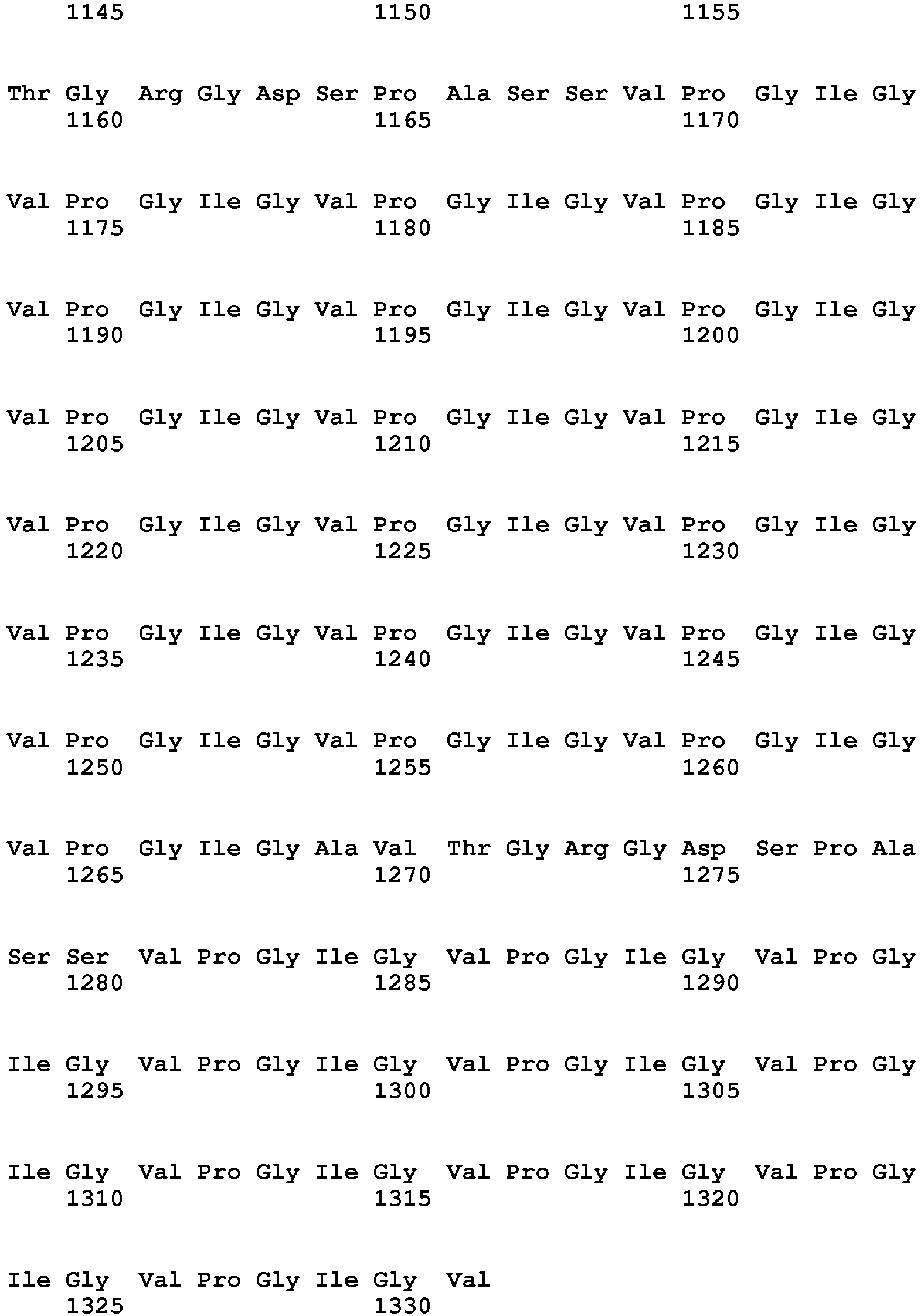
wherein the immobile phase is any previously polymerized hydrogel.
wherein the immobile phase is any previously polymerized hydrogel.
Claims (15)
- A method for manufacturing a three-dimensional (3D) biomimetic scaffold comprising:a) contacting a mobile phase with an immobile phase through an electrical insulating material containing the mobile phase, said electrical insulating material selected from the group consisting of:(i) a porous membrane placed on the surface of the top of the immobile phase, said porous membrane defining the geometrical regions through which the mobile phase passes;(ii) at least a tube inserted into the top of the immobile phase; and(iii) a combination of (i) and (ii);b) subjecting said mobile phase and said immobile phase in the presence of the electrical insulating material to a direct current (DC) electric field created inside a chamber and applied in closed circuit by a pair of electrodes in vertical or horizontal configuration, said chamber containing the mobile phase, the immobile phase and the electrical insulating material covered by a buffer; andc) patterning the immobile phase with the components of the mobile phase, in which said components move by forming patterns within the immobile phase and are not separated by their molecular weight and in which the location of the components of the mobile phase is controlled as desired by modulating the electric field and the time;wherein the mobile phase is an aqueous or organic medium comprising at least a charged compound and/or a charged macromolecular entity; and
wherein the immobile phase is any previously polymerized hydrogel. - The method according to claim 1, wherein the compound of the mobile phase is selected from the group consisting of a peptide, a protein, a nucleic acid, a nanoparticle, a microparticle, a dendrimer or a peptide-dendrimer conjugate.
- The method according to claim 1, wherein the compound of the mobile phase is selected from the group consisting of a hormone, a growth factor, an enzyme, a fluorescent molecule, a drug, a fluorescent molecule-dendrimer conjugate or a drug-dendrimer conjugate.
- The method according to claim 1, wherein the macromolecular entity is a cell or a fragment thereof.
- The method according to any of claims 1 to 4, wherein the hydrogel is an agarose hydrogel.
- The method according to any of claims 1 to 3, wherein the hydrogel is a polyacrylamide hydrogel.
- The method according to any of claims 1 to 6, wherein the geometrical regions of the porous membrane are defined by holes in said membrane.
- The method according to any of claims 1 to 6, wherein the geometrical regions of the porous membrane are defined by tubes inserted in the holes of said membrane.
- The method according to any of claims 1 to 8, wherein the porous membrane is a polydimethylsiloxane membrane.
- A method of culturing cells, comprising culturing the cells on a biomimetic scaffold obtained according to the method of claim 1.
- A method for culturing cells comprising manufacturing a three-dimensional (3D) biomimetic scaffold bya) contacting a mobile phase with an immobile phase through an electrical insulating material containing the mobile phase, said electrical insulating material selected from the group consisting of:(i) a porous membrane placed on the surface of the top of the immobile phase, said porous membrane defining the geometrical regions through which the mobile phase passes;(ii) at least a tube inserted into the top of the immobile phase; and(iii) a combination of (i) and (ii);b) subjecting said mobile phase and said immobile phase in the presence of the electrical insulating material to a direct current (DC) electric field created inside a chamber and applied in closed circuit by a pair of electrodes in vertical or horizontal configuration, said chamber containing the mobile phase, the immobile phase and the electrical insulating material covered by a buffer; andc) patterning the immobile phase with the components of the mobile phase, in which said components move by forming patterns within the immobile phase and are not separated by their molecular weight and in which the location of the components of the mobile phase is controlled as desired by modulating the electric field and the time;wherein the mobile phase is an aqueous or organic medium comprising at least a charged compound and/or a charged macromolecular entity; and
wherein the immobile phase is any previously polymerized hydrogel; and
culturing the cells in or on the product resulting from step c). - A three-dimensional (3D) biomimetic scaffold obtained according to the method of any of claims 1 to 9, or a fragment thereof, suitable for use in medicine.
- A three- dimensional (3D) biomimetic scaffold obtained according to the method of any of claims 1 to 9, or a fragment thereof, suitable for use in the in vivo replacement, reparation or regeneration of a damaged, dysfunctional or pathological tissue.
- Use of a three-dimensional (3D) biomimetic scaffold obtained according to the method of any of claims 1 to 9, or a fragment thereof, as an in vivo-like model for drug screening or as a model for molecular or cell biology studies.
- Use in vitro of a three-dimensional (3D) biomimetic scaffold obtained according to the method of any of claims 1 to 9, or a fragment thereof, to support cell growth.
Priority Applications (1)
| Application Number | Priority Date | Filing Date | Title |
|---|---|---|---|
| EP13702949.2A EP2828377B1 (en) | 2012-03-21 | 2013-01-25 | Method for manufacturing a three-dimensional biomimetic scaffold and uses thereof |
Applications Claiming Priority (3)
| Application Number | Priority Date | Filing Date | Title |
|---|---|---|---|
| EP12382102.7A EP2641965A1 (en) | 2012-03-21 | 2012-03-21 | Method for manufacturing a three-dimensional biomimetic scaffold and uses thereof |
| EP13702949.2A EP2828377B1 (en) | 2012-03-21 | 2013-01-25 | Method for manufacturing a three-dimensional biomimetic scaffold and uses thereof |
| PCT/EP2013/051465 WO2013139508A1 (en) | 2012-03-21 | 2013-01-25 | Method for manufacturing a three-dimensional biomimetic scaffold and uses thereof |
Publications (2)
| Publication Number | Publication Date |
|---|---|
| EP2828377A1 EP2828377A1 (en) | 2015-01-28 |
| EP2828377B1 true EP2828377B1 (en) | 2018-08-22 |
Family
ID=47678721
Family Applications (2)
| Application Number | Title | Priority Date | Filing Date |
|---|---|---|---|
| EP12382102.7A Withdrawn EP2641965A1 (en) | 2012-03-21 | 2012-03-21 | Method for manufacturing a three-dimensional biomimetic scaffold and uses thereof |
| EP13702949.2A Not-in-force EP2828377B1 (en) | 2012-03-21 | 2013-01-25 | Method for manufacturing a three-dimensional biomimetic scaffold and uses thereof |
Family Applications Before (1)
| Application Number | Title | Priority Date | Filing Date |
|---|---|---|---|
| EP12382102.7A Withdrawn EP2641965A1 (en) | 2012-03-21 | 2012-03-21 | Method for manufacturing a three-dimensional biomimetic scaffold and uses thereof |
Country Status (3)
| Country | Link |
|---|---|
| US (1) | US9631172B2 (en) |
| EP (2) | EP2641965A1 (en) |
| WO (1) | WO2013139508A1 (en) |
Families Citing this family (1)
| Publication number | Priority date | Publication date | Assignee | Title |
|---|---|---|---|---|
| ES2754824A1 (en) * | 2018-10-17 | 2020-04-20 | Univ Valladolid | COMPOSITION BASED ON RECOMBINANT BIOPOLYMERS AND USES OF THE SAME AS BIOTINTA (Machine-translation by Google Translate, not legally binding) |
Family Cites Families (1)
| Publication number | Priority date | Publication date | Assignee | Title |
|---|---|---|---|---|
| WO2004006840A2 (en) * | 2002-07-12 | 2004-01-22 | The Regents Of The University Of California | Three dimensional cell patterned bioploymer scaffolds and method of making the same |
-
2012
- 2012-03-21 EP EP12382102.7A patent/EP2641965A1/en not_active Withdrawn
-
2013
- 2013-01-25 WO PCT/EP2013/051465 patent/WO2013139508A1/en not_active Ceased
- 2013-01-25 EP EP13702949.2A patent/EP2828377B1/en not_active Not-in-force
- 2013-01-25 US US14/386,804 patent/US9631172B2/en not_active Expired - Fee Related
Non-Patent Citations (1)
| Title |
|---|
| None * |
Also Published As
| Publication number | Publication date |
|---|---|
| US9631172B2 (en) | 2017-04-25 |
| WO2013139508A1 (en) | 2013-09-26 |
| EP2641965A1 (en) | 2013-09-25 |
| US20150072429A1 (en) | 2015-03-12 |
| EP2828377A1 (en) | 2015-01-28 |
Similar Documents
| Publication | Publication Date | Title |
|---|---|---|
| Rojek et al. | Microfluidic formulation of topological hydrogels for microtissue engineering | |
| Levato et al. | From shape to function: the next step in bioprinting | |
| Gonzalez-Fernandez et al. | Pore-forming bioinks to enable spatio-temporally defined gene delivery in bioprinted tissues | |
| Gaspar et al. | Advanced bottom‐up engineering of living architectures | |
| Costantini et al. | Microfluidic-enhanced 3D bioprinting of aligned myoblast-laden hydrogels leads to functionally organized myofibers in vitro and in vivo | |
| Hussey et al. | Extracellular matrix-based materials for regenerative medicine | |
| Wei et al. | Directing stem cell differentiation via electrochemical reversible switching between nanotubes and nanotips of polypyrrole array | |
| Lee et al. | 3D printing nano conductive multi-walled carbon nanotube scaffolds for nerve regeneration | |
| Kang et al. | Three-dimensional printable gelatin hydrogels incorporating graphene oxide to enable spontaneous myogenic differentiation | |
| Wang et al. | Smart hydrogels for 3D bioprinting | |
| Mitrovic et al. | Peptide hydrogels and nanostructures controlling biological machinery | |
| Annabi et al. | 25th anniversary article: Rational design and applications of hydrogels in regenerative medicine | |
| Skardal et al. | Bioprinting cellularized constructs using a tissue-specific hydrogel bioink | |
| Steffens et al. | Update on the main use of biomaterials and techniques associated with tissue engineering | |
| Barrs et al. | Engineering a chemically defined hydrogel bioink for direct bioprinting of microvasculature | |
| Zhou et al. | Organoids and organoid extracellular vesicles-based disease treatment strategies | |
| Bordbar et al. | Cartilage tissue engineering using decellularized biomatrix hydrogel containing TGF-β-loaded alginate microspheres in mechanically loaded bioreactor | |
| Cecen et al. | Selection of natural biomaterials for micro‐tissue and organ‐on‐chip models | |
| Yao et al. | Bilayered BMP2 eluting coatings on graphene foam by electrophoretic deposition: Electroresponsive BMP2 release and enhancement of osteogenic differentiation | |
| Hahn et al. | Electroactive 4D porous scaffold based on conducting polymer as a responsive and dynamic in vitro cell culture platform | |
| Pereira et al. | Photopolymerizable hydrogels in regenerative medicine and drug delivery | |
| CN110753537A (en) | Non-covalently assembled conductive hydrogels | |
| US20210189327A1 (en) | 3d stimulated tissue constructs and methods of making thereof | |
| Sprenger et al. | Composite alginate dialdehyde-gelatin (ADA-GEL) hydrogel containing short ribbon-shaped fillers for skeletal muscle tissue biofabrication | |
| Wang et al. | Nanostructured GelMA colloidal gels as bioinks for freeform multi-mode 3D printing: better replacement for the classical GelMA polymeric inks |
Legal Events
| Date | Code | Title | Description |
|---|---|---|---|
| PUAI | Public reference made under article 153(3) epc to a published international application that has entered the european phase |
Free format text: ORIGINAL CODE: 0009012 |
|
| 17P | Request for examination filed |
Effective date: 20141015 |
|
| AK | Designated contracting states |
Kind code of ref document: A1 Designated state(s): AL AT BE BG CH CY CZ DE DK EE ES FI FR GB GR HR HU IE IS IT LI LT LU LV MC MK MT NL NO PL PT RO RS SE SI SK SM TR |
|
| AX | Request for extension of the european patent |
Extension state: BA ME |
|
| DAX | Request for extension of the european patent (deleted) | ||
| 17Q | First examination report despatched |
Effective date: 20160202 |
|
| STAA | Information on the status of an ep patent application or granted ep patent |
Free format text: STATUS: EXAMINATION IS IN PROGRESS |
|
| GRAP | Despatch of communication of intention to grant a patent |
Free format text: ORIGINAL CODE: EPIDOSNIGR1 |
|
| STAA | Information on the status of an ep patent application or granted ep patent |
Free format text: STATUS: GRANT OF PATENT IS INTENDED |
|
| INTG | Intention to grant announced |
Effective date: 20180323 |
|
| GRAS | Grant fee paid |
Free format text: ORIGINAL CODE: EPIDOSNIGR3 |
|
| GRAA | (expected) grant |
Free format text: ORIGINAL CODE: 0009210 |
|
| STAA | Information on the status of an ep patent application or granted ep patent |
Free format text: STATUS: THE PATENT HAS BEEN GRANTED |
|
| AK | Designated contracting states |
Kind code of ref document: B1 Designated state(s): AL AT BE BG CH CY CZ DE DK EE ES FI FR GB GR HR HU IE IS IT LI LT LU LV MC MK MT NL NO PL PT RO RS SE SI SK SM TR |
|
| REG | Reference to a national code |
Ref country code: GB Ref legal event code: FG4D |
|
| REG | Reference to a national code |
Ref country code: CH Ref legal event code: EP |
|
| REG | Reference to a national code |
Ref country code: AT Ref legal event code: REF Ref document number: 1032507 Country of ref document: AT Kind code of ref document: T Effective date: 20180915 |
|
| REG | Reference to a national code |
Ref country code: IE Ref legal event code: FG4D |
|
| REG | Reference to a national code |
Ref country code: DE Ref legal event code: R096 Ref document number: 602013042336 Country of ref document: DE |
|
| REG | Reference to a national code |
Ref country code: NL Ref legal event code: MP Effective date: 20180822 |
|
| REG | Reference to a national code |
Ref country code: LT Ref legal event code: MG4D |
|
| PG25 | Lapsed in a contracting state [announced via postgrant information from national office to epo] |
Ref country code: LT Free format text: LAPSE BECAUSE OF FAILURE TO SUBMIT A TRANSLATION OF THE DESCRIPTION OR TO PAY THE FEE WITHIN THE PRESCRIBED TIME-LIMIT Effective date: 20180822 Ref country code: NL Free format text: LAPSE BECAUSE OF FAILURE TO SUBMIT A TRANSLATION OF THE DESCRIPTION OR TO PAY THE FEE WITHIN THE PRESCRIBED TIME-LIMIT Effective date: 20180822 Ref country code: FI Free format text: LAPSE BECAUSE OF FAILURE TO SUBMIT A TRANSLATION OF THE DESCRIPTION OR TO PAY THE FEE WITHIN THE PRESCRIBED TIME-LIMIT Effective date: 20180822 Ref country code: RS Free format text: LAPSE BECAUSE OF FAILURE TO SUBMIT A TRANSLATION OF THE DESCRIPTION OR TO PAY THE FEE WITHIN THE PRESCRIBED TIME-LIMIT Effective date: 20180822 Ref country code: IS Free format text: LAPSE BECAUSE OF FAILURE TO SUBMIT A TRANSLATION OF THE DESCRIPTION OR TO PAY THE FEE WITHIN THE PRESCRIBED TIME-LIMIT Effective date: 20181222 Ref country code: NO Free format text: LAPSE BECAUSE OF FAILURE TO SUBMIT A TRANSLATION OF THE DESCRIPTION OR TO PAY THE FEE WITHIN THE PRESCRIBED TIME-LIMIT Effective date: 20181122 Ref country code: SE Free format text: LAPSE BECAUSE OF FAILURE TO SUBMIT A TRANSLATION OF THE DESCRIPTION OR TO PAY THE FEE WITHIN THE PRESCRIBED TIME-LIMIT Effective date: 20180822 Ref country code: BG Free format text: LAPSE BECAUSE OF FAILURE TO SUBMIT A TRANSLATION OF THE DESCRIPTION OR TO PAY THE FEE WITHIN THE PRESCRIBED TIME-LIMIT Effective date: 20181122 Ref country code: GR Free format text: LAPSE BECAUSE OF FAILURE TO SUBMIT A TRANSLATION OF THE DESCRIPTION OR TO PAY THE FEE WITHIN THE PRESCRIBED TIME-LIMIT Effective date: 20181123 |
|
| REG | Reference to a national code |
Ref country code: AT Ref legal event code: MK05 Ref document number: 1032507 Country of ref document: AT Kind code of ref document: T Effective date: 20180822 |
|
| PG25 | Lapsed in a contracting state [announced via postgrant information from national office to epo] |
Ref country code: AL Free format text: LAPSE BECAUSE OF FAILURE TO SUBMIT A TRANSLATION OF THE DESCRIPTION OR TO PAY THE FEE WITHIN THE PRESCRIBED TIME-LIMIT Effective date: 20180822 Ref country code: LV Free format text: LAPSE BECAUSE OF FAILURE TO SUBMIT A TRANSLATION OF THE DESCRIPTION OR TO PAY THE FEE WITHIN THE PRESCRIBED TIME-LIMIT Effective date: 20180822 Ref country code: HR Free format text: LAPSE BECAUSE OF FAILURE TO SUBMIT A TRANSLATION OF THE DESCRIPTION OR TO PAY THE FEE WITHIN THE PRESCRIBED TIME-LIMIT Effective date: 20180822 |
|
| PG25 | Lapsed in a contracting state [announced via postgrant information from national office to epo] |
Ref country code: IT Free format text: LAPSE BECAUSE OF FAILURE TO SUBMIT A TRANSLATION OF THE DESCRIPTION OR TO PAY THE FEE WITHIN THE PRESCRIBED TIME-LIMIT Effective date: 20180822 Ref country code: AT Free format text: LAPSE BECAUSE OF FAILURE TO SUBMIT A TRANSLATION OF THE DESCRIPTION OR TO PAY THE FEE WITHIN THE PRESCRIBED TIME-LIMIT Effective date: 20180822 Ref country code: ES Free format text: LAPSE BECAUSE OF FAILURE TO SUBMIT A TRANSLATION OF THE DESCRIPTION OR TO PAY THE FEE WITHIN THE PRESCRIBED TIME-LIMIT Effective date: 20180822 Ref country code: PL Free format text: LAPSE BECAUSE OF FAILURE TO SUBMIT A TRANSLATION OF THE DESCRIPTION OR TO PAY THE FEE WITHIN THE PRESCRIBED TIME-LIMIT Effective date: 20180822 Ref country code: CZ Free format text: LAPSE BECAUSE OF FAILURE TO SUBMIT A TRANSLATION OF THE DESCRIPTION OR TO PAY THE FEE WITHIN THE PRESCRIBED TIME-LIMIT Effective date: 20180822 Ref country code: RO Free format text: LAPSE BECAUSE OF FAILURE TO SUBMIT A TRANSLATION OF THE DESCRIPTION OR TO PAY THE FEE WITHIN THE PRESCRIBED TIME-LIMIT Effective date: 20180822 Ref country code: EE Free format text: LAPSE BECAUSE OF FAILURE TO SUBMIT A TRANSLATION OF THE DESCRIPTION OR TO PAY THE FEE WITHIN THE PRESCRIBED TIME-LIMIT Effective date: 20180822 |
|
| REG | Reference to a national code |
Ref country code: DE Ref legal event code: R097 Ref document number: 602013042336 Country of ref document: DE |
|
| PG25 | Lapsed in a contracting state [announced via postgrant information from national office to epo] |
Ref country code: DK Free format text: LAPSE BECAUSE OF FAILURE TO SUBMIT A TRANSLATION OF THE DESCRIPTION OR TO PAY THE FEE WITHIN THE PRESCRIBED TIME-LIMIT Effective date: 20180822 Ref country code: SM Free format text: LAPSE BECAUSE OF FAILURE TO SUBMIT A TRANSLATION OF THE DESCRIPTION OR TO PAY THE FEE WITHIN THE PRESCRIBED TIME-LIMIT Effective date: 20180822 Ref country code: SK Free format text: LAPSE BECAUSE OF FAILURE TO SUBMIT A TRANSLATION OF THE DESCRIPTION OR TO PAY THE FEE WITHIN THE PRESCRIBED TIME-LIMIT Effective date: 20180822 |
|
| PLBE | No opposition filed within time limit |
Free format text: ORIGINAL CODE: 0009261 |
|
| STAA | Information on the status of an ep patent application or granted ep patent |
Free format text: STATUS: NO OPPOSITION FILED WITHIN TIME LIMIT |
|
| 26N | No opposition filed |
Effective date: 20190523 |
|
| PG25 | Lapsed in a contracting state [announced via postgrant information from national office to epo] |
Ref country code: SI Free format text: LAPSE BECAUSE OF FAILURE TO SUBMIT A TRANSLATION OF THE DESCRIPTION OR TO PAY THE FEE WITHIN THE PRESCRIBED TIME-LIMIT Effective date: 20180822 Ref country code: MC Free format text: LAPSE BECAUSE OF FAILURE TO SUBMIT A TRANSLATION OF THE DESCRIPTION OR TO PAY THE FEE WITHIN THE PRESCRIBED TIME-LIMIT Effective date: 20180822 |
|
| REG | Reference to a national code |
Ref country code: CH Ref legal event code: PL |
|
| PG25 | Lapsed in a contracting state [announced via postgrant information from national office to epo] |
Ref country code: LU Free format text: LAPSE BECAUSE OF NON-PAYMENT OF DUE FEES Effective date: 20190125 |
|
| REG | Reference to a national code |
Ref country code: BE Ref legal event code: MM Effective date: 20190131 |
|
| REG | Reference to a national code |
Ref country code: IE Ref legal event code: MM4A |
|
| PG25 | Lapsed in a contracting state [announced via postgrant information from national office to epo] |
Ref country code: BE Free format text: LAPSE BECAUSE OF NON-PAYMENT OF DUE FEES Effective date: 20190131 |
|
| PG25 | Lapsed in a contracting state [announced via postgrant information from national office to epo] |
Ref country code: CH Free format text: LAPSE BECAUSE OF NON-PAYMENT OF DUE FEES Effective date: 20190131 Ref country code: LI Free format text: LAPSE BECAUSE OF NON-PAYMENT OF DUE FEES Effective date: 20190131 |
|
| PG25 | Lapsed in a contracting state [announced via postgrant information from national office to epo] |
Ref country code: IE Free format text: LAPSE BECAUSE OF NON-PAYMENT OF DUE FEES Effective date: 20190125 |
|
| PG25 | Lapsed in a contracting state [announced via postgrant information from national office to epo] |
Ref country code: TR Free format text: LAPSE BECAUSE OF FAILURE TO SUBMIT A TRANSLATION OF THE DESCRIPTION OR TO PAY THE FEE WITHIN THE PRESCRIBED TIME-LIMIT Effective date: 20180822 |
|
| PGFP | Annual fee paid to national office [announced via postgrant information from national office to epo] |
Ref country code: DE Payment date: 20200123 Year of fee payment: 8 Ref country code: GB Payment date: 20200116 Year of fee payment: 8 |
|
| PG25 | Lapsed in a contracting state [announced via postgrant information from national office to epo] |
Ref country code: MT Free format text: LAPSE BECAUSE OF NON-PAYMENT OF DUE FEES Effective date: 20190125 Ref country code: PT Free format text: LAPSE BECAUSE OF FAILURE TO SUBMIT A TRANSLATION OF THE DESCRIPTION OR TO PAY THE FEE WITHIN THE PRESCRIBED TIME-LIMIT Effective date: 20181222 |
|
| PGFP | Annual fee paid to national office [announced via postgrant information from national office to epo] |
Ref country code: FR Payment date: 20200123 Year of fee payment: 8 |
|
| PG25 | Lapsed in a contracting state [announced via postgrant information from national office to epo] |
Ref country code: CY Free format text: LAPSE BECAUSE OF FAILURE TO SUBMIT A TRANSLATION OF THE DESCRIPTION OR TO PAY THE FEE WITHIN THE PRESCRIBED TIME-LIMIT Effective date: 20180822 |
|
| PG25 | Lapsed in a contracting state [announced via postgrant information from national office to epo] |
Ref country code: HU Free format text: LAPSE BECAUSE OF FAILURE TO SUBMIT A TRANSLATION OF THE DESCRIPTION OR TO PAY THE FEE WITHIN THE PRESCRIBED TIME-LIMIT; INVALID AB INITIO Effective date: 20130125 |
|
| REG | Reference to a national code |
Ref country code: DE Ref legal event code: R119 Ref document number: 602013042336 Country of ref document: DE |
|
| GBPC | Gb: european patent ceased through non-payment of renewal fee |
Effective date: 20210125 |
|
| PG25 | Lapsed in a contracting state [announced via postgrant information from national office to epo] |
Ref country code: FR Free format text: LAPSE BECAUSE OF NON-PAYMENT OF DUE FEES Effective date: 20210131 |
|
| PG25 | Lapsed in a contracting state [announced via postgrant information from national office to epo] |
Ref country code: DE Free format text: LAPSE BECAUSE OF NON-PAYMENT OF DUE FEES Effective date: 20210803 Ref country code: GB Free format text: LAPSE BECAUSE OF NON-PAYMENT OF DUE FEES Effective date: 20210125 |
|
| PG25 | Lapsed in a contracting state [announced via postgrant information from national office to epo] |
Ref country code: MK Free format text: LAPSE BECAUSE OF FAILURE TO SUBMIT A TRANSLATION OF THE DESCRIPTION OR TO PAY THE FEE WITHIN THE PRESCRIBED TIME-LIMIT Effective date: 20180822 |