EP2817315B1 - Selective detection of thiols - Google Patents
Selective detection of thiols Download PDFInfo
- Publication number
- EP2817315B1 EP2817315B1 EP13752087.0A EP13752087A EP2817315B1 EP 2817315 B1 EP2817315 B1 EP 2817315B1 EP 13752087 A EP13752087 A EP 13752087A EP 2817315 B1 EP2817315 B1 EP 2817315B1
- Authority
- EP
- European Patent Office
- Prior art keywords
- probe
- compound
- gsh
- reaction
- reaction mixture
- Prior art date
- Legal status (The legal status is an assumption and is not a legal conclusion. Google has not performed a legal analysis and makes no representation as to the accuracy of the status listed.)
- Not-in-force
Links
- 150000003573 thiols Chemical class 0.000 title claims description 54
- 238000001514 detection method Methods 0.000 title description 25
- RWSXRVCMGQZWBV-WDSKDSINSA-N glutathione Chemical compound OC(=O)[C@@H](N)CCC(=O)N[C@@H](CS)C(=O)NCC(O)=O RWSXRVCMGQZWBV-WDSKDSINSA-N 0.000 claims description 292
- 239000000523 sample Substances 0.000 claims description 271
- 229960003180 glutathione Drugs 0.000 claims description 147
- 150000001875 compounds Chemical class 0.000 claims description 76
- 238000006243 chemical reaction Methods 0.000 claims description 55
- 230000008859 change Effects 0.000 claims description 46
- -1 α,β-unsaturated aliphatic ester Chemical group 0.000 claims description 45
- 239000011541 reaction mixture Substances 0.000 claims description 44
- 125000000217 alkyl group Chemical group 0.000 claims description 41
- 238000000862 absorption spectrum Methods 0.000 claims description 32
- 125000003396 thiol group Chemical group [H]S* 0.000 claims description 25
- 210000004369 blood Anatomy 0.000 claims description 23
- 239000008280 blood Substances 0.000 claims description 23
- 125000003277 amino group Chemical group 0.000 claims description 22
- 238000000034 method Methods 0.000 claims description 22
- XUJNEKJLAYXESH-UHFFFAOYSA-N cysteine Natural products SCC(N)C(O)=O XUJNEKJLAYXESH-UHFFFAOYSA-N 0.000 claims description 20
- 235000018417 cysteine Nutrition 0.000 claims description 20
- 238000000295 emission spectrum Methods 0.000 claims description 20
- 125000003545 alkoxy group Chemical group 0.000 claims description 17
- 125000002887 hydroxy group Chemical group [H]O* 0.000 claims description 17
- 239000003093 cationic surfactant Substances 0.000 claims description 15
- 239000000126 substance Substances 0.000 claims description 15
- 238000002835 absorbance Methods 0.000 claims description 13
- 125000004122 cyclic group Chemical group 0.000 claims description 13
- 125000001997 phenyl group Chemical group [H]C1=C([H])C([H])=C(*)C([H])=C1[H] 0.000 claims description 12
- 210000002700 urine Anatomy 0.000 claims description 12
- 108010024636 Glutathione Proteins 0.000 claims description 11
- 125000003178 carboxy group Chemical group [H]OC(*)=O 0.000 claims description 11
- 229910052736 halogen Inorganic materials 0.000 claims description 11
- 150000002367 halogens Chemical class 0.000 claims description 11
- 125000002924 primary amino group Chemical group [H]N([H])* 0.000 claims description 11
- 229910052717 sulfur Inorganic materials 0.000 claims description 11
- 125000004181 carboxyalkyl group Chemical group 0.000 claims description 9
- 108090000623 proteins and genes Proteins 0.000 claims description 9
- 125000000008 (C1-C10) alkyl group Chemical group 0.000 claims description 8
- 229910006069 SO3H Inorganic materials 0.000 claims description 8
- 125000001624 naphthyl group Chemical group 0.000 claims description 8
- 235000018102 proteins Nutrition 0.000 claims description 8
- 102000004169 proteins and genes Human genes 0.000 claims description 8
- 239000000306 component Substances 0.000 claims description 7
- 229910052760 oxygen Inorganic materials 0.000 claims description 7
- 239000003638 chemical reducing agent Substances 0.000 claims description 6
- 239000000463 material Substances 0.000 claims description 6
- 239000002904 solvent Substances 0.000 claims description 6
- 125000003282 alkyl amino group Chemical group 0.000 claims description 5
- 239000010836 blood and blood product Substances 0.000 claims description 5
- 239000012503 blood component Substances 0.000 claims description 5
- 229940125691 blood product Drugs 0.000 claims description 5
- 125000002252 acyl group Chemical group 0.000 claims description 4
- 239000002250 absorbent Substances 0.000 claims description 3
- 230000002745 absorbent Effects 0.000 claims description 3
- 230000001376 precipitating effect Effects 0.000 claims description 2
- XUJNEKJLAYXESH-REOHCLBHSA-N L-Cysteine Chemical compound SC[C@H](N)C(O)=O XUJNEKJLAYXESH-REOHCLBHSA-N 0.000 description 119
- LZZYPRNAOMGNLH-UHFFFAOYSA-M Cetrimonium bromide Chemical compound [Br-].CCCCCCCCCCCCCCCC[N+](C)(C)C LZZYPRNAOMGNLH-UHFFFAOYSA-M 0.000 description 67
- FFFHZYDWPBMWHY-VKHMYHEASA-N L-homocysteine Chemical compound OC(=O)[C@@H](N)CCS FFFHZYDWPBMWHY-VKHMYHEASA-N 0.000 description 56
- 238000002189 fluorescence spectrum Methods 0.000 description 38
- 239000000243 solution Substances 0.000 description 32
- 239000008363 phosphate buffer Substances 0.000 description 31
- 238000007363 ring formation reaction Methods 0.000 description 21
- HSSLDCABUXLXKM-UHFFFAOYSA-N resorufin Chemical class C1=CC(=O)C=C2OC3=CC(O)=CC=C3N=C21 HSSLDCABUXLXKM-UHFFFAOYSA-N 0.000 description 20
- 239000004094 surface-active agent Substances 0.000 description 20
- YMWUJEATGCHHMB-UHFFFAOYSA-N Dichloromethane Chemical compound ClCCl YMWUJEATGCHHMB-UHFFFAOYSA-N 0.000 description 18
- 229910052739 hydrogen Inorganic materials 0.000 description 16
- 239000001257 hydrogen Substances 0.000 description 16
- IAZDPXIOMUYVGZ-UHFFFAOYSA-N Dimethylsulphoxide Chemical compound CS(C)=O IAZDPXIOMUYVGZ-UHFFFAOYSA-N 0.000 description 14
- 125000001931 aliphatic group Chemical group 0.000 description 14
- 230000036962 time dependent Effects 0.000 description 13
- ZMANZCXQSJIPKH-UHFFFAOYSA-N Triethylamine Chemical compound CCN(CC)CC ZMANZCXQSJIPKH-UHFFFAOYSA-N 0.000 description 12
- 230000015572 biosynthetic process Effects 0.000 description 12
- 150000002431 hydrogen Chemical class 0.000 description 12
- DBMJMQXJHONAFJ-UHFFFAOYSA-M Sodium laurylsulphate Chemical compound [Na+].CCCCCCCCCCCCOS([O-])(=O)=O DBMJMQXJHONAFJ-UHFFFAOYSA-M 0.000 description 11
- 125000004429 atom Chemical group 0.000 description 11
- 230000004044 response Effects 0.000 description 11
- 229910019142 PO4 Inorganic materials 0.000 description 10
- HEDRZPFGACZZDS-MICDWDOJSA-N Trichloro(2H)methane Chemical compound [2H]C(Cl)(Cl)Cl HEDRZPFGACZZDS-MICDWDOJSA-N 0.000 description 10
- 229920004890 Triton X-100 Polymers 0.000 description 10
- NBIIXXVUZAFLBC-UHFFFAOYSA-K phosphate Chemical compound [O-]P([O-])([O-])=O NBIIXXVUZAFLBC-UHFFFAOYSA-K 0.000 description 10
- 125000003118 aryl group Chemical group 0.000 description 9
- 239000000203 mixture Substances 0.000 description 9
- 239000010452 phosphate Substances 0.000 description 9
- 125000001424 substituent group Chemical group 0.000 description 9
- RIOQSEWOXXDEQQ-UHFFFAOYSA-N triphenylphosphine Chemical compound C1=CC=CC=C1P(C=1C=CC=CC=1)C1=CC=CC=C1 RIOQSEWOXXDEQQ-UHFFFAOYSA-N 0.000 description 9
- XIWFQDBQMCDYJT-UHFFFAOYSA-M benzyl-dimethyl-tridecylazanium;chloride Chemical compound [Cl-].CCCCCCCCCCCCC[N+](C)(C)CC1=CC=CC=C1 XIWFQDBQMCDYJT-UHFFFAOYSA-M 0.000 description 8
- 230000005284 excitation Effects 0.000 description 8
- 239000013504 Triton X-100 Substances 0.000 description 7
- 235000001014 amino acid Nutrition 0.000 description 7
- 229940024606 amino acid Drugs 0.000 description 7
- 150000001413 amino acids Chemical class 0.000 description 7
- 238000003402 intramolecular cyclocondensation reaction Methods 0.000 description 7
- 239000000693 micelle Substances 0.000 description 7
- 238000002156 mixing Methods 0.000 description 7
- JKMHFZQWWAIEOD-UHFFFAOYSA-N 2-[4-(2-hydroxyethyl)piperazin-1-yl]ethanesulfonic acid Chemical compound OCC[NH+]1CCN(CCS([O-])(=O)=O)CC1 JKMHFZQWWAIEOD-UHFFFAOYSA-N 0.000 description 6
- OKKJLVBELUTLKV-UHFFFAOYSA-N Methanol Chemical compound OC OKKJLVBELUTLKV-UHFFFAOYSA-N 0.000 description 6
- 239000007853 buffer solution Substances 0.000 description 6
- 238000003379 elimination reaction Methods 0.000 description 6
- 238000005259 measurement Methods 0.000 description 6
- 239000000047 product Substances 0.000 description 6
- 230000035484 reaction time Effects 0.000 description 6
- 238000004252 FT/ICR mass spectrometry Methods 0.000 description 5
- 239000007995 HEPES buffer Substances 0.000 description 5
- PZBFGYYEXUXCOF-UHFFFAOYSA-N TCEP Chemical compound OC(=O)CCP(CCC(O)=O)CCC(O)=O PZBFGYYEXUXCOF-UHFFFAOYSA-N 0.000 description 5
- 125000004432 carbon atom Chemical group C* 0.000 description 5
- 150000002148 esters Chemical class 0.000 description 5
- 125000000524 functional group Chemical group 0.000 description 5
- 125000004435 hydrogen atom Chemical group [H]* 0.000 description 5
- 125000001570 methylene group Chemical group [H]C([H])([*:1])[*:2] 0.000 description 5
- 229940083575 sodium dodecyl sulfate Drugs 0.000 description 5
- 235000019333 sodium laurylsulphate Nutrition 0.000 description 5
- WEVYAHXRMPXWCK-UHFFFAOYSA-N Acetonitrile Chemical compound CC#N WEVYAHXRMPXWCK-UHFFFAOYSA-N 0.000 description 4
- PWKSKIMOESPYIA-BYPYZUCNSA-N L-N-acetyl-Cysteine Chemical compound CC(=O)N[C@@H](CS)C(O)=O PWKSKIMOESPYIA-BYPYZUCNSA-N 0.000 description 4
- 229920005654 Sephadex Polymers 0.000 description 4
- 229960004308 acetylcysteine Drugs 0.000 description 4
- 150000001252 acrylic acid derivatives Chemical class 0.000 description 4
- HFBMWMNUJJDEQZ-UHFFFAOYSA-N acryloyl chloride Chemical compound ClC(=O)C=C HFBMWMNUJJDEQZ-UHFFFAOYSA-N 0.000 description 4
- 229960000686 benzalkonium chloride Drugs 0.000 description 4
- 238000011088 calibration curve Methods 0.000 description 4
- 125000002915 carbonyl group Chemical group [*:2]C([*:1])=O 0.000 description 4
- 230000005494 condensation Effects 0.000 description 4
- 238000011161 development Methods 0.000 description 4
- 230000000694 effects Effects 0.000 description 4
- 239000007850 fluorescent dye Substances 0.000 description 4
- 238000004896 high resolution mass spectrometry Methods 0.000 description 4
- ZTVZLYBCZNMWCF-UHFFFAOYSA-N homocystine Chemical compound [O-]C(=O)C([NH3+])CCSSCCC([NH3+])C([O-])=O ZTVZLYBCZNMWCF-UHFFFAOYSA-N 0.000 description 4
- 150000002430 hydrocarbons Chemical class 0.000 description 4
- 230000035945 sensitivity Effects 0.000 description 4
- 239000007787 solid Substances 0.000 description 4
- 238000001228 spectrum Methods 0.000 description 4
- 238000012360 testing method Methods 0.000 description 4
- 238000005160 1H NMR spectroscopy Methods 0.000 description 3
- ODWNBAWYDSWOAF-UHFFFAOYSA-N 2,4,4-trimethylpentan-2-yloxybenzene Chemical compound CC(C)(C)CC(C)(C)OC1=CC=CC=C1 ODWNBAWYDSWOAF-UHFFFAOYSA-N 0.000 description 3
- JICTZUFURQRLDW-UHFFFAOYSA-N 5-oxo-1,4-thiazepane-3-carboxylic acid Chemical compound OC(=O)C1CSCCC(=O)N1 JICTZUFURQRLDW-UHFFFAOYSA-N 0.000 description 3
- UHOVQNZJYSORNB-UHFFFAOYSA-N Benzene Chemical compound C1=CC=CC=C1 UHOVQNZJYSORNB-UHFFFAOYSA-N 0.000 description 3
- OKTJSMMVPCPJKN-UHFFFAOYSA-N Carbon Chemical compound [C] OKTJSMMVPCPJKN-UHFFFAOYSA-N 0.000 description 3
- 239000002202 Polyethylene glycol Substances 0.000 description 3
- YXFVVABEGXRONW-UHFFFAOYSA-N Toluene Chemical compound CC1=CC=CC=C1 YXFVVABEGXRONW-UHFFFAOYSA-N 0.000 description 3
- 239000013060 biological fluid Substances 0.000 description 3
- 239000012472 biological sample Substances 0.000 description 3
- 239000000872 buffer Substances 0.000 description 3
- 125000002091 cationic group Chemical group 0.000 description 3
- 238000009833 condensation Methods 0.000 description 3
- 230000008030 elimination Effects 0.000 description 3
- 238000002474 experimental method Methods 0.000 description 3
- 125000001072 heteroaryl group Chemical group 0.000 description 3
- 238000001819 mass spectrum Methods 0.000 description 3
- 230000001404 mediated effect Effects 0.000 description 3
- 239000002736 nonionic surfactant Substances 0.000 description 3
- 229920001223 polyethylene glycol Polymers 0.000 description 3
- 230000005855 radiation Effects 0.000 description 3
- 150000003254 radicals Chemical class 0.000 description 3
- 238000011084 recovery Methods 0.000 description 3
- 238000003786 synthesis reaction Methods 0.000 description 3
- 150000003568 thioethers Chemical class 0.000 description 3
- FANCTJAFZSYTIS-IQUVVAJASA-N (1r,3s,5z)-5-[(2e)-2-[(1r,3as,7ar)-7a-methyl-1-[(2r)-4-(phenylsulfonimidoyl)butan-2-yl]-2,3,3a,5,6,7-hexahydro-1h-inden-4-ylidene]ethylidene]-4-methylidenecyclohexane-1,3-diol Chemical compound C([C@@H](C)[C@@H]1[C@]2(CCCC(/[C@@H]2CC1)=C\C=C\1C([C@@H](O)C[C@H](O)C/1)=C)C)CS(=N)(=O)C1=CC=CC=C1 FANCTJAFZSYTIS-IQUVVAJASA-N 0.000 description 2
- HJIAMFHSAAEUKR-UHFFFAOYSA-N (2-hydroxyphenyl)-phenylmethanone Chemical class OC1=CC=CC=C1C(=O)C1=CC=CC=C1 HJIAMFHSAAEUKR-UHFFFAOYSA-N 0.000 description 2
- 238000001644 13C nuclear magnetic resonance spectroscopy Methods 0.000 description 2
- 208000024827 Alzheimer disease Diseases 0.000 description 2
- 239000004475 Arginine Substances 0.000 description 2
- 239000004215 Carbon black (E152) Substances 0.000 description 2
- 208000024172 Cardiovascular disease Diseases 0.000 description 2
- HEDRZPFGACZZDS-UHFFFAOYSA-N Chloroform Chemical compound ClC(Cl)Cl HEDRZPFGACZZDS-UHFFFAOYSA-N 0.000 description 2
- RTZKZFJDLAIYFH-UHFFFAOYSA-N Diethyl ether Chemical compound CCOCC RTZKZFJDLAIYFH-UHFFFAOYSA-N 0.000 description 2
- MHAJPDPJQMAIIY-UHFFFAOYSA-N Hydrogen peroxide Chemical compound OO MHAJPDPJQMAIIY-UHFFFAOYSA-N 0.000 description 2
- ONIBWKKTOPOVIA-BYPYZUCNSA-N L-Proline Chemical compound OC(=O)[C@@H]1CCCN1 ONIBWKKTOPOVIA-BYPYZUCNSA-N 0.000 description 2
- QNAYBMKLOCPYGJ-REOHCLBHSA-N L-alanine Chemical compound C[C@H](N)C(O)=O QNAYBMKLOCPYGJ-REOHCLBHSA-N 0.000 description 2
- ODKSFYDXXFIFQN-BYPYZUCNSA-P L-argininium(2+) Chemical compound NC(=[NH2+])NCCC[C@H]([NH3+])C(O)=O ODKSFYDXXFIFQN-BYPYZUCNSA-P 0.000 description 2
- CKLJMWTZIZZHCS-REOHCLBHSA-N L-aspartic acid Chemical compound OC(=O)[C@@H](N)CC(O)=O CKLJMWTZIZZHCS-REOHCLBHSA-N 0.000 description 2
- LEVWYRKDKASIDU-IMJSIDKUSA-N L-cystine Chemical compound [O-]C(=O)[C@@H]([NH3+])CSSC[C@H]([NH3+])C([O-])=O LEVWYRKDKASIDU-IMJSIDKUSA-N 0.000 description 2
- ZDXPYRJPNDTMRX-VKHMYHEASA-N L-glutamine Chemical compound OC(=O)[C@@H](N)CCC(N)=O ZDXPYRJPNDTMRX-VKHMYHEASA-N 0.000 description 2
- HNDVDQJCIGZPNO-YFKPBYRVSA-N L-histidine Chemical compound OC(=O)[C@@H](N)CC1=CN=CN1 HNDVDQJCIGZPNO-YFKPBYRVSA-N 0.000 description 2
- AGPKZVBTJJNPAG-WHFBIAKZSA-N L-isoleucine Chemical compound CC[C@H](C)[C@H](N)C(O)=O AGPKZVBTJJNPAG-WHFBIAKZSA-N 0.000 description 2
- ROHFNLRQFUQHCH-YFKPBYRVSA-N L-leucine Chemical compound CC(C)C[C@H](N)C(O)=O ROHFNLRQFUQHCH-YFKPBYRVSA-N 0.000 description 2
- KDXKERNSBIXSRK-YFKPBYRVSA-N L-lysine Chemical compound NCCCC[C@H](N)C(O)=O KDXKERNSBIXSRK-YFKPBYRVSA-N 0.000 description 2
- FFEARJCKVFRZRR-BYPYZUCNSA-N L-methionine Chemical compound CSCC[C@H](N)C(O)=O FFEARJCKVFRZRR-BYPYZUCNSA-N 0.000 description 2
- LRQKBLKVPFOOQJ-YFKPBYRVSA-N L-norleucine Chemical compound CCCC[C@H]([NH3+])C([O-])=O LRQKBLKVPFOOQJ-YFKPBYRVSA-N 0.000 description 2
- COLNVLDHVKWLRT-QMMMGPOBSA-N L-phenylalanine Chemical compound OC(=O)[C@@H](N)CC1=CC=CC=C1 COLNVLDHVKWLRT-QMMMGPOBSA-N 0.000 description 2
- AYFVYJQAPQTCCC-GBXIJSLDSA-N L-threonine Chemical compound C[C@@H](O)[C@H](N)C(O)=O AYFVYJQAPQTCCC-GBXIJSLDSA-N 0.000 description 2
- QIVBCDIJIAJPQS-VIFPVBQESA-N L-tryptophane Chemical compound C1=CC=C2C(C[C@H](N)C(O)=O)=CNC2=C1 QIVBCDIJIAJPQS-VIFPVBQESA-N 0.000 description 2
- OUYCCCASQSFEME-QMMMGPOBSA-N L-tyrosine Chemical compound OC(=O)[C@@H](N)CC1=CC=C(O)C=C1 OUYCCCASQSFEME-QMMMGPOBSA-N 0.000 description 2
- KZSNJWFQEVHDMF-BYPYZUCNSA-N L-valine Chemical compound CC(C)[C@H](N)C(O)=O KZSNJWFQEVHDMF-BYPYZUCNSA-N 0.000 description 2
- ROHFNLRQFUQHCH-UHFFFAOYSA-N Leucine Natural products CC(C)CC(N)C(O)=O ROHFNLRQFUQHCH-UHFFFAOYSA-N 0.000 description 2
- 206010067125 Liver injury Diseases 0.000 description 2
- KDXKERNSBIXSRK-UHFFFAOYSA-N Lysine Natural products NCCCCC(N)C(O)=O KDXKERNSBIXSRK-UHFFFAOYSA-N 0.000 description 2
- 239000004472 Lysine Substances 0.000 description 2
- AFVFQIVMOAPDHO-UHFFFAOYSA-N Methanesulfonic acid Chemical compound CS(O)(=O)=O AFVFQIVMOAPDHO-UHFFFAOYSA-N 0.000 description 2
- 239000007832 Na2SO4 Substances 0.000 description 2
- ONIBWKKTOPOVIA-UHFFFAOYSA-N Proline Natural products OC(=O)C1CCCN1 ONIBWKKTOPOVIA-UHFFFAOYSA-N 0.000 description 2
- 239000012507 Sephadex™ Substances 0.000 description 2
- PMZURENOXWZQFD-UHFFFAOYSA-L Sodium Sulfate Chemical compound [Na+].[Na+].[O-]S([O-])(=O)=O PMZURENOXWZQFD-UHFFFAOYSA-L 0.000 description 2
- AYFVYJQAPQTCCC-UHFFFAOYSA-N Threonine Natural products CC(O)C(N)C(O)=O AYFVYJQAPQTCCC-UHFFFAOYSA-N 0.000 description 2
- 239000004473 Threonine Substances 0.000 description 2
- QIVBCDIJIAJPQS-UHFFFAOYSA-N Tryptophan Natural products C1=CC=C2C(CC(N)C(O)=O)=CNC2=C1 QIVBCDIJIAJPQS-UHFFFAOYSA-N 0.000 description 2
- KZSNJWFQEVHDMF-UHFFFAOYSA-N Valine Natural products CC(C)C(N)C(O)=O KZSNJWFQEVHDMF-UHFFFAOYSA-N 0.000 description 2
- 238000010521 absorption reaction Methods 0.000 description 2
- NIXOWILDQLNWCW-UHFFFAOYSA-M acrylate group Chemical group C(C=C)(=O)[O-] NIXOWILDQLNWCW-UHFFFAOYSA-M 0.000 description 2
- 125000003647 acryloyl group Chemical group O=C([*])C([H])=C([H])[H] 0.000 description 2
- 235000004279 alanine Nutrition 0.000 description 2
- 150000001335 aliphatic alkanes Chemical class 0.000 description 2
- 150000001336 alkenes Chemical class 0.000 description 2
- 150000001345 alkine derivatives Chemical class 0.000 description 2
- VSCWAEJMTAWNJL-UHFFFAOYSA-K aluminium trichloride Chemical compound Cl[Al](Cl)Cl VSCWAEJMTAWNJL-UHFFFAOYSA-K 0.000 description 2
- 150000001412 amines Chemical class 0.000 description 2
- 239000003945 anionic surfactant Substances 0.000 description 2
- ODKSFYDXXFIFQN-UHFFFAOYSA-N arginine Natural products OC(=O)C(N)CCCNC(N)=N ODKSFYDXXFIFQN-UHFFFAOYSA-N 0.000 description 2
- 125000003710 aryl alkyl group Chemical group 0.000 description 2
- 235000003704 aspartic acid Nutrition 0.000 description 2
- 238000003556 assay Methods 0.000 description 2
- OQFSQFPPLPISGP-UHFFFAOYSA-N beta-carboxyaspartic acid Natural products OC(=O)C(N)C(C(O)=O)C(O)=O OQFSQFPPLPISGP-UHFFFAOYSA-N 0.000 description 2
- 229910052799 carbon Inorganic materials 0.000 description 2
- 210000004027 cell Anatomy 0.000 description 2
- 238000005119 centrifugation Methods 0.000 description 2
- 125000000753 cycloalkyl group Chemical group 0.000 description 2
- 229960003067 cystine Drugs 0.000 description 2
- 238000011033 desalting Methods 0.000 description 2
- 208000037265 diseases, disorders, signs and symptoms Diseases 0.000 description 2
- 239000003480 eluent Substances 0.000 description 2
- 238000001704 evaporation Methods 0.000 description 2
- 230000008020 evaporation Effects 0.000 description 2
- 238000002523 gelfiltration Methods 0.000 description 2
- ZDXPYRJPNDTMRX-UHFFFAOYSA-N glutamine Natural products OC(=O)C(N)CCC(N)=O ZDXPYRJPNDTMRX-UHFFFAOYSA-N 0.000 description 2
- 231100000234 hepatic damage Toxicity 0.000 description 2
- HNDVDQJCIGZPNO-UHFFFAOYSA-N histidine Natural products OC(=O)C(N)CC1=CN=CN1 HNDVDQJCIGZPNO-UHFFFAOYSA-N 0.000 description 2
- 229930195733 hydrocarbon Natural products 0.000 description 2
- AGPKZVBTJJNPAG-UHFFFAOYSA-N isoleucine Natural products CCC(C)C(N)C(O)=O AGPKZVBTJJNPAG-UHFFFAOYSA-N 0.000 description 2
- 229960000310 isoleucine Drugs 0.000 description 2
- 239000007788 liquid Substances 0.000 description 2
- 230000008818 liver damage Effects 0.000 description 2
- 230000004060 metabolic process Effects 0.000 description 2
- 229930182817 methionine Natural products 0.000 description 2
- 125000002496 methyl group Chemical group [H]C([H])([H])* 0.000 description 2
- VLKZOEOYAKHREP-UHFFFAOYSA-N methyl pentane Natural products CCCCCC VLKZOEOYAKHREP-UHFFFAOYSA-N 0.000 description 2
- 238000005580 one pot reaction Methods 0.000 description 2
- 150000007524 organic acids Chemical class 0.000 description 2
- 150000002894 organic compounds Chemical class 0.000 description 2
- 230000036542 oxidative stress Effects 0.000 description 2
- COLNVLDHVKWLRT-UHFFFAOYSA-N phenylalanine Natural products OC(=O)C(N)CC1=CC=CC=C1 COLNVLDHVKWLRT-UHFFFAOYSA-N 0.000 description 2
- 229910052938 sodium sulfate Inorganic materials 0.000 description 2
- 238000003756 stirring Methods 0.000 description 2
- 239000006228 supernatant Substances 0.000 description 2
- OUYCCCASQSFEME-UHFFFAOYSA-N tyrosine Natural products OC(=O)C(N)CC1=CC=C(O)C=C1 OUYCCCASQSFEME-UHFFFAOYSA-N 0.000 description 2
- 239000004474 valine Substances 0.000 description 2
- XLYOFNOQVPJJNP-UHFFFAOYSA-N water Substances O XLYOFNOQVPJJNP-UHFFFAOYSA-N 0.000 description 2
- 150000004915 1,4-thiazepines Chemical class 0.000 description 1
- LGRFSURHDFAFJT-IDEBNGHGSA-N 2-benzofuran-1,3-dione Chemical class [13CH]1=[13CH][13CH]=[13C]2C(=O)OC(=O)[13C]2=[13CH]1 LGRFSURHDFAFJT-IDEBNGHGSA-N 0.000 description 1
- 208000030507 AIDS Diseases 0.000 description 1
- QGZKDVFQNNGYKY-UHFFFAOYSA-O Ammonium Chemical compound [NH4+] QGZKDVFQNNGYKY-UHFFFAOYSA-O 0.000 description 1
- 102000004506 Blood Proteins Human genes 0.000 description 1
- 108010017384 Blood Proteins Proteins 0.000 description 1
- CPELXLSAUQHCOX-UHFFFAOYSA-M Bromide Chemical compound [Br-] CPELXLSAUQHCOX-UHFFFAOYSA-M 0.000 description 1
- MVTNWLCYPTUWNU-UHFFFAOYSA-N C=CC(Oc(cc1)cc(O2)c1N=C(C=C1)C2=CC1=O)=O Chemical compound C=CC(Oc(cc1)cc(O2)c1N=C(C=C1)C2=CC1=O)=O MVTNWLCYPTUWNU-UHFFFAOYSA-N 0.000 description 1
- BWGNESOTFCXPMA-UHFFFAOYSA-N Dihydrogen disulfide Chemical compound SS BWGNESOTFCXPMA-UHFFFAOYSA-N 0.000 description 1
- 108010053070 Glutathione Disulfide Proteins 0.000 description 1
- UFHFLCQGNIYNRP-UHFFFAOYSA-N Hydrogen Chemical compound [H][H] UFHFLCQGNIYNRP-UHFFFAOYSA-N 0.000 description 1
- 206010024264 Lethargy Diseases 0.000 description 1
- 238000005481 NMR spectroscopy Methods 0.000 description 1
- 229910002651 NO3 Inorganic materials 0.000 description 1
- 206010028980 Neoplasm Diseases 0.000 description 1
- NHNBFGGVMKEFGY-UHFFFAOYSA-N Nitrate Chemical compound [O-][N+]([O-])=O NHNBFGGVMKEFGY-UHFFFAOYSA-N 0.000 description 1
- IOVCWXUNBOPUCH-UHFFFAOYSA-M Nitrite anion Chemical compound [O-]N=O IOVCWXUNBOPUCH-UHFFFAOYSA-M 0.000 description 1
- 206010030113 Oedema Diseases 0.000 description 1
- 208000018737 Parkinson disease Diseases 0.000 description 1
- 201000004681 Psoriasis Diseases 0.000 description 1
- VYPSYNLAJGMNEJ-UHFFFAOYSA-N Silicium dioxide Chemical compound O=[Si]=O VYPSYNLAJGMNEJ-UHFFFAOYSA-N 0.000 description 1
- UCKMPCXJQFINFW-UHFFFAOYSA-N Sulphide Chemical compound [S-2] UCKMPCXJQFINFW-UHFFFAOYSA-N 0.000 description 1
- ZMZDMBWJUHKJPS-UHFFFAOYSA-M Thiocyanate anion Chemical compound [S-]C#N ZMZDMBWJUHKJPS-UHFFFAOYSA-M 0.000 description 1
- 230000002159 abnormal effect Effects 0.000 description 1
- 238000007259 addition reaction Methods 0.000 description 1
- 150000001299 aldehydes Chemical class 0.000 description 1
- 150000007824 aliphatic compounds Chemical class 0.000 description 1
- 125000004414 alkyl thio group Chemical group 0.000 description 1
- 150000001408 amides Chemical class 0.000 description 1
- 125000000129 anionic group Chemical group 0.000 description 1
- 239000003963 antioxidant agent Substances 0.000 description 1
- 230000003078 antioxidant effect Effects 0.000 description 1
- 239000007864 aqueous solution Substances 0.000 description 1
- 150000001491 aromatic compounds Chemical class 0.000 description 1
- 150000004945 aromatic hydrocarbons Chemical class 0.000 description 1
- 150000001540 azides Chemical class 0.000 description 1
- 239000002585 base Substances 0.000 description 1
- 230000033228 biological regulation Effects 0.000 description 1
- OSVHLUXLWQLPIY-KBAYOESNSA-N butyl 2-[(6aR,9R,10aR)-1-hydroxy-9-(hydroxymethyl)-6,6-dimethyl-6a,7,8,9,10,10a-hexahydrobenzo[c]chromen-3-yl]-2-methylpropanoate Chemical compound C(CCC)OC(C(C)(C)C1=CC(=C2[C@H]3[C@H](C(OC2=C1)(C)C)CC[C@H](C3)CO)O)=O OSVHLUXLWQLPIY-KBAYOESNSA-N 0.000 description 1
- 201000011510 cancer Diseases 0.000 description 1
- 238000001460 carbon-13 nuclear magnetic resonance spectrum Methods 0.000 description 1
- 150000003857 carboxamides Chemical class 0.000 description 1
- 150000007942 carboxylates Chemical class 0.000 description 1
- 239000003054 catalyst Substances 0.000 description 1
- 238000006555 catalytic reaction Methods 0.000 description 1
- 230000001413 cellular effect Effects 0.000 description 1
- CKFLKUKQCSXXBI-UHFFFAOYSA-N chembl1509336 Chemical compound COC1=CC=CC(C=2SC3=CC=CC=C3N=2)=C1O CKFLKUKQCSXXBI-UHFFFAOYSA-N 0.000 description 1
- 239000003795 chemical substances by application Substances 0.000 description 1
- 238000003271 compound fluorescence assay Methods 0.000 description 1
- 238000006482 condensation reaction Methods 0.000 description 1
- 239000012043 crude product Substances 0.000 description 1
- XLJMAIOERFSOGZ-UHFFFAOYSA-M cyanate Chemical compound [O-]C#N XLJMAIOERFSOGZ-UHFFFAOYSA-M 0.000 description 1
- 230000006378 damage Effects 0.000 description 1
- 230000007812 deficiency Effects 0.000 description 1
- 230000035614 depigmentation Effects 0.000 description 1
- 238000001784 detoxification Methods 0.000 description 1
- 125000004663 dialkyl amino group Chemical group 0.000 description 1
- 201000010099 disease Diseases 0.000 description 1
- 208000035475 disorder Diseases 0.000 description 1
- 150000002019 disulfides Chemical class 0.000 description 1
- 239000000975 dye Substances 0.000 description 1
- 238000002330 electrospray ionisation mass spectrometry Methods 0.000 description 1
- 150000002118 epoxides Chemical class 0.000 description 1
- 238000000605 extraction Methods 0.000 description 1
- 230000002349 favourable effect Effects 0.000 description 1
- 238000003818 flash chromatography Methods 0.000 description 1
- 238000001506 fluorescence spectroscopy Methods 0.000 description 1
- 125000001153 fluoro group Chemical group F* 0.000 description 1
- 102000034238 globular proteins Human genes 0.000 description 1
- 108091005896 globular proteins Proteins 0.000 description 1
- YPZRWBKMTBYPTK-BJDJZHNGSA-N glutathione disulfide Chemical compound OC(=O)[C@@H](N)CCC(=O)N[C@H](C(=O)NCC(O)=O)CSSC[C@@H](C(=O)NCC(O)=O)NC(=O)CC[C@H](N)C(O)=O YPZRWBKMTBYPTK-BJDJZHNGSA-N 0.000 description 1
- 125000005843 halogen group Chemical group 0.000 description 1
- 125000000623 heterocyclic group Chemical group 0.000 description 1
- ZMZDMBWJUHKJPS-UHFFFAOYSA-N hydrogen thiocyanate Natural products SC#N ZMZDMBWJUHKJPS-UHFFFAOYSA-N 0.000 description 1
- 230000007062 hydrolysis Effects 0.000 description 1
- 238000006460 hydrolysis reaction Methods 0.000 description 1
- 125000001867 hydroperoxy group Chemical group [*]OO[H] 0.000 description 1
- 125000001165 hydrophobic group Chemical group 0.000 description 1
- 150000003949 imides Chemical class 0.000 description 1
- 230000001771 impaired effect Effects 0.000 description 1
- 239000002563 ionic surfactant Substances 0.000 description 1
- 239000012948 isocyanate Substances 0.000 description 1
- 150000002513 isocyanates Chemical class 0.000 description 1
- 150000002576 ketones Chemical class 0.000 description 1
- 210000000265 leukocyte Anatomy 0.000 description 1
- 230000014759 maintenance of location Effects 0.000 description 1
- 230000007246 mechanism Effects 0.000 description 1
- 230000010534 mechanism of action Effects 0.000 description 1
- 229940098779 methanesulfonic acid Drugs 0.000 description 1
- 125000001434 methanylylidene group Chemical group [H]C#[*] 0.000 description 1
- SNVLJLYUUXKWOJ-UHFFFAOYSA-N methylidenecarbene Chemical compound C=[C] SNVLJLYUUXKWOJ-UHFFFAOYSA-N 0.000 description 1
- 230000003278 mimic effect Effects 0.000 description 1
- 239000003068 molecular probe Substances 0.000 description 1
- 210000003205 muscle Anatomy 0.000 description 1
- 208000010125 myocardial infarction Diseases 0.000 description 1
- 150000002825 nitriles Chemical class 0.000 description 1
- 125000004971 nitroalkyl group Chemical group 0.000 description 1
- YPZRWBKMTBYPTK-UHFFFAOYSA-N oxidized gamma-L-glutamyl-L-cysteinylglycine Natural products OC(=O)C(N)CCC(=O)NC(C(=O)NCC(O)=O)CSSCC(C(=O)NCC(O)=O)NC(=O)CCC(N)C(O)=O YPZRWBKMTBYPTK-UHFFFAOYSA-N 0.000 description 1
- 238000001139 pH measurement Methods 0.000 description 1
- 125000000864 peroxy group Chemical group O(O*)* 0.000 description 1
- 210000002381 plasma Anatomy 0.000 description 1
- 239000003880 polar aprotic solvent Substances 0.000 description 1
- 238000001556 precipitation Methods 0.000 description 1
- 238000001243 protein synthesis Methods 0.000 description 1
- 238000000425 proton nuclear magnetic resonance spectrum Methods 0.000 description 1
- 238000000746 purification Methods 0.000 description 1
- 239000003642 reactive oxygen metabolite Substances 0.000 description 1
- 230000009467 reduction Effects 0.000 description 1
- 238000012552 review Methods 0.000 description 1
- 238000006798 ring closing metathesis reaction Methods 0.000 description 1
- 239000000741 silica gel Substances 0.000 description 1
- 229910002027 silica gel Inorganic materials 0.000 description 1
- 206010040882 skin lesion Diseases 0.000 description 1
- 231100000444 skin lesion Toxicity 0.000 description 1
- 230000003595 spectral effect Effects 0.000 description 1
- 125000003107 substituted aryl group Chemical group 0.000 description 1
- 229940124530 sulfonamide Drugs 0.000 description 1
- 150000003456 sulfonamides Chemical class 0.000 description 1
- 125000000472 sulfonyl group Chemical group *S(*)(=O)=O 0.000 description 1
- 208000011580 syndromic disease Diseases 0.000 description 1
- 125000005309 thioalkoxy group Chemical group 0.000 description 1
- 230000014616 translation Effects 0.000 description 1
- 125000002023 trifluoromethyl group Chemical group FC(F)(F)* 0.000 description 1
- 238000002371 ultraviolet--visible spectrum Methods 0.000 description 1
- 230000000007 visual effect Effects 0.000 description 1
- 238000011179 visual inspection Methods 0.000 description 1
- 239000001018 xanthene dye Substances 0.000 description 1
- 239000002888 zwitterionic surfactant Substances 0.000 description 1
Images








Classifications
-
- G—PHYSICS
- G01—MEASURING; TESTING
- G01N—INVESTIGATING OR ANALYSING MATERIALS BY DETERMINING THEIR CHEMICAL OR PHYSICAL PROPERTIES
- G01N33/00—Investigating or analysing materials by specific methods not covered by groups G01N1/00 - G01N31/00
- G01N33/48—Biological material, e.g. blood, urine; Haemocytometers
- G01N33/50—Chemical analysis of biological material, e.g. blood, urine; Testing involving biospecific ligand binding methods; Immunological testing
- G01N33/68—Chemical analysis of biological material, e.g. blood, urine; Testing involving biospecific ligand binding methods; Immunological testing involving proteins, peptides or amino acids
- G01N33/6803—General methods of protein analysis not limited to specific proteins or families of proteins
- G01N33/6806—Determination of free amino acids
- G01N33/6812—Assays for specific amino acids
- G01N33/6815—Assays for specific amino acids containing sulfur, e.g. cysteine, cystine, methionine, homocysteine
-
- C—CHEMISTRY; METALLURGY
- C07—ORGANIC CHEMISTRY
- C07D—HETEROCYCLIC COMPOUNDS
- C07D265/00—Heterocyclic compounds containing six-membered rings having one nitrogen atom and one oxygen atom as the only ring hetero atoms
- C07D265/28—1,4-Oxazines; Hydrogenated 1,4-oxazines
- C07D265/34—1,4-Oxazines; Hydrogenated 1,4-oxazines condensed with carbocyclic rings
- C07D265/38—[b, e]-condensed with two six-membered rings
-
- C—CHEMISTRY; METALLURGY
- C07—ORGANIC CHEMISTRY
- C07D—HETEROCYCLIC COMPOUNDS
- C07D493/00—Heterocyclic compounds containing oxygen atoms as the only ring hetero atoms in the condensed system
- C07D493/02—Heterocyclic compounds containing oxygen atoms as the only ring hetero atoms in the condensed system in which the condensed system contains two hetero rings
- C07D493/10—Spiro-condensed systems
-
- C—CHEMISTRY; METALLURGY
- C09—DYES; PAINTS; POLISHES; NATURAL RESINS; ADHESIVES; COMPOSITIONS NOT OTHERWISE PROVIDED FOR; APPLICATIONS OF MATERIALS NOT OTHERWISE PROVIDED FOR
- C09B—ORGANIC DYES OR CLOSELY-RELATED COMPOUNDS FOR PRODUCING DYES, e.g. PIGMENTS; MORDANTS; LAKES
- C09B11/00—Diaryl- or thriarylmethane dyes
- C09B11/04—Diaryl- or thriarylmethane dyes derived from triarylmethanes, i.e. central C-atom is substituted by amino, cyano, alkyl
- C09B11/06—Hydroxy derivatives of triarylmethanes in which at least one OH group is bound to an aryl nucleus and their ethers or esters
- C09B11/08—Phthaleins; Phenolphthaleins; Fluorescein
-
- C—CHEMISTRY; METALLURGY
- C09—DYES; PAINTS; POLISHES; NATURAL RESINS; ADHESIVES; COMPOSITIONS NOT OTHERWISE PROVIDED FOR; APPLICATIONS OF MATERIALS NOT OTHERWISE PROVIDED FOR
- C09B—ORGANIC DYES OR CLOSELY-RELATED COMPOUNDS FOR PRODUCING DYES, e.g. PIGMENTS; MORDANTS; LAKES
- C09B19/00—Oxazine dyes
-
- G—PHYSICS
- G01—MEASURING; TESTING
- G01N—INVESTIGATING OR ANALYSING MATERIALS BY DETERMINING THEIR CHEMICAL OR PHYSICAL PROPERTIES
- G01N33/00—Investigating or analysing materials by specific methods not covered by groups G01N1/00 - G01N31/00
- G01N33/48—Biological material, e.g. blood, urine; Haemocytometers
- G01N33/50—Chemical analysis of biological material, e.g. blood, urine; Testing involving biospecific ligand binding methods; Immunological testing
- G01N33/52—Use of compounds or compositions for colorimetric, spectrophotometric or fluorometric investigation, e.g. use of reagent paper and including single- and multilayer analytical elements
-
- G—PHYSICS
- G01—MEASURING; TESTING
- G01N—INVESTIGATING OR ANALYSING MATERIALS BY DETERMINING THEIR CHEMICAL OR PHYSICAL PROPERTIES
- G01N33/00—Investigating or analysing materials by specific methods not covered by groups G01N1/00 - G01N31/00
- G01N33/48—Biological material, e.g. blood, urine; Haemocytometers
- G01N33/50—Chemical analysis of biological material, e.g. blood, urine; Testing involving biospecific ligand binding methods; Immunological testing
- G01N33/58—Chemical analysis of biological material, e.g. blood, urine; Testing involving biospecific ligand binding methods; Immunological testing involving labelled substances
- G01N33/582—Chemical analysis of biological material, e.g. blood, urine; Testing involving biospecific ligand binding methods; Immunological testing involving labelled substances with fluorescent label
-
- G—PHYSICS
- G01—MEASURING; TESTING
- G01N—INVESTIGATING OR ANALYSING MATERIALS BY DETERMINING THEIR CHEMICAL OR PHYSICAL PROPERTIES
- G01N33/00—Investigating or analysing materials by specific methods not covered by groups G01N1/00 - G01N31/00
- G01N33/48—Biological material, e.g. blood, urine; Haemocytometers
- G01N33/50—Chemical analysis of biological material, e.g. blood, urine; Testing involving biospecific ligand binding methods; Immunological testing
- G01N33/68—Chemical analysis of biological material, e.g. blood, urine; Testing involving biospecific ligand binding methods; Immunological testing involving proteins, peptides or amino acids
- G01N33/6803—General methods of protein analysis not limited to specific proteins or families of proteins
- G01N33/6848—Methods of protein analysis involving mass spectrometry
Definitions
- Embodiments of probes, methods, and kits for selectively detecting compounds comprising a thiol group and an amino group are disclosed.
- GSH glutathione
- GSH is related to several diseases thought to be mediated by oxidative stress, including Alzheimer's disease, acquired immune deficiency syndrome, psoriasis, cancer, cardiovascular disease, heart attack, liver damage, leukocyte loss, Parkinson's disease, and stroke.
- Cysteine (Cys) is involved in protein synthesis, detoxification, and metabolic processes. Abnormal levels of Cys are related to many disorders, such as impaired growth, Alzheimer's disease. and cardiovascular disease. Cys deficiency is involved in slowed growth, hair depigmentation, edema, lethargy, liver damage, muscle and fat loss, skin lesions, and weakness ( Shahrokhian, Anal. Chem. 2001, 73:5972-5978 ).
- Probes for detecting thiols are already known.
- Yang et al. Angew. Chem. Int. Ed., 2011, 50:10690-10693 disclose a probe capable of detecting and differentiating between cysteine and homocysteine in which the hydroxy group in the fluorophore of 2-(2'-hydroxy-3'-methoxyphenyl)benzothiazole (HMBT) is masked with an ⁇ , ⁇ -unsaturated carbonyl moiety.
- HMBT 2-(2'-hydroxy-3'-methoxyphenyl)benzothiazole
- the probe is also capable of detecting GSH.
- WO 2011/082354 A2 discloses compounds based on bridged viologens which are capable of selectively detecting thiols, such as cysteine, homocysteine and GSH.
- molecular probes include WO 2005/042504 A1 , which discloses fluorinated resorufin compounds, and their use in detecting reactive oxygen species, such as hydrogen peroxide.
- Embodiments of probes for selectively detecting compounds comprising a thiol group and an amino group are disclosed.
- exemplary compounds include biological thiols, such as cysteine, homocysteine, and glutathione.
- Embodiments of the disclosed probes when reacted with one or more thiols in solution produce a detectable change in the solution's absorbance spectrum and/or emission spectrum. Methods and kits for performing the detection also are disclosed.
- Embodiments of the disclosed probes have a chemical structure according to general formula I.
- the probe has a structure according to general formula IA where R 7 is O, and X 2 is N.
- probe 8 One exemplary probe according to general formula IA (“probe 8") has the chemical structure
- R 7 and R 8 together with the ring to which they are attached form a naphthyl ring
- X 2 is CR 9
- the probe has a chemical structure according to general formula II.
- probe 5 One exemplary probe according to general formula II (“probe 5”) has the chemical structure:
- Embodiments of a method for selectively detecting a compound comprising a thiol group and an amino group include (i) preparing a reaction mixture by combining a sample potentially comprising at least one compound comprising a thiol group and an amino group with a solution comprising a probe as disclosed herein; (ii) allowing a reaction between the sample and the probe to proceed for an effective period of time to produce a detectable change in the reaction mixture's absorbance spectrum, emission spectrum, or both, where the change indicates that the compound is present; and (iii) detecting the change.
- the sample comprises blood, a blood product or component, urine, or a urine product or component.
- the reaction mixture further includes a surfactant, and the surfactant may affect probe selectivity.
- Embodiments of a kit for detecting a compound comprising a thiol group and an amino group include at least one probe as disclosed herein.
- the kit may further comprise at least one buffer solution with a pH of 6-7.5 and, optionally, a surfactant.
- the kit includes a plurality of disposable containers in which a reaction between the probe and the compound can be performed. An amount of the probe effective to undergo a detectable change in the absorbance spectrum, the emission spectrum, or both when reacted with a compound comprising a thiol group and an amino group may be premeasured into the plurality of disposable containers.
- the kit also may include a color comparison chart for evaluating a color change produced by the reaction.
- Embodiments of disclosed probes that can selectively detect and differentiate between GSH and Cys at physiologic pH. Embodiments of disclosed probes also can discriminate GSH and Cys from other amino acids and thiols ( e.g.. Hcy) at physiologic pH.
- Embodiments of the disclosed probes include a fluorophore moiety to facilitate detection and a moiety capable of undergoing a condensation-cyclization reaction (referred to as a "cyclization moiety") with GSH and/or Cys.
- the cyclization moiety is an ⁇ . ⁇ -unsaturated carbonyl moiety, which is covalently bonded to the fluorophore moiety.
- Embodiments of probes for selectively detecting compounds comprising a thiol group and an amino group are disclosed.
- exemplary compounds include biological thiols, such as cysteine, homocysteine, and glutathione.
- Embodiments of the disclosed probes when reacted with one or more thiols in solution produce a detectable change in the solution's absorbance spectrum and/or emission spectrum. Methods and kits for performing the detection also are disclosed.
- Embodiments of the disclosed probes have a chemical structure according to general formula I.
- X 1 is O or S
- R 7 is O
- R 7 and R 8 together form a phenyl ring.
- R 2 may be an acrylate ester.
- R 1 and R 3 -R 6 independently may be H or lower alkyl.
- R 1 is lower alkyl and R 3 -R 6 are H.
- R 7 is O and R 8 is H.
- X 1 may be O
- X 2 may be N or CR 9
- R 1 may be lower alkyl or H
- R 2 may be an acrylate ester
- R 3 -R 6 may be H
- R 7 may be O and R 8 may be H
- R 7 and R 8 together may form a phenyl ring.
- R 7 may be O, and X 2 may be N.
- R 2 may be an acrylate ester.
- R 2 is an acrylate ester, R 7 is O, and X 2 is CH or N.
- R 2 is an acrylate ester, R 7 is O, X 1 is O, and X 2 is N.
- R 1 , R 3 -R 6 , and R 8 independently may be H or lower alkyl.
- An exemplary probe has the structure below.
- R 7 and R 8 together with the ring to which they are attached form a naphthyl ring and X 2 is CR 9 .
- the compound has a chemical structure according to general formula II:
- each of R 2 and R 11 is an acrylate ester.
- R 18 may be -RCOOH or R 18 may be -C(O)O- and form a ring system with rings B and E.
- R 1 , R 3 -R 6 , R 10 and R 12 -R 17 independently may be H or lower alkyl.
- R 1 is lower alkyl
- R 3 -R 6 , R 10 and R 12 -R 17 are H; optionally, X 1 is O.
- An exemplary probe according to general formula II has the structure below.
- Embodiments of a method for selectively detecting a compound comprising a thiol group and an amino group include (i) combining a sample potentially comprising at least one compound comprising a thiol group and an amino group with a solution comprising a probe having a structure according to general formula I to produce a reaction mixture; (ii) allowing a reaction between the sample and the probe to proceed for an effective period of time to produce a detectable change in the reaction mixture's absorbance spectrum, emission spectrum, or both, where the change indicates that the compound is present; and (iii) detecting the change.
- the compound is cysteine, homocysteine, glutathione, or a combination thereof.
- the effective period of time may be ⁇ 60 minutes.
- the sample may comprise blood, a blood product or component, urine, or a urine product or component.
- the method further includes obtaining plasma, reducing the plasma by addition of a reducing agent, precipitating proteins in the plasma, separating precipitated proteins from the plasma to form the sample, and subsequently combining the sample with the solution comprising the probe.
- the method further includes obtaining a dried blood spot prepared by spotting whole blood onto an absorbent material and allowing the blood to dry, extracting the dried blood spot with a solvent to produce an extract, reducing the extract by addition of a reducing agent to form a reduced extract, fractionating the reduced extract to obtain the sample, and subsequently combining the sample with the solution comprising the probe.
- detecting the change may include (i) comparing a color of the reaction mixture before the reaction to a color of the reaction mixture after the reaction, (ii) detecting a change in absorbance of the reaction mixture at one or more wavelengths after the reaction has proceeded for the effective period of time, (iii) comparing an absorbance spectrum of the reaction mixture at a first time after combining the sample and the probe to an absorbance spectrum of the reaction mixture after the reaction has proceeded for the effective period of time, (iv) detecting a change in emission of the reaction mixture at one or more wavelengths after the reaction has proceeded for the effective period of time, (v) comparing an emission spectrum of the reaction mixture at a first time after combining the sample and the probe to an emission spectrum of the solution after the reaction has proceeded for the effective period of time, (vi) or any combination thereof.
- the probe may be probe 8.
- detecting the compound comprises detecting fluorescence of the reaction mixture at 580-590 nm.
- the reaction mixture further includes a cationic surfactant.
- Exemplary cationic surfactants include cetyltrimethylammonium bromide and benzalkonium chloride.
- the reaction mixture when the probe is probe 8 and the compound is cysteine, the reaction mixture further includes a surfactant other than a cationic surfactant, e.g., sodium dodecyl sulfate or polyethylene glycol tert -octylphenyl ether.
- the reaction mixture further comprises dimethyl sulfoxide.
- the compound may be cysteine and the probe may be probe 5 .
- the reaction mixture further comprises cetyltrimethylammonium bromide.
- kits for detecting at least one compound comprising a thiol group and an amino group include an embodiment of the disclosed probes having a structure according to general formula I.
- the kit further includes at least one buffer solution with a pH of 6-7.5.
- the buffer solution may include a surfactant.
- the surfactant is cetyltrimethylammonium bromide, benzalkonium chloride, sodium dodecyl sulfate, or polyethylene glycol tert -octylphenyl ether.
- the kit may further include a plurality of disposable containers in which a reaction between the probe and the compound can be performed.
- an amount of the probe effective to undergo a detectable change in the absorbance spectrum, the emission spectrum, or both when reacted with a compound comprising a thiol group and an amino group is premeasured into the plurality of disposable containers.
- the kit may include a color comparison chart for evaluating a color change produced by a reaction between (a) the probe and (b) cysteine, homocysteine, glutathione. or a combination thereof.
- the probe may be probe 5 or probe 8.
- Cysteine is capable of undergoing condensation with certain acrylates to form substituted 1,4-thiazepines ( Blondeau et al., Can. J. Chem. 1971, 49. 3866-3876 ; Leonard et al., J. Org. Chem. 1966, 31, 3928-3935 ).
- the reaction involves the conjugate addition of Cys to acrylates ( 1 : R is alkyl, such as methyl) to generate thioether ( 2a ), which can further undergo an intramolecular cyclization to yield the desired compound 3a (3-carboxy-5-oxoperhydro-1,4-thiazepine), as illustrated in Scheme 1.
- Cysteine (Cys) and homocysteine (Hcy) are structurally related, differing in the presence of a single extra methylene group in the side chain of Hcy.
- An analogous thioether ( 2b ) is generated from Hcy ( Khatik, Org. Lett. 2006, 8, 2433-2436 ).
- the intramolecular cyclization reaction to form an eight-membered ring ( 3b ) is kinetically disfavored relative to the seven-membered ring as in the case of Cys, i.e. , homocysteine's cyclization rate is expected to be less than cysteine's cyclization rate.
- CTAB cetyltrimethylammonium bromide
- probes that can be used to selectively detect GSH or Cys, as well as methods of making and using the probes. Some embodiments of the probes are suitable for detecting GSH or Cys in biological media.
- Probes useful for selective detection of biological thiols are disclosed. Some embodiments of the disclosed probes undergo long-wavelength (e.g ., 550-700 nm) emission when excited, making them suitable for selectively detecting thiols in biological samples (e.g. , blood, plasma, urine, or products or components thereof).
- the disclosed probes include a fluorophore moiety and a cyclization moiety capable of undergoing a condensation-cyclization reaction with a compound comprising a thiol group and an amino group. Prior to reaction, the probe is colorless and non-fluorescent. After cyclization occurs, the cyclization moiety and thiol-containing compound are eliminating, thereby releasing the fluorophore. The released fluorophore may appear colored in solution.
- the probes undergo a conjugate addition and micelle-catalyzed large-membered ring formation/elimination sequence.
- R 2 is a cyclization moiety capable of reacting with a compound comprising a thiol group and an amino group.
- X 1 is O or S, and R 7 is O, or R 7 and R 8 together form a phenyl ring.
- R 2 is an acrylate ester
- X 1 is O or S
- R 7 is O, or R 7 and R 8 together form a phenyl ring.
- R 1 and R 3 - R 6 independently are H or lower alkyl
- X 1 is O or S
- R 7 is O, or R 7 and R 8 together form a phenyl ring.
- R 1 is lower alkyl and R 3 -R 6 are H.
- X 1 is O or S
- R 7 is O
- R 8 is H.
- X 1 is O
- X 2 is N or CR 9
- R 1 is lower alkyl or H
- R 2 is an acrylate ester
- R 3 -R 6 are H
- R 7 is O and R 8 is H, or R 7 and R 8 together form a phenyl ring.
- the probe has a structure according to general formula IA where R 7 is O, and X 2 is N.
- R 2 is an acrylate ester.
- X 2 is N, and R 7 is O.
- R 2 is an acrylate ester, X 2 is N, and R 7 is O.
- R 2 is an acrylate ester, R 7 is O, X 1 is O, and X 2 is N.
- R 2 is an acrylate ester, R 7 is O, X 1 is O, X 2 is N, and R 1 , R 3 -R 6 , and R 8 independently are H or lower alkyl.
- R 2 is an unsubstituted acrylate ester
- X 1 is O
- X 2 is N
- R 7 is O
- R 1 , R 3 -R 6 , and R 8 are H ( i.e., "probe 8 ").
- R 7 and R 8 together with the ring to which they are attached form a naphthyl ring
- X 2 is CR 9
- the probe is a seminaphthofluorescein (SNF) with a chemical structure according to general formula II:
- each of R 2 and R 11 is an acrylate ester. In another embodiment, each of R 2 and R 11 is an acrylate ester, and R 18 is -COOH or R 18 is -C(O)O- and forms a ring system with rings B and E. In yet another embodiment, each of R 2 and R 11 is an acrylate ester, R 1 , R 3 -R 6 , R 10 and R 12 -R 17 independently are H or lower alkyl, and R 18 is -COOH or R 18 is -C(O)O- and forms a ring system with rings B and E. In still another embodiment, X 1 is O, each of R 2 and R 11 is an acrylate ester.
- R 1 , R 3 -R 6 , R 10 and R 12 -R 7 independently are H or lower alkyl, and R 18 is -COOH or R 18 is -C(O)O- and forms a ring system with rings B and E.
- each of R 1 and R 11 is an acrylate ester
- R 1 is lower alkyl
- R 3 -R 6 , R 10 and R 12 -R 17 are H
- R 18 is -COOH or R 18 is -C(O)O- and forms a ring system with rings B and E.
- X 1 is O
- each of R 2 and R 11 is an acrylate ester.
- R 1 is methyl
- R 3 -R 6 , R 10 and R 12 -R 17 are H
- R 18 is -C(O)O- and forms a ring system with rings B and E ( i.e., "probe 5 ").
- probe 5 can be used to selectively detect Cys by the mechanism shown in Scheme 2.
- Probe 5 is a colorless, non-fluorescent compound. Each acrylate group on the probe undergoes a condensation, cyclization, elimination, and xanthene dye spirolactone-opening sequence with Cys, producing a colored, fluorescent seminaphthofluorescein (see, e.g., FIG. 3 ).
- the probe also reacts with Hcy and GSH to a much lesser extent, but the reaction produces very little fluorescence and no visible color change is seen ( FIGS. 2-3 ).
- probe 5 can be used in a time-dependent assay to differentiate between Cys and Hcy/GSH.
- probe 5 also is suitable for use as a selective "naked-eye" dosimeter for Cys.
- Probe 8 a non-fluorescent compound, undergoes a similar sequence of reactions, releasing highly fluorescent resorufin 7 as shown in Scheme 3. In some solutions, probe 8 selectively detects Cys, and to a lesser extent. Hcy.
- CetyJtrimethylammonium bromide a cationic surfactant forms micelles that can catalyze the intramolecular ring closure of some rings, thereby reversing the expected kinetics based on ring size such that large rings may be closed faster than smaller rings ( Wei et al., Langmuir, 1991, 7, 1336-1339 ).
- GSH has been shown to be capable of binding to the surface of cationic CTAB micelles ( Huang et al., Langmuir, 2005, 23, 1518-1522 ).
- probe 8 is small and substantially planar, it is capable of interacting with the CTAB micelle to facilitate large ring closure and selective detection of GSH.
- the mechanism of action involves the conjugate addition of GSH to non-fluorescent probe 8 to generate compound 9, which in turn undergoes an intramolecular cyclization/elimination reaction sequence catalyzed by the CTAB micelle releasing a 12-membered ring 10 and the free resorufin dye 7, and resulting in the recovery of the fluorescence (Scheme 4).
- High-resolution mass spectrometry demonstrated formation of the 12-membered ring 10 ( FIG. 1 ) and resorufin 7..
- Other thiols such as Cys and Hcy did not exhibit significant reactions with probe 8 in the presence of CTAB.
- BC benzyl-dimethyl-tridecyl-azanium chloride
- Embodiments of the disclosed probes are suitable for selectively detecting compounds comprising a thiol group and an amino group.
- Exemplary compounds include the biological thiols Cys, Hcy, and GSH,
- a sample potentially comprising at least one thiol-containing compound comprising a thiol group and an amino group is combined with a solution comprising a probe according to general formula I.
- the sample may be a biological fluid, such as blood (e.g. , whole blood or a blood product or component, such as plasma) or urine (or a urine product or component). The reaction is allowed to proceed for an effective period of time.
- an effective period of time is sufficient time for a condensation, cyclization, and elimination reaction to occur if the thiol-containing compound is present in the sample, thereby producing a change in the reaction mixture's absorbance spectrum, fluorescence emission spectrum, or both.
- the effective period of time is ⁇ 60 minutes, ⁇ 30 minutes, ⁇ 20 minutes, ⁇ 10 minutes, or ⁇ 5 minutes, such as 10 seconds to 30 minutes, 1-30 minutes, 1-25 minutes, 1-20 minutes, or 1-10 minutes.
- Presence of the thiol-containing compound is detected by i) comparing a color of the reaction mixture before the reaction to a color of the reaction mixture after the reaction with the compound, (ii) detecting a change in absorbance of the reaction mixture at one or more wavelengths after the reaction has proceeded for the effective period of time, (iii) comparing an absorbance spectrum of the reaction mixture at a first time after combining the sample and the probe to an absorbance spectrum of the reaction mixture after the reaction has proceeded for the effective period of time, (iv) detecting a change in emission of the reaction mixture at one or more wavelengths after the reaction has proceeded for the effective period of time, (v) comparing an emission spectrum of the reaction mixture at a first time after combining the sample and the compound to an emission spectrum of the reaction mixture after the reaction has proceeded for the effective period of time, (vi) or any combination thereof.. In the absence of a thiol-containing compound, no change is detected.
- a change in the probe's absorbance spectrum may be detected by obtaining an absorbance spectrum, by measuring absorbance at an appropriate wavelength, or by visually detecting a color change ( i.e., with the naked eye).
- a change in the probe's fluorescence spectrum may be detected by obtaining a fluorescence emission spectrum, or by measuring fluorescence intensity at an appropriate wavelength. Absorbance and/or fluorescence changes over time may be measured.
- a concentration of a thiol-containing compound in a sample may be determined.
- the concentration of Cys or GSH may be determined.
- a sample potentially comprising the thiol then is combined with the probe under the same conditions (solution composition, pH, time, temperature), and absorbance and/or fluorescence then is measured and compared to the calibration curve to determine the thiol concentration in the sample.
- the sample is buffered to a pH of 5.5-8.0, such as a pH of 6.0-7.5. Fluorescence may develop more quickly at higher pH; however, selectivity may be enhanced at a lower pH. In certain embodiments, the sample was buffered to a pH of 6 when detecting GSH in biological fluids.
- the sample comprises sulfhydryl-containing compounds other than Cys, Hcy, and/or GSH, and/or exhibits significant absorbance. fluorescence, and/or scatter in the visible wavelength region.
- the sample is a biological fluid, such as blood, a blood product or component (e.g ., plasma), urine, or a urine product or component.
- the sample comprises plasma
- the method further includes combining the plasma with a reducing agent (e.g ., triphenylphosphine or TCEP (tris-(2-carboxyethyl)-phosphine)), and removing proteins from the plasma. Proteins may be removed, for example, by precipitation.
- the plasma may be diluted ( e.g ., to a concentration of 5-20%, such as to 10%) with a buffer. In one example, a 50 mM phosphate buffer, pH 6.0 was used.
- a probe according to general formula I is combined with the reduced, deproteinized plasma. Presence of a thiol is determined by detecting a change in the reaction mixture's absorbance spectrum, emission spectrum, or both.
- the sample comprises whole blood.
- Whole blood is spotted onto absorbent paper and dried.
- Thiols are extracted from the dried blood spot.
- a phosphate buffer 100 mM, pH 6.0
- the extract is reduced, e.g ., with TCEP or triphenylphospine.
- Reduced thiols are isolated by fractionating the reduced extract, such as by gel filtration, to remove proteins and other high molecular weight molecules.
- a desalting column comprising a cross-linked dextran gel capable of fractionating globular proteins having a molecular weight of 1000-5000 Daltons (Sephadex® G-25, GE Healthcare) was used with 100 mM phosphate buffer as an eluent.
- An isolated fraction is combined with a probe according to general formula I. Presence of a thiol is determined by detecting a change in the reaction mixture's absorbance spectrum, emission spectrum, or both.
- Cys is selectively detected by combining a sample potentially comprising Cys with a probe according to formula IA in the absence of a cationic surfactant. In certain embodiments, Cys is selectively detected with probe 8.
- the reaction may be performed at pH of 6-7.5 for 1-60 minutes.
- the sample and/or probe may be provided in a solution comprising an anionic or non-ionic surfactant.
- Triton® X-100 polyethylene glycol tert -octylphenyl ether
- fluorescence may be produced with an excitation wavelength of 565 nm. Fluorescence is detected by measuring fluorescence intensity at an emission wavelength of 580-590 nm, or by obtaining a fluorescence emission spectrum. In some examples, Cys can be detected visually by observing development of a pink color in the reaction mixture.
- Cys can be selectively detected by combining a sample potentially comprising Cys with a probe according to formula II.
- probe 5 is used to selectively detect Cys.
- the reaction may be performed at a pH of 6-7.5 for 1-30 minutes.
- the sample and/or probe may be provided in a solution comprising a surfactant, e.g ., a cationic surfactant such as CTAB.
- Cys is detected by detecting a change in the reaction mixture's absorbance spectrum, emission spectrum, or both.
- fluorescence may be produced with an excitation wavelength of 550 nm. Fluorescence is detected by measuring fluorescence intensity at a wavelength of 615-635 nm, or by obtaining a fluorescence emission spectrum.
- Cys can be detected visually by observing development of a pink color in the reaction mixture.
- GSH can be selectively detected by combining a sample potentially comprising GSH with a probe according to formula IA in the presence of a cationic surfactant.
- GSH is selectively detected with probe 8.
- Suitable cationic surfactants include CTAB and BC.
- the reaction may be performed at a pH of 6-7.5 for 0-30 minutes.
- GSH also may be selectively detected by probe 8 in certain solvents, such as a polar aprotic solvent. For example, probe 8 selectively detected GSH in a 1:1 solution of DMSO:H 2 O (pH 7.4).
- GSH is detected by detecting a change in the reaction mixture's absorbance spectrum, emission spectrum, or both.
- fluorescence may be produced with an excitation wavelength of 565 nm. Fluorescence is detected by measuring fluorescence intensity at an emission wavelength of 580-590 nm, or by obtaining a fluorescence emission spectrum. In some embodiments, GSH can be detected visually by observing development of a pink color in the solution.
- a probe having a structure according to general formula IA is synthesized in a one-step reaction as shown below in Scheme 5.
- Resorufin, or a resorufin analog is combined with an acryloyl salt, such as an acryloyl chloride, in the presence of dichloromethane and triethylamine to produce the acrylate resorufin analog.
- R 1 and R 3 -R 8 are as previously defined.
- a probe having a structure according to general formula II is synthesized as shown below in Scheme 6.
- a hydroxybenzophenone derivative is prepared by reacting a 1,3-dihydroxyphenyl derivative with a 2-benzofuran-1,3-dione derivative with aluminum chloride.
- the hydroxybenzophenone derivative and a 1,8-naphthalene derivative are condensed by reaction with methanesulfonic acid to produce a seminaphthofluorescein (SNF) derivative.
- SNF derivative is then reacted with an acryloyl salt in the presence of triethylamine and dichloromethane to produce the acrylate SNF derivative.
- R 1 , R 3 -R 6 , R 10 , and R 12 -R 17 are as previously defined.
- kits are also a feature of this disclosure.
- Embodiments of the kits include at least one probe as disclosed herein, wherein the probe is suitable for selectively detecting one or more thiol-containing compounds, particularly one or more compounds including a thiol group and an amino group (e.g ., cysteine, homocysteine, glutathione).
- the kit further includes a buffer solution at physiologic pH.
- the buffer is a phosphate buffer at pH 6-7.5, such as a 20-50 mM phosphate or HEPES buffer at pH 7.4 or a 50-100 mM phosphate buffer at pH 6.0.
- the probe may be dissolved in the buffer, or the probe may be included in a dry form and the user can combine the probe and the buffer solution at or before the time of use.
- the buffer solution further includes a surfactant.
- the kits also may include one or more containers, such as a disposable test tube or cuvette, in which the detection can be performed. In some embodiments, an amount of the probe effective to undergo a detectable change in color and/or a detectable change in the fluorescence emission spectrum when reacted with the at least one compound is premeasured into the disposable containers.
- the kits may further include instructions for performing the detection.
- the kits include control samples of thiol-containing compounds, e.g ., cysteine, homocysteine, and/or glutathione. Typically the control samples are provided in solid form.
- Fluorescence spectra were collected on a Cary Eclipse (Varian, Inc.) fluorescence spectrophotometer with slit widths set at 5 nm for both excitation and emission, respectively.
- the high voltage of the fluorescence spectrophotometer was set at 500 V for probe 8.
- the pH measurements were carried out with an Orion 410A pH meter.
- Probe 5 (10 ⁇ M) was combined with 1 equivalent of Cys, Hcy, GSH, leucine, proline, arginine, histidine, valine, methionine, threonine, glutamine, alanine, aspartic acid, norleucine, isoleucine, lysine, tryptophan, tyrosine, phenylalanine, cystine, or homocystine for 25 minutes in 1.0 mM CTAB buffered at pH 7.4 (HEPES buffer. 20 mM). Fluorescence spectra were obtained at an excitation wavelength of 550 nm. As shown in FIG. 2A , only Cys resulted in significant fluorescence.
- FIG. 3 is a photograph illustrating the visible color change obtained when probe 5 reacted with Cys. No color change was observed with Hcy or GSH.
- Probe 8 was synthesized in a one-step reaction of resorufin 7 with acryloyl chloride (Scheme 6). To a solution of resorufin ( 7, 220 mg) and triethylamine (1.5 equiv) in 15 mL of anhydrous CH 2 Cl 2 , acryloyl chloride (2.0 equiv in 5 mL of CH 2 Cl 2 ) was added dropwise at 0 °C. After stirring at this temperature for 60 minutes, the resulting mixture was allowed to cool to room temperature and stirred overnight. The mixture was diluted with CH 2 Cl 2 (20 mL), washed with H 2 O (10 mL ⁇ 3) and dried over anhydrous Na 2 SO 4 .
- probe 8 showed excellent selectivity towards Cys due to the kinetically favored 7-membered ring formation.
- the conjugate addition product 8a is formed, which undergoes a rapid cyclization reaction to produce 3a while releasing the free resorufin (Scheme 3, above).
- a significant color change and fluorescence enhancement was observed in response to Cys ( FIGS. 4, 5 ).
- Hey because it has an additional methylene group in its side chain, a kinetically less favored 8-membered ring forms.
- FIG. 7 is a graph illustrating the linear increase (correlation coefficient of 0.995) in fluorescence intensity at 593 nm as a function of Cys concentration. Linear response with submicromolar sensitivity was observed, demonstrating that concentrations as low as 0.2 ⁇ M Cys can be detected.
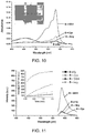
- FIG. 10 shows absorbance spectra of probe 8 combined with the thiols.
- Inset is a color photograph illustrating selective detection of GSH; the photograph was taken immediately upon the addition of CTAB. A strong pink color was obtained immediately upon addition of CTAB in the presence of GSH (inset). Hcy and Cys did not exhibit significant changes under the same conditions.
- SDS sodium dodecyl sulfate
- FIGS. 12-15 The results are shown in FIGS. 12-15 .
- Cys was preferentially detected by probe 8 in the presence of SDS and Triton X-100.
- SDS SDS
- Triton® X-100 FIG. 13
- maximum fluorescence was achieved within 1-2 minutes.
- FIG. 14 both Cys and GSH were detected in the presence of BC, with GSH producing greater fluorescence.
- CTAB CTAB
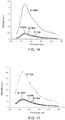
- 16 and 17 are fluorescence spectra of probe 8 in the presence of CTAB ( FIG. 16 ) and Triton X-100 ( FIG. 17 ), again showing that probe 8 is selective for GSH in the presence of the cationic surfactant CTAB, but selective for Cys in the presence of Triton X-100.
- probe 8 is an indicator for GSH in the presence of cationic surfactants such as BC and CTAB.
- cationic surfactants such as BC and CTAB.
- favorable Coulombic attractions between cationic micelles and GSH may play a role in the selectivity of probe 8 towards GSH in the presence of cationic surfactants.
- Negatively-charged SDS suppressed the response of probe 8 towards all three thiols; while the non-ionic surfactant Triton X-100 enhanced the selectivity towards Cys and resulted in zero response for Hey or GSH.
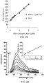
- FIG. 21 shows a linear relationship between fluorescence intensity and GSH concentration with a correlation coefficient of 0.990, as well as a linear relationship with a mixture of GSH and Cys having a correlation coefficient of 0.9894.
- probe 8 -CTAB-GSH signal was high at pH 7.4 (see, e.g ., Example 6), it is known that acrylate esters are susceptible to hydrolysis at high pH and that GSH is also relatively unstable under the same conditions (half-life of 9 hours at pH 7.4 compared to 16 hours at pH 6.5; Stevens et al., Biochem. Educ., 1983, 11, 70 ).
- the spectral behavior of the probe 8 -CTAB system was investigated over a wide range of pH values (pH 5.5 to pH 8), and its response to the various thiols was studied at lower pH values.
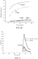
- Probe 8 (2.5 ⁇ M) was added to CTAB media (2 mM) buffered at pH 5.5 to 8, and the absorbance was monitored over time at a wavelength of 580 nm ( FIG. 24 ). Probe 8 showed the greatest background signal at the higher pH values investigated.
- the probe 8 -CTAB system was most stable at lower pH values.
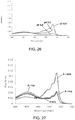
- the absorbance spectra of probe 8 in CTAB media at pH 6.0, 7.0, and 8.0 after 1 minute and 10 minutes are shown in FIGS. 25 and 26 . respectively.
- the fluorescence emission showed a significant increase with addition of reduced plasma.
- the fluorescence emission of solutions including probe 5 showed no obvious fluorescence increase when controls of either triphenylphosphine alone or deproteinized plasma (without reducing agent) were added, demonstrating that the fluorescence increase was due to the reaction of probe 5 with Cys.
- the results demonstrate that probe 5 may be useful for quantitative detection of Cys in biological samples.
- Plasma proteins were precipitated using MeCN (two thirds of the reconstitution volume) and removed by centrifugation at 4,000 rpm for 30 min. The supernatant liquid was diluted to provide 10% deproteinized plasma, and added to a solution of probe 8 (1-1.5 ⁇ M) in phosphate buffer (pH 6, 50 mM) in the presence of 2.0 mM CTAB. Some samples were spiked with GSH (0-2.0 ⁇ M) or a mixture of GSH (2 ⁇ M) and Cys (2 ⁇ M.
- Pig blood (10, 20, and 40 ⁇ L) was spotted on Whatman 903TM filter paper, and dried at room temperature for 3-4 hours.
- Oxidized glutathione was extracted from the dried blood spot (DBS) with 2.5 mL phosphate buffer (100 mM, pH 7.4).
- the extract was reduced with TCEP (tris-(2-carboxyethyl)-phosphine) (35 mM) to reduce disulfides.
- Reduced GSH was isolated by gel filtration of the reduced extract on a PD-10 desalting column (Sephadex® G-25 (cross-linked dextran gel), GE Healthcare) using phosphate buffer (100 mM, pH 7.4) as eluent.
- FIG. 33 is a fluorescence spectrum demonstrating that probe 8 detected GSH in the extract obtained 10 ⁇ L dried pig blood.
Landscapes
- Life Sciences & Earth Sciences (AREA)
- Health & Medical Sciences (AREA)
- Chemical & Material Sciences (AREA)
- Engineering & Computer Science (AREA)
- Molecular Biology (AREA)
- Hematology (AREA)
- Immunology (AREA)
- Biomedical Technology (AREA)
- Organic Chemistry (AREA)
- Urology & Nephrology (AREA)
- Physics & Mathematics (AREA)
- Cell Biology (AREA)
- Biotechnology (AREA)
- Medicinal Chemistry (AREA)
- Microbiology (AREA)
- Analytical Chemistry (AREA)
- Biochemistry (AREA)
- General Health & Medical Sciences (AREA)
- General Physics & Mathematics (AREA)
- Pathology (AREA)
- Food Science & Technology (AREA)
- Bioinformatics & Computational Biology (AREA)
- Bioinformatics & Cheminformatics (AREA)
- Biophysics (AREA)
- Proteomics, Peptides & Aminoacids (AREA)
- Spectroscopy & Molecular Physics (AREA)
- Investigating Or Analysing Materials By The Use Of Chemical Reactions (AREA)
- Investigating Or Analysing Biological Materials (AREA)
Description
- Embodiments of probes, methods, and kits for selectively detecting compounds comprising a thiol group and an amino group are disclosed.
- Biological thiols perform significant functions. For example, glutathione (GSH) serves as an antioxidant. It protects cells from oxidative stress by trapping free radicals that damage DNA and RNA. It also has important roles in maintaining the reducing environment in cells, catalysis, metabolism, transport, and gene regulation. GSH is the most abundant of the low molecular weight cellular thiols. It is present in millimolar concentrations; however, extracellular concentrations are relatively low, e.g., 2-20 µM in plasma. GSH is related to several diseases thought to be mediated by oxidative stress, including Alzheimer's disease, acquired immune deficiency syndrome, psoriasis, cancer, cardiovascular disease, heart attack, liver damage, leukocyte loss, Parkinson's disease, and stroke. Cysteine (Cys) is involved in protein synthesis, detoxification, and metabolic processes. Abnormal levels of Cys are related to many disorders, such as impaired growth, Alzheimer's disease. and cardiovascular disease. Cys deficiency is involved in slowed growth, hair depigmentation, edema, lethargy, liver damage, muscle and fat loss, skin lesions, and weakness (Shahrokhian, Anal. Chem. 2001, 73:5972-5978).
- Probes for detecting thiols are already known. For example, Yang et al. Angew. Chem. Int. Ed., 2011, 50:10690-10693, disclose a probe capable of detecting and differentiating between cysteine and homocysteine in which the hydroxy group in the fluorophore of 2-(2'-hydroxy-3'-methoxyphenyl)benzothiazole (HMBT) is masked with an α,β-unsaturated carbonyl moiety. The probe is also capable of detecting GSH.
- In addition,
WO 2011/082354 A2 discloses compounds based on bridged viologens which are capable of selectively detecting thiols, such as cysteine, homocysteine and GSH. - Other disclosures of molecular probes include
WO 2005/042504 A1 , which discloses fluorinated resorufin compounds, and their use in detecting reactive oxygen species, such as hydrogen peroxide. - Embodiments of probes for selectively detecting compounds comprising a thiol group and an amino group (referred to herein as "thiols") are disclosed. Exemplary compounds include biological thiols, such as cysteine, homocysteine, and glutathione. Embodiments of the disclosed probes when reacted with one or more thiols in solution produce a detectable change in the solution's absorbance spectrum and/or emission spectrum. Methods and kits for performing the detection also are disclosed.
- Embodiments of the disclosed probes have a chemical structure according to general formula I.

- where each bond depicted as "------" is a single or double bond as needed to satisfy valence requirements; R1, R3-R6 and R8 independently are hydrogen, hydroxyl, thiol, lower alkyl (C1-C10 alkyl), carboxyalkyl, amino, alkoxy, or halogen; R2 is an α,β-unsaturated aliphatic ester;
- X1 is O, CH2, S, or NH and
- (i) R7 is O and X2 is N; or
- (ii) R7 and R8 together, with the ring to which they are attached form a naphthyl ring and X2 is CR9 where R9 is phenyl.
- In some embodiments, the probe has a structure according to general formula IA where R7 is O, and X2 is N.

- One exemplary probe according to general formula IA ("
probe 8") has the chemical structure
- In some embodiments, R7 and R8 together with the ring to which they are attached form a naphthyl ring, X2 is CR9, and the probe has a chemical structure according to general formula II.

- where "-------" indicates a single or double bond as needed to satisfy valence requirements and "------" indicates an optional single bond; R2 and R11 independently are an α,β-unsaturated aliphatic ester; R10, R12, and R13 independently are hydrogen, hydroxyl, thiol, lower alkyl, carboxyalkyl, amino, alkoxy, or halogen;
- R14-R17 independently are hydrogen, alkyl, acyl, carboxyl, nitro, amino, alkyl amino, or -SO3H; and R18 is hydrogen, hydroxyl, lower alkyl, lower alkoxy, -SO3H or-COOR19 where R19 is hydrogen or lower alkyl and the bond depicted as "------" in ring B is a double bond, or R18 is one or more atoms forming a ring system with rings B and E and the bond depicted as "------" in ring B is a single bond.
- One exemplary probe according to general formula II ("
probe 5") has the chemical structure:
- Embodiments of a method for selectively detecting a compound comprising a thiol group and an amino group include (i) preparing a reaction mixture by combining a sample potentially comprising at least one compound comprising a thiol group and an amino group with a solution comprising a probe as disclosed herein; (ii) allowing a reaction between the sample and the probe to proceed for an effective period of time to produce a detectable change in the reaction mixture's absorbance spectrum, emission spectrum, or both, where the change indicates that the compound is present; and (iii) detecting the change. In some examples, the sample comprises blood, a blood product or component, urine, or a urine product or component. In some embodiments, the reaction mixture further includes a surfactant, and the surfactant may affect probe selectivity.
- Embodiments of a kit for detecting a compound comprising a thiol group and an amino group include at least one probe as disclosed herein. The kit may further comprise at least one buffer solution with a pH of 6-7.5 and, optionally, a surfactant. In some embodiments, the kit includes a plurality of disposable containers in which a reaction between the probe and the compound can be performed. An amount of the probe effective to undergo a detectable change in the absorbance spectrum, the emission spectrum, or both when reacted with a compound comprising a thiol group and an amino group may be premeasured into the plurality of disposable containers. The kit also may include a color comparison chart for evaluating a color change produced by the reaction.
- The foregoing and other objects, features, and advantages of the invention will become more apparent from the following detailed description, which proceeds with reference to the accompanying figures.
-
-
FIG. 1 is a high-resolution mass spectrum demonstrating formation of a 12-memberedring 9 whenprobe 8 is reacted with glutathione (GSH) in the presence of the surfactant cetyltrimethylammonium bromide (CTAB); the reaction time was 5 minutes. Background: negative; RT: 0.01; AV: 1; NL: 1.75E5; T: FTMS - pESI Full ms[300.00-400.00]. -
FIGS. 2A-2B show fluorescence spectra (λex = 550 nm) obtained from the reaction ofprobe 5 with cysteine (Cys), homocysteine (Hcy), GSH, and other amino acids in buffered 1.0 mM CTAB; selective detection of cysteine resulted. -
FIG. 3 is a color photograph illustrating selective detection of Cys withprobe 5 in 1.0 mM CTAB after 25 minutes. -
FIG. 4 shows absorption spectra obtained from the reaction ofprobe 8 with Cys, Hcy, or GSH in phosphate buffer (pH 7.4, 50 mM) after 20 minutes. -
FIG. 5 shows fluorescence spectra (λex = 565 nm, λem = 587 nm) obtained from the reaction ofprobe 8 with Cys, Hcy, or GSH in phosphate buffer (pH 7.4, 50 mM) after 20 minutes: the time-dependent fluorescence changes also were monitored (inset). -
FIG. 6 shows fluorescence spectra obtained from the reaction ofprobe 8 with varying concentrations of Cys at pH 7.4 (phosphate buffer, 50 mM); reaction time of 60 minutes. -
FIG. 7 is a graph illustrating the relationship between fluorescence intensity and Cys concentration when Cys is reacted withprobe 8 at pH 7.4 (phosphate buffer. 50 mM); reaction time of 60 minutes. -
FIG. 8 is a high-resolution mass spectrum demonstrating formation of 3-carboxy-5-oxoperhydro-1.4-thiazepine and release of resorufin whenprobe 8 is reacted with Cys in phosphate buffer (50 mM. pH 7.4). RT: 0.09; AV: 1; NL: 7.21E7; T: FTMS - pESI Full ms[100.00-500.00]. -
FIG. 9 is a high-resolution mass spectrum demonstrating formation of an 8-membered ring and release of resorufin whenprobe 8 is reacted with Hcy in phosphate buffer (50 mM, pH 7.4). RT: 0,11-0.21; AV: 7; NL: 7.09E7; T: FTMS - pESI Full ms[100.00-1000.00]. -
FIG. 10 shows absorbance spectra ofprobe 8 combined with Cys, Hcy, or GSH in 2.0 mM CTAB buffered at pH 7.4. Inset is a color photograph illustrating selective detection of GSH; the photograph was taken immediately upon the addition of CTAB. -
FIG. 11 shows fluorescence spectra (λex = 565 nm; λem = 587 nm) obtained from the reaction ofprobe 8 with Cys, Hcy, GSH, and other amino acids in buffered 2.0 mM CTAB: selective detection of GSH resulted. Inset is a graph of fluorescence versus time. -
FIG. 12 is a graph illustrating the time-dependent change in fluorescence emission (λex/λem = 565/590 nm) whenprobe 8 was combined with Cys, Hcy, or GSH in phosphate-buffered media (50 mM, pH = 7.4) including 10 mM sodium dodecylsulfate. -
FIG. 13 is a graph illustrating the time-dependent change in fluorescence emission (λex/λem = 565/590 nm) whenprobe 8 was combined with Cys, Hcy, or GSH in phosphate-buffered media (50 mM, pH = 7.4) including 0.3 mM Triton X-100. -
FIG. 14 is a graph illustrating the time-dependent change in fluorescence emission (λex/λem = 565/590 nm) whenprobe 8 was combined with Cys, Hcy, or GSH in phosphate-buffered media (50 mM. pH = 7.4) including 0.05 mM benzyl-dimethyl-tridecyl-azanium chloride. -
FIG. 15 is a graph illustrating the time-dependent change in fluorescence emission (λex/λem = 565/590 nm) whenprobe 8 was combined with Cys, Hcy, or GSH in phosphate-buffered media (50 mM, pH = 7.4) including 2 mM CTAB. -
FIG. 16 shows fluorescence spectra (λex = 565 nm) obtained when probe 8 (2.5 µM) was combined with 2 equivalents of Cys, Hcy, or GSH is phosphate-buffered media (50 mM, pH = 7.4) with 2 mM CTAB. -
FIG. 17 shows fluorescence spectra (λex = 565 nm) obtained when probe 8 (2.5 µM) was combined with 2 equivalents of Cys, Hcy, or GSH is phosphate-buffered media (50 mM, pH = 7.4) with 0.3 mM Triton X-100. -
FIG. 18 shows fluorescence spectra obtained when probe 8 (2.5 µM) was combined with 2 equivalents of GSH in phosphate-buffered media (50 mM, pH = 7.4) with 10 mM SDS, 0.3 mM Triton X-100, 0.05 mM BC, or 2 mM CTAB. Spectra were obtained immediately upon addition of the surfactants; the inset shows time-dependent fluorescence changes (λem = 587 nm) of the same system. -
FIG. 19 shows fluorescence spectra obtained from the reaction ofprobe 8 with Cys, Hcy, GSH, and other amino acids in buffered 2.0 mM CTAB; selective detection of GSH resulted. -
FIG. 20 shows time-dependent changes in fluorescence emission (λex/λem = 565/594 nm) whenprobe 8 was combined with Cys, Hcy, or GSH in DMSO/H2O 1:1 buffered at pH 7.4. -
FIG. 21 shows fluorescence spectra of the CTAB-mediated response ofprobe 8 towards 2 µM Cys, 2 µM GSH, or a combination of 2 µM Cys and 2 µM GSH. -
FIG. 22 is a graph illustrating the relationship between fluorescence intensity and GSH concentration when GSH is reacted withprobe 8 at pH 7.4 (phosphate buffer, 50 mM), or withprobe -
FIG. 23 shows fluorescence spectra obtained from the reaction ofprobe 8 with varying concentrations of GSH in buffered 2.0 mM CTAB; inset is a graph illustrating the linear relationship between fluorescence intensity and GSH concentration when GSH is reacted withprobe 8 in buffered 2.0 mM CTAB. -
FIG. 24 is a graph illustrating the time-dependent absorbance changes (at 580 nm) ofprobe 8 in CTAB media buffered at pH 5.5 to 8. -
FIG. 25 shows absorbance spectra ofprobe 8 in CTAB media buffered at pH 6.0. 7.0. and 8.0 after one minute. -
FIG. 26 shows absorbance spectra ofprobe 8 in CTAB media buffered at pH 6.0, 7.0, and 8.0 after 10 minutes. -
FIG. 27 shows absorbance spectra ofprobe 8 with Cys, Hcy, and GSH in CTAB media buffered at pH 6.0 after 20 minutes. -
FIG. 28 shows fluorescence spectra (λex = 565 nm) ofprobe 8 with Cys, Hcy, and GSH in CTAB media buffered at pH 6.0 after 20 minutes; the inset shows the time-dependent fluorescence changes (λem = 587 nm). -
FIG. 29 shows fluorescence spectra (λex = 550 nm) ofprobe 5 with triphenylphosphine, deproteinized human plasma, and reduced deproteinized plasma in pH 7.4 HEPES buffer solution (0.1 M) in the presence of 1.0 mM CTAB; spectra were obtained after 25 minutes. -
FIG. 30 is a calibration curve of probe 8 (1.0 µM) in 2.0 mM CTAB media buffered at pH 6 (phosphate buffer, 50 mM) showing a linear relationship between fluorescence intensity (λex = 565 nm, λem = 587 nm) and GSH concentration (0.1-1.2 µM). Measurements were taken 8 minutes after mixing. -
FIG. 31 shows fluorescence spectra (λex = 565 nm) of probe 8 (1.0 µM) upon addition of GSH (0-1.0 µM) to 10% deproteinized plasma diluted with 2.0 mM CTAB media buffered at pH 6.0 (phosphate buffer, 50 mM). Measurements were taken 8 minutes after mixing. -
FIG. 32 shows fluorescence spectra (λex = 565 nm) obtained when probe 8 (1.5 µM) was reacted with GSH (0-2.0 µM) or a mixture of GSH (2 µM) and Cys (2 µM) in 10% deproteinized plasma diluted with 2.0 mM CTAB media buffered at pH 6.0 (phosphate buffer, 50 mM). -
FIG. 33 is a fluorescence spectrum (λex = 565 nm) obtained whenprobe 8 was combined with a dried blood spot (DBS) extract in 2.0 mM CTAB media buffered at pH 6.0 (phosphate buffer, 50 mM); the probe reacted with GSH in the extract. - Disclosed herein are embodiments of probes that can selectively detect and differentiate between GSH and Cys at physiologic pH. Embodiments of disclosed probes also can discriminate GSH and Cys from other amino acids and thiols (e.g.. Hcy) at physiologic pH. Embodiments of the disclosed probes include a fluorophore moiety to facilitate detection and a moiety capable of undergoing a condensation-cyclization reaction (referred to as a "cyclization moiety") with GSH and/or Cys. In some embodiments, the cyclization moiety is an α.β-unsaturated carbonyl moiety, which is covalently bonded to the fluorophore moiety.
- The following explanations of terms and abbreviations are provided to better describe the present disclosure and to guide those of ordinary skill in the art in the practice of the present disclosure. As used herein, "comprising" means "including" and the singular forms "a" or "an" or "the" include plural references unless the context clearly dictates otherwise. The term "or" refers to a single element of stated alternative elements or a combination of two or more elements, unless the context clearly indicates otherwise.
- Unless explained otherwise, all technical and scientific terms used herein have the same meaning as commonly understood to one of ordinary skill in the art to which this disclosure belongs. Although methods and materials similar or equivalent to those described herein can be used in the practice or testing of the present disclosure, suitable methods and materials are described below. The materials, methods, and examples are illustrative only and not intended to be limiting. Other features of the disclosure are apparent from the following detailed description and the claims.
- Unless otherwise indicated, all numbers expressing quantities of components, molecular weights, percentages, temperatures, times, and so forth, as used in the specification or claims are to be understood as being modified by the term "about." Accordingly, unless otherwise indicated, implicitly or explicitly, the numerical parameters set forth are approximations that may depend on the desired properties sought and/or limits of detection under standard test conditions/methods. When directly and explicitly distinguishing embodiments from discussed prior art the embodiment numbers are not approximates unless the word "about" is recited.
- Definitions of other common terms in chemistry may be found in Richard J. Lewis, Sr. (ed.), Hawley's Condensed Chemical Dictionary, published by John Wiley & Sons, Inc., 1997 (ISBN 0-471-29205-2).
- In order to facilitate review of the various embodiments of the disclosure, the following explanations of specific terms are provided:
- Absorbance: The retention by a compound or substance of certain wavelengths of radiation incident upon it; a measure of the amount of light at a particular wavelength absorbed as the light passes through a compound or substance, or through a solution of a compound or substance,
- Acrylate ester: A group having a general formula -OC(O)CR=CR'R", where R, R' and R" individually are hydrogen, halogen, hydroxyl, lower alkoxy, lower aliphatic, or aryl.
- Aliphatic: A substantially hydrocarbon-based compound, or a radical thereof (e.g., C6H13, for a hexane radical), including alkanes, alkenes, alkynes. including cyclic versions thereof, and further including straight- and branched-chain arrangements, and all stereo and position isomers as well. Unless expressly stated otherwise, an aliphatic group contains from one to twenty-five carbon atoms; for example, from one to fifteen, from one to ten, from one to six, or from one to four carbon atoms. The term "lower aliphatic" refers to an aliphatic group containing from one to ten carbon atoms. An aliphatic chain may be substituted or unsubstituted. An aliphatic group can be substituted with one or more substituents (up to two substituents for each methylene carbon in an aliphatic chain, or up to one substituent for each carbon of a -C=C- double bond in an aliphatic chain, or up to one substituent for a carbon of a terminal methine group). Exemplary aliphatic substituents include, for instance, amine, amide, sulfonamide, halogen, cyano, carboxyl, hydroxyl, mercapto, trifluoromethyl, alkyl, alkoxy, alkylthio, thioalkoxy, arylalkyl, heteroaryl, alkylamino, dialkylamino, or other functionality.
- Alkyl: A hydrocarbon group having a saturated carbon chain. The chain may be branched or unbranched. The term lower alkyl means the chain includes 1-10 carbon atoms.
- Alkoxy: A functional group having the formula -OR where R is an alkyl group. The term lower alkoxy means that the alkyl group includes 1-10 carbon atoms.
- Aromatic or aryl: Compounds or groups that typically are unsaturated, cyclic hydrocarbons having alternate single and double bonds. Benzene, a 6-carbon ring containing three double bonds, is a typical aromatic compound.
- BC: Benzalkonium chloride, benzyl-dimethyl-tridecyl-azanium chloride.
- Carboxyalkyl: A group having a general formula -C(O)OR, where R is an alkyl group.
- Carboxyl: A group having a general formula -COOH.
- CTAB: Cetyltrimethylammonium bromide
- Cyclization moiety: As used herein, the term "cyclization moiety" refers to a portion of a molecule capable of undergoing a condensation-cyclization reaction with a target compound, such as a compound comprising a thiol group and an amino group.
- Cys: Cysteine
- Detect: To determine if an agent (such as a target molecule) is present or absent, for example, in a sample. "Detecting" refers to any method of determining if something exists, or does not exist, such as determining if a target molecule is present in a biological sample. For example, "detecting" can include using a visual or a mechanical device to determine if a sample displays a specific characteristic.
- Effective period of time: As defined herein, an effective period of time is a sufficient amount of time to allow a chemical reaction to occur. With respect to the present disclosure, an effective period of time is an amount of time sufficient to allow condensation of a thiol-containing compound with an embodiment of the disclosed probes and subsequent cyclization of the thiol-containing compound to occur.
- Emission or emission signal: The light of a particular wavelength generated from a source. In particular examples, an emission signal is emitted from a fluorophore after the fluorophore absorbs light at its excitation wavelength(s).
- Ester: A chemical compound derived from an organic acid (general formula: RCO2H) where the hydrogen of the -OH (hydroxyl) group is replaced by an aliphatic, alkyl or aryl group. A general formula for an ester derived from an organic acid is shown below:

- Fluorescence: The emission of visible radiation by an atom or molecule passing from a higher to a lower electronic state, wherein the time interval between absorption and emission of energy is 10-8 to 10-3 second. Fluorescence occurs when the atom or molecule absorbs energy from an excitation source (e.g., an ultraviolet lamp) and then emits the energy as visible radiation,
- Fluorophore or fluorogen: A compound capable of fluorescence, such as a fluorescent dye. The term "fluorophore" also refers to the portion of a molecule that causes the molecule to fluoresce when exposed to an excitation source.
- Functional group: A specific group of atoms within a molecule that is responsible for the characteristic chemical reactions of the molecule. Exemplary functional groups include, without limitation, alkane, alkene, alkyne, arene, halo (fluoro, chloro, bromo, iodo), epoxide, hydroxyl, carbonyl (ketone), aldehyde, carbonate ester, carboxylate, ether, ester, peroxy. hydroperoxy. carboxamide, amine (primary, secondary, tertiary), ammonium, imide, azide, cyanate. isocyanate, thiocyanate, nitrate, nitrite, nitrile, nitroalkane, nitroso, pyridyl. phosphate, sulfonyl, sulfide, thiol (sulfhydryl), disulfide.
- GSH: Glutathione
- Hcy: Homocysteine
- HEPES: 2-[4-(2-hydroxyethyl)piperazin-1-yl]ethanesulfonic acid
- Probe: As used herein, the term "probe" refers to a molecule capable of selectively reacting with a molecule of interest (i.e., a molecule for which the presence and/or concentration is to be determined) and producing a detectable signal or change as a result of the reaction. A detectable signal or change may include a change in the absorbance spectrum and/or emission spectrum of the probe and/or the molecule of interest.
- SDS: Sodium dodecyl sulfate
- Substituent: An atom or group of atoms that replaces another atom in a molecule as the result of a reaction. The term "substituent" typically refers to an atom or group of atoms that replaces a hydrogen atom on a parent hydrocarbon chain or ring.
- Substituted: A fundamental compound, such as an aryl or aliphatic compound, or a radical thereof, having coupled thereto, typically in place of a hydrogen atom, a substituent. For example, substituted aryl compounds or substituents may have an aliphatic group coupled to the closed ring of the aryl base, such as with toluene. Again solely by way of example and without limitation, a long-chain hydrocarbon may have a substituent bonded thereto, such as one or more halogens, an aryl group, a cyclic group, a heteroaryl group or a heterocyclic group. Unless expressly referred to as "unsubstituted," functional groups recited herein (e.g., aliphatic, alkyl, alkoxy, amino, aryl, cycloalkyl, ester, etc.) can either be unsubstituted or substituted.
- Surfactant: A compound that reduces surface tension when dissolved in water or aqueous solutions. Surfactants typically are amphiphilic organic compounds, i.e., organic compounds that contain both hydrophobic groups and hydrophilic groups. Surfactants may be characterized by their hydrophilic groups, or heads. A non-ionic surfactant includes no formal charge in its head. Ionic surfactants include hydrophilic groups having a net charge. If the charge is negative, the surfactant is an anionic surfactant. If the charge is positive, it is a cationic surfactant. If the head contains two oppositely charged groups, it is a zwitterionic surfactant.
- Thiol: A functional group having the formula -SH. As used herein with respect to a compound, the term "thiol" refers to a compound comprising a thiol group and an amino group.
- Embodiments of probes for selectively detecting compounds comprising a thiol group and an amino group (referred to herein as "thiols") are disclosed. Exemplary compounds include biological thiols, such as cysteine, homocysteine, and glutathione. Embodiments of the disclosed probes when reacted with one or more thiols in solution produce a detectable change in the solution's absorbance spectrum and/or emission spectrum. Methods and kits for performing the detection also are disclosed.
- Embodiments of the disclosed probes have a chemical structure according to general formula I.

- where each bond depicted as "-------" is a single or double bond as needed to satisfy valence requirements; R1, R3-R6 and R8 independently are H, hydroxyl, thiol, lower alkyl (C1-C10 alkyl), carboxyalkyl, amino, alkoxy, or halogen; R2 is an α,β-unsaturated aliphatic ester;
- X1 is O, CH2, S, or NH and
- (i) R7 is O and X2 is N; or
- (ii) R7 and R8 together with the ring to which they are attached form a naphthyl ring and X2 is CR9 where R9 is phenyl.
- In some embodiments, X1 is O or S, and R7 is O , or R7 and R8 together form a phenyl ring. In any or all of the above embodiments, R2 may be an acrylate ester. In any or all of the above embodiments, R1 and R3-R6 independently may be H or lower alkyl. In some embodiments, R1 is lower alkyl and R3-R6 are H. In some embodiments, R7 is O and R8 is H. In any or all of the above embodiments, X1 may be O, X2 may be N or CR9, R1 may be lower alkyl or H, R2 may be an acrylate ester, R3-R6 may be H, and R7 may be O and R8 may be H, or R7 and R8 together may form a phenyl ring.
- In any or all of the above embodiments, R7 may be O, and X2 may be N. In any or all of the above embodiments, R2 may be an acrylate ester. In some embodiments, R2 is an acrylate ester, R7 is O, and X2 is CH or N. In some embodiments, R2 is an acrylate ester, R7 is O, X1 is O, and X2 is N. In any or all embodiments where R2 is an acrylate ester, R7 is O, X1 is O, and X2 is N, R1, R3-R6, and R8 independently may be H or lower alkyl. An exemplary probe has the structure below.

- In some embodiments, R7 and R8 together with the ring to which they are attached form a naphthyl ring and X2 is CR9. In such embodiments, the compound has a chemical structure according to general formula II:

- where "------" indicates a single or double bond as needed to satisfy valence requirements and "------" indicates an optional single bond; R2 and R11 independently are an α,β-unsaturated aliphatic ester; R10, R12, and R13 independently are hydrogen, hydroxyl, thiol, lower alkyl, carboxyalkyl, amino, alkoxy, or halogen;
- R14-R17 independently are hydrogen, alkyl, acyl, carboxyl, nitro, amino, alkyl amino, or -SO3H; and R18 is hydrogen, hydroxyl, lower alkyl, lower alkoxy, -SO3H or - COOR19 where R19 is hydrogen or lower alkyl and the bond depicted as "------" in ring B is a double bond, or R18 is one or more atoms forming a ring system with rings B and E and the bond depicted as "------" in ring B is a single bond.
- In some embodiments, each of R2 and R11 is an acrylate ester. In any or all of the above embodiments, R18 may be -RCOOH or R18 may be -C(O)O- and form a ring system with rings B and E. In any or all of the above embodiments, R1, R3-R6, R10 and R12-R17 independently may be H or lower alkyl. In some embodiments, R1 is lower alkyl, and R3-R6, R10 and R12-R17 are H; optionally, X1 is O. An exemplary probe according to general formula II has the structure below.

- Embodiments of a method for selectively detecting a compound comprising a thiol group and an amino group include (i) combining a sample potentially comprising at least one compound comprising a thiol group and an amino group with a solution comprising a probe having a structure according to general formula I to produce a reaction mixture; (ii) allowing a reaction between the sample and the probe to proceed for an effective period of time to produce a detectable change in the reaction mixture's absorbance spectrum, emission spectrum, or both, where the change indicates that the compound is present; and (iii) detecting the change. In some embodiments, the compound is cysteine, homocysteine, glutathione, or a combination thereof.
- In any or all of the above embodiments, the effective period of time may be ≤ 60 minutes. In any or all of the above embodiments, the sample may comprise blood, a blood product or component, urine, or a urine product or component. In one embodiment, the method further includes obtaining plasma, reducing the plasma by addition of a reducing agent, precipitating proteins in the plasma, separating precipitated proteins from the plasma to form the sample, and subsequently combining the sample with the solution comprising the probe. In another embodiment, the method further includes obtaining a dried blood spot prepared by spotting whole blood onto an absorbent material and allowing the blood to dry, extracting the dried blood spot with a solvent to produce an extract, reducing the extract by addition of a reducing agent to form a reduced extract, fractionating the reduced extract to obtain the sample, and subsequently combining the sample with the solution comprising the probe.
- In any or all of the above embodiments, detecting the change may include (i) comparing a color of the reaction mixture before the reaction to a color of the reaction mixture after the reaction, (ii) detecting a change in absorbance of the reaction mixture at one or more wavelengths after the reaction has proceeded for the effective period of time, (iii) comparing an absorbance spectrum of the reaction mixture at a first time after combining the sample and the probe to an absorbance spectrum of the reaction mixture after the reaction has proceeded for the effective period of time, (iv) detecting a change in emission of the reaction mixture at one or more wavelengths after the reaction has proceeded for the effective period of time, (v) comparing an emission spectrum of the reaction mixture at a first time after combining the sample and the probe to an emission spectrum of the solution after the reaction has proceeded for the effective period of time, (vi) or any combination thereof.
- In any or all of the above embodiments, the probe may be
probe 8. In some embodiments, detecting the compound comprises detecting fluorescence of the reaction mixture at 580-590 nm. In some embodiments, when the probe isprobe 8 and the compound is glutathione, the reaction mixture further includes a cationic surfactant. Exemplary cationic surfactants include cetyltrimethylammonium bromide and benzalkonium chloride. In some embodiments, when the probe isprobe 8 and the compound is cysteine, the reaction mixture further includes a surfactant other than a cationic surfactant, e.g., sodium dodecyl sulfate or polyethylene glycol tert-octylphenyl ether. In certain embodiments, the reaction mixture further comprises dimethyl sulfoxide. - In any or all of the above embodiments, the compound may be cysteine and the probe may be
probe 5. In some embodiments, the reaction mixture further comprises cetyltrimethylammonium bromide. - Embodiments of a kit for detecting at least one compound comprising a thiol group and an amino group include an embodiment of the disclosed probes having a structure according to general formula I. In some embodiments, the kit further includes at least one buffer solution with a pH of 6-7.5. The buffer solution may include a surfactant. In some embodiments, the surfactant is cetyltrimethylammonium bromide, benzalkonium chloride, sodium dodecyl sulfate, or polyethylene glycol tert-octylphenyl ether.
- In any or all of the above embodiments, the kit may further include a plurality of disposable containers in which a reaction between the probe and the compound can be performed. In some embodiments, an amount of the probe effective to undergo a detectable change in the absorbance spectrum, the emission spectrum, or both when reacted with a compound comprising a thiol group and an amino group is premeasured into the plurality of disposable containers.
- In any or all of the above embodiments, the kit may include a color comparison chart for evaluating a color change produced by a reaction between (a) the probe and (b) cysteine, homocysteine, glutathione. or a combination thereof. In any or all of the above embodiments, the probe may be
probe 5 orprobe 8. - Cysteine is capable of undergoing condensation with certain acrylates to form substituted 1,4-thiazepines (Blondeau et al., Can. J. Chem. 1971, 49. 3866-3876; Leonard et al., J. Org. Chem. 1966, 31, 3928-3935). The reaction involves the conjugate addition of Cys to acrylates (1: R is alkyl, such as methyl) to generate thioether (2a), which can further undergo an intramolecular cyclization to yield the desired compound 3a (3-carboxy-5-oxoperhydro-1,4-thiazepine), as illustrated in
Scheme 1.
- To achieve highly selective detection of GSH over Cys and Hcy, one would need to overcome the formidable challenge of favoring formation of a larger 12-membered ring over a 7- or 8-membered ring. It has been reported that cetyltrimethylammonium bromide (CTAB) micelles can catalyze the intramolecular ring closure of larger rings, reversing the expected kinetics based on ring size (Wei et al., Langmuir, 1991, 7, 1336-1339). GSH is capable of binding to the surface of cationic CTAB micelles (Huang et al., Langmuir, 2005, 23, 1518-1522).
- Disclosed herein are embodiments of probes that can be used to selectively detect GSH or Cys, as well as methods of making and using the probes. Some embodiments of the probes are suitable for detecting GSH or Cys in biological media.
- Probes useful for selective detection of biological thiols are disclosed. Some embodiments of the disclosed probes undergo long-wavelength (e.g., 550-700 nm) emission when excited, making them suitable for selectively detecting thiols in biological samples (e.g., blood, plasma, urine, or products or components thereof). The disclosed probes include a fluorophore moiety and a cyclization moiety capable of undergoing a condensation-cyclization reaction with a compound comprising a thiol group and an amino group. Prior to reaction, the probe is colorless and non-fluorescent. After cyclization occurs, the cyclization moiety and thiol-containing compound are eliminating, thereby releasing the fluorophore. The released fluorophore may appear colored in solution. In some embodiments, the probes undergo a conjugate addition and micelle-catalyzed large-membered ring formation/elimination sequence.
- Embodiments of the disclosed probes have a chemical structure according to general formula I:

- where each bond depicted as "-------" is a single or double bond as needed to satisfy valence requirements; R1, R3-R6 and R8 independently are hydrogen, hydroxyl, thiol, lower alkyl (C1-C10 alkyl), carboxyalkyl, amino, alkoxy, or halogen; R2 is an α,β-unsaturated aliphatic ester;
- X1 is O, CH2, S, or NH and
- (i) R7 is O and X2 is N; or
- (ii) R7 and R8 together with the ring to which they are attached form a naphthyl ring and X2 is CR9 where R9 is phenyl.
- R2 is a cyclization moiety capable of reacting with a compound comprising a thiol group and an amino group.
- In some embodiments, X1 is O or S, and R7 is O, or R7 and R8 together form a phenyl ring. In one embodiment, R2 is an acrylate ester, X1 is O or S, and R7 is O, or R7 and R8 together form a phenyl ring. In another embodiment, R1 and R3 - R6 independently are H or lower alkyl, X1 is O or S, and R7 is O, or R7 and R8 together form a phenyl ring. In one example, R1 is lower alkyl and R3-R6 are H. In another embodiment, X1 is O or S, R7 is O, and R8 is H. In yet another embodiment, X1 is O, X2 is N or CR9, R1 is lower alkyl or H, R2 is an acrylate ester, R3-R6 are H, and R7 is O and R8 is H, or R7 and R8 together form a phenyl ring.
- In some embodiments, the probe has a structure according to general formula IA where R7 is O, and X2 is N.

probe 8").
- In some embodiments, R7 and R8 together with the ring to which they are attached form a naphthyl ring, X2 is CR9, and the probe is a seminaphthofluorescein (SNF) with a chemical structure according to general formula II:

- where "------" indicates a single or double bond as needed to satisfy valence requirements and "------" indicates an optional single bond; X1 and R1-R6 are as previously defined; R2 and R11 independently are an α,β-unsaturated aliphatic ester;
- R10, R12, and R13 independently are hydrogen, hydroxyl, thiol, lower alkyl, carboxyalkyl, amino, alkoxy, or halogen; R14-R17 independently are hydrogen, alkyl, acyl, carboxyl, nitro, amino, alkyl amino, or -SO3H; and R18 is hydrogen, hydroxyl, lower alkyl, lower alkoxy, -SO3H or -COOR19 where R19 is hydrogen or lower alkyl and the bond depicted as "------" in ring B is a double bond, or R18 is one or more atoms forming a ring system with rings B and E and the bond depicted as "------" in ring B is a single bond. R2 and R11 are cyclization moieties capable of reacting with a compound comprising a thiol group and an amino group.
- In one embodiment, each of R2 and R11 is an acrylate ester. In another embodiment, each of R2 and R11 is an acrylate ester, and R18 is -COOH or R18 is -C(O)O- and forms a ring system with rings B and E. In yet another embodiment, each of R2 and R11 is an acrylate ester, R1, R3-R6, R10 and R12-R17 independently are H or lower alkyl, and R18 is -COOH or R18 is -C(O)O- and forms a ring system with rings B and E. In still another embodiment, X1 is O, each of R2 and R11 is an acrylate ester. R1, R3-R6, R10 and R12-R7 independently are H or lower alkyl, and R18 is -COOH or R18 is -C(O)O- and forms a ring system with rings B and E. In another embodiment, each of R1 and R11 is an acrylate ester, R1 is lower alkyl, R3-R6, R10 and R12-R17 are H, and R18 is -COOH or R18 is -C(O)O- and forms a ring system with rings B and E. In one embodiment, X1 is O, each of R2 and R11 is an acrylate ester. R1 is methyl, R3-R6, R10 and R12-R17 are H, and R18 is -C(O)O- and forms a ring system with rings B and E (i.e., "
probe 5").
- Without wishing to be bound by any particular theory of operation,
probe 5 can be used to selectively detect Cys by the mechanism shown inScheme 2.Probe 5 is a colorless, non-fluorescent compound. Each acrylate group on the probe undergoes a condensation, cyclization, elimination, and xanthene dye spirolactone-opening sequence with Cys, producing a colored, fluorescent seminaphthofluorescein (see, e.g.,FIG. 3 ). In some embodiments, the probe also reacts with Hcy and GSH to a much lesser extent, but the reaction produces very little fluorescence and no visible color change is seen (FIGS. 2-3 ). Thus.probe 5 can be used in a time-dependent assay to differentiate between Cys and Hcy/GSH. In some embodiments,probe 5 also is suitable for use as a selective "naked-eye" dosimeter for Cys.
-
Probe 8, a non-fluorescent compound, undergoes a similar sequence of reactions, releasing highlyfluorescent resorufin 7 as shown inScheme 3. In some solutions,probe 8 selectively detects Cys, and to a lesser extent. Hcy.
- To selectively detect GSH over Cys and Hcy, the formidable challenge of favoring the formation of a larger (12-membered ring in the case of GSH) over a 7- and 8-membered ring (as in 3a and 3b) must be overcome. Inclusion of certain surfactants was discovered to dramatically alter the intrinsic selectivity of
probe 8. - CetyJtrimethylammonium bromide (CTAB), a cationic surfactant forms micelles that can catalyze the intramolecular ring closure of some rings, thereby reversing the expected kinetics based on ring size such that large rings may be closed faster than smaller rings (Wei et al., Langmuir, 1991, 7, 1336-1339). GSH has been shown to be capable of binding to the surface of cationic CTAB micelles (Huang et al., Langmuir, 2005, 23, 1518-1522).
- Because
probe 8 is small and substantially planar, it is capable of interacting with the CTAB micelle to facilitate large ring closure and selective detection of GSH. Without wishing to be bound by any particular theory of operation, the mechanism of action involves the conjugate addition of GSH tonon-fluorescent probe 8 to generatecompound 9, which in turn undergoes an intramolecular cyclization/elimination reaction sequence catalyzed by the CTAB micelle releasing a 12-membered ring 10 and thefree resorufin dye 7, and resulting in the recovery of the fluorescence (Scheme 4). High-resolution mass spectrometry demonstrated formation of the 12-membered ring 10 (FIG. 1 ) andresorufin 7.. Other thiols such as Cys and Hcy did not exhibit significant reactions withprobe 8 in the presence of CTAB.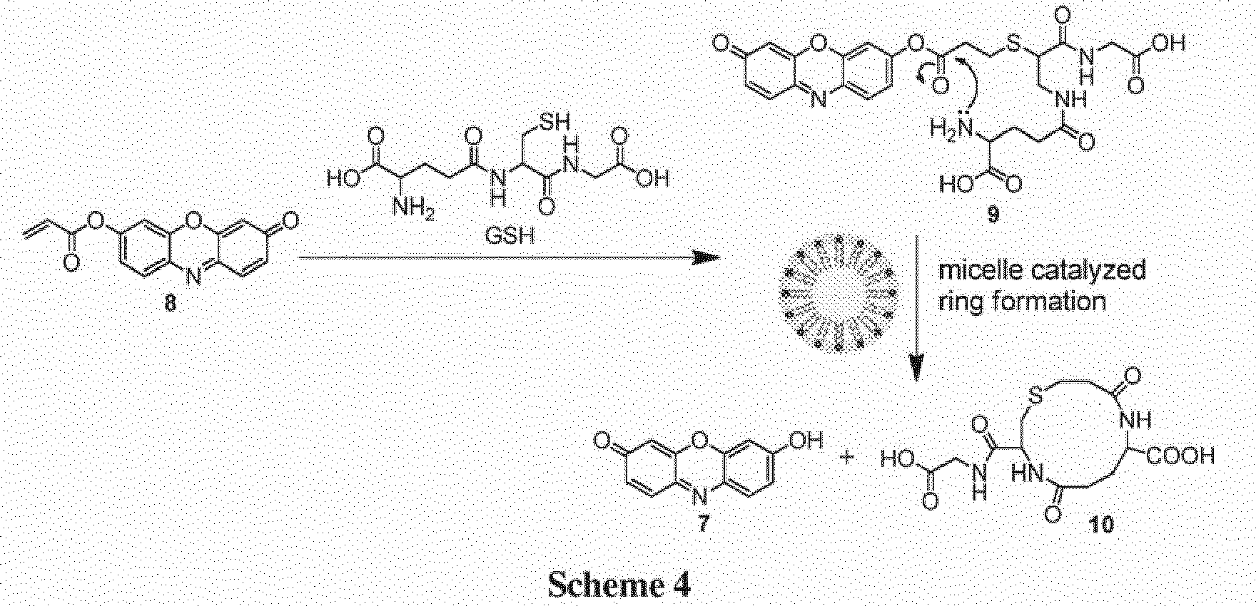
- Another cationic surfactant, benzyl-dimethyl-tridecyl-azanium chloride (BC), also produced selective detection of GSH with
probe 8. Cys also was detected when BC was included in the solution; however, GSH produced greater fluorescence. - Embodiments of the disclosed probes are suitable for selectively detecting compounds comprising a thiol group and an amino group. Exemplary compounds include the biological thiols Cys, Hcy, and GSH, A sample potentially comprising at least one thiol-containing compound comprising a thiol group and an amino group is combined with a solution comprising a probe according to general formula I. The sample may be a biological fluid, such as blood (e.g., whole blood or a blood product or component, such as plasma) or urine (or a urine product or component). The reaction is allowed to proceed for an effective period of time. An effective period of time is sufficient time for a condensation, cyclization, and elimination reaction to occur if the thiol-containing compound is present in the sample, thereby producing a change in the reaction mixture's absorbance spectrum, fluorescence emission spectrum, or both. In some embodiments, the effective period of time is ≤ 60 minutes, ≤ 30 minutes, ≤ 20 minutes, ≤ 10 minutes, or ≤ 5 minutes, such as 10 seconds to 30 minutes, 1-30 minutes, 1-25 minutes, 1-20 minutes, or 1-10 minutes. Presence of the thiol-containing compound is detected by i) comparing a color of the reaction mixture before the reaction to a color of the reaction mixture after the reaction with the compound, (ii) detecting a change in absorbance of the reaction mixture at one or more wavelengths after the reaction has proceeded for the effective period of time, (iii) comparing an absorbance spectrum of the reaction mixture at a first time after combining the sample and the probe to an absorbance spectrum of the reaction mixture after the reaction has proceeded for the effective period of time, (iv) detecting a change in emission of the reaction mixture at one or more wavelengths after the reaction has proceeded for the effective period of time, (v) comparing an emission spectrum of the reaction mixture at a first time after combining the sample and the compound to an emission spectrum of the reaction mixture after the reaction has proceeded for the effective period of time, (vi) or any combination thereof.. In the absence of a thiol-containing compound, no change is detected.
- A change in the probe's absorbance spectrum may be detected by obtaining an absorbance spectrum, by measuring absorbance at an appropriate wavelength, or by visually detecting a color change (i.e., with the naked eye). A change in the probe's fluorescence spectrum may be detected by obtaining a fluorescence emission spectrum, or by measuring fluorescence intensity at an appropriate wavelength. Absorbance and/or fluorescence changes over time may be measured.
- In certain embodiments, a concentration of a thiol-containing compound in a sample may be determined. For example, the concentration of Cys or GSH may be determined. A standard curve is be prepared by combining a probe according to formula I with solutions comprising known amounts of a thiol-containing compound. After an effective period of time, absorbance and/or fluorescence is measured at an appropriate wavelength. In some examples, when
probe 8 was used, the effective period of time was less than 1 minute, 2 minutes, 8 minutes, 10 minutes, 20 minutes, or 60 minutes, and fluorescence emission was measured at 585-590 nm (λex = 565 nm); an absorbance maximum was seen at 585-590 nm. In certain examples whenprobe 5 was used, the effective period of time was 25 minutes, and a fluorescence maximum was seen at 615-620 nm (λex = 550 nm). A sample potentially comprising the thiol then is combined with the probe under the same conditions (solution composition, pH, time, temperature), and absorbance and/or fluorescence then is measured and compared to the calibration curve to determine the thiol concentration in the sample. - In some embodiments, the sample is buffered to a pH of 5.5-8.0, such as a pH of 6.0-7.5. Fluorescence may develop more quickly at higher pH; however, selectivity may be enhanced at a lower pH. In certain embodiments, the sample was buffered to a pH of 6 when detecting GSH in biological fluids.
- In some examples, the sample comprises sulfhydryl-containing compounds other than Cys, Hcy, and/or GSH, and/or exhibits significant absorbance. fluorescence, and/or scatter in the visible wavelength region. In certain embodiments, the sample is a biological fluid, such as blood, a blood product or component (e.g., plasma), urine, or a urine product or component.
- In one embodiment, the sample comprises plasma, and the method further includes combining the plasma with a reducing agent (e.g., triphenylphosphine or TCEP (tris-(2-carboxyethyl)-phosphine)), and removing proteins from the plasma. Proteins may be removed, for example, by precipitation. The plasma may be diluted (e.g., to a concentration of 5-20%, such as to 10%) with a buffer. In one example, a 50 mM phosphate buffer, pH 6.0 was used. A probe according to general formula I is combined with the reduced, deproteinized plasma. Presence of a thiol is determined by detecting a change in the reaction mixture's absorbance spectrum, emission spectrum, or both.
- In another embodiment, the sample comprises whole blood. Whole blood is spotted onto absorbent paper and dried. Thiols are extracted from the dried blood spot. In one example, a phosphate buffer (100 mM, pH 6.0) was used to perform the extraction. The extract is reduced, e.g., with TCEP or triphenylphospine. Reduced thiols are isolated by fractionating the reduced extract, such as by gel filtration, to remove proteins and other high molecular weight molecules. In one example, a desalting column comprising a cross-linked dextran gel capable of fractionating globular proteins having a molecular weight of 1000-5000 Daltons (Sephadex® G-25, GE Healthcare) was used with 100 mM phosphate buffer as an eluent. An isolated fraction is combined with a probe according to general formula I. Presence of a thiol is determined by detecting a change in the reaction mixture's absorbance spectrum, emission spectrum, or both.
- In some embodiments. Cys is selectively detected by combining a sample potentially comprising Cys with a probe according to formula IA in the absence of a cationic surfactant. In certain embodiments, Cys is selectively detected with
probe 8. The reaction may be performed at pH of 6-7.5 for 1-60 minutes. The sample and/or probe may be provided in a solution comprising an anionic or non-ionic surfactant. For example. Triton® X-100 (polyethylene glycol tert-octylphenyl ether) may increase probe selectivity for Cys and/or may increase the rate at the reaction occurs, e.g., 10-15 minutes. Cys is detected by detecting a change in the reaction mixture's absorbance spectrum, emission spectrum, or both. Whenprobe 8 is used, fluorescence may be produced with an excitation wavelength of 565 nm. Fluorescence is detected by measuring fluorescence intensity at an emission wavelength of 580-590 nm, or by obtaining a fluorescence emission spectrum. In some examples, Cys can be detected visually by observing development of a pink color in the reaction mixture. - Alternatively, Cys can be selectively detected by combining a sample potentially comprising Cys with a probe according to formula II. In certain embodiments,
probe 5 is used to selectively detect Cys. The reaction may be performed at a pH of 6-7.5 for 1-30 minutes. The sample and/or probe may be provided in a solution comprising a surfactant, e.g., a cationic surfactant such as CTAB. Cys is detected by detecting a change in the reaction mixture's absorbance spectrum, emission spectrum, or both. Whenprobe 5 is used, fluorescence may be produced with an excitation wavelength of 550 nm. Fluorescence is detected by measuring fluorescence intensity at a wavelength of 615-635 nm, or by obtaining a fluorescence emission spectrum. In some examples, Cys can be detected visually by observing development of a pink color in the reaction mixture. - GSH can be selectively detected by combining a sample potentially comprising GSH with a probe according to formula IA in the presence of a cationic surfactant. In some embodiments, GSH is selectively detected with
probe 8. Suitable cationic surfactants include CTAB and BC. The reaction may be performed at a pH of 6-7.5 for 0-30 minutes. GSH also may be selectively detected byprobe 8 in certain solvents, such as a polar aprotic solvent. For example,probe 8 selectively detected GSH in a 1:1 solution of DMSO:H2O (pH 7.4). GSH is detected by detecting a change in the reaction mixture's absorbance spectrum, emission spectrum, or both. Whenprobe 8 is used, fluorescence may be produced with an excitation wavelength of 565 nm. Fluorescence is detected by measuring fluorescence intensity at an emission wavelength of 580-590 nm, or by obtaining a fluorescence emission spectrum. In some embodiments, GSH can be detected visually by observing development of a pink color in the solution. - In some embodiments, a probe having a structure according to general formula IA is synthesized in a one-step reaction as shown below in
Scheme 5. Resorufin, or a resorufin analog, is combined with an acryloyl salt, such as an acryloyl chloride, in the presence of dichloromethane and triethylamine to produce the acrylate resorufin analog. R1 and R3-R8 are as previously defined.
- In certain embodiments, a probe having a structure according to general formula II is synthesized as shown below in
Scheme 6. A hydroxybenzophenone derivative is prepared by reacting a 1,3-dihydroxyphenyl derivative with a 2-benzofuran-1,3-dione derivative with aluminum chloride. The hydroxybenzophenone derivative and a 1,8-naphthalene derivative are condensed by reaction with methanesulfonic acid to produce a seminaphthofluorescein (SNF) derivative. The SNF derivative is then reacted with an acryloyl salt in the presence of triethylamine and dichloromethane to produce the acrylate SNF derivative. R1, R3-R6, R10, and R12-R17 are as previously defined.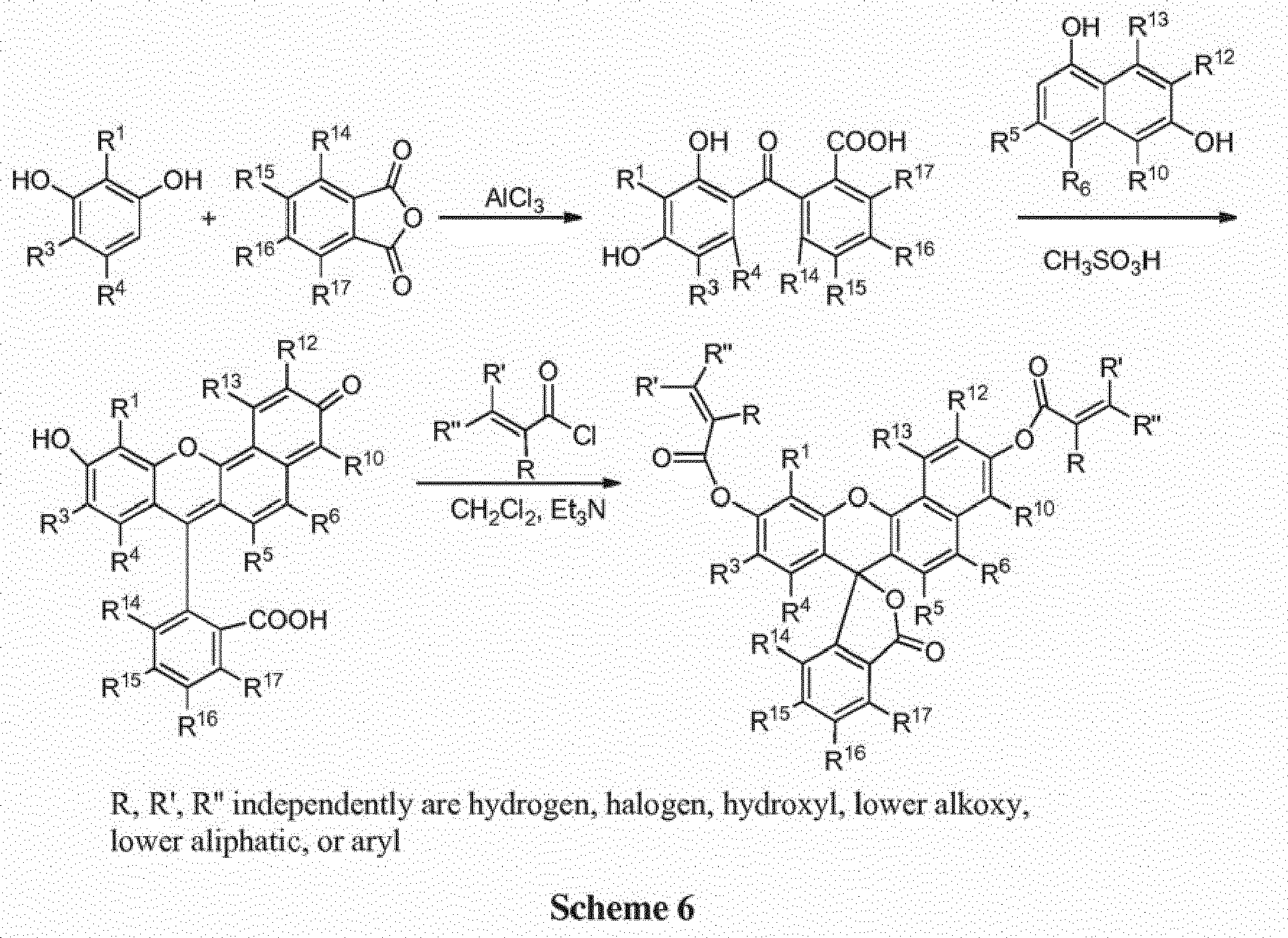
- Kits are also a feature of this disclosure. Embodiments of the kits include at least one probe as disclosed herein, wherein the probe is suitable for selectively detecting one or more thiol-containing compounds, particularly one or more compounds including a thiol group and an amino group (e.g., cysteine, homocysteine, glutathione). In some embodiments, the kit further includes a buffer solution at physiologic pH. In certain embodiments, the buffer is a phosphate buffer at pH 6-7.5, such as a 20-50 mM phosphate or HEPES buffer at pH 7.4 or a 50-100 mM phosphate buffer at pH 6.0. The probe may be dissolved in the buffer, or the probe may be included in a dry form and the user can combine the probe and the buffer solution at or before the time of use. In some embodiments, the buffer solution further includes a surfactant. The kits also may include one or more containers, such as a disposable test tube or cuvette, in which the detection can be performed. In some embodiments, an amount of the probe effective to undergo a detectable change in color and/or a detectable change in the fluorescence emission spectrum when reacted with the at least one compound is premeasured into the disposable containers. The kits may further include instructions for performing the detection. In some embodiments, the kits include control samples of thiol-containing compounds, e.g., cysteine, homocysteine, and/or glutathione. Typically the control samples are provided in solid form.
- Materials and Instruments: All chemicals were purchased from Sigma-Aldrich or Acros and used without further purification. In all experiments enantiomerically pure natural amino acids were used except for Hcy, which was used as the racemate. 1H-NMR and 13C-NMR spectra were recorded on a Bruker AMX-400 NMR spectrometer, using TMS as an internal standard. ESI-HRMS (high-resolution mass spectrometry) spectra were obtained on a Thermo Electron LTQ Orbitrap hybrid mass spectrometer. UV-visible spectra were collected on a
Cary 50 UV-Vis spectrophotometer. Fluorescence spectra were collected on a Cary Eclipse (Varian, Inc.) fluorescence spectrophotometer with slit widths set at 5 nm for both excitation and emission, respectively. The high voltage of the fluorescence spectrophotometer was set at 500 V forprobe 8. The pH measurements were carried out with an Orion 410A pH meter. -

- SNF was synthesized in two steps according to a published method (Chang et at., Proc. Natl. Acad. Sci. USA, 2004, 101, 1129-1134). 1H NMR (CDCl3, 400MHz), δ (ppm): 8.44 (d, 1H, J = 9.2 Hz ), 8.04 (d, 1H, J = 7.2 Hz), 7.74 (m, 2H), 7.30 (d, 1H, J = 9.2 Hz), 7.26 (dd, 1H, J1 = 2.4 Hz, J 2= 9.2 Hz), 7.19 (d, 1H, J = 7.6 Hz), 7.12 (s, 1H), 6.62 (m, 2H), 6.51 (d, 1H. J = 8.8 Hz). 2.51(s, 3H). ESI-FTMS m/z = 395.0923 [M-H]-, calc. 395.0919 for C25H15O5.
- To a solution of SNF (120 mg, 0.30 mmol) and triethylamine (2 equiv) in 10 mL of anhydrous CH2Cl2, acryloyl chloride (2.5 eq, mixed with 5 mL of CH2Cl2) was added dropwise at 0°C. After stirring at this temperature for 90 minutes, the resulting mixture was allowed to warm to room temperature and stirred overnight. The mixture was then diluted with CH2Cl2 (25 mL), washed with water (12 mL ×3) and dried over anhydrous Na2SO4. The solvent was removed by evaporation to get crude product as yellow solids, which was then purified by flash column chromatography (silica gel, CHCl3/EtAC 100:6) to get 4 as light yellow solids (99 mg, 65% yield). 1H NMR (CDCl3, 400MHz), δ (ppm): 8.58 (d, 1H, J =9.2 Hz), 8.07 (d, 1H, J = 8.0 Hz), 7.66 (m. 3H), 7.46 (m, 2H), 7.16 (d, 1H, J = 6.4 Hz), 6.83 (m. 2H), 6.76 (d, 1H, J = 8.4 Hz), 6.70 (br s, 1H), 6.66 (br s, 1H), 6.41 (dd, 1H, J 1 =1.6 Hz, J 2 = 10.4 Hz), 6.37 (dd, 1H, J 1 =1.6 Hz, J 2 = 10.4 Hz), 6.08 (m, 2H), 2.51 (s,3H). 13C NMR (CDCl3, 100 MHz), δ 169.52, 164.57, 164.05, 153.67, 150.31, 150.15, 149.84, 146.93, 135.38, 135.27, 133.49, 133.22, 130.09, 127.90, 127.49, 125.31, 124.83, 124.28, 123.96, 123.60, 122.09, 121.99, 119.45, 118.88, 118.15, 116.49, 112.42, 82.92, 9.87. ESI-FTMS m/z = 505.1308 [M+H]+, calc. 505.1287 for C31H21O7.
- Probe 5 (10 µM) was combined with 1 equivalent of Cys, Hcy, GSH, leucine, proline, arginine, histidine, valine, methionine, threonine, glutamine, alanine, aspartic acid, norleucine, isoleucine, lysine, tryptophan, tyrosine, phenylalanine, cystine, or homocystine for 25 minutes in 1.0 mM CTAB buffered at pH 7.4 (HEPES buffer. 20 mM). Fluorescence spectra were obtained at an excitation wavelength of 550 nm. As shown in
FIG. 2A , only Cys resulted in significant fluorescence. A minor amount of fluorescence was obtained following reaction with Hcy and GSH (FIG. 2B ), but no significant fluorescence was obtained from the other evaluated compounds.FIG. 3 is a photograph illustrating the visible color change obtained whenprobe 5 reacted with Cys. No color change was observed with Hcy or GSH. -

-
Probe 8 was synthesized in a one-step reaction ofresorufin 7 with acryloyl chloride (Scheme 6). To a solution of resorufin (7, 220 mg) and triethylamine (1.5 equiv) in 15 mL of anhydrous CH2Cl2, acryloyl chloride (2.0 equiv in 5 mL of CH2Cl2) was added dropwise at 0 °C. After stirring at this temperature for 60 minutes, the resulting mixture was allowed to cool to room temperature and stirred overnight. The mixture was diluted with CH2Cl2 (20 mL), washed with H2O (10 mL × 3) and dried over anhydrous Na2SO4. The solvent was removed by evaporation to afford an orange solid (172 mg, 70% yield). 1H NMR (CDCl3, 400 MHz), δ (ppm): 7.92 (d, 1H, J = 8.7 Hz), 7.59 (d, 1H, J = 9.8 Hz), 7.49 (s, 1H), 7.33 (dd, 1H, J 1 = 2.4 Hz, J 2 = 2.5 Hz), 6.83 (dd, 1H, J 1 = 2.0 Hz, J 2 = 2.0 Hz), 6.63 (d, 1H, J = 17.2 Hz), 6.49 (m, 1H), 6.24 (s, 1H), 6.22 (d. 1H, J = 1.0 Hz). 13C NMR (CDCl3, 100 MHz). δ 186.48, 163.84, 153.24, 148.87. 147.78, 143.55, 134.85, 134.26, 134.15. 131.22, 131.16, 127.18, 118.82, 109.94, 105.86. ESI-MS m/z = 268.0833 [M+H]+, calc. 268.0810 for C15H10NO4. - Probe 8 (2.5 µM) was combined with thiols (2 equivalents) in phosphate buffer (pH. 7.4, 50 mM). After 20 minutes, absorption and fluorescence (λex = 565 nm) spectra were obtained. Fluorescence changes (λem = 587 nm) over time also were monitored.
- In the phosphate buffer,
probe 8 showed excellent selectivity towards Cys due to the kinetically favored 7-membered ring formation. Upon mixing Cys withprobe 8, the conjugate addition product 8a is formed, which undergoes a rapid cyclization reaction to produce 3a while releasing the free resorufin (Scheme 3, above). A significant color change and fluorescence enhancement was observed in response to Cys (FIGS. 4, 5 ). In the case of Hey, because it has an additional methylene group in its side chain, a kinetically less favored 8-membered ring forms. With GSH, 1,4-addition of thiols to the α,β-unsaturated carbonyl moieties ofprobe 8 can occur readily; however, the ensuing intramolecular cyclization similar to that of Cys does not proceed in phosphate buffer. No significant color formation or fluorescence response was seen in the case of Hcy or GSH under these conditions (FIGS. 4, 5 ). - The time course of the fluorescence assay is shown in
FIG. 5 (inset). It can be seen that the fluorescence upon reaction with Cys increased with time and reached a plateau after about 90 minutes, whereas for Hcy and GSH the reactions were significantly slower. - The sensitivity and linearity of the response of
probe 8 towards Cys was also investigated. Various concentrations (0-3 µM) of Cys were combined with probe 8 (2.5 µM) in at pH 7.4 (phosphate buffer, 50 mM), and the reaction was allowed to proceed for 60 minutes.FIG. 6 shows the fluorescence spectra (λex = 565 nm) obtained 60 minutes after Cys andprobe 8 were combined.FIG. 7 is a graph illustrating the linear increase (correlation coefficient of 0.995) in fluorescence intensity at 593 nm as a function of Cys concentration. Linear response with submicromolar sensitivity was observed, demonstrating that concentrations as low as 0.2 µM Cys can be detected. - High-resolution mass spectrometry demonstrated the formation of compound 3a (3-carboxy-5-oxoperhydro-1,4-thiazepine) and release of
resorufin 7 when probe 8 (20 µM) was combined with Cys (20 µM) in 1:1 MeOH:H2O (FIG. 8 ), The reaction time was 3 hours. Similarly, compound 3b was formed and resorufin was released when probe 8 (20 µM) was combined with Hcy (20 µM) in 1: 1 MeOH:H2O (reaction time = 3 hours) (FIG. 9 ). - To demonstrate the necessity of a free NH2 group, experiments using N-acetyl cysteine (NAC) were performed. Cys and NAC (12.5 µM in phosphate buffer (50 mM, pH=7.4) were added to solutions containing probe 8 (2.5 µM). Absorbance and emission spectra were obtained over a time course of 90 minutes. No significant absorbance or emission was obtained from the reactions including NAC. The results showed that the free NH2 group was required for an intramolecular cyclization and addition/elimination reaction to occur.
- Cys, Hcy, or GSH (2 equivalents) was added to probe 8 (10 µM) in 4 mL phosphate buffer (pH 7.4, 50 mM). CTAB was then added to form a solution containing 2 mM CTAB. A strong pink color formed in the GSH-containing solution immediately upon addition of CTAB.
FIG. 10 shows absorbance spectra ofprobe 8 combined with the thiols. Inset is a color photograph illustrating selective detection of GSH; the photograph was taken immediately upon the addition of CTAB. A strong pink color was obtained immediately upon addition of CTAB in the presence of GSH (inset). Hcy and Cys did not exhibit significant changes under the same conditions. These results indicate that a probe 8-CTAB system can serve as a selective visual inspection dosimeter for GSH. - Fluorescence spectra (λex = 565 nm; λem = 587 nm) also were obtained immediately after addition of CTAB (
FIG. 11 ). Significant fluorescence was obtained only with GSH. High resolution mass spectrometry confirmed formation of a 12-membered ring in the CTAB medium. A major peak corresponding to the ring was seen at m/z 360.08 (FIG. 1 ). - The reaction of probe 8 (2.5 µM) with 2 equivalents Cys. Hcy, or GSH in 2.0 mM CTAB buffered at pH 7.4 was monitored over time by fluorescence spectroscopy - λex = 565 nm; λem = 587 nm. As shown in the inset of
FIG. 11 , immediate fluorescence was seen with GSH, and the fluorescence increased rapidly and reached a plateau in less than 2 minutes. In contrast, Cys and Hcy initially produced very little fluorescence; the fluorescence increased gradually over time, but remained much lower than the fluorescence obtained with GSH. - The thiol selectivity of
probe 8 in the presence of various surfactants was evaluated. Probe 8 (2.5 µM) was combined with 2 equivalents of Cys, Hcy, or GSH in phosphate-buffered media (50 mM, pH = 7.4) with a surfactant: (a) sodium dodecyl sulfate (SDS). 10 mM; (b) Triton® X-100, 0.3 mM; (c) benzyl-dimethyl-tridecyl-azanium chloride (benzalkonium chloride, BC), 0.05 mM, or (d) CTAB, 2 mM.

FIGS. 12-15 . As shown inFIGS. 12 and 13 , Cys was preferentially detected byprobe 8 in the presence of SDS and Triton X-100. In the presence of SDS (FIG. 12 ), fluorescence increased slowly over the measured timeframe of 50 minutes. In the presence of Triton® X-100 (FIG. 13 ), fluorescence increased much more quickly, and greater selectivity towards Cys was observed. When BC and CTAB were used, maximum fluorescence was achieved within 1-2 minutes. As seen inFIG. 14 , both Cys and GSH were detected in the presence of BC, with GSH producing greater fluorescence. In the presence of CTAB (FIG. 15 ), much greater selectivity toward GSH was observed.FIGS. 16 and 17 are fluorescence spectra ofprobe 8 in the presence of CTAB (FIG. 16 ) and Triton X-100 (FIG. 17 ), again showing thatprobe 8 is selective for GSH in the presence of the cationic surfactant CTAB, but selective for Cys in the presence of Triton X-100. - The results show that
probe 8 is an indicator for GSH in the presence of cationic surfactants such as BC and CTAB. Without wishing to be bound by any particular theory, favorable Coulombic attractions between cationic micelles and GSH may play a role in the selectivity ofprobe 8 towards GSH in the presence of cationic surfactants. Negatively-charged SDS suppressed the response ofprobe 8 towards all three thiols; while the non-ionic surfactant Triton X-100 enhanced the selectivity towards Cys and resulted in zero response for Hey or GSH. The results with GSH are summarized inFIG. 18 , which shows the fluorescence spectra obtained immediately upon addition of the surfactants. Inset is graph showing time-dependent fluorescence changes (λem = 587 nm) of the same system. - In another assay, probe 8 (2.5 µM) was combined with 2 equivalents Cys, Hcy, GSH, leucine, proline, arginine, histidine, valine, methionine, threonine, glutamine, alanine, aspartic acid, norleucine, isoleucine, lysine, tryptophan, tyrosine, phenylalanine, cystine, or homocystine in 2.0 mM CTAB buffered at pH 7.4 (phosphate buffer. 50 mM). Fluorescence emission spectra (λex = 565 nm) were obtained immediately after addition of the thiols and other amino acids. As shown in
FIG. 19 , only GSH resulted in significant fluorescence. To further test the selectivity ofprobe 8 toward GSH, competition experiments were conducted with excess of other amino acids added to GSH, and no significant fluorescence intensity difference was observed in comparison to solutions containing GSH only. - Since surfactants can mimic organic environments, selectivity in DMSO:H2O (1:1, buffered at pH 7.4) was studied. Cys, Hcy, and GSH (2 equiv) were added to probe 8 (2.5 µM) in DMSO:H2O (1:1, buffered at pH 7.4), and fluorescence was measured over time. Selectivity of
probe 8 toward GSH was demonstrated (FIG. 20 ). - The CTAB-mediated response of
probe 8 towards GSH in the presence of Cys was evaluated. A mixture of Cys (2 µM) and increasing concentrations of GSH (0-2 µM) at pH 7.4 (phosphate buffer, 50 mM) were added to probe 8 (1.5 µM), and fluorescence spectra (λex = 565 nm) were obtained after 2 minutes. As shown inFIG. 21 , there was no significant difference in the fluorescence spectrum ofprobe 8 when Cys was present. However, fluorescence increased greatly when GSH or GSH + Cys were reacted withprobe 8. Thus,probe 8 is selective for GSH even in the presence of Cys.FIG. 22 shows a linear relationship between fluorescence intensity and GSH concentration with a correlation coefficient of 0.990, as well as a linear relationship with a mixture of GSH and Cys having a correlation coefficient of 0.9894. - The effect of GSH concentration was evaluated by combining various concentrations (0-6 µM) of GSH with probe 8 (2.5 µM) in 2.0 mM CTAB media buffered at pH 7.4 (phosphate buffer, 50 mM). The GSH concentrations were within the range of GSH in human plasma.
FIG. 23 shows the fluorescence spectra (λex = 565 nm) obtained immediately upon addition of CTAB.FIG. 23 inset is a graph illustrating submicromolar sensitivity and a linear relationship (correlation coefficient of 0.998) between fluorescence intensity (λex = 565 nm, λem = 587 nm) and GSH concentration. Measurements were taken immediately after addition of CTAB. These results demonstrate that the probe 8-CTAB system functions well within the range of typical GSH concentrations in human plasma. - While the probe 8-CTAB-GSH signal was high at pH 7.4 (see, e.g., Example 6), it is known that acrylate esters are susceptible to hydrolysis at high pH and that GSH is also relatively unstable under the same conditions (half-life of 9 hours at pH 7.4 compared to 16 hours at pH 6.5; Stevens et al., Biochem. Educ., 1983, 11, 70).
- The spectral behavior of the probe 8-CTAB system was investigated over a wide range of pH values (pH 5.5 to pH 8), and its response to the various thiols was studied at lower pH values. Probe 8 (2.5 µM) was added to CTAB media (2 mM) buffered at pH 5.5 to 8, and the absorbance was monitored over time at a wavelength of 580 nm (
FIG. 24 ).Probe 8 showed the greatest background signal at the higher pH values investigated. The probe 8-CTAB system was most stable at lower pH values. The absorbance spectra ofprobe 8 in CTAB media at pH 6.0, 7.0, and 8.0 after 1 minute and 10 minutes are shown inFIGS. 25 and26 . respectively. - The reaction with thiols at
pH 6 was evaluated. Absorption spectra of probe 8 (2.5 µM) upon addition of thiols (2 equivalents) in 2.0 mM CTAB media buffered at pH 6 (phosphate buffer. 50 mM) were obtained after 20 minutes (FIG. 27 ). Fluorescence spectra (λex = 565 nm) also were obtained (FIG. 28 ). The inset inFIG. 28 shows the time-dependent fluorescence changes (λem = 587 nm); the fluorescence increase was slightly less rapid as compared to pH 7.4 (see, e.g.,FIG. 15 ); however, the selectivity towards GSH was further enhanced through the complete elimination of any residual Cys and Hcy response. ThuspH 6 was chosen for further application studies in plasma. - Human plasma (0.5 mL) was reduced using HCl (0.2 M, 40 µL) in the presence of triphenylphosphine (0.1 M, 80 µL) as a catalyst for 15 minutes at room temperature. Proteins present in the sample after reduction were precipitated by addition of acetonitrile (0.5 mL), followed by centrifugation (4000 rpm) of the sample for 20 minutes. The supernatant liquid was then added to a solution of probe 5 (10 µM) in pH 7.4 HEPES buffer solution (0.1 M, 5 mL) in the presence of 1.0 mM CTAB. Fluorescence spectra (λex = 550 nm) were obtained after 25 minutes.
- As shown in
FIG. 29 , the fluorescence emission showed a significant increase with addition of reduced plasma. However, the fluorescence emission ofsolutions including probe 5 showed no obvious fluorescence increase when controls of either triphenylphosphine alone or deproteinized plasma (without reducing agent) were added, demonstrating that the fluorescence increase was due to the reaction ofprobe 5 with Cys. The amount of Cys in the plasma sample as determined by the standard addition method to be 172.8 ± 8.7 µM (n = 3), which is well within the reported Cys concentration range (135.8 - 266.5 µM) for human plasma samples from healthy individuals. The results demonstrate thatprobe 5 may be useful for quantitative detection of Cys in biological samples. - Plasma proteins were precipitated using MeCN (two thirds of the reconstitution volume) and removed by centrifugation at 4,000 rpm for 30 min. The supernatant liquid was diluted to provide 10% deproteinized plasma, and added to a solution of probe 8 (1-1.5 µM) in phosphate buffer (
pH - The fluorescence response of replicate (n = 3) samples and GSH-spiked samples were monitored as above and the GSH content in the plasma sample was determined from the regression equation of a standard calibration curve (
FIG. 30 ,probe 8 = 1 µM). Measurements were taken 8 minutes after mixing. Fluorescence emission spectra (λex = 565 nm) also were obtained 8 minutes after mixing (FIG. 31 .probe 8 = 1 µM). Using the calibration curve, the GSH content of the plasma sample was determined to be 3.24 ± 0.14 µM, which is well within the reported GSH concentration range for human plasma samples from healthy individuals. Recoveries of the known spiked amounts of GSH were between 99.2% and 102.3% with a satisfactory precision (Table 1). As shown in Table 1, the relative standard deviation for 3 replicate samples was less than 6%. These results revealed the potential applicability and reliability of usingprobe 8 in quantitative detection of GSH in human plasma. - Sensitivity and selectivity were evaluated by obtaining fluorescence spectra of plasma alone, plasma + 0.5 µM GSH, plasma + 2 µM GSH, and plasma + 2 µM GSH and 2 µM Cys (
probe 8 = 1.5 µM). Measurements were taken 8 minutes after mixing. The results are shown inFIG. 32 . Notably, addition of Cys did not exhibit significant interference with the GSH determination.Table 1 Determination of GSH content in 10% deproteinized human plasma samples. Sample GSH Spiked (µM) GSH Measured (µM) Recovery (%) RSD (%) 10 % Plasma 0 0.324 4.42 10% Plasma + GSH 0.3 0.631 102.3 4.90 1 0% Plasma + GSH 0.6 0.919 99.2 4.24 10% Plasma + GSH 0.9 1.224 100.0 5.88 - Pig blood (10, 20, and 40 µL) was spotted on Whatman 903™ filter paper, and dried at room temperature for 3-4 hours. Oxidized glutathione was extracted from the dried blood spot (DBS) with 2.5 mL phosphate buffer (100 mM, pH 7.4). The extract was reduced with TCEP (tris-(2-carboxyethyl)-phosphine) (35 mM) to reduce disulfides. Reduced GSH was isolated by gel filtration of the reduced extract on a PD-10 desalting column (Sephadex® G-25 (cross-linked dextran gel), GE Healthcare) using phosphate buffer (100 mM, pH 7.4) as eluent. Fractions of about 1.5-2.5 mL were collected in 4 mL vials. CTAB (2.0 mM) and probe 8 (2.5 µM) were added to the vials. The solution was buffered at pH 6.0 (phosphate buffer, 50 mM).
- GSH was detected in the fourth and fifth eluted fractions by development of a pink color after addition of CTAB and
probe 8. Absorption and fluorescence spectra (λex = 565 nm) were obtained from the fifth fraction (FIGS. 33 and 34, respectively). Measurements were taken immediately after mixing.FIG. 33 is a fluorescence spectrum demonstrating thatprobe 8 detected GSH in the extract obtained 10 µL dried pig blood. - In view of the many possible embodiments to which the principles of the disclosed invention may be applied, it should be recognized that the illustrated embodiments are only preferred examples of the invention and should not be taken as limiting the scope of the invention. Rather, the scope of the invention is defined by the following claims.
Claims (15)
- A compound having a chemical structure according to general formula I:
 where each bond depicted as "------" is a single or double bond as needed to satisfy valence requirements;R1, R3-R6 and R8 independently are H, hydroxyl, thiol, C1-C10 alkyl, carboxyalkyl, amino, alkoxy, or halogen;R2 is an α,β-unsaturated aliphatic ester;X1 is O, CH2, S, or NH; and(i) R7 is O and X2 is N, or(ii) R7 and R8 together with the ring to which they are attached form a naphthyl ring and X2 is CR9 where R9 is phenyl.
 - The compound of claim 1, wherein when X1 is O, then R2 is an acrylate ester, X2 is N, R7 is O, and R1, R3-R6, and R8 independently are H or lower alkyl, or
when X1 is O or S, then R7 and R8 together form a phenyl ring. - The compound of claim 1 where X1 is O or S, and R1 and R3-R6 independently are H or C1-C10 alkyl; or
where X1 is O, R7 and R8 together form a phenyl ring, X2 is CR9, R1 is C1-C10 alkyl or H, R2 is an acrylate ester, and R3-R6 are H. - The compound of claim 1 having the structure:

- The compound of claim 1, wherein R7 and R8 together with the ring to which they are attached form a naphthyl ring and X2 is CR9, the compound having a chemical structure according to general formula II:
 where "------" indicates a single or double bond as needed to satisfy valence requirements and "------" indicates an optional single bond;R2 and R11 independently are an α,β-unsaturated aliphatic ester;R10, R12, and R13 independently are H, hydroxyl, thiol, C1-C10 alkyl, carboxyalkyl, amino, alkoxy, or halogen;R14-R17 independently are H, alkyl, acyl, carboxyl, nitro, amino, alkyl amino, or -SO3H; andR18 is H, hydroxyl, lower alkyl, lower alkoxy, -SO3H or -COOR19 where R19 is H or lower alkyl and the bond depicted as "------" in ring B is a double bond, or R18 is one or more atoms forming a ring system with rings B and E and the bond depicted as "------" in ring B is a single bond.
 - The compound of claim 5 where each of R2 and R11 is an acrylate ester, and R18 is -COOH or R18 is -C(O)O- and forms a ring system with rings B and E.
- The compound of claim 6 where R1, R3-R6, R10 and R12-R17 independently are H or C1-C10 alkyl.
- The compound of claim 7 where X1 is O.
- The compound of claim 5 having the structure:

- A method for selectively detecting a compound comprising a thiol group and an amino group, comprising:combining a sample potentially comprising at least one compound comprising a thiol group and an amino group with a solution comprising a probe having a structure according to any one of claims 1-9 to produce a reaction mixture;allowing a reaction between the sample and the probe to proceed for an effective period of time to produce a detectable change in the reaction mixture's absorbance spectrum, emission spectrum, or both, where the change indicates that the compound is present; anddetecting the change,and optionally the sample comprises blood, a blood product, a blood component, urine, a urine product, or a urine component.
- The method of claim 10, further comprising (i) or (ii), wherein (i) is:obtaining plasma;reducing the plasma by addition of a reducing agent;precipitating proteins in the plasma;separating precipitated proteins from the plasma to form the sample; andsubsequently combining the sample with the solution comprising the probe;and (ii) is:obtaining a dried blood spot prepared by spotting whole blood onto an absorbent material and allowing the blood to dry;extracting the dried blood spot with a solvent to produce an extract;reducing the extract by addition of a reducing agent to form a reduced extract;fractionating the reduced extract to obtain the sample; andsubsequently combining the sample with the solution comprising the probe.
- The method of claim 10 where detecting the change comprises (i) comparing a color of the reaction mixture before the reaction to a color of the reaction mixture after the reaction, (ii) detecting a change in absorbance of the reaction mixture at one or more wavelengths after the reaction has proceeded for the effective period of time, (iii) comparing an absorbance spectrum of the reaction mixture at a first time after combining the sample and the probe to an absorbance spectrum of the reaction mixture after the reaction has proceeded for the effective period of time, (iv) detecting a change in emission of the reaction mixture at one or more wavelengths after the reaction has proceeded for the effective period of time, (v) comparing an emission spectrum of the reaction mixture at a first time after combining the sample and the probe to an emission spectrum of the solution after the reaction has proceeded for the effective period of time, (vi) or any combination thereof.
- The method of claim 10 where the probe is

- The method of claim 10 where the at least one compound is cysteine, and the probe is

- A kit for detecting at least one compound comprising a thiol group and an amino group, comprising at least one probe according to any one of claims 1-9, preferably where the probe is the compound of claim 4 or claim 9, or a combination thereof.
Applications Claiming Priority (2)
| Application Number | Priority Date | Filing Date | Title |
|---|---|---|---|
| US201261602581P | 2012-02-23 | 2012-02-23 | |
| PCT/US2013/027482 WO2013126816A1 (en) | 2012-02-23 | 2013-02-22 | Selective detection of thiols |
Publications (3)
| Publication Number | Publication Date |
|---|---|
| EP2817315A1 EP2817315A1 (en) | 2014-12-31 |
| EP2817315A4 EP2817315A4 (en) | 2015-10-07 |
| EP2817315B1 true EP2817315B1 (en) | 2018-09-19 |
Family
ID=49006272
Family Applications (1)
| Application Number | Title | Priority Date | Filing Date |
|---|---|---|---|
| EP13752087.0A Not-in-force EP2817315B1 (en) | 2012-02-23 | 2013-02-22 | Selective detection of thiols |
Country Status (5)
| Country | Link |
|---|---|
| US (1) | US9506929B2 (en) |
| EP (1) | EP2817315B1 (en) |
| JP (1) | JP6196988B2 (en) |
| RU (1) | RU2014138047A (en) |
| WO (1) | WO2013126816A1 (en) |
Families Citing this family (16)
| Publication number | Priority date | Publication date | Assignee | Title |
|---|---|---|---|---|
| CN104673278A (en) * | 2015-02-15 | 2015-06-03 | 浙江理工大学 | Fluorescence probe for detecting glutathione as well as preparation method and use method of fluorescence probe |
| WO2016136328A1 (en) * | 2015-02-27 | 2016-09-01 | 国立大学法人 東京大学 | Super-resolution fluorescent imaging probe |
| KR101712294B1 (en) * | 2015-12-30 | 2017-03-03 | 경북대학교 산학협력단 | Sensor for detecting of biothiol |
| CN105820810B (en) * | 2016-04-27 | 2018-03-30 | 中南大学 | A kind of synthesis and application for identifying cysteine and homocysteine novel fluorescence probe |
| US11191850B2 (en) | 2016-05-23 | 2021-12-07 | Portland State University | Molecular probes for detection and imaging of pancreatic cancer |
| WO2018200592A1 (en) | 2017-04-24 | 2018-11-01 | Regents Of The University Of California | Biosynthesis of bioorthogonal amino acids |
| CN106995451B (en) * | 2017-04-26 | 2019-03-15 | 许昌学院 | A kind of reactive cysteine probe and preparation method thereof |
| CN107141303B (en) * | 2017-04-26 | 2019-03-15 | 许昌学院 | A kind of reactive thiol compound probe and preparation method thereof |
| CN107556303B (en) * | 2017-09-30 | 2021-08-20 | 潍坊医学院 | A kind of compound, its synthetic method and the test kit made with this compound and its application in the detection of glutathione |
| WO2020028396A1 (en) | 2018-07-30 | 2020-02-06 | Georgetown University | Chirality sensing with molecular click chemistry probes |
| WO2020056012A1 (en) | 2018-09-11 | 2020-03-19 | Georgetown University | Quantitative auxiliary-free chirality sensing with a metal probe |
| US20220119293A1 (en) | 2019-05-28 | 2022-04-21 | Nouryon Chemicals International B.V. | Method of controlling scale in aqueous systems |
| EP4247899A1 (en) * | 2020-11-20 | 2023-09-27 | Nouryon Chemicals International B.V. | Fluorescent polymers and solutions thereof for scale control in aqueous systems |
| CN113025324B (en) * | 2021-03-17 | 2022-06-14 | 辽宁大学 | Dual excitation dual emission fluorescent probe CQDs-O-NBD and its application |
| CN113817010B (en) * | 2021-08-19 | 2023-03-14 | 西北农林科技大学 | Water-soluble multifunctional glycosyl fluorescent probe and application thereof |
| CN118307493A (en) * | 2024-04-03 | 2024-07-09 | 常州大学 | A resorufin fluorescent probe based on cinnamoyl chloride and its preparation method and application |
Family Cites Families (17)
| Publication number | Priority date | Publication date | Assignee | Title |
|---|---|---|---|---|
| JPS4418580Y1 (en) * | 1966-02-24 | 1969-08-11 | ||
| GB1447583A (en) * | 1974-02-04 | 1976-08-25 | Ici Ltd | Xanthene derivatives |
| CA1340759C (en) | 1988-07-01 | 1999-09-21 | James P. Albarella | Isobenzothiazolone chromogenic thiol indicators and their derivatives |
| US5227487A (en) | 1990-04-16 | 1993-07-13 | Molecular Probes, Inc. | Certain tricyclic and pentacyclic-hetero nitrogen rhodol dyes |
| US5750409A (en) | 1991-11-18 | 1998-05-12 | Boehringer Mannheim Gmbh | Pentacyclic compounds and their use as absorption or fluorescent dyes |
| US6534316B2 (en) | 2001-02-05 | 2003-03-18 | Board Of Supervisors Of Louisiana State University And Agricultural And Mechanical College | Colorimetric and fluorimetric analysis of carbohydrates |
| EP2298312B1 (en) | 2003-10-31 | 2018-09-26 | Molecular Probes Inc. | Fluorinated resorufin compounds and their application in detecting hydrogen peroxide |
| US9201075B2 (en) | 2004-04-30 | 2015-12-01 | Board Of Supervisors Of Louisiana State University And Agricultural And Mechanical College | Colorimetric and fluorometric determination of homocysteine and cysteine |
| KR100645357B1 (en) * | 2005-08-22 | 2006-11-14 | 심현호 | Fluorescent labeling substance having a double bond ester group, a method for labeling and detecting the same |
| US20070220621A1 (en) | 2005-10-31 | 2007-09-20 | Clarke Michael F | Genetic characterization and prognostic significance of cancer stem cells in cancer |
| JP4878932B2 (en) * | 2006-06-23 | 2012-02-15 | 株式会社リコー | Acrylic ester and hydroxy compound, (co) polymer of acrylic ester, electrophotographic photosensitive member using the same, image forming method, image forming apparatus, and process cartridge for image forming apparatus |
| US8350064B2 (en) | 2006-07-21 | 2013-01-08 | Board Of Supervisors Of Louisiana State University And Agricultural And Mechanical College | Fluorescent xanthenes and white light fluorophores |
| EP2022848A1 (en) | 2007-08-10 | 2009-02-11 | Hubrecht Institut | A method for identifying, expanding, and removing adult stem cells and cancer stem cells |
| WO2009121244A1 (en) * | 2008-04-03 | 2009-10-08 | Versitech Limited | Fluorophore compounds |
| WO2011082354A2 (en) | 2009-12-30 | 2011-07-07 | The State Of Oregon Acting By And Through The State Board Of Higher Education On Behalf Of Portland State University | Thiol detection |
| RU2014102629A (en) | 2011-06-29 | 2015-08-10 | Зе Орегон Стейт Боард Оф Хайе Эдьюкейшн Он Бехалф Оф Портланд Стейт Юниверсити | IDENTIFICATION OF ANALYTES WITH USE OF NEAR INFRARED REGION FLUOROPHORES |
| US9250246B2 (en) | 2011-06-29 | 2016-02-02 | Portland State University | Near-infrared fluorescent dyes with large stokes shifts |
-
2013
- 2013-02-22 RU RU2014138047A patent/RU2014138047A/en unknown
- 2013-02-22 JP JP2014558904A patent/JP6196988B2/en not_active Expired - Fee Related
- 2013-02-22 US US14/380,704 patent/US9506929B2/en not_active Expired - Fee Related
- 2013-02-22 EP EP13752087.0A patent/EP2817315B1/en not_active Not-in-force
- 2013-02-22 WO PCT/US2013/027482 patent/WO2013126816A1/en not_active Ceased
Non-Patent Citations (1)
| Title |
|---|
| None * |
Also Published As
| Publication number | Publication date |
|---|---|
| WO2013126816A1 (en) | 2013-08-29 |
| JP2015516470A (en) | 2015-06-11 |
| JP6196988B2 (en) | 2017-09-13 |
| US9506929B2 (en) | 2016-11-29 |
| US20150010938A1 (en) | 2015-01-08 |
| EP2817315A1 (en) | 2014-12-31 |
| EP2817315A4 (en) | 2015-10-07 |
| RU2014138047A (en) | 2016-04-10 |
Similar Documents
| Publication | Publication Date | Title |
|---|---|---|
| US9506929B2 (en) | Selective detection of amino group containing thiols | |
| US9709572B2 (en) | Fluorescence detection of cysteine and homocysteine | |
| Chen et al. | Broadly applicable strategy for the fluorescence based detection and differentiation of glutathione and cysteine/homocysteine: demonstration in vitro and in vivo | |
| Zeng et al. | Two-photon fluorescent probe for detection of exogenous and endogenous hydrogen persulfide and polysulfide in living organisms | |
| Bruemmer et al. | Development of a general aza-cope reaction trigger applied to fluorescence imaging of formaldehyde in living cells | |
| Fan et al. | A two-photon ratiometric fluorescent probe for highly selective sensing of mitochondrial cysteine in live cells | |
| Zhang et al. | Naphthalimide fluorescent skeleton for facile and accurate quantification of glutathione | |
| Zhou et al. | Fluorescent probe for highly selective detection of cysteine in living cells | |
| Kang et al. | Two red-emission single and double ‘arms’ fluorescent materials stemed from ‘one-pot’reaction for hydrogen sulfide vivo imaging | |
| Yu et al. | A colorimetric and near-infrared fluorescent probe for biothiols and its application in living cells | |
| Yu et al. | Visualizing Nitric oxide in mitochondria and lysosomes of living cells with N-Nitrosation of BODIPY-based fluorescent probes | |
| Mulay et al. | A fluorogenic and red-shifted diphenyl phosphinate-based probe for selective peroxynitrite detection as demonstrated in fixed cells | |
| Gao et al. | BODIPY-based turn-on fluorescent probes for cysteine and homocysteine | |
| Gao et al. | Imaging of intracellular sulfane sulfur expression changes under hypoxic stress via a selenium-containing near-infrared fluorescent probe | |
| Wang et al. | A water-soluble turn-on fluorescent probe for rapid discrimination and imaging of Cys/Hcy and GSH in cells and zebrafish through different fluorescent channels | |
| Liu et al. | A squaraine-based red emission off–on chemosensor for biothiols and its application in living cells imaging | |
| Wang et al. | A colorimetric and ratiometric dual-site fluorescent probe with 2, 4-dinitrobenzenesulfonyl and aldehyde groups for imaging of aminothiols in living cells and zebrafish | |
| Cao et al. | A ratiometric fluorescent probe for detection of endogenous and exogenous hydrogen sulfide in living cells | |
| Wu et al. | A red-to-near-infrared fluorescent probe for the detection of thiophenol based on a novel hydroxylflavone-quinoline-amino molecular system with large Stokes shift | |
| Li et al. | Rational design of cysteine-specific ratiometric probe based on steric hindrance effect and its biological application | |
| Zhang et al. | Development of a novel near-infrared fluorescence light-up probe with a large Stokes shift for sensing of cysteine in aqueous solution, living cells and zebrafish | |
| Manna et al. | A Michael addition–cyclization-based switch-on fluorescent chemodosimeter for cysteine and its application in live cell imaging | |
| Guo et al. | A fluorescent probe derived from benzofuranone for turn-on detection of sulfite in living cells and mice | |
| Zhang et al. | Dimethylamino naphthalene-based fluorescent probes for hydrogen sulfide detection and living cell imaging | |
| Chen et al. | A large Stokes shift NIR fluorescent probe for visual monitoring of mitochondrial peroxynitrite during inflammation and ferroptosis and in an Alzheimer's disease model |
Legal Events
| Date | Code | Title | Description |
|---|---|---|---|
| PUAI | Public reference made under article 153(3) epc to a published international application that has entered the european phase |
Free format text: ORIGINAL CODE: 0009012 |
|
| 17P | Request for examination filed |
Effective date: 20140922 |
|
| AK | Designated contracting states |
Kind code of ref document: A1 Designated state(s): AL AT BE BG CH CY CZ DE DK EE ES FI FR GB GR HR HU IE IS IT LI LT LU LV MC MK MT NL NO PL PT RO RS SE SI SK SM TR |
|
| AX | Request for extension of the european patent |
Extension state: BA ME |
|
| DAX | Request for extension of the european patent (deleted) | ||
| RA4 | Supplementary search report drawn up and despatched (corrected) |
Effective date: 20150904 |
|
| RIC1 | Information provided on ipc code assigned before grant |
Ipc: G01N 33/58 20060101ALI20150831BHEP Ipc: C07D 265/38 20060101ALI20150831BHEP Ipc: G01N 33/68 20060101ALI20150831BHEP Ipc: G01N 33/52 20060101ALI20150831BHEP Ipc: C07D 493/10 20060101AFI20150831BHEP |
|
| 17Q | First examination report despatched |
Effective date: 20160420 |
|
| STAA | Information on the status of an ep patent application or granted ep patent |
Free format text: STATUS: EXAMINATION IS IN PROGRESS |
|
| GRAP | Despatch of communication of intention to grant a patent |
Free format text: ORIGINAL CODE: EPIDOSNIGR1 |
|
| STAA | Information on the status of an ep patent application or granted ep patent |
Free format text: STATUS: GRANT OF PATENT IS INTENDED |
|
| INTG | Intention to grant announced |
Effective date: 20180329 |
|
| GRAS | Grant fee paid |
Free format text: ORIGINAL CODE: EPIDOSNIGR3 |
|
| GRAA | (expected) grant |
Free format text: ORIGINAL CODE: 0009210 |
|
| STAA | Information on the status of an ep patent application or granted ep patent |
Free format text: STATUS: THE PATENT HAS BEEN GRANTED |
|
| AK | Designated contracting states |
Kind code of ref document: B1 Designated state(s): AL AT BE BG CH CY CZ DE DK EE ES FI FR GB GR HR HU IE IS IT LI LT LU LV MC MK MT NL NO PL PT RO RS SE SI SK SM TR |
|
| REG | Reference to a national code |
Ref country code: GB Ref legal event code: FG4D |
|
| REG | Reference to a national code |
Ref country code: CH Ref legal event code: EP |
|
| REG | Reference to a national code |
Ref country code: AT Ref legal event code: REF Ref document number: 1043117 Country of ref document: AT Kind code of ref document: T Effective date: 20181015 |
|
| REG | Reference to a national code |
Ref country code: IE Ref legal event code: FG4D |
|
| REG | Reference to a national code |
Ref country code: DE Ref legal event code: R096 Ref document number: 602013043900 Country of ref document: DE |
|
| REG | Reference to a national code |
Ref country code: NL Ref legal event code: MP Effective date: 20180919 |
|
| PG25 | Lapsed in a contracting state [announced via postgrant information from national office to epo] |
Ref country code: BG Free format text: LAPSE BECAUSE OF FAILURE TO SUBMIT A TRANSLATION OF THE DESCRIPTION OR TO PAY THE FEE WITHIN THE PRESCRIBED TIME-LIMIT Effective date: 20181219 Ref country code: GR Free format text: LAPSE BECAUSE OF FAILURE TO SUBMIT A TRANSLATION OF THE DESCRIPTION OR TO PAY THE FEE WITHIN THE PRESCRIBED TIME-LIMIT Effective date: 20181220 Ref country code: LT Free format text: LAPSE BECAUSE OF FAILURE TO SUBMIT A TRANSLATION OF THE DESCRIPTION OR TO PAY THE FEE WITHIN THE PRESCRIBED TIME-LIMIT Effective date: 20180919 Ref country code: RS Free format text: LAPSE BECAUSE OF FAILURE TO SUBMIT A TRANSLATION OF THE DESCRIPTION OR TO PAY THE FEE WITHIN THE PRESCRIBED TIME-LIMIT Effective date: 20180919 Ref country code: NO Free format text: LAPSE BECAUSE OF FAILURE TO SUBMIT A TRANSLATION OF THE DESCRIPTION OR TO PAY THE FEE WITHIN THE PRESCRIBED TIME-LIMIT Effective date: 20181219 Ref country code: SE Free format text: LAPSE BECAUSE OF FAILURE TO SUBMIT A TRANSLATION OF THE DESCRIPTION OR TO PAY THE FEE WITHIN THE PRESCRIBED TIME-LIMIT Effective date: 20180919 Ref country code: FI Free format text: LAPSE BECAUSE OF FAILURE TO SUBMIT A TRANSLATION OF THE DESCRIPTION OR TO PAY THE FEE WITHIN THE PRESCRIBED TIME-LIMIT Effective date: 20180919 |
|
| REG | Reference to a national code |
Ref country code: LT Ref legal event code: MG4D |
|
| PG25 | Lapsed in a contracting state [announced via postgrant information from national office to epo] |
Ref country code: HR Free format text: LAPSE BECAUSE OF FAILURE TO SUBMIT A TRANSLATION OF THE DESCRIPTION OR TO PAY THE FEE WITHIN THE PRESCRIBED TIME-LIMIT Effective date: 20180919 Ref country code: AL Free format text: LAPSE BECAUSE OF FAILURE TO SUBMIT A TRANSLATION OF THE DESCRIPTION OR TO PAY THE FEE WITHIN THE PRESCRIBED TIME-LIMIT Effective date: 20180919 Ref country code: LV Free format text: LAPSE BECAUSE OF FAILURE TO SUBMIT A TRANSLATION OF THE DESCRIPTION OR TO PAY THE FEE WITHIN THE PRESCRIBED TIME-LIMIT Effective date: 20180919 |
|
| REG | Reference to a national code |
Ref country code: AT Ref legal event code: MK05 Ref document number: 1043117 Country of ref document: AT Kind code of ref document: T Effective date: 20180919 |
|
| PG25 | Lapsed in a contracting state [announced via postgrant information from national office to epo] |
Ref country code: NL Free format text: LAPSE BECAUSE OF FAILURE TO SUBMIT A TRANSLATION OF THE DESCRIPTION OR TO PAY THE FEE WITHIN THE PRESCRIBED TIME-LIMIT Effective date: 20180919 Ref country code: AT Free format text: LAPSE BECAUSE OF FAILURE TO SUBMIT A TRANSLATION OF THE DESCRIPTION OR TO PAY THE FEE WITHIN THE PRESCRIBED TIME-LIMIT Effective date: 20180919 Ref country code: PL Free format text: LAPSE BECAUSE OF FAILURE TO SUBMIT A TRANSLATION OF THE DESCRIPTION OR TO PAY THE FEE WITHIN THE PRESCRIBED TIME-LIMIT Effective date: 20180919 Ref country code: IS Free format text: LAPSE BECAUSE OF FAILURE TO SUBMIT A TRANSLATION OF THE DESCRIPTION OR TO PAY THE FEE WITHIN THE PRESCRIBED TIME-LIMIT Effective date: 20190119 Ref country code: EE Free format text: LAPSE BECAUSE OF FAILURE TO SUBMIT A TRANSLATION OF THE DESCRIPTION OR TO PAY THE FEE WITHIN THE PRESCRIBED TIME-LIMIT Effective date: 20180919 Ref country code: CZ Free format text: LAPSE BECAUSE OF FAILURE TO SUBMIT A TRANSLATION OF THE DESCRIPTION OR TO PAY THE FEE WITHIN THE PRESCRIBED TIME-LIMIT Effective date: 20180919 Ref country code: IT Free format text: LAPSE BECAUSE OF FAILURE TO SUBMIT A TRANSLATION OF THE DESCRIPTION OR TO PAY THE FEE WITHIN THE PRESCRIBED TIME-LIMIT Effective date: 20180919 Ref country code: RO Free format text: LAPSE BECAUSE OF FAILURE TO SUBMIT A TRANSLATION OF THE DESCRIPTION OR TO PAY THE FEE WITHIN THE PRESCRIBED TIME-LIMIT Effective date: 20180919 Ref country code: ES Free format text: LAPSE BECAUSE OF FAILURE TO SUBMIT A TRANSLATION OF THE DESCRIPTION OR TO PAY THE FEE WITHIN THE PRESCRIBED TIME-LIMIT Effective date: 20180919 |
|
| PG25 | Lapsed in a contracting state [announced via postgrant information from national office to epo] |
Ref country code: PT Free format text: LAPSE BECAUSE OF FAILURE TO SUBMIT A TRANSLATION OF THE DESCRIPTION OR TO PAY THE FEE WITHIN THE PRESCRIBED TIME-LIMIT Effective date: 20190119 Ref country code: SK Free format text: LAPSE BECAUSE OF FAILURE TO SUBMIT A TRANSLATION OF THE DESCRIPTION OR TO PAY THE FEE WITHIN THE PRESCRIBED TIME-LIMIT Effective date: 20180919 Ref country code: SM Free format text: LAPSE BECAUSE OF FAILURE TO SUBMIT A TRANSLATION OF THE DESCRIPTION OR TO PAY THE FEE WITHIN THE PRESCRIBED TIME-LIMIT Effective date: 20180919 |
|
| REG | Reference to a national code |
Ref country code: DE Ref legal event code: R097 Ref document number: 602013043900 Country of ref document: DE |
|
| PLBE | No opposition filed within time limit |
Free format text: ORIGINAL CODE: 0009261 |
|
| STAA | Information on the status of an ep patent application or granted ep patent |
Free format text: STATUS: NO OPPOSITION FILED WITHIN TIME LIMIT |
|
| PG25 | Lapsed in a contracting state [announced via postgrant information from national office to epo] |
Ref country code: DK Free format text: LAPSE BECAUSE OF FAILURE TO SUBMIT A TRANSLATION OF THE DESCRIPTION OR TO PAY THE FEE WITHIN THE PRESCRIBED TIME-LIMIT Effective date: 20180919 |
|
| 26N | No opposition filed |
Effective date: 20190620 |
|
| REG | Reference to a national code |
Ref country code: DE Ref legal event code: R119 Ref document number: 602013043900 Country of ref document: DE |
|
| REG | Reference to a national code |
Ref country code: CH Ref legal event code: PL |
|
| GBPC | Gb: european patent ceased through non-payment of renewal fee |
Effective date: 20190222 |
|
| PG25 | Lapsed in a contracting state [announced via postgrant information from national office to epo] |
Ref country code: LU Free format text: LAPSE BECAUSE OF NON-PAYMENT OF DUE FEES Effective date: 20190222 Ref country code: SI Free format text: LAPSE BECAUSE OF FAILURE TO SUBMIT A TRANSLATION OF THE DESCRIPTION OR TO PAY THE FEE WITHIN THE PRESCRIBED TIME-LIMIT Effective date: 20180919 Ref country code: MC Free format text: LAPSE BECAUSE OF FAILURE TO SUBMIT A TRANSLATION OF THE DESCRIPTION OR TO PAY THE FEE WITHIN THE PRESCRIBED TIME-LIMIT Effective date: 20180919 |
|
| REG | Reference to a national code |
Ref country code: BE Ref legal event code: MM Effective date: 20190228 |
|
| REG | Reference to a national code |
Ref country code: IE Ref legal event code: MM4A |
|
| PG25 | Lapsed in a contracting state [announced via postgrant information from national office to epo] |
Ref country code: CH Free format text: LAPSE BECAUSE OF NON-PAYMENT OF DUE FEES Effective date: 20190228 Ref country code: LI Free format text: LAPSE BECAUSE OF NON-PAYMENT OF DUE FEES Effective date: 20190228 |
|
| PG25 | Lapsed in a contracting state [announced via postgrant information from national office to epo] |
Ref country code: DE Free format text: LAPSE BECAUSE OF NON-PAYMENT OF DUE FEES Effective date: 20190903 Ref country code: GB Free format text: LAPSE BECAUSE OF NON-PAYMENT OF DUE FEES Effective date: 20190222 Ref country code: IE Free format text: LAPSE BECAUSE OF NON-PAYMENT OF DUE FEES Effective date: 20190222 |
|
| PG25 | Lapsed in a contracting state [announced via postgrant information from national office to epo] |
Ref country code: FR Free format text: LAPSE BECAUSE OF NON-PAYMENT OF DUE FEES Effective date: 20190228 Ref country code: BE Free format text: LAPSE BECAUSE OF NON-PAYMENT OF DUE FEES Effective date: 20190228 |
|
| PG25 | Lapsed in a contracting state [announced via postgrant information from national office to epo] |
Ref country code: TR Free format text: LAPSE BECAUSE OF FAILURE TO SUBMIT A TRANSLATION OF THE DESCRIPTION OR TO PAY THE FEE WITHIN THE PRESCRIBED TIME-LIMIT Effective date: 20180919 |
|
| PG25 | Lapsed in a contracting state [announced via postgrant information from national office to epo] |
Ref country code: MT Free format text: LAPSE BECAUSE OF NON-PAYMENT OF DUE FEES Effective date: 20190222 |
|
| PG25 | Lapsed in a contracting state [announced via postgrant information from national office to epo] |
Ref country code: CY Free format text: LAPSE BECAUSE OF FAILURE TO SUBMIT A TRANSLATION OF THE DESCRIPTION OR TO PAY THE FEE WITHIN THE PRESCRIBED TIME-LIMIT Effective date: 20180919 |
|
| PG25 | Lapsed in a contracting state [announced via postgrant information from national office to epo] |
Ref country code: HU Free format text: LAPSE BECAUSE OF FAILURE TO SUBMIT A TRANSLATION OF THE DESCRIPTION OR TO PAY THE FEE WITHIN THE PRESCRIBED TIME-LIMIT; INVALID AB INITIO Effective date: 20130222 |
|
| PG25 | Lapsed in a contracting state [announced via postgrant information from national office to epo] |
Ref country code: MK Free format text: LAPSE BECAUSE OF FAILURE TO SUBMIT A TRANSLATION OF THE DESCRIPTION OR TO PAY THE FEE WITHIN THE PRESCRIBED TIME-LIMIT Effective date: 20180919 |