EP2805715A1 - Inhibitors of RNR2 subunit of ribonucleotide reductase to impair reverse transcription of hiv related lentiviruses in macrophages and use thereof for the treatment of hiv infected patients - Google Patents
Inhibitors of RNR2 subunit of ribonucleotide reductase to impair reverse transcription of hiv related lentiviruses in macrophages and use thereof for the treatment of hiv infected patients Download PDFInfo
- Publication number
- EP2805715A1 EP2805715A1 EP13305654.9A EP13305654A EP2805715A1 EP 2805715 A1 EP2805715 A1 EP 2805715A1 EP 13305654 A EP13305654 A EP 13305654A EP 2805715 A1 EP2805715 A1 EP 2805715A1
- Authority
- EP
- European Patent Office
- Prior art keywords
- hiv
- rnr2
- inhibitor
- macrophages
- mdms
- Prior art date
- Legal status (The legal status is an assumption and is not a legal conclusion. Google has not performed a legal analysis and makes no representation as to the accuracy of the status listed.)
- Withdrawn
Links
- 108010041388 Ribonucleotide Reductases Proteins 0.000 title claims abstract description 180
- 102000000505 Ribonucleotide Reductases Human genes 0.000 title claims abstract description 180
- 239000003112 inhibitor Substances 0.000 title claims abstract description 87
- 210000002540 macrophage Anatomy 0.000 title claims abstract description 84
- 238000010839 reverse transcription Methods 0.000 title claims abstract description 66
- 101150002896 RNR2 gene Proteins 0.000 title claims abstract description 41
- 241000713666 Lentivirus Species 0.000 title claims abstract description 21
- 101100302211 Arabidopsis thaliana RNR2A gene Proteins 0.000 title claims abstract 33
- 238000011282 treatment Methods 0.000 title claims description 40
- 230000014509 gene expression Effects 0.000 claims abstract description 62
- 238000000034 method Methods 0.000 claims abstract description 21
- 238000012216 screening Methods 0.000 claims abstract description 9
- 210000004980 monocyte derived macrophage Anatomy 0.000 claims description 253
- 108020004459 Small interfering RNA Proteins 0.000 claims description 83
- 230000010076 replication Effects 0.000 claims description 68
- 230000000694 effects Effects 0.000 claims description 42
- 208000015181 infectious disease Diseases 0.000 claims description 35
- 208000031886 HIV Infections Diseases 0.000 claims description 24
- 230000008685 targeting Effects 0.000 claims description 24
- 108090000623 proteins and genes Proteins 0.000 claims description 22
- 102000004169 proteins and genes Human genes 0.000 claims description 13
- 229960001425 deferoxamine mesylate Drugs 0.000 claims description 12
- IDDIJAWJANBQLJ-UHFFFAOYSA-N desferrioxamine B mesylate Chemical compound [H+].CS([O-])(=O)=O.CC(=O)N(O)CCCCCNC(=O)CCC(=O)N(O)CCCCCNC(=O)CCC(=O)N(O)CCCCCN IDDIJAWJANBQLJ-UHFFFAOYSA-N 0.000 claims description 12
- 230000003362 replicative effect Effects 0.000 claims description 12
- 230000001225 therapeutic effect Effects 0.000 claims description 12
- VSNHCAURESNICA-UHFFFAOYSA-N Hydroxyurea Chemical group NC(=O)NO VSNHCAURESNICA-UHFFFAOYSA-N 0.000 claims description 11
- 229960001330 hydroxycarbamide Drugs 0.000 claims description 11
- 230000002265 prevention Effects 0.000 claims description 10
- 108010073807 IgG Receptors Proteins 0.000 claims description 9
- 102000009490 IgG Receptors Human genes 0.000 claims description 9
- 230000004069 differentiation Effects 0.000 claims description 9
- 239000000203 mixture Substances 0.000 claims description 9
- 210000001616 monocyte Anatomy 0.000 claims description 9
- 108091032973 (ribonucleotides)n+m Proteins 0.000 claims description 8
- XEEYBQQBJWHFJM-UHFFFAOYSA-N Iron Chemical compound [Fe] XEEYBQQBJWHFJM-UHFFFAOYSA-N 0.000 claims description 8
- 208000037357 HIV infectious disease Diseases 0.000 claims description 7
- 208000012902 Nervous system disease Diseases 0.000 claims description 7
- 208000025966 Neurological disease Diseases 0.000 claims description 7
- 238000011225 antiretroviral therapy Methods 0.000 claims description 7
- 210000004369 blood Anatomy 0.000 claims description 7
- 239000008280 blood Substances 0.000 claims description 7
- 208000033519 human immunodeficiency virus infectious disease Diseases 0.000 claims description 7
- 210000000056 organ Anatomy 0.000 claims description 7
- 239000008194 pharmaceutical composition Substances 0.000 claims description 7
- 101000946889 Homo sapiens Monocyte differentiation antigen CD14 Proteins 0.000 claims description 6
- 101000835093 Homo sapiens Transferrin receptor protein 1 Proteins 0.000 claims description 6
- 210000004322 M2 macrophage Anatomy 0.000 claims description 6
- 102100035877 Monocyte differentiation antigen CD14 Human genes 0.000 claims description 6
- 102100026144 Transferrin receptor protein 1 Human genes 0.000 claims description 6
- 210000004556 brain Anatomy 0.000 claims description 6
- 239000001963 growth medium Substances 0.000 claims description 6
- 238000000338 in vitro Methods 0.000 claims description 6
- 210000004072 lung Anatomy 0.000 claims description 6
- 230000010287 polarization Effects 0.000 claims description 6
- 108020000948 Antisense Oligonucleotides Proteins 0.000 claims description 5
- 108700008625 Reporter Genes Proteins 0.000 claims description 5
- 239000000074 antisense oligonucleotide Substances 0.000 claims description 5
- 238000012230 antisense oligonucleotides Methods 0.000 claims description 5
- 239000002738 chelating agent Substances 0.000 claims description 5
- 230000002452 interceptive effect Effects 0.000 claims description 5
- 210000004185 liver Anatomy 0.000 claims description 5
- 101001046686 Homo sapiens Integrin alpha-M Proteins 0.000 claims description 4
- 102100022338 Integrin alpha-M Human genes 0.000 claims description 4
- 238000009472 formulation Methods 0.000 claims description 4
- 229910052742 iron Inorganic materials 0.000 claims description 4
- 230000002093 peripheral effect Effects 0.000 claims description 4
- 239000002516 radical scavenger Substances 0.000 claims description 4
- HBOMLICNUCNMMY-XLPZGREQSA-N zidovudine Chemical compound O=C1NC(=O)C(C)=CN1[C@@H]1O[C@H](CO)[C@@H](N=[N+]=[N-])C1 HBOMLICNUCNMMY-XLPZGREQSA-N 0.000 claims description 4
- 239000004480 active ingredient Substances 0.000 claims description 3
- 231100000226 haematotoxicity Toxicity 0.000 claims description 3
- 238000003306 harvesting Methods 0.000 claims description 3
- 239000002777 nucleoside Substances 0.000 claims description 3
- 230000035899 viability Effects 0.000 claims description 3
- VSNHCAURESNICA-NJFSPNSNSA-N 1-oxidanylurea Chemical compound N[14C](=O)NO VSNHCAURESNICA-NJFSPNSNSA-N 0.000 claims description 2
- 108091034117 Oligonucleotide Proteins 0.000 claims description 2
- 229940042399 direct acting antivirals protease inhibitors Drugs 0.000 claims description 2
- 239000003937 drug carrier Substances 0.000 claims description 2
- 229940124524 integrase inhibitor Drugs 0.000 claims description 2
- 239000002850 integrase inhibitor Substances 0.000 claims description 2
- 230000003902 lesion Effects 0.000 claims description 2
- 150000003833 nucleoside derivatives Chemical class 0.000 claims description 2
- 239000000137 peptide hydrolase inhibitor Substances 0.000 claims description 2
- 239000008024 pharmaceutical diluent Substances 0.000 claims description 2
- 238000002360 preparation method Methods 0.000 claims description 2
- 239000004055 small Interfering RNA Substances 0.000 claims description 2
- 241001430294 unidentified retrovirus Species 0.000 claims description 2
- 230000003828 downregulation Effects 0.000 abstract description 13
- 230000003834 intracellular effect Effects 0.000 abstract description 12
- 230000000754 repressing effect Effects 0.000 abstract description 5
- 101000891649 Homo sapiens Transcription elongation factor A protein-like 1 Proteins 0.000 description 166
- 101000596402 Mus musculus Neuronal vesicle trafficking-associated protein 1 Proteins 0.000 description 166
- 101000800539 Mus musculus Translationally-controlled tumor protein Proteins 0.000 description 166
- 101000781972 Schizosaccharomyces pombe (strain 972 / ATCC 24843) Protein wos2 Proteins 0.000 description 166
- 101001009610 Toxoplasma gondii Dense granule protein 5 Proteins 0.000 description 166
- 102100040250 Transcription elongation factor A protein-like 1 Human genes 0.000 description 162
- 241000713772 Human immunodeficiency virus 1 Species 0.000 description 135
- 210000004027 cell Anatomy 0.000 description 73
- 241000725303 Human immunodeficiency virus Species 0.000 description 58
- 102100024026 Transcription factor E2F1 Human genes 0.000 description 50
- 101000904152 Homo sapiens Transcription factor E2F1 Proteins 0.000 description 49
- 108020004999 messenger RNA Proteins 0.000 description 38
- 238000003197 gene knockdown Methods 0.000 description 34
- 238000001890 transfection Methods 0.000 description 34
- 238000011529 RT qPCR Methods 0.000 description 30
- 230000001404 mediated effect Effects 0.000 description 24
- 238000011002 quantification Methods 0.000 description 24
- 108060001084 Luciferase Proteins 0.000 description 20
- 230000015572 biosynthetic process Effects 0.000 description 20
- 230000005764 inhibitory process Effects 0.000 description 20
- 230000035897 transcription Effects 0.000 description 20
- 238000013518 transcription Methods 0.000 description 20
- 230000000638 stimulation Effects 0.000 description 19
- 239000005089 Luciferase Substances 0.000 description 18
- 102000047164 SAM Domain and HD Domain-Containing Protein 1 Human genes 0.000 description 18
- 108700019718 SAM Domain and HD Domain-Containing Protein 1 Proteins 0.000 description 18
- 101150114242 SAMHD1 gene Proteins 0.000 description 17
- 230000006698 induction Effects 0.000 description 17
- 238000001262 western blot Methods 0.000 description 17
- 230000003612 virological effect Effects 0.000 description 16
- 101710118360 Ribonucleoside-diphosphate reductase subunit M2 B Proteins 0.000 description 14
- 230000008859 change Effects 0.000 description 14
- 238000003786 synthesis reaction Methods 0.000 description 14
- 102100038013 Ribonucleoside-diphosphate reductase subunit M2 B Human genes 0.000 description 13
- 230000001413 cellular effect Effects 0.000 description 13
- 230000001965 increasing effect Effects 0.000 description 13
- 230000007246 mechanism Effects 0.000 description 13
- 230000003247 decreasing effect Effects 0.000 description 12
- 239000002609 medium Substances 0.000 description 12
- 230000001351 cycling effect Effects 0.000 description 10
- 230000002441 reversible effect Effects 0.000 description 10
- 241000713340 Human immunodeficiency virus 2 Species 0.000 description 9
- 210000001519 tissue Anatomy 0.000 description 9
- 210000001744 T-lymphocyte Anatomy 0.000 description 8
- 238000004458 analytical method Methods 0.000 description 8
- 230000000903 blocking effect Effects 0.000 description 8
- 210000004443 dendritic cell Anatomy 0.000 description 8
- 229910003460 diamond Inorganic materials 0.000 description 8
- 239000010432 diamond Substances 0.000 description 8
- 230000030279 gene silencing Effects 0.000 description 8
- 239000000047 product Substances 0.000 description 8
- 108091003079 Bovine Serum Albumin Proteins 0.000 description 7
- 102000004190 Enzymes Human genes 0.000 description 7
- 108090000790 Enzymes Proteins 0.000 description 7
- 101001109698 Homo sapiens Nuclear receptor subfamily 4 group A member 2 Proteins 0.000 description 7
- 102100022676 Nuclear receptor subfamily 4 group A member 2 Human genes 0.000 description 7
- 241000700605 Viruses Species 0.000 description 7
- NHVNXKFIZYSCEB-XLPZGREQSA-N dTTP Chemical compound O=C1NC(=O)C(C)=CN1[C@@H]1O[C@H](COP(O)(=O)OP(O)(=O)OP(O)(O)=O)[C@@H](O)C1 NHVNXKFIZYSCEB-XLPZGREQSA-N 0.000 description 7
- 238000002474 experimental method Methods 0.000 description 7
- 239000012091 fetal bovine serum Substances 0.000 description 7
- 230000037361 pathway Effects 0.000 description 7
- 230000002829 reductive effect Effects 0.000 description 7
- 230000029812 viral genome replication Effects 0.000 description 7
- 208000030507 AIDS Diseases 0.000 description 6
- 230000004913 activation Effects 0.000 description 6
- 230000033228 biological regulation Effects 0.000 description 6
- 238000010804 cDNA synthesis Methods 0.000 description 6
- 229940121372 histone deacetylase inhibitor Drugs 0.000 description 6
- 239000003276 histone deacetylase inhibitor Substances 0.000 description 6
- 230000002401 inhibitory effect Effects 0.000 description 6
- 238000005259 measurement Methods 0.000 description 6
- 230000009467 reduction Effects 0.000 description 6
- 230000001105 regulatory effect Effects 0.000 description 6
- 108050006400 Cyclin Proteins 0.000 description 5
- 102100031181 Glyceraldehyde-3-phosphate dehydrogenase Human genes 0.000 description 5
- 206010028980 Neoplasm Diseases 0.000 description 5
- 230000029918 bioluminescence Effects 0.000 description 5
- 238000005415 bioluminescence Methods 0.000 description 5
- SUYVUBYJARFZHO-RRKCRQDMSA-N dATP Chemical compound C1=NC=2C(N)=NC=NC=2N1[C@H]1C[C@H](O)[C@@H](COP(O)(=O)OP(O)(=O)OP(O)(O)=O)O1 SUYVUBYJARFZHO-RRKCRQDMSA-N 0.000 description 5
- SUYVUBYJARFZHO-UHFFFAOYSA-N dATP Natural products C1=NC=2C(N)=NC=NC=2N1C1CC(O)C(COP(O)(=O)OP(O)(=O)OP(O)(O)=O)O1 SUYVUBYJARFZHO-UHFFFAOYSA-N 0.000 description 5
- 230000007423 decrease Effects 0.000 description 5
- 108020004445 glyceraldehyde-3-phosphate dehydrogenase Proteins 0.000 description 5
- 230000001629 suppression Effects 0.000 description 5
- 102000007469 Actins Human genes 0.000 description 4
- 108010085238 Actins Proteins 0.000 description 4
- ABMKGESNDNGXSF-UHFFFAOYSA-N Delavayin Natural products C1CC2=CC=3OCOC=3C(OC)=C2C23C=C(OC)C(=O)CC21N(C)CC3 ABMKGESNDNGXSF-UHFFFAOYSA-N 0.000 description 4
- 102100034343 Integrase Human genes 0.000 description 4
- 102100036691 Proliferating cell nuclear antigen Human genes 0.000 description 4
- 108010092799 RNA-directed DNA polymerase Proteins 0.000 description 4
- 230000015556 catabolic process Effects 0.000 description 4
- 239000002299 complementary DNA Substances 0.000 description 4
- 238000006731 degradation reaction Methods 0.000 description 4
- 229930187023 delavaine Natural products 0.000 description 4
- PUSOZTGNPFHUMX-UHFFFAOYSA-N delavaine Chemical compound C1CC2=CC=3OCOC=3C=C2C2(CCN3C)C13C(OC)=C(OC)C(=O)C2 PUSOZTGNPFHUMX-UHFFFAOYSA-N 0.000 description 4
- JCUPDVBWNHFOAD-UHFFFAOYSA-N delavaine A Natural products C1CC(OC)C2(C3C4OC)C5CC(C(C6)OC)C(OC)C5C6(O)C4(O)C2N(CC)CC31COC(=O)C1=CC=CC=C1NC(=O)C(C)CC(=O)OC JCUPDVBWNHFOAD-UHFFFAOYSA-N 0.000 description 4
- VVIAIZXJXAAIDF-UHFFFAOYSA-N delavaine B Natural products CCN1CC2(COC(=O)c3ccccc3NC(=O)CC(C)C(=O)OC)CCC(OC)C45C6CC7C(CC(O)(C6C7OC)C(O)(C(OC)C24)C15)OC VVIAIZXJXAAIDF-UHFFFAOYSA-N 0.000 description 4
- 238000001514 detection method Methods 0.000 description 4
- 208000037265 diseases, disorders, signs and symptoms Diseases 0.000 description 4
- 230000006870 function Effects 0.000 description 4
- 230000010354 integration Effects 0.000 description 4
- 230000003993 interaction Effects 0.000 description 4
- 210000000865 mononuclear phagocyte system Anatomy 0.000 description 4
- 239000002773 nucleotide Substances 0.000 description 4
- DAEPDZWVDSPTHF-UHFFFAOYSA-M sodium pyruvate Chemical compound [Na+].CC(=O)C([O-])=O DAEPDZWVDSPTHF-UHFFFAOYSA-M 0.000 description 4
- UCSJYZPVAKXKNQ-HZYVHMACSA-N streptomycin Chemical compound CN[C@H]1[C@H](O)[C@@H](O)[C@H](CO)O[C@H]1O[C@@H]1[C@](C=O)(O)[C@H](C)O[C@H]1O[C@@H]1[C@@H](NC(N)=N)[C@H](O)[C@@H](NC(N)=N)[C@H](O)[C@H]1O UCSJYZPVAKXKNQ-HZYVHMACSA-N 0.000 description 4
- 230000002103 transcriptional effect Effects 0.000 description 4
- 230000006820 DNA synthesis Effects 0.000 description 3
- 108010063774 E2F1 Transcription Factor Proteins 0.000 description 3
- 102000015699 E2F1 Transcription Factor Human genes 0.000 description 3
- OKKJLVBELUTLKV-UHFFFAOYSA-N Methanol Chemical compound OC OKKJLVBELUTLKV-UHFFFAOYSA-N 0.000 description 3
- 102100022563 Tubulin polymerization-promoting protein Human genes 0.000 description 3
- -1 acyclic nucleoside Chemical class 0.000 description 3
- 230000002776 aggregation Effects 0.000 description 3
- 238000004220 aggregation Methods 0.000 description 3
- 230000006907 apoptotic process Effects 0.000 description 3
- 238000003556 assay Methods 0.000 description 3
- 230000027455 binding Effects 0.000 description 3
- 201000011510 cancer Diseases 0.000 description 3
- 230000022131 cell cycle Effects 0.000 description 3
- 230000010261 cell growth Effects 0.000 description 3
- 150000001875 compounds Chemical class 0.000 description 3
- 230000000875 corresponding effect Effects 0.000 description 3
- 208000035475 disorder Diseases 0.000 description 3
- 231100000673 dose–response relationship Toxicity 0.000 description 3
- 239000002158 endotoxin Substances 0.000 description 3
- 210000002950 fibroblast Anatomy 0.000 description 3
- 210000005260 human cell Anatomy 0.000 description 3
- 238000003384 imaging method Methods 0.000 description 3
- 230000001939 inductive effect Effects 0.000 description 3
- 230000000670 limiting effect Effects 0.000 description 3
- 210000004379 membrane Anatomy 0.000 description 3
- 239000012528 membrane Substances 0.000 description 3
- 125000003729 nucleotide group Chemical group 0.000 description 3
- 108010062490 p27 antigen Proteins 0.000 description 3
- 230000023603 positive regulation of transcription initiation, DNA-dependent Effects 0.000 description 3
- 230000002062 proliferating effect Effects 0.000 description 3
- 230000008439 repair process Effects 0.000 description 3
- JKMHFZQWWAIEOD-UHFFFAOYSA-N 2-[4-(2-hydroxyethyl)piperazin-1-yl]ethanesulfonic acid Chemical compound OCC[NH+]1CCN(CCS([O-])(=O)=O)CC1 JKMHFZQWWAIEOD-UHFFFAOYSA-N 0.000 description 2
- KDCGOANMDULRCW-UHFFFAOYSA-N 7H-purine Chemical compound N1=CNC2=NC=NC2=C1 KDCGOANMDULRCW-UHFFFAOYSA-N 0.000 description 2
- 206010065040 AIDS dementia complex Diseases 0.000 description 2
- 108091007914 CDKs Proteins 0.000 description 2
- 206010057854 Cerebral Toxoplasmosis Diseases 0.000 description 2
- 241000254173 Coleoptera Species 0.000 description 2
- 102000004127 Cytokines Human genes 0.000 description 2
- 108090000695 Cytokines Proteins 0.000 description 2
- 241000701022 Cytomegalovirus Species 0.000 description 2
- IGXWBGJHJZYPQS-SSDOTTSWSA-N D-Luciferin Chemical compound OC(=O)[C@H]1CSC(C=2SC3=CC=C(O)C=C3N=2)=N1 IGXWBGJHJZYPQS-SSDOTTSWSA-N 0.000 description 2
- 230000033616 DNA repair Effects 0.000 description 2
- 230000004543 DNA replication Effects 0.000 description 2
- AHCYMLUZIRLXAA-SHYZEUOFSA-N Deoxyuridine 5'-triphosphate Chemical compound O1[C@H](COP(O)(=O)OP(O)(=O)OP(O)(O)=O)[C@@H](O)C[C@@H]1N1C(=O)NC(=O)C=C1 AHCYMLUZIRLXAA-SHYZEUOFSA-N 0.000 description 2
- 108010093502 E2F Transcription Factors Proteins 0.000 description 2
- 102000001388 E2F Transcription Factors Human genes 0.000 description 2
- 101150102539 E2F1 gene Proteins 0.000 description 2
- 201000008808 Fibrosarcoma Diseases 0.000 description 2
- 230000035519 G0 Phase Effects 0.000 description 2
- 230000010190 G1 phase Effects 0.000 description 2
- 239000007995 HEPES buffer Substances 0.000 description 2
- 101900297506 Human immunodeficiency virus type 1 group M subtype B Reverse transcriptase/ribonuclease H Proteins 0.000 description 2
- ZDXPYRJPNDTMRX-VKHMYHEASA-N L-glutamine Chemical compound OC(=O)[C@@H](N)CCC(N)=O ZDXPYRJPNDTMRX-VKHMYHEASA-N 0.000 description 2
- 229930182816 L-glutamine Natural products 0.000 description 2
- 241001529936 Murinae Species 0.000 description 2
- 229930182555 Penicillin Natural products 0.000 description 2
- JGSARLDLIJGVTE-MBNYWOFBSA-N Penicillin G Chemical compound N([C@H]1[C@H]2SC([C@@H](N2C1=O)C(O)=O)(C)C)C(=O)CC1=CC=CC=C1 JGSARLDLIJGVTE-MBNYWOFBSA-N 0.000 description 2
- BELBBZDIHDAJOR-UHFFFAOYSA-N Phenolsulfonephthalein Chemical compound C1=CC(O)=CC=C1C1(C=2C=CC(O)=CC=2)C2=CC=CC=C2S(=O)(=O)O1 BELBBZDIHDAJOR-UHFFFAOYSA-N 0.000 description 2
- 101150020201 RB gene Proteins 0.000 description 2
- 108091028733 RNTP Proteins 0.000 description 2
- 239000012980 RPMI-1640 medium Substances 0.000 description 2
- 201000000582 Retinoblastoma Diseases 0.000 description 2
- FAPWRFPIFSIZLT-UHFFFAOYSA-M Sodium chloride Chemical compound [Na+].[Cl-] FAPWRFPIFSIZLT-UHFFFAOYSA-M 0.000 description 2
- 102000040945 Transcription factor Human genes 0.000 description 2
- 108091023040 Transcription factor Proteins 0.000 description 2
- 230000003321 amplification Effects 0.000 description 2
- 230000001093 anti-cancer Effects 0.000 description 2
- 230000000259 anti-tumor effect Effects 0.000 description 2
- 230000006696 biosynthetic metabolic pathway Effects 0.000 description 2
- 210000001185 bone marrow Anatomy 0.000 description 2
- 230000006652 catabolic pathway Effects 0.000 description 2
- 238000006555 catalytic reaction Methods 0.000 description 2
- 210000003169 central nervous system Anatomy 0.000 description 2
- 238000005119 centrifugation Methods 0.000 description 2
- 230000000295 complement effect Effects 0.000 description 2
- 239000002875 cyclin dependent kinase inhibitor Substances 0.000 description 2
- 229940043378 cyclin-dependent kinase inhibitor Drugs 0.000 description 2
- 230000001419 dependent effect Effects 0.000 description 2
- 230000000779 depleting effect Effects 0.000 description 2
- 239000001177 diphosphate Substances 0.000 description 2
- 235000011180 diphosphates Nutrition 0.000 description 2
- 230000002222 downregulating effect Effects 0.000 description 2
- 229940079593 drug Drugs 0.000 description 2
- 239000003814 drug Substances 0.000 description 2
- 206010014599 encephalitis Diseases 0.000 description 2
- 239000003797 essential amino acid Substances 0.000 description 2
- 235000020776 essential amino acid Nutrition 0.000 description 2
- 210000002557 fixed macrophage Anatomy 0.000 description 2
- 210000002080 free macrophage Anatomy 0.000 description 2
- 210000003958 hematopoietic stem cell Anatomy 0.000 description 2
- 230000003116 impacting effect Effects 0.000 description 2
- 238000001727 in vivo Methods 0.000 description 2
- 238000010348 incorporation Methods 0.000 description 2
- 239000006166 lysate Substances 0.000 description 2
- 210000004962 mammalian cell Anatomy 0.000 description 2
- 239000007758 minimum essential medium Substances 0.000 description 2
- 238000003199 nucleic acid amplification method Methods 0.000 description 2
- 238000011275 oncology therapy Methods 0.000 description 2
- 244000052769 pathogen Species 0.000 description 2
- 229940049954 penicillin Drugs 0.000 description 2
- 208000033808 peripheral neuropathy Diseases 0.000 description 2
- 229960003531 phenolsulfonphthalein Drugs 0.000 description 2
- 239000002243 precursor Substances 0.000 description 2
- 206010036807 progressive multifocal leukoencephalopathy Diseases 0.000 description 2
- 238000013207 serial dilution Methods 0.000 description 2
- FLNVBBPBGKOJHN-KKAOYSRWSA-N sivmac Chemical compound O=C([C@H](C)NC(=O)[C@H](CO)NC(=O)[C@H]([C@@H](C)O)NC(=O)[C@@H]1CCCN1C(=O)[C@H](C)NC(=O)[C@H](C)NC(=O)[C@H]([C@@H](C)O)NC(=O)[C@H]([C@@H](C)O)NC(=O)[C@@H](NC(=O)[C@@H](NC(=O)[C@@H](NC(=O)[C@H](CO)NC(=O)[C@H](CO)NC(=O)[C@H](CCCCN)NC(=O)[C@@H](NC(=O)[C@H](CC(C)C)NC(=O)CNC(=O)[C@H](CC=1C2=CC=CC=C2NC=1)NC(=O)[C@H](CCCNC(N)=N)NC(=O)[C@H](CC(O)=O)NC(=O)[C@@H](NC(=O)[C@H](CCC(N)=O)NC(=O)[C@H](CO)NC(=O)[C@H](CCCCN)NC(=O)[C@H](CC(N)=O)NC(=O)[C@H](CS)NC(=O)[C@@H](N)CCCNC(N)=N)[C@@H](C)O)[C@@H](C)O)[C@@H](C)O)[C@@H](C)O)[C@@H](C)CC)N1CCC[C@H]1C(=O)N[C@@H](C(C)C)C(=O)N[C@@H](CO)C(=O)N[C@@H](CCC(O)=O)C(=O)N[C@@H](CCCCN)C(O)=O FLNVBBPBGKOJHN-KKAOYSRWSA-N 0.000 description 2
- 235000020183 skimmed milk Nutrition 0.000 description 2
- 229940054269 sodium pyruvate Drugs 0.000 description 2
- 229960005322 streptomycin Drugs 0.000 description 2
- 208000024891 symptom Diseases 0.000 description 2
- 238000012360 testing method Methods 0.000 description 2
- 231100000331 toxic Toxicity 0.000 description 2
- 230000002588 toxic effect Effects 0.000 description 2
- 239000001226 triphosphate Substances 0.000 description 2
- 235000011178 triphosphate Nutrition 0.000 description 2
- 230000003827 upregulation Effects 0.000 description 2
- 239000003981 vehicle Substances 0.000 description 2
- 239000011782 vitamin Substances 0.000 description 2
- 229930003231 vitamin Natural products 0.000 description 2
- 229940088594 vitamin Drugs 0.000 description 2
- 235000013343 vitamin Nutrition 0.000 description 2
- DGVVWUTYPXICAM-UHFFFAOYSA-N β‐Mercaptoethanol Chemical compound OCCS DGVVWUTYPXICAM-UHFFFAOYSA-N 0.000 description 2
- QKNYBSVHEMOAJP-UHFFFAOYSA-N 2-amino-2-(hydroxymethyl)propane-1,3-diol;hydron;chloride Chemical compound Cl.OCC(N)(CO)CO QKNYBSVHEMOAJP-UHFFFAOYSA-N 0.000 description 1
- 102000012758 APOBEC-1 Deaminase Human genes 0.000 description 1
- 108010079649 APOBEC-1 Deaminase Proteins 0.000 description 1
- HRPVXLWXLXDGHG-UHFFFAOYSA-N Acrylamide Chemical compound NC(=O)C=C HRPVXLWXLXDGHG-UHFFFAOYSA-N 0.000 description 1
- 108010088751 Albumins Proteins 0.000 description 1
- 208000014644 Brain disease Diseases 0.000 description 1
- 208000005623 Carcinogenesis Diseases 0.000 description 1
- 229940123587 Cell cycle inhibitor Drugs 0.000 description 1
- 206010007953 Central nervous system lymphoma Diseases 0.000 description 1
- 108091026890 Coding region Proteins 0.000 description 1
- 206010009944 Colon cancer Diseases 0.000 description 1
- 208000006081 Cryptococcal meningitis Diseases 0.000 description 1
- 102000016736 Cyclin Human genes 0.000 description 1
- 102000015792 Cyclin-Dependent Kinase 2 Human genes 0.000 description 1
- 108010024986 Cyclin-Dependent Kinase 2 Proteins 0.000 description 1
- 108090000266 Cyclin-dependent kinases Proteins 0.000 description 1
- 102000003903 Cyclin-dependent kinases Human genes 0.000 description 1
- 102100026846 Cytidine deaminase Human genes 0.000 description 1
- 108010031325 Cytidine deaminase Proteins 0.000 description 1
- 102100040263 DNA dC->dU-editing enzyme APOBEC-3A Human genes 0.000 description 1
- 230000004568 DNA-binding Effects 0.000 description 1
- 102000007260 Deoxyribonuclease I Human genes 0.000 description 1
- 108010008532 Deoxyribonuclease I Proteins 0.000 description 1
- 102000016911 Deoxyribonucleases Human genes 0.000 description 1
- 108010053770 Deoxyribonucleases Proteins 0.000 description 1
- 101100136092 Drosophila melanogaster peng gene Proteins 0.000 description 1
- KCXVZYZYPLLWCC-UHFFFAOYSA-N EDTA Chemical compound OC(=O)CN(CC(O)=O)CCN(CC(O)=O)CC(O)=O KCXVZYZYPLLWCC-UHFFFAOYSA-N 0.000 description 1
- 238000008157 ELISA kit Methods 0.000 description 1
- 208000032274 Encephalopathy Diseases 0.000 description 1
- 241000588724 Escherichia coli Species 0.000 description 1
- VTLYFUHAOXGGBS-UHFFFAOYSA-N Fe3+ Chemical compound [Fe+3] VTLYFUHAOXGGBS-UHFFFAOYSA-N 0.000 description 1
- 108010078851 HIV Reverse Transcriptase Proteins 0.000 description 1
- 108700010908 HIV-1 proteins Proteins 0.000 description 1
- 229940121710 HMGCoA reductase inhibitor Drugs 0.000 description 1
- 208000007514 Herpes zoster Diseases 0.000 description 1
- 102100026122 High affinity immunoglobulin gamma Fc receptor I Human genes 0.000 description 1
- 101000964378 Homo sapiens DNA dC->dU-editing enzyme APOBEC-3A Proteins 0.000 description 1
- 101000913074 Homo sapiens High affinity immunoglobulin gamma Fc receptor I Proteins 0.000 description 1
- 101000917826 Homo sapiens Low affinity immunoglobulin gamma Fc region receptor II-a Proteins 0.000 description 1
- 101000917824 Homo sapiens Low affinity immunoglobulin gamma Fc region receptor II-b Proteins 0.000 description 1
- 101000917858 Homo sapiens Low affinity immunoglobulin gamma Fc region receptor III-A Proteins 0.000 description 1
- 101000917839 Homo sapiens Low affinity immunoglobulin gamma Fc region receptor III-B Proteins 0.000 description 1
- 101000575639 Homo sapiens Ribonucleoside-diphosphate reductase subunit M2 Proteins 0.000 description 1
- 108010001336 Horseradish Peroxidase Proteins 0.000 description 1
- 108010048209 Human Immunodeficiency Virus Proteins Proteins 0.000 description 1
- 108060003951 Immunoglobulin Proteins 0.000 description 1
- 238000012313 Kruskal-Wallis test Methods 0.000 description 1
- 102100029204 Low affinity immunoglobulin gamma Fc region receptor II-a Human genes 0.000 description 1
- 102100029185 Low affinity immunoglobulin gamma Fc region receptor III-B Human genes 0.000 description 1
- 206010064912 Malignant transformation Diseases 0.000 description 1
- 206010027209 Meningitis cryptococcal Diseases 0.000 description 1
- 108020005196 Mitochondrial DNA Proteins 0.000 description 1
- 208000003926 Myelitis Diseases 0.000 description 1
- 206010028570 Myelopathy Diseases 0.000 description 1
- 208000001388 Opportunistic Infections Diseases 0.000 description 1
- 239000012124 Opti-MEM Substances 0.000 description 1
- 238000012408 PCR amplification Methods 0.000 description 1
- 239000002033 PVDF binder Substances 0.000 description 1
- 108091005804 Peptidases Proteins 0.000 description 1
- 206010057249 Phagocytosis Diseases 0.000 description 1
- 102000004160 Phosphoric Monoester Hydrolases Human genes 0.000 description 1
- 108090000608 Phosphoric Monoester Hydrolases Proteins 0.000 description 1
- 102000009097 Phosphorylases Human genes 0.000 description 1
- 108010073135 Phosphorylases Proteins 0.000 description 1
- 206010036105 Polyneuropathy Diseases 0.000 description 1
- 241000288906 Primates Species 0.000 description 1
- 239000004365 Protease Substances 0.000 description 1
- 102000001253 Protein Kinase Human genes 0.000 description 1
- 101710149109 Protein Vpx Proteins 0.000 description 1
- CZPWVGJYEJSRLH-UHFFFAOYSA-N Pyrimidine Chemical compound C1=CN=CN=C1 CZPWVGJYEJSRLH-UHFFFAOYSA-N 0.000 description 1
- 108050002653 Retinoblastoma protein Proteins 0.000 description 1
- 102100037486 Reverse transcriptase/ribonuclease H Human genes 0.000 description 1
- 102100026006 Ribonucleoside-diphosphate reductase subunit M2 Human genes 0.000 description 1
- 108091028664 Ribonucleotide Proteins 0.000 description 1
- 230000018199 S phase Effects 0.000 description 1
- AJLFOPYRIVGYMJ-UHFFFAOYSA-N SJ000287055 Natural products C12C(OC(=O)C(C)CC)CCC=C2C=CC(C)C1CCC1CC(O)CC(=O)O1 AJLFOPYRIVGYMJ-UHFFFAOYSA-N 0.000 description 1
- 108010071811 Simian immunodeficiency virus Gag protein p27 Proteins 0.000 description 1
- 239000004809 Teflon Substances 0.000 description 1
- 229920006362 Teflon® Polymers 0.000 description 1
- 102100036764 Transmembrane emp24 domain-containing protein 7 Human genes 0.000 description 1
- 229920004890 Triton X-100 Polymers 0.000 description 1
- 239000013504 Triton X-100 Substances 0.000 description 1
- 108020005202 Viral DNA Proteins 0.000 description 1
- 208000028354 Vulto-van Silfout-de Vries syndrome Diseases 0.000 description 1
- XMYKNCNAZKMVQN-NYYWCZLTSA-N [(e)-(3-aminopyridin-2-yl)methylideneamino]thiourea Chemical compound NC(=S)N\N=C\C1=NC=CC=C1N XMYKNCNAZKMVQN-NYYWCZLTSA-N 0.000 description 1
- 230000001594 aberrant effect Effects 0.000 description 1
- 238000009825 accumulation Methods 0.000 description 1
- 229960004150 aciclovir Drugs 0.000 description 1
- MKUXAQIIEYXACX-UHFFFAOYSA-N aciclovir Chemical compound N1C(N)=NC(=O)C2=C1N(COCCO)C=N2 MKUXAQIIEYXACX-UHFFFAOYSA-N 0.000 description 1
- 230000003213 activating effect Effects 0.000 description 1
- 230000008649 adaptation response Effects 0.000 description 1
- XAGFODPZIPBFFR-UHFFFAOYSA-N aluminium Chemical compound [Al] XAGFODPZIPBFFR-UHFFFAOYSA-N 0.000 description 1
- 229910052782 aluminium Inorganic materials 0.000 description 1
- 125000000539 amino acid group Chemical group 0.000 description 1
- 150000001413 amino acids Chemical group 0.000 description 1
- 230000000852 anti-lentiviral effect Effects 0.000 description 1
- 230000001028 anti-proliverative effect Effects 0.000 description 1
- 230000002622 anti-tumorigenesis Effects 0.000 description 1
- 230000000840 anti-viral effect Effects 0.000 description 1
- 230000030741 antigen processing and presentation Effects 0.000 description 1
- 239000002246 antineoplastic agent Substances 0.000 description 1
- 238000013459 approach Methods 0.000 description 1
- 230000001580 bacterial effect Effects 0.000 description 1
- 239000000872 buffer Substances 0.000 description 1
- 230000036952 cancer formation Effects 0.000 description 1
- 231100000504 carcinogenesis Toxicity 0.000 description 1
- 230000003197 catalytic effect Effects 0.000 description 1
- 230000006369 cell cycle progression Effects 0.000 description 1
- 230000030833 cell death Effects 0.000 description 1
- 239000013592 cell lysate Substances 0.000 description 1
- 230000004663 cell proliferation Effects 0.000 description 1
- 108091092356 cellular DNA Proteins 0.000 description 1
- 230000005754 cellular signaling Effects 0.000 description 1
- 238000006243 chemical reaction Methods 0.000 description 1
- 208000029742 colonic neoplasm Diseases 0.000 description 1
- 229940047120 colony stimulating factors Drugs 0.000 description 1
- 210000001608 connective tissue cell Anatomy 0.000 description 1
- 238000012937 correction Methods 0.000 description 1
- 230000002596 correlated effect Effects 0.000 description 1
- 239000013078 crystal Substances 0.000 description 1
- 239000012228 culture supernatant Substances 0.000 description 1
- 230000000120 cytopathologic effect Effects 0.000 description 1
- 239000000824 cytostatic agent Substances 0.000 description 1
- 229940127089 cytotoxic agent Drugs 0.000 description 1
- 230000006735 deficit Effects 0.000 description 1
- 239000005547 deoxyribonucleotide Substances 0.000 description 1
- 125000002637 deoxyribonucleotide group Chemical group 0.000 description 1
- 238000013461 design Methods 0.000 description 1
- 238000010790 dilution Methods 0.000 description 1
- 239000012895 dilution Substances 0.000 description 1
- XPPKVPWEQAFLFU-UHFFFAOYSA-J diphosphate(4-) Chemical class [O-]P([O-])(=O)OP([O-])([O-])=O XPPKVPWEQAFLFU-UHFFFAOYSA-J 0.000 description 1
- 201000010099 disease Diseases 0.000 description 1
- 238000001962 electrophoresis Methods 0.000 description 1
- 230000002708 enhancing effect Effects 0.000 description 1
- 230000007613 environmental effect Effects 0.000 description 1
- 230000004049 epigenetic modification Effects 0.000 description 1
- 210000003527 eukaryotic cell Anatomy 0.000 description 1
- 239000000284 extract Substances 0.000 description 1
- 229960002963 ganciclovir Drugs 0.000 description 1
- IRSCQMHQWWYFCW-UHFFFAOYSA-N ganciclovir Chemical compound O=C1NC(N)=NC2=C1N=CN2COC(CO)CO IRSCQMHQWWYFCW-UHFFFAOYSA-N 0.000 description 1
- 210000001035 gastrointestinal tract Anatomy 0.000 description 1
- 229960005277 gemcitabine Drugs 0.000 description 1
- SDUQYLNIPVEERB-QPPQHZFASA-N gemcitabine Chemical compound O=C1N=C(N)C=CN1[C@H]1C(F)(F)[C@H](O)[C@@H](CO)O1 SDUQYLNIPVEERB-QPPQHZFASA-N 0.000 description 1
- 210000003714 granulocyte Anatomy 0.000 description 1
- 230000036541 health Effects 0.000 description 1
- 210000003701 histiocyte Anatomy 0.000 description 1
- 108091008147 housekeeping proteins Proteins 0.000 description 1
- 230000002209 hydrophobic effect Effects 0.000 description 1
- 239000002471 hydroxymethylglutaryl coenzyme A reductase inhibitor Substances 0.000 description 1
- 102000018358 immunoglobulin Human genes 0.000 description 1
- 229940072221 immunoglobulins Drugs 0.000 description 1
- 231100000405 induce cancer Toxicity 0.000 description 1
- 230000002757 inflammatory effect Effects 0.000 description 1
- 230000015788 innate immune response Effects 0.000 description 1
- 238000007918 intramuscular administration Methods 0.000 description 1
- 238000001990 intravenous administration Methods 0.000 description 1
- 210000003734 kidney Anatomy 0.000 description 1
- 230000002147 killing effect Effects 0.000 description 1
- 210000001865 kupffer cell Anatomy 0.000 description 1
- 210000000265 leukocyte Anatomy 0.000 description 1
- 230000004807 localization Effects 0.000 description 1
- 210000001165 lymph node Anatomy 0.000 description 1
- 239000012139 lysis buffer Substances 0.000 description 1
- 230000036212 malign transformation Effects 0.000 description 1
- 239000000463 material Substances 0.000 description 1
- 230000002503 metabolic effect Effects 0.000 description 1
- 230000037353 metabolic pathway Effects 0.000 description 1
- 230000004060 metabolic process Effects 0.000 description 1
- 230000001394 metastastic effect Effects 0.000 description 1
- 206010061289 metastatic neoplasm Diseases 0.000 description 1
- AJLFOPYRIVGYMJ-INTXDZFKSA-N mevastatin Chemical compound C([C@H]1[C@@H](C)C=CC2=CCC[C@@H]([C@H]12)OC(=O)[C@@H](C)CC)C[C@@H]1C[C@@H](O)CC(=O)O1 AJLFOPYRIVGYMJ-INTXDZFKSA-N 0.000 description 1
- 229950009116 mevastatin Drugs 0.000 description 1
- BOZILQFLQYBIIY-UHFFFAOYSA-N mevastatin hydroxy acid Natural products C1=CC(C)C(CCC(O)CC(O)CC(O)=O)C2C(OC(=O)C(C)CC)CCC=C21 BOZILQFLQYBIIY-UHFFFAOYSA-N 0.000 description 1
- 210000000274 microglia Anatomy 0.000 description 1
- 230000011278 mitosis Effects 0.000 description 1
- 210000002864 mononuclear phagocyte Anatomy 0.000 description 1
- 210000000066 myeloid cell Anatomy 0.000 description 1
- 210000004897 n-terminal region Anatomy 0.000 description 1
- 210000000653 nervous system Anatomy 0.000 description 1
- 201000001119 neuropathy Diseases 0.000 description 1
- 230000007823 neuropathy Effects 0.000 description 1
- 208000002040 neurosyphilis Diseases 0.000 description 1
- 238000010606 normalization Methods 0.000 description 1
- 238000010899 nucleation Methods 0.000 description 1
- 230000003287 optical effect Effects 0.000 description 1
- 239000002245 particle Substances 0.000 description 1
- 230000008506 pathogenesis Effects 0.000 description 1
- 230000001717 pathogenic effect Effects 0.000 description 1
- 210000005259 peripheral blood Anatomy 0.000 description 1
- 239000011886 peripheral blood Substances 0.000 description 1
- 210000001539 phagocyte Anatomy 0.000 description 1
- 230000008782 phagocytosis Effects 0.000 description 1
- 230000026731 phosphorylation Effects 0.000 description 1
- 238000006366 phosphorylation reaction Methods 0.000 description 1
- 239000004033 plastic Substances 0.000 description 1
- 229920003023 plastic Polymers 0.000 description 1
- 230000004983 pleiotropic effect Effects 0.000 description 1
- 230000007824 polyneuropathy Effects 0.000 description 1
- 229920000136 polysorbate Polymers 0.000 description 1
- 229920002981 polyvinylidene fluoride Polymers 0.000 description 1
- 230000003389 potentiating effect Effects 0.000 description 1
- 208000016800 primary central nervous system lymphoma Diseases 0.000 description 1
- 210000005211 primary lymphoid organ Anatomy 0.000 description 1
- 230000008569 process Effects 0.000 description 1
- XJMOSONTPMZWPB-UHFFFAOYSA-M propidium iodide Chemical compound [I-].[I-].C12=CC(N)=CC=C2C2=CC=C(N)C=C2[N+](CCC[N+](C)(CC)CC)=C1C1=CC=CC=C1 XJMOSONTPMZWPB-UHFFFAOYSA-M 0.000 description 1
- 108060006633 protein kinase Proteins 0.000 description 1
- 230000004063 proteosomal degradation Effects 0.000 description 1
- 150000003254 radicals Chemical class 0.000 description 1
- 238000009790 rate-determining step (RDS) Methods 0.000 description 1
- 238000003753 real-time PCR Methods 0.000 description 1
- 238000011160 research Methods 0.000 description 1
- 230000000284 resting effect Effects 0.000 description 1
- 238000012552 review Methods 0.000 description 1
- 239000002342 ribonucleoside Substances 0.000 description 1
- 239000002336 ribonucleotide Substances 0.000 description 1
- 125000002652 ribonucleotide group Chemical group 0.000 description 1
- 239000003419 rna directed dna polymerase inhibitor Substances 0.000 description 1
- 210000005212 secondary lymphoid organ Anatomy 0.000 description 1
- 238000005204 segregation Methods 0.000 description 1
- 230000001953 sensory effect Effects 0.000 description 1
- 238000000926 separation method Methods 0.000 description 1
- 210000002966 serum Anatomy 0.000 description 1
- 239000011780 sodium chloride Substances 0.000 description 1
- 210000000952 spleen Anatomy 0.000 description 1
- 238000007619 statistical method Methods 0.000 description 1
- 239000000758 substrate Substances 0.000 description 1
- 230000004083 survival effect Effects 0.000 description 1
- 208000010648 susceptibility to HIV infection Diseases 0.000 description 1
- 238000011222 transcriptome analysis Methods 0.000 description 1
- 238000010361 transduction Methods 0.000 description 1
- 230000026683 transduction Effects 0.000 description 1
- 238000012546 transfer Methods 0.000 description 1
- 230000007704 transition Effects 0.000 description 1
- 238000011269 treatment regimen Methods 0.000 description 1
- 229960005526 triapine Drugs 0.000 description 1
- 125000002264 triphosphate group Chemical class [H]OP(=O)(O[H])OP(=O)(O[H])OP(=O)(O[H])O* 0.000 description 1
- UNXRWKVEANCORM-UHFFFAOYSA-N triphosphoric acid Chemical compound OP(O)(=O)OP(O)(=O)OP(O)(O)=O UNXRWKVEANCORM-UHFFFAOYSA-N 0.000 description 1
- 230000004614 tumor growth Effects 0.000 description 1
- 230000009447 viral pathogenesis Effects 0.000 description 1
- 230000017613 viral reproduction Effects 0.000 description 1
Images






Classifications
-
- A—HUMAN NECESSITIES
- A61—MEDICAL OR VETERINARY SCIENCE; HYGIENE
- A61K—PREPARATIONS FOR MEDICAL, DENTAL OR TOILETRY PURPOSES
- A61K31/00—Medicinal preparations containing organic active ingredients
- A61K31/70—Carbohydrates; Sugars; Derivatives thereof
- A61K31/7042—Compounds having saccharide radicals and heterocyclic rings
- A61K31/7052—Compounds having saccharide radicals and heterocyclic rings having nitrogen as a ring hetero atom, e.g. nucleosides, nucleotides
- A61K31/706—Compounds having saccharide radicals and heterocyclic rings having nitrogen as a ring hetero atom, e.g. nucleosides, nucleotides containing six-membered rings with nitrogen as a ring hetero atom
- A61K31/7064—Compounds having saccharide radicals and heterocyclic rings having nitrogen as a ring hetero atom, e.g. nucleosides, nucleotides containing six-membered rings with nitrogen as a ring hetero atom containing condensed or non-condensed pyrimidines
- A61K31/7068—Compounds having saccharide radicals and heterocyclic rings having nitrogen as a ring hetero atom, e.g. nucleosides, nucleotides containing six-membered rings with nitrogen as a ring hetero atom containing condensed or non-condensed pyrimidines having oxo groups directly attached to the pyrimidine ring, e.g. cytidine, cytidylic acid
- A61K31/7072—Compounds having saccharide radicals and heterocyclic rings having nitrogen as a ring hetero atom, e.g. nucleosides, nucleotides containing six-membered rings with nitrogen as a ring hetero atom containing condensed or non-condensed pyrimidines having oxo groups directly attached to the pyrimidine ring, e.g. cytidine, cytidylic acid having two oxo groups directly attached to the pyrimidine ring, e.g. uridine, uridylic acid, thymidine, zidovudine
-
- A—HUMAN NECESSITIES
- A61—MEDICAL OR VETERINARY SCIENCE; HYGIENE
- A61K—PREPARATIONS FOR MEDICAL, DENTAL OR TOILETRY PURPOSES
- A61K31/00—Medicinal preparations containing organic active ingredients
- A61K31/16—Amides, e.g. hydroxamic acids
-
- A—HUMAN NECESSITIES
- A61—MEDICAL OR VETERINARY SCIENCE; HYGIENE
- A61K—PREPARATIONS FOR MEDICAL, DENTAL OR TOILETRY PURPOSES
- A61K31/00—Medicinal preparations containing organic active ingredients
- A61K31/16—Amides, e.g. hydroxamic acids
- A61K31/17—Amides, e.g. hydroxamic acids having the group >N—C(O)—N< or >N—C(S)—N<, e.g. urea, thiourea, carmustine
-
- A—HUMAN NECESSITIES
- A61—MEDICAL OR VETERINARY SCIENCE; HYGIENE
- A61K—PREPARATIONS FOR MEDICAL, DENTAL OR TOILETRY PURPOSES
- A61K31/00—Medicinal preparations containing organic active ingredients
- A61K31/70—Carbohydrates; Sugars; Derivatives thereof
- A61K31/7088—Compounds having three or more nucleosides or nucleotides
- A61K31/713—Double-stranded nucleic acids or oligonucleotides
-
- A—HUMAN NECESSITIES
- A61—MEDICAL OR VETERINARY SCIENCE; HYGIENE
- A61P—SPECIFIC THERAPEUTIC ACTIVITY OF CHEMICAL COMPOUNDS OR MEDICINAL PREPARATIONS
- A61P31/00—Antiinfectives, i.e. antibiotics, antiseptics, chemotherapeutics
- A61P31/12—Antivirals
- A61P31/14—Antivirals for RNA viruses
- A61P31/18—Antivirals for RNA viruses for HIV
Definitions
- the invention relates to the use of inhibitors for the RNR2 subunit of the human ribonucleotide reductase to impair reverse transcription of HIV related lentiviruses in macrophages through down regulation of the intracellular dNTPs pool by repressing expression of RNR2.
- Macrophages targeted by the inhibitors used according to the invention are especially Monocyte Derived Macrophages (MDM).
- MDM encompass macrophages that are differentiated quiescent long-lived cells in the G0/G1 phase of cell cycle. Said inhibitors are used in the invention for the therapeutic treatment of human patients infected with a HIV lentivirus.
- the invention also concerns a method of screening inhibitors of reverse transcription of HIV related lentiviruses in macrophages.
- HIV related Lentiviruses of the invention encompass in particular human lentiviruses including HIV-1 and HIV-2 and simian lentiviruses including SIV.
- Macrophages are widely distributed in body tissues and can serve as vehicles for HIV-1 dissemination throughout the organs, including the lungs and the brain (Gartner, Markovits et al. 1986; Koenig, Gendelman et al. 1986). Because of their long half-life and relative resistance to the cytopathogenic effect of the HIV-1, macrophages may transmit the virus to other target cells, including CD4 + T lymphocytes, and contribute to the establishment of viral reservoirs (Orenstein, Fox et al. 1997; Aquaro, Bagnarelli et al. 2002; Sharova, Swingler et al.
- HIV-1 infection of macrophages affect their functions in innate and adaptive responses to pathogens, such as phagocytosis, intracellular pathogen killing and antigen presentation (reviewed in (Kedzierska, Azzam et al. 2003).
- pathogens such as phagocytosis, intracellular pathogen killing and antigen presentation
- the susceptibility of macrophages to HIV-1 infection varies greatly according to their differentiation status and tissue localization (Ancuta, Kunststoffman et al. 2006; Shen, Richter et al. 2009; Cassol, Cassetta et al. 2010).
- cytokines such as cytokines, bacterial products as LPS, or circulating immune complexes bound to Fc ⁇ receptors (Fc ⁇ R) on macrophage membrane
- Fc ⁇ R Fc ⁇ receptors
- HIV-1 replication in macrophages is regulated by cellular factors that can enhance or limit various steps of the viral life-cycle (Bergamaschi and Pancino 2010).
- HIV-1 reverse transcription in Monocyte Derived Macrophages is much slower than in activated T cells, and this has been mainly attributed to the limited availability of nucleotide precursors in these non-dividing cells, in which the average of dNTP concentrations ( ⁇ 0.026 ⁇ M) is about 200 times lower than in PHA-activated T cells (O'Brien, Namazi et al. 1994; Diamond, Roshal et al. 2004; Triques and Stevenson 2004).
- SAMHD1 as an HIV-1 restriction factor in dendritic cells and macrophages, counteracted by the HIV-2 and SIV Vpx protein that promotes its degradation
- SAMHD1 functions as a deoxynucleoside triphosphate (dNTP) triphosphohydrolase (Goldstone, Ennis-Adeniran et al. 2011; Powell, Holland et al. 2011), and the inventors have demonstrated that SAMHD1 degrades dNTPs keeping their concentration low and thus limiting HIV-1 replication in non-dividing cells (Lahouassa, Daddacha et al. 2012).
- dNTP deoxynucleoside triphosphate
- APOBEC3A catalytic polypeptide-like 3
- p21 Waf1/Cip1 Waf1/Cip1
- Fc ⁇ R activating Fc ⁇ receptors
- siRNA-mediated p21 depletion rescued the viral replication (Bergamaschi, David et al. 2009).
- p21 limits HIV replication also in unstimulated MDM (Bergamaschi, David et al. 2009) and has been involved in the resistance of human hematopoietic stem cells to HIV-1 replication (Zhang, Scadden et al. 2007).
- p21 may interfere with or modulate the expression of other cellular factor(s) necessary for these steps of HIV replication and have performed the present invention by identifying a cellular factor in macrophages that is regulated by p21 and that happens to interfere with reverse transcription of lentiviruses such as HIV or related viruses such as SIV.
- p21 belongs to the Cip/Kip family of cyclin-dependent kinase inhibitors (CDKIs) (Gu Y 1993; Harper JW 1993; Xiong Y 1993). p21 was first described as a cell cycle inhibitor that blocks cell cycling at the G1/S interface and plays a critical role in the control of cell growth. p21 can influence cell cycle progression through pleiotropic mechanisms, including cyclin/CDK inhibition, binding to PCNA (Chen, Jackson et al. 1995), and control of the E2F transcription and activity (Sherr and Roberts 1995; Delavaine and La Thangue 1999). Although p21 is not a transcription factor, it may exert indirect effects on cellular gene expression. p21 induction in a fibrosarcoma cell line regulates the transcription of several genes, including genes involved in DNA replication, segregation and repair, either repressing or enhancing their expression (Chang, Watanabe et al. 2000).
- MDMs are terminally differentiated, non-replicating and long-lived cells in which p21 is highly expressed comparing to cycling cells and plays important roles in monocyte differentiation and survival by inhibiting apoptosis (Liu, lavarone et al. 1996; Asada M 1999; Xaus J 1999; Steinman RA 2000; Coqueret O 2003).
- the cellular factor accordingly identified by the inventors as impacting reverse transcription of lentiviral genome such as HIV or SIV RNA in macrophages is a subunit of a ribonucleotide reductase protein.
- Ribonucleotide reductase is a multisubunit enzyme that catalyses the reduction of ribonucleotides to their corresponding deoxyribonucleotides and accordingly impacts the synthesis of DNA and its repair. Increased RR activity has been associated with malignant transformation of cells and tumor growth. Accordingly RR inhibitors have been targeted with a view to design anticancer chemotherapeutic agents and various molecules have been proposed including Triapine ( WO 02/085358 ) Gemcitabine, GTI-2040. Properties of RR in relation with tumorigenesis and effect of their inhibitors in cancer treatment have been reported (Shao J. et al 2006).
- RR is an enzyme belonging to Class I enzymes and to subclass designated Class la, Class I enzymes being found in most eukaryotic cells.
- the human RR has two subunits designated R1 (large subunit) and R2 (small subunit) also designated RNR2 or RRM2 whose crystal structures have been determined separately in mouse cells for R1 and in E.coli for R2 (for R1: Uhling U. et al 1994; for R2: Nordlung P. et al 1993).
- R2 has a homolog (80%) named p53R2 the expression of which follows two different pathways and impacts on dNTP supply in human cells.
- R2 and p53R2 show conservation of multiple functional domains but exhibit sequence differences including the fact that p53R2 lacks 33 amino acid residues in its N-terminal end with respect to R2 (Shao J. et al 2006).
- RNR2 human ribonucleotide reductase
- MDM Monocyte Derived Macrophages
- the invention provides means to inhibit lentiviral infection in macrophages and accordingly compounds and methods of use affecting viral reservoirs in patients infected with a lentivirus such as HIV-1 or HIV-2 and also impacting viral spread to other target cells.
- the compounds and methods of use of the invention accordingly constitute means for treating HIV infection in a human patient, including for treating HIV reservoirs in various organs of tissues and/or for preventing dissemination of the HIV lentivirus in patient's organs or tissues.
- the invention concerns a RNR2 inhibitor for use in the therapeutic treatment of a human patient infected with a HIV lentivirus, wherein the RNR2 inhibitor is administered in dosage conditions that enable it to interfere with reverse transcription of the lentiviral genome in HIV infected macrophages thereby altering lentiviral replication.
- the sequence of the gene encoding human RNR2 is provided as SEQ ID No.13 and the amino acid sequence of the human RNR2 protein is provided as SEQ ID No.14.
- treatment or “therapeutic treatment” it is meant that the performed steps and dosage conditions of administration of a RNR2 inhibitor according to the invention result in improving the clinical condition of a human patient in need thereof, who has been diagnosed for infection by a HIV lentivirus and who suffers or may suffer in the future from disorder(s) associated with the infection as a result of chronic HIV infection.
- Such treatment aims at improving the clinical status of the human patient, by eliminating or alleviating the symptoms associated with the disorder(s) related to the HIV infection.
- the treatment according to the invention enables restoring to health.
- HIV lentivirus encompasses human lentivirus of the HIV type including HIV-1 and HIV-2.
- HIV related lentivirus encompasses non-human lentiviruses such as simian lentivirus SIV, which has structural similarities with HIV-2 and in particular expresses Vpx protein.
- the ability to "interfere” with reverse transcription of the lentivirus genome encompasses the capacity to restrict, to down regulate or to abolish or inhibit said reverse transcription in human macrophages.
- Measurement of the effects of the administration of a RNR2 inhibitor according to the invention and thus of their capacity to interfere with reverse transcription may encompass measurement of the expression products of the HIV genes in particular measurement of the HIV-1 p24 protein or measurement of Late Reverse Transcription products of the HIV genome or may encompass measurement of the viral load.
- viral load it is meant either the HIV RNA (which is derived from viral particles and present in plasma) or the HIV DNA (which is integrated in the cell genome and present in cells).
- HIV-infected patient it is meant a subject or patient who has been positively and accurately diagnosed for a HIV virus, and for whom HIV-infection has been confirmed following relevant testing. HIV-infected patients may be classified according to several parameters such as viral load, CD4 T cells number or clinical symptoms of AIDS.
- a RNR2 inhibitor for use in the prevention or therapeutic treatment of a human patient infected with a HIV lentivirus targets macrophages that are differentiated non-replicating long-lived macrophages, in particular Monocyte Derived Macrophages (MDM).
- MDM Monocyte Derived Macrophages
- Bone marrow-derived monocytes are released into the blood where they circulate for a few days. Circulating Mos extravasate into the lungs, gastrointestinal tract, kidney, primary and secondary lymphoid organs and the central nervous system (CNS).
- CNS central nervous system
- Monocyte Derived Macrophages are large phagocytic cells, which belong to the Reticuloendothelial System (RES) or Mononuclear Phagocyte System (MPS) and are derived from blood monocytes. MDMs can also be divided as fixed or free macrophages: fixed macrophages such as histiocytes of connective tissue or Kupffer cells of liver and spleen, cells of lymph nodes... and free macrophages in inflammatory regions.
- RES Reticuloendothelial System
- MMS Mononuclear Phagocyte System
- MDMs Monocyte Derived Macrophages
- the invention also relates to a RNR2 inhibitor for use in the prevention of constitution of HIV reservoirs or in the therapeutic treatment of replenishment of HIV reservoirs, in differentiated non-replicating macrophages in a human patient infected with a HIV lentivirus, wherein the RNR2 inhibitor is administered in dosage conditions that enable it to impair reverse transcription of the lentiviral genome in Monocyte Derived Macrophages of the patient.
- the invention also relates to a RNR2 inhibitor for use in the prevention or the therapeutic treatment of dissemination of the HIV lentivirus in a human patient infected with said HIV lentivirus, in particular for the prevention or the treatment of lentivirus dissemination in peripheral organs such as lungs, brain or liver or the prevention or treatment of lesions in peripheral organs such as lungs, brain or liver, wherein the RNR2 inhibitor is administered in dosage conditions that impair reverse transcription of the lentiviral genome in Monocyte Derived Macrophages of the patient.
- a RNR2 inhibitor for use in the treatment of a human patient infected with a HIV lentivirus according to the invention is administered in dosage conditions, which do not affect Monocyte Derived Macrophages viability.
- RNR2 inhibitors for use in the treatment of a human patient infected with a HIV lentivirus according to the invention, are selected among radical scavengers, iron chelators, antisense oligonucleotides, and interfering RNA.
- RNR2 inhibitor for use in the treatment of a human patient infected with a HIV lentivirus are selected among Hydroxyurea (specific inhibitor of DNA synthesis in vivo, CH 4 N 2 O 2 )( Merck Index 12th edition Entry 4898 ), and Deferoxamine mesylate (aluminum and iron (III) chelator, C 25 H 48 N 6 O 8 .CH 4 O 3 S)( Merck Index 12th edition Entry 2914 ).
- RNR2 inhibitors such as Hydroxyurea or Deferoxamine mesylate may be administered in dosage conditions that lead to a concentration in the targeted cells, which are lower than those usually showing hematological toxicity when used for antitumor activity.
- Antisense oligonucleotides having an activity as RNR2 inhibitor encompass GTI-2040.
- GTI-2040 is a 20-mer oligonucleotide that is complementary to a coding region in the mRNA of the RNR2 subunit of human ribonucleotide reductase (Lee Y et al 2003).
- siRNA Small interfering RNAs
- siRNA are those having the sequence disclosed as SEQ ID No. 1 to 4.
- the examples provided herein show that these siRNA, optionally used as a pool, prevent the transcription of the RNR2 gene in macrophages such as MDMs.
- RNR2 inhibitor for use in the treatment of a human patient infected with a HIV lentivirus according to the invention is used in a human patient who has been diagnosed for early phase infection with HIV, especially before Setpoint of HIV replication.
- the invention also relates to a RNR2 inhibitor for use in the treatment of a human patient infected with a HIV lentivirus, where the patient is under combination antiretroviral therapy.
- a RNR2 inhibitor for use in the treatment of a human patient infected with a HIV lentivirus is used in combination antiretroviral therapy (cART) with a acyclic nucleoside such as acyclovir or gancyclovir, nucleoside analog, such as dideoxyinoside (DDI) and/or with 3'-azido-3'-deoxythymidine (AZT), and/or a reverse transcriptase inhibitor and/or a protease inhibitors and/or integrase inhibitors.
- a acyclic nucleoside such as acyclovir or gancyclovir
- nucleoside analog such as dideoxyinoside (DDI) and/or with 3'-azido-3'-deoxythymidine (AZT)
- DKI dideoxyinoside
- AZA 3'-azido-3'-deoxythymidine
- a RNR2 inhibitor for use in the treatment of a human patient infected with a HIV lentivirus according to the invention is used in a patient who has been diagnosed for neurological disorders.
- neurological disorders associated with HIV infection encompasses neurological disorders of the nervous system which are caused directly by the HIV virus, by certain cancers and/or opportunistic infections, as well as disorders of unknown origin which are influenced by but are not known to be caused directly by the virus. Some of these neurological disorders associated with AIDS may be characteristic of the state of progression of the disease.
- AIDS dementia complex or HIV-associated encephalopathy
- central nervous system lymphomas cryptococcal meningitis
- CMV cytomegalovirus
- encephalitis encephalitis and myelitis caused by the herpes zoster virus
- neuropathy peripheral neuropathy and distal sensory polyneuropathy
- neurosyphilis progressive multifocal leukoencephalopathy (PML)
- toxoplasma encephalitis or cerebral toxoplasmosis and vacuolar myelopathy Some patients with controlled viral load (VL) who are not at the AIDS stage, as for example patients under potent antiretroviral therapy (HAART) or cART may develop neurological disorders.
- VL controlled viral load
- HAART potent antiretroviral therapy
- cART potent antiretroviral therapy
- the invention also concerns a pharmaceutical formulation comprising an inhibitor of the RNR2 subunit of the ribonucleotide reductase in an amount sufficient to inhibit HIV replication in differentiated, non-replicating macrophages in a human patient infected with a HIV lentivirus.
- a particular pharmaceutical formulation for use according to the invention in therapeutically treating a human patient previously diagnosed for infection by a HIV lentivirus is a formulation which comprises active ingredients including a RNR2 inhibitor in combination with a pharmaceutical carrier or diluents, wherein the RNR2 inhibitor is present in the formulation in a form and amount adapted to release RNR2 inhibitor after administration to the patient, such that RNR2 inhibitor release causes RNR2 inhibitor to be detectable in the plasma of the patient in a concentration that does not induce hematological toxicity, in particular in a concentration of 0.1 to 2 ⁇ M, in particular 0.2 to 1 ⁇ M of the RNR2 inhibitor in plasma.
- a pharmaceutical composition according to the invention of for use according to the invention is one where the RNR2 inhibitor is selected among radical scavengers, iron chelators antisense oligonucleotides and interfering RNA, in particular the RNR2 inhibitor is Hydroxyurea or Deferoxamine mesylate.
- the RNR2 inhibitor is Hydroxyurea or Deferoxamine mesylate.
- the RNR2 inhibitor may be administered by any suitable administration route including parenteral and in particular intravenous route or intramuscular or via mucosal administration or via oral route.
- the invention thus also relates to a method of therapeutically treating a human patient previously diagnosed for infection by a HIV lentivirus, comprising:
- the embodiments disclosed herein in relation to the use of the RNR2 inhibitors according to the invention apply to the method of treating defined above.
- the method of therapeutically treating a human patient previously diagnosed for infection by a HIV lentivirus may be part of a cART.
- the invention also relates to a method of in vitro screening RNR2 inhibitors candidates for their capacity to interfere with reverse transcription of a HIV or SIV lentiviral genome in human differentiated non-replicating macrophages, in particular in Monocyte Derived Macrophages, comprising the steps of:
- a macrophage culture medium may be RPMI 1640 supplemented with 200mM L-glutamine, 100 U of penicillin, 100 ⁇ g of streptomycin, 10mM HEPES, 10mM sodium pyruvate, 50 ⁇ M ⁇ -mercaptoethanol, 1% minimum essential medium vitamins, 1% non essential amino acids.
- a method of in vitro screening RNR2 inhibitors for their capacity to interfere with reverse transcription of a HIV or SIV lentiviral genome in human differentiated non-replicating macrophages, in particular in Monocyte Derived Macrophages, according to the invention may further comprise steps for preparation of MDMs including:
- the HIV virus used for the infection of the macrophages is a replicating virus. Its replication may be detected and possibly quantified by measurement of HIV gene products (such as p24) or LRTs (Late Reverse Transcripts). Quantification of LRTs may be performed by quantitative PCR (RT-qPCR).
- the infecting HIV virus used to perform the screening method is a pseudotyped HIV virus such as a VSV-G pseudotyped virus, expressing a reporter gene such as the luciferase gene.
- Antiviral activity after infection with a VSV-G pseudotyped HIV virus that expresses luciferase can be easily assessed by measure of luciferase activity with a luminometer or by bioluminescence.
- IPD 3 Imaging Photon Detector
- CCD camera Coolsnap HQ, Roper Scientific
- a method of in vitro screening according to the invention further comprises a step of assaying specificity of the candidate RNR2 inhibitor for targeting RNR2.
- FIG. 1 Exogenous deoxynucleosides (dNs) rescue p21-mediated restriction of HIV-1 and p21 knockdown increases dNTP levels in MDM.
- dNs deoxynucleosides
- HIV-1 replication was quantified by measuring luciferase activity in lysates of infected MDM (B).
- B Unstimulated (US) and IVIg-activated MDM were infected with HIV-1/VSV-G 2 h after IVIg stimulation and were then treated with increasing amounts of dNs (0.25-1 mM) for 24 hours. HIV-1 replication was determined by measuring luciferase activity 72 hours post infection (hpi). The data are means ⁇ standard deviation of triplicate wells.
- the data are means ⁇ standard deviation of triplicate wells; MDM were lysed for the quantification of intracellular dATP and dTTP using the single-nucleotide incorporation gel analysis (Diamond T. et al 2004) (F).
- G The graphs represent the mean of duplicate absolute values in fmoles/million cells of intracellular dATP and dTTP in p21 knockdown (sip21) unstimulated (US) and IVIg-activated MDM from three different donors.
- dNTP levels in unstimulated and IVIg-activated MDM treated with control siRNA (siCTL) were too low to allow quantification and are indicated as non-quantifiable (nq).
- FIG. 2 p21 inhibits the expression of the metabolic dNTP enzyme RNR2.
- U Unstimulated
- IVIg activated MDM were transfected with p21 siRNA (sip21) and a pool of non-targeting control siRNAs (siCTL).
- Sip21 Unstimulated siRNA
- siCTL non-targeting control siRNAs
- WB Western blot
- p21 and RNR2 mRNA from unstimulated and IVIg-activated control (siCTL) and p21 silenced (sip21) MDM were quantified by qRT-PCR.
- the data are the fold change (column left: p21 mRNA; column right: RNR2 mRNA) with respect to control cells (unstimulated siCTL) ⁇ standard error of MDM from five donors.
- C In parallel, unstimulated and IVIg activated siCTL and sip21 MDM were infected with HIV-1/VSV-G. HIV-1 replication was determined by measuring luciferase activity 72 hpi. The data are the fold change with respect to control MDM (unstimulated siCTL) ⁇ standard error for MDM from six donors.
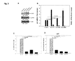
- FIG. 3 RNR2 knockdown inhibits HIV-1 reverse transcription.
- U Unstimulated
- IVIg activated MDM were transfected with a pool of four siRNAs against the RNR2 gene (siRNR2) or with a pool of non-targeting control siRNAs (siCTL).
- siRNR2 siRNAs against the RNR2 gene
- siCTL non-targeting control siRNAs
- MDM were analyzed by WB with antibodies against RNR2, p53R2 and actin.
- B RNR2 (column right) and p21 (column left) mRNAs in unstimulated (US) and IVIg activated siCTL and siRNR2 MDM were quantified by qRT-PCR.
- the data are the fold change with respect to control MDM (siCTL unstimulated) ⁇ standard error for MDM from seven donors.
- C Unstimulated and IVIg activated siCTL and siRNR2 MDM were infected with HIV-1/VSV-G. Viral replication was determined by measuring luciferase activity 72 hpi. The data are the fold change with respect to control MDM (siCTL unstimulated) ⁇ standard error for MDM from seven donors.
- FIG. 4 p21 inhibits HIV-1 reverse transcription by down-regulating RNR2.
- siRNA against the p21 gene sip21
- sip21+siRNR2 siRNA against the RNR2 gene
- Control MDM were transfected with a pool of non-targeting siRNAs (siCTL).
- p21 and RNR2 mRNAs were quantified by qRT-PCR. The data are the mean fold change with respect to control MDM (unstimulated siCTL) in the two donors.
- FIG. 5 p21-mediated restriction is unrelated to SAMHD1 and inhibits SIVmac251 through RNR2 suppression.
- A Unstimulated (US) and IVIg activated MDM were controlled by WB for p21, SAMHD1 and actin expressions at 24 hours post IVIg activation using the indicated antibodies.
- B-C MDM were infected with HIV-1/VSV-G or co-infected with HIV-1/VSV-G and SIVmac251 (+SIV). MDM were controlled for SAMHD1, p21 and actin expressions at 24 hpi (B) and for HIV-1 replication by measuring luciferase activity at 72 hpi (C). The data are means ⁇ standard deviation of triplicate wells.
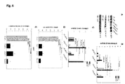
- FIG. 6 p21 represses RNR2 transcription by down-regulating E2F1.
- U2F1 Unstimulated (US) and IVIg activated MDM were transfected with p21 siRNA (sip21) and a pool of non-targeting control siRNAs (siCTL). At 24 h after IVIg stimulation and 22 h after siRNA transfection, MDM were analyzed by WB for E2F1, p21 and actin protein expression.
- (B) p21 (column left), E2F1 and RNR2 (column right) mRNAs from siCTL and sip21 (unstimulated or IVIg-treated) MDM were quantified by qRT-PCR 24 h after IVIg stimulation and 22 h after siRNA transfection. The data are the fold change with respect to control cells (unstimulated siCTL) ⁇ standard error for MDM from four donors.
- E2F1, RNR2 (column right) and p21 (column left) mRNAs were quantified by qRT-PCR.
- the data are the fold change with respect to control MDM (siCTL unstimulated) ⁇ standard error for MDM from four donors.
- siCTL and siE2F1 (unstimulated or IVIg-treated) MDM were infected with HIV-1/VSV-G. Viral replication was quantified by measuring luciferase activity at 72 hpi.
- the data are the fold change with respect to control MDM (siCTL unstimulated) ⁇ standard error for MDM from four donors.
- FIG. 7 RB (retinoblastoma protein) is not involved in the p21-mediated mechanisms of HIV-1 restriction in MDMs.
- U Un-stimulated
- IVIg MDMs were transfected with siRNA against p21 gene (sip21) or with a non-targeting pool control siRNAs (siCTL).
- siRNA against p21 gene sip21
- siCTL non-targeting pool control siRNAs
- siRNAs transfection RB left grey column
- E2F1 light grey column
- RNR2 black column
- mRNA were quantified by qRT-PCR.
- siCTL US and IVIg
- siRB US and IVIg
- sip21 IVIg
- MDMs were infected with HIV-1/VSV-G at 24 hours post IVIg stimulation and 22 hours post-siRNAs transfection.
- the HIV-1 replication was determined by the Luciferase activity at 72 hpi.
- the data are represented in fold change with respect to control MDMs (siCTL US) ⁇ standard deviations of triplicate wells.
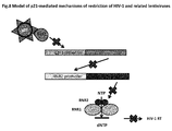
- FIG 8 Model of p21-mediated restriction of HIV-1 and related lentiviruses in MDMs. p21 inhibits E2F1 expression. The RNR2 transactivation is consequently inhibited. The RNR2 down-regulation decreases the dNTPs biosynthesis. The HIV-1 cDNA synthesis is blocked due the unavailability of dNTPs for reverse transcription (RT) catalysis.
- RT reverse transcription
- FIG. 9 RNR2 inhibitors block HIV-1 infection in macrophages.
- Dose dependent inhibition of HIV-1 replication by RNR2 inhibitors Monocyte-derived-macrophages were infected with HIV-1/VSV-G and then treated with decreasing concentrations of hydroxyurea (HU) (40 to 4 mM) (A) or Deferoxamine Mesylate (DFO) (250 to 100 ⁇ M) (B). HIV-1 replication was determined by measuring luciferase activity 72 hours post infection. Results are expressed as fold changes of luciferase activity in drug-treated cells with respect to mock treated cells. The data are means ⁇ standard deviation of triplicate wells.
- dNs deoxynucleosides
- Exogenous dNs are taken up by the cells and converted into dNTPs through the salvage pathway of the dNTPs synthesis, which results in a larger intracellular dNTP pools (Meyerhans, Vartanian et al. 1994; O'Brien, Namazi et al. 1994; Jamburuthugoda, Chugh et al. 2006).
- MDMs from nine donors were activated by seeding on immobilized IVIg or left unstimulated (US) and were infected with HIV-1/VSV-G. Pyrimidine and purine dNs were subsequently added or not to the culture medium. HIV-1 replication was quantified by measuring luciferase activity at 72 hours post-infection (Hpi) ( Figure 1A ). As expected, IVIg stimulation of MDMs significantly inhibited HIV-1 replication ( Figure 1B ).
- dNs strongly enhanced HIV-1 replication in US MDMs (mean of 9: 3.2 folds), consistently with our previous report (Lahouassa, Daddacha et al. 2012).
- dNs treatment was also able to rescue HIV-1 replication in the IVIg activated MDMs to levels comparable to those of the dNs treated US MDMs ( Figure 1B ).
- the treatment of the IVIg-activated MDMs with increasing amounts of dNs (0.25-1.5 mM) relieved the viral blocking of HIV-1 replication in a dose dependent manner( Figure 1C ).
- dNs supply is able to relieve the p21-mediated HIV-1 restriction in MDMs. This suggests that p21 inhibits HIV-1 replication by affecting negatively the dNTP pools available for HIV-1 reverse transcription.
- Intracellular dNTP concentrations were quantified in unstimulated and in IVIg stimulated MDM in which p21 had been silenced by transfection with a small interfering RNA (siRNA) targeting the p21 gene (sip21).
- Control cells were transfected with a non-targeting siRNA pool (siCTL).
- Figures 1D-F show results for cells from a representative donor (donor 1).
- the efficiency of p21 silencing, determined by p21 mRNA quantitative real-time PCR (qRT-PCR) was 72% and 83% in sip21 unstimulated MDM and in IVIg stimulated MDM, respectively ( Figure 1D ).
- p21 inhibits the expression of the ribonucleotide reductase subunit R2: RNR2
- RNR2 knockdown inhibits HIV-1 replication at the reverse transcription step
- RNR2 was depleted by the transfection of MDMs with a pool of 4 siRNAs targeting specifically the RNR2 gene and then infected with HIV-1/VSV-G.
- the control cells were transfected with a pool of non-targeting siRNAs (siCTL).
- siCTL non-targeting siRNAs
- HIV-1 late reverse transcription products were quantified by qRT-PCR in MDMs from six of the seven donors.
- RNR2 knockdown siRNR2
- decreased significantly (p 0.002) HIV-1 reverse transcription products by 80 to 93% in the US MDMs ( Figure 3D ) accordingly to the inhibition of luciferase activity.
- Similar reduction of HIV-1 reverse transcription products was achieved (70-80%) ( Figure 3D ).
- RNR2 knockdown alone was sufficient to inhibit the reverse transcription step indicating that the RNR2 is an essential HIV-1 co-factor in MDMs that promotes reverse transcription in MDMs.
- p21 inhibits HIV-1 reverse transcription through the down-regulation of RNR2
- SAMHD1 was recently demonstrated to restrict HIV-1 replication in macrophages by depleting the intracellular pool of dNTPs (Hrecka, Hao et al. 2011; Laguette, Sobhian et al. 2011; Lahouassa, Daddacha et al. 2012). Moreover, SAMHD1 has been shown to be degraded by HIV-2 and SIV Vpx, which confers these two viruses the ability to escape the SAMHD1 restriction in macrophages and dendritic cells. Although we have previously demonstrated that p21 inhibits HIV-2 and SIV (Bergamaschi, David et al.
- the IVIg activated MDMs were also infected with SIVmac251.
- FIG. 5D shows that the SIV-mac251 LRTs, quantified by the qRT-PCR, were strongly decreased (94%) in the RNR2 knockdown MDMs (siRNR2) indicating that RNR2 is an essential cellular co-factor for SIVmac251 reverse transcription in MDMs.
- RNR2 knockdown is able to inhibit SIV replication at the same extent as HIV-1 indicating that the p21-mediated mechanisms of inhibition of HIV-1 and related-lentiviruses are both related to the same mechanism and un-related to the SAMHD1 restriction pathways.
- E2F1 protein band was very faint in US MDMs but was undetectable after p21 induction by IVIg ( Figure 6A ). Importantly, when p21 was knocked down, the E2F1 protein level was strongly increased indicating that p21 is inhibiting the E2F1 expression ( Figure 6A ).
- siCTL non-targeting siRNAs pool
- E2F1 knockdown gives the same phenotype of HIV-1 replication and reverse transcription inhibition as observed after RNR2 knockdown.
- p21 can repress the transcription of E2F1 target genes through a well-established mechanism in which p21, by inhibiting the CDKs (Cyclin-Dependant protein kinases), maintains RB (retinoblastoma tumor repression protein) in the activated under-phosphorylated state able to retain E2F1 and to inhibit its transactivation activity (Dotto 2000; Perkins 2002).
- p21 has been shown to bind E2F1 through its N-terminal region (1-90 aa) (Delavaine and La Thangue 1999) and p21 is also able to inhibit E2F1 activity directly and independently from RB (Dimri, Nakanishi et al. 1996; Delavaine and La Thangue 1999).
- RB expression was reduced modestly (20-40%) in the IVIg MDMs from only three donors (see for example donor in Fig.7C ). Since the transcription repression activity of RB depends on its phosphorylation state (Dotto 2000; Perkins 2002), the absence of the regulation of its expression by p21 does not rule out a role for RB in p21-mediated HIV-1 restriction. Therefore, we silenced RB in MDMs to assess whether its depletion is able to rescue HIV-1 replication similarly to the effect of p21 depletion. The silencing of RB was performed through the transfection of a pool of four siRNAs specific to RB gene (siRB1).
- the control MDMs were transfected with a pool of non-targeting siRNAs (siCTL) and in parallel p21 was also knocked down (sip21) in the IVIg MDMs to compare the effects of the two factors on HIV-1 replication.
- RB siRNAs transfection efficiently silenced RB in MDMs (70%) and IVIg-activated MDM (85%)as verified by Western Blot ( Figure 7B ) and mRNA quantification (Figure78C).
- Figure 7C mRNA quantification
- RB silencing did not modify either E2F1 or RNR2 gene expressions ( Figure 7C ) indicating that in MDMs, differently from what described in cell lines (Angus, Wheeler et al.
- Macrophages are target cells for HIV-1 and play a crucial role in the establishment of viral reservoirs and in pathogenesis.
- p21 as a limiting factor of HIV-1 and related primate lentiviruses in macrophages and showed that its induction by different stimuli, including Fc ⁇ R aggregation, blocks the accumulation of viral reverse transcripts and eventually suppresses viral integration (Bergamaschi, David et al. 2009).
- Fc ⁇ R aggregation blocks the accumulation of viral reverse transcripts and eventually suppresses viral integration
- p21-mediated HIV-1 inhibition in macrophages we show for the first time that p21 causes the reduction of cellular dNTP pools that are needed for HIV reverse transcription, by suppressing the expression of the RNR subunit RNR2 through the down-regulation of the transcription factor E2F1. Therefore, the mechanism of p21-mediated lentiviral restriction affects the synthesis of dNTP and differs from SAMHD1 catabolic pathway.
- Lentiviral reverse transcription in non-cycling cells such as macrophages is much slower than in activated CD4+ T cells (O'Brien, Namazi et al. 1994).
- a rate-limiting step in viral cDNA synthesis in non-dividing cells is the low content of cellular dNTP that are approximately 200 fold lower than in activated T cells(Diamond, Roshal et al. 2004).
- lentiviral reverse transcriptase has acquired the ability to catalyze DNA synthesis in conditions of limited dNTP levels (Diamond, Roshal et al. 2004), it still needs dNTP above a critic threshold.
- dNTP degradation by the cellular DNA phosphorylase SAMHD1 impairs HIV-1 reverse transcription in non-cycling myeloid cells, such as dendritic cells, macrophages and resting CD4 T cells (Hrecka, Hao et al. 2011; Laguette, Sobhian et al. 2011; Baldauf, Pan et al. 2012; Descours, Cribier et al. 2012) by decreasing dNTPs to levels insufficient to an efficient reverse transcriptase processivity (Hrecka, Hao et al. 2011; Laguette, Sobhian et al. 2011; Lahouassa, Daddacha et al. 2012).
- SIV and HIV-2 counteract SAMHD1 by the protein Vpx that triggers SAMHD1 proteosomal degradation (Hrecka, Hao et al. 2011; Laguette, Sobhian et al. 2011).
- the inventors show here that p21 restricts reverse transcription of HIV-1 and HIV related lentiviruses such as SIV by affecting the de novo synthesis of dNTP, through the suppression of a key enzyme of this pathway, RNR2.
- dNTP synthesis and degradation pathways are finely tuned to ensure the balance of the four dNTP pools (Rampazzo, Miazzi et al. 2010).
- RNR ribonucleotide reductase
- RNR2 another small subunit homologous, p53R2
- p53R2 has been described (Tanaka, Arakawa et al. 2000). So far, the regulation of RNR activity has been studied in proliferating and quiescent cell lines, mostly of murine origin, and never in terminally differentiated primary cells including macrophages. In cycling cells, RNR is a complex between RNR1 and RNR2. RNR2 is degraded during late mitosis (Chabes, PNAS, 2003). Quiescent fibroblast cell lines contain p53R2 and little or no RNR2 (Bjorklund, Skog et al. 1990; Hakansson, Hofer et al. 2006).
- RNR2 suppression through p21 induction and by siRNAs mediated knockdown was sufficient to block the synthesis of HIV-1 cDNA by reverse transcriptase, indicating that RNR2 is a necessary cofactor of HIV-1 replication in macrophages. Accordingly, an increase in RNR2 concentrations in macrophages differentiated upon Leihsmania infection has been associated with an enhancement of HIV-1 replication (Mock, Hollenbaugh et al. 2012).
- RB-sensitive E2F activity is induced late in G1 in proliferating cells (Dimri, Nakanishi et al. 1996; Verona, Moberg et al. 1997).
- p21 maintains RB in the active hypo-phosphorylated form, which negatively regulates the activity of E2F transcription factors, by the inhibition of CDK2 kinase during the transition through G1 in cycling cells.
- RB has been shown to down-regulate the transcription of several genes involved in dNTP synthesis and consequently to reduce dNTP pools in rat cell lines (Angus, Wheeler et al. 2002).
- studies on p21 impact on E2F1 transcriptional activity have been essentially related to p21 activity in cell growth inhibition in proliferating cell lines and are not directly transposable to macrophages.
- Our results show that RB silencing does not significantly affect RNR2 expression or HIV-1 replication in macrophages.
- transcriptome analysis showed an up-regulation of p21 and a down-regulation of E2F1 and RNR enzymes, including RNR2, in activated by immune complexes murine dendritic cells (van Montfoort, t Hoen et al. 2012). Whether the p21-E2F1-RNR2 pathway controls HIV replication in human dendritic cells and in hematopoietic stem cells (Zhang, Scadden et al. 2007) remains to be determined.
- a RNR inhibitor hydroxyurea
- nucleotide analogs in the treatment of AIDS, to decrease the processivity of HIV reverse transcriptase.
- Our results suggest that dNTP pool size and hence HIV-1 cDNA synthesis may be affected either by targeting directly RNR2 or by inhibiting its expression through p21 or E2F1 targeting. All these molecules are actively studied as targets in cancer therapy. Statins have been shown to have anti-proliferative activity by inducing p21 (Ukomadu and Dutta 2003).
- HDACi Histone deacetylase inhibitors
- HDACi are also actively studied in AIDS treatment in combination with highly active antiretroviral therapy to eradicate HIV-1 from latent cellular reservoirs (Keedy, Archin et al. 2009). Besides their activity in reactivating latent provirus expression, HDACi might also block HIV-1 replication in newly infected macrophages by inducing p21. Either induction or repression of E2F1 has been proposed to induce cancer cell apoptosis or reducing their metastatic potential respectively (Engelmann and Putzer 2012). However targeting directly E2F1 may be challenging owing to its opposite functions in cell death and proliferation in cycling cells.
- RNR is an important therapeutic target for cancer therapy: RNR inhibition decreasing dNTP concentration inhibits DNA synthesis and induces cycle arrest and apoptosis of cancer cells.
- Inhibitors specific that inhibit free radical formation in RNR2 are currently studied as cytostatic agents (Shao, Zhou et al. 2006). Considering the present results, considering the extension of the treatment with RNR2 inhibitors to HIV-1 infected patients, is a valuable approach including in complement to cART.
- MDMs Monocyte Derived Macrophages
- Buffy coats fraction of anticoagulated blood sample that contains most of white blood cells following gradient density centrifugation
- EFS EFS-Institute Pasteur Convention.
- Written informed consent was obtained from each donor to use the cells for clinical research in accordance with French law.
- Monocytes were isolated from buffy coats and differentiated into macrophages as previously described (David, Saez-Cirion et al. 2006; Bergamaschi, David et al. 2009).
- monocytes were separated from peripheral blood mononuclear by adherence to the plastic and then detached and cultured for 6 to 7 days in hydrophobic Teflon dishes (Lumox; Dutsher) in macrophages medium (RPMI 1640 supplemented with 200 mM L-glutamine, 100 U of penicillin, 100 ⁇ g streptomycin, 10 mM HEPES, 10 mM sodium pyruvate, 50 ⁇ M ⁇ -mercaptoethanol, 1% minimum essential medium vitamins, 1% non-essential amino acids) supplemented with 15% of heat inactivated human AB serum.
- RPMI 1640 supplemented with 200 mM L-glutamine, 100 U of penicillin, 100 ⁇ g streptomycin, 10 mM HEPES, 10 mM sodium pyruvate, 50 ⁇ M ⁇ -mercaptoethanol, 1% minimum essential medium vitamins, 1% non-essential amino acids
- MDMs were harvested and resuspended in macrophage medium containing 10% of heat inactivated fetal bovine serum (FBS).
- FBS heat inactivated fetal bovine serum
- MDMs obtained with this method are 91 to 96 % CD14, they express: the Fc ⁇ receptors (CD16, CD32 and CD64), the differentiation markers (C11b and CD71) and the M2 macrophage polarization markers (CD163 and CD206).
- Fc ⁇ receptors stimulation wells of 96-well plate, 24-well plate and 12-well plate were pre-coated for 2 hours at 37 °C with respectively 100 ⁇ l, 250 ⁇ l and 500 ⁇ l of 1 mg/ml of human IgG for therapeutic use (IVIg) (Endobuline; Baxter) in PBS.
- MDMs (1x10 6 /ml of macrophage medium + 10% FBS) were then seeded on the un-coated or IVIg pre-coated wells (6.5x10 4 MDMs/well of 96-well plate, 2.5x10 5 MDMs/ well of 24-well plate and 0.5x10 6 MDMs/ well of 12-well plate).
- Unstimulated (US) and IVIg stimulated MDMs were let to be attached on the plates for 2 hours at 37 °C before infections or before siRNAs transfection.
- siRNAs small interfering RNAs
- the siRNA against p21 gene is on target plus siRNA n.12: 5' AGA CCA GCA UGA CAG AUU U 3'.
- the siRNAs against RNR2 (siRNR2), E2F1 (siE2F1) and RB (siRB) genes are siGenome smart pool selected composed each of a pool of four siRNAs.
- siRNAs have the following sequences: siRNR2: 1- 5' GCA CUC UAA UGA AGC AAU A 3' (SEQ ID No.1), 2- 5' GAA GAG UAG GGA GUA U 3' (SEQ ID No.2), 3- 5' GAG UAG AGA ACC CAU UUG A 3' (SEQ ID No.3) and 4- 5' GGC UCA GCU UGG UCG ACA A 3' (SEQ ID No.4); siE2F1: 1- 5' GAA CAG GGC CAC UGA CUC U 3' (SEQ ID No.5), 2- 5' UGG ACC ACC UGA UGA AUA U 3' (SEQ ID No.6), 3- 5' CCC AGG AGG UCA CUU CUG A 3' (SEQ ID No.7) and 4- 5' GGC UGG ACC UGG AAA CUG A 3' (SEQ ID No.8).
- siRB 1- 3' GAA AGG ACA UGU GAA CUU A 5' (SEQ ID No.9), 2-3'GAA GAA GUA UGA UGU AUU G 5' (SEQ ID No.10), 3- 3' GAA AUG ACU UCU ACU CGA A 5' (SEQ ID No.11) and 4- 3' GGU UCA ACU ACG CGU GUA A 5' (SEQ ID No.12).
- the control siRNAs are a pool of four on target plus non-targeting siRNAs.
- siRNAs transfection was performed using INTERFERin kits (Polyplus Transfection).MDMs (1x10 6 /ml of macrophages medium + 10% FBS) were seeded on un-coated or IVIg pre-coated wells (6.5x10 4 MDMs/well of 96-well plate, 2.5x10 5 MDMs/well of 24-well plate and 0.5x10 6 MDMs/ well of 12-well plate) and let to be attached at 37°C for 2 hours prior siRNAs transfection. The siRNAs transfections were performed with the INTERFERin (Polyplus Transfection).
- siRNAs were pre-diluted in 1 ml of Opti-MEM in which 20 ⁇ l of INTERFERin were added and the transfection mix was let to incubate at room temperature for 10 minutes.
- the transfection mix volumes 32 ⁇ l, 125 ⁇ l and 250 ⁇ l were added respectively to 6.5x10 4 , 2.5x10 5 and 0.5x10 6 MDMs at the final concentrations of 50 nM for sip21, siRNR2 and siE2F1 siRNAs and of 25 nM for siRB1 siRNAs.
- Equal amounts of the on target plus non-targeting siRNAs were added to the control MDMs. The MDMs were then incubated at 37°C for 22 hours.
- the medium was replaced with fresh macrophage medium supplemented with 10% FBS prior to the infections.
- Cell lysates were assayed for protein expression by Western Blot and for the quantification of mRNA by the Real-Time quantitative PCR (RT-qPCR) to determine the efficiency of gene knockdown at the moment of infection.
- RT-qPCR Real-Time quantitative PCR
- the NL4.3-Luc HIV-1 pseudotyped with VSV-G was produced and quantified as previously described (David, Saez-Cirion et al. 2006; Bergamaschi, David et al. 2009).
- the SIVmac251 replication competent was produced through the infection of CEM x 174 T cell lines.
- MDMs (1x10 6 /ml of macrophage medium + 10% FBS) were seeded either on the un-coated or IVIg coated wells (6.5x10 4 MDMs/well of 96-well plate, 2.5x10 5 MDMs/ well of 24-well plate and 0.5x10 6 MDMs/ well of 12-well plate).
- MDMs were let to be attached on the plate for 2 hours at 37°C prior to the infections. For knockdown cells the infections were performed 24 hours post IVIg stimulation and 22 hours post siRNAs transfections. MDMs were infected with 4 - 5 ng of p24 of HIV-1/VSV-G or 300 ng p27 of SIV-mac 251 for 1x10 6 cells by 1 hour of spinoculation (1,200 g at room temperature) followed by 1 hour at 37°C. Cells were then washed with PBS and incubated at 37°C in fresh macrophage medium supplemented with 10% FBS for 72 hours for HIV-1 replication determined by luciferase activity and LRTs (Late Reverse Transcripts) quantification by qRT-PCR.
- LRTs Late Reverse Transcripts
- the culture supernatants were harvest 7 days post infection and quantified for their p27 antigen content using the SIV p27 ELISA kit (Zeptometrix Crop).
- the SIV LRTs quantification was performed at 48 hours post infection.
- the viral stocks were pretreated with DNase I (Roche Diagnostics) for 1 hour at room temperature.
- DNase I Roche Diagnostics
- the un-stimulated (US) and IVIg stimulated MDMs were treated with the indicated concentrations of dNs immediately after infection for 24 hours at 37 °C.
- the deoxynucleosides consisted of mixture of dA (D8668), dC (D0776), dG (D0901) and dT (T1895; all from Sigma Aldrich).
- the dNTPs were quantified as previously described (Diamond, Roshal et al. 2004).
- the p21 knockdown (sip21) and control (siCTL) MDMs (3x10 6 MDMs each) were washed with cold PBS and lyzed, at 22 hours post-siRNAs transfection, in 2 ml of 60% (vol/vol) of aqueous methanol. Lysates were heated at 95 °C for 7 minutes, clarified by centrifugation at 16,000 g for 15 minutes at 4 °C, dried under vacuum. Extracted dNTPs were then resuspended and quantified as previously described (Diamond, T. L., 2004).
- RNA quantification total RNA from 2.5x10 5 to 0.5x10 6 MDMs was extracted with the RNeasy kit (Qiagen) and treated with DNase according to the manufacturer's instructions. The RNA was transcribed with the SuperScript II RT (Invitrogen).
- PCR amplifications of cDNA was carried out in duplicate in MicroAmp Optical 96-well reaction plate (30 ⁇ l/well), using 5 ⁇ l cDNA template, 15 ⁇ l of 2x Taqman Universal Master mix (Applied biosystems), 1.5 ⁇ l of Taqman gene expression premade mix (Applied biosystems) (GAPDH: Hs99999905_m1; RNR2: Hs00357247_g1; p21: Hs00355782; RB: Hs01078066_m1; E2F1: Hs00153451_m1) and completed with H 2 O up to 30 ⁇ l.
- the amplification conditions were: 50°C for 2 min and 95°C for 10 min followed by 40 cycles at 95°C for 15 s and 60°C for 1 min on ABI prism 7000 Sequence Detection System.
- the data were analyzed with the cycle threshold (C T ) method, and the amount of target mRNA in each sample was normalized to GAPDH mRNA as an endogenous reference.
- LRTs Late Reverse Transcripts
- the MDMs DNA was extracted with the DNeasy Tissue Kit (Qiagen) at 72 hours post-infection.
- the qRT-PCR analysis of LRTs through the quantification of the U5-Gag viral DNA were carried on an ABI prism 7000 Sequence Detection System as previously described (David, Saez-Cirion et al. 2006; Bergamaschi, David et al. 2009).
- Standards for U5-Gag amplification products were generated by serial dilution of DNA extracted from HIV-1 8E5 cells containing one integrated copy of HIV-1 per cell.
- the quantification of the SIVmac251 LRTs was performed as previously described (Hofmann-Lehmann, Swenerton et al. 2000). The amounts of LRTs were normalized to the endogenous reference gene albumin. Standard curve were generated by serial dilution of a commercial human genomic DNA (Roche).
- the data acquisition software allowed superposition of photon events with brightfield images and selective quantification of photons in delimited surfaces. 10 5 MDM were plated after infection on glass-bottomed dishes (MatTek corp, MA, USA), washed and cultured in complete medium for 5 days before analysis. The analyses were conducted in PBS + 5% FCS or complete phenol red free medium at room temperature in presence of endotoxin free beetle D-luciferin (Promega) (final concentration 1 mM).
- MDMs (0.5x10 6 ) were lyzed in 100 ⁇ l of lysis buffer (50 mM Tris-HCl pH 7.4, 150 mM NaCl, 1mM EDTA and 1% Triton X-100) provided with protease and phosphatase cocktails inhibitors (Roche). Protein were quantified with Bradford Kit (Biorad) and 30 to 50 ⁇ g of cell extracts were diluted in the Leammli buffer, boiled for 5 min at 95 °C and loaded on the mini-protean-TGX 4-15 % precast acrylamide gel (Biorad) for electrophoresis separation. Proteins were then blotted on the PVDF transfer membrane (GE healthcare).
- lysis buffer 50 mM Tris-HCl pH 7.4, 150 mM NaCl, 1mM EDTA and 1% Triton X-100
- protease and phosphatase cocktails inhibitors (Roche). Protein were quantified with Bradford Kit (Biorad) and 30 to 50 ⁇
- the membrane were incubated with the primary antibodies followed by the secondary horseradish peroxidase anti-goat or anti-rabbit antibodies (Sigma) or anti-mouse (Cell Signaling).
- the proteins were revealed on hyperfilms (Amersham) by using Super Signal West pico chemiluminescent substrate (Pierce).
- the primary anti-PCNA (PC10), ant-RNR2 (N-18), anti-E2F1 (KH95), anti-RB (IF8) and anti-RNR1 (H-300) antibodies were all purchased from Santa Cruz and all used at the dilutions 1:200 in 1% skimmed milk in PBS-0.05 % tween.
- the primary anti-p53R2 (1:500) and anti-GAPDH (1:1000) antibodies were obtained from Abcam.
- the anti-p21 (1:1000) is from Millipore and the anti-Actin (1:5000) is from Sigma.
- RNR2 inhibitors block HIV-1 infection in macrophages.
- RNR2 inhibitors for their effect on reverse transcription in macrophages and especially tested dose dependent inhibition of HIV-1 replication by RNR2 inhibitors consisting of hydroxyurea (HU) or Deferoxamine Mesylate (DFO).
- Monocyte-derived-macrophages were infected with HIV-1/VSV-G and then treated with decreasing concentrations of hydroxyurea (HU) (40 to 4 mM) (A) or Deferoxamine Mesylate (DFO) (250 to 100 ⁇ M) (B).
- HIV-1 replication was determined by measuring luciferase activity 72 hours post infection. Results are expressed as fold changes of luciferase activity in drug-treated cells with respect to mock treated cells. The data are means ⁇ standard deviation of triplicate wells.
- MDM viability was not affected at the concentrations of HU and DFO used in the assays (not shown).
Landscapes
- Health & Medical Sciences (AREA)
- Life Sciences & Earth Sciences (AREA)
- Chemical & Material Sciences (AREA)
- Public Health (AREA)
- Medicinal Chemistry (AREA)
- Pharmacology & Pharmacy (AREA)
- Animal Behavior & Ethology (AREA)
- General Health & Medical Sciences (AREA)
- Veterinary Medicine (AREA)
- Epidemiology (AREA)
- Molecular Biology (AREA)
- Virology (AREA)
- AIDS & HIV (AREA)
- Tropical Medicine & Parasitology (AREA)
- Biochemistry (AREA)
- Communicable Diseases (AREA)
- Oncology (AREA)
- Chemical Kinetics & Catalysis (AREA)
- General Chemical & Material Sciences (AREA)
- Nuclear Medicine, Radiotherapy & Molecular Imaging (AREA)
- Organic Chemistry (AREA)
- Pharmaceuticals Containing Other Organic And Inorganic Compounds (AREA)
Abstract
The invention relates to the use of inhibitors for the RNR2 subunit of the human ribonucleotide reductase to impair reverse transcription of HIV related lentiviruses in macrophages through down regulation of the intracellular dNTPs pool by repressing expression of RNR2. The invention also concerns a method of screening inhibitors of reverse transcription of HIV related lentiviruses in macrophages.
Description
- The invention relates to the use of inhibitors for the RNR2 subunit of the human ribonucleotide reductase to impair reverse transcription of HIV related lentiviruses in macrophages through down regulation of the intracellular dNTPs pool by repressing expression of RNR2. Macrophages targeted by the inhibitors used according to the invention are especially Monocyte Derived Macrophages (MDM). MDM encompass macrophages that are differentiated quiescent long-lived cells in the G0/G1 phase of cell cycle. Said inhibitors are used in the invention for the therapeutic treatment of human patients infected with a HIV lentivirus.
- The invention also concerns a method of screening inhibitors of reverse transcription of HIV related lentiviruses in macrophages.
- HIV related Lentiviruses of the invention encompass in particular human lentiviruses including HIV-1 and HIV-2 and simian lentiviruses including SIV.
- HIV infection of macrophages greatly contributes to viral pathogenesis and progression to AIDS (recent review in (Verani, Gras et al. 2005). Macrophages are widely distributed in body tissues and can serve as vehicles for HIV-1 dissemination throughout the organs, including the lungs and the brain (Gartner, Markovits et al. 1986; Koenig, Gendelman et al. 1986). Because of their long half-life and relative resistance to the cytopathogenic effect of the HIV-1, macrophages may transmit the virus to other target cells, including CD4+ T lymphocytes, and contribute to the establishment of viral reservoirs (Orenstein, Fox et al. 1997; Aquaro, Bagnarelli et al. 2002; Sharova, Swingler et al. 2005). In addition, HIV-1 infection of macrophages affect their functions in innate and adaptive responses to pathogens, such as phagocytosis, intracellular pathogen killing and antigen presentation (reviewed in (Kedzierska, Azzam et al. 2003). However, the susceptibility of macrophages to HIV-1 infection varies greatly according to their differentiation status and tissue localization (Ancuta, Kunstman et al. 2006; Shen, Richter et al. 2009; Cassol, Cassetta et al. 2010). Environmental stimuli, such as cytokines, bacterial products as LPS, or circulating immune complexes bound to Fcγ receptors (FcγR) on macrophage membrane, may influence macrophage permissiveness to HIV-1 infection (Herbein, Coaquette et al. 2002). Moreover, HIV-1 replication in macrophages is regulated by cellular factors that can enhance or limit various steps of the viral life-cycle (Bergamaschi and Pancino 2010). In particular, HIV-1 reverse transcription in Monocyte Derived Macrophages (MDM) is much slower than in activated T cells, and this has been mainly attributed to the limited availability of nucleotide precursors in these non-dividing cells, in which the average of dNTP concentrations (∼0.026 µM) is about 200 times lower than in PHA-activated T cells (O'Brien, Namazi et al. 1994; Diamond, Roshal et al. 2004; Triques and Stevenson 2004). The recent identification of SAMHD1 as an HIV-1 restriction factor in dendritic cells and macrophages, counteracted by the HIV-2 and SIV Vpx protein that promotes its degradation (Hrecka, Hao et al. 2011; Laguette, Sobhian et al. 2011) supports this hypothesis. Indeed, SAMHD1 functions as a deoxynucleoside triphosphate (dNTP) triphosphohydrolase (Goldstone, Ennis-Adeniran et al. 2011; Powell, Holland et al. 2011), and the inventors have demonstrated that SAMHD1 degrades dNTPs keeping their concentration low and thus limiting HIV-1 replication in non-dividing cells (Lahouassa, Daddacha et al. 2012). Besides SAMHD1, other cellular factors have been reported to affect early post-entry steps of HIV-1 replication in macrophages, including the apolipoprotein B mRNA editing enzyme, catalytic polypeptide-like 3 (APOBEC3) cytidine deaminase family member APOBEC3A (Peng, Greenwell-Wild et al. 2007; Berger, Durand et al. 2011).
- The inventors have previously shown that the p53-independent induction of p21Waf1/Cip1 (hereafter p21) by the aggregation of activating Fcγ receptors (FcγR) on MDM causes a strong decrease of reverse transcripts and integrated proviruses of HIV-1, HIV-2, SIVmac and SIVagm in MDM while siRNA-mediated p21 depletion rescued the viral replication (Bergamaschi, David et al. 2009). Remarkably, p21 limits HIV replication also in unstimulated MDM (Bergamaschi, David et al. 2009) and has been involved in the resistance of human hematopoietic stem cells to HIV-1 replication (Zhang, Scadden et al. 2007). In previous studies, the inventors have not detected p21 interactions with HIV-1 proteins associated to the reverse transcription and pre-integration complexes (RTC/PIC) that are implicated in the reverse transcription/integration steps (Bergamaschi, David et al. 2009) and Allouch A. unpublished results).
- The inventors have now addressed the hypothesis that p21 may interfere with or modulate the expression of other cellular factor(s) necessary for these steps of HIV replication and have performed the present invention by identifying a cellular factor in macrophages that is regulated by p21 and that happens to interfere with reverse transcription of lentiviruses such as HIV or related viruses such as SIV.
- p21 belongs to the Cip/Kip family of cyclin-dependent kinase inhibitors (CDKIs) (Gu Y 1993; Harper JW 1993; Xiong Y 1993). p21 was first described as a cell cycle inhibitor that blocks cell cycling at the G1/S interface and plays a critical role in the control of cell growth. p21 can influence cell cycle progression through pleiotropic mechanisms, including cyclin/CDK inhibition, binding to PCNA (Chen, Jackson et al. 1995), and control of the E2F transcription and activity (Sherr and Roberts 1995; Delavaine and La Thangue 1999). Although p21 is not a transcription factor, it may exert indirect effects on cellular gene expression. p21 induction in a fibrosarcoma cell line regulates the transcription of several genes, including genes involved in DNA replication, segregation and repair, either repressing or enhancing their expression (Chang, Watanabe et al. 2000).
- MDMs are terminally differentiated, non-replicating and long-lived cells in which p21 is highly expressed comparing to cycling cells and plays important roles in monocyte differentiation and survival by inhibiting apoptosis (Liu, lavarone et al. 1996; Asada M 1999; Xaus J 1999; Steinman RA 2000; Coqueret O 2003).
- The cellular factor accordingly identified by the inventors as impacting reverse transcription of lentiviral genome such as HIV or SIV RNA in macrophages is a subunit of a ribonucleotide reductase protein.
- Ribonucleotide reductase (RR) is a multisubunit enzyme that catalyses the reduction of ribonucleotides to their corresponding deoxyribonucleotides and accordingly impacts the synthesis of DNA and its repair. Increased RR activity has been associated with malignant transformation of cells and tumor growth. Accordingly RR inhibitors have been targeted with a view to design anticancer chemotherapeutic agents and various molecules have been proposed including Triapine (
WO 02/085358 - The inventors have found that the R2 subunit of human ribonucleotide reductase (RNR2) may be targeted in order to inhibit lentiviral reverse transcription, illustrated with HIV and SIV lentiviruses, in quiescent cells and in particular in macrophages such as Monocyte Derived Macrophages (MDM). The obtained results were unexpected as it was previously found that RNR2 expression was thought to be restricted to S phase in dividing cells. From their experimental results the inventors were able to propose a novel and specific anti-lentiviral treatment strategy that would encompass targeting lentiviral replication to alter it in macrophages by inhibiting the reverse transcription of lentiviral genome and accordingly formation of lentiviral gene products in these cells.
- Thus the invention provides means to inhibit lentiviral infection in macrophages and accordingly compounds and methods of use affecting viral reservoirs in patients infected with a lentivirus such as HIV-1 or HIV-2 and also impacting viral spread to other target cells.
- The compounds and methods of use of the invention accordingly constitute means for treating HIV infection in a human patient, including for treating HIV reservoirs in various organs of tissues and/or for preventing dissemination of the HIV lentivirus in patient's organs or tissues.
- The invention concerns a RNR2 inhibitor for use in the therapeutic treatment of a human patient infected with a HIV lentivirus, wherein the RNR2 inhibitor is administered in dosage conditions that enable it to interfere with reverse transcription of the lentiviral genome in HIV infected macrophages thereby altering lentiviral replication.
- The sequence of the gene encoding human RNR2 is provided as SEQ ID No.13 and the amino acid sequence of the human RNR2 protein is provided as SEQ ID No.14.
- By "treatment" or "therapeutic treatment", it is meant that the performed steps and dosage conditions of administration of a RNR2 inhibitor according to the invention result in improving the clinical condition of a human patient in need thereof, who has been diagnosed for infection by a HIV lentivirus and who suffers or may suffer in the future from disorder(s) associated with the infection as a result of chronic HIV infection. Such treatment aims at improving the clinical status of the human patient, by eliminating or alleviating the symptoms associated with the disorder(s) related to the HIV infection. In a preferred embodiment, the treatment according to the invention enables restoring to health.
- "HIV lentivirus" encompasses human lentivirus of the HIV type including HIV-1 and HIV-2. "HIV related lentivirus" encompasses non-human lentiviruses such as simian lentivirus SIV, which has structural similarities with HIV-2 and in particular expresses Vpx protein.
- The ability to "interfere "with reverse transcription of the lentivirus genome encompasses the capacity to restrict, to down regulate or to abolish or inhibit said reverse transcription in human macrophages.
- Measurement of the effects of the administration of a RNR2 inhibitor according to the invention and thus of their capacity to interfere with reverse transcription may encompass measurement of the expression products of the HIV genes in particular measurement of the HIV-1 p24 protein or measurement of Late Reverse Transcription products of the HIV genome or may encompass measurement of the viral load. By "viral load", it is meant either the HIV RNA (which is derived from viral particles and present in plasma) or the HIV DNA (which is integrated in the cell genome and present in cells).
- By "HIV-infected patient" it is meant a subject or patient who has been positively and accurately diagnosed for a HIV virus, and for whom HIV-infection has been confirmed following relevant testing. HIV-infected patients may be classified according to several parameters such as viral load, CD4 T cells number or clinical symptoms of AIDS.
- In a preferred embodiment of the invention, a RNR2 inhibitor for use in the prevention or therapeutic treatment of a human patient infected with a HIV lentivirus according to the invention, targets macrophages that are differentiated non-replicating long-lived macrophages, in particular Monocyte Derived Macrophages (MDM).
- Bone marrow-derived monocytes (Mos) are released into the blood where they circulate for a few days. Circulating Mos extravasate into the lungs, gastrointestinal tract, kidney, primary and secondary lymphoid organs and the central nervous system (CNS). In tissues, Mos undergo differentiation into tissue-specific macrophages (Mφ) and dendritic cells (DC).Resident tissue MΦ are long-lived cells with a half-life ranging from an average of 6 weeks (alveolar MΦ) to several years (resident brain MΦ).HIV-infected mononuclear phagocytes (bone marrow (BM) and blood Mo, tissue Mφ, microglia) can thus serve as vehicles for dissemination and reservoirs of HIV-1 infection (Crowe S et al 2003).
- Monocyte Derived Macrophages (MDMs) are large phagocytic cells, which belong to the Reticuloendothelial System (RES) or Mononuclear Phagocyte System (MPS) and are derived from blood monocytes. MDMs can also be divided as fixed or free macrophages: fixed macrophages such as histiocytes of connective tissue or Kupffer cells of liver and spleen, cells of lymph nodes... and free macrophages in inflammatory regions.
- A particular subgroup of Monocyte Derived Macrophages (MDMs) of interest in accordance with the invention is MDMs that express the following markers: CD14, Fcγ receptors, differentiation markers including CD11b and/or CD71 and M2 macrophage polarization markers including CD163 and/or CD206.
- The invention also relates to a RNR2 inhibitor for use in the prevention of constitution of HIV reservoirs or in the therapeutic treatment of replenishment of HIV reservoirs, in differentiated non-replicating macrophages in a human patient infected with a HIV lentivirus, wherein the RNR2 inhibitor is administered in dosage conditions that enable it to impair reverse transcription of the lentiviral genome in Monocyte Derived Macrophages of the patient.
- The invention also relates to a RNR2 inhibitor for use in the prevention or the therapeutic treatment of dissemination of the HIV lentivirus in a human patient infected with said HIV lentivirus, in particular for the prevention or the treatment of lentivirus dissemination in peripheral organs such as lungs, brain or liver or the prevention or treatment of lesions in peripheral organs such as lungs, brain or liver, wherein the RNR2 inhibitor is administered in dosage conditions that impair reverse transcription of the lentiviral genome in Monocyte Derived Macrophages of the patient.
- In a particular embodiment of the invention, a RNR2 inhibitor for use in the treatment of a human patient infected with a HIV lentivirus according to the invention is administered in dosage conditions, which do not affect Monocyte Derived Macrophages viability.
- Examples of RNR2 inhibitors for use in the treatment of a human patient infected with a HIV lentivirus according to the invention, are selected among radical scavengers, iron chelators, antisense oligonucleotides, and interfering RNA.
- In particular, RNR2 inhibitor for use in the treatment of a human patient infected with a HIV lentivirus according to the invention, are selected among Hydroxyurea (specific inhibitor of DNA synthesis in vivo, CH4N2O2)(Merck Index 12th edition Entry 4898), and Deferoxamine mesylate (aluminum and iron (III) chelator, C25H48N6O8.CH4O3S)(Merck Index 12th edition Entry 2914).
- RNR2 inhibitors such as Hydroxyurea or Deferoxamine mesylate may be administered in dosage conditions that lead to a concentration in the targeted cells, which are lower than those usually showing hematological toxicity when used for antitumor activity.
- Antisense oligonucleotides having an activity as RNR2 inhibitor encompass GTI-2040. GTI-2040 is a 20-mer oligonucleotide that is complementary to a coding region in the mRNA of the RNR2 subunit of human ribonucleotide reductase (Lee Y et al 2003).
- Small interfering RNAs (siRNA) are disclosed in the examples which follow and can be used as a pool of siRNA. In particular according to the invention, siRNA are those having the sequence disclosed as SEQ ID No. 1 to 4. The examples provided herein show that these siRNA, optionally used as a pool, prevent the transcription of the RNR2 gene in macrophages such as MDMs.
- According to a preferred embodiment of the invention, RNR2 inhibitor for use in the treatment of a human patient infected with a HIV lentivirus according to the invention, is used in a human patient who has been diagnosed for early phase infection with HIV, especially before Setpoint of HIV replication.
- The invention also relates to a RNR2 inhibitor for use in the treatment of a human patient infected with a HIV lentivirus, where the patient is under combination antiretroviral therapy.
- In particular, a RNR2 inhibitor for use in the treatment of a human patient infected with a HIV lentivirus is used in combination antiretroviral therapy (cART) with a acyclic nucleoside such as acyclovir or gancyclovir, nucleoside analog, such as dideoxyinoside (DDI) and/or with 3'-azido-3'-deoxythymidine (AZT), and/or a reverse transcriptase inhibitor and/or a protease inhibitors and/or integrase inhibitors.
- In a particular embodiment a RNR2 inhibitor for use in the treatment of a human patient infected with a HIV lentivirus according to the invention, is used in a patient who has been diagnosed for neurological disorders.
- The expression "neurological disorders" associated with HIV infection" encompasses neurological disorders of the nervous system which are caused directly by the HIV virus, by certain cancers and/or opportunistic infections, as well as disorders of unknown origin which are influenced by but are not known to be caused directly by the virus. Some of these neurological disorders associated with AIDS may be characteristic of the state of progression of the disease. Examples of neurological disorders associated with AIDS are AIDS dementia complex (ADC) or HIV-associated encephalopathy, central nervous system lymphomas, cryptococcal meningitis, cytomegalovirus (CMV) encephalitis, encephalitis and myelitis caused by the herpes zoster virus, neuropathy (peripheral neuropathy and distal sensory polyneuropathy), neurosyphilis, progressive multifocal leukoencephalopathy (PML), toxoplasma encephalitis or cerebral toxoplasmosis and vacuolar myelopathy. Some patients with controlled viral load (VL) who are not at the AIDS stage, as for example patients under potent antiretroviral therapy (HAART) or cART may develop neurological disorders.
- The invention also concerns a pharmaceutical formulation comprising an inhibitor of the RNR2 subunit of the ribonucleotide reductase in an amount sufficient to inhibit HIV replication in differentiated, non-replicating macrophages in a human patient infected with a HIV lentivirus.
- A particular pharmaceutical formulation for use according to the invention in therapeutically treating a human patient previously diagnosed for infection by a HIV lentivirus, is a formulation which comprises active ingredients including a RNR2 inhibitor in combination with a pharmaceutical carrier or diluents, wherein the RNR2 inhibitor is present in the formulation in a form and amount adapted to release RNR2 inhibitor after administration to the patient, such that RNR2 inhibitor release causes RNR2 inhibitor to be detectable in the plasma of the patient in a concentration that does not induce hematological toxicity, in particular in a concentration of 0.1 to 2µM, in particular 0.2 to 1µM of the RNR2 inhibitor in plasma.
- A pharmaceutical composition according to the invention of for use according to the invention is one where the RNR2 inhibitor is selected among radical scavengers, iron chelators antisense oligonucleotides and interfering RNA, in particular the RNR2 inhibitor is Hydroxyurea or Deferoxamine mesylate. The particular examples provided in the present application are examples of active ingredient for such pharmaceutical formulations.
- The RNR2 inhibitor may be administered by any suitable administration route including parenteral and in particular intravenous route or intramuscular or via mucosal administration or via oral route.
- The invention thus also relates to a method of therapeutically treating a human patient previously diagnosed for infection by a HIV lentivirus, comprising:
- administering a RNR2 inhibitor in dosage conditions that enable it to interfere with reverse transcription of the lentiviral genome in HIV infected macrophages thereby altering lentiviral replication in vivo,
- following the clinical condition of the treated patient.
- The embodiments disclosed herein in relation to the use of the RNR2 inhibitors according to the invention apply to the method of treating defined above. In particular, the method of therapeutically treating a human patient previously diagnosed for infection by a HIV lentivirus may be part of a cART.
- The invention also relates to a method of in vitro screening RNR2 inhibitors candidates for their capacity to interfere with reverse transcription of a HIV or SIV lentiviral genome in human differentiated non-replicating macrophages, in particular in Monocyte Derived Macrophages, comprising the steps of:
- providing in a macrophages culture medium, differentiated Monocyte Derived Macrophages (MDMs) that express the following markers: CD14, Fcγ receptors, differentiation markers including CD11b and/or CD71 and M2 macrophage polarization markers including CD163 and/or CD206;
- infecting MDMs with a HIV retrovirus or a derivative thereof wherein said HIV or derivative optionally expresses a reporter gene;
- contacting the infected MDMs with determined concentrations of a candidate RNR2 inhibitor;
- determining HIV replication gene products, in particular measuring the expression or the activity of the reporter gene, thereby determining efficacy of RNR2 inhibitor candidate to block HIV infection of MDMs.
- A macrophage culture medium may be RPMI 1640 supplemented with 200mM L-glutamine, 100 U of penicillin, 100 µg of streptomycin, 10mM HEPES, 10mM sodium pyruvate, 50 µM β-mercaptoethanol, 1% minimum essential medium vitamins, 1% non essential amino acids.
- A method of in vitro screening RNR2 inhibitors for their capacity to interfere with reverse transcription of a HIV or SIV lentiviral genome in human differentiated non-replicating macrophages, in particular in Monocyte Derived Macrophages, according to the invention may further comprise steps for preparation of MDMs including:
- providing buffy coat of anticoagulated blood sample of a healthy donor;
- differentiating monocytes of the buffy coat into macrophages and harvesting Monocyte Derived Macrophages (MDMs) that express the following markers: CD14, Fcγ receptors, differentiation markers including CD11b and/or CD71 and M2 macrophage polarization markers including CD163 and/or CD206;
- resuspending MDMs in macrophage culture medium.
- The HIV virus used for the infection of the macrophages is a replicating virus. Its replication may be detected and possibly quantified by measurement of HIV gene products (such as p24) or LRTs (Late Reverse Transcripts). Quantification of LRTs may be performed by quantitative PCR (RT-qPCR).
- According to a particular embodiment of the invention, the infecting HIV virus used to perform the screening method is a pseudotyped HIV virus such as a VSV-G pseudotyped virus, expressing a reporter gene such as the luciferase gene.
- Antiviral activity after infection with a VSV-G pseudotyped HIV virus that expresses luciferase can be easily assessed by measure of luciferase activity with a luminometer or by bioluminescence.
- The technique for quantification of bioluminescence in infected cells has been described in the examples. Briefly, the system includes a fully automated inverted microscope (200M, Carl Zeiss, Germany) and is housed in a light-tight dark box. Low level light emission was collected using an Imaging Photon Detector (
IPD 3, Photek Ltd. East Sussex, UK) and brightfield images were obtained with a CCD camera (Coolsnap HQ, Roper Scientific). Observations were made with Plan-Neofluar 10X (air, NA=0.5), 25X (oil, NA=0.8) objectives (Carl Zeiss, Germany). The data acquisition software allowed superposition of photon events with brightfield images and selective quantification of photons in delimited surfaces. 105 MDM were plated after infection on glass-bottomed dishes (MatTek corp, MA, USA), washed and cultured in complete medium for 5 days before analysis. The analyses were conducted in PBS + 5% FCS or complete phenol red free medium at room temperature in presence of endotoxin free beetle D-luciferin (Promega) (final concentration 1 mM). - In a particular embodiment, a method of in vitro screening according to the invention, further comprises a step of assaying specificity of the candidate RNR2 inhibitor for targeting RNR2.
- The invention will be further disclosed in the figures and examples which follow, especially for the description of particular embodiments or features that are transposable to the above defined compounds and methods.
-
Figure 1 . Exogenous deoxynucleosides (dNs) rescue p21-mediated restriction of HIV-1 and p21 knockdown increases dNTP levels in MDM. (A-B) Unstimulated (US) and IVIg-activated MDM were infected with HIV-1/VSV-G 2 h after IVIg stimulation. HIV-1 replication was quantified at five days post infection in living cells by bioluminescence imaging of the infected living MDM (integration = 80 seconds) (A).The numbers of positive cells indicated above the images are means ± s.d. of 9 and 13 random fields for US and IVIg MDM respectively. In parallel samples, HIV-1 replication was quantified by measuring luciferase activity in lysates of infected MDM (B). (C) Unstimulated (US) and IVIg-activated MDM were infected with HIV-1/VSV-G 2 h after IVIg stimulation and were then treated with increasing amounts of dNs (0.25-1 mM) for 24 hours. HIV-1 replication was determined by measuring luciferase activity 72 hours post infection (hpi). The data are means ± standard deviation of triplicate wells. (D-F) Unstimulated (US) and IVIg activated MDM from one donor were p21-silenced by transfection with p21 siRNAs (sip21), while control MDM were transfected with a pool of non-targeting siRNAs (siCTL). At 22 h post-siRNAs transfection and 24 hours post IVIg stimulation: p21 mRNA was quantified by qRT-PCR (D); MDM were infected with HIV-1/VSV-G and viral replication was determined by luciferase activity at 72 hpi (E). The data are means ± standard deviation of triplicate wells; MDM were lysed for the quantification of intracellular dATP and dTTP using the single-nucleotide incorporation gel analysis (Diamond T. et al 2004) (F). (G) The graphs represent the mean of duplicate absolute values in fmoles/million cells of intracellular dATP and dTTP in p21 knockdown (sip21) unstimulated (US) and IVIg-activated MDM from three different donors. dNTP levels in unstimulated and IVIg-activated MDM treated with control siRNA (siCTL) were too low to allow quantification and are indicated as non-quantifiable (nq). -
Figure 2 . p21 inhibits the expression of the metabolic dNTP enzyme RNR2. (A) Unstimulated (US) and IVIg activated MDM were transfected with p21 siRNA (sip21) and a pool of non-targeting control siRNAs (siCTL). At 24 h after IVIg stimulation and 22 h after siRNA transfection, MDM were analyzed by Western blot (WB) for p21, RNR2, RNR1, p53R2, PCNA and GAPDH protein expression, using the indicated antibodies. (B) p21 and RNR2 mRNA from unstimulated and IVIg-activated control (siCTL) and p21 silenced (sip21) MDM were quantified by qRT-PCR. The data are the fold change (column left: p21 mRNA; column right: RNR2 mRNA) with respect to control cells (unstimulated siCTL) ± standard error of MDM from five donors. (C) In parallel, unstimulated and IVIg activated siCTL and sip21 MDM were infected with HIV-1/VSV-G. HIV-1 replication was determined by measuring luciferase activity 72 hpi. The data are the fold change with respect to control MDM (unstimulated siCTL) ± standard error for MDM from six donors. -
Figure 3 . RNR2 knockdown inhibits HIV-1 reverse transcription. (A) Unstimulated (US) and IVIg activated MDM were transfected with a pool of four siRNAs against the RNR2 gene (siRNR2) or with a pool of non-targeting control siRNAs (siCTL). At 24 h after IVIg stimulation and 22 h after siRNA transfection, MDM were analyzed by WB with antibodies against RNR2, p53R2 and actin. (B) RNR2 (column right) and p21 (column left) mRNAs in unstimulated (US) and IVIg activated siCTL and siRNR2 MDM were quantified by qRT-PCR. The data are the fold change with respect to control MDM (siCTL unstimulated) ± standard error for MDM from seven donors. (C) Unstimulated and IVIg activated siCTL and siRNR2 MDM were infected with HIV-1/VSV-G. Viral replication was determined by measuring luciferase activity 72 hpi. The data are the fold change with respect to control MDM (siCTL unstimulated) ± standard error for MDM from seven donors. (D) Unstimulated and IVIg activated siCTL and siRNR2 MDM were infected with HIV-1/VSV-G. Reverse transcription was measured by quantifying late reverse transcripts (LRTs) at 72 hpi by means of qRT-PCR. The data are the fold change with respect to control MDM (siCTL unstimulated) ± standard error for MDM from six donors. -
Figure 4 . p21 inhibits HIV-1 reverse transcription by down-regulating RNR2. (A) Unstimulated and IVIg-treated MDM from two donors were transfected with siRNA against the p21 gene (sip21) or co-transfected simultaneously with sip21 and a pool of four siRNAs against the RNR2 gene (sip21+siRNR2). Control MDM were transfected with a pool of non-targeting siRNAs (siCTL). At 24 h after IVIg stimulation and 22 h after siRNA transfection, p21 and RNR2 mRNAs were quantified by qRT-PCR. The data are the mean fold change with respect to control MDM (unstimulated siCTL) in the two donors. Light grey circles and grey circles represent the values for each donor. (B) Unstimulated and IVIg-treated MDM (siCTL, sip21 and sip21+siRNR2) at 24 hours post IVIg activation and 22 hours post siRNA transfection were infected with HIV-1/VSV-G. Viral replication was determined by measuring luciferase activity 72 hpi. The data are the mean fold change with respect to control MDM (siCTL unstimulated) in the two donors. (C) HIV-1 reverse transcription in unstimulated and IVIg-treated siCTL, sip21, and sip21+siRNR2 MDM infected with HIV-1/VSV-G was determined by quantifying LRTs at 72 hpi by qRT-PCR. The data are the mean fold change with respect to control MDM (unstimulated siCTL) in the two donors. Light grey circles and grey circles represent the values for each donor. -
Figure 5 . p21-mediated restriction is unrelated to SAMHD1 and inhibits SIVmac251 through RNR2 suppression. (A) Unstimulated (US) and IVIg activated MDM were controlled by WB for p21, SAMHD1 and actin expressions at 24 hours post IVIg activation using the indicated antibodies. (B-C) MDM were infected with HIV-1/VSV-G or co-infected with HIV-1/VSV-G and SIVmac251 (+SIV). MDM were controlled for SAMHD1, p21 and actin expressions at 24 hpi (B) and for HIV-1 replication by measuring luciferase activity at 72 hpi (C). The data are means ± standard deviation of triplicate wells. (D) Unstimulated and IVIg activated MDM from one donor were silenced for RNR2 by transfecting four specific siRNAs (siRNR2) and infected with SIVmac251 at 22 h post siRNAs transfection and 24 h post IVIg activation. Control MDM were transfected with non-targeting control siRNA (siCTL). Viral replication was measured by quantifying p27 antigen 7 days post-infection. The data are means ± standard deviation of triplicate wells. These data are representative of experiments with MDM from 3 donors. (E) SIV LRTs were determined by qRT-PCR at 48 hpi. -
Figure 6 . p21 represses RNR2 transcription by down-regulating E2F1. (A) Unstimulated (US) and IVIg activated MDM were transfected with p21 siRNA (sip21) and a pool of non-targeting control siRNAs (siCTL). At 24 h after IVIg stimulation and 22 h after siRNA transfection, MDM were analyzed by WB for E2F1, p21 and actin protein expression. (B) p21 (column left), E2F1 and RNR2 (column right) mRNAs from siCTL and sip21 (unstimulated or IVIg-treated) MDM were quantified by qRT-PCR 24 h after IVIg stimulation and 22 h after siRNA transfection. The data are the fold change with respect to control cells (unstimulated siCTL) ± standard error for MDM from four donors. (C) unstimulated and IVIg activated MDM were transfected with a pool of four siRNAs against the E2F1 gene (siE2F1) or with control siRNAs (siCTL). At 24 h after IVIg stimulation and 22 h after siRNA transfection, E2F1, RNR2 (column right) and p21 (column left) mRNAs were quantified by qRT-PCR. The data are the fold change with respect to control MDM (siCTL unstimulated) ± standard error for MDM from four donors. (D) siCTL and siE2F1 (unstimulated or IVIg-treated) MDM were infected with HIV-1/VSV-G. Viral replication was quantified by measuring luciferase activity at 72 hpi. The data are the fold change with respect to control MDM (siCTL unstimulated) ± standard error for MDM from four donors. (E) HIV-1 reverse transcription in siCTL and siE2F1 (unstimulated or IVIg) MDM infected with HIV-1/VSV-G was measured by quantifying LRTs by qRT-PCR 72 hpi. The data are the fold change with respect to control MDM (siCTL unstimulated) ± standard error for MDM from three donors. -
Figure 7 . RB (retinoblastoma protein) is not involved in the p21-mediated mechanisms of HIV-1 restriction in MDMs. (A) Un-stimulated (US) and IVIg MDMs were transfected with siRNA against p21 gene (sip21) or with a non-targeting pool control siRNAs (siCTL). At 24 hours post IVIg stimulation and 22 hours post siRNAs transfection p21 (column left) and RB1 (column right) mRNAs were quantified by qRT-PCR. The data are represented in fold change with respect to control MDMs (siCTL US) ± standard errors of experiments on three different donors. (B) US and IVIg stimulated MDMs were transfected with either a pool of four siRNAs against RB gene (siRB) or with a pool of non-targeting siRNAs (siCTL). At 48 hours post IVIg stimulation and 46 hours post siRNAs transfection, MDMs were analyzed by Western Blot (WB) with anti-RB and anti-Actin antibodies. (C) US and IVIg MDMs were transfected with siRB and siCTL siRNAs. The IVIg MDMs were also transfected with the sip21 siRNA. At 24 hours post IVIg stimulation and 22 hours post siRNAs transfection RB (left grey column), E2F1 (light grey column) and RNR2 (black column) mRNA were quantified by qRT-PCR. (C) siCTL (US and IVIg), siRB (US and IVIg) and sip21 (IVIg) MDMs were infected with HIV-1/VSV-G at 24 hours post IVIg stimulation and 22 hours post-siRNAs transfection. The HIV-1 replication was determined by the Luciferase activity at 72 hpi. The data are represented in fold change with respect to control MDMs (siCTL US) ± standard deviations of triplicate wells. -
Figure 8 . Model of p21-mediated restriction of HIV-1 and related lentiviruses in MDMs. p21 inhibits E2F1 expression. The RNR2 transactivation is consequently inhibited. The RNR2 down-regulation decreases the dNTPs biosynthesis. The HIV-1 cDNA synthesis is blocked due the unavailability of dNTPs for reverse transcription (RT) catalysis. -
Figure 9 . RNR2 inhibitors block HIV-1 infection in macrophages. Dose dependent inhibition of HIV-1 replication by RNR2 inhibitors: Monocyte-derived-macrophages were infected with HIV-1/VSV-G and then treated with decreasing concentrations of hydroxyurea (HU) (40 to 4 mM) (A) or Deferoxamine Mesylate (DFO) (250 to 100 µM) (B). HIV-1 replication was determined by measuring luciferase activity 72 hours post infection. Results are expressed as fold changes of luciferase activity in drug-treated cells with respect to mock treated cells. The data are means ± standard deviation of triplicate wells. - The inventors investigated whether p21 could lower the dNTP pool in macrophages below the levels needed for HIV-1 reverse transcriptase processivity interfering with dNTP metabolic pathways.
- The phenotype of HIV-1 restriction associated to p21 expression in MDM that we have previously described (Bergamaschi, David et al. 2009)is resumed by the experiment shown in immunoglobulins (IVIg) inhibits MDM transduction by a VSV-G pseudotyped NL4.3-Luc HIV-1 virus (HIV-1/VSV-G), containing a reporter luciferase gene, while p21 knockdown (sip21) rescues viral replication both in un-stimulated (US) or IVIg stimulated MDMs. We have previously shown that the major blocking effect caused by p21 in HIV-1 replication is at the level of reverse transcription (Bergamaschi, David et al. 2009). It is known that the delivery of extracellular deoxynucleosides (dNs) as dNTP precursors accelerates HIV-1 reverse transcription in MDMs by increasing the intracellular dNTPs pool that is a limiting factor in these cells (Collin and Gordon 1994; Furge and Guengerich 1997; Diamond, Roshal et al. 2004). Exogenous dNs are taken up by the cells and converted into dNTPs through the salvage pathway of the dNTPs synthesis, which results in a larger intracellular dNTP pools (Meyerhans, Vartanian et al. 1994; O'Brien, Namazi et al. 1994; Jamburuthugoda, Chugh et al. 2006). We therefore investigated whether the addition of dNs to MDM cultures was able to counteract the reverse transcription inhibition induced by p21. To this aim, MDMs from nine donors were activated by seeding on immobilized IVIg or left unstimulated (US) and were infected with HIV-1/VSV-G. Pyrimidine and purine dNs were subsequently added or not to the culture medium. HIV-1 replication was quantified by measuring luciferase activity at 72 hours post-infection (Hpi) (
Figure 1A ). As expected, IVIg stimulation of MDMs significantly inhibited HIV-1 replication (Figure 1B ). In contrast, dNs strongly enhanced HIV-1 replication in US MDMs (mean of 9: 3.2 folds), consistently with our previous report (Lahouassa, Daddacha et al. 2012). Remarkably, dNs treatment was also able to rescue HIV-1 replication in the IVIg activated MDMs to levels comparable to those of the dNs treated US MDMs (Figure 1B ). The treatment of the IVIg-activated MDMs with increasing amounts of dNs (0.25-1.5 mM) relieved the viral blocking of HIV-1 replication in a dose dependent manner(Figure 1C ). Thus, dNs supply is able to relieve the p21-mediated HIV-1 restriction in MDMs. This suggests that p21 inhibits HIV-1 replication by affecting negatively the dNTP pools available for HIV-1 reverse transcription. - We then investigated whether p21 expression modulates the intracellular dNTP pool. Intracellular dNTP concentrations were quantified in unstimulated and in IVIg stimulated MDM in which p21 had been silenced by transfection with a small interfering RNA (siRNA) targeting the p21 gene (sip21). Control cells were transfected with a non-targeting siRNA pool (siCTL).
Figures 1D-F show results for cells from a representative donor (donor 1). The efficiency of p21 silencing, determined by p21 mRNA quantitative real-time PCR (qRT-PCR), was 72% and 83% in sip21 unstimulated MDM and in IVIg stimulated MDM, respectively (Figure 1D ). As expected, HIV-1/VSV-G infection of p21-silenced MDM (sip21) was more efficient than in control cells (siCTL) (Figure 1E ). Intracellular dNTP concentrations were measured with a single-nucleotide incorporation assay (11, 19). dNTP levels in unstimulated and IVIg stimulated MDM treated with control siRNA (siCTL) were too low to allow quantification (Figure 1F ) and thus are indicated as non-quantifiable (nq) inFigure 1G . Remarkably, dATP and dTTP intracellular concentrations increased to quantifiable levels after p21 knockdown (sip21) (Figure 1F and Figure 1G ). Results from three different donors showed that, in sip21-knockdown unstimulated MDM, dATP and dTTP concentrations, expressed as femtomoles per million cells, were 34.3, 27.8, 111.3 and 22.8, 14.6, 136.8, fordonor Figure 1G ). In sip21-knockdown IVIg stimulated MDM, dATP concentrations were 27.6, 22.1, 62 fordonor donor 1 and 3 (Figure1G ). These results show for the first time that p21 interferes with dNTP metabolism in MDM, depleting the dNTP pool and thereby restricting HIV-1 replication. - We then asked whether the p21-mediated down-regulation of dNTP pool size was due to an interference of p21 with the dNTP biosynthesis pathways in MDMs. To this aim the expressions of the proteins involved in the de novo synthesis of dNTPs (RNR1, RNR2 and p53R2) were analyzed by Western Blot (WB) in US MDMs silenced or not for p21 and as a control in the IVIg-stimulated MDMs. The expressions of the p21 interacting protein PCNA and of the housekeeping protein GAPDH were also analyzed as a further control and for normalization of the samples respectively. As shown in
Figure 2A IVIg stimulation increased, as expected, p21 protein levels whereas p21 siRNAs (sip21) mediated knockdown reduced p21 in comparison to US MDMs transfected with control siRNA (siCTL). RNR1 and p53R2 protein expressions remained unchanged whatever p21 levels increased or decreased (Figure 2A ). In contrast, RNR2 protein was depleted upon p21 induction and was strongly increased after p21 knockdown (sip21) (Figure 2A ). Following p21 silencing no variation in MDM number was observed in comparison with siCTL transfected cells. The expression levels of PCNA and GAPDH were unchanged indicating equal amounts of proteins were loaded (Figure 2A ). These data are representative of experiments with MDM from three different donors (data not shown) and demonstrate that p21 down-regulates the expression levels of RNR2 protein in MDMs. To investigate whether p21 regulates RNR2 expression at transcriptional level RNR2 and p21 mRNAs were quantified by qRT-PCR under the same conditions described above. Results with MDM from five different donors showed that, similarly to the proteins, RNR2 mRNA levels were significantly (p=0.005) decreased (60 to 90%) in parallel with the increase of p21 mRNA following IVIg stimulation of MDMs. In contrast, RNR2 mRNA was significantly (p=0.005) increased (5 to 30 fold) in MDMs depleted of p21 (sip21) (Figure 2B ). The opposite variations of p21 and RNR2 expressions were well correlated with the effects of p21 induction or depletion on HIV-1 replication in all the donors (Figure 2C ). After MDMs infection with HIV-1/VSV-G, in the IVIg stimulated MDMs (high p21 and low RNR2) HIV-1 replication was inhibited while in the p21 knockdown MDMs (low p21 and high RNR2) HIV-1 replication was enhanced (Figure 2C ). These results suggest that p21 inhibits the reverse transcription of HIV-1 by blocking dNTPs biosynthesis through the inhibition of RNR2 expression. - In order to address this hypothesis and evaluate the role of RNR2 in HIV-1 replication in MDMs, RNR2 was depleted by the transfection of MDMs with a pool of 4 siRNAs targeting specifically the RNR2 gene and then infected with HIV-1/VSV-G. The control cells were transfected with a pool of non-targeting siRNAs (siCTL).
Figure 3A shows that RNR2 siRNAs - (siRNR2) efficiently depleted the protein in US MDMs with respect to control cells (siCTL), as MDM activation by IVIg did. Due to the high sequence homology of RNR2 gene with p53R2 gene (Tanaka, Arakawa et al. 2000), the p53R2 protein expression was also controlled by Western Blot showing that RNR2 siRNAs did not affect p53R2 expression (
Figure 3A ). mRNA quantification in MDMs from seven different donors confirmed that the increase of p21 expression upon IVIg activation inhibits RNR2 transcription significantly (p=0.0008) (Figure 3B ). US MDMs transfection with RNR2 siRNAs reduced RNR2 mRNA by 65 to 98% (p=0.0008) (Figure 3B ). Conversely, the levels of p21 mRNA were not significantly affected by RNR2 knockdown in the MDMs (Figure 3 B) . After MDM infection with HIV-1/VSV-G, RNR2 knockdown (siRNR2) in US MDMs significantly (p=0.0008) inhibited HIV-1 replication in the seven donors (50 to 98% inhibition) as compared to control US MDM (siCTL) (Figure 3C ) indicating that RNR2 is required for HIV-1 replication in MDMs. IVIg activation of MDMs in parallel experiments inhibited HIV-1 replication by similar extent (64 to 94%) (Figure 34C). HIV-1 late reverse transcription products (LRTs) were quantified by qRT-PCR in MDMs from six of the seven donors. RNR2 knockdown (siRNR2) decreased significantly (p=0.002) HIV-1 reverse transcription products by 80 to 93% in the US MDMs (Figure 3D ) accordingly to the inhibition of luciferase activity. In the IVIg MDMs similar reduction of HIV-1 reverse transcription products was achieved (70-80%) (Figure 3D ). - In conclusion, RNR2 knockdown alone was sufficient to inhibit the reverse transcription step indicating that the RNR2 is an essential HIV-1 co-factor in MDMs that promotes reverse transcription in MDMs.
- We then assessed whether p21 inhibits HIV-1 reverse transcription in US MDM and in IVIg MDMs through the down-regulation of RNR2 expression. To this aim US and IVIg MDMs were either silenced for p21 or simultaneously co-silenced for p21 and for RNR2 through the transfection of their respective specific siRNAs (sip21 + siRNR2) and then infected with HIV-1/VSV-G. Control MDMs were transfected with equal amounts of non-targeting siRNAs pools. The p21 and RNR2 expressions were determined through mRNA quantification by qRT-PCR. The use of different amounts of the siCTLs (50 or 100 nM) did affect neither p21 and RNR2 expressions nor the HIV-1 replication. In
Figure 4 are represented the results obtained with MDMs from two different donors where the silencing of RNR2 in p21 knockdown cells was successfully obtained. In spite to quantitative variations, the results were consistent between the donors. As seen before, the induction of p21 expression in the IVIg MDMs went with a repression of the RNR2 transcription and reciprocally p21 knockdown (sip21) up-regulated RNR2 expression in both US and IVIg MDMs (Figure 4A ). Whereas silencing p21 alone increased or rescued HIV-1 replication (Figures 4B and 4C ), HIV-1 replication and reverse transcription remained inhibited in US and IVIg MDMs from both donors simultaneously co-silenced for p21 and RNR2 (sip21 + siRNR2) (Figures 4A, 4B and 4C ). These data confirm that p21 inhibits HIV-1 reverse transcription through the down-regulation of RNR2 expression. - SAMHD1 was recently demonstrated to restrict HIV-1 replication in macrophages by depleting the intracellular pool of dNTPs (Hrecka, Hao et al. 2011; Laguette, Sobhian et al. 2011; Lahouassa, Daddacha et al. 2012). Moreover, SAMHD1 has been shown to be degraded by HIV-2 and SIV Vpx, which confers these two viruses the ability to escape the SAMHD1 restriction in macrophages and dendritic cells. Although we have previously demonstrated that p21 inhibits HIV-2 and SIV (Bergamaschi, David et al. 2009), we wanted to verify that the p21-mediated mechanisms of HIV-1 reverse transcription described here are also involved in the inhibition of the related-lentiviruses expressing Vpx. We first controlled the expression of SAMHD1 by WB in unstimulated and IVIg activated MDMs. As shown in
Figure 5A the induction of p21 protein in IVIg stimulated MDM did not affect SAMHD1 protein levels comparing to unstimulated MDM.Unstimulated (US) MDMs were then silenced for RNR2 through the transfection of RNR2 siRNAs (siRNR2) and infected with SIVmac251 vpx+. The control MDMs were transfected with non-targeting siRNAs pool (siCTL). The IVIg activated MDMs were also infected with SIVmac251. The RNR2 mRNA, quantified by the qRT-PCR, was reduced as expected in the IVIg stimulated MDMs and in the US MDMs silenced for RNR2 (siRNR2) comparing to control MDMs (siCTL) (Figure 5D ). The replication of SIVmac251, determined by the quantification of the p27 antigen at 7 days post-infection, was inhibited in the IVIg MDMs (Figure 5D ) confirming the previous results (Bergamaschi, David et al. 2009). Importantly, the SIV-mac251 replication was significantly inhibited in the RNR2 knockdown MDMs (siRNR2) (Figure 5D). Figure 5E shows that the SIV-mac251 LRTs, quantified by the qRT-PCR, were strongly decreased (94%) in the RNR2 knockdown MDMs (siRNR2) indicating that RNR2 is an essential cellular co-factor for SIVmac251 reverse transcription in MDMs. As conclusion, RNR2 knockdown is able to inhibit SIV replication at the same extent as HIV-1 indicating that the p21-mediated mechanisms of inhibition of HIV-1 and related-lentiviruses are both related to the same mechanism and un-related to the SAMHD1 restriction pathways. - The induction of RNR2 transcription has been shown to depend on the transcription factor E2F1 in fibroblast cell lines (DeGregori, Kowalik et al. 1995; DeGregori, Leone et al. 1997; Ishida, Huang et al. 2001). In order to assess whether p21 is repressing RNR2 gene expression through the regulation of E2F1 in MDMs, we first studied the effect of the induction of p21 by IVIg stimulation or siRNA-mediated p21 knockdown (sip21) on E2F1 protein and mRNA levels, by Western Blot and qRT-PCR respectively. E2F1 protein band was very faint in US MDMs but was undetectable after p21 induction by IVIg (
Figure 6A ). Importantly, when p21 was knocked down, the E2F1 protein level was strongly increased indicating that p21 is inhibiting the E2F1 expression (Figure 6A ). The quantification of E2F1 mRNA in MDMs from four different donors shows that, similarly to the RNR2 mRNA, E2F1 mRNA was reduced by 65% to 80% (p=0.014) along with the increase of p21 expression while is up-regulated by 2,15 to 8 fold after p21 depletion (p=0.014) (Figure 6B ). p21 knockdown, increased E2F1 mRNA levels 1.33- to 4.37- fold (p=0.014). In IVIg activated MDM (Figure 6B ). These data demonstrate that p21 is repressing the E2F1 transcription. To assess the effect of E2F1 on the RNR2 transcription in MDMs, we then silenced E2F1 through the transfection of a pool of four specific siRNAs (siE2F1) and the RNR2 and p21 mRNAs were quantified by qRT-PCR. In E2F1 silenced MDMs (siE2F1) from four different donors the E2F1 mRNA was reduced by 50 to 80% compared to control cells transfected with non-targeting siRNAs pool (siCTL) (Figure 6C ) (p=0.014). Importantly, in E2F1 knocked down MDM RNR2 mRNA was decreased by 30 to 70% (p=0.014) while the p21 mRNA was unchanged indicating that E2F1 specifically trans-activates RNR2 transcription in MDMs (Figure 6C ). Finally, we explored whether the E2F1 knockdown gives the same phenotype of HIV-1 replication and reverse transcription inhibition as observed after RNR2 knockdown. The replication of HIV-1/VSVG in MDMs silenced for E2F1 (siE2F1) from four different donors was inhibited by 73 to 92% (p=0.014) (Figure 6D ). Accordingly, quantification of the HIV-1 LRTs in MDMs from three of these four donors showed that the E2F1 knockdown inhibited reverse transcription by 56 to 87% (p=0.03) (Figure 6E ) indicating that E2F1 is a cellular co-factor that promotes HIV-1 reverse transcription in MDMs through the transactivation of RNR2 expression. - Altogether these data demonstrate that p21 inhibits HIV-1 reverse transcription through the repression of E2F1 transcription which successively results in the down-regulation of RNR2 gene expression which causes the blocking of the dNTP biosynthesis and reduces the availability of dNTPs for HIV-1 cDNA synthesis.
- It has been described that p21 can repress the transcription of E2F1 target genes through a well-established mechanism in which p21, by inhibiting the CDKs (Cyclin-Dependant protein kinases), maintains RB (retinoblastoma tumor repression protein) in the activated under-phosphorylated state able to retain E2F1 and to inhibit its transactivation activity (Dotto 2000; Perkins 2002). p21 has been shown to bind E2F1 through its N-terminal region (1-90 aa) (Delavaine and La Thangue 1999) and p21 is also able to inhibit E2F1 activity directly and independently from RB (Dimri, Nakanishi et al. 1996; Delavaine and La Thangue 1999). Therefore we wanted to understand whether RB is involved or not in p21-mediated suppression of E2F1 that we have observed in MDMs. Moreover, we wanted to investigate the potential role of RB in the mechanism of p21-mediated restriction of HIV-1 reverse transcription because activated RB has been shown to regulate gene expression of dNTPs biosynthesis enzymes in cell lines (Angus, Wheeler et al. 2002). To this aim the RB mRNA was first quantified by qRT-PCR following the up-regulation or the knockdown of p21 in MDMs. Results from three different healthy donors showed that the RB mRNA was not affected significantly following the increase or the decrease of p21 expression (
Figure 7A ). Among seven donors analyzed by mRNA quantification or Western Blot the RB expression was reduced modestly (20-40%) in the IVIg MDMs from only three donors (see for example donor inFig.7C ). Since the transcription repression activity of RB depends on its phosphorylation state (Dotto 2000; Perkins 2002), the absence of the regulation of its expression by p21 does not rule out a role for RB in p21-mediated HIV-1 restriction. Therefore, we silenced RB in MDMs to assess whether its depletion is able to rescue HIV-1 replication similarly to the effect of p21 depletion. The silencing of RB was performed through the transfection of a pool of four siRNAs specific to RB gene (siRB1). The control MDMs were transfected with a pool of non-targeting siRNAs (siCTL) and in parallel p21 was also knocked down (sip21) in the IVIg MDMs to compare the effects of the two factors on HIV-1 replication. RB siRNAs transfection efficiently silenced RB in MDMs (70%) and IVIg-activated MDM (85%)as verified by Western Blot (Figure 7B ) and mRNA quantification (Figure78C). Importantly, in contrast to p21 knockdown, RB silencing did not modify either E2F1 or RNR2 gene expressions (Figure 7C ) indicating that in MDMs, differently from what described in cell lines (Angus, Wheeler et al. 2002), RB did not affect the transcription of RNR2. Consistently, RB knockdown was not able to increase HIV-1 replication in US MDMs or to rescue HIV-1 replication in IVIg MDMs differently from p21 silencing (Figure 7D ). These data rule out a determinant contribution of RB in p21-mediated restriction of HIV-1 replication and suggest a direct modulation of E2F1 expression by p21. - Macrophages are target cells for HIV-1 and play a crucial role in the establishment of viral reservoirs and in pathogenesis. We have previously identified p21 as a limiting factor of HIV-1 and related primate lentiviruses in macrophages and showed that its induction by different stimuli, including FcγR aggregation, blocks the accumulation of viral reverse transcripts and eventually suppresses viral integration (Bergamaschi, David et al. 2009). Here we unraveled the mechanism underlying p21-mediated HIV-1 inhibition in macrophages: we show for the first time that p21 causes the reduction of cellular dNTP pools that are needed for HIV reverse transcription, by suppressing the expression of the RNR subunit RNR2 through the down-regulation of the transcription factor E2F1. Therefore, the mechanism of p21-mediated lentiviral restriction affects the synthesis of dNTP and differs from SAMHD1 catabolic pathway.
- Lentiviral reverse transcription in non-cycling cells such as macrophages is much slower than in activated CD4+ T cells (O'Brien, Namazi et al. 1994). A rate-limiting step in viral cDNA synthesis in non-dividing cells is the low content of cellular dNTP that are approximately 200 fold lower than in activated T cells(Diamond, Roshal et al. 2004). Although lentiviral reverse transcriptase has acquired the ability to catalyze DNA synthesis in conditions of limited dNTP levels (Diamond, Roshal et al. 2004), it still needs dNTP above a critic threshold. Indeed, dNTP degradation by the cellular DNA phosphorylase SAMHD1 impairs HIV-1 reverse transcription in non-cycling myeloid cells, such as dendritic cells, macrophages and resting CD4 T cells (Hrecka, Hao et al. 2011; Laguette, Sobhian et al. 2011; Baldauf, Pan et al. 2012; Descours, Cribier et al. 2012) by decreasing dNTPs to levels insufficient to an efficient reverse transcriptase processivity (Hrecka, Hao et al. 2011; Laguette, Sobhian et al. 2011; Lahouassa, Daddacha et al. 2012). SIV and HIV-2 counteract SAMHD1 by the protein Vpx that triggers SAMHD1 proteosomal degradation (Hrecka, Hao et al. 2011; Laguette, Sobhian et al. 2011). The inventors show here that p21 restricts reverse transcription of HIV-1 and HIV related lentiviruses such as SIV by affecting the de novo synthesis of dNTP, through the suppression of a key enzyme of this pathway, RNR2. In mammalian cells, dNTP synthesis and degradation pathways are finely tuned to ensure the balance of the four dNTP pools (Rampazzo, Miazzi et al. 2010). The predominant pathway to dNTP synthesis requires the reduction of ribonucleoside diphosphates to the corresponding deoxynucleoside diphosphates that are then phosphorylated to triphosphates. The de novo synthesis of dNTP is thightly regulated by the regulation of the enzyme ribonucleotide reductase (RNR) (Thelander and Reichard 1979; Nordlund and Reichard 2006). In mammalian cells RNR consists of two non-identical subunits: the RNR1 large subunit contains binding sites for catalysis and regulation. The RNR2 small subunit provides a tyrosyl free radical essential for nucleotide reduction. Besides RNR2 another small subunit homologous, p53R2, has been described (Tanaka, Arakawa et al. 2000). So far, the regulation of RNR activity has been studied in proliferating and quiescent cell lines, mostly of murine origin, and never in terminally differentiated primary cells including macrophages. In cycling cells, RNR is a complex between RNR1 and RNR2. RNR2 is degraded during late mitosis (Chabes, PNAS, 2003). Quiescent fibroblast cell lines contain p53R2 and little or no RNR2 (Bjorklund, Skog et al. 1990; Hakansson, Hofer et al. 2006). Therefore it is thought that p53R2 together with RNR1 supports de novo synthesis of dNTPs for basal DNA repair in non-cycling cells that contain insufficient RNR2. However, recent reports suggest that p53R2 in quiescent cells is mainly required for mitochondrial DNA replication and repair (Bourdon, Minai et al. 2007; Pontarin, Ferraro et al. 2012).
- We easily detected both RNR2 transcripts and protein in un-stimulated MDM that do not cycle. Some reports described minor subpopulations of monocytes that re-enter the cell cycle after treatment with macrophage- and granulocyte macrophage-colony stimulating factors (GM-CSF) (Mock, Hollenbaugh et al 2012). In our cultures, monocytes are not differentiated using these cytokines. Moreover, cell cycle analysis by propidium iodide showed that MDM were all in G0/G1 phase. The detection of RNR2 expression in MDM, although at low levels, suggests that this subunit is active in the RNR complex and contributes to the synthesis of dNTPs for DNA repair in these cells. Importantly, RNR2 suppression through p21 induction and by siRNAs mediated knockdown was sufficient to block the synthesis of HIV-1 cDNA by reverse transcriptase, indicating that RNR2 is a necessary cofactor of HIV-1 replication in macrophages. Accordingly, an increase in RNR2 concentrations in macrophages differentiated upon Leihsmania infection has been associated with an enhancement of HIV-1 replication (Mock, Hollenbaugh et al. 2012).
- Our data indicate that p21 regulates RNR2 expression in MDM at transcriptional level, in agreement with previous studies performed on a p21 inducible fibrosarcoma cell lines (Chang, Watanabe et al. 2000; Angus, Wheeler et al. 2002; Broude, Swift et al. 2007). However, in contrast to these cell lines, we found that p21 did not modulate the RNR1 expression in macrophages. As p21 does not contain any putative DNA-binding domain, the interaction with RNR2 promoter must be indirect, i.e., through another transcription factor or co-factor. RNR2 promoter contains E2F elements (Chaboute, Clement et al. 2000; Pham, Kos et al. 2006) and RNR2 transcription in fibroblast cell lines is dependent on the transcription factor E2F1 (DeGregori, Kowalik et al. 1995; DeGregori, Leone et al. 1997; Ishida, Huang et al. 2001). p21 has been implicated in the repression of gene transcription through the control of E2F transcription factors (DeGregori, Kowalik et al. 1995; DeGregori, Leone et al. 1997; Ishida, Huang et al. 2001). p21 can control E2F transcriptional activity through two different pathways : one implicating RB (Angus, Wheeler et al. 2002) and another independent of RB acting through a direct interaction between p21 and E2F1 (Dimri, Nakanishi et al. 1996; Delavaine and La Thangue 1999). The RB-sensitive E2F activity is induced late in G1 in proliferating cells (Dimri, Nakanishi et al. 1996; Verona, Moberg et al. 1997). p21 maintains RB in the active hypo-phosphorylated form, which negatively regulates the activity of E2F transcription factors, by the inhibition of CDK2 kinase during the transition through G1 in cycling cells. Accordingly, RB has been shown to down-regulate the transcription of several genes involved in dNTP synthesis and consequently to reduce dNTP pools in rat cell lines (Angus, Wheeler et al. 2002). However, studies on p21 impact on E2F1 transcriptional activity have been essentially related to p21 activity in cell growth inhibition in proliferating cell lines and are not directly transposable to macrophages. Our results show that RB silencing does not significantly affect RNR2 expression or HIV-1 replication in macrophages. These data strongly suggest that RB is not involved in p21-mediated RNR2 regulation in macrophages and rule out a determinant role for RB in p21-mediated HIV-1 restriction.
- In contrast with the weak or no effects on RB, the depletion of p21 in MDM strongly induced the expression of E2F1 transcripts and protein, the increase in E2F1 paralleling that of RNR2. More important, siRNA-mediated depletion of E2F1 down-regulated RNR2 expression and blocked HIV-1 replication by decreasing HIV-1 reverse transcripts. Thus reproducing the phenotype of restriction associated to p21 induction by FcγR aggregation.
- These data strongly support the following mechanism (
Figure 8 ): p21 regulates E2F1 expression by interaction with E2F1; p21 induction strongly down-regulates E2F1 transcription, possibly blocking its auto-activation from the E2F-binding sites in the promoter(Johnson, Ohtani et al. 1994; Neuman, Flemington et al. 1994). The inhibition of E2F1 expression by p21 is followed by the suppression of RNR2 transcription that blocks the de novo synthesis of dNTP. Continuous dNTP degradation by cellular enzymes in the absence of dNTP synthesis leads to the decrease of dNTP pool available for HIV-1 cDNA synthesis impairing the reverse transcription process. - It is likely that this mechanism affects HIV-1 reverse transcription specifically in cells, such as macrophages and dendritic cells, with limited dNTP resources. Interestingly, transcriptome analysis showed an up-regulation of p21 and a down-regulation of E2F1 and RNR enzymes, including RNR2, in activated by immune complexes murine dendritic cells (van Montfoort, t Hoen et al. 2012). Whether the p21-E2F1-RNR2 pathway controls HIV replication in human dendritic cells and in hematopoietic stem cells (Zhang, Scadden et al. 2007) remains to be determined.
- Besides impairment of HIV cDNA synthesis, the depletion of dNTP pool that follows RNR2 down-regulation may also increase the misincorporation of cellular rNTP that are efficiently incorporated in HIV-1 DNA in macrophages (Ferraro NAR 2010). Since dTTP levels were increased in macrophages upon p21 knockdown, a corresponding reduction of dTTP levels after induction of p21 may also favor the misincorporation of dUTP that is abundant in macrophages in the nascent HIV-1 DNA (Kennedy, Daddacha et al.). Both rNTP and dUTP misincorporations would lead to a reduced processivity of viral reverse transcriptase and to an enhanced degradation of the reverse transcripts.
- The absolute requirement of dNTP availability for HIV-1 reverse transcriptase activity has suggested that a RNR inhibitor, hydroxyurea, may be used, in combination with nucleotide analogs, in the treatment of AIDS, to decrease the processivity of HIV reverse transcriptase (Gao, Cara et al. 1993). Our results suggest that dNTP pool size and hence HIV-1 cDNA synthesis may be affected either by targeting directly RNR2 or by inhibiting its expression through p21 or E2F1 targeting. All these molecules are actively studied as targets in cancer therapy. Statins have been shown to have anti-proliferative activity by inducing p21 (Ukomadu and Dutta 2003). p21-dependent inhibition of colon cancer cell growth by mevastatin is independent of inhibition of G1 cyclin-dependent kinases and are being investigated for their anti-tumorigenic activities. Histone deacetylase inhibitors (HDACi) are being investigated as anticancer targets with the aim to reverse aberrant epigenetic modifications associated with cancer (Bolden, Peart et al. 2006). HDACi function, at least partly, through their ability to promote the induction of p21 (Ocker and Schneider-Stock 2007). The inventors have previously shown that treatment of macrophages with HDACi induced p21 and inhibited HIV-1 reverse replication (Bergamaschi, David et al. 2009). Noteworthy, HDACi are also actively studied in AIDS treatment in combination with highly active antiretroviral therapy to eradicate HIV-1 from latent cellular reservoirs (Keedy, Archin et al. 2009). Besides their activity in reactivating latent provirus expression, HDACi might also block HIV-1 replication in newly infected macrophages by inducing p21. Either induction or repression of E2F1 has been proposed to induce cancer cell apoptosis or reducing their metastatic potential respectively (Engelmann and Putzer 2012). However targeting directly E2F1 may be challenging owing to its opposite functions in cell death and proliferation in cycling cells. RNR is an important therapeutic target for cancer therapy: RNR inhibition decreasing dNTP concentration inhibits DNA synthesis and induces cycle arrest and apoptosis of cancer cells. Inhibitors specific that inhibit free radical formation in RNR2, are currently studied as cytostatic agents (Shao, Zhou et al. 2006). Considering the present results, considering the extension of the treatment with RNR2 inhibitors to HIV-1 infected patients, is a valuable approach including in complement to cART.
- Buffy coats (fraction of anticoagulated blood sample that contains most of white blood cells following gradient density centrifugation) from blood healthy donors were obtained through the French blood bank (Etablissement Francais du sang (EFS)) as part of EFS-Institute Pasteur Convention. Written informed consent was obtained from each donor to use the cells for clinical research in accordance with French law. Monocytes were isolated from buffy coats and differentiated into macrophages as previously described (David, Saez-Cirion et al. 2006; Bergamaschi, David et al. 2009). Briefly, monocytes were separated from peripheral blood mononuclear by adherence to the plastic and then detached and cultured for 6 to 7 days in hydrophobic Teflon dishes (Lumox; Dutsher) in macrophages medium (RPMI 1640 supplemented with 200 mM L-glutamine, 100 U of penicillin, 100 µg streptomycin, 10 mM HEPES, 10 mM sodium pyruvate, 50 µM β-mercaptoethanol, 1% minimum essential medium vitamins, 1% non-essential amino acids) supplemented with 15% of heat inactivated human AB serum. For experiments, MDMs were harvested and resuspended in macrophage medium containing 10% of heat inactivated fetal bovine serum (FBS). MDMs obtained with this method are 91 to 96 % CD14, they express: the Fcγ receptors (CD16, CD32 and CD64), the differentiation markers (C11b and CD71) and the M2 macrophage polarization markers (CD163 and CD206). For Fcγ receptors stimulation, wells of 96-well plate, 24-well plate and 12-well plate were pre-coated for 2 hours at 37 °C with respectively 100 µl, 250 µl and 500 µl of 1 mg/ml of human IgG for therapeutic use (IVIg) (Endobuline; Baxter) in PBS. The inventors have previously shown that immobilized IVIg and preformed immune complexes have the same impact both on the induction of p21 and the inhibition of HIV-1 replication in MDM (David, Saez-Cirion et al. 2006). MDMs (1x10⁶/ml of macrophage medium + 10% FBS) were then seeded on the un-coated or IVIg pre-coated wells (6.5x10⁴ MDMs/well of 96-well plate, 2.5x10⁵ MDMs/ well of 24-well plate and 0.5x10⁶ MDMs/ well of 12-well plate). Unstimulated (US) and IVIg stimulated MDMs were let to be attached on the plates for 2 hours at 37 °C before infections or before siRNAs transfection.
- The small interfering RNAs (siRNAs) were all purchased from Dharmacon. The siRNA against p21 gene (sip21) is on target plus siRNA n.12: 5' AGA CCA GCA UGA CAG AUU U 3'. The siRNAs against RNR2 (siRNR2), E2F1 (siE2F1) and RB (siRB) genes are siGenome smart pool selected composed each of a pool of four siRNAs. The different siRNAs have the following sequences: siRNR2: 1- 5' GCA CUC UAA UGA AGC AAU A 3' (SEQ ID No.1), 2- 5' GAA GAG UAG GGA GUA U 3' (SEQ ID No.2), 3- 5' GAG UAG AGA ACC CAU UUG A 3' (SEQ ID No.3) and 4- 5' GGC UCA GCU UGG UCG ACA A 3' (SEQ ID No.4); siE2F1: 1- 5' GAA CAG GGC CAC UGA CUC U 3' (SEQ ID No.5), 2- 5' UGG ACC ACC UGA UGA AUA U 3' (SEQ ID No.6), 3- 5' CCC AGG AGG UCA CUU CUG A 3' (SEQ ID No.7) and 4- 5' GGC UGG ACC UGG AAA CUG A 3' (SEQ ID No.8). siRB: 1- 3' GAA AGG ACA UGU GAA CUU A 5' (SEQ ID No.9), 2-3'GAA GAA GUA UGA UGU AUU G 5' (SEQ ID No.10), 3- 3' GAA AUG ACU UCU ACU CGA A 5' (SEQ ID No.11) and 4- 3' GGU UCA ACU ACG CGU GUA A 5' (SEQ ID No.12). The control siRNAs are a pool of four on target plus non-targeting siRNAs.
- siRNAs transfection was performed using INTERFERin kits (Polyplus Transfection).MDMs (1x10⁶/ml of macrophages medium + 10% FBS) were seeded on un-coated or IVIg pre-coated wells (6.5x10⁴ MDMs/well of 96-well plate, 2.5x10⁵ MDMs/well of 24-well plate and 0.5x10⁶ MDMs/ well of 12-well plate) and let to be attached at 37°C for 2 hours prior siRNAs transfection. The siRNAs transfections were performed with the INTERFERin (Polyplus Transfection). Different amounts of siRNAs were pre-diluted in 1 ml of Opti-MEM in which 20 µl of INTERFERin were added and the transfection mix was let to incubate at room temperature for 10 minutes. The transfection mix volumes 32 µl, 125 µl and 250 µl were added respectively to 6.5x10⁴, 2.5x10⁵ and 0.5x10⁶ MDMs at the final concentrations of 50 nM for sip21, siRNR2 and siE2F1 siRNAs and of 25 nM for siRB1 siRNAs. Equal amounts of the on target plus non-targeting siRNAs were added to the control MDMs. The MDMs were then incubated at 37°C for 22 hours. The medium was replaced with fresh macrophage medium supplemented with 10% FBS prior to the infections. Cell lysates were assayed for protein expression by Western Blot and for the quantification of mRNA by the Real-Time quantitative PCR (RT-qPCR) to determine the efficiency of gene knockdown at the moment of infection. The Western Blot analysis of the RB knockdown MDMs was performed at 44 hours post siRNAs transfection.
- The NL4.3-Luc HIV-1 pseudotyped with VSV-G (HIV-1/VSVS-G) was produced and quantified as previously described (David, Saez-Cirion et al. 2006; Bergamaschi, David et al. 2009). The SIVmac251 replication competent was produced through the infection of CEM x 174 T cell lines. MDMs (1x10⁶/ml of macrophage medium + 10% FBS) were seeded either on the un-coated or IVIg coated wells (6.5x10⁴ MDMs/well of 96-well plate, 2.5x10⁵ MDMs/ well of 24-well plate and 0.5x10⁶ MDMs/ well of 12-well plate). MDMs were let to be attached on the plate for 2 hours at 37°C prior to the infections. For knockdown cells the infections were performed 24 hours post IVIg stimulation and 22 hours post siRNAs transfections. MDMs were infected with 4 - 5 ng of p24 of HIV-1/VSV-G or 300 ng p27 of SIV-mac 251 for 1x10⁶ cells by 1 hour of spinoculation (1,200 g at room temperature) followed by 1 hour at 37°C. Cells were then washed with PBS and incubated at 37°C in fresh macrophage medium supplemented with 10% FBS for 72 hours for HIV-1 replication determined by luciferase activity and LRTs (Late Reverse Transcripts) quantification by qRT-PCR. For SIVmac251 replication the culture supernatants were harvest 7 days post infection and quantified for their p27 antigen content using the SIV p27 ELISA kit (Zeptometrix Crop). The SIV LRTs quantification was performed at 48 hours post infection. In the experiments for viral cDNA detection with qRT-PCR, the viral stocks were pretreated with DNase I (Roche Diagnostics) for 1 hour at room temperature. For the addition of the deoxynucleosides (dNs), the un-stimulated (US) and IVIg stimulated MDMs were treated with the indicated concentrations of dNs immediately after infection for 24 hours at 37 °C. The deoxynucleosides consisted of mixture of dA (D8668), dC (D0776), dG (D0901) and dT (T1895; all from Sigma Aldrich).
- The dNTPs were quantified as previously described (Diamond, Roshal et al. 2004). The p21 knockdown (sip21) and control (siCTL) MDMs (3x10⁶ MDMs each) were washed with cold PBS and lyzed, at 22 hours post-siRNAs transfection, in 2 ml of 60% (vol/vol) of aqueous methanol. Lysates were heated at 95 °C for 7 minutes, clarified by centrifugation at 16,000 g for 15 minutes at 4 °C, dried under vacuum. Extracted dNTPs were then resuspended and quantified as previously described (Diamond, T. L., 2004).
- For the mRNA quantification, total RNA from 2.5x10⁵ to 0.5x10⁶ MDMs was extracted with the RNeasy kit (Qiagen) and treated with DNase according to the manufacturer's instructions. The RNA was transcribed with the SuperScript II RT (Invitrogen). PCR amplifications of cDNA was carried out in duplicate in MicroAmp Optical 96-well reaction plate (30 µl/well), using 5 µl cDNA template, 15 µl of 2x Taqman Universal Master mix (Applied biosystems), 1.5 µl of Taqman gene expression premade mix (Applied biosystems) (GAPDH: Hs99999905_m1; RNR2: Hs00357247_g1; p21: Hs00355782; RB: Hs01078066_m1; E2F1: Hs00153451_m1) and completed with H2O up to 30 µl. The amplification conditions were: 50°C for 2 min and 95°C for 10 min followed by 40 cycles at 95°C for 15 s and 60°C for 1 min on ABI prism 7000 Sequence Detection System. The data were analyzed with the cycle threshold (CT) method, and the amount of target mRNA in each sample was normalized to GAPDH mRNA as an endogenous reference.
- For the quantification of the HIV-1 Late Reverse Transcripts (LRTs), the MDMs DNA was extracted with the DNeasy Tissue Kit (Qiagen) at 72 hours post-infection. The qRT-PCR analysis of LRTs through the quantification of the U5-Gag viral DNA were carried on an ABI prism 7000 Sequence Detection System as previously described (David, Saez-Cirion et al. 2006; Bergamaschi, David et al. 2009). Standards for U5-Gag amplification products were generated by serial dilution of DNA extracted from HIV-1 8E5 cells containing one integrated copy of HIV-1 per cell. The quantification of the SIVmac251 LRTs was performed as previously described (Hofmann-Lehmann, Swenerton et al. 2000). The amounts of LRTs were normalized to the endogenous reference gene albumin. Standard curve were generated by serial dilution of a commercial human genomic DNA (Roche).
- The technique for quantification of bioluminescence in infected cells has been described in detail elsewhere (Saez-Cirion, A.et al 2004). Briefly, the system includes a fully automated inverted microscope (200M, Carl Zeiss, Germany) and is housed in a light-tight dark box. Low level light emission was collected using an Imaging Photon Detector (
IPD 3, Photek Ltd. East Sussex, UK) and brightfield images were obtained with a CCD camera (Coolsnap HQ, Roper Scientific). Observations were made with Plan-Neofluar 10X (air, NA=0.5), 25X (oil, NA=0.8) objectives (Carl Zeiss, Germany). The data acquisition software allowed superposition of photon events with brightfield images and selective quantification of photons in delimited surfaces. 105 MDM were plated after infection on glass-bottomed dishes (MatTek corp, MA, USA), washed and cultured in complete medium for 5 days before analysis. The analyses were conducted in PBS + 5% FCS or complete phenol red free medium at room temperature in presence of endotoxin free beetle D-luciferin (Promega) (final concentration 1 mM). - MDMs (0.5x10⁶) were lyzed in 100 µl of lysis buffer (50 mM Tris-HCl pH 7.4, 150 mM NaCl, 1mM EDTA and 1% Triton X-100) provided with protease and phosphatase cocktails inhibitors (Roche). Protein were quantified with Bradford Kit (Biorad) and 30 to 50 µg of cell extracts were diluted in the Leammli buffer, boiled for 5 min at 95 °C and loaded on the mini-protean-TGX 4-15 % precast acrylamide gel (Biorad) for electrophoresis separation. Proteins were then blotted on the PVDF transfer membrane (GE healthcare). After blocking with 5% skimmed milk, the membrane were incubated with the primary antibodies followed by the secondary horseradish peroxidase anti-goat or anti-rabbit antibodies (Sigma) or anti-mouse (Cell Signaling). The proteins were revealed on hyperfilms (Amersham) by using Super Signal West pico chemiluminescent substrate (Pierce). The primary anti-PCNA (PC10), ant-RNR2 (N-18), anti-E2F1 (KH95), anti-RB (IF8) and anti-RNR1 (H-300) antibodies were all purchased from Santa Cruz and all used at the dilutions 1:200 in 1% skimmed milk in PBS-0.05 % tween. The primary anti-p53R2 (1:500) and anti-GAPDH (1:1000) antibodies were obtained from Abcam. The anti-p21 (1:1000) is from Millipore and the anti-Actin (1:5000) is from Sigma.
- Differences among groups were analyzed with the Kruskal-Wallis test (nonparametric test). When a significant difference was detected among the different groups, two-by-two comparisons were performed, applying the Bonferroni correction. Data were analyzed with STATA software version 12.0 (Stata Corporation, College Station, Texas).
- The inventors assayed RNR2 inhibitors for their effect on reverse transcription in macrophages and especially tested dose dependent inhibition of HIV-1 replication by RNR2 inhibitors consisting of hydroxyurea (HU) or Deferoxamine Mesylate (DFO). Monocyte-derived-macrophages were infected with HIV-1/VSV-G and then treated with decreasing concentrations of hydroxyurea (HU) (40 to 4 mM) (A) or Deferoxamine Mesylate (DFO) (250 to 100 µM) (B). HIV-1 replication was determined by measuring luciferase activity 72 hours post infection. Results are expressed as fold changes of luciferase activity in drug-treated cells with respect to mock treated cells. The data are means ± standard deviation of triplicate wells.
- Similar results were obtained with MDM from another donor.
- MDM viability was not affected at the concentrations of HU and DFO used in the assays (not shown).
- It is observed that concentrations inferior to those usually known to have a toxic effect on cells were suitable to obtain efficiency on reverse transcription inhibition in the assay. The reason might be that dNTP pool size in macrophages is hundreds times lower than in cycling cells (Diamond TL et al, 2013). RNR2 levels are also extremely decreased in macrophages in comparison with cycling cells. Consequently, the concentrations of RNR2 inhibitors required for blocking HIV-1 infection in macrophages should be lower than those currently known to be needed for anti-tumor activity. Therefore, should RNR2 inhibitors be included in antiretroviral therapy, their potential toxic side effects will also be decreased.
-
- Ancuta, P., K. J. Kunstman, et al. (2006). "CD16+ monocytes exposed to HIV promote highly efficient viral replication upon differentiation into macrophages and interaction with T cells." Virology 344(2): 267-76.
- Angus, S. P., L. J. Wheeler, et al. (2002). "Retinoblastoma tumor suppressor targets dNTP metabolism to regulate DNA replication." J Biol Chem 277(46): 44376-84.
- Aquaro, S., P. Bagnarelli, et al. (2002). "Long-term survival and virus production in human primary macrophages infected by human immunodeficiency virus." J Med Virol 68(4): 479-88.
- Asada M, Y. T., Ichijo H, Delia D, Miyazono K, Fukumuro K, Mizutani S (1999). "Apoptosis inhibitory activity of cytoplasmic p21(Cip1/WAF1) in monocytic differentiation." EMBO J 18: 1223-1234.
- Baldauf, H. M., X. Pan, et al. (2012). "SAMHD1 restricts HIV-1 infection in resting CD4(+) T cells." Nat Med 18(11): 1682-9.
- Bergamaschi, A., A. David, et al. (2009). "The CDK inhibitor p21Cip1/WAF1 is induced by FcgammaR activation and restricts the replication of human .
- Bergamaschi, A., A. David, et al. (2009). "The CDK inhibitor p21Cip1/WAF1 is induced by FcR activation and restricts the replication of HIV-1 and related primate lentiviruses in human macrophages " J Virol In press.

- Bergamaschi, A. and G. Pancino (2010). "Host hindrance to HIV-1 replication in monocytes and macrophages." Retrovirology 7: 31.
- Berger, G., S. Durand, et al. (2011). "APOBEC3A is a specific inhibitor of the early phases of HIV-1 infection in myeloid cells." PLoS Pathog 7(9): e1002221.
- Bjorklund, S., S. Skog, et al. (1990). "S-phase-specific expression of mammalian ribonucleotide reductase R1 and R2 subunit mRNAs." Biochemistry 29(23): 5452-8.
- Bolden, J. E., M. J. Peart, et al. (2006). "Anticancer activities of histone deacetylase inhibitors." Nat Rev Drug Discov 5(9): 769-84.
- Bourdon, A., L. Minai, et al. (2007). "Mutation of RRM2B, encoding p53-controlled ribonucleotide reductase (p53R2), causes severe mitochondrial DNA depletion." Nat Genet 39(6): 776-80.
- Broude, E. V., M. E. Swift, et al. (2007). "p21(Waf1/Cip1/Sdi1) mediates retinoblastoma protein degradation." Oncogene 26(48): 6954-8.
- Cassol, E., L. Cassetta, et al. (2010). "Macrophage polarization and HIV-1 infection." J Leukoc Biol 87(4): 599-608.
- Chaboute, M. E., B. Clement, et al. (2000). "Cell cycle regulation of the tobacco ribonucleotide reductase small subunit gene is mediated by E2F-like elements." Plant Cell 12(10): 1987-2000.
- Chang, B. D., K. Watanabe, et al. (2000). "Effects of p21Waf1/Cip1/Sdi1 on cellular gene expression: implications for carcinogenesis, senescence, and age-related diseases." Proc Natl Acad Sci U S A 97(8): 4291-6.
- Chen, J., P. K. Jackson, et al. (1995). "Separate domains of p21 involved in the inhibition of Cdk kinase and PCNA." Nature 374(6520): 386-8.
- Cobos-Jimenez, V., T. Booiman, et al. (2011). "Macrophages and HIV-1." Curr Opin HIV AIDS 6(5): 385-90.
- Collin, M. and S. Gordon (1994). "The kinetics of human immunodeficiency virus reverse transcription are slower in primary human macrophages than in a lymphoid cell line." Virology 200(1): 114-20.
- Coqueret O (2003). "New roles for p21 and p27 cell-cycle inhibitors: a function for each cell compartment?" Trends Cell Biol 13: 65-70.
- Crowe S, Zhu T, Muller WA: The contribution of monocyte infection and trafficking to viral persistence, and maintenance of the viral reservoir in HIV infection. J Leuk Biol 2003, 74:635-641)
- David, A., A. Saez-Cirion, et al. (2006). "The engagement of activating FcgammaRs inhibits primate lentivirus replication in human macrophages." J Immunol 177(9): 6291-300.
- DeGregori, J., T. Kowalik, et al. (1995). "Cellular targets for activation by the E2F1 transcription factor include DNA synthesis- and G1/S-regulatory genes." Mol Cell Biol 15(8): 4215-24.
- DeGregori, J., G. Leone, et al. (1997). "Distinct roles for E2F proteins in cell growth control and apoptosis." Proc Natl Acad Sci U S A 94(14): 7245-50.
- Delavaine, L. and N. B. La Thangue (1999). "Control of E2F activity by p21Waf1/Cip1." Oncogene 18(39): 5381-92.
- Descours, B., A. Cribier, et al. (2012). "SAMHD1 restricts HIV-1 reverse transcription in quiescent CD4+ T-cells." Retrovirology 9: 87.
- Diamond, T. L., M. Roshal, et al. (2004). "Macrophage tropism of HIV-1 depends on efficient cellular dNTP utilization by reverse transcriptase." J Biol Chem 279(49): 51545-53.
- Diamond TL et al, J Biol Chem 2004; Amie SM et al Virology, 2013
- Dimri, G. P., M. Nakanishi, et al. (1996). "Inhibition of E2F activity by the cyclin-dependent protein kinase inhibitor p21 in cells expressing or lacking a functional retinoblastoma protein." Mol Cell Biol 16(6): 2987-97.
- Dotto, G. P. (2000). "p21(WAF1/Cip1): more than a break to the cell cycle?" Biochim Biophys Acta 1471(1): M43-56.
- Engelmann, D. and B. M. Putzer (2012). "The dark side of E2F1: in transit beyond apoptosis." Cancer Res 72(3): 571-5.
- Furge, L. L. and F. P. Guengerich (1997). "Analysis of nucleotide insertion and extension at 8-oxo-7,8-dihydroguanine by replicative T7 polymerase exo- and human immunodeficiency virus-1 reverse transcriptase using steady-state and pre-steady-state kinetics." Biochemistry 36(21): 6475-87.
- Gao, W. Y., A. Cara, et al. (1993). "Low levels of deoxynucleotides in peripheral blood lymphocytes: a strategy to inhibit human .
- Gartner, S., P. Markovits, et al. (1986). "The role of mononuclear phagocytes in HTLV-III/LAV infection." Science 233(4760): 215-9.
- Goldstone, D. C., V. Ennis-Adeniran, et al. (2011). "HIV-1 restriction factor SAMHD1 is a deoxynucleoside triphosphate triphosphohydrolase." Nature 480(7377): 379-82.
- Gu Y, T. C., Morgan DO (1993). "Inhibition of CDK2 activity in vivo by an associated 20K regulatory subunit." Nature 366: 707-710.
- Hakansson, P., A. Hofer, et al. (2006). "Regulation of mammalian ribonucleotide reduction and dNTP pools after DNA damage and in resting cells." J Biol Chem 281(12): 7834-41.
- Harper JW, A. G., Wei N, Keyomarsi K, Elledge SJ (1993). "The p21 Cdk-interacting protein Cip1 is a potent inhibitor of G1 cyclin-dependent kinases." Cell 75: 805-816.
- Herbein, G., A. Coaquette, et al. (2002). "Macrophage activation and HIV infection: can the Trojan horse turn into a fortress?" Curr Mol Med 2(8): 723-38.
- Hofmann-Lehmann, R., R. K. Swenerton, et al. (2000). "Sensitive and robust one-tube real-time reverse transcriptase-polymerase chain reaction to quantify SIV RNA load: comparison of one- versus two-enzyme systems." AIDS Res Hum Retroviruses 16(13): 1247-57.
- Hrecka, K., C. Hao, et al. (2011). "Vpx relieves inhibition of HIV-1 infection of macrophages mediated by the SAMHD1 protein." Nature 474(7353): 658-61.
- Ishida, S., E. Huang, et al. (2001). "Role for E2F in control of both DNA replication and mitotic functions as revealed from DNA microarray analysis." Mol Cell Biol 21(14): 4684-99.
- Jamburuthugoda, V. K., P. Chugh, et al. (2006). "Modification of human .
- Johnson, D. G., K. Ohtani, et al. (1994). "Autoregulatory control of E2F1 expression in response to positive and negative regulators of cell cycle progression." Genes Dev 8(13): 1514-25.
- Kedzierska, K., R. Azzam, et al. (2003). "Defective phagocytosis by human monocyte/macrophages following HIV-1 infection: underlying mechanisms and modulation by adjunctive cytokine therapy." J Clin Virol 26(2): 247-63.
- Keedy, K. S., N. M. Archin, et al. (2009). "A limited group of class I histone deacetylases acts to repress human .
- Kennedy, E. M., W. Daddacha, et al. "Abundant non-canonical dUTP found in primary human macrophages drives its frequent incorporation by HIV-1 reverse transcriptase." J Biol Chem 286(28): 25047-55.
- Koenig, S., H. E. Gendelman, et al. (1986). "Detection of AIDS virus in macrophages in brain tissue from AIDS patients with encephalopathy." Science 233(4768): 1089-93.
- Laguette, N., B. Sobhian, et al. (2011). "SAMHD1 is the dendritic- and myeloid-cell-specific HIV-1 restriction factor counteracted by Vpx." Nature 474(7353): 654-7.
- Lahouassa, H., W. Daddacha, et al. (2012). "SAMHD1 restricts the replication of human .
- Lahouassa, H., W. Daddacha, et al. (2012). "SAMHD1 restricts the replication of human .
- Lee Y et al Cancer Res (2003). "Biochemical Modulation of Aracytidine (Ara-C) Effects by GTI-2040, a Ribonucleotide Reductase Inhibitor, in K562 Human Leukemia Cells",
- Liu, M., A. lavarone, et al. (1996). "Transcriptional activation of the human p21(WAF1/CIP1) gene by retinoic acid receptor. Correlation with retinoid induction of U937 cell differentiation." J Biol Chem 271(49): 31723-8.
- Meyerhans, A., J. P. Vartanian, et al. (1994). "Restriction and enhancement of human .
- Mock, D. J., J. A. Hollenbaugh, et al. (2012). "Leishmania induces survival, proliferation and elevated cellular dNTP levels in human monocytes promoting acceleration of HIV co-infection." PLoS Pathog 8(4): e1002635.
- Neuman, E., E. K. Flemington, et al. (1994). "Transcription of the E2F-1 gene is rendered cell cycle dependent by E2F DNA-binding sites within its promoter." Mol Cell Biol 14(10): 6607-15.
- Nordlund, P.et al (1993) "Structure and function of the Escherichia Coli ribonucleotide reductase protein R2" J. Mol. Biol. 232, 123-164
- Nordlund, P. and P. Reichard (2006). "Ribonucleotide reductases." Annu Rev Biochem 75: 681-706.
- O'Brien, W. A., A. Namazi, et al. (1994). "Kinetics of human .
- Ocker, M. and R. Schneider-Stock (2007). "Histone deacetylase inhibitors: signalling towards p21cip1/waf1." Int J Biochem Cell Biol 39(7-8): 1367-74.
- Orenstein, J. M., C. Fox, et al. (1997). "Macrophages as a source of HIV during opportunistic infections." Science 276(5320): 1857-61.
- Peng, G., T. Greenwell-Wild, et al. (2007). "Myeloid differentiation and susceptibility to HIV-1 are linked to APOBEC3 expression." Blood 110(1): 393-400.
- Perkins, N. D. (2002). "Not just a CDK inhibitor: regulation of transcription by p21(WAF1/CIP1/SD11)." Cell Cycle 1(1): 39-41.
- Pham, D. Q., P. J. Kos, et al. (2006). "Regulation of the ribonucleotide reductase small subunit (R2) in the yellow fever mosquito, Aedes aegypti." Gene 372: 182-90.
- Pontarin, G., P. Ferraro, et al. (2012). "Mammalian ribonucleotide reductase subunit p53R2 is required for mitochondrial DNA replication and DNA repair in quiescent cells." Proc Natl Acad Sci U S A 109(33): 13302-7.
- Powell, R. D., P. J. Holland, et al. (2011). "Aicardi-Goutieres syndrome gene and HIV-1 restriction factor SAMHD1 is a dGTP-regulated deoxynucleotide triphosphohydrolase." J Biol Chem 286(51): 43596-600.
- Rampazzo, C., C. Miazzi, et al. (2010). "Regulation by degradation, a cellular defense against deoxyribonucleotide pool imbalances." Mutat Res 703(1): 2-10.
- Saez-Cirion, A., Nicola, M. A., Pancino, G., & Shorte, S. L. (2006) Biotechnology journal 1,682-689.
- Shao, J., B. Zhou, et al. (2006). "Ribonucleotide reductase inhibitors and future drug design." Curr Cancer Drug Targets 6(5): 409-31.
- Sharova, N., C. Swingler, et al. (2005). "Macrophages archive HIV-1 virions for dissemination in trans." Embo J 24(13): 2481-9.
- Shen, R., H. E. Richter, et al. (2009). "Macrophages in vaginal but not intestinal mucosa are monocyte-like and permissive to human .
- Sherr, C. J. and J. M. Roberts (1995). "Inhibitors of mammalian G1 cyclin-dependent kinases." Genes Dev 9(10): 1149-63.
- Steinman RA, J. D. (2000). "p21WAF1 prevents down-modulation of the apoptotic inhibitor protein c-IAP1 and inhibits leukemic apoptosis." Mol Med 6: 736-749.
- Tanaka, H., H. Arakawa, et al. (2000). "A ribonucleotide reductase gene involved in a p53-dependent cell-cycle checkpoint for DNA damage." Nature 404(6773): 42-9.
- Thelander, L. and P. Reichard (1979). "Reduction of ribonucleotides." Annu Rev Biochem 48: 133-58.
- Triques, K. and M. Stevenson (2004). "Characterization of restrictions to human .
- Uhling U. et al (1994) " Structure of ribonucleotide reductase protein R1" Nature 370, 533-539
- Ukomadu, C. and A. Dutta (2003). "p21-dependent inhibition of colon cancer cell growth by mevastatin is independent of inhibition of G1 cyclin-dependent kinases." J Biol Chem 278(44): 43586-94.
- van Montfoort, N., P. A. t Hoen, et al. (2012). "Fcgamma receptor IIb strongly regulates Fcgamma receptor-facilitated T cell activation by dendritic cells." J Immunol 189(1): 92-101.
- Verani, A., G. Gras, et al. (2005). "Macrophages and HIV-1: dangerous liaisons." Mol Immunol 42(2): 195-212.
- Verona, R., K. Moberg, et al. (1997). "E2F activity is regulated by cell cycle-dependent changes in subcellular localization." Mol Cell Biol 17(12): 7268-82.
- Xaus J, C. M., Valledor AF, Soler C, Lloberas J, Celada A (1999). "Interferon gamma induces the expression of p21waf-1 and arrests macrophage cell cycle, preventing induction of apoptosis." Immunity 11: 103-113.
- Xiong Y, H. G., Zhang H, Casso D, Kobayashi R, Beach D (1993). "p21 is a universal inhibitor of cyclin kinases." Nature 366: 701-704.
- Zhang, J., D. T. Scadden, et al. (2007). "Primitive hematopoietic cells resist HIV-1 infection via p21." J Clin Invest 117(2): 473-81.


Claims (17)
- A RNR2 inhibitor for use in the therapeutic treatment of a human patient infected with a HIV lentivirus, wherein the RNR2 inhibitor is administered in dosage conditions that enable it to interfere with reverse transcription of the lentiviral genome in HIV infected macrophages thereby altering lentiviral replication.
- A RNR2 inhibitor for use in the prevention of constitution of HIV reservoirs or in the therapeutic treatment of replenishment of HIV reservoirs, in differentiated non-replicating macrophages in a human patient infected with a HIV lentivirus, wherein the RNR2 inhibitor is administered in dosage conditions that enable it to impair reverse transcription of the lentiviral genome in Monocyte Derived Macrophages of the patient.
- A RNR2 inhibitor for use in the prevention or the therapeutic treatment of dissemination of the HIV lentivirus in a human patient infected with said HIV lentivirus, in particular for the prevention or the treatment of lentivirus dissemination in peripheral organs such as lungs, brain or liver or the prevention or treatment of lesions in peripheral organs such as lungs, brain or liver, wherein the RNR2 inhibitor is administered in dosage conditions that impair reverse transcription of the lentiviral genome in Monocyte Derived Macrophages of the patient.
- A RNR2 inhibitor for use in the prevention or therapeutic treatment of a human patient infected with a HIV lentivirus according to claim 1, wherein the targeted macrophages are differentiated non replicating macrophages, in particular Monocyte Derived Macrophages (MDMs).
- A RNR2 inhibitor for use in the treatment of a human patient infected with a HIV lentivirus according to any of claims 1 to 4 wherein the RNR2 inhibitor is administered in dosage conditions, which do not affect Monocyte Derived Macrophages viability.
- A RNR2 inhibitor for use in the treatment of a human patient infected with a HIV lentivirus according to any of claims 1 to 5, wherein the inhibitor is selected among radical scavengers, iron chelators, antisense oligonucleotides, and interfering RNA.
- A RNR2 inhibitor for use in the treatment of a human patient infected with a HIV lentivirus according to any of claims 1 to 6, wherein the inhibitor is selected among Hydroxyurea, Deferoxamine mesylate, oligonucleotide GTI-2040 and small interfering RNA.
- A RNR2 inhibitor for use in the treatment of a human patient infected with a HIV lentivirus according to any of claims 1 to 7, where the human patient has been diagnosed for early phase infection with HIV.
- A RNR2 inhibitor for use in the treatment of a human patient infected with a HIV lentivirus according to any of claims 1 to 8, where the patient is under combination antiretroviral therapy.
- A RNR2 inhibitor for use in the treatment of a human patient infected with a HIV lentivirus according to claim 9, in combination antiretroviral therapy (cART) with a nucleoside analog, such as dideoxyinoside (DDI) and/or with 3'-azido-3'-deoxythymidine (AZT), and/or a protease inhibitors and/or integrase inhibitors.
- A RNR2 inhibitor for use in the treatment of a human patient infected with a HIV lentivirus according to any of claims 1 to 10, wherein the patient has been diagnosed for neurological disorders.
- A pharmaceutical formulation comprising an inhibitor of the RNR2 subunit of the ribonucleotide reductase in an amount sufficient to inhibit HIV replication in differentiated, non-replicating macrophages.
- A pharmaceutical formulation for use according to any of claims 1 to 11 in therapeutically treating a human patient previously diagnosed for infection by a HIV lentivirus, which formulation comprises active ingredients including a RNR2 inhibitor in combination with a pharmaceutical carrier or diluents, wherein the RNR2 inhibitor is present in the formulation in a form and amount adapted to release RNR2 inhibitor after administration to the patient, such that RNR2 inhibitor release causes RNR2 inhibitor to be detectable in the plasma of the patient in a concentration that does not induce hematological toxicity, in particular in a concentration of 0.1 to 2µM, in particular 0.2 to 1µM of the RNR2 inhibitor in plasma.
- A pharmaceutical composition according to any of claims 12 or 13, where the RNR2 inhibitor is selected among radical scavengers, iron chelators antisense oligonucleotides and interfering RNA, in particular the RNR2 inhibitor is Hydroxyurea or Deferoxamine mesylate.
- A method of in vitro screening RNR2 inhibitors candidates for their capacity to interfere with reverse transcription of a HIV or SIV lentiviral genome in human differentiated non-replicating macrophages, in particular in Monocyte Derived Macrophages, comprising the steps of:- providing in a macrophages culture medium, differentiated Monocyte Derived Macrophages (MDMs) that express the following markers: CD14, Fcγ receptors, differentiation markers including CD11b and/or CD71 and M2 macrophage polarization markers including CD163 and/or CD206;- infecting MDMs with a HIV retrovirus or a derivative thereof wherein said HIV or derivative optionally expresses a reporter gene;- contacting the infected MDMs with determined concentrations of a candidate RNR2 inhibitor;- determining HIV replication gene products, in particular measuring the expression or the activity of the reporter gene, thereby determining efficacy of RNR2 inhibitor candidate to block HIV infection of MDMs.
- A method of in vitro screening RNR2 inhibitors for their capacity to interfere with reverse transcription of a HIV or SIV lentiviral genome in human differentiated non-replicating macrophages, in particular in Monocyte Derived Macrophages, according to claim 15, comprising steps for preparation of MDMs including:- providing buffy coat of anticoagulated blood sample of a healthy donor;- differentiating monocytes of the buffy coat into macrophages and harvesting Monocyte Derived Macrophages (MDMs) that express the following markers: CD14, Fcγ receptors, differentiation markers including C-11b and/or CD71 and M2 macrophage polarization markers including CD163 and/or CD206;- resuspending MDMs in macrophage culture medium.
- A method of in vitro screening according to any of claims 15 or 16, which further comprises a step of assaying specificity of the candidate RNR2 inhibitor for targeting RNR2.
Priority Applications (1)
| Application Number | Priority Date | Filing Date | Title |
|---|---|---|---|
| EP13305654.9A EP2805715A1 (en) | 2013-05-21 | 2013-05-21 | Inhibitors of RNR2 subunit of ribonucleotide reductase to impair reverse transcription of hiv related lentiviruses in macrophages and use thereof for the treatment of hiv infected patients |
Applications Claiming Priority (1)
| Application Number | Priority Date | Filing Date | Title |
|---|---|---|---|
| EP13305654.9A EP2805715A1 (en) | 2013-05-21 | 2013-05-21 | Inhibitors of RNR2 subunit of ribonucleotide reductase to impair reverse transcription of hiv related lentiviruses in macrophages and use thereof for the treatment of hiv infected patients |
Publications (1)
| Publication Number | Publication Date |
|---|---|
| EP2805715A1 true EP2805715A1 (en) | 2014-11-26 |
Family
ID=48570034
Family Applications (1)
| Application Number | Title | Priority Date | Filing Date |
|---|---|---|---|
| EP13305654.9A Withdrawn EP2805715A1 (en) | 2013-05-21 | 2013-05-21 | Inhibitors of RNR2 subunit of ribonucleotide reductase to impair reverse transcription of hiv related lentiviruses in macrophages and use thereof for the treatment of hiv infected patients |
Country Status (1)
| Country | Link |
|---|---|
| EP (1) | EP2805715A1 (en) |
Cited By (1)
| Publication number | Priority date | Publication date | Assignee | Title |
|---|---|---|---|---|
| CN114450012A (en) * | 2019-07-19 | 2022-05-06 | 古斯塔夫鲁西研究所 | P21-expressing monocytes for cancer cell therapy |
Citations (3)
| Publication number | Priority date | Publication date | Assignee | Title |
|---|---|---|---|---|
| WO1999048504A1 (en) * | 1998-03-26 | 1999-09-30 | Julianna Lisziewicz | Compositions for inhibiting human immunodeficiency virus using hydroxyurea and a reverse transcriptase inhibitor |
| US5977086A (en) * | 1997-03-07 | 1999-11-02 | R.I.G.H.T. | Method of inhibiting human immunodeficiency virus by combined use of hydroxyurea, a nucleoside analog, and a protease inhibitor |
| WO2002085358A2 (en) | 2001-04-20 | 2002-10-31 | Vion Pharmaceuticals, Inc. | Antiviral agents and methods of treating viral infections |
-
2013
- 2013-05-21 EP EP13305654.9A patent/EP2805715A1/en not_active Withdrawn
Patent Citations (3)
| Publication number | Priority date | Publication date | Assignee | Title |
|---|---|---|---|---|
| US5977086A (en) * | 1997-03-07 | 1999-11-02 | R.I.G.H.T. | Method of inhibiting human immunodeficiency virus by combined use of hydroxyurea, a nucleoside analog, and a protease inhibitor |
| WO1999048504A1 (en) * | 1998-03-26 | 1999-09-30 | Julianna Lisziewicz | Compositions for inhibiting human immunodeficiency virus using hydroxyurea and a reverse transcriptase inhibitor |
| WO2002085358A2 (en) | 2001-04-20 | 2002-10-31 | Vion Pharmaceuticals, Inc. | Antiviral agents and methods of treating viral infections |
Non-Patent Citations (90)
Cited By (1)
| Publication number | Priority date | Publication date | Assignee | Title |
|---|---|---|---|---|
| CN114450012A (en) * | 2019-07-19 | 2022-05-06 | 古斯塔夫鲁西研究所 | P21-expressing monocytes for cancer cell therapy |
Similar Documents
| Publication | Publication Date | Title |
|---|---|---|
| Khan et al. | HIV-1 Vpr antagonizes innate immune activation by targeting karyopherin-mediated NF-κB/IRF3 nuclear transport | |
| Aarreberg et al. | Interleukin-1β induces mtDNA release to activate innate immune signaling via cGAS-STING | |
| Allouch et al. | p21-mediated RNR2 repression restricts HIV-1 replication in macrophages by inhibiting dNTP biosynthesis pathway | |
| Chen et al. | Alpha interferon potently enhances the anti-human immunodeficiency virus type 1 activity of APOBEC3G in resting primary CD4 T cells | |
| Mlcochova et al. | DNA damage induced by topoisomerase inhibitors activates SAMHD1 and blocks HIV‐1 infection of macrophages | |
| Doyle et al. | HIV-1 and interferons: who's interfering with whom? | |
| Dapp et al. | 5-Azacytidine can induce lethal mutagenesis in human immunodeficiency virus type 1 | |
| Klockow et al. | The HIV-1 protein Vpr targets the endoribonuclease Dicer for proteasomal degradation to boost macrophage infection | |
| Carthagena et al. | Implication of TRIMalpha and TRIMCyp in interferon-induced anti-retroviral restriction activities | |
| Pauls et al. | Palbociclib, a selective inhibitor of cyclin-dependent kinase4/6, blocks HIV-1 reverse transcription through the control of sterile α motif and HD domain-containing protein-1 (SAMHD1) activity | |
| Ruiz et al. | Cyclin D3-dependent control of the dNTP pool and HIV-1 replication in human macrophages | |
| Valle-Casuso et al. | p21 restricts HIV-1 in monocyte-derived dendritic cells through the reduction of deoxynucleoside triphosphate biosynthesis and regulation of SAMHD1 antiviral activity | |
| US20090010941A1 (en) | Methods for treating HIV | |
| Hofmann et al. | Inhibition of CUL4A Neddylation causes a reversible block to SAMHD1-mediated restriction of HIV-1 | |
| Sabbatucci et al. | Endogenous CCL2 neutralization restricts HIV-1 replication in primary human macrophages by inhibiting viral DNA accumulation | |
| Jain et al. | Myocyte enhancer factor (MEF)-2 plays essential roles in T-cell transformation associated with HTLV-1 infection by stabilizing complex between Tax and CREB | |
| Harper et al. | IFN-α treatment inhibits acute Friend retrovirus replication primarily through the antiviral effector molecule Apobec3 | |
| Chukwurah et al. | ADAR1 and PACT contribute to efficient translation of transcripts containing HIV-1 trans-activating response (TAR) element | |
| Eltalkhawy et al. | iPS cell–derived model to study the interaction between tissue macrophage and HIV-1 | |
| US20180098972A1 (en) | Treatment of infectious diseases | |
| US20080193563A1 (en) | Harnessing wnt signaling to restrict hiv replication | |
| Giroud et al. | HIV-1-associated PKA acts as a cofactor for genome reverse transcription | |
| Osiecki et al. | Identification of granulocyte–macrophage colony-stimulating factor and lipopolysaccharide-induced signal transduction pathways that synergize to stimulate HIV type 1 production by monocytes from HIV type 1 transgenic mice | |
| Szaniawski et al. | SAMHD1 phosphorylation coordinates the anti-HIV-1 response by diverse interferons and tyrosine kinase inhibition. mBio 9: e00819-18 | |
| Du et al. | microRNA-422a promotes HIV replication and innate immune evasion by targeting MECP2 |
Legal Events
| Date | Code | Title | Description |
|---|---|---|---|
| PUAI | Public reference made under article 153(3) epc to a published international application that has entered the european phase |
Free format text: ORIGINAL CODE: 0009012 |
|
| 17P | Request for examination filed |
Effective date: 20130521 |
|
| AK | Designated contracting states |
Kind code of ref document: A1 Designated state(s): AL AT BE BG CH CY CZ DE DK EE ES FI FR GB GR HR HU IE IS IT LI LT LU LV MC MK MT NL NO PL PT RO RS SE SI SK SM TR |
|
| AX | Request for extension of the european patent |
Extension state: BA ME |
|
| STAA | Information on the status of an ep patent application or granted ep patent |
Free format text: STATUS: THE APPLICATION IS DEEMED TO BE WITHDRAWN |
|
| 18D | Application deemed to be withdrawn |
Effective date: 20150527 |