EP2799551A1 - Method for detecting compound-binding protein - Google Patents
Method for detecting compound-binding protein Download PDFInfo
- Publication number
- EP2799551A1 EP2799551A1 EP12860693.6A EP12860693A EP2799551A1 EP 2799551 A1 EP2799551 A1 EP 2799551A1 EP 12860693 A EP12860693 A EP 12860693A EP 2799551 A1 EP2799551 A1 EP 2799551A1
- Authority
- EP
- European Patent Office
- Prior art keywords
- construct
- cell
- medium
- fluorescent protein
- bait
- Prior art date
- Legal status (The legal status is an assumption and is not a legal conclusion. Google has not performed a legal analysis and makes no representation as to the accuracy of the status listed.)
- Granted
Links
Images







Classifications
-
- C—CHEMISTRY; METALLURGY
- C12—BIOCHEMISTRY; BEER; SPIRITS; WINE; VINEGAR; MICROBIOLOGY; ENZYMOLOGY; MUTATION OR GENETIC ENGINEERING
- C12N—MICROORGANISMS OR ENZYMES; COMPOSITIONS THEREOF; PROPAGATING, PRESERVING, OR MAINTAINING MICROORGANISMS; MUTATION OR GENETIC ENGINEERING; CULTURE MEDIA
- C12N15/00—Mutation or genetic engineering; DNA or RNA concerning genetic engineering, vectors, e.g. plasmids, or their isolation, preparation or purification; Use of hosts therefor
- C12N15/09—Recombinant DNA-technology
- C12N15/63—Introduction of foreign genetic material using vectors; Vectors; Use of hosts therefor; Regulation of expression
-
- G—PHYSICS
- G01—MEASURING; TESTING
- G01N—INVESTIGATING OR ANALYSING MATERIALS BY DETERMINING THEIR CHEMICAL OR PHYSICAL PROPERTIES
- G01N33/00—Investigating or analysing materials by specific methods not covered by groups G01N1/00 - G01N31/00
- G01N33/48—Biological material, e.g. blood, urine; Haemocytometers
- G01N33/50—Chemical analysis of biological material, e.g. blood, urine; Testing involving biospecific ligand binding methods; Immunological testing
- G01N33/5005—Chemical analysis of biological material, e.g. blood, urine; Testing involving biospecific ligand binding methods; Immunological testing involving human or animal cells
- G01N33/5008—Chemical analysis of biological material, e.g. blood, urine; Testing involving biospecific ligand binding methods; Immunological testing involving human or animal cells for testing or evaluating the effect of chemical or biological compounds, e.g. drugs, cosmetics
- G01N33/502—Chemical analysis of biological material, e.g. blood, urine; Testing involving biospecific ligand binding methods; Immunological testing involving human or animal cells for testing or evaluating the effect of chemical or biological compounds, e.g. drugs, cosmetics for testing non-proliferative effects
- G01N33/5035—Chemical analysis of biological material, e.g. blood, urine; Testing involving biospecific ligand binding methods; Immunological testing involving human or animal cells for testing or evaluating the effect of chemical or biological compounds, e.g. drugs, cosmetics for testing non-proliferative effects on sub-cellular localization
-
- C—CHEMISTRY; METALLURGY
- C12—BIOCHEMISTRY; BEER; SPIRITS; WINE; VINEGAR; MICROBIOLOGY; ENZYMOLOGY; MUTATION OR GENETIC ENGINEERING
- C12N—MICROORGANISMS OR ENZYMES; COMPOSITIONS THEREOF; PROPAGATING, PRESERVING, OR MAINTAINING MICROORGANISMS; MUTATION OR GENETIC ENGINEERING; CULTURE MEDIA
- C12N15/00—Mutation or genetic engineering; DNA or RNA concerning genetic engineering, vectors, e.g. plasmids, or their isolation, preparation or purification; Use of hosts therefor
- C12N15/09—Recombinant DNA-technology
- C12N15/10—Processes for the isolation, preparation or purification of DNA or RNA
- C12N15/1034—Isolating an individual clone by screening libraries
- C12N15/1055—Protein x Protein interaction, e.g. two hybrid selection
-
- C—CHEMISTRY; METALLURGY
- C12—BIOCHEMISTRY; BEER; SPIRITS; WINE; VINEGAR; MICROBIOLOGY; ENZYMOLOGY; MUTATION OR GENETIC ENGINEERING
- C12N—MICROORGANISMS OR ENZYMES; COMPOSITIONS THEREOF; PROPAGATING, PRESERVING, OR MAINTAINING MICROORGANISMS; MUTATION OR GENETIC ENGINEERING; CULTURE MEDIA
- C12N5/00—Undifferentiated human, animal or plant cells, e.g. cell lines; Tissues; Cultivation or maintenance thereof; Culture media therefor
- C12N5/10—Cells modified by introduction of foreign genetic material
-
- C—CHEMISTRY; METALLURGY
- C12—BIOCHEMISTRY; BEER; SPIRITS; WINE; VINEGAR; MICROBIOLOGY; ENZYMOLOGY; MUTATION OR GENETIC ENGINEERING
- C12N—MICROORGANISMS OR ENZYMES; COMPOSITIONS THEREOF; PROPAGATING, PRESERVING, OR MAINTAINING MICROORGANISMS; MUTATION OR GENETIC ENGINEERING; CULTURE MEDIA
- C12N9/00—Enzymes; Proenzymes; Compositions thereof; Processes for preparing, activating, inhibiting, separating or purifying enzymes
- C12N9/10—Transferases (2.)
- C12N9/12—Transferases (2.) transferring phosphorus containing groups, e.g. kinases (2.7)
-
- G—PHYSICS
- G01—MEASURING; TESTING
- G01N—INVESTIGATING OR ANALYSING MATERIALS BY DETERMINING THEIR CHEMICAL OR PHYSICAL PROPERTIES
- G01N33/00—Investigating or analysing materials by specific methods not covered by groups G01N1/00 - G01N31/00
- G01N33/48—Biological material, e.g. blood, urine; Haemocytometers
- G01N33/50—Chemical analysis of biological material, e.g. blood, urine; Testing involving biospecific ligand binding methods; Immunological testing
- G01N33/5005—Chemical analysis of biological material, e.g. blood, urine; Testing involving biospecific ligand binding methods; Immunological testing involving human or animal cells
- G01N33/5008—Chemical analysis of biological material, e.g. blood, urine; Testing involving biospecific ligand binding methods; Immunological testing involving human or animal cells for testing or evaluating the effect of chemical or biological compounds, e.g. drugs, cosmetics
-
- G—PHYSICS
- G01—MEASURING; TESTING
- G01N—INVESTIGATING OR ANALYSING MATERIALS BY DETERMINING THEIR CHEMICAL OR PHYSICAL PROPERTIES
- G01N33/00—Investigating or analysing materials by specific methods not covered by groups G01N1/00 - G01N31/00
- G01N33/48—Biological material, e.g. blood, urine; Haemocytometers
- G01N33/50—Chemical analysis of biological material, e.g. blood, urine; Testing involving biospecific ligand binding methods; Immunological testing
- G01N33/52—Use of compounds or compositions for colorimetric, spectrophotometric or fluorometric investigation, e.g. use of reagent paper and including single- and multilayer analytical elements
-
- G—PHYSICS
- G01—MEASURING; TESTING
- G01N—INVESTIGATING OR ANALYSING MATERIALS BY DETERMINING THEIR CHEMICAL OR PHYSICAL PROPERTIES
- G01N33/00—Investigating or analysing materials by specific methods not covered by groups G01N1/00 - G01N31/00
- G01N33/48—Biological material, e.g. blood, urine; Haemocytometers
- G01N33/50—Chemical analysis of biological material, e.g. blood, urine; Testing involving biospecific ligand binding methods; Immunological testing
- G01N33/58—Chemical analysis of biological material, e.g. blood, urine; Testing involving biospecific ligand binding methods; Immunological testing involving labelled substances
- G01N33/581—Chemical analysis of biological material, e.g. blood, urine; Testing involving biospecific ligand binding methods; Immunological testing involving labelled substances with enzyme label (including co-enzymes, co-factors, enzyme inhibitors or substrates)
-
- G—PHYSICS
- G01—MEASURING; TESTING
- G01N—INVESTIGATING OR ANALYSING MATERIALS BY DETERMINING THEIR CHEMICAL OR PHYSICAL PROPERTIES
- G01N33/00—Investigating or analysing materials by specific methods not covered by groups G01N1/00 - G01N31/00
- G01N33/48—Biological material, e.g. blood, urine; Haemocytometers
- G01N33/50—Chemical analysis of biological material, e.g. blood, urine; Testing involving biospecific ligand binding methods; Immunological testing
- G01N33/58—Chemical analysis of biological material, e.g. blood, urine; Testing involving biospecific ligand binding methods; Immunological testing involving labelled substances
- G01N33/582—Chemical analysis of biological material, e.g. blood, urine; Testing involving biospecific ligand binding methods; Immunological testing involving labelled substances with fluorescent label
-
- G—PHYSICS
- G01—MEASURING; TESTING
- G01N—INVESTIGATING OR ANALYSING MATERIALS BY DETERMINING THEIR CHEMICAL OR PHYSICAL PROPERTIES
- G01N2333/00—Assays involving biological materials from specific organisms or of a specific nature
- G01N2333/90—Enzymes; Proenzymes
- G01N2333/902—Oxidoreductases (1.)
- G01N2333/906—Oxidoreductases (1.) acting on nitrogen containing compounds as donors (1.4, 1.5, 1.7)
- G01N2333/9065—Oxidoreductases (1.) acting on nitrogen containing compounds as donors (1.4, 1.5, 1.7) acting on CH-NH groups of donors (1.5)
- G01N2333/90655—Oxidoreductases (1.) acting on nitrogen containing compounds as donors (1.4, 1.5, 1.7) acting on CH-NH groups of donors (1.5) with NAD or NADP as acceptor (1.5.1) in general
- G01N2333/90661—Oxidoreductases (1.) acting on nitrogen containing compounds as donors (1.4, 1.5, 1.7) acting on CH-NH groups of donors (1.5) with NAD or NADP as acceptor (1.5.1) in general with a definite EC number (1.5.1.-)
- G01N2333/90666—Dihydrofolate reductase [DHFR] (1.5.1.3)
-
- G—PHYSICS
- G01—MEASURING; TESTING
- G01N—INVESTIGATING OR ANALYSING MATERIALS BY DETERMINING THEIR CHEMICAL OR PHYSICAL PROPERTIES
- G01N2333/00—Assays involving biological materials from specific organisms or of a specific nature
- G01N2333/90—Enzymes; Proenzymes
- G01N2333/91—Transferases (2.)
- G01N2333/912—Transferases (2.) transferring phosphorus containing groups, e.g. kinases (2.7)
-
- G—PHYSICS
- G01—MEASURING; TESTING
- G01N—INVESTIGATING OR ANALYSING MATERIALS BY DETERMINING THEIR CHEMICAL OR PHYSICAL PROPERTIES
- G01N2500/00—Screening for compounds of potential therapeutic value
- G01N2500/02—Screening involving studying the effect of compounds C on the interaction between interacting molecules A and B (e.g. A = enzyme and B = substrate for A, or A = receptor and B = ligand for the receptor)
-
- G—PHYSICS
- G01—MEASURING; TESTING
- G01N—INVESTIGATING OR ANALYSING MATERIALS BY DETERMINING THEIR CHEMICAL OR PHYSICAL PROPERTIES
- G01N2500/00—Screening for compounds of potential therapeutic value
- G01N2500/10—Screening for compounds of potential therapeutic value involving cells
-
- G—PHYSICS
- G01—MEASURING; TESTING
- G01N—INVESTIGATING OR ANALYSING MATERIALS BY DETERMINING THEIR CHEMICAL OR PHYSICAL PROPERTIES
- G01N33/00—Investigating or analysing materials by specific methods not covered by groups G01N1/00 - G01N31/00
- G01N33/48—Biological material, e.g. blood, urine; Haemocytometers
- G01N33/50—Chemical analysis of biological material, e.g. blood, urine; Testing involving biospecific ligand binding methods; Immunological testing
- G01N33/5005—Chemical analysis of biological material, e.g. blood, urine; Testing involving biospecific ligand binding methods; Immunological testing involving human or animal cells
- G01N33/5008—Chemical analysis of biological material, e.g. blood, urine; Testing involving biospecific ligand binding methods; Immunological testing involving human or animal cells for testing or evaluating the effect of chemical or biological compounds, e.g. drugs, cosmetics
- G01N33/5011—Chemical analysis of biological material, e.g. blood, urine; Testing involving biospecific ligand binding methods; Immunological testing involving human or animal cells for testing or evaluating the effect of chemical or biological compounds, e.g. drugs, cosmetics for testing antineoplastic activity
Definitions
- the present invention relates to a method for detecting an interaction between biomaterials, and more specifically, to a method for screening a prey which interacts with a bait, the method including (a) preparing a cell which expresses (i) a first construct comprising a translocation module, a first labeling material, and a first medium, and (ii) a third construct comprising a prey and a second labeling material; (b) introducing (iii) a second construct into the prepared cell, the second construct comprising a bait and a second medium binding with the first medium; (c) allowing the prey with the bait to interact each other; and (d) confirming the interaction between the prey and the bait by detecting intracellular distributions of the first construct and the third construct; along with a cell expressing the first construct and the third construct; and a screening kit comprising the cell and the second construct.
- Growth, differentiation, migration, death and the like of cells are mediated by macromolecular interactions such as protein-protein or protein-nucleic acid interactions.
- Signals from outside of cells pass through receptors located on the cellular membrane and are transmitted to the nucleus of a cell through various biochemical reactions, where they express specific genes. This transfer of external signals into a cell is accomplished by protein interactions of several stages. For example, growth factors or cytokines bind to corresponding cell-surface receptors. This binding induces the receptors to cluster. The clustering of receptors by ligands induces clustering of the intracellular domains of the receptors, thereby causing interactions with signaling-related proteins.
- the methods for analyzing the interactions of biomolecules, particularly the binding properties thereof, include traditional in vitro methods such as cross-linking, affinity chromatography, immunoprecipitation (IP), or the like. These methods require the production, isolation and purification of a protein and are disadvantageous in that an information different from the actual interaction may be obtained depending on the buffer condition in test tubes, a secondary modification of extracted proteins, or the like.
- in-cell methods such as yeast two-hybrid (Y2H), fluorescence resonance energy transfer (FRET) and bimolecular fluorescence complementation (Bi-FC) techniques have been developed.
- Y2H yeast two-hybrid
- FRET fluorescence resonance energy transfer
- Bi-FC bimolecular fluorescence complementation
- Y2H is currently the most widely used technique along with immunoprecipitation. It is advantageous in that large-scale screening is possible using a gene library, but is disadvantageous in that investigation of membrane proteins or nuclear proteins such as transcriptase is difficult and there is a high probability of false positive. Besides, this method is inappropriate to find a substance capable of regulating protein-protein interactions.
- the interaction between two proteins is detected based on the color change of colony to blue as X-gal compound in a medium is decomposed when ⁇ -galactosidase is expressed by the reporter gene.
- the screening technique of detecting the color change from blue back to white by a candidate substance is a negative screening, it is probable that a substance which has actually an inhibitory effect may be unnoticed. Further, since the detection itself is somewhat ambiguous, the technique is not suitable for general drug screening.
- the FRET method provides good accuracy, but it is disadvantageous in that positioning of fluorescent proteins or fluorescent materials, which is required for the fluorescence resonance energy transfer to occur, is difficult, thereby having low rate of experimental success.
- the Bi-FC method is advantageous in that it is applicable for detecting membrane proteins or nuclear proteins. However, like the FRET method, it is disadvantageous in that relative positioning of proteins for complementary binding is difficult, thereby having low rate of success.
- the present inventors had developed a system in which a first construct comprising a protein kinase C, a first labeling material and a bait, and a second construct comprising a prey and a second labeling material are expressed in a cell, thereby confirming whether the expressed bait and prey interact each other, and obtained a patent for such a system (Korean Patent Registration No. 10-0948767 ).
- this system is usable only when both the bait and prey, of which the interaction is to be confirmed, are proteins generated in the cell, but not usable when the bait is not a protein expressed in the cell.
- an object of the present invention provides a method for screening a prey which interacts with a bait, the method including
- Another object of the present invention is to provide a cell expressing (i) a first construct comprising a translocation module, a first labeling material, and a first medium, and (ii) a third construct comprising a prey and a second labeling material.
- Still another object of the present invention is to provide a kit for screening a prey which interacts with a bait, the kit comprising the cell and (iii) a second construct comprising a second medium binding with a first medium and a bait.
- a method for screening a prey which interacts with a bait including: (a) preparing a cell which expresses (i) a first construct comprising a translocation module, a first labeling material, and a first medium, and (ii) a third construct comprising a prey and a second labeling material; (b) introducing (iii) a second construct into the prepared cell, the second construct comprising a bait and a second medium binding with the first medium; (c) allowing the prey and the bait to interact each other; and (d) confirming the interaction between the prey and the bait by detecting intracellular distributions of the first construct and the third construct.
- a cell expressing: (i) a first construct comprising a translocation module, a first labeling material, and a first medium; and (ii) a third construct comprising a prey and a second labeling material.
- kits for screening a prey which interacts with a bait comprising the cell; and (iii) a second construct comprising a second medium binding with a first medium and a bait.
- the intracellular protein translocation occurred by an external stimulation or an intrinsic signaling mechanism is used in the present invention.
- the first construct is designed to include a translocation module which relocates by an external stimulation or an intrinsic signaling mechanism, a labeling material capable of tracing the translocation module, and a first medium enabling the binding of the translocation module and a bait.
- the second construct is designed to include a bait which is one object of the interaction, and a second medium which binds with the first medium to bind the bait to the first construct.
- the third construct is designed to include a prey interacting with the bait, and a second labeling material capable of tracing the prey.
- the first construct and the third construct are allowed to be expressed in a cell, the first construct is allowed to bind with the second construct, and the interaction between the bait and the prey is allowed to be analyzed directly and in real time in the cell.
- the present invention provides a method for screening a prey which interacts with a bait, the method including
- a bait i.e., a material of interest
- a prey i.e., a target material
- the bait and the prey may be protein, polypeptide, small organic molecule, polysaccharide or polynucleotide, respectively.
- they may be protein or polypeptide.
- they may be a natural product, synthetic compound, chemical compound or a combination of two or more of them.
- the bait may be a known material to a person conducting the detection or screening, while the prey may be an unknown material. But, without being limited thereto, the bait and the prey may be interchangeably included in the second construct or third construct.
- a first labeling material and a second labeling material refer to a material capable of generating a signal that can be distinctively detected by those skilled in the art.
- Examples include fluorescent materials, ligands, light-emitting materials, microparticles, redox molecules, radioactive isotopes, or the like.
- fluorescent materials without being limited thereto, fluorescent protein, fluorescin, isothiocyanate, rhodamine, phycoerythrin, phycocyanin, allo phycocyanin, fluorescinisothiocyanate may be used.
- fluorescent protein those which are well known in the art may be used.
- GFP Green Fluorescent Protein
- EGFP Enhanced Green Fluorescent Protein
- RFP Red Fluorescent Protein
- mRFP Monitoring Red Fluorescent Protein
- DsRed Discosoma sp. red fluorescent protein
- CFP Cyan Fluorescent Protein
- CGFP Cyan Green Fluorescent Protein
- YFP Yellow Fluorescent Protein
- AzG AzG(Azami Green), HcR(HcRed, Heteractis crispa red fluorescent protein), BFP(Blue Fluorescent Protein).
- light-emitting materials without being limited thereto, there are acridinium ester, luciferin, luciferase, or the like.
- microparticles without being limited thereto, there are colloid gold, iron, colored latex or the like.
- redox molecules without being limited thereto, there are ferrocene, ruthenium complex compounds, biologen, quinone, Ti ion, Cs ion, diimides, 1,4-benzoquinone, hydroquinone.
- radioactive isotopes without being limited thereto, there are 3 H, 14 C, 32 P, 35 S, 36 Cl, 51 Cr, 57 Co, 58 Co, 59 Fe, 90 Y, 125 I, 131 I, 186 Re or the like.
- any one which could be used for detecting labelled materials can be utilized as well as the above material examples.
- a first labeling material and a second labeling material according to the present invention may be fluorescent proteins. More preferably, a first labeling material and a second labeling material of the present invention may be GFP; EGFP; RFP; mRFP; DsRed(Discosoma sp. red fluorescent protein); CFP(Cyan Fluorescent Protein); CGFP(Cyan Green Fluorescent Protein); YFP(Yellow Fluorescent Protein); AzG(Azami Green), HcR(HcRed, Heteractis crispa red fluorescent protein), or BFP(Blue Fluorescent Protein).
- the translocation module serves to move the first construct to a specific region in a cell.
- the translocation to the specific region may be induced by an external signal or induced intrinsically.
- the specific region in a cell refers to an intracellular structure which is separate, discreet and identifiable.
- the specific region may be membranous structures such as cell membrane, plasma membrane, and nuclear membrane; organelles such as endoplasmic reticulum, Golgi apparatus, mitochondria, and lysosome; or other specific regions in a cell.
- the translocation module may be different depending on the particular specific region in a cell.
- it may be protein kinase C (PKC), including classical PKCs (cPKCs; PKC-alpha, PKC-beta and PKC-gamma), novel PKCs (nPKCs; PKC-delta, PKC-epsilon, PKC-eta and PKC-theta), atypical PKCs (aPKCs; PKC-zeta and PKC-lambda/iota) and their variants, as known in the art.
- PKC protein kinase C
- cPKCs classical PKCs
- nPKCs novel PKCs
- PKC-delta PKC-epsilon, PKC-eta and PKC-theta
- aPKCs PKC-zeta and PKC-lambda/iota
- a variant of PKC may be used as the translocation module of the present invention. More preferably, the variant may be one from which the internal phosphorylation active site of PKC is removed in order to minimize interference caused by an internal signaling mechanism.
- the translocation module of the present invention may have an amino acid sequence of SEQ ID NO: 1 (PRKCD), SEQ ID NO: 3 (TMA), SEQ ID NO: 5 (TMB) or SEQ ID NO: 7(TMD), or a nucleotide sequence of SEQ ID NO: 2 (PRKCD), SEQ ID NO: 4 (TMA), SEQ ID NO: 6 (TMB) or SEQ ID NO: 8 (TMD).
- the media according to the present invention are used for the intracellular binding of the first construct and the second construct, and thus it is favorable that the binding specificity and the binding strength therebetween are high.
- the first medium can be directly produced through gene translation and gene expression in a cell.
- As the second medium a material is favorable which has a small molecular weight and thus can easily penetrate into a cell.
- the first medium may be preferably streptavidin and the second medium may be preferably biotin.
- the second medium may be methotrexate (MTX), or when the first medium is a histidine polymer (His-tag), the second medium may be nickle-nitrilotriacetic acid (Ni-NTA).
- the translocation of the first construct and the third construct may be detected in a cell, and may be preferably detected in the plasma membrane.
- the first construct and the third construct may be detected in a cell, and preferably may be detected in the cytosol.
- the translocation of the third construct to the plasma membrane is conducted by an interaction between the bait of the second construct and the prey of the third construct. Therefore, the translocation of the third construct to the plasma membrane indicates the interaction between the bait of the second construct and the prey of the third construct.
- the method of the present invention enables real-time monitoring of direct binding or complex binding of biomolecules in a living cell through imaging, and provides the following advantages over existing techniques:
- the translocation module of the present invention may move to a specific region in a cell by an external signal.
- the inventive method for screening a prey which interacts with a bait may further include performing treatment with a signaling material. That is, the method of the present invention may include
- the signaling material refers to a material which generates an external signal inducing the translocation of the translocation module.
- the signaling material may be phorbol-12-myristate 13-acetate (PMA; phorbol ester), 12-0-tetradecanoylphorbol-13-acetate (TPA), phorbol-12,13-dibutyrate (PDBu), adenosine triphosphate (ATP), tridecanoic acid, arachidonic acid, linoleic acid, DiC8, 130C937, PKC activation-related growth factors or other PKC activating materials.
- the signaling material may be phorbol-12-myristate 13-acetate.
- PMA may be treated at a concentration of preferably 50 nM to 5 ⁇ M, more preferably 1 ⁇ M. If the PMA concentration is below 50 nM, translocation of the PKC translocation module may be insufficient. Otherwise, if it exceeds 5 ⁇ M, excessive treatment of the chemical may lead to such undesired phenomena as cell death, signaling interference or the like.
- the present invention provides a cell expressing: (i) a first construct comprising a translocation module, a first labeling material, and a first medium; and (ii) a third construct comprising a prey and a second labeling material.
- the translocation module, the first labeling material, the first medium, the prey, and the second labeling material are as described above.
- the cell may be a cell of an animal, a plant, an yeast or a bacteria.
- the cell may be a cell which is capable of effectively accepting the first construct introduced from outside and has well-defined boundaries of cytoplasm, nucleus and organelles.
- the cell may be CHO-k1(ATCC CCL-61, Cricetulus griseus, hamster, Chinese), HEK293(ATCC CRL-1573, Homo sapiens, human), HeLa(ATCC CCL-2, Homo sapiens, human), SH-SY5Y(ATCC CRL-2266, Homo sapiens, human), Swiss 3T3(ATCC CCL-92, Mus musculus, mouse), 3T3-L1(ATCC CL-173, Mus musculus, mouse), NIH/3T3(ATCC CRL-1658, Mus musculus, mouse), L-929(ATCC CCL-1, Mus musculus, mouse), Rat2(ATCC CRL-1764, Rattus norvegicus, rat), RBL-2H3(ATCC CRL-2256, Rattus norvegicus, rat), or MDCK(ATCC CCL-34, Canis familiaris ).
- the cell may be stem cells, cells extracted from various tissues and
- the cell comprising the first construct and third construct may be prepared by molecular biological techniques known in the art.
- expression vectors capable of expressing each of the first construct and the third construct, respectively, or an expression vector capable of expressing both the first construct and third construct may be introduced into a cell, so that the first construct and third construct are preferably expressed by the expression vector(s).
- an expression vector comprising a promoter and a nucleotide encoding a first medium, a first labeling material and a translocation module, which is operably linked thereto, may be constructed.
- an expression vector comprising a promoter and a nucleotide encoding a prey and a second labeling material, which is operably linked thereto, may be constructed.
- the two expression vectors may be simultaneously or sequentially introduced into a single cell, so that the first construct and third construct are expressed by the expression vectors.
- the sequence of the first medium, the first labeling material and the translocation module in the nucleotide is not important, as long as the function of the present invention is exerted. The same is true for the nucleotide encoding the prey and the second labeling material.
- the "promoter” means a DNA sequence regulating the expression of nucleic acid sequence operably linked to the promoter in a specific host cell, and the term “operably linked” means that one nucleic acid fragment is linked to other nucleic acid fragment so that the function or expression thereof is affected by the other nucleic acid fragment.
- the promoter may include a operator sequence for controlling transcription, a sequence encoding a suitable mRNA ribosome-binding site, and a sequence controlling the termination of transcription and translation.
- an expression vector to a cell may be performed by the transfection methods which are well known in the art, for example, calcium phosphate method, calcium chloride method, rubidium chloride method, microprojectile bombardment, electroporation, particle gun bombardment, Silicon carbide whiskers, sonication, PEG-mediated fusion, microinjection, liposome-mediated method, magnetic nanoparticle-mediated method and the like.
- the second construct when the second construct comprising the bait and the second medium is introduced into the cell of the present invention, the second construct binds with the first construct expressed in the cell, and the second construct and the third construct bind to each other depending on whether the bait and the prey interact with each other. Therefore, the interaction between the bait and the prey can be confirmed by detecting the first labeling material and the second labeling material included in the first construct and the third construct. In this way, the interaction between a material which is not directly produced through gene translation and gene expression in the cell and a material expressed in the cell can be measured.
- the present invention provides a kit for screening a prey interacting with a bait, the kit comprising a cell comprising a first construct and a third construct according to the present invention; and (iii) a second construct comprising a second medium binding with a first medium and a bait.
- the kit of the present invention may preferably comprise a signal material for enabling the interaction between the prey and the bait.
- the signal material is as described above, and may be preferably phorbol 12-myristate 13-acetate (PMA, phorbol ester).
- the first medium may be streptavidin and the second medium may be biotin.
- the first medium may be dihydrofolate reductase (DHFR) and the second medium may be methotrexate (MTX).
- DHFR dihydrofolate reductase
- MTX methotrexate
- the kit of the present invention may further comprise a tool and/or a reagent, which are used to detect a labeling material and are known in the art, in addition to the cell comprising the first construct and the third construct of the present invention and (iii) the second construct comprising the second medium binding with the first medium and the bait.
- the kit of the present invention may further comprise a tube for mixing each components, a well plate, an instruction manual describing how to use, or the like, if necessary.
- the present invention provides a method for screening a prey which interacts with a bait, the method including (a) preparing a cell which expresses (i) a first construct comprising a translocation module, a first labeling material, and a first medium, and (ii) a third construct comprising a prey and a second labeling material; (b) introducing (iii) a second construct into the prepared cell, the second construct comprising a bait and a second medium binding with the first medium; (c) allowing the prey and the bait to interact each other; and (d) confirming the interaction between the prey and the bait by detecting intracellular distributions of the first construct and the third construct.
- the present invention provides a cell expressing the first construct and the third construct. Still further, the present invention provides a screening kit comprising the cell and the second construct.
- the methods of the present invention can overcome disadvantages including inaccuracy and complexity of the existing techniques for biomaterial interaction detection, and can measure the interaction between a material which is not directly produced through gene translation and gene expression in the cell and a material expressed in a cell. Thus, the methods of the present invention are effective in detecting interactions of a broad range of biomaterials.
- CHO-k1 (ATCC CCL-61, Cricetulus griseus, hamster, Chinese), HEK293 (ATCC CRL-1573, Homo sapiens, human), HeLa (ATCC CCL-2, Homo sapiens, human) and SH-SY5Y (ATCC CRL-2266, Homo sapiens, human) cell lines were used.
- the animal cells were cultured according to the instructions of ATCC (American Type Culture Collection) for the individual cells.
- CHO-k1 cells were cultured by using F-12 medium, while HEK293, HeLa and SH-SY5Y cells were cultured using DMEM medium. Other culturing conditions were the same.
- the cells were cultured in pH 7.4 medium (F-12 and DMEM) containing 25 mM HEPES, 10% fetal bovine serum (FBS, v/v), 100 units/ml penicillin and 100 ⁇ g/ml streptomycin in a 5% CO 2 incubator maintained at 37 °C.
- pH 7.4 medium F-12 and DMEM
- FBS fetal bovine serum
- FBS fetal bovine serum
- genes were introduced into the cells using ExGene 500 (Fermentas Life Science), one of the liposome-based techniques. All the conditions for the gene introduction including gene concentration were pursuant to the manufacturer's instructions. More specifically, after transferring the subcultured cells to a 12-well plate with a cover slip, followed by culturing for a day, the culture medium was replaced with 0.9 ml of fresh medium. About 1 ⁇ g of the transformation sample was added to 0.1 ml of 150 mM NaCl solution. After completely mixing, 3.3 ⁇ l of ExGene reagent was added and mixed by vortexing for 15 seconds. The resultant solution was allowed to stand at room temperature for 10 minutes and then added to each well of the 12-well plate in which the cells were growing. The cells were allowed to be transformed by culturing for 18 hours.
- ExGene 500 Fermentas Life Science
- a first construct according to the present invention is a fusion construct composed of a translocation module capable of moving a protein uniformly expressed in the cytoplasm of a cell toward the plasma membrane, a first labeling material analyzable by using a microscope, and a first medium capable of binding to a second medium.
- protein kinase C was used as a translocation module, mRFP as a first labeling material, and streptavidin as a first medium.
- the translocation module was cloned by PCR techniques using the pCMV-SPORT6-PRKCD vector (GenBank accession No. BC043350; purchased from Openbiosystem (http://www.openbiosystems.com/); Catalog No. EHS1001-410108-BC043350) as a template, and then inserted at the NheI/AgeI site of the pmRFP-C3 vector (mRFP; GenBank accession No. DQ903889, SEQ ID NO: 14).
- Streptavidin as a first medium was cloned by PCR techniques using GenBank Acc. No. X03591 (SEQ ID NO: 13) as a template, and then inserted at the EcoRI/BamHI site of the pmRFP-C3 vector (mRFP; GenBank accession No. DQ903889, SEQ ID NO: 14).
- a second construct is composed of a second medium capable of binding to the first medium and a bait.
- biotin was used as a second medium for streptavidin, while dasatinib and rapamycin were used as a bait.
- Commercialized dasatinib and rapamycin were respectively substituted with an amine group, and then commercialized NHS-Dpeg12-biotin was allowed to chemically bind thereto, thereby synthesizing dasatinib-biotin and rapamycin-biotin, respectively (See FIG. 4 ).
- the third construct comprises a prey which has characteristic for binding to the bait of the second construct and a labeling material for analyzing the movement of the prey.
- the third construct was prepared using a fluorescent material other than used in the first construct. Using green fluorescent protein (EGFP, AzG), red fluorescent protein (mRFP) and infrared fluorescent protein (HcR), the third construct was prepared by the same method described for the first construct.
- EGFP green fluorescent protein
- AzG red fluorescent protein
- HcR infrared fluorescent protein
- EGFP-C3 vector (Clontech; SEQ ID NO: 15) was used.
- mRFP a pmRFP-C3 vector was used.
- AzG a pAzG-C3 vector was used.
- HcR a pHcR-C3 vector was used.
- the C3 vectors had been prepared by substituting the EGFP gene sequence site of the pEGFP-C3 vector with AzG and HcR genes, as follows.
- the pAzG-C3 vector was prepared as follows. PCR was carried out using a pPM-mAG1 vector (purchased from MBL, Catalog No. AM-V0203; Karasawa, S., et al. 2003, J. Biol. Chem. 278, 34167-34171 ) as a template and using SEQ ID NO: 16 (AzG-F: 5'-GGCACCGGTCGCCACCATGGACCCCATGGTGAGTGTGAT-3') and SEQ ID NO: 17 (AzG-R: 5'-GGCAGATCTGACAGCTTGGCCTGACTCGGCAGCAT-3') as primers. Then, the EGFP nucleotide sequence of the pEGFP-C3 vector was substituted at the AgeI/NotI site by the resultant PCR product.
- the pHcR-C3 vector was prepared as follows. PCR was carried out using pHcRed-Tandem-N1 (purchased from Avrogen, Catalog No. FP204; Gurskaya et al., 2001, FEBS Lett. 507, 16-20 .) as a template and using SEQ ID NO: 18 (HcR-F: 5'-GCCACCGGTCGCCACCATGGTGAG-3') and SEQ ID NO: 19 (HcR-R: 5'-GCCGCGGCCGCTTATCAGTTGGCCTTCTCGGGCAGGTC-3') as primers. Then, the EGFP nucleotide sequence of the pEGFP-C3 vector was substituted at the AgeI/NotI site by the resultant PCR product.
- a cover slip containing the cells in which the first construct and the second construct vectors had been introduced was fixed to a perfusion chamber and mounted on the object stage of a confocal laser fluorescence microscope (Carl Zeiss LSM510). Images of the construct vectors were taken before and after external stimulation (treatment with 1 ⁇ M PMA).
- confocal laser fluorescence microscope 488 nm argon laser (EGFP or AzG), 543 nm HeNe laser (mRFP) or 561 nm DPSS laser (HcR) was used to induce the excitation of the fluorescent label, and the fluorescence signal generated by each fluorescent label was filtered through the band path filter BP505-530 (EGFP or AzG), long path filter LP560 or BP560-630 (mRFP) or long path filter LP650 (HcR). Images were taken after completely removing the interference between the fluorescences.
- the respective proteins (green) were simultaneously translocated to the plasma membrane by PMA as a stimulation for translocation while the first construct (red) and the second construct (dasatinib-biotin) were present.
- the first construct PKC-mRFP-streptavidin, the third construct GFP-protein expressing vector, and the second construct dasatinib-biotin were prepared as follows.
- the first construct PKC-mRFP-streptavidin was prepared according to the method of Example ⁇ 2-1>.
- the second construct dasatinib-biotin was prepared according to the method of Example ⁇ 2-2>.
- the third construct EGFP-protein was prepared by PCR amplification using a human-derived gene as a template, which is obvious to those skilled in the art, and then inserting the resultant PCR product into the pEGFP-C3 vector.
- the first construct (PKC-mRFP-streptavidin) and the third construct (EGFP-protein) were introduced into CHO-k1 cells by the methods described in Example ⁇ 1-2> (ExGene 500), and then allowed to be expressed in an environment into which the second construct (dasatinib-biotin) was introduced. After the 18-hour culturing and 1 uM PMA treatment for 5 minutes, the fluorescent distribution was observed as in Example ⁇ 3-1>.
- the second construct dasatinib-biotin was introduced into a medium in which cells expressing the first construct and the second construct were grown, and then was allowed to infiltrate into the cells by culturing for 4 hours.
- the present invention provides a method for screening a prey which interacts with a bait, the method including: (a) preparing a cell which expresses (i) a first construct comprising a translocation module, a first labeling material, and a first medium, and (ii) a third construct comprising a prey and a second labeling material; (b) introducing (iii) a second construct into the prepared cell, the second construct comprising a bait and a second medium binding with the first medium; (c) allowing the prey and the bait to interact each other; and (d) confirming the interaction between the prey and the bait by detecting intracellular distributions of the first construct and the third construct.
- the present invention provides a cell expressing the first construct and the third construct. Further, the present invention provides a screening kit comprising the cell and the second construct.
- the methods of the present invention can overcome disadvantages including inaccuracy and complexity of the existing techniques for biomaterial interaction detection, and can measure the interaction between a material which is not directly produced through gene translation and gene expression in the cell and a material expressed in the cell, and thus are effective in detecting interactions of a broad range of biomaterials so that the present invention is highly industrially applicable.
Landscapes
- Health & Medical Sciences (AREA)
- Life Sciences & Earth Sciences (AREA)
- Engineering & Computer Science (AREA)
- Biomedical Technology (AREA)
- Chemical & Material Sciences (AREA)
- Immunology (AREA)
- Molecular Biology (AREA)
- Hematology (AREA)
- Biotechnology (AREA)
- Urology & Nephrology (AREA)
- Genetics & Genomics (AREA)
- Bioinformatics & Cheminformatics (AREA)
- Microbiology (AREA)
- Biochemistry (AREA)
- General Health & Medical Sciences (AREA)
- Physics & Mathematics (AREA)
- Zoology (AREA)
- Wood Science & Technology (AREA)
- Organic Chemistry (AREA)
- Medicinal Chemistry (AREA)
- Cell Biology (AREA)
- Analytical Chemistry (AREA)
- General Physics & Mathematics (AREA)
- Pathology (AREA)
- Food Science & Technology (AREA)
- General Engineering & Computer Science (AREA)
- Tropical Medicine & Parasitology (AREA)
- Toxicology (AREA)
- Biophysics (AREA)
- Plant Pathology (AREA)
- Crystallography & Structural Chemistry (AREA)
- Bioinformatics & Computational Biology (AREA)
- Measuring Or Testing Involving Enzymes Or Micro-Organisms (AREA)
- Investigating Or Analysing Biological Materials (AREA)
- Micro-Organisms Or Cultivation Processes Thereof (AREA)
Abstract
Description
- This application claims priority from and the benefit of Korean Patent Application No.
10-2011-0137868 filed on December 20, 2011 - The present invention relates to a method for detecting an interaction between biomaterials, and more specifically, to a method for screening a prey which interacts with a bait, the method including (a) preparing a cell which expresses (i) a first construct comprising a translocation module, a first labeling material, and a first medium, and (ii) a third construct comprising a prey and a second labeling material; (b) introducing (iii) a second construct into the prepared cell, the second construct comprising a bait and a second medium binding with the first medium; (c) allowing the prey with the bait to interact each other; and (d) confirming the interaction between the prey and the bait by detecting intracellular distributions of the first construct and the third construct; along with a cell expressing the first construct and the third construct; and a screening kit comprising the cell and the second construct.
- Growth, differentiation, migration, death and the like of cells are mediated by macromolecular interactions such as protein-protein or protein-nucleic acid interactions. Signals from outside of cells pass through receptors located on the cellular membrane and are transmitted to the nucleus of a cell through various biochemical reactions, where they express specific genes. This transfer of external signals into a cell is accomplished by protein interactions of several stages. For example, growth factors or cytokines bind to corresponding cell-surface receptors. This binding induces the receptors to cluster. The clustering of receptors by ligands induces clustering of the intracellular domains of the receptors, thereby causing interactions with signaling-related proteins. Through this signaling mechanism, intermediate proteins capable of transferring signals are produced by phosphorylation by protein kinases, dephosphorylation by protein phosphatases, or the like. As a result, the signals are transmitted to transcriptional activator proteins (Helden, C. H., (1995) ). The activated transcriptional activator proteins bind to DNAs and interact with basal transcriptional regulator proteins such as RNA polymerases to activate specific genes. Such interactions enable transcription to occur specifically in specific tissues, in specific embryologic stages or in response to external stimulations. Abnormal modification, inhibition or acceleration of such interactions between specific proteins, which may be caused by intrusion of foreign matters, genetic modification of internal activator proteins or the like, may be the cause of a disorder. Accordingly, relevant researches have been continuously conducted because substances that can regulate the above said interactions may provide a way to treat the associated disorders.
- The methods for analyzing the interactions of biomolecules, particularly the binding properties thereof, include traditional in vitro methods such as cross-linking, affinity chromatography, immunoprecipitation (IP), or the like. These methods require the production, isolation and purification of a protein and are disadvantageous in that an information different from the actual interaction may be obtained depending on the buffer condition in test tubes, a secondary modification of extracted proteins, or the like.
- In order to make up for these drawbacks of the in vitro methods, in-cell methods such as yeast two-hybrid (Y2H), fluorescence resonance energy transfer (FRET) and bimolecular fluorescence complementation (Bi-FC) techniques have been developed. Each of these methods has advantages and disadvantages mentioned below.
- Y2H is currently the most widely used technique along with immunoprecipitation. It is advantageous in that large-scale screening is possible using a gene library, but is disadvantageous in that investigation of membrane proteins or nuclear proteins such as transcriptase is difficult and there is a high probability of false positive. Besides, this method is inappropriate to find a substance capable of regulating protein-protein interactions. In the Y2H technique, the interaction between two proteins is detected based on the color change of colony to blue as X-gal compound in a medium is decomposed when β-galactosidase is expressed by the reporter gene. Since the screening technique of detecting the color change from blue back to white by a candidate substance is a negative screening, it is probable that a substance which has actually an inhibitory effect may be unnoticed. Further, since the detection itself is somewhat ambiguous, the technique is not suitable for general drug screening.
- The FRET method provides good accuracy, but it is disadvantageous in that positioning of fluorescent proteins or fluorescent materials, which is required for the fluorescence resonance energy transfer to occur, is difficult, thereby having low rate of experimental success. The Bi-FC method is advantageous in that it is applicable for detecting membrane proteins or nuclear proteins. However, like the FRET method, it is disadvantageous in that relative positioning of proteins for complementary binding is difficult, thereby having low rate of success.
- Therefore, various modified methods have been proposed to overcome the disadvantages of the above-described methods. However, there is a consistent need for an effective method for detecting the binding of biomaterials. Particularly, a detecting system enabling the detection of proteins interacting with target proteins and enabling a more efficient detection of regulator materials that inhibit or promote the interactions between two proteins is urgently needed.
- The present inventors had developed a system in which a first construct comprising a protein kinase C, a first labeling material and a bait, and a second construct comprising a prey and a second labeling material are expressed in a cell, thereby confirming whether the expressed bait and prey interact each other, and obtained a patent for such a system (Korean Patent Registration No.
10-0948767 - While conducting researches on a more efficient search system which can detect a regulatory material capable of inhibiting or promoting an interaction between two proteins, the present inventors have developed a novel system capable of separating a bait from a translocation module and a first labeling material, and introducing the separated bait, and have completed the present invention.
- Accordingly, an object of the present invention provides a method for screening a prey which interacts with a bait, the method including
- (a) preparing a cell which expresses (i) a first construct comprising a translocation module, a first labeling material, and a first medium, and (ii) a third construct comprising a prey and a second labeling material;
- (b) introducing (iii) a second construct into the prepared cell, the second construct comprising a bait and a second medium binding with the first medium;
- (c) allowing the prey and the bait to interact each other; and
- (d) confirming the interaction between the prey and the bait by detecting intracellular distributions of the first construct and the third construct.
- Another object of the present invention is to provide a cell expressing (i) a first construct comprising a translocation module, a first labeling material, and a first medium, and (ii) a third construct comprising a prey and a second labeling material.
- Still another object of the present invention is to provide a kit for screening a prey which interacts with a bait, the kit comprising the cell and (iii) a second construct comprising a second medium binding with a first medium and a bait.
- In accordance with an aspect of the present invention, there is provided a method for screening a prey which interacts with a bait, the method including: (a) preparing a cell which expresses (i) a first construct comprising a translocation module, a first labeling material, and a first medium, and (ii) a third construct comprising a prey and a second labeling material; (b) introducing (iii) a second construct into the prepared cell, the second construct comprising a bait and a second medium binding with the first medium; (c) allowing the prey and the bait to interact each other; and (d) confirming the interaction between the prey and the bait by detecting intracellular distributions of the first construct and the third construct.
- In accordance with another aspect of the present invention, there is provided a cell expressing: (i) a first construct comprising a translocation module, a first labeling material, and a first medium; and (ii) a third construct comprising a prey and a second labeling material.
- In accordance with still another aspect of the present invention, there is provided a kit for screening a prey which interacts with a bait, the kit comprising the cell; and (iii) a second construct comprising a second medium binding with a first medium and a bait.
- Hereafter, the present invention will be described in detail.
- For the direct and real-time analysis of the interaction between biomaterials, the intracellular protein translocation occurred by an external stimulation or an intrinsic signaling mechanism is used in the present invention. In other words, the first construct is designed to include a translocation module which relocates by an external stimulation or an intrinsic signaling mechanism, a labeling material capable of tracing the translocation module, and a first medium enabling the binding of the translocation module and a bait. In addition, the second construct is designed to include a bait which is one object of the interaction, and a second medium which binds with the first medium to bind the bait to the first construct. In addition, the third construct is designed to include a prey interacting with the bait, and a second labeling material capable of tracing the prey. Further, in an environment into which the second construct is introduced, the first construct and the third construct are allowed to be expressed in a cell, the first construct is allowed to bind with the second construct, and the interaction between the bait and the prey is allowed to be analyzed directly and in real time in the cell.
- Accordingly, the present invention provides a method for screening a prey which interacts with a bait, the method including
- (a) preparing a cell which expresses (i) a first construct comprising a translocation module, a first labeling material, and a first medium, and (ii) a third construct comprising a prey and a second labeling material;
- (b) introducing (iii) a second construct into the prepared cell, the second construct comprising a bait and a second medium binding with the first medium;
- (c) allowing the prey and the bait to interact each other; and
- (d) confirming the interaction between the prey and the bait by detecting intracellular distributions of the first construct and the third construct.
- As used herein, "a bait" (i.e., a material of interest) and "a prey" (i.e., a target material) refer to materials that are subjected to an interaction, respectively. Each of the bait and the prey may be protein, polypeptide, small organic molecule, polysaccharide or polynucleotide, respectively. Preferably, they may be protein or polypeptide. Further, they may be a natural product, synthetic compound, chemical compound or a combination of two or more of them. For the purpose of detection or screening of an interaction, the bait may be a known material to a person conducting the detection or screening, while the prey may be an unknown material. But, without being limited thereto, the bait and the prey may be interchangeably included in the second construct or third construct.
- As used herein, a first labeling material and a second labeling material refer to a material capable of generating a signal that can be distinctively detected by those skilled in the art. Examples include fluorescent materials, ligands, light-emitting materials, microparticles, redox molecules, radioactive isotopes, or the like. As for fluorescent materials, without being limited thereto, fluorescent protein, fluorescin, isothiocyanate, rhodamine, phycoerythrin, phycocyanin, allo phycocyanin, fluorescinisothiocyanate may be used. Among the above materials, as for fluorescent protein, those which are well known in the art may be used. Examples include GFP(Green Fluorescent Protein); EGFP(Enhanced Green Fluorescent Protein); RFP(Red Fluorescent Protein); mRFP(Monomeric Red Fluorescent Protein); DsRed(Discosoma sp. red fluorescent protein); CFP(Cyan Fluorescent Protein); CGFP(Cyan Green Fluorescent Protein); YFP(Yellow Fluorescent Protein); AzG(Azami Green), HcR(HcRed, Heteractis crispa red fluorescent protein), BFP(Blue Fluorescent Protein).
- As for light-emitting materials, without being limited thereto, there are acridinium ester, luciferin, luciferase, or the like. As for microparticles, without being limited thereto, there are colloid gold, iron, colored latex or the like. As for redox molecules, without being limited thereto, there are ferrocene, ruthenium complex compounds, biologen, quinone, Ti ion, Cs ion, diimides, 1,4-benzoquinone, hydroquinone. As for radioactive isotopes, without being limited thereto, there are 3H, 14C, 32P, 35S, 36Cl, 51Cr, 57Co, 58Co, 59Fe, 90Y, 125I, 131I, 186Re or the like. However, any one which could be used for detecting labelled materials can be utilized as well as the above material examples.
- Preferably, a first labeling material and a second labeling material according to the present invention may be fluorescent proteins. More preferably, a first labeling material and a second labeling material of the present invention may be GFP; EGFP; RFP; mRFP; DsRed(Discosoma sp. red fluorescent protein); CFP(Cyan Fluorescent Protein); CGFP(Cyan Green Fluorescent Protein); YFP(Yellow Fluorescent Protein); AzG(Azami Green), HcR(HcRed, Heteractis crispa red fluorescent protein), or BFP(Blue Fluorescent Protein).
- In the present invention, the translocation module serves to move the first construct to a specific region in a cell. The translocation to the specific region may be induced by an external signal or induced intrinsically. The specific region in a cell refers to an intracellular structure which is separate, discreet and identifiable. Preferably, the specific region may be membranous structures such as cell membrane, plasma membrane, and nuclear membrane; organelles such as endoplasmic reticulum, Golgi apparatus, mitochondria, and lysosome; or other specific regions in a cell.
- As used herein, the translocation module may be different depending on the particular specific region in a cell. Preferably, it may be protein kinase C (PKC), including classical PKCs (cPKCs; PKC-alpha, PKC-beta and PKC-gamma), novel PKCs (nPKCs; PKC-delta, PKC-epsilon, PKC-eta and PKC-theta), atypical PKCs (aPKCs; PKC-zeta and PKC-lambda/iota) and their variants, as known in the art.
- All of them have the C1 domain in common. When diacylglycerol (DAG) or phorbol ester (TPA or PMA) binds at the C1 domain, they are induced to move toward the cell membrane. Preferably, a variant of PKC may be used as the translocation module of the present invention. More preferably, the variant may be one from which the internal phosphorylation active site of PKC is removed in order to minimize interference caused by an internal signaling mechanism. More preferably, the translocation module of the present invention may have an amino acid sequence of SEQ ID NO: 1 (PRKCD), SEQ ID NO: 3 (TMA), SEQ ID NO: 5 (TMB) or SEQ ID NO: 7(TMD), or a nucleotide sequence of SEQ ID NO: 2 (PRKCD), SEQ ID NO: 4 (TMA), SEQ ID NO: 6 (TMB) or SEQ ID NO: 8 (TMD).
- The media according to the present invention are used for the intracellular binding of the first construct and the second construct, and thus it is favorable that the binding specificity and the binding strength therebetween are high. The first medium can be directly produced through gene translation and gene expression in a cell. As the second medium, a material is favorable which has a small molecular weight and thus can easily penetrate into a cell.
- As for the media of the present invention, the first medium may be preferably streptavidin and the second medium may be preferably biotin. Alternatively, when the first medium is dihydrofolate reductase (DHFR), the second medium may be methotrexate (MTX), or when the first medium is a histidine polymer (His-tag), the second medium may be nickle-nitrilotriacetic acid (Ni-NTA).
- Meanwhile, the translocation of the first construct and the third construct may be detected in a cell, and may be preferably detected in the plasma membrane. In the absence of the translocation signal, the first construct and the third construct may be detected in a cell, and preferably may be detected in the cytosol. The translocation of the third construct to the plasma membrane is conducted by an interaction between the bait of the second construct and the prey of the third construct. Therefore, the translocation of the third construct to the plasma membrane indicates the interaction between the bait of the second construct and the prey of the third construct.
- Thus, the method of the present invention enables real-time monitoring of direct binding or complex binding of biomolecules in a living cell through imaging, and provides the following advantages over existing techniques:
- 1) All bindings occurring in a living cell can be analyzed.
- 2) Accurate analysis is possible because the positional change in a cell is monitored, differently from other methods where the whole cell is monitored.
- 3) Unlike in vitro methods, no influence from external environment occurs, because binding occurring in a living cell is monitored.
- 4) The binding of a bait and a prey can be monitored in real time.
- 5) The complex binding of a bait with multiple preys can be monitored.
- 6) Screening of binding of a prey to an unknown biomaterial is possible by using a mass marker library for the prey.
- 7) A high-throughput system can be implemented in association with a high-content screening (HCS) system.
- 8) Binding characteristics can be analyzed for different signaling pathways by changing the kind of external stimulation.
- 9) Relative quantification of the bait and the prey is possible by labeling both the first construct and second construct with labeling materials. False positive or false negative responses can be significantly reduced because experimental errors related to the translocation of the prey in response to external stimulation or via intrinsic signaling mechanisms can be simultaneously verified.
- 10) The interaction between a material which is not directly produced through gene translation and gene expression in a cell and a material which is expressed in a cell can be measured.
- As described above, the translocation module of the present invention may move to a specific region in a cell by an external signal. Accordingly, the inventive method for screening a prey which interacts with a bait may further include performing treatment with a signaling material. That is, the method of the present invention may include
- (a) preparing a cell which expresses (i) a first construct comprising a translocation module, a first labeling material, and a first medium, and (ii) a third construct comprising a prey and a second labeling material;
- (b) introducing (iii) a second construct into the prepared cell, the second construct comprising a bait and a second medium binding with the first medium;
- (c) allowing the prey and the bait to interact each other; and
- (d) confirming the interaction between the prey and the bait by detecting intracellular distributions of the first construct and the third construct.
- The signaling material refers to a material which generates an external signal inducing the translocation of the translocation module. For example, if PKC is used as the translocation module, the signaling material may be phorbol-12-myristate 13-acetate (PMA; phorbol ester), 12-0-tetradecanoylphorbol-13-acetate (TPA), phorbol-12,13-dibutyrate (PDBu), adenosine triphosphate (ATP), tridecanoic acid, arachidonic acid, linoleic acid, DiC8, 130C937, PKC activation-related growth factors or other PKC activating materials. Preferably, the signaling material may be phorbol-12-myristate 13-acetate.
- PMA may be treated at a concentration of preferably 50 nM to 5 µM, more preferably 1 µM. If the PMA concentration is below 50 nM, translocation of the PKC translocation module may be insufficient. Otherwise, if it exceeds 5 µM, excessive treatment of the chemical may lead to such undesired phenomena as cell death, signaling interference or the like.
- In additioin, the present invention provides a cell expressing: (i) a first construct comprising a translocation module, a first labeling material, and a first medium; and (ii) a third construct comprising a prey and a second labeling material. The translocation module, the first labeling material, the first medium, the prey, and the second labeling material are as described above.
- As used herein, the cell may be a cell of an animal, a plant, an yeast or a bacteria. Preferably, except for bacteria, the cell may be a cell which is capable of effectively accepting the first construct introduced from outside and has well-defined boundaries of cytoplasm, nucleus and organelles. More preferably, the cell may be CHO-k1(ATCC CCL-61, Cricetulus griseus, hamster, Chinese), HEK293(ATCC CRL-1573, Homo sapiens, human), HeLa(ATCC CCL-2, Homo sapiens, human), SH-SY5Y(ATCC CRL-2266, Homo sapiens, human), Swiss 3T3(ATCC CCL-92, Mus musculus, mouse), 3T3-L1(ATCC CL-173, Mus musculus, mouse), NIH/3T3(ATCC CRL-1658, Mus musculus, mouse), L-929(ATCC CCL-1, Mus musculus, mouse), Rat2(ATCC CRL-1764, Rattus norvegicus, rat), RBL-2H3(ATCC CRL-2256, Rattus norvegicus, rat), or MDCK(ATCC CCL-34, Canis familiaris). In addition, the cell may be stem cells, cells extracted from various tissues and the artificially-prepared mimic cell membrane structure.
- In the present invention, the cell comprising the first construct and third construct may be prepared by molecular biological techniques known in the art. Although not limited thereto, expression vectors capable of expressing each of the first construct and the third construct, respectively, or an expression vector capable of expressing both the first construct and third construct may be introduced into a cell, so that the first construct and third construct are preferably expressed by the expression vector(s).
- To this end, as for the first construct, an expression vector comprising a promoter and a nucleotide encoding a first medium, a first labeling material and a translocation module, which is operably linked thereto, may be constructed. As for the third construct, an expression vector comprising a promoter and a nucleotide encoding a prey and a second labeling material, which is operably linked thereto, may be constructed. The two expression vectors may be simultaneously or sequentially introduced into a single cell, so that the first construct and third construct are expressed by the expression vectors. The sequence of the first medium, the first labeling material and the translocation module in the nucleotide is not important, as long as the function of the present invention is exerted. The same is true for the nucleotide encoding the prey and the second labeling material.
- As used herein, the "promoter" means a DNA sequence regulating the expression of nucleic acid sequence operably linked to the promoter in a specific host cell, and the term "operably linked" means that one nucleic acid fragment is linked to other nucleic acid fragment so that the function or expression thereof is affected by the other nucleic acid fragment. Additionally, the promoter may include a operator sequence for controlling transcription, a sequence encoding a suitable mRNA ribosome-binding site, and a sequence controlling the termination of transcription and translation.
- The introduction of an expression vector to a cell may be performed by the transfection methods which are well known in the art, for example, calcium phosphate method, calcium chloride method, rubidium chloride method, microprojectile bombardment, electroporation, particle gun bombardment, Silicon carbide whiskers, sonication, PEG-mediated fusion, microinjection, liposome-mediated method, magnetic nanoparticle-mediated method and the like.
- Meanwhile, as for the cell according to the present invention, when the second construct comprising the bait and the second medium is introduced into the cell of the present invention, the second construct binds with the first construct expressed in the cell, and the second construct and the third construct bind to each other depending on whether the bait and the prey interact with each other. Therefore, the interaction between the bait and the prey can be confirmed by detecting the first labeling material and the second labeling material included in the first construct and the third construct. In this way, the interaction between a material which is not directly produced through gene translation and gene expression in the cell and a material expressed in the cell can be measured.
- Meanwhile, general recombinant DNA and molecular cloning techniques of the present invention are well known in the art and they are well describe in the follwing references(Sambrook, J., Fritsch, E. F. and Maniatis, T., Molecular Cloning: A Laboratory Manual, 2nd ed., Cold Spring Harbor Laboratory: Cold Spring Harbor, NY (1989); Silhavy, T. J., Bennan, M. L. and Enquist, L. W., Experiments with Gene Fusions, Cold Spring Harbor Laboratory: Cold Spring Harbor, NY (1984); and Ausubel, F. M. et al., Current Protocols in Molecular Biology, published by Greene Publishing Assoc. and Wiley-Interscience (1987)).
- Further, the present invention provides a kit for screening a prey interacting with a bait, the kit comprising a cell comprising a first construct and a third construct according to the present invention; and (iii) a second construct comprising a second medium binding with a first medium and a bait.
- The kit of the present invention may preferably comprise a signal material for enabling the interaction between the prey and the bait. The signal material is as described above, and may be preferably phorbol 12-myristate 13-acetate (PMA, phorbol ester).
- Preferably, as for the media of the kit of the present invention, the first medium may be streptavidin and the second medium may be biotin. Alternatively, the first medium may be dihydrofolate reductase (DHFR) and the second medium may be methotrexate (MTX).
- The kit of the present invention may further comprise a tool and/or a reagent, which are used to detect a labeling material and are known in the art, in addition to the cell comprising the first construct and the third construct of the present invention and (iii) the second construct comprising the second medium binding with the first medium and the bait. The kit of the present invention may further comprise a tube for mixing each components, a well plate, an instruction manual describing how to use, or the like, if necessary.
- Experimental procedures, reagents, and reaction conditions that may be used in the methods of the present invention may be those commonly known in the art and will be obvious to those skilled in the art.
- Accordingly, the present invention provides a method for screening a prey which interacts with a bait, the method including (a) preparing a cell which expresses (i) a first construct comprising a translocation module, a first labeling material, and a first medium, and (ii) a third construct comprising a prey and a second labeling material; (b) introducing (iii) a second construct into the prepared cell, the second construct comprising a bait and a second medium binding with the first medium; (c) allowing the prey and the bait to interact each other; and (d) confirming the interaction between the prey and the bait by detecting intracellular distributions of the first construct and the third construct. Further, the present invention provides a cell expressing the first construct and the third construct. Still further, the present invention provides a screening kit comprising the cell and the second construct. The methods of the present invention can overcome disadvantages including inaccuracy and complexity of the existing techniques for biomaterial interaction detection, and can measure the interaction between a material which is not directly produced through gene translation and gene expression in the cell and a material expressed in a cell. Thus, the methods of the present invention are effective in detecting interactions of a broad range of biomaterials.
-
-
FIG. 1 shows a basic conceptual view showing the detection of binding according to the present invention. -
FIG. 2 illustrates results confirming whether a second construct can be detected by a first construct (-PMA: before treatment with signal material (PMA), +PMA: after treatment with signal material, SA: detection images of a first construct comprising streptavidin and RFP, Oregon green biocytin: detection images of a third construct comprising GFP, Merge: merging of the detection images of SA and detection images of Oregon green biocytin). -
FIG. 3 shows a chemical structure of a compound-biotin (biocytin) used inFIG. 2 . -
FIG. 4 shows synthetic mechanisms and chemical structures of dasatinib-biotin and rapamycin-biotin, which are synthesized for the verification of the present invention. -
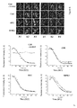
FIG. 5 illustrates results of detecting the binding with respect to CSK, SRC, EPHA4, and RIPK2 proteins, the binding being detected by dasatinib-biotin used as a second medium (CSK: C-src tyrosine kinase, SRC: Proto-oncogene tyrosine-protein kinase Src, EPHA4: ephrin type-A receptor 4, RIPK2: Receptor-interacting serine-threonine kinase 2, (-): before treatment with signal material (PMA), +PMA: after treatment with signal material, PKC-mRFP-SA: detection images of a first construct comprising streptavidin and RFP, EGFP-kinase: detection images of a third construct comprising GFP, Merge: merging of the detection images of PKC-mRFP-SA and detection images of EGFP-kinase) -
FIG. 6 illustrates results of detecting the binding with respect to CSK, SRC, and RIPK2 and charting the fluorescence change (reduction) in the cytosol (CSK: C-src tyrosine kinase, SRC: Proto-oncogene tyrosine-protein kinase Src, EPHA4: ephrin type-A receptor 4, RIPK2: Receptor-interacting serine-threonine kinase 2, A1: detection images of a first construct comprising RFP before treatment with PMA, A2: detection images of a third construct comprising GFP before treatment with PMA, A3: merging of the detection images of A1 and A2, B1: detection images of a first construct comprising RFP after treatment with PMA, B2: detection images of a third construct comprising GFP after treatment with PMA, B3: merging of the detection images of B1 and B2). -
FIG. 7 illustrates results of detecting the binding with respect to CSK, SRC, and RIPK2 and charting the fluorescence change (increase) in the cytosol (CSK: C-src tyrosine kinase, SRC: Proto-oncogene tyrosine-protein kinase Src, EPHA4: ephrin type-A receptor 4, RIPK2: Receptor-interacting serine-threonine kinase 2, A1: detection images of a first construct comprising RFP before treatment with PMA, A2: detection images of a third construct comprising GFP before treatment with PMA, A3: merging of the detection images of A1 and A2, B1: detection images of a first construct comprising RFP after treatment with PMA, B2: detection images of a third construct comprising GFP after treatment with PMA, B3: merging of the detection images of B1 and B2). -
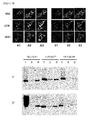
FIG. 8 illustrates detection images of the binding with respect to ABL kinase family binding with dasatinib and results of immunoprecipitation (IP) for verifying the binding (CSK: C-src tyrosine kinase, ABL1: V-abl Abelson murine leukemiaviral oncogene homolog 1, A1: detection images of a first construct comprising RFP before treatment with PMA, A2: detection images of a third construct comprising GFP before treatment with PMA, A3: merging of the detection images of A1 and A2, B1: detection images of a first construct comprising RFP after treatment with PMA, B2: detection images of a third construct comprising GFP after treatment with PMA, B3: merging of the detection images of B1 and B2). -
FIG. 9 illustrates detection images of the binding with respect to SRC kinase family binding with dasatinib and results of immunoprecipitation (IP) for verifying the binding (SRC: Proto-oncogene tyrosine-protein kinase Src, LYN: Tyrosine-protein kinase Lyn, YES1: Proto-oncogene tyrosine-protein kinase Yes, A1: detection images of a first construct comprising RFP before treatment with PMA, A2: detection images of a third construct comprising GFP before treatment with PMA, A3: merging of the detection images of A1 and A2, B1: detection images of a first construct comprising RFP after treatment with PMA, B2: detection images of a third construct comprising GFP after treatment with PMA, B3: merging of the detection images of B1 and B2). -
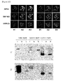
FIG. 10 illustrates detection images of the binding with respect to RIPK, MAP4K5, and SAPK2A proteins, which bind with dasatinib and of which functional correlation is not known, and results of immunoprecipitation (IP) for verifying the binding (RIPK2: Receptor-interacting serine-threonine kinase 2, MAP4K5: Mitogen-activated protein kinasekinase kinase kinase 5, SAPK2A: Stress Activated Protein Kinase 2a, A1: detection images of a first construct comprising RFP before treatment with PMA, A2: detection images of a third construct comprising GFP before treatment with PMA, A3: merging of the detection images of A1 and A2, B1: detection images of a first construct comprising RFP after treatment with PMA, B2: detection images of a third construct comprising GFP after treatment with PMA, B3: merging of the detection images of B1 and B2). -
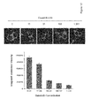
FIG. 11 illustrates results of verifying that phosphorylation activity of original unbiotinylated dasatinib was similar to that of biotinylated dasatinib. -
FIG. 12 illustrates the binding inhibitory effect of original unbiotinylated dasatinib on CSK protein which is translocated to the membrane by dasatinib-biotin (Dasatinib: unbiotinylated dasatinib). -
FIG. 13 shows (a) a schematic view of FRB and FKBP proteins of which the binding is detected by rapamycin-biotin, (b) a structure of rapamycin-biotin, and (c) the translocation to the plasma membrane and the translocation inhibitory effect by FK506 ((-) on the horizontal axis: before treatment with signal material (PMA), +PMA: after treatment with signal material, (-) on the vertical axis: rapamycin-biotin untreated group, FK506: tacrolimus, FKBP12:FK506 binding protein 12, FRB: FKBP12-rapamycin binding domain, PCK(C1A)-mRFP-SA: detection images of a first construct comprising streptavidin and RFP, EGFP-FRB: detection images of a third construct comprising GFP and FRB, TagBFP-FKBP12: detection images of a third construct including BFP and FKBP12, Merge: merging of the detection images of PCK(C1A)-mRFP-SA, EGFP-FRB, and TagBFP-FKBP12). -
FIG. 14 illustrates results for verifying the inhibition of the translocation of FRB protein depending on the concentration of FK506. - Hereinafter, the present invention will be described in detail by referring to the examples.
- However, the following examples are for illustrative purposes only and are not intended to limit the scope of the present invention.
- CHO-k1 (ATCC CCL-61, Cricetulus griseus, hamster, Chinese), HEK293 (ATCC CRL-1573, Homo sapiens, human), HeLa (ATCC CCL-2, Homo sapiens, human) and SH-SY5Y (ATCC CRL-2266, Homo sapiens, human) cell lines were used. The animal cells were cultured according to the instructions of ATCC (American Type Culture Collection) for the individual cells. CHO-k1 cells were cultured by using F-12 medium, while HEK293, HeLa and SH-SY5Y cells were cultured using DMEM medium. Other culturing conditions were the same. Culturing conditions commonly shared for the said cells as follows (Those skilled in the art may modify the specific conditions depending on purposes.). The cells were cultured in pH 7.4 medium (F-12 and DMEM) containing 25 mM HEPES, 10% fetal bovine serum (FBS, v/v), 100 units/ml penicillin and 100 µg/ml streptomycin in a 5% CO2 incubator maintained at 37 °C.
- In the Examples of the present invention, genes were introduced into the cells using ExGene 500 (Fermentas Life Science), one of the liposome-based techniques. All the conditions for the gene introduction including gene concentration were pursuant to the manufacturer's instructions. More specifically, after transferring the subcultured cells to a 12-well plate with a cover slip, followed by culturing for a day, the culture medium was replaced with 0.9 ml of fresh medium. About 1 µg of the transformation sample was added to 0.1 ml of 150 mM NaCl solution. After completely mixing, 3.3 µl of ExGene reagent was added and mixed by vortexing for 15 seconds. The resultant solution was allowed to stand at room temperature for 10 minutes and then added to each well of the 12-well plate in which the cells were growing. The cells were allowed to be transformed by culturing for 18 hours.
- A first construct according to the present invention is a fusion construct composed of a translocation module capable of moving a protein uniformly expressed in the cytoplasm of a cell toward the plasma membrane, a first labeling material analyzable by using a microscope, and a first medium capable of binding to a second medium.
- In the present example, protein kinase C was used as a translocation module, mRFP as a first labeling material, and streptavidin as a first medium.
- The translocation module was cloned by PCR techniques using the pCMV-SPORT6-PRKCD vector (GenBank accession No. BC043350; purchased from Openbiosystem (http://www.openbiosystems.com/); Catalog No. EHS1001-410108-BC043350) as a template, and then inserted at the NheI/AgeI site of the pmRFP-C3 vector (mRFP; GenBank accession No. DQ903889, SEQ ID NO: 14).
- Streptavidin as a first medium was cloned by PCR techniques using GenBank Acc. No. X03591 (SEQ ID NO: 13) as a template, and then inserted at the EcoRI/BamHI site of the pmRFP-C3 vector (mRFP; GenBank accession No. DQ903889, SEQ ID NO: 14).
- A second construct is composed of a second medium capable of binding to the first medium and a bait.
- In the present example, biotin was used as a second medium for streptavidin, while dasatinib and rapamycin were used as a bait. Commercialized dasatinib and rapamycin were respectively substituted with an amine group, and then commercialized NHS-Dpeg12-biotin was allowed to chemically bind thereto, thereby synthesizing dasatinib-biotin and rapamycin-biotin, respectively (See
FIG. 4 ). - The third construct comprises a prey which has characteristic for binding to the bait of the second construct and a labeling material for analyzing the movement of the prey. The third construct was prepared using a fluorescent material other than used in the first construct. Using green fluorescent protein (EGFP, AzG), red fluorescent protein (mRFP) and infrared fluorescent protein (HcR), the third construct was prepared by the same method described for the first construct.
- When EGFP was used as the second labeling material, a pEGFP-C3 vector (Clontech; SEQ ID NO: 15) was used. When mRFP was used, a pmRFP-C3 vector was used. When AzG was used, a pAzG-C3 vector was used. When HcR was used, a pHcR-C3 vector was used. The C3 vectors had been prepared by substituting the EGFP gene sequence site of the pEGFP-C3 vector with AzG and HcR genes, as follows.
- The pAzG-C3 vector was prepared as follows. PCR was carried out using a pPM-mAG1 vector (purchased from MBL, Catalog No. AM-V0203; Karasawa, S., et al. 2003, J. Biol. Chem. 278, 34167-34171) as a template and using SEQ ID NO: 16 (AzG-F: 5'-GGCACCGGTCGCCACCATGGACCCCATGGTGAGTGTGAT-3') and SEQ ID NO: 17 (AzG-R: 5'-GGCAGATCTGACAGCTTGGCCTGACTCGGCAGCAT-3') as primers. Then, the EGFP nucleotide sequence of the pEGFP-C3 vector was substituted at the AgeI/NotI site by the resultant PCR product.
- The pHcR-C3 vector was prepared as follows. PCR was carried out using pHcRed-Tandem-N1 (purchased from Avrogen, Catalog No. FP204; Gurskaya et al., 2001, FEBS Lett. 507, 16-20.) as a template and using SEQ ID NO: 18 (HcR-F: 5'-GCCACCGGTCGCCACCATGGTGAG-3') and SEQ ID NO: 19 (HcR-R: 5'-GCCGCGGCCGCTTATCAGTTGGCCTTCTCGGGCAGGTC-3') as primers. Then, the EGFP nucleotide sequence of the pEGFP-C3 vector was substituted at the AgeI/NotI site by the resultant PCR product.
- A cover slip containing the cells in which the first construct and the second construct vectors had been introduced was fixed to a perfusion chamber and mounted on the object stage of a confocal laser fluorescence microscope (Carl Zeiss LSM510). Images of the construct vectors were taken before and after external stimulation (treatment with 1 µM PMA).
- As for the confocal laser fluorescence microscope, 488 nm argon laser (EGFP or AzG), 543 nm HeNe laser (mRFP) or 561 nm DPSS laser (HcR) was used to induce the excitation of the fluorescent label, and the fluorescence signal generated by each fluorescent label was filtered through the band path filter BP505-530 (EGFP or AzG), long path filter LP560 or BP560-630 (mRFP) or long path filter LP650 (HcR). Images were taken after completely removing the interference between the fluorescences.
- An experiment for verifying a basic concept of the present invention that a compound (Oregon green) bound to biotin as the second medium of the second construct can be translocated to the plasma membrane by streptavidin as the first medium attached to the first construct was conducted. As a result, the translocation of the second construct to the plasma membrane by the translocation of the first construct was confirmed through images (See
FIG. 2 ). - For the verification of drug targets of dasatinib used as an anticancer drug, experiments of analyzing the binding of dasatinib with CSK, SRC, EPHA4, and RIPK2 proteins, which have been known as proteins binding to dasatinib, were conducted.
- As a result, as can be seen from
FIG. 5 , the respective proteins (green) were simultaneously translocated to the plasma membrane by PMA as a stimulation for translocation while the first construct (red) and the second construct (dasatinib-biotin) were present. - For the verification of the intracellular binding with respect to CSK, SRC, EPHA4, and RIPK2 proteins, the first construct PKC-mRFP-streptavidin, the third construct GFP-protein expressing vector, and the second construct dasatinib-biotin were prepared as follows. The first construct PKC-mRFP-streptavidin was prepared according to the method of Example <2-1>. The second construct dasatinib-biotin was prepared according to the method of Example <2-2>. The third construct EGFP-protein was prepared by PCR amplification using a human-derived gene as a template, which is obvious to those skilled in the art, and then inserting the resultant PCR product into the pEGFP-C3 vector.
- The first construct (PKC-mRFP-streptavidin) and the third construct (EGFP-protein) were introduced into CHO-k1 cells by the methods described in Example <1-2> (ExGene 500), and then allowed to be expressed in an environment into which the second construct (dasatinib-biotin) was introduced. After the 18-hour culturing and 1 uM PMA treatment for 5 minutes, the fluorescent distribution was observed as in Example <3-1>.
- The second construct dasatinib-biotin was introduced into a medium in which cells expressing the first construct and the second construct were grown, and then was allowed to infiltrate into the cells by culturing for 4 hours.
- As a result, it can be seen that the fluorescence of the third construct (green) to which the prey binding to dasatinib was bound was translocated to the plasma membrane at the time of PMA treatment, like in the red fluorescence (the first construct) to which the translocation module was bound (
FIGS. 5 ,6 ,7 ,8 ,9 ,10 , and12 ) - On the contrary, as for treatment with unbiotinylated dasatinib in its different concentrations, as can be seen from the results shown in
FIG. 12 , the translocation of CSK protein (third construct, green) to the plasma membrane was inhibited by dasatinib in a concentration-dependent manner. - The proteins bound to dasatinib as a compound were confirmed through imaging analysis, and the binding of the respective proteins was again verified through immunoprecipitation (IP), which is obvious to those skilled in the art (See lower panels of
FIGS. 8 ,9 , and10 , I: Input, B: biotin, D: biotin-dasatinib). As can be seen fromFIG. 11 , the phosphorylation activity of the biotinylated compound (dasatinib) was very similar to that of the original unbiotinylated compound. Therefore, it was confirmed that a prey protein can be identified under physiological conditions in which the present invention does not affect the change in drug target activity. - The above-described examples verified the effectiveness of the present invention. Also, the generalization of the methods of the present invention was confirmed by using other compounds (See
FIG. 13 ). When the biotin was bound to rapamycin as another anticancer drug, it was confirmed that FRB and FKBP12 proteins which have been known to bind with rapamycin were simultaneously detected, and the binding was inhibited by another compound FK506 which has been known as a competitive inhibitor against rapamycin. This verifies that the protein binding changes depending on the change in concentration of the competitive inhibitor, and thus the target protein FRB and FKBP12 specifically bind to rapamycin-biotin. - As set forth above, the present invention provides a method for screening a prey which interacts with a bait, the method including: (a) preparing a cell which expresses (i) a first construct comprising a translocation module, a first labeling material, and a first medium, and (ii) a third construct comprising a prey and a second labeling material; (b) introducing (iii) a second construct into the prepared cell, the second construct comprising a bait and a second medium binding with the first medium; (c) allowing the prey and the bait to interact each other; and (d) confirming the interaction between the prey and the bait by detecting intracellular distributions of the first construct and the third construct. Further, the present invention provides a cell expressing the first construct and the third construct. Further, the present invention provides a screening kit comprising the cell and the second construct. The methods of the present invention can overcome disadvantages including inaccuracy and complexity of the existing techniques for biomaterial interaction detection, and can measure the interaction between a material which is not directly produced through gene translation and gene expression in the cell and a material expressed in the cell, and thus are effective in detecting interactions of a broad range of biomaterials so that the present invention is highly industrially applicable.


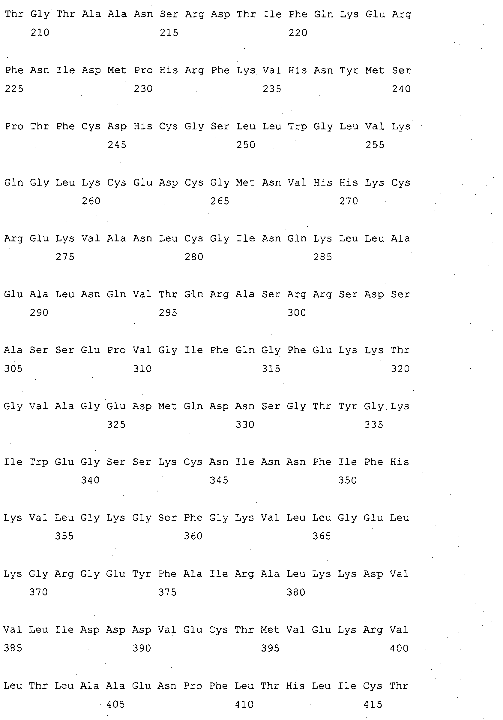




Claims (16)
- A method for screening a prey which interacts with a bait, the method including(a) preparing a cell which expresses (i) a first construct comprising a translocation module, a first labeling material, and a first medium, and (ii) a third construct comprising a prey and a second labeling material;(b) introducing (iii) a second construct into the prepared cell, the second construct comprising a bait and a second medium binding with the first medium;(c) allowing the prey and the bait to interact each other; and(d) confirming the interaction between the prey and the bait by detecting intracellular distributions of the first construct and the third construct.
- The method of claim 1, wherein in step (c), the prey and the bait are allowed to interact each other by treatment with a signal material.
- The method of claim 1 or 2, wherein the translocation module is selected from the group consisting of protein kinase C and a variant thereof.
- The method of claim 3, wherein the protein kinase C and the variant thereof have amino acid sequences selected from the group consisting of SEQ ID NO: 1, SEQ ID NO: 3, SEQ ID NO: 5, and SEQ ID NO: 7.
- The method of claim 1 or 2, wherein the first labeling material is selected from the group consisting of green fluorescent protein (GFP), enhanced green fluorescent protein (EGFP), red fluorescent protein (RFP), monomeric red fluorescent protein (mRFP), Discosoma sp. red fluorescent protein (DsRed), cyan fluorescent protein (CFP), cyan green fluorescent protein (CGFP), yellow fluorescent protein (YFP), azami green (AzG), Heteractis crispa red fluorescent protein (HcR, HcRed), and blue fluorescent protein (BFP).
- The method of claim 1 or 2, wherein the second labeling material is selected from the group consisting of green fluorescent protein (GFP), enhanced green fluorescent protein (EGFP), red fluorescent protein (RFP), monomeric red fluorescent protein (mRFP), Discosoma sp. red fluorescent protein (DsRed), cyan fluorescent protein (CFP), cyan green fluorescent protein (CGFP), yellow fluorescent protein (YFP), azami green (AzG), Heteractis crispa red fluorescent protein (HcR, HcRed), and blue fluorescent protein (BFP).
- The method of claim 2, wherein the treatment with a signal material is performed by 50 nM to 5 uM of phorbol 12-myristate 13-acetate (PMA, phorbol ester).
- The method of claim 1 or 2, wherein the first medium is streptavidin and the second medium is biotin.
- The method of claim 1 or 2, wherein the first medium is dihydrofolate reductase (DHFR) and the second medium is methotrexate (MTX).
- A cell expressing (i) a first construct comprising a translocation module, a first labeling material, and a first medium; and (ii) a third construct comprising a prey and a second labeling material.
- The cell of claim 10, wherein the cell is selected from the group consisting of CHO-k1 cell, HEK293 cell, HeLa cell, SH-SY5Y cell, Swiss 3T3 cell, 3T3-L1 cell, NIH/3T3 cell, L-929 cell, Rat2 cell, RBL-2H3 cell, and MDCK cell.
- The cell of claim 10, wherein the first medium is streptavidin or dihydrofolate reductase (DHFR).
- A kit for screening a prey interacting with a bait, the kit comprising the cell of claim 10; and (iii) a second construct comprising a second medium binding with a first medium and a bait.
- The kit of claim 13, wherein the first medium is streptavidin and the second medium is biotin.
- The kit of claim 13, wherein the first medium is dihydrofolate reductase (DHFR) and the second medium is methotrexate (MTX).
- The kit of claim 13, further comprising a signal material for allowing the prey and the bait to interact each other.
Applications Claiming Priority (2)
| Application Number | Priority Date | Filing Date | Title |
|---|---|---|---|
| KR1020110137868A KR101212029B1 (en) | 2011-12-20 | 2011-12-20 | Method for detecting interactions between molecular compound and its binding proteins |
| PCT/KR2012/011195 WO2013095014A1 (en) | 2011-12-20 | 2012-12-20 | Method for detecting compound-binding protein |
Publications (3)
| Publication Number | Publication Date |
|---|---|
| EP2799551A1 true EP2799551A1 (en) | 2014-11-05 |
| EP2799551A4 EP2799551A4 (en) | 2015-03-11 |
| EP2799551B1 EP2799551B1 (en) | 2018-07-04 |
Family
ID=47907604
Family Applications (1)
| Application Number | Title | Priority Date | Filing Date |
|---|---|---|---|
| EP12860693.6A Not-in-force EP2799551B1 (en) | 2011-12-20 | 2012-12-20 | Method for detecting compound-binding protein |
Country Status (5)
| Country | Link |
|---|---|
| US (1) | US20140335545A1 (en) |
| EP (1) | EP2799551B1 (en) |
| JP (1) | JP2015502174A (en) |
| KR (1) | KR101212029B1 (en) |
| WO (1) | WO2013095014A1 (en) |
Cited By (1)
| Publication number | Priority date | Publication date | Assignee | Title |
|---|---|---|---|---|
| EP3994251A4 (en) * | 2019-07-01 | 2023-09-20 | The Governing Council of the University of Toronto | DETECTION OF PROTEIN-PROTEIN INTERACTIONS |
Families Citing this family (1)
| Publication number | Priority date | Publication date | Assignee | Title |
|---|---|---|---|---|
| KR101820381B1 (en) | 2016-11-23 | 2018-01-19 | 한국과학기술연구원 | AZD4547 Novel Derivatives |
Family Cites Families (10)
| Publication number | Priority date | Publication date | Assignee | Title |
|---|---|---|---|---|
| US6551843B1 (en) * | 1999-01-29 | 2003-04-22 | Immunivest Corporation | Methods for enhancing binding interactions between members of specific binding pairs |
| US6656700B2 (en) * | 2000-05-26 | 2003-12-02 | Amersham Plc | Isoforms of human pregnancy-associated protein-E |
| CA2444857A1 (en) * | 2001-04-20 | 2002-10-31 | President And Fellows Of Harvard College | Compositions and methods for the identification of protein interactions in vertebrate cells |
| WO2003029827A2 (en) * | 2001-10-01 | 2003-04-10 | Bioimage A/S | A method of detecting intracellular protein-protein interactions using three heterologous conjugates |
| US20080242557A1 (en) * | 2004-07-20 | 2008-10-02 | Cgk Co., Ltd. | System For Detecting Molecular Interactions |
| JP4123251B2 (en) * | 2005-07-07 | 2008-07-23 | セイコーエプソン株式会社 | Semiconductor device manufacturing substrate and semiconductor device manufacturing method |
| US20100112602A1 (en) * | 2006-11-10 | 2010-05-06 | Taylor Lansing D | Protein-Protein Interaction Biosensors and Methods of Use Thereof |
| WO2009049892A1 (en) * | 2007-10-17 | 2009-04-23 | European Molecular Biology Laboratory (Embl) | Polypeptides comprising an annexin core domain, compositions, methods and use thereof |
| WO2009117409A2 (en) * | 2008-03-18 | 2009-09-24 | Merck & Co., Inc. | A high throughput protein interaction assay |
| KR100948767B1 (en) * | 2008-11-12 | 2010-03-23 | 한국기초과학지원연구원 | Method for detecting interactions between two and more biological macromolecules |
-
2011
- 2011-12-20 KR KR1020110137868A patent/KR101212029B1/en not_active Expired - Fee Related
-
2012
- 2012-12-20 WO PCT/KR2012/011195 patent/WO2013095014A1/en not_active Ceased
- 2012-12-20 EP EP12860693.6A patent/EP2799551B1/en not_active Not-in-force
- 2012-12-20 JP JP2014548666A patent/JP2015502174A/en active Pending
-
2014
- 2014-06-20 US US14/310,758 patent/US20140335545A1/en not_active Abandoned
Cited By (1)
| Publication number | Priority date | Publication date | Assignee | Title |
|---|---|---|---|---|
| EP3994251A4 (en) * | 2019-07-01 | 2023-09-20 | The Governing Council of the University of Toronto | DETECTION OF PROTEIN-PROTEIN INTERACTIONS |
Also Published As
| Publication number | Publication date |
|---|---|
| WO2013095014A1 (en) | 2013-06-27 |
| US20140335545A1 (en) | 2014-11-13 |
| WO2013095014A8 (en) | 2014-06-26 |
| EP2799551B1 (en) | 2018-07-04 |
| KR101212029B1 (en) | 2012-12-13 |
| EP2799551A4 (en) | 2015-03-11 |
| JP2015502174A (en) | 2015-01-22 |
Similar Documents
| Publication | Publication Date | Title |
|---|---|---|
| Kim et al. | Time-gated detection of protein-protein interactions with transcriptional readout | |
| EP2187213B1 (en) | Methods for detecting interactions between two and more polypeptides | |
| EP2647993B1 (en) | Methods for detecting molecular interactions | |
| Donthamsetti et al. | Using bioluminescence resonance energy transfer (BRET) to characterize agonist‐induced arrestin recruitment to modified and unmodified G protein‐coupled receptors | |
| Saka et al. | Nuclear accumulation of Smad complexes occurs only after the midblastula transition in Xenopus | |
| DK2699910T3 (en) | A method for determining ligand binding to a target protein using a thermal shift assay | |
| CN114487386A (en) | ELISA detection method for poultry-derived exosomes | |
| EP2799551B1 (en) | Method for detecting compound-binding protein | |
| Smith et al. | High throughput screening for compounds to the orphan nuclear receptor NR2F6 | |
| JP6986054B2 (en) | Human cell model with biosensor | |
| KR101232752B1 (en) | Method for detecting molecular interactions and kit therefor | |
| Wehr et al. | Monitoring G protein-coupled receptor activation using the protein fragment complementation technique split TEV | |
| JP2010530970A (en) | Method for screening selective modulator of NF-κB pathway activation | |
| US9193984B2 (en) | Method for detecting interactions between two and more biological macromolecules | |
| Van Wijk et al. | Selective monitoring of ubiquitin signals with genetically encoded ubiquitin chain–specific sensors | |
| WO2007058454A1 (en) | System for detecting molecular interactions | |
| KR101444995B1 (en) | Method for detecting protein-protein interaction of cytosolic proteins | |
| Salgania et al. | ReLo: a simple colocalization assay to identify and characterize physical protein-protein interactions | |
| KR101749670B1 (en) | Method for screening anti-cancer agents comprising detecting interactions of IkBα and RPS6KA2 | |
| Loechel et al. | High content translocation assays for pathway profiling | |
| Zou et al. | OptoRibo-seq for spatiotemporally resolved mapping of local protein translatome | |
| CN108570451A (en) | A kind of application of cell model and its construction method and screening HRH1 target drugs | |
| CN112391351A (en) | Opioid receptor allosteric modulator screening model and application | |
| Tran | Evaluation of the phage display protocol for target identification of small molecules: Identification and characterization of mitosis modulators | |
| Umezawa | Detecting mitochondrial RNA and other cellular events in living cells |
Legal Events
| Date | Code | Title | Description |
|---|---|---|---|
| PUAI | Public reference made under article 153(3) epc to a published international application that has entered the european phase |
Free format text: ORIGINAL CODE: 0009012 |
|
| 17P | Request for examination filed |
Effective date: 20140624 |
|
| AK | Designated contracting states |
Kind code of ref document: A1 Designated state(s): AL AT BE BG CH CY CZ DE DK EE ES FI FR GB GR HR HU IE IS IT LI LT LU LV MC MK MT NL NO PL PT RO RS SE SI SK SM TR |
|
| RIN1 | Information on inventor provided before grant (corrected) |
Inventor name: LEE, KYUNG-BOK Inventor name: KIM, SEUNG II Inventor name: HWANG, JUNG ME Inventor name: KIM, SOOHYUN Inventor name: LEE, ZEE-WON |
|
| A4 | Supplementary search report drawn up and despatched |
Effective date: 20150211 |
|
| RIC1 | Information provided on ipc code assigned before grant |
Ipc: C12N 15/10 20060101AFI20150205BHEP Ipc: G01N 33/50 20060101ALI20150205BHEP Ipc: G01N 33/58 20060101ALI20150205BHEP Ipc: C12N 5/00 20060101ALI20150205BHEP |
|
| DAX | Request for extension of the european patent (deleted) | ||
| 17Q | First examination report despatched |
Effective date: 20151120 |
|
| STAA | Information on the status of an ep patent application or granted ep patent |
Free format text: STATUS: EXAMINATION IS IN PROGRESS |
|
| REG | Reference to a national code |
Ref country code: DE Ref legal event code: R079 Ref document number: 602012048225 Country of ref document: DE Free format text: PREVIOUS MAIN CLASS: C12N0015630000 Ipc: C12N0015100000 |
|
| GRAP | Despatch of communication of intention to grant a patent |
Free format text: ORIGINAL CODE: EPIDOSNIGR1 |
|
| STAA | Information on the status of an ep patent application or granted ep patent |
Free format text: STATUS: GRANT OF PATENT IS INTENDED |
|
| RIC1 | Information provided on ipc code assigned before grant |
Ipc: C12N 15/10 20060101AFI20180202BHEP Ipc: G01N 33/58 20060101ALI20180202BHEP Ipc: G01N 33/50 20060101ALI20180202BHEP |
|
| INTG | Intention to grant announced |
Effective date: 20180220 |
|
| GRAS | Grant fee paid |
Free format text: ORIGINAL CODE: EPIDOSNIGR3 |
|
| GRAA | (expected) grant |
Free format text: ORIGINAL CODE: 0009210 |
|
| STAA | Information on the status of an ep patent application or granted ep patent |
Free format text: STATUS: THE PATENT HAS BEEN GRANTED |
|
| AK | Designated contracting states |
Kind code of ref document: B1 Designated state(s): AL AT BE BG CH CY CZ DE DK EE ES FI FR GB GR HR HU IE IS IT LI LT LU LV MC MK MT NL NO PL PT RO RS SE SI SK SM TR |
|
| REG | Reference to a national code |
Ref country code: GB Ref legal event code: FG4D |
|
| REG | Reference to a national code |
Ref country code: CH Ref legal event code: EP |
|
| REG | Reference to a national code |
Ref country code: AT Ref legal event code: REF Ref document number: 1014552 Country of ref document: AT Kind code of ref document: T Effective date: 20180715 |
|
| REG | Reference to a national code |
Ref country code: IE Ref legal event code: FG4D |
|
| REG | Reference to a national code |
Ref country code: DE Ref legal event code: R096 Ref document number: 602012048225 Country of ref document: DE |
|
| REG | Reference to a national code |
Ref country code: NL Ref legal event code: MP Effective date: 20180704 |
|
| REG | Reference to a national code |
Ref country code: LT Ref legal event code: MG4D |
|
| REG | Reference to a national code |
Ref country code: AT Ref legal event code: MK05 Ref document number: 1014552 Country of ref document: AT Kind code of ref document: T Effective date: 20180704 |
|
| PG25 | Lapsed in a contracting state [announced via postgrant information from national office to epo] |
Ref country code: NL Free format text: LAPSE BECAUSE OF FAILURE TO SUBMIT A TRANSLATION OF THE DESCRIPTION OR TO PAY THE FEE WITHIN THE PRESCRIBED TIME-LIMIT Effective date: 20180704 |
|
| PG25 | Lapsed in a contracting state [announced via postgrant information from national office to epo] |
Ref country code: SE Free format text: LAPSE BECAUSE OF FAILURE TO SUBMIT A TRANSLATION OF THE DESCRIPTION OR TO PAY THE FEE WITHIN THE PRESCRIBED TIME-LIMIT Effective date: 20180704 Ref country code: BG Free format text: LAPSE BECAUSE OF FAILURE TO SUBMIT A TRANSLATION OF THE DESCRIPTION OR TO PAY THE FEE WITHIN THE PRESCRIBED TIME-LIMIT Effective date: 20181004 Ref country code: AT Free format text: LAPSE BECAUSE OF FAILURE TO SUBMIT A TRANSLATION OF THE DESCRIPTION OR TO PAY THE FEE WITHIN THE PRESCRIBED TIME-LIMIT Effective date: 20180704 Ref country code: GR Free format text: LAPSE BECAUSE OF FAILURE TO SUBMIT A TRANSLATION OF THE DESCRIPTION OR TO PAY THE FEE WITHIN THE PRESCRIBED TIME-LIMIT Effective date: 20181005 Ref country code: LT Free format text: LAPSE BECAUSE OF FAILURE TO SUBMIT A TRANSLATION OF THE DESCRIPTION OR TO PAY THE FEE WITHIN THE PRESCRIBED TIME-LIMIT Effective date: 20180704 Ref country code: PL Free format text: LAPSE BECAUSE OF FAILURE TO SUBMIT A TRANSLATION OF THE DESCRIPTION OR TO PAY THE FEE WITHIN THE PRESCRIBED TIME-LIMIT Effective date: 20180704 Ref country code: FI Free format text: LAPSE BECAUSE OF FAILURE TO SUBMIT A TRANSLATION OF THE DESCRIPTION OR TO PAY THE FEE WITHIN THE PRESCRIBED TIME-LIMIT Effective date: 20180704 Ref country code: CZ Free format text: LAPSE BECAUSE OF FAILURE TO SUBMIT A TRANSLATION OF THE DESCRIPTION OR TO PAY THE FEE WITHIN THE PRESCRIBED TIME-LIMIT Effective date: 20180704 Ref country code: IS Free format text: LAPSE BECAUSE OF FAILURE TO SUBMIT A TRANSLATION OF THE DESCRIPTION OR TO PAY THE FEE WITHIN THE PRESCRIBED TIME-LIMIT Effective date: 20181104 Ref country code: RS Free format text: LAPSE BECAUSE OF FAILURE TO SUBMIT A TRANSLATION OF THE DESCRIPTION OR TO PAY THE FEE WITHIN THE PRESCRIBED TIME-LIMIT Effective date: 20180704 Ref country code: NO Free format text: LAPSE BECAUSE OF FAILURE TO SUBMIT A TRANSLATION OF THE DESCRIPTION OR TO PAY THE FEE WITHIN THE PRESCRIBED TIME-LIMIT Effective date: 20181004 |
|
| PG25 | Lapsed in a contracting state [announced via postgrant information from national office to epo] |
Ref country code: LV Free format text: LAPSE BECAUSE OF FAILURE TO SUBMIT A TRANSLATION OF THE DESCRIPTION OR TO PAY THE FEE WITHIN THE PRESCRIBED TIME-LIMIT Effective date: 20180704 Ref country code: HR Free format text: LAPSE BECAUSE OF FAILURE TO SUBMIT A TRANSLATION OF THE DESCRIPTION OR TO PAY THE FEE WITHIN THE PRESCRIBED TIME-LIMIT Effective date: 20180704 Ref country code: ES Free format text: LAPSE BECAUSE OF FAILURE TO SUBMIT A TRANSLATION OF THE DESCRIPTION OR TO PAY THE FEE WITHIN THE PRESCRIBED TIME-LIMIT Effective date: 20180704 Ref country code: AL Free format text: LAPSE BECAUSE OF FAILURE TO SUBMIT A TRANSLATION OF THE DESCRIPTION OR TO PAY THE FEE WITHIN THE PRESCRIBED TIME-LIMIT Effective date: 20180704 |
|
| REG | Reference to a national code |
Ref country code: DE Ref legal event code: R097 Ref document number: 602012048225 Country of ref document: DE |
|
| PG25 | Lapsed in a contracting state [announced via postgrant information from national office to epo] |
Ref country code: RO Free format text: LAPSE BECAUSE OF FAILURE TO SUBMIT A TRANSLATION OF THE DESCRIPTION OR TO PAY THE FEE WITHIN THE PRESCRIBED TIME-LIMIT Effective date: 20180704 Ref country code: IT Free format text: LAPSE BECAUSE OF FAILURE TO SUBMIT A TRANSLATION OF THE DESCRIPTION OR TO PAY THE FEE WITHIN THE PRESCRIBED TIME-LIMIT Effective date: 20180704 Ref country code: EE Free format text: LAPSE BECAUSE OF FAILURE TO SUBMIT A TRANSLATION OF THE DESCRIPTION OR TO PAY THE FEE WITHIN THE PRESCRIBED TIME-LIMIT Effective date: 20180704 |
|
| PLBE | No opposition filed within time limit |
Free format text: ORIGINAL CODE: 0009261 |
|
| STAA | Information on the status of an ep patent application or granted ep patent |
Free format text: STATUS: NO OPPOSITION FILED WITHIN TIME LIMIT |
|
| PG25 | Lapsed in a contracting state [announced via postgrant information from national office to epo] |
Ref country code: SK Free format text: LAPSE BECAUSE OF FAILURE TO SUBMIT A TRANSLATION OF THE DESCRIPTION OR TO PAY THE FEE WITHIN THE PRESCRIBED TIME-LIMIT Effective date: 20180704 Ref country code: DK Free format text: LAPSE BECAUSE OF FAILURE TO SUBMIT A TRANSLATION OF THE DESCRIPTION OR TO PAY THE FEE WITHIN THE PRESCRIBED TIME-LIMIT Effective date: 20180704 Ref country code: SM Free format text: LAPSE BECAUSE OF FAILURE TO SUBMIT A TRANSLATION OF THE DESCRIPTION OR TO PAY THE FEE WITHIN THE PRESCRIBED TIME-LIMIT Effective date: 20180704 |
|
| 26N | No opposition filed |
Effective date: 20190405 |
|
| REG | Reference to a national code |
Ref country code: CH Ref legal event code: PL |
|
| PG25 | Lapsed in a contracting state [announced via postgrant information from national office to epo] |
Ref country code: LU Free format text: LAPSE BECAUSE OF NON-PAYMENT OF DUE FEES Effective date: 20181220 Ref country code: SI Free format text: LAPSE BECAUSE OF FAILURE TO SUBMIT A TRANSLATION OF THE DESCRIPTION OR TO PAY THE FEE WITHIN THE PRESCRIBED TIME-LIMIT Effective date: 20180704 Ref country code: MC Free format text: LAPSE BECAUSE OF FAILURE TO SUBMIT A TRANSLATION OF THE DESCRIPTION OR TO PAY THE FEE WITHIN THE PRESCRIBED TIME-LIMIT Effective date: 20180704 |
|
| REG | Reference to a national code |
Ref country code: IE Ref legal event code: MM4A |
|
| REG | Reference to a national code |
Ref country code: BE Ref legal event code: MM Effective date: 20181231 |
|
| PG25 | Lapsed in a contracting state [announced via postgrant information from national office to epo] |
Ref country code: IE Free format text: LAPSE BECAUSE OF NON-PAYMENT OF DUE FEES Effective date: 20181220 |
|
| PG25 | Lapsed in a contracting state [announced via postgrant information from national office to epo] |
Ref country code: BE Free format text: LAPSE BECAUSE OF NON-PAYMENT OF DUE FEES Effective date: 20181231 |
|
| PG25 | Lapsed in a contracting state [announced via postgrant information from national office to epo] |
Ref country code: LI Free format text: LAPSE BECAUSE OF NON-PAYMENT OF DUE FEES Effective date: 20181231 Ref country code: CH Free format text: LAPSE BECAUSE OF NON-PAYMENT OF DUE FEES Effective date: 20181231 |
|
| PG25 | Lapsed in a contracting state [announced via postgrant information from national office to epo] |
Ref country code: MT Free format text: LAPSE BECAUSE OF NON-PAYMENT OF DUE FEES Effective date: 20181220 |
|
| PG25 | Lapsed in a contracting state [announced via postgrant information from national office to epo] |
Ref country code: TR Free format text: LAPSE BECAUSE OF FAILURE TO SUBMIT A TRANSLATION OF THE DESCRIPTION OR TO PAY THE FEE WITHIN THE PRESCRIBED TIME-LIMIT Effective date: 20180704 |
|
| PG25 | Lapsed in a contracting state [announced via postgrant information from national office to epo] |
Ref country code: PT Free format text: LAPSE BECAUSE OF FAILURE TO SUBMIT A TRANSLATION OF THE DESCRIPTION OR TO PAY THE FEE WITHIN THE PRESCRIBED TIME-LIMIT Effective date: 20180704 |
|
| PG25 | Lapsed in a contracting state [announced via postgrant information from national office to epo] |
Ref country code: MK Free format text: LAPSE BECAUSE OF NON-PAYMENT OF DUE FEES Effective date: 20180704 Ref country code: HU Free format text: LAPSE BECAUSE OF FAILURE TO SUBMIT A TRANSLATION OF THE DESCRIPTION OR TO PAY THE FEE WITHIN THE PRESCRIBED TIME-LIMIT; INVALID AB INITIO Effective date: 20121220 Ref country code: CY Free format text: LAPSE BECAUSE OF FAILURE TO SUBMIT A TRANSLATION OF THE DESCRIPTION OR TO PAY THE FEE WITHIN THE PRESCRIBED TIME-LIMIT Effective date: 20180704 |
|
| REG | Reference to a national code |
Ref country code: DE Ref legal event code: R081 Ref document number: 602012048225 Country of ref document: DE Owner name: LEE, ZEE WON, DAEJEON, KR Free format text: FORMER OWNER: KOREA BASIC SCIENCE INSTITUTE, DAEJEON, KR |
|
| PGFP | Annual fee paid to national office [announced via postgrant information from national office to epo] |
Ref country code: DE Payment date: 20211027 Year of fee payment: 10 Ref country code: FR Payment date: 20211115 Year of fee payment: 10 Ref country code: GB Payment date: 20211028 Year of fee payment: 10 |
|
| REG | Reference to a national code |
Ref country code: GB Ref legal event code: 732E Free format text: REGISTERED BETWEEN 20220203 AND 20220209 |
|
| REG | Reference to a national code |
Ref country code: DE Ref legal event code: R119 Ref document number: 602012048225 Country of ref document: DE |
|
| GBPC | Gb: european patent ceased through non-payment of renewal fee |
Effective date: 20221220 |
|
| PG25 | Lapsed in a contracting state [announced via postgrant information from national office to epo] |
Ref country code: GB Free format text: LAPSE BECAUSE OF NON-PAYMENT OF DUE FEES Effective date: 20221220 Ref country code: DE Free format text: LAPSE BECAUSE OF NON-PAYMENT OF DUE FEES Effective date: 20230701 |
|
| PG25 | Lapsed in a contracting state [announced via postgrant information from national office to epo] |
Ref country code: FR Free format text: LAPSE BECAUSE OF NON-PAYMENT OF DUE FEES Effective date: 20221231 |