EP2799008A1 - Method for continuous and non-invasive determination of effective lung volume and cardiac output - Google Patents
Method for continuous and non-invasive determination of effective lung volume and cardiac output Download PDFInfo
- Publication number
- EP2799008A1 EP2799008A1 EP14173101.8A EP14173101A EP2799008A1 EP 2799008 A1 EP2799008 A1 EP 2799008A1 EP 14173101 A EP14173101 A EP 14173101A EP 2799008 A1 EP2799008 A1 EP 2799008A1
- Authority
- EP
- European Patent Office
- Prior art keywords
- subject
- carbon dioxide
- breaths
- ventilation
- dioxide content
- Prior art date
- Legal status (The legal status is an assumption and is not a legal conclusion. Google has not performed a legal analysis and makes no representation as to the accuracy of the status listed.)
- Granted
Links
Images


Classifications
-
- A—HUMAN NECESSITIES
- A61—MEDICAL OR VETERINARY SCIENCE; HYGIENE
- A61B—DIAGNOSIS; SURGERY; IDENTIFICATION
- A61B5/00—Measuring for diagnostic purposes; Identification of persons
- A61B5/08—Detecting, measuring or recording devices for evaluating the respiratory organs
- A61B5/082—Evaluation by breath analysis, e.g. determination of the chemical composition of exhaled breath
-
- A—HUMAN NECESSITIES
- A61—MEDICAL OR VETERINARY SCIENCE; HYGIENE
- A61B—DIAGNOSIS; SURGERY; IDENTIFICATION
- A61B5/00—Measuring for diagnostic purposes; Identification of persons
- A61B5/72—Signal processing specially adapted for physiological signals or for diagnostic purposes
-
- A—HUMAN NECESSITIES
- A61—MEDICAL OR VETERINARY SCIENCE; HYGIENE
- A61B—DIAGNOSIS; SURGERY; IDENTIFICATION
- A61B5/00—Measuring for diagnostic purposes; Identification of persons
- A61B5/02—Detecting, measuring or recording pulse, heart rate, blood pressure or blood flow; Combined pulse/heart-rate/blood pressure determination; Evaluating a cardiovascular condition not otherwise provided for, e.g. using combinations of techniques provided for in this group with electrocardiography or electroauscultation; Heart catheters for measuring blood pressure
- A61B5/026—Measuring blood flow
- A61B5/029—Measuring or recording blood output from the heart, e.g. minute volume
-
- A—HUMAN NECESSITIES
- A61—MEDICAL OR VETERINARY SCIENCE; HYGIENE
- A61B—DIAGNOSIS; SURGERY; IDENTIFICATION
- A61B5/00—Measuring for diagnostic purposes; Identification of persons
- A61B5/08—Detecting, measuring or recording devices for evaluating the respiratory organs
- A61B5/083—Measuring rate of metabolism by using breath test, e.g. measuring rate of oxygen consumption
- A61B5/0836—Measuring rate of CO2 production
-
- A—HUMAN NECESSITIES
- A61—MEDICAL OR VETERINARY SCIENCE; HYGIENE
- A61B—DIAGNOSIS; SURGERY; IDENTIFICATION
- A61B5/00—Measuring for diagnostic purposes; Identification of persons
- A61B5/08—Detecting, measuring or recording devices for evaluating the respiratory organs
- A61B5/087—Measuring breath flow
-
- A—HUMAN NECESSITIES
- A61—MEDICAL OR VETERINARY SCIENCE; HYGIENE
- A61M—DEVICES FOR INTRODUCING MEDIA INTO, OR ONTO, THE BODY; DEVICES FOR TRANSDUCING BODY MEDIA OR FOR TAKING MEDIA FROM THE BODY; DEVICES FOR PRODUCING OR ENDING SLEEP OR STUPOR
- A61M16/00—Devices for influencing the respiratory system of patients by gas treatment, e.g. mouth-to-mouth respiration; Tracheal tubes
- A61M16/0051—Devices for influencing the respiratory system of patients by gas treatment, e.g. mouth-to-mouth respiration; Tracheal tubes with alarm devices
-
- A—HUMAN NECESSITIES
- A61—MEDICAL OR VETERINARY SCIENCE; HYGIENE
- A61M—DEVICES FOR INTRODUCING MEDIA INTO, OR ONTO, THE BODY; DEVICES FOR TRANSDUCING BODY MEDIA OR FOR TAKING MEDIA FROM THE BODY; DEVICES FOR PRODUCING OR ENDING SLEEP OR STUPOR
- A61M16/00—Devices for influencing the respiratory system of patients by gas treatment, e.g. mouth-to-mouth respiration; Tracheal tubes
- A61M16/021—Devices for influencing the respiratory system of patients by gas treatment, e.g. mouth-to-mouth respiration; Tracheal tubes operated by electrical means
- A61M16/022—Control means therefor
- A61M16/024—Control means therefor including calculation means, e.g. using a processor
- A61M16/026—Control means therefor including calculation means, e.g. using a processor specially adapted for predicting, e.g. for determining an information representative of a flow limitation during a ventilation cycle by using a root square technique or a regression analysis
-
- G—PHYSICS
- G16—INFORMATION AND COMMUNICATION TECHNOLOGY [ICT] SPECIALLY ADAPTED FOR SPECIFIC APPLICATION FIELDS
- G16H—HEALTHCARE INFORMATICS, i.e. INFORMATION AND COMMUNICATION TECHNOLOGY [ICT] SPECIALLY ADAPTED FOR THE HANDLING OR PROCESSING OF MEDICAL OR HEALTHCARE DATA
- G16H50/00—ICT specially adapted for medical diagnosis, medical simulation or medical data mining; ICT specially adapted for detecting, monitoring or modelling epidemics or pandemics
- G16H50/50—ICT specially adapted for medical diagnosis, medical simulation or medical data mining; ICT specially adapted for detecting, monitoring or modelling epidemics or pandemics for simulation or modelling of medical disorders
-
- A—HUMAN NECESSITIES
- A61—MEDICAL OR VETERINARY SCIENCE; HYGIENE
- A61M—DEVICES FOR INTRODUCING MEDIA INTO, OR ONTO, THE BODY; DEVICES FOR TRANSDUCING BODY MEDIA OR FOR TAKING MEDIA FROM THE BODY; DEVICES FOR PRODUCING OR ENDING SLEEP OR STUPOR
- A61M16/00—Devices for influencing the respiratory system of patients by gas treatment, e.g. mouth-to-mouth respiration; Tracheal tubes
- A61M16/0003—Accessories therefor, e.g. sensors, vibrators, negative pressure
- A61M2016/003—Accessories therefor, e.g. sensors, vibrators, negative pressure with a flowmeter
- A61M2016/0033—Accessories therefor, e.g. sensors, vibrators, negative pressure with a flowmeter electrical
- A61M2016/0036—Accessories therefor, e.g. sensors, vibrators, negative pressure with a flowmeter electrical in the breathing tube and used in both inspiratory and expiratory phase
-
- A—HUMAN NECESSITIES
- A61—MEDICAL OR VETERINARY SCIENCE; HYGIENE
- A61M—DEVICES FOR INTRODUCING MEDIA INTO, OR ONTO, THE BODY; DEVICES FOR TRANSDUCING BODY MEDIA OR FOR TAKING MEDIA FROM THE BODY; DEVICES FOR PRODUCING OR ENDING SLEEP OR STUPOR
- A61M2230/00—Measuring parameters of the user
- A61M2230/40—Respiratory characteristics
- A61M2230/43—Composition of exhalation
- A61M2230/432—Composition of exhalation partial CO2 pressure (P-CO2)
Definitions
- the present invention relates to a method, a computer program and a device for determining physiological parameters related to the effective lung volume, the cardiac output, and/or the carbon dioxide content of venous blood of a subject.
- the invention relates to non-invasive and continuous determination of such parameters during ventilatory treatment of a patient, based on measured expiratory flow and carbon dioxide content of expiration gases.
- Cardiac output is the rate at which blood is pumped by the heart to the organs of the body.
- a parameter that is closely related to cardiac output is pulmonary capillary blood flow (PCBF), i.e. the alveolar blood flow.
- PCBF pulmonary capillary blood flow
- the effective (non-shunt) pulmonary capillary blood flow equals the cardiac output in case of no or neglected cardiac shunt flow.
- Cardiac output and effective pulmonary capillary blood flow are important measures of cardiovascular stability.
- Effective lung volume is normally defined as the volume of the lung that takes part in gas exchange, and so is an important measure of the lung function.
- Monitoring parameters related to cardiac output and effective lung volume is important when the cardiovascular stability and/or the lung function is potentially threatened, such as during surgery and in critically ill patients. For example, it is often desired to monitor the effective lung volume and sometimes also cardiac output during ventilatory treatment of a patient.
- a differential form of the carbon dioxide Fick equation is used to non-invasively determine the cardiac output of the patient.
- Differential Fick techniques are based on the premise that cardiac output and effective pulmonary blood flow can be estimated based on the changes of other measurable parameters when a change in the effective ventilation of the patient occurs.
- a change in effective ventilation may be effectuated e.g. by varying the degree of rebreathing of expiration gases or by changing the tidal volume, the respiratory rate or the so called insp-hold pause between inspiratory phases and expiratory phases.
- EP1257201 discloses an apparatus for non-invasively measuring pulmonary capillary blood flow and cardiac output using known rebreathing techniques.
- data on carbon dioxide elimination (VCO 2 ) and data on carbon dioxide content of the arterial blood of the patient (CaCO 2 ) are obtained and a correlation coefficient between the carbon dioxide elimination data and the data on the carbon dioxide content is determined. This correlation coefficient is then used to calculate at least one of the mixed venous carbon dioxide content, the pulmonary capillary blood flow, and the cardiac output.
- US7699788 and WO2006047212 disclose methods for non-invasively estimating functional residual capacity or effective lung volume by obtaining carbon dioxide and flow measurements at or near the mouth of a patient.
- the measurements are obtained during baseline breathing and during and shortly after inducement of a change in the subject's effective ventilation.
- the obtained measurements are evaluated to determine the amount of time required for exhaled carbon dioxide levels to return to normal - effectively an evaluation of carbon dioxide "washout" from the subject's lungs.
- carbon dioxide and flow measurements may be evaluated to determine the amount of time it takes carbon dioxide to "wash in,” or reach peak levels within, the lungs of the subject following the change in the subject's effective ventilation.
- Measures of the effective lung volume of the patient are then determined from relationships between parameters relating to carbon dioxide elimination and parameters relating to the carbon dioxide content of the arterial blood.
- US6217524 describes a method of continuously, non-invasively determining the cardiac output of a patient.
- the method includes intermittently measuring the cardiac output, the volume of carbon dioxide exhaled by the patient per breath, and determining the arterial-venous gradient of the patient or a similar substantially constant value by dividing the volume of carbon dioxide exhaled by the measured cardiac output.
- the arterial-venous gradient or similar substantially constant value may then be employed to determine the cardiac output of the patient on a breath-by-breath basis.
- the carbon dioxide elimination which is non-invasively measured as the volume of carbon dioxide exhaled by the patient per breath, is divided by the arterial-venous gradient or the substantially constant value to determine the cardiac output.
- the method may also include generating a signal to compensate for any non-metabolic changes in the carbon dioxide elimination, arterial-venous gradient, or other respiratory or blood gas profile measurements that may be caused by a change in ventilation or breathing of the patient.
- Gedeon et al. "Pulmonary blood flow (cardiac output) and volume determined from a short breath hold using the differential Fick method", J. CLIN. MONIT. 17:313-321 (2002 ), describes a non-invasive method for determining the effective lung volume of a subject using breath-holding techniques.
- Gedeon et al. also describes equations that relate the pulmonary capillary blood flow of the subject to the subject's effective lung volume. The method is believed to provide inaccurate data as it is based on the assumption that CO 2 inflow may not be significantly affected by breath-holding, while breath-holding will cause a change in partial pressure of carbon dioxide. This assumption is inconsistent with the Fick equation, in which carbon dioxide elimination changes linearly with the partial pressure of carbon dioxide while the pulmonary capillary blood flow and the carbon dioxide content of the venous blood (C v CO 2 ) remain constant.
- WO 2006/119546 A1 This capnodynamic method is further described in WO 2006/119546 A1 .
- the method is herein described with reference to a continuous alternating/cyclic alveolar ventilation of a subject, with each period of alveolar ventilation at a particular level (hyperventilation or hypoventilation) constituting a half cycle.
- a cycle comprises 6 to 20 breaths, typically 12 breaths; a half cycle being half of this number of breaths.
- the method employs a "calibration equation" which has to be solved for breaths that occur at periods in the half cycles during which washin or washout of carbon dioxide is minimised, i.e. for breaths occurring when the carbon dioxide concentration has reached a steady state following a change in effective ventilation.
- US7135001 discloses a differential Fick technique for noninvasively determining the pulmonary capillary blood flow or cardiac output of a patient.
- the technique includes effecting a change-inducing phase in the respiration of the patient, allowing the respiration to return to normal, then immediately repeating the change-inducing phase of respiration.
- the technique is characterised in that the typical recovery period, where a patient's respiration is allowed to return to normal or baseline levels before again measuring respiratory carbon dioxide and flow is omitted.
- the durations of the normal respiration and change-inducing phases can be abbreviated relative to the time lengths of the corresponding phases in conventional differential Fick techniques.
- the duration of each phase may be optimized for a patient by evaluating the patient's ventilation but should be within the interval of approximately eighteen to approximately forty-two seconds.
- It is a further object of the invention is to enable simultaneous determination of parameters relating to both the cardiac output and the effective lung volume of a subject.
- Yet another object of the invention is to provide a method for continuous monitoring of parameters relating to cardiac output and effective lung volume of a subject undergoing ventilatory treatment, which method provides more reliable results than methods according to prior art.
- Yet another object of the invention is to provide a method for continuous monitoring of parameters relating to cardiac output and effective lung volume of a subject undergoing ventilatory treatment, which method offers shorter response times than methods according to prior art.
- Yet another object of the invention is to provide a method for continuous monitoring of parameters relating to cardiac output and effective lung volume of a subject undergoing ventilatory treatment, which method minimizes potentially adverse effects on the patient caused by changes in effective ventilation.
- the method comprises the steps of measuring at least an expiratory flow of expiration gas exhaled by the subject and a carbon dioxide (CO 2 ) content of said expiration gas. Furthermore, the method comprises the steps of ventilating the subject using a cyclic ventilation pattern wherein each cycle comprises a first number of breaths of increased ventilation and a second number of breaths of decreased ventilation, wherein the total number of breaths in each cycle of the cyclic ventilation pattern is five or less, and determining said at least one physiological parameter from at least the expiratory flow and the CO 2 content of at least the expiration gas, measured during said cyclic ventilation of the subject.
- CO 2 carbon dioxide
- the cycles of the proposed ventilation pattern are hence shorter than the cycles of the ventilation patterns used for the same purpose in methods according to prior art.
- An advantageous effect of the short cycles of the cyclic ventilation pattern is that the response time of the proposed method is reduced compared to known methods using longer ventilation cykles.
- the short cycles of the cyclic ventilation pattern has the effect of minimizing the risk that changes in the effective ventilation of the subject that are used to create the cyclic ventilation pattern introduce variations in the carbon dioxide content of venous blood (CvCO 2 ) of the subject, which risk is particularly high at high levels of cardiac output.
- CvCO 2 venous blood
- Most methods, including a preferred embodiment of the method described in this application, are based on the assumption that CvCO 2 is substantially constant during the analysed sequence of breaths and, consequently, variations in CvCO 2 during the analysed sequence of breaths may introduce errors in the determination of the physiological parameter(s). Therefore, the proposed method is more reliable than known methods in which changes in the effective ventilation of a subject are introduced to alter the CO 2 content in expiration gas exhaled by the subject.
- the short cycles of the ventilation pattern used in the proposed method reduce potentially adverse effects on the patient caused by the changes in effective ventilation. For example, in embodiments where breaths of decreased ventilation are generated by prolonging the inspiratory pause compared to the inspiratory pause of breaths of increased ventilation, the periods of increased pressure in the subject's lungs are shortened, which mitigates the risk of adversely affecting the hemodynamics of the subject.
- the breaths of increased ventilation are hyperventilated breaths and the breaths of decreased ventilation are hypoventilated breaths.
- the total ventilation over time can be made to correspond to a desired optimal ventilation of the subject.
- the determination of said at least one physiological parameter is made using an algorithm which does not require a steady state of carbon dioxide content in the expiration gas exhaled by the subject.
- the step of determining said at least one physiological parameter comprises the steps of:
- the number of breaths in said sequence of breaths corresponds to the number of breaths in each cycle of the cyclic ventilation pattern
- the method preferably comprises the steps of measuring also the carbon dioxide content of the inspiration gas, and to take this content into account in the determination of the first, second and third parameters related to F A CO2, CaCO 2 and VCO 2 , respectively.
- the proposed method is based on a mathematical model describing the dynamics of ventilation and perfusion of a lung.
- the method employs a capnodynamic equation describing how the fraction of alveolar carbon dioxide, F A CO2, varies from one respiratory cycle to the next.
- the capnodynamic equation may be based on a single-compartment lung model or a multi-compartment lung model.
- V ⁇ ⁇ F A ⁇ CO 2 ⁇ t ⁇ Q ⁇ CvCO 2 - CaCO 2 - VTCO 2
- V is the effective lung volume of the subject during the respiratory cycle
- ⁇ F A CO 2 is the change in volume fraction of alveolar carbon dioxide of the subject during the respiratory cycle
- ⁇ t is the time between two subsequent expirations and so the duration (in time) of a respiratory cycle
- Q is the effective or non-shunted pulmonary capillary blood flow (PCBF) of the subject during the respiratory cycle
- CvCO 2 is the carbon dioxide content of venous blood of the subject during the respiratory cycle
- CaCO 2 is the carbon dioxide content of arterial blood of the subject during the respiratory cycle
- VTCO 2 is the tidal volume elimination of carbon dioxide of the subject, i.e. the volume of carbon dioxide eliminated by the subject during the respiratory cycle.
- the parameters ⁇ F A CO 2 , CaCO 2 and VTCO 2 can be calculated for that respiratory cycle.
- the unknown physiological parameters V, Q and CvCO 2 corresponding to the effective lung volume, the effective pulmonary capillary blood flow, and the carbon dioxide content of venous blood of the subject, respectively, can all be determined simultaneously based on the correlation of the parameters ⁇ F A CO 2 , CaCO 2 and VTCO 2 in the different respiratory cycles of the analysed sequence of respiratory cycles. That all of said physiological parameters can be determined simultaneously means that they can all be determined by finding the solution to a single system of capnodynamic equations describing the relationships between said physiological parameters and said first, second and third parameters, as described in greater detail below.
- the sequence of respiratory cycles analysed to determine the physiological parameters V, Q and CvCO 2 should comprise more than three respiratory cycles. Calculating the values of the parameters ⁇ F A CO 2 , CaCO 2 and VTCO 2 and inserting the parameter values together with the duration of the respiratory cycle ( ⁇ t) into the above equation (Eq. 1) for each respiratory cycle yields an overdetermined system of equations comprising one equation for each respiratory cycle in the analysed sequence of respiratory cycles. This overdetermined system of equations can then be solved with respect to the unknown physiological parameters V, Q and CvCO 2 , e.g. using the method of least squares or any other method suitable for finding an approximate solution of an overdetermined system of equations. The solution depends on the correlation between the parameters ⁇ F A CO 2 , CaCO 2 and VTCO 2 in the different respiratory cycles.
- the above described calculation relies on the assumption that the physiological parameters V, Q and CvCO 2 are substantially constant, or at least that they do not vary too much, during the sequence of analysed respiratory cycles. It should thus be noted that the calculated values of the physiological parameters V, Q and CvCO 2 can be said to represent mean values during the analysed sequence of respiratory cycles.
- the effective lung volume, the effective pulmonary capillary blood flow and the carbon dioxide content of venous blood of the subject can be continuously monitored in an effective and reliable manner.
- An advantage with the proposed method is that it can be used to determine parameters relating to the effective lung volume, the cardiac output, and the carbon dioxide content of venous blood of the subject simultaneously in an efficient and reliable manner, as they are all given by the solution to a single system of equations.
- the proposed method can be used for any given sequence of respiratory cycles as long as the carbon dioxide content in the expiration gas exhaled by the subject varies slightly during the analysed sequence of respiratory cycles.
- the method does not require any particular breaths in the analysed sequence of respiratory cycles to be identified and compared with each other. Instead, the method treats all breaths (i.e. respiratory cycles) equally and provides updated values for the physiological parameters V, Q and CvCO 2 for each respiratory cycle, no matter any change in the effective ventilation of the patient.
- the method is independent of the ventilation pattern of the subject.
- the carbon dioxide content in the expiration gas exhaled by the subject during the analyzed sequence of respiratory cycles should vary with at least 0,5%, and preferably between 0,5-1%.
- the required variation in carbon dioxide content of the expiration gas during the analysed sequence of respiratory cycles may occur naturally during supported ventilation of a spontaneously breathing subject.
- Such a change in effective ventilation may be effectuated e.g.
- the changes in ventilation are preferably effectuated such that the patient is alternately subjected to hyperventilation and hypoventilation in a manner making the mean ventilation over time correspond to an optimal degree of ventilation of the patient.
- the effective ventilation is changed such that the variation in carbon dioxide content in the expiration gas exhaled by the subject is 0,5-1% during the sequence of analysed respiratory cycles.
- known variations in the subject's effective lung volume during the analysed sequence of respiratory cycles may be used to obtain an updated value of the effective lung volume of the subject, which reflects the current effective lung volume of the subject more accurately than the value determined from the correlation analysis.
- This updated or "current" value of the effective lung volume may be determined based on the differences between the volume of inspired and expired gas in the respiratory cycles of the analysed sequence of respiratory cycles, and the parameter related to the effective lung volume determined from the correlation analysis.
- a priori information providing an a priori value of one or more of the physiological parameters V, Q and CvCO 2 may be used to obtain more accurate measures of the unknown quantities.
- the a priori information may originate from other methods for measuring these physiological parameters, including but not limited to blood gas measurements providing an a priori value of the carbon dioxide content of venous blood (CvCO 2 ) of the subject, a wash-out procedure providing an a priori value of the effective lung volume (V) of the subject, and obtaining the body weight and measuring the heart rate of the subject to provide an a priori value of the cardiac output or effective pulmonary capillary blood flow (Q) of the subject.
- blood gas measurements providing an a priori value of the carbon dioxide content of venous blood (CvCO 2 ) of the subject
- a wash-out procedure providing an a priori value of the effective lung volume (V) of the subject
- obtaining the body weight and measuring the heart rate of the subject to provide an a priori value of the cardiac output or effective pulmonary capillar
- the a priori information may be used to set a start value for one or more of the parameters V, Q and CvCO 2 , or to lock one or two of the parameters to known and fix values.
- the above discussed overdetermined system of equations is expanded with additional equations comprising a priori values of one or more of the parameters V, Q and CvCO 2 , as discussed in greater detail in the detailed description following hereinafter.
- the method further involves a step of calculating an error indicative of the uncertainty in the determination of the physiological parameters.
- the measurable and/or known data ⁇ F A CO2, CaCO2, VTCO 2 , ⁇ t, and any additional a priori values of V, Q and CvCO 2
- the error will be small. If however, the measurable and/or known data are inconsistent with the model, the error becomes big. In case of a big error, an alarm signal indicating that the mathematical model is currently unreliable may be generated.
- the error is calculated continuously, i.e. on a breath-by-breath basis, and the alarm signal is generated if the uncertainty exceeds a predetermined threshold value.
- a device capable of performing the above method.
- the device comprises at least one flow sensor for measuring at least an expiratory flow of expiration gas exhaled by the subject. It also comprises at least one gas analyser for measuring the carbon dioxide content of at least the expiration gas exhaled by the subject. Furthermore, the device comprises a control unit configured to control the ventilation of the subject and to determine said at least one physiological parameter from the measured flows and carbon dioxide content.
- the control unit is configured to control the ventilation of the subject such that the subject is ventilated using a cyclic ventilation pattern wherein each cycle comprises a first number of breaths of increased ventilation and a second number of breaths of decreased ventilation, wherein the total number of breaths in each cycle of the cyclic ventilation pattern is five or less, and to determine said at least one physiological parameter from flow and carbon dioxide content measurements obtained during said cyclic ventilation.
- the device may be a stand-alone device exclusively used for monitoring physiological parameters related to the effective lung volume, the cardiac output, and/or the carbon dioxide content of venous blood of a subject
- the above described functionality is particularly intended to be incorporated into a breathing apparatus for providing breathing assist to a patient undergoing ventilatory treatment, such as a ventilator or an anaesthesia machine.
- such a breathing apparatus is equipped with a flow sensor and a gas analyser arranged in or close to a Y-piece connecting an inspiratory branch and an expiratory branch of the breathing apparatus with the patient.
- the flow sensor may be configured to measure the inspiratory and expiratory flow to and from the patient continuously to obtain a continuous flow curve representing the flow of gas into and out of the airways of the patient over time.
- the gas analyser may be configured to measure the carbon dioxide content in the inspiration gas and the expiration gas continuously to obtain a continuous CO 2 fraction curve representing the carbon dioxide content inhaled and exhaled by the patient over time.
- a control unit of the breathing apparatus may be configured to use the flow and carbon dioxide content measurements to determine the first, second and third parameters related to F A CO2, CaCO 2 and VCO 2 , respectively, for each respiratory cycle in an analysed sequence of respiratory cycles, and to determine the at least one physiological parameter related to the effective lung volume, the cardiac output, and/or the carbon dioxide content of venous blood of the subject based on the correlation of said first, second and third parameters in said sequence of respiratory cycles.
- the control unit of the breathing apparatus is further configured to control the ventilation provided to the patient by the breathing apparatus, and is preferably configured introduce a change in the effective ventilation of the patient to ensure that the carbon dioxide content of the expiration gas varies with at least 0,5% and preferably between 0,5% and 1% during the analysed sequence of respiratory cycles.
- the control unit is preferably operable to cause the breathing apparatus to apply a ventilation pattern to the patient comprising a sequence of hyperventilated breaths followed by a sequence of hypoventilated breaths.
- This ventilation pattern may be applied to the patient during the entire ventilatory treatment, meaning that the patient is always either hyperventilated or hypoventilated.
- the sequences of hyperventilated and hypoventilated breaths are preferably controlled such that the total ventilation over time corresponds to a desired, optimal ventilation of the patient.
- the control unit is operable to cause a change in the duration of the insp-hold-pause between inspiration phases and expiration phases to make the breathing apparatus switch between hyperventilation and hypoventilation.
- control unit may be operable to cause a change in one or more of the tidal volume of breathing gas delivered to the patient during inspiration, the respiratory rate, and the degree of rebreathing of expiration gases exhaled by the patient.
- the logic required to enable the device (a stand-alone device or a breathing apparatus) to carry out the method is preferably implemented by means of software.
- a computer program for determining at least one physiological parameter related to the effective lung volume, the cardiac output, and/or the carbon dioxide content of venous blood of a subject is provided.
- the computer program comprises computer readable code which, when executed by a processor of a device configured as described above, causes the device to carry out the inventive method.
- Fig. 1 illustrates a device 1 for continuous and non-invasive determination of one or more physiological parameters related to the effective lung volume (ELV), the cardiac output, and/or the carbon dioxide content of venous blood (CvCO 2 ) of a subject 3, according to an exemplary embodiment of the invention.
- ELV effective lung volume
- CvCO 2 carbon dioxide content of venous blood
- the device 1 is a breathing apparatus, such as a ventilator or an anaesthesia machine, for providing breathing assist to the subject 3.
- the breathing apparatus comprises a control unit 5 for controlling the ventilation of the subject based on preset parameters and measurements obtained by various sensors of the breathing apparatus.
- the breathing apparatus comprises an inspiratory branch 7 for conveying inspiration gases to the subject 3 and an expiration branch 9 for conveying expiration gases away from the patient.
- the inspiratory and expiratory branches are connected to a patient connector 11 via a Y-piece 13.
- a flow sensor 15 and a gas analyser 17 are arranged in the Y-piece 13 and operable to measure the flows and the carbon dioxide (CO 2 ) content, respectively, of the inspiration and expiration gases to and from the subject 3.
- the control unit 5 is configured to determine the physiological parameter(s) based on the measurements obtained by the flow sensor 15 and the gas analyser 17.
- the control unit 5 comprises a non-volatile memory 19 storing a computer program that causes the control unit 5 to calculate the physiological parameter(s) according to the principles described below, when executed by a processing unit 21 of the control unit 5. Unless stated otherwise, all steps of the inventive method described hereinafter are performed by the control unit 5 of the device 1 through execution of a computer program.
- Fig. 2 illustrates the basic principles of the proposed method for continuous and non-invasive determination of the one or more physiological parameters.
- simultaneous reference will be made to the breathing apparatus in Fig. 1 .
- a respiratory cycle comprises an inspiration phase and an expiration phase and the time or duration of a respiratory cycle is typically defined as the time between the end of an expiration phase and the end of the next expiration phase.
- the duration of the respiratory cycles may vary depending on the mode of ventilation and/or the ventilation pattern provided by the breathing apparatus.
- a first step S1
- the inspiratory flow and the expiratory flow, as well as the carbon dioxide content of at least the expiration gas but preferably also the inspiration gas are measured by the flow sensor 15 and the gas analyser 17.
- a first parameter related to the fraction of alveolar carbon dioxide (F A CO 2 ) of the subject 3, a second parameter related to the carbon dioxide content of the arterial blood (CaCO 2 ) of the subject 3, and a third parameter related to carbon dioxide elimination (VCO 2 ) of the subject 3, are determined based on the measurements received from the flow sensor 15 and the gas analyser 17.
- the at least one physiological parameter related to the effective lung volume, the cardiac output, and/or the carbon dioxide content of venous blood of the subject 3 is determined based on the correlation of the first, second and third parameters determined in step S2 in a sequence of N respiratory cycles.
- the sequence of respiratory cycles is preferably but not necessarily a sequence of consecutive respiratory cycles comprising the last completed respiratory cycle and the N-1 immediately preceding respiratory cycles.
- the number of respiratory cycles, N, in the sequence on which the correlation analysis is performed is preferably fixed.
- the number of respiratory cycles in the analysed sequence should be more than three.
- the first parameter determined in step S2 and related to the fraction of alveolar carbon dioxide is preferably the change in volume fraction of alveolar carbon dioxide of the subject during the respiratory cycle, i.e. the difference in volume fraction of alveolar carbon dioxide ( ⁇ F A CO 2 ) between the current respiratory cycle and the preceding respiratory cycle.
- This parameter may be estimated from the measured fraction of carbon dioxide in the expiration gas, e.g. as the difference between the end-tidal carbon dioxide fractions (FetCO 2 ) in the current previous respiratory cycle and the previous respiratory cycle.
- the second parameter determined in step S2 and related to the carbon dioxide content of the arterial blood is preferably the carbon dioxide content of the arterial blood (CaCO 2 ) itself, measured in [mL CO2,gas /L blood] .
- the dissociation curve function can then be determined e.g. using equations 6 and 8 in Capek JM, Roy RJ, "Noninvasive measurement of cardiac output using partial CO2 rebreathing", IEEE Trans Biomed Eng 1988; 35: 653-61 .
- the third parameter determined in step S2 and related to carbon dioxide elimination is preferably the tidal elimination of carbon dioxide (VTCO 2 ), measured in [mL CO2,gas ].
- This parameter may be derived from the measured inspiratory and expiratory flows and the measured carbon dioxide content of the inspiratory and expiratory gas by integrating the flow curve ( ⁇ (t)) obtained by means of the flow sensor 15, and the carbon dioxide fraction curve (FCO 2 (t)) obtained by means of the gas analyser 17, during the respiratory cycle.
- VTCO 2 ⁇ t ee - ⁇ t t ee ⁇ ⁇ t ⁇ FCO 2 t ⁇ dt , where t ee is the point in time where the expiration phase ends (the end-expiratory time) and ⁇ t is the duration of the respiratory cycle.
- the flow curve ⁇ (t) is here defined as positive in the direction of expiratory flow. Unless the subject is ventilated using rebreathing techniques or by means of a breathing apparatus having a significant dead volume, the carbon dioxide content in the inspiration gas is very low and can be ignored. In this case, it is not necessary to measure the carbon dioxide content of the inspiration gas inhaled by the subject.
- the analysed sequence of respiratory cycles N comprises more than three breaths (i.e when N>3)
- the approximate solution to an overdetermined system of equations can be calculated in different ways, e.g. using the method of least squares. No matter which method is used, the solution to the overdetermined system of equations will depend on the correlation of the parameters ⁇ F A CO 2 , CaCO 2 and VTCO 2 in the respiratory cycles of the analyses sequence of respiratory cycles.
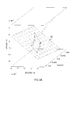
- the parameter triplet ⁇ F A CO2/ ⁇ t, CaCO2, VTCO2/ ⁇ t ⁇ define a data point in a three dimensional space spanned by said parameter triplet, and the solution to the overdetermined system of equations can be visualized in form of a plane that is fitted to the data points from the analysed sequence of respiratory cycles in this three dimensional space.
- a plane 23 is illustrated in Figs. 3A and 3B , wherein the data points defined by the parameter triplet ⁇ F A CO2/ ⁇ t, CaCO2, VTCO2/ ⁇ t ⁇ for each respiratory cycle is represented by a circle.
- Fig. 3A illustrates a perspective view of the plane 23, and Fig.
- 3B illustrates a cross-sectional view of the plane 23 in said three dimensional space.
- the unknown physiological parameters V and Q define the normal direction to the plane in the three dimensional space, and the parameter Q ⁇ CvCO 2 defines the translation of the plane along the VTCO2/ ⁇ t axis.
- the unknown physiological parameters V, Q and CvCO 2 can hence be said to be determined by fitting a plane to a plurality of 3D data points, where each data point is given by the values of the parameter triplet ⁇ F A CO2/ ⁇ t, CaCO2, VTCO2/ ⁇ t ⁇ for a respective respiratory cycle in the analysed sequence, N, of respiratory cycles.
- the model may be adjusted to take known variations in the subject's effective lung volume during the analysed sequence of respiratory cycles into account.
- a fast update of the current effective lung volume of the subject i.e. the effective lung volume of the latest respiratory cycle, corresponding to respiratory cycle number N in the analysed sequence of respiratory cycles
- the effective lung volume V N of the last respiratory cycle in the analysed sequence of respiratory cycles from the approximate value of the effective lung volume V obtained by solving the above overdetermined system of equations (Eq. 3 or Eq. 6).
- the approximation of the effective lung volume V obtained by solving the above overdetermined system of equations (Eq. 3 or Eq. 6) must be interpreted as "the best mean value" of the effective lung volume of the subject during the analysed sequence of respiratory cycles 1 to N.
- Equations 8-10 means that the unknown physiological parameters Q and CvCO 2 relating to the cardiac output and the carbon dioxide content of venous blood of the subject, respectively, are assumed to be substantially constant during the analysed sequence of respiratory cycles, while the effective lung volume V of the subject is allowed to vary from breath to breath (i.e. from one respiratory cycle to another).
- the approximation of the effective lung volume V obtained by solving the overdetermined system of equations (Eq. 3 or Eq. 6) is assumed to correspond to the effective lung volume of the subject in the respiratory cycle immediately preceding the analysed sequence of respiratory cycles.
- the calculated approximation V corresponds to the effective lung volume of the last respiratory cycle in the sequence of analysed respiratory cycles, and to count "backwards" to determine the effective lung volume of each respiratory cycle in the analysed sequence taking the difference between the volumes of inhaled inspiration gas and exhaled expiration gas into account.
- this requires the expression for VTCO 2 (Eq. 9) to be modified accordingly.
- the mathematical model may be adjusted by incorporation of a priori information on one or more of these physiological parameters.
- the a priori information may comprise one or more values of one or more of the physiological parameters. Such values may be obtained through other methods of measurement, by estimating the values based on physiological information about the subject, or from the analysis of one or more sequences of respiratory cycles preceding the sequence of respiratory cycles to be analysed using the method described herein.
- an a priori value of the carbon dioxide content of venous blood (CvCO 2 ) of the subject may be obtained from blood gas measurements, as well known in the art.
- An a priori value of the effective pulmonary capillary blood flow of the subject 3 may be estimated based on the body weight and the heart rate of the subject, as suggested e.g. by Jegier et al. in Br Heart J. 1963 July; 25(4): 425-430 .
- a priori values of the physiological parameters may be provided to the control unit 15 of the device 1 through user input on a user interface of the device 1 (not shown).
- An a priori value of the effective lung volume (V) of the subject can be determined from a wash-out process, as also well known in the art.
- control unit 15 of the device in Fig. 1 may be adapted to effectuate a wash-out process according to prior art for determining the effective lung volume V of the subject 3, prior to determination of the physiological parameters V, Q and CvCO 2 by means of the method described herein.
- the a priori information may be incorporated into the mathematical model by expanding the above described system of equations through addition of one equation for each unknown parameter V, Q and CvCO 2 .
- the method further comprises the step of calculating an error indicating the uncertainty in the determination of the one or more physiological parameters.
- Equation 3 An error, E ⁇ FACO2, that is easier to interpret can be determined by normalising Equation 3 with V.
- E ⁇ FACO 2 A ⁇ x C - a V T ⁇ A ⁇ x C - a V ,
- an error can be determined for the expanded capnodynamic model (Eq. 15) comprising the additional "a priori" equations (Eq. 11-13):
- E C C ⁇ x C - c T ⁇ C ⁇ x C - c ,
- a big error may be an indication that some of the unknown physiological parameters that are assumed to be substantially constant during the analysed sequence of respiratory cycles (CvCO 2 , Q and in some embodiments also V) actually varies.
- a big error may also be an indication that the flow sensor 15 or gas analyser 17 malfunctions, or that some other requirement that must be fulfilled in order for the model to properly reflect the reality is not fulfilled. If the calculated error exceeds a predetermined threshold value, an alarm signal indicating that the model is currently inconsistent with observed data may be generated and provided visually or aurally to an operator of the device 1 serving to monitor the physiological parameters of the subject 3.
- the control unit 5 of the breathing apparatus is preferably operable to change the effective ventilation of the subject 3 during the sequence of analysed respiratory cycles so as to cause a change in the carbon dioxide content in the expiration gas exhaled by the subject of at least 0,5% during said sequence.
- a change in carbon dioxide content in the expiration gas means that some or all of the parameters F A CO2, CaCO2, ⁇ t and VTCO2 varies during the analysed sequence of respiratory cycles, which is a requirement in order to solve the above discussed overdetermined systems of equations with respect to the unknown physiological parameters.
- the control unit is configured to cause a change in carbon dioxide content in the expiration gas of 0,5%-1% during the analysed sequence of respiratory cycles.
- An advantage with the inventive method as compared to other methods for determining effective lung volume, cardiac output or carbon dioxide content of venous blood of a subject is that it is independent of the type of change in ventilation, and independent of the ventilation pattern provided to the subject.
- the following is a list of non-exclusive examples of how the change in effective ventilation of the subject may be effectuated by the control unit 5:
- An advantage with the techniques 3 to 5 is that the expiration phase of the respiratory cycle is unaffected using these techniques, which is particularly advantageous as the inventive method relies on measurements of the CO 2 content in expiration gas exhaled by the subject and so requires sampling of the CO 2 content during the expiratory phases.
- An advantage with the techniques 1-3 is that most breathing apparatuses (e.g. ventilators) of today can be adapted to carry out the techniques merely by updating the software controlling the operation of the breathing apparatus.
- the techniques 4-5 typically require use of hardware components not normally included in breathing apparatuses.
- control unit 5 is preferably configured to continuously monitor the physiological parameters V, CvCO 2 and Q of the subject 3 during the respiratory treatment provided by the breathing apparatus, which may require a repetitive change in the effective ventilation of the subject in order for the carbon dioxide content in the expiration gas to change during each analysed sequence of respiratory cycles.
- control unit 5 is preferably configured to vary the effective ventilation of the subject 3 such that the subject is alternately subjected to hyperventilation and hypoventilation in a manner making the mean ventilation over time correspond to an optimal degree of ventilation of the patient. This means that a change in the effective ventilation of the subject is always directly followed by a change in the "opposite direction" - there is no baseline ventilation (i.e. "normal” ventilation) of the subject in between the hyperventilation phases and the hypoventilation phases.
- control unit 5 may be configured to determine, based on ventilation parameters input to the breathing apparatus by an operator and indicating a desired baseline ventilation to be provided to the subject, an optimal ventilation pattern in form of a sequence of hyperventilated and hypoventilated breaths, which ventilation pattern gives the same effect in terms of ventilation as the desired baseline ventilation.
- the ventilation parameters input by the operator may comprise a parameter indicating a desired minute ventilation of the subject, whereby the control unit may be configured to determine a sequence of hyperventilated and hypoventilated breaths together resulting in said desired minute ventilation. The sequence of hyperventilated and hypoventilated breaths is then continuously repeated throughout the ventilatory treatment, meaning that a new sequence starts directly after the last breath of a previous sequence.
- this ventilation pattern is generated by changing the duration of the insp-hold-pause between inspiratory phases and expiratory phases.
- the insp-hold-pause For five breaths, the insp-hold-pause is shortened compared to a "normal" insp-hold-pause that would result in a desired baseline ventilation of the subject, so as to deliver five hyperventilated breaths to the subject, and for the following five breaths the insp-hold-pause is prolonged compared to said "normal" insp-hold-pause, so as to deliver five hypoventilated breaths.
- This pattern may be repeated as long as there is a desire to monitor the physiological parameters using the inventive method described herein.
- the durations of the "shortened" and "prolonged" insp-hold-pauses are preferably selected to make the minute ventilation of the subject correspond to the minute ventilation that would have been obtained using the desired baseline ventilation.
- the repetitive changes in effective ventilation of the subject are achieved by means of a cyclic ventilation pattern wherein each cycle comprises a first number of breaths of increased ventilation and a second number of breaths of decreased ventilation, where the total number of breaths in each cycle is five or less.
- a non-invasive method for determining at least one physiological parameter related to the effective lung volume, the cardiac output, and/or the carbon dioxide content of venous blood of a subject comprising the steps of:
- the cycles of the proposed ventilation pattern are hence shorter than the cycles of the ventilation patterns used for the same purpose in methods according to prior art.
- the proposed cyclic ventilation pattern has at least the following advantages compared to ventilation patterns having longer sequences of increased and decreased ventilation:
- the breaths of increased ventilation are hyperventilated breaths (i.e. breaths of increased ventilation compared to a desired baseline ventilation of the subject) while the breaths of decreased ventilation are hypoventilated breaths (i.e. breaths of decreased ventilation compared to a desired baseline ventilation).
- the determination of the at least one physiological parameter is preferably made using an algorithm which does not require a steady state of carbon dioxide content in the expiration gas exhaled by the subject since the sequences of increased and decreased ventilation in the proposed ventilation pattern generally are too short in order for the carbon dioxide content to reach a steady state level.
- the determination of the physiological parameter(s) is made using the above described method of analysing the correlation between parameters that are derivable from the measured quantities.
- the method comprises the steps of:
- the number of breaths in the analysed sequence of breaths corresponds to the number of breaths in each cycle of the cyclic ventilation pattern.
- other alternatives are possible. For example, it would be possible to use a cyclic ventilation pattern wherein each cycle comprises five breaths, and to determine the unknown physiological parameter(s) by studying the correlation of the first, second and third parameters in a sequence of 10 breaths.
- the changes in effective ventilation should preferably cause a change in the carbon dioxide content in the expiration gas exhaled by the subject of at least 0,5% during the analysed sequence of breaths.
- the cyclic ventilation pattern should be such that the carbon dioxide content in the expiration gas exhaled by the subject changes with at least 0,5% during any sequence of breaths having the same length (i.e. number of breaths) as the cycles of the ventilation pattern.
- each cycle of the cyclic ventilation pattern comprises three breaths of increased ventilation and two breaths of decreased ventilation.
- each cycle comprises four breaths of increased ventilation and one breath of decreased ventilation.
- a suitable cyclic ventilation pattern may have cycles comprising three breaths of increased ventilation, each with a duration of three seconds and no inspiratory pause, and two breaths of decreased ventilation, each with a duration of three seconds and an inspiratory pause of four seconds.
- the duration of the sequence of increased ventilation is 9 seconds (3x3s)
- the duration of the sequence of decreased ventilation is 14 seconds (2x(3s+4s))
- the duration of each cycle in the cyclic ventilation pattern is 23 seconds (9s+14s). It has been found that for any given sequence of five consecutive breaths, this ventilation pattern causes a sufficient change in carbon dioxide content in the expiration gas exhaled by the subject.
- This exemplary cyclic ventilation pattern is illustrated in Fig. 4 , in which the letter “I” indicates sequences of increased ventilation, the letter “D” indicates sequences of decreased ventilation, and the letter “B” indicates breaths in the sequences of increased and decreased ventilation.
- the effective respiratory rate using this cyclic ventilation pattern is 13 breaths per minute ((5/23)x60).
- VCO 2 VCO 2 of the subject, based on the measured inspiratory flow, expiratory flow and carbon dioxide content, and - determining said at least one physiological parameter based on the correlation of the first, second and third parameters in the sequence of respiratory cycles.
- Aspect 2 The method according to aspect 1, wherein the step of determining said at least one physiological parameter involves simultaneous determination of a physiological parameter related to the effective lung volume of the subject and a physiological parameter related to the cardiac output of the subject, based on said correlation.
- Aspect 3 The method according to aspect 1, wherein the step of determining said at least one physiological parameter involves simultaneous determination of a physiological parameter related to the effective lung volume of the subject, a physiological parameter related to the cardiac output of the subject, and a physiological parameter related the carbon dioxide content of venous blood of the subject, based on said correlation.
- Aspect 4 The method according to any of the preceding aspects, further comprising the steps of:
- PCBF pulmonary capillary blood flow
- Aspect 6 The method according to any of the preceding aspects, wherein the at least one physiological parameter is determined on a breath-by-breath basis by, for each respiratory cycle, replacing the values of the first, second Cand third parameters obtained during the oldest respiratory cycle in the sequence of respiratory cycles with the values obtained during the most recent respiratory cycle.
- Aspect 7 The method according to any of the preceding aspects, wherein at least a physiological parameter related to the effective lung volume of the subject 3 is determined, further comprising the steps of:
- Aspect 8 The method according to any of the preceding aspects, further comprising the steps of obtaining, prior to determination of the at least one physiological parameter, a priori information comprising at least one value of at least one of said physiological parameters using another method for determination of at least one of said physiological parameters, and using said a priori information in the determination of the at least one physiological parameter.
- Aspect 9 The method according to any of the preceding aspects, further comprising the steps of determining an error in the determination of the at least one physiological parameter based on the correlation of the first, second and third parameters in the sequence of respiratory cycles, and generating an alarm signal if the error indicates that the correlation is weak.
- Aspect 12 The device 1 according to aspect 11, wherein said control unit 5 comprises a memory 19 storing the computer program according to aspect 10, and a processing unit 21 for executing the computer program to cause the device 1 to carry out the method according to any of the aspects 1-10.
- Aspect 13 The device 1 according to aspect 11 or 12, wherein said device 1 is a breathing apparatus for ventilatory treatment of the subject 3, such as a ventilator or an anaesthesia machine.
- Aspect 14 The device 1 according to aspect 13, wherein the control unit 5 is operable to cause a change in the effective ventilation of the subject 3 such that the carbon dioxide content of the expiration gas exhaled by the subject varies with 0,5%-1% during said sequence of respiratory cycles.
- Aspect 15 The device 1 according to aspect 14, wherein the control unit 5 is configured to change the effective ventilation of the subject 3 such that the breathing apparatus delivers a ventilation pattern with alternating sequences of breaths of increased ventilation and breaths of decreased ventilation to the subject 3.
Abstract
Description
- The present invention relates to a method, a computer program and a device for determining physiological parameters related to the effective lung volume, the cardiac output, and/or the carbon dioxide content of venous blood of a subject. In particular, the invention relates to non-invasive and continuous determination of such parameters during ventilatory treatment of a patient, based on measured expiratory flow and carbon dioxide content of expiration gases.
- Cardiac output (CO) is the rate at which blood is pumped by the heart to the organs of the body. A parameter that is closely related to cardiac output is pulmonary capillary blood flow (PCBF), i.e. the alveolar blood flow. The effective (non-shunt) pulmonary capillary blood flow equals the cardiac output in case of no or neglected cardiac shunt flow. Cardiac output and effective pulmonary capillary blood flow are important measures of cardiovascular stability.
- Effective lung volume (ELV) is normally defined as the volume of the lung that takes part in gas exchange, and so is an important measure of the lung function.
- Monitoring parameters related to cardiac output and effective lung volume is important when the cardiovascular stability and/or the lung function is potentially threatened, such as during surgery and in critically ill patients. For example, it is often desired to monitor the effective lung volume and sometimes also cardiac output during ventilatory treatment of a patient.
- There are several solutions according to prior art for non-invasive determination of parameters relating to cardiac output and/or effective lung volume. Some of these solutions make use of various variations of the carbon dioxide Fick method where the cardiac output of a patient is determined using the following basic relationship:

- Typically, a differential form of the carbon dioxide Fick equation is used to non-invasively determine the cardiac output of the patient. Differential Fick techniques are based on the premise that cardiac output and effective pulmonary blood flow can be estimated based on the changes of other measurable parameters when a change in the effective ventilation of the patient occurs. During mechanical ventilation of a patient, such a change in effective ventilation may be effectuated e.g. by varying the degree of rebreathing of expiration gases or by changing the tidal volume, the respiratory rate or the so called insp-hold pause between inspiratory phases and expiratory phases.
-
EP1257201 discloses an apparatus for non-invasively measuring pulmonary capillary blood flow and cardiac output using known rebreathing techniques. In one embodiment, data on carbon dioxide elimination (VCO2) and data on carbon dioxide content of the arterial blood of the patient (CaCO2) are obtained and a correlation coefficient between the carbon dioxide elimination data and the data on the carbon dioxide content is determined. This correlation coefficient is then used to calculate at least one of the mixed venous carbon dioxide content, the pulmonary capillary blood flow, and the cardiac output. -
US7699788 andWO2006047212 disclose methods for non-invasively estimating functional residual capacity or effective lung volume by obtaining carbon dioxide and flow measurements at or near the mouth of a patient. The measurements are obtained during baseline breathing and during and shortly after inducement of a change in the subject's effective ventilation. The obtained measurements are evaluated to determine the amount of time required for exhaled carbon dioxide levels to return to normal - effectively an evaluation of carbon dioxide "washout" from the subject's lungs. Conversely, carbon dioxide and flow measurements may be evaluated to determine the amount of time it takes carbon dioxide to "wash in," or reach peak levels within, the lungs of the subject following the change in the subject's effective ventilation. Measures of the effective lung volume of the patient are then determined from relationships between parameters relating to carbon dioxide elimination and parameters relating to the carbon dioxide content of the arterial blood. -
US6217524 describes a method of continuously, non-invasively determining the cardiac output of a patient. The method includes intermittently measuring the cardiac output, the volume of carbon dioxide exhaled by the patient per breath, and determining the arterial-venous gradient of the patient or a similar substantially constant value by dividing the volume of carbon dioxide exhaled by the measured cardiac output. The arterial-venous gradient or similar substantially constant value may then be employed to determine the cardiac output of the patient on a breath-by-breath basis. The carbon dioxide elimination, which is non-invasively measured as the volume of carbon dioxide exhaled by the patient per breath, is divided by the arterial-venous gradient or the substantially constant value to determine the cardiac output. The method may also include generating a signal to compensate for any non-metabolic changes in the carbon dioxide elimination, arterial-venous gradient, or other respiratory or blood gas profile measurements that may be caused by a change in ventilation or breathing of the patient. - Gedeon et al., "Pulmonary blood flow (cardiac output) and volume determined from a short breath hold using the differential Fick method", J. CLIN. MONIT. 17:313-321 (2002), describes a non-invasive method for determining the effective lung volume of a subject using breath-holding techniques. Gedeon et al. also describes equations that relate the pulmonary capillary blood flow of the subject to the subject's effective lung volume. The method is believed to provide inaccurate data as it is based on the assumption that CO2 inflow may not be significantly affected by breath-holding, while breath-holding will cause a change in partial pressure of carbon dioxide. This assumption is inconsistent with the Fick equation, in which carbon dioxide elimination changes linearly with the partial pressure of carbon dioxide while the pulmonary capillary blood flow and the carbon dioxide content of the venous blood (CvCO2) remain constant.
- Peyton et al., "Noninvasive, automated and continuous cardiac output monitoring by pulmonary capnodynamics: breath-by-breath comparison with ultrasonic flow probe", Anesthesiology 2006 Jul; 105(1):72-80, describes a technique termed the capnodynamic method for breath-to-breath measurement of pulmonary blood flow from lung carbon dioxide mass balance, using measured carbon dioxide elimination and end-tidal concentration. Here, a capnodynamic equation is used to eliminate the parameter relating to carbon dioxide content of the venous blood of the patient (CvCO2) in order to obtain resulting equations from which the effective lung volume and the cardiac output can be derived iteratively. To obtain the resulting equations from which the effective lung volume and the cardiac output can be derived, measurements must be made during two substantially equal breaths (two hyperventilated or two hypoventilated breaths) and during two transient breaths (one hyperventilated breath and one hypoventilated breath). This makes the method proposed by Peyton et al. dependent on a certain ventilation pattern.
- This capnodynamic method is further described in
WO 2006/119546 A1 . The method is herein described with reference to a continuous alternating/cyclic alveolar ventilation of a subject, with each period of alveolar ventilation at a particular level (hyperventilation or hypoventilation) constituting a half cycle. Preferably, a cycle comprises 6 to 20 breaths, typically 12 breaths; a half cycle being half of this number of breaths. The method employs a "calibration equation" which has to be solved for breaths that occur at periods in the half cycles during which washin or washout of carbon dioxide is minimised, i.e. for breaths occurring when the carbon dioxide concentration has reached a steady state following a change in effective ventilation. -
US7135001 discloses a differential Fick technique for noninvasively determining the pulmonary capillary blood flow or cardiac output of a patient. The technique includes effecting a change-inducing phase in the respiration of the patient, allowing the respiration to return to normal, then immediately repeating the change-inducing phase of respiration. The technique is characterised in that the typical recovery period, where a patient's respiration is allowed to return to normal or baseline levels before again measuring respiratory carbon dioxide and flow is omitted. Thereby, the durations of the normal respiration and change-inducing phases can be abbreviated relative to the time lengths of the corresponding phases in conventional differential Fick techniques. The duration of each phase may be optimized for a patient by evaluating the patient's ventilation but should be within the interval of approximately eighteen to approximately forty-two seconds. - It is an object of the invention to enable non-invasive determination of parameters relating to the cardiac output and/or the effective lung volume of a subject.
- It is a further object of the invention is to enable simultaneous determination of parameters relating to both the cardiac output and the effective lung volume of a subject.
- Yet another object of the invention is to provide a method for continuous monitoring of parameters relating to cardiac output and effective lung volume of a subject undergoing ventilatory treatment, which method provides more reliable results than methods according to prior art.
- Yet another object of the invention is to provide a method for continuous monitoring of parameters relating to cardiac output and effective lung volume of a subject undergoing ventilatory treatment, which method offers shorter response times than methods according to prior art.
- Yet another object of the invention is to provide a method for continuous monitoring of parameters relating to cardiac output and effective lung volume of a subject undergoing ventilatory treatment, which method minimizes potentially adverse effects on the patient caused by changes in effective ventilation.
- These and other objects are achieved by a non-invasive method for determining at least one physiological parameter related to the effective lung volume (ELV), the cardiac output (CO), and/or the carbon dioxide content of venous blood (CvCO2) of the subject as defined by
independent claim 1. - In one embodiment, the method comprises the steps of measuring at least an expiratory flow of expiration gas exhaled by the subject and a carbon dioxide (CO2) content of said expiration gas. Furthermore, the method comprises the steps of ventilating the subject using a cyclic ventilation pattern wherein each cycle comprises a first number of breaths of increased ventilation and a second number of breaths of decreased ventilation, wherein the total number of breaths in each cycle of the cyclic ventilation pattern is five or less, and determining said at least one physiological parameter from at least the expiratory flow and the CO2 content of at least the expiration gas, measured during said cyclic ventilation of the subject.
- The cycles of the proposed ventilation pattern are hence shorter than the cycles of the ventilation patterns used for the same purpose in methods according to prior art.
- An advantageous effect of the short cycles of the cyclic ventilation pattern is that the response time of the proposed method is reduced compared to known methods using longer ventilation cykles.
- Furthermore, the short cycles of the cyclic ventilation pattern has the effect of minimizing the risk that changes in the effective ventilation of the subject that are used to create the cyclic ventilation pattern introduce variations in the carbon dioxide content of venous blood (CvCO2) of the subject, which risk is particularly high at high levels of cardiac output. Most methods, including a preferred embodiment of the method described in this application, are based on the assumption that CvCO2 is substantially constant during the analysed sequence of breaths and, consequently, variations in CvCO2 during the analysed sequence of breaths may introduce errors in the determination of the physiological parameter(s). Therefore, the proposed method is more reliable than known methods in which changes in the effective ventilation of a subject are introduced to alter the CO2 content in expiration gas exhaled by the subject.
- Yet further, the short cycles of the ventilation pattern used in the proposed method reduce potentially adverse effects on the patient caused by the changes in effective ventilation. For example, in embodiments where breaths of decreased ventilation are generated by prolonging the inspiratory pause compared to the inspiratory pause of breaths of increased ventilation, the periods of increased pressure in the subject's lungs are shortened, which mitigates the risk of adversely affecting the hemodynamics of the subject.
- Preferably, the breaths of increased ventilation are hyperventilated breaths and the breaths of decreased ventilation are hypoventilated breaths. Hereby, the total ventilation over time can be made to correspond to a desired optimal ventilation of the subject.
- Preferably, the determination of said at least one physiological parameter is made using an algorithm which does not require a steady state of carbon dioxide content in the expiration gas exhaled by the subject.
- Preferably, the step of determining said at least one physiological parameter comprises the steps of:
- determining, for each breath in a sequence of breaths, a first parameter related to the fraction of alveolar carbon dioxide (FACO2) of the subject, a second parameter related to the carbon dioxide content of the arterial blood (CaCO2) of the subject, and a third parameter related to carbon dioxide elimination (VCO2) of the subject, based on at least the measured expiratory flow and the carbon dioxide content of the expiration gas, and
- determining said at least one physiological parameter based on the correlation of the first, second and third parameters in said sequence of breaths.
- Preferably, the number of breaths in said sequence of breaths corresponds to the number of breaths in each cycle of the cyclic ventilation pattern
- In situations where the inspiration gas inhaled by the subject comprises non-negligible amounts of carbon dioxide, e.g. during full or partial rebreathing of expiration gases, the method preferably comprises the steps of measuring also the carbon dioxide content of the inspiration gas, and to take this content into account in the determination of the first, second and third parameters related to FACO2, CaCO2 and VCO2, respectively.
- The proposed method is based on a mathematical model describing the dynamics of ventilation and perfusion of a lung. In a preferred embodiment of the invention, the method employs a capnodynamic equation describing how the fraction of alveolar carbon dioxide, FACO2, varies from one respiratory cycle to the next. The capnodynamic equation may be based on a single-compartment lung model or a multi-compartment lung model. In one exemplary embodiment of the invention, the following capnodynamic equation for a single-compartment lung model is used:

- By measuring at least the expiratory flow and the carbon dioxide content of the expiration gas during a respiratory cycle, the parameters ΔFACO2, CaCO2 and VTCO2 can be calculated for that respiratory cycle. By calculating these parameters during a sequence of respiratory cycles, each having a predetermined or measurable duration (Δt), the unknown physiological parameters V, Q and CvCO2, corresponding to the effective lung volume, the effective pulmonary capillary blood flow, and the carbon dioxide content of venous blood of the subject, respectively, can all be determined simultaneously based on the correlation of the parameters ΔFACO2, CaCO2 and VTCO2 in the different respiratory cycles of the analysed sequence of respiratory cycles. That all of said physiological parameters can be determined simultaneously means that they can all be determined by finding the solution to a single system of capnodynamic equations describing the relationships between said physiological parameters and said first, second and third parameters, as described in greater detail below.
- The sequence of respiratory cycles analysed to determine the physiological parameters V, Q and CvCO2 should comprise more than three respiratory cycles. Calculating the values of the parameters ΔFACO2, CaCO2 and VTCO2 and inserting the parameter values together with the duration of the respiratory cycle (Δt) into the above equation (Eq. 1) for each respiratory cycle yields an overdetermined system of equations comprising one equation for each respiratory cycle in the analysed sequence of respiratory cycles. This overdetermined system of equations can then be solved with respect to the unknown physiological parameters V, Q and CvCO2, e.g. using the method of least squares or any other method suitable for finding an approximate solution of an overdetermined system of equations. The solution depends on the correlation between the parameters ΔFACO2, CaCO2 and VTCO2 in the different respiratory cycles.
- As understood by the skilled person, the above described calculation relies on the assumption that the physiological parameters V, Q and CvCO2 are substantially constant, or at least that they do not vary too much, during the sequence of analysed respiratory cycles. It should thus be noted that the calculated values of the physiological parameters V, Q and CvCO2 can be said to represent mean values during the analysed sequence of respiratory cycles.
- By replacing the parameter values of ΔFACO2, CaCO2, VTCO2 and Δt for the "oldest" respiratory cycle in the overdetermined system of equations with corresponding parameter values for the most recent respiratory cycle, the effective lung volume, the effective pulmonary capillary blood flow and the carbon dioxide content of venous blood of the subject can be continuously monitored in an effective and reliable manner.
- An advantage with the proposed method is that it can be used to determine parameters relating to the effective lung volume, the cardiac output, and the carbon dioxide content of venous blood of the subject simultaneously in an efficient and reliable manner, as they are all given by the solution to a single system of equations.
- Another advantage is that the proposed method can be used for any given sequence of respiratory cycles as long as the carbon dioxide content in the expiration gas exhaled by the subject varies slightly during the analysed sequence of respiratory cycles. The method does not require any particular breaths in the analysed sequence of respiratory cycles to be identified and compared with each other. Instead, the method treats all breaths (i.e. respiratory cycles) equally and provides updated values for the physiological parameters V, Q and CvCO2 for each respiratory cycle, no matter any change in the effective ventilation of the patient. Thus, the method is independent of the ventilation pattern of the subject.
- Preferably, the carbon dioxide content in the expiration gas exhaled by the subject during the analyzed sequence of respiratory cycles should vary with at least 0,5%, and preferably between 0,5-1%. The required variation in carbon dioxide content of the expiration gas during the analysed sequence of respiratory cycles may occur naturally during supported ventilation of a spontaneously breathing subject. However, there may be a desire to actively induce a variation in carbon dioxide content of the expiration gas over time by introducing a change in the effective ventilation of the subject. Such a change in effective ventilation may be effectuated e.g. by varying the degree of rebreathing of expiration gases exhaled by the subject or by changing the tidal volume, the respiratory rate or the inspiratory pause (often called insp-hold pause) between inspiratory phases and expiratory phases.
- If the method is used to continuously monitor physiological parameters of a patient undergoing ventilatory treatment where changes in the effective ventilation are introduced to actively vary the carbon dioxide content in the expiration gas exhaled by the patient, the changes in ventilation are preferably effectuated such that the patient is alternately subjected to hyperventilation and hypoventilation in a manner making the mean ventilation over time correspond to an optimal degree of ventilation of the patient. Preferably, the effective ventilation is changed such that the variation in carbon dioxide content in the expiration gas exhaled by the subject is 0,5-1% during the sequence of analysed respiratory cycles.
- In a refined embodiment of the invention, known variations in the subject's effective lung volume during the analysed sequence of respiratory cycles may be used to obtain an updated value of the effective lung volume of the subject, which reflects the current effective lung volume of the subject more accurately than the value determined from the correlation analysis. This updated or "current" value of the effective lung volume may be determined based on the differences between the volume of inspired and expired gas in the respiratory cycles of the analysed sequence of respiratory cycles, and the parameter related to the effective lung volume determined from the correlation analysis.
- Yet another advantage with the proposed method is that a priori information providing an a priori value of one or more of the physiological parameters V, Q and CvCO2 may be used to obtain more accurate measures of the unknown quantities. The a priori information may originate from other methods for measuring these physiological parameters, including but not limited to blood gas measurements providing an a priori value of the carbon dioxide content of venous blood (CvCO2) of the subject, a wash-out procedure providing an a priori value of the effective lung volume (V) of the subject, and obtaining the body weight and measuring the heart rate of the subject to provide an a priori value of the cardiac output or effective pulmonary capillary blood flow (Q) of the subject. The a priori information may be used to set a start value for one or more of the parameters V, Q and CvCO2, or to lock one or two of the parameters to known and fix values. In another embodiment, the above discussed overdetermined system of equations is expanded with additional equations comprising a priori values of one or more of the parameters V, Q and CvCO2, as discussed in greater detail in the detailed description following hereinafter.
- Preferably, the method further involves a step of calculating an error indicative of the uncertainty in the determination of the physiological parameters. As long as the measurable and/or known data (ΔFACO2, CaCO2, VTCO2, Δt, and any additional a priori values of V, Q and CvCO2) are consistent with the mathematical model used, the error will be small. If however, the measurable and/or known data are inconsistent with the model, the error becomes big. In case of a big error, an alarm signal indicating that the mathematical model is currently unreliable may be generated. Preferably, the error is calculated continuously, i.e. on a breath-by-breath basis, and the alarm signal is generated if the uncertainty exceeds a predetermined threshold value.
- According to another aspect of the invention, a device capable of performing the above method is provided. To this end, the device comprises at least one flow sensor for measuring at least an expiratory flow of expiration gas exhaled by the subject. It also comprises at least one gas analyser for measuring the carbon dioxide content of at least the expiration gas exhaled by the subject. Furthermore, the device comprises a control unit configured to control the ventilation of the subject and to determine said at least one physiological parameter from the measured flows and carbon dioxide content. The control unit is configured to control the ventilation of the subject such that the subject is ventilated using a cyclic ventilation pattern wherein each cycle comprises a first number of breaths of increased ventilation and a second number of breaths of decreased ventilation, wherein the total number of breaths in each cycle of the cyclic ventilation pattern is five or less, and to determine said at least one physiological parameter from flow and carbon dioxide content measurements obtained during said cyclic ventilation.
- Although the device may be a stand-alone device exclusively used for monitoring physiological parameters related to the effective lung volume, the cardiac output, and/or the carbon dioxide content of venous blood of a subject, the above described functionality is particularly intended to be incorporated into a breathing apparatus for providing breathing assist to a patient undergoing ventilatory treatment, such as a ventilator or an anaesthesia machine.
- Preferably, such a breathing apparatus is equipped with a flow sensor and a gas analyser arranged in or close to a Y-piece connecting an inspiratory branch and an expiratory branch of the breathing apparatus with the patient. The flow sensor may be configured to measure the inspiratory and expiratory flow to and from the patient continuously to obtain a continuous flow curve representing the flow of gas into and out of the airways of the patient over time. Likewise, the gas analyser may be configured to measure the carbon dioxide content in the inspiration gas and the expiration gas continuously to obtain a continuous CO2 fraction curve representing the carbon dioxide content inhaled and exhaled by the patient over time. A control unit of the breathing apparatus may be configured to use the flow and carbon dioxide content measurements to determine the first, second and third parameters related to FACO2, CaCO2 and VCO2, respectively, for each respiratory cycle in an analysed sequence of respiratory cycles, and to determine the at least one physiological parameter related to the effective lung volume, the cardiac output, and/or the carbon dioxide content of venous blood of the subject based on the correlation of said first, second and third parameters in said sequence of respiratory cycles.
- The control unit of the breathing apparatus is further configured to control the ventilation provided to the patient by the breathing apparatus, and is preferably configured introduce a change in the effective ventilation of the patient to ensure that the carbon dioxide content of the expiration gas varies with at least 0,5% and preferably between 0,5% and 1% during the analysed sequence of respiratory cycles.
- The control unit is preferably operable to cause the breathing apparatus to apply a ventilation pattern to the patient comprising a sequence of hyperventilated breaths followed by a sequence of hypoventilated breaths. This ventilation pattern may be applied to the patient during the entire ventilatory treatment, meaning that the patient is always either hyperventilated or hypoventilated. The sequences of hyperventilated and hypoventilated breaths are preferably controlled such that the total ventilation over time corresponds to a desired, optimal ventilation of the patient. In one embodiment, the control unit is operable to cause a change in the duration of the insp-hold-pause between inspiration phases and expiration phases to make the breathing apparatus switch between hyperventilation and hypoventilation.
- In other embodiments, to make the breathing apparatus switch between hyperventilation and hypoventilation, the control unit may be operable to cause a change in one or more of the tidal volume of breathing gas delivered to the patient during inspiration, the respiratory rate, and the degree of rebreathing of expiration gases exhaled by the patient.
- The logic required to enable the device (a stand-alone device or a breathing apparatus) to carry out the method is preferably implemented by means of software. Thus, according to another aspect of the invention, a computer program for determining at least one physiological parameter related to the effective lung volume, the cardiac output, and/or the carbon dioxide content of venous blood of a subject is provided. The computer program comprises computer readable code which, when executed by a processor of a device configured as described above, causes the device to carry out the inventive method.
- Installing such a computer program on existing breathing apparatuses equipped with means for measuring at least the flow of expiration gases and the carbon dioxide content of expiration gases allows existing breathing apparatuses to carry out the inventive method.
- The present invention will become more fully understood from the detailed description provided hereinafter and the accompanying drawings which are given by way of illustration only. In the different drawings, same reference numerals correspond to the same element.
-
Fig. 1 illustrates a device in form of a breathing apparatus according to an exemplary embodiment of the invention. -
Fig. 2 is a flowchart illustrating the basic principles of the invention. -
Figs. 3A and3B illustrate a visualization of a solution to an overdetermined equation system which is solved to determine the unknown physiological parameters according to an embodiment of the invention. -
Fig. 4 illustrates a ventilation pattern that may be applied to a subject undergoing ventilatory treatment in order to determine parameters related to the effective lung volume, the cardiac output, and/or the carbon dioxide content of venous blood of the subject. -
Fig. 1 illustrates adevice 1 for continuous and non-invasive determination of one or more physiological parameters related to the effective lung volume (ELV), the cardiac output, and/or the carbon dioxide content of venous blood (CvCO2) of a subject 3, according to an exemplary embodiment of the invention. - In this embodiment, the
device 1 is a breathing apparatus, such as a ventilator or an anaesthesia machine, for providing breathing assist to thesubject 3. The breathing apparatus comprises acontrol unit 5 for controlling the ventilation of the subject based on preset parameters and measurements obtained by various sensors of the breathing apparatus. Furthermore, the breathing apparatus comprises aninspiratory branch 7 for conveying inspiration gases to thesubject 3 and anexpiration branch 9 for conveying expiration gases away from the patient. The inspiratory and expiratory branches are connected to apatient connector 11 via a Y-piece 13. - A
flow sensor 15 and agas analyser 17 are arranged in the Y-piece 13 and operable to measure the flows and the carbon dioxide (CO2) content, respectively, of the inspiration and expiration gases to and from thesubject 3. Thecontrol unit 5 is configured to determine the physiological parameter(s) based on the measurements obtained by theflow sensor 15 and thegas analyser 17. To this end, thecontrol unit 5 comprises anon-volatile memory 19 storing a computer program that causes thecontrol unit 5 to calculate the physiological parameter(s) according to the principles described below, when executed by aprocessing unit 21 of thecontrol unit 5. Unless stated otherwise, all steps of the inventive method described hereinafter are performed by thecontrol unit 5 of thedevice 1 through execution of a computer program. -
Fig. 2 illustrates the basic principles of the proposed method for continuous and non-invasive determination of the one or more physiological parameters. In the description of the inventive method following hereinafter, simultaneous reference will be made to the breathing apparatus inFig. 1 . - Each step of the method illustrated in
Fig. 2 is performed once for each respiratory cycle of thesubject 3. A respiratory cycle comprises an inspiration phase and an expiration phase and the time or duration of a respiratory cycle is typically defined as the time between the end of an expiration phase and the end of the next expiration phase. The duration of the respiratory cycles may vary depending on the mode of ventilation and/or the ventilation pattern provided by the breathing apparatus. - In a first step, S1, the inspiratory flow and the expiratory flow, as well as the carbon dioxide content of at least the expiration gas but preferably also the inspiration gas are measured by the
flow sensor 15 and thegas analyser 17. - In a second step, S2, a first parameter related to the fraction of alveolar carbon dioxide (FACO2) of the subject 3, a second parameter related to the carbon dioxide content of the arterial blood (CaCO2) of the subject 3, and a third parameter related to carbon dioxide elimination (VCO2) of the subject 3, are determined based on the measurements received from the
flow sensor 15 and thegas analyser 17. - In a third and last step, S3, the at least one physiological parameter related to the effective lung volume, the cardiac output, and/or the carbon dioxide content of venous blood of the subject 3 is determined based on the correlation of the first, second and third parameters determined in step S2 in a sequence of N respiratory cycles. The sequence of respiratory cycles is preferably but not necessarily a sequence of consecutive respiratory cycles comprising the last completed respiratory cycle and the N-1 immediately preceding respiratory cycles.
- The number of respiratory cycles, N, in the sequence on which the correlation analysis is performed is preferably fixed. When using a preferred mathematical model described in more detail below, the number of respiratory cycles in the analysed sequence should be more than three. By analysing more than three respiratory cycles, all three parameters related to the effective lung volume, the cardiac output, and the carbon dioxide content of venous blood of the subject 3 can be determined from the relationships between the three dimensional data points defined by the values of the first, second and third parameter in each respiratory cycle.
- The first parameter determined in step S2 and related to the fraction of alveolar carbon dioxide is preferably the change in volume fraction of alveolar carbon dioxide of the subject during the respiratory cycle, i.e. the difference in volume fraction of alveolar carbon dioxide (ΔFACO2) between the current respiratory cycle and the preceding respiratory cycle. This parameter may be estimated from the measured fraction of carbon dioxide in the expiration gas, e.g. as the difference between the end-tidal carbon dioxide fractions (FetCO2) in the current previous respiratory cycle and the previous respiratory cycle.
- The second parameter determined in step S2 and related to the carbon dioxide content of the arterial blood is preferably the carbon dioxide content of the arterial blood (CaCO2) itself, measured in [mLCO2,gas/Lblood]. As well known in the art, CaCO2 can be calculated from a CO2 dissociation curve function for the solubility of carbon dioxide in arterial blood. CaCO2 is assumed to depend on the partial pressure of carbon dioxide in the arterial blood (PaCO2), which may be approximated as the partial alveolar pressure of carbon dioxide (PACO2), which in turn can be derived from the fraction of alveolar carbon dioxide (FACO2) and the barometric pressure (PACO2=Pbar·FACO2). The dissociation curve function can then be determined e.g. using
equations 6 and 8 in Capek JM, Roy RJ, "Noninvasive measurement of cardiac output using partial CO2 rebreathing", IEEE Trans Biomed Eng 1988; 35: 653-61. - The third parameter determined in step S2 and related to carbon dioxide elimination is preferably the tidal elimination of carbon dioxide (VTCO2), measured in [mLCO2,gas]. This parameter may be derived from the measured inspiratory and expiratory flows and the measured carbon dioxide content of the inspiratory and expiratory gas by integrating the flow curve (ø(t)) obtained by means of the
flow sensor 15, and the carbon dioxide fraction curve (FCO2(t)) obtained by means of thegas analyser 17, during the respiratory cycle. The tidal elimination of carbon dioxide may hence be calculated as:
where tee is the point in time where the expiration phase ends (the end-expiratory time) and Δt is the duration of the respiratory cycle. The flow curve ø(t) is here defined as positive in the direction of expiratory flow. Unless the subject is ventilated using rebreathing techniques or by means of a breathing apparatus having a significant dead volume, the carbon dioxide content in the inspiration gas is very low and can be ignored. In this case, it is not necessary to measure the carbon dioxide content of the inspiration gas inhaled by the subject. - To determine the unknown physiological parameters related to the effective lung volume, the cardiac output, and/or the carbon dioxide content of venous blood of the subject, the method preferably employs the following capnodynamic equation for a single-chamber lung model, which describes how the fraction of alveolar carbon dioxide (FACO2) varies from one respiratory cycle to the next:

where V is the effective lung volume of the subject during the respiratory cycle, ΔFACO2 is the change in volume fraction of alveolar carbon dioxide of the subject during the respiratory cycle, Δt is the time between two subsequent expirations and so the duration (in time) of a respiratory cycle, Q is the effective pulmonary capillary blood flow (PCBF) of the subject during the respiratory cycle, CvCO2 is the carbon dioxide content of venous blood of the subject during the respiratory cycle, CaCO2 is the carbon dioxide content of arterial blood of the subject during the respiratory cycle, and VTCO2 is the tidal volume elimination of carbon dioxide of the subject, i.e. the volume of carbon dioxide eliminated by the subject during the respiratory cycle. - Introducing an index 'k' indicating the number of the respiratory cycle in the analysed sequence of respirator cycles, and rearranging
Equation 1 such that the unknown parameters are gathered on the left-hand side of the equation yields:
- Writing this equation in matrix form for the respiratory cycles k=1, 2,...,N in the analysed sequence of respiratory cycles yields:

- When the analysed sequence of respiratory cycles N comprises more than three breaths (i.e when N>3), this becomes an overdetermined system of equations and the unknown parameter triplet {V, Q·CvCO2, Q} and hence the physiological parameters V, Q, and CvCO2 relating to the effective lung volume, the cardiac output, and the carbon dioxide content of venous blood of the subject, respectively, can be determined by finding an approximate solution to the overdetermined system of equation. As well known in the art, the approximate solution to an overdetermined system of equations can be calculated in different ways, e.g. using the method of least squares. No matter which method is used, the solution to the overdetermined system of equations will depend on the correlation of the parameters ΔFACO2, CaCO2 and VTCO2 in the respiratory cycles of the analyses sequence of respiratory cycles.
- This system of equations (Eq. 3) may be rewritten as A·xA = a, where

- An approximate solution for the parameter triplet {V, Q·CvCO2, Q} can then be determined by minimizing the error |A·xA-a|. Using the method of least squares, the solution may be calculated as:

- In another embodiment, the basic capnodynamic equation (Eq. 1) may be modified by dividing each term thereof with the duration of the respiratory cycle, Δt, to obtain the following adjusted capnodynamic equation:

- Writing this equation in matrix form for the respiratory cycles k=1, 2,...,N in the analysed sequence of respiratory cycles yields:

- Using the method of least squares, the optimal solution to this overdetermined system of equations (Eq. 6) can be calculated as:

- In the case where the duration, Δt, of the analysed respiratory cycles varies, the division by Δtk implies a re-weighting of the contribution of the respective respiratory cycle to the overall solution. This means that x̃ A (Eq. 7) will differ slightly from xA (Eq. 4) if Δt varies in the analysed sequence of respiratory cycles.
- Thus, in order to ensure that all respiratory cycles in the analysed sequence, N, of respiratory cycles is given equal weight, the basic capnodynamic equation (Eq. 1) is advantageous compared to the modified capnodynamic equation (Eq. 5).
- For each respiratory cycle, the parameter triplet {ΔFACO2/Δt, CaCO2, VTCO2/Δt} define a data point in a three dimensional space spanned by said parameter triplet, and the solution to the overdetermined system of equations can be visualized in form of a plane that is fitted to the data points from the analysed sequence of respiratory cycles in this three dimensional space. Such a
plane 23 is illustrated inFigs. 3A and3B , wherein the data points defined by the parameter triplet {ΔFACO2/Δt, CaCO2, VTCO2/Δt} for each respiratory cycle is represented by a circle.Fig. 3A illustrates a perspective view of theplane 23, andFig. 3B illustrates a cross-sectional view of theplane 23 in said three dimensional space. The unknown physiological parameters V and Q define the normal direction to the plane in the three dimensional space, and the parameter Q·CvCO2 defines the translation of the plane along the VTCO2/Δt axis. The unknown physiological parameters V, Q and CvCO2 can hence be said to be determined by fitting a plane to a plurality of 3D data points, where each data point is given by the values of the parameter triplet {ΔFACO2/Δt, CaCO2, VTCO2/Δt} for a respective respiratory cycle in the analysed sequence, N, of respiratory cycles. - In order to obtain a more accurate value of the current effective lung volume of the subject, the model may be adjusted to take known variations in the subject's effective lung volume during the analysed sequence of respiratory cycles into account. The
control unit 5 is preferably configured to calculate the volume of inspiration gas inhaled by the subject 3 in each inspiration phase and the volume of expiration gas exhaled by the subject in each expiration phase. This may be achieved by integrating the inspiration and expiration flows measured by theflow sensor 15. Thereby, a difference ΔV between the inhaled gas volume, VTi, and the exhaled gas volume, VTe, for each respiratory cycle can be determined as ΔV=VTi-VTe. Then, for a given respiratory cycle n in the sequence of analysed respiratory cycles, the end-expiratory lung volume of that respiratory cycle can be expressed as:
- Thus, all end-expiratory lung volumes in the analysed sequence of respiratory cycles can be expressed as parts of an unknown end-expiratory volume V of the respiratory cycle immediately preceding the analysed sequence. By inserting Vn in the basic capnodynamic equation (Eq. 1), the right-hand side of
Equation 2 can be expressed as follows:
- In this way, a fast update of the current effective lung volume of the subject (i.e. the effective lung volume of the latest respiratory cycle, corresponding to respiratory cycle number N in the analysed sequence of respiratory cycles) can be obtained by calculating the effective lung volume VN of the last respiratory cycle in the analysed sequence of respiratory cycles from the approximate value of the effective lung volume V obtained by solving the above overdetermined system of equations (Eq. 3 or Eq. 6). The effective lung volume VN of the most recent respiratory cycle can then be determined as:

- The approximation of the effective lung volume V obtained by solving the above overdetermined system of equations (Eq. 3 or Eq. 6) must be interpreted as "the best mean value" of the effective lung volume of the subject during the analysed sequence of
respiratory cycles 1 to N. Using the proposed addition of taking known variations in the subject's effective lung volume into account (Equations 8-10) means that the unknown physiological parameters Q and CvCO2 relating to the cardiac output and the carbon dioxide content of venous blood of the subject, respectively, are assumed to be substantially constant during the analysed sequence of respiratory cycles, while the effective lung volume V of the subject is allowed to vary from breath to breath (i.e. from one respiratory cycle to another). - In the above example of compensation for variations in effective lung volume, the approximation of the effective lung volume V obtained by solving the overdetermined system of equations (Eq. 3 or Eq. 6) is assumed to correspond to the effective lung volume of the subject in the respiratory cycle immediately preceding the analysed sequence of respiratory cycles. Of course, it is also possible to assume that the calculated approximation V corresponds to the effective lung volume of the last respiratory cycle in the sequence of analysed respiratory cycles, and to count "backwards" to determine the effective lung volume of each respiratory cycle in the analysed sequence taking the difference between the volumes of inhaled inspiration gas and exhaled expiration gas into account. As understood by the skilled person, this requires the expression for VTCO2 (Eq. 9) to be modified accordingly.
- In order to calculate more accurate approximations of the physiological parameters V, Q and CvCO2, the mathematical model may be adjusted by incorporation of a priori information on one or more of these physiological parameters. The a priori information may comprise one or more values of one or more of the physiological parameters. Such values may be obtained through other methods of measurement, by estimating the values based on physiological information about the subject, or from the analysis of one or more sequences of respiratory cycles preceding the sequence of respiratory cycles to be analysed using the method described herein.
- For example, an a priori value of the carbon dioxide content of venous blood (CvCO2) of the subject may be obtained from blood gas measurements, as well known in the art. An a priori value of the effective pulmonary capillary blood flow of the subject 3 may be estimated based on the body weight and the heart rate of the subject, as suggested e.g. by Jegier et al. in Br Heart J. 1963 July; 25(4): 425-430. A priori values of the physiological parameters may be provided to the
control unit 15 of thedevice 1 through user input on a user interface of the device 1 (not shown). An a priori value of the effective lung volume (V) of the subject can be determined from a wash-out process, as also well known in the art. To this end, thecontrol unit 15 of the device inFig. 1 may be adapted to effectuate a wash-out process according to prior art for determining the effective lung volume V of the subject 3, prior to determination of the physiological parameters V, Q and CvCO2 by means of the method described herein. - The a priori information may be incorporated into the mathematical model by expanding the above described system of equations through addition of one equation for each unknown parameter V, Q and CvCO2. For example, the system of equations can be written as:


where w1, w2 and w3 are weighting coefficients that give different weights to the additional equations (Eq. 11 to Eq. 13) dependent on the size of the coefficients in relation to the norm of the matrix A (or Ã) as defined above. The additional equations can be re-written on matrix form as:
- The total system of equations is then given by:


- Preferably, the method further comprises the step of calculating an error indicating the uncertainty in the determination of the one or more physiological parameters. Using the basic capnodynamic model (Eq. 1) described above as an example, the error, EA, can be determined as the error in the best fit of (xC) to the model:

- An error, EΔFACO2, that is easier to interpret can be determined by normalising
Equation 3 with V. In this way, the system of equations Ax/V=a/V becomes dimensionless and contains terms that should balance the change in volume fraction of alveolar carbon dioxide (ΔFACO2) of the subject during the respiratory cycles:
- In another embodiment, an error can be determined for the expanded capnodynamic model (Eq. 15) comprising the additional "a priori" equations (Eq. 11-13):

- As long as the values of the input parameters (ΔFACO2, VTCO2, Δt, CaCO2) and any additional a priori values of V, CvCO2 or Q fit well to the proposed lung model, the error will be small. If the error is big, however, this indicates that the model is not optimally adapted to the prevailing circumstances. For example, a big error may be an indication that some of the unknown physiological parameters that are assumed to be substantially constant during the analysed sequence of respiratory cycles (CvCO2, Q and in some embodiments also V) actually varies. A big error may also be an indication that the
flow sensor 15 orgas analyser 17 malfunctions, or that some other requirement that must be fulfilled in order for the model to properly reflect the reality is not fulfilled. If the calculated error exceeds a predetermined threshold value, an alarm signal indicating that the model is currently inconsistent with observed data may be generated and provided visually or aurally to an operator of thedevice 1 serving to monitor the physiological parameters of thesubject 3. - It should be appreciated that the error as calculated according to any of the above described principles, just like the determined values of physiological parameters V, CvCO2 and Q, will depend on the correlation between the data points defined by the values of ΔFACO2, VTCO2 and CaCO2 in the respiratory cycles of the analysed sequence of respiratory cycles.
- With reference again made to
Fig. 1 , thecontrol unit 5 of the breathing apparatus is preferably operable to change the effective ventilation of the subject 3 during the sequence of analysed respiratory cycles so as to cause a change in the carbon dioxide content in the expiration gas exhaled by the subject of at least 0,5% during said sequence. A change in carbon dioxide content in the expiration gas means that some or all of the parameters FACO2, CaCO2, Δt and VTCO2 varies during the analysed sequence of respiratory cycles, which is a requirement in order to solve the above discussed overdetermined systems of equations with respect to the unknown physiological parameters. Preferably, the control unit is configured to cause a change in carbon dioxide content in the expiration gas of 0,5%-1% during the analysed sequence of respiratory cycles. - An advantage with the inventive method as compared to other methods for determining effective lung volume, cardiac output or carbon dioxide content of venous blood of a subject is that it is independent of the type of change in ventilation, and independent of the ventilation pattern provided to the subject. The following is a list of non-exclusive examples of how the change in effective ventilation of the subject may be effectuated by the control unit 5:
- 1) by varying the tidal volume delivered to the
subject 3 - 2) by varying the respiratory rate of the
subject 3 - 3) by varying the so called insp-hold pause between inspiratory phases and expiratory phases,
- 4) by varying the degree of rebreathing of expiration gases exhaled by the subject 3 by means of partial rebreathing through a so called NICO loop (NICO - Non-Invasive Cardiac Output)
- 5) by varying the degree of rebreathing of expiration gases exhaled by the subject 3 by means of partial rebreathing through the
inspiratory branch 7 of the breathing apparatus - An advantage with the
techniques 3 to 5 is that the expiration phase of the respiratory cycle is unaffected using these techniques, which is particularly advantageous as the inventive method relies on measurements of the CO2 content in expiration gas exhaled by the subject and so requires sampling of the CO2 content during the expiratory phases. An advantage with the techniques 1-3 is that most breathing apparatuses (e.g. ventilators) of today can be adapted to carry out the techniques merely by updating the software controlling the operation of the breathing apparatus. The techniques 4-5, on the other hand, typically require use of hardware components not normally included in breathing apparatuses. - As previously mentioned, the
control unit 5 is preferably configured to continuously monitor the physiological parameters V, CvCO2 and Q of the subject 3 during the respiratory treatment provided by the breathing apparatus, which may require a repetitive change in the effective ventilation of the subject in order for the carbon dioxide content in the expiration gas to change during each analysed sequence of respiratory cycles. In order to achieve a desired total ventilation of the subject, thecontrol unit 5 is preferably configured to vary the effective ventilation of the subject 3 such that the subject is alternately subjected to hyperventilation and hypoventilation in a manner making the mean ventilation over time correspond to an optimal degree of ventilation of the patient. This means that a change in the effective ventilation of the subject is always directly followed by a change in the "opposite direction" - there is no baseline ventilation (i.e. "normal" ventilation) of the subject in between the hyperventilation phases and the hypoventilation phases. - To this end, the
control unit 5 may be configured to determine, based on ventilation parameters input to the breathing apparatus by an operator and indicating a desired baseline ventilation to be provided to the subject, an optimal ventilation pattern in form of a sequence of hyperventilated and hypoventilated breaths, which ventilation pattern gives the same effect in terms of ventilation as the desired baseline ventilation. For example, the ventilation parameters input by the operator may comprise a parameter indicating a desired minute ventilation of the subject, whereby the control unit may be configured to determine a sequence of hyperventilated and hypoventilated breaths together resulting in said desired minute ventilation. The sequence of hyperventilated and hypoventilated breaths is then continuously repeated throughout the ventilatory treatment, meaning that a new sequence starts directly after the last breath of a previous sequence. - An example of a ventilation pattern that has been found suitable when the analysed sequence of respiratory cycles comprises ten breaths (i.e. N=10) is a ventilation pattern comprising continuous alternations between five hypoventilated breaths and five hypoventilated breaths. In a preferred embodiment, this ventilation pattern is generated by changing the duration of the insp-hold-pause between inspiratory phases and expiratory phases. For five breaths, the insp-hold-pause is shortened compared to a "normal" insp-hold-pause that would result in a desired baseline ventilation of the subject, so as to deliver five hyperventilated breaths to the subject, and for the following five breaths the insp-hold-pause is prolonged compared to said "normal" insp-hold-pause, so as to deliver five hypoventilated breaths. This pattern may be repeated as long as there is a desire to monitor the physiological parameters using the inventive method described herein. As mentioned above, the durations of the "shortened" and "prolonged" insp-hold-pauses are preferably selected to make the minute ventilation of the subject correspond to the minute ventilation that would have been obtained using the desired baseline ventilation.
- As previously mentioned, the above described method as well as most methods described in the background portion of this application typically require a repetitive change in the effective ventilation of the subject in order to determine the unknown physiological parameters related to the subject's effective lung volume, cardiac output, and/or carbon dioxide content of venous blood. Notwithstanding the above example in which such a repetitive change in effective ventilation is achieved by means of a ventilation pattern comprising alternating sequences of five hyperventilated breaths and five hypoventilated breaths, more recent research has shown that it is possible and advantageous to use a ventilation pattern with shorter sequences of increased and decreased ventilation. Preferably, the repetitive changes in effective ventilation of the subject are achieved by means of a cyclic ventilation pattern wherein each cycle comprises a first number of breaths of increased ventilation and a second number of breaths of decreased ventilation, where the total number of breaths in each cycle is five or less.
- Thus, according to another aspect, there is provided a non-invasive method for determining at least one physiological parameter related to the effective lung volume, the cardiac output, and/or the carbon dioxide content of venous blood of a subject, comprising the steps of:
- ventilating the subject using a cyclic ventilation pattern wherein each cycle comprises a first number of breaths of increased ventilation and a second number of breaths of decreased ventilation,
- measuring an inspiratory flow of inspiration gas inhaled by the subject, an expiratory flow of expiration gas exhaled by the subject, and the carbon dioxide content of at least the expiration gas, and
- determining said at least one physiological parameter from the measured flows and carbon dioxide content,
- The cycles of the proposed ventilation pattern are hence shorter than the cycles of the ventilation patterns used for the same purpose in methods according to prior art. The proposed cyclic ventilation pattern has at least the following advantages compared to ventilation patterns having longer sequences of increased and decreased ventilation:
- The effective ventilation of the subject is changed at a higher frequency which makes it possible to determine the physiological parameters related to the effective lung volume, the cardiac output, and/or the carbon dioxide content of venous blood of the subject from a shorter sequence of breaths. This reduces the response time in the monitoring of the physiological parameter(s).
- The short sequences of increased and decreased ventilation reduce the risk of introducing variations in the carbon dioxide content of the venous blood, CvCO2, of the subject, which risk is particularly high at high levels of cardiac output. Most methods, including the method described in this application, are based on the assumption that CvCO2 is substantially constant during the analysed sequence of breaths and, consequently, variations in CvCO2 during the analysed sequence of breaths may introduce errors in the determination of the physiological parameter(s).
- The short sequences of increased and decreased ventilation reduce potentially adverse effects on the patient caused by the changes in effective ventilation. For example, in embodiments where breaths of decreased ventilation are generated by prolonging the inspiratory pause compared to the inspiratory pause of breaths of increased ventilation, the periods of increased pressure in the subject's lungs are shortened, which mitigates the risk of adversely affecting the hemodynamics of the subject.
- Preferably, for reasons described above, the breaths of increased ventilation are hyperventilated breaths (i.e. breaths of increased ventilation compared to a desired baseline ventilation of the subject) while the breaths of decreased ventilation are hypoventilated breaths (i.e. breaths of decreased ventilation compared to a desired baseline ventilation).
- The determination of the at least one physiological parameter is preferably made using an algorithm which does not require a steady state of carbon dioxide content in the expiration gas exhaled by the subject since the sequences of increased and decreased ventilation in the proposed ventilation pattern generally are too short in order for the carbon dioxide content to reach a steady state level.
- Preferably, the determination of the physiological parameter(s) is made using the above described method of analysing the correlation between parameters that are derivable from the measured quantities. Thus, in one embodiment, the method comprises the steps of:
- ventilating the subject using a cyclic ventilation pattern wherein each cycle comprises a first number of breaths of increased ventilation and a second number of breaths of decreased ventilation, wherein the total number of breaths in each cycle of the cyclic ventilation pattern is five or less, and, during a sequence of breaths:
- measuring an inspiratory flow of inspiration gas inhaled by the subject, an expiratory flow of expiration gas exhaled by the subject and the carbon dioxide content of at least the expiration gas,
- determining, for each breath in the sequence of breaths, a first parameter,(ΔFACO2) related to the fraction of alveolar carbon dioxide (FACO2) of the subject, a second parameter (CaCO2) related to the carbon dioxide content of the arterial blood (CaCO2) of the subject, and a third parameter (VTCO2) related to carbon dioxide elimination (VCO2) of the subject, based on the measured inspiratory flow, expiratory flow and carbon dioxide content, and
- determining said at least one physiological parameter (V, Q, CvCO2) based on the correlation of the first (ΔFACO2), second (CaCO2) and third (VTCO2) parameters in said sequence of breaths.
- Preferably, the number of breaths in the analysed sequence of breaths corresponds to the number of breaths in each cycle of the cyclic ventilation pattern. However, other alternatives are possible. For example, it would be possible to use a cyclic ventilation pattern wherein each cycle comprises five breaths, and to determine the unknown physiological parameter(s) by studying the correlation of the first, second and third parameters in a sequence of 10 breaths.
- As discussed above, the changes in effective ventilation should preferably cause a change in the carbon dioxide content in the expiration gas exhaled by the subject of at least 0,5% during the analysed sequence of breaths. Thus, in the preferred embodiment where the number of breaths in the analysed sequence of breaths equals the number of breaths in each cycle of the cyclic ventilation pattern, the cyclic ventilation pattern should be such that the carbon dioxide content in the expiration gas exhaled by the subject changes with at least 0,5% during any sequence of breaths having the same length (i.e. number of breaths) as the cycles of the ventilation pattern.
- In a preferred embodiment, there are five breaths in each cycle of the cyclic ventilation pattern. Preferably but not necessarily, the number of breaths of increased ventilation is higher than the number of breaths of decreased ventilation in each cycle of the cyclic ventilation pattern. In an exemplary embodiment, each cycle of the cyclic ventilation pattern comprises three breaths of increased ventilation and two breaths of decreased ventilation. In another embodiment, each cycle comprises four breaths of increased ventilation and one breath of decreased ventilation.
- In a preferred embodiment the changes in effective ventilation are made by prolonging the inspiratory pause for breaths of decreased ventilation compared to the inspiratory pause for breaths of increased ventilation. For example, a suitable cyclic ventilation pattern may have cycles comprising three breaths of increased ventilation, each with a duration of three seconds and no inspiratory pause, and two breaths of decreased ventilation, each with a duration of three seconds and an inspiratory pause of four seconds. This means that the duration of the sequence of increased ventilation is 9 seconds (3x3s), the duration of the sequence of decreased ventilation is 14 seconds (2x(3s+4s)), and the duration of each cycle in the cyclic ventilation pattern is 23 seconds (9s+14s). It has been found that for any given sequence of five consecutive breaths, this ventilation pattern causes a sufficient change in carbon dioxide content in the expiration gas exhaled by the subject.
- This exemplary cyclic ventilation pattern is illustrated in
Fig. 4 , in which the letter "I" indicates sequences of increased ventilation, the letter "D" indicates sequences of decreased ventilation, and the letter "B" indicates breaths in the sequences of increased and decreased ventilation. The effective respiratory rate using this cyclic ventilation pattern is 13 breaths per minute ((5/23)x60). - Some aspects of the present disclosure are listed below.
-
Aspect 1. A non-invasive method for determining at least one physiological parameter related to the effective lung volume, the cardiac output, and/or the carbon dioxide content of venous blood of a subject 3, the method comprising the steps of, during a sequence of respiratory cycles: - measuring an inspiratory flow of inspiration gas inhaled by the subject and an expiratory flow of expiration gas exhaled by the subject, and
- measuring the carbon dioxide content of at least the expiration gas, the method further comprising the steps of:
- determining, for each respiratory cycle in the sequence of respiratory cycles, a first parameter related to the fraction of alveolar carbon dioxide (FACO2) of the subject, a second parameter related to the carbon dioxide content of the arterial blood (CaCO2) of the subject, and a third parameter related to carbon dioxide elimination
- (VCO2) of the subject, based on the measured inspiratory flow, expiratory flow and carbon dioxide content, and - determining said at least one physiological parameter based on the correlation of the first, second and third parameters in the sequence of respiratory cycles.
-
Aspect 2. The method according toaspect 1, wherein the step of determining said at least one physiological parameter involves simultaneous determination of a physiological parameter related to the effective lung volume of the subject and a physiological parameter related to the cardiac output of the subject, based on said correlation. -
Aspect 3. The method according toaspect 1, wherein the step of determining said at least one physiological parameter involves simultaneous determination of a physiological parameter related to the effective lung volume of the subject, a physiological parameter related to the cardiac output of the subject, and a physiological parameter related the carbon dioxide content of venous blood of the subject, based on said correlation. - Aspect 4. The method according to any of the preceding aspects, further comprising the steps of:
- for each respiratory cycle in the sequence of respiratory cycles, inserting the determined values of the first, second and third parameters into a capnodynamic equation describing a relationship between parameters relating to effective lung volume, cardiac output, carbon dioxide content of venous blood, fraction of alveolar carbon dioxide, carbon dioxide content of arterial blood and carbon dioxide elimination of a subject, so as to form an overdetermined system of equations, and
- determining the at least one physiological parameter by finding an approximate solution to said overdetermined system of equations.
-
Aspect 5. The method according to aspect 4, wherein said capnodynamic equation is:
where V is the effective lung volume during the respiratory cycle, ΔFACO2 is the change in volume fraction of alveolar carbon dioxide during the respiratory cycle, Δt is the duration in time of the respiratory cycle, Q is the effective pulmonary capillary blood flow (PCBF) during the respiratory cycle, CvCO2 is the carbon dioxide content of venous blood during the respiratory cycle, CaCO2 is the carbon dioxide content of arterial blood during the respiratory cycle, and VTCO2 is the tidal volume elimination of carbon dioxide during the respiratory cycle. -
Aspect 6. The method according to any of the preceding aspects, wherein the at least one physiological parameter is determined on a breath-by-breath basis by, for each respiratory cycle, replacing the values of the first, second Cand third parameters obtained during the oldest respiratory cycle in the sequence of respiratory cycles with the values obtained during the most recent respiratory cycle. -
Aspect 7. The method according to any of the preceding aspects, wherein at least a physiological parameter related to the effective lung volume of the subject 3 is determined, further comprising the steps of: - calculating the difference in volume of inspired and expired gas in each respiratory cycle from the measured inspiratory and expiratory flows, and
- determining an updated value of a current effective lung volume of the subject based on the determined physiological parameter and said differences.
- Aspect 8. The method according to any of the preceding aspects, further comprising the steps of obtaining, prior to determination of the at least one physiological parameter, a priori information comprising at least one value of at least one of said physiological parameters using another method for determination of at least one of said physiological parameters, and using said a priori information in the determination of the at least one physiological parameter.
-
Aspect 9. The method according to any of the preceding aspects, further comprising the steps of determining an error in the determination of the at least one physiological parameter based on the correlation of the first, second and third parameters in the sequence of respiratory cycles, and generating an alarm signal if the error indicates that the correlation is weak. - Aspect 10. A computer program for non-invasive determination of at least one physiological parameter related to the effective lung volume, the cardiac output, and/or the carbon dioxide content of venous blood of a subject 3 by means of a
device 1 comprising at least oneflow sensor 15 for measuring an inspiratory flow of inspiration gas inhaled by the subject and an expiratory flow of expiration gas exhaled by the subject, at least onegas analyser 17 for measuring the carbon dioxide content of at least the expiration gas exhaled by the subject, and aprocessing unit 21,
wherein the computer program comprises computer readable code which, when executed by saidprocessing unit 21, causes thedevice 1 to carry out the method according to any of the preceding aspects. -
Aspect 11. Adevice 1 for non-invasive determination of at least one physiological parameter related to the effective lung volume, the cardiac output, and/or the carbon dioxide content of venous blood of a subject 3, the device comprising: comprising the steps of, during a sequence of respiratory cycles: - at least one
flow sensor 15 for measuring an inspiratory flow of inspiration gas inhaled by the subject and an expiratory flow of expiration gas exhaled by the subject, and - at least one
gas analyser 17 for measuring the carbon dioxide content of at least the expiration gas,
the device further comprising acontrol unit 5 configured to: - determine, for each respiratory cycle in the sequence of respiratory cycles, a first parameter related to the fraction of alveolar carbon dioxide (FACO2) of the subject, a second parameter related to the carbon dioxide content of the arterial blood (CaCO2) of the subject, and a third parameter related to carbon dioxide elimination (VCO2) of the subject, based on the measured inspiratory flow, expiratory flow and carbon dioxide content, and
- determining said at least one physiological parameter based on the correlation of the first, second and third parameters in the sequence of respiratory cycles.
- Aspect 12. The
device 1 according toaspect 11, wherein saidcontrol unit 5 comprises amemory 19 storing the computer program according to aspect 10, and aprocessing unit 21 for executing the computer program to cause thedevice 1 to carry out the method according to any of the aspects 1-10. - Aspect 13. The
device 1 according toaspect 11 or 12, wherein saiddevice 1 is a breathing apparatus for ventilatory treatment of the subject 3, such as a ventilator or an anaesthesia machine. - Aspect 14. The
device 1 according to aspect 13, wherein thecontrol unit 5 is operable to cause a change in the effective ventilation of the subject 3 such that the carbon dioxide content of the expiration gas exhaled by the subject varies with 0,5%-1% during said sequence of respiratory cycles. -
Aspect 15. Thedevice 1 according to aspect 14, wherein thecontrol unit 5 is configured to change the effective ventilation of the subject 3 such that the breathing apparatus delivers a ventilation pattern with alternating sequences of breaths of increased ventilation and breaths of decreased ventilation to thesubject 3.
Claims (15)
- A non-invasive method for determining at least one physiological parameter (V, Q, CvCO2) related to the effective lung volume, the cardiac output, and/or the carbon dioxide content of venous blood of a subject (3), comprising the steps of:- measuring at least an expiratory flow of expiration gas exhaled by the subject and the carbon dioxide content of at least the expiration gas, and- determining said at least one physiological parameter (V, Q, CvCO2) from the measured flow and carbon dioxide content,characterised by the steps of ventilating the subject using a cyclic ventilation pattern wherein each cycle comprises a first number of breaths of increased ventilation and a second number of breaths of decreased ventilation, wherein the total number of breaths in each cycle of the cyclic ventilation pattern is five or less, and determining said at least one physiological parameter (V, Q, CvCO2) from flow and carbon dioxide content measurements obtained during said cyclic ventilation.
- The method according to claim 1, wherein said breaths of increased ventilation are hyperventilated breaths and said breaths of decreased ventilation are hypoventilated breaths.
- The method according to claim 1 or 2, wherein the determination of said at least one physiological parameter (V, Q, CvCO2) is made using an algorithm which does not require a steady state of carbon dioxide content in the expiration gas exhaled by the subject (3).
- The method according to any of the preceding claims, wherein the step of determining said at least one physiological parameter (V, Q, CvCO2) comprises the steps of:- determining, for each breath in a sequence of breaths, a first parameter,(ΔFACO2) related to the fraction of alveolar carbon dioxide (FACO2) of the subject, a second parameter (CaCO2) related to the carbon dioxide content of the arterial blood (CaCO2) of the subject, and a third parameter (VTCO2) related to carbon dioxide elimination (VCO2) of the subject, based on the measured inspiratory flow, expiratory flow and carbon dioxide content, and- determining said at least one physiological parameter (V, Q, CvCO2) based on the correlation of the first (ΔFACO2), second (CaCO2) and third (VTCO2) parameters in said sequence of breaths.
- The method according to claim 4, wherein the number of breaths in said sequence of breaths corresponds to the number of breaths in each cycle of the cyclic ventilation pattern.
- The method according to any of the preceding claims, wherein the number of breaths of increased ventilation is higher than the number of breaths of decreased ventilation in each cycle of said cyclic ventilation pattern.
- The method according to any of the preceding claims, wherein the total number of breaths in each cycle of said cyclic ventilation pattern is five.
- The method according to any of the preceding claims, comprising the step of causing a change in effective ventilation of the subject (3) by prolonging the inspiratory pause for breaths of decreased ventilation compared to the inspiratory pause for breaths of increased ventilation.
- A computer program for non-invasive determination of at least one physiological parameter (V, Q, CvCO2) related to the effective lung volume, the cardiac output, and/or the carbon dioxide content of venous blood of a subject (3) by means of a device (1) comprising at least one flow sensor (15) for measuring at least an expiratory flow of expiration gas exhaled by the subject, at least one gas analyser (17) for measuring the carbon dioxide content of at least the expiration gas exhaled by the subject, and a processing unit (21),
characterised in that the computer program comprises computer readable code which, when executed by said processing unit (21), causes the device (1) to carry out the method according to any of the preceding claims. - A device (1) for non-invasive determination of at least one physiological parameter (V, Q, CvCO2) related to the effective lung volume, the cardiac output, and/or the carbon dioxide content of venous blood of a ventilated subject (3), the device comprising:- at least one flow sensor (15) for measuring at least an expiratory flow of expiration gas exhaled by the subject,- at least one gas analyser (17) for measuring the carbon dioxide content of said expiration gas, and- a control unit (5) configured to control the ventilation of said subject (3) and to determine said at least one physiological parameter (V, Q, CvCO2) from the measured flow and carbon dioxide content,characterised in that said control unit (5) is configured to control the ventilation of said subject (3) such that the subject is ventilated using a cyclic ventilation pattern wherein each cycle comprises a first number of breaths of increased ventilation and a second number of breaths of decreased ventilation, and to determine said at least one physiological parameter (V, Q, CvCO2) from flow and carbon dioxide content measurements obtained during said cyclic ventilation, wherein the total number of breaths in each cycle of the cyclic ventilation pattern is five or less.
- The device (1) according to claim 10, wherein said control unit (5) is configured to control the ventilation of the subject (3) such that said breaths of increased ventilation are hyperventilated breaths and said breaths of decreased ventilation are hypoventilated breaths.
- The device (1) according to claim 10 or 11, wherein said control unit (5) is configured to determine said at least one physiological parameter (V, Q, CvCO2) using an algorithm which does not require a steady state of carbon dioxide content in the expiration gas exhaled by the subject (3).
- The device (1) according to any of the claims 10 to 12, wherein said control unit (5) is configured to determine, for each breath in a sequence of breaths, a first parameter (ΔFACO2) related to the fraction of alveolar carbon dioxide (FACO2) of the subject, a second parameter (CaCO2) related to the carbon dioxide content of the arterial blood (CaCO2) of the subject, and a third parameter (VTCO2) related to carbon dioxide elimination (VCO2) of the subject, based on the measured inspiratory flow, expiratory flow and carbon dioxide content, and to determine said at least one physiological parameter (V, Q, CvCO2) based on the correlation of the first (ΔFACO2), second (CaCO2) and third (VTCO2) parameters in said sequence of breaths.
- The device (1) according to claim 10, wherein said control unit (5) comprises a memory (19) storing the computer program according to claim 9, and a processing unit (21) for executing the computer program to cause the device (1) to carry out the method according to any of the claims 1-8.
- The device (1) according to any of the claims 10 to 14, wherein said device (1) is a breathing apparatus for ventilatory treatment of the subject (3), such as a ventilator or an anaesthesia machine.
Applications Claiming Priority (2)
| Application Number | Priority Date | Filing Date | Title |
|---|---|---|---|
| PCT/SE2012/050312 WO2013141766A1 (en) | 2012-03-21 | 2012-03-21 | Method for continuous and non-invasive determination of effective lung volume and cardiac output |
| EP13159957.3A EP2641536B1 (en) | 2012-03-21 | 2013-03-19 | Method for continuous and non-invasive determination of effective lung volume and cardiac output |
Related Parent Applications (2)
| Application Number | Title | Priority Date | Filing Date |
|---|---|---|---|
| EP13159957.3A Division EP2641536B1 (en) | 2012-03-21 | 2013-03-19 | Method for continuous and non-invasive determination of effective lung volume and cardiac output |
| EP13159957.3A Division-Into EP2641536B1 (en) | 2012-03-21 | 2013-03-19 | Method for continuous and non-invasive determination of effective lung volume and cardiac output |
Publications (2)
| Publication Number | Publication Date |
|---|---|
| EP2799008A1 true EP2799008A1 (en) | 2014-11-05 |
| EP2799008B1 EP2799008B1 (en) | 2018-10-10 |
Family
ID=47953441
Family Applications (2)
| Application Number | Title | Priority Date | Filing Date |
|---|---|---|---|
| EP14173101.8A Active EP2799008B1 (en) | 2012-03-21 | 2013-03-19 | Method for continuous and non-invasive determination of effective lung volume and cardiac output |
| EP13159957.3A Active EP2641536B1 (en) | 2012-03-21 | 2013-03-19 | Method for continuous and non-invasive determination of effective lung volume and cardiac output |
Family Applications After (1)
| Application Number | Title | Priority Date | Filing Date |
|---|---|---|---|
| EP13159957.3A Active EP2641536B1 (en) | 2012-03-21 | 2013-03-19 | Method for continuous and non-invasive determination of effective lung volume and cardiac output |
Country Status (4)
| Country | Link |
|---|---|
| US (1) | US10085673B2 (en) |
| EP (2) | EP2799008B1 (en) |
| CN (2) | CN108814581B (en) |
| WO (1) | WO2013141766A1 (en) |
Cited By (4)
| Publication number | Priority date | Publication date | Assignee | Title |
|---|---|---|---|---|
| WO2017105304A1 (en) | 2015-12-16 | 2017-06-22 | Maquet Critical Care Ab | Ventilation pattern for non-invasive determination of elv, epbf, cardiac output and/or co2 content in venous blood |
| WO2017192077A1 (en) | 2016-05-03 | 2017-11-09 | Maquet Critical Care Ab | Capnotracking of cardiac output or effective pulmonary blood floow during mechanical ventilation |
| WO2017192076A1 (en) | 2016-05-03 | 2017-11-09 | Maquet Critical Care Ab | Determination of cardiac output or effective pulmonary blood floow during mechanical ventilation |
| WO2021118419A1 (en) | 2019-12-10 | 2021-06-17 | Maquet Critical Care Ab | Estimation of mixed venous oxygen saturation |
Families Citing this family (15)
| Publication number | Priority date | Publication date | Assignee | Title |
|---|---|---|---|---|
| WO2015094060A1 (en) | 2013-12-20 | 2015-06-25 | Maquet Critical Care Ab | Method and apparatus for estimating shunt |
| US11291786B2 (en) | 2014-03-11 | 2022-04-05 | Koninklijke Philips N.V. | Reducing hypercapnic respiratory failure during mechanical ventilation |
| WO2016032375A1 (en) | 2014-08-27 | 2016-03-03 | Maquet Critical Care Ab | Method and apparatus for prediction of fluid responsiveness in mechanically ventilated subjects |
| HUP1400421A2 (en) * | 2014-09-10 | 2016-03-29 | Gyarmathy Agnes Dr Pomozine | Instrument for adjusting and/or controlling co2 content of inhaled air |
| US10058661B2 (en) | 2014-12-04 | 2018-08-28 | Norton (Waterford) Limited | Inhalation monitoring system and method |
| CN108267182A (en) * | 2017-01-04 | 2018-07-10 | 沈阳新松医疗科技股份有限公司 | The lung function instrument and method of a kind of synchro measure breathing gas flow and content |
| EP3606423A4 (en) | 2017-04-05 | 2020-12-30 | Fisher&Paykel Healthcare Limited | Flow therapy system and method |
| CN108042106A (en) * | 2017-11-14 | 2018-05-18 | 李明 | A kind of artificial intelligence method for correcting error for improving human body physical sign Non-invasive detection equipment accuracy of detection |
| CN112261964A (en) * | 2018-06-11 | 2021-01-22 | 马奎特紧急护理公司 | Ventilation mode for hemodynamic parameter determination during mechanical ventilation |
| EP3809960A1 (en) * | 2018-06-19 | 2021-04-28 | Abiomed, Inc. | Systems and methods for system identification |
| RU2693442C1 (en) * | 2018-06-29 | 2019-07-02 | Федеральное Государственное Бюджетное Научное Учреждение "Федеральный Научно-Клинический Центр Реаниматологии И Реабилитологии" (Фнкц Рр) | Method for determination of readiness of weaning from equipment of artificial ventilation of light patients with cerebral injuries |
| US20220401676A1 (en) * | 2019-10-08 | 2022-12-22 | Fisher & Paykel Healthcare Limited | Flow therapy system and method |
| CN112466471A (en) * | 2020-12-16 | 2021-03-09 | 丁贤根 | Method for monitoring and adjusting wisdom |
| CN113082412B (en) * | 2021-03-30 | 2023-11-17 | 湖南万脉医疗科技有限公司 | Inhalation gas oxygen concentration fraction control system of breathing machine |
| SE545548C2 (en) * | 2022-11-25 | 2023-10-17 | Sensebreath Ab | Lung function measurement arrangement |
Citations (7)
| Publication number | Priority date | Publication date | Assignee | Title |
|---|---|---|---|---|
| US6217524B1 (en) | 1998-09-09 | 2001-04-17 | Ntc Technology Inc. | Method of continuously, non-invasively monitoring pulmonary capillary blood flow and cardiac output |
| EP1238631A1 (en) * | 2001-03-05 | 2002-09-11 | Instrumentarium Corporation | A method for non-invasively determining conditions in the circulatory system of a subject |
| WO2002074166A1 (en) * | 2001-03-20 | 2002-09-26 | Ntc Technology Inc. | Rebreathing methods including oscillating, substantially equal rebreathing and non-rebreathing periods |
| EP1257201A1 (en) | 2000-02-22 | 2002-11-20 | NTC Technology Inc. | Noninvasive determination of cardiac output, pulmonary blood flow, and blood gas content |
| WO2006047212A1 (en) | 2002-04-11 | 2006-05-04 | Ric Investments, Llc | Method and apparatus for effective lung volume estimation |
| WO2006119546A1 (en) | 2005-05-06 | 2006-11-16 | Philip John Peyton | Pulmonary capnodynamic method for continuous non-invasive measurement of cardiac output |
| US7699788B2 (en) | 2000-02-22 | 2010-04-20 | Ric Investments, Llc. | Noninvasive effective lung volume estimation |
Family Cites Families (17)
| Publication number | Priority date | Publication date | Assignee | Title |
|---|---|---|---|---|
| JP3325673B2 (en) * | 1993-10-25 | 2002-09-17 | アークレイ株式会社 | Method for correcting component concentration in breath and breath analyzer |
| US5503148A (en) * | 1994-11-01 | 1996-04-02 | Ohmeda Inc. | System for pulse oximetry SPO2 determination |
| JP2001506872A (en) * | 1996-09-28 | 2001-05-29 | テヒニシェ ウニヴェルジテットゥ ドレスデン | Effective pulmonary blood flow measurement device |
| US5971934A (en) * | 1996-10-04 | 1999-10-26 | Trustees Of The University Of Pennsylvania | Noninvasive method and apparatus for determining cardiac output |
| US6102868A (en) * | 1998-10-16 | 2000-08-15 | University Of Florida | Method and system for measuring the cardiac output of a living being |
| US20070000494A1 (en) * | 1999-06-30 | 2007-01-04 | Banner Michael J | Ventilator monitor system and method of using same |
| US20030225339A1 (en) * | 2002-05-06 | 2003-12-04 | Respironics Novametrix | Methods for inducing temporary changes in ventilation for estimation of hemodynamic performance |
| US7117070B2 (en) | 2003-06-30 | 2006-10-03 | Rensselaer Polytechnic Institute | Power flow controller responsive to power circulation demand for optimizing power transfer |
| US7425199B2 (en) * | 2003-12-30 | 2008-09-16 | General Electric Company | Method and apparatus for ultrasonic continuous, non-invasive blood pressure monitoring |
| US9341565B2 (en) * | 2004-07-07 | 2016-05-17 | Masimo Corporation | Multiple-wavelength physiological monitor |
| WO2006119545A1 (en) | 2005-05-06 | 2006-11-16 | Bayside Health | Bone fracture risk assay based on c-reactive protein levels |
| US8371298B2 (en) * | 2005-12-06 | 2013-02-12 | Maquet Critical Care Ab | Method and apparatus for lung volume estimation |
| GB0607939D0 (en) * | 2006-04-21 | 2006-05-31 | Imp College Innovations Ltd | Method and device for stabilising disordered breathing |
| US8166971B2 (en) * | 2007-03-15 | 2012-05-01 | Ric Investments, Llc | End-tidal gas estimation system and method |
| US20090105556A1 (en) * | 2007-09-28 | 2009-04-23 | Tiax Llc | Measurement of physiological signals |
| WO2010027652A1 (en) * | 2008-08-26 | 2010-03-11 | Cardiac Pacemakers, Inc. | Cardiac output estimation using pulmonary artery pressure |
| WO2010116276A1 (en) * | 2009-04-08 | 2010-10-14 | Koninklijke Philips Electronics, N.V. | System and method for monitoring pulmonary congestion |
-
2012
- 2012-03-21 WO PCT/SE2012/050312 patent/WO2013141766A1/en active Application Filing
-
2013
- 2013-03-19 EP EP14173101.8A patent/EP2799008B1/en active Active
- 2013-03-19 EP EP13159957.3A patent/EP2641536B1/en active Active
- 2013-03-21 CN CN201810306838.1A patent/CN108814581B/en active Active
- 2013-03-21 CN CN201310092665.5A patent/CN103315730B/en active Active
- 2013-03-21 US US13/848,304 patent/US10085673B2/en active Active
Patent Citations (8)
| Publication number | Priority date | Publication date | Assignee | Title |
|---|---|---|---|---|
| US6217524B1 (en) | 1998-09-09 | 2001-04-17 | Ntc Technology Inc. | Method of continuously, non-invasively monitoring pulmonary capillary blood flow and cardiac output |
| EP1257201A1 (en) | 2000-02-22 | 2002-11-20 | NTC Technology Inc. | Noninvasive determination of cardiac output, pulmonary blood flow, and blood gas content |
| US7699788B2 (en) | 2000-02-22 | 2010-04-20 | Ric Investments, Llc. | Noninvasive effective lung volume estimation |
| EP1238631A1 (en) * | 2001-03-05 | 2002-09-11 | Instrumentarium Corporation | A method for non-invasively determining conditions in the circulatory system of a subject |
| WO2002074166A1 (en) * | 2001-03-20 | 2002-09-26 | Ntc Technology Inc. | Rebreathing methods including oscillating, substantially equal rebreathing and non-rebreathing periods |
| US7135001B2 (en) | 2001-03-20 | 2006-11-14 | Ric Investments, Llc | Rebreathing methods including oscillating, substantially equal rebreathing and nonrebreathing periods |
| WO2006047212A1 (en) | 2002-04-11 | 2006-05-04 | Ric Investments, Llc | Method and apparatus for effective lung volume estimation |
| WO2006119546A1 (en) | 2005-05-06 | 2006-11-16 | Philip John Peyton | Pulmonary capnodynamic method for continuous non-invasive measurement of cardiac output |
Non-Patent Citations (4)
| Title |
|---|
| CAPEK JM; ROY RJ: "Noninvasive measurement of cardiac output using partial C02 rebreathing", IEEE TRANS BIOMED ENG, vol. 35, 1988, pages 653 - 61 |
| GEDEON ET AL.: "Pulmonary blood flow (cardiac output) and volume determined from a short breath hold using the differential Fick method", J. CLIN. MONIT., vol. 17, 2002, pages 313 - 321 |
| JEGIER ET AL., BR HEART J., vol. 25, no. 4, July 1963 (1963-07-01), pages 425 - 430 |
| PEYTON ET AL.: "Noninvasive, automated and continuous cardiac output monitoring by pulmonary capnodynamics: breath-by-breath comparison with ultrasonic flow probe", ANESTHESIOLOGY, vol. 105, no. 1, July 2006 (2006-07-01), pages 72 - 80 |
Cited By (7)
| Publication number | Priority date | Publication date | Assignee | Title |
|---|---|---|---|---|
| WO2017105304A1 (en) | 2015-12-16 | 2017-06-22 | Maquet Critical Care Ab | Ventilation pattern for non-invasive determination of elv, epbf, cardiac output and/or co2 content in venous blood |
| WO2017192077A1 (en) | 2016-05-03 | 2017-11-09 | Maquet Critical Care Ab | Capnotracking of cardiac output or effective pulmonary blood floow during mechanical ventilation |
| WO2017192076A1 (en) | 2016-05-03 | 2017-11-09 | Maquet Critical Care Ab | Determination of cardiac output or effective pulmonary blood floow during mechanical ventilation |
| CN109069061A (en) * | 2016-05-03 | 2018-12-21 | 马奎特紧急护理公司 | The carbon dioxide of cardiac output or effective pulmonary blood flow volume is tracked during mechanical ventilation |
| US11045105B2 (en) | 2016-05-03 | 2021-06-29 | Maquet Critical Care Ab | Determination of cardiac output or effective pulmonary blood flow during mechanical ventilation |
| CN109069061B (en) * | 2016-05-03 | 2021-10-19 | 马奎特紧急护理公司 | Carbon dioxide tracking of cardiac output or effective pulmonary blood flow during mechanical ventilation |
| WO2021118419A1 (en) | 2019-12-10 | 2021-06-17 | Maquet Critical Care Ab | Estimation of mixed venous oxygen saturation |
Also Published As
| Publication number | Publication date |
|---|---|
| EP2799008B1 (en) | 2018-10-10 |
| US10085673B2 (en) | 2018-10-02 |
| EP2641536A1 (en) | 2013-09-25 |
| WO2013141766A1 (en) | 2013-09-26 |
| EP2641536B1 (en) | 2018-04-25 |
| CN103315730B (en) | 2018-05-04 |
| CN108814581A (en) | 2018-11-16 |
| CN103315730A (en) | 2013-09-25 |
| US20130253359A1 (en) | 2013-09-26 |
| CN108814581B (en) | 2021-04-30 |
Similar Documents
| Publication | Publication Date | Title |
|---|---|---|
| EP2641536B1 (en) | Method for continuous and non-invasive determination of effective lung volume and cardiac output | |
| EP1372480B1 (en) | Rebreathing system including oscillating rebreathing and non-rebreathing periods | |
| US10828444B2 (en) | Simultaneous estimation of respiratory parameters by regional fitting of respiratory parameters | |
| US20200069194A1 (en) | Non-invasive cardiac output determination | |
| JP2019509791A (en) | Enhancement of respiratory parameter estimation and asynchronous detection algorithm through the use of central venous pressure manometry | |
| US6210342B1 (en) | Bi-directional partial re-breathing method | |
| US20060004297A1 (en) | Lung model-based cardiopulmonary performance determination | |
| US8201557B2 (en) | Method to compensate for the effect of recirculation of inert blood soluble gas on the determination of pulmonary blood flow in repeated inert gas rebreathing tests | |
| JP2018536510A (en) | Simultaneous estimation of respiratory mechanics and patient effort by parameter optimization | |
| US20200297956A1 (en) | Ventilation Pattern for Non-Invasive Determination of ELV, EPBF, Cardiac Output and/or CO2 Content in Venous Blood | |
| EP3451923B1 (en) | Capnotracking of cardiac output or effective pulmonary blood floow during mechanical ventilation | |
| US11045105B2 (en) | Determination of cardiac output or effective pulmonary blood flow during mechanical ventilation | |
| US20210290073A1 (en) | Non-invasive estimation of hemodynamic parameters during mechanical ventilation | |
| US20220378323A1 (en) | Estimation of mixed venous oxygen saturation |
Legal Events
| Date | Code | Title | Description |
|---|---|---|---|
| PUAI | Public reference made under article 153(3) epc to a published international application that has entered the european phase |
Free format text: ORIGINAL CODE: 0009012 |
|
| 17P | Request for examination filed |
Effective date: 20140619 |
|
| AC | Divisional application: reference to earlier application |
Ref document number: 2641536 Country of ref document: EP Kind code of ref document: P |
|
| AK | Designated contracting states |
Kind code of ref document: A1 Designated state(s): AL AT BE BG CH CY CZ DE DK EE ES FI FR GB GR HR HU IE IS IT LI LT LU LV MC MK MT NL NO PL PT RO RS SE SI SK SM TR |
|
| R17P | Request for examination filed (corrected) |
Effective date: 20150506 |
|
| RBV | Designated contracting states (corrected) |
Designated state(s): AL AT BE BG CH CY CZ DE DK EE ES FI FR GB GR HR HU IE IS IT LI LT LU LV MC MK MT NL NO PL PT RO RS SE SI SK SM TR |
|
| STAA | Information on the status of an ep patent application or granted ep patent |
Free format text: STATUS: EXAMINATION IS IN PROGRESS |
|
| 17Q | First examination report despatched |
Effective date: 20161223 |
|
| RIC1 | Information provided on ipc code assigned before grant |
Ipc: A61M 16/00 20060101ALN20180323BHEP Ipc: G06F 19/00 20110101ALN20180323BHEP Ipc: A61B 5/029 20060101ALI20180323BHEP Ipc: A61B 5/087 20060101ALI20180323BHEP Ipc: A61B 5/083 20060101AFI20180323BHEP |
|
| GRAP | Despatch of communication of intention to grant a patent |
Free format text: ORIGINAL CODE: EPIDOSNIGR1 |
|
| STAA | Information on the status of an ep patent application or granted ep patent |
Free format text: STATUS: GRANT OF PATENT IS INTENDED |
|
| RIC1 | Information provided on ipc code assigned before grant |
Ipc: A61B 5/083 20060101AFI20180413BHEP Ipc: A61B 5/029 20060101ALI20180413BHEP Ipc: G06F 19/00 20110101ALN20180413BHEP Ipc: A61M 16/00 20060101ALN20180413BHEP Ipc: A61B 5/087 20060101ALI20180413BHEP |
|
| INTG | Intention to grant announced |
Effective date: 20180507 |
|
| GRAS | Grant fee paid |
Free format text: ORIGINAL CODE: EPIDOSNIGR3 |
|
| GRAA | (expected) grant |
Free format text: ORIGINAL CODE: 0009210 |
|
| STAA | Information on the status of an ep patent application or granted ep patent |
Free format text: STATUS: THE PATENT HAS BEEN GRANTED |
|
| AC | Divisional application: reference to earlier application |
Ref document number: 2641536 Country of ref document: EP Kind code of ref document: P |
|
| AK | Designated contracting states |
Kind code of ref document: B1 Designated state(s): AL AT BE BG CH CY CZ DE DK EE ES FI FR GB GR HR HU IE IS IT LI LT LU LV MC MK MT NL NO PL PT RO RS SE SI SK SM TR |
|
| REG | Reference to a national code |
Ref country code: GB Ref legal event code: FG4D |
|
| REG | Reference to a national code |
Ref country code: CH Ref legal event code: EP Ref country code: AT Ref legal event code: REF Ref document number: 1050289 Country of ref document: AT Kind code of ref document: T Effective date: 20181015 |
|
| REG | Reference to a national code |
Ref country code: DE Ref legal event code: R096 Ref document number: 602013045092 Country of ref document: DE Ref country code: IE Ref legal event code: FG4D |
|
| REG | Reference to a national code |
Ref country code: NL Ref legal event code: MP Effective date: 20181010 |
|
| REG | Reference to a national code |
Ref country code: LT Ref legal event code: MG4D |
|
| REG | Reference to a national code |
Ref country code: AT Ref legal event code: MK05 Ref document number: 1050289 Country of ref document: AT Kind code of ref document: T Effective date: 20181010 |
|
| PG25 | Lapsed in a contracting state [announced via postgrant information from national office to epo] |
Ref country code: NL Free format text: LAPSE BECAUSE OF FAILURE TO SUBMIT A TRANSLATION OF THE DESCRIPTION OR TO PAY THE FEE WITHIN THE PRESCRIBED TIME-LIMIT Effective date: 20181010 |
|
| PG25 | Lapsed in a contracting state [announced via postgrant information from national office to epo] |
Ref country code: ES Free format text: LAPSE BECAUSE OF FAILURE TO SUBMIT A TRANSLATION OF THE DESCRIPTION OR TO PAY THE FEE WITHIN THE PRESCRIBED TIME-LIMIT Effective date: 20181010 Ref country code: LV Free format text: LAPSE BECAUSE OF FAILURE TO SUBMIT A TRANSLATION OF THE DESCRIPTION OR TO PAY THE FEE WITHIN THE PRESCRIBED TIME-LIMIT Effective date: 20181010 Ref country code: HR Free format text: LAPSE BECAUSE OF FAILURE TO SUBMIT A TRANSLATION OF THE DESCRIPTION OR TO PAY THE FEE WITHIN THE PRESCRIBED TIME-LIMIT Effective date: 20181010 Ref country code: PL Free format text: LAPSE BECAUSE OF FAILURE TO SUBMIT A TRANSLATION OF THE DESCRIPTION OR TO PAY THE FEE WITHIN THE PRESCRIBED TIME-LIMIT Effective date: 20181010 Ref country code: NO Free format text: LAPSE BECAUSE OF FAILURE TO SUBMIT A TRANSLATION OF THE DESCRIPTION OR TO PAY THE FEE WITHIN THE PRESCRIBED TIME-LIMIT Effective date: 20190110 Ref country code: BG Free format text: LAPSE BECAUSE OF FAILURE TO SUBMIT A TRANSLATION OF THE DESCRIPTION OR TO PAY THE FEE WITHIN THE PRESCRIBED TIME-LIMIT Effective date: 20190110 Ref country code: LT Free format text: LAPSE BECAUSE OF FAILURE TO SUBMIT A TRANSLATION OF THE DESCRIPTION OR TO PAY THE FEE WITHIN THE PRESCRIBED TIME-LIMIT Effective date: 20181010 Ref country code: IS Free format text: LAPSE BECAUSE OF FAILURE TO SUBMIT A TRANSLATION OF THE DESCRIPTION OR TO PAY THE FEE WITHIN THE PRESCRIBED TIME-LIMIT Effective date: 20190210 Ref country code: FI Free format text: LAPSE BECAUSE OF FAILURE TO SUBMIT A TRANSLATION OF THE DESCRIPTION OR TO PAY THE FEE WITHIN THE PRESCRIBED TIME-LIMIT Effective date: 20181010 Ref country code: AT Free format text: LAPSE BECAUSE OF FAILURE TO SUBMIT A TRANSLATION OF THE DESCRIPTION OR TO PAY THE FEE WITHIN THE PRESCRIBED TIME-LIMIT Effective date: 20181010 |
|
| PG25 | Lapsed in a contracting state [announced via postgrant information from national office to epo] |
Ref country code: SE Free format text: LAPSE BECAUSE OF FAILURE TO SUBMIT A TRANSLATION OF THE DESCRIPTION OR TO PAY THE FEE WITHIN THE PRESCRIBED TIME-LIMIT Effective date: 20181010 Ref country code: AL Free format text: LAPSE BECAUSE OF FAILURE TO SUBMIT A TRANSLATION OF THE DESCRIPTION OR TO PAY THE FEE WITHIN THE PRESCRIBED TIME-LIMIT Effective date: 20181010 Ref country code: GR Free format text: LAPSE BECAUSE OF FAILURE TO SUBMIT A TRANSLATION OF THE DESCRIPTION OR TO PAY THE FEE WITHIN THE PRESCRIBED TIME-LIMIT Effective date: 20190111 Ref country code: RS Free format text: LAPSE BECAUSE OF FAILURE TO SUBMIT A TRANSLATION OF THE DESCRIPTION OR TO PAY THE FEE WITHIN THE PRESCRIBED TIME-LIMIT Effective date: 20181010 Ref country code: PT Free format text: LAPSE BECAUSE OF FAILURE TO SUBMIT A TRANSLATION OF THE DESCRIPTION OR TO PAY THE FEE WITHIN THE PRESCRIBED TIME-LIMIT Effective date: 20190210 |
|
| REG | Reference to a national code |
Ref country code: DE Ref legal event code: R097 Ref document number: 602013045092 Country of ref document: DE |
|
| PG25 | Lapsed in a contracting state [announced via postgrant information from national office to epo] |
Ref country code: CZ Free format text: LAPSE BECAUSE OF FAILURE TO SUBMIT A TRANSLATION OF THE DESCRIPTION OR TO PAY THE FEE WITHIN THE PRESCRIBED TIME-LIMIT Effective date: 20181010 Ref country code: DK Free format text: LAPSE BECAUSE OF FAILURE TO SUBMIT A TRANSLATION OF THE DESCRIPTION OR TO PAY THE FEE WITHIN THE PRESCRIBED TIME-LIMIT Effective date: 20181010 Ref country code: IT Free format text: LAPSE BECAUSE OF FAILURE TO SUBMIT A TRANSLATION OF THE DESCRIPTION OR TO PAY THE FEE WITHIN THE PRESCRIBED TIME-LIMIT Effective date: 20181010 |
|
| PLBE | No opposition filed within time limit |
Free format text: ORIGINAL CODE: 0009261 |
|
| STAA | Information on the status of an ep patent application or granted ep patent |
Free format text: STATUS: NO OPPOSITION FILED WITHIN TIME LIMIT |
|
| PG25 | Lapsed in a contracting state [announced via postgrant information from national office to epo] |
Ref country code: SK Free format text: LAPSE BECAUSE OF FAILURE TO SUBMIT A TRANSLATION OF THE DESCRIPTION OR TO PAY THE FEE WITHIN THE PRESCRIBED TIME-LIMIT Effective date: 20181010 Ref country code: RO Free format text: LAPSE BECAUSE OF FAILURE TO SUBMIT A TRANSLATION OF THE DESCRIPTION OR TO PAY THE FEE WITHIN THE PRESCRIBED TIME-LIMIT Effective date: 20181010 Ref country code: SM Free format text: LAPSE BECAUSE OF FAILURE TO SUBMIT A TRANSLATION OF THE DESCRIPTION OR TO PAY THE FEE WITHIN THE PRESCRIBED TIME-LIMIT Effective date: 20181010 Ref country code: EE Free format text: LAPSE BECAUSE OF FAILURE TO SUBMIT A TRANSLATION OF THE DESCRIPTION OR TO PAY THE FEE WITHIN THE PRESCRIBED TIME-LIMIT Effective date: 20181010 |
|
| 26N | No opposition filed |
Effective date: 20190711 |
|
| PG25 | Lapsed in a contracting state [announced via postgrant information from national office to epo] |
Ref country code: SI Free format text: LAPSE BECAUSE OF FAILURE TO SUBMIT A TRANSLATION OF THE DESCRIPTION OR TO PAY THE FEE WITHIN THE PRESCRIBED TIME-LIMIT Effective date: 20181010 Ref country code: MC Free format text: LAPSE BECAUSE OF FAILURE TO SUBMIT A TRANSLATION OF THE DESCRIPTION OR TO PAY THE FEE WITHIN THE PRESCRIBED TIME-LIMIT Effective date: 20181010 |
|
| REG | Reference to a national code |
Ref country code: CH Ref legal event code: PL |
|
| GBPC | Gb: european patent ceased through non-payment of renewal fee |
Effective date: 20190319 |
|
| PG25 | Lapsed in a contracting state [announced via postgrant information from national office to epo] |
Ref country code: LU Free format text: LAPSE BECAUSE OF NON-PAYMENT OF DUE FEES Effective date: 20190319 |
|
| REG | Reference to a national code |
Ref country code: BE Ref legal event code: MM Effective date: 20190331 |
|
| PG25 | Lapsed in a contracting state [announced via postgrant information from national office to epo] |
Ref country code: CH Free format text: LAPSE BECAUSE OF NON-PAYMENT OF DUE FEES Effective date: 20190331 Ref country code: LI Free format text: LAPSE BECAUSE OF NON-PAYMENT OF DUE FEES Effective date: 20190331 Ref country code: IE Free format text: LAPSE BECAUSE OF NON-PAYMENT OF DUE FEES Effective date: 20190319 Ref country code: GB Free format text: LAPSE BECAUSE OF NON-PAYMENT OF DUE FEES Effective date: 20190319 |
|
| PG25 | Lapsed in a contracting state [announced via postgrant information from national office to epo] |
Ref country code: BE Free format text: LAPSE BECAUSE OF NON-PAYMENT OF DUE FEES Effective date: 20190331 |
|
| PG25 | Lapsed in a contracting state [announced via postgrant information from national office to epo] |
Ref country code: TR Free format text: LAPSE BECAUSE OF FAILURE TO SUBMIT A TRANSLATION OF THE DESCRIPTION OR TO PAY THE FEE WITHIN THE PRESCRIBED TIME-LIMIT Effective date: 20181010 |
|
| PG25 | Lapsed in a contracting state [announced via postgrant information from national office to epo] |
Ref country code: MT Free format text: LAPSE BECAUSE OF NON-PAYMENT OF DUE FEES Effective date: 20190319 |
|
| PG25 | Lapsed in a contracting state [announced via postgrant information from national office to epo] |
Ref country code: CY Free format text: LAPSE BECAUSE OF FAILURE TO SUBMIT A TRANSLATION OF THE DESCRIPTION OR TO PAY THE FEE WITHIN THE PRESCRIBED TIME-LIMIT Effective date: 20181010 |
|
| PG25 | Lapsed in a contracting state [announced via postgrant information from national office to epo] |
Ref country code: HU Free format text: LAPSE BECAUSE OF FAILURE TO SUBMIT A TRANSLATION OF THE DESCRIPTION OR TO PAY THE FEE WITHIN THE PRESCRIBED TIME-LIMIT; INVALID AB INITIO Effective date: 20130319 |
|
| REG | Reference to a national code |
Ref country code: DE Ref legal event code: R082 Ref document number: 602013045092 Country of ref document: DE Representative=s name: ZACCO LEGAL RECHTSANWALTSGESELLSCHAFT MBH, DE |
|
| PG25 | Lapsed in a contracting state [announced via postgrant information from national office to epo] |
Ref country code: MK Free format text: LAPSE BECAUSE OF FAILURE TO SUBMIT A TRANSLATION OF THE DESCRIPTION OR TO PAY THE FEE WITHIN THE PRESCRIBED TIME-LIMIT Effective date: 20181010 |
|
| PGFP | Annual fee paid to national office [announced via postgrant information from national office to epo] |
Ref country code: FR Payment date: 20230221 Year of fee payment: 11 |
|
| PGFP | Annual fee paid to national office [announced via postgrant information from national office to epo] |
Ref country code: DE Payment date: 20230131 Year of fee payment: 11 |
|
| P01 | Opt-out of the competence of the unified patent court (upc) registered |
Effective date: 20230526 |