EP2767245A1 - Device for attaching a macroporous material for regenerating articular cartilage - Google Patents
Device for attaching a macroporous material for regenerating articular cartilage Download PDFInfo
- Publication number
- EP2767245A1 EP2767245A1 EP12840053.8A EP12840053A EP2767245A1 EP 2767245 A1 EP2767245 A1 EP 2767245A1 EP 12840053 A EP12840053 A EP 12840053A EP 2767245 A1 EP2767245 A1 EP 2767245A1
- Authority
- EP
- European Patent Office
- Prior art keywords
- sponge
- atttachment
- supporting piece
- bioresorbable
- tool
- Prior art date
- Legal status (The legal status is an assumption and is not a legal conclusion. Google has not performed a legal analysis and makes no representation as to the accuracy of the status listed.)
- Withdrawn
Links
- 239000000463 material Substances 0.000 title claims abstract description 77
- 210000001188 articular cartilage Anatomy 0.000 title claims abstract description 18
- 230000001172 regenerating effect Effects 0.000 title abstract description 4
- 210000005065 subchondral bone plate Anatomy 0.000 claims abstract description 28
- 230000008929 regeneration Effects 0.000 claims abstract description 9
- 238000011069 regeneration method Methods 0.000 claims abstract description 9
- 210000004027 cell Anatomy 0.000 claims description 15
- 239000011148 porous material Substances 0.000 claims description 12
- 239000000203 mixture Substances 0.000 claims description 9
- 229920000747 poly(lactic acid) Polymers 0.000 claims description 9
- 239000004626 polylactic acid Substances 0.000 claims description 9
- 239000004632 polycaprolactone Substances 0.000 claims description 8
- 229920001610 polycaprolactone Polymers 0.000 claims description 8
- 230000003848 cartilage regeneration Effects 0.000 claims description 6
- 229920001577 copolymer Polymers 0.000 claims description 6
- 210000001612 chondrocyte Anatomy 0.000 claims description 5
- 229940079593 drug Drugs 0.000 claims description 5
- 239000003814 drug Substances 0.000 claims description 5
- 150000004676 glycans Chemical class 0.000 claims description 5
- 229920000193 polymethacrylate Polymers 0.000 claims description 5
- 229920001282 polysaccharide Polymers 0.000 claims description 5
- 239000005017 polysaccharide Substances 0.000 claims description 5
- 229920000954 Polyglycolide Polymers 0.000 claims description 4
- 239000004372 Polyvinyl alcohol Substances 0.000 claims description 4
- 229920000058 polyacrylate Polymers 0.000 claims description 4
- 239000004633 polyglycolic acid Substances 0.000 claims description 4
- 229920002451 polyvinyl alcohol Polymers 0.000 claims description 4
- KIUKXJAPPMFGSW-DNGZLQJQSA-N (2S,3S,4S,5R,6R)-6-[(2S,3R,4R,5S,6R)-3-Acetamido-2-[(2S,3S,4R,5R,6R)-6-[(2R,3R,4R,5S,6R)-3-acetamido-2,5-dihydroxy-6-(hydroxymethyl)oxan-4-yl]oxy-2-carboxy-4,5-dihydroxyoxan-3-yl]oxy-5-hydroxy-6-(hydroxymethyl)oxan-4-yl]oxy-3,4,5-trihydroxyoxane-2-carboxylic acid Chemical compound CC(=O)N[C@H]1[C@H](O)O[C@H](CO)[C@@H](O)[C@@H]1O[C@H]1[C@H](O)[C@@H](O)[C@H](O[C@H]2[C@@H]([C@@H](O[C@H]3[C@@H]([C@@H](O)[C@H](O)[C@H](O3)C(O)=O)O)[C@H](O)[C@@H](CO)O2)NC(C)=O)[C@@H](C(O)=O)O1 KIUKXJAPPMFGSW-DNGZLQJQSA-N 0.000 claims description 3
- 229920001661 Chitosan Polymers 0.000 claims description 3
- RTAQQCXQSZGOHL-UHFFFAOYSA-N Titanium Chemical compound [Ti] RTAQQCXQSZGOHL-UHFFFAOYSA-N 0.000 claims description 3
- 229920002674 hyaluronan Polymers 0.000 claims description 3
- 229960003160 hyaluronic acid Drugs 0.000 claims description 3
- 239000010936 titanium Substances 0.000 claims description 3
- 229910052719 titanium Inorganic materials 0.000 claims description 3
- 102000009123 Fibrin Human genes 0.000 claims description 2
- 108010073385 Fibrin Proteins 0.000 claims description 2
- BWGVNKXGVNDBDI-UHFFFAOYSA-N Fibrin monomer Chemical compound CNC(=O)CNC(=O)CN BWGVNKXGVNDBDI-UHFFFAOYSA-N 0.000 claims description 2
- 238000000605 extraction Methods 0.000 claims description 2
- 229950003499 fibrin Drugs 0.000 claims description 2
- 229910052715 tantalum Inorganic materials 0.000 claims description 2
- GUVRBAGPIYLISA-UHFFFAOYSA-N tantalum atom Chemical compound [Ta] GUVRBAGPIYLISA-UHFFFAOYSA-N 0.000 claims description 2
- 239000000560 biocompatible material Substances 0.000 claims 1
- 210000000988 bone and bone Anatomy 0.000 abstract description 11
- 230000006378 damage Effects 0.000 abstract description 10
- 230000007547 defect Effects 0.000 description 14
- 210000001519 tissue Anatomy 0.000 description 13
- 238000000034 method Methods 0.000 description 10
- 239000007943 implant Substances 0.000 description 8
- 239000003361 porogen Substances 0.000 description 6
- 208000027418 Wounds and injury Diseases 0.000 description 5
- 210000000845 cartilage Anatomy 0.000 description 5
- 208000014674 injury Diseases 0.000 description 5
- OMIGHNLMNHATMP-UHFFFAOYSA-N 2-hydroxyethyl prop-2-enoate Chemical compound OCCOC(=O)C=C OMIGHNLMNHATMP-UHFFFAOYSA-N 0.000 description 4
- LFQSCWFLJHTTHZ-UHFFFAOYSA-N Ethanol Chemical compound CCO LFQSCWFLJHTTHZ-UHFFFAOYSA-N 0.000 description 4
- JIGUQPWFLRLWPJ-UHFFFAOYSA-N Ethyl acrylate Chemical compound CCOC(=O)C=C JIGUQPWFLRLWPJ-UHFFFAOYSA-N 0.000 description 4
- 239000004005 microsphere Substances 0.000 description 4
- PNJWIWWMYCMZRO-UHFFFAOYSA-N pent‐4‐en‐2‐one Natural products CC(=O)CC=C PNJWIWWMYCMZRO-UHFFFAOYSA-N 0.000 description 4
- 239000002904 solvent Substances 0.000 description 4
- 210000001185 bone marrow Anatomy 0.000 description 3
- 229920001432 poly(L-lactide) Polymers 0.000 description 3
- 229920000642 polymer Polymers 0.000 description 3
- CSCPPACGZOOCGX-UHFFFAOYSA-N Acetone Chemical compound CC(C)=O CSCPPACGZOOCGX-UHFFFAOYSA-N 0.000 description 2
- 239000004971 Cross linker Substances 0.000 description 2
- 208000013201 Stress fracture Diseases 0.000 description 2
- 238000013459 approach Methods 0.000 description 2
- 230000008512 biological response Effects 0.000 description 2
- 239000008280 blood Substances 0.000 description 2
- 239000011248 coating agent Substances 0.000 description 2
- 238000000576 coating method Methods 0.000 description 2
- 150000001875 compounds Chemical class 0.000 description 2
- 238000001816 cooling Methods 0.000 description 2
- 210000000968 fibrocartilage Anatomy 0.000 description 2
- 210000003035 hyaline cartilage Anatomy 0.000 description 2
- 230000003116 impacting effect Effects 0.000 description 2
- JVTAAEKCZFNVCJ-UHFFFAOYSA-N lactic acid Chemical compound CC(O)C(O)=O JVTAAEKCZFNVCJ-UHFFFAOYSA-N 0.000 description 2
- 238000004519 manufacturing process Methods 0.000 description 2
- 229910052751 metal Inorganic materials 0.000 description 2
- 239000002184 metal Substances 0.000 description 2
- 239000000178 monomer Substances 0.000 description 2
- 201000008482 osteoarthritis Diseases 0.000 description 2
- 238000006116 polymerization reaction Methods 0.000 description 2
- 230000008569 process Effects 0.000 description 2
- 238000001356 surgical procedure Methods 0.000 description 2
- 230000002194 synthesizing effect Effects 0.000 description 2
- DBCAQXHNJOFNGC-UHFFFAOYSA-N 4-bromo-1,1,1-trifluorobutane Chemical compound FC(F)(F)CCCBr DBCAQXHNJOFNGC-UHFFFAOYSA-N 0.000 description 1
- 206010007710 Cartilage injury Diseases 0.000 description 1
- 102000016359 Fibronectins Human genes 0.000 description 1
- 108010067306 Fibronectins Proteins 0.000 description 1
- 206010023203 Joint destruction Diseases 0.000 description 1
- JVTAAEKCZFNVCJ-REOHCLBHSA-N L-lactic acid Chemical group C[C@H](O)C(O)=O JVTAAEKCZFNVCJ-REOHCLBHSA-N 0.000 description 1
- 208000002804 Osteochondritis Diseases 0.000 description 1
- 201000009859 Osteochondrosis Diseases 0.000 description 1
- 239000002253 acid Substances 0.000 description 1
- NIXOWILDQLNWCW-UHFFFAOYSA-N acrylic acid group Chemical group C(C=C)(=O)O NIXOWILDQLNWCW-UHFFFAOYSA-N 0.000 description 1
- 238000004458 analytical method Methods 0.000 description 1
- 229910052586 apatite Inorganic materials 0.000 description 1
- 230000000975 bioactive effect Effects 0.000 description 1
- 239000012620 biological material Substances 0.000 description 1
- 230000003592 biomimetic effect Effects 0.000 description 1
- 230000015572 biosynthetic process Effects 0.000 description 1
- 230000000740 bleeding effect Effects 0.000 description 1
- 210000004369 blood Anatomy 0.000 description 1
- 210000000601 blood cell Anatomy 0.000 description 1
- 239000002639 bone cement Substances 0.000 description 1
- 239000000919 ceramic Substances 0.000 description 1
- 238000012512 characterization method Methods 0.000 description 1
- 239000000470 constituent Substances 0.000 description 1
- 238000007334 copolymerization reaction Methods 0.000 description 1
- 230000003247 decreasing effect Effects 0.000 description 1
- 230000004069 differentiation Effects 0.000 description 1
- 239000012153 distilled water Substances 0.000 description 1
- 230000003511 endothelial effect Effects 0.000 description 1
- 125000001495 ethyl group Chemical group [H]C([H])([H])C([H])([H])* 0.000 description 1
- STVZJERGLQHEKB-UHFFFAOYSA-N ethylene glycol dimethacrylate Substances CC(=C)C(=O)OCCOC(=O)C(C)=C STVZJERGLQHEKB-UHFFFAOYSA-N 0.000 description 1
- 210000002950 fibroblast Anatomy 0.000 description 1
- 239000011796 hollow space material Substances 0.000 description 1
- 230000002209 hydrophobic effect Effects 0.000 description 1
- 238000000338 in vitro Methods 0.000 description 1
- 239000003999 initiator Substances 0.000 description 1
- 230000000977 initiatory effect Effects 0.000 description 1
- 230000001788 irregular Effects 0.000 description 1
- 235000014655 lactic acid Nutrition 0.000 description 1
- 239000004310 lactic acid Substances 0.000 description 1
- 239000007791 liquid phase Substances 0.000 description 1
- 230000007246 mechanism Effects 0.000 description 1
- 239000000155 melt Substances 0.000 description 1
- 238000002844 melting Methods 0.000 description 1
- 230000008018 melting Effects 0.000 description 1
- 239000002245 particle Substances 0.000 description 1
- 230000037361 pathway Effects 0.000 description 1
- VSIIXMUUUJUKCM-UHFFFAOYSA-D pentacalcium;fluoride;triphosphate Chemical compound [F-].[Ca+2].[Ca+2].[Ca+2].[Ca+2].[Ca+2].[O-]P([O-])([O-])=O.[O-]P([O-])([O-])=O.[O-]P([O-])([O-])=O VSIIXMUUUJUKCM-UHFFFAOYSA-D 0.000 description 1
- 238000005191 phase separation Methods 0.000 description 1
- 229920003229 poly(methyl methacrylate) Polymers 0.000 description 1
- 239000004926 polymethyl methacrylate Substances 0.000 description 1
- 239000000843 powder Substances 0.000 description 1
- 230000000750 progressive effect Effects 0.000 description 1
- 230000001737 promoting effect Effects 0.000 description 1
- 102000004169 proteins and genes Human genes 0.000 description 1
- 108090000623 proteins and genes Proteins 0.000 description 1
- 206010039073 rheumatoid arthritis Diseases 0.000 description 1
- 239000007787 solid Substances 0.000 description 1
- 238000001179 sorption measurement Methods 0.000 description 1
- 230000002269 spontaneous effect Effects 0.000 description 1
- 210000000130 stem cell Anatomy 0.000 description 1
- 230000004936 stimulating effect Effects 0.000 description 1
- 229920002994 synthetic fiber Polymers 0.000 description 1
- 229920001059 synthetic polymer Polymers 0.000 description 1
- 230000000472 traumatic effect Effects 0.000 description 1
- 238000005406 washing Methods 0.000 description 1
- XLYOFNOQVPJJNP-UHFFFAOYSA-N water Chemical compound O XLYOFNOQVPJJNP-UHFFFAOYSA-N 0.000 description 1
Images


Classifications
-
- A—HUMAN NECESSITIES
- A61—MEDICAL OR VETERINARY SCIENCE; HYGIENE
- A61F—FILTERS IMPLANTABLE INTO BLOOD VESSELS; PROSTHESES; DEVICES PROVIDING PATENCY TO, OR PREVENTING COLLAPSING OF, TUBULAR STRUCTURES OF THE BODY, e.g. STENTS; ORTHOPAEDIC, NURSING OR CONTRACEPTIVE DEVICES; FOMENTATION; TREATMENT OR PROTECTION OF EYES OR EARS; BANDAGES, DRESSINGS OR ABSORBENT PADS; FIRST-AID KITS
- A61F2/00—Filters implantable into blood vessels; Prostheses, i.e. artificial substitutes or replacements for parts of the body; Appliances for connecting them with the body; Devices providing patency to, or preventing collapsing of, tubular structures of the body, e.g. stents
- A61F2/02—Prostheses implantable into the body
- A61F2/30—Joints
- A61F2/30756—Cartilage endoprostheses
-
- A—HUMAN NECESSITIES
- A61—MEDICAL OR VETERINARY SCIENCE; HYGIENE
- A61B—DIAGNOSIS; SURGERY; IDENTIFICATION
- A61B17/00—Surgical instruments, devices or methods
- A61B17/56—Surgical instruments or methods for treatment of bones or joints; Devices specially adapted therefor
- A61B17/58—Surgical instruments or methods for treatment of bones or joints; Devices specially adapted therefor for osteosynthesis, e.g. bone plates, screws or setting implements
- A61B17/68—Internal fixation devices, including fasteners and spinal fixators, even if a part thereof projects from the skin
-
- A—HUMAN NECESSITIES
- A61—MEDICAL OR VETERINARY SCIENCE; HYGIENE
- A61L—METHODS OR APPARATUS FOR STERILISING MATERIALS OR OBJECTS IN GENERAL; DISINFECTION, STERILISATION OR DEODORISATION OF AIR; CHEMICAL ASPECTS OF BANDAGES, DRESSINGS, ABSORBENT PADS OR SURGICAL ARTICLES; MATERIALS FOR BANDAGES, DRESSINGS, ABSORBENT PADS OR SURGICAL ARTICLES
- A61L27/00—Materials for grafts or prostheses or for coating grafts or prostheses
- A61L27/02—Inorganic materials
- A61L27/04—Metals or alloys
- A61L27/06—Titanium or titanium alloys
-
- A—HUMAN NECESSITIES
- A61—MEDICAL OR VETERINARY SCIENCE; HYGIENE
- A61L—METHODS OR APPARATUS FOR STERILISING MATERIALS OR OBJECTS IN GENERAL; DISINFECTION, STERILISATION OR DEODORISATION OF AIR; CHEMICAL ASPECTS OF BANDAGES, DRESSINGS, ABSORBENT PADS OR SURGICAL ARTICLES; MATERIALS FOR BANDAGES, DRESSINGS, ABSORBENT PADS OR SURGICAL ARTICLES
- A61L27/00—Materials for grafts or prostheses or for coating grafts or prostheses
- A61L27/14—Macromolecular materials
- A61L27/16—Macromolecular materials obtained by reactions only involving carbon-to-carbon unsaturated bonds
-
- A—HUMAN NECESSITIES
- A61—MEDICAL OR VETERINARY SCIENCE; HYGIENE
- A61L—METHODS OR APPARATUS FOR STERILISING MATERIALS OR OBJECTS IN GENERAL; DISINFECTION, STERILISATION OR DEODORISATION OF AIR; CHEMICAL ASPECTS OF BANDAGES, DRESSINGS, ABSORBENT PADS OR SURGICAL ARTICLES; MATERIALS FOR BANDAGES, DRESSINGS, ABSORBENT PADS OR SURGICAL ARTICLES
- A61L27/00—Materials for grafts or prostheses or for coating grafts or prostheses
- A61L27/14—Macromolecular materials
- A61L27/18—Macromolecular materials obtained otherwise than by reactions only involving carbon-to-carbon unsaturated bonds
-
- A—HUMAN NECESSITIES
- A61—MEDICAL OR VETERINARY SCIENCE; HYGIENE
- A61L—METHODS OR APPARATUS FOR STERILISING MATERIALS OR OBJECTS IN GENERAL; DISINFECTION, STERILISATION OR DEODORISATION OF AIR; CHEMICAL ASPECTS OF BANDAGES, DRESSINGS, ABSORBENT PADS OR SURGICAL ARTICLES; MATERIALS FOR BANDAGES, DRESSINGS, ABSORBENT PADS OR SURGICAL ARTICLES
- A61L27/00—Materials for grafts or prostheses or for coating grafts or prostheses
- A61L27/50—Materials characterised by their function or physical properties, e.g. injectable or lubricating compositions, shape-memory materials, surface modified materials
- A61L27/56—Porous materials, e.g. foams or sponges
-
- A—HUMAN NECESSITIES
- A61—MEDICAL OR VETERINARY SCIENCE; HYGIENE
- A61L—METHODS OR APPARATUS FOR STERILISING MATERIALS OR OBJECTS IN GENERAL; DISINFECTION, STERILISATION OR DEODORISATION OF AIR; CHEMICAL ASPECTS OF BANDAGES, DRESSINGS, ABSORBENT PADS OR SURGICAL ARTICLES; MATERIALS FOR BANDAGES, DRESSINGS, ABSORBENT PADS OR SURGICAL ARTICLES
- A61L27/00—Materials for grafts or prostheses or for coating grafts or prostheses
- A61L27/50—Materials characterised by their function or physical properties, e.g. injectable or lubricating compositions, shape-memory materials, surface modified materials
- A61L27/58—Materials at least partially resorbable by the body
-
- A—HUMAN NECESSITIES
- A61—MEDICAL OR VETERINARY SCIENCE; HYGIENE
- A61L—METHODS OR APPARATUS FOR STERILISING MATERIALS OR OBJECTS IN GENERAL; DISINFECTION, STERILISATION OR DEODORISATION OF AIR; CHEMICAL ASPECTS OF BANDAGES, DRESSINGS, ABSORBENT PADS OR SURGICAL ARTICLES; MATERIALS FOR BANDAGES, DRESSINGS, ABSORBENT PADS OR SURGICAL ARTICLES
- A61L31/00—Materials for other surgical articles, e.g. stents, stent-grafts, shunts, surgical drapes, guide wires, materials for adhesion prevention, occluding devices, surgical gloves, tissue fixation devices
- A61L31/02—Inorganic materials
- A61L31/022—Metals or alloys
-
- A—HUMAN NECESSITIES
- A61—MEDICAL OR VETERINARY SCIENCE; HYGIENE
- A61F—FILTERS IMPLANTABLE INTO BLOOD VESSELS; PROSTHESES; DEVICES PROVIDING PATENCY TO, OR PREVENTING COLLAPSING OF, TUBULAR STRUCTURES OF THE BODY, e.g. STENTS; ORTHOPAEDIC, NURSING OR CONTRACEPTIVE DEVICES; FOMENTATION; TREATMENT OR PROTECTION OF EYES OR EARS; BANDAGES, DRESSINGS OR ABSORBENT PADS; FIRST-AID KITS
- A61F2/00—Filters implantable into blood vessels; Prostheses, i.e. artificial substitutes or replacements for parts of the body; Appliances for connecting them with the body; Devices providing patency to, or preventing collapsing of, tubular structures of the body, e.g. stents
- A61F2/02—Prostheses implantable into the body
- A61F2/30—Joints
- A61F2/30721—Accessories
- A61F2/30749—Fixation appliances for connecting prostheses to the body
- A61F2002/30751—Fixation appliances for connecting prostheses to the body for attaching cartilage scaffolds to underlying bone
-
- A—HUMAN NECESSITIES
- A61—MEDICAL OR VETERINARY SCIENCE; HYGIENE
- A61F—FILTERS IMPLANTABLE INTO BLOOD VESSELS; PROSTHESES; DEVICES PROVIDING PATENCY TO, OR PREVENTING COLLAPSING OF, TUBULAR STRUCTURES OF THE BODY, e.g. STENTS; ORTHOPAEDIC, NURSING OR CONTRACEPTIVE DEVICES; FOMENTATION; TREATMENT OR PROTECTION OF EYES OR EARS; BANDAGES, DRESSINGS OR ABSORBENT PADS; FIRST-AID KITS
- A61F2/00—Filters implantable into blood vessels; Prostheses, i.e. artificial substitutes or replacements for parts of the body; Appliances for connecting them with the body; Devices providing patency to, or preventing collapsing of, tubular structures of the body, e.g. stents
- A61F2/02—Prostheses implantable into the body
- A61F2/30—Joints
- A61F2/30756—Cartilage endoprostheses
- A61F2002/30766—Scaffolds for cartilage ingrowth and regeneration
-
- A—HUMAN NECESSITIES
- A61—MEDICAL OR VETERINARY SCIENCE; HYGIENE
- A61L—METHODS OR APPARATUS FOR STERILISING MATERIALS OR OBJECTS IN GENERAL; DISINFECTION, STERILISATION OR DEODORISATION OF AIR; CHEMICAL ASPECTS OF BANDAGES, DRESSINGS, ABSORBENT PADS OR SURGICAL ARTICLES; MATERIALS FOR BANDAGES, DRESSINGS, ABSORBENT PADS OR SURGICAL ARTICLES
- A61L2430/00—Materials or treatment for tissue regeneration
- A61L2430/06—Materials or treatment for tissue regeneration for cartilage reconstruction, e.g. meniscus
Definitions
- the present invention relates to a device for attaching a macroporous material to the subchondral bone, said material being biocompatible with a function to regenerate the articular cartilage. More specifically, the invention refers to a device for filling a condral defect with a material, said material being mounted on a fixation system to the bone, such as for example, a screw, nail, a pin or a staple, promoting cartilage regeneration.
- the articular cartilage has a very limited reparing capacity due to low cellularity and absence of vascularization. Traumatic articular defects, osteochondritis, degenerative joint diseases such as osteoarthritis or rheumatoid arthritis lead to severe cartilage damage usually associated with pain, loss of mobility, stiffness and progressive joint destruction
- Spontaneous cartilage regeneration occurs only when the chondral defect reaches the subchondral bone, and a pathway by which pluripotent cells from bone marrow reach the site of injury from the subchondral bone, is created.
- Some surgical techniques are aimed at stimulating the regenerative ability of the bone marrow mesenchymal cells, for example, those such as microfracture, whereby the subchondral bone is drilled below the injury in order that the resulting bleeding forms a clot in the area of the chondral defect. This clot will be invaded within days by mesenchymal cells, initiating the formation of a cartilaginous tissue.
- this tissue is normally fibrocartilage tissue instead of hyaline cartilage which the one characteristic of the joint.
- the newly formed tissue does not have adequate mechanical strength and quickly degenerates.
- Another surgical technique is osteochondral mosaicplasty whereby cylinder portions of the joint that bear a low load are removed and are transplanted into the defect site seeking to cover it at least 85% of its surface. The problem with this technique is obviously the damage in the donor site and the fact that the properties of cartilage at the extraction site are probably not the same as those needed in the damaged site. This is due to passing from a non loaded area to a loaded area.
- tissue engineering techniques have allowed the combined use of synthetic materials and cells.
- One approach has employed microfracture techniques with the implant of the support material free of cells, so that the cells that produce regenerated tissue are mesenchymal cells coming from the subchondral bone of the patient himself. Said cells invade one scaffold, sponge of biocompatible highly porous mterial that is placed at the defect site.
- Other approaches have used previously expanded ex-vivo autologous chondrocytes or mesenchymal cells from bone marrow or fat, that are sown on a scaffold that is placed in the defect site in the hope that the implanted cells produce cartilaginous tissue while the synthetic scaffold is resorbed.
- chondral defect is irregular and any material placed on its surface comes off easily.
- Different products on the market today involve osteochondral implants having a porous portion of ceramic or polymeric material entering the subchondral bone, and that is to be invaded by bone tissue, and another part that must be occupied by the regenerated cartilaginous tissue.
- the implant of each of these cylinders of more than 10 mm in height, requires a deep bore in the subchondral bone.
- the implant with a diameter slightly bigger than the bore diameter, is placed under pressure afterwards.
- the sponge comprised in the device of the present invention is formed by a very porous and generally, weak material.
- the device of the present invention comprises a supporting piece which is solidly joined to said sponge, allowing its binding to the atttachment system.
- the supporting piece is located inside the sponge.
- the supporting piece is located at a minimum distance of 0.5 mm and a maximum distance of up to 50% of the thickness of the sponge, with regard to the sponge base.
- the distance of the supporting piece to the sponge base depends on the mechanical characteristics of the material used in the manufacture of the sponge, in such a way that, once the device is placed, the part in contact with the subchondral bone is the sponge (see figures 1 , 2 and 3 ).
- the supporting piece as defined in this application, consists mainly of a material capable of withstanding mechanical stresses. Additionally, this piece is characterized by having holes of sufficient size to permit blood and stem cells flowing between the subchondral bone and the sponge, thus allowing regeneration of articular cartilage.
- the device of the present invention is characterized in that the supporting piece is formed mainly by a non-porous material, and in that it has holes of a diameter greater than 500 ⁇ m. These holes are partially filled with the same macroporous material comprised in the sponge such as defined in this patent application. These holes allow the continuity between the regenerated tissue inside the sponge and the bone from where he blood and mesenchymal cells have to originate.
- the non-porous material which is the main constituent of the supporting piece suitable for attaching the sponge to the atttachment system device of the present invention is a partially bioresorbable or bioresorbable material.
- said non-porous material is selected from the group consisting of polylactic acid, a copolymer of polylactic acid / polyglycolic acid, polycaprolactone, polyvinyl alcohol and mixtures thereof.
- the non-porous material comprised in the supporting piece, as defined in this application is a bio-stable material. Still more preferably, said non-porous material is selected from the group consisting of n-alkyl polyacrylate, n-alkyl polymethacrylate and mixtures thereof.
- the device of the present invention is characterized in that the supporting piece suitable for attaching the sponge to the atttachment system, has a ring shape.
- the supporting piece has the shape of a flat ring.
- the device of the present invention is characterized in that the atttachment system suitable to secure the device to the subchondral bone, and the sponge that comprises a biocompatible macroporous material suitable for articular cartilage regeneration, can move independently from each other.
- the atttachment system and sponge as defined in this patent application, can rotate and slide independently.
- the device for fixing a material suitable for the regeneration of articular cartilage, to the subchondral bone of the present invention is characterized in that the atttachment system and the supporting piece, as defined in this patent application, are a single piece.
- the device of the invention is also characterized in that the atttachment system and the sponge can not move independently from each other.
- the device for fixing a material suitable for the regeneration of articular cartilage to the subchondral bone is characterized in that the atttachment system comprises one or more fixing elements independently selected from the group consisting of a screw, a nail, a pin and a staple.
- the device of the present invention may comprise more than one fastening point.
- the device of the present invention allows to fill the chondral defect using devices with a single fastening element along with other devices comprising several fastening elements, wherein said fastening elements may be identical or different from each other.
- the atttachment system to the subchondral bone comprises a screw
- a torque is required to secure the screw to the bone. If the screw and the sponge were solidly united, the torque directly exerted on the sponge could damage the biocompatible macroporous material suitable for the regeneration of articular cartilage comprised in said sponge.
- the atttachment system to the subchondral bone comprises a nail, a pin or a staple
- an impactor that in case of being directly applied onto the sponge, for its attachment to the bone, it could also damage the sponge.
- the torque or impaction takes place on the supporting piece instead of on the sponge, thus avoiding that the biocompatible macroporous material comprised in the sponge to be damaged.
- the device of the invention allows free movement between the atttachment system and the sponge. Consequently, the supporting piece allows the screw to rotate without the sponge rotating, further decreasing the possibility for the sponge to be damaged.
- the device of the present invention is characterized in that when the attachment system suitable for fixing the device to subchondral bone comprises at least a screw, it can rotate independently of the sponge.
- the device of the invention allows to tighten the screw to the bone without the torsion damaging the sponge.
- the device as described in this patent application is characterized in that the atttachment system suitable to secure the device to the subchondral bone is a metallic atttachment system or an attachment system mainly formed by a bioresorbable or partially bioresorbable material.
- the metallic atttachment system comprises titanium or porous tantalum.
- the atttachment system is mainly made of a bioresorbable or partially bioresorbable material, this is the same material comprised by the sponge.
- the atttachment system is bioresorbable or partially bioresorbable, independently on whether the material is the same as the sponge material or not, the system comprises a material selected from the group consisting of polylactic acid, a copolymer of lactic acid / polyglycolic acid, polycaprolactone, polyvinyl alcohol and mixtures thereof.
- the device described in this patent application is characterized in that the biocompatible macroporous material comprised in the sponge is a partially bioresorbable or bioresorbable material.
- the material comprised in the sponge is a partially bioresorbable or birebsorbable material, so is the material forming the supporting piece and the atttachment system.
- the sponge, the atttachment system and the supporting piece comprise the same partially bioresorbable or bioresorbable material.
- the bioresorbable or partially bioresorbable material is selected from the group consisting of polylactic acid, a copolymer of polylactic acid / polyglycolic acid, polycaprolactone, polyvinyl alcohol and mixtures thereof.
- the sponge can be produced for example by a method of solid - liquid phase separation in the presence of a porogen, as described in M. Levier, J. Suay Ant6n, J.L. Gómez Ribelles Hybrid structure in PCL-HAp scaffold resulting from biomimetic apatite growth. Journal of Materials Science. Materials in Medicine 21 (1) 33-44 (2010 ).
- the device obtaining process is carried out in a mold in which the atttachment system and the supporting piece, both previously manufactured, are placed. After removal of solvents and porogen, the sponge and the supporting piece are solidly united, and the atttachment system is included in the sponge with ability to rotate or slide with respect to the supporting piece.
- the device can be manufactured by introducing into a mold, in which the atttachment system and the supporting piece in the right position have been previously placed, the polymer to make the sponge in powder form, mixed with the porogen.
- the assembly is heated until the polymer melts, and upon cooling, the porogen is extracted with suitable solvents.
- the device described in this patent application is characterized in that the biocompatible macroporous material comprised in the sponge is a bio-stable material.
- the bio-stable material comprised in the sponge is the same as the material comprised in the supporting piece for joining the sponge with the atttachment system, as described in this patent application.
- the atttachment system is an atttachment system of metallic fixing, as defined in this patent application.
- bio-stable material is selected from the group comprising an n-alkyl polyacrylate, n-alkyl polymethacrylate, and mixtures thereof.
- the polymers of the family of n-alkyl polyacrylates or n-alkyl polymethacrylates may be more or less hydrophilic, for example, copolymers of ethyl acrylate and hydroxyethyl acrylate in different proportions. These materials are good candidates because they have been used in other biomedical applications as permanent implants: bone cements, filling for vertebrae, infraocular lenses.
- the porous structure of the sponge can be prepared, when the atttachment system is made of metal, from a template, as described in: R. Brigido Diego, Olmedilla M. Pbrez, A. Serrano Aroca, JL Ribelles G6mez, M. Monleon Pradas, G. Gallego Ferrer, M. Salmerón Sánchez Acrylic scaffolds with interconnected spherical pores and controlled hydrophilicity for tissue engineering; J.Mat. Mat Sci. Medicine 16, 693-698 ( 2005 ); A.J. Campillo - Fernandez, R. E. Unger, K. Peters , S. Halstenberg, M. Santos, M.
- the device as described in this patent application is characterized in that the pores of the material comprised in the sponge are coated with fibrin or a polysaccharide.
- the polysaccharide is hyaluronic acid or chitosan.
- the device of this invention as described in this patent application is characterized in that the sponge comprises additional compounds useful in the regeneration of articular cartilage.
- these compounds are autologous expanded chondrocytes or pluripotent cells.
- the device as described in this patent application is characterized in that the sponge may further comprise at least one drug.
- the drug is gradually released in the region of the implant to help in articular cartilage regeneration.
- the device of the present invention as described in this patent application is characterized in that the supporting piece has at least one slot of a given shape and the sponge has a cross section with the same shape as the slot.
- said slot is cross shaped.
- the cross section in the sponge can be made with a sharp blade or die, once the sponge is already well embebded into the device of the invention.
- the device as described in this patent application is characterized in that it further comprises a tool for fixing the system to the subchondral bone.
- the atttachment system comprises at least one screw
- the tool described in this patent application allows one to screw the screw to the subchondral bone.
- the atttachment system comprises at least one nail, a pin or a staple
- the tool described in this patent application allows said fastening element to impact the subchondral bone.
- the supporting piece has a slot with a flat or any other suitable shape, so that a mechanical tool fits in said slot.
- Said mechanical tool is rod shaped and will impact the atttachment system nailing it to the bone.
- the sponge has a cross shaped bore or cut in its center that allows the passage of the tool and that will be closed when the tool is removed.
- the tool for screwing or impacting the atttachment system in the bone is a rod, preferably made of metal, with at least one end that fits into the slot of the supporting piece.
- the tool is suitable for screwing it has a cylindrical shape what permits it to rotate with regard to the sponge without damaging it.
- the rod described in this patent application comprises a handle at the end opposite to the one that fits into the slot of the supporting piece of the device of the invention.
- This rod is slidable inside a cylinder, preferably also a metallic cylinder, intended to provide support for extracting the inner rod from the supporting piece once the implant is fixed.
- the tool length is sufficient to allow intervention by arthroscopy.
- the entire assembly, including the device mounted on one end of the tool can be sterilized and mounted, and presented - as previously mounted - in a sterile container.
- the device of the present invention is characterized in that the macroporous sponge material has sufficient elasticity to close the space left after removal of the tool.
- the device of the present invention that comprises a tool for setting, preferably by screwing or impacting, the device to the subchondral bone, as defined in this patent application, is characterized in that it is sterilized.
- the device of the present invention may have different shapes and sizes depending on the chondral defect to be filled.
- a device is manufactured wherein the following elements are used: a metallic titanium screw, a porous sponge which is manufactured in a network of copolymer of hydroxyethyl acrylate (HEA) and ethyl acrylate (EA) using ethylene glycol dimethacrylate as cross-linker, and a ring shaped supporting piece of the same material as the sponge.
- HSA hydroxyethyl acrylate
- EA ethyl acrylate
- cross-linker ethylene glycol dimethacrylate
- the supporting ring is first prepared and punched, synthesizing the material for copolymerization of hydroxyethyl acrylate and ethyl acrylate as a sheet or plate of suitable thickness.
- the device is prepared by synthesizing the porous sponge wrapping the screw head and the supporting ring, in such a way that the screw can freely rotate inside the support ring and the sponge, as described in figure 1 .
- the mold is cylindrically shaped so that the sponge as synthesized also has this cylindrical shape.
- the sponge has a height or thickness usually from 3 to 7 mm, according to the thickness of the cartilage to be treated.
- the porous sponge is synthesized by setting the metallic screw and the supporting piece inside the mold, both surrounded by polymethylmethacrylate microspheres of 200 microns diameter, said microspheres filling the volume of the mold.
- the microspheres are sintered at a temperature of 180 ° C and under a slight pressure, forming a template for the porous structure.
- the mixture of monomers and the crosslinker, initiator of the photosensitive polymerization are introduced into the mold, in the hollow space left by the porous template.
- the polymerization is performed under ultraviolet light at room temperature.
- the supporting piece is demolded, repeatedly washed with acetone to remove the porogen template, and any other monomer remains, then it is washed with ethanol, changing the solvent finally and gradually for distilled water.
- the supporting piece is dried in vacuum, packed into a suitable bag and sterilized by a process suitable to the material of which the implant is made.
- the outer diameter of the sponge can be from 3 to 15 mm. As many devices as may be necessary are used for the treatment of an injury or defect in the cartilage, to cover the affected area, analogous to the classical treatment of mosaicplasty.
- Example 2 bioresorbable system
- a device is prepared in which the following componentes are used: a nail made of polylactic acid (PLLA), a porous sponge manufactured in polycaprolactone (PCL) and a ring shaped supporting piece of the same material as the one of the nail.
- PLLA polylactic acid
- PCL polycaprolactone
- the nail and the supporting ring are manufactured to form a solidly attached single piece.
- the assembly of the nail and the supporting piece are fixed inside the mold to manufacture the piece, and they are surrounded with a mixture of ethyl polymethacrylate microspheres of 90 microns in diameter and polycaprolactone pulverized to obtain a particle size of about 50 microns.
- the mold temperature is increased to 65 ° C melting the polycaprolactone under slight pressure.
- the piece is demolded and the porogen is dissolved with repeated washing in ethanol.
- the dimensions are similar to those describe in Example 1. See Figure 2 .
- Example 3 System for heterogeneous cartilage defect
- a device as in Example 1 or 2 is prepared, wherein the sponge does not have a cylindrical shape, but is taylor made according to the shape of the cartilage defect or injury to be treated.
- a mold for the sponge with the shape of the injury obtained from an image, thomogram, or scan, is prepared.
- the manufactured device includes as many fasteners with the appropriate type as needed according to the surgeon in charge of the treatment. See Figure 3 .
Landscapes
- Health & Medical Sciences (AREA)
- Chemical & Material Sciences (AREA)
- Life Sciences & Earth Sciences (AREA)
- General Health & Medical Sciences (AREA)
- Veterinary Medicine (AREA)
- Public Health (AREA)
- Animal Behavior & Ethology (AREA)
- Oral & Maxillofacial Surgery (AREA)
- Transplantation (AREA)
- Epidemiology (AREA)
- Medicinal Chemistry (AREA)
- Dermatology (AREA)
- Orthopedic Medicine & Surgery (AREA)
- Heart & Thoracic Surgery (AREA)
- Vascular Medicine (AREA)
- Engineering & Computer Science (AREA)
- Surgery (AREA)
- Biomedical Technology (AREA)
- Chemical Kinetics & Catalysis (AREA)
- Inorganic Chemistry (AREA)
- Dispersion Chemistry (AREA)
- Rheumatology (AREA)
- Cardiology (AREA)
- Neurology (AREA)
- Nuclear Medicine, Radiotherapy & Molecular Imaging (AREA)
- Medical Informatics (AREA)
- Molecular Biology (AREA)
- Prostheses (AREA)
- Materials For Medical Uses (AREA)
Abstract
Description
- The present invention relates to a device for attaching a macroporous material to the subchondral bone, said material being biocompatible with a function to regenerate the articular cartilage. More specifically, the invention refers to a device for filling a condral defect with a material, said material being mounted on a fixation system to the bone, such as for example, a screw, nail, a pin or a staple, promoting cartilage regeneration.
- The articular cartilage has a very limited reparing capacity due to low cellularity and absence of vascularization. Traumatic articular defects, osteochondritis, degenerative joint diseases such as osteoarthritis or rheumatoid arthritis lead to severe cartilage damage usually associated with pain, loss of mobility, stiffness and progressive joint destruction
- Spontaneous cartilage regeneration occurs only when the chondral defect reaches the subchondral bone, and a pathway by which pluripotent cells from bone marrow reach the site of injury from the subchondral bone, is created. Some surgical techniques are aimed at stimulating the regenerative ability of the bone marrow mesenchymal cells, for example, those such as microfracture, whereby the subchondral bone is drilled below the injury in order that the resulting bleeding forms a clot in the area of the chondral defect. This clot will be invaded within days by mesenchymal cells, initiating the formation of a cartilaginous tissue. However, this tissue is normally fibrocartilage tissue instead of hyaline cartilage which the one characteristic of the joint. The newly formed tissue does not have adequate mechanical strength and quickly degenerates. Another surgical technique is osteochondral mosaicplasty whereby cylinder portions of the joint that bear a low load are removed and are transplanted into the defect site seeking to cover it at least 85% of its surface. The problem with this technique is obviously the damage in the donor site and the fact that the properties of cartilage at the extraction site are probably not the same as those needed in the damaged site. This is due to passing from a non loaded area to a loaded area.
- More recent tissue engineering techniques have allowed the combined use of synthetic materials and cells. One approach has employed microfracture techniques with the implant of the support material free of cells, so that the cells that produce regenerated tissue are mesenchymal cells coming from the subchondral bone of the patient himself. Said cells invade one scaffold, sponge of biocompatible highly porous mterial that is placed at the defect site. Other approaches have used previously expanded ex-vivo autologous chondrocytes or mesenchymal cells from bone marrow or fat, that are sown on a scaffold that is placed in the defect site in the hope that the implanted cells produce cartilaginous tissue while the synthetic scaffold is resorbed.
- Despite the progress made in terms of the identification of the mechanisms leading to the differentiation of mesenchymal cells or ex vivo expanded chondrocytes, to the phenotype of the chondrocytes of hyaline cartilage, one cannot ensure nowadays that tissue regenerated by either of these cell types does not degenerate over time into fibrocartilage, as indeed happens in practice in a significant number of cases with any of the available techniques.
- Currently the application of tissue engineering techniques typically presents problems due to the poor fixation of the synthetic scaffold at the site of the defect. The chondral defect is irregular and any material placed on its surface comes off easily. Different products on the market today involve osteochondral implants having a porous portion of ceramic or polymeric material entering the subchondral bone, and that is to be invaded by bone tissue, and another part that must be occupied by the regenerated cartilaginous tissue. The implant of each of these cylinders, of more than 10 mm in height, requires a deep bore in the subchondral bone. The implant, with a diameter slightly bigger than the bore diameter, is placed under pressure afterwards.
- The present invention provides a device for fixing a material suitable for the regeneration of articular cartilage to subchondral bone characterized by comprising:
- a) A sponge comprising a biocompatible macroporous material suitable for articular cartilage regeneration,
- b) A atttachment system suitable for securing the device to the subchondral bone, and
- c) A supporting piece suitable for attaching the sponge to the atttachment system.
- The sponge comprised in the device of the present invention is formed by a very porous and generally, weak material. To avoid that the effort necessary to secure the device to the bone, damages the sponge of the biocompatible macroporous material suitable for the regeneration of articular cartilage, the device of the present invention comprises a supporting piece which is solidly joined to said sponge, allowing its binding to the atttachment system.
- In a preferred embodiment, the supporting piece is located inside the sponge. Preferably, the supporting piece is located at a minimum distance of 0.5 mm and a maximum distance of up to 50% of the thickness of the sponge, with regard to the sponge base. The distance of the supporting piece to the sponge base depends on the mechanical characteristics of the material used in the manufacture of the sponge, in such a way that, once the device is placed, the part in contact with the subchondral bone is the sponge (see
figures 1 ,2 and3 ). - The supporting piece, as defined in this application, consists mainly of a material capable of withstanding mechanical stresses. Additionally, this piece is characterized by having holes of sufficient size to permit blood and stem cells flowing between the subchondral bone and the sponge, thus allowing regeneration of articular cartilage.
- Thus, in another preferred embodiment, the device of the present invention is characterized in that the supporting piece is formed mainly by a non-porous material, and in that it has holes of a diameter greater than 500 µm. These holes are partially filled with the same macroporous material comprised in the sponge such as defined in this patent application. These holes allow the continuity between the regenerated tissue inside the sponge and the bone from where he blood and mesenchymal cells have to originate.
- Preferably, the non-porous material which is the main constituent of the supporting piece suitable for attaching the sponge to the atttachment system device of the present invention, is a partially bioresorbable or bioresorbable material. Still more preferably, said non-porous material is selected from the group consisting of polylactic acid, a copolymer of polylactic acid / polyglycolic acid, polycaprolactone, polyvinyl alcohol and mixtures thereof.
- Preferably, the non-porous material comprised in the supporting piece, as defined in this application, is a bio-stable material. Still more preferably, said non-porous material is selected from the group consisting of n-alkyl polyacrylate, n-alkyl polymethacrylate and mixtures thereof.
- In another preferred embodiment, the device of the present invention is characterized in that the supporting piece suitable for attaching the sponge to the atttachment system, has a ring shape. Preferably, the supporting piece has the shape of a flat ring.
- In another preferred embodiment, the device of the present invention is characterized in that the atttachment system suitable to secure the device to the subchondral bone, and the sponge that comprises a biocompatible macroporous material suitable for articular cartilage regeneration, can move independently from each other. Preferably, the atttachment system and sponge, as defined in this patent application, can rotate and slide independently.
- In another preferred embodiment, the device for fixing a material suitable for the regeneration of articular cartilage, to the subchondral bone of the present invention, is characterized in that the atttachment system and the supporting piece, as defined in this patent application, are a single piece. In this case, the device of the invention is also characterized in that the atttachment system and the sponge can not move independently from each other.
- In another preferred embodiment, the device for fixing a material suitable for the regeneration of articular cartilage to the subchondral bone, as described in this patent application, is characterized in that the atttachment system comprises one or more fixing elements independently selected from the group consisting of a screw, a nail, a pin and a staple.
- Preferably, the device of the present invention may comprise more than one fastening point. Thus, the device of the present invention allows to fill the chondral defect using devices with a single fastening element along with other devices comprising several fastening elements, wherein said fastening elements may be identical or different from each other.
- When the atttachment system to the subchondral bone comprises a screw, a torque is required to secure the screw to the bone. If the screw and the sponge were solidly united, the torque directly exerted on the sponge could damage the biocompatible macroporous material suitable for the regeneration of articular cartilage comprised in said sponge.
- Similarly, when the atttachment system to the subchondral bone comprises a nail, a pin or a staple, one needs to use an impactor, that in case of being directly applied onto the sponge, for its attachment to the bone, it could also damage the sponge.
- In contrast, in the device of the present invention the torque or impaction takes place on the supporting piece instead of on the sponge, thus avoiding that the biocompatible macroporous material comprised in the sponge to be damaged.
- Furthermore, when the atttachment system comprises at least one screw and said atttachment system does not form a single piece with the supporting piece, the device of the invention allows free movement between the atttachment system and the sponge. Consequently, the supporting piece allows the screw to rotate without the sponge rotating, further decreasing the possibility for the sponge to be damaged.
- In an even more preferred embodiment, the device of the present invention is characterized in that when the attachment system suitable for fixing the device to subchondral bone comprises at least a screw, it can rotate independently of the sponge. Thus, the device of the invention allows to tighten the screw to the bone without the torsion damaging the sponge.
- In another preferred embodiment, the device as described in this patent application, is characterized in that the atttachment system suitable to secure the device to the subchondral bone is a metallic atttachment system or an attachment system mainly formed by a bioresorbable or partially bioresorbable material.
- Preferably, the metallic atttachment system comprises titanium or porous tantalum.
- Preferably, if the atttachment system is mainly made of a bioresorbable or partially bioresorbable material, this is the same material comprised by the sponge. Preferably, if the the atttachment system is bioresorbable or partially bioresorbable, independently on whether the material is the same as the sponge material or not, the system comprises a material selected from the group consisting of polylactic acid, a copolymer of lactic acid / polyglycolic acid, polycaprolactone, polyvinyl alcohol and mixtures thereof.
- Different architectures and different pore of bioresorbable and bio-stable materials have been successfully tested as sponge material. Architectures of interconnected pores (spherical, cylindrical, heterogeneous forms) with average sizes between 50 and 400 microns which include pores have been successfully tested. The materials successfully employed were those pertaining to the family of naturally occurring polysaccharides, proteins and synthetic polymers.
- In another preferred embodiment, the device described in this patent application is characterized in that the biocompatible macroporous material comprised in the sponge is a partially bioresorbable or bioresorbable material.
- Preferably, when the material comprised in the sponge is a partially bioresorbable or birebsorbable material, so is the material forming the supporting piece and the atttachment system. Still more preferably, the sponge, the atttachment system and the supporting piece comprise the same partially bioresorbable or bioresorbable material.
- Preferably the bioresorbable or partially bioresorbable material, such as described above, is selected from the group consisting of polylactic acid, a copolymer of polylactic acid / polyglycolic acid, polycaprolactone, polyvinyl alcohol and mixtures thereof.
- The sponge can be produced for example by a method of solid - liquid phase separation in the presence of a porogen, as described in M. Lebourg, J. Suay Ant6n, J.L. Gómez Ribelles Hybrid structure in PCL-HAp scaffold resulting from biomimetic apatite growth. Journal of Materials Science. Materials in Medicine 21 (1) 33-44 (2010). The device obtaining process is carried out in a mold in which the atttachment system and the supporting piece, both previously manufactured, are placed. After removal of solvents and porogen, the sponge and the supporting piece are solidly united, and the atttachment system is included in the sponge with ability to rotate or slide with respect to the supporting piece.
- Similarly the device can be manufactured by introducing into a mold, in which the atttachment system and the supporting piece in the right position have been previously placed, the polymer to make the sponge in powder form, mixed with the porogen. The assembly is heated until the polymer melts, and upon cooling, the porogen is extracted with suitable solvents.
- In another preferred embodiment, the device described in this patent application is characterized in that the biocompatible macroporous material comprised in the sponge is a bio-stable material.
- Preferably, the bio-stable material comprised in the sponge is the same as the material comprised in the supporting piece for joining the sponge with the atttachment system, as described in this patent application.
- In an even more preferred embodiment, when the material comprised in the sponge and in the supporting piece is the same bio-stable material, the atttachment system is an atttachment system of metallic fixing, as defined in this patent application.
- Additionally it is preferred that the bio-stable material, as described in the preceding paragraphs, is selected from the group comprising an n-alkyl polyacrylate, n-alkyl polymethacrylate, and mixtures thereof.
- The polymers of the family of n-alkyl polyacrylates or n-alkyl polymethacrylates may be more or less hydrophilic, for example, copolymers of ethyl acrylate and hydroxyethyl acrylate in different proportions. These materials are good candidates because they have been used in other biomedical applications as permanent implants: bone cements, filling for vertebrae, infraocular lenses.
- In this case the porous structure of the sponge can be prepared, when the atttachment system is made of metal, from a template, as described in: R. Brigido Diego, Olmedilla M. Pbrez, A. Serrano Aroca, JL Ribelles G6mez, M. Monleon Pradas, G. Gallego Ferrer, M. Salmerón Sánchez Acrylic scaffolds with interconnected spherical pores and controlled hydrophilicity for tissue engineering; J.Mat. Mat Sci. Medicine 16, 693-698 ( 2005); A.J. Campillo - Fernandez, R. E. Unger, K. Peters , S. Halstenberg, M. Santos, M. Salmerón Sánchez, JM Meseguer Duenas, M. Monleón Pradas, J. L. Ribelles Gómez, C. J. Kirkpatrick Analysis of the biological response of endothelial and fibroblast cells cultured on synthetic scaffolds with various hydrophilic / hydrophobic ratios. Influence of fibronectin adsorption and conformation. Tissue Engineering 15, 1331-1341 (2009), including the atttachment system for example, a screw, and the supporting piece, in the mold. Subsequently the sponge material is polymerized in the free space of the template, which is then dissolved using suitable solvents. Thus, the sponge remains solidly connected to the supporting piece.
- In another preferred embodiment, the device as described in this patent application, is characterized in that the pores of the material comprised in the sponge are coated with fibrin or a polysaccharide. Preferably, the polysaccharide is hyaluronic acid or chitosan.
- The method of coating the inner walls of the sponge, as described in the references [Joao F. Mano, Graham Hungerford, José L. Gómez Ribelles Bioactive poly (L-lactic acid) -chitosan hybrid scaffolds Mat. Sci. Eng. C 28, 1356-1365 (2008), J. Costa Antunes, J.M. Soria, J. Mano, J.L. Gómez Ribelles. Biodegradable Poly (L-lactic acid) scaffolds with internal hyaluronic acid coating. Biological response in vitro. (Abstract) Tissue Engineering: Part A 14, 814 (2008); J.C. Antunes, J.M. Oliveira, R.L. Reis, J.M. Soria, J.L. Gómez Ribelles, J.F. Mano, Novel poly (L-lactic acid) lhyaluronic acid macroporous hybrid scaffolds: Characterization and assessment of citotoxicity Journal of Biomedical Materials Research. Part B- Applied Biomaterials published on line)] can be performed on the entire device, with the sponge already mounted on the atttachment system as described in this patent application.
- In another preferred embodiment, the device of this invention, as described in this patent application is characterized in that the sponge comprises additional compounds useful in the regeneration of articular cartilage. Preferably these compounds are autologous expanded chondrocytes or pluripotent cells.
- In another preferred embodiment, the device as described in this patent application is characterized in that the sponge may further comprise at least one drug. Preferably, the drug is gradually released in the region of the implant to help in articular cartilage regeneration.
- In another preferred embodiment, the device of the present invention as described in this patent application, is characterized in that the supporting piece has at least one slot of a given shape and the sponge has a cross section with the same shape as the slot. Preferably, said slot is cross shaped.
- The cross section in the sponge can be made with a sharp blade or die, once the sponge is already well embebded into the device of the invention.
- In another preferred embodiment, the device as described in this patent application is characterized in that it further comprises a tool for fixing the system to the subchondral bone.
- Preferably, when the atttachment system comprises at least one screw, the tool described in this patent application allows one to screw the screw to the subchondral bone.
- Preferably, when the atttachment system comprises at least one nail, a pin or a staple, the tool described in this patent application allows said fastening element to impact the subchondral bone.
- Preferably, when the atttachment system is a nail, pin or staple, the supporting piece has a slot with a flat or any other suitable shape, so that a mechanical tool fits in said slot. Said mechanical tool is rod shaped and will impact the atttachment system nailing it to the bone. In this case the sponge has a cross shaped bore or cut in its center that allows the passage of the tool and that will be closed when the tool is removed.
- In an even more preferred embodiment, the tool for screwing or impacting the atttachment system in the bone is a rod, preferably made of metal, with at least one end that fits into the slot of the supporting piece. When the tool is suitable for screwing it has a cylindrical shape what permits it to rotate with regard to the sponge without damaging it.
- In another even more preferred embodiment, the rod described in this patent application comprises a handle at the end opposite to the one that fits into the slot of the supporting piece of the device of the invention.
- This rod is slidable inside a cylinder, preferably also a metallic cylinder, intended to provide support for extracting the inner rod from the supporting piece once the implant is fixed.
- The tool length is sufficient to allow intervention by arthroscopy. The entire assembly, including the device mounted on one end of the tool can be sterilized and mounted, and presented - as previously mounted - in a sterile container.
- In an even more preferred embodiment, the device of the present invention is characterized in that the macroporous sponge material has sufficient elasticity to close the space left after removal of the tool.
- In another even more preferred embodiment, the device of the present invention that comprises a tool for setting, preferably by screwing or impacting, the device to the subchondral bone, as defined in this patent application, is characterized in that it is sterilized.
- In another preferred embodiment, the device of the present invention may have different shapes and sizes depending on the chondral defect to be filled.
-
-
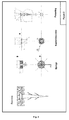
Figure 1 : Device comprising a metallic screw, and sponge and supporting piece made made of a bio-stable material, or biodegradable material wherein the screw and the sponge can rotate freelywith respect to each other. A specific case is described in example 1 -
Figure 2 : Device comprising a nail, a supporting piece and sponge, the sponge made of bioresorbable material, wherein the nail and supporting piece form a single piece solidly united to each other, as in the example 2. -
Figure 3 : A device comprising several fastening elements and that does not have a cylindrical shape as described in example 3. - A device is manufactured wherein the following elements are used: a metallic titanium screw, a porous sponge which is manufactured in a network of copolymer of hydroxyethyl acrylate (HEA) and ethyl acrylate (EA) using ethylene glycol dimethacrylate as cross-linker, and a ring shaped supporting piece of the same material as the sponge.
- The supporting ring is first prepared and punched, synthesizing the material for copolymerization of hydroxyethyl acrylate and ethyl acrylate as a sheet or plate of suitable thickness.
- In a transparent mold prepared for that purpose the device is prepared by synthesizing the porous sponge wrapping the screw head and the supporting ring, in such a way that the screw can freely rotate inside the support ring and the sponge, as described in
figure 1 . The mold is cylindrically shaped so that the sponge as synthesized also has this cylindrical shape. The sponge has a height or thickness usually from 3 to 7 mm, according to the thickness of the cartilage to be treated. - The porous sponge is synthesized by setting the metallic screw and the supporting piece inside the mold, both surrounded by polymethylmethacrylate microspheres of 200 microns diameter, said microspheres filling the volume of the mold. The microspheres are sintered at a temperature of 180 ° C and under a slight pressure, forming a template for the porous structure. The mixture of monomers and the crosslinker, initiator of the photosensitive polymerization, are introduced into the mold, in the hollow space left by the porous template. The polymerization is performed under ultraviolet light at room temperature. Subsequently the supporting piece is demolded, repeatedly washed with acetone to remove the porogen template, and any other monomer remains, then it is washed with ethanol, changing the solvent finally and gradually for distilled water. The supporting piece is dried in vacuum, packed into a suitable bag and sterilized by a process suitable to the material of which the implant is made.
- The outer diameter of the sponge can be from 3 to 15 mm. As many devices as may be necessary are used for the treatment of an injury or defect in the cartilage, to cover the affected area, analogous to the classical treatment of mosaicplasty.
- A device is prepared in which the following componentes are used: a nail made of polylactic acid (PLLA), a porous sponge manufactured in polycaprolactone (PCL) and a ring shaped supporting piece of the same material as the one of the nail. The nail and the supporting ring are manufactured to form a solidly attached single piece.
- The assembly of the nail and the supporting piece are fixed inside the mold to manufacture the piece, and they are surrounded with a mixture of ethyl polymethacrylate microspheres of 90 microns in diameter and polycaprolactone pulverized to obtain a particle size of about 50 microns. The mold temperature is increased to 65 ° C melting the polycaprolactone under slight pressure. After cooling the mold, the piece is demolded and the porogen is dissolved with repeated washing in ethanol. The dimensions are similar to those describe in Example 1. See
Figure 2 . - A device as in Example 1 or 2 is prepared, wherein the sponge does not have a cylindrical shape, but is taylor made according to the shape of the cartilage defect or injury to be treated. To this end, a mold for the sponge with the shape of the injury obtained from an image, thomogram, or scan, is prepared. The manufactured device includes as many fasteners with the appropriate type as needed according to the surgeon in charge of the treatment. See
Figure 3 .
Claims (26)
- A device for fastening a material suitable for the regeneration of articular cartilage to the subchondral bone characterized in that it comprises:a) a sponge comprising a biocompatible macroporous material suitable for articular cartilage regeneration,b) an atttachment system suitable for securing the device to the subchondral bone, andc) a supporting piece suitable for attaching the sponge to the atttachment system.
- Device according to claim 1, wherein the supporting piece is mainly formed by a non-porous material and has holes of a diameter greater than 500 µm partially filled with the same macroporous material comprised in the sponge.
- Device according to any of the preceding claims, wherein the supporting piece has the shape of a flat ring.
- Device according to any of the preceding claims, wherein the atttachment system and the sponge move independently from each other.
- Device according to any one of claims 1 to 3, wherein the atttachment system and the supporting piece form a single piece.
- Device according to any of the preceding claims, wherein the atttachment system comprises one or more fixing elements independently selected from the group consisting of a screw, a nail, a pin and a staple.
- Device according to any of the preceding claims, wherein the atttachment system is a metallic attachement system or an atttachment system mainly consisting of a bioresorbable or partially bioresorbable material.
- Device according to claim 7, wherein the metallic atttachment system comprises titanium or porous tantalum.
- Device according to claim 7, wherein the atttachment system comprising a bioresorbable or partially bioresorbable material is mainly composed of the same material comprised in the sponge.
- Device according to any of the preceding claims, wherein the macroporous biocompatible material comprised in the sponge is a partially bioresorbable or bioresorbable material.
- Device according to claim 10, wherein the sponge, the atttachment system and the supporting piece comprise the same bioresorbable or partially bioresorbable material.
- Device according to any one of claims 9 to 11, wherein the bioresorbable or partially bioresorbable material is selected from the group consisting of polylactic acid, a copolymer of polylactic acid / polyglycolic acid, polycaprolactone, polyvinyl alcohol and mixtures thereof.
- Device according to any one of claims 1 to 8, wherein the biocompatible macroporous material comprised in the sponge is a bio-stable material.
- Device according to claim 13, wherein the supporting piece comprises the same bio-stable material as the sponge, and the atttachment system is a metallic atttachment system.
- Device according to any one of claims 13 or 14, wherein the bio-stable material is selected from the group comprising an n-alkyl polyacrylate, n-alkyl polymethacrylate, and mixtures thereof.
- Device according to any one of the preceding claims, wherein the pores of the material comprised in the sponge are coated with fibrin or a polysaccharide.
- Device according to claim 16, wherein the polysaccharide is chitosan or hyaluronic acid.
- Device according to any of the preceding claims, wherein the sponge comprises expanded autologous chondrocytes or pluripotent cells.
- Device according to any of the preceding claims, wherein the sponge further comprises at least one drug.
- Device according to any of the preceding claims, wherein the supporting piece has at least one slot of a specific shape and the sponge comprises a cross section with the same shape as the slot.
- Device according to any of the preceding claims, wherein the device further comprises a tool for screwing the device to the subchondral bone.
- Device according to any of the preceding claims, wherein the device further comprises a tool to impact the device onto the subchondral bone.
- Device according to claim any one of claims 21 or 22, wherein the tool is a rod that fits in the slot of the supporting piece by at least one of its ends.
- Device according to claim 23, wherein the tool has a handle at the end opposite to the end that fits into the slot of the supporting piece.
- Device according to any one of claims 21 to 24, characterized in that the macroporous material comprised in the sponge has an elasticity sufficient to close the space left after extraction of the tool.
- Device according to any one of claims 21 to 25, wherein said device is sterilized.
Applications Claiming Priority (2)
| Application Number | Priority Date | Filing Date | Title |
|---|---|---|---|
| ES201131625A ES2402035B1 (en) | 2011-10-10 | 2011-10-10 | DEVICE FOR SETTING A MACROPOROUS MATERIAL FOR THE REGENERATION OF ARTICULAR CARTRIDGE |
| PCT/ES2012/070700 WO2013053966A1 (en) | 2011-10-10 | 2012-10-09 | Device for attaching a macroporous material for regenerating articular cartilage |
Publications (2)
| Publication Number | Publication Date |
|---|---|
| EP2767245A1 true EP2767245A1 (en) | 2014-08-20 |
| EP2767245A4 EP2767245A4 (en) | 2015-07-01 |
Family
ID=48050986
Family Applications (1)
| Application Number | Title | Priority Date | Filing Date |
|---|---|---|---|
| EP12840053.8A Withdrawn EP2767245A4 (en) | 2011-10-10 | 2012-10-09 | Device for attaching a macroporous material for regenerating articular cartilage |
Country Status (3)
| Country | Link |
|---|---|
| EP (1) | EP2767245A4 (en) |
| ES (1) | ES2402035B1 (en) |
| WO (1) | WO2013053966A1 (en) |
Cited By (1)
| Publication number | Priority date | Publication date | Assignee | Title |
|---|---|---|---|---|
| WO2024064835A3 (en) * | 2022-09-21 | 2024-06-13 | Subchondral Solutions, Inc. | Methods to reconstruct bone and joint surfaces in femorotibial and patellofemorial joint |
Families Citing this family (1)
| Publication number | Priority date | Publication date | Assignee | Title |
|---|---|---|---|---|
| CN116763991B (en) * | 2023-06-21 | 2025-11-18 | 重庆润泽医药有限公司 | A cartilage repair scaffold |
Family Cites Families (10)
| Publication number | Priority date | Publication date | Assignee | Title |
|---|---|---|---|---|
| US5632745A (en) * | 1995-02-07 | 1997-05-27 | R&D Biologicals, Inc. | Surgical implantation of cartilage repair unit |
| US6251143B1 (en) * | 1999-06-04 | 2001-06-26 | Depuy Orthopaedics, Inc. | Cartilage repair unit |
| US6508821B1 (en) * | 2000-01-28 | 2003-01-21 | Depuy Orthopaedics, Inc. | Soft tissue repair material fixation apparatus and method |
| WO2001078798A1 (en) * | 2000-02-10 | 2001-10-25 | Regeneration Technologies, Inc. | Assembled implant |
| US20020183858A1 (en) * | 2001-06-05 | 2002-12-05 | Contiliano Joseph H. | Attachment of absorbable tissue scaffolds to scaffold fixation devices |
| WO2003005938A1 (en) * | 2001-07-12 | 2003-01-23 | Osteotech, Inc. | Intervertebral impant with movement resistant structure |
| US7011684B2 (en) * | 2002-01-17 | 2006-03-14 | Concept Matrix, Llc | Intervertebral disk prosthesis |
| US7048739B2 (en) * | 2002-12-31 | 2006-05-23 | Depuy Spine, Inc. | Bone plate and resilient screw system allowing bi-directional assembly |
| US7666230B2 (en) * | 2003-12-08 | 2010-02-23 | Depuy Products, Inc. | Implant device for cartilage regeneration in load bearing articulation regions |
| US20070162131A1 (en) * | 2004-12-23 | 2007-07-12 | Friedman Craig D | Repair of spinal annular defects |
-
2011
- 2011-10-10 ES ES201131625A patent/ES2402035B1/en active Active
-
2012
- 2012-10-09 WO PCT/ES2012/070700 patent/WO2013053966A1/en not_active Ceased
- 2012-10-09 EP EP12840053.8A patent/EP2767245A4/en not_active Withdrawn
Cited By (2)
| Publication number | Priority date | Publication date | Assignee | Title |
|---|---|---|---|---|
| WO2024064835A3 (en) * | 2022-09-21 | 2024-06-13 | Subchondral Solutions, Inc. | Methods to reconstruct bone and joint surfaces in femorotibial and patellofemorial joint |
| WO2024064834A3 (en) * | 2022-09-21 | 2024-06-20 | Subchondral Solutions, Inc. | Methods to reconstruct bone and joint surfaces in a hip joint |
Also Published As
| Publication number | Publication date |
|---|---|
| ES2402035A1 (en) | 2013-04-26 |
| ES2402035B1 (en) | 2014-03-07 |
| WO2013053966A1 (en) | 2013-04-18 |
| EP2767245A4 (en) | 2015-07-01 |
Similar Documents
| Publication | Publication Date | Title |
|---|---|---|
| AU2004203671B2 (en) | Method and apparatus for resurfacing an articular surface | |
| CA2548661C (en) | Compressed high density fibrous polymers suitable for implant | |
| CA2503904C (en) | Devices for treating defects in the tissue of a living being | |
| US8177854B2 (en) | Porous and nonporous materials for tissue grafting and repair | |
| JP2007531557A (en) | Spinal ring defects and ring nucleation regeneration | |
| EP1976452B1 (en) | Pliable medical composition and method of use | |
| US20150265745A1 (en) | Porous and Nonporous Materials for Tissue Grafting and Repair | |
| US11179493B2 (en) | Methods and compositions for inducing multi-targeted healing of intervertebral disc defects | |
| JP2022068212A (en) | Scaffold material, methods, and uses | |
| KR20180127640A (en) | Composite medical implants and methods for their use and manufacturing | |
| US10105207B2 (en) | Porous and nonporous materials for tissue grafting and repair | |
| ES2402035B1 (en) | DEVICE FOR SETTING A MACROPOROUS MATERIAL FOR THE REGENERATION OF ARTICULAR CARTRIDGE | |
| CN102283725A (en) | Vertebral body fusion device and manufacturing method thereof |
Legal Events
| Date | Code | Title | Description |
|---|---|---|---|
| PUAI | Public reference made under article 153(3) epc to a published international application that has entered the european phase |
Free format text: ORIGINAL CODE: 0009012 |
|
| 17P | Request for examination filed |
Effective date: 20140512 |
|
| AK | Designated contracting states |
Kind code of ref document: A1 Designated state(s): AL AT BE BG CH CY CZ DE DK EE ES FI FR GB GR HR HU IE IS IT LI LT LU LV MC MK MT NL NO PL PT RO RS SE SI SK SM TR |
|
| RAP1 | Party data changed (applicant data changed or rights of an application transferred) |
Owner name: UNIVERSITAT DE VALENCIA, ESTUDI GENERAL Owner name: UNIVERSITAT POLITECNICA DE VALENCIA |
|
| DAX | Request for extension of the european patent (deleted) | ||
| RA4 | Supplementary search report drawn up and despatched (corrected) |
Effective date: 20150601 |
|
| RIC1 | Information provided on ipc code assigned before grant |
Ipc: A61L 27/56 20060101ALI20150522BHEP Ipc: A61F 2/30 20060101ALI20150522BHEP Ipc: A61B 17/68 20060101AFI20150522BHEP Ipc: A61L 27/58 20060101ALI20150522BHEP |
|
| STAA | Information on the status of an ep patent application or granted ep patent |
Free format text: STATUS: THE APPLICATION IS DEEMED TO BE WITHDRAWN |
|
| 18D | Application deemed to be withdrawn |
Effective date: 20160105 |