EP2762559A1 - Melanocyte or progenitor cell thereof adapted to keratinocyte, and preparation method thereof - Google Patents
Melanocyte or progenitor cell thereof adapted to keratinocyte, and preparation method thereof Download PDFInfo
- Publication number
- EP2762559A1 EP2762559A1 EP12834846.3A EP12834846A EP2762559A1 EP 2762559 A1 EP2762559 A1 EP 2762559A1 EP 12834846 A EP12834846 A EP 12834846A EP 2762559 A1 EP2762559 A1 EP 2762559A1
- Authority
- EP
- European Patent Office
- Prior art keywords
- melanocyte
- keratinocyte
- medium
- expression level
- melanoblast
- Prior art date
- Legal status (The legal status is an assumption and is not a legal conclusion. Google has not performed a legal analysis and makes no representation as to the accuracy of the status listed.)
- Granted
Links
- 210000002752 melanocyte Anatomy 0.000 title claims abstract description 308
- 210000002510 keratinocyte Anatomy 0.000 title claims abstract description 198
- 210000000130 stem cell Anatomy 0.000 title claims description 26
- 238000002360 preparation method Methods 0.000 title 1
- 230000014509 gene expression Effects 0.000 claims abstract description 116
- 210000003866 melanoblast Anatomy 0.000 claims abstract description 108
- 108090000623 proteins and genes Proteins 0.000 claims abstract description 39
- 238000012258 culturing Methods 0.000 claims abstract description 36
- 238000004519 manufacturing process Methods 0.000 claims abstract description 25
- 102000004169 proteins and genes Human genes 0.000 claims abstract description 23
- 102000003425 Tyrosinase Human genes 0.000 claims abstract description 21
- 108060008724 Tyrosinase Proteins 0.000 claims abstract description 21
- 230000000694 effects Effects 0.000 claims abstract description 17
- 230000036564 melanin content Effects 0.000 claims abstract description 10
- 239000002609 medium Substances 0.000 claims description 203
- 210000004027 cell Anatomy 0.000 claims description 157
- PHEDXBVPIONUQT-UHFFFAOYSA-N Cocarcinogen A1 Natural products CCCCCCCCCCCCCC(=O)OC1C(C)C2(O)C3C=C(C)C(=O)C3(O)CC(CO)=CC2C2C1(OC(C)=O)C2(C)C PHEDXBVPIONUQT-UHFFFAOYSA-N 0.000 claims description 80
- PHEDXBVPIONUQT-RGYGYFBISA-N phorbol 13-acetate 12-myristate Chemical compound C([C@]1(O)C(=O)C(C)=C[C@H]1[C@@]1(O)[C@H](C)[C@H]2OC(=O)CCCCCCCCCCCCC)C(CO)=C[C@H]1[C@H]1[C@]2(OC(C)=O)C1(C)C PHEDXBVPIONUQT-RGYGYFBISA-N 0.000 claims description 80
- 241000282414 Homo sapiens Species 0.000 claims description 41
- 101000572986 Homo sapiens POU domain, class 3, transcription factor 2 Proteins 0.000 claims description 41
- 102100026459 POU domain, class 3, transcription factor 2 Human genes 0.000 claims description 40
- 101000801254 Homo sapiens Tumor necrosis factor receptor superfamily member 16 Proteins 0.000 claims description 37
- 102100033725 Tumor necrosis factor receptor superfamily member 16 Human genes 0.000 claims description 37
- 239000013589 supplement Substances 0.000 claims description 26
- OYPRJOBELJOOCE-UHFFFAOYSA-N Calcium Chemical compound [Ca] OYPRJOBELJOOCE-UHFFFAOYSA-N 0.000 claims description 25
- 239000011575 calcium Substances 0.000 claims description 25
- 229910052791 calcium Inorganic materials 0.000 claims description 25
- JYGXADMDTFJGBT-VWUMJDOOSA-N hydrocortisone Chemical compound O=C1CC[C@]2(C)[C@H]3[C@@H](O)C[C@](C)([C@@](CC4)(O)C(=O)CO)[C@@H]4[C@@H]3CCC2=C1 JYGXADMDTFJGBT-VWUMJDOOSA-N 0.000 claims description 18
- 238000000034 method Methods 0.000 claims description 17
- NOESYZHRGYRDHS-UHFFFAOYSA-N insulin Chemical compound N1C(=O)C(NC(=O)C(CCC(N)=O)NC(=O)C(CCC(O)=O)NC(=O)C(C(C)C)NC(=O)C(NC(=O)CN)C(C)CC)CSSCC(C(NC(CO)C(=O)NC(CC(C)C)C(=O)NC(CC=2C=CC(O)=CC=2)C(=O)NC(CCC(N)=O)C(=O)NC(CC(C)C)C(=O)NC(CCC(O)=O)C(=O)NC(CC(N)=O)C(=O)NC(CC=2C=CC(O)=CC=2)C(=O)NC(CSSCC(NC(=O)C(C(C)C)NC(=O)C(CC(C)C)NC(=O)C(CC=2C=CC(O)=CC=2)NC(=O)C(CC(C)C)NC(=O)C(C)NC(=O)C(CCC(O)=O)NC(=O)C(C(C)C)NC(=O)C(CC(C)C)NC(=O)C(CC=2NC=NC=2)NC(=O)C(CO)NC(=O)CNC2=O)C(=O)NCC(=O)NC(CCC(O)=O)C(=O)NC(CCCNC(N)=N)C(=O)NCC(=O)NC(CC=3C=CC=CC=3)C(=O)NC(CC=3C=CC=CC=3)C(=O)NC(CC=3C=CC(O)=CC=3)C(=O)NC(C(C)O)C(=O)N3C(CCC3)C(=O)NC(CCCCN)C(=O)NC(C)C(O)=O)C(=O)NC(CC(N)=O)C(O)=O)=O)NC(=O)C(C(C)CC)NC(=O)C(CO)NC(=O)C(C(C)O)NC(=O)C1CSSCC2NC(=O)C(CC(C)C)NC(=O)C(NC(=O)C(CCC(N)=O)NC(=O)C(CC(N)=O)NC(=O)C(NC(=O)C(N)CC=1C=CC=CC=1)C(C)C)CC1=CN=CN1 NOESYZHRGYRDHS-UHFFFAOYSA-N 0.000 claims description 16
- 239000007640 basal medium Substances 0.000 claims description 13
- 102100031413 L-dopachrome tautomerase Human genes 0.000 claims description 12
- 239000003550 marker Substances 0.000 claims description 12
- 239000003102 growth factor Substances 0.000 claims description 11
- 102000013760 Microphthalmia-Associated Transcription Factor Human genes 0.000 claims description 10
- 108010050345 Microphthalmia-Associated Transcription Factor Proteins 0.000 claims description 10
- 229940088597 hormone Drugs 0.000 claims description 10
- 241000283690 Bos taurus Species 0.000 claims description 9
- 101000773083 Homo sapiens 5,6-dihydroxyindole-2-carboxylic acid oxidase Proteins 0.000 claims description 9
- 102000004338 Transferrin Human genes 0.000 claims description 9
- 108090000901 Transferrin Proteins 0.000 claims description 9
- 239000000872 buffer Substances 0.000 claims description 9
- 229960000890 hydrocortisone Drugs 0.000 claims description 9
- 230000001817 pituitary effect Effects 0.000 claims description 9
- 239000012581 transferrin Substances 0.000 claims description 9
- 102100030310 5,6-dihydroxyindole-2-carboxylic acid oxidase Human genes 0.000 claims description 8
- 108091003079 Bovine Serum Albumin Proteins 0.000 claims description 8
- 102000003974 Fibroblast growth factor 2 Human genes 0.000 claims description 8
- 108090000379 Fibroblast growth factor 2 Proteins 0.000 claims description 8
- HTTJABKRGRZYRN-UHFFFAOYSA-N Heparin Chemical compound OC1C(NC(=O)C)C(O)OC(COS(O)(=O)=O)C1OC1C(OS(O)(=O)=O)C(O)C(OC2C(C(OS(O)(=O)=O)C(OC3C(C(O)C(O)C(O3)C(O)=O)OS(O)(=O)=O)C(CO)O2)NS(O)(=O)=O)C(C(O)=O)O1 HTTJABKRGRZYRN-UHFFFAOYSA-N 0.000 claims description 8
- 102000004877 Insulin Human genes 0.000 claims description 8
- 108090001061 Insulin Proteins 0.000 claims description 8
- 101710093778 L-dopachrome tautomerase Proteins 0.000 claims description 8
- 239000003242 anti bacterial agent Substances 0.000 claims description 8
- 230000001857 anti-mycotic effect Effects 0.000 claims description 8
- 229940088710 antibiotic agent Drugs 0.000 claims description 8
- 239000002543 antimycotic Substances 0.000 claims description 8
- 239000003797 essential amino acid Substances 0.000 claims description 8
- 235000020776 essential amino acid Nutrition 0.000 claims description 8
- 239000012091 fetal bovine serum Substances 0.000 claims description 8
- 229960002897 heparin Drugs 0.000 claims description 8
- 229920000669 heparin Polymers 0.000 claims description 8
- 239000005556 hormone Substances 0.000 claims description 8
- 229940125396 insulin Drugs 0.000 claims description 8
- 210000000933 neural crest Anatomy 0.000 claims description 8
- 150000002894 organic compounds Chemical class 0.000 claims description 8
- 150000003839 salts Chemical class 0.000 claims description 8
- 239000011573 trace mineral Substances 0.000 claims description 8
- 235000013619 trace mineral Nutrition 0.000 claims description 8
- 239000011782 vitamin Substances 0.000 claims description 8
- 229940088594 vitamin Drugs 0.000 claims description 8
- 235000013343 vitamin Nutrition 0.000 claims description 8
- 229930003231 vitamin Natural products 0.000 claims description 8
- 238000005406 washing Methods 0.000 claims description 8
- 101000967299 Homo sapiens Endothelin receptor type B Proteins 0.000 claims description 7
- 101000633054 Homo sapiens Zinc finger protein SNAI2 Proteins 0.000 claims description 7
- 102000017930 EDNRB Human genes 0.000 claims description 6
- 102100029570 Zinc finger protein SNAI2 Human genes 0.000 claims description 6
- 102100033902 Endothelin-1 Human genes 0.000 claims description 5
- 101800004490 Endothelin-1 Proteins 0.000 claims description 5
- 239000002771 cell marker Substances 0.000 claims description 5
- 238000011534 incubation Methods 0.000 claims description 4
- 108010072897 transcription factor Brn-2 Proteins 0.000 claims description 2
- XUMBMVFBXHLACL-UHFFFAOYSA-N Melanin Chemical compound O=C1C(=O)C(C2=CNC3=C(C(C(=O)C4=C32)=O)C)=C2C4=CNC2=C1C XUMBMVFBXHLACL-UHFFFAOYSA-N 0.000 description 40
- 230000035755 proliferation Effects 0.000 description 22
- 239000002953 phosphate buffered saline Substances 0.000 description 18
- 230000004069 differentiation Effects 0.000 description 17
- 229960000074 biopharmaceutical Drugs 0.000 description 13
- 210000002919 epithelial cell Anatomy 0.000 description 12
- 230000012010 growth Effects 0.000 description 11
- 238000000338 in vitro Methods 0.000 description 10
- 210000001519 tissue Anatomy 0.000 description 9
- 108010010803 Gelatin Proteins 0.000 description 8
- 101150056978 HMGS gene Proteins 0.000 description 8
- 101100011891 Saccharomyces cerevisiae (strain ATCC 204508 / S288c) ERG13 gene Proteins 0.000 description 8
- 229920000159 gelatin Polymers 0.000 description 8
- 239000008273 gelatin Substances 0.000 description 8
- 235000019322 gelatine Nutrition 0.000 description 8
- 235000011852 gelatine desserts Nutrition 0.000 description 8
- 230000001939 inductive effect Effects 0.000 description 8
- 239000008188 pellet Substances 0.000 description 8
- IAZDPXIOMUYVGZ-UHFFFAOYSA-N Dimethylsulphoxide Chemical compound CS(C)=O IAZDPXIOMUYVGZ-UHFFFAOYSA-N 0.000 description 7
- 230000018109 developmental process Effects 0.000 description 7
- 239000000049 pigment Substances 0.000 description 7
- 238000004458 analytical method Methods 0.000 description 6
- 230000015572 biosynthetic process Effects 0.000 description 6
- 230000004663 cell proliferation Effects 0.000 description 6
- 238000005119 centrifugation Methods 0.000 description 6
- 239000000463 material Substances 0.000 description 6
- 230000035899 viability Effects 0.000 description 6
- WOVKYSAHUYNSMH-RRKCRQDMSA-N 5-bromodeoxyuridine Chemical compound C1[C@H](O)[C@@H](CO)O[C@H]1N1C(=O)NC(=O)C(Br)=C1 WOVKYSAHUYNSMH-RRKCRQDMSA-N 0.000 description 5
- 101000664703 Homo sapiens Transcription factor SOX-10 Proteins 0.000 description 5
- 102100038808 Transcription factor SOX-10 Human genes 0.000 description 5
- 230000008859 change Effects 0.000 description 5
- 210000005175 epidermal keratinocyte Anatomy 0.000 description 5
- 239000012139 lysis buffer Substances 0.000 description 5
- 108020004999 messenger RNA Proteins 0.000 description 5
- 210000003491 skin Anatomy 0.000 description 5
- WTDRDQBEARUVNC-LURJTMIESA-N L-DOPA Chemical compound OC(=O)[C@@H](N)CC1=CC=C(O)C(O)=C1 WTDRDQBEARUVNC-LURJTMIESA-N 0.000 description 4
- 108010076089 accutase Proteins 0.000 description 4
- AFYNADDZULBEJA-UHFFFAOYSA-N bicinchoninic acid Chemical compound C1=CC=CC2=NC(C=3C=C(C4=CC=CC=C4N=3)C(=O)O)=CC(C(O)=O)=C21 AFYNADDZULBEJA-UHFFFAOYSA-N 0.000 description 4
- 239000002299 complementary DNA Substances 0.000 description 4
- 210000004748 cultured cell Anatomy 0.000 description 4
- 230000037230 mobility Effects 0.000 description 4
- 238000003757 reverse transcription PCR Methods 0.000 description 4
- 102000012422 Collagen Type I Human genes 0.000 description 3
- 108010022452 Collagen Type I Proteins 0.000 description 3
- 101000613490 Homo sapiens Paired box protein Pax-3 Proteins 0.000 description 3
- OKKJLVBELUTLKV-UHFFFAOYSA-N Methanol Chemical compound OC OKKJLVBELUTLKV-UHFFFAOYSA-N 0.000 description 3
- 102100040891 Paired box protein Pax-3 Human genes 0.000 description 3
- 238000011529 RT qPCR Methods 0.000 description 3
- 238000002835 absorbance Methods 0.000 description 3
- 238000003501 co-culture Methods 0.000 description 3
- 210000001787 dendrite Anatomy 0.000 description 3
- 238000011161 development Methods 0.000 description 3
- 210000002615 epidermis Anatomy 0.000 description 3
- 230000006870 function Effects 0.000 description 3
- 239000012528 membrane Substances 0.000 description 3
- 230000008569 process Effects 0.000 description 3
- 238000011002 quantification Methods 0.000 description 3
- 238000000926 separation method Methods 0.000 description 3
- XLYOFNOQVPJJNP-UHFFFAOYSA-N water Substances O XLYOFNOQVPJJNP-UHFFFAOYSA-N 0.000 description 3
- 108091032973 (ribonucleotides)n+m Proteins 0.000 description 2
- 206010001557 Albinism Diseases 0.000 description 2
- UXVMQQNJUSDDNG-UHFFFAOYSA-L Calcium chloride Chemical compound [Cl-].[Cl-].[Ca+2] UXVMQQNJUSDDNG-UHFFFAOYSA-L 0.000 description 2
- 238000002965 ELISA Methods 0.000 description 2
- 102000004190 Enzymes Human genes 0.000 description 2
- 108090000790 Enzymes Proteins 0.000 description 2
- 206010014970 Ephelides Diseases 0.000 description 2
- 101500025419 Homo sapiens Epidermal growth factor Proteins 0.000 description 2
- 101000620359 Homo sapiens Melanocyte protein PMEL Proteins 0.000 description 2
- 102100040445 Keratin, type I cytoskeletal 14 Human genes 0.000 description 2
- 108010066321 Keratin-14 Proteins 0.000 description 2
- WTDRDQBEARUVNC-UHFFFAOYSA-N L-Dopa Natural products OC(=O)C(N)CC1=CC=C(O)C(O)=C1 WTDRDQBEARUVNC-UHFFFAOYSA-N 0.000 description 2
- 102100022430 Melanocyte protein PMEL Human genes 0.000 description 2
- 208000003351 Melanosis Diseases 0.000 description 2
- 206010028980 Neoplasm Diseases 0.000 description 2
- 208000007256 Nevus Diseases 0.000 description 2
- 206010040865 Skin hyperpigmentation Diseases 0.000 description 2
- 206010047642 Vitiligo Diseases 0.000 description 2
- 238000003556 assay Methods 0.000 description 2
- -1 c-KIT Proteins 0.000 description 2
- 239000001110 calcium chloride Substances 0.000 description 2
- 229910001628 calcium chloride Inorganic materials 0.000 description 2
- 201000011510 cancer Diseases 0.000 description 2
- 238000012512 characterization method Methods 0.000 description 2
- 229940096422 collagen type i Drugs 0.000 description 2
- 239000002537 cosmetic Substances 0.000 description 2
- 230000032459 dedifferentiation Effects 0.000 description 2
- LOKCTEFSRHRXRJ-UHFFFAOYSA-I dipotassium trisodium dihydrogen phosphate hydrogen phosphate dichloride Chemical compound P(=O)(O)(O)[O-].[K+].P(=O)(O)([O-])[O-].[Na+].[Na+].[Cl-].[K+].[Cl-].[Na+] LOKCTEFSRHRXRJ-UHFFFAOYSA-I 0.000 description 2
- 229940079593 drug Drugs 0.000 description 2
- 239000003814 drug Substances 0.000 description 2
- 238000002474 experimental method Methods 0.000 description 2
- 238000003633 gene expression assay Methods 0.000 description 2
- 239000000122 growth hormone Substances 0.000 description 2
- 229940116978 human epidermal growth factor Drugs 0.000 description 2
- 230000002401 inhibitory effect Effects 0.000 description 2
- 206010024217 lentigo Diseases 0.000 description 2
- 208000002741 leukoplakia Diseases 0.000 description 2
- 230000008099 melanin synthesis Effects 0.000 description 2
- 201000001441 melanoma Diseases 0.000 description 2
- 210000002780 melanosome Anatomy 0.000 description 2
- 230000005012 migration Effects 0.000 description 2
- 238000013508 migration Methods 0.000 description 2
- 239000000203 mixture Substances 0.000 description 2
- GVUGOAYIVIDWIO-UFWWTJHBSA-N nepidermin Chemical compound C([C@@H](C(=O)N[C@@H]([C@@H](C)CC)C(=O)NCC(=O)N[C@@H](CCC(O)=O)C(=O)N[C@@H](CCCNC(N)=N)C(=O)N[C@@H](CS)C(=O)N[C@@H](CCC(N)=O)C(=O)N[C@@H](CC=1C=CC(O)=CC=1)C(=O)N[C@@H](CCCNC(N)=N)C(=O)N[C@@H](CC(O)=O)C(=O)N[C@@H](CC(C)C)C(=O)N[C@@H](CCCCN)C(=O)N[C@@H](CC=1C2=CC=CC=C2NC=1)C(=O)N[C@@H](CC=1C2=CC=CC=C2NC=1)C(=O)N[C@@H](CCC(O)=O)C(=O)N[C@@H](CC(C)C)C(=O)N[C@@H](CCCNC(N)=N)C(O)=O)NC(=O)CNC(=O)[C@@H](NC(=O)[C@@H](NC(=O)[C@H](CS)NC(=O)[C@H](CC(N)=O)NC(=O)[C@H](CS)NC(=O)[C@H](C)NC(=O)[C@H](CC=1C=CC(O)=CC=1)NC(=O)[C@H](CCCCN)NC(=O)[C@H](CC(O)=O)NC(=O)[C@H](CC(C)C)NC(=O)[C@H](C)NC(=O)[C@H](CCC(O)=O)NC(=O)[C@@H](NC(=O)[C@H](CC=1C=CC(O)=CC=1)NC(=O)[C@H](CCSC)NC(=O)[C@H](CS)NC(=O)[C@@H](NC(=O)CNC(=O)[C@H](CC(O)=O)NC(=O)[C@H](CC=1NC=NC=1)NC(=O)[C@H](CC(C)C)NC(=O)[C@H](CS)NC(=O)[C@H](CC=1C=CC(O)=CC=1)NC(=O)CNC(=O)[C@H](CC(O)=O)NC(=O)[C@H](CC=1NC=NC=1)NC(=O)[C@H](CO)NC(=O)[C@H](CC(C)C)NC(=O)[C@H]1N(CCC1)C(=O)[C@H](CS)NC(=O)[C@H](CCC(O)=O)NC(=O)[C@H](CO)NC(=O)[C@H](CC(O)=O)NC(=O)[C@H](CO)NC(=O)[C@@H](N)CC(N)=O)C(C)C)[C@@H](C)CC)C(C)C)C(C)C)C1=CC=C(O)C=C1 GVUGOAYIVIDWIO-UFWWTJHBSA-N 0.000 description 2
- 239000008363 phosphate buffer Substances 0.000 description 2
- 230000035484 reaction time Effects 0.000 description 2
- 230000009257 reactivity Effects 0.000 description 2
- 230000002829 reductive effect Effects 0.000 description 2
- 230000008929 regeneration Effects 0.000 description 2
- 238000011069 regeneration method Methods 0.000 description 2
- 238000010186 staining Methods 0.000 description 2
- 230000004083 survival effect Effects 0.000 description 2
- 238000003786 synthesis reaction Methods 0.000 description 2
- 230000002087 whitening effect Effects 0.000 description 2
- UCTWMZQNUQWSLP-VIFPVBQESA-N (R)-adrenaline Chemical compound CNC[C@H](O)C1=CC=C(O)C(O)=C1 UCTWMZQNUQWSLP-VIFPVBQESA-N 0.000 description 1
- 229930182837 (R)-adrenaline Natural products 0.000 description 1
- APKFDSVGJQXUKY-KKGHZKTASA-N Amphotericin-B Natural products O[C@H]1[C@@H](N)[C@H](O)[C@@H](C)O[C@H]1O[C@H]1C=CC=CC=CC=CC=CC=CC=C[C@H](C)[C@@H](O)[C@@H](C)[C@H](C)OC(=O)C[C@H](O)C[C@H](O)CC[C@@H](O)[C@H](O)C[C@H](O)C[C@](O)(C[C@H](O)[C@H]2C(O)=O)O[C@H]2C1 APKFDSVGJQXUKY-KKGHZKTASA-N 0.000 description 1
- 238000009020 BCA Protein Assay Kit Methods 0.000 description 1
- 101001011741 Bos taurus Insulin Proteins 0.000 description 1
- 102000019034 Chemokines Human genes 0.000 description 1
- 108010012236 Chemokines Proteins 0.000 description 1
- 229920000742 Cotton Polymers 0.000 description 1
- 108020004414 DNA Proteins 0.000 description 1
- 102000002045 Endothelin Human genes 0.000 description 1
- 108050009340 Endothelin Proteins 0.000 description 1
- 229930182566 Gentamicin Natural products 0.000 description 1
- CEAZRRDELHUEMR-URQXQFDESA-N Gentamicin Chemical compound O1[C@H](C(C)NC)CC[C@@H](N)[C@H]1O[C@H]1[C@H](O)[C@@H](O[C@@H]2[C@@H]([C@@H](NC)[C@@](C)(O)CO2)O)[C@H](N)C[C@@H]1N CEAZRRDELHUEMR-URQXQFDESA-N 0.000 description 1
- 239000012981 Hank's balanced salt solution Substances 0.000 description 1
- 101000994365 Homo sapiens Integrin alpha-6 Proteins 0.000 description 1
- 101000796203 Homo sapiens L-dopachrome tautomerase Proteins 0.000 description 1
- 101001008874 Homo sapiens Mast/stem cell growth factor receptor Kit Proteins 0.000 description 1
- 101000578784 Homo sapiens Melanoma antigen recognized by T-cells 1 Proteins 0.000 description 1
- 101001011628 Homo sapiens Microphthalmia-associated transcription factor Proteins 0.000 description 1
- 101001094545 Homo sapiens Retrotransposon-like protein 1 Proteins 0.000 description 1
- 102100032816 Integrin alpha-6 Human genes 0.000 description 1
- 102100027754 Mast/stem cell growth factor receptor Kit Human genes 0.000 description 1
- 102100028389 Melanoma antigen recognized by T-cells 1 Human genes 0.000 description 1
- 108010032605 Nerve Growth Factor Receptors Proteins 0.000 description 1
- 102000007339 Nerve Growth Factor Receptors Human genes 0.000 description 1
- 108010088225 Nestin Proteins 0.000 description 1
- 102000008730 Nestin Human genes 0.000 description 1
- 239000000020 Nitrocellulose Substances 0.000 description 1
- 102000001759 Notch1 Receptor Human genes 0.000 description 1
- 108010029755 Notch1 Receptor Proteins 0.000 description 1
- 208000012641 Pigmentation disease Diseases 0.000 description 1
- 208000000453 Skin Neoplasms Diseases 0.000 description 1
- GLNADSQYFUSGOU-GPTZEZBUSA-J Trypan blue Chemical compound [Na+].[Na+].[Na+].[Na+].C1=C(S([O-])(=O)=O)C=C2C=C(S([O-])(=O)=O)C(/N=N/C3=CC=C(C=C3C)C=3C=C(C(=CC=3)\N=N\C=3C(=CC4=CC(=CC(N)=C4C=3O)S([O-])(=O)=O)S([O-])(=O)=O)C)=C(O)C2=C1N GLNADSQYFUSGOU-GPTZEZBUSA-J 0.000 description 1
- 102000004142 Trypsin Human genes 0.000 description 1
- 108090000631 Trypsin Proteins 0.000 description 1
- 102100027881 Tumor protein 63 Human genes 0.000 description 1
- 101710140697 Tumor protein 63 Proteins 0.000 description 1
- APKFDSVGJQXUKY-INPOYWNPSA-N amphotericin B Chemical compound O[C@H]1[C@@H](N)[C@H](O)[C@@H](C)O[C@H]1O[C@H]1/C=C/C=C/C=C/C=C/C=C/C=C/C=C/[C@H](C)[C@@H](O)[C@@H](C)[C@H](C)OC(=O)C[C@H](O)C[C@H](O)CC[C@@H](O)[C@H](O)C[C@H](O)C[C@](O)(C[C@H](O)[C@H]2C(O)=O)O[C@H]2C1 APKFDSVGJQXUKY-INPOYWNPSA-N 0.000 description 1
- 229960003942 amphotericin b Drugs 0.000 description 1
- 210000000270 basal cell Anatomy 0.000 description 1
- 230000008901 benefit Effects 0.000 description 1
- IXIBAKNTJSCKJM-BUBXBXGNSA-N bovine insulin Chemical compound C([C@@H](C(=O)N[C@@H](CC(C)C)C(=O)N[C@H]1CSSC[C@H]2C(=O)N[C@@H](C)C(=O)N[C@@H](CO)C(=O)N[C@H](C(=O)N[C@H](C(N[C@@H](CO)C(=O)N[C@@H](CC(C)C)C(=O)N[C@@H](CC=3C=CC(O)=CC=3)C(=O)N[C@@H](CCC(N)=O)C(=O)N[C@@H](CC(C)C)C(=O)N[C@@H](CCC(O)=O)C(=O)N[C@@H](CC(N)=O)C(=O)N[C@@H](CC=3C=CC(O)=CC=3)C(=O)N[C@@H](CSSC[C@H](NC(=O)[C@H](C(C)C)NC(=O)[C@H](CC(C)C)NC(=O)[C@H](CC=3C=CC(O)=CC=3)NC(=O)[C@H](CC(C)C)NC(=O)[C@H](C)NC(=O)[C@H](CCC(O)=O)NC(=O)[C@H](C(C)C)NC(=O)[C@H](CC(C)C)NC(=O)[C@H](CC=3NC=NC=3)NC(=O)[C@H](CO)NC(=O)CNC1=O)C(=O)NCC(=O)N[C@@H](CCC(O)=O)C(=O)N[C@@H](CCCNC(N)=N)C(=O)NCC(=O)N[C@@H](CC=1C=CC=CC=1)C(=O)N[C@@H](CC=1C=CC=CC=1)C(=O)N[C@@H](CC=1C=CC(O)=CC=1)C(=O)N[C@@H]([C@@H](C)O)C(=O)N1[C@@H](CCC1)C(=O)N[C@@H](CCCCN)C(=O)N[C@@H](C)C(O)=O)C(=O)N[C@@H](CC(N)=O)C(O)=O)=O)CSSC[C@@H](C(N2)=O)NC(=O)[C@H](CCC(N)=O)NC(=O)[C@H](CCC(O)=O)NC(=O)[C@H](C(C)C)NC(=O)[C@@H](NC(=O)CN)[C@@H](C)CC)C(C)C)NC(=O)[C@H](CCC(N)=O)NC(=O)[C@H](CC(N)=O)NC(=O)[C@@H](NC(=O)[C@@H](N)CC=1C=CC=CC=1)C(C)C)C1=CN=CN1 IXIBAKNTJSCKJM-BUBXBXGNSA-N 0.000 description 1
- 230000015556 catabolic process Effects 0.000 description 1
- 238000004113 cell culture Methods 0.000 description 1
- 230000032823 cell division Effects 0.000 description 1
- 230000010261 cell growth Effects 0.000 description 1
- 230000012292 cell migration Effects 0.000 description 1
- 210000003855 cell nucleus Anatomy 0.000 description 1
- 239000011248 coating agent Substances 0.000 description 1
- 238000000576 coating method Methods 0.000 description 1
- 210000000805 cytoplasm Anatomy 0.000 description 1
- 230000003247 decreasing effect Effects 0.000 description 1
- 238000006731 degradation reaction Methods 0.000 description 1
- 108010007093 dispase Proteins 0.000 description 1
- 108010051081 dopachrome isomerase Proteins 0.000 description 1
- ZUBDGKVDJUIMQQ-UBFCDGJISA-N endothelin-1 Chemical compound C([C@@H](C(=O)N[C@@H](CC(C)C)C(=O)N[C@@H](CC(O)=O)C(=O)N[C@@H]([C@@H](C)CC)C(=O)N[C@@H]([C@@H](C)CC)C(=O)N[C@@H](CC=1C2=CC=CC=C2NC=1)C(O)=O)NC(=O)[C@H]1NC(=O)[C@H](CC=2C=CC=CC=2)NC(=O)[C@@H](CC=2C=CC(O)=CC=2)NC(=O)[C@H](C(C)C)NC(=O)[C@H]2CSSC[C@@H](C(N[C@H](CO)C(=O)N[C@@H](CO)C(=O)N[C@H](CC(C)C)C(=O)N[C@@H](CCSC)C(=O)N[C@H](CC(O)=O)C(=O)N[C@@H](CCCCN)C(=O)N[C@@H](CCC(O)=O)C(=O)N2)=O)NC(=O)[C@@H](CO)NC(=O)[C@H](N)CSSC1)C1=CNC=N1 ZUBDGKVDJUIMQQ-UBFCDGJISA-N 0.000 description 1
- YQGOJNYOYNNSMM-UHFFFAOYSA-N eosin Chemical compound [Na+].OC(=O)C1=CC=CC=C1C1=C2C=C(Br)C(=O)C(Br)=C2OC2=C(Br)C(O)=C(Br)C=C21 YQGOJNYOYNNSMM-UHFFFAOYSA-N 0.000 description 1
- 229960005139 epinephrine Drugs 0.000 description 1
- 238000010195 expression analysis Methods 0.000 description 1
- 229960002518 gentamicin Drugs 0.000 description 1
- 239000011521 glass Substances 0.000 description 1
- 102000043619 human EDNRB Human genes 0.000 description 1
- 102000055351 human MITF Human genes 0.000 description 1
- 102000044928 human SNAI2 Human genes 0.000 description 1
- 102000046634 human TYRP1 Human genes 0.000 description 1
- 239000004615 ingredient Substances 0.000 description 1
- 238000002372 labelling Methods 0.000 description 1
- 210000005055 nestin Anatomy 0.000 description 1
- 229920001220 nitrocellulos Polymers 0.000 description 1
- 210000004940 nucleus Anatomy 0.000 description 1
- 230000019612 pigmentation Effects 0.000 description 1
- 238000007747 plating Methods 0.000 description 1
- 239000011148 porous material Substances 0.000 description 1
- 230000001376 precipitating effect Effects 0.000 description 1
- 230000002062 proliferating effect Effects 0.000 description 1
- 230000001681 protective effect Effects 0.000 description 1
- 238000003762 quantitative reverse transcription PCR Methods 0.000 description 1
- 230000004044 response Effects 0.000 description 1
- 239000012266 salt solution Substances 0.000 description 1
- 201000000849 skin cancer Diseases 0.000 description 1
- 239000000243 solution Substances 0.000 description 1
- 230000000638 stimulation Effects 0.000 description 1
- 239000006228 supernatant Substances 0.000 description 1
- 238000012360 testing method Methods 0.000 description 1
- 239000012588 trypsin Substances 0.000 description 1
- 210000003934 vacuole Anatomy 0.000 description 1
- 238000001262 western blot Methods 0.000 description 1
Images





Classifications
-
- C—CHEMISTRY; METALLURGY
- C12—BIOCHEMISTRY; BEER; SPIRITS; WINE; VINEGAR; MICROBIOLOGY; ENZYMOLOGY; MUTATION OR GENETIC ENGINEERING
- C12N—MICROORGANISMS OR ENZYMES; COMPOSITIONS THEREOF; PROPAGATING, PRESERVING, OR MAINTAINING MICROORGANISMS; MUTATION OR GENETIC ENGINEERING; CULTURE MEDIA
- C12N5/00—Undifferentiated human, animal or plant cells, e.g. cell lines; Tissues; Cultivation or maintenance thereof; Culture media therefor
- C12N5/06—Animal cells or tissues; Human cells or tissues
- C12N5/0602—Vertebrate cells
- C12N5/0625—Epidermal cells, skin cells; Cells of the oral mucosa
- C12N5/0626—Melanocytes
-
- C—CHEMISTRY; METALLURGY
- C12—BIOCHEMISTRY; BEER; SPIRITS; WINE; VINEGAR; MICROBIOLOGY; ENZYMOLOGY; MUTATION OR GENETIC ENGINEERING
- C12N—MICROORGANISMS OR ENZYMES; COMPOSITIONS THEREOF; PROPAGATING, PRESERVING, OR MAINTAINING MICROORGANISMS; MUTATION OR GENETIC ENGINEERING; CULTURE MEDIA
- C12N2500/00—Specific components of cell culture medium
- C12N2500/05—Inorganic components
- C12N2500/10—Metals; Metal chelators
- C12N2500/12—Light metals, i.e. alkali, alkaline earth, Be, Al, Mg
- C12N2500/14—Calcium; Ca chelators; Calcitonin
-
- C—CHEMISTRY; METALLURGY
- C12—BIOCHEMISTRY; BEER; SPIRITS; WINE; VINEGAR; MICROBIOLOGY; ENZYMOLOGY; MUTATION OR GENETIC ENGINEERING
- C12N—MICROORGANISMS OR ENZYMES; COMPOSITIONS THEREOF; PROPAGATING, PRESERVING, OR MAINTAINING MICROORGANISMS; MUTATION OR GENETIC ENGINEERING; CULTURE MEDIA
- C12N2506/00—Differentiation of animal cells from one lineage to another; Differentiation of pluripotent cells
- C12N2506/09—Differentiation of animal cells from one lineage to another; Differentiation of pluripotent cells from epidermal cells, from skin cells, from oral mucosa cells
- C12N2506/094—Differentiation of animal cells from one lineage to another; Differentiation of pluripotent cells from epidermal cells, from skin cells, from oral mucosa cells from keratinocytes
Definitions
- the present disclosure relates to melanocytes or melanoblasts as progenitor cells thereof, which are adapted to keratinocytes. Further, the present disclosure relates to a method for producing the melanocytes or melanoblasts as progenitor cells thereof, which are adapted to keratinocytes.
- melanocytes in epidermis are very few as much as 1/10 of the number of keratinocytes.
- melanocytes are separated by: additionally treating an epidermal sheet, which is obtained by treating the skin tissue with dispase, with an enzyme such as trypsin to make single cells, and then providing a medium containing specific growth factors or chemokines to the cells to make only melanocytes survive.
- an epidermal sheet which is obtained by treating the skin tissue with dispase, with an enzyme such as trypsin to make single cells, and then providing a medium containing specific growth factors or chemokines to the cells to make only melanocytes survive.
- the melanocytes exist very few and there is no specific separation method except a medium, directly separating melanocytes from a living body is inefficient. For this reason, the price of commercial melanocytes is relatively expensive, compared with that of keratinocytes.
- the melanocytes separated from a living body have limited homogeneity and proliferation capacity. Further, when using melanocytes in vitro shortly after directly separating the melanocytes from the epidermal sheet, it was reported that the cells have no response to stimulation such as UVB, or rather the expression of the genes related to melanin synthesis was reduced. In order to solve the above problems, measures, for example, treating UVB to co-culture of keratinocytes and melanocytes, or treating UVB to single-culture of keratinocytes followed by providing the culture to melanocytes, were needed.
- melanocytes are terminally differentiated from melanocyte stem cells, which are derived from neural crest during a developmental process, and melanoblasts as progenitor cells.
- the melanoblasts have better proliferation capacity and differentiation capacity than melanocytes, and have cell mobility (migration) as a characteristic of a developmental process.
- the present disclosure is directed to providing a melanocyte, which has good proliferation capacity resulting from its good homogeneity, engraftment capacity and viability.
- the present disclosure is also directed to providing a melanocyte having melanin forming capacity.
- the present disclosure is also directed to providing a melanocyte, which may be studied alone, without measures such as treating UVB to co-culture of a keratinocyte and a melanocyte, or treating UVB to single-culture of a keratinocyte followed by providing the culture to a melanocyte.
- the present disclosure is also directed to providing a melanocyte, which is adapted to a keratinocyte in vitro in a state of maintaining relationship with the keratinocyte.
- the present disclosure is also directed to providing a separated human melanoblast and a method for producing thereof.
- the present disclosure is also directed to providing a human melanoblast adapted to a keratinocyte and a method for producing thereof.
- a method for producing melanocytes or melanoblasts as progenitor cells thereof adapted to a keratinocyte which includes: (a) culturing keratinocytes in a keratinocyte medium in a dish; (b) removing the keratinocytes from the culture; and (c) collecting cells attached to the bottom of the dish from the culture, in which the keratinocyte is removed, and culturing the collected cells in a melanocyte medium or a melanoblast medium.
- the method according to one aspect of the present disclosure may be a method for producing melanocytes
- the melanocyte medium may be a basal medium supplemented with supplements, wherein the basal medium comprises essential and non-essential amino acids, vitamins, organic compounds, trace minerals and inorganic salts, but do not comprise antibiotics, antimycotics, hormones, growth factors or proteins
- the supplements comprise fetal bovine serum, basic fibroblast growth factor, bovine pituitary extract, heparin, hydrocortisone, insulin, transferrin and phorbol 12-myristate 13-acetate (PMA).
- the supplement may further include Dimethyl sulfoxide (DMSO).
- the method according to one aspect of the present disclosure may be a method for producing melanoblasts
- the melanoblast medium may be basal medium supplemented with supplements, wherein the basal medium comprises essential and non-essential amino acids, vitamins, organic compounds, trace minerals and inorganic salts, but do not comprise antibiotics, antimycotics, hormones, growth factors or proteins
- the supplements comprise fetal bovine serum, basic fibroblast growth factor, bovine pituitary extract, heparin, hydrocortisone, insulin, transferrin and endothelin-1.
- the melanocytes adapted to a keratinocyte may be melanocytes adapted to a keratinocyte, which has at least one characteristic selected from the following characteristics: (i) an expression level of p75NTR, which is a neural crest stem cell marker lower than a primary melanocyte; (ii) an expression level of BRN2, which is expressed in a melanoblast higher than a primary melanocyte; (iii) a melanin content higher than a primary melanocyte; (iv) a tyrosinase activity higher than a primary melanocyte; (v) an increased expression level of p75NTR higher than a primary melanocyte when cultured in a phorbol 12-myristate 13-acetate (PMA)-free medium; (vi) an increased expression level of BRN2 lower than a primary melanocyte when cultured in a PMA-free medium; (vii) a characteristic that a relative ratio of the p75NTR expression
- the melanoblast according to one aspect of the present disclosure may be a melanoblast adapted to a keratinocyte, which has a characteristic that the expression level of at least one melanoblast marker selected from the group consisting of MITF, DCT, TYRP1, SNAI2, C-KIT and EDNRB is higher than a primary melanocyte.
- the present disclosure may provide a melanocyte or a melanoblast having good proliferation capacity resulting from its excellent homogeneity, engraftment capacity and viability.
- the present disclosure may provide a melanocyte having melanin forming capacity.
- the present disclosure may provide a melanocyte, which may be studied alone, without measures such as treating UVB to co-culture of a keratinocyte and a melanocyte, or treating UVB to single-culture of a keratinocyte followed by providing the culture to a melanocyte.
- the present disclosure may provide a melanocyte or a melanoblast adapted in vitro while keeping the relation with a keratinocyte.
- the present disclosure may make sure feasibility of studies of a melanocyte or a melanoblast conducted in vitro.
- risk factors and treatment methods of skin hyperpigmentation such as nevi, lentigo and age spot, achromatosis such as vitiligo (leukoplakia and albinism) or canities, or cancer such as melanoma may be found through studies of survival, propagation, differentiation and regeneration of a melanocyte or a melanoblast.
- the present disclosure may facilitate the development of whitening cosmetics and medicines, which have purposes on reducing melisma and freckles, and inhibiting melanin pigment formation.
- FIG. 13 is images of state of each cell cultured for a long time in a PMA- or PMA+ medium (Scale bar, 200 ⁇ m).
- PMC primary melanocyte
- melanoblast adapted to a keratinocyte refers to a melanoblast obtained by culturing a keratinocyte in vitro.
- a melanocyte adapted to a keratinocyte refers to a melanocyte, which is obtained by culturing a keratinocyte in vitro, and has a basic characteristic of a primary melanocyte but also at least one different characteristic.
- keratinocyte medium refers to a medium for culturing a human keratinocyte, and it may be any medium known in the art.
- the keratinocyte medium may be a medium containing Bovine Pituitary Extract (BPE), human epidermal growth factor (hEGF), bovine Insulin, Hydrocortisone, and gentamicin and amphotericin-B (GA-1000).
- BPE Bovine Pituitary Extract
- hEGF human epidermal growth factor
- hEGF human epidermal growth factor
- G-1000 gentamicin and amphotericin-B
- the medium which may be commercially obtained, may be KGMTM (Cat. No. CC-3001; Lonza), KGMTM BulletKitTM (Cat. No. CC-3111; Lonza), KGMTM 2 BulletKitTM (Cat. No. CC-3107; Lonza) and KGM-GoldTM BulletKitTM (Cat. No. 192060; Lonza).
- melanocyte medium refers to a medium for culturing a melanocyte, and it may be any medium known in the art.
- it may be a melanin forming medium, which contains essential and non-essential amino acids, vitamins, organic compounds, trace mineral and inorganic salts, but does not contain antibiotics, antimycotics, hormone, growth factor or protein.
- the melanocyte medium may be a medium supplemented with a supplement containing growth factor, hormone and tissue extract, which are essential for melanocyte growth, in order to plate human melanocyte and proliferate them for a long time.
- the supplements may contain fetal bovine serum, basic fibroblast growth factor, bovine pituitary extract, heparin, hydrocortisone, insulin, transferrin and phorbol 12-myristate 13-acetate (PMA).
- the supplement may further include Dimethyl sulfoxide(DMSO)
- the example of the supplement may be Human Melanocyte Growth Supplement (HMGS, cat. # S-002-5, Cascade Biologics).
- HMGS Human Melanocyte Growth Supplement
- An example of a commercially available basic medium for a melanocyte supplemented with HMGS may be M254 medium (Cascade Biologics).
- the term "melanoblast medium” used herein refers to a medium for culturing a melanoblast, and it may be any medium known in the art.
- it may be a melanoblast medium, which contains essential and non-essential amino acids, vitamins, organic compounds, trace mineral and inorganic salts, but does not contain antibiotics, antimycotics, hormone, growth factor or protein.
- the melanoblast medium may be a medium supplemented with a supplement containing growth factor, hormone and tissue extract, which are essential for melanoblast growth, in order to plate human melanoblast and proliferate them for a long time.
- the supplement may contain fetal bovine serum, basic fibroblast growth factor, bovine pituitary extract, heparin, hydrocortisone, insulin, transferrin and endothelin-1.
- the supplement may be PMA-Free Human Melanocyte Growth Supplement-2 (PMA-Free HMGS-2, cat. # S-016-5, Cascade Biologics).
- PMA-Free HMGS-2 cat. # S-016-5, Cascade Biologics
- An example of a commercially available basal medium for a melanoblast supplemented with HMGS-2 may be M254 medium (Cascade Biologics).
- PMA-containing medium refers to a medium containing phorbol 12-myristate 13-acetate (PMA), which helps differentiation to a melanocyte.
- calcium medium refers to a keratinocyte medium added with calcium.
- it may be a calcium medium, wherein CaCl 2 is added to a keratinocyte medium at the concentration of about 1.0 to about 1.6 mM, or about 1.2 to about 1.4 mM.
- phorbol 12-myristate 13-acetate (PMA)-free medium refers to a medium for culturing a human melanoblast, which does not contain PMA but contains endothelin-1.
- PMA phorbol 12-myristate 13-acetate
- An example of a commercially available medium may be M254 medium supplemented with PMA-free HMGS-2 (Cascade Biologics, Cat. No.: S-016-5).
- p75NTR refers to a gene of low affinity neurotrophin receptor as a neural crest stem cell marker, which has ACCESSION No. NM_002507, VERSION: NM_002507.3, GI: 295842401.
- BPN2 refers to mRNA of Homo sapiens POU class 3 homeobox 2 (POU3F2) as a gene expressed in a melanoblast. It is deposited as ACCESSION No. NM_005604, VERSION: NM_005604.2, GI: 51702520.
- EDNRB refers to a human endothelin receptor type B (NM_001122659.2,) gene, and it is located on Chr. 13: 78469616 - 78492966.
- C-KIT refers to a human v-kit Hardy-Zuckerman 4 feline sarcoma viral oncogene homolog (NM_001093772.1) gene, and it is located on Chr. 4:55524095 - 55606881.
- SNAI2 refers to a human snail homolog 2 (NM_003068.4), and it is located on Chr. 8: 49830236 - 49833988.
- MEF microphthalmia-associated transcription factor
- DCT refers to a human dopachrome tautomerase, and it is located on Chr. 13: 95091835 - 95131936.
- TYRP1 refers to a human tyrosinase-related protein 1 (NM_000550.2), and it is located on Chr. 9: 12693386 - 12710266.
- PMA phorbol 12-myristate 13-acetate
- a melanocyte is a cell, which produces protective melanin pigment, has dendrites in terms of morphology and exists in the basal layer of epidermis at the ratio of about 1:4 to about 1:10 to basal cells.
- One melanocyte is contacting to 36 keratinocytes (pigmentation unit) making up a basal layer or a spinous layer through dendrites.
- Melanin has a function of protecting a nucleus of a keratinocyte from UV. Accordingly, it was known that people having small amount of melanin is likely to get skin cancer.
- the melanocyte is the cell derived from a neural crest during a developmental process, and differentiates from a melanocyte stem cell and a melanoblast as a progenitor cell thereof to a melanocyte which synthesizes and releases melanin.
- the method for producing melanocytes or melanoblasts as a progenitor cell thereof, which is adapted to a keratinocyte includes: (a) culturing keratinocytes in a keratinocyte medium in a dish; (b) removing the keratinocytes from the culture; and (c) collecting cells attached to the bottom of the dish from the culture, in which the keratinocyte is removed, and culturing the collected cells in a melanocyte medium or a melanoblast medium.
- the keratinocyte of the step (a) may be a human keratinocyte.
- the human keratinocyte may be any keratinocyte derived from human.
- a keratinocyte which is directly separated from human or cultured after separation, and a keratinocyte derived from other cells also may be used.
- Examples of a commercially available human keratinocyte may be NHEK-Neo, Pooled (Neonatal Normal Human Epidermal Keratinocytes, Pooled: Cat. No. 00192906, Tissue acquisition No. P867, white people), NHEK-Neo (Cat. No. 00192907, Tissue acquisition No.: 20647, white people), NHEK-Adult (Cat. No.
- a dish may be coated with about 0.1 to about 0.2% gelatin or about 1 to about 10 ⁇ g/ml collagen-type I, specifically.
- the cell may be cultured in an incubator of about 35 to about 37°C, about 5 to about 10% CO 2 , and may be subcultured when reached to about 70 to about 80% density.
- the step (b) may include: (i) culturing the cultured keratinocytes in a calcium medium, in which calcium is added to a keratinocyte medium; (ii) removing the calcium medium, washing the cell, exchanging the medium with a keratinocyte medium and culturing the cells again; (iii) removing the medium, washing the cell and then incubating the cell; and (iv) separating the keratinocyte in a sheet form by adding a buffer or a keratinocyte medium to the incubated cell.
- the step (a) moves on the step (b) when the cell is cultured in a culture dish at about 80 to about 100% confluency as the result of culturing the keratinocyte in a keratinocyte medium in said (a).
- the time point may be when the cell is cultured at about 90 to about 100% confluency.
- the step (i) of (b) may be culturing the keratinocyte in a calcium medium, in which calcium is added to a keratinocyte medium, for about 2 to 6 about days.
- the culture may be conducted for about 2 to about 3 days. Culturing for the above culture time may help effective differentiation of the keratinocyte, i.e., forming a cultured epithelial cell layer.
- the culture in a calcium medium also may be conducted at an incubator of about 35 to about 37°Cand about 5 to about 10% CO 2 .
- the step (ii) may be culturing the cell by washing the keratinocyte cultured in a calcium medium with a buffer or a keratinocyte medium so as to remove calcium, and then adding a keratinocyte medium thereto again.
- the buffer is not particularly limited, and for example, it may be Phosphate Buffered Saline (PBS, pH 7.4) and/or Hank's Buffered Salt Solution (HBSS, pH 7.4).
- the cell also may be washed with a keratinocyte medium several times instead of washing with a buffer.
- the washed cell may be additionally cultured in a keratinocyte medium for another about 3 to about 7 days.
- the medium may be removed when the color of the newly exchanged medium is not changed any more.
- the medium may be removed within the additional culture time of about 3 to about 7 days.
- the cell may be washed with a buffer and then incubated.
- the buffer used for washing may be, for example, PBS.
- the incubation may be conducted by covering the washed cell with the lid, and then storing in an incubator for about 5 to about 10 min. By incubating for the above time, the keratinocyte may be easily separated in the form of a cultured epithelial cell layer.
- the cultured epithelial cell layer may be spontaneously separated in a sheet form in the process adding a buffer or a keratinocyte medium again to the incubated cell.
- the buffer may be PBS, for example.
- the step (c) may be collecting the cell attached to the bottom of a dish after the keratinocyte is removed in the step (b), and then culturing the cell in a melanoblast medium or a melanoblast medium.
- the dish may be washed several times with PBS and then treated with accutase (Millipore; Cat. No., SCR005) for about 3 to about 10 min.
- accutase Millipore; Cat. No., SCR005
- the separated cell is precipitated by centrifugation, and then transferred to a general culture dish or a dish coated with gelatin, for example, 0.1 % gelatin. Since then, the cell may be cultured by using a melanocyte medium.
- the melanocyte medium may be basal medium supplemented with supplements, wherein the basal medium comprises essential and non-essential amino acids, vitamins, organic compounds, trace minerals and inorganic salts, but do not comprise antibiotics, antimycotics, hormones, growth factors or proteins, wherein the supplements comprise fetal bovine serum, basic fibroblast growth factor, bovine pituitary extract, DMSO, heparin, hydrocortisone, insulin, transferrin and phorbol 12-myristate 13-acetate (PMA).
- the basal medium comprises essential and non-essential amino acids, vitamins, organic compounds, trace minerals and inorganic salts, but do not comprise antibiotics, antimycotics, hormones, growth factors or proteins
- the supplements comprise fetal bovine serum, basic fibroblast growth factor, bovine pituitary extract, DMSO, heparin, hydrocortisone, insulin, transferrin and phorbol 12-myristate 13-acetate (PMA).
- PMA phorbol 12-myri
- the dish may be washed several times with PBS, and then treated with accutase (Millipore; Cat. No., SCR005) for about 3 to about 10 min.
- accutase Millipore; Cat. No., SCR005
- the cell After precipitating the separated cell by centrifugation, the cell may be transferred to a general culture dish or a dish coated with gelatin, for example, 0.1% gelatin. Since then, the cell may be cultured by using a melanoblast medium.
- the melanoblast medium may be basal medium supplemented with supplements, wherein the basal medium comprises essential and non-essential amino acids, vitamins, organic compounds, trace minerals and inorganic salts, but do not comprise antibiotics, antimycotics, hormones, growth factors or proteins, wherein the supplements comprise fetal bovine serum, basic fibroblast growth factor, bovine pituitary extract, heparin, hydrocortisone, insulin, transferrin and endothelin-1.
- the basal medium comprises essential and non-essential amino acids, vitamins, organic compounds, trace minerals and inorganic salts, but do not comprise antibiotics, antimycotics, hormones, growth factors or proteins
- the supplements comprise fetal bovine serum, basic fibroblast growth factor, bovine pituitary extract, heparin, hydrocortisone, insulin, transferrin and endothelin-1.
- the supplements comprise fetal bovine serum, basic fibroblast growth factor, bovine pituitary extract, heparin, hydrocortis
- the dish is washed with PBS, the M254 medium supplemented with HMGS-2 is added thereto right after for additional culture, and then the cell may be transferred to a new dish when the cell density becomes about 70 to about 90% and then the cell may be cultured.
- the keratinocyte-adapted melanocyte may be a melanocyte, which has at least one characteristic selected from the following characteristics: (i) an expression level of p75NTR, which is a neural crest stem cell marker lower than a primary melanocyte; (ii) an expression level of BRN2, which is expressed in a melanoblast higher than a primary melanocyte; (iii) a melanin content higher than a primary melanocyte; (iv) a tyrosinase activity higher than a primary melanocyte; (v) an increased expression level of p75NTR higher than a primary melanocyte when cultured in a phorbol 12-myristate 13-acetate (PMA)-free medium; (vi) lower increased expression level of BRN2 than a primary melanocyte when cultured in a PMA-free medium; (vii) a characteristic that a relative ratio of the p75NTR expression level of the keratinocyte-adapted melanocyte culture
- the expression level of p75NTR of (i) may be about 1 /10 time lower than the primary melanocyte. Further, the expression level of p75NTR of (i) may be about 1/20, 1/30, 1/40, 1/50, 1/60 or 1/70 time lower than the primary melanocyte. Further, the expression level of p75NTR of (i) may be about 1/10 to about 1/1000 time lower than the primary melanocyte. Further, the expression level of p75NTR of (i) may be about 1/20 to about 1/500 time, about 1/30 to about 1/280 time, about 1/40 to about 1/190 time or about 1/50 to about 1/140 time lower than the primary melanocyte.
- the characteristic of the keratinocyte-adapted melanocyte according to one embodiment of the present disclosure for example, ability of keeping melanin forming capacity without a keratinocyte as well as better homogeneity and proliferation capacity, compared with the primary melanocyte, may be maximized.
- the expression level of BRN2 of (ii) may be about 5 times higher than the primary melanocyte. Further, the expression level of BRN2 of (ii) may be about 10 time or about 15 times higher than the primary melanocyte. Further, the expression level of BRN2 of (ii) may be about 5 times to about 50 times, about 10 times to about 40 times, about 15 times to about 30 times or about 16 times to about 20 times higher than the primary melanocyte. Due to this difference on the expression level, the characteristic of the keratinocyte-adapted melanocyte according to one embodiment of the present disclosure, for example, ability of keeping melanin forming capacity without a keratinocyte as well as better homogeneity and proliferation capacity, compared with the primary melanocyte, may be maximized.
- the melanin content of (iii) may be about 2 times or higher than the primary melanocyte. Further the melanin content of (iii) may be about 3 times or about 4 times higher than the primary melanocyte. Further, melanin content of (iii) may be about 2 times to about 10 times, about 3 times to about 5 times or about 3 times to about 4 times higher than the primary melanocyte. Due to this difference on the content, the characteristic of the keratinocyte-adapted melanocyte according to one embodiment of the present disclosure, for example, ability of keeping melanin forming capacity without a keratinocyte as well as better homogeneity and proliferation capacity, compared with the primary melanocyte, may be maximized.
- the tyrosinase activity of (iv) may be about 2 times or more, about 4 times or more, or about 8 times or more higher than the primary melanocyte when incubated at 37°C for about 120 min. Further, it may be about 2 times to about 32 times, about 4 times to about 16 times, or about 6 times or about 10 times than the primary melanocyte. Due to this difference on the activity, the characteristic of the keratinocyte-adapted melanocyte according to one embodiment of the present disclosure, for example, ability of keeping melanin forming capacity without a keratinocyte as well as better homogeneity and proliferation capacity, compared with the primary melanocyte, may be maximized.
- the expression level of (v) may be 2 times or more of the expression level of the primary melanocyte. Further, the expression level of (v) may be about 2 times to about 18 times, about 3 times to about 10 times, or about 4 times to about 8 times of the expression level of the primary melanocyte. Due to this difference on the expression level, the characteristic of the keratinocyte-adapted melanocyte according to one embodiment of the present disclosure, for example, ability of keeping melanin forming capacity without a keratinocyte as well as better homogeneity and proliferation capacity, compared with the primary melanocyte, may be maximized.
- the expression level of (vi) may be about 9/10 to about 1/10 time of the expression level of the primary melanocyte. Further, the expression level of (vi) may be about 7/10 to about 3/10 time of the expression level of the primary melanocyte. Due to this difference on the expression level, the characteristic of the keratinocyte-adapted melanocyte according to one embodiment of the present disclosure, for example, ability of keeping melanin forming capacity without a keratinocyte as well as better homogeneity and proliferation capacity, compared with the primary melanocyte, may be maximized.
- the relative ratio of the p75NTR expression level of the keratinocyte-adapted melanocyte cultured in a PMA-free medium, to the p75NTR expression level of the keratinocyte-adapted melanocyte cultured in a PMA-containing melanocyte medium may be equivalent to about 60 to about 160%, about 80 to about 140% or about 90 to about 130% of the ratio of the p75NTR expression level of the primary melanocyte cultured in a PMA-containing melanocyte medium, to the p75NTR expression level of the keratinocyte-adapted melanocyte cultured in a PMA-containing melanocyte medium.
- the characteristic of the keratinocyte-adapted melanocyte according to one embodiment of the present disclosure for example, ability of keeping melanin forming capacity without a keratinocyte as well as better homogeneity and proliferation capacity, compared with the primary melanocyte, may be maximized.
- the relative ratio of the BRN2 expression level of the keratinocyte-adapted melanocyte cultured in a PMA-free medium, to the BRN2 expression level of the keratinocyte-adapted melanocyte cultured in a PMA-containing melanocyte medium is equivalent to about 1 to about 10 times, about 2 to about 8 times, about 3 to about 7 times or about 4 to about 6 times of the ratio of the BRN2 expression level of the keratinocyte-adapted melanocyte cultured in a PMA-containing melanocyte medium, to the BRN2 expression level of the primary melanocyte cultured in a PMA-containing melanocyte medium.
- the characteristic of the keratinocyte-adapted melanocyte according to one embodiment of the present disclosure for example, ability of keeping melanin forming capacity without a keratinocyte as well as better homogeneity and proliferation capacity, compared with the primary melanocyte, may be maximized.
- the ratio of the cell attached to the bottom of a dish after about 2 hour subculture of (ix) may be about 80% or more.
- the ratio of the cell attached to the bottom of a dish after about 2 hour subculture of (ix) may be about 90% or more.
- the ratio of the cell attached to the bottom of a dish after about 2 hour subculture of (ix) may be about 95% or more.
- the keratinocyte-adapted melanocyte may be the cell derived from a keratinocyte. In particular, it may be the cell derived from a human keratinocyte.
- the keratinocyte-adapted melanocyte according to another embodiment of the present disclosure may be the cell produced by any one method described above.
- the keratinocyte-adapted melanocyte according to one preferred embodiment of the present disclosure is KCTC (Korean Collection for Type Culture) as an International Depositary Authority under the Budapest Treaty on September 14, 2011, as Accession No. KCTC 12015BP.
- the melanoblast which is separated from the culture of a keratinocyte, according to one embodiment of the present disclosure may be the keratinocyte-adapted melanoblast, and may have a characteristic that the expression level of at least one melanoblast marker selected from the group consisting of MITF, DCT, TYRP1, SNAI2, C-KIT and EDNRB is higher than a primary melanocyte or a keratinocyte-adapted melanocyte.
- the melanoblast may have a characteristic that the expression level of at least one melanoblast marker selected from MITF and DCT is about 2 times or more, about 3 times or more, about 4 times or more, or about 5 times or more higher than a primary melanocyte or a keratinocyte-adapted melanocyte.
- Such high expression level may be a distinctive characteristic, which may distinguish the melanoblast described in this description from a primary melanocyte or a keratinocyte-adapted melanocyte (KaMC).
- the melanoblast may be the cell having at least one characteristic selected from higher BRN2 protein expression level and lower TYR protein expression level, than the keratinocyte-adapted melanocyte.
- the melanoblast may be the cell showing the cell number increased about 20 times or more, about 25 times or more, about 30 times or more, or about 35 times or more than the initial cell number when cultured in a melanoblast medium for about 9 days.
- Such high cell proliferation rate may be a distinctive characteristic, which may distinguish the melanoblast described in this description from a primary melanocyte or a keratinocyte-adapted melanocyte (KaMC).
- the melanoblast may be the cell continuously growing even when subcultured about 6 times or more, about 7 times or more, about 8 times or more, or 9 times or more in a melanoblast medium.
- Such characteristic of maintaining the cell proliferation capacity for a long time may be a distinctive characteristic, which may distinguish the melanoblast described in this description from a primary melanocyte or a keratinocyte-adapted melanocyte (KaMC).
- the keratinocyte-adapted melanoblast may be the cell derived from a keratinocyte.
- it may be the cell derived from a human keratinocyte.
- the keratinocyte-adapted melanoblast according to another embodiment of the present disclosure may be the cell produced by any one method described above.
- the keratinocyte-adapted melanoblast according to one preferred embodiment of the present disclosure is KCTC (Korean Collection for Type Culture) as an International Depositary Authority under the Budapest Treaty on July 24, 2012, as Accession No. KCTC 12250BP.
- a human keratinocyte Normal Human Epidermal Keratinocytes
- NHEK-Neo Pooled (Neonatal Normal Human Epidermal Keratinocytes, Pooled: Cat. No. 00192906) from Lonza
- KGM-GoldTM BulletKitTM Cat. No. 192060; Lonza
- a dish coated with 10 ⁇ g/ml collagen type I is used. The cell is cultured in a 37°C, 5% CO 2 incubator, and subcultured when the cell density becomes about 70%.
- KGM-2 BulletKit Cat. No. CC-3107; Lonza
- 0.1 % gelatin coating are used.
- a cultured epithelial cell layer a calcium medium, in which calcium is finally added at high concentration, i.e., 1.2 mM CaCl 2 , to a keratinocyte medium, is used.
- the culture in the calcium-enriched medium is also conducted at the same 37°C, 5% CO 2 incubator.
- the keratinocyte medium is exchanged with a calcium-enriched medium at the time point when the keratinocyte is cultured until the cell covers the bottom of the culture dish (about 90% confluency), and then cultured for 3 days.
- the cultured keratinocyte is washed three times with Phosphate Buffered Saline (PBS, pH 7.4) to remove highly concentrated calcium, the medium is exchanged with a keratinocyte medium, and then the cell is additionally cultured for another 4 days.
- the medium is removed when the color of the newly exchanged medium is not changed any more within this period, and the cell is washed twice with PBS, covered with the lid, and then stored in the incubator for 5 min.
- PBS Phosphate Buffered Saline
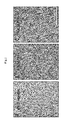
- FIG. 1(A) The remained cell is observed with a microscope, and its image is shown in FIG. 1(A) . As shown in FIG. 1(A) , the cell has a longish shape unlike the keratinocyte.
- the dish After removing the cultured epithelial cell layer, the dish is washed twice with PBS, and then treated with accutase (Millipore; Cat. No., SCR005) for 5 min. The separated cell is precipitated by centrifugation, and then transferred to a dish coated with 0.1% gelatin. Since then, M254 (Cascade Biologics, Cat. No.: M-254-500) medium supplemented with human melanocyte growth supplement (HMGS; Cascase Biology; Cat. No.: S-002-5) is used. As a result, the cultured cell is observed with a microscope, and its image is shown in FIG. 1(B) . As shown in FIG. 1 (B) , the cell has a bipolar shape.
- HMGS human melanocyte growth supplement
- the cell When the cell density becomes about 80%, the cell is subcultured.
- the subcultured cell is observed with a microscope, and its image is shown in FIG. 1(C) .
- the melanocyte separated from a keratinocyte is possible to be subcultured, and has good proliferation capacity.
- NHEM-Neo Neonatal normal human melanocytes, Cat. No. cc-2504 from Lonza is used.
- the separated KaMC ; NHEK-Neo, Pooled (Neonatal Normal Human Epidermal Keratinocytes, Pooled: Cat. No. 00192906) as a keratinocyte (Normal Human Epidermal Keratinocyte) ( KC ) from Lonza; and a primary melanocyte ( PMC ) are washed with PBS, respectively, and total RNA is isolated therefrom by using a kit (miRNeasy mini kit, Qiagen, Cat. No. 217004). Then, cDNA is synthesized by using a RT-PCR kit (SuperScript III First-Strand Synthesis System for RT-PCR, Invitrogen, Cat. No. 18080-051).
- Changes on mRNA expression of SOX10, PAX3, MITF, TYR, TYRP1, DCT, PMEL and NESTIN as a marker of the melanocyte lineage are measured by using the synthesized cDNA as a template, primers, which specifically bind to a gene (TaqMan ® Gene Expression Assays, Applied Biosystems), and SYBR (TaqMan ® Universal PCR Master Mix, Applied Biosystems; Cat. No., 4304437).
- FIG. 2 The result is shown in FIG. 2 .
- the KaMC expresses melanocyte-specific genes more than the keratinocyte (KC).
- the cell After removing the medium, the cell is washed twice with PBS, and then a lysis buffer (Sigma) is added thereto so as to collect the cell. Protein in the cell is extracted while rocking at 4°C for 1 hour, and then cell pellet and extract are separated from each other by centrifugation (1300 rpm, 15 min).
- a lysis buffer Sigma
- FIG. 5(A) is pictures of the pellet taken after separating the PMC and KaMC grown in a melanocyte medium.
- KaMC keratinocyte-adapted melanocyte
- PMC primary melanocyte
- the separated extract is transferred to a new E-tube, and then protein quantification is conducted by using BCA (bicinchoninic acid) (Pierce, Cat. No.:23227).
- BCA bicinchoninic acid
- a lysis buffer is added to 40 ⁇ g protein to the final volume of 100 ⁇ l in a 96-well plate, and then 100 ⁇ l L-Dopa (Sigma, Cat. No. D-9628, 2 mg/ml in 0.1 M phosphate buffer, filtrated) is added thereto followed by reacting them at 37°C for various time (15, 30, 60, 120 min). After each reaction time, absorbance is measured by using a microplate reader at 490 nm.
- FIG. 5(B) The result is shown in FIG. 5(B) .
- the tyrosinase activity of the KaMC is gradually increased, and after incubated for 120 min, the tyrosinase activity of the KaMC is up to about 8 times higher than the PMC.
- M-254 medium supplemented with PMA-free HMGS-2 (Cascade Biologics, Cat. No.: S-016-5) is used for subculture ( PMA- ).
- PMA- M-254 medium supplemented with PMA-free HMGS-2
- PMA+ human melanocyte growth supplement
- the ratio of the p75NTR expression level in the KaMC cultured in the PMA- medium, to the p75NTR expression level in the KaMC cultured in the PMA+ medium is increased to the similar level with the ratio of the p75NTR expression level in the PMC, to that in the KaMC cultured in the PMA+ medium ( FIG. 4(B) ) ( FIG. 7(C) ).
- the ratio of the BRN2 expression level in the PMC cultured in the PMA- medium, to the BRN2 expression level in the PMC cultured in the PMA+ medium is increased to the similar level with the ratio of the BRN2 expression level in the KaMC, to that in the PMC cultured in the PMA+ medium ( FIG. 4(C) ) ( FIG. 7(D) ).
- the color of the KaMC pellet also becomes light ( FIG. 7(E) ).
- the differentiation and dedifferentiation of a melanocyte may be induced by controlling the expressions of the p75NTR and the BRN2, and the pigment formation may be also controlled by the same way.
- the state of the cell after subculturing at the same condition for 2 hours is observed. Specifically, the KaMC and the PMC are transferred to a dish coated with 0.1% gelatin, and then cultured in M254 (Cascade Biologics, Cat. No.: M-254-500) medium supplemented with human melanocyte growth supplement (HMGS; Cascase Biology; Cat. No.: S-002-5) at 37°C for 2 hours. The result is shown in FIG. 8(A) . As shown in FIG.
- Keratinocyte-Adapted Melanoblast KaMB
- a Human keratinocyte is cultured with the same method described in 1.(a).
- the keratinocyte is separated in a sheet form with the same method described in 1. (b). As a result, there are still some cells remained at the bottom of a dish.
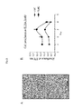
- FIG. 9(A) The remained cell is observed with a microscope, and its image is shown in FIG. 9(A) . As shown in FIG. 9(A) , the cell has a longish shape unlike the keratinocyte.
- the dish After removing the cultured epithelial cell layer, the dish is washed twice with PBS, and then treated with accutase (Millipore; Cat. No., SCR005) for 5 min. The separated cell is precipitated by centrifugation, and then transferred to a new dish. Since then, M254 (Cascade Biologics, Cat. No.: M-254-500) medium supplemented with human melanocyte growth supplement-2 (HMGS-2, PMA-free; Cascase Biology; Cat. No.: S-016-5) is used. As a result, the cultured cell is observed with a microscope, and its image is shown in FIG. 9(B) . When the cell density becomes about 80%, the cell is subcultured. When the cell of FIG. 9(B) is cultured in a melanocyte medium (M254/HMGS) supplemented with PMA, a representative material for inducing melanocyte differentiation, it shows a bipolar shape ( FIG. 9(C) ).
- a primary melanocyte (PMC) for comparison with the KaMB As a primary melanocyte (PMC) for comparison with the KaMB, NHEM-Neo (Neonatal normal human melanocytes, Cat. No. cc-2504) from Lonza is used. Further, a keratinocyte-adapted melanocyte (KaMC) for comparison with the KaMB is also prepared.
- the KaMC is obtained by the same method with the KaMB except for culturing the cell separated in the step (c) in M254 (Cascade Biologics, Cat. No.: M-254-500) medium supplemented with human melanocyte growth supplement (HMGS; Cascase Biology; Cat. No.: S-002-5). Further, the cell is cultured in a PMA+ medium and a PMA- medium, respectively.
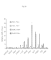
- the separated KaMB, KaMC and a primary melanocyte (PMC) are washed with PBS, respectively, and total RNA is isolated therefrom by using a kit (miRNeasy mini kit, Qiagen, Cat. No. 217004). Then, cDNA is synthesized by using a RT-PCR kit (SuperScript III First-Strand Synthesis System for RT-PCR, Invitrogen, Cat. No. 18080-051).
- FIG. 10 The result is shown in FIG. 10 .
- the melanoblast markers such as EDNRB, c-KIT, SNAI2, MITF, DCT and TYRP1 relatively more than the PMC cultured in the PMA- or PMA+ medium and the KaMC cultured in the PMA+ medium.
- the cell After removing the medium, the cell is washed twice with PBS, and then a lysis buffer (Sigma) is added thereto so as to collect the cell. Protein in the cell is extracted while rocking at 4°C for 1 hour, and then cell pellet and extract are separated from each other by centrifugation (1300 rpm, 15 min).
- a lysis buffer Sigma
- FIG. 11(A) is pictures of the pellet taken after separating the KaMB grown in the medium, which does not contain PMA, a melanocyte differentiation inducing material, and the KaMC, which is derived from the KaMB grown in the PMA-containing medium. From this, it is found that the KaMB may be differentiated to a melanocyte, which may form pigment at a PMA-containing condition.
- the separated extract is transferred to a new E-tube, and then protein quantification is conducted by using BCA (bicinchoninic acid) (Pierce, Cat. No.:23227).
- BCA bicinchoninic acid
- a lysis buffer is added to 40 ⁇ g protein to the final volume of 100 ⁇ l in a 96-well plate, and then 100 ⁇ l L-Dopa (Sigma, Cat. No. D-9628, 2 mg/ml in 0.1 M phosphate buffer, filtrated) is added thereto followed by reacting them at 37°C for various time (15, 30, 60, 120 min). After each reaction time, absorbance is measured by using a microplate reader at 490 nm.
- FIG. 11 (B) The result is shown in FIG. 11 (B) .
- the incubation time increases, the tyrosinase activity of the KaMC is increased, compared with the KaMB.
- Each type of cell (5 x 10 4 ) is seeded in a cell culture insert (Becton Dickinson, Cat. No.: 353097, pore size: 8.0 mm) coated with collagen I (final concentration: 10 ⁇ g/ml), and then cultured in a 24-well for 4 hours. After removing the medium, the cell is fixed with methanol for about 5 to 10 min. Cell nucleus is stained with hematoxilin for 5 min and washed several times with water, and then soaked in eosin for about 1 to 5 min for cytoplasm staining. After washing several times with water, excess water is clearly drained from the inside of the insert using a cotton swab.
- the KaMB shows dominant migration within 4 hours, compared with the differentiated KaMC. According to the fact that a melanoblast has cell mobility as a characteristic of a developmental process in a living body, it is confirmed that the KaMB has the melanoblastic characteristic.
- the KaMB and the KaMC are cultured in a 100-mm dish, respectively, and the cell density becomes about 80 to 90%, the cell is washed twice with PBS. Then a lysis buffer (Sigma) about 100 to 150 ⁇ l is added thereto, and the cell is collected using a cell scraper (SPL Life Sciences). The cell is incubated in a low temperature refrigerator (4 to 8°C) for 10 min, certrifuged for 10 min. Then, a supernatant containing various proteins is transferred to a new tube, and protein quantification is conducted by using a BCA protein assay kit (Thermo Scientific; Cat. No., 23227).
- a BCA protein assay kit Thermo Scientific; Cat. No., 23227.
- 15 ⁇ g protein is electrophoresed and transferred to a nitrocellulose membrane, and then western blot is conducted by using each antibody, SOX10 (R&D systems, MAB2864), PAX3 (R&D systems, MAB2457), BRN2 (ProteinTech Group, 14596-1-AP), MART1 (Thermo Scientific, MS-716), TYRP2 (Santa Cruz biotechnology, C-9, sc-74439), TYRP1 (Santa Cruz biotechnology, H-90, sc-25543), TYR (Santa Cruz biotechnology, H-109, sc-15341) and GADPH (Santa Cruz biotechnology, FL-335, sc-25778).
- SOX10 R&D systems, MAB2864
- PAX3 R&D systems, MAB2457
- BRN2 ProteinTech Group, 14596-1-AP
- MART1 Thermo Scientific, MS-716
- TYRP2 Santa Cruz biotechnology, C-9,
- the primary melanocyte and the KaMB are cultured in a melanocyte medium containing PMA, a melanocyte differentiation inducing material, and a melanoblast medium not containing PMA, respectively.
- the primary melanocyte is properly grown in the PMA-containing medium, but is hardly grown in the melanoblast medium not containing PMA ( FIG. 12(A) , 3 rd , 4 th panels).
- the KaMB separated from a keratinocyte shows superior proliferation capacity in the melanoblast medium not containing PMA than in the PMA-containing melanocyte medium ( FIG. 12(A) , 1 st , 2 nd panels). From this result, it is found that the KaMB is the cell having different characteristics from the dedifferentiated cells, which may be obtained by simply culturing in a melanoblast medium.
- Each type of cell is seeded in 5 wells of a 24-well plate at the concentration of 3.4 x 10 4 cells per well, and the cell number is measured at 0, 3, 6, 9 and 12 th day, respectively.
- the KaMB cell shows the highest proliferation capacity ( FIG. 12(B) )
- Each cell is seeded in each well of a 96-well plate at the concentration of 1 x 10 4 cells per well, and cultured for 3 days. Then, the cell is labeled using Cell Proliferation ELISA, BrdU kit (Roche, Cat. No.: 11 647 229 001), and absorbance is measured with 10, 20 and 30 min interval.
- the BrdU has a characteristic of being attached to DNA of a dividing cell. As a result, it is found that the KaMB in the melanoblast medium not containing PMA divides and proliferates the most ( FIG. 12(C) ). There is no big change while culturing the KaMB in the medium not containing PMA for a long time (passage 9) ( FIG.
- the primary melanocyte hardly proliferates in this medium and also form many vacuoles in the cell when cultured for a long time (passage 6) ( FIG. 13 , 1 st panel).
- the KaMC and the PMC are cultured in the PMA-containing melanocyte medium, respectively, the KaMC shows better proliferation capacity than the PMC ( FIG. 12 (B,C)).
- the KaMB separated from the keratinocyte culture shows the best proliferation capacity in the PMA- melanoblast medium, specifically expresses gene markers and proteins of a melanoblast, and may be differentiated to a melanocyte by PMA, a differentiation inducing material. Thus, it is a melanoblast.
- the KaMC derived from the KaMB which is separated from the keratinocyte culture, has better proliferation capacity than a primary melanocyte. This indicates that the melanocyte, which is separated after being adapted to in vitro condition under keratinocyte environment, has much better proliferation capacity than the primary melanocyte directly separated from the skin tissue.
- the present disclosure may provide a melanocyte or a melanoblast adapted in vitro while keeping the relation with a keratinocyte.
- the present disclosure may make sure feasibility of studies of a melanocyte or a melanoblast conducted in vitro.
- risk factors and treatment methods of skin hyperpigmentation such as nevi, lentigo and age spot, achromatosis such as vitiligo (leukoplakia and albinism) or canities, or cancer such as melanoma may be found through studies of survival, propagation, differentiation and regeneration of a melanocyte or a melanoblast.
- the present disclosure may facilitate the development of whitening cosmetics and medicines, which have purposes on reducing melisma and freckles, and inhibiting melanin pigment formation.
Landscapes
- Health & Medical Sciences (AREA)
- Engineering & Computer Science (AREA)
- Life Sciences & Earth Sciences (AREA)
- Biomedical Technology (AREA)
- Genetics & Genomics (AREA)
- Zoology (AREA)
- Organic Chemistry (AREA)
- Bioinformatics & Cheminformatics (AREA)
- Biotechnology (AREA)
- Chemical & Material Sciences (AREA)
- Wood Science & Technology (AREA)
- Dermatology (AREA)
- Microbiology (AREA)
- Biochemistry (AREA)
- General Engineering & Computer Science (AREA)
- General Health & Medical Sciences (AREA)
- Cell Biology (AREA)
- Micro-Organisms Or Cultivation Processes Thereof (AREA)
Abstract
Description
- The present disclosure relates to melanocytes or melanoblasts as progenitor cells thereof, which are adapted to keratinocytes. Further, the present disclosure relates to a method for producing the melanocytes or melanoblasts as progenitor cells thereof, which are adapted to keratinocytes.
- The number of melanocytes in epidermis is very few as much as 1/10 of the number of keratinocytes. According to conventional separation methods, melanocytes are separated by: additionally treating an epidermal sheet, which is obtained by treating the skin tissue with dispase, with an enzyme such as trypsin to make single cells, and then providing a medium containing specific growth factors or chemokines to the cells to make only melanocytes survive. In respects that the melanocytes exist very few and there is no specific separation method except a medium, directly separating melanocytes from a living body is inefficient. For this reason, the price of commercial melanocytes is relatively expensive, compared with that of keratinocytes.
- On the other hand, the melanocytes separated from a living body have limited homogeneity and proliferation capacity. Further, when using melanocytes in vitro shortly after directly separating the melanocytes from the epidermal sheet, it was reported that the cells have no response to stimulation such as UVB, or rather the expression of the genes related to melanin synthesis was reduced. In order to solve the above problems, measures, for example, treating UVB to co-culture of keratinocytes and melanocytes, or treating UVB to single-culture of keratinocytes followed by providing the culture to melanocytes, were needed.
- It is needed to develop a method for separating melanocytes: which overcome the above shortcomings of the conventionally separated melanocytes, and thereby, having good homogeneity and proliferation capacity, and whose function can be studied without co-culturing them because their characteristics in vitro are maintained similar with the characteristics of when they are present in the body.
- On the other hand, it is known that melanocytes are terminally differentiated from melanocyte stem cells, which are derived from neural crest during a developmental process, and melanoblasts as progenitor cells. The melanoblasts have better proliferation capacity and differentiation capacity than melanocytes, and have cell mobility (migration) as a characteristic of a developmental process.
- Studies about development and differentiation from neural crest stem cell-melanoblast to melanocyte, and functions of the genes related to the above process have been done a lot in a mouse, but as a result of trial to separate melanoblasts by using some differentiation initial markers, yield was less than 5%. Furthermore, because the epidermal structure of the mouse is different from that of human, and there is no specific marker for dividing each differentiation steps, reports about methods for identifying the presence of melanoblasts in the human adult epidermis, or for directly separating melanoblasts from the skin tissue and culturing them are very rare. Some methods for inducing melanoblastic characters by changing a medium of the already separated melanocytes were reported, but it is hard to say that the cells are the melanoblasts existing in the human skin.
- The present disclosure is directed to providing a melanocyte, which has good proliferation capacity resulting from its good homogeneity, engraftment capacity and viability.
- The present disclosure is also directed to providing a melanocyte having melanin forming capacity.
- The present disclosure is also directed to providing a melanocyte, which may be studied alone, without measures such as treating UVB to co-culture of a keratinocyte and a melanocyte, or treating UVB to single-culture of a keratinocyte followed by providing the culture to a melanocyte.
- The present disclosure is also directed to providing a melanocyte, which is adapted to a keratinocyte in vitro in a state of maintaining relationship with the keratinocyte.
- The present disclosure is also directed to providing a separated human melanoblast and a method for producing thereof.
- The present disclosure is also directed to providing a human melanoblast adapted to a keratinocyte and a method for producing thereof.
- In one aspect, there is provided a method for producing melanocytes or melanoblasts as progenitor cells thereof adapted to a keratinocyte, which includes: (a) culturing keratinocytes in a keratinocyte medium in a dish; (b) removing the keratinocytes from the culture; and (c) collecting cells attached to the bottom of the dish from the culture, in which the keratinocyte is removed, and culturing the collected cells in a melanocyte medium or a melanoblast medium.
- The method according to one aspect of the present disclosure may be a method for producing melanocytes, and the melanocyte medium may be a basal medium supplemented with supplements, wherein the basal medium comprises essential and non-essential amino acids, vitamins, organic compounds, trace minerals and inorganic salts, but do not comprise antibiotics, antimycotics, hormones, growth factors or proteins, wherein the supplements comprise fetal bovine serum, basic fibroblast growth factor, bovine pituitary extract, heparin, hydrocortisone, insulin, transferrin and phorbol 12-myristate 13-acetate (PMA). The supplement may further include Dimethyl sulfoxide (DMSO).
- The method according to one aspect of the present disclosure may be a method for producing melanoblasts, and the melanoblast medium may be basal medium supplemented with supplements, wherein the basal medium comprises essential and non-essential amino acids, vitamins, organic compounds, trace minerals and inorganic salts, but do not comprise antibiotics, antimycotics, hormones, growth factors or proteins, wherein the supplements comprise fetal bovine serum, basic fibroblast growth factor, bovine pituitary extract, heparin, hydrocortisone, insulin, transferrin and endothelin-1.
- The melanocytes adapted to a keratinocyte according to one aspect of the present disclosure may be melanocytes adapted to a keratinocyte, which has at least one characteristic selected from the following characteristics: (i) an expression level of p75NTR, which is a neural crest stem cell marker lower than a primary melanocyte; (ii) an expression level of BRN2, which is expressed in a melanoblast higher than a primary melanocyte; (iii) a melanin content higher than a primary melanocyte; (iv) a tyrosinase activity higher than a primary melanocyte; (v) an increased expression level of p75NTR higher than a primary melanocyte when cultured in a phorbol 12-myristate 13-acetate (PMA)-free medium; (vi) an increased expression level of BRN2 lower than a primary melanocyte when cultured in a PMA-free medium; (vii) a characteristic that a relative ratio of the p75NTR expression level of the melanocyte adapted to a keratinocyte cultured in a PMA-free medium, to the p75NTR expression level of the melanocyte adapted to a keratinocyte cultured in a PMA-containing melanocyte medium, is equivalent to about 60 to about 160% of the ratio of the p75NTR expression level of the primary melanocyte cultured in a PMA-containing melanocyte medium, to the p75NTR expression level of the melanocyte adapted to a keratinocyte cultured in a PMA-containing melanocyte medium; (viii) a characteristic that a relative ratio of the BRN2 expression level of the melanocyte adapted to a keratinocyte cultured in a PMA-free medium, to the BRN2 expression level of the melanocyte adapted to a keratinocyte cultured in a PMA-containing melanocyte medium, is equivalent to about 1 to about 10 times of the ratio of the BRN2 expression level of the melanocyte adapted to a keratinocyte cultured in a PMA-containing melanocyte medium, to the BRN2 expression level of the primary melanocyte cultured in a PMA-containing melanocyte medium; and (ix) a ratio of the cell attached to the bottom of a dish after 2 hour subculture higher than a primary melanocyte.
- The melanoblast according to one aspect of the present disclosure may be a melanoblast adapted to a keratinocyte, which has a characteristic that the expression level of at least one melanoblast marker selected from the group consisting of MITF, DCT, TYRP1, SNAI2, C-KIT and EDNRB is higher than a primary melanocyte.
- From one aspect, the present disclosure may provide a melanocyte or a melanoblast having good proliferation capacity resulting from its excellent homogeneity, engraftment capacity and viability.
- From one aspect, the present disclosure may provide a melanocyte having melanin forming capacity.
- From one aspect, the present disclosure may provide a melanocyte, which may be studied alone, without measures such as treating UVB to co-culture of a keratinocyte and a melanocyte, or treating UVB to single-culture of a keratinocyte followed by providing the culture to a melanocyte.
- From one aspect, the present disclosure may provide a melanocyte or a melanoblast adapted in vitro while keeping the relation with a keratinocyte.
- From one aspect, the present disclosure may make sure feasibility of studies of a melanocyte or a melanoblast conducted in vitro.
- From one aspect of the present disclosure, risk factors and treatment methods of skin hyperpigmentation such as nevi, lentigo and age spot, achromatosis such as vitiligo (leukoplakia and albinism) or canities, or cancer such as melanoma may be found through studies of survival, propagation, differentiation and regeneration of a melanocyte or a melanoblast.
- From one aspect, the present disclosure may facilitate the development of whitening cosmetics and medicines, which have purposes on reducing melisma and freckles, and inhibiting melanin pigment formation.
- The above and other aspects, features and advantages of the disclosed exemplary embodiments will be more apparent from the following detailed description taken in conjunction with the accompanying drawings in which:
-
FIG. 1 is images showing cells remained at the bottom of a dish right after separating a cultured cell layer (A), the cells, which are remained at the bottom of a dish, growing in a melanocyte medium (B), and melanocytes growing with high density after subculturing the cells of (B) (C); -
FIG. 2 andFIG. 3 are graphs comparing expression patterns of genes specific to the melanocyte adapted to a keratinocyte according to one embodiment of the present disclosure, i.e., melanocytes separated from keratinocytes (keratinocyte-adapted melanocyte, KaMC) (A,B), and genes specific to keratinocytes (C), with keratinocytes; -
FIG. 4 is graphs comparing gene expression patterns in primary melanocyte (PMC) and KaMC; -
FIG. 5 is images comparing pellet color of PMC and KaMC (A) and a graph comparing tyrosinase activities thereof (B); -
FIG. 6 is graphs showing change on expression in PMC and KaMC depending on the presence of PMA, respectively (A, B); -
FIG. 7 is graphs showing change on expression of p75NTR and BRN2 in PMC and KaMC depending on the presence of PMA (C, D), and an image showing color change of pellet (E); -
FIG. 8 is images comparing initial engraftment capacities of PMC and KaMC (A) and cell proliferation capacities thereof (B); -
FIG. 9 are images showing cells remained in the bottom of a dish right after separating a cultured cell layer (A), the cells, which are remained in the bottom of a dish, growing in a PMA- melanoblast medium (B), and melanocytes growing in a PMA+ melanocyte medium after subculturing the cells of (B) (C) (Scale bar, 250µm); -
FIG. 10 is a graph comparing expression level of each gene in each cell depending on the presence of PMA; -
FIG. 11 is an image comparing pellet color of KaMB cultured in a PMA-medium and KaMC cultured in PMA+ medium (A), a graph comparing tyrosinase activities of the above cells (B), images comparing cell mobilities of the above cells (C), and images comparing expression levels of various proteins in the above cells (D); -
FIG. 12 is images of each cell growing in a PMA- medium or a PMA+ medium (A), a graph showing cell number of each cell with time (B), and a graph comparing proliferation capacity of each cell by BrdU labeling (C) (Scale bar, 250µm); and -
FIG. 13 is images of state of each cell cultured for a long time in a PMA- or PMA+ medium (Scale bar, 200µm). - Hereinafter, the present disclosure is described in detail.
- In this description, the term "primary melanocyte ("PMC")" refers to a melanocyte directly separated from a living body.
- In this description, the term "melanoblast adapted to a keratinocyte (Keratinocyte-adapted Melanoblast, "KaMB")" refers to a melanoblast obtained by culturing a keratinocyte in vitro.
- In this description, the term "a melanocyte adapted to a keratinocyte (Keratinocyte-adapted Melanocyte, "KaMC")" refers to a melanocyte, which is obtained by culturing a keratinocyte in vitro, and has a basic characteristic of a primary melanocyte but also at least one different characteristic.
- The term "keratinocyte medium" used herein refers to a medium for culturing a human keratinocyte, and it may be any medium known in the art. For example, the keratinocyte medium may be a medium containing Bovine Pituitary Extract (BPE), human epidermal growth factor (hEGF), bovine Insulin, Hydrocortisone, and gentamicin and amphotericin-B (GA-1000). Further, it also may be a medium further containing epinephrine and transferrin in addition to the above ingredients. Examples of the medium, which may be commercially obtained, may be KGM™ (Cat. No. CC-3001; Lonza), KGM™ BulletKit™ (Cat. No. CC-3111; Lonza),
KGM™ 2 BulletKit™ (Cat. No. CC-3107; Lonza) and KGM-Gold™ BulletKit™ (Cat. No. 192060; Lonza). - The term "melanocyte medium" used herein refers to a medium for culturing a melanocyte, and it may be any medium known in the art. For example, it may be a melanin forming medium, which contains essential and non-essential amino acids, vitamins, organic compounds, trace mineral and inorganic salts, but does not contain antibiotics, antimycotics, hormone, growth factor or protein. Specifically, the melanocyte medium may be a medium supplemented with a supplement containing growth factor, hormone and tissue extract, which are essential for melanocyte growth, in order to plate human melanocyte and proliferate them for a long time. For example, the supplements may contain fetal bovine serum, basic fibroblast growth factor, bovine pituitary extract, heparin, hydrocortisone, insulin, transferrin and phorbol 12-myristate 13-acetate (PMA). The supplement may further include Dimethyl sulfoxide(DMSO)
- The example of the supplement may be Human Melanocyte Growth Supplement (HMGS, cat. # S-002-5, Cascade Biologics). An example of a commercially available basic medium for a melanocyte supplemented with HMGS may be M254 medium (Cascade Biologics).
- The term "melanoblast medium" used herein refers to a medium for culturing a melanoblast, and it may be any medium known in the art. For example, it may be a melanoblast medium, which contains essential and non-essential amino acids, vitamins, organic compounds, trace mineral and inorganic salts, but does not contain antibiotics, antimycotics, hormone, growth factor or protein. Specifically, the melanoblast medium may be a medium supplemented with a supplement containing growth factor, hormone and tissue extract, which are essential for melanoblast growth, in order to plate human melanoblast and proliferate them for a long time. For example, the supplement may contain fetal bovine serum, basic fibroblast growth factor, bovine pituitary extract, heparin, hydrocortisone, insulin, transferrin and endothelin-1.
- The supplement may be PMA-Free Human Melanocyte Growth Supplement-2 (PMA-Free HMGS-2, cat. # S-016-5, Cascade Biologics). An example of a commercially available basal medium for a melanoblast supplemented with HMGS-2 may be M254 medium (Cascade Biologics).
- The term "PMA-containing medium" used herein refers to a medium containing phorbol 12-myristate 13-acetate (PMA), which helps differentiation to a melanocyte.
- The term "calcium medium" used herein refers to a keratinocyte medium added with calcium. For example, it may be a calcium medium, wherein CaCl2 is added to a keratinocyte medium at the concentration of about 1.0 to about 1.6 mM, or about 1.2 to about 1.4 mM.
- The term " phorbol 12-myristate 13-acetate (PMA)-free medium" used herein refers to a medium for culturing a human melanoblast, which does not contain PMA but contains endothelin-1. An example of a commercially available medium may be M254 medium supplemented with PMA-free HMGS-2 (Cascade Biologics, Cat. No.: S-016-5).
- The term "p75NTR" used herein refers to a gene of low affinity neurotrophin receptor as a neural crest stem cell marker, which has ACCESSION No. NM_002507, VERSION: NM_002507.3, GI: 295842401.
- The term "BRN2" used herein refers to mRNA of Homo
sapiens POU class 3 homeobox 2 (POU3F2) as a gene expressed in a melanoblast. It is deposited as ACCESSION No. NM_005604, VERSION: NM_005604.2, GI: 51702520. - The term "EDNRB" used herein refers to a human endothelin receptor type B (NM_001122659.2,) gene, and it is located on Chr. 13: 78469616 - 78492966.
- The term "C-KIT" used herein refers to a human v-kit Hardy-
Zuckerman 4 feline sarcoma viral oncogene homolog (NM_001093772.1) gene, and it is located on Chr. 4:55524095 - 55606881. - The term "SNAI2" used herein refers to a human snail homolog 2 (NM_003068.4), and it is located on Chr. 8: 49830236 - 49833988.
- The term "MITF" used herein refers to a human microphthalmia-associated transcription factor (NM_198158.2), and it is located on Chr. 3: 69788586 - 70017488.
- The term "DCT" used herein refers to a human dopachrome tautomerase, and it is located on Chr. 13: 95091835 - 95131936.
- The term "TYRP1" used herein refers to a human tyrosinase-related protein 1 (NM_000550.2), and it is located on Chr. 9: 12693386 - 12710266.
- The term "phorbol 12-myristate 13-acetate (PMA)" used herein refers to a material inducing melanocyte differentiation.
- A melanocyte is a cell, which produces protective melanin pigment, has dendrites in terms of morphology and exists in the basal layer of epidermis at the ratio of about 1:4 to about 1:10 to basal cells. One melanocyte is contacting to 36 keratinocytes (pigmentation unit) making up a basal layer or a spinous layer through dendrites. Melanin has a function of protecting a nucleus of a keratinocyte from UV. Accordingly, it was known that people having small amount of melanin is likely to get skin cancer. There is no difference on the number of melanocytes between races, but the number and size of melanosome, the degree of melanization, the distribution of melanosome and the amount of melanin accumulated by degradation in a keratinocyte are different between races. Accordingly, skin color is determined by those factors. It was known that the number of tyrosinase, an enzyme important for melanin synthesis and the number of melanocytes are decreased as age increase. The melanocyte is the cell derived from a neural crest during a developmental process, and differentiates from a melanocyte stem cell and a melanoblast as a progenitor cell thereof to a melanocyte which synthesizes and releases melanin.
- The method for producing melanocytes or melanoblasts as a progenitor cell thereof, which is adapted to a keratinocyte, according to one embodiment of the present disclosure, includes: (a) culturing keratinocytes in a keratinocyte medium in a dish; (b) removing the keratinocytes from the culture; and (c) collecting cells attached to the bottom of the dish from the culture, in which the keratinocyte is removed, and culturing the collected cells in a melanocyte medium or a melanoblast medium.
- The keratinocyte of the step (a) may be a human keratinocyte.
- The human keratinocyte may be any keratinocyte derived from human. A keratinocyte, which is directly separated from human or cultured after separation, and a keratinocyte derived from other cells also may be used. Examples of a commercially available human keratinocyte may be NHEK-Neo, Pooled (Neonatal Normal Human Epidermal Keratinocytes, Pooled: Cat. No. 00192906, Tissue acquisition No. P867, white people), NHEK-Neo (Cat. No. 00192907, Tissue acquisition No.: 20647, white people), NHEK-Adult (Cat. No. 00192627, Tissue acquisition No.: 21155, white people), NHEK-Neo (Cat. No. 00192907, Tissue acquisition No.: 18080, black people), provided from Lonza. In order to maintain a characteristic that a keratinocyte is attached to a base membrane and proliferates, a dish may be coated with about 0.1 to about 0.2% gelatin or about 1 to about 10 µg/ml collagen-type I, specifically. The cell may be cultured in an incubator of about 35 to about 37°C, about 5 to about 10% CO2, and may be subcultured when reached to about 70 to about 80% density.
- The step (b) may include: (i) culturing the cultured keratinocytes in a calcium medium, in which calcium is added to a keratinocyte medium; (ii) removing the calcium medium, washing the cell, exchanging the medium with a keratinocyte medium and culturing the cells again; (iii) removing the medium, washing the cell and then incubating the cell; and (iv) separating the keratinocyte in a sheet form by adding a buffer or a keratinocyte medium to the incubated cell.
- The step (a) moves on the step (b) when the cell is cultured in a culture dish at about 80 to about 100% confluency as the result of culturing the keratinocyte in a keratinocyte medium in said (a). Specifically, the time point may be when the cell is cultured at about 90 to about 100% confluency. By exchanging the medium to a calcium medium at the time point, differentiation of a keratinocyte may be stimulated so as to induce to form a sheet.
- The step (i) of (b) may be culturing the keratinocyte in a calcium medium, in which calcium is added to a keratinocyte medium, for about 2 to 6 about days. The culture may be conducted for about 2 to about 3 days. Culturing for the above culture time may help effective differentiation of the keratinocyte, i.e., forming a cultured epithelial cell layer. The culture in a calcium medium also may be conducted at an incubator of about 35 to about 37°Cand about 5 to about 10% CO2.
- The step (ii) may be culturing the cell by washing the keratinocyte cultured in a calcium medium with a buffer or a keratinocyte medium so as to remove calcium, and then adding a keratinocyte medium thereto again. The buffer is not particularly limited, and for example, it may be Phosphate Buffered Saline (PBS, pH 7.4) and/or Hank's Buffered Salt Solution (HBSS, pH 7.4). The cell also may be washed with a keratinocyte medium several times instead of washing with a buffer. The washed cell may be additionally cultured in a keratinocyte medium for another about 3 to about 7 days.
- In the step (iii), the medium may be removed when the color of the newly exchanged medium is not changed any more. The medium may be removed within the additional culture time of about 3 to about 7 days. After removing the medium, the cell may be washed with a buffer and then incubated. The buffer used for washing may be, for example, PBS. The incubation may be conducted by covering the washed cell with the lid, and then storing in an incubator for about 5 to about 10 min. By incubating for the above time, the keratinocyte may be easily separated in the form of a cultured epithelial cell layer.
- In the step (iv), the cultured epithelial cell layer may be spontaneously separated in a sheet form in the process adding a buffer or a keratinocyte medium again to the incubated cell. The buffer may be PBS, for example.
- The step (c) may be collecting the cell attached to the bottom of a dish after the keratinocyte is removed in the step (b), and then culturing the cell in a melanoblast medium or a melanoblast medium.
- In the step (c), after removing the cultured epithelial cell layer, the dish may be washed several times with PBS and then treated with accutase (Millipore; Cat. No., SCR005) for about 3 to about 10 min. The separated cell is precipitated by centrifugation, and then transferred to a general culture dish or a dish coated with gelatin, for example, 0.1 % gelatin. Since then, the cell may be cultured by using a melanocyte medium. The melanocyte medium may be basal medium supplemented with supplements, wherein the basal medium comprises essential and non-essential amino acids, vitamins, organic compounds, trace minerals and inorganic salts, but do not comprise antibiotics, antimycotics, hormones, growth factors or proteins, wherein the supplements comprise fetal bovine serum, basic fibroblast growth factor, bovine pituitary extract, DMSO, heparin, hydrocortisone, insulin, transferrin and phorbol 12-myristate 13-acetate (PMA). For example, it may be M254 (Cascade Biologics, Cat. No.: M-254-500) medium supplemented with human melanocyte growth supplement (HMGS; Cascase Biology; Cat. No.: S-002-5). When the cell density becomes about 70 to about 90%, the cell may be subcultured.
- In the step (c), after removing the cultured epithelial cell layer, the dish may be washed several times with PBS, and then treated with accutase (Millipore; Cat. No., SCR005) for about 3 to about 10 min. After precipitating the separated cell by centrifugation, the cell may be transferred to a general culture dish or a dish coated with gelatin, for example, 0.1% gelatin. Since then, the cell may be cultured by using a melanoblast medium. The melanoblast medium may be basal medium supplemented with supplements, wherein the basal medium comprises essential and non-essential amino acids, vitamins, organic compounds, trace minerals and inorganic salts, but do not comprise antibiotics, antimycotics, hormones, growth factors or proteins, wherein the supplements comprise fetal bovine serum, basic fibroblast growth factor, bovine pituitary extract, heparin, hydrocortisone, insulin, transferrin and endothelin-1. For example, it may be M254 (Cascade Biologics, Cat. No.: M-254-500) medium supplemented with human melanocyte growth supplement-2 (HMGS-2, PMA-free; Cascase Biology; Cat. No.: S-016-5). When the cell density becomes about 70 to about 90%, the cell may be subcultured.
- Or, in the step (c), after removing the cultured epithelial cell layer, the dish is washed with PBS, the M254 medium supplemented with HMGS-2 is added thereto right after for additional culture, and then the cell may be transferred to a new dish when the cell density becomes about 70 to about 90% and then the cell may be cultured.
- The keratinocyte-adapted melanocyte according to one embodiment of the present disclosure may be a melanocyte, which has at least one characteristic selected from the following characteristics: (i) an expression level of p75NTR, which is a neural crest stem cell marker lower than a primary melanocyte; (ii) an expression level of BRN2, which is expressed in a melanoblast higher than a primary melanocyte; (iii) a melanin content higher than a primary melanocyte; (iv) a tyrosinase activity higher than a primary melanocyte; (v) an increased expression level of p75NTR higher than a primary melanocyte when cultured in a phorbol 12-myristate 13-acetate (PMA)-free medium; (vi) lower increased expression level of BRN2 than a primary melanocyte when cultured in a PMA-free medium; (vii) a characteristic that a relative ratio of the p75NTR expression level of the keratinocyte-adapted melanocyte cultured in a PMA-free medium, to the p75NTR expression level of the keratinocyte-adapted melanocyte cultured in a PMA-containing melanocyte medium, is equivalent to about 60 to about 160% of the ratio of the p75NTR expression level of the primary melanocyte cultured in a PMA-containing melanocyte medium, to the p75NTR expression level of the keratinocyte-adapted melanocyte cultured in a PMA-containing melanocyte medium; (viii) a characteristic that a relative ratio of the BRN2 expression level of the keratinocyte-adapted melanocyte cultured in a PMA-free medium, to the BRN2 expression level of the keratinocyte-adapted melanocyte cultured in a PMA-containing melanocyte medium, is equivalent to about 1 to about 10 times of the ratio of the BRN2 expression level of the keratinocyte-adapted melanocyte cultured in a PMA-containing melanocyte medium, to the BRN2 expression level of the primary melanocyte cultured in a PMA-containing melanocyte medium; and (ix) a ratio of the cell attached to the bottom of a dish after 2 hour subculture higher than a primary melanocyte.
- The expression level of p75NTR of (i) may be about 1 /10 time lower than the primary melanocyte. Further, the expression level of p75NTR of (i) may be about 1/20, 1/30, 1/40, 1/50, 1/60 or 1/70 time lower than the primary melanocyte. Further, the expression level of p75NTR of (i) may be about 1/10 to about 1/1000 time lower than the primary melanocyte. Further, the expression level of p75NTR of (i) may be about 1/20 to about 1/500 time, about 1/30 to about 1/280 time, about 1/40 to about 1/190 time or about 1/50 to about 1/140 time lower than the primary melanocyte. Due to this difference on the expression level, the characteristic of the keratinocyte-adapted melanocyte according to one embodiment of the present disclosure, for example, ability of keeping melanin forming capacity without a keratinocyte as well as better homogeneity and proliferation capacity, compared with the primary melanocyte, may be maximized.
- The expression level of BRN2 of (ii) may be about 5 times higher than the primary melanocyte. Further, the expression level of BRN2 of (ii) may be about 10 time or about 15 times higher than the primary melanocyte. Further, the expression level of BRN2 of (ii) may be about 5 times to about 50 times, about 10 times to about 40 times, about 15 times to about 30 times or about 16 times to about 20 times higher than the primary melanocyte. Due to this difference on the expression level, the characteristic of the keratinocyte-adapted melanocyte according to one embodiment of the present disclosure, for example, ability of keeping melanin forming capacity without a keratinocyte as well as better homogeneity and proliferation capacity, compared with the primary melanocyte, may be maximized.
- The melanin content of (iii) may be about 2 times or higher than the primary melanocyte. Further the melanin content of (iii) may be about 3 times or about 4 times higher than the primary melanocyte. Further, melanin content of (iii) may be about 2 times to about 10 times, about 3 times to about 5 times or about 3 times to about 4 times higher than the primary melanocyte. Due to this difference on the content, the characteristic of the keratinocyte-adapted melanocyte according to one embodiment of the present disclosure, for example, ability of keeping melanin forming capacity without a keratinocyte as well as better homogeneity and proliferation capacity, compared with the primary melanocyte, may be maximized.
- The tyrosinase activity of (iv) may be about 2 times or more, about 4 times or more, or about 8 times or more higher than the primary melanocyte when incubated at 37°C for about 120 min. Further, it may be about 2 times to about 32 times, about 4 times to about 16 times, or about 6 times or about 10 times than the primary melanocyte. Due to this difference on the activity, the characteristic of the keratinocyte-adapted melanocyte according to one embodiment of the present disclosure, for example, ability of keeping melanin forming capacity without a keratinocyte as well as better homogeneity and proliferation capacity, compared with the primary melanocyte, may be maximized.
- The expression level of (v) may be 2 times or more of the expression level of the primary melanocyte. Further, the expression level of (v) may be about 2 times to about 18 times, about 3 times to about 10 times, or about 4 times to about 8 times of the expression level of the primary melanocyte. Due to this difference on the expression level, the characteristic of the keratinocyte-adapted melanocyte according to one embodiment of the present disclosure, for example, ability of keeping melanin forming capacity without a keratinocyte as well as better homogeneity and proliferation capacity, compared with the primary melanocyte, may be maximized.
- The expression level of (vi) may be about 9/10 to about 1/10 time of the expression level of the primary melanocyte. Further, the expression level of (vi) may be about 7/10 to about 3/10 time of the expression level of the primary melanocyte. Due to this difference on the expression level, the characteristic of the keratinocyte-adapted melanocyte according to one embodiment of the present disclosure, for example, ability of keeping melanin forming capacity without a keratinocyte as well as better homogeneity and proliferation capacity, compared with the primary melanocyte, may be maximized.
- In the expression level of (vii), the relative ratio of the p75NTR expression level of the keratinocyte-adapted melanocyte cultured in a PMA-free medium, to the p75NTR expression level of the keratinocyte-adapted melanocyte cultured in a PMA-containing melanocyte medium, may be equivalent to about 60 to about 160%, about 80 to about 140% or about 90 to about 130% of the ratio of the p75NTR expression level of the primary melanocyte cultured in a PMA-containing melanocyte medium, to the p75NTR expression level of the keratinocyte-adapted melanocyte cultured in a PMA-containing melanocyte medium. Due to this difference on the expression level, the characteristic of the keratinocyte-adapted melanocyte according to one embodiment of the present disclosure, for example, ability of keeping melanin forming capacity without a keratinocyte as well as better homogeneity and proliferation capacity, compared with the primary melanocyte, may be maximized.
- In the expression level of (viii), the relative ratio of the BRN2 expression level of the keratinocyte-adapted melanocyte cultured in a PMA-free medium, to the BRN2 expression level of the keratinocyte-adapted melanocyte cultured in a PMA-containing melanocyte medium, is equivalent to about 1 to about 10 times, about 2 to about 8 times, about 3 to about 7 times or about 4 to about 6 times of the ratio of the BRN2 expression level of the keratinocyte-adapted melanocyte cultured in a PMA-containing melanocyte medium, to the BRN2 expression level of the primary melanocyte cultured in a PMA-containing melanocyte medium. Due to this difference on the expression level, the characteristic of the keratinocyte-adapted melanocyte according to one embodiment of the present disclosure, for example, ability of keeping melanin forming capacity without a keratinocyte as well as better homogeneity and proliferation capacity, compared with the primary melanocyte, may be maximized.
- The ratio of the cell attached to the bottom of a dish after about 2 hour subculture of (ix) may be about 80% or more. The ratio of the cell attached to the bottom of a dish after about 2 hour subculture of (ix) may be about 90% or more. The ratio of the cell attached to the bottom of a dish after about 2 hour subculture of (ix) may be about 95% or more. The keratinocyte-adapted melanocyte according to one embodiment of the present disclosure may be the cell derived from a keratinocyte. In particular, it may be the cell derived from a human keratinocyte.
- The keratinocyte-adapted melanocyte according to another embodiment of the present disclosure may be the cell produced by any one method described above.
- The keratinocyte-adapted melanocyte according to one preferred embodiment of the present disclosure is KCTC (Korean Collection for Type Culture) as an International Depositary Authority under the Budapest Treaty on September 14, 2011, as Accession No. KCTC 12015BP.
- The melanoblast, which is separated from the culture of a keratinocyte, according to one embodiment of the present disclosure may be the keratinocyte-adapted melanoblast, and may have a characteristic that the expression level of at least one melanoblast marker selected from the group consisting of MITF, DCT, TYRP1, SNAI2, C-KIT and EDNRB is higher than a primary melanocyte or a keratinocyte-adapted melanocyte.
- In another embodiment of the present disclosure, the melanoblast may have a characteristic that the expression level of at least one melanoblast marker selected from MITF and DCT is about 2 times or more, about 3 times or more, about 4 times or more, or about 5 times or more higher than a primary melanocyte or a keratinocyte-adapted melanocyte. Such high expression level may be a distinctive characteristic, which may distinguish the melanoblast described in this description from a primary melanocyte or a keratinocyte-adapted melanocyte (KaMC).
- Further, in one embodiment, the melanoblast may be the cell having at least one characteristic selected from higher BRN2 protein expression level and lower TYR protein expression level, than the keratinocyte-adapted melanocyte.
- In another embodiment, the melanoblast may be the cell showing the cell number increased about 20 times or more, about 25 times or more, about 30 times or more, or about 35 times or more than the initial cell number when cultured in a melanoblast medium for about 9 days. Such high cell proliferation rate may be a distinctive characteristic, which may distinguish the melanoblast described in this description from a primary melanocyte or a keratinocyte-adapted melanocyte (KaMC).
- In further another embodiment, the melanoblast may be the cell continuously growing even when subcultured about 6 times or more, about 7 times or more, about 8 times or more, or 9 times or more in a melanoblast medium. Such characteristic of maintaining the cell proliferation capacity for a long time may be a distinctive characteristic, which may distinguish the melanoblast described in this description from a primary melanocyte or a keratinocyte-adapted melanocyte (KaMC).
- The keratinocyte-adapted melanoblast according to one embodiment of the present disclosure may be the cell derived from a keratinocyte. In particular, it may be the cell derived from a human keratinocyte.
- The keratinocyte-adapted melanoblast according to another embodiment of the present disclosure may be the cell produced by any one method described above.
- The keratinocyte-adapted melanoblast according to one preferred embodiment of the present disclosure is KCTC (Korean Collection for Type Culture) as an International Depositary Authority under the Budapest Treaty on July 24, 2012, as Accession No. KCTC 12250BP.
- The examples (and experiments) will now be described. The following examples (and experiments) are for illustrative purposes only and not intended to limit the scope of the present disclosure.
- As a human keratinocyte (Normal Human Epidermal Keratinocytes), NHEK-Neo, Pooled (Neonatal Normal Human Epidermal Keratinocytes, Pooled: Cat. No. 00192906) from Lonza is used. As a keratinocyte medium for culturing the human keratinocyte, KGM-Gold™ BulletKit™ Cat. No. 192060; Lonza) is used. In order to maintain a characteristic that a keratinocyte is attached to a base membrane and proliferates, a dish coated with 10 µg/ml collagen type I is used. The cell is cultured in a 37°C, 5% CO2 incubator, and subcultured when the cell density becomes about 70%. For inducing the keratinocyte to a cultured epithelial cell layer, KGM-2 BulletKit (Cat. No. CC-3107; Lonza) and 0.1 % gelatin coating are used.
- In order to remove the keratinocyte from the dish in the form of a sheet, a cultured epithelial cell layer, a calcium medium, in which calcium is finally added at high concentration, i.e., 1.2 mM CaCl2, to a keratinocyte medium, is used. The culture in the calcium-enriched medium is also conducted at the same 37°C, 5% CO2 incubator. The keratinocyte medium is exchanged with a calcium-enriched medium at the time point when the keratinocyte is cultured until the cell covers the bottom of the culture dish (about 90% confluency), and then cultured for 3 days. The cultured keratinocyte is washed three times with Phosphate Buffered Saline (PBS, pH 7.4) to remove highly concentrated calcium, the medium is exchanged with a keratinocyte medium, and then the cell is additionally cultured for another 4 days. The medium is removed when the color of the newly exchanged medium is not changed any more within this period, and the cell is washed twice with PBS, covered with the lid, and then stored in the incubator for 5 min. During a process adding PBS again, the cultured epithelial cell layer is spontaneously separated. Right after the cultured epithelial cell layer is removed, there is a cell still remained at the bottom of the dish.
- The remained cell is observed with a microscope, and its image is shown in
FIG. 1(A) . As shown inFIG. 1(A) , the cell has a longish shape unlike the keratinocyte. - After removing the cultured epithelial cell layer, the dish is washed twice with PBS, and then treated with accutase (Millipore; Cat. No., SCR005) for 5 min. The separated cell is precipitated by centrifugation, and then transferred to a dish coated with 0.1% gelatin. Since then, M254 (Cascade Biologics, Cat. No.: M-254-500) medium supplemented with human melanocyte growth supplement (HMGS; Cascase Biology; Cat. No.: S-002-5) is used. As a result, the cultured cell is observed with a microscope, and its image is shown in
FIG. 1(B) . As shown inFIG. 1 (B) , the cell has a bipolar shape. - When the cell density becomes about 80%, the cell is subcultured. The subcultured cell is observed with a microscope, and its image is shown in
FIG. 1(C) . As shown inFIG. 1(C) , the melanocyte separated from a keratinocyte is possible to be subcultured, and has good proliferation capacity. - As a primary melanocyte (PMC) for comparison with the KaMC, NHEM-Neo (Neonatal normal human melanocytes, Cat. No. cc-2504) from Lonza is used.
- The separated KaMC; NHEK-Neo, Pooled (Neonatal Normal Human Epidermal Keratinocytes, Pooled: Cat. No. 00192906) as a keratinocyte (Normal Human Epidermal Keratinocyte) (KC) from Lonza; and a primary melanocyte (PMC) are washed with PBS, respectively, and total RNA is isolated therefrom by using a kit (miRNeasy mini kit, Qiagen, Cat. No. 217004). Then, cDNA is synthesized by using a RT-PCR kit (SuperScript III First-Strand Synthesis System for RT-PCR, Invitrogen, Cat. No. 18080-051). Changes on mRNA expression of SOX10, PAX3, MITF, TYR, TYRP1, DCT, PMEL and NESTIN as a marker of the melanocyte lineage are measured by using the synthesized cDNA as a template, primers, which specifically bind to a gene (TaqMan® Gene Expression Assays, Applied Biosystems), and SYBR (TaqMan® Universal PCR Master Mix, Applied Biosystems; Cat. No., 4304437).
- The result is shown in
FIG. 2 . As shown inFIG. 2 , it is found that the KaMC expresses melanocyte-specific genes more than the keratinocyte (KC). - On the other hand, change on mRNA expression of SOX10 as a marker of the melanocyte lineage is measured. The result is shown in
FIG. 3(B) . As shown inFIG. 3(B) , it is found that the KaMC expresses the SOX10 as a melanocyte-specific gene more than the keratinocyte (KC) as much as 9 million times. - On the other hand, changes on mRNA expression of ITGA6, TP63 and keratin14 (KRT14) as a marker of a keratinocyte are measured. As a result, the expression is significantly reduced in the KaMC, compared with the keratinocyte (KC) (
FIG. 3(C) ). - Further, when comparing with the primary melanocyte (PMC), it is found that there is no difference on the expression levels of overall melanocyte-specific genes (
FIG. 4(A) ), but the p75NTR as a neural crest stem cell marker is overexpressed in the PMC (FIG. 4(B) ) and the BRN2, which is known as being expressed in a melanoblast, is relatively highly expressed in the KaMC (FIG. 4(C) ). - After removing the medium, the cell is washed twice with PBS, and then a lysis buffer (Sigma) is added thereto so as to collect the cell. Protein in the cell is extracted while rocking at 4°C for 1 hour, and then cell pellet and extract are separated from each other by centrifugation (1300 rpm, 15 min).
-
FIG. 5(A) is pictures of the pellet taken after separating the PMC and KaMC grown in a melanocyte medium. As shown inFIG. 5(A) , it is found that the keratinocyte-adapted melanocyte (KaMC) according to one embodiment of the present disclosure contains more melanin than the primary melanocyte (PMC). Both of the cells are derived from white people. Accordingly, the difference on the melanin content is notable. - On the other hand, the separated extract is transferred to a new E-tube, and then protein quantification is conducted by using BCA (bicinchoninic acid) (Pierce, Cat. No.:23227). A lysis buffer is added to 40 µg protein to the final volume of 100 µl in a 96-well plate, and then 100 µl L-Dopa (Sigma, Cat. No. D-9628, 2 mg/ml in 0.1 M phosphate buffer, filtrated) is added thereto followed by reacting them at 37°C for various time (15, 30, 60, 120 min). After each reaction time, absorbance is measured by using a microplate reader at 490 nm.
- The result is shown in
FIG. 5(B) . As shown inFIG. 5(B) , as the incubation time increases, the tyrosinase activity of the KaMC is gradually increased, and after incubated for 120 min, the tyrosinase activity of the KaMC is up to about 8 times higher than the PMC. - In order to check the effect of PMA, which induces differentiation of a melanocyte, M-254 medium supplemented with PMA-free HMGS-2 (Cascade Biologics, Cat. No.: S-016-5) is used for subculture (PMA-). The result is compared with the result from the cell cultured in M254 (Cascade Biologics, Cat. No.: M-254-500) medium supplemented with human melanocyte growth supplement (HMGS; Cascase Biology; Cat. No.: S-002-5) (PMA+). The result is shown in
FIG. 6 andFIG. 7 . - When adding the PMA- medium containing endothelin, which is known as dedifferentiating the differentiated melanocyte, there is little difference on the expression levels of the melanocyte-specific genes in the PMC and the KaMC (
FIG. 6 (A and B)), but when cultured in the PMA- medium, the expression level of the p75NTR in the KaMC and the expression level of the BRN2 in the PMC are highly increased, compared with the result when cultured in the PMA+ medium (FIG. 7 (C and D)). In particular, the ratio of the p75NTR expression level in the KaMC cultured in the PMA- medium, to the p75NTR expression level in the KaMC cultured in the PMA+ medium, is increased to the similar level with the ratio of the p75NTR expression level in the PMC, to that in the KaMC cultured in the PMA+ medium (FIG. 4(B) ) (FIG. 7(C) ). Further, the ratio of the BRN2 expression level in the PMC cultured in the PMA- medium, to the BRN2 expression level in the PMC cultured in the PMA+ medium, is increased to the similar level with the ratio of the BRN2 expression level in the KaMC, to that in the PMC cultured in the PMA+ medium (FIG. 4(C) ) (FIG. 7(D) ). - In addition, as a result of the culture in the PMA- medium, it is observed that the color of the KaMC pellet also becomes light (
FIG. 7(E) ). This refers that the KaMC forms more pigment than the PMC, and this pigment formation may be controlled depending on the presence of the PMA, a material inducing melanocyte differentiation. - In short, it is expected that the differentiation and dedifferentiation of a melanocyte may be induced by controlling the expressions of the p75NTR and the BRN2, and the pigment formation may be also controlled by the same way.
- In order to check engraftment capacity and viability at the early stage of subculture, the state of the cell after subculturing at the same condition for 2 hours is observed. Specifically, the KaMC and the PMC are transferred to a dish coated with 0.1% gelatin, and then cultured in M254 (Cascade Biologics, Cat. No.: M-254-500) medium supplemented with human melanocyte growth supplement (HMGS; Cascase Biology; Cat. No.: S-002-5) at 37°C for 2 hours. The result is shown in
FIG. 8(A) . As shown inFIG. 8(A) , it is found that in the case of the KaMC, 95% or more cells are attached to the bottom of the dish within 2 hours, thereby forming a shape with stretched dendrites, but in the case of the PMC, the cells are not attached well and most of the cells are floating at the same period. InFIG. 8(A) , arrows indicate the floating cells. - On the other hand, in order to check engraftment capacity, viability and cell proliferation capacity, ELISA using BrdU (Roche, Cat. No.: 11647229001) is used. The cell number is counted by using 0.4% trypan blue staining and a cell counter. Both of the PMC and the KaMC are plated with the same cell number (3 x 10⁴). Nonetheless, it is found that the number of the PMC labeled with BrdU of 1 day post plating becomes much lower than the KaMC (
FIG. 8(B) ). There is little difference on the doubling time. Accordingly, good proliferation capacity of the KaMC as shown inFIG. 1 (C) indicates that there are many cells having excellent engraftment capacity and viability, and proliferating and having ability to proliferate, during subculture. - A Human keratinocyte is cultured with the same method described in 1.(a).
- The keratinocyte is separated in a sheet form with the same method described in 1. (b). As a result, there are still some cells remained at the bottom of a dish.
- The remained cell is observed with a microscope, and its image is shown in
FIG. 9(A) . As shown inFIG. 9(A) , the cell has a longish shape unlike the keratinocyte. - After removing the cultured epithelial cell layer, the dish is washed twice with PBS, and then treated with accutase (Millipore; Cat. No., SCR005) for 5 min. The separated cell is precipitated by centrifugation, and then transferred to a new dish. Since then, M254 (Cascade Biologics, Cat. No.: M-254-500) medium supplemented with human melanocyte growth supplement-2 (HMGS-2, PMA-free; Cascase Biology; Cat. No.: S-016-5) is used. As a result, the cultured cell is observed with a microscope, and its image is shown in
FIG. 9(B) . When the cell density becomes about 80%, the cell is subcultured. When the cell ofFIG. 9(B) is cultured in a melanocyte medium (M254/HMGS) supplemented with PMA, a representative material for inducing melanocyte differentiation, it shows a bipolar shape (FIG. 9(C) ). - As a primary melanocyte (PMC) for comparison with the KaMB, NHEM-Neo (Neonatal normal human melanocytes, Cat. No. cc-2504) from Lonza is used. Further, a keratinocyte-adapted melanocyte (KaMC) for comparison with the KaMB is also prepared. The KaMC is obtained by the same method with the KaMB except for culturing the cell separated in the step (c) in M254 (Cascade Biologics, Cat. No.: M-254-500) medium supplemented with human melanocyte growth supplement (HMGS; Cascase Biology; Cat. No.: S-002-5). Further, the cell is cultured in a PMA+ medium and a PMA- medium, respectively.
- The separated KaMB, KaMC and a primary melanocyte (PMC) are washed with PBS, respectively, and total RNA is isolated therefrom by using a kit (miRNeasy mini kit, Qiagen, Cat. No. 217004). Then, cDNA is synthesized by using a RT-PCR kit (SuperScript III First-Strand Synthesis System for RT-PCR, Invitrogen, Cat. No. 18080-051). Changes on mRNA expression of NOTCH1, SOX10, PAX3, EDNRB, c-KIT, SNAI2, MITF, DCT, TYRP1, TYR and PMEL as a marker of the melanoblast/melanocyte lineage are measured by using the synthesized cDNA as a template, primers, which specifically bind to a gene (TaqMan® Gene Expression Assays, Applied Biosystems), and SYBR(TaqMan® Universal PCR Master Mix, Applied Biosystems; Cat. No., 4304437).
- The result is shown in
FIG. 10 . As shown inFIG. 10 , it is found that, as a result of RT-qPCR of the KaMB, it expresses the melanoblast markers such as EDNRB, c-KIT, SNAI2, MITF, DCT and TYRP1 relatively more than the PMC cultured in the PMA- or PMA+ medium and the KaMC cultured in the PMA+ medium. - After removing the medium, the cell is washed twice with PBS, and then a lysis buffer (Sigma) is added thereto so as to collect the cell. Protein in the cell is extracted while rocking at 4°C for 1 hour, and then cell pellet and extract are separated from each other by centrifugation (1300 rpm, 15 min).
-
FIG. 11(A) is pictures of the pellet taken after separating the KaMB grown in the medium, which does not contain PMA, a melanocyte differentiation inducing material, and the KaMC, which is derived from the KaMB grown in the PMA-containing medium. From this, it is found that the KaMB may be differentiated to a melanocyte, which may form pigment at a PMA-containing condition. - On the other hand, the separated extract is transferred to a new E-tube, and then protein quantification is conducted by using BCA (bicinchoninic acid) (Pierce, Cat. No.:23227). A lysis buffer is added to 40 µg protein to the final volume of 100 µl in a 96-well plate, and then 100 µl L-Dopa (Sigma, Cat. No. D-9628, 2 mg/ml in 0.1 M phosphate buffer, filtrated) is added thereto followed by reacting them at 37°C for various time (15, 30, 60, 120 min). After each reaction time, absorbance is measured by using a microplate reader at 490 nm.
- The result is shown in
FIG. 11 (B) . As shown inFIG. 11 (B) , as the incubation time increases, the tyrosinase activity of the KaMC is increased, compared with the KaMB. - Each type of cell (5 x 10⁴) is seeded in a cell culture insert (Becton Dickinson, Cat. No.: 353097, pore size: 8.0 mm) coated with collagen I (final concentration: 10 µg/ml), and then cultured in a 24-well for 4 hours. After removing the medium, the cell is fixed with methanol for about 5 to 10 min. Cell nucleus is stained with hematoxilin for 5 min and washed several times with water, and then soaked in eosin for about 1 to 5 min for cytoplasm staining. After washing several times with water, excess water is clearly drained from the inside of the insert using a cotton swab. Only a filter is removed from the insert using a knife, and then mounted on a slide glass. The result is shown in
FIG. 11(C) . As a result of checking the cell migration using a transwell, the KaMB shows dominant migration within 4 hours, compared with the differentiated KaMC. According to the fact that a melanoblast has cell mobility as a characteristic of a developmental process in a living body, it is confirmed that the KaMB has the melanoblastic characteristic. - When the KaMB and the KaMC are cultured in a 100-mm dish, respectively, and the cell density becomes about 80 to 90%, the cell is washed twice with PBS. Then a lysis buffer (Sigma) about 100 to 150 µl is added thereto, and the cell is collected using a cell scraper (SPL Life Sciences). The cell is incubated in a low temperature refrigerator (4 to 8°C) for 10 min, certrifuged for 10 min. Then, a supernatant containing various proteins is transferred to a new tube, and protein quantification is conducted by using a BCA protein assay kit (Thermo Scientific; Cat. No., 23227). 15 µg protein is electrophoresed and transferred to a nitrocellulose membrane, and then western blot is conducted by using each antibody, SOX10 (R&D systems, MAB2864), PAX3 (R&D systems, MAB2457), BRN2 (ProteinTech Group, 14596-1-AP), MART1 (Thermo Scientific, MS-716), TYRP2 (Santa Cruz biotechnology, C-9, sc-74439), TYRP1 (Santa Cruz biotechnology, H-90, sc-25543), TYR (Santa Cruz biotechnology, H-109, sc-15341) and GADPH (Santa Cruz biotechnology, FL-335, sc-25778). As a result, specific BRN2 expression is observed in the KaMB (
FIG. 11 (D) , asterisk), and it is found that the expression of tyrosinase (TYR), an essential protein for forming melanin, is increased in the KaMC (FIG. 11(D) , arrow). The BRN2 is a marker of a human melanoblast, and it is confirmed that the KaMB separated from the keratinocyte culture is a melanoblast. - In order to check whether the melanoblastic characteristic is resulted from dedifferentiation simply caused by medium exchange or not, the primary melanocyte and the KaMB are cultured in a melanocyte medium containing PMA, a melanocyte differentiation inducing material, and a melanoblast medium not containing PMA, respectively. As a result, the primary melanocyte is properly grown in the PMA-containing medium, but is hardly grown in the melanoblast medium not containing PMA (
FIG. 12(A) ,3 rd, 4th panels). On the contrary, the KaMB separated from a keratinocyte shows superior proliferation capacity in the melanoblast medium not containing PMA than in the PMA-containing melanocyte medium (FIG. 12(A) ,1 st, 2nd panels). From this result, it is found that the KaMB is the cell having different characteristics from the dedifferentiated cells, which may be obtained by simply culturing in a melanoblast medium. - Each type of cell is seeded in 5 wells of a 24-well plate at the concentration of 3.4 x 10⁴ cells per well, and the cell number is measured at 0, 3, 6, 9 and 12th day, respectively. As a result, the KaMB cell shows the highest proliferation capacity (
FIG. 12(B) ) - Each cell is seeded in each well of a 96-well plate at the concentration of 1 x 10⁴ cells per well, and cultured for 3 days. Then, the cell is labeled using Cell Proliferation ELISA, BrdU kit (Roche, Cat. No.: 11 647 229 001), and absorbance is measured with 10, 20 and 30 min interval. The BrdU has a characteristic of being attached to DNA of a dividing cell. As a result, it is found that the KaMB in the melanoblast medium not containing PMA divides and proliferates the most (
FIG. 12(C) ). There is no big change while culturing the KaMB in the medium not containing PMA for a long time (passage 9) (FIG. 13 ,3 rd panel), but the primary melanocyte hardly proliferates in this medium and also form many vacuoles in the cell when cultured for a long time (passage 6) (FIG. 13 ,1 st panel). Even when the KaMC and the PMC are cultured in the PMA-containing melanocyte medium, respectively, the KaMC shows better proliferation capacity than the PMC (FIG. 12 (B,C)). - From such test result, the KaMB separated from the keratinocyte culture shows the best proliferation capacity in the PMA- melanoblast medium, specifically expresses gene markers and proteins of a melanoblast, and may be differentiated to a melanocyte by PMA, a differentiation inducing material. Thus, it is a melanoblast. Further, it is found that the KaMC derived from the KaMB, which is separated from the keratinocyte culture, has better proliferation capacity than a primary melanocyte. This indicates that the melanocyte, which is separated after being adapted to in vitro condition under keratinocyte environment, has much better proliferation capacity than the primary melanocyte directly separated from the skin tissue.
- From one aspect, the present disclosure may provide a melanocyte or a melanoblast adapted in vitro while keeping the relation with a keratinocyte.
- From one aspect, the present disclosure may make sure feasibility of studies of a melanocyte or a melanoblast conducted in vitro.
- From one aspect of the present disclosure, risk factors and treatment methods of skin hyperpigmentation such as nevi, lentigo and age spot, achromatosis such as vitiligo (leukoplakia and albinism) or canities, or cancer such as melanoma may be found through studies of survival, propagation, differentiation and regeneration of a melanocyte or a melanoblast.
- From one aspect, the present disclosure may facilitate the development of whitening cosmetics and medicines, which have purposes on reducing melisma and freckles, and inhibiting melanin pigment formation.
Claims (38)
- A method for producing melanocytes or melanoblasts as progenitor cells thereof adapted to a keratinocyte, which comprises following steps:(a) culturing keratinocytes in a keratinocyte medium in a dish;(b) removing the keratinocytes from the culture; and(c) collecting cells attached to the bottom of the dish from the culture, in which the keratinocyte is removed, and culturing the collected cells in a melanocyte medium or a melanoblast medium.
- The method for producing melanocytes or progenitor cells thereof according to claim 1, which is a method for producing melanocytes, and wherein the melanocyte medium of the step (c) is a basal medium supplemented with supplements,
wherein the basal medium comprises essential and non-essential amino acids, vitamins, organic compounds, trace minerals and inorganic salts, but do not comprise antibiotics, antimycotics, hormones, growth factors or proteins,
wherein the supplements comprise fetal bovine serum, basic fibroblast growth factor, bovine pituitary extract, heparin, hydrocortisone, insulin, transferrin and phorbol 12-myristate 13-acetate (PMA). - The method for producing melanocytes or progenitor cells thereof according to claim 1, which is a method for producing melanoblasts, and wherein the melanoblast medium of the step (c) is a basal medium supplemented with supplements,
wherein the basal medium comprises essential and non-essential amino acids, vitamins, organic compounds, trace minerals and inorganic salts, but do not comprise antibiotics, antimycotics, hormones, growth factors or proteins,
wherein the supplements comprise fetal bovine serum, basic fibroblast growth factor, bovine pituitary extract, heparin, hydrocortisone, insulin, transferrin and endothelin-1. - The method for producing melanocytes or progenitor cells thereof adapted to a keratinocyte according to claim 1, wherein the keratinocytes of the step (a) is human keratinocytes.
- The method for producing melanocytes or progenitor cells thereof adapted to a keratinocyte according to claim 1, wherein the step (b) comprises culturing the keratinocytes in a calcium medium, in which calcium is added to a keratinocyte medium.
- The method for producing melanocytes or progenitor cells thereof adapted to a keratinocyte according to claim 5, in which the step (b) comprises following steps:(i) culturing the cultured keratinocytes in a calcium medium in which calcium is added to a keratinocyte medium;(ii) removing the calcium medium, washing the cells, exchanging the medium with a keratinocyte medium and culturing the cells again;(iii) removing the medium, washing the cells and incubating the cell; and(iv) separating the keratinocytes in a sheet form from the incubated cell.
- The method for producing melanocytes or progenitor cells thereof adapted to a keratinocyte according to claim 6, wherein the step (iv) comprises separating the keratinocytes in a sheet form by adding a buffer or a keratinocyte medium to the incubated cells.
- The method for producing melanocytes or progenitor cells thereof adapted to a keratinocyte according to claim 6, wherein the step (i) is culturing the keratinocytes in a calcium medium, in which calcium is added to a keratinocyte medium, for 2 to 6 days.
- The method for producing melanocytes or progenitor cells thereof adapted to a keratinocyte according to claim 6, which comprises exchanging the keratinocyte medium to a calcium medium at the time point when the cells are cultured in a culture dish at 80 to 100% confluency as the result of culturing the keratinocytes in a keratinocyte medium in the step (a).
- The method for producing melanocytes or progenitor cells thereof adapted to a keratinocyte according to claim 6, in the step (ii), the cells are cultured in the keratinocyte medium for 3 to 7 days.
- The method for producing melanocytes or progenitor cells thereof adapted to a keratinocyte according to claim 6, in the step (iii), the medium is removed at the time point when the color of the medium is not changed any more.
- The method for producing melanocytes or progenitor cells thereof adapted to a keratinocyte according to claim 6, in the step (iii), the incubation is conducted for 5 to 10 min.
- A melanocyte adapted to a keratinocyte, which has at least one characteristic selected from the following characteristics:(i) an expression level of p75NTR, which is a neural crest stem cell marker lower than a primary melanocyte;(ii) an expression level of BRN2, which is expressed in a melanoblast higher than a primary melanocyte;(iii) a melanin content higher than a primary melanocyte;(iv) a tyrosinase activity higher than a primary melanocyte;(v) an increased expression level of p75NTR higher than a primary melanocyte when cultured in a phorbol 12-myristate 13-acetate (PMA)-free medium;(vi) an increased expression level of BRN2 lower than a primary melanocyte when cultured in a PMA-free medium;(vii) a characteristic that a relative ratio of a p75NTR expression level of the melanocyte adapted to a keratinocyte cultured in a PMA-free medium, to the p75NTR expression level of the melanocyte adapted to a keratinocyte cultured in a PMA-containing melanocyte medium, is equivalent to 60 to 160% of the ratio of the p75NTR expression level of a primary melanocyte cultured in a PMA-containing melanocyte medium, to the p75NTR expression level of the melanocyte adapted to a keratinocyte cultured in a PMA-containing melanocyte medium;(viii) a characteristic that a relative ratio of a BRN2 expression level of the melanocyte adapted to a keratinocyte cultured in a PMA-free medium, to the BRN2 expression level of the melanocyte adapted to a keratinocyte cultured in a PMA-containing melanocyte medium, is equivalent to 1 to 10 times of the ratio of the BRN2 expression level of the melanocyte adapted to a keratinocyte cultured in a PMA-containing melanocyte medium, to the BRN2 expression level of the primary melanocyte cultured in a PMA-containing melanocyte medium; and(ix) a ratio of the cell attached to the bottom of a dish after 2 hour subculture higher than a primary melanocyte.
- The melanocyte adapted to a keratinocyte according to claim 13, wherein the expression level of p75NTR of (i) is 1/10 time lower than the primary melanocyte.
- The melanocyte adapted to a keratinocyte according to claim 13, wherein the expression level of BRN2 of (ii) is 5 times or higher than the primary melanocyte.
- The melanocyte adapted to a keratinocyte according to claim 13, wherein the melanin content of (iii) is higher than the primary melanocyte.
- The melanocyte adapted to a keratinocyte according to claim 13, wherein the tyrosinase activity of (iv) is 2 times or higher than the primary melanocyte when incubated at 37°C for 120 min.
- The melanocyte adapted to a keratinocyte according to claim 13, wherein the expression level of (v) is 2 times or more of the expression level of the primary melanocyte.
- The melanocyte adapted to a keratinocyte according to claim 13, wherein the expression level of (vi) is 9/10 time or less of the expression level of the primary melanocyte.
- The melanocyte adapted to a keratinocyte according to claim 13, wherein, in (vii), the p75NTR expression level of the melanocyte adapted to a keratinocyte cultured in a PMA-free medium is equivalent to 80 to 120% of the p75NTR expression level of the primary melanocyte cultured in a melanocyte medium.
- The melanocyte adapted to a keratinocyte according to claim 13, wherein, in (viii), the BRN2 expression level of the melanocyte adapted to a keratinocyte cultured in a PMA-free medium is equivalent to 3 to 7 times of the BRN2 expression level of the primary melanocyte cultured in a melanocyte medium.
- The melanocyte adapted to a keratinocyte according to claim 13, wherein, in (ix), the ratio of the cell attached to the bottom of a dish after 2 hour subculture is 80% or more.
- The melanocyte adapted to a keratinocyte according to claim 13, which is the melanocyte derived from a keratinocyte.
- The melanocyte adapted to a keratinocyte according to claim 23, wherein the keratinocyte is a human keratinocyte.
- The melanocyte adapted to a keratinocyte according to claim 23, which is the melanocyte produced by any one method according to claims 1 to 12.
- The melanocyte adapted to a keratinocyte according to claim 13, which is the cell deposited as Accession No. KCTC 12015BP.
- A melanoblast adapted to a keratinocyte, which has a characteristic that the expression level of at least one melanoblast marker selected from the group consisting of MITF, DCT, TYRP1, SNAI2, C-KIT and EDNRB is higher than a primary melanocyte or a melanocyte adapted to a keratinocyte.
- The melanoblast adapted to a keratinocyte according to claim 27, which has a characteristic that the expression level of at least one melanoblast marker selected from MITF and DCT is 2 times or higher than a primary melanocyte or a melanocyte adapted to a keratinocyte.
- The melanoblast adapted to a keratinocyte according to claim 27, which has a characteristic that the expression level of at least one melanoblast marker selected from MITF and DCT is 4 times or higher than a primary melanocyte or a melanocyte adapted to a keratinocyte.
- The melanoblast adapted to a keratinocyte according to claim 27, which has at least one characteristic selected from higher BRN2 protein expression level and lower TYR protein expression level, than a melanocyte adapted to a keratinocyte.
- The melanoblast adapted to a keratinocyte according to claim 27, which is the cell showing the cell number increased 25 times or more than the initial cell number when cultured in a melanoblast medium for 9 days.
- The melanoblast adapted to a keratinocyte according to claim 27, which is the cell showing the cell number increased 30 times or more than the initial cell number when cultured in a melanoblast medium for 9 days.
- The melanoblast adapted to a keratinocyte according to claim 27, which is the cell continuously growing even when subcultured 6 times or more in a melanoblast medium.
- The melanoblast adapted to a keratinocyte according to claim 27, which is the cell continuously growing even when subcultured 9 times or more in a melanoblast medium.
- The melanoblast adapted to a keratinocyte according to claim 27, which is the melanoblast derived from a keratinocyte.
- The melanoblast adapted to a keratinocyte according to claim 35, wherein the keratinocyte is a human keratinocyte.
- The melanoblast adapted to a keratinocyte according to claim 36, which is the melanoblast produced by any one method of claims 1 to 12.
- The melanoblast adapted to a keratinocyte according to claim 27, which is the cell deposited as Accession No. KCTC 12250BP.
Applications Claiming Priority (3)
| Application Number | Priority Date | Filing Date | Title |
|---|---|---|---|
| KR20110099728 | 2011-09-30 | ||
| KR1020120093805A KR101921631B1 (en) | 2012-08-27 | 2012-08-27 | Keratinocyte-adapted Melanoblast and method for producing Keratinocyte-adapted Melanoblast |
| PCT/KR2012/007815 WO2013048139A1 (en) | 2011-09-30 | 2012-09-27 | Melanocyte or progenitor cell thereof adapted to keratinocyte, and preparation method thereof |
Publications (3)
| Publication Number | Publication Date |
|---|---|
| EP2762559A1 true EP2762559A1 (en) | 2014-08-06 |
| EP2762559A4 EP2762559A4 (en) | 2015-03-11 |
| EP2762559B1 EP2762559B1 (en) | 2019-03-13 |
Family
ID=47996030
Family Applications (1)
| Application Number | Title | Priority Date | Filing Date |
|---|---|---|---|
| EP12834846.3A Not-in-force EP2762559B1 (en) | 2011-09-30 | 2012-09-27 | Melanocyte or progenitor cell thereof adapted to keratinocyte, and preparation method thereof |
Country Status (5)
| Country | Link |
|---|---|
| US (1) | US10344260B2 (en) |
| EP (1) | EP2762559B1 (en) |
| JP (2) | JP6216318B2 (en) |
| CN (1) | CN104039953B (en) |
| WO (1) | WO2013048139A1 (en) |
Families Citing this family (9)
| Publication number | Priority date | Publication date | Assignee | Title |
|---|---|---|---|---|
| JP2729149B2 (en) | 1994-08-09 | 1998-03-18 | 株式会社エヌ、アイ、テイ | Construction method and injection device for reinforcing bar insertion hardening layer and reinforcing bar tie |
| CN105112353A (en) * | 2015-07-29 | 2015-12-02 | 赫柏慧康生物科技无锡有限公司 | Mixed cultivation method of keratinocyte and melanocyte and application |
| CN106520671A (en) * | 2016-11-24 | 2017-03-22 | 扬州大学 | In vitro isolated culture method for rabbit melanophore |
| CN107287151B (en) * | 2017-06-18 | 2023-06-16 | 广东博溪生物科技有限公司 | Method for constructing in-vitro skin test model containing melanocytes |
| CN107151648B (en) * | 2017-06-18 | 2023-07-21 | 广东博溪生物科技有限公司 | Culture medium for co-culture of melanocytes and keratinocytes |
| CN108060083A (en) * | 2017-12-23 | 2018-05-22 | 安徽睿知信信息科技有限公司 | A kind of agricultural microbiological culture media and its production method |
| CN108373990B (en) * | 2018-03-19 | 2021-08-24 | 沈阳艾米奥生物工程技术研发中心有限公司 | Culture medium for inducing differentiation from human amniotic epithelial stem cells to melanocytes and method thereof |
| KR102184684B1 (en) * | 2019-07-05 | 2020-12-02 | 연세대학교 산학협력단 | Composition for preventing or treating hypopigmentation disorders |
| CN111100838B (en) * | 2019-12-27 | 2023-05-09 | 广东博溪生物科技有限公司 | Low-temperature preservation and transportation culture medium |
Family Cites Families (10)
| Publication number | Priority date | Publication date | Assignee | Title |
|---|---|---|---|---|
| EP0117486A3 (en) | 1983-02-25 | 1987-05-13 | Sloan-Kettering Institute For Cancer Research | Method for growth and purification of human cell lines from heterogeneous cell populations in tissue culture |
| US4757019A (en) | 1983-02-25 | 1988-07-12 | Sloan-Kettering Institute For Cancer Research | Method for obtaining pure, stable tissue cultures of human melanocytes |
| IL94611A (en) | 1989-06-05 | 1994-12-29 | Organogenesis Inc | Cell culture medium containing insulin or an insulin-like growth factor, transferrin or ferrous ion and triiodothyronine or thyroxin and a method for its use |
| ATE307599T1 (en) * | 1991-11-20 | 2005-11-15 | Innogenetics Nv | KERATINOCYTE DERIVED GRANULES FOR USE AS A WOUND HEALING AGENT |
| CN101605566A (en) * | 2006-10-13 | 2009-12-16 | 利莱恩斯生命科学有限公司 | Cultured melanocytes on biopolymers |
| KR100981088B1 (en) | 2007-12-21 | 2010-09-08 | 동국대학교 산학협력단 | Isolation of Melanin Progenitor Cells from Skin Cells |
| WO2010039681A1 (en) | 2008-09-30 | 2010-04-08 | Trustees Of The University Of Pennsylvania | Compositions and methods for the treatment of vitiligo |
| KR101144151B1 (en) | 2010-01-26 | 2012-05-11 | 동국대학교 산학협력단 | Method for Isolation of melanocyte stem cells from stem cells of hair follicle |
| CN101856517B (en) * | 2010-06-18 | 2013-04-03 | 杭州市第三人民医院 | Tissue engineering material-based culture method and applications of melanophore |
| CN101892286B (en) | 2010-07-05 | 2012-11-21 | 许爱娥 | Melanocyte bioactivity detection and personalized culture methods |
-
2012
- 2012-09-27 US US14/346,995 patent/US10344260B2/en not_active Expired - Fee Related
- 2012-09-27 JP JP2014533201A patent/JP6216318B2/en not_active Expired - Fee Related
- 2012-09-27 CN CN201280048174.4A patent/CN104039953B/en not_active Expired - Fee Related
- 2012-09-27 WO PCT/KR2012/007815 patent/WO2013048139A1/en not_active Ceased
- 2012-09-27 EP EP12834846.3A patent/EP2762559B1/en not_active Not-in-force
-
2017
- 2017-07-21 JP JP2017142054A patent/JP6426799B2/en not_active Expired - Fee Related
Also Published As
| Publication number | Publication date |
|---|---|
| WO2013048139A1 (en) | 2013-04-04 |
| US10344260B2 (en) | 2019-07-09 |
| EP2762559B1 (en) | 2019-03-13 |
| EP2762559A4 (en) | 2015-03-11 |
| HK1196848A1 (en) | 2014-12-24 |
| CN104039953B (en) | 2016-08-17 |
| JP6426799B2 (en) | 2018-11-21 |
| JP2014528238A (en) | 2014-10-27 |
| JP6216318B2 (en) | 2017-10-18 |
| JP2017184769A (en) | 2017-10-12 |
| US20140227779A1 (en) | 2014-08-14 |
| CN104039953A (en) | 2014-09-10 |
Similar Documents
| Publication | Publication Date | Title |
|---|---|---|
| US10344260B2 (en) | Melanocyte or progenitor cell thereof adapted to keratinocyte, and preparation method thereof | |
| AU2021209278B2 (en) | Methods and compositions to modulate hair growth | |
| KR100832592B1 (en) | Coculture of Stem Cells and Feeder Cells Using Polymer Membranes | |
| KR20060110637A (en) | Volume replacement method of body by transplantation of differentiated young fat cells and biodegradable polymer | |
| Kawakami et al. | Approach for the derivation of melanocytes from induced pluripotent stem cells | |
| JP7080647B2 (en) | Method for producing highly differentiated pluripotent stem cell-derived cells | |
| KR101921631B1 (en) | Keratinocyte-adapted Melanoblast and method for producing Keratinocyte-adapted Melanoblast | |
| KR101921632B1 (en) | Keratinocyte-adapted Melanocyte and method for producing Keratinocyte-adapted Melanocyte | |
| KR20180117070A (en) | A novel stem cell therapeutics | |
| KR101768632B1 (en) | Method of separating cultured epithelial sheet by calcium pulse | |
| HK1196848B (en) | Melanocyte or progenitor cell thereof adapted to keratinocyte, and preparation method thereof | |
| KR101418876B1 (en) | A method for regulating differentiation of skin adult stem cell | |
| Fan et al. | Melanocytes derived from mouse hair follicles: A novel study model to assess pigmentation disorders | |
| EP1627053A1 (en) | Method for increasing the yield of proliferating primary keratinocytes | |
| EP1961810A1 (en) | Method for obtaining intestinal stem-precursor cell | |
| HK40080429A (en) | Methods and compositions to modulate hair growth | |
| CN114107169A (en) | Method for inducing pluripotent stem cells to differentiate into melanocytes in serum-free culture solution | |
| HK1235430A1 (en) | Methods and compositions to modulate hair growth | |
| HK1235430B (en) | Methods and compositions to modulate hair growth |
Legal Events
| Date | Code | Title | Description |
|---|---|---|---|
| PUAI | Public reference made under article 153(3) epc to a published international application that has entered the european phase |
Free format text: ORIGINAL CODE: 0009012 |
|
| 17P | Request for examination filed |
Effective date: 20140408 |
|
| AK | Designated contracting states |
Kind code of ref document: A1 Designated state(s): AL AT BE BG CH CY CZ DE DK EE ES FI FR GB GR HR HU IE IS IT LI LT LU LV MC MK MT NL NO PL PT RO RS SE SI SK SM TR |
|
| DAX | Request for extension of the european patent (deleted) | ||
| A4 | Supplementary search report drawn up and despatched |
Effective date: 20150205 |
|
| RIC1 | Information provided on ipc code assigned before grant |
Ipc: C12N 5/071 20100101AFI20150130BHEP Ipc: C12N 5/02 20060101ALI20150130BHEP |
|
| 17Q | First examination report despatched |
Effective date: 20160610 |
|
| STAA | Information on the status of an ep patent application or granted ep patent |
Free format text: STATUS: EXAMINATION IS IN PROGRESS |
|
| GRAP | Despatch of communication of intention to grant a patent |
Free format text: ORIGINAL CODE: EPIDOSNIGR1 |
|
| STAA | Information on the status of an ep patent application or granted ep patent |
Free format text: STATUS: GRANT OF PATENT IS INTENDED |
|
| INTG | Intention to grant announced |
Effective date: 20181122 |
|
| GRAS | Grant fee paid |
Free format text: ORIGINAL CODE: EPIDOSNIGR3 |
|
| GRAA | (expected) grant |
Free format text: ORIGINAL CODE: 0009210 |
|
| STAA | Information on the status of an ep patent application or granted ep patent |
Free format text: STATUS: THE PATENT HAS BEEN GRANTED |
|
| AK | Designated contracting states |
Kind code of ref document: B1 Designated state(s): AL AT BE BG CH CY CZ DE DK EE ES FI FR GB GR HR HU IE IS IT LI LT LU LV MC MK MT NL NO PL PT RO RS SE SI SK SM TR |
|
| REG | Reference to a national code |
Ref country code: GB Ref legal event code: FG4D |
|
| REG | Reference to a national code |
Ref country code: CH Ref legal event code: EP Ref country code: AT Ref legal event code: REF Ref document number: 1107683 Country of ref document: AT Kind code of ref document: T Effective date: 20190315 |
|
| REG | Reference to a national code |
Ref country code: DE Ref legal event code: R096 Ref document number: 602012057858 Country of ref document: DE |
|
| REG | Reference to a national code |
Ref country code: IE Ref legal event code: FG4D |
|
| REG | Reference to a national code |
Ref country code: NL Ref legal event code: MP Effective date: 20190313 |
|
| REG | Reference to a national code |
Ref country code: LT Ref legal event code: MG4D |
|
| PG25 | Lapsed in a contracting state [announced via postgrant information from national office to epo] |
Ref country code: LT Free format text: LAPSE BECAUSE OF FAILURE TO SUBMIT A TRANSLATION OF THE DESCRIPTION OR TO PAY THE FEE WITHIN THE PRESCRIBED TIME-LIMIT Effective date: 20190313 Ref country code: SE Free format text: LAPSE BECAUSE OF FAILURE TO SUBMIT A TRANSLATION OF THE DESCRIPTION OR TO PAY THE FEE WITHIN THE PRESCRIBED TIME-LIMIT Effective date: 20190313 Ref country code: NO Free format text: LAPSE BECAUSE OF FAILURE TO SUBMIT A TRANSLATION OF THE DESCRIPTION OR TO PAY THE FEE WITHIN THE PRESCRIBED TIME-LIMIT Effective date: 20190613 Ref country code: FI Free format text: LAPSE BECAUSE OF FAILURE TO SUBMIT A TRANSLATION OF THE DESCRIPTION OR TO PAY THE FEE WITHIN THE PRESCRIBED TIME-LIMIT Effective date: 20190313 |
|
| PG25 | Lapsed in a contracting state [announced via postgrant information from national office to epo] |
Ref country code: HR Free format text: LAPSE BECAUSE OF FAILURE TO SUBMIT A TRANSLATION OF THE DESCRIPTION OR TO PAY THE FEE WITHIN THE PRESCRIBED TIME-LIMIT Effective date: 20190313 Ref country code: LV Free format text: LAPSE BECAUSE OF FAILURE TO SUBMIT A TRANSLATION OF THE DESCRIPTION OR TO PAY THE FEE WITHIN THE PRESCRIBED TIME-LIMIT Effective date: 20190313 Ref country code: GR Free format text: LAPSE BECAUSE OF FAILURE TO SUBMIT A TRANSLATION OF THE DESCRIPTION OR TO PAY THE FEE WITHIN THE PRESCRIBED TIME-LIMIT Effective date: 20190614 Ref country code: BG Free format text: LAPSE BECAUSE OF FAILURE TO SUBMIT A TRANSLATION OF THE DESCRIPTION OR TO PAY THE FEE WITHIN THE PRESCRIBED TIME-LIMIT Effective date: 20190613 Ref country code: RS Free format text: LAPSE BECAUSE OF FAILURE TO SUBMIT A TRANSLATION OF THE DESCRIPTION OR TO PAY THE FEE WITHIN THE PRESCRIBED TIME-LIMIT Effective date: 20190313 Ref country code: NL Free format text: LAPSE BECAUSE OF FAILURE TO SUBMIT A TRANSLATION OF THE DESCRIPTION OR TO PAY THE FEE WITHIN THE PRESCRIBED TIME-LIMIT Effective date: 20190313 |
|
| REG | Reference to a national code |
Ref country code: AT Ref legal event code: MK05 Ref document number: 1107683 Country of ref document: AT Kind code of ref document: T Effective date: 20190313 |
|
| PG25 | Lapsed in a contracting state [announced via postgrant information from national office to epo] |
Ref country code: SK Free format text: LAPSE BECAUSE OF FAILURE TO SUBMIT A TRANSLATION OF THE DESCRIPTION OR TO PAY THE FEE WITHIN THE PRESCRIBED TIME-LIMIT Effective date: 20190313 Ref country code: PT Free format text: LAPSE BECAUSE OF FAILURE TO SUBMIT A TRANSLATION OF THE DESCRIPTION OR TO PAY THE FEE WITHIN THE PRESCRIBED TIME-LIMIT Effective date: 20190713 Ref country code: AL Free format text: LAPSE BECAUSE OF FAILURE TO SUBMIT A TRANSLATION OF THE DESCRIPTION OR TO PAY THE FEE WITHIN THE PRESCRIBED TIME-LIMIT Effective date: 20190313 Ref country code: CZ Free format text: LAPSE BECAUSE OF FAILURE TO SUBMIT A TRANSLATION OF THE DESCRIPTION OR TO PAY THE FEE WITHIN THE PRESCRIBED TIME-LIMIT Effective date: 20190313 Ref country code: ES Free format text: LAPSE BECAUSE OF FAILURE TO SUBMIT A TRANSLATION OF THE DESCRIPTION OR TO PAY THE FEE WITHIN THE PRESCRIBED TIME-LIMIT Effective date: 20190313 Ref country code: IT Free format text: LAPSE BECAUSE OF FAILURE TO SUBMIT A TRANSLATION OF THE DESCRIPTION OR TO PAY THE FEE WITHIN THE PRESCRIBED TIME-LIMIT Effective date: 20190313 Ref country code: RO Free format text: LAPSE BECAUSE OF FAILURE TO SUBMIT A TRANSLATION OF THE DESCRIPTION OR TO PAY THE FEE WITHIN THE PRESCRIBED TIME-LIMIT Effective date: 20190313 Ref country code: EE Free format text: LAPSE BECAUSE OF FAILURE TO SUBMIT A TRANSLATION OF THE DESCRIPTION OR TO PAY THE FEE WITHIN THE PRESCRIBED TIME-LIMIT Effective date: 20190313 |
|
| PG25 | Lapsed in a contracting state [announced via postgrant information from national office to epo] |
Ref country code: SM Free format text: LAPSE BECAUSE OF FAILURE TO SUBMIT A TRANSLATION OF THE DESCRIPTION OR TO PAY THE FEE WITHIN THE PRESCRIBED TIME-LIMIT Effective date: 20190313 Ref country code: PL Free format text: LAPSE BECAUSE OF FAILURE TO SUBMIT A TRANSLATION OF THE DESCRIPTION OR TO PAY THE FEE WITHIN THE PRESCRIBED TIME-LIMIT Effective date: 20190313 |
|
| REG | Reference to a national code |
Ref country code: DE Ref legal event code: R097 Ref document number: 602012057858 Country of ref document: DE |
|
| PG25 | Lapsed in a contracting state [announced via postgrant information from national office to epo] |
Ref country code: AT Free format text: LAPSE BECAUSE OF FAILURE TO SUBMIT A TRANSLATION OF THE DESCRIPTION OR TO PAY THE FEE WITHIN THE PRESCRIBED TIME-LIMIT Effective date: 20190313 Ref country code: IS Free format text: LAPSE BECAUSE OF FAILURE TO SUBMIT A TRANSLATION OF THE DESCRIPTION OR TO PAY THE FEE WITHIN THE PRESCRIBED TIME-LIMIT Effective date: 20190713 |
|
| PLBE | No opposition filed within time limit |
Free format text: ORIGINAL CODE: 0009261 |
|
| STAA | Information on the status of an ep patent application or granted ep patent |
Free format text: STATUS: NO OPPOSITION FILED WITHIN TIME LIMIT |
|
| PG25 | Lapsed in a contracting state [announced via postgrant information from national office to epo] |
Ref country code: DK Free format text: LAPSE BECAUSE OF FAILURE TO SUBMIT A TRANSLATION OF THE DESCRIPTION OR TO PAY THE FEE WITHIN THE PRESCRIBED TIME-LIMIT Effective date: 20190313 |
|
| 26N | No opposition filed |
Effective date: 20191216 |
|
| PG25 | Lapsed in a contracting state [announced via postgrant information from national office to epo] |
Ref country code: SI Free format text: LAPSE BECAUSE OF FAILURE TO SUBMIT A TRANSLATION OF THE DESCRIPTION OR TO PAY THE FEE WITHIN THE PRESCRIBED TIME-LIMIT Effective date: 20190313 |
|
| PG25 | Lapsed in a contracting state [announced via postgrant information from national office to epo] |
Ref country code: TR Free format text: LAPSE BECAUSE OF FAILURE TO SUBMIT A TRANSLATION OF THE DESCRIPTION OR TO PAY THE FEE WITHIN THE PRESCRIBED TIME-LIMIT Effective date: 20190313 |
|
| PG25 | Lapsed in a contracting state [announced via postgrant information from national office to epo] |
Ref country code: MC Free format text: LAPSE BECAUSE OF FAILURE TO SUBMIT A TRANSLATION OF THE DESCRIPTION OR TO PAY THE FEE WITHIN THE PRESCRIBED TIME-LIMIT Effective date: 20190313 |
|
| PG25 | Lapsed in a contracting state [announced via postgrant information from national office to epo] |
Ref country code: LU Free format text: LAPSE BECAUSE OF NON-PAYMENT OF DUE FEES Effective date: 20190927 Ref country code: IE Free format text: LAPSE BECAUSE OF NON-PAYMENT OF DUE FEES Effective date: 20190927 |
|
| REG | Reference to a national code |
Ref country code: BE Ref legal event code: MM Effective date: 20190930 |
|
| PG25 | Lapsed in a contracting state [announced via postgrant information from national office to epo] |
Ref country code: BE Free format text: LAPSE BECAUSE OF NON-PAYMENT OF DUE FEES Effective date: 20190930 |
|
| GBPC | Gb: european patent ceased through non-payment of renewal fee |
Effective date: 20190927 |
|
| PG25 | Lapsed in a contracting state [announced via postgrant information from national office to epo] |
Ref country code: GB Free format text: LAPSE BECAUSE OF NON-PAYMENT OF DUE FEES Effective date: 20190927 |
|
| PG25 | Lapsed in a contracting state [announced via postgrant information from national office to epo] |
Ref country code: CY Free format text: LAPSE BECAUSE OF FAILURE TO SUBMIT A TRANSLATION OF THE DESCRIPTION OR TO PAY THE FEE WITHIN THE PRESCRIBED TIME-LIMIT Effective date: 20190313 |
|
| PG25 | Lapsed in a contracting state [announced via postgrant information from national office to epo] |
Ref country code: HU Free format text: LAPSE BECAUSE OF FAILURE TO SUBMIT A TRANSLATION OF THE DESCRIPTION OR TO PAY THE FEE WITHIN THE PRESCRIBED TIME-LIMIT; INVALID AB INITIO Effective date: 20120927 Ref country code: MT Free format text: LAPSE BECAUSE OF FAILURE TO SUBMIT A TRANSLATION OF THE DESCRIPTION OR TO PAY THE FEE WITHIN THE PRESCRIBED TIME-LIMIT Effective date: 20190313 |
|
| PGFP | Annual fee paid to national office [announced via postgrant information from national office to epo] |
Ref country code: FR Payment date: 20210625 Year of fee payment: 10 |
|
| PGFP | Annual fee paid to national office [announced via postgrant information from national office to epo] |
Ref country code: CH Payment date: 20210805 Year of fee payment: 10 Ref country code: DE Payment date: 20210622 Year of fee payment: 10 |
|
| PG25 | Lapsed in a contracting state [announced via postgrant information from national office to epo] |
Ref country code: MK Free format text: LAPSE BECAUSE OF FAILURE TO SUBMIT A TRANSLATION OF THE DESCRIPTION OR TO PAY THE FEE WITHIN THE PRESCRIBED TIME-LIMIT Effective date: 20190313 |
|
| REG | Reference to a national code |
Ref country code: DE Ref legal event code: R119 Ref document number: 602012057858 Country of ref document: DE |
|
| REG | Reference to a national code |
Ref country code: CH Ref legal event code: PL |
|
| PG25 | Lapsed in a contracting state [announced via postgrant information from national office to epo] |
Ref country code: LI Free format text: LAPSE BECAUSE OF NON-PAYMENT OF DUE FEES Effective date: 20220930 Ref country code: FR Free format text: LAPSE BECAUSE OF NON-PAYMENT OF DUE FEES Effective date: 20220930 Ref country code: DE Free format text: LAPSE BECAUSE OF NON-PAYMENT OF DUE FEES Effective date: 20230401 Ref country code: CH Free format text: LAPSE BECAUSE OF NON-PAYMENT OF DUE FEES Effective date: 20220930 |