EP2757879B1 - Platelet lysate, uses and method for the preparation thereof - Google Patents
Platelet lysate, uses and method for the preparation thereof Download PDFInfo
- Publication number
- EP2757879B1 EP2757879B1 EP12780851.7A EP12780851A EP2757879B1 EP 2757879 B1 EP2757879 B1 EP 2757879B1 EP 12780851 A EP12780851 A EP 12780851A EP 2757879 B1 EP2757879 B1 EP 2757879B1
- Authority
- EP
- European Patent Office
- Prior art keywords
- inactivated
- msc
- cells
- platelet
- cell
- Prior art date
- Legal status (The legal status is an assumption and is not a legal conclusion. Google has not performed a legal analysis and makes no representation as to the accuracy of the status listed.)
- Active
Links
- 238000000034 method Methods 0.000 title claims description 41
- 239000006166 lysate Substances 0.000 title claims description 30
- 238000002360 preparation method Methods 0.000 title description 19
- 210000004443 dendritic cell Anatomy 0.000 claims description 40
- 210000000130 stem cell Anatomy 0.000 claims description 20
- 239000003795 chemical substances by application Substances 0.000 claims description 18
- ZCCUUQDIBDJBTK-UHFFFAOYSA-N psoralen Chemical group C1=C2OC(=O)C=CC2=CC2=C1OC=C2 ZCCUUQDIBDJBTK-UHFFFAOYSA-N 0.000 claims description 18
- 210000002950 fibroblast Anatomy 0.000 claims description 17
- 238000007710 freezing Methods 0.000 claims description 17
- 210000004369 blood Anatomy 0.000 claims description 16
- 239000008280 blood Substances 0.000 claims description 16
- 230000008014 freezing Effects 0.000 claims description 16
- 230000012010 growth Effects 0.000 claims description 15
- 238000010257 thawing Methods 0.000 claims description 12
- VXGRJERITKFWPL-UHFFFAOYSA-N 4',5'-Dihydropsoralen Natural products C1=C2OC(=O)C=CC2=CC2=C1OCC2 VXGRJERITKFWPL-UHFFFAOYSA-N 0.000 claims description 9
- 102000005789 Vascular Endothelial Growth Factors Human genes 0.000 claims description 8
- 108010019530 Vascular Endothelial Growth Factors Proteins 0.000 claims description 8
- 238000002955 isolation Methods 0.000 claims description 7
- 102000004887 Transforming Growth Factor beta Human genes 0.000 claims description 6
- 108090001012 Transforming Growth Factor beta Proteins 0.000 claims description 6
- 230000037338 UVA radiation Effects 0.000 claims description 6
- ZRKFYGHZFMAOKI-QMGMOQQFSA-N tgfbeta Chemical compound C([C@H](NC(=O)[C@H](C(C)C)NC(=O)CNC(=O)[C@H](CCC(O)=O)NC(=O)[C@H](CCCNC(N)=N)NC(=O)[C@H](CC(N)=O)NC(=O)[C@H](CC(C)C)NC(=O)[C@H]([C@@H](C)O)NC(=O)[C@H](CCC(O)=O)NC(=O)[C@H]([C@@H](C)O)NC(=O)[C@H](CC(C)C)NC(=O)CNC(=O)[C@H](C)NC(=O)[C@H](CO)NC(=O)[C@H](CCC(N)=O)NC(=O)[C@@H](NC(=O)[C@H](C)NC(=O)[C@H](C)NC(=O)[C@@H](NC(=O)[C@H](CC(C)C)NC(=O)[C@@H](N)CCSC)C(C)C)[C@@H](C)CC)C(=O)N[C@@H]([C@@H](C)O)C(=O)N[C@@H](C(C)C)C(=O)N[C@@H](CC=1C=CC=CC=1)C(=O)N[C@@H](C)C(=O)N1[C@@H](CCC1)C(=O)N[C@@H]([C@@H](C)O)C(=O)N[C@@H](CC(N)=O)C(=O)N[C@@H](CCC(O)=O)C(=O)N[C@@H](C)C(=O)N[C@@H](CC=1C=CC=CC=1)C(=O)N[C@@H](CCCNC(N)=N)C(=O)N[C@@H](C)C(=O)N[C@@H](CC(C)C)C(=O)N1[C@@H](CCC1)C(=O)N1[C@@H](CCC1)C(=O)N[C@@H](CCCNC(N)=N)C(=O)N[C@@H](CCC(O)=O)C(=O)N[C@@H](CCCNC(N)=N)C(=O)N[C@@H](CO)C(=O)N[C@@H](CCCNC(N)=N)C(=O)N[C@@H](CC(C)C)C(=O)N[C@@H](CC(C)C)C(O)=O)C1=CC=C(O)C=C1 ZRKFYGHZFMAOKI-QMGMOQQFSA-N 0.000 claims description 6
- FERWCFYKULABCE-UHFFFAOYSA-N 3-(2-aminoethoxymethyl)-2,5,9-trimethylfuro[3,2-g]chromen-7-one Chemical compound O1C(=O)C=C(C)C2=C1C(C)=C1OC(C)=C(COCCN)C1=C2 FERWCFYKULABCE-UHFFFAOYSA-N 0.000 claims description 5
- 230000009471 action Effects 0.000 claims description 5
- 229950004267 amotosalen Drugs 0.000 claims description 5
- 230000005855 radiation Effects 0.000 claims description 4
- 230000009089 cytolysis Effects 0.000 claims description 3
- 239000000203 mixture Substances 0.000 claims description 3
- 239000007791 liquid phase Substances 0.000 claims description 2
- 210000002901 mesenchymal stem cell Anatomy 0.000 description 85
- 210000004027 cell Anatomy 0.000 description 75
- 210000001772 blood platelet Anatomy 0.000 description 52
- 230000002779 inactivation Effects 0.000 description 29
- 239000000047 product Substances 0.000 description 27
- 230000004069 differentiation Effects 0.000 description 24
- 239000003634 thrombocyte concentrate Substances 0.000 description 24
- 238000004519 manufacturing process Methods 0.000 description 20
- 239000002609 medium Substances 0.000 description 20
- 244000052769 pathogen Species 0.000 description 15
- 230000014509 gene expression Effects 0.000 description 14
- 230000001717 pathogenic effect Effects 0.000 description 14
- 101150041491 MSC6 gene Proteins 0.000 description 13
- 239000001963 growth medium Substances 0.000 description 13
- 230000001413 cellular effect Effects 0.000 description 12
- 238000011194 good manufacturing practice Methods 0.000 description 12
- 230000008569 process Effects 0.000 description 12
- 102100037241 Endoglin Human genes 0.000 description 11
- WQZGKKKJIJFFOK-GASJEMHNSA-N Glucose Natural products OC[C@H]1OC(O)[C@H](O)[C@@H](O)[C@@H]1O WQZGKKKJIJFFOK-GASJEMHNSA-N 0.000 description 11
- 101000881679 Homo sapiens Endoglin Proteins 0.000 description 11
- 210000000577 adipose tissue Anatomy 0.000 description 11
- 239000008103 glucose Substances 0.000 description 11
- 239000002953 phosphate buffered saline Substances 0.000 description 11
- 238000011282 treatment Methods 0.000 description 11
- 230000035899 viability Effects 0.000 description 11
- IJGRMHOSHXDMSA-UHFFFAOYSA-N Atomic nitrogen Chemical compound N#N IJGRMHOSHXDMSA-UHFFFAOYSA-N 0.000 description 10
- 210000001185 bone marrow Anatomy 0.000 description 10
- 239000000306 component Substances 0.000 description 10
- 230000010261 cell growth Effects 0.000 description 9
- 210000002381 plasma Anatomy 0.000 description 9
- 101000800116 Homo sapiens Thy-1 membrane glycoprotein Proteins 0.000 description 8
- 102100033523 Thy-1 membrane glycoprotein Human genes 0.000 description 8
- 201000010099 disease Diseases 0.000 description 8
- 208000037265 diseases, disorders, signs and symptoms Diseases 0.000 description 8
- 238000000338 in vitro Methods 0.000 description 8
- 230000002188 osteogenic effect Effects 0.000 description 8
- 108091003079 Bovine Serum Albumin Proteins 0.000 description 7
- -1 MSC4 Proteins 0.000 description 7
- 241001465754 Metazoa Species 0.000 description 7
- 108010073929 Vascular Endothelial Growth Factor A Proteins 0.000 description 7
- 238000004113 cell culture Methods 0.000 description 7
- 238000005119 centrifugation Methods 0.000 description 7
- 238000011109 contamination Methods 0.000 description 7
- 239000012894 fetal calf serum Substances 0.000 description 7
- 238000000684 flow cytometry Methods 0.000 description 7
- 230000001965 increasing effect Effects 0.000 description 7
- 210000002966 serum Anatomy 0.000 description 7
- 239000000243 solution Substances 0.000 description 7
- 238000010186 staining Methods 0.000 description 7
- 238000012360 testing method Methods 0.000 description 7
- CIWBSHSKHKDKBQ-JLAZNSOCSA-N Ascorbic acid Chemical compound OC[C@H](O)[C@H]1OC(=O)C(O)=C1O CIWBSHSKHKDKBQ-JLAZNSOCSA-N 0.000 description 6
- IAZDPXIOMUYVGZ-UHFFFAOYSA-N Dimethylsulphoxide Chemical compound CS(C)=O IAZDPXIOMUYVGZ-UHFFFAOYSA-N 0.000 description 6
- 239000006146 Roswell Park Memorial Institute medium Substances 0.000 description 6
- 241000700605 Viruses Species 0.000 description 6
- 230000003321 amplification Effects 0.000 description 6
- 238000004458 analytical method Methods 0.000 description 6
- 230000005540 biological transmission Effects 0.000 description 6
- 230000003833 cell viability Effects 0.000 description 6
- 238000001514 detection method Methods 0.000 description 6
- 230000012202 endocytosis Effects 0.000 description 6
- 238000011156 evaluation Methods 0.000 description 6
- 210000001616 monocyte Anatomy 0.000 description 6
- 238000003199 nucleic acid amplification method Methods 0.000 description 6
- 238000003860 storage Methods 0.000 description 6
- 230000001225 therapeutic effect Effects 0.000 description 6
- 230000003612 virological effect Effects 0.000 description 6
- 102100022464 5'-nucleotidase Human genes 0.000 description 5
- 102100024210 CD166 antigen Human genes 0.000 description 5
- 101000678236 Homo sapiens 5'-nucleotidase Proteins 0.000 description 5
- 101000980840 Homo sapiens CD166 antigen Proteins 0.000 description 5
- 101000835093 Homo sapiens Transferrin receptor protein 1 Proteins 0.000 description 5
- 101100078001 Saccharomyces cerevisiae (strain ATCC 204508 / S288c) MSC2 gene Proteins 0.000 description 5
- 102100026144 Transferrin receptor protein 1 Human genes 0.000 description 5
- 238000003556 assay Methods 0.000 description 5
- 230000009816 chondrogenic differentiation Effects 0.000 description 5
- 230000000694 effects Effects 0.000 description 5
- 239000002158 endotoxin Substances 0.000 description 5
- MHMNJMPURVTYEJ-UHFFFAOYSA-N fluorescein-5-isothiocyanate Chemical compound O1C(=O)C2=CC(N=C=S)=CC=C2C21C1=CC=C(O)C=C1OC1=CC(O)=CC=C21 MHMNJMPURVTYEJ-UHFFFAOYSA-N 0.000 description 5
- 239000003102 growth factor Substances 0.000 description 5
- 229910052757 nitrogen Inorganic materials 0.000 description 5
- 238000010899 nucleation Methods 0.000 description 5
- 230000009818 osteogenic differentiation Effects 0.000 description 5
- 239000000523 sample Substances 0.000 description 5
- 230000000638 stimulation Effects 0.000 description 5
- 241000894006 Bacteria Species 0.000 description 4
- 108090000379 Fibroblast growth factor 2 Proteins 0.000 description 4
- WSFSSNUMVMOOMR-UHFFFAOYSA-N Formaldehyde Chemical compound O=C WSFSSNUMVMOOMR-UHFFFAOYSA-N 0.000 description 4
- 241000283973 Oryctolagus cuniculus Species 0.000 description 4
- 102000010780 Platelet-Derived Growth Factor Human genes 0.000 description 4
- 108010038512 Platelet-Derived Growth Factor Proteins 0.000 description 4
- GLNADSQYFUSGOU-GPTZEZBUSA-J Trypan blue Chemical compound [Na+].[Na+].[Na+].[Na+].C1=C(S([O-])(=O)=O)C=C2C=C(S([O-])(=O)=O)C(/N=N/C3=CC=C(C=C3C)C=3C=C(C(=CC=3)\N=N\C=3C(=CC4=CC(=CC(N)=C4C=3O)S([O-])(=O)=O)S([O-])(=O)=O)C)=C(O)C2=C1N GLNADSQYFUSGOU-GPTZEZBUSA-J 0.000 description 4
- 102000004142 Trypsin Human genes 0.000 description 4
- 108090000631 Trypsin Proteins 0.000 description 4
- 102000013127 Vimentin Human genes 0.000 description 4
- 108010065472 Vimentin Proteins 0.000 description 4
- 230000009815 adipogenic differentiation Effects 0.000 description 4
- 239000002671 adjuvant Substances 0.000 description 4
- 230000001580 bacterial effect Effects 0.000 description 4
- 210000004271 bone marrow stromal cell Anatomy 0.000 description 4
- 238000012512 characterization method Methods 0.000 description 4
- 238000012258 culturing Methods 0.000 description 4
- UREBDLICKHMUKA-CXSFZGCWSA-N dexamethasone Chemical compound C1CC2=CC(=O)C=C[C@]2(C)[C@]2(F)[C@@H]1[C@@H]1C[C@@H](C)[C@@](C(=O)CO)(O)[C@@]1(C)C[C@@H]2O UREBDLICKHMUKA-CXSFZGCWSA-N 0.000 description 4
- 210000003743 erythrocyte Anatomy 0.000 description 4
- 238000002474 experimental method Methods 0.000 description 4
- 230000001939 inductive effect Effects 0.000 description 4
- 230000036512 infertility Effects 0.000 description 4
- NOESYZHRGYRDHS-UHFFFAOYSA-N insulin Chemical compound N1C(=O)C(NC(=O)C(CCC(N)=O)NC(=O)C(CCC(O)=O)NC(=O)C(C(C)C)NC(=O)C(NC(=O)CN)C(C)CC)CSSCC(C(NC(CO)C(=O)NC(CC(C)C)C(=O)NC(CC=2C=CC(O)=CC=2)C(=O)NC(CCC(N)=O)C(=O)NC(CC(C)C)C(=O)NC(CCC(O)=O)C(=O)NC(CC(N)=O)C(=O)NC(CC=2C=CC(O)=CC=2)C(=O)NC(CSSCC(NC(=O)C(C(C)C)NC(=O)C(CC(C)C)NC(=O)C(CC=2C=CC(O)=CC=2)NC(=O)C(CC(C)C)NC(=O)C(C)NC(=O)C(CCC(O)=O)NC(=O)C(C(C)C)NC(=O)C(CC(C)C)NC(=O)C(CC=2NC=NC=2)NC(=O)C(CO)NC(=O)CNC2=O)C(=O)NCC(=O)NC(CCC(O)=O)C(=O)NC(CCCNC(N)=N)C(=O)NCC(=O)NC(CC=3C=CC=CC=3)C(=O)NC(CC=3C=CC=CC=3)C(=O)NC(CC=3C=CC(O)=CC=3)C(=O)NC(C(C)O)C(=O)N3C(CCC3)C(=O)NC(CCCCN)C(=O)NC(C)C(O)=O)C(=O)NC(CC(N)=O)C(O)=O)=O)NC(=O)C(C(C)CC)NC(=O)C(CO)NC(=O)C(C(C)O)NC(=O)C1CSSCC2NC(=O)C(CC(C)C)NC(=O)C(NC(=O)C(CCC(N)=O)NC(=O)C(CC(N)=O)NC(=O)C(NC(=O)C(N)CC=1C=CC=CC=1)C(C)C)CC1=CN=CN1 NOESYZHRGYRDHS-UHFFFAOYSA-N 0.000 description 4
- 239000003550 marker Substances 0.000 description 4
- 101150117600 msc1 gene Proteins 0.000 description 4
- 230000003071 parasitic effect Effects 0.000 description 4
- 108010000685 platelet-derived growth factor AB Proteins 0.000 description 4
- 230000002062 proliferating effect Effects 0.000 description 4
- XJMOSONTPMZWPB-UHFFFAOYSA-M propidium iodide Chemical compound [I-].[I-].C12=CC(N)=CC=C2C2=CC=C(N)C=C2[N+](CCC[N+](C)(CC)CC)=C1C1=CC=CC=C1 XJMOSONTPMZWPB-UHFFFAOYSA-M 0.000 description 4
- 238000012216 screening Methods 0.000 description 4
- 238000000926 separation method Methods 0.000 description 4
- 239000006228 supernatant Substances 0.000 description 4
- 238000002560 therapeutic procedure Methods 0.000 description 4
- 238000012546 transfer Methods 0.000 description 4
- 230000001228 trophic effect Effects 0.000 description 4
- 239000012588 trypsin Substances 0.000 description 4
- 210000005048 vimentin Anatomy 0.000 description 4
- YXHLJMWYDTXDHS-IRFLANFNSA-N 7-aminoactinomycin D Chemical compound C[C@H]1OC(=O)[C@H](C(C)C)N(C)C(=O)CN(C)C(=O)[C@@H]2CCCN2C(=O)[C@@H](C(C)C)NC(=O)[C@H]1NC(=O)C1=C(N)C(=O)C(C)=C2OC(C(C)=C(N)C=C3C(=O)N[C@@H]4C(=O)N[C@@H](C(N5CCC[C@H]5C(=O)N(C)CC(=O)N(C)[C@@H](C(C)C)C(=O)O[C@@H]4C)=O)C(C)C)=C3N=C21 YXHLJMWYDTXDHS-IRFLANFNSA-N 0.000 description 3
- 108700012813 7-aminoactinomycin D Proteins 0.000 description 3
- 102100032912 CD44 antigen Human genes 0.000 description 3
- 241001451794 Cerus Species 0.000 description 3
- 238000012413 Fluorescence activated cell sorting analysis Methods 0.000 description 3
- 241000233866 Fungi Species 0.000 description 3
- 101000868273 Homo sapiens CD44 antigen Proteins 0.000 description 3
- 101000946889 Homo sapiens Monocyte differentiation antigen CD14 Proteins 0.000 description 3
- 101000610551 Homo sapiens Prominin-1 Proteins 0.000 description 3
- 101000738771 Homo sapiens Receptor-type tyrosine-protein phosphatase C Proteins 0.000 description 3
- 102100027754 Mast/stem cell growth factor receptor Kit Human genes 0.000 description 3
- 102100035877 Monocyte differentiation antigen CD14 Human genes 0.000 description 3
- 102100024616 Platelet endothelial cell adhesion molecule Human genes 0.000 description 3
- 102100040120 Prominin-1 Human genes 0.000 description 3
- 102100037422 Receptor-type tyrosine-protein phosphatase C Human genes 0.000 description 3
- FAPWRFPIFSIZLT-UHFFFAOYSA-M Sodium chloride Chemical compound [Na+].[Cl-] FAPWRFPIFSIZLT-UHFFFAOYSA-M 0.000 description 3
- 230000011759 adipose tissue development Effects 0.000 description 3
- 241001148470 aerobic bacillus Species 0.000 description 3
- 230000000735 allogeneic effect Effects 0.000 description 3
- 239000000427 antigen Substances 0.000 description 3
- 102000036639 antigens Human genes 0.000 description 3
- 108091007433 antigens Proteins 0.000 description 3
- 229960005070 ascorbic acid Drugs 0.000 description 3
- 235000010323 ascorbic acid Nutrition 0.000 description 3
- 239000011668 ascorbic acid Substances 0.000 description 3
- 238000001574 biopsy Methods 0.000 description 3
- 210000000988 bone and bone Anatomy 0.000 description 3
- 238000011161 development Methods 0.000 description 3
- 230000018109 developmental process Effects 0.000 description 3
- 239000003814 drug Substances 0.000 description 3
- 230000035876 healing Effects 0.000 description 3
- 238000010166 immunofluorescence Methods 0.000 description 3
- 239000007788 liquid Substances 0.000 description 3
- 229940126601 medicinal product Drugs 0.000 description 3
- 230000000813 microbial effect Effects 0.000 description 3
- 210000005087 mononuclear cell Anatomy 0.000 description 3
- 244000045947 parasite Species 0.000 description 3
- PHEDXBVPIONUQT-RGYGYFBISA-N phorbol 13-acetate 12-myristate Chemical compound C([C@]1(O)C(=O)C(C)=C[C@H]1[C@@]1(O)[C@H](C)[C@H]2OC(=O)CCCCCCCCCCCCC)C(CO)=C[C@H]1[C@H]1[C@]2(OC(C)=O)C1(C)C PHEDXBVPIONUQT-RGYGYFBISA-N 0.000 description 3
- 230000035755 proliferation Effects 0.000 description 3
- 230000004936 stimulating effect Effects 0.000 description 3
- 241001148471 unidentified anaerobic bacterium Species 0.000 description 3
- 238000002255 vaccination Methods 0.000 description 3
- 238000005406 washing Methods 0.000 description 3
- FWBHETKCLVMNFS-UHFFFAOYSA-N 4',6-Diamino-2-phenylindol Chemical compound C1=CC(C(=N)N)=CC=C1C1=CC2=CC=C(C(N)=N)C=C2N1 FWBHETKCLVMNFS-UHFFFAOYSA-N 0.000 description 2
- 208000029483 Acquired immunodeficiency Diseases 0.000 description 2
- 108010088751 Albumins Proteins 0.000 description 2
- 102000009027 Albumins Human genes 0.000 description 2
- 108020004774 Alkaline Phosphatase Proteins 0.000 description 2
- 102000002260 Alkaline Phosphatase Human genes 0.000 description 2
- 229920002307 Dextran Polymers 0.000 description 2
- KCXVZYZYPLLWCC-UHFFFAOYSA-N EDTA Chemical compound OC(=O)CN(CC(O)=O)CCN(CC(O)=O)CC(O)=O KCXVZYZYPLLWCC-UHFFFAOYSA-N 0.000 description 2
- 102000003974 Fibroblast growth factor 2 Human genes 0.000 description 2
- 102100024785 Fibroblast growth factor 2 Human genes 0.000 description 2
- DHCLVCXQIBBOPH-UHFFFAOYSA-N Glycerol 2-phosphate Chemical compound OCC(CO)OP(O)(O)=O DHCLVCXQIBBOPH-UHFFFAOYSA-N 0.000 description 2
- 102100039620 Granulocyte-macrophage colony-stimulating factor Human genes 0.000 description 2
- 241000700721 Hepatitis B virus Species 0.000 description 2
- 101000746373 Homo sapiens Granulocyte-macrophage colony-stimulating factor Proteins 0.000 description 2
- 108091006905 Human Serum Albumin Proteins 0.000 description 2
- 102000008100 Human Serum Albumin Human genes 0.000 description 2
- 102000004877 Insulin Human genes 0.000 description 2
- 108090001061 Insulin Proteins 0.000 description 2
- 208000022120 Jeavons syndrome Diseases 0.000 description 2
- 239000012980 RPMI-1640 medium Substances 0.000 description 2
- 210000001744 T-lymphocyte Anatomy 0.000 description 2
- 108060008682 Tumor Necrosis Factor Proteins 0.000 description 2
- 102000000852 Tumor Necrosis Factor-alpha Human genes 0.000 description 2
- 241000713893 Xenotropic murine leukemia virus Species 0.000 description 2
- 230000004913 activation Effects 0.000 description 2
- 230000001464 adherent effect Effects 0.000 description 2
- 230000002293 adipogenic effect Effects 0.000 description 2
- 108010004469 allophycocyanin Proteins 0.000 description 2
- 238000013459 approach Methods 0.000 description 2
- 239000010836 blood and blood product Substances 0.000 description 2
- 239000012503 blood component Substances 0.000 description 2
- 238000004820 blood count Methods 0.000 description 2
- 229940125691 blood product Drugs 0.000 description 2
- KQNZDYYTLMIZCT-KQPMLPITSA-N brefeldin A Chemical compound O[C@@H]1\C=C\C(=O)O[C@@H](C)CCC\C=C\[C@@H]2C[C@H](O)C[C@H]21 KQNZDYYTLMIZCT-KQPMLPITSA-N 0.000 description 2
- JUMGSHROWPPKFX-UHFFFAOYSA-N brefeldin-A Natural products CC1CCCC=CC2(C)CC(O)CC2(C)C(O)C=CC(=O)O1 JUMGSHROWPPKFX-UHFFFAOYSA-N 0.000 description 2
- 210000000845 cartilage Anatomy 0.000 description 2
- 239000003153 chemical reaction reagent Substances 0.000 description 2
- 230000001427 coherent effect Effects 0.000 description 2
- 150000001875 compounds Chemical class 0.000 description 2
- 239000012141 concentrate Substances 0.000 description 2
- 239000000356 contaminant Substances 0.000 description 2
- 230000002596 correlated effect Effects 0.000 description 2
- XEYBRNLFEZDVAW-ARSRFYASSA-N dinoprostone Chemical compound CCCCC[C@H](O)\C=C\[C@H]1[C@H](O)CC(=O)[C@@H]1C\C=C/CCCC(O)=O XEYBRNLFEZDVAW-ARSRFYASSA-N 0.000 description 2
- 238000005516 engineering process Methods 0.000 description 2
- 230000007613 environmental effect Effects 0.000 description 2
- 239000003797 essential amino acid Substances 0.000 description 2
- 230000003328 fibroblastic effect Effects 0.000 description 2
- 230000002538 fungal effect Effects 0.000 description 2
- 230000036541 health Effects 0.000 description 2
- XLYOFNOQVPJJNP-ZSJDYOACSA-N heavy water Substances [2H]O[2H] XLYOFNOQVPJJNP-ZSJDYOACSA-N 0.000 description 2
- 230000036039 immunity Effects 0.000 description 2
- 238000011534 incubation Methods 0.000 description 2
- CGIGDMFJXJATDK-UHFFFAOYSA-N indomethacin Chemical compound CC1=C(CC(O)=O)C2=CC(OC)=CC=C2N1C(=O)C1=CC=C(Cl)C=C1 CGIGDMFJXJATDK-UHFFFAOYSA-N 0.000 description 2
- 208000015181 infectious disease Diseases 0.000 description 2
- 230000002458 infectious effect Effects 0.000 description 2
- 229940125396 insulin Drugs 0.000 description 2
- PGHMRUGBZOYCAA-ADZNBVRBSA-N ionomycin Chemical compound O1[C@H](C[C@H](O)[C@H](C)[C@H](O)[C@H](C)/C=C/C[C@@H](C)C[C@@H](C)C(/O)=C/C(=O)[C@@H](C)C[C@@H](C)C[C@@H](CCC(O)=O)C)CC[C@@]1(C)[C@@H]1O[C@](C)([C@@H](C)O)CC1 PGHMRUGBZOYCAA-ADZNBVRBSA-N 0.000 description 2
- PGHMRUGBZOYCAA-UHFFFAOYSA-N ionomycin Natural products O1C(CC(O)C(C)C(O)C(C)C=CCC(C)CC(C)C(O)=CC(=O)C(C)CC(C)CC(CCC(O)=O)C)CCC1(C)C1OC(C)(C(C)O)CC1 PGHMRUGBZOYCAA-UHFFFAOYSA-N 0.000 description 2
- 238000002372 labelling Methods 0.000 description 2
- 210000000265 leukocyte Anatomy 0.000 description 2
- 238000012454 limulus amebocyte lysate test Methods 0.000 description 2
- 150000002632 lipids Chemical class 0.000 description 2
- 230000006609 metabolic stress Effects 0.000 description 2
- 210000003205 muscle Anatomy 0.000 description 2
- 239000013642 negative control Substances 0.000 description 2
- 102000039446 nucleic acids Human genes 0.000 description 2
- 108020004707 nucleic acids Proteins 0.000 description 2
- 150000007523 nucleic acids Chemical class 0.000 description 2
- 230000003287 optical effect Effects 0.000 description 2
- 230000020477 pH reduction Effects 0.000 description 2
- 238000004806 packaging method and process Methods 0.000 description 2
- 230000001936 parietal effect Effects 0.000 description 2
- 239000008188 pellet Substances 0.000 description 2
- 210000003819 peripheral blood mononuclear cell Anatomy 0.000 description 2
- 239000012071 phase Substances 0.000 description 2
- 239000002243 precursor Substances 0.000 description 2
- 238000012545 processing Methods 0.000 description 2
- 239000001397 quillaja saponaria molina bark Substances 0.000 description 2
- 230000008439 repair process Effects 0.000 description 2
- 230000010076 replication Effects 0.000 description 2
- 238000005070 sampling Methods 0.000 description 2
- 229930182490 saponin Natural products 0.000 description 2
- 150000007949 saponins Chemical class 0.000 description 2
- 239000004017 serum-free culture medium Substances 0.000 description 2
- 238000004659 sterilization and disinfection Methods 0.000 description 2
- 239000000126 substance Substances 0.000 description 2
- 230000008093 supporting effect Effects 0.000 description 2
- 230000004083 survival effect Effects 0.000 description 2
- 239000000725 suspension Substances 0.000 description 2
- 230000017423 tissue regeneration Effects 0.000 description 2
- XLYOFNOQVPJJNP-UHFFFAOYSA-N water Substances O XLYOFNOQVPJJNP-UHFFFAOYSA-N 0.000 description 2
- GNENVASJJIUNER-UHFFFAOYSA-N 2,4,6-tricyclohexyloxy-1,3,5,2,4,6-trioxatriborinane Chemical compound C1CCCCC1OB1OB(OC2CCCCC2)OB(OC2CCCCC2)O1 GNENVASJJIUNER-UHFFFAOYSA-N 0.000 description 1
- APIXJSLKIYYUKG-UHFFFAOYSA-N 3 Isobutyl 1 methylxanthine Chemical compound O=C1N(C)C(=O)N(CC(C)C)C2=C1N=CN2 APIXJSLKIYYUKG-UHFFFAOYSA-N 0.000 description 1
- JKYKXTRKURYNGW-UHFFFAOYSA-N 3,4-dihydroxy-9,10-dioxo-9,10-dihydroanthracene-2-sulfonic acid Chemical compound O=C1C2=CC=CC=C2C(=O)C2=C1C(O)=C(O)C(S(O)(=O)=O)=C2 JKYKXTRKURYNGW-UHFFFAOYSA-N 0.000 description 1
- MHLAMQBABOJZQW-UHFFFAOYSA-N 3-(2-aminoethoxymethyl)-2,5,9-trimethylfuro[3,2-g]chromen-7-one;hydron;chloride Chemical compound Cl.O1C(=O)C=C(C)C2=C1C(C)=C1OC(C)=C(COCCN)C1=C2 MHLAMQBABOJZQW-UHFFFAOYSA-N 0.000 description 1
- LHYQAEFVHIZFLR-UHFFFAOYSA-L 4-(4-diazonio-3-methoxyphenyl)-2-methoxybenzenediazonium;dichloride Chemical compound [Cl-].[Cl-].C1=C([N+]#N)C(OC)=CC(C=2C=C(OC)C([N+]#N)=CC=2)=C1 LHYQAEFVHIZFLR-UHFFFAOYSA-L 0.000 description 1
- NMUSYJAQQFHJEW-UHFFFAOYSA-N 5-Azacytidine Natural products O=C1N=C(N)N=CN1C1C(O)C(O)C(CO)O1 NMUSYJAQQFHJEW-UHFFFAOYSA-N 0.000 description 1
- NMUSYJAQQFHJEW-KVTDHHQDSA-N 5-azacytidine Chemical compound O=C1N=C(N)N=CN1[C@H]1[C@H](O)[C@H](O)[C@@H](CO)O1 NMUSYJAQQFHJEW-KVTDHHQDSA-N 0.000 description 1
- 206010067484 Adverse reaction Diseases 0.000 description 1
- 239000012103 Alexa Fluor 488 Substances 0.000 description 1
- 239000012110 Alexa Fluor 594 Substances 0.000 description 1
- 206010001935 American trypanosomiasis Diseases 0.000 description 1
- 208000035143 Bacterial infection Diseases 0.000 description 1
- 208000035049 Blood-Borne Infections Diseases 0.000 description 1
- 102100036301 C-C chemokine receptor type 7 Human genes 0.000 description 1
- 101150013553 CD40 gene Proteins 0.000 description 1
- 102100035793 CD83 antigen Human genes 0.000 description 1
- 208000004434 Calcinosis Diseases 0.000 description 1
- 241000283707 Capra Species 0.000 description 1
- 208000024699 Chagas disease Diseases 0.000 description 1
- 201000009182 Chikungunya Diseases 0.000 description 1
- 206010008874 Chronic Fatigue Syndrome Diseases 0.000 description 1
- 102000029816 Collagenase Human genes 0.000 description 1
- 108060005980 Collagenase Proteins 0.000 description 1
- 206010011026 Corneal lesion Diseases 0.000 description 1
- 101710088194 Dehydrogenase Proteins 0.000 description 1
- 208000001490 Dengue Diseases 0.000 description 1
- 206010012310 Dengue fever Diseases 0.000 description 1
- 239000012591 Dulbecco’s Phosphate Buffered Saline Substances 0.000 description 1
- 239000006144 Dulbecco’s modified Eagle's medium Substances 0.000 description 1
- 108050001049 Extracellular proteins Proteins 0.000 description 1
- 108010010803 Gelatin Proteins 0.000 description 1
- 238000002738 Giemsa staining Methods 0.000 description 1
- 206010069767 H1N1 influenza Diseases 0.000 description 1
- 102000006354 HLA-DR Antigens Human genes 0.000 description 1
- 108010058597 HLA-DR Antigens Proteins 0.000 description 1
- 208000031220 Hemophilia Diseases 0.000 description 1
- 208000009292 Hemophilia A Diseases 0.000 description 1
- 241000711549 Hepacivirus C Species 0.000 description 1
- HTTJABKRGRZYRN-UHFFFAOYSA-N Heparin Chemical compound OC1C(NC(=O)C)C(O)OC(COS(O)(=O)=O)C1OC1C(OS(O)(=O)=O)C(O)C(OC2C(C(OS(O)(=O)=O)C(OC3C(C(O)C(O)C(O3)C(O)=O)OS(O)(=O)=O)C(CO)O2)NS(O)(=O)=O)C(C(O)=O)O1 HTTJABKRGRZYRN-UHFFFAOYSA-N 0.000 description 1
- 241000282412 Homo Species 0.000 description 1
- 101000716065 Homo sapiens C-C chemokine receptor type 7 Proteins 0.000 description 1
- 101000946856 Homo sapiens CD83 antigen Proteins 0.000 description 1
- 101000635804 Homo sapiens Tissue factor Proteins 0.000 description 1
- 101500025614 Homo sapiens Transforming growth factor beta-1 Proteins 0.000 description 1
- 101000808011 Homo sapiens Vascular endothelial growth factor A Proteins 0.000 description 1
- 241000701044 Human gammaherpesvirus 4 Species 0.000 description 1
- 206010020751 Hypersensitivity Diseases 0.000 description 1
- 239000012480 LAL reagent Substances 0.000 description 1
- JVTAAEKCZFNVCJ-UHFFFAOYSA-M Lactate Chemical compound CC(O)C([O-])=O JVTAAEKCZFNVCJ-UHFFFAOYSA-M 0.000 description 1
- 208000004554 Leishmaniasis Diseases 0.000 description 1
- 101000917158 Mus musculus Filaggrin-2 Proteins 0.000 description 1
- 108010052989 Naphthol AS D Esterase Proteins 0.000 description 1
- 206010028980 Neoplasm Diseases 0.000 description 1
- NPGIHFRTRXVWOY-UHFFFAOYSA-N Oil red O Chemical compound Cc1ccc(C)c(c1)N=Nc1cc(C)c(cc1C)N=Nc1c(O)ccc2ccccc12 NPGIHFRTRXVWOY-UHFFFAOYSA-N 0.000 description 1
- 229910019142 PO4 Inorganic materials 0.000 description 1
- 229930040373 Paraformaldehyde Natural products 0.000 description 1
- 208000030852 Parasitic disease Diseases 0.000 description 1
- 241000288906 Primates Species 0.000 description 1
- 108010067787 Proteoglycans Proteins 0.000 description 1
- 102000016611 Proteoglycans Human genes 0.000 description 1
- 241001245182 Resnova Species 0.000 description 1
- 206010072170 Skin wound Diseases 0.000 description 1
- 229920004890 Triton X-100 Polymers 0.000 description 1
- 239000013504 Triton X-100 Substances 0.000 description 1
- 241000223109 Trypanosoma cruzi Species 0.000 description 1
- 102100040245 Tumor necrosis factor receptor superfamily member 5 Human genes 0.000 description 1
- 206010058874 Viraemia Diseases 0.000 description 1
- 241000710886 West Nile virus Species 0.000 description 1
- 206010052428 Wound Diseases 0.000 description 1
- 208000027418 Wounds and injury Diseases 0.000 description 1
- UYRDHEJRPVSJFM-VSWVFQEASA-N [(1s,3r)-3-hydroxy-4-[(3e,5e,7e,9e,11z)-11-[4-[(e)-2-[(1r,3s,6s)-3-hydroxy-1,5,5-trimethyl-7-oxabicyclo[4.1.0]heptan-6-yl]ethenyl]-5-oxofuran-2-ylidene]-3,10-dimethylundeca-1,3,5,7,9-pentaenylidene]-3,5,5-trimethylcyclohexyl] acetate Chemical compound C[C@@]1(O)C[C@@H](OC(=O)C)CC(C)(C)C1=C=C\C(C)=C\C=C\C=C\C=C(/C)\C=C/1C=C(\C=C\[C@]23[C@@](O2)(C)C[C@@H](O)CC3(C)C)C(=O)O\1 UYRDHEJRPVSJFM-VSWVFQEASA-N 0.000 description 1
- 238000009825 accumulation Methods 0.000 description 1
- 230000003213 activating effect Effects 0.000 description 1
- 230000006838 adverse reaction Effects 0.000 description 1
- 238000013019 agitation Methods 0.000 description 1
- 230000004075 alteration Effects 0.000 description 1
- 230000002491 angiogenic effect Effects 0.000 description 1
- 230000002364 anti-haemorrhagic effect Effects 0.000 description 1
- 230000000259 anti-tumor effect Effects 0.000 description 1
- 210000000612 antigen-presenting cell Anatomy 0.000 description 1
- 229960002756 azacitidine Drugs 0.000 description 1
- 201000008680 babesiosis Diseases 0.000 description 1
- 238000012925 biological evaluation Methods 0.000 description 1
- 230000000903 blocking effect Effects 0.000 description 1
- 239000003914 blood derivative Substances 0.000 description 1
- 230000036770 blood supply Effects 0.000 description 1
- 239000000872 buffer Substances 0.000 description 1
- 244000309466 calf Species 0.000 description 1
- 201000011510 cancer Diseases 0.000 description 1
- 230000000747 cardiac effect Effects 0.000 description 1
- 210000004413 cardiac myocyte Anatomy 0.000 description 1
- 230000001269 cardiogenic effect Effects 0.000 description 1
- 230000022159 cartilage development Effects 0.000 description 1
- 239000006143 cell culture medium Substances 0.000 description 1
- 230000024245 cell differentiation Effects 0.000 description 1
- 230000006727 cell loss Effects 0.000 description 1
- 239000002771 cell marker Substances 0.000 description 1
- 230000011748 cell maturation Effects 0.000 description 1
- 210000000170 cell membrane Anatomy 0.000 description 1
- 230000004663 cell proliferation Effects 0.000 description 1
- 238000002659 cell therapy Methods 0.000 description 1
- 108010004215 chloroacetate esterase Proteins 0.000 description 1
- 229930002875 chlorophyll Natural products 0.000 description 1
- 235000019804 chlorophyll Nutrition 0.000 description 1
- ATNHDLDRLWWWCB-AENOIHSZSA-M chlorophyll a Chemical compound C1([C@@H](C(=O)OC)C(=O)C2=C3C)=C2N2C3=CC(C(CC)=C3C)=[N+]4C3=CC3=C(C=C)C(C)=C5N3[Mg-2]42[N+]2=C1[C@@H](CCC(=O)OC\C=C(/C)CCC[C@H](C)CCC[C@H](C)CCCC(C)C)[C@H](C)C2=C5 ATNHDLDRLWWWCB-AENOIHSZSA-M 0.000 description 1
- 210000001612 chondrocyte Anatomy 0.000 description 1
- 230000002648 chondrogenic effect Effects 0.000 description 1
- 210000000349 chromosome Anatomy 0.000 description 1
- 229960002424 collagenase Drugs 0.000 description 1
- 239000003636 conditioned culture medium Substances 0.000 description 1
- 239000000470 constituent Substances 0.000 description 1
- 230000002559 cytogenic effect Effects 0.000 description 1
- 230000007547 defect Effects 0.000 description 1
- 208000025729 dengue disease Diseases 0.000 description 1
- 230000008021 deposition Effects 0.000 description 1
- 229960003957 dexamethasone Drugs 0.000 description 1
- 238000002405 diagnostic procedure Methods 0.000 description 1
- 238000010586 diagram Methods 0.000 description 1
- 230000029087 digestion Effects 0.000 description 1
- NBIIXXVUZAFLBC-UHFFFAOYSA-M dihydrogenphosphate Chemical compound OP(O)([O-])=O NBIIXXVUZAFLBC-UHFFFAOYSA-M 0.000 description 1
- 239000012470 diluted sample Substances 0.000 description 1
- 238000010790 dilution Methods 0.000 description 1
- 239000012895 dilution Substances 0.000 description 1
- 230000006806 disease prevention Effects 0.000 description 1
- 238000010494 dissociation reaction Methods 0.000 description 1
- 230000005593 dissociations Effects 0.000 description 1
- 229940079593 drug Drugs 0.000 description 1
- 238000012377 drug delivery Methods 0.000 description 1
- 230000002255 enzymatic effect Effects 0.000 description 1
- 230000007717 exclusion Effects 0.000 description 1
- 239000000284 extract Substances 0.000 description 1
- 238000001914 filtration Methods 0.000 description 1
- GNBHRKFJIUUOQI-UHFFFAOYSA-N fluorescein Chemical compound O1C(=O)C2=CC=CC=C2C21C1=CC=C(O)C=C1OC1=CC(O)=CC=C21 GNBHRKFJIUUOQI-UHFFFAOYSA-N 0.000 description 1
- 238000005194 fractionation Methods 0.000 description 1
- 239000004023 fresh frozen plasma Substances 0.000 description 1
- 239000012737 fresh medium Substances 0.000 description 1
- 229920000159 gelatin Polymers 0.000 description 1
- 239000008273 gelatin Substances 0.000 description 1
- 235000019322 gelatine Nutrition 0.000 description 1
- 235000011852 gelatine desserts Nutrition 0.000 description 1
- 230000003394 haemopoietic effect Effects 0.000 description 1
- 230000000025 haemostatic effect Effects 0.000 description 1
- 229960002897 heparin Drugs 0.000 description 1
- 229920000669 heparin Polymers 0.000 description 1
- 230000002440 hepatic effect Effects 0.000 description 1
- 102000058223 human VEGFA Human genes 0.000 description 1
- 210000005260 human cell Anatomy 0.000 description 1
- 210000004408 hybridoma Anatomy 0.000 description 1
- 230000002519 immonomodulatory effect Effects 0.000 description 1
- 238000009169 immunotherapy Methods 0.000 description 1
- 238000010874 in vitro model Methods 0.000 description 1
- 238000001727 in vivo Methods 0.000 description 1
- 229960000905 indomethacin Drugs 0.000 description 1
- 239000012678 infectious agent Substances 0.000 description 1
- 230000002757 inflammatory effect Effects 0.000 description 1
- 238000001802 infusion Methods 0.000 description 1
- 230000000977 initiatory effect Effects 0.000 description 1
- 238000002347 injection Methods 0.000 description 1
- 239000007924 injection Substances 0.000 description 1
- 230000003834 intracellular effect Effects 0.000 description 1
- 238000010212 intracellular staining Methods 0.000 description 1
- 229940126602 investigational medicinal product Drugs 0.000 description 1
- 230000003902 lesion Effects 0.000 description 1
- 230000007774 longterm Effects 0.000 description 1
- 210000003563 lymphoid tissue Anatomy 0.000 description 1
- 239000012139 lysis buffer Substances 0.000 description 1
- 201000004792 malaria Diseases 0.000 description 1
- 239000000463 material Substances 0.000 description 1
- 239000011159 matrix material Substances 0.000 description 1
- 230000006680 metabolic alteration Effects 0.000 description 1
- 238000012009 microbiological test Methods 0.000 description 1
- 230000005012 migration Effects 0.000 description 1
- 238000013508 migration Methods 0.000 description 1
- 238000010172 mouse model Methods 0.000 description 1
- 210000002894 multi-fate stem cell Anatomy 0.000 description 1
- 208000029766 myalgic encephalomeyelitis/chronic fatigue syndrome Diseases 0.000 description 1
- 230000003680 myocardial damage Effects 0.000 description 1
- 210000000651 myofibroblast Anatomy 0.000 description 1
- 230000001272 neurogenic effect Effects 0.000 description 1
- 210000004940 nucleus Anatomy 0.000 description 1
- 230000011164 ossification Effects 0.000 description 1
- 229920002866 paraformaldehyde Polymers 0.000 description 1
- 239000002245 particle Substances 0.000 description 1
- 230000001575 pathological effect Effects 0.000 description 1
- 230000007170 pathology Effects 0.000 description 1
- UTIQDNPUHSAVDN-UHFFFAOYSA-N peridinin Natural products CC(=O)OC1CC(C)(C)C(=C=CC(=CC=CC=CC=C2/OC(=O)C(=C2)C=CC34OC3(C)CC(O)CC4(C)C)C)C(C)(O)C1 UTIQDNPUHSAVDN-UHFFFAOYSA-N 0.000 description 1
- 230000003239 periodontal effect Effects 0.000 description 1
- 210000005259 peripheral blood Anatomy 0.000 description 1
- 239000011886 peripheral blood Substances 0.000 description 1
- 230000002093 peripheral effect Effects 0.000 description 1
- NBIIXXVUZAFLBC-UHFFFAOYSA-K phosphate Chemical compound [O-]P([O-])([O-])=O NBIIXXVUZAFLBC-UHFFFAOYSA-K 0.000 description 1
- 239000010452 phosphate Substances 0.000 description 1
- 230000002186 photoactivation Effects 0.000 description 1
- 229920003023 plastic Polymers 0.000 description 1
- 230000010118 platelet activation Effects 0.000 description 1
- 210000004623 platelet-rich plasma Anatomy 0.000 description 1
- 229920000136 polysorbate Polymers 0.000 description 1
- 230000003389 potentiating effect Effects 0.000 description 1
- 238000001243 protein synthesis Methods 0.000 description 1
- 235000018102 proteins Nutrition 0.000 description 1
- 108090000623 proteins and genes Proteins 0.000 description 1
- 102000004169 proteins and genes Human genes 0.000 description 1
- 238000012797 qualification Methods 0.000 description 1
- 238000004451 qualitative analysis Methods 0.000 description 1
- 238000003908 quality control method Methods 0.000 description 1
- 238000011084 recovery Methods 0.000 description 1
- 230000009467 reduction Effects 0.000 description 1
- 230000001172 regenerating effect Effects 0.000 description 1
- 230000000241 respiratory effect Effects 0.000 description 1
- 238000007789 sealing Methods 0.000 description 1
- 238000004062 sedimentation Methods 0.000 description 1
- 230000035945 sensitivity Effects 0.000 description 1
- 230000000405 serological effect Effects 0.000 description 1
- 238000009589 serological test Methods 0.000 description 1
- 239000002356 single layer Substances 0.000 description 1
- 238000011172 small scale experimental method Methods 0.000 description 1
- 239000011780 sodium chloride Substances 0.000 description 1
- 238000001179 sorption measurement Methods 0.000 description 1
- 241000894007 species Species 0.000 description 1
- 230000001954 sterilising effect Effects 0.000 description 1
- 210000002536 stromal cell Anatomy 0.000 description 1
- 238000001356 surgical procedure Methods 0.000 description 1
- 201000010740 swine influenza Diseases 0.000 description 1
- 230000009885 systemic effect Effects 0.000 description 1
- 210000000115 thoracic cavity Anatomy 0.000 description 1
- 210000001519 tissue Anatomy 0.000 description 1
- 229950003937 tolonium Drugs 0.000 description 1
- HNONEKILPDHFOL-UHFFFAOYSA-M tolonium chloride Chemical compound [Cl-].C1=C(C)C(N)=CC2=[S+]C3=CC(N(C)C)=CC=C3N=C21 HNONEKILPDHFOL-UHFFFAOYSA-M 0.000 description 1
- 230000002110 toxicologic effect Effects 0.000 description 1
- 231100000027 toxicology Toxicity 0.000 description 1
- 230000014616 translation Effects 0.000 description 1
- 238000002054 transplantation Methods 0.000 description 1
- 238000000108 ultra-filtration Methods 0.000 description 1
- 210000003934 vacuole Anatomy 0.000 description 1
- 244000052613 viral pathogen Species 0.000 description 1
- 230000002747 voluntary effect Effects 0.000 description 1
- 206010048282 zoonosis Diseases 0.000 description 1
Images








Classifications
-
- A—HUMAN NECESSITIES
- A01—AGRICULTURE; FORESTRY; ANIMAL HUSBANDRY; HUNTING; TRAPPING; FISHING
- A01N—PRESERVATION OF BODIES OF HUMANS OR ANIMALS OR PLANTS OR PARTS THEREOF; BIOCIDES, e.g. AS DISINFECTANTS, AS PESTICIDES OR AS HERBICIDES; PEST REPELLANTS OR ATTRACTANTS; PLANT GROWTH REGULATORS
- A01N1/00—Preservation of bodies of humans or animals, or parts thereof
- A01N1/10—Preservation of living parts
- A01N1/12—Chemical aspects of preservation
- A01N1/122—Preservation or perfusion media
- A01N1/124—Disinfecting agents, e.g. antimicrobials
-
- A—HUMAN NECESSITIES
- A01—AGRICULTURE; FORESTRY; ANIMAL HUSBANDRY; HUNTING; TRAPPING; FISHING
- A01N—PRESERVATION OF BODIES OF HUMANS OR ANIMALS OR PLANTS OR PARTS THEREOF; BIOCIDES, e.g. AS DISINFECTANTS, AS PESTICIDES OR AS HERBICIDES; PEST REPELLANTS OR ATTRACTANTS; PLANT GROWTH REGULATORS
- A01N1/00—Preservation of bodies of humans or animals, or parts thereof
- A01N1/10—Preservation of living parts
- A01N1/16—Physical preservation processes
-
- A—HUMAN NECESSITIES
- A01—AGRICULTURE; FORESTRY; ANIMAL HUSBANDRY; HUNTING; TRAPPING; FISHING
- A01N—PRESERVATION OF BODIES OF HUMANS OR ANIMALS OR PLANTS OR PARTS THEREOF; BIOCIDES, e.g. AS DISINFECTANTS, AS PESTICIDES OR AS HERBICIDES; PEST REPELLANTS OR ATTRACTANTS; PLANT GROWTH REGULATORS
- A01N1/00—Preservation of bodies of humans or animals, or parts thereof
- A01N1/10—Preservation of living parts
- A01N1/16—Physical preservation processes
- A01N1/168—Physical preservation processes using electromagnetic fields or radiation; using acoustic waves or corpuscular radiation
Definitions
- the present invention relates to a rapid, simple, effective and economical method for preparing platelet lysate (LP or PL) which is free of contaminants that can generate the transmission of diseases caused by viral, microbial, fungal or parasitic agents, also called inactivated lysate (LP or PL).
- the present invention relates to the use of the platelet lysate obtained as an adjuvant for the culture, growth and/or expansion of stem cells, in particular mesenchymal stem cells derived from adipose tissue, fibroblasts and dendritic cells.
- MSC Mesenchymal stem cells
- Mesenchymal stem cells are multipotent progenitors capable of self-replication and of orienting themselves along different lines of differentiation. Given its characteristics and potentialities, this cell source has aroused strong interest among researchers and clinicians in consideration of the possible therapeutic applications, especially in regenerative medicine (1,2) .
- Bone marrow is the most widely used and studied source of MSC. Though mesenchymal precursors are scantly represented in bone marrow (0.001-0.01%), they can be easily expanded ex vivo and can actively replicate themselves in appropriate culture media. The possibility of expanding MSC ex vivo has opened new horizons and opportunities for therapeutic applications in tissue regeneration and use as cellular means for systemic immunomodulation. However, the majority of the protocols initially adopted for the expansion of MSC ex vivo used culture media supplemented with sera of animal origin that had a potential risk of transmitting pathogens or inducing allergic reactions.
- Platelet lysate (LP or PL) can be an alternative to animal serum as a source for MSC growth (3,4) .
- EP 2 100 954 refers to the use of platelet lysate as foetal animal serum substitute in a culture medium intended to the proliferation or propagation of primate ES cells maintaining their pluripotency feature.
- EP 2 100 954 refers to a PL derived from platelet-rich plasma, concentrated by ultrafiltration.
- WO 89/10398 refers to a blood platelet lysate which has been obtained from plasma from animal whole blood and to a method for preparing said platelet lysate and a cell culture medium containing the same.
- the platelet fraction is separated from plasma and concentrated, whereupon the platelets are caused to lyse.
- the obtained lysate of platelets can be used to wholly or partly replace foetal calf serum in cell culture, such as in culture of hybridoma cells for preparing monoclonal antibodies.
- platelets are a rich source of numerous growth factors such as PDGF (platelet-derived growth factor), b-FGF (basic-fibroblast growth factor), VEGF (vascular endothelial growth factor) and TGF- ⁇ (transforming growth factor- ⁇ ).
- PDGF platelet-derived growth factor
- b-FGF basic-fibroblast growth factor
- VEGF vascular endothelial growth factor
- TGF- ⁇ transforming growth factor- ⁇
- HBV hepatitis B virus
- HCV hepatitis C virus C
- the frequency of bacterial contamination is around 1 case out of 1,500 and the consequent frequency of adverse reactions potentially related to the re-infusion of bacterial species with platelet transfusion is around 1 case every 10,000-100,000 (6) .
- these are assembled products originating from at least 10-12 donors given the necessity of amplifying the pool of source individuals so that the release of trophic factors is as homogenous as possible and the variability of individual donors is minimized. Therefore, each risk mentioned should be increased by at least one log, making bacterial contamination likely in 1 case out of every 150 products and HBV transmission possible in 1 case out of every 30,000 products.
- the screenings relied on to ensure the safety of transfusions do not in fact explore, except in certain defined circumstances, the presence of silent, asymptomatic infectious forms such as Chagas disease, malaria, babesiosis, dengue, leishmaniasis, viremia caused by the West Nile virus, chikungunya, H1N1 influenza, Epstein Barr virus or viral pathogens of recent interest such as the xenotropic murine leukemia virus (XMLV), associable with some forms of cancer in humans and chronic fatigue syndrome.
- silent, asymptomatic infectious forms such as Chagas disease, malaria, babesiosis, dengue, leishmaniasis, viremia caused by the West Nile virus, chikungunya, H1N1 influenza, Epstein Barr virus or viral pathogens of recent interest such as the xenotropic murine leukemia virus (XMLV), associable with some forms of cancer in humans and chronic fatigue syndrome.
- XMLV xenotropic murine
- the registered substances presently available for pathogen inactivation of platelet concentrates are the Intercept® system by Cerus and the Mirasol® system by Caridian.
- the two systems possess similar biological virus, bacteria and parasite inactivation capabilities, but the Intercept® system boasts a by now well-established use in relation to thousands of inactivated products successfully used in transfusion therapies for subjects with a platelet defect.
- the Mirasol® system can only boast a single phase III study and modest application in clinical settings (7) .
- the Intercept® system relies on a UVA radiation-activatable substance represented by amotosalen, which is then removed from the inactivated component after photoillumination.
- Van Rhenen et al. "Functional characteristics of S-59 photochemically treated platelet concentrates derived from buffy coats", Vox Sanguinis, vol. 79, no. 4, p. 206-214, 2000 , refers to the preparation of platelet concentrates from buffy coats exposed to a photochemical treatment utilizing psoralen and UVA light for increasing safety of platelets transfusion.
- the platelets undergoing treatment with Intercept® show functional and metabolic alterations ascribable to a greater pH reduction during storage, a lower availability of excess bases (correlated with the more marked pH reduction), higher pO2 and pCO2, a higher consumption of glucose and production of lactate, a higher release of lactic dehydrogenase and a higher expression of p-selectin, a marker of platelet activation (9) .
- these phenomena demonstrate that the photoactivation of amotosalene, given a nearly complete sterilization of the product in terms of parasitic, viral and microbial contamination, brings about respiratory and metabolic stress in the treated platelets, as well as the activation thereof.
- MSC have been isolated from various mesodermal tissues, including bone marrow (13) , muscle (14) , perichondrium (15) and adipose tissue (16-17) and share key characteristics, such as an ability to adhere to plastic to form colonies of a fibroblastic type (CFU-F), an ample proliferative capacity, an ability to differentiate into various mesodermal cell lines such as bone, muscle, cartilage and fat and the expression of different common surface antigens.
- CFU-F fibroblastic type
- MSC derived from human adipose (hADMSCs) have become an attractive cellular source because they can be recovered in large numbers, are easily expanded in vitro and express the same surface markers as MSC derived from bone marrow (18-19) .
- hADMSCs have demonstrated a strong angiogenic potential (20) and their differentiation toward adipogenic, osteogenic, chondrogenic, neurogenic, haematopoietic and cardiogenic phenotypes has been described (21-22) .
- cardiac differentiation of hADMSCs could be obtained spontaneously (23-24) by using 5-azacytidine (25) or exposing them to extracts of rat cardiomyocytes (25) .
- human ADMSCs have been demonstrated to reduce the infarcted area after injection in mouse models of myocardial damage (27-29) .
- MSC can be obtained from omental adipose deposits (30) and show no differences from parietal fat.
- a population of multipotent stem cells from mediastinal deposits has been further characterized (31) , providing the first evidence of a population of stem cells in a site other than the body.
- TFs Tenon's fibroblasts
- new therapeutic molecules 32,33
- new in vitro diagnostic methods new products and toxicological screening.
- TFs are still widely studied to gain a better understanding of the role of growth factors involved in inflammatory pathologies and in cellular differentiation into myofibroblasts (34) .
- Cell culture models of human TFs are essential in order to understand important processes such as the healing of wounds, extracellular protein synthesis, drug delivery, and migration and proliferation in pathological or physiological eye conditions (35) or in the presence of specific growth factors (36-38) .
- IMPs Investigational Medicinal Products, in particular ophthalmological products
- TFs are still cultured in the presence of fetal calf serum or autologous serum.
- both of these systems have limits: firstly because of the potential risk tied to zoonosis and secondly because of the limited availability of autologous human serum for this type of long-term culture.
- a possible option for overcoming the above-mentioned problems could be to use PL, already successfully tested in various in vivo systems, such as the healing of skin wounds, orthopaedic repair and repair of periodontal lesions, and in eye treatments (39-43) .
- PL already successfully tested in various in vivo systems, such as the healing of skin wounds, orthopaedic repair and repair of periodontal lesions, and in eye treatments (39-43) .
- TFs cultured with PL could represent an alternative in vitro model for studying the healing process during corneal lesions.
- DCs Dendritic cells
- DCs are the most important antigen-presenting cells because they have a central role in the initiation and development of immunity, being capable of activating naive T cells and contributing to inducing and maintaining peripheral tolerance. DCs undergo a process of differentiation, during which they acquire antigens, migrate toward lymphoid tissues and increase the activation potential of T cells (45) . Thanks to these properties, DCs are today considered as the primary constituent of cell-based immunotherapy protocols for anti-tumour vaccination (46) .
- a method for producing PLs was established and it was demonstrated that the PL thus produced is capable of supporting the isolation and/or cellular growth of mesenchymal stem cells, including those obtained from adipose tissue. Moreover, this PL enables fibroblasts and dendritic cells to be isolated and/or expanded in vitro.
- the subject matter of the invention relates to a method for preparing a pathogen-inactivated platelet lysate capable of acting as a coadjuvant for the isolation and/or growth and/or expansion of stem cells, fibroblasts or dendritic cells, comprising the steps of:
- a platelet lysate essentially free of contaminating agents and capable of acting as an adjuvant for the isolation and/or growth and/or expansion of stem cells, fibroblasts or dendritic cells obtainable with the method according to any preceding claim.
- platelet lysate essentially free of contaminating agents of the invention as an adjuvant for the isolation and/or growth and/or expansion of stem cells, fibroblasts or dendritic cells.
- the stem cells are mesenchymal. More preferably, the stem cells are derived from adipose tissue.
- the fibroblasts are Tenon's fibroblasts.
- the platelet lysate essentially free of contaminating agents and capable of acting as an adjuvant for the isolation and/or growth and/or expansion of stem cells, fibroblasts or dendritic cells is added to the culture medium for said cells at a concentration of between 5% and 10%.
- the present invention makes it possible to obtain a platelet lysate essentially free of contaminating agents, usable for the culture of stem cells, in particular mesenchymal stem cells, dendritic cells and fibroblasts, in particular Tenon's fibroblasts.
- the buffy coat fraction is defined as a transfusion level concentrate of leukocytes and platelets, with modest red blood cell contamination; it is obtained by centrifugation of donated blood and constitutes the basic unit for the assembly of platelet concentrates for therapeutic use made up of 6-12 different buffy coats obtained from an equal number of healthy donors.
- a platelet lysate essentially free of contaminating agents is defined as a sterile platelet preparation essentially free of infecting agents such as encapsulated and non-encapsulated viruses, parasites, bacteria and endotoxins, i.e. a platelet lysate containing such infecting agents in a quantity that cannot be detected with culture-based, serological or molecular identification systems or with the LAL test insofar as endotoxins are concerned.
- the cells cultured in this manner can be advantageously used for therapy and reintroduced into subjects.
- PKT platelet concentrates
- the blood units were processed within six hours after collection. After it was verified that the whole blood withdrawal conformed to requirements (volume withdrawn, absence of visible coagulates, absence of chylosis), the unit underwent centrifugation at 6477 relative centrifugal force (rcf) x g for 12 min.
- the BC products were stored in an incubator at 22°C under continuous agitation and assembled to produce the pool within 24 h.
- the pool of 6 units of BC was prepared by connecting the BCs (Composeal, Fresenius Kabi AG) together under sterile conditions in a dedicated production environment, with InterSol saline solution (Fenwal Inc. Lake Zurich, Illinois, with the following composition: Na-citrate 2H 2 O 318mg, Na 2 -anhydrous phosphate 305mg, Na Dihydrogen-phosphate 2H 2 O 105mg, Na-Acetate 3H 2 O 442mg, NaCl 452mg, H 2 O 100mL) and the pool collection bag (BioP-plus single-use kit, Fresenius Kabi AG).
- the BC and final pool collection bags were identified by means of a specific label with a bar code associated with the pool being produced.
- the collection bag with the 6 BCs was closed and sealed (Composeal, Fresenius Kabi AG), disconnected from the processing kit and centrifuged at 480 rcf x g for 9 min.
- the platelet concentrate was separated from the residual BC using the automatic Compomat G4 separator.
- the bag of the pool was sterilely connected to the INTERCEPT kit and the inactivation procedure was started; it takes place in a completely closed system and comprises the following steps:
- the final PLT pool subjected to the inactivation treatment described was identified with a label bearing the barcode associated with the pool at the time of production and the specification "Inactivated”.
- the PLT units were resuspended in InterSol with a residual plasma percentage of about 20-30%.
- Table I Laboratory classification Max number of particles/m 3 At rest During operation Grade 0.5 ⁇ m 5 ⁇ m 0.5 ⁇ m 5 ⁇ m A 3,500 1 3,500 1 B 3,500 1 350,000 2,000 C 3,500,000 2,000 3,500,000 20,000 D 3,500,000 20,000 not defined not defined
- Samples were drawn from the 2 PLT pools by inserting a sterile sampling device (Terumo, Tokyo, Japan) into the inlet provided in the bag (sterile "sampling site” of Fresenius Kabi AG collection devices) in order to determine the platelet count and sterility.
- a sterile sampling device Teumo, Tokyo, Japan
- sterile "sampling site" of Fresenius Kabi AG collection devices sterile "sampling site” of Fresenius Kabi AG collection devices
- the platelet counts were carried out using the ABX Pentra DX 120 hematology analyzer (Horiba ABX, adjoin, France). The sterility tests were performed with the BacT/ALERT System using media specific for aerobic and anaerobic bacteria and fungi (bioMérieux SA, Marcy L'Etoile, France).
- the volume of the pool of 6 units was 300 mL on average and platelet content ranged between 2.5 and 3.5 x 10 11 .
- the 2 pools of 6 units of inactivated PLT were subsequently combined in a single bag, via a sterile connection (TSCD II, Terumo, Tokyo, Japan), so as to obtain a final pool with a volume of approximately 600 mL, made up of 12 units of inactivated PLT.
- the overall pool underwent 3 successive cycles of freezing (-80°C; kept at this temperature for 24 hours in a mechanical freezer) and thawing (37°C; thawing in a biological defroster for plasma units (Bloodline, KW, Siena, Italy), temperature maintained for 60 minutes) to ensure breakage of the platelets and the release of growth factors.
- the pool was divided into 2 bags and each of the 2 bags was sterilely connected (TSCD II) with a second transfer bag (600 ml, JMS Singapore PTE LTD, Singapore) so as to the remove the supernatant after 45' centrifugation at room temperature and 5313 rcf x g.
- the residual bag containing the cellular stroma was removed after double sealing and the centrifugation procedure was repeated using the same methods.
- samples were drawn for the final sterility test.
- the PL products were identified with a suitable code in written and barcode form serving to identify the production batch, aliquoted via a sterile connection (TSCD II) into paediatric bags (100 ml transfer bags for fresh frozen plasma, Fresenius Kabi AG), with identification of the sub-batches.
- TSCD II sterile connection
- Each sub-batch unit contained 40-50 mL of PL, equivalent to about 3 doses (to be intended as 3 doses for large-scale culture activities, each dose being sufficient to prepare a clinical grade culture with a starting volume of 100-150 ml) for the ex vivo expansion of MSC in closed systems - CellSTACK® CORNING® cell culture chambers (MacoPharma, Mouvaux, France; Corning Incorporated, Corning, New York, USA) - if used at a concentration of 10%.
- inactivated PL was produced as a non-inactivated control.
- the lymphomonocytic component was re-evaluated through a blood count after separation on a gradient and a flow cytometry analysis was performed to evaluated the mesenchymal component identified by the cells CD71+ (allophycocyanin, APC, conjugated anti-CD71; Becton Dickinson, San Jose, California, USA), CD73+ (phycoerytrhin, PE, conjugated anti-CD73; Becton Dickinson), CD90+ (allophycocyanin, APC, conjugated anti-CD90, Becton-Dickinson), CD105+ (peridinin chlorophyll protein complex, PerCP, conjugated anti-CD105; Becton Dickinson; or fluorescein isothiocyanate, FITC, conjugated anti-CD105; Partec GmbH, Munster, Germany), CD166+ (PE conjugated anti-CD 166; Becton Dickinson), CD45-(FITC, conjugated anti-CD45, Becton-Dickinson)
- the adhering cells were removed with a trypsin treatment (GMP grade, Li Star Fish), centrifuged and counted with the flow cytometer using propidium iodide (Calbiochem, Merck KGSA, Darmstadt, Germany) and fluorospheres (Flow Count Fluorospheres, Beckman Coulter, Galway, Ireland).
- the propidium solution containing Tween is capable of permeabilizing the cell membrane so that the fluorescent propidium (FL2) can enter the cell and bind to the nucleic acids.
- the addition of fluorospheres at a known concentration (12) enables an absolute count of the MSC marked with propidium.
- the cells were moreover characterized as MSC using the same panel of surface markers indicated for the initial typing. This first passage was identified as P1.
- the cells were seeded at a concentration of 4000 cell/cm 2 (20,000 cell/mL) using the same culture conditions as described.
- passage 2 the cells were again removed, counted, typed and further expanded.
- P3 the cells were counted, characterized by flow cytometry analysis, which also included an evaluation of viability using 7-Amino-Actinomycin D (7AAD, Becton Dickinson), tested for their potential to differentiate in an osteogenic, adipose and chondrocytic direction and frozen in liquid nitrogen in order to verify their viability and replication and differentiation potential after freezing.
- 7AAD 7-Amino-Actinomycin D
- the expansion of the mesenchymal cells was calculated starting from Passage 1, in which over 95% of the cells showed the MSC phenotype (i.e. cells expressing the markers CD105, CD166, CD90, CD71, CD73).
- the MSC were cultured in 35 millimetre Petri dishes (NUNC A/S, Roskilde, Denmark) at a concentration of 5000 cell/mL in an appropriately conditioned culture medium for 3 weeks, with replacement of the medium every 3-4 days. At the end of the 21 days of culture, selective staining was performed to detect the different cell types.
- cryotubes were removed from the liquid nitrogen vapours and rapidly thawed in a temperature controlled bath set to 37°C. Once completely thawed, the cellular suspension was immediately diluted in PBS-EDTA (Miltenyi Biotec, Bergisch Gladbach, Germany) with 5% albumin and washed 3 times to completely remove the DMSO. At the end of the final wash, the MSC were counted with the flow cytometer and assessed for viability.
- PBS-EDTA Miltenyi Biotec, Bergisch Gladbach, Germany
- the cells were placed in a culture medium at a concentration of 7000 cells/cm2, under the same culture conditions as previously described, and an analysis of the karyotype was performed in accordance with the protocols described in the International System for Human Cytogenetics Nomenclature.
- Adipose tissue from mediastinal and parietal biopsies were obtained from patients underwent thoracic surgery. Samples were stored in physiological solution and processed within 24 hours. Biopsies were washed with PBS then minced and digested by PBS/Trypsin/EDTA/1mg/ml Collagenase for 45 minutes at 37°C in a shaking water bath. Digestion was stopped using PBS 20% FBS. After filtration through 100 ⁇ m filters and centrifugation at 1200 rpm for 5 minutes, the pellet was treated with the Erythrocyte lysis buffer to remove the red blood cells.
- Cells were then counted, resuspended in complete medium (DMEM-low glucose containing 10% PL/5U/ml of heparin/1%PenStrep/1%Glutammine/1% Non-essential Aminoacids) and seeded at 4000 cells/cm 2 . All non-adherent cells were removed after 72 hours. The medium was changed every 3 days. Cultures were expanded until passage 6 by Trypsin/EDTA, then harvested, washed and centrifuged at 1200 rpm for 5 minutes. Cell count and viability was measured by Trypan Blue.
- complete medium DMEM-low glucose containing 10% PL/5U/ml of heparin/1%PenStrep/1%Glutammine/1% Non-essential Aminoacids
- the phenotype of human mediastinal ADMSCs was evaluated by FACS Analysis using the following panel of primary and secondary antibodies: 0.2mg/ml mouse monoclonal CD105, 0.1 mg/ml mouse monoclonal CD44, 0.1 mg/ml mouse monoclonal CD90 (Abcam human Mesenchymal stromal cell marker panel Cat., 1:100 mouse monoclonal CD31, 0.25 mg/ml rabbit polyclonal CD117, 0.3 mg/ml rabbit polyclonal CD45, 1 mg/ml rabbit polyclonal CD133, rabbit alexafluor-488, mouse Fluorescein (FITC)- conjugated affinity pure goat antimouse IgG (H+L).
- FITC Fluorescein
- 2x10 5 cells were stained with the monoclonal antibody for 30 minutes at room temperature. After washing in PBS/BSA1%, cells were incubated with secondary antibody for 30 minutes in the dark at room temperature. Unstained cells and cells stained with secondary antibody alone were respectively used for the detection of autofluorescence and as isotype control. Cells were acquired by cytofluorimetry (FACSAria Becton & Dickinson, Mountain View, CA, USA) and analyzed by Software DiVa (v6.1.1).
- Adipogenic differentiation was induced using the StemPro® Adipogenesis Differentiation kit (GIBCO, Invitrogen, Grand Island, NY), Cat. N. A10410-01). Briefly, 1x10 4 cells/cm 2 were seeded in a 12 well plates and cultured for 3 weeks. Medium was changed every 3 days. Accumulation of lipid droplets was tested by Oil Red Oil staining after fixing the cells with 4% formalin. Osteogenic differentiation was induced using culturing the cells in medium containing 10 -7 M dexamethasone, 10 ⁇ M ascorbic acid and 0.2M ⁇ -glycerophosphate.
- TFs Tenon's fibroblasts
- TFs isolation and the expansion of TFs was performed as already described (44) . Briefly, the explants were rinsed with PBS/1%Pen/Strep then chopped using a sterile n.21 scalpel and forceps into small, round pinhead size pieces and leant onto a 35mm petri dish applying a light pressure on them. Carefully 500 ⁇ l of complete medium (DMEM/1%PenStep/1%Glutammine/1%Non-essential aminoacids/10% FBS or 10% PL) were added. The following day 200 ⁇ l of complete medium was slowly added, and subsequently changed every 3 days after which the cultures were monitored daily by an optical microscope.
- complete medium DMEM/1%PenStep/1%Glutammine/1%Non-essential aminoacids/10% FBS or 10% PL
- the explants were removed from the dish using forceps, transferred into a 15ml sterile tube, the cells trypsinized with the Cell Dissociation solution (Non enzymatic component animal free, Resnova Cat. N. 03-071-1B) and transferred into the same 15 ml tube where the small pieces of explant were allowed to lay. The supernatant was carefully removed, centrifuged at 1200rpm for 10 minutes and finally the cells were cultured into a new 35mm petri dish with complete medium and incubated at 37°C at 5% CO 2 . TFs cultures were maintained up to passage 6 in complete medium with FBS or PL.
- the Cell Dissociation solution Non enzymatic component animal free, Resnova Cat. N. 03-071-1B
- DCs Dendritic cells
- DCs were generated from CD14+ cells (monocytes) obtained from PBMCs of 8 healthy donors and purified by means of magnetic balls (Miltenyi). The monocytes were then cultured for 5 days in: RPMI 1640 (Hyclone) + 10%FCS (Hyclone); RPMI 1640 +10%PL; CellGro (CellGenix); Cellgro + 10%PL; AIMV (Invitrogen); AIMV+ 10%PL in the presence of IL4 (2000 U/mL) (R&D System) and GMCSF (50 ng/ml) (R&D System) added on day 0 and day 2. After 5 days the DCs are considered immature (iDCs).
- the mature DCs were obtained by adding TNF ⁇ (10 ng/ml), IL6 ⁇ (10 ng/ml), IL1 (10 ng/ml) and PGE 2 (1 ⁇ g/mL) (all R&D system) to the culture of iDCs on the fifth day for 16 hours at 37°C.
- the DCs were characterized using the following panel of monoclonal antibodies: anti-HLA-DR FITC, anti-CD14-PE, anti-CD86-FITC, anti-CD83-PE, CCR7-FITC and anti-CD40-PE (Becton Dickinson). IgG 1 or IgG 2a -FITC and IgG 1 or IgG 2a -PE were used as the isotype control. 10 4 events were acquired using the BD FACSCanto II and the data were processed with BD FACSDiva software.
- the endocytosis of the iDCs and mDCs was evaluated on the 6th day of differentiation as the uptake of Dextran conjugated to the FITC.
- the cells were resuspended in the respective culture media (100,000/100 ⁇ L of medium) in the presence of 1mg/ml of Dextran-FITC (Sigma) for 2 hours at 4°C (negative control) or 37°C.
- the DCs were then washed in PBS and acquired on a BD FACSCanto II flow cytometer.
- the mDCs were stimulated with Phorbol Myristate Acetate (PMA) (25 ng/ml) (Sigma) and Ionomycin (250 ng/mL) (Sigma) for 16 hours in the presence of Brefeldin A (10 ⁇ g /mL) (Sigma).
- PMA Phorbol Myristate Acetate
- Ionomycin 250 ng/mL
- the cells were fixed with 2% paraformaldehyde and permeabilized with a solution of 0.5 % Saponin (Sigma). They were then stained with the anti-IFN ⁇ -FITC antibody (Becton Dickinson), acquired by flow cytometry using the BD FACSCanto II and analyzed with BD FACsDiva software.
- the assay of soluble factors with a trophic-regenerative action typical of PL - namely, PDGF-AB, VEGF, bFGF and TGF-b - showed the presence of expected values, comparable to those observed in other experiences of preparing PL.
- no relevant differences were observed between the concentrations of the factors in the control PLs and inactivated PLs, demonstrating that the entire process of preparation, including the delicate inactivation phase, enables an optimal release of the expected factors, in the expected concentrations.
- Table II shows the concentrations of each factor detected in the individual preparations of PL. Table II.
- the concentrations of the factors detected in the various PLs are homogeneous.
- the assembly of products originating from 12 donors serves to minimize interindividual variability in the release of the factors themselves, a characteristic that was also preserved by the inactivation process.
- the evaluation of concentrations in the diluted samples gave results that were consistent with the dilution factor introduced.
- MSC1, MSC2, MSC4, MSC6 and MSC8 already in P1 (Passage 1, see the Methods section).
- All the PLs were evaluated for their ability to support growth of all lines of MSC in PI, P2 and P3 and to sustain a coherent, homogeneous phenotype with respect to line characteristics, cell viability and the osteogenic, adipose and chondrocytic potential of the MSC at P3.
- Figure 3 shows, on a logarithmic scale, the numerical increase of the various samples of MNCs of medullary blood from seeding in the culture medium until the reaching of P1 in the presence of the different PLs, and MSC expansion from this experimental point up to P3.
- each cell selection and expansion experiment was set up with an initial number of 10x10 6 bone marrow MNCs overall.
- the reaching of P1 coincided with the obtainment of an adherent, homogeneous monolayer which, as shown by the subsequent fig. 4 , consisted of MSC with a purity of at least 90%, based on the expression of the line markers.
- the total cell counts obtained after amplifying, for each sample, all of the MSC obtained at P1 are shown for P1 to P3.
- the MSC in a pure state expand, in some cases reaching a total number of elements greater than 150x10 6 .
- the rate of expansion expressed as the numerical amplification obtained at P3 starting from the MSC seeded at PI, is shown in Table III.
- Table III Expansion of the different samples of MSC from P1 to P3 in the presence of the various PLs. The expansion of each sample is expressed as the number of amplifications of the cell count; these numerical figures were calculated by dividing the absolute number of MSC recovered at P3 by the number of cells seeded at P1. The number of days of culture elapsing between the seeding and the reaching of P3 in the different MSC cultures in the presence of the various PLs is shown in brackets.
- the amplification rate observed from P1 to P3 ranged from a minimum of 65 (MSC2) to a maximum of 320 times (MSC4).
- the degree of purity and characterization of the different MSC, cultured in the presence of the various PLs, were evaluated at P1 and P3 by flow cytometry analysis of the expression of the specific surface markers (CD105, CD166, CD90, CD71, CD73). A substantial degree of purity was already demonstrated at PI, when all markers were present on at least 90% of the MSC obtained from all the cultures in the presence of the various PLs.
- the cytometric analysis enabled an evaluation both of the congruous phenotype expression of the MSC expanded from the different samples in the presence of the various PLs and of cell viability (associated with the analysis of the specific MSC antigen CD105).
- the concomitant analysis of viability demonstrated that in all of samples obtained at PI, P2 and P3, cell viability was constantly greater than 95% in all samples cultured with the control or inactivated PLs.
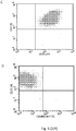
- Figure 5 provides an example of phenotype characterization with an assessment of viability (7-AAD) associated with the analysis of the marker CD105 of MSC4 at P3 in the presence of the inactivated PL0004 (a,b,c,d) and control PL0003 (e,f,g,h); it shows a viability of 95.85 % and 94.75 % of MSC4, respectively.
- the cell populations obtained at P3 in the "clinical grade” experiments were examined to determine the ability of the MSC to produce a differentiation in vitro in an osteogenic, adipose and chondrocytic direction.
- the MSC of P3 obtained both with the control and inactivated LP maintained the ability to differentiate in 21 days along the line of the osseous, adipose and cartilaginous precursors.
- Figure 6 shows an example of detection of the differentiation taking place in the samples cultured in differentiating media compared to controls cultured in standard media and originating from the line MSC8, in the presence of the LP control 0003 or LP inactivated 0004.
- the specific stains demonstrate the cellular specialization still obtainable from the MSC cultured in the presence of the control and inactivated PLs, versus the undifferentiated control. This attests to the preserved functional capacity of the selected and expanded MSC which reached the experimental point P3.
- the MSC6 and MSC8 were expanded and brought to P3 also using a large-scale "clinical grade” procedure and, being quantitatively abundant, they were also used to evaluate the biological potential of these cells after they underwent freezing/thawing with optimized controlled temperature descent procedures in the presence of a cryoprotector and temporary storage in nitrogen vapours for 3 months.
- the thawed MSC6 and MSC8 were characterized with respect to viability and phenotype and such assessments served to demonstrate a viability that was always greater than 85% as well as a phenotype totally consistent with the line they belonged to, the frequency of the cellular elements expressing the markers CD105, CD166, CD90, CD71, CD73 being always higher than 95%.
- the MSC re-expanded from MSC8 were induced to differentiate in an osteogenic, adipose and chondrocytic direction and the results obtained in terms of cellular specialization (revealed by the specific stains shown previously) were comparable to those observed for the same MSC8 prior to freezing, with an equal differentiation capacity for the MSC8 re-expanded both in the presence of the LP control 0003 and in the presence of the LP inactivated 0004.
- the present invention demonstrates the possibility of preparing a pathogen-inactivated PL from a pool of units from several blood donors (in example 12) which undergo the appropriate qualification tests and processing in order to extract and assemble, in 2 initial semi-products (in the example, two distinct pools from 6 donors), each platelet suspension in storage solutions and reduced amounts of plasma.
- the two semi-products then undergo a pathogen inactivation process using the Intercept® system, which includes exposure to UVA radiation, in combination with systems designed to maintain circuit sterility and continuity; they are subjected to 3 freezing/thawing cycles to achieve platelet lysis, centrifuged and resuspended to remove the stroma and aliquoted into sterile bags that can be labelled and stored at -80°C and contain about 3 doses of LP per large-scale MSC culture.
- the production process was conducted in class C protected environments (see methods) using sterile connection systems.
- the MSC obtained with the inactivated LPs can also undergo vital-freezing and 3 months' storage in nitrogen vapours, preserving intact their expansion capacity if again subjected to the action of the inactivated LPs.
- the results presented and discussed appear comparable to those previously reported in the literature, as similar levels of trophic factors can be found in the LPs described, as well as a similar ability to sustain the growth of MSC, with the starting elements expanded by about 100-200 times, and a similar capacity for the differentiation of cellular growth after expansion
- the present invention demonstrates how a process of inactivation with a photochemical agent preserves the trophic properties and functional and stimulation capacities of the LP used for the selection and amplification of phenotypically valid MSC, which are intact in terms of differentiation potential and moreover cryopreservable.
- the product of the present invention also defined as "Plasma-poor platelet lysate from pathogen-inactivated mini-pools” demonstrates to be feasible, effective and potentially free from contaminants that can generate the transmission of diseases caused by viral, microbial, fungal or parasitic agents.
- the standardization of the human mediastinal ADMSCs was achieved until passage 6 (14 patienrs, mean age 55 ⁇ 11 years) according to the following criteria: 1) the weight of the biopsy ⁇ 10 ⁇ 2g,
- human mediastinal ADMSCs isolated and cultured in PL are morphologically different from those in FBS.
- cells maintain a more rounded phenotype and a less differentiated spindle-like morphology ( Fig. 7A ), reaching the in vitro confluence faster than those in FBS ( Fig. 7B ).
- the FBS probably induces at later passages (Passage 4-6) a cell growth arrest, inducing a vacuolization of the ADMSCs as observed Figure 7C .
- human mediastinal ADMSCs were cultured for 3 weeks in commercial available media inducing osteogenic, adipogenic and chondrogenic differentiation towards the three mesodermal lineages.
- Our results showed that human mediastinal ADMSCs subjected to adipogenesis displayed lipid vacuoles, positive for Oil Red O staining ( Figure 12A ), whereas if subjected to osteogenic differentiation cells are positive for Alizarin Red S, due to the presence of calcium deposits ( Figure 12B ) and if differentiated toward cartilage they are positive for Alcian Blue, responsible of the staining of sulfated proteoglycans ( Figure 12C ).
- Figure 13A shows that PL and FBS exhibit the same proliferative properties (p>0.05) until Passage 6.
- the cell viability was maintained high (mean all passages 98.1 ⁇ 0.48 FBS 98.4 ⁇ 0.49 PL) and comparable to TFs cultured in FBS ( Fig. 13B ).
- the immunofluorescence shows a clear expression of the vimentin (Intermediate-filament associated protein in the fibroblasts). No variation were observed between PL and FBS ( Figure 14 ).
- DCs Dendritic cells
- the PL of the invention can replace fetal calf serum (FCS) for the differentiation of DCs derived from monocytes and can be used to improve the functionality of the dendritic cells generated in commercially available serum-free GMP media (CellGro and X-VIVO15).
- FCS fetal calf serum
- the DCs cultured in the presence of PL show a cellular yield that is similar or superior to the yield generated in the absence of PL ( Fig. 15 ).
- the PL of the invention up-regulates - in a statistically significant manner - the expression of CD86 during DC differentiation ( Fig. 16 ).
- the PL of the invention is capable of improving the endocytosis of immature DCs ( Fig. 17 ) and increases the production of IFN ⁇ by DCs after non-specific stimulation ( Fig.18 ).
Landscapes
- Life Sciences & Earth Sciences (AREA)
- Health & Medical Sciences (AREA)
- Engineering & Computer Science (AREA)
- Dentistry (AREA)
- General Health & Medical Sciences (AREA)
- Wood Science & Technology (AREA)
- Zoology (AREA)
- Environmental Sciences (AREA)
- Medicines Containing Material From Animals Or Micro-Organisms (AREA)
- Micro-Organisms Or Cultivation Processes Thereof (AREA)
Description
- The present invention relates to a rapid, simple, effective and economical method for preparing platelet lysate (LP or PL) which is free of contaminants that can generate the transmission of diseases caused by viral, microbial, fungal or parasitic agents, also called inactivated lysate (LP or PL). The present invention relates to the use of the platelet lysate obtained as an adjuvant for the culture, growth and/or expansion of stem cells, in particular mesenchymal stem cells derived from adipose tissue, fibroblasts and dendritic cells.
- Mesenchymal stem cells (MSC) are multipotent progenitors capable of self-replication and of orienting themselves along different lines of differentiation. Given its characteristics and potentialities, this cell source has aroused strong interest among researchers and clinicians in consideration of the possible therapeutic applications, especially in regenerative medicine(1,2). Bone marrow is the most widely used and studied source of MSC. Though mesenchymal precursors are scantly represented in bone marrow (0.001-0.01%), they can be easily expanded ex vivo and can actively replicate themselves in appropriate culture media. The possibility of expanding MSC ex vivo has opened new horizons and opportunities for therapeutic applications in tissue regeneration and use as cellular means for systemic immunomodulation. However, the majority of the protocols initially adopted for the expansion of MSC ex vivo used culture media supplemented with sera of animal origin that had a potential risk of transmitting pathogens or inducing allergic reactions.
- Platelet lysate (LP or PL) can be an alternative to animal serum as a source for MSC growth (3,4).
-
EP 2 100 954EP 2 100 954 -
WO 89/10398 - By virtue of their physiological tissue repair function, platelets are a rich source of numerous growth factors such as PDGF (platelet-derived growth factor), b-FGF (basic-fibroblast growth factor), VEGF (vascular endothelial growth factor) and TGF-β (transforming growth factor-β). However, the concentration of such factors in platelet lysate is highly variable. Therefore, the preparation of platelet lysate intended for an MSC expansion procedure for clinical purposes requires a multiple source in order to compensate for individual variability and a standardized protocol.
- Although current donation screening strategies for the prevention of diseases transmissible through transfusion therapy include highly sensitive virological, molecular and serological tests, the risk of transmission of hepatic viruses, acquired immunodeficiency and other pathogens (bacteria, parasites) or new emerging viruses represents an element of uncertainty in the adoption of PL preparations in protocols for MSC to be expanded for therapeutic purposes. For a better understanding of the problem tied to the residual possibility of transmitting diseases caused by pathogenic agents contaminating blood products, it is useful to consider that still today the risk of transmission of hepatitis B virus (HBV) is equivalent to 1 case out of 280,000-350,000 in developed countries, with higher frequencies in Mediterranean countries and Eastern Europe, 1 case out of 1,500,000-4,000,000 for the acquired immunodeficiency virus (HIV) and 1 case out of 1,000,000-2,000,000 for hepatitis C virus C (HCV)(5). As regards the bacterial contamination of blood components, this occurs with a prevalent frequency in platelet products. It most frequently derives from bacteria of cutaneous origin and hence from normal saprophytes of human skin or, alternatively, from environmental contamination of the places where blood components are processed and stored. In platelet products, the frequency of bacterial contamination is around 1 case out of 1,500 and the consequent frequency of adverse reactions potentially related to the re-infusion of bacterial species with platelet transfusion is around 1 case every 10,000-100,000(6). In the context of PLs, it should also be considered that these are assembled products originating from at least 10-12 donors given the necessity of amplifying the pool of source individuals so that the release of trophic factors is as homogenous as possible and the variability of individual donors is minimized. Therefore, each risk mentioned should be increased by at least one log, making bacterial contamination likely in 1 case out of every 150 products and HBV transmission possible in 1 case out of every 30,000 products.
- Moreover, the risk of transmittable disease must also be correlated with new forms of contagion ascribable to the greater inclination of donors to travel and stay in regions where viral, parasitic and bacterial diseases are endemic and the ever increasing contribution of donations from communities that have migrated to our countries from countries to which diseases with an infectious etiology and potentially transmittable through blood products are endemic. The screenings relied on to ensure the safety of transfusions do not in fact explore, except in certain defined circumstances, the presence of silent, asymptomatic infectious forms such as Chagas disease, malaria, babesiosis, dengue, leishmaniasis, viremia caused by the West Nile virus, chikungunya, H1N1 influenza, Epstein Barr virus or viral pathogens of recent interest such as the xenotropic murine leukemia virus (XMLV), associable with some forms of cancer in humans and chronic fatigue syndrome. This increases the residual risk of transmission of bloodborne disease by a further, incalculable factor, especially in the case of platelet products which are systematically subjected to leukocyte removal (the self-sterilization capacity of blood thus being completely lost) and stored at room temperature in considerable quantities of plasma (plasma constitutes a medium for survival or optimal growth for a number of pathogens).
- The epidemiological context described casts serious doubts upon the advisability of using PL as such as a source for stimulating the growth and amplification of MSC. In fact, though on one hand it constitutes a potent product - which is all the more homogeneous in terms of the degree of stimulation when it is obtained from a large number of donors - on the other hand it represents a component that also carries a greater risk, with respect to diseases transmittable by known and unknown pathogenic agents, precisely when the number of source donors increases. This circumstance poses a limit that is difficult to overcome in the production and use of a source of stimulation like PL where the latter must be used for procedures leading to clinical grade therapies and for therapeutic approaches that are still experimental and whose health impact on the respective pathogenic forms has not been firmly established. Therefore, a condition of health uncertainty would arise, as was experienced in the last century when anti-haemorrhagic blood derivatives (anti-haemophilia factors VIII and IX) were produced without industrial forms of fractionation and pathogen inactivation, implying a risk of transmittable disease that is by now unacceptable for any type of blood-derived drug, all the more so in the case of preparations used in experimental, unconfirmed settings. A realistic possibility of increasing the safety of a blood-derived product obtained not through industrial processes but rather in laboratories, a donor screening approach providing prompt, cost-effective detection of the above-mentioned infectious agents not being feasible, consists in the possibility of relying on forms of pathogen inactivation of ascertained effectiveness. The registered substances presently available for pathogen inactivation of platelet concentrates are the Intercept® system by Cerus and the Mirasol® system by Caridian. The two systems possess similar biological virus, bacteria and parasite inactivation capabilities, but the Intercept® system boasts a by now well-established use in relation to thousands of inactivated products successfully used in transfusion therapies for subjects with a platelet defect. In contrast, the Mirasol® system can only boast a single phase III study and modest application in clinical settings(7). The Intercept® system relies on a UVA radiation-activatable substance represented by amotosalen, which is then removed from the inactivated component after photoillumination.
- Van Rhenen et al., "Functional characteristics of S-59 photochemically treated platelet concentrates derived from buffy coats", Vox Sanguinis, vol. 79, no. 4, p. 206-214, 2000, refers to the preparation of platelet concentrates from buffy coats exposed to a photochemical treatment utilizing psoralen and UVA light for increasing safety of platelets transfusion.
- H. G. Klein, "Pathogen inactivation technology: cleansing the blood supply", Journal of internal medicine, vol, 257, no. 3, p. 224-237, 2005, refers to preparation of platelet concentrates, for use in blood transfusion, exposed to amatosalen and UVA-light for pathogen inactivation. Lily Lin et al., "Inactivation of viruses in platelet concentrates by photochemical treatment with amotosalen and long-wavelength ultraviolet light", Transfusion, vol. 45, no. 4, p. 580-590, 2005 refers to photochemical treatment with amatosolan-HCl and UVA of single-donor PLT concentrates. Said platelet concentrates are for use in blood transfusion.
- Clinical experience demonstrates that the platelets treated with Intercept® produce a valid haemostatic effect, but with less valid increases in the platelet count after transfusion. In fact, inactivation brings about a reduction in the number of
available cellular elements in the preparations, as well as a reduced post-transfusion platelet recovery due to a negative dose-independent effect on the survival of the same after transfusion. This effect, which appears to be independent from the dose administered, period of storage and compatibility between donor and recipient, explains at least 40-50% of the reduced increases observed after inactivation(8). From a biological viewpoint, the platelets undergoing treatment with Intercept® show functional and metabolic alterations ascribable to a greater pH reduction during storage, a lower availability of excess bases (correlated with the more marked pH reduction), higher pO2 and pCO2, a higher consumption of glucose and production of lactate, a higher release of lactic dehydrogenase and a higher expression of p-selectin, a marker of platelet activation(9). Taken together, these phenomena demonstrate that the photoactivation of amotosalene, given a nearly complete sterilization of the product in terms of parasitic, viral and microbial contamination, brings about respiratory and metabolic stress in the treated platelets, as well as the activation thereof. In reference to the foregoing considerations, it becomes of absolute importance to evaluate how the platelet lysates obtained from pools of donors and then inactivated with well-established inactivation systems such as Intercept® maintain, downstream of inactivation, a trophic and stimulation capacity vis-à-vis MSC which is comparable to that of conventional platelet lysates. In fact, the metabolic stress observed could negatively impact the ability of the platelet lysate thus treated to release valid concentrations of stimulating factors and stimulate MSC growth and expansion. - Moreover, MSC have been isolated from various mesodermal tissues, including bone marrow(13), muscle(14), perichondrium(15) and adipose tissue(16-17) and share key characteristics, such as an ability to adhere to plastic to form colonies of a fibroblastic type (CFU-F), an ample proliferative capacity, an ability to differentiate into various mesodermal cell lines such as bone, muscle, cartilage and fat and the expression of different common surface antigens. In particular, MSC derived from human adipose (hADMSCs) have become an attractive cellular source because they can be recovered in large numbers, are easily expanded in vitro and express the same surface markers as MSC derived from bone marrow(18-19). Furthermore, hADMSCs have demonstrated a strong angiogenic potential(20) and their differentiation toward adipogenic, osteogenic, chondrogenic, neurogenic, haematopoietic and cardiogenic phenotypes has been described(21-22). Specifically, it has been demonstrated that cardiac differentiation of hADMSCs could be obtained spontaneously(23-24) by using 5-azacytidine(25) or exposing them to extracts of rat cardiomyocytes(25). Moreover, human ADMSCs have been demonstrated to reduce the infarcted area after injection in mouse models of myocardial damage(27-29). In 2009 it was reported that MSC can be obtained from omental adipose deposits(30) and show no differences from parietal fat. Recently, a population of multipotent stem cells from mediastinal deposits has been further characterized(31), providing the first evidence of a population of stem cells in a site other than the body.
- Tenon's fibroblasts (TFs) represent a preferential system for in vitro experimentation with new therapeutic molecules(32,33) or the development of new in vitro diagnostic methods, new products and toxicological screening. In addition, TFs are still widely studied to gain a better understanding of the role of growth factors involved in inflammatory pathologies and in cellular differentiation into myofibroblasts(34). Cell culture models of human TFs are essential in order to understand important processes such as the healing of wounds, extracellular protein synthesis, drug delivery, and migration and proliferation in pathological or physiological eye conditions(35) or in the presence of specific growth factors(36-38). Moreover, the standardized methods for planning a manufacturing process for IMPs (Investigational Medicinal Products, in particular ophthalmological products) require preliminary steps that are indispensable for all applications in which safety and reliability are fundamental. At present, TFs are still cultured in the presence of fetal calf serum or autologous serum. However, both of these systems have limits: firstly because of the potential risk tied to zoonosis and secondly because of the limited availability of autologous human serum for this type of long-term culture. A possible option for overcoming the above-mentioned problems could be to use PL, already successfully tested in various in vivo systems, such as the healing of skin wounds, orthopaedic repair and repair of periodontal lesions, and in eye treatments(39-43). To date, and to the best of our knowledge, the possibility of culturing human Tenon's fibroblasts in the presence of PL has never been reported. In addition, TFs cultured with PL could represent an alternative in vitro model for studying the healing process during corneal lesions.
- Dendritic cells (DCs) are the most important antigen-presenting cells because they have a central role in the initiation and development of immunity, being capable of activating naive T cells and contributing to inducing and maintaining peripheral tolerance. DCs undergo a process of differentiation, during which they acquire antigens, migrate toward lymphoid tissues and increase the activation potential of T cells(45). Thanks to these properties, DCs are today considered as the primary constituent of cell-based immunotherapy protocols for anti-tumour vaccination(46).
- With the growing development of vaccination strategies for modulating immunity in a variety of diseases, there is strong interest in identifying and defining protocols and reagents which enable optimal production of DCs from peripheral blood monocytes. The replacement of serum components of animal origin is one of the points of interest for the production of clinical grade DCs. In the past decade, a number of serum-free media have been introduced for the generation of DCs, but these cells often show a decrease in performance.
- Overall, the above considerations bring to light the importance of producing platelet lysates from an appropriate number of donors as an optimal stimulus for MSC, adopting a form of pathogen inactivation which reduces tranfusional risk and evaluating the effects of the inactivation process on an optimized PL production method using clinical grade production systems. Moreover, there exists a need to produce a PL that is suitable for generating and/or sustaining the growth of mesenchymal stem cells derived from adipose tissue and clinical grade DCs and for isolating and/or expanding Tenon's fibroblasts in vitro. In particular with the objective of abolishing the addition of serum components of animal origin to generate cultures of clinical grade DCs used for human vaccination protocols and of improving the performance of DCs generated in serum-free media.
- With the aim of producing a PL with a maximum level of safety, a protocol was developed for the preparation of PL in a closed system which uses buffy coat platelet units previously subjected to a pathogen inactivation procedure. The products thus obtained were evaluated in terms of effectiveness and safety and compared to control PLs produced in a similar manner, but omitting the inactivation process.
- In the present invention, a method for producing PLs was established and it was demonstrated that the PL thus produced is capable of supporting the isolation and/or cellular growth of mesenchymal stem cells, including those obtained from adipose tissue. Moreover, this PL enables fibroblasts and dendritic cells to be isolated and/or expanded in vitro.
- Therefore, the subject matter of the invention relates to a method for preparing a pathogen-inactivated platelet lysate capable of acting as a coadjuvant for the isolation and/or growth and/or expansion of stem cells, fibroblasts or dendritic cells, comprising the steps of:
- a) isolating a buffy coat fraction from a mixture of whole blood specimens obtained from at least two subjects;
- b) exposing the isolated buffy coat fraction to a photochemical agent and UV radiation under conditions such as to obtain an isolated pathogen-inactivated buffy coat fraction;
- c) subjecting said fraction to at least one freezing/thawing cycle in such a manner as to achieve lysis of the platelets contained therein;
- d) centrifuging said fraction and collecting the liquid phase.
- Also described is a platelet lysate essentially free of contaminating agents and capable of acting as an adjuvant for the isolation and/or growth and/or expansion of stem cells, fibroblasts or dendritic cells obtainable with the method according to any preceding claim.
- Also described is the use of the platelet lysate essentially free of contaminating agents of the invention as an adjuvant for the isolation and/or growth and/or expansion of stem cells, fibroblasts or dendritic cells.
- Preferably, the stem cells are mesenchymal. More preferably, the stem cells are derived from adipose tissue. Preferably, the fibroblasts are Tenon's fibroblasts. In a preferred embodiment the platelet lysate essentially free of contaminating agents and capable of acting as an adjuvant for the isolation and/or growth and/or expansion of stem cells, fibroblasts or dendritic cells is added to the culture medium for said cells at a concentration of between 5% and 10%.
- The present invention makes it possible to obtain a platelet lysate essentially free of contaminating agents, usable for the culture of stem cells, in particular mesenchymal stem cells, dendritic cells and fibroblasts, in particular Tenon's fibroblasts.
- In present invention the buffy coat fraction is defined as a transfusion level concentrate of leukocytes and platelets, with modest red blood cell contamination; it is obtained by centrifugation of donated blood and constitutes the basic unit for the assembly of platelet concentrates for therapeutic use made up of 6-12 different buffy coats obtained from an equal number of healthy donors.
- A platelet lysate essentially free of contaminating agents is defined as a sterile platelet preparation essentially free of infecting agents such as encapsulated and non-encapsulated viruses, parasites, bacteria and endotoxins, i.e. a platelet lysate containing such infecting agents in a quantity that cannot be detected with culture-based, serological or molecular identification systems or with the LAL test insofar as endotoxins are concerned.
- The cells cultured in this manner can be advantageously used for therapy and reintroduced into subjects.
- The present invention will be described through non-restrictive examples, making reference to the following figures:
-
Fig. 1 : Diagram of the protocol for preparing the platelet lysate. PLT: platelet concentrate. -
Fig. 2 : Exemplary sample of PL (PL0006 sub-batch 1, inactivated) after final packaging, labelling and freezing. -
Fig. 3 : Cellular growth of MNCs of bone marrow from seeding to the first passage (P1), and of the MSC derived from the first passage P1 to the third passage (P3) in the presence of the various LP: LP control 0001, LP control 0002,LP control 0003, LP inactivated 0004, LP inactivated 0005, LP inactivated 0006, LP inactivated 0007. They were used in cultures of MSC obtained from 5 different subjects (MSC1, MSC2, MSC4, MSC6, MSC8). -
Fig. 4 : Degree of purity and line fidelity of the different MSC cultured in the presence of the various LP (LP control 0001, LP control 0002,LP control 0003, LP inactivated 0004, LP inactivated 0005, LP inactivated 0006, LP inactivated 0007), evaluated at the first passage (P1) and third passage (P3) by flow cytometry analysis of the expression of specific surface markers A: CD105, B: CD166, C: CD90, D: CD71, E: CD73). -
Fig. 5 : Example of phenotype characterization with an evaluation of viability (7-AAD) associated with an analysis of the marker CD105 of MSC4 at the third passage (P3) in the presence of the LP inactivated 0004 (A,B,C,D) and the LP control 0003 (E,F,G,H). -
Fig. 6 : Example of MSC8 differentiation toward bone (O), adipose tissue (A) and chondrocytes (Co) after treatment with the LP control 0003 (O1, A1 and Co1) or after treatment with the LP inactivated 0004 (O2, A2 and Co2). C1 (LP control 0003) and C2 (LP inactivated 0004) are an example of undifferentiated controls stained for bone differentiation. -
Fig. 7 : human mediastinal ADMSCs cultured in PL (A) and FBS (B) atPassage 2 and in FBS atpassage 4. Magnification 10X (A,B) and 20X (C) -
Fig. 8 : PL significantly increased the percentage of successful cultures obtained from Human mediastinal biospsies (p<0.05). -
Fig. 9 : Comparison between human mediastinal ADMSCs cultures in DMEM-LG and DMEM-HG with PL -
Fig. 10 : Comparison between human mediastinal ADMSCs cultures in DMEM-LG and DMEM-HG with FBS at passage 1 (A) and 2 (B). The best combination of DMEM-LG and PL increased the cell growth compared to FBS (C). -
Fig. 11 : Immunohenotype of human mediastinal ADMSCs at passage 3 (A). CFU-F of human mediastinal ADMSCs cultured in PL (B) or FBS (C) -
Fig. 12 : Images of human mediastinal ADMSCs cultured in PL and trans-differentiated in the adipogenic (A), osteogenic (B) and chondorgenic (C) lineage. -
Fig. 13 : Proliferative trend (A) and Cell viability (B) of TFs cultured in PL and FBS. -
Fig. 14 : Immunofluorescence for vimentin, red. DAPI, blue indicates nuclei. -
Fig. 15 : DC yield. CD14+ cells (monocytes) were immunomagnetically purified from PBMCs of 8 healthy donors. Monocytes were culture in different media (RPMI+10%FCS, RPMI+10%LP, CellGro, Cellgro+LP, AIMV and AIMV+LP) in presence of IL4 (2000U/mL) and GMCSF (50ng/mL) added atday 0 andday 2. After 5 days, immature DCs (iDCs) were collected and counted using Trypan Blue reagent. The results are reported as cell number per mL and represent the average values among 8 donors. The blue columns report DCs cultured in RPMI+FCS and RPMI+10%LP; the green columns describe the yield of DCs differentiated in Cellgro and Cellgro+LP; the red columns show the cells generated in AIMV and AIMV+LP. -
Fig. 16 : Expression of CD86 in differently treated iDCs. After 5 days of culture, iDCs derived from 8 donors were characterized for the expression of HLA-DR, CD14, CD86, CD83, CCR7, CD40 by cytofluorimetry (FACsCantoII, software FACsDiva). Data are reported as average value among 8 donors of Mean Fluorescence Intensity (MFI) of CD86. The blue columns report DCs cultured in RPMI+FCS and RPMI+10%LP; the green columns describe the yield of DCs differentiated in Cellgro and Cellgro+LP; the red columns show the cells generated in AIMV and AIMV+LP. -
Fig. 17 : DC endocytosis:. DC endocytosis were evaluated as FITC-Dextran up-take (1mg/ml) performed at 4° C (negative control) and 37°C for 2 hours. Data are reported as ratio between positive and negative samples. Endocytosis was studied in iDCs and matured DCs (mDCs) at day 6. Cell maturation was carried out atday 5 until day 6 (16 hours) and was induced adding TNFα (10ng/mL), IL6β (10ng/mL), IL1 (10ng/mL) and PGE2 (1µg/mL) to the cells. Results represent the average values of 4 donors. -
Fig. 18 : Intracellular IPNγ staining. mDCs at day 6 of culture were stimulated with PMA (25ng/ml) and Ionomycin (250ng/mL) for 16 hours in presence of Brefeldin A(10µg/mL). At day 7 cells were permeabilized with Saponin solution at 0,5% and were treated with an anti-IFNγ antibody. Stained cells were analyzed by BD FACScCantoII flow cytometer running BD FACsDiva data acquisition and analysis software. Results are reported as %IFNγ and represent the average values among 4 donors. - The platelet concentrates (PLT) were produced from units of whole blood from suitable voluntary donors, collected in dedicated sterile circuits provided with a quintuple bag (Fresenius Kabi AG, Bad Homburg, Germany).
- In order to produce PLT from buffy coats, the blood units were processed within six hours after collection. After it was verified that the whole blood withdrawal conformed to requirements (volume withdrawn, absence of visible coagulates, absence of chylosis), the unit underwent centrifugation at 6477 relative centrifugal force (rcf) x g for 12 min.
- The following were extracted from each unit using Compomat G4 separators (Fresenius Kabi AG):
- 1 unit of fresh plasma
- 1 unit of buffy coat-depleted red blood cells
- 1 unit of buffy coat (BC) containing the platelet component.
- The BC products were stored in an incubator at 22°C under continuous agitation and assembled to produce the pool within 24 h. The pool of 6 units of BC was prepared by connecting the BCs (Composeal, Fresenius Kabi AG) together under sterile conditions in a dedicated production environment, with InterSol saline solution (Fenwal Inc. Lake Zurich, Illinois, with the following composition: Na-citrate 2H2O 318mg, Na2-anhydrous phosphate 305mg, Na Dihydrogen-phosphate 2H2O 105mg, Na-Acetate 3H2O 442mg, NaCl 452mg, H2O 100mL) and the pool collection bag (BioP-plus single-use kit, Fresenius Kabi AG). The BC and final pool collection bags were identified by means of a specific label with a bar code associated with the pool being produced. At the end of the assembly procedure, the collection bag with the 6 BCs was closed and sealed (Composeal, Fresenius Kabi AG), disconnected from the processing kit and centrifuged at 480 rcf x g for 9 min. At the end of centrifugation, the platelet concentrate was separated from the residual BC using the automatic Compomat G4 separator.
- Two pools of 6 PLT units were subjected to pathogen inactivation by means of a photochemical treatment with psoralens and UV exposure (INTERCEPT Blood System for Platelets, Cerus Corporation, Concord, California USA). During the pathogen inactivation process using INTERCEPT technology, a compound of the psoralen family (Amotosalen hydrochloride) was introduced into the PLT pool; this compound, following to the exposure to UVA radiation, forms a covalent bond with any nucleic acids that may be present in the product and blocks the process of duplication of DNA and RNA helices.
- For the inactivation procedure, the bag of the pool was sterilely connected to the INTERCEPT kit and the inactivation procedure was started; it takes place in a completely closed system and comprises the following steps:
- introduction, into the PLT pool, of the psoralen Amotosalen (S-59, INTERCEPT Blood System for Platelets, Cerus Corporation, Concord, California USA; product code INT2204B) at a final concentration of 150 µmol/L;
- exposure of the PLT pool supplemented with psoralen to UVA radiation 3 J/cm2
- transfer of the product into a bag containing a specific device (CAD Compound Adsorption Device) for the removal of the psoralen;
- transfer into the final bag for storage of the product "Inactivated PLT Pool" until the use thereof.
- The final PLT pool subjected to the inactivation treatment described was identified with a label bearing the barcode associated with the pool at the time of production and the specification "Inactivated".
- At the end the inactivation procedure, the PLT units were resuspended in InterSol with a residual plasma percentage of about 20-30%.
- From this step onward, the further operations of production of the PL were carried out in a class C controlled environment classified according to international guidelines for Good Manufacturing Practices (GMP), implemented at the European Community level through the following legislative provisions: EU GMP Directive "Manufacturing of Sterile Medicinal Products",-
Annex 1, EU GMP Directive "Manufacture of Biological Medicinal Products for Human Use" -Annex 2, EMEA Guideline on Human Cell Based Medicinal Products - EMEA/CHMP/CPWP/323774/2005. The GMP guidelines provide for specific environmental control of particulate contamination which enables a laboratory to be classified as grade A, B, C or D according to the table below.Table I: Laboratory classification Max number of particles/m3 At rest During operation Grade 0.5 µm 5 µm 0.5 µm 5 µm A 3,500 1 3,500 1 B 3,500 1 350,000 2,000 C 3,500,000 2,000 3,500,000 20,000 D 3,500,000 20,000 not defined not defined - Samples were drawn from the 2 PLT pools by inserting a sterile sampling device (Terumo, Tokyo, Japan) into the inlet provided in the bag (sterile "sampling site" of Fresenius Kabi AG collection devices) in order to determine the platelet count and sterility.
- The platelet counts were carried out using the ABX Pentra DX 120 hematology analyzer (Horiba ABX, Montpellier, France). The sterility tests were performed with the BacT/ALERT System using media specific for aerobic and anaerobic bacteria and fungi (bioMérieux SA, Marcy L'Etoile, France).
- At the end of the complete procedure, the volume of the pool of 6 units was 300 mL on average and platelet content ranged between 2.5 and 3.5 x 10¹¹.
- The 2 pools of 6 units of inactivated PLT were subsequently combined in a single bag, via a sterile connection (TSCD II, Terumo, Tokyo, Japan), so as to obtain a final pool with a volume of approximately 600 mL, made up of 12 units of inactivated PLT. The overall pool underwent 3 successive cycles of freezing (-80°C; kept at this temperature for 24 hours in a mechanical freezer) and thawing (37°C; thawing in a biological defroster for plasma units (Bloodline, KW, Siena, Italy), temperature maintained for 60 minutes) to ensure breakage of the platelets and the release of growth factors.
- In order to remove the cellular stroma, the pool was divided into 2 bags and each of the 2 bags was sterilely connected (TSCD II) with a second transfer bag (600 ml, JMS Singapore PTE LTD, Singapore) so as to the remove the supernatant after 45' centrifugation at room temperature and 5313 rcf x g. The residual bag containing the cellular stroma was removed after double sealing and the centrifugation procedure was repeated using the same methods. At the end of preparation, samples were drawn for the final sterility test. In order to be able to use the PL produced according to the present invention in cell cultures for cellular therapies, in addition to aerobic and anaerobic bacteria and fungi, endotoxins were also tested for using the LAL test (Limulus Amebocyte Lysate; International PBI spa, Milan, Italy) with a sensitivity of 0.06 UE/mL.
- The PL products were identified with a suitable code in written and barcode form serving to identify the production batch, aliquoted via a sterile connection (TSCD II) into paediatric bags (100 ml transfer bags for fresh frozen plasma, Fresenius Kabi AG), with identification of the sub-batches. Each sub-batch unit contained 40-50 mL of PL, equivalent to about 3 doses (to be intended as 3 doses for large-scale culture activities, each dose being sufficient to prepare a clinical grade culture with a starting volume of 100-150 ml) for the ex vivo expansion of MSC in closed systems - CellSTACK® CORNING® cell culture chambers (MacoPharma, Mouvaux, France; Corning Incorporated, Corning, New York, USA) - if used at a concentration of 10%.
- Following the same operational scheme, excluding the viral inactivation step, inactivated PL was produced as a non-inactivated control.
- The procedure for preparing the PLs is schematically summarized in the flowchart shown in
Fig. 1 . - An immunoenzymatic assay was performed on the various PL products to determine the following factors using samples diluted to a varying degree, from 1:2 to 1:10:
- PDGF-AB (Quantikine Human PDGF-AB, R&D Systems, Minneapolis, Minnesota, USA)
- VEGF (Quantikine Human VEGF, R&D System)
- basic-FGF (Quantikine Human FGF basic, R&D System)
- TGF-Beta (Quantikine Human TGF-
Beta 1, R&D System) - All the preparations of LP or PL (control PL 0001, control PL 0002,
control PL 0003, inactivated PL0004, inactivated PL0005, inactivated PL0006, inactivated PL0007) were used in MSC cultures obtained from 5 different subjects (MSC1, MSC2, MSC4, MSC6, MSC8), starting from aliquots of bone marrow aspirated during the donation of stem cells for an allogeneic transplant, after specific informed consent was obtained (see next paragraph for details regarding the MSC cultures). - 5 mL samples of bone marrow from a donor of allogeneic stem cells were used for the separation of mononuclear cells (MNC). After a complete blood count was performed to evaluate the initial lymphomonocytic component, the bone marrow was diluted 1:3 in Dulbecco's Phosphate Buffered Saline (PBS, Biowest S.A.S, Nuaillé, France) and the separation of MNCs was achieved by gradient sedimentation using Lympholyte (Cedarlane, Burlington, Ontario, Canada) as the separation medium and 30 minutes' centrifugation at 652 rcf x g at room temperature. Then followed 2 washings with PBS supplemented with 2% human albumin (Uman Albumin, Kedrion S.p.A., Lucca, Italy) and final resuspension of the cells in D-MEM GMP grade culture medium (Li-Star Fish, Milan, Italy). The lymphomonocytic component was re-evaluated through a blood count after separation on a gradient and a flow cytometry analysis was performed to evaluated the mesenchymal component identified by the cells CD71+ (allophycocyanin, APC, conjugated anti-CD71; Becton Dickinson, San Jose, California, USA), CD73+ (phycoerytrhin, PE, conjugated anti-CD73; Becton Dickinson), CD90+ (allophycocyanin, APC, conjugated anti-CD90, Becton-Dickinson), CD105+ (peridinin chlorophyll protein complex, PerCP, conjugated anti-CD105; Becton Dickinson; or fluorescein isothiocyanate, FITC, conjugated anti-CD105; Partec GmbH, Munster, Germany), CD166+ (PE conjugated anti-CD 166; Becton Dickinson), CD45-(FITC, conjugated anti-CD45, Becton-Dickinson) and CD34- (FITC, conjugated anti-CD34, Becton-Dickinson) using a FacsCalibur flow cytometer (Becton-Dickinson) and the above-mentioned monoclonal antibodies.
- 1 x 10⁶ MNCs/mL were placed in a D-MEM GMP grade culture medium supplemented with 10% PL (PL concentrations of 5% were also used and evaluated and gave results similar to those obtained with 10%) in T25 flasks (Asahi Techno Glass, Iwaki, Japan) in an incubator at 37°C, 5% CO2. For the purpose of testing expansion on a large scale, an MSC expansion was carried out using CellSTACK® CORNING® cell culture chambers (MacoPharma; Corning Incorporated). 48 h after the start of the culture, the supernatant was removed and replaced with fresh medium. Thereafter, the medium was replaced every 4 days and the cells were examined under a microscope to evaluate adhesion, growth and confluence. When a cellular confluence of about 80% was reached, the adhering cells were removed with a trypsin treatment (GMP grade, Li Star Fish), centrifuged and counted with the flow cytometer using propidium iodide (Calbiochem, Merck KGSA, Darmstadt, Germany) and fluorospheres (Flow Count Fluorospheres, Beckman Coulter, Galway, Ireland). The propidium solution containing Tween is capable of permeabilizing the cell membrane so that the fluorescent propidium (FL2) can enter the cell and bind to the nucleic acids. The addition of fluorospheres at a known concentration(12) enables an absolute count of the MSC marked with propidium. The cells were moreover characterized as MSC using the same panel of surface markers indicated for the initial typing. This first passage was identified as P1.
- For subsequent expansion, the cells were seeded at a concentration of 4000 cell/cm2 (20,000 cell/mL) using the same culture conditions as described. When confluence was subsequently reached, passage 2 (P2), the cells were again removed, counted, typed and further expanded. At the third passage (P3), the cells were counted, characterized by flow cytometry analysis, which also included an evaluation of viability using 7-Amino-Actinomycin D (7AAD, Becton Dickinson), tested for their potential to differentiate in an osteogenic, adipose and chondrocytic direction and frozen in liquid nitrogen in order to verify their viability and replication and differentiation potential after freezing.
- The expansion of the mesenchymal cells was calculated starting from
Passage 1, in which over 95% of the cells showed the MSC phenotype (i.e. cells expressing the markers CD105, CD166, CD90, CD71, CD73). - For all lines of differentiation, the MSC were cultured in 35 millimetre Petri dishes (NUNC A/S, Roskilde, Denmark) at a concentration of 5000 cell/mL in an appropriately conditioned culture medium for 3 weeks, with replacement of the medium every 3-4 days. At the end of the 21 days of culture, selective staining was performed to detect the different cell types.
-
- Culture medium: α-MEM (Gibco Invitrogen, San Giuliano Milanese, Italy) supplemented with Desametasone 10-7M (Sigma-Aldrich, St Louis, Missouri, USA), Ascorbic Acid 10-5M (Sigma-Aldrich), β-Glycerophosphate 10-2M (Sigma-Aldrich).
- Detection stain: Fast Blue, Naftol AS-D Chloroacetate Esterase and α-Naphthyl Acetate Esterase (Sigma-Aldrich).
-
- Culture medium: α-MEM (Gibco Invitrogen) supplemented with Desametasone 10-7M (Sigma-Aldrich), Ascorbic Acid 10-4M (Sigma-Aldrich), β-Transforming Growth Factor 10ng/mL (Sigma-Aldrich), Insulin 10µg/mL (Sigma-Aldrich).
- Detection stain: Toluidine Blue (Bio-Optica SpA, Milan, Italy).
-
- Culture medium: α-MEM (Gibco Invitrogen) supplemented with Desametasone 10-6M (Sigma-Aldrich), Insulin 10µg/mL (Sigma-Aldrich), 1-methyl-3-isobutylxanthine 5 x 10⁻⁴ M (Sigma-Aldrich), Indomethacin 60µM (Sigma-Aldrich).
- Detection stain: Oil RED O (Bio-Optica)
- Aliquots of 2 x 10⁶ MSC (obtained at P3) in D-MEM GMP grade medium supplemented with 5% human albumin and 10% DMSO (WAK-Chemie Medical GmbH, Steinbach, Germany) as the final concentration were frozen in cryotubes using a freezing system with a dedicated programmed temperature descent protocol validated for freezing stem cells for transplantation purposes (Kryo 560-16 Planer; Planer PLC Sunbury-On-Thames, United Kingdom). The cells were cryopreserved in liquid nitrogen vapours for 3 months.
- For thawing, the cryotubes were removed from the liquid nitrogen vapours and rapidly thawed in a temperature controlled bath set to 37°C. Once completely thawed, the cellular suspension was immediately diluted in PBS-EDTA (Miltenyi Biotec, Bergisch Gladbach, Germany) with 5% albumin and washed 3 times to completely remove the DMSO. At the end of the final wash, the MSC were counted with the flow cytometer and assessed for viability.
- 15,000 viable MSC/cm2 were again placed in D-MEM GMP grade culture medium supplemented with the same preparation of PL used for selection and expansion prior to freezing (with concentrations of PL ranging between 5% and 10%). Upon reaching confluence, the adhering MSC were detached by means of a trypsin treatment, typed by flow cytometry analysis using the panel of monoclonal antibodies described above and expanded according to the "basic" protocol (i.e. the protocol as detailed in the previous paragraph "Protocol for selecting and expanding MSC") in order to evaluate the replication and differentiation potential after freezing.
- At the end of passages P1 and P3 the cells were placed in a culture medium at a concentration of 7000 cells/cm2, under the same culture conditions as previously described, and an analysis of the karyotype was performed in accordance with the protocols described in the International System for Human Cytogenetics Nomenclature.
- Adipose tissue from mediastinal and parietal biopsies were obtained from patients underwent thoracic surgery. Samples were stored in physiological solution and processed within 24 hours. Biopsies were washed with PBS then minced and digested by PBS/Trypsin/EDTA/1mg/ml Collagenase for 45 minutes at 37°C in a shaking water bath. Digestion was stopped using
PBS 20% FBS. After filtration through 100□m filters and centrifugation at 1200 rpm for 5 minutes, the pellet was treated with the Erythrocyte lysis buffer to remove the red blood cells. Cells were then counted, resuspended in complete medium (DMEM-low glucose containing 10% PL/5U/ml of heparin/1%PenStrep/1%Glutammine/1% Non-essential Aminoacids) and seeded at 4000 cells/cm2. All non-adherent cells were removed after 72 hours. The medium was changed every 3 days. Cultures were expanded until passage 6 by Trypsin/EDTA, then harvested, washed and centrifuged at 1200 rpm for 5 minutes. Cell count and viability was measured by Trypan Blue. - The phenotype of human mediastinal ADMSCs was evaluated by FACS Analysis using the following panel of primary and secondary antibodies: 0.2mg/ml mouse monoclonal CD105, 0.1 mg/ml mouse monoclonal CD44, 0.1 mg/ml mouse monoclonal CD90 (Abcam human Mesenchymal stromal cell marker panel Cat., 1:100 mouse monoclonal CD31, 0.25 mg/ml rabbit polyclonal CD117, 0.3 mg/ml rabbit polyclonal CD45, 1 mg/ml rabbit polyclonal CD133, rabbit alexafluor-488, mouse Fluorescein (FITC)- conjugated affinity pure goat antimouse IgG (H+L). Briefly, 2x10⁵ cells were stained with the monoclonal antibody for 30 minutes at room temperature. After washing in PBS/BSA1%, cells were incubated with secondary antibody for 30 minutes in the dark at room temperature. Unstained cells and cells stained with secondary antibody alone were respectively used for the detection of autofluorescence and as isotype control. Cells were acquired by cytofluorimetry (FACSAria Becton & Dickinson, Mountain View, CA, USA) and analyzed by Software DiVa (v6.1.1).
- Human mediastinal ADMSCs were seeded at
passage 3 in complete medium (cell density of 20-40 cells/cm2/100mm petri dish). Medium was changed after 72 hours. After 3 weeks of incubation, visible colonies were stained with Giemsa. Briefly, the petri dishes were washed with PBS and incubated for 2 minutes withGiemsa 100% (Sigma). Colonies were washed with distillated water then further treated with a PBS/Giemsa solution (1:20) for 13 minutes, washed again and then dried. Clusters of cells >5mm were considered positive and observed by microscope. - Human ADMSCs were subjected to osteogenic, adipogenic and chondrogenic differentiation. Adipogenic differentiation was induced using the StemPro® Adipogenesis Differentiation kit (GIBCO, Invitrogen, Grand Island, NY), Cat. N. A10410-01). Briefly, 1x10⁴ cells/cm2 were seeded in a 12 well plates and cultured for 3 weeks. Medium was changed every 3 days. Accumulation of lipid droplets was tested by Oil Red Oil staining after fixing the cells with 4% formalin. Osteogenic differentiation was induced using culturing the cells in medium containing 10-7 M dexamethasone, 10 µM ascorbic acid and 0.2M □-glycerophosphate. Briefly, 1x10⁴ cells/cm2 were seeded in a 12 well plates and cultured for 3 weeks. Medium was changed every 3 days. The calcified matrix deposition was observed by staining with Alkaline phosphatase after fixing the cells with 4% formalin. Finally, the chondrogenic differentiation was induced using the StemPro® Chondrogenic Differentiation kit (GIBCO, Invitrogen, Grand Island, NY Cat N. A10069-01). Briefly, 2x10⁵ cells were centrifuged in a 15 ml tube. After removing the supernatant, the differentiation medium was slowly added leaving the pellet undisturbed and culturing the cells for 3 weeks. Medium was changed every 3 days. Chondrospheres were stained with Alkaline phosphatase.
- The isolation and the expansion of TFs was performed as already described(44). Briefly, the explants were rinsed with PBS/1%Pen/Strep then chopped using a sterile n.21 scalpel and forceps into small, round pinhead size pieces and leant onto a 35mm petri dish applying a light pressure on them. Carefully 500µl of complete medium (DMEM/1%PenStep/1%Glutammine/1%Non-essential aminoacids/10% FBS or 10% PL) were added. The following day 200µl of complete medium was slowly added, and subsequently changed every 3 days after which the cultures were monitored daily by an optical microscope. Between day 10-30, the explants were removed from the dish using forceps, transferred into a 15ml sterile tube, the cells trypsinized with the Cell Dissociation solution (Non enzymatic component animal free, Resnova Cat. N. 03-071-1B) and transferred into the same 15 ml tube where the small pieces of explant were allowed to lay. The supernatant was carefully removed, centrifuged at 1200rpm for 10 minutes and finally the cells were cultured into a new 35mm petri dish with complete medium and incubated at 37°C at 5% CO2. TFs cultures were maintained up to passage 6 in complete medium with FBS or PL.
-
- a) cell growth and viability was measured by Trypan Blue exclusion according to the formula (Ang 2006):

- DCs were generated from CD14+ cells (monocytes) obtained from PBMCs of 8 healthy donors and purified by means of magnetic balls (Miltenyi). The monocytes were then cultured for 5 days in: RPMI 1640 (Hyclone) + 10%FCS (Hyclone); RPMI 1640 +10%PL; CellGro (CellGenix); Cellgro + 10%PL; AIMV (Invitrogen); AIMV+ 10%PL in the presence of IL4 (2000 U/mL) (R&D System) and GMCSF (50 ng/ml) (R&D System) added on
day 0 andday 2. After 5 days the DCs are considered immature (iDCs). The mature DCs (mDCs) were obtained by adding TNFα (10 ng/ml), IL6β (10 ng/ml), IL1 (10 ng/ml) and PGE2 (1 µg/mL) (all R&D system) to the culture of iDCs on the fifth day for 16 hours at 37°C. - The DCs were characterized using the following panel of monoclonal antibodies:
anti-HLA-DR FITC, anti-CD14-PE, anti-CD86-FITC, anti-CD83-PE, CCR7-FITC and anti-CD40-PE (Becton Dickinson). IgG1 or IgG2a-FITC and IgG1 or IgG2a-PE were used as the isotype control. 104 events were acquired using the BD FACSCanto II and the data were processed with BD FACSDiva software. - The endocytosis of the iDCs and mDCs was evaluated on the 6th day of differentiation as the uptake of Dextran conjugated to the FITC. The cells were resuspended in the respective culture media (100,000/100µL of medium) in the presence of 1mg/ml of Dextran-FITC (Sigma) for 2 hours at 4°C (negative control) or 37°C. The DCs were then washed in PBS and acquired on a BD FACSCanto II flow cytometer.
- On the 6th day of culture, the mDCs were stimulated with Phorbol Myristate Acetate (PMA) (25 ng/ml) (Sigma) and Ionomycin (250 ng/mL) (Sigma) for 16 hours in the presence of Brefeldin A (10 µg /mL) (Sigma). On day 7, the cells were fixed with 2% paraformaldehyde and permeabilized with a solution of 0.5 % Saponin (Sigma). They were then stained with the anti-IFNγ-FITC antibody (Becton Dickinson), acquired by flow cytometry using the BD FACSCanto II and analyzed with BD FACsDiva software.
- 7 preparations of PL were produced, identified respectively as PL (control) 0001, PL (control) 0002, PL (control) 0003, PL (inactivated) 0004, PL (inactivated) 0005, PL (inactivated) 0006, PL (inactivated) 0007, 3 controls and the remaining 4 subjected to an inactivation process with the Intercept® system. Microbiological tests (for aerobic and anaerobic bacteria, fungi and endotoxins) showed all 7 preparations to be sterile and they were thus used in the protocols for the selection and expansion of MSC from bone marrow.
Figure 2 shows an exemplary sample of PL (PL0006, inactivated) after final packaging, labelling and freezing. The assay of soluble factors with a trophic-regenerative action typical of PL - namely, PDGF-AB, VEGF, bFGF and TGF-b - showed the presence of expected values, comparable to those observed in other experiences of preparing PL. In particular, no relevant differences were observed between the concentrations of the factors in the control PLs and inactivated PLs, demonstrating that the entire process of preparation, including the delicate inactivation phase, enables an optimal release of the expected factors, in the expected concentrations. Table II shows the concentrations of each factor detected in the individual preparations of PL.Table II. Concentrations of factors in the various PLs, measured with the immunoenzymatic method PDGF-AB (pg/ml) VEGF (pg/ml) bFGF (pg/ml) TGF-b (pg/ml) Control PL 0001 20,700 920 110 15,000 Control PL 0002 28,830 751 99 11,000 Control PL 000321,710 530 100 14,000 AVERAGE control PL 23,75 733,67 103 13,33 Inactivated PL0004 22,760 560 118 10,200 Inactivated PL0005 20,530 490 88 16,990 Inactivated PL0006 27,500 880 97 13,000 Inactivated PL0007 24,360 990 100 14,800 AVERAGE inactivated PL 23,78 730 100,75 13,75 - Moreover, the concentrations of the factors detected in the various PLs are homogeneous. The assembly of products originating from 12 donors serves to minimize interindividual variability in the release of the factors themselves, a characteristic that was also preserved by the inactivation process. The evaluation of concentrations in the diluted samples gave results that were consistent with the dilution factor introduced.
- From the donors of allogeneic stem cells the authors obtained, by physically isolating the MNCs and culturing the MSC in the presence of the various preparations of PL produced according to the protocol of the invention, 5 distinct primary lines of MSC, called MSC1, MSC2, MSC4, MSC6 and MSC8 already in P1 (
Passage 1, see the Methods section). - All the PLs were evaluated for their ability to support growth of all lines of MSC in PI, P2 and P3 and to sustain a coherent, homogeneous phenotype with respect to line characteristics, cell viability and the osteogenic, adipose and chondrocytic potential of the MSC at P3.
-
Figure 3 shows, on a logarithmic scale, the numerical increase of the various samples of MNCs of medullary blood from seeding in the culture medium until the reaching of P1 in the presence of the different PLs, and MSC expansion from this experimental point up to P3. - At the time of seeding, each cell selection and expansion experiment was set up with an initial number of 10x106 bone marrow MNCs overall. The reaching of P1 coincided with the obtainment of an adherent, homogeneous monolayer which, as shown by the subsequent
fig. 4 , consisted of MSC with a purity of at least 90%, based on the expression of the line markers. The total cell counts obtained after amplifying, for each sample, all of the MSC obtained at P1 are shown for P1 to P3. - From
Fig. 3 it may be observed that the different PLs, both the controls and inactivated preparations, supported the cellular growth of the various samples in a comparable manner, both in terms of reaching P1 and in the expansion of MSC from P1 to P3. - The results obtained at P2 are not shown, but at this point in the experimentation as well the various PLs displayed an equal and comparable capacity for supporting the cellular expansion of the different MSC. In general it is possible to observe that from the time of seeding until P1 the selection and expansion of the MSC line is obtained with a substantial cellular loss of the undesired contaminating MNC component.
- From step P1 to P3, the MSC in a pure state expand, in some cases reaching a total number of elements greater than 150x106. The rate of expansion, expressed as the numerical amplification obtained at P3 starting from the MSC seeded at PI, is shown in Table III.
Table III. Expansion of the different samples of MSC from P1 to P3 in the presence of the various PLs. The expansion of each sample is expressed as the number of amplifications of the cell count; these numerical figures were calculated by dividing the absolute number of MSC recovered at P3 by the number of cells seeded at P1. The number of days of culture elapsing between the seeding and the reaching of P3 in the different MSC cultures in the presence of the various PLs is shown in brackets. MSC1 P1→P3 MSC2 P1→P3 MSC4 P1→P3 MSC6 P1→P3 MSC8 P1→P3 Control PL 0001 200.7 (27) 73.5 (27) 263.6 (28) 112.5 (27) 158.8 (27) Control PL 0002 209.6 (28) 76.6 (30) 300 (27) 112.3 (30) 129.4(28) Control PL 0003171.5 (28) 65.7 (31) 311.1 (30) 103.5 (30) 143.7 (31) Inactivated PL0004 197.7 (31) 75 (28) 238.4(29) 108 (27) 111.7 (27) Inactivated PL0005 218.3 (27) 68.9 (27) 320 (30) 111.1 (27) 122.2 (31) Inactivated PL0006 200.2 (27) 67.7 (29) 275 (27) 130 (28) 141.1 (27) Inactivated PL0007 180.4 (30) 85.7 (28) 272.7 (27) 128.5 (30) 150 (28) - Overall, the amplification rate observed from P1 to P3 ranged from a minimum of 65 (MSC2) to a maximum of 320 times (MSC4).
- Furthermore, the degree of purity of the MSC obtained in the various passages, as shown by the expression of the specific line markers (evaluated by flow cytometry) is considerable and in any case comparable among the different samples of MSC obtained in the presence of the various PLs.
Figure 4 shows the percentage of MSC expressing each line marker in the different MSC cultures in the presence of the various PLs. - The degree of purity and characterization of the different MSC, cultured in the presence of the various PLs, were evaluated at P1 and P3 by flow cytometry analysis of the expression of the specific surface markers (CD105, CD166, CD90, CD71, CD73). A substantial degree of purity was already demonstrated at PI, when all markers were present on at least 90% of the MSC obtained from all the cultures in the presence of the various PLs.
- At P3, the degree of purity became absolute, with expression of the line markers in approximately 100% of the MSC in all cases analyzed.
- The data for P2 are not shown in the figure but are comparable to those illustrated for P3.
- The cytometric analysis enabled an evaluation both of the congruous phenotype expression of the MSC expanded from the different samples in the presence of the various PLs and of cell viability (associated with the analysis of the specific MSC antigen CD105). The concomitant analysis of viability demonstrated that in all of samples obtained at PI, P2 and P3, cell viability was constantly greater than 95% in all samples cultured with the control or inactivated PLs.
-
Figure 5 provides an example of phenotype characterization with an assessment of viability (7-AAD) associated with the analysis of the marker CD105 of MSC4 at P3 in the presence of the inactivated PL0004 (a,b,c,d) and control PL0003 (e,f,g,h); it shows a viability of 95.85 % and 94.75 % of MSC4, respectively. - Using the samples derived from MSC6 (expanded in the presence of the
LP control 0003 and LP inactivated 0004) and MSC8 (expanded in the presence of theLP control 0003 and LP inactivated 0004) large-scale clinical grade cultures were set up in CellSTACK® CORNING® cell culture chambers and processed adopting the measures prescribed by GMPs, that is, operating in class A hoods located in class B background environments, with incubations in incubators at 37°C with 5% CO2, located in the same class B laboratories. The expansions observed in these experiments were similar to those obtained in the small-scale experiments and, in particular, the authors observed an expansion of 98 times and 105 times for the MSC6 expanded with the control PL0003 and inactivated PL0004, respectively, and an expansion of 110 times and 100 times for the MSC8 expanded with the control PL0003 and inactivated PL0004, respectively. - The cell populations obtained at P3 in the "clinical grade" experiments (MSC6 expanded in the presence of the
LP control 0003 and LP inactivated 0004 and MSC8 expanded in the presence of theLP control 0003 and LP inactivated 0004) were examined to determine the ability of the MSC to produce a differentiation in vitro in an osteogenic, adipose and chondrocytic direction. - In all cases analyzed, the MSC of P3 obtained both with the control and inactivated LP maintained the ability to differentiate in 21 days along the line of the osseous, adipose and cartilaginous precursors.
Figure 6 shows an example of detection of the differentiation taking place in the samples cultured in differentiating media compared to controls cultured in standard media and originating from the line MSC8, in the presence of theLP control 0003 or LP inactivated 0004. - The specific stains demonstrate the cellular specialization still obtainable from the MSC cultured in the presence of the control and inactivated PLs, versus the undifferentiated control. This attests to the preserved functional capacity of the selected and expanded MSC which reached the experimental point P3.
- Similar results in terms of differentiations taking place in an osteogenic, adipose and chondrocytic direction were obtained with the
LP control 0003 and LP inactivated 0004 on the MSC6 line. - As already illustrated, the MSC6 and MSC8 were expanded and brought to P3 also using a large-scale "clinical grade" procedure and, being quantitatively abundant, they were also used to evaluate the biological potential of these cells after they underwent freezing/thawing with optimized controlled temperature descent procedures in the presence of a cryoprotector and temporary storage in nitrogen vapours for 3 months.
- Immediately after thawing and washing to remove the cryoprotector, the thawed MSC6 and MSC8 (for these further experiments use was made of the MSC6 and MSC8 expanded up to P3 with LP inactivated 0004) were characterized with respect to viability and phenotype and such assessments served to demonstrate a viability that was always greater than 85% as well as a phenotype totally consistent with the line they belonged to, the frequency of the cellular elements expressing the markers CD105, CD166, CD90, CD71, CD73 being always higher than 95%.
- The same thawed MSC were then subjected to a new in vitro expansion cycle, where a condition of growth and confluence was obtained which is comparable to that observed from P2 to P3 for the same cells, with amplifications of a further 100 (MSC6) and 105 (MSC8) times in the presence of the
LP control 0003, and 105 (MSC6) and 120 (MSC8) times in the presence of the LP inactivated 0004. - The MSC re-expanded from MSC8 were induced to differentiate in an osteogenic, adipose and chondrocytic direction and the results obtained in terms of cellular specialization (revealed by the specific stains shown previously) were comparable to those observed for the same MSC8 prior to freezing, with an equal differentiation capacity for the MSC8 re-expanded both in the presence of the
LP control 0003 and in the presence of the LP inactivated 0004. - In no MSC sample at P1, P2 and P3 and under no experimental condition of cellular growth - either on a small scale or on a large "clinical grade" scale, in the presence of either the control LP or LP inactivated did the authors observe any alterations in the karyotype of the MSC. The absence of such conditions was also demonstrated for all the MSC re-expanded after the freezing/thawing procedure.
- The present invention demonstrates the possibility of preparing a pathogen-inactivated PL from a pool of units from several blood donors (in example 12) which undergo the appropriate qualification tests and processing in order to extract and assemble, in 2 initial semi-products (in the example, two distinct pools from 6 donors), each platelet suspension in storage solutions and reduced amounts of plasma. The two semi-products then undergo a pathogen inactivation process using the Intercept® system, which includes exposure to UVA radiation, in combination with systems designed to maintain circuit sterility and continuity; they are subjected to 3 freezing/thawing cycles to achieve platelet lysis, centrifuged and resuspended to remove the stroma and aliquoted into sterile bags that can be labelled and stored at -80°C and contain about 3 doses of LP per large-scale MSC culture. The production process was conducted in class C protected environments (see methods) using sterile connection systems. These measures, together with the pathogen inactivation process applied to the LPs considered, made it possible to obtain products that were always sterile and free of endotoxins and thus suitable for release and use on a clinical scale. The qualitative analysis of the inactivated LPs, compared to identical non-inactivated products, showed:
- a substantial equivalence of the products in terms of concentration of factors with a trophic-regenerative action, namely, PDGF, VEGF, TGF-beta and basic FGF;
- an equivalence from a functional standpoint, understood as the ability to select and expand lines of MSC obtained from different donors (evaluated in parallel with non-inactivated PLs) with a coherent, homogeneous phenotype; and
- an intact capacity for differentiation in an osteogenic, adipose and chondrocytic direction.
- The MSC obtained with the inactivated LPs can also undergo vital-freezing and 3 months' storage in nitrogen vapours, preserving intact their expansion capacity if again subjected to the action of the inactivated LPs. As regards both the control LPs and inactivated LPs, the results presented and discussed appear comparable to those previously reported in the literature, as similar levels of trophic factors can be found in the LPs described, as well as a similar ability to sustain the growth of MSC, with the starting elements expanded by about 100-200 times, and a similar capacity for the differentiation of cellular growth after expansion
- Specifically, the present invention demonstrates how a process of inactivation with a photochemical agent preserves the trophic properties and functional and stimulation capacities of the LP used for the selection and amplification of phenotypically valid MSC, which are intact in terms of differentiation potential and moreover cryopreservable.
- Therefore, the product of the present invention, also defined as "Plasma-poor platelet lysate from pathogen-inactivated mini-pools" demonstrates to be feasible, effective and potentially free from contaminants that can generate the transmission of diseases caused by viral, microbial, fungal or parasitic agents.
- The standardization of the human mediastinal ADMSCs was achieved until passage 6 (14 patienrs, mean age 55±11 years) according to the following criteria: 1) the weight of the biopsy ≥10±2g,
- 2) cells were grown in DMEM-Low glucose or High glucose and combined with PL or FBS,
- 3) morphology, cell count and viability was checked at each passage by optical microscope and Trypan Blue,
- 4)
Passage 2 was set up as the minimum threshold to consider a ADMSCs culture as successful. - As shown in
Figure 7 , human mediastinal ADMSCs isolated and cultured in PL are morphologically different from those in FBS. In fact, cells maintain a more rounded phenotype and a less differentiated spindle-like morphology (Fig. 7A ), reaching the in vitro confluence faster than those in FBS (Fig. 7B ). In addition, the FBS probably induces at later passages (Passage 4-6) a cell growth arrest, inducing a vacuolization of the ADMSCs as observedFigure 7C . - In addition the PL was able to significantly increase (p<0.05) the percentage of successful cultures, indicating that PL is a better substitute of the FBS in culture (
Figure 8 ). - Afterwards to investigate the best medium condition and its optimal combination with PL or FBS, we have evaluated the number of cells at
passage Figure 9 ) that the combination DMEM-Low glucose and PL significantly increased the cell growth compared to DMEM-High glucose and PL at both passages 1 (p=0.01) and 2 (p=0.05). - Besides, as
Figure 10A-B shows, any combination of DMEM-Low glucose and DMEM-High glucose with FBS (p=0,35 and p=0,8 passagge 1e 2 respectively) was not able to significantly reproduce the same effects observed with the use of the PL, confirming that PL was able to preserve the cell proliferation of human mediastinal ADMSCs. In summary, in our experience, the best combination to achieve the best proliferation rate was DMEM-Low glucose and PL (Figure 10C ). - To assess the stem cells purity of the cultures, we analyzed the expression of the canonical panel of stem cell markers for MSC at passage 3: CD105, CD44, CD90, CD133, CD31, CD45, CD117. As shown in
Figure 5A , human mediastinal ADMSCs cultures simultaneously expressed by citofluorimetry ≥75% of CD105+, CD90+, CD44+ displaying a negative phenotype for CD133, CD45, CD31, CD117. No difference in the same markers were observed when ADMSCs were cultured in FBS. As consequence, the cultures were considered stem cells-enriched. Afterwards, to test the ability of human mediastinal ADMSCs cultures to function as stem cells, cells were plated at low density, incubated for 2 weeks and then observed by microscope after Giemsa staining. As shown inFigure 11B-C , multiple colonies are generated from single stem cells atpassage 3, thus meaning that human mediastinal adipose tissue holds high-frequency percentage of stem cells. In line with the FACS Analysis we did not observe differences between cells cultured in presence of PL or FBS. - To test whether PL was able to influence the differentiation ability, human mediastinal ADMSCs were cultured for 3 weeks in commercial available media inducing osteogenic, adipogenic and chondrogenic differentiation towards the three mesodermal lineages. Our results showed that human mediastinal ADMSCs subjected to adipogenesis displayed lipid vacuoles, positive for Oil Red O staining (
Figure 12A ), whereas if subjected to osteogenic differentiation cells are positive for Alizarin Red S, due to the presence of calcium deposits (Figure 12B ) and if differentiated toward cartilage they are positive for Alcian Blue, responsible of the staining of sulfated proteoglycans (Figure 12C ). -
Figure 13A shows that PL and FBS exhibit the same proliferative properties (p>0.05) until Passage 6. The cell viability was maintained high (mean all passages 98.1 ±0.48 FBS 98.4± 0.49 PL) and comparable to TFs cultured in FBS (Fig. 13B ). The immunofluorescence shows a clear expression of the vimentin (Intermediate-filament associated protein in the fibroblasts). No variation were observed between PL and FBS (Figure 14 ). - Furthermore, as demonstrated in
figures 15-18 , the PL of the invention can replace fetal calf serum (FCS) for the differentiation of DCs derived from monocytes and can be used to improve the functionality of the dendritic cells generated in commercially available serum-free GMP media (CellGro and X-VIVO15). In particular, the DCs cultured in the presence of PL show a cellular yield that is similar or superior to the yield generated in the absence of PL (Fig. 15 ). The PL of the invention up-regulates - in a statistically significant manner - the expression of CD86 during DC differentiation (Fig. 16 ). Moreover, the PL of the invention is capable of improving the endocytosis of immature DCs (Fig. 17 ) and increases the production of IFNγ by DCs after non-specific stimulation (Fig.18 ). -
- 1) Pittenger MF, Mackay AM, Beck SC et al. Science 1999; 284:143-147.
- 2) Lazarus HM, et al. Bone Marrow Transplant 1995; 16: 557-564.
- 3) Doucet C, Ernou I, Zhang Y et al. J Cell Physiol 2005; 205: 228-236.
- 4) Schallmoser K, Bartmann C, Rohde E et al. Transfusion 2007; 47: 1436-1446.
- 5) Dwyre DM, Fernando LP, Holland PV. Vox Sanguinis 2011; 100:92-98.
- 6) Wagner SJ. Vox Sanguinis 2004; 86: 157-163.
- 7) Vamvakas EC Transfusion 2011;51: 1058-1071.
- 8) Quaglietta A, Pierelli L, Ostuni A et al. Vox Sanguinis 2011; 101 Supplement 1: 185.
- 9) Picker SM, Speer R, Gathof BS Transfusion 2004; 44: 320-329.
- 10) Kocaoemer A, Kern S, Kluter H et al. Stem Cells 2007; 25: 1270-1278.
- 11) Parazzi V, Lazzari L, Rebulla P. Platelets 2010; 21: 549-554.
- 12) Brando B, Barnett D, Janossy G et al. Cytometry, 2000; 42: 327-346.
- 13) Pittenger, M. F., et al. Science, 284, 143-147, 1999
- 14) Howell, J. C., et al. Ann. N. Y. Acad. Sci., 996, 158-173, 2003
- 15) Arai, F., et al. J. Exp. Med., 195, 1549-1563, 2002
- 16) Zuk, P. A., et al. Mol. Biol. Cell, 13, 4279-4295, 2002
- 17) Gronthos, S., et al. J. Cell Sci., 116, 1827-1835, 2003
- 18) De Ugarte DA, et al. Immunol Lett., 89 (2-3):267-270, 2003
- 19) Boquest AC, et al. Mol Biol Cell, vol. 16(3):1131-1141, 2005
- 20) Casteilla L, et al. Curr Stem Cell Res Ther, Jun;5(2):141-4, 2010
- 21) Schaffler A, Buchler C., Stem Cells, 25(4):818-827, 2007
- 22) Strem BM, et al. Keio J Med, 54(3):132-141, 2005
- 23) Planat-Benard V et al. Circulation, 109:656-663, 2004
- 24) Planat-Benard V et al. Circ Res, 94: 223-29, 2004
- 25) Rangappa S, et al. Ann Thorac Surg. 2003 Mar;75(3):775-9
- 26) Gaustad KG et al. Biochem Biophys Res Commun., 314:420-427, 2004
- 27) Strem BM et al. Cytotherapy, 7:282-91, 2005
- 28) Yamada Y et al. Biochem Biophys Res Commun., 342:662- 70, 2006
- 29) Miyahara Y et al. Nat Med., 12:459-65, 2006
- 30) Baglioni, S., et al. FASEBJ. 10:3494-3505, 2009
- 31) Patel A.N., et al. Cell transplantation, 2012
- 32) Smyth RJ, More JJ, Shapourifar-Tehrani S, Lee DA J Ocul Pharmacol Winter;7(4):329-38, 1991
- 33) O'Neil EC, Qin Q, Van Berger NJ et al. Invest Ophtalmol Vis Sci. 51(12):6524-32, 2010
- 34) Denk, P.O., Hoppe, J., Hoppe, V. & Knorr, M. Curr. Eye Res. 27(1):35-44, 2003
- 35) Choritz L, ET AL. Graefes Arch Clin Exp Ophtalmol Feb;248(2):197-206, 2010
- 36) Kottler UB, Jünemann AG, Aigner T et al. Exp. Eye Res. 80:121-134, 2005
- 37) Wendt MD, Soparkar CN, Louie K, et al. J. Glaucoma 6(6):402-7, 1997
- 38) Leng F, Liu P, Li H et al. Curr. Eye Res. 36(4):301-309, 2011
- 39) Crovetti G et al. Transfus Apher Sci 30: 145-151, 2004
- 40) Intini G. Biomaterials 30: 4956-4966, 2009
- 41) Mazzucco L et al. Transfusion; 44:1013-1018, 2004
- 42) Ogino Y et al. Oral Surg Oral Med Oral Pathol Oral Radiol Endod; 101: 724-729, 2006
- 43) Anitua E et al. Biomaterials; 28: 4551-4560, 2007
- 44) De Falco E., et al. Cell Tissue Bank, 2012
- 45) Shakhar G, Nat Immunol, 2005
- 46) Palucka K, Nat Rev Cancer, 2012
Claims (4)
- A method for preparing a pathogen-inactivated platelet lysate capable of acting as a coadjuvant for the isolation and/or growth and/or expansion of stem cells, fibroblasts or dendritic cells, comprising the steps of:a) isolating a buffy coat fraction from a mixture of whole blood samples obtained from at least two subjects;b) exposing the isolated buffy coat fraction to a photochemical agent and to UV radiation under conditions such as to obtain an isolated pathogen-inactivated buffy coat fraction;c) subjecting said fraction to at least one freezing/thawing cycle in such a manner as to achieve lysis of the platelets contained therein;d) centrifuging said fraction and collecting the liquid phase,wherein the photochemical agent is psoralen and the UV radiation is UVA radiation and
wherein the obtained pathogen-inactivated platelet lysate is substantially equivalent in terms of concentrations of factors with a trophic-regenerative action, namely PDGF, VEGF, TGF-beta and basic FGF, compared with an identical non-inactivated platelet lysate. - The method according to claim 1, wherein the psoralen is amotosalen.
- The method according to any one of previous claims wherein the photochemical agent is used at a final concentration of 150 umol/L.
- The method according to any one of previous claims, wherein the exposure to UVA radiation is 3 J/cm2.
Priority Applications (1)
| Application Number | Priority Date | Filing Date | Title |
|---|---|---|---|
| SM20190118T SMT201900118T1 (en) | 2011-09-23 | 2012-09-24 | Platelet lysate, uses and method for the preparation thereof |
Applications Claiming Priority (2)
| Application Number | Priority Date | Filing Date | Title |
|---|---|---|---|
| IT000500A ITRM20110500A1 (en) | 2011-09-23 | 2011-09-23 | LYSATE PIASTRINICO, USE OF IT AND METHOD FOR ITS PREPARATION |
| PCT/IB2012/055062 WO2013042095A1 (en) | 2011-09-23 | 2012-09-24 | Platelet lysate, uses and method for the preparation thereof |
Publications (2)
| Publication Number | Publication Date |
|---|---|
| EP2757879A1 EP2757879A1 (en) | 2014-07-30 |
| EP2757879B1 true EP2757879B1 (en) | 2018-08-29 |
Family
ID=44993768
Family Applications (1)
| Application Number | Title | Priority Date | Filing Date |
|---|---|---|---|
| EP12780851.7A Active EP2757879B1 (en) | 2011-09-23 | 2012-09-24 | Platelet lysate, uses and method for the preparation thereof |
Country Status (4)
| Country | Link |
|---|---|
| EP (1) | EP2757879B1 (en) |
| IT (1) | ITRM20110500A1 (en) |
| SM (1) | SMT201900118T1 (en) |
| WO (1) | WO2013042095A1 (en) |
Cited By (2)
| Publication number | Priority date | Publication date | Assignee | Title |
|---|---|---|---|---|
| WO2020161746A1 (en) * | 2019-02-08 | 2020-08-13 | Regrow Biosciences Private Limited | Platelet lysate, and method of preparation thereof |
| US11786556B2 (en) | 2016-11-18 | 2023-10-17 | Power Of Platelets Pte. Ltd. | Method for preparing a growth factors containing platelet releasate |
Families Citing this family (11)
| Publication number | Priority date | Publication date | Assignee | Title |
|---|---|---|---|---|
| WO2016014854A1 (en) | 2014-07-23 | 2016-01-28 | Cerus Corporation | Methods for preparing platelet products |
| FR3036707B1 (en) | 2015-05-29 | 2019-05-17 | Maco Pharma | PROCESS FOR STERILIZING A PLATELET LYSATE |
| KR102777940B1 (en) | 2017-12-22 | 2025-03-11 | 키에시 파르마슈티시 엣스. 피. 에이. | Method for obtaining mesenchymal stromal cells from mesenchymal stromal cells and umbilical cord |
| FR3077824B1 (en) | 2018-02-15 | 2022-01-14 | Maco Pharma Sa | METHOD FOR PREPARING AN IRRADATED PLATELET LYSAT |
| FR3077823B1 (en) | 2018-02-15 | 2022-01-14 | Maco Pharma Sa | METHOD FOR PREPARING AN IRRADATED PLATELET LYSAT |
| EP3633028A1 (en) * | 2018-10-02 | 2020-04-08 | PL BioScience GmbH | Method for producing a virus-free platelet lysate material |
| US12391922B2 (en) | 2019-02-15 | 2025-08-19 | Terumo Kabushiki Kaisha | Platelet lysate production method, production system, and bag set |
| EP3973051A1 (en) * | 2019-05-22 | 2022-03-30 | Thankstem S.R.L. | Method for expanding adult stem cells from whole blood |
| ES2934719A1 (en) | 2021-08-24 | 2023-02-24 | Fundacion Para La Investig E Innovacion Biosanitaria Del Principado De Asturias Finba | Method for obtaining a platelet-derived secretome and uses thereof (Machine-translation by Google Translate, not legally binding) |
| WO2024120329A1 (en) * | 2022-12-06 | 2024-06-13 | 苏州血霁生物科技有限公司 | Method for differentiating platelets, culture medium, and use |
| CN119060949B (en) * | 2024-11-05 | 2025-03-18 | 深圳市茵冠生物科技有限公司 | A method for culturing human decidual mesenchymal stem cells and its application |
Family Cites Families (2)
| Publication number | Priority date | Publication date | Assignee | Title |
|---|---|---|---|---|
| SE8801537D0 (en) * | 1988-04-26 | 1988-04-26 | Ellco Food Ab | CELL CULTURE MEDIUM AND PROCEDURES FOR ITS PREPARATION |
| EP2100954A1 (en) * | 2008-03-10 | 2009-09-16 | Assistance Publique - Hopitaux de Paris | Method for generating primate cardiac progenitor cells for clinical use from primate embryonic stem cells, and their applications |
-
2011
- 2011-09-23 IT IT000500A patent/ITRM20110500A1/en unknown
-
2012
- 2012-09-24 EP EP12780851.7A patent/EP2757879B1/en active Active
- 2012-09-24 WO PCT/IB2012/055062 patent/WO2013042095A1/en not_active Ceased
- 2012-09-24 SM SM20190118T patent/SMT201900118T1/en unknown
Non-Patent Citations (2)
| Title |
|---|
| HARRY M. GEORGIOU ET AL: "Proteomic analysis of human plasma: Failure of centrifugal ultrafiltration to remove albumin and other high molecular weight proteins", PROTEOMICS, vol. 1, no. 12, 1 November 2001 (2001-11-01), DE, pages 1503 - 1506, XP055379811, ISSN: 1615-9853, DOI: 10.1002/1615-9861(200111)1:12<1503::AID-PROT1503>3.0.CO;2-M * |
| SUSANNE M. PICKER ET AL: "Functional characteristics of buffy-coat PLTs photochemically treated with amotosalen-HCl for pathogen inactivation", TRANSFUSION., vol. 44, no. 3, 1 March 2004 (2004-03-01), US, pages 320 - 329, XP055379839, ISSN: 0041-1132, DOI: 10.1111/j.1537-2995.2003.00590.x * |
Cited By (3)
| Publication number | Priority date | Publication date | Assignee | Title |
|---|---|---|---|---|
| US11786556B2 (en) | 2016-11-18 | 2023-10-17 | Power Of Platelets Pte. Ltd. | Method for preparing a growth factors containing platelet releasate |
| WO2020161746A1 (en) * | 2019-02-08 | 2020-08-13 | Regrow Biosciences Private Limited | Platelet lysate, and method of preparation thereof |
| US12097221B2 (en) | 2019-02-08 | 2024-09-24 | Regrow Biosciences Private Limited | Platelet lysate, and method of preparation thereof |
Also Published As
| Publication number | Publication date |
|---|---|
| WO2013042095A1 (en) | 2013-03-28 |
| ITRM20110500A1 (en) | 2013-03-24 |
| EP2757879A1 (en) | 2014-07-30 |
| SMT201900118T1 (en) | 2019-05-10 |
Similar Documents
| Publication | Publication Date | Title |
|---|---|---|
| EP2757879A1 (en) | Platelet lysate, uses and method for the preparation thereof | |
| Tsagias et al. | Isolation of mesenchymal stem cells using the total length of umbilical cord for transplantation purposes | |
| Hua et al. | Comparison of different methods for the isolation of mesenchymal stem cells from umbilical cord matrix: proliferation and multilineage differentiation as compared to mesenchymal stem cells from umbilical cord blood and bone marrow | |
| KR101507172B1 (en) | Survivable cells derived from frozen cord tissue | |
| CN102660502B (en) | Methods for freezing and thawing whole cell of umbilical cord and separating and augmenting stem cell | |
| CN101748096B (en) | Subtotipotent stem cells, preparation method and use thereof | |
| Castiglia et al. | Inactivated human platelet lysate with psoralen: a new perspective for mesenchymal stromal cell production in Good Manufacturing Practice conditions | |
| US10087421B2 (en) | Stem cell suspension | |
| EP2574665A1 (en) | Procurement, isolation and cryopreservation of endometrial/menstrual cells | |
| US20260007702A1 (en) | Stem cell therapy based on adipose-derived stem cells | |
| JP7401865B2 (en) | Methods for obtaining enriched populations of functional mesenchymal stem cells, cells obtained thereby, and compositions comprising the cells | |
| CN105518126B (en) | Method for culturing mesenchymal stem cells according to cell size | |
| CN109234229B (en) | Method for separating mesenchymal stem cells from placental blood vessels and digestive enzyme composition used in same | |
| JP2018531269A6 (en) | Stem cell therapy based on adipose-derived stem cells | |
| WO2007128115A1 (en) | Immune privileged and modulatory progenitor cells | |
| KR20100084620A (en) | Cell composition for tissue regeneration | |
| Maslova et al. | Enrichment of umbilical cord blood mononuclears with hemopoietic precursors in co-culture with mesenchymal stromal cells from human adipose tissue | |
| Koliakos et al. | Mesenchymal cells isolation from Wharton's jelly, in perspective to clinical applications | |
| KR101756429B1 (en) | PHARMACEUTICAL COMPOSITIONS FOR Erectile Dysfunction COMPRISING Clonal Mesenchymal Stem Cells | |
| HK1259517B (en) | Stem cell therapy based on adipose-derived stem cells | |
| HK1259517A1 (en) | Stem cell therapy based on adipose-derived stem cells |
Legal Events
| Date | Code | Title | Description |
|---|---|---|---|
| PUAI | Public reference made under article 153(3) epc to a published international application that has entered the european phase |
Free format text: ORIGINAL CODE: 0009012 |
|
| 17P | Request for examination filed |
Effective date: 20140319 |
|
| AK | Designated contracting states |
Kind code of ref document: A1 Designated state(s): AL AT BE BG CH CY CZ DE DK EE ES FI FR GB GR HR HU IE IS IT LI LT LU LV MC MK MT NL NO PL PT RO RS SE SI SK SM TR |
|
| AX | Request for extension of the european patent |
Extension state: BA ME |
|
| 17Q | First examination report despatched |
Effective date: 20150707 |
|
| GRAP | Despatch of communication of intention to grant a patent |
Free format text: ORIGINAL CODE: EPIDOSNIGR1 |
|
| INTG | Intention to grant announced |
Effective date: 20180322 |
|
| GRAS | Grant fee paid |
Free format text: ORIGINAL CODE: EPIDOSNIGR3 |
|
| GRAA | (expected) grant |
Free format text: ORIGINAL CODE: 0009210 |
|
| AK | Designated contracting states |
Kind code of ref document: B1 Designated state(s): AL AT BE BG CH CY CZ DE DK EE ES FI FR GB GR HR HU IE IS IT LI LT LU LV MC MK MT NL NO PL PT RO RS SE SI SK SM TR |
|
| AX | Request for extension of the european patent |
Extension state: BA ME |
|
| REG | Reference to a national code |
Ref country code: GB Ref legal event code: FG4D |
|
| REG | Reference to a national code |
Ref country code: CH Ref legal event code: EP |
|
| REG | Reference to a national code |
Ref country code: AT Ref legal event code: REF Ref document number: 1034068 Country of ref document: AT Kind code of ref document: T Effective date: 20180915 |
|
| REG | Reference to a national code |
Ref country code: IE Ref legal event code: FG4D |
|
| REG | Reference to a national code |
Ref country code: DE Ref legal event code: R096 Ref document number: 602012050439 Country of ref document: DE |
|
| REG | Reference to a national code |
Ref country code: NL Ref legal event code: MP Effective date: 20180829 |
|
| REG | Reference to a national code |
Ref country code: LT Ref legal event code: MG4D |
|
| RAP2 | Party data changed (patent owner data changed or rights of a patent transferred) |
Owner name: FUTURA STEM CELLS SA Owner name: UNIVERSITA DEGLI STUDI DI ROMA "LA SAPIENZA" |
|
| PG25 | Lapsed in a contracting state [announced via postgrant information from national office to epo] |
Ref country code: GR Free format text: LAPSE BECAUSE OF FAILURE TO SUBMIT A TRANSLATION OF THE DESCRIPTION OR TO PAY THE FEE WITHIN THE PRESCRIBED TIME-LIMIT Effective date: 20181130 Ref country code: LT Free format text: LAPSE BECAUSE OF FAILURE TO SUBMIT A TRANSLATION OF THE DESCRIPTION OR TO PAY THE FEE WITHIN THE PRESCRIBED TIME-LIMIT Effective date: 20180829 Ref country code: BG Free format text: LAPSE BECAUSE OF FAILURE TO SUBMIT A TRANSLATION OF THE DESCRIPTION OR TO PAY THE FEE WITHIN THE PRESCRIBED TIME-LIMIT Effective date: 20181129 Ref country code: SE Free format text: LAPSE BECAUSE OF FAILURE TO SUBMIT A TRANSLATION OF THE DESCRIPTION OR TO PAY THE FEE WITHIN THE PRESCRIBED TIME-LIMIT Effective date: 20180829 Ref country code: NO Free format text: LAPSE BECAUSE OF FAILURE TO SUBMIT A TRANSLATION OF THE DESCRIPTION OR TO PAY THE FEE WITHIN THE PRESCRIBED TIME-LIMIT Effective date: 20181129 Ref country code: NL Free format text: LAPSE BECAUSE OF FAILURE TO SUBMIT A TRANSLATION OF THE DESCRIPTION OR TO PAY THE FEE WITHIN THE PRESCRIBED TIME-LIMIT Effective date: 20180829 Ref country code: IS Free format text: LAPSE BECAUSE OF FAILURE TO SUBMIT A TRANSLATION OF THE DESCRIPTION OR TO PAY THE FEE WITHIN THE PRESCRIBED TIME-LIMIT Effective date: 20181229 Ref country code: FI Free format text: LAPSE BECAUSE OF FAILURE TO SUBMIT A TRANSLATION OF THE DESCRIPTION OR TO PAY THE FEE WITHIN THE PRESCRIBED TIME-LIMIT Effective date: 20180829 Ref country code: RS Free format text: LAPSE BECAUSE OF FAILURE TO SUBMIT A TRANSLATION OF THE DESCRIPTION OR TO PAY THE FEE WITHIN THE PRESCRIBED TIME-LIMIT Effective date: 20180829 |
|
| REG | Reference to a national code |
Ref country code: AT Ref legal event code: MK05 Ref document number: 1034068 Country of ref document: AT Kind code of ref document: T Effective date: 20180829 |
|
| PG25 | Lapsed in a contracting state [announced via postgrant information from national office to epo] |
Ref country code: LV Free format text: LAPSE BECAUSE OF FAILURE TO SUBMIT A TRANSLATION OF THE DESCRIPTION OR TO PAY THE FEE WITHIN THE PRESCRIBED TIME-LIMIT Effective date: 20180829 Ref country code: HR Free format text: LAPSE BECAUSE OF FAILURE TO SUBMIT A TRANSLATION OF THE DESCRIPTION OR TO PAY THE FEE WITHIN THE PRESCRIBED TIME-LIMIT Effective date: 20180829 Ref country code: AL Free format text: LAPSE BECAUSE OF FAILURE TO SUBMIT A TRANSLATION OF THE DESCRIPTION OR TO PAY THE FEE WITHIN THE PRESCRIBED TIME-LIMIT Effective date: 20180829 |
|
| PG25 | Lapsed in a contracting state [announced via postgrant information from national office to epo] |
Ref country code: ES Free format text: LAPSE BECAUSE OF FAILURE TO SUBMIT A TRANSLATION OF THE DESCRIPTION OR TO PAY THE FEE WITHIN THE PRESCRIBED TIME-LIMIT Effective date: 20180829 Ref country code: EE Free format text: LAPSE BECAUSE OF FAILURE TO SUBMIT A TRANSLATION OF THE DESCRIPTION OR TO PAY THE FEE WITHIN THE PRESCRIBED TIME-LIMIT Effective date: 20180829 Ref country code: AT Free format text: LAPSE BECAUSE OF FAILURE TO SUBMIT A TRANSLATION OF THE DESCRIPTION OR TO PAY THE FEE WITHIN THE PRESCRIBED TIME-LIMIT Effective date: 20180829 Ref country code: IT Free format text: LAPSE BECAUSE OF FAILURE TO SUBMIT A TRANSLATION OF THE DESCRIPTION OR TO PAY THE FEE WITHIN THE PRESCRIBED TIME-LIMIT Effective date: 20180829 Ref country code: RO Free format text: LAPSE BECAUSE OF FAILURE TO SUBMIT A TRANSLATION OF THE DESCRIPTION OR TO PAY THE FEE WITHIN THE PRESCRIBED TIME-LIMIT Effective date: 20180829 Ref country code: PL Free format text: LAPSE BECAUSE OF FAILURE TO SUBMIT A TRANSLATION OF THE DESCRIPTION OR TO PAY THE FEE WITHIN THE PRESCRIBED TIME-LIMIT Effective date: 20180829 Ref country code: CZ Free format text: LAPSE BECAUSE OF FAILURE TO SUBMIT A TRANSLATION OF THE DESCRIPTION OR TO PAY THE FEE WITHIN THE PRESCRIBED TIME-LIMIT Effective date: 20180829 |
|
| PG25 | Lapsed in a contracting state [announced via postgrant information from national office to epo] |
Ref country code: SK Free format text: LAPSE BECAUSE OF FAILURE TO SUBMIT A TRANSLATION OF THE DESCRIPTION OR TO PAY THE FEE WITHIN THE PRESCRIBED TIME-LIMIT Effective date: 20180829 Ref country code: DK Free format text: LAPSE BECAUSE OF FAILURE TO SUBMIT A TRANSLATION OF THE DESCRIPTION OR TO PAY THE FEE WITHIN THE PRESCRIBED TIME-LIMIT Effective date: 20180829 |
|
| REG | Reference to a national code |
Ref country code: DE Ref legal event code: R097 Ref document number: 602012050439 Country of ref document: DE |
|
| REG | Reference to a national code |
Ref country code: IE Ref legal event code: MM4A |
|
| PG25 | Lapsed in a contracting state [announced via postgrant information from national office to epo] |
Ref country code: LU Free format text: LAPSE BECAUSE OF NON-PAYMENT OF DUE FEES Effective date: 20180924 Ref country code: MC Free format text: LAPSE BECAUSE OF FAILURE TO SUBMIT A TRANSLATION OF THE DESCRIPTION OR TO PAY THE FEE WITHIN THE PRESCRIBED TIME-LIMIT Effective date: 20180829 |
|
| PLBE | No opposition filed within time limit |
Free format text: ORIGINAL CODE: 0009261 |
|
| STAA | Information on the status of an ep patent application or granted ep patent |
Free format text: STATUS: NO OPPOSITION FILED WITHIN TIME LIMIT |
|
| PG25 | Lapsed in a contracting state [announced via postgrant information from national office to epo] |
Ref country code: IE Free format text: LAPSE BECAUSE OF NON-PAYMENT OF DUE FEES Effective date: 20180924 |
|
| 26N | No opposition filed |
Effective date: 20190531 |
|
| PG25 | Lapsed in a contracting state [announced via postgrant information from national office to epo] |
Ref country code: SI Free format text: LAPSE BECAUSE OF FAILURE TO SUBMIT A TRANSLATION OF THE DESCRIPTION OR TO PAY THE FEE WITHIN THE PRESCRIBED TIME-LIMIT Effective date: 20180829 |
|
| REG | Reference to a national code |
Ref country code: CH Ref legal event code: NV Representative=s name: DENNEMEYER AG, CH |
|
| REG | Reference to a national code |
Ref country code: DE Ref legal event code: R082 Ref document number: 602012050439 Country of ref document: DE Representative=s name: LEDERER & KELLER PATENTANWAELTE PARTNERSCHAFT , DE Ref country code: DE Ref legal event code: R081 Ref document number: 602012050439 Country of ref document: DE Owner name: UNIVERSITA' DEGLI STUDI DI ROMA "LA SAPIENZA", IT Free format text: FORMER OWNERS: FUTURA STEM CELLS SA, LUGANO, CH; UNIVERSITA' DEGLI STUDI DI ROMA "LA SAPIENZA", ROMA, IT Ref country code: DE Ref legal event code: R081 Ref document number: 602012050439 Country of ref document: DE Owner name: FUTURA STEM CELLS SA, CH Free format text: FORMER OWNERS: FUTURA STEM CELLS SA, LUGANO, CH; UNIVERSITA' DEGLI STUDI DI ROMA "LA SAPIENZA", ROMA, IT |
|
| PG25 | Lapsed in a contracting state [announced via postgrant information from national office to epo] |
Ref country code: MT Free format text: LAPSE BECAUSE OF NON-PAYMENT OF DUE FEES Effective date: 20180924 |
|
| PG25 | Lapsed in a contracting state [announced via postgrant information from national office to epo] |
Ref country code: TR Free format text: LAPSE BECAUSE OF FAILURE TO SUBMIT A TRANSLATION OF THE DESCRIPTION OR TO PAY THE FEE WITHIN THE PRESCRIBED TIME-LIMIT Effective date: 20180829 |
|
| PG25 | Lapsed in a contracting state [announced via postgrant information from national office to epo] |
Ref country code: HU Free format text: LAPSE BECAUSE OF FAILURE TO SUBMIT A TRANSLATION OF THE DESCRIPTION OR TO PAY THE FEE WITHIN THE PRESCRIBED TIME-LIMIT; INVALID AB INITIO Effective date: 20120924 Ref country code: PT Free format text: LAPSE BECAUSE OF FAILURE TO SUBMIT A TRANSLATION OF THE DESCRIPTION OR TO PAY THE FEE WITHIN THE PRESCRIBED TIME-LIMIT Effective date: 20180829 |
|
| PG25 | Lapsed in a contracting state [announced via postgrant information from national office to epo] |
Ref country code: MK Free format text: LAPSE BECAUSE OF NON-PAYMENT OF DUE FEES Effective date: 20180829 Ref country code: CY Free format text: LAPSE BECAUSE OF FAILURE TO SUBMIT A TRANSLATION OF THE DESCRIPTION OR TO PAY THE FEE WITHIN THE PRESCRIBED TIME-LIMIT Effective date: 20180829 |
|
| PGFP | Annual fee paid to national office [announced via postgrant information from national office to epo] |
Ref country code: SM Payment date: 20200930 Year of fee payment: 9 Ref country code: FR Payment date: 20200930 Year of fee payment: 9 |
|
| PGFP | Annual fee paid to national office [announced via postgrant information from national office to epo] |
Ref country code: BE Payment date: 20200930 Year of fee payment: 9 |
|
| PGFP | Annual fee paid to national office [announced via postgrant information from national office to epo] |
Ref country code: CH Payment date: 20201021 Year of fee payment: 9 Ref country code: DE Payment date: 20201022 Year of fee payment: 9 Ref country code: GB Payment date: 20201022 Year of fee payment: 9 |
|
| REG | Reference to a national code |
Ref country code: DE Ref legal event code: R119 Ref document number: 602012050439 Country of ref document: DE |
|
| REG | Reference to a national code |
Ref country code: CH Ref legal event code: PL |
|
| REG | Reference to a national code |
Ref country code: BE Ref legal event code: MM Effective date: 20210930 |
|
| GBPC | Gb: european patent ceased through non-payment of renewal fee |
Effective date: 20210924 |
|
| PG25 | Lapsed in a contracting state [announced via postgrant information from national office to epo] |
Ref country code: SM Free format text: LAPSE BECAUSE OF NON-PAYMENT OF DUE FEES Effective date: 20220411 Ref country code: GB Free format text: LAPSE BECAUSE OF NON-PAYMENT OF DUE FEES Effective date: 20210924 Ref country code: FR Free format text: LAPSE BECAUSE OF NON-PAYMENT OF DUE FEES Effective date: 20210930 Ref country code: DE Free format text: LAPSE BECAUSE OF NON-PAYMENT OF DUE FEES Effective date: 20220401 Ref country code: BE Free format text: LAPSE BECAUSE OF NON-PAYMENT OF DUE FEES Effective date: 20210930 |
|
| PG25 | Lapsed in a contracting state [announced via postgrant information from national office to epo] |
Ref country code: LI Free format text: LAPSE BECAUSE OF NON-PAYMENT OF DUE FEES Effective date: 20210930 Ref country code: CH Free format text: LAPSE BECAUSE OF NON-PAYMENT OF DUE FEES Effective date: 20210930 |