EP2756299B1 - An in-vitro process for the quick determination of the infection status of an infection with an influenza virus type - Google Patents
An in-vitro process for the quick determination of the infection status of an infection with an influenza virus type Download PDFInfo
- Publication number
- EP2756299B1 EP2756299B1 EP12758488.6A EP12758488A EP2756299B1 EP 2756299 B1 EP2756299 B1 EP 2756299B1 EP 12758488 A EP12758488 A EP 12758488A EP 2756299 B1 EP2756299 B1 EP 2756299B1
- Authority
- EP
- European Patent Office
- Prior art keywords
- influenza
- cells
- specific
- antigen
- infection
- Prior art date
- Legal status (The legal status is an assumption and is not a legal conclusion. Google has not performed a legal analysis and makes no representation as to the accuracy of the status listed.)
- Not-in-force
Links
Images





Classifications
-
- G—PHYSICS
- G01—MEASURING; TESTING
- G01N—INVESTIGATING OR ANALYSING MATERIALS BY DETERMINING THEIR CHEMICAL OR PHYSICAL PROPERTIES
- G01N33/00—Investigating or analysing materials by specific methods not covered by groups G01N1/00 - G01N31/00
- G01N33/48—Biological material, e.g. blood, urine; Haemocytometers
- G01N33/50—Chemical analysis of biological material, e.g. blood, urine; Testing involving biospecific ligand binding methods; Immunological testing
- G01N33/53—Immunoassay; Biospecific binding assay; Materials therefor
- G01N33/569—Immunoassay; Biospecific binding assay; Materials therefor for microorganisms, e.g. protozoa, bacteria, viruses
- G01N33/56983—Viruses
-
- G—PHYSICS
- G01—MEASURING; TESTING
- G01N—INVESTIGATING OR ANALYSING MATERIALS BY DETERMINING THEIR CHEMICAL OR PHYSICAL PROPERTIES
- G01N33/00—Investigating or analysing materials by specific methods not covered by groups G01N1/00 - G01N31/00
- G01N33/48—Biological material, e.g. blood, urine; Haemocytometers
- G01N33/50—Chemical analysis of biological material, e.g. blood, urine; Testing involving biospecific ligand binding methods; Immunological testing
-
- G—PHYSICS
- G01—MEASURING; TESTING
- G01N—INVESTIGATING OR ANALYSING MATERIALS BY DETERMINING THEIR CHEMICAL OR PHYSICAL PROPERTIES
- G01N33/00—Investigating or analysing materials by specific methods not covered by groups G01N1/00 - G01N31/00
- G01N33/48—Biological material, e.g. blood, urine; Haemocytometers
- G01N33/50—Chemical analysis of biological material, e.g. blood, urine; Testing involving biospecific ligand binding methods; Immunological testing
- G01N33/53—Immunoassay; Biospecific binding assay; Materials therefor
- G01N33/569—Immunoassay; Biospecific binding assay; Materials therefor for microorganisms, e.g. protozoa, bacteria, viruses
-
- G—PHYSICS
- G01—MEASURING; TESTING
- G01N—INVESTIGATING OR ANALYSING MATERIALS BY DETERMINING THEIR CHEMICAL OR PHYSICAL PROPERTIES
- G01N33/00—Investigating or analysing materials by specific methods not covered by groups G01N1/00 - G01N31/00
- G01N33/48—Biological material, e.g. blood, urine; Haemocytometers
- G01N33/50—Chemical analysis of biological material, e.g. blood, urine; Testing involving biospecific ligand binding methods; Immunological testing
- G01N33/68—Chemical analysis of biological material, e.g. blood, urine; Testing involving biospecific ligand binding methods; Immunological testing involving proteins, peptides or amino acids
- G01N33/6863—Cytokines, i.e. immune system proteins modifying a biological response such as cell growth proliferation or differentiation, e.g. TNF, CNF, GM-CSF, lymphotoxin, MIF or their receptors
- G01N33/6866—Interferon
-
- G—PHYSICS
- G01—MEASURING; TESTING
- G01N—INVESTIGATING OR ANALYSING MATERIALS BY DETERMINING THEIR CHEMICAL OR PHYSICAL PROPERTIES
- G01N33/00—Investigating or analysing materials by specific methods not covered by groups G01N1/00 - G01N31/00
- G01N33/48—Biological material, e.g. blood, urine; Haemocytometers
- G01N33/50—Chemical analysis of biological material, e.g. blood, urine; Testing involving biospecific ligand binding methods; Immunological testing
- G01N33/68—Chemical analysis of biological material, e.g. blood, urine; Testing involving biospecific ligand binding methods; Immunological testing involving proteins, peptides or amino acids
- G01N33/6863—Cytokines, i.e. immune system proteins modifying a biological response such as cell growth proliferation or differentiation, e.g. TNF, CNF, GM-CSF, lymphotoxin, MIF or their receptors
- G01N33/6869—Interleukin
-
- G—PHYSICS
- G01—MEASURING; TESTING
- G01N—INVESTIGATING OR ANALYSING MATERIALS BY DETERMINING THEIR CHEMICAL OR PHYSICAL PROPERTIES
- G01N2333/00—Assays involving biological materials from specific organisms or of a specific nature
- G01N2333/005—Assays involving biological materials from specific organisms or of a specific nature from viruses
- G01N2333/08—RNA viruses
- G01N2333/11—Orthomyxoviridae, e.g. influenza virus
-
- G—PHYSICS
- G01—MEASURING; TESTING
- G01N—INVESTIGATING OR ANALYSING MATERIALS BY DETERMINING THEIR CHEMICAL OR PHYSICAL PROPERTIES
- G01N2333/00—Assays involving biological materials from specific organisms or of a specific nature
- G01N2333/435—Assays involving biological materials from specific organisms or of a specific nature from animals; from humans
- G01N2333/52—Assays involving cytokines
- G01N2333/54—Interleukins [IL]
- G01N2333/55—IL-2
-
- G—PHYSICS
- G01—MEASURING; TESTING
- G01N—INVESTIGATING OR ANALYSING MATERIALS BY DETERMINING THEIR CHEMICAL OR PHYSICAL PROPERTIES
- G01N2333/00—Assays involving biological materials from specific organisms or of a specific nature
- G01N2333/435—Assays involving biological materials from specific organisms or of a specific nature from animals; from humans
- G01N2333/52—Assays involving cytokines
- G01N2333/555—Interferons [IFN]
- G01N2333/57—IFN-gamma
-
- G—PHYSICS
- G01—MEASURING; TESTING
- G01N—INVESTIGATING OR ANALYSING MATERIALS BY DETERMINING THEIR CHEMICAL OR PHYSICAL PROPERTIES
- G01N2333/00—Assays involving biological materials from specific organisms or of a specific nature
- G01N2333/435—Assays involving biological materials from specific organisms or of a specific nature from animals; from humans
- G01N2333/705—Assays involving receptors, cell surface antigens or cell surface determinants
- G01N2333/70503—Immunoglobulin superfamily, e.g. VCAMs, PECAM, LFA-3
- G01N2333/70521—CD28, CD152
-
- G—PHYSICS
- G01—MEASURING; TESTING
- G01N—INVESTIGATING OR ANALYSING MATERIALS BY DETERMINING THEIR CHEMICAL OR PHYSICAL PROPERTIES
- G01N2333/00—Assays involving biological materials from specific organisms or of a specific nature
- G01N2333/435—Assays involving biological materials from specific organisms or of a specific nature from animals; from humans
- G01N2333/705—Assays involving receptors, cell surface antigens or cell surface determinants
- G01N2333/70546—Integrin superfamily, e.g. VLAs, leuCAM, GPIIb/GPIIIa, LPAM
- G01N2333/7055—Integrin beta1-subunit-containing molecules, e.g. CD29, CD49
Definitions
- the present invention pertains to an in-vitro process for the quick determination of the infection status of an infection with an influenza virus type from whole blood in terms of an acute influenza infection or a lasting-immunization due to an previous influenza infection or vaccination.
- Influenza commonly referred to as the flu
- the most common symptoms of the disease are chills, fever, sore throat, muscle pains, severe headache, coughing, weakness/fatigue and general discomfort.
- New influenza viruses are constantly evolving by mutation or by reassortment. Mutations can cause small changes in the hemagglutinin and neuraminidase antigens on the surface of the virus. This is called antigenic drift, which slowly creates an increasing variety of strains until one evolves that can infect people who are immune to the pre-existing strains. This new variant then replaces the older strains as it rapidly sweeps through the human population-often causing an epidemic. However, since the strains produced by drift will still be reasonably similar to the older strains, some people will still be immune to them. In contrast, when influenza viruses reassort, they acquire completely new antigens-for example by reassortment between avian strains and human strains; this is called antigenic shift. If a human influenza virus is produced that has entirely new antigens, everybody will be susceptible, and the novel influenza will spread uncontrollably, causing a pandemic.
- influenza is transmitted through the air by coughs or sneezes, creating aerosols containing the virus.
- Influenza can also be transmitted by direct contact with bird droppings or nasal secretions, or through contact with contaminated surfaces. Airborne aerosols have been thought to cause most infections, although which means of transmission is most important is not absolutely clear.
- influenza is caused by a variety of species and strains of viruses, in any given year some strains can die out while others create epidemics, while yet another strain can cause a pandemic. Typically, in a year's normal two flu seasons (one per hemisphere), there are between three and five million cases of severe illness and up to 500,000 deaths worldwide, which by some definitions is a yearly influenza epidemic.
- H5N1 An avian strain named H5N1 raised the concern of a new influenza pandemic, after it emerged in Asia in the 1990s, but it has not evolved to a form that spreads easily between people.
- influenza viruses In virus classification influenza viruses are RNA viruses that make up three of the five genera of the family Orthomyxoviridae: Influenza virus A, B, and C.
- Wild aquatic birds are the natural hosts for a large variety of influenza A. Occasionally, viruses are transmitted to other species and may then cause devastating outbreaks in domestic poultry or give rise to human influenza pandemics.
- the type A viruses are the most virulent human pathogens among the three influenza types and cause the most severe disease.
- the influenza A virus can be subdivided into different serotypes based on the antibody response to these viruses. The serotypes that have been confirmed in humans, ordered by the number of known human pandemic deaths, are:
- Influenza B almost exclusively infects humans and is less common than influenza A.
- the only other animals known to be susceptible to influenza B infection are the seal and the ferret.
- This type of influenza mutates at a rate 2-3 times slower than type A and consequently is less genetically diverse, with only one influenza B serotype.
- This reduced rate of antigenic change combined with its limited host range (inhibiting cross species antigenic shift), ensures that pandemics of influenza B do not occur.
- Influenzavirus C infects humans, dogs and pigs, sometimes causing both severe illness and local epidemics. However, influenza C is less common than the other types and usually only causes mild disease in children.
- Influenza virus is frequently changing its structures which are recognized by the immune system.
- Various types of influenza virus can be active at the same time e.g. influenza viruses which are responsible for the "normal" seasonal influenza diseases for example H3N2 and more dangerous ones for example H5N1 (bird's flue).
- Vaccinations against influenza are usually made available to people in developed countries. Farmed poultry is often vaccinated to avoid decimation of the flocks.
- the most common human vaccine is the trivalent influenza vaccine (TIV) that contains purified and inactivated antigens against three viral strains. Typically, this vaccine includes material from two influenza A virus subtypes and one influenza B virus strain.
- the TIV carries no risk of transmitting the disease, and it has very low reactivity.
- a vaccine formulated for one year may be ineffective in the following year, since the influenza virus evolves rapidly, and new strains quickly replace the older ones.
- Antiviral drugs can be used to treat influenza, with neuraminidase inhibitors (such as Tamiflu or Relenza) being particularly effective.
- H3N2 and more dangerous ones for example H5N1 birds's flue.
- Vaccinations against influenza are usually made available to people in developed countries. Farmed poultry is often vaccinated to avoid decimation of the flocks.
- the most common human vaccine is the trivalent influenza vaccine (TIV) that contains purified and inactivated antigens against three viral strains. Typically, this vaccine includes material from two influenza A virus subtypes and one influenza B virus strain. The TIV carries no risk of transmitting the disease, and it has very low reactivity. A vaccine formulated for one year may be ineffective in the following year, since the influenza virus evolves rapidly, and new strains quickly replace the older ones.
- Antiviral drugs can be used to treat influenza, with neuraminidase inhibitors (such as Tamiflu or Relenza) being particularly effective.
- WO 2007/107714 A2 discolses a method of assessing an intracellular pathogen infection and/or monitoring an intracellular pathogen infection in an individual comprising the determination whether the individual has (a) T-cells that secrete IFN- ⁇ only, (b) T-cells that secrete IL-2 only or (c) T-cells that secrete both IFN- y and IL-2 in response to an intracellular pathogen antigen and optionally determining any change in this cytokine profile.
- the pathogens described in Detail are CMV, hepatitis virus, EBV or HSV. These viruses do not rapidly change its antigenic pattern like influenza viruses, so that the method known from WO 2007/107714 A2 cannot readily adopted to solve the problem underlying the invention.
- tuberculosis infection and BCG-vaccination were recruited.
- PPD specific CD4 T cells secreting both IFN- ⁇ and IL-2 predominated in treated TB, latent infection and BCG-vaccination, whilst in active TB the cytokine profile was shifted towards cells secreting IFN- ⁇ only (p ⁇ 0.0001).
- Cytokine profiles of ESAT-6 or CFP-10 reactive CD4 T cells did not differ between the groups.
- Receiver operator characteristics (ROC) analysis revealed that frequencies of PPD specific IFN- ⁇ /IL-2 dual-positive T cells below 56% were an accurate marker for active TB (specificity 100%, sensitivity 70%) enabling effective discrimination from non-active states.
- a frequency lower than 56% IFN- ⁇ /IL-2 dual positive PPD-specific circulating CD4 T-cells is strongly indicative of active TB.
- the invention is based on the observation that T cellular immune responses against different Influenza strains could be detected and characterized with the aid of antigen specific stimulation of T cells within whole blood samples.
- Influenza reactive T cells could be detected after leukocyte fixation and lysis of red blood cells with the aid of intracellular cytokine staining and subsequent analysis of their frequency among all respective T cells by flow cytometry. It was observed in some individuals a low level pre existing influenza specific T cells and in all individuals a distinct increase of influenza specific T cells after vaccination. Interestingly this increase was accompanied by a change in the cytokine profile of the activated T cells which was most dominant one week after vaccination.
- influenza specific T cells In some other unvaccinated individuals, which presented with clinical signs suggestive for acute influenza infection, the analysis of influenza specific T cells was performed to confirm a newly induced immune response. In these individuals, there was an even more striking change in cytokine profile of activated T cells.
- the in-vitro process of the invention allows for the determination of an actual infection status of an individual.
- the process of the invention is an in-vitro process for the quick determination of the infection status of an infection with an influenza virus type from whole blood of humans and animals in terms of an acute influenza infection, and induced immune response due to vaccination or a pre-immunization due to previous, overcome influenza infection, comprising the steps of:
- a negative control can be performed by adding a physiological buffer such as PBS 0.9% NaCl or 5% Glucose to an antigen-specific T cell that is present in the whole blood of a second sample, which is authentic with said first sample.
- a physiological buffer such as PBS 0.9% NaCl or 5% Glucose
- a positive control can be performed by adding SEB to an antigen-specific T cell that is present in the whole blood of a third sample, which is authentic with said first sample.
- a vaccination control can take place by adding the vaccination antigen to an antigen-specific T-cell that is present in the whole blood of a fourth sample which is authentic with said first sample.
- the secretion inhibitor is selected from the group consisting of brefeldin A, monensin or the like.
- the determination of the cytokine profile can be effected by flow cytometry, automated microscopy or multiplex elisa methods.
- pandemic H1N1 vaccine 19 immunocompetent individuals were vaccinated with the pandemic H1N1 vaccine
- Pandemrix and CD4-T-cell frequencies specific for pandemic H1N1 and for seasonal Flu vaccine were monitored before and 1, 2 and 10 weeks after vaccination using intracellular cytokine-staining (IFN- ⁇ , IL-2, IL-17).
- Stimulation with staphylococcus aureus enterotoxin B (SEB) served as positive control.
- SEB staphylococcus aureus enterotoxin B
- T-cells were phenotypically characterised by memory and maturation markers.
- induction of specific antibodies was monitored by ELISA and neutralisation assay.
- 7 patients with acute H1N1 infection were analysed.
- pandemic H1N1-specific CD4-T-cells Prior to vaccination, levels of pandemic H1N1-specific CD4-T-cells were below detection limit in 68% of cases. In contrast, immunity towards seasonal Flu was detectable in 79%. Specific T-cell responses after vaccination rose to above detection limit in all cases. Median frequencies increased by 463% (IQR 291-715%) and remained stable throughout follow-up. Interestingly, median T-cell levels towards seasonal vaccine concomitantly rose from 0.10% to 0.24% whereas SEB-reactive T-cells were not specifically induced after vaccination. Influenza-specific cells before and after vaccination were co-expressing IFN- ⁇ and IL-2, whereas IL-17 was not detectable. In contrast, T cells in patients with active H1N1 infection predominantly expressed IFN- ⁇ only. In contrast to the cellular immunity, only H1N1-pandemic antibody responses but no cross-reactive antibodies against the seasonal Flu vaccine were induced.
- pandemic H1N1 vaccination also induces a cross-protective T-cell response towards seasonal influenza vaccines.
- cytokine-profiling may be applied as diagnostic measure to identify patients with active H1N1 infection.
- the vaccination study was conducted among 19 immunocompetent individuals in October/November 2009. Among them, 10 persons (mean age 43.64 ⁇ 10.05 years) had been pre-vaccinated against seasonal influenza (Begrivac 2009/2010, Novartis, Marburg, Germany) about 3 weeks before (mean 18.9 ⁇ 7.9 days), whereas 9 (mean age 37.8 ⁇ 15.9 years) had not. All individuals received one standard dose of the pdmH1N1 vaccine (Pandemrix, GlaxoSmithKline Biologicals s.a., Rixensart, Belgium), and all gave informed consent. Whole blood samples were drawn prior to vaccination with Pandemrix as well as 1, 2, and 10 weeks thereafter. To characterize cellular immunity in acute infection, additional whole blood samples of 7 individuals (mean age 49.28 ⁇ 11.07 years) who were newly diagnosed with influenza A/H1N1 infection on a clinical/epidemiological or virological basis were analyzed.
- T cells from heparinized whole blood sample were stimulated in vitro for 6 h as described previously [1].
- a total of 450 ⁇ l blood was used per stimulus.
- PdmH1N1 antigen pdmH1N1, 30 ⁇ g/ml HA content, derived from A/california(H1N1) was used in the pandemic vaccine Pandemrix) kindly provided by GlaxoSmithKline Biologicals s.a., Rixensart, Belgium and the seasonal influenza 2009/2010 vaccine Begrivac (Flu09, 30 ⁇ g/ml HA content; Novartis, 1977, Germany) were used as stimuli in the presence of 1 ⁇ g/ml anti-CD28 and anti-CD49d (clones L293 and 9F10; BD, Heidelberg, Germany), respectively, to induce antigen-specific activation and cytokine expression.
- cells were stimulated with phosphate buffered saline (PBS), and 2.5 ⁇ g/ml Staphylococcus aureus Enterotoxin B (SEB, Sigma, Deisenhofen, Germany). Stimulated cells were fixed and immunostained using titered amounts of anti-CD4 (clone SK3), anti-CD69 (clone L78), anti-IFN- ⁇ (clone 4S.B3), anti-IL-2 (clone MQ1-17H12), anti-IL-17A (clone SCPL1362), anti-CD152 (CTLA-4, clone BNI3; all from BD) and anti-CD127 (clone eBioRDR5, eBioscience-NatuTec, Frankfurt, Germany).
- PBS phosphate buffered saline
- SEB Staphylococcus aureus Enterotoxin B
- CD4 T-cells were analyzed on a FACS Canto II (BD) using FACSdiva software V 6.1.3..
- FACSDiva software V 6.1.3. For analyses of antigen-specific CD4 T cells, frequencies of activated CD4 T cells after stimulation with the antigen were subtracted for the respective negative controls. The detection limit was 0.05% IFN- ⁇ -producing CD69+ CD4 T cells.
- the diluted sera were mixed with an equal volume (100 ⁇ l) of virus diluent containing influenza A virus [NYMC X-179A (H1N1) derived from A/California/7/2009] at 2x10 3 TCID 50 /ml, incubated for 90min (37°C, 5% CO 2 ) in a moist chamber and 100 ⁇ l were transferred to MDCK cell monolayers (ECCC, Salisbury, UK) in a 96-well plate. Eight control wells containing influenza virus in virus diluent (virus control) or virus diluent alone (cell control) were included on each plate.
- overlay medium [1:1 virus diluent and 3.2% carboxy-methylcellulose (CMC, Sigma-Aldrich)] was added to each well for 24 to 28h. Then, the monolayers were fixed with ice cold acetone:methanol (40%:60%; 10min, RT).
- influenza-specific cellular immunity was assessed in a total of 19 individuals before as well as one, two and 10 weeks after immunisation with the pandemic vaccine Pandemrix. Among those, 10 individuals had been pre-vaccinated using the seasonal influenza vaccine.
- the frequencies of CD4 T cells specific for both the pandemic (pdmH1N1) and cross-reactive seasonal influenza-antigens (Flu09) were determined directly ex vivo from whole blood samples.
- Antigen-specific CD4 T cells were identified using flow-cytometry based on the induction of the activation marker CD69 and the cytokine IFN- ⁇ after specific stimulation in vitro .
- pandemic influenza vaccine Pandemrix induces a specific CD4 T-cell immunity towards pandemic H1N1 antigens that cross-reacts with those of the seasonal influenza vaccine.
- Vaccination mainly induces Th1-cells of an effector memory phenotype.
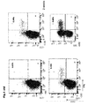
- FIG. 2 Vaccination mainly induces Th1-cells of an effector memory phenotype. Phenotypical characterisation of pdmH1N1-specific CD4 T cells after vaccination with Pandemrix.
- A Flow cytometric analysis of IFN- ⁇ and CD69, IL-2, IL-17, CTLA-4 and CD127 expression of CD4 T cells stimulated with pdmH1N1 antigen in blood samples of a representative individual (not pre-vaccinated against seasonal influenza) one week after vaccination.
- a pre-existing immunity prevents strong induction of antigen specific CD4 T cells
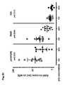
- Figure 3 A pre-existing immunity prevents strong induction of antigen specific CD4 T cells.
- Frequencies of reactive CD4 T cells specific for the pandemic H1N1 antigen (pdmH1N1), the seasonal influenza vaccine Begrivac (Flu09) and Staphylococcus aureus enterotoxin B (SEB, positive control) in individuals that have not been pre-vaccinated (A) and those that have been pre-vaccinated with the seasonal influenza vaccine (B) are depicted over time.
- the detection limit (0.05% IFN- ⁇ -producing CD69+ CD4 T cells) is represented by a horizontal line.
- Vaccine-induced antibodies show only minor cross-reactivity with seasonal antigens
- FIG. 4 Vaccine-induced antibodies show only minor cross-reactivity with seasonal antigens.
- the induction of specific antibody responses towards pandemic H1N1 antigen was monitored by ELISA in individuals that have not been pre-vaccinated (A) and those that have been pre-vaccinated with the seasonal influenza vaccine (B).
- Serum samples of not pre-vaccinated (C) and pre-vaccinated individuals (D) were tested for virus-neutralising antibodies against pdmH1N1 and the single components of the seasonal Flu vaccine (H1N1seasonal, H3N2,
- the IC50 value represents the respective serum dilution which leads to 50% virus neutralisation.
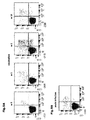
- Figure 5 Differences in the cytokine expression profile of pdmH1N1-specific CD4 T cells discriminate memory or vaccination responses from acute infection. Representative dotplots of the flow cytometric analysis of IFN- ⁇ and IL-2 expression in CD4 T cells after stimulation with pdmH1N1 antigen in blood samples of (A) one healthy individual (not pre-vaccinated against seasonal influenza) after vaccination with the pandemic influenza vaccine and (B) one patient with acute pandemic H1N1-influenza infection within one week after onset of symptoms.
- IFN- ⁇ /IL-2 cytokine-profiling was performed in pdmH1N1 specific CD4 T cells of (C) all 19 individuals after immunisation with the pandemic influenza vaccine over time and (D) of 7 patients with acute pandemic H1N1 influenza infection. Shown are the median values and IQR of absolute numbers of IFN- ⁇ single positive (black circles), IFN- ⁇ /IL-2 double positive (grey circles) and IL-2 single positive (white circles) CD4 T cells per ⁇ l whole blood. Respective values after stimulation with SEB (positive control) are depicted for the vaccination cohort (E) and the infected patients (F).
- FIG. 5A and B Representative dotplots of influenza-specific IFN- ⁇ /IL-2 cytokine profiles before and after vaccination and in an acutely infected patient are shown in figure 5A and B .
- the T-cell response in vaccinees was generally dominated by multifunctional CD4 T cells co-expressing both IFN- ⁇ and IL-2 whilst single-cytokine producing cells were less abundant ( Fig. 5C ).
- those exclusively expressing IL-2 generally dominated over those producing IFN- ⁇ only. This ratio changed only one week after vaccination. Thereafter, the profile largely normalised to that found prior to vaccination ( Fig. 5C ).
- this cytokine profile was markedly different from that found in patients with acute infections, where IFN- ⁇ only expressing cells were most abundant and IL-2 single producing cells were completely absent.
- these profiles are indeed influenza-specific, as the cytokine profiles of SEB-reactive CD4 T cells were unaffected by vaccination ( Fig. 5E ) or acute infection ( Fig. 5F ).
- pdmH1N1 Flu09 SEB Median frequencies Basal (0 weeks) % (IQR) 0.03% (0.04%) 0.10% (0.07%) 3.40% (2.53%) 1 week 0.18% (0.18%) 0.24% (0.18%) 3.21% (3.34%) 2 weeks 0.14% (0.19%) 0.21% (0.19%) 3.13% (2.45%) 10 weeks 0.12% (0.14%) 0.24% (0.18%) 4.01% (3.05%) Median fold increase Week 1 vs.
Landscapes
- Health & Medical Sciences (AREA)
- Life Sciences & Earth Sciences (AREA)
- Engineering & Computer Science (AREA)
- Immunology (AREA)
- Molecular Biology (AREA)
- Urology & Nephrology (AREA)
- Chemical & Material Sciences (AREA)
- Biomedical Technology (AREA)
- Hematology (AREA)
- Cell Biology (AREA)
- Pathology (AREA)
- Analytical Chemistry (AREA)
- Biotechnology (AREA)
- Microbiology (AREA)
- Food Science & Technology (AREA)
- Medicinal Chemistry (AREA)
- Physics & Mathematics (AREA)
- General Physics & Mathematics (AREA)
- Biochemistry (AREA)
- General Health & Medical Sciences (AREA)
- Virology (AREA)
- Tropical Medicine & Parasitology (AREA)
- Proteomics, Peptides & Aminoacids (AREA)
- Medicines Containing Antibodies Or Antigens For Use As Internal Diagnostic Agents (AREA)
- Measuring Or Testing Involving Enzymes Or Micro-Organisms (AREA)
- Investigating Or Analysing Biological Materials (AREA)
Description
- The present invention pertains to an in-vitro process for the quick determination of the infection status of an infection with an influenza virus type from whole blood in terms of an acute influenza infection or a lasting-immunization due to an previous influenza infection or vaccination.
- Influenza, commonly referred to as the flu, is an infectious disease caused by the influenza viruses, that affects birds and mammals. The most common symptoms of the disease are chills, fever, sore throat, muscle pains, severe headache, coughing, weakness/fatigue and general discomfort.
- New influenza viruses are constantly evolving by mutation or by reassortment. Mutations can cause small changes in the hemagglutinin and neuraminidase antigens on the surface of the virus. This is called antigenic drift, which slowly creates an increasing variety of strains until one evolves that can infect people who are immune to the pre-existing strains. This new variant then replaces the older strains as it rapidly sweeps through the human population-often causing an epidemic. However, since the strains produced by drift will still be reasonably similar to the older strains, some people will still be immune to them. In contrast, when influenza viruses reassort, they acquire completely new antigens-for example by reassortment between avian strains and human strains; this is called antigenic shift. If a human influenza virus is produced that has entirely new antigens, everybody will be susceptible, and the novel influenza will spread uncontrollably, causing a pandemic.
- Typically, influenza is transmitted through the air by coughs or sneezes, creating aerosols containing the virus. Influenza can also be transmitted by direct contact with bird droppings or nasal secretions, or through contact with contaminated surfaces. Airborne aerosols have been thought to cause most infections, although which means of transmission is most important is not absolutely clear.
- As influenza is caused by a variety of species and strains of viruses, in any given year some strains can die out while others create epidemics, while yet another strain can cause a pandemic. Typically, in a year's normal two flu seasons (one per hemisphere), there are between three and five million cases of severe illness and up to 500,000 deaths worldwide, which by some definitions is a yearly influenza epidemic.
- Three influenza pandemics occurred in the 20th century and killed tens of millions of people, with each of these pandemics being caused by the appearance of a new strain of the virus in humans. Often, these new strains appear when an existing flu virus spreads to humans from other animal species, or when an existing human strain picks up new genes from a virus that usually infects birds or pigs. An avian strain named H5N1 raised the concern of a new influenza pandemic, after it emerged in Asia in the 1990s, but it has not evolved to a form that spreads easily between people. In April 2009 a novel flu strain evolved that combined genes from human, pig, and bird flu, initially dubbed "swine flu" and also known as influenza A/H1N1, emerged in Mexico, the United States, and several other nations. The World Health Organization officially declared the outbreak to be a pandemic on June 11, 2009.
- In virus classification influenza viruses are RNA viruses that make up three of the five genera of the family Orthomyxoviridae: Influenza virus A, B, and C.
- Wild aquatic birds are the natural hosts for a large variety of influenza A. Occasionally, viruses are transmitted to other species and may then cause devastating outbreaks in domestic poultry or give rise to human influenza pandemics. The type A viruses are the most virulent human pathogens among the three influenza types and cause the most severe disease. The influenza A virus can be subdivided into different serotypes based on the antibody response to these viruses. The serotypes that have been confirmed in humans, ordered by the number of known human pandemic deaths, are:
- ▪ H1N1, which caused Spanish Flu in 1918, and Swine Flu in 2009
- ▪ H2N2, which caused Asian Flu in 1957
- ▪ H3N2, which caused Hong Kong Flu in 1968
- ▪ H5N1, which caused Bird Flu in 2004
- ▪ H7N7, which has unusual zoonotic potential[23]
- ▪ H1N2, endemic in humans, pigs and birds
- ▪ H9N2
- ▪ H7N2
- ▪ H7N3
- ▪ H10N7
- Influenza B almost exclusively infects humans and is less common than influenza A. The only other animals known to be susceptible to influenza B infection are the seal and the ferret. This type of influenza mutates at a rate 2-3 times slower than type A and consequently is less genetically diverse, with only one influenza B serotype. This reduced rate of antigenic change, combined with its limited host range (inhibiting cross species antigenic shift), ensures that pandemics of influenza B do not occur.
- Influenzavirus C infects humans, dogs and pigs, sometimes causing both severe illness and local epidemics. However, influenza C is less common than the other types and usually only causes mild disease in children.
- As prophylaxis against an influenza A and B virus infections vaccination is possible. Influenza virus is frequently changing its structures which are recognized by the immune system. Various types of influenza virus can be active at the same time e.g. influenza viruses which are responsible for the "normal" seasonal influenza diseases for example H3N2 and more dangerous ones for example H5N1 (bird's flue). Vaccinations against influenza are usually made available to people in developed countries. Farmed poultry is often vaccinated to avoid decimation of the flocks. The most common human vaccine is the trivalent influenza vaccine (TIV) that contains purified and inactivated antigens against three viral strains. Typically, this vaccine includes material from two influenza A virus subtypes and one influenza B virus strain. The TIV carries no risk of transmitting the disease, and it has very low reactivity. A vaccine formulated for one year may be ineffective in the following year, since the influenza virus evolves rapidly, and new strains quickly replace the older ones. Antiviral drugs can be used to treat influenza, with neuraminidase inhibitors (such as Tamiflu or Relenza) being particularly effective.
- H3N2 and more dangerous ones for example H5N1 (bird's flue). Vaccinations against influenza are usually made available to people in developed countries. Farmed poultry is often vaccinated to avoid decimation of the flocks. The most common human vaccine is the trivalent influenza vaccine (TIV) that contains purified and inactivated antigens against three viral strains. Typically, this vaccine includes material from two influenza A virus subtypes and one influenza B virus strain. The TIV carries no risk of transmitting the disease, and it has very low reactivity. A vaccine formulated for one year may be ineffective in the following year, since the influenza virus evolves rapidly, and new strains quickly replace the older ones. Antiviral drugs can be used to treat influenza, with neuraminidase inhibitors (such as Tamiflu or Relenza) being particularly effective.
-
WO 2007/107714 A2 discolses a method of assessing an intracellular pathogen infection and/or monitoring an intracellular pathogen infection in an individual comprising the determination whether the individual has (a) T-cells that secrete IFN-γ only, (b) T-cells that secrete IL-2 only or (c) T-cells that secrete both IFN- y and IL-2 in response to an intracellular pathogen antigen and optionally determining any change in this cytokine profile. The pathogens described in Detail are CMV, hepatitis virus, EBV or HSV. These viruses do not rapidly change its antigenic pattern like influenza viruses, so that the method known fromWO 2007/107714 A2 cannot readily adopted to solve the problem underlying the invention. - Urban Sester et al., in PLoS one, vol. 6, no. 3, pages E17813 disclose that T-cell based IFN-γ release assays do not permit distinction of active tuberculosis (TB) from successfully treated disease or latent M. tuberculosis infection. The authors postulated that IFN-γ and IL-2 cytokine profiles of antigen-specific T cells measured by flow-cytometry ex vivo might correlate with TB disease activity in vivo. Tuberculin (PPD), ESAT-6 and CFP-10 were used as stimuli to determine antigen-specific cytokine profiles in CD4 T cells from 24 patients with active TB and 28 patients with successfully treated TB using flow-cytometry. Moreover, 25 individuals with immunity consistent with latent M. tuberculosis infection and BCG-vaccination, respectively, were recruited. Although the frequency of cytokine secreting PPD reactive CD4 T cells was higher in patients with active TB compared to patients with treated TB (median 0.81% vs. 0.39% of CD4 T cells, p = 0.02), the overlap in frequencies precluded distinction between the groups on an individual basis. When assessing cytokine profiles, PPD specific CD4 T cells secreting both IFN-γ and IL-2 predominated in treated TB, latent infection and BCG-vaccination, whilst in active TB the cytokine profile was shifted towards cells secreting IFN-γ only (p<0.0001). Cytokine profiles of ESAT-6 or CFP-10 reactive CD4 T cells did not differ between the groups. Receiver operator characteristics (ROC) analysis revealed that frequencies of PPD specific IFN-γ/IL-2 dual-positive T cells below 56% were an accurate marker for active TB (
specificity 100%,sensitivity 70%) enabling effective discrimination from non-active states. In conclusion, a frequency lower than 56% IFN-γ/IL-2 dual positive PPD-specific circulating CD4 T-cells is strongly indicative of active TB. - Prior to vaccination it is desirable to know whether a patient is not yet infected, is at present suffering from an active influenza and if so whether the patient is infected by "normal" influenza viruses or e.g. H5N1.
- The invention is based on the observation that T cellular immune responses against different Influenza strains could be detected and characterized with the aid of antigen specific stimulation of T cells within whole blood samples. Influenza reactive T cells could be detected after leukocyte fixation and lysis of red blood cells with the aid of intracellular cytokine staining and subsequent analysis of their frequency among all respective T cells by flow cytometry. It was observed in some individuals a low level pre existing influenza specific T cells and in all individuals a distinct increase of influenza specific T cells after vaccination. Interestingly this increase was accompanied by a change in the cytokine profile of the activated T cells which was most dominant one week after vaccination. In some other unvaccinated individuals, which presented with clinical signs suggestive for acute influenza infection, the analysis of influenza specific T cells was performed to confirm a newly induced immune response. In these individuals, there was an even more striking change in cytokine profile of activated T cells. The in-vitro process of the invention allows for the determination of an actual infection status of an individual.
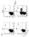
- Fig. 1:
- Immunisation with the pandemic influenza vaccine induces a specific CD4 T-cell response towards both pandemic and seasonal influenza antigens.
- Fig. 2:
- Vaccination mainly induces Th1-cells of an effector memory phenotype.
- Fig. 3:
- A pre-existing immunity prevents strong induction of antigen specific CD4 T cells.
- Fig. 4:
- Vaccine-induced antibodies show only minor cross-reactivity with seasonal antigens.
- Fig. 5:
- Differences in the cytokine expression profile of pdmH1N1 - specific CD4 T cells discriminate memory or vaccination responses from acute infection.
- The process of the invention is an in-vitro process for the quick determination of the infection status of an infection with an influenza virus type from whole blood of humans and animals in terms of an acute influenza infection, and induced immune response due to vaccination or a pre-immunization due to previous, overcome influenza infection, comprising the steps of:
- stimulating an antigen-specific T cell that is present in a first sample of whole blood with antigens of the influenza virus type, optionally in the presence of antibodies against CD28, or CD28 and CD49d;
- processing of the influenza antigens by antigen-presenting cells (APC) by incubation for a sufficient time, in particular by incubation of at least 0,5 h, particularly of 1.5 h to 2.5 h, especially for 2 h, at 35 °C to 39 °C, especially at 37 °C, optionally with adding CO2;
- then adding a secretion inhibitor;
- effecting an intensive mixing;
- followed by a second incubation step for a period of at least 1 h, in particular 2,5 h, particularly of 3.5 h to 6 h, especially for 4 h, at 35 °C to 39 °C, especially at 37 °C, optionally with adding CO2;
- determining a cytokine profile from both the intracellular INF-γ production and the intracellular IL-2 production of the antigen-specific T cell; wherein
- the presence of an acute influenza infection is indicated by a shift of the profile towards IFN-γ single positive cells accompanied by a ratio of IFN-y/IL-2 single positive T cells larger than 1 compared to the ratio of IFN-y/IL-2 single positive T cells of patients successfully vaccinated against, pre-infected or non-infected by the influenza virus which is smaller than 1.
- According to the invention a negative control can be performed by adding a physiological buffer such as PBS 0.9% NaCl or 5% Glucose to an antigen-specific T cell that is present in the whole blood of a second sample, which is authentic with said first sample.
- A positive control can be performed by adding SEB to an antigen-specific T cell that is present in the whole blood of a third sample, which is authentic with said first sample.
- In still another embodiment of the invention a vaccination control can take place by adding the vaccination antigen to an antigen-specific T-cell that is present in the whole blood of a fourth sample which is authentic with said first sample.
In the process according to the invention the secretion inhibitor is selected from the group consisting of brefeldin A, monensin or the like. - The determination of the cytokine profile can be effected by flow cytometry, automated microscopy or multiplex elisa methods.
- The invention is further illustrated by way of the following more specific examples and descriptions which are not intended to limit the scope of the claims. Induction of antigen-specific antibody responses is well-characterised after vaccination with pandemic H1N1 or seasonal influenza (Flu) vaccines. However, the kinetics of cellular immune responses towards pandemic H1N1 and cross-reactivities towards seasonal Flu vaccine in respect to changes in cytokine profiles of antigen specific T cells are not known so far.
- 19 immunocompetent individuals were vaccinated with the pandemic H1N1 vaccine Pandemrix and CD4-T-cell frequencies specific for pandemic H1N1 and for seasonal Flu vaccine were monitored before and 1, 2 and 10 weeks after vaccination using intracellular cytokine-staining (IFN-γ, IL-2, IL-17). Stimulation with staphylococcus aureus enterotoxin B (SEB) served as positive control. Apart from cytokine analyses, specific T-cells were phenotypically characterised by memory and maturation markers. Moreover, induction of specific antibodies was monitored by ELISA and neutralisation assay. In addition, 7 patients with acute H1N1 infection were analysed.
- Prior to vaccination, levels of pandemic H1N1-specific CD4-T-cells were below detection limit in 68% of cases. In contrast, immunity towards seasonal Flu was detectable in 79%. Specific T-cell responses after vaccination rose to above detection limit in all cases. Median frequencies increased by 463% (IQR 291-715%) and remained stable throughout follow-up. Interestingly, median T-cell levels towards seasonal vaccine concomitantly rose from 0.10% to 0.24% whereas SEB-reactive T-cells were not specifically induced after vaccination. Influenza-specific cells before and after vaccination were co-expressing IFN-γ and IL-2, whereas IL-17 was not detectable. In contrast, T cells in patients with active H1N1 infection predominantly expressed IFN-γ only. In contrast to the cellular immunity, only H1N1-pandemic antibody responses but no cross-reactive antibodies against the seasonal Flu vaccine were induced.
- In conclusion, besides specific induction of H1N1-reactive T-cell immunity, pandemic H1N1 vaccination also induces a cross-protective T-cell response towards seasonal influenza vaccines. In addition, cytokine-profiling may be applied as diagnostic measure to identify patients with active H1N1 infection.
- The vaccination study was conducted among 19 immunocompetent individuals in October/November 2009. Among them, 10 persons (mean age 43.64±10.05 years) had been pre-vaccinated against seasonal influenza (Begrivac 2009/2010, Novartis, Marburg, Germany) about 3 weeks before (mean 18.9±7.9 days), whereas 9 (mean age 37.8±15.9 years) had not. All individuals received one standard dose of the pdmH1N1 vaccine (Pandemrix, GlaxoSmithKline Biologicals s.a., Rixensart, Belgium), and all gave informed consent. Whole blood samples were drawn prior to vaccination with Pandemrix as well as 1, 2, and 10 weeks thereafter. To characterize cellular immunity in acute infection, additional whole blood samples of 7 individuals (mean age 49.28±11.07 years) who were newly diagnosed with influenza A/H1N1 infection on a clinical/epidemiological or virological basis were analyzed.
- T cells from heparinized whole blood sample were stimulated in vitro for 6 h as described previously [1]. A total of 450µl blood was used per stimulus. PdmH1N1 antigen (pdmH1N1, 30µg/ml HA content, derived from A/california(H1N1) was used in the pandemic vaccine Pandemrix) kindly provided by GlaxoSmithKline Biologicals s.a., Rixensart, Belgium and the seasonal influenza 2009/2010 vaccine Begrivac (Flu09, 30µg/ml HA content; Novartis, Nürnberg, Germany) were used as stimuli in the presence of 1µg/ml anti-CD28 and anti-CD49d (clones L293 and 9F10; BD, Heidelberg, Germany), respectively, to induce antigen-specific activation and cytokine expression. As negative and positive controls, respectively, cells were stimulated with phosphate buffered saline (PBS), and 2.5µg/ml Staphylococcus aureus Enterotoxin B (SEB, Sigma, Deisenhofen, Germany). Stimulated cells were fixed and immunostained using titered amounts of anti-CD4 (clone SK3), anti-CD69 (clone L78), anti-IFN-γ (clone 4S.B3), anti-IL-2 (clone MQ1-17H12), anti-IL-17A (clone SCPL1362), anti-CD152 (CTLA-4, clone BNI3; all from BD) and anti-CD127 (clone eBioRDR5, eBioscience-NatuTec, Frankfurt, Germany). At least 10.000 CD4 T-cells were analyzed on a FACS Canto II (BD) using FACSdiva software V 6.1.3.. For analyses of antigen-specific CD4 T cells, frequencies of activated CD4 T cells after stimulation with the antigen were subtracted for the respective negative controls. The detection limit was 0.05% IFN-γ-producing CD69+ CD4 T cells.
- Kinetics of pdmH1N1-specific IgG induction was analyzed using a standard ELISA (Virotech, Rüsselsheim, Germany), according to the manufacturer's instructions. Moreover, the presence of virus-neutralizing antibodies against pdmH1N1 and against the individual components of the seasonal influenza vaccine (H1N1seasonal, H3N2, Influenza B) in serum samples of subjects not pre-vaccinated with Begrivac was determined by respective microneutralization assays based on previously described procedures [2, 3]. In detail, heat inactivated (30min, 56°C) sera were used to prepare two-fold serial dilutions in duplicate from 1:10 to 1:1280 in virus diluent [EMEM (Biochrom, Germany) + 0,1% antibiotics + 2% FBS (Biochrom, Germany)]. The diluted sera were mixed with an equal volume (100 µl) of virus diluent containing influenza A virus [NYMC X-179A (H1N1) derived from A/California/7/2009] at 2x10³ TCID50/ml, incubated for 90min (37°C, 5% CO2) in a moist chamber and 100µl were transferred to MDCK cell monolayers (ECCC, Salisbury, UK) in a 96-well plate. Eight control wells containing influenza virus in virus diluent (virus control) or virus diluent alone (cell control) were included on each plate. After 1h incubation, 100 µl of overlay medium [1:1 virus diluent and 3.2% carboxy-methylcellulose (CMC, Sigma-Aldrich)] was added to each well for 24 to 28h. Then, the monolayers were fixed with ice cold acetone:methanol (40%:60%; 10min, RT). After blocking with 1% bovine serum albumin (Merck, Germany), 0.1% thimerosal (Sigma-Aldrich) in Dulbecco A wash buffer (8.0 g NaCl, 2.9 g Na2HPO4 0.2 g KCl and 0.2 g K2HPO4 dissolved in 1000 ml of deionized water, pH 7.3) for 30 min at 37°C, 5% CO2), fixed cells were subsequently incubated with 25µl anti-influenza Monoclonal antibodies to nucleoprotein (MAB8251, Millipore), diluted to 1:10.000 in blocking buffer. and 25 µl/well horseradish peroxidase-labeled rabbit anti-mouse IgG (P0161, Dako-Cytomation, Denmark) diluted 1:1000 in blocking buffer for 30 min at 37°C, 5% CO2 respectively and substrate reaction was performed for 30min using 0.03% H2O2 in 50µl/well freshly prepared 3-amino-9-ethylcarbazole (Sigma-Aldrich) dissolved in 4.0 ml N,N-dimethylformamide (Sigma-Aldrich) and 16ml 0.05 M acetate buffer (pH 4.95-5.05). Immunostained foci were counted and recorded using automated ELISPOT instrumentation (AID Diagnostika GmbH, Straßberg, Germany). The neutralizing-antibody titer was expressed as the reciprocal of the highest dilution that reduced the number of immunostained foci to 50% or less of the control value.
- Immunisation with the pandemic influenza vaccine induces a specific CD4 T-cell response towards both the pandemic and seasonal influenza-antigens.
- The induction of influenza-specific cellular immunity was assessed in a total of 19 individuals before as well as one, two and 10 weeks after immunisation with the pandemic vaccine Pandemrix. Among those, 10 individuals had been pre-vaccinated using the seasonal influenza vaccine. The frequencies of CD4 T cells specific for both the pandemic (pdmH1N1) and cross-reactive seasonal influenza-antigens (Flu09) were determined directly ex vivo from whole blood samples. Antigen-specific CD4 T cells were identified using flow-cytometry based on the induction of the activation marker CD69 and the cytokine IFN-γ after specific stimulation in vitro. When basal T-cell frequencies in all 19 individuals were assessed, low levels of CD4 T cells specific for pdmH1N1 (median (IQR) 0.03% (0.04%)) and seasonal influenza antigens were found (0.10% (0.07%), table 1).
Figure 1 : Immunisation with the pandemic influenza vaccine induces a specific CD4 T-cell response towards both pandemic and seasonal influenza antigens. Whole blood samples taken prior to as well as one, two and 10 weeks after immunisation with the pandemic influenza vaccine were stimulated with phosphate buffered saline (PBS, negative control), the pandemic H1N1 antigen (pdmH1N1), the 2009 seasonal influenza vaccine Begrivac (Flu09) and the Staphylococcus aureus enterotoxin B (SEB, positive control), respectively, and reactive CD4 T cells (CD69+/IFN-γ+) were counted using flow cytometry. Typical dotplots of one representative individual that was not pre-vaccinated with the seasonal vaccine are shown. The numbers indicate the percentage of antigen-reactive CD4 T cells. As shown in a representative example (Fig. 1 ), low levels of specific T cells present prior to vaccination increased thereafter and peak values were found 1 week after vaccination (Fig. 1 and table 1). In general, Pandemrix vaccination resulted in the strongest induction of specific CD4-T-cell frequencies towards the pdmH1N1 antigen present in the vaccine (4.63-fold median increase after one week, table 1). Interestingly, although less pronounced, this was associated with a concomitant increase in the percentage of cells cross-reacting towards seasonal influenza antigens (2.71-fold median increase). In contrast, frequencies of T cells reactive against the positive control stimulus SEB did not change over time (Fig. 1 and table 1). These results show that vaccination with the pandemic influenza vaccine Pandemrix induces a specific CD4 T-cell immunity towards pandemic H1N1 antigens that cross-reacts with those of the seasonal influenza vaccine. Vaccination mainly induces Th1-cells of an effector memory phenotype. - In order to analyse changes in phenotypical properties of vaccine-induced pdmH1N1-specific CD4 T cells in more detail, we monitored the expression of different functional markers (IFN-γ, IL-2, IL-17, CD69, CTLA-4) and of the memory marker CD127.
-
Figure 2 : Vaccination mainly induces Th1-cells of an effector memory phenotype. Phenotypical characterisation of pdmH1N1-specific CD4 T cells after vaccination with Pandemrix. (A) Flow cytometric analysis of IFN-γ and CD69, IL-2, IL-17, CTLA-4 and CD127 expression of CD4 T cells stimulated with pdmH1N1 antigen in blood samples of a representative individual (not pre-vaccinated against seasonal influenza) one week after vaccination. (B) Mean fluorescence intensity (MFI) of the CTLA-4-signal of all IFN-γ+ CD4 T cells and (C) percentage of CD127-expressing cells of all IFN-γ+ CD4 T cells after stimulation with the pdmH1N1 antigen of all tested individuals are depicted over time (n=10 in week 10). Bars indicate median values and IQR. - As shown in representative dotplots of pdmH1N1-specific T cells one week after Pandemrix vaccination, predominant co-expression of effector cytokines IL-2 and IFN-γ with CD69 indicated that specific T cells were mainly Th1 effector-type cells, whereas no IL-17-expressing cells were detected (
Fig. 2A ). In line with the kinetics in frequency, CTLA-4, the most important negative co-stimulatory surface receptor of T-cell activation, peaked one week after Pandemrix vaccination and then decreased to almost pre-vaccination levels (p<0.0001,Fig. 2B ). On the other hand, median (IQR) percentages of CD127-expressing influenza-specific CD4 T cells initially decreased from 97.6% (5.0%) to 80.0% (12.1%) during the first week post vaccination, but increased again in the follow-up to 98.0% (6.0%) after 10 weeks indicating activation of naive T cells followed by differentiation into effector memory cells (p<0.0001,Fig. 2C ). The CD4 T cells that were specific for the seasonal influenza vaccine showed similar expression profiles and properties (data not shown). In summary, phenotypical characterisation of the influenza-specific CD4 T cells showed that the vaccine leads to induction of mainly Th1 effector memory cells that show dynamic changes in the immediate post-vaccination period. - A pre-existing immunity prevents strong induction of antigen specific CD4 T cells To assess the impact of pre-existing cellular immunity against seasonal influenza antigens on basal levels and on the induction of influenza specific immunity after Pandemrix vaccination, individuals were separated into those not pre-vaccinated and pre-vaccinated with the seasonal influenza vaccine.
-
Figure 3 : A pre-existing immunity prevents strong induction of antigen specific CD4 T cells. Frequencies of reactive CD4 T cells specific for the pandemic H1N1 antigen (pdmH1N1), the seasonal influenza vaccine Begrivac (Flu09) and Staphylococcus aureus enterotoxin B (SEB, positive control) in individuals that have not been pre-vaccinated (A) and those that have been pre-vaccinated with the seasonal influenza vaccine (B) are depicted over time. The detection limit (0.05% IFN-γ-producing CD69+ CD4 T cells) is represented by a horizontal line. (C) The increase of CD4 T-cell frequencies specific for pdmH1N1, Flu09 and SEB during the first week after vaccination with the pandemic vaccine was expressed as a multiple of the basal frequencies obtained before vaccination. Tested individuals were subdivided according to pre-vaccination status (pre-vaccinated against seasonal influenza or not). T-cell frequencies of each individual as well as median values and IQR are shown. - The kinetics in influenza- and SEB-reactive CD4 T cells in both groups are shown in
figure 3A and B . Interestingly, the median increase in pdmH1N1 specific T-cell frequencies was significantly stronger in individuals that had not been pre-vaccinated against seasonal influenza (7.15-fold), as compared with individuals that had been pre-vaccinated (2.94-fold, p=0.01,Fig. 3C ). Of note, this difference between the two groups was also found for CD4 T cells reacting towards seasonal influenza antigens (4.03-fold versus 1.74-fold, p=0.0041). The frequencies of SEB reactive T cells did not increase in any of the groups (p=0.11,Fig. 3C ). The results indicate that a pre-existing immunity is associated with a less pronounced induction of influenza specific CD4 T cells after vaccination with Pandemrix. - To compare the effects of the Pandemrix vaccine on cellular and humoral responses, induction of influenza-specific antibodies was determined. Importantly, vaccination induced a significant continuous increase in pdmH1N1-specific IgG titers which peaked about two weeks post vaccination and remained almost constant over the following eight weeks.
-
Figure 4 : Vaccine-induced antibodies show only minor cross-reactivity with seasonal antigens. The induction of specific antibody responses towards pandemic H1N1 antigen was monitored by ELISA in individuals that have not been pre-vaccinated (A) and those that have been pre-vaccinated with the seasonal influenza vaccine (B). Serum samples of not pre-vaccinated (C) and pre-vaccinated individuals (D) were tested for virus-neutralising antibodies against pdmH1N1 and the single components of the seasonal Flu vaccine (H1N1seasonal, H3N2, - Influenza B (Inf B)). The IC50 value represents the respective serum dilution which leads to 50% virus neutralisation.
- These kinetics were independent of pre-vaccination status (
Fig. 4A and B ). In order to assess potential cross-reactivity of antibodies induced by Pandemrix, plasma samples were analysed from individuals that were not pre-vaccinated against seasonal influenza for the presence of neutralising antibodies against pdmH1N1 and the individual compounds of the seasonal influenza vaccine (H1N1seasonal, H3N2, Influenza B), respectively. Consistent with results from ELISA (Fig. 4A ), maximum titers of pdmH1N1-specific neutralising antibodies were induced already during the first two weeks following Pandemrix vaccination and remained stable throughout week 10 (Fig. 4C ). In contrast, only a minor increase in seasonal H1N1 titers was observed indicating only slight neutralising cross-reactivity against the seasonal H1N1 influenza strain. Likewise, there was no significant effect on H3N2- and Influenza B-neutralising antibody titers. Individuals that had been pre-vaccinated against seasonal influenza had higher levels of neutralising antibodies towards the components of the seasonal vaccine that largely remained stable after Pandemrix vaccination (Fig. 4D ). Interestingly, titers of pdmH1N1 neutralising antibodies before immunisation with the pandemic vaccine were also slightly elevated which may indicate a certain extent of cross-reactivity induced by the the seasonal vaccine. Of note, those titers showed an increase after Pandemrix vaccination, but did not reach the levels of those in individuals that had not been pre-vaccinated against seasonal influenza. Thus, these results demonstrate that vaccination with Pandemrix induces a pdmH1N1-specific antibody response with a certain extent of cross-reactivity towards seasonal H1N1 antigens. In line with cellular immunity, a pre-existing humoral immunity was associated with a less pronounced induction of pdmH1N1-specific titers after vaccination with Pandemrix. - Differences in the cytokine expression profiles of pdmH1N1-specific CD4 T cells discriminate vaccination responses from acute infection
- Previous studies have indicated that acute viral or bacterial infections are associated with changes in cytokine expression profiles of antigen-specific T cells. To examine whether this is also the case in acute infection with pandemic H1N1 influenza virus, the IFN-γ/IL-2 expression profiles of specific T cells induced after vaccination were comparatively analysed with that of 7 patients that were suffering from a symptomatic infection with pandemic H1N1-influenza. Whole blood was stimulated with the pandemic and seasonal antigens as described before, and CD4 T cells were analysed flow-cytometrically for single or double expression of IFN-γ and IL-2. Stimulation with SEB served as positive control for cells with specificities unrelated to influenza.
-
Figure 5 : Differences in the cytokine expression profile of pdmH1N1-specific CD4 T cells discriminate memory or vaccination responses from acute infection. Representative dotplots of the flow cytometric analysis of IFN-γ and IL-2 expression in CD4 T cells after stimulation with pdmH1N1 antigen in blood samples of (A) one healthy individual (not pre-vaccinated against seasonal influenza) after vaccination with the pandemic influenza vaccine and (B) one patient with acute pandemic H1N1-influenza infection within one week after onset of symptoms. IFN-γ/IL-2 cytokine-profiling was performed in pdmH1N1 specific CD4 T cells of (C) all 19 individuals after immunisation with the pandemic influenza vaccine over time and (D) of 7 patients with acute pandemic H1N1 influenza infection. Shown are the median values and IQR of absolute numbers of IFN-γ single positive (black circles), IFN-γ/IL-2 double positive (grey circles) and IL-2 single positive (white circles) CD4 T cells per µl whole blood. Respective values after stimulation with SEB (positive control) are depicted for the vaccination cohort (E) and the infected patients (F). - Representative dotplots of influenza-specific IFN-γ/IL-2 cytokine profiles before and after vaccination and in an acutely infected patient are shown in
figure 5A and B . Interestingly, the T-cell response in vaccinees was generally dominated by multifunctional CD4 T cells co-expressing both IFN-γ and IL-2 whilst single-cytokine producing cells were less abundant (Fig. 5C ). Among the latter, those exclusively expressing IL-2 generally dominated over those producing IFN-γ only. This ratio changed only one week after vaccination. Thereafter, the profile largely normalised to that found prior to vaccination (Fig. 5C ). As shown infigure 5D , this cytokine profile was markedly different from that found in patients with acute infections, where IFN-γ only expressing cells were most abundant and IL-2 single producing cells were completely absent. Of note, these profiles are indeed influenza-specific, as the cytokine profiles of SEB-reactive CD4 T cells were unaffected by vaccination (Fig. 5E ) or acute infection (Fig. 5F ). These results suggest that cytokine profiling of H1N1-specific CD4 T cells may allow distinction between vaccination responses and acute infection with the pandemic H1N1 influenza. TableTable 1: Kinetics of antigen-specific CD4 T-cell frequencies prior to and after immunisation with the pandemic influenza vaccine and fold increase during the first week following vaccination in all individuals (n=19). pdmH1N1 Flu09 SEB Median frequencies Basal (0 weeks) % (IQR) 0.03% (0.04%) 0.10% (0.07%) 3.40% (2.53%) 1 week 0.18% (0.18%) 0.24% (0.18%) 3.21% (3.34%) 2 weeks 0.14% (0.19%) 0.21% (0.19%) 3.13% (2.45%) 10 weeks 0.12% (0.14%) 0.24% (0.18%) 4.01% (3.05%) Median fold increase Week 1 vs. basal in specific T cells (IQR)4.63 (4.16) 2.71 (2.26) 1.03 (0.25) Shown are median (IQR) frequencies of antigen-specific CD4 T cells (CD69+ IFN-γ+) that were calculated from values subtracted for the respective negative controls. -
- 1. Sester, U., et al., PD-1 expression and IL-2 loss of cytomegalovirus- specific T cells correlates with viremia and reversible functional anergy. Am J Transplant, 2008. 8(7): p. 1486-97.
- 2. Zielinska, E., et al., Development of an improved microneutralization assay for respiratory syncytial virus by automated plaque counting using imaging analysis. Virol J, 2005. 2: p. 84.
- 3. Rowe, T., et al., Detection of antibody to avian influenza A (H5N1) virus in human serum by using a combination of serologic assays. J Clin Microbiol, 1999. 37(4): p. 937-43.
Claims (7)
- An in-vitro process for the quick determination of the infection status of an infection with an influenza virus type from whole blood in terms of an acute influenza infection or a pre-immunization after previous, overcome influenza infection or induced immune response due to vaccination, comprising the steps of:• stimulating an antigen-specific T cell that is present in a first sample of whole blood with antigens of the influenza virus type, optionally in the presence of antibodies against CD28, or CD28 and CD49d;• processing of influenza antigens by antigen-presenting cells (APC) by incubation for a sufficient time, in particular by incubation of at least 0,5 h, particularly of 1.5 h to 2.5 h, especially for 2 h, at 35 °C to 39 °C, especially at 37 °C, optionally with adding CO2;• then adding a secretion inhibitor;• effecting an intensive mixing;• followed by a second incubation step for a period of at least 1 h, in particular 2.5 h, at a temperature of from 35 °C to 39 °C, and• determining a profile from both the intracellular IFN-γ production and the intracellular IL-2 production of the antigen-specific T cell; wherein• the presence of an acute influenza infection is indicated by a shift of the profile towards IFN-γ single positive cells accompanied by a ratio of IFN-γ/IL-2 single positive T cells larger than 1 compared to the ratio of IFN-γ/IL-2 single positive T cells of patients successfully vaccinated against, pre-infected or non-infected by the influenza virus which is smaller than 1.
- The process according to claim 1, wherein a negative control is performed by adding a physiological buffer such as PBS 0.9% NaCl or 5% Glucose to an antigen-specific T cell that is present in the whole blood of a second sample, which is authentic with said first sample.
- The process according to at least one of claims 1 or 2, wherein a positive control is performed by adding SEB to an antigen-specific T cell that is present in the whole blood of a third sample, which is authentic with said first sample.
- The process according to at least one of the claims 1 to 3 wherein a vaccination control took place by adding the vaccination antigen to an antigen-specific T-cell that is present in the whole blood of a fourth sample which is authentic with said first sample.
- The process according to at least one of claims 1 to 4, wherein said secretion inhibitor is selected from the group consisting of brefeldin A, monensin or the like.
- The process according to at least one of claims 1 to 5, wherein the determination of the profile is effected by flow cytometry, automated microscopy or multiplex elisa methods.
- The process according to at least one of claims 1 to 6, wherein the whole blood of the samples is derived from humans or animals.
Priority Applications (1)
| Application Number | Priority Date | Filing Date | Title |
|---|---|---|---|
| EP12758488.6A EP2756299B1 (en) | 2011-09-13 | 2012-09-13 | An in-vitro process for the quick determination of the infection status of an infection with an influenza virus type |
Applications Claiming Priority (4)
| Application Number | Priority Date | Filing Date | Title |
|---|---|---|---|
| US201161534058P | 2011-09-13 | 2011-09-13 | |
| EP11181119 | 2011-09-13 | ||
| EP12758488.6A EP2756299B1 (en) | 2011-09-13 | 2012-09-13 | An in-vitro process for the quick determination of the infection status of an infection with an influenza virus type |
| PCT/EP2012/067928 WO2013037878A1 (en) | 2011-09-13 | 2012-09-13 | An in-vitro process for the quick determination of the infection status of an infection with an influenza virus type |
Publications (2)
| Publication Number | Publication Date |
|---|---|
| EP2756299A1 EP2756299A1 (en) | 2014-07-23 |
| EP2756299B1 true EP2756299B1 (en) | 2016-11-09 |
Family
ID=47882654
Family Applications (1)
| Application Number | Title | Priority Date | Filing Date |
|---|---|---|---|
| EP12758488.6A Not-in-force EP2756299B1 (en) | 2011-09-13 | 2012-09-13 | An in-vitro process for the quick determination of the infection status of an infection with an influenza virus type |
Country Status (8)
| Country | Link |
|---|---|
| EP (1) | EP2756299B1 (en) |
| JP (1) | JP6170923B2 (en) |
| AU (1) | AU2012307425A1 (en) |
| CA (1) | CA2848614A1 (en) |
| IL (1) | IL231511A0 (en) |
| SG (1) | SG11201400629UA (en) |
| WO (1) | WO2013037878A1 (en) |
| ZA (1) | ZA201402645B (en) |
Families Citing this family (3)
| Publication number | Priority date | Publication date | Assignee | Title |
|---|---|---|---|---|
| JP7416445B2 (en) * | 2019-02-18 | 2024-01-17 | 株式会社栄養・病理学研究所 | A method for evaluating the degree of T cell activation in pigs, an agent for evaluating T cell activation in pigs, a method for screening a pig T cell activator, a method for testing pathogen infection in pigs, and a hybridoma |
| US20230305003A1 (en) * | 2020-07-10 | 2023-09-28 | Beckman Coulter, Inc. | Automated sample preparation platform for cellular analysis |
| CN113234676B (en) * | 2021-04-13 | 2022-12-13 | 华南农业大学 | Method for promoting duck T cell proliferation and application thereof |
Family Cites Families (1)
| Publication number | Priority date | Publication date | Assignee | Title |
|---|---|---|---|---|
| GB0605474D0 (en) * | 2006-03-17 | 2006-04-26 | Isis Innovation | Clinical correlates |
-
2012
- 2012-09-13 JP JP2014530213A patent/JP6170923B2/en not_active Expired - Fee Related
- 2012-09-13 SG SG11201400629UA patent/SG11201400629UA/en unknown
- 2012-09-13 CA CA2848614A patent/CA2848614A1/en not_active Abandoned
- 2012-09-13 EP EP12758488.6A patent/EP2756299B1/en not_active Not-in-force
- 2012-09-13 WO PCT/EP2012/067928 patent/WO2013037878A1/en not_active Ceased
- 2012-09-13 AU AU2012307425A patent/AU2012307425A1/en not_active Abandoned
-
2014
- 2014-03-13 IL IL231511A patent/IL231511A0/en unknown
- 2014-04-11 ZA ZA2014/02645A patent/ZA201402645B/en unknown
Non-Patent Citations (1)
| Title |
|---|
| None * |
Also Published As
| Publication number | Publication date |
|---|---|
| ZA201402645B (en) | 2017-05-31 |
| JP6170923B2 (en) | 2017-07-26 |
| WO2013037878A1 (en) | 2013-03-21 |
| CA2848614A1 (en) | 2013-03-21 |
| EP2756299A1 (en) | 2014-07-23 |
| JP2014527177A (en) | 2014-10-09 |
| SG11201400629UA (en) | 2014-09-26 |
| NZ622402A (en) | 2015-10-30 |
| IL231511A0 (en) | 2014-04-30 |
| AU2012307425A1 (en) | 2014-04-03 |
Similar Documents
| Publication | Publication Date | Title |
|---|---|---|
| Moris et al. | H5N1 influenza vaccine formulated with AS03A induces strong cross-reactive and polyfunctional CD4 T-cell responses | |
| Karron et al. | Evaluation of two live attenuated cold-adapted H5N1 influenza virus vaccines in healthy adults | |
| Talaat et al. | A live attenuated H7N3 influenza virus vaccine is well tolerated and immunogenic in a Phase I trial in healthy adults | |
| Schmidt et al. | CD 4+ T‐cell immunity after pandemic influenza vaccination cross‐reacts with seasonal antigens and functionally differs from active influenza infection | |
| Vanderven et al. | Antibody-dependent cellular cytotoxicity responses to seasonal influenza vaccination in older adults | |
| Wong et al. | The immune correlates of protection for an avian influenza H5N1 vaccine in the ferret model using oil-in-water adjuvants | |
| Patel et al. | Evaluation of conserved and variable influenza antigens for immunization against different isolates of H5N1 viruses | |
| Jia et al. | Increased sensitivity for detecting avian influenza-specific antibodies by a modified hemagglutination inhibition assay using horse erythrocytes | |
| JP6170923B2 (en) | Rapid in vitro determination of infection status of specific influenza virus type infections | |
| Morrison et al. | Antibody-dependent NK cell degranulation as a marker for assessing antibody-dependent cytotoxicity against pandemic 2009 influenza A (H1N1) infection in human plasma and influenza-vaccinated transchromosomic bovine intravenous immunoglobulin therapy | |
| Sun et al. | Effect of seasonal influenza vaccines on avian influenza A (H5N1) clade 2.3. 4.4 b virus infection in ferrets | |
| Yamanaka et al. | Rapid diagnosis of equine influenza by highly sensitive silver amplification immunochromatography system | |
| Ruiz-Hernandez et al. | An infected chicken kidney cell co-culture ELISpot for enhanced detection of T cell responses to avian influenza and vaccination | |
| Venturi et al. | Humoral immunity in natural infection by tick‐borne encephalitis virus | |
| Madsen et al. | No evidence of antigenic seniority in hemagglutinin specific antibody responses after adjuvanted pandemic 2009 influenza vaccination | |
| Cherukuri et al. | Vaccine-specific antibody secreting cells are a robust early marker of LAIV-induced B-cell response in ferrets | |
| Henriques et al. | Development and validation of a blocking ELISA test for the detection of avian influenza antibodies in poultry species | |
| NZ622402B2 (en) | An in-vitro process for the quick determination of the infection status of an infection with an influenza virus type | |
| Guercio et al. | Pandemic influenza A/H1N1 virus in a swine farm house in Sicily, Italy | |
| Chin et al. | Impaired dendritic cell maturation in response to pandemic H1N109 influenza virus | |
| US9833506B2 (en) | Vaccine—screening method | |
| Qi et al. | Seroprevalence of avian origin H3N2 canine influenza virus infection in pet dogs in Shenzhen, China | |
| Chauvat et al. | Pitfalls in anti-influenza T cell detection by Elispot using thimerosal containing pandemic H1N1 vaccine as antigen | |
| TW201312116A (en) | An in-vitro process for the quick determination of the infection status of an infection with an influenza virus type | |
| Akhtar et al. | The comparison of sensitivity and specificity of ELISA-based microneutralization test with hemagglutination inhibition test to evaluate neutralizing antibody against Influenza Virus (H1N1) |
Legal Events
| Date | Code | Title | Description |
|---|---|---|---|
| PUAI | Public reference made under article 153(3) epc to a published international application that has entered the european phase |
Free format text: ORIGINAL CODE: 0009012 |
|
| 17P | Request for examination filed |
Effective date: 20140331 |
|
| AK | Designated contracting states |
Kind code of ref document: A1 Designated state(s): AL AT BE BG CH CY CZ DE DK EE ES FI FR GB GR HR HU IE IS IT LI LT LU LV MC MK MT NL NO PL PT RO RS SE SI SK SM TR |
|
| DAX | Request for extension of the european patent (deleted) | ||
| GRAP | Despatch of communication of intention to grant a patent |
Free format text: ORIGINAL CODE: EPIDOSNIGR1 |
|
| INTG | Intention to grant announced |
Effective date: 20160309 |
|
| GRAJ | Information related to disapproval of communication of intention to grant by the applicant or resumption of examination proceedings by the epo deleted |
Free format text: ORIGINAL CODE: EPIDOSDIGR1 |
|
| GRAR | Information related to intention to grant a patent recorded |
Free format text: ORIGINAL CODE: EPIDOSNIGR71 |
|
| GRAS | Grant fee paid |
Free format text: ORIGINAL CODE: EPIDOSNIGR3 |
|
| INTC | Intention to grant announced (deleted) | ||
| INTG | Intention to grant announced |
Effective date: 20160826 |
|
| GRAA | (expected) grant |
Free format text: ORIGINAL CODE: 0009210 |
|
| AK | Designated contracting states |
Kind code of ref document: B1 Designated state(s): AL AT BE BG CH CY CZ DE DK EE ES FI FR GB GR HR HU IE IS IT LI LT LU LV MC MK MT NL NO PL PT RO RS SE SI SK SM TR |
|
| REG | Reference to a national code |
Ref country code: GB Ref legal event code: FG4D |
|
| REG | Reference to a national code |
Ref country code: AT Ref legal event code: REF Ref document number: 844407 Country of ref document: AT Kind code of ref document: T Effective date: 20161115 Ref country code: CH Ref legal event code: EP |
|
| REG | Reference to a national code |
Ref country code: IE Ref legal event code: FG4D |
|
| REG | Reference to a national code |
Ref country code: DE Ref legal event code: R096 Ref document number: 602012025177 Country of ref document: DE |
|
| RAP2 | Party data changed (patent owner data changed or rights of a patent transferred) |
Owner name: RED FLAG DIAGNOSTICS GMBH |
|
| REG | Reference to a national code |
Ref country code: CH Ref legal event code: NV Representative=s name: SCHMAUDER AND PARTNER AG PATENT- UND MARKENANW, CH Ref country code: CH Ref legal event code: PUE Owner name: RED FLAG DIAGNOSTICS GMBH, DE Free format text: FORMER OWNER: SOVICELL GMBH, DE |
|
| PG25 | Lapsed in a contracting state [announced via postgrant information from national office to epo] |
Ref country code: LV Free format text: LAPSE BECAUSE OF FAILURE TO SUBMIT A TRANSLATION OF THE DESCRIPTION OR TO PAY THE FEE WITHIN THE PRESCRIBED TIME-LIMIT Effective date: 20161109 |
|
| REG | Reference to a national code |
Ref country code: LT Ref legal event code: MG4D |
|
| REG | Reference to a national code |
Ref country code: NL Ref legal event code: MP Effective date: 20161109 |
|
| REG | Reference to a national code |
Ref country code: AT Ref legal event code: MK05 Ref document number: 844407 Country of ref document: AT Kind code of ref document: T Effective date: 20161109 |
|
| PG25 | Lapsed in a contracting state [announced via postgrant information from national office to epo] |
Ref country code: LT Free format text: LAPSE BECAUSE OF FAILURE TO SUBMIT A TRANSLATION OF THE DESCRIPTION OR TO PAY THE FEE WITHIN THE PRESCRIBED TIME-LIMIT Effective date: 20161109 Ref country code: NO Free format text: LAPSE BECAUSE OF FAILURE TO SUBMIT A TRANSLATION OF THE DESCRIPTION OR TO PAY THE FEE WITHIN THE PRESCRIBED TIME-LIMIT Effective date: 20170209 Ref country code: SE Free format text: LAPSE BECAUSE OF FAILURE TO SUBMIT A TRANSLATION OF THE DESCRIPTION OR TO PAY THE FEE WITHIN THE PRESCRIBED TIME-LIMIT Effective date: 20161109 Ref country code: GR Free format text: LAPSE BECAUSE OF FAILURE TO SUBMIT A TRANSLATION OF THE DESCRIPTION OR TO PAY THE FEE WITHIN THE PRESCRIBED TIME-LIMIT Effective date: 20170210 Ref country code: NL Free format text: LAPSE BECAUSE OF FAILURE TO SUBMIT A TRANSLATION OF THE DESCRIPTION OR TO PAY THE FEE WITHIN THE PRESCRIBED TIME-LIMIT Effective date: 20161109 |
|
| PG25 | Lapsed in a contracting state [announced via postgrant information from national office to epo] |
Ref country code: AT Free format text: LAPSE BECAUSE OF FAILURE TO SUBMIT A TRANSLATION OF THE DESCRIPTION OR TO PAY THE FEE WITHIN THE PRESCRIBED TIME-LIMIT Effective date: 20161109 Ref country code: PL Free format text: LAPSE BECAUSE OF FAILURE TO SUBMIT A TRANSLATION OF THE DESCRIPTION OR TO PAY THE FEE WITHIN THE PRESCRIBED TIME-LIMIT Effective date: 20161109 Ref country code: HR Free format text: LAPSE BECAUSE OF FAILURE TO SUBMIT A TRANSLATION OF THE DESCRIPTION OR TO PAY THE FEE WITHIN THE PRESCRIBED TIME-LIMIT Effective date: 20161109 Ref country code: RS Free format text: LAPSE BECAUSE OF FAILURE TO SUBMIT A TRANSLATION OF THE DESCRIPTION OR TO PAY THE FEE WITHIN THE PRESCRIBED TIME-LIMIT Effective date: 20161109 Ref country code: ES Free format text: LAPSE BECAUSE OF FAILURE TO SUBMIT A TRANSLATION OF THE DESCRIPTION OR TO PAY THE FEE WITHIN THE PRESCRIBED TIME-LIMIT Effective date: 20161109 Ref country code: PT Free format text: LAPSE BECAUSE OF FAILURE TO SUBMIT A TRANSLATION OF THE DESCRIPTION OR TO PAY THE FEE WITHIN THE PRESCRIBED TIME-LIMIT Effective date: 20170309 Ref country code: IS Free format text: LAPSE BECAUSE OF FAILURE TO SUBMIT A TRANSLATION OF THE DESCRIPTION OR TO PAY THE FEE WITHIN THE PRESCRIBED TIME-LIMIT Effective date: 20170309 Ref country code: FI Free format text: LAPSE BECAUSE OF FAILURE TO SUBMIT A TRANSLATION OF THE DESCRIPTION OR TO PAY THE FEE WITHIN THE PRESCRIBED TIME-LIMIT Effective date: 20161109 |
|
| PG25 | Lapsed in a contracting state [announced via postgrant information from national office to epo] |
Ref country code: RO Free format text: LAPSE BECAUSE OF FAILURE TO SUBMIT A TRANSLATION OF THE DESCRIPTION OR TO PAY THE FEE WITHIN THE PRESCRIBED TIME-LIMIT Effective date: 20161109 Ref country code: CZ Free format text: LAPSE BECAUSE OF FAILURE TO SUBMIT A TRANSLATION OF THE DESCRIPTION OR TO PAY THE FEE WITHIN THE PRESCRIBED TIME-LIMIT Effective date: 20161109 Ref country code: SK Free format text: LAPSE BECAUSE OF FAILURE TO SUBMIT A TRANSLATION OF THE DESCRIPTION OR TO PAY THE FEE WITHIN THE PRESCRIBED TIME-LIMIT Effective date: 20161109 Ref country code: DK Free format text: LAPSE BECAUSE OF FAILURE TO SUBMIT A TRANSLATION OF THE DESCRIPTION OR TO PAY THE FEE WITHIN THE PRESCRIBED TIME-LIMIT Effective date: 20161109 Ref country code: EE Free format text: LAPSE BECAUSE OF FAILURE TO SUBMIT A TRANSLATION OF THE DESCRIPTION OR TO PAY THE FEE WITHIN THE PRESCRIBED TIME-LIMIT Effective date: 20161109 |
|
| REG | Reference to a national code |
Ref country code: DE Ref legal event code: R097 Ref document number: 602012025177 Country of ref document: DE |
|
| PG25 | Lapsed in a contracting state [announced via postgrant information from national office to epo] |
Ref country code: BE Free format text: LAPSE BECAUSE OF FAILURE TO SUBMIT A TRANSLATION OF THE DESCRIPTION OR TO PAY THE FEE WITHIN THE PRESCRIBED TIME-LIMIT Effective date: 20161109 Ref country code: BG Free format text: LAPSE BECAUSE OF FAILURE TO SUBMIT A TRANSLATION OF THE DESCRIPTION OR TO PAY THE FEE WITHIN THE PRESCRIBED TIME-LIMIT Effective date: 20170209 Ref country code: SM Free format text: LAPSE BECAUSE OF FAILURE TO SUBMIT A TRANSLATION OF THE DESCRIPTION OR TO PAY THE FEE WITHIN THE PRESCRIBED TIME-LIMIT Effective date: 20161109 Ref country code: IT Free format text: LAPSE BECAUSE OF FAILURE TO SUBMIT A TRANSLATION OF THE DESCRIPTION OR TO PAY THE FEE WITHIN THE PRESCRIBED TIME-LIMIT Effective date: 20161109 |
|
| PLBE | No opposition filed within time limit |
Free format text: ORIGINAL CODE: 0009261 |
|
| STAA | Information on the status of an ep patent application or granted ep patent |
Free format text: STATUS: NO OPPOSITION FILED WITHIN TIME LIMIT |
|
| REG | Reference to a national code |
Ref country code: FR Ref legal event code: PLFP Year of fee payment: 6 |
|
| 26N | No opposition filed |
Effective date: 20170810 |
|
| PG25 | Lapsed in a contracting state [announced via postgrant information from national office to epo] |
Ref country code: SI Free format text: LAPSE BECAUSE OF FAILURE TO SUBMIT A TRANSLATION OF THE DESCRIPTION OR TO PAY THE FEE WITHIN THE PRESCRIBED TIME-LIMIT Effective date: 20161109 |
|
| REG | Reference to a national code |
Ref country code: IE Ref legal event code: MM4A |
|
| PG25 | Lapsed in a contracting state [announced via postgrant information from national office to epo] |
Ref country code: LU Free format text: LAPSE BECAUSE OF NON-PAYMENT OF DUE FEES Effective date: 20170913 |
|
| PG25 | Lapsed in a contracting state [announced via postgrant information from national office to epo] |
Ref country code: IE Free format text: LAPSE BECAUSE OF NON-PAYMENT OF DUE FEES Effective date: 20170913 |
|
| REG | Reference to a national code |
Ref country code: FR Ref legal event code: PLFP Year of fee payment: 7 |
|
| PG25 | Lapsed in a contracting state [announced via postgrant information from national office to epo] |
Ref country code: MT Free format text: LAPSE BECAUSE OF NON-PAYMENT OF DUE FEES Effective date: 20170913 |
|
| PG25 | Lapsed in a contracting state [announced via postgrant information from national office to epo] |
Ref country code: HU Free format text: LAPSE BECAUSE OF FAILURE TO SUBMIT A TRANSLATION OF THE DESCRIPTION OR TO PAY THE FEE WITHIN THE PRESCRIBED TIME-LIMIT; INVALID AB INITIO Effective date: 20120913 |
|
| PG25 | Lapsed in a contracting state [announced via postgrant information from national office to epo] |
Ref country code: CY Free format text: LAPSE BECAUSE OF NON-PAYMENT OF DUE FEES Effective date: 20161109 |
|
| PG25 | Lapsed in a contracting state [announced via postgrant information from national office to epo] |
Ref country code: MK Free format text: LAPSE BECAUSE OF FAILURE TO SUBMIT A TRANSLATION OF THE DESCRIPTION OR TO PAY THE FEE WITHIN THE PRESCRIBED TIME-LIMIT Effective date: 20161109 |
|
| PG25 | Lapsed in a contracting state [announced via postgrant information from national office to epo] |
Ref country code: TR Free format text: LAPSE BECAUSE OF FAILURE TO SUBMIT A TRANSLATION OF THE DESCRIPTION OR TO PAY THE FEE WITHIN THE PRESCRIBED TIME-LIMIT Effective date: 20161109 |
|
| PG25 | Lapsed in a contracting state [announced via postgrant information from national office to epo] |
Ref country code: AL Free format text: LAPSE BECAUSE OF FAILURE TO SUBMIT A TRANSLATION OF THE DESCRIPTION OR TO PAY THE FEE WITHIN THE PRESCRIBED TIME-LIMIT Effective date: 20161109 |
|
| PGFP | Annual fee paid to national office [announced via postgrant information from national office to epo] |
Ref country code: GB Payment date: 20200923 Year of fee payment: 9 Ref country code: MC Payment date: 20200921 Year of fee payment: 9 Ref country code: FR Payment date: 20200922 Year of fee payment: 9 |
|
| PGFP | Annual fee paid to national office [announced via postgrant information from national office to epo] |
Ref country code: CH Payment date: 20200925 Year of fee payment: 9 |
|
| PGFP | Annual fee paid to national office [announced via postgrant information from national office to epo] |
Ref country code: DE Payment date: 20201002 Year of fee payment: 9 |
|
| REG | Reference to a national code |
Ref country code: DE Ref legal event code: R119 Ref document number: 602012025177 Country of ref document: DE |
|
| REG | Reference to a national code |
Ref country code: CH Ref legal event code: PL |
|
| GBPC | Gb: european patent ceased through non-payment of renewal fee |
Effective date: 20210913 |
|
| PG25 | Lapsed in a contracting state [announced via postgrant information from national office to epo] |
Ref country code: MC Free format text: LAPSE BECAUSE OF NON-PAYMENT OF DUE FEES Effective date: 20210930 |
|
| PG25 | Lapsed in a contracting state [announced via postgrant information from national office to epo] |
Ref country code: GB Free format text: LAPSE BECAUSE OF NON-PAYMENT OF DUE FEES Effective date: 20210913 Ref country code: FR Free format text: LAPSE BECAUSE OF NON-PAYMENT OF DUE FEES Effective date: 20210930 Ref country code: DE Free format text: LAPSE BECAUSE OF NON-PAYMENT OF DUE FEES Effective date: 20220401 |
|
| PG25 | Lapsed in a contracting state [announced via postgrant information from national office to epo] |
Ref country code: LI Free format text: LAPSE BECAUSE OF NON-PAYMENT OF DUE FEES Effective date: 20210930 Ref country code: CH Free format text: LAPSE BECAUSE OF NON-PAYMENT OF DUE FEES Effective date: 20210930 |