EP2753235B1 - Electrocardiogram baseline removal - Google Patents
Electrocardiogram baseline removal Download PDFInfo
- Publication number
- EP2753235B1 EP2753235B1 EP12727565.9A EP12727565A EP2753235B1 EP 2753235 B1 EP2753235 B1 EP 2753235B1 EP 12727565 A EP12727565 A EP 12727565A EP 2753235 B1 EP2753235 B1 EP 2753235B1
- Authority
- EP
- European Patent Office
- Prior art keywords
- electrocardiogram signal
- baseline variation
- filter
- estimate
- savitzky
- Prior art date
- Legal status (The legal status is an assumption and is not a legal conclusion. Google has not performed a legal analysis and makes no representation as to the accuracy of the status listed.)
- Active
Links
Images


Classifications
-
- A—HUMAN NECESSITIES
- A61—MEDICAL OR VETERINARY SCIENCE; HYGIENE
- A61B—DIAGNOSIS; SURGERY; IDENTIFICATION
- A61B5/00—Measuring for diagnostic purposes; Identification of persons
- A61B5/72—Signal processing specially adapted for physiological signals or for diagnostic purposes
- A61B5/7235—Details of waveform analysis
- A61B5/725—Details of waveform analysis using specific filters therefor, e.g. Kalman or adaptive filters
-
- A—HUMAN NECESSITIES
- A61—MEDICAL OR VETERINARY SCIENCE; HYGIENE
- A61B—DIAGNOSIS; SURGERY; IDENTIFICATION
- A61B5/00—Measuring for diagnostic purposes; Identification of persons
- A61B5/24—Detecting, measuring or recording bioelectric or biomagnetic signals of the body or parts thereof
- A61B5/316—Modalities, i.e. specific diagnostic methods
-
- A—HUMAN NECESSITIES
- A61—MEDICAL OR VETERINARY SCIENCE; HYGIENE
- A61B—DIAGNOSIS; SURGERY; IDENTIFICATION
- A61B5/00—Measuring for diagnostic purposes; Identification of persons
- A61B5/72—Signal processing specially adapted for physiological signals or for diagnostic purposes
- A61B5/7203—Signal processing specially adapted for physiological signals or for diagnostic purposes for noise prevention, reduction or removal
-
- G—PHYSICS
- G06—COMPUTING OR CALCULATING; COUNTING
- G06F—ELECTRIC DIGITAL DATA PROCESSING
- G06F2218/00—Aspects of pattern recognition specially adapted for signal processing
- G06F2218/02—Preprocessing
Definitions
- the subject matter described herein relates to techniques, methods, systems and articles for removing low frequency signal components from physiological signals such as electrocardiogram signals.
- An electrocardiogram is a test that records the electrical activity of the heart as recorded by electrodes attached to the outer surface of the skin. Impedance variation between the recording electrode and the skin due to respiration or other body movement can cause baseline variations (also referred to as low frequency wander) in the ECG signal. Baseline variation is one type of noise in electrocardiogram signals.
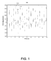
- FIG. 1 is a plot 100 showing an example ECG signal containing baseline variation. QRS complexes 110 can be identified by sharp spikes in the signal.
- a system in one aspect, includes a low pass-filter and a Savitzky-Golay filter.
- the low-pass filter receives and processes a first electrocardiogram signal.
- the filter removes at least the high frequency components of the first electrocardiogram signal.
- the Savitzky-Golay filter estimates a baseline variation of the first electrocardiogram signal from the filtered first electrocardiogram signal.
- a first electrocardiogram signal is received.
- the first electrocardiogram signal is processed in a filter to remove at least high frequency components of the first electrocardiogram signal.
- the filtered first electrocardiogram signal is processed in a Savitzky-Golay filter to estimate a baseline variation of the first electrocardiogram signal. The estimate of the baseline variation is provided.
- a delay module and a combination module can be included.
- the delay module can time shift the first electrocardiogram signal and the combination module can combine the time shifted first electrocardiogram signal and the baseline variation estimate to produce a second electrocardiogram signal with the baseline variation removed.
- a down-sampler and an up-sampler can be included. The down-sampler can reduce the sampling rate of the filtered first electrocardiogram signal and the up-sampler can increase the sampling rate of the baseline variation estimate.
- the low-pass filter can have a cutoff frequency of about 0.8 hertz.
- the first electrocardiogram signal can be sampled at a rate of about 500 hertz.
- the polynomial order or degree can be 2 or greater.
- the polynomial degree of the Savitzky-Golay filter can be between about 2 and about 10.
- the window size of the Savitzky-Golay filter can be less than about 750.
- the down-sampler can reduce the sample rate of the filtered first electrocardiogram signal by a factor of about 40 and the up-sampler can increase the sample rate of the baseline variation estimate by a factor of about 40.
- Articles of manufacture are also described that comprise computer executable instructions permanently stored (e.g., non-transitorily stored, etc.) on computer readable media, which, when executed by a computer, causes the computer to perform operations herein.
- computer systems are also described that may include a processor and a memory coupled to the processor. The memory may temporarily or permanently store one or more programs that cause the processor to perform one or more of the operations described herein.
- methods can be implemented by one or more data processors either within a single computing system or distributed among two or more computing systems.
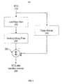
- FIG. 2 is a system diagram 200 of an ECG baseline variation removal system.
- An input ECG 210 is passed to a low-pass filter 220 to produce a filtered signal which has the high frequency components of the signal removed.
- the filtered signal is then passed to a Savitzky-Golay (SG) filter 230 which creates an estimate of the baseline variation of the input ECG 210.
- the SG filter is a filter that performs a local polynomial regression (of degree k) on a series of values (of at least k+1 points) to determine a smoothed value for each point.
- the ECG 210 can also be passed to a delay module 240 to be delayed in time (e.g., time shifted).
- the delay can compensate for any delays introduced by the low-pass filter 220 and SG filter 230 processing so that the input ECG 210 can, in a combination module 250, be combined with the estimated baseline variation produced by the SG filter 230 to produce an output ECG with the baseline variation removed 260.
- the low-pass filter can be, for example, a symmetric finite impulse response (FIR) filter with 100 taps and a cutoff frequency of 0.8 Hz.
- the low-pass FIR filter can remove the sharp QRS spikes (e.g., label 110 in FIG. 1 ) to improve the estimation of the baseline by the SG filter.
- the properties of the SG filter are determined by a window length and polynomial order or degree.
- the polynomial order or degree can be 2 or greater.
- the polynomial order or degree can be specified to be between about 2 and about 10 and when the input ECG is sampled at 500 Hertz, the window size can be about 750 samples or less.
- the window size can depend on the input ECG sample rate and be adjusted accordingly.
- the SG filter will introduce about 500 milliseconds of delay and the computational load will be equivalent to a FIR filter with 650 taps.
- FIG. 5 is a plot illustrating an example input ECG 510, a corresponding filtered signal 520 generated by the low-pass filter 220, a corresponding baseline variation 530 generated by the SG filter 230, and an output ECG 540 produced by the combination module 250.
- the duration of each signal shown is four seconds, and the average R-wave peak amplitude is 0.9mV.
- the example input ECG was collected from a patient and the baseline variation caused by respiration is evident.
- the QRS complexes have been removed from the filtered signal 520; however P and Q waves have not been removed.
- the baseline variation estimate 530 is a smooth signal that approximates the baseline variation evident in the example ECG input 510.
- the output ECG 540 is the example input ECG 510 with the baseline variation estimate 530 removed.
- FIG. 8 is a plot comparing the performance between the current subject matter, a high-pass filter method and a cubic spline method.
- An input ECG containing baseline variation which has been divided into one-heart-beat segments and time shifted to align their R-wave peaks and superimposed is presented at 810.
- a similar presentation is provided for ECGs that have been processed by a high-pass method 820, a cubic spline 830, and the current subject matter 840. It is evident from FIG. 8 that 840 presents an output ECG with the least variability and therefore is an improvement over the high-pass method, cubic spline method, or no processing at all. Additional tests were performed using an input ECG containing motion artifacts and an artificial 0.8Hz sinusoid and similar results were found.
- the delay can compensate for any delays introduced by the anti-aliasing low-pass filter 220, down- sampler 310, SG filter 230, and up-sampler 320 processing so that the input ECG 210 can, in combination module 250, be combined with the estimated baseline variation produced by the SG filter 230 to produce an output ECG with the baseline variation removed 340.
- FIG. 9 is a process flow diagram 900 illustrating a method a removing baseline variation from an ECG.
- the ECG is received.
- the ECG can have a sample rate of about 500 Hz.
- at 920 at least the high frequency components of the ECG are removed.
- the high frequency components can be frequencies above about 0.8 Hz.
- the processed ECG can be down-sampled.
- a baseline variation of the ECG can be estimated using a SG filter.
- the window length and polynomial degree of the SG filter can be selected based on the sample rate and baseline variation frequency.
- the estimate can be up-sampled.
- the estimate can be combined with a delayed ECG to produce an ECG with the baseline variation removed.
- the baseline variation estimate can be provided for further processing, transmission, storage or display.
- the SG filter determines a smoothed value for each data point in a series by performing a local polynomial fit in a window of predetermined length.
- the smoothed value is determined by a 0 only.
- a 0 is the inner product between the first row in ( B T B ) -1 B T and X.
- the matrix B is determined by the window size, 2 * N + 1 , and the polynomial order, M . Therefore, ( B T B ) -1 B T can be known once the window size and the polynomial order are known.
Landscapes
- Health & Medical Sciences (AREA)
- Life Sciences & Earth Sciences (AREA)
- Engineering & Computer Science (AREA)
- Surgery (AREA)
- General Health & Medical Sciences (AREA)
- Biophysics (AREA)
- Biomedical Technology (AREA)
- Heart & Thoracic Surgery (AREA)
- Medical Informatics (AREA)
- Molecular Biology (AREA)
- Physics & Mathematics (AREA)
- Animal Behavior & Ethology (AREA)
- Pathology (AREA)
- Public Health (AREA)
- Veterinary Medicine (AREA)
- Signal Processing (AREA)
- Artificial Intelligence (AREA)
- Computer Vision & Pattern Recognition (AREA)
- Physiology (AREA)
- Psychiatry (AREA)
- Measurement And Recording Of Electrical Phenomena And Electrical Characteristics Of The Living Body (AREA)
- Measuring Pulse, Heart Rate, Blood Pressure Or Blood Flow (AREA)
Description
- The subject matter described herein relates to techniques, methods, systems and articles for removing low frequency signal components from physiological signals such as electrocardiogram signals.
- An electrocardiogram (ECG) is a test that records the electrical activity of the heart as recorded by electrodes attached to the outer surface of the skin. Impedance variation between the recording electrode and the skin due to respiration or other body movement can cause baseline variations (also referred to as low frequency wander) in the ECG signal. Baseline variation is one type of noise in electrocardiogram signals.
FIG. 1 is aplot 100 showing an example ECG signal containing baseline variation.QRS complexes 110 can be identified by sharp spikes in the signal. - There are a variety of methods for baseline removal from the ECG, including high-pass filtering, adaptive filtering, wavelet transform, time-frequency analysis, curve fitting, etc. One approach, which is a special type of curve fitting, is the cubic spline method. A cubic spline is fitted on isoelectric reference points to estimate the baseline. The cubic spline method can be prone to error in the calculation of the isoelectric reference points, especially in the presence of noise.
- ECG baseline variation can comprise a low frequency signal within a range of 0 to 0.8Hz. According to the American Health Association (AHA), the frequency in the ECG signal is typically above 0.05Hz. Since the frequency band of the baseline noise overlaps with the ECG signal of interest, a simple high-pass filter is not sufficient for removing the baseline.
- Another approach to baseline removal is to use a high-pass filter. However, since the baseline is a type of in-band noise, a cut-off frequency cannot be set that would completely separate the ECG signal from the baseline. An approach adapting the cutoff frequency of the baseline filter to the heart rate was introduced by L. Lundstrom in 1995. According to Fourier theory, the frequency spectrum of a periodic signal is non-zero only on the base frequency and harmonics. This means that if the period is T, the lowest frequency is 1/T. An ideal ECG, which has constant heart rate and identical morphology for each heart beat, can be treated as a periodic signal, such that the lowest frequency is HeartRate/60 (Hz). If the cutoff frequency is set to this value, the low frequency noise can be removed. However, when the heart rate is low, this approach can not remove the baseline variation completely.
- In one aspect, a system includes a low pass-filter and a Savitzky-Golay filter. The low-pass filter receives and processes a first electrocardiogram signal. The filter removes at least the high frequency components of the first electrocardiogram signal. The Savitzky-Golay filter estimates a baseline variation of the first electrocardiogram signal from the filtered first electrocardiogram signal.
- In another aspect, a first electrocardiogram signal is received. The first electrocardiogram signal is processed in a filter to remove at least high frequency components of the first electrocardiogram signal. The filtered first electrocardiogram signal is processed in a Savitzky-Golay filter to estimate a baseline variation of the first electrocardiogram signal. The estimate of the baseline variation is provided.
- One or more of the following features can be included. For example, a delay module and a combination module can be included. The delay module can time shift the first electrocardiogram signal and the combination module can combine the time shifted first electrocardiogram signal and the baseline variation estimate to produce a second electrocardiogram signal with the baseline variation removed. A down-sampler and an up-sampler can be included. The down-sampler can reduce the sampling rate of the filtered first electrocardiogram signal and the up-sampler can increase the sampling rate of the baseline variation estimate.
- The low-pass filter can have a cutoff frequency of about 0.8 hertz. The first electrocardiogram signal can be sampled at a rate of about 500 hertz. The polynomial order or degree can be 2 or greater. The polynomial degree of the Savitzky-Golay filter can be between about 2 and about 10. The window size of the Savitzky-Golay filter can be less than about 750. The down-sampler can reduce the sample rate of the filtered first electrocardiogram signal by a factor of about 40 and the up-sampler can increase the sample rate of the baseline variation estimate by a factor of about 40. The up-sampler can perform linear interpolation. Providing the estimate of the baseline variation can include providing for further processing, storage, transmission, or display.
- Articles of manufacture are also described that comprise computer executable instructions permanently stored (e.g., non-transitorily stored, etc.) on computer readable media, which, when executed by a computer, causes the computer to perform operations herein. Similarly, computer systems are also described that may include a processor and a memory coupled to the processor. The memory may temporarily or permanently store one or more programs that cause the processor to perform one or more of the operations described herein. In addition, methods can be implemented by one or more data processors either within a single computing system or distributed among two or more computing systems.
- The subject matter described herein provides many advantages. For example, knowledge of heart rate is not required to perform baseline variation removal. Additionally, distortion can be reduced by using filters with linear phase and the baseline variation can be completely removed even when there is spectral overlap between the baseline and the ECG signal. Further, information about the isoelectric points of the ECG signal may not be required and the delay introduced by processing can be minimal. The current subject matter can be insensitive to noise, easier to implement and suited to implementation on ECG monitoring systems.
- The details of one or more variations of the subject matter described herein are set forth in the accompanying drawings and the description below. Other features and advantages of the subject matter described herein will be apparent from the description and drawings, and from the claims.
-
-
FIG. 1 is a plot showing an example ECG signal containing baseline variation; -
FIG. 2 is a system diagram of an ECG baseline variation removal system; -
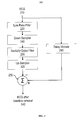
FIG. 3 is a system diagram of an ECG baseline variation removal system with down and up samplers; -
FIG. 4 is a plot of a time series illustrating a window used for a Savitzky-Golay polynomial fit; -
FIG. 5 is a plot illustrating an example ECG, and signals measured from the output of different components of a system in accordance with the current subject matter; -
FIG. 6 is a plot showing a comparison of an input ECG and an output ECG of a system in accordance with the current subject matter; -
FIG. 7 is a plot illustrating the limited distortion introduced by the current subject matter; -
FIG 8 is a plot showing a performance comparison between the current subject matter, a high-pass filter method and a cubic spline method; and -
FIG. 9 is a process flow diagram illustrating a method a removing baseline variation from an ECG. - Like reference symbols in the various drawings indicate like elements.
-
FIG. 2 is a system diagram 200 of an ECG baseline variation removal system. Aninput ECG 210 is passed to a low-pass filter 220 to produce a filtered signal which has the high frequency components of the signal removed. The filtered signal is then passed to a Savitzky-Golay (SG)filter 230 which creates an estimate of the baseline variation of theinput ECG 210. The SG filter is a filter that performs a local polynomial regression (of degree k) on a series of values (of at least k+1 points) to determine a smoothed value for each point. One advantage of this approach is that it preserves features of the distribution such as relative maxima, minima and width, which are usually 'flattened' by other adjacent averaging techniques (like moving averages, for example). TheECG 210 can also be passed to adelay module 240 to be delayed in time (e.g., time shifted). The delay can compensate for any delays introduced by the low-pass filter 220 andSG filter 230 processing so that theinput ECG 210 can, in acombination module 250, be combined with the estimated baseline variation produced by theSG filter 230 to produce an output ECG with the baseline variation removed 260. - The low-pass filter can be, for example, a symmetric finite impulse response (FIR) filter with 100 taps and a cutoff frequency of 0.8 Hz. The low-pass FIR filter can remove the sharp QRS spikes (e.g.,
label 110 inFIG. 1 ) to improve the estimation of the baseline by the SG filter. The properties of the SG filter are determined by a window length and polynomial order or degree. The polynomial order or degree can be 2 or greater. For example, the polynomial order or degree can be specified to be between about 2 and about 10 and when the input ECG is sampled at 500 Hertz, the window size can be about 750 samples or less. The window size can depend on the input ECG sample rate and be adjusted accordingly. The longer the window and the lower the polynomial order, the lower the cutoff frequency. For example, if the ECG signal is sampled at 500 Hz, a window size of 2*250+1 and a polynomial order of 2 is specified, then the SG filter will introduce about 500 milliseconds of delay and the computational load will be equivalent to a FIR filter with 650 taps. - Referring now to
FIG. 5 with further reference toFIG. 2 .FIG. 5 is a plot illustrating anexample input ECG 510, a corresponding filteredsignal 520 generated by the low-pass filter 220, acorresponding baseline variation 530 generated by theSG filter 230, and anoutput ECG 540 produced by thecombination module 250. The duration of each signal shown is four seconds, and the average R-wave peak amplitude is 0.9mV. The example input ECG was collected from a patient and the baseline variation caused by respiration is evident. The QRS complexes have been removed from the filteredsignal 520; however P and Q waves have not been removed. Thebaseline variation estimate 530 is a smooth signal that approximates the baseline variation evident in theexample ECG input 510. Theoutput ECG 540 is theexample input ECG 510 with thebaseline variation estimate 530 removed. -
FIG. 6 is a plot showing forty-nine beats of theexample input ECG 510 andoutput ECG 540 that have been divided into one-heart-beat segments and time shifted to align their R-wave peaks. At 610, each segment of the example ECG has been superimposed and a high variation between heart beat segments is evident. At 620, each segment of the output signal has been superimposed and a low variation between heart beat segments is evident. At 630 and 640, the average of the segments is shown for the example input ECG and output ECG respectively. Since the baseline variation noise is not synchronized to the heart rate, the average of the example input ECG segments can be considered baseline free (i.e., the variation averages to zero). At 650, the difference between 630 and 640 is shown. The difference is close or almost zero (i.e. a straight line), showing that the baseline variation has been successfully removed. -
FIG. 7 is a plot illustrating the limited distortion introduced by the current subject matter. To further characterize performance, an ECG without any baseline variation is used as input.FIG. 7 shows an ECG without baseline variation and the ECG after baseline variation removal. Both have been divided into multiple one-heart-beat segments and time shifted to align their R-wave peaks. At 710 each segment of the input ECG is superimposed and 730 shows their average. At 720, each segment of the output ECG is superimposed and 740 shows their average. 750 is the difference between 730 and 740. The difference is near zero and therefore indicates that the distortion provided by the current subject matter is minimal. -
FIG. 8 is a plot comparing the performance between the current subject matter, a high-pass filter method and a cubic spline method. An input ECG containing baseline variation which has been divided into one-heart-beat segments and time shifted to align their R-wave peaks and superimposed is presented at 810. A similar presentation is provided for ECGs that have been processed by a high-pass method 820, acubic spline 830, and thecurrent subject matter 840. It is evident fromFIG. 8 that 840 presents an output ECG with the least variability and therefore is an improvement over the high-pass method, cubic spline method, or no processing at all. Additional tests were performed using an input ECG containing motion artifacts and an artificial 0.8Hz sinusoid and similar results were found. -
FIG. 3 is a system diagram 300 of an ECG baseline variation removal system. Theinput ECG 210 is passed to an anti-aliasing low-pass filter 220. The anti-aliasing filter low-pass filter can be an infinite impulse response (IIR) filter. The filtered ECG is down-sampled by down-sampler 310 and passed to SG filter 330. The SG filter creates an estimate of the baseline variation of theinput ECG 210. The estimate is up-sampled by up-sampler 320. The up-sampling can be, for example, a linear interpolation. The ECG signal 210 can also be delayed bydelay module 240. The delay can compensate for any delays introduced by the anti-aliasing low-pass filter 220, down-sampler 310,SG filter 230, and up-sampler 320 processing so that theinput ECG 210 can, incombination module 250, be combined with the estimated baseline variation produced by theSG filter 230 to produce an output ECG with the baseline variation removed 340. - If the
input ECG 210 has a sampling rate of 500 Hz, is down-sampled by a factor of 40 to a 12.5 Hz rate, and assuming the SG filter window size is 2*6+1 and the polynomial order is 2, then the delay will be 540 milliseconds and the computational load will be equivalent to a FIR filter with 17 taps. The baseline variation estimate can be up-sampled by a factor of 40 to combine the input ECG. -
FIG. 9 is a process flow diagram 900 illustrating a method a removing baseline variation from an ECG. At 910, the ECG is received. The ECG can have a sample rate of about 500 Hz. At 920, at least the high frequency components of the ECG are removed. The high frequency components can be frequencies above about 0.8 Hz. Optionally, at 930, the processed ECG can be down-sampled. At 940, a baseline variation of the ECG can be estimated using a SG filter. The window length and polynomial degree of the SG filter can be selected based on the sample rate and baseline variation frequency. Optionally, at 950, the estimate can be up-sampled. Optionally, at 960, the estimate can be combined with a delayed ECG to produce an ECG with the baseline variation removed. Additionally, the baseline variation estimate can be provided for further processing, transmission, storage or display. - The SG filter determines a smoothed value for each data point in a series by performing a local polynomial fit in a window of predetermined length. The polynomial function can be defined as:

- Mis the polynomial order, n is a independent variable, and a0 , a1, ..., aM are polynomial coefficients.
FIG. 4 is aplot 400 of atime series 410. x(i) represents thetime series 410 at any arbitrary sample i. The window length can be 2 *N+1, and then a least squares polynomial fit centered at the ith sample can be expressed as a matrix equation BA=X, namely,
- A least-squares solution can be expressed as A = (BTB)-1 BT X and the smoothed value of the ith sample, denoted as y(i), can then be calculated as y(i)=a 0 n 0 + a 1 n 1 + a 2 n 2 +···+ aMnM | n=0 = a 0.
- From the above-mentioned equation, the smoothed value is determined by a0 only. a0 is the inner product between the first row in (BTB)-1 BT and X. The matrix B is determined by the window size, 2 *N+1, and the polynomial order, M. Therefore, (BTB)-1 BT can be known once the window size and the polynomial order are known. Let the first row in (BTB)-1 BT be [h(-N) ··· h(-1) h(0) h(1) ··· h(N)], then y(i) can be written as

- As the first row in (BTB)-1 BT is symmetric with respect to the central point, n=0, y(i) can be written as

- The right hand side of the above equation is the convolution between h(n) and x(n). Therefore, the output of the SG filter can be expressed as the input filtered by a FIR filter that is determined by the window size and the polynomial order. Further, the SG filter has a linear phase response and a delay of half the window size.
- Various implementations of the subject matter described herein may be realized in digital electronic circuitry, integrated circuitry, specially designed ASICs (application specific integrated circuits), computer hardware, firmware, software, and/or combinations thereof. These various implementations may include implementation in one or more computer programs that are executable and/or interpretable on a programmable system including at least one programmable processor, which may be special or general purpose, coupled to receive data and instructions from, and to transmit data and instructions to, a storage system, at least one input device, and at least one output device.
- These computer programs (also known as programs, software, software applications or code) include machine instructions for a programmable processor, and may be implemented in a high-level procedural and/or object-oriented programming language, and/or in assembly/machine language. As used herein, the term "machine-readable medium" refers to any computer program product, apparatus and/or device (e.g., magnetic discs, optical disks, memory, Programmable Logic Devices (PLDs)) used to provide machine instructions and/or data to a programmable processor, including a machine-readable medium that receives machine instructions as a machine-readable signal. The term "machine-readable signal" refers to any signal used to provide machine instructions and/or data to a programmable processor.
- Although a few variations have been described in detail above, other modifications are possible. For example, the logic flow depicted in the accompanying figures and described herein do not require the particular order shown, or sequential order, to achieve desirable results. Other embodiments are within the scope of the following claims.
Claims (18)
- A system comprising:a low-pass filter to receive and process a first electrocardiogram signal, the filter removing at least high frequency components of the first electrocardiogram signal; anda Savitzky-Golay filter to estimate a baseline variation of the first electrocardiogram signal from the filtered first electrocardiogram signal.
- The system of claim 1, further comprising:a delay module to time shift the first electrocardiogram signal; anda combination module configured to combine the time shifted first electrocardiogram signal and the baseline variation estimate thereby producing a second electrocardiogram signal with the baseline variation removed.
- The system of claim 2, further comprising:a down-sampler configured to reduce the sampling rate of the filtered first electrocardiogram signal; andan up-sampler configured to increase the sampling rate of the baseline variation estimate.
- The system as in any one of the preceding claims, wherein the low-pass filter has a cutoff frequency of about 0.8 hertz.
- The system as in any one of the preceding claims, wherein the first electrocardiogram signal is sampled at a rate of about 500 hertz.
- The system as in any one of the preceding claims, wherein the polynomial degree of the Savitzky-Golay filter is between about 2 and about 10.
- The system as in any one of the preceding claims, wherein the window size of the Savitzky-Golay filter is less than about 750.
- The system as in any one of claims 3-7, wherein the down-sampler reduces the sample rate of the filtered first electrocardiogram signal by a factor of about 40 and the up-sampler increases the sample rate of the baseline variation estimate by a factor of about 40.
- The system as in anyone of claims 3-8, wherein the up-sampler performs linear interpolation.
- A method comprising:receiving a first electrocardiogram signal;processing the first electrocardiogram signal in a filter thereby removing at least high frequency components of the first electrocardiogram signal;processing the filtered first electrocardiogram signal in a Savitzky-Golay filter to estimate a baseline variation of the first electrocardiogram signal; andproviding the estimate of the baseline variation.
- The method of claim 10, further comprising:delaying the first electrocardiogram signal; andcombining the delayed first electrocardiogram signal and the baseline variation estimate thereby producing a second electrocardiogram signal with the baseline variation removed.
- The method of claim 11, further comprising:down-sampling the filtered first electrocardiogram signal; andup-sampling the baseline variation estimate.
- The method as in any one of claims 10-12,
wherein at least the frequencies above about 0.8 hertz are removed from the received first electrocardiogram signal; and/or
wherein the received first electrocardiogram signal has been sampled at a rate of about 500 hertz; and/or
wherein the polynomial degree of the Savitzky-Golay filter is between about 2 and about 10; and/or
wherein the window size of the Savitzky-Golay filter is less than about 750. - The method as in any one of claims 10-13, wherein providing the estimate of the baseline variation includes providing for further processing, storage, transmission, or display.
- The method as in any one of claims 10-14, further comprising selecting a window length of the Savitzky-Golay based on a sample rate and a baseline variation frequency.
- The method as in any one of claims 10-15, further comprising selecting a polynomial degree of the Savitzky-Golay filter based on a sample rate and a baseline variation frequency.
- A system comprising:at least one data processor;memory storing instructions which, when executed by the at least one data processor, causes the at least one data processor to perform a method as in any of claims 10-16.
- A computer program product storing instructions, which when executed by at least one programmable processor of at least one computing system, implement a method according to any of claims 10-16.
Applications Claiming Priority (2)
| Application Number | Priority Date | Filing Date | Title |
|---|---|---|---|
| US201161532327P | 2011-09-08 | 2011-09-08 | |
| PCT/US2012/041386 WO2013036307A1 (en) | 2011-09-08 | 2012-06-07 | Electrocardiogram baseline removal |
Publications (2)
| Publication Number | Publication Date |
|---|---|
| EP2753235A1 EP2753235A1 (en) | 2014-07-16 |
| EP2753235B1 true EP2753235B1 (en) | 2015-08-12 |
Family
ID=46276057
Family Applications (1)
| Application Number | Title | Priority Date | Filing Date |
|---|---|---|---|
| EP12727565.9A Active EP2753235B1 (en) | 2011-09-08 | 2012-06-07 | Electrocardiogram baseline removal |
Country Status (4)
| Country | Link |
|---|---|
| US (1) | US9480411B2 (en) |
| EP (1) | EP2753235B1 (en) |
| CN (1) | CN103857327B (en) |
| WO (1) | WO2013036307A1 (en) |
Cited By (1)
| Publication number | Priority date | Publication date | Assignee | Title |
|---|---|---|---|---|
| CN108024750A (en) * | 2015-08-25 | 2018-05-11 | 皇家飞利浦有限公司 | Evaluation of High/Low Frequency Signal Quality of ECG Lead Signals |
Families Citing this family (12)
| Publication number | Priority date | Publication date | Assignee | Title |
|---|---|---|---|---|
| US8509882B2 (en) | 2010-06-08 | 2013-08-13 | Alivecor, Inc. | Heart monitoring system usable with a smartphone or computer |
| US9351654B2 (en) | 2010-06-08 | 2016-05-31 | Alivecor, Inc. | Two electrode apparatus and methods for twelve lead ECG |
| WO2014074913A1 (en) | 2012-11-08 | 2014-05-15 | Alivecor, Inc. | Electrocardiogram signal detection |
| US9220430B2 (en) | 2013-01-07 | 2015-12-29 | Alivecor, Inc. | Methods and systems for electrode placement |
| WO2014145927A1 (en) | 2013-03-15 | 2014-09-18 | Alivecor, Inc. | Systems and methods for processing and analyzing medical data |
| US9247911B2 (en) | 2013-07-10 | 2016-02-02 | Alivecor, Inc. | Devices and methods for real-time denoising of electrocardiograms |
| WO2015089484A1 (en) | 2013-12-12 | 2015-06-18 | Alivecor, Inc. | Methods and systems for arrhythmia tracking and scoring |
| WO2016183515A1 (en) | 2015-05-13 | 2016-11-17 | Alivecor, Inc. | Discordance monitoring |
| US9872652B2 (en) * | 2015-06-09 | 2018-01-23 | University Of Connecticut | Method and apparatus for heart rate monitoring using an electrocardiogram sensor |
| CN110263520A (en) * | 2019-06-21 | 2019-09-20 | 深圳市奋达智能技术有限公司 | A kind of auth method and system |
| TWI742844B (en) * | 2020-09-10 | 2021-10-11 | 廣達電腦股份有限公司 | Ecg noise filtering device |
| CN112966566B (en) * | 2021-02-05 | 2023-07-07 | 武汉中旗生物医疗电子有限公司 | Electrocardiosignal baseline filtering method and device |
Family Cites Families (12)
| Publication number | Priority date | Publication date | Assignee | Title |
|---|---|---|---|---|
| CN1084044A (en) * | 1992-09-11 | 1994-03-23 | 中日友好医院 | Three-D high frequency electrocardio signal analytical system and method |
| US5762068A (en) * | 1995-11-27 | 1998-06-09 | Quinton Instrument Company | ECG filter and slew rate limiter for filtering an ECG signal |
| US5913308A (en) * | 1996-12-19 | 1999-06-22 | Hewlett-Packard Company | Apparatus and method for determining respiratory effort from muscle tremor information in ECG signals |
| US6535570B2 (en) | 1999-06-17 | 2003-03-18 | Her Majesty The Queen In Right Of Canada, As Represented By The Minister Of National Defence Of Her Majesty's Canadian Government | Method for tracing organ motion and removing artifacts for computed tomography imaging systems |
| US6839655B2 (en) * | 2001-05-25 | 2005-01-04 | University Of Chicago | System for monitoring non-coincident, nonstationary process signals |
| CN101083938B (en) * | 2004-12-22 | 2010-05-12 | 大日本住友制药株式会社 | electrocardiogram waveform correction display device and electrocardiogram waveform correction display method |
| EP1885240B1 (en) | 2005-05-17 | 2016-02-03 | Bio Signal Analysis ltd. | Analysis of electrocardiogram signals |
| JP2010504793A (en) * | 2006-09-29 | 2010-02-18 | コーニンクレッカ フィリップス エレクトロニクス エヌ ヴィ | Template synthesis for ECG / PPG based biometrics |
| KR100860875B1 (en) * | 2007-03-21 | 2008-09-29 | (주)락싸 | Method of extracting response intensity and reaction time from frequency domain response pattern of stimulus induction |
| CN101361650B (en) * | 2007-08-07 | 2012-07-04 | 深圳市理邦精密仪器股份有限公司 | Zero phase implementation method of IIR filter and zero phase IIR fiter |
| JP5911726B2 (en) | 2008-11-10 | 2016-04-27 | カーディオインサイト テクノロジーズ インコーポレイテッド | Visualization of electrophysiological data |
| US20110066042A1 (en) * | 2009-09-15 | 2011-03-17 | Texas Instruments Incorporated | Estimation of blood flow and hemodynamic parameters from a single chest-worn sensor, and other circuits, devices and processes |
-
2012
- 2012-06-07 US US14/343,359 patent/US9480411B2/en active Active
- 2012-06-07 EP EP12727565.9A patent/EP2753235B1/en active Active
- 2012-06-07 CN CN201280049396.8A patent/CN103857327B/en active Active
- 2012-06-07 WO PCT/US2012/041386 patent/WO2013036307A1/en not_active Ceased
Cited By (1)
| Publication number | Priority date | Publication date | Assignee | Title |
|---|---|---|---|---|
| CN108024750A (en) * | 2015-08-25 | 2018-05-11 | 皇家飞利浦有限公司 | Evaluation of High/Low Frequency Signal Quality of ECG Lead Signals |
Also Published As
| Publication number | Publication date |
|---|---|
| US20140222350A1 (en) | 2014-08-07 |
| CN103857327B (en) | 2017-03-29 |
| EP2753235A1 (en) | 2014-07-16 |
| WO2013036307A1 (en) | 2013-03-14 |
| US9480411B2 (en) | 2016-11-01 |
| CN103857327A (en) | 2014-06-11 |
Similar Documents
| Publication | Publication Date | Title |
|---|---|---|
| US9480411B2 (en) | Electrocardiogram baseline removal | |
| Kumar et al. | Removal of noise from electrocardiogram using digital FIR and IIR filters with various methods | |
| Zhao et al. | A new method for removal of baseline wander and power line interference in ECG signals | |
| Cyrill et al. | Adaptive comb filters for quasiperiodic physiologic signals | |
| US20170215756A1 (en) | Devices and methods for real-time denoising of electrocardiograms | |
| EP2978368B1 (en) | Apparatus and method for ecg motion artifact removal | |
| WO2005101229A1 (en) | Nonlinear method and apparatus for electrocardiogram pacemaker signal filtering | |
| Rashmi et al. | ECG denoising using wavelet transform and filters | |
| Kaur et al. | An efficient R-peak detection using Riesz fractional-order digital differentiator | |
| Hargittai | Efficient and fast ECG baseline wander reduction without distortion of important clinical information | |
| Bhogeshwar et al. | To verify and compare denoising of ECG signal using various denoising algorithms of IIR and FIR filters | |
| Zheng et al. | Low-distortion baseline removal algorithm for electrocardiogram signals | |
| Krishnamurthy et al. | Comparison of various filtering techniques used for removing high frequency noise in ECG signal | |
| Fasano et al. | Baseline wander removal in ECG and AHA recommendations | |
| Muppalla et al. | Performance evaluation of different denoising techniques for physiological signals | |
| Chandra et al. | A computationally efficient approach for ECG signal denoising and data compression | |
| Chandramouleeswaran et al. | Wavelet diagnosis of ECG signals with kaiser based noise diminution | |
| KR101833254B1 (en) | Device for processing bio-signal | |
| Wadhwani et al. | Filtration of ECG signal by using various filter | |
| Zhao et al. | Baseline wander removal of ECG signals using empirical mode decomposition and adaptive filter | |
| Ziad et al. | Analysis of Noise Cancellation Techniques for Enhancing the ECG Signals | |
| CN111772617B (en) | ECG display filtering method, device and storage medium | |
| Daqrouq et al. | The discrete wavelet transform based electrocardiographic baseline wander reduction method for better signal diagnosis | |
| Salem et al. | Effectiveness of Advanced ECG Noise Reduction Filters | |
| Dewanta et al. | Performance Evaluation of IIR and FIR Filters Using Windowing Techniques for EEG |
Legal Events
| Date | Code | Title | Description |
|---|---|---|---|
| PUAI | Public reference made under article 153(3) epc to a published international application that has entered the european phase |
Free format text: ORIGINAL CODE: 0009012 |
|
| 17P | Request for examination filed |
Effective date: 20140327 |
|
| AK | Designated contracting states |
Kind code of ref document: A1 Designated state(s): AL AT BE BG CH CY CZ DE DK EE ES FI FR GB GR HR HU IE IS IT LI LT LU LV MC MK MT NL NO PL PT RO RS SE SI SK SM TR |
|
| DAX | Request for extension of the european patent (deleted) | ||
| GRAP | Despatch of communication of intention to grant a patent |
Free format text: ORIGINAL CODE: EPIDOSNIGR1 |
|
| RAP1 | Party data changed (applicant data changed or rights of an application transferred) |
Owner name: DRAEGER MEDICAL GMBH |
|
| INTG | Intention to grant announced |
Effective date: 20150327 |
|
| GRAS | Grant fee paid |
Free format text: ORIGINAL CODE: EPIDOSNIGR3 |
|
| GRAA | (expected) grant |
Free format text: ORIGINAL CODE: 0009210 |
|
| AK | Designated contracting states |
Kind code of ref document: B1 Designated state(s): AL AT BE BG CH CY CZ DE DK EE ES FI FR GB GR HR HU IE IS IT LI LT LU LV MC MK MT NL NO PL PT RO RS SE SI SK SM TR |
|
| REG | Reference to a national code |
Ref country code: GB Ref legal event code: FG4D |
|
| REG | Reference to a national code |
Ref country code: CH Ref legal event code: EP |
|
| REG | Reference to a national code |
Ref country code: AT Ref legal event code: REF Ref document number: 741380 Country of ref document: AT Kind code of ref document: T Effective date: 20150815 |
|
| REG | Reference to a national code |
Ref country code: IE Ref legal event code: FG4D |
|
| REG | Reference to a national code |
Ref country code: DE Ref legal event code: R082 Ref document number: 602012009566 Country of ref document: DE Representative=s name: HASELTINE LAKE LLP, DE Ref country code: DE Ref legal event code: R081 Ref document number: 602012009566 Country of ref document: DE Owner name: DRAEGERWERK AG & CO. KGAA, DE Free format text: FORMER OWNER: DRAEGER MEDICAL GMBH, 23558 LUEBECK, DE |
|
| REG | Reference to a national code |
Ref country code: DE Ref legal event code: R096 Ref document number: 602012009566 Country of ref document: DE |
|
| REG | Reference to a national code |
Ref country code: LT Ref legal event code: MG4D |
|
| REG | Reference to a national code |
Ref country code: AT Ref legal event code: MK05 Ref document number: 741380 Country of ref document: AT Kind code of ref document: T Effective date: 20150812 |
|
| REG | Reference to a national code |
Ref country code: NL Ref legal event code: MP Effective date: 20150812 |
|
| PG25 | Lapsed in a contracting state [announced via postgrant information from national office to epo] |
Ref country code: FI Free format text: LAPSE BECAUSE OF FAILURE TO SUBMIT A TRANSLATION OF THE DESCRIPTION OR TO PAY THE FEE WITHIN THE PRESCRIBED TIME-LIMIT Effective date: 20150812 Ref country code: NO Free format text: LAPSE BECAUSE OF FAILURE TO SUBMIT A TRANSLATION OF THE DESCRIPTION OR TO PAY THE FEE WITHIN THE PRESCRIBED TIME-LIMIT Effective date: 20151112 Ref country code: GR Free format text: LAPSE BECAUSE OF FAILURE TO SUBMIT A TRANSLATION OF THE DESCRIPTION OR TO PAY THE FEE WITHIN THE PRESCRIBED TIME-LIMIT Effective date: 20151113 Ref country code: LT Free format text: LAPSE BECAUSE OF FAILURE TO SUBMIT A TRANSLATION OF THE DESCRIPTION OR TO PAY THE FEE WITHIN THE PRESCRIBED TIME-LIMIT Effective date: 20150812 Ref country code: LV Free format text: LAPSE BECAUSE OF FAILURE TO SUBMIT A TRANSLATION OF THE DESCRIPTION OR TO PAY THE FEE WITHIN THE PRESCRIBED TIME-LIMIT Effective date: 20150812 |
|
| PG25 | Lapsed in a contracting state [announced via postgrant information from national office to epo] |
Ref country code: IS Free format text: LAPSE BECAUSE OF FAILURE TO SUBMIT A TRANSLATION OF THE DESCRIPTION OR TO PAY THE FEE WITHIN THE PRESCRIBED TIME-LIMIT Effective date: 20151212 Ref country code: ES Free format text: LAPSE BECAUSE OF FAILURE TO SUBMIT A TRANSLATION OF THE DESCRIPTION OR TO PAY THE FEE WITHIN THE PRESCRIBED TIME-LIMIT Effective date: 20150812 Ref country code: SE Free format text: LAPSE BECAUSE OF FAILURE TO SUBMIT A TRANSLATION OF THE DESCRIPTION OR TO PAY THE FEE WITHIN THE PRESCRIBED TIME-LIMIT Effective date: 20150812 Ref country code: PL Free format text: LAPSE BECAUSE OF FAILURE TO SUBMIT A TRANSLATION OF THE DESCRIPTION OR TO PAY THE FEE WITHIN THE PRESCRIBED TIME-LIMIT Effective date: 20150812 Ref country code: RS Free format text: LAPSE BECAUSE OF FAILURE TO SUBMIT A TRANSLATION OF THE DESCRIPTION OR TO PAY THE FEE WITHIN THE PRESCRIBED TIME-LIMIT Effective date: 20150812 Ref country code: AT Free format text: LAPSE BECAUSE OF FAILURE TO SUBMIT A TRANSLATION OF THE DESCRIPTION OR TO PAY THE FEE WITHIN THE PRESCRIBED TIME-LIMIT Effective date: 20150812 Ref country code: PT Free format text: LAPSE BECAUSE OF FAILURE TO SUBMIT A TRANSLATION OF THE DESCRIPTION OR TO PAY THE FEE WITHIN THE PRESCRIBED TIME-LIMIT Effective date: 20151214 Ref country code: HR Free format text: LAPSE BECAUSE OF FAILURE TO SUBMIT A TRANSLATION OF THE DESCRIPTION OR TO PAY THE FEE WITHIN THE PRESCRIBED TIME-LIMIT Effective date: 20150812 |
|
| PG25 | Lapsed in a contracting state [announced via postgrant information from national office to epo] |
Ref country code: NL Free format text: LAPSE BECAUSE OF FAILURE TO SUBMIT A TRANSLATION OF THE DESCRIPTION OR TO PAY THE FEE WITHIN THE PRESCRIBED TIME-LIMIT Effective date: 20150812 |
|
| PG25 | Lapsed in a contracting state [announced via postgrant information from national office to epo] |
Ref country code: EE Free format text: LAPSE BECAUSE OF FAILURE TO SUBMIT A TRANSLATION OF THE DESCRIPTION OR TO PAY THE FEE WITHIN THE PRESCRIBED TIME-LIMIT Effective date: 20150812 Ref country code: SK Free format text: LAPSE BECAUSE OF FAILURE TO SUBMIT A TRANSLATION OF THE DESCRIPTION OR TO PAY THE FEE WITHIN THE PRESCRIBED TIME-LIMIT Effective date: 20150812 Ref country code: IT Free format text: LAPSE BECAUSE OF FAILURE TO SUBMIT A TRANSLATION OF THE DESCRIPTION OR TO PAY THE FEE WITHIN THE PRESCRIBED TIME-LIMIT Effective date: 20150812 Ref country code: DK Free format text: LAPSE BECAUSE OF FAILURE TO SUBMIT A TRANSLATION OF THE DESCRIPTION OR TO PAY THE FEE WITHIN THE PRESCRIBED TIME-LIMIT Effective date: 20150812 Ref country code: CZ Free format text: LAPSE BECAUSE OF FAILURE TO SUBMIT A TRANSLATION OF THE DESCRIPTION OR TO PAY THE FEE WITHIN THE PRESCRIBED TIME-LIMIT Effective date: 20150812 |
|
| REG | Reference to a national code |
Ref country code: DE Ref legal event code: R097 Ref document number: 602012009566 Country of ref document: DE |
|
| PG25 | Lapsed in a contracting state [announced via postgrant information from national office to epo] |
Ref country code: RO Free format text: LAPSE BECAUSE OF FAILURE TO SUBMIT A TRANSLATION OF THE DESCRIPTION OR TO PAY THE FEE WITHIN THE PRESCRIBED TIME-LIMIT Effective date: 20150812 |
|
| PLBE | No opposition filed within time limit |
Free format text: ORIGINAL CODE: 0009261 |
|
| STAA | Information on the status of an ep patent application or granted ep patent |
Free format text: STATUS: NO OPPOSITION FILED WITHIN TIME LIMIT |
|
| REG | Reference to a national code |
Ref country code: FR Ref legal event code: PLFP Year of fee payment: 5 |
|
| 26N | No opposition filed |
Effective date: 20160513 |
|
| PG25 | Lapsed in a contracting state [announced via postgrant information from national office to epo] |
Ref country code: SI Free format text: LAPSE BECAUSE OF FAILURE TO SUBMIT A TRANSLATION OF THE DESCRIPTION OR TO PAY THE FEE WITHIN THE PRESCRIBED TIME-LIMIT Effective date: 20150812 |
|
| PG25 | Lapsed in a contracting state [announced via postgrant information from national office to epo] |
Ref country code: BE Free format text: LAPSE BECAUSE OF FAILURE TO SUBMIT A TRANSLATION OF THE DESCRIPTION OR TO PAY THE FEE WITHIN THE PRESCRIBED TIME-LIMIT Effective date: 20150812 |
|
| PG25 | Lapsed in a contracting state [announced via postgrant information from national office to epo] |
Ref country code: MC Free format text: LAPSE BECAUSE OF FAILURE TO SUBMIT A TRANSLATION OF THE DESCRIPTION OR TO PAY THE FEE WITHIN THE PRESCRIBED TIME-LIMIT Effective date: 20150812 |
|
| REG | Reference to a national code |
Ref country code: CH Ref legal event code: PL |
|
| REG | Reference to a national code |
Ref country code: IE Ref legal event code: MM4A |
|
| PG25 | Lapsed in a contracting state [announced via postgrant information from national office to epo] |
Ref country code: LI Free format text: LAPSE BECAUSE OF NON-PAYMENT OF DUE FEES Effective date: 20160630 Ref country code: CH Free format text: LAPSE BECAUSE OF NON-PAYMENT OF DUE FEES Effective date: 20160630 |
|
| PG25 | Lapsed in a contracting state [announced via postgrant information from national office to epo] |
Ref country code: IE Free format text: LAPSE BECAUSE OF NON-PAYMENT OF DUE FEES Effective date: 20160607 |
|
| REG | Reference to a national code |
Ref country code: FR Ref legal event code: PLFP Year of fee payment: 6 |
|
| REG | Reference to a national code |
Ref country code: GB Ref legal event code: 732E Free format text: REGISTERED BETWEEN 20170720 AND 20170726 |
|
| REG | Reference to a national code |
Ref country code: FR Ref legal event code: TP Owner name: DRAGERWERK AG & CO. KGAA, DE Effective date: 20171120 |
|
| PG25 | Lapsed in a contracting state [announced via postgrant information from national office to epo] |
Ref country code: HU Free format text: LAPSE BECAUSE OF FAILURE TO SUBMIT A TRANSLATION OF THE DESCRIPTION OR TO PAY THE FEE WITHIN THE PRESCRIBED TIME-LIMIT; INVALID AB INITIO Effective date: 20120607 Ref country code: SM Free format text: LAPSE BECAUSE OF FAILURE TO SUBMIT A TRANSLATION OF THE DESCRIPTION OR TO PAY THE FEE WITHIN THE PRESCRIBED TIME-LIMIT Effective date: 20150812 |
|
| REG | Reference to a national code |
Ref country code: FR Ref legal event code: PLFP Year of fee payment: 7 |
|
| PG25 | Lapsed in a contracting state [announced via postgrant information from national office to epo] |
Ref country code: LU Free format text: LAPSE BECAUSE OF NON-PAYMENT OF DUE FEES Effective date: 20160607 Ref country code: CY Free format text: LAPSE BECAUSE OF FAILURE TO SUBMIT A TRANSLATION OF THE DESCRIPTION OR TO PAY THE FEE WITHIN THE PRESCRIBED TIME-LIMIT Effective date: 20150812 Ref country code: MK Free format text: LAPSE BECAUSE OF FAILURE TO SUBMIT A TRANSLATION OF THE DESCRIPTION OR TO PAY THE FEE WITHIN THE PRESCRIBED TIME-LIMIT Effective date: 20150812 Ref country code: MT Free format text: LAPSE BECAUSE OF NON-PAYMENT OF DUE FEES Effective date: 20160630 |
|
| PG25 | Lapsed in a contracting state [announced via postgrant information from national office to epo] |
Ref country code: BG Free format text: LAPSE BECAUSE OF FAILURE TO SUBMIT A TRANSLATION OF THE DESCRIPTION OR TO PAY THE FEE WITHIN THE PRESCRIBED TIME-LIMIT Effective date: 20150812 |
|
| PG25 | Lapsed in a contracting state [announced via postgrant information from national office to epo] |
Ref country code: TR Free format text: LAPSE BECAUSE OF FAILURE TO SUBMIT A TRANSLATION OF THE DESCRIPTION OR TO PAY THE FEE WITHIN THE PRESCRIBED TIME-LIMIT Effective date: 20150812 Ref country code: AL Free format text: LAPSE BECAUSE OF FAILURE TO SUBMIT A TRANSLATION OF THE DESCRIPTION OR TO PAY THE FEE WITHIN THE PRESCRIBED TIME-LIMIT Effective date: 20150812 |
|
| PGFP | Annual fee paid to national office [announced via postgrant information from national office to epo] |
Ref country code: FR Payment date: 20190625 Year of fee payment: 8 |
|
| PGFP | Annual fee paid to national office [announced via postgrant information from national office to epo] |
Ref country code: GB Payment date: 20190627 Year of fee payment: 8 |
|
| REG | Reference to a national code |
Ref country code: DE Ref legal event code: R079 Ref document number: 602012009566 Country of ref document: DE Free format text: PREVIOUS MAIN CLASS: A61B0005040000 Ipc: A61B0005240000 |
|
| REG | Reference to a national code |
Ref country code: DE Ref legal event code: R082 Ref document number: 602012009566 Country of ref document: DE Representative=s name: HL KEMPNER PATENTANWAELTE, SOLICITORS (ENGLAND, DE Ref country code: DE Ref legal event code: R082 Ref document number: 602012009566 Country of ref document: DE Representative=s name: HL KEMPNER PATENTANWALT, RECHTSANWALT, SOLICIT, DE Ref country code: DE Ref legal event code: R082 Ref document number: 602012009566 Country of ref document: DE Representative=s name: HL KEMPNER PARTG MBB, DE |
|
| GBPC | Gb: european patent ceased through non-payment of renewal fee |
Effective date: 20200607 |
|
| PG25 | Lapsed in a contracting state [announced via postgrant information from national office to epo] |
Ref country code: GB Free format text: LAPSE BECAUSE OF NON-PAYMENT OF DUE FEES Effective date: 20200607 Ref country code: FR Free format text: LAPSE BECAUSE OF NON-PAYMENT OF DUE FEES Effective date: 20200630 |
|
| PGFP | Annual fee paid to national office [announced via postgrant information from national office to epo] |
Ref country code: DE Payment date: 20250626 Year of fee payment: 14 |