EP2740485B1 - Protein slurp-1 for use in the treatment of ocular diseases - Google Patents
Protein slurp-1 for use in the treatment of ocular diseases Download PDFInfo
- Publication number
- EP2740485B1 EP2740485B1 EP12196007.4A EP12196007A EP2740485B1 EP 2740485 B1 EP2740485 B1 EP 2740485B1 EP 12196007 A EP12196007 A EP 12196007A EP 2740485 B1 EP2740485 B1 EP 2740485B1
- Authority
- EP
- European Patent Office
- Prior art keywords
- protein
- composition
- slurp
- use according
- seq
- Prior art date
- Legal status (The legal status is an assumption and is not a legal conclusion. Google has not performed a legal analysis and makes no representation as to the accuracy of the status listed.)
- Not-in-force
Links
- 108090000623 proteins and genes Proteins 0.000 title claims description 37
- 102000004169 proteins and genes Human genes 0.000 title claims description 35
- 208000022873 Ocular disease Diseases 0.000 title claims description 11
- 239000000203 mixture Substances 0.000 claims description 30
- 238000002347 injection Methods 0.000 claims description 17
- 239000007924 injection Substances 0.000 claims description 17
- 230000000699 topical effect Effects 0.000 claims description 11
- 229920002307 Dextran Polymers 0.000 claims description 7
- 229920000249 biocompatible polymer Polymers 0.000 claims description 7
- 208000015181 infectious disease Diseases 0.000 claims description 6
- 229920000642 polymer Polymers 0.000 claims description 6
- 206010061218 Inflammation Diseases 0.000 claims description 5
- 230000004054 inflammatory process Effects 0.000 claims description 5
- 229920001282 polysaccharide Polymers 0.000 claims description 5
- KIUKXJAPPMFGSW-DNGZLQJQSA-N (2S,3S,4S,5R,6R)-6-[(2S,3R,4R,5S,6R)-3-Acetamido-2-[(2S,3S,4R,5R,6R)-6-[(2R,3R,4R,5S,6R)-3-acetamido-2,5-dihydroxy-6-(hydroxymethyl)oxan-4-yl]oxy-2-carboxy-4,5-dihydroxyoxan-3-yl]oxy-5-hydroxy-6-(hydroxymethyl)oxan-4-yl]oxy-3,4,5-trihydroxyoxane-2-carboxylic acid Chemical group CC(=O)N[C@H]1[C@H](O)O[C@H](CO)[C@@H](O)[C@@H]1O[C@H]1[C@H](O)[C@@H](O)[C@H](O[C@H]2[C@@H]([C@@H](O[C@H]3[C@@H]([C@@H](O)[C@H](O)[C@H](O3)C(O)=O)O)[C@H](O)[C@@H](CO)O2)NC(C)=O)[C@@H](C(O)=O)O1 KIUKXJAPPMFGSW-DNGZLQJQSA-N 0.000 claims description 4
- 229920001661 Chitosan Polymers 0.000 claims description 4
- QAOWNCQODCNURD-UHFFFAOYSA-L Sulfate Chemical compound [O-]S([O-])(=O)=O QAOWNCQODCNURD-UHFFFAOYSA-L 0.000 claims description 4
- 229920002674 hyaluronan Polymers 0.000 claims description 4
- 229960003160 hyaluronic acid Drugs 0.000 claims description 4
- 230000001939 inductive effect Effects 0.000 claims description 4
- 239000002105 nanoparticle Substances 0.000 claims description 4
- 229920001983 poloxamer Polymers 0.000 claims description 4
- 229920001710 Polyorthoester Polymers 0.000 claims description 3
- 125000002057 carboxymethyl group Chemical group [H]OC(=O)C([H])([H])[*] 0.000 claims description 3
- 239000000499 gel Substances 0.000 claims description 3
- 229910021653 sulphate ion Inorganic materials 0.000 claims description 3
- IIZPXYDJLKNOIY-JXPKJXOSSA-N 1-palmitoyl-2-arachidonoyl-sn-glycero-3-phosphocholine Chemical compound CCCCCCCCCCCCCCCC(=O)OC[C@H](COP([O-])(=O)OCC[N+](C)(C)C)OC(=O)CCC\C=C/C\C=C/C\C=C/C\C=C/CCCCC IIZPXYDJLKNOIY-JXPKJXOSSA-N 0.000 claims description 2
- 102000008186 Collagen Human genes 0.000 claims description 2
- 108010035532 Collagen Proteins 0.000 claims description 2
- 102000009123 Fibrin Human genes 0.000 claims description 2
- 108010073385 Fibrin Proteins 0.000 claims description 2
- BWGVNKXGVNDBDI-UHFFFAOYSA-N Fibrin monomer Chemical compound CNC(=O)CNC(=O)CN BWGVNKXGVNDBDI-UHFFFAOYSA-N 0.000 claims description 2
- 229920002000 Xyloglucan Polymers 0.000 claims description 2
- 229920001436 collagen Polymers 0.000 claims description 2
- 229950003499 fibrin Drugs 0.000 claims description 2
- 239000000787 lecithin Substances 0.000 claims description 2
- 229940067606 lecithin Drugs 0.000 claims description 2
- 235000010445 lecithin Nutrition 0.000 claims description 2
- 108010054442 polyalanine Proteins 0.000 claims description 2
- 229920001610 polycaprolactone Polymers 0.000 claims description 2
- 229920001223 polyethylene glycol Polymers 0.000 claims description 2
- 230000035876 healing Effects 0.000 description 29
- 235000018102 proteins Nutrition 0.000 description 20
- 210000004087 cornea Anatomy 0.000 description 18
- 206010052428 Wound Diseases 0.000 description 14
- 208000027418 Wounds and injury Diseases 0.000 description 14
- 230000029663 wound healing Effects 0.000 description 13
- 230000000694 effects Effects 0.000 description 12
- 238000000034 method Methods 0.000 description 9
- 230000003902 lesion Effects 0.000 description 8
- GNBHRKFJIUUOQI-UHFFFAOYSA-N fluorescein Chemical compound O1C(=O)C2=CC=CC=C2C21C1=CC=C(O)C=C1OC1=CC(O)=CC=C21 GNBHRKFJIUUOQI-UHFFFAOYSA-N 0.000 description 7
- 239000000243 solution Substances 0.000 description 7
- 238000012360 testing method Methods 0.000 description 7
- 102000009816 urokinase plasminogen activator receptor activity proteins Human genes 0.000 description 7
- 108040001269 urokinase plasminogen activator receptor activity proteins Proteins 0.000 description 7
- 206010051625 Conjunctival hyperaemia Diseases 0.000 description 6
- 241000270295 Serpentes Species 0.000 description 6
- 230000002401 inhibitory effect Effects 0.000 description 6
- LFQSCWFLJHTTHZ-UHFFFAOYSA-N Ethanol Chemical compound CCO LFQSCWFLJHTTHZ-UHFFFAOYSA-N 0.000 description 5
- 102100038579 Secreted Ly-6/uPAR domain-containing protein 2 Human genes 0.000 description 5
- 101710171964 Secreted Ly-6/uPAR domain-containing protein 2 Proteins 0.000 description 5
- 208000025865 Ulcer Diseases 0.000 description 5
- 235000001014 amino acid Nutrition 0.000 description 5
- 150000001413 amino acids Chemical class 0.000 description 5
- 239000002581 neurotoxin Substances 0.000 description 5
- 231100000618 neurotoxin Toxicity 0.000 description 5
- 241000282414 Homo sapiens Species 0.000 description 4
- 230000007547 defect Effects 0.000 description 4
- 238000002474 experimental method Methods 0.000 description 4
- 230000008569 process Effects 0.000 description 4
- 230000011664 signaling Effects 0.000 description 4
- 208000024891 symptom Diseases 0.000 description 4
- 231100000397 ulcer Toxicity 0.000 description 4
- 206010055665 Corneal neovascularisation Diseases 0.000 description 3
- 241001465754 Metazoa Species 0.000 description 3
- 102000019315 Nicotinic acetylcholine receptors Human genes 0.000 description 3
- 108050006807 Nicotinic acetylcholine receptors Proteins 0.000 description 3
- 206010033554 Palmoplantar keratoderma Diseases 0.000 description 3
- 108010076504 Protein Sorting Signals Proteins 0.000 description 3
- 230000004913 activation Effects 0.000 description 3
- 238000004873 anchoring Methods 0.000 description 3
- 201000000159 corneal neovascularization Diseases 0.000 description 3
- 230000004453 corneal transparency Effects 0.000 description 3
- 230000003993 interaction Effects 0.000 description 3
- 210000002510 keratinocyte Anatomy 0.000 description 3
- 206010023332 keratitis Diseases 0.000 description 3
- 201000001999 mal de Meleda Diseases 0.000 description 3
- 102000005962 receptors Human genes 0.000 description 3
- 108020003175 receptors Proteins 0.000 description 3
- 102100022002 CD59 glycoprotein Human genes 0.000 description 2
- 208000028006 Corneal injury Diseases 0.000 description 2
- 208000003556 Dry Eye Syndromes Diseases 0.000 description 2
- 206010013774 Dry eye Diseases 0.000 description 2
- WQZGKKKJIJFFOK-GASJEMHNSA-N Glucose Natural products OC[C@H]1OC(O)[C@H](O)[C@@H](O)[C@@H]1O WQZGKKKJIJFFOK-GASJEMHNSA-N 0.000 description 2
- 229920000837 Heparan sulfate analogue Polymers 0.000 description 2
- 101001065559 Homo sapiens Lymphocyte antigen 6D Proteins 0.000 description 2
- 101001065568 Homo sapiens Lymphocyte antigen 6E Proteins 0.000 description 2
- 102100032131 Lymphocyte antigen 6E Human genes 0.000 description 2
- 102100036735 Prostate stem cell antigen Human genes 0.000 description 2
- 101710120463 Prostate stem cell antigen Proteins 0.000 description 2
- 102000001253 Protein Kinase Human genes 0.000 description 2
- FAPWRFPIFSIZLT-UHFFFAOYSA-M Sodium chloride Chemical compound [Na+].[Cl-] FAPWRFPIFSIZLT-UHFFFAOYSA-M 0.000 description 2
- 125000003275 alpha amino acid group Chemical group 0.000 description 2
- 238000000540 analysis of variance Methods 0.000 description 2
- 210000004027 cell Anatomy 0.000 description 2
- 239000003795 chemical substances by application Substances 0.000 description 2
- 229910017052 cobalt Inorganic materials 0.000 description 2
- 239000010941 cobalt Substances 0.000 description 2
- GUTLYIVDDKVIGB-UHFFFAOYSA-N cobalt atom Chemical compound [Co] GUTLYIVDDKVIGB-UHFFFAOYSA-N 0.000 description 2
- 231100000673 dose–response relationship Toxicity 0.000 description 2
- 210000002919 epithelial cell Anatomy 0.000 description 2
- 210000003560 epithelium corneal Anatomy 0.000 description 2
- 230000014509 gene expression Effects 0.000 description 2
- 239000008103 glucose Substances 0.000 description 2
- 230000005764 inhibitory process Effects 0.000 description 2
- 230000002197 limbic effect Effects 0.000 description 2
- 239000000463 material Substances 0.000 description 2
- 230000001023 pro-angiogenic effect Effects 0.000 description 2
- 108060006633 protein kinase Proteins 0.000 description 2
- 230000002829 reductive effect Effects 0.000 description 2
- 239000007787 solid Substances 0.000 description 2
- 238000010186 staining Methods 0.000 description 2
- 238000001356 surgical procedure Methods 0.000 description 2
- 210000002700 urine Anatomy 0.000 description 2
- FPIPGXGPPPQFEQ-UHFFFAOYSA-N 13-cis retinol Natural products OCC=C(C)C=CC=C(C)C=CC1=C(C)CCCC1(C)C FPIPGXGPPPQFEQ-UHFFFAOYSA-N 0.000 description 1
- 201000004569 Blindness Diseases 0.000 description 1
- 108010055167 CD59 Antigens Proteins 0.000 description 1
- 108010009685 Cholinergic Receptors Proteins 0.000 description 1
- 208000036086 Chromosome Duplication Diseases 0.000 description 1
- 206010010741 Conjunctivitis Diseases 0.000 description 1
- 108091035707 Consensus sequence Proteins 0.000 description 1
- 206010010984 Corneal abrasion Diseases 0.000 description 1
- 206010015911 Eye burns Diseases 0.000 description 1
- 206010015946 Eye irritation Diseases 0.000 description 1
- 206010015958 Eye pain Diseases 0.000 description 1
- 108010043121 Green Fluorescent Proteins Proteins 0.000 description 1
- 229920002971 Heparan sulfate Polymers 0.000 description 1
- 208000003923 Hereditary Corneal Dystrophies Diseases 0.000 description 1
- 101000897400 Homo sapiens CD59 glycoprotein Proteins 0.000 description 1
- 206010020751 Hypersensitivity Diseases 0.000 description 1
- 108090000862 Ion Channels Proteins 0.000 description 1
- 102000004310 Ion Channels Human genes 0.000 description 1
- 102100032127 Lymphocyte antigen 6D Human genes 0.000 description 1
- 102000015728 Mucins Human genes 0.000 description 1
- 108010063954 Mucins Proteins 0.000 description 1
- 206010029113 Neovascularisation Diseases 0.000 description 1
- 208000003251 Pruritus Diseases 0.000 description 1
- 241000700159 Rattus Species 0.000 description 1
- 102000003990 Urokinase-type plasminogen activator Human genes 0.000 description 1
- 108090000435 Urokinase-type plasminogen activator Proteins 0.000 description 1
- 206010046851 Uveitis Diseases 0.000 description 1
- FPIPGXGPPPQFEQ-BOOMUCAASA-N Vitamin A Natural products OC/C=C(/C)\C=C\C=C(\C)/C=C/C1=C(C)CCCC1(C)C FPIPGXGPPPQFEQ-BOOMUCAASA-N 0.000 description 1
- OIPILFWXSMYKGL-UHFFFAOYSA-N acetylcholine Chemical compound CC(=O)OCC[N+](C)(C)C OIPILFWXSMYKGL-UHFFFAOYSA-N 0.000 description 1
- 229960004373 acetylcholine Drugs 0.000 description 1
- 102000034337 acetylcholine receptors Human genes 0.000 description 1
- 239000000654 additive Substances 0.000 description 1
- 230000000996 additive effect Effects 0.000 description 1
- 239000000556 agonist Substances 0.000 description 1
- FPIPGXGPPPQFEQ-OVSJKPMPSA-N all-trans-retinol Chemical compound OC\C=C(/C)\C=C\C=C(/C)\C=C\C1=C(C)CCCC1(C)C FPIPGXGPPPQFEQ-OVSJKPMPSA-N 0.000 description 1
- 208000026935 allergic disease Diseases 0.000 description 1
- 230000007815 allergy Effects 0.000 description 1
- 238000004458 analytical method Methods 0.000 description 1
- 230000006907 apoptotic process Effects 0.000 description 1
- 208000003464 asthenopia Diseases 0.000 description 1
- 230000001580 bacterial effect Effects 0.000 description 1
- 230000008901 benefit Effects 0.000 description 1
- 239000008280 blood Substances 0.000 description 1
- 210000004369 blood Anatomy 0.000 description 1
- 210000004899 c-terminal region Anatomy 0.000 description 1
- 230000021164 cell adhesion Effects 0.000 description 1
- 230000003915 cell function Effects 0.000 description 1
- 230000009087 cell motility Effects 0.000 description 1
- 230000005754 cellular signaling Effects 0.000 description 1
- 230000001713 cholinergic effect Effects 0.000 description 1
- 210000000349 chromosome Anatomy 0.000 description 1
- 230000001684 chronic effect Effects 0.000 description 1
- 230000000295 complement effect Effects 0.000 description 1
- 150000001875 compounds Chemical class 0.000 description 1
- 239000012141 concentrate Substances 0.000 description 1
- 210000000795 conjunctiva Anatomy 0.000 description 1
- 125000000151 cysteine group Chemical group N[C@@H](CS)C(=O)* 0.000 description 1
- 230000006378 damage Effects 0.000 description 1
- 230000003247 decreasing effect Effects 0.000 description 1
- 230000007812 deficiency Effects 0.000 description 1
- 206010012601 diabetes mellitus Diseases 0.000 description 1
- 201000010099 disease Diseases 0.000 description 1
- 208000037265 diseases, disorders, signs and symptoms Diseases 0.000 description 1
- 239000003814 drug Substances 0.000 description 1
- 239000000839 emulsion Substances 0.000 description 1
- 230000002708 enhancing effect Effects 0.000 description 1
- 210000000981 epithelium Anatomy 0.000 description 1
- 231100000013 eye irritation Toxicity 0.000 description 1
- 239000012530 fluid Substances 0.000 description 1
- 239000007850 fluorescent dye Substances 0.000 description 1
- 238000001215 fluorescent labelling Methods 0.000 description 1
- 230000006870 function Effects 0.000 description 1
- 230000002538 fungal effect Effects 0.000 description 1
- 210000003917 human chromosome Anatomy 0.000 description 1
- 230000000887 hydrating effect Effects 0.000 description 1
- 239000000017 hydrogel Substances 0.000 description 1
- 230000008595 infiltration Effects 0.000 description 1
- 238000001764 infiltration Methods 0.000 description 1
- 238000005342 ion exchange Methods 0.000 description 1
- 201000010666 keratoconjunctivitis Diseases 0.000 description 1
- 238000002430 laser surgery Methods 0.000 description 1
- 239000007788 liquid Substances 0.000 description 1
- 230000033001 locomotion Effects 0.000 description 1
- 230000001050 lubricating effect Effects 0.000 description 1
- 238000012423 maintenance Methods 0.000 description 1
- 230000014759 maintenance of location Effects 0.000 description 1
- 230000007246 mechanism Effects 0.000 description 1
- 230000035772 mutation Effects 0.000 description 1
- 210000002569 neuron Anatomy 0.000 description 1
- 230000003448 neutrophilic effect Effects 0.000 description 1
- 210000000056 organ Anatomy 0.000 description 1
- 230000036961 partial effect Effects 0.000 description 1
- 230000037361 pathway Effects 0.000 description 1
- 230000002085 persistent effect Effects 0.000 description 1
- 238000013081 phylogenetic analysis Methods 0.000 description 1
- 230000008092 positive effect Effects 0.000 description 1
- 230000002035 prolonged effect Effects 0.000 description 1
- 230000001737 promoting effect Effects 0.000 description 1
- 238000000746 purification Methods 0.000 description 1
- 238000011084 recovery Methods 0.000 description 1
- 230000001172 regenerating effect Effects 0.000 description 1
- 230000022983 regulation of cell cycle Effects 0.000 description 1
- 238000011160 research Methods 0.000 description 1
- 238000002271 resection Methods 0.000 description 1
- 230000004044 response Effects 0.000 description 1
- 230000028327 secretion Effects 0.000 description 1
- 230000035807 sensation Effects 0.000 description 1
- 230000035945 sensitivity Effects 0.000 description 1
- 210000003491 skin Anatomy 0.000 description 1
- 239000011780 sodium chloride Substances 0.000 description 1
- 239000000758 substrate Substances 0.000 description 1
- 239000000725 suspension Substances 0.000 description 1
- 230000009885 systemic effect Effects 0.000 description 1
- 229920001187 thermosetting polymer Polymers 0.000 description 1
- 210000001519 tissue Anatomy 0.000 description 1
- 238000011200 topical administration Methods 0.000 description 1
- 238000002691 topical anesthesia Methods 0.000 description 1
- OUYCCCASQSFEME-UHFFFAOYSA-N tyrosine Natural products OC(=O)C(N)CC1=CC=C(O)C=C1 OUYCCCASQSFEME-UHFFFAOYSA-N 0.000 description 1
- 125000001493 tyrosinyl group Chemical group [H]OC1=C([H])C([H])=C(C([H])=C1[H])C([H])([H])C([H])(N([H])[H])C(*)=O 0.000 description 1
- 230000036269 ulceration Effects 0.000 description 1
- 229960005356 urokinase Drugs 0.000 description 1
- 239000013598 vector Substances 0.000 description 1
- 230000003612 virological effect Effects 0.000 description 1
- 230000004304 visual acuity Effects 0.000 description 1
- 235000019155 vitamin A Nutrition 0.000 description 1
- 239000011719 vitamin A Substances 0.000 description 1
- 229940045997 vitamin a Drugs 0.000 description 1
Images



Classifications
-
- A—HUMAN NECESSITIES
- A61—MEDICAL OR VETERINARY SCIENCE; HYGIENE
- A61K—PREPARATIONS FOR MEDICAL, DENTAL OR TOILETRY PURPOSES
- A61K38/00—Medicinal preparations containing peptides
- A61K38/16—Peptides having more than 20 amino acids; Gastrins; Somatostatins; Melanotropins; Derivatives thereof
- A61K38/17—Peptides having more than 20 amino acids; Gastrins; Somatostatins; Melanotropins; Derivatives thereof from animals; from humans
- A61K38/177—Receptors; Cell surface antigens; Cell surface determinants
-
- A—HUMAN NECESSITIES
- A61—MEDICAL OR VETERINARY SCIENCE; HYGIENE
- A61K—PREPARATIONS FOR MEDICAL, DENTAL OR TOILETRY PURPOSES
- A61K38/00—Medicinal preparations containing peptides
- A61K38/16—Peptides having more than 20 amino acids; Gastrins; Somatostatins; Melanotropins; Derivatives thereof
- A61K38/17—Peptides having more than 20 amino acids; Gastrins; Somatostatins; Melanotropins; Derivatives thereof from animals; from humans
- A61K38/1703—Peptides having more than 20 amino acids; Gastrins; Somatostatins; Melanotropins; Derivatives thereof from animals; from humans from vertebrates
- A61K38/1709—Peptides having more than 20 amino acids; Gastrins; Somatostatins; Melanotropins; Derivatives thereof from animals; from humans from vertebrates from mammals
-
- A—HUMAN NECESSITIES
- A61—MEDICAL OR VETERINARY SCIENCE; HYGIENE
- A61K—PREPARATIONS FOR MEDICAL, DENTAL OR TOILETRY PURPOSES
- A61K47/00—Medicinal preparations characterised by the non-active ingredients used, e.g. carriers or inert additives; Targeting or modifying agents chemically bound to the active ingredient
- A61K47/30—Macromolecular organic or inorganic compounds, e.g. inorganic polyphosphates
- A61K47/36—Polysaccharides; Derivatives thereof, e.g. gums, starch, alginate, dextrin, hyaluronic acid, chitosan, inulin, agar or pectin
-
- A—HUMAN NECESSITIES
- A61—MEDICAL OR VETERINARY SCIENCE; HYGIENE
- A61K—PREPARATIONS FOR MEDICAL, DENTAL OR TOILETRY PURPOSES
- A61K9/00—Medicinal preparations characterised by special physical form
- A61K9/0012—Galenical forms characterised by the site of application
- A61K9/0048—Eye, e.g. artificial tears
-
- A—HUMAN NECESSITIES
- A61—MEDICAL OR VETERINARY SCIENCE; HYGIENE
- A61P—SPECIFIC THERAPEUTIC ACTIVITY OF CHEMICAL COMPOUNDS OR MEDICINAL PREPARATIONS
- A61P27/00—Drugs for disorders of the senses
- A61P27/02—Ophthalmic agents
Definitions
- the present invention relates to a protein comprising SEQ ID NO:1 and a composition comprising the same for use in the treatment of an ocular disease.
- Ocular, in particular corneal and conjunctival, lesions are one of the most diagnosed conditions in patients consulting their physician, and one of the major causes of sight loss. These lesions may be of various origins but are mainly due to allergy, infections (bacterial, viral and fungal), dry eye syndrome, surgery and other traumatisms. These lesions are harmful and very painful. Symptoms of these lesions may be dryness, burning and a sandy-gritty eye irritation. Symptoms may also be described as itchy, scratchy, stingy or tired eyes. Other symptoms are ocular pain, redness, a pulling sensation, and pressure behind the eye. The damage to the eye surface increases discomfort and sensitivity to bright light.
- Ocular surface lesions need to be treated and healed up very rapidly to avoid worsening of the situation and complications such as ulceration, which may lead to loss of visual acuity and blindness in the most severe cases.
- Human component B (hereinafter referred to as SLURP-1) is a member of the Ly-6/uPAR superfamily as demonstrated via amino acid sequence comparison.
- This superfamily contains a C-terminal consensus sequence CCXXXXCN and different numbers of Ly-6/uPAR domain repeats. The whole sequence contains multiple cysteine residues (from eight to ten) [1-3] leading to protein specific disulfide bonds.
- the Ly-6/uPAR superfamily can be divided in two subfamilies depending on the presence of GPI anchoring signal sequences [4] .
- SLURP-1 belongs to the first subfamily of the Ly-6/uPAR superfamily and has no GPI-anchoring signal sequence.
- SLURP-2 is also a member of the same subfamily [4,5] .
- Patients with Mal de Meleda (MdM) are often associated with a mutation in SLURP-1 and the MdM gene is located in a cluster of Ly-6 genes on chromosome 8q24.3 [6-9] .
- the second subfamily comprises proteins with a GPI anchoring signal and includes several proteins: retinoic acid-induced gene E (RIG-E), E48 antigen (human Ly-6D), Ly6H, prostate stem cell antigen (PSCA), CD59 or protectin, Lynx1 and uPAR (urokinase receptor) [10-15] .
- RIG-E retinoic acid-induced gene E
- E48 antigen human Ly-6D
- Ly6H human Ly-6D
- PSCA prostate stem cell antigen
- CD59 or protectin Lynx1
- uPAR urokinase receptor
- SLURP-1 is described in WO 94/14959 . Briefly, the protein was first discovered and purified from a dialyzed urine concentrate after treatment with an adsorbing agent and a specific purification process. It is an 81 amino acid protein in its mature form (103 amino acid with the N-terminal signal peptide) which has a molecular weight around 8.9 kD. Its sequence is located on the long arm of human chromosome 8 as many of the Ly6/uPAR superfamily members, confirming a potential co-evolution after chromosomal duplication events. [4]
- SLURP-1 exists in two different forms depending on the presence of a sulfate group on the tyrosine at position 39: type 1 is sulfated while type 2 is not. SLURP-1 is detected in multiple organs and fluids like blood and urine. It is mainly produced by keratinocytes and epithelial cells, hence having a main epithelial tissue distribution [19-24] .
- phylogenetic analysis demonstrated close relationship between SLURP-1 and Ly-6/uPAR superfamily members but also with snake neurotoxins [4] .
- Snake neurotoxins show an inhibitory effect on acetylcholine receptors, hence inhibiting ion exchange between cells and extracellular media and subsequently preventing cell signalling [16,17] .
- SLURP-1 is likely to have a "three-finger" appearance, a specific feature of snake proteins [18,19]
- SLURP-1 might be used to bind and interact with ion channels like nicotinic acetylcholine receptors.
- nAChRs Activation of nAChRs in non-neuronal cells could modify gene expression of proteins implicated in processes such as cell cycle regulation, apoptosis, cell-cell and cell-substrate interaction.
- the main studied homomeric receptor is composed of ⁇ 7 subunits; the activation of this specific receptor is transduced by different initial signals however leading to a common end point. Transduced signals simultaneously involve ionic events and protein kinase signaling. Chernyavsky et al concluded that this twin activation (ionic events and protein kinase signaling) could lead to gene expression and simultaneous changes in cell morphology (and subsequent locomotion of keratinocytes) [20] .
- SLURP-1 The interaction between SLURP-1 and nicotinic receptors was confirmed in different publications and in the presence of acetylcholine, an agonist activity (contrary to snake neurotoxins) was discovered on human keratinocytes while using SLURP-1 [21] .
- This interaction regulates cell functions through cholinergic pathways and probably leads to epithelial cell adhesion, motility and wound healing [22,23] , however discordant results were obtained by a team leading to conclude that SLURP-1 was slowing down healing process while SLURP-2 was accelerating it.
- Swamynathan S. et al., IOVS, Vol. 53, no.13: 8433-8446 discloses the functions of SLURP-1 in mouse cornea.
- this document discloses that mouse corneas infected with SLURP-1 expressing adenoviral vectors display reduced signs of inflammation and restricted neutrophilic infiltration in a model of corneal inflammation compared to controls.
- a protein comprising SEQ ID NO:1 is used in the treatment of an ocular disease.
- the ocular disease is selected from the group consisting of diabetic keratinopathy, keratitis, conjunctivitis, keratoconjunctivitis, uveitis, corneal trauma, corneal abrasion, corneal burns, corneal chronic ulcer, corneal dystrophies, persistent corneal epithelial defect (PED), corneal epithelial defect after laser surgeries, corneal epithelial defect post-PRK, corneal epithelial defect post-transcorneal transplant.
- diabetic keratinopathy keratitis, conjunctivitis, keratoconjunctivitis, uveitis
- corneal trauma corneal abrasion
- corneal burns corneal chronic ulcer
- corneal dystrophies persistent corneal epithelial defect (PED)
- corneal epithelial defect after laser surgeries corneal epithelial defect post-PRK
- corneal epithelial defect post-transcorneal transplant corneal epithelial defect post-
- SEQ ID NO:1 corresponds to the 81 amino acid mature form of SLURP-1.
- the protein comprising SEQ ID NO:1 is preferably used in inducing or accelerating cicatrisation in the eye of a subject, or in reducing inflammation in the eye of a subject, or in preventing infection in the eye of a subject.
- composition comprising a protein comprising SEQ ID NO:1 is used in the treatment of an ocular disease, in particular in inducing or accelerating cicatrisation, or in reducing inflammation, or in preventing infection in the eye of a subject.
- the composition comprises at least one biocompatible polymer.
- the biocompatible polymer is selected from the group consisting of hyaluronic acid, sugar polymers, lecithin gels, polyalanine derivates, pluronics, poly(ethylene)glycol, poloxamers, chitosan, xyloglucan, collagen, fibrin, polyorthoesters and mixtures thereof.
- biocompatible polymer is hyaluronic acid.
- the biocompatible polymer is preferably a sugar polymer, even more preferably dextran (a glucose polymer).
- the dextran is a carboxymethyl dextran sulphate polymer, more preferably a ReGeneraTing Agent (RGTA®) , even more preferably OTR4120 (formula I) which is marketed under the name of Cacicol 20®.
- RGTA® ReGeneraTing Agent
- OTR4120 formula I
- Formula I represents an analogue subunit of heparan sulfate with a glucose subunit based backbone.
- the composition can be solid, semi-solid, gel-like or liquid; moreover, it can be a solution, a suspension, an emulsion or a thermosetting gel.
- composition comprises at least one nanoparticle carrier.
- the nanoparticle carrier is preferably selected from the group consisting of poly- ⁇ -caprolactone, polycyanocrylate and chitosan.
- the composition is in the form of an injectable viscous polymer composition.
- the polymers of the composition are polyorthoesters.
- composition is preferably administered by topical treatment or subconjunctival injection.
- the amount of protein comprising SEQ ID NO:1 is from 5 ng to 50 ⁇ g, more preferably from 10 ng to 10 ⁇ g.
- composition is administered by subconjunctival injection and the amount of protein comprising SEQ ID NO:1 is from 20 ng to 90 ng, even more preferably from 40 ng to 60 ng.
- Example 1 corneal wound healing after topical or sub-conjunctival administration.
- Figures 2 and 3 report the data of a first experiment in which the cicatrizing/ocular wound healing activity of SLURP-1 was compared using two routes of administration in a des-epithelialization model in rats.
- the first route of administration is a common route for ocular products used for moisture maintenance of the cornea and specifically for its ease of use: instillation (topical application). This route was used with a six times/day administration frequency at days 0 and 3 after a surgical complete des-epithelialization. Following the same surgical treatment, a second set of animals was treated by a different route of administration: the sub-conjunctival route. The latter administration was practiced only once daily at days 0 and 3.
- both routes of administration have a common feature, namely the SLURP-1 applied quantities: the group receiving instillations was treated with a solution corresponding to the highest dose used in the group treated sub-conjunctively. It could be easily conceivable that products administered via the sub-conjunctival route would be available for a longer time during wound treatment as they might be slowly released by the conjunctiva tank they are stored in; conversely treatment administered topically would be quickly eliminated from the eye surface. Following this hypothesis, the dose regimen used in the sub-conjunctival treatment was in a range from the dose used topically (5 ⁇ g) and gradually decreasing to 50 ng.
- both routes of administration were capable of accelerating the wound healing process.
- the total administered quantity was calculated in both routes of administration regimen giving similar efficacy results (that is 1 ⁇ g/mL for sub-conjunctival and 100 ⁇ g/mL for topical route).
- the experiment involved mechanical resection of the whole corneal epithelium performed under a surgical microscope using a 70% ethanol-soaked microsponge and a 15° surgical knife.
- the cornea was then rinsed with 0.9% NaCl and the effect of the treatment on corneal re-epithelialization was evaluated using topical 0.5% fluorescein, at 5 time-points after des-epithelialization (D1, D2, D3, D6 and D7 corresponding respectively to T24h, T48h, T72h, T144h and T168h).
- the model induced a limbic insufficiency leading to corneal neovascularization and slowing the healing process. Corneas that presented a strong central ulcer were excluded from the study.
- the healing rate after a sub-conjunctival injection of 50 ng of SLURP-1 is significantly higher as compared to a sub-conjunctival injection of PBS (p ⁇ 0.05 Dunnett) and controls (p ⁇ 0.01 Dunnett).
- the healing rate after instillation of SLURP-1 is also significantly higher than controls (p ⁇ 0.05, Dunnett), but is not significantly different than after PBS injection.
- the healing rates of the SCJ 50 ng SLURP-1 treated group and instillation group are significantly higher than the control group (p ⁇ 0.05, Dunnett) but are not significantly different than the healing rate with PBS.
- neovascularization neovessels were visible in each group just after the beginning of the re-epithelialization process.
- the qualitative follow up of the corneal neovascularization did not demonstrate any difference between the SLURP-1-treated groups and controls. This is a very important observation showing that the healing properties of SLURP-1 are not associated with proangiogenic effects and do not impair restoration of corneal transparency.
- SLURP-1 showed a significant corneal effect after sub-conjunctival administration and instillation, in the absence of side effects, and particularly, in the absence of corneal neovascularization that is associated with limbal deficiency, demonstrating that the healing effect is not linked to the corneal trans-differentiation process.
- Example 2 corneal wound healing after topical or sub-conjunctival administrations in a calibrated corneal des-epithelialization model
- a calibrated model of alcohol-induced des-epithelialization was chosen as a second study model in order to focus on the corneal healing properties of SLURP-1.
- the objective of this study was to confirm the preliminary results showing an enhancement of the re-epithelialization by the tested protein as compared to controls, and to evaluate a possible dose-response induced by the protein.
- the model involved a calibrated wound created using a trephine causing a 4mm diameter circular incision. Healing of such a minute wound is very fast even in the absence of any treatment (sometime around 2 days). Hence administrations were performed only once, on the day of incision, and efficacy was evaluated during the first 48h by an examination twice a day.
- the injected volume was adjusted from the first experiment; doses were equivalent with twice the administration volume and half concentrated solutions. This different dosing was chosen to increase the retention time in the tank created by sub-conjunctival injection leading to a prolonged presence of SLURP-1 on the corneal surface to compensate the unique administration performed in this standardized model.
- corneal epithelial wounds were obtained as described by Hattori et al [25]. Briefly, after systemic and topical anesthesia, a trephine was used to make a 4-mm-diameter circular incision that was centered on the cornea. Then, the 4-mm diameter circular filter paper, which had been soaked with 70% ethanol, was placed on the incised area for 5 seconds. After a gentle wash with 5 mL saline, the detached corneal epithelium was removed.
- Treatment was administered only in the right eye via 1 single subconjunctival injection or 6 instillations at D0 (time of corneal des-epithelialization).
- Five groups of 4-6 animals received the following treatment:
- the right corneas were examined with a biomicroscope. Each right cornea received one drop of 0.5% fluorescein (Novartis pharma S.A.S) and was examined using a cobalt blue light. Digital photographs were taken through the binocular. During the healing follow-up in the calibrated corneal des-epithelialization models, corneas were examined at day 0, day 1 AM (T23h), day 1 PM (T28h), day 2 AM (T47h) and day 2 PM (T56h) ( Figure 4 ).
- FIG. 5A The time courses of the corneal wound closure in the control and SLURP-1 treated groups are shown in Figure 5A .
- SCJ injection of 500 ng and 5 ⁇ g of SLURP-1 significantly increased the healing rate as compared to the instillation or PBS sub-conjunctival administration groups ( Figure 5B ).
- FIG. 5B There was a dose response effect since no significant effect was observed in the 50 ng-SCJ injection group.
- the number of totally healed corneas was higher in the groups treated with sub-conjunctival injection of SLURP-1 at either 500 ng or 5 ⁇ g ( Figure 5C ), with a noticed difference between the 500 ng (4 out of 5 corneas) and 5 ⁇ g (5 out of 5 corneas) as compared to the control group (3 out of 6 corneas).
- an effective association between a specific concentration of SLURP-1 and a particular route of administration gives optimal results in terms of accelerated healing rate and faster reduced wound area, in particular protecting ocular surface from infection.
- the healing properties of SLURP-1 are not associated with proangiogenic effects and do not impair restoration of corneal transparency.
Landscapes
- Health & Medical Sciences (AREA)
- Life Sciences & Earth Sciences (AREA)
- Chemical & Material Sciences (AREA)
- Veterinary Medicine (AREA)
- Public Health (AREA)
- General Health & Medical Sciences (AREA)
- Animal Behavior & Ethology (AREA)
- Medicinal Chemistry (AREA)
- Pharmacology & Pharmacy (AREA)
- Epidemiology (AREA)
- Engineering & Computer Science (AREA)
- Bioinformatics & Cheminformatics (AREA)
- Proteomics, Peptides & Aminoacids (AREA)
- Immunology (AREA)
- Gastroenterology & Hepatology (AREA)
- Zoology (AREA)
- Ophthalmology & Optometry (AREA)
- Cell Biology (AREA)
- Marine Sciences & Fisheries (AREA)
- Inorganic Chemistry (AREA)
- Chemical Kinetics & Catalysis (AREA)
- General Chemical & Material Sciences (AREA)
- Nuclear Medicine, Radiotherapy & Molecular Imaging (AREA)
- Organic Chemistry (AREA)
- Medicines That Contain Protein Lipid Enzymes And Other Medicines (AREA)
- Peptides Or Proteins (AREA)
- Medicinal Preparation (AREA)
- Oil, Petroleum & Natural Gas (AREA)
- Biochemistry (AREA)
- Molecular Biology (AREA)
Description
- The present invention relates to a protein comprising SEQ ID NO:1 and a composition comprising the same for use in the treatment of an ocular disease.
- Ocular, in particular corneal and conjunctival, lesions are one of the most diagnosed conditions in patients consulting their physician, and one of the major causes of sight loss. These lesions may be of various origins but are mainly due to allergy, infections (bacterial, viral and fungal), dry eye syndrome, surgery and other traumatisms. These lesions are harmful and very painful. Symptoms of these lesions may be dryness, burning and a sandy-gritty eye irritation. Symptoms may also be described as itchy, scratchy, stingy or tired eyes. Other symptoms are ocular pain, redness, a pulling sensation, and pressure behind the eye. The damage to the eye surface increases discomfort and sensitivity to bright light.
- Ocular surface lesions need to be treated and healed up very rapidly to avoid worsening of the situation and complications such as ulceration, which may lead to loss of visual acuity and blindness in the most severe cases. For the treatment of lesions due to dry eye syndrome, many lubricating solutions and hydrating hydrogels exist. However, these products only relieve the symptoms but do not accelerate the healing process of the lesions.
- For deeper lesions, there are some vitamin A solutions and compositions enhancing mucin secretion, which may help in healing but have a limited efficiency.
- Although there are some chemical compounds and biological molecules which have been found to be implicated in wound healing in other tissues, the choice for the specific application to the eye is still very narrow and no effective molecules are currently available.
- Human component B (hereinafter referred to as SLURP-1) is a member of the Ly-6/uPAR superfamily as demonstrated via amino acid sequence comparison. This superfamily contains a C-terminal consensus sequence CCXXXXCN and different numbers of Ly-6/uPAR domain repeats. The whole sequence contains multiple cysteine residues (from eight to ten)[1-3] leading to protein specific disulfide bonds.
- The Ly-6/uPAR superfamily can be divided in two subfamilies depending on the presence of GPI anchoring signal sequences[4].
- SLURP-1 belongs to the first subfamily of the Ly-6/uPAR superfamily and has no GPI-anchoring signal sequence. SLURP-2 is also a member of the same subfamily[4,5]. Patients with Mal de Meleda (MdM) are often associated with a mutation in SLURP-1 and the MdM gene is located in a cluster of Ly-6 genes on chromosome 8q24.3[6-9].
- The second subfamily comprises proteins with a GPI anchoring signal and includes several proteins: retinoic acid-induced gene E (RIG-E), E48 antigen (human Ly-6D), Ly6H, prostate stem cell antigen (PSCA), CD59 or protectin, Lynx1 and uPAR (urokinase receptor) [10-15].
- SLURP-1 is described in
WO 94/14959 - SLURP-1 exists in two different forms depending on the presence of a sulfate group on the tyrosine at position 39:
type 1 is sulfated whiletype 2 is not. SLURP-1 is detected in multiple organs and fluids like blood and urine. It is mainly produced by keratinocytes and epithelial cells, hence having a main epithelial tissue distribution[19-24]. - Moreover, phylogenetic analysis demonstrated close relationship between SLURP-1 and Ly-6/uPAR superfamily members but also with snake neurotoxins[4]. Snake neurotoxins show an inhibitory effect on acetylcholine receptors, hence inhibiting ion exchange between cells and extracellular media and subsequently preventing cell signalling[16,17]. The important sequence homology between snake neurotoxins and SLURP-1 is emphasized by the three-dimensional structure similarity: SLURP-1 is likely to have a "three-finger" appearance, a specific feature of snake proteins[18,19] The resemblance of both proteins and the inhibitory activity of snake neurotoxins pointed out that SLURP-1 might be used to bind and interact with ion channels like nicotinic acetylcholine receptors.
- Activation of nAChRs in non-neuronal cells could modify gene expression of proteins implicated in processes such as cell cycle regulation, apoptosis, cell-cell and cell-substrate interaction. The main studied homomeric receptor is composed of α7 subunits; the activation of this specific receptor is transduced by different initial signals however leading to a common end point. Transduced signals simultaneously involve ionic events and protein kinase signaling. Chernyavsky et al concluded that this twin activation (ionic events and protein kinase signaling) could lead to gene expression and simultaneous changes in cell morphology (and subsequent locomotion of keratinocytes) [20]. The simultaneous and complementary
- signaling was clearly demonstrated by inhibiting either ionic or protein signaling which led to partial inhibition while inhibiting both mechanisms led to complete inhibition of their effects.
- The interaction between SLURP-1 and nicotinic receptors was confirmed in different publications and in the presence of acetylcholine, an agonist activity (contrary to snake neurotoxins) was discovered on human keratinocytes while using SLURP-1[21]. This interaction regulates cell functions through cholinergic pathways and probably leads to epithelial cell adhesion, motility and wound healing[22,23], however discordant results were obtained by a team leading to conclude that SLURP-1 was slowing down healing process while SLURP-2 was accelerating it.[26] These results were yet to be compared with a third experiment from the same group stating that the combination of SLURP-1 and SLURP-2 leads to an additive positive effect on the skin healing process when compared to either SLURP-1 inhibiting or SLURP-2 accelerating activities on their own.
- Swamynathan S. et al., IOVS, Vol. 53, no.13: 8433-8446 (2012) discloses the functions of SLURP-1 in mouse cornea. In particular, this document discloses that mouse corneas infected with SLURP-1 expressing adenoviral vectors display reduced signs of inflammation and restricted neutrophilic infiltration in a model of corneal inflammation compared to controls.
- Further research is needed to ascertain whether SLURP-1 or SLURP-2 are useful for wound healing in general.
- Moreover, the need is strongly felt in the field of ophthalmics to develop medicaments and related compositions to promote healing following ocular disease with high efficiency, no counter-effects and recovery of complete corneal transparency.
- It is an object of the present invention to provide biological molecules and/or compositions for use in the treatment of ocular disease, in particular in inducing or accelerating cicatrisation, reducing inflammation, and preventing infection in the eye of a subject.
- The above said object is achieved by the present invention, as it relates to a protein as defined in claim for use in the treatment of an ocular disease.
- Unless defined otherwise, all technical and scientific terms used herein have the same meaning as commonly understood by one of ordinary skill in the art to which this invention pertains. Although many methods and materials similar or equivalent to those described herein may be used in the practice or testing of the present invention, preferred methods and materials are described below. Unless mentioned otherwise, the techniques described herein for use with the invention are standard methodologies well known to persons of ordinary skill in the art.
-
-
Figure 1 shows a sequence comparison between the amino acid sequence of the full-length and mature SLURP-1 protein (respectively amino acids 1-103 and amino acids 23-103) -
Figure 2 shows images of fluorescein staining of corneal epithelial wounds at the indicated time points after total alcohol-induced des-epithelialization in control and SLURP-1 treated groups. The control groups include one group that received no treatment (control) and one group that was treated with subconjunctival injection of the vehicle (PBS) (SCJ vehicle). Bold percentages indicate the group means of the corneal wound surface, and percentages in brackets indicate the value of the representative cornea for the considered group. -
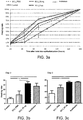
Figure 3A is a graph showing the time course of corneal wound healing after total des-epithelialization in control and SLURP-1 treated groups. Calculated healing rate means ± SEM. -
Figure 3B shows a graph of the healing rate on day two after total des-epithelialization in control and SLURP-1 treated groups (subconjunctival administration with 50 ng of SLURP and instillation). -
Figure 3C shows a graph of the healing rate on day three after total des-epithelialization in control and SLURP-1 treated groups (subconjunctival administration with 50 ng of SLURP and instillation). -
Figure 4 shows images of fluorescein staining of corneal epithelial wounds at the indicated time points after calibrated alcohol-induced des-epithelialization in PBS and SLURP-1 treated groups. Bold percentages indicate the group mean of the corneal wound surface, and percentages in brackets indicate the value of the representative cornea for the considered group. -
Figure 5A is a graph showing the time course of measured wound area fromday 0 today 2 after des-epithelialization in control and SLURP-1 treated groups. -
Figure 5B is a graph of the calculated healing rate between T0h and T47h after calibrated des-epithelialization in control and SLURP-1 treated groups. -
Figure 5C is a graph of the degree of wound healing at T47h after calibrated des-epithelialization. The degree of wound healing was calculated by the ratio: area of the fluorescein wound at T47h / area of the fluorescein wound at baseline, for each cornea, for each group, and expressed in percentage. - According to the present invention a protein comprising SEQ ID NO:1 is used in the treatment of an ocular disease.
- Preferably the ocular disease is selected from the group consisting of diabetic keratinopathy, keratitis, conjunctivitis, keratoconjunctivitis, uveitis, corneal trauma, corneal abrasion, corneal burns, corneal chronic ulcer, corneal dystrophies, persistent corneal epithelial defect (PED), corneal epithelial defect after laser surgeries, corneal epithelial defect post-PRK, corneal epithelial defect post-transcorneal transplant.
- SEQ ID NO:1 corresponds to the 81 amino acid mature form of SLURP-1.
- The protein comprising SEQ ID NO:1 is preferably used in inducing or accelerating cicatrisation in the eye of a subject, or in reducing inflammation in the eye of a subject, or in preventing infection in the eye of a subject.
- In an alternative embodiment, according to the present invention a composition comprising a protein comprising SEQ ID NO:1 is used in the treatment of an ocular disease, in particular in inducing or accelerating cicatrisation, or in reducing inflammation, or in preventing infection in the eye of a subject.
- The composition comprises at least one biocompatible polymer.
- More preferably, the biocompatible polymer is selected from the group consisting of hyaluronic acid, sugar polymers, lecithin gels, polyalanine derivates, pluronics, poly(ethylene)glycol, poloxamers, chitosan, xyloglucan, collagen, fibrin, polyorthoesters and mixtures thereof.
- Even more preferably the biocompatible polymer is hyaluronic acid.
- Alternatively, the biocompatible polymer is preferably a sugar polymer, even more preferably dextran (a glucose polymer).
- Preferably, the dextran is a carboxymethyl dextran sulphate polymer, more preferably a ReGeneraTing Agent (RGTA®) , even more preferably OTR4120 (formula I) which is marketed under the name of
Cacicol 20®.
- Formula I represents an analogue subunit of heparan sulfate with a glucose subunit based backbone.
- The composition can be solid, semi-solid, gel-like or liquid; moreover, it can be a solution, a suspension, an emulsion or a thermosetting gel.
- In an alternative embodiment the composition comprises at least one nanoparticle carrier.
- The nanoparticle carrier is preferably selected from the group consisting of poly-ε-caprolactone, polycyanocrylate and chitosan.
- In an alternative embodiment, the composition is in the form of an injectable viscous polymer composition. In particular the polymers of the composition are polyorthoesters.
- The composition is preferably administered by topical treatment or subconjunctival injection.
- More preferably, the amount of protein comprising SEQ ID NO:1 is from 5 ng to 50 µg, more preferably from 10 ng to 10 µg.
- In a preferred embodiment the composition is administered by subconjunctival injection and the amount of protein comprising SEQ ID NO:1 is from 20 ng to 90 ng, even more preferably from 40 ng to 60 ng.
-
Figures 2 and3 report the data of a first experiment in which the cicatrizing/ocular wound healing activity of SLURP-1 was compared using two routes of administration in a des-epithelialization model in rats. The first route of administration is a common route for ocular products used for moisture maintenance of the cornea and specifically for its ease of use: instillation (topical application). This route was used with a six times/day administration frequency atdays days - Both routes of administration have a common feature, namely the SLURP-1 applied quantities: the group receiving instillations was treated with a solution corresponding to the highest dose used in the group treated sub-conjunctively. It could be easily conceivable that products administered via the sub-conjunctival route would be available for a longer time during wound treatment as they might be slowly released by the conjunctiva tank they are stored in; conversely treatment administered topically would be quickly eliminated from the eye surface. Following this hypothesis, the dose regimen used in the sub-conjunctival treatment was in a range from the dose used topically (5 µg) and gradually decreasing to 50 ng.
- As shown in
Figure 3A , both routes of administration were capable of accelerating the wound healing process. - On
day 2 after total des-epithelialization, the healing rate after a sub-conjunctival injection of 50 ng SLURP-1 was significantly higher as compared to a sub-conjunctival injection of PBS (p<0.05) (Figure 3B ). - The unexpected discovery was concerning the sub-conjunctival route of administration in which the cicatrizing effect was more pronounced at the lowest dose (50 ng) when compared to the dose used in topical administration which in terms of quantity is much higher.
- The total administered quantity was calculated in both routes of administration regimen giving similar efficacy results (that is 1 µg/mL for sub-conjunctival and 100 µg/mL for topical route). In the topical treatment, 6 administrations of 50 µL drops were performed per day on two timely separate days with a 100 µg/mL solution. Then the final quantity used in this treatment group was 6*2*5 µg = 60 µg. In the sub-conjunctival treatment, a 1 µg/mL solution was injected twice (50 µL volume) thus the overall quantity given by this route was 2*0.05 µg = 0.1 µg. When comparing both administered quantities leading to similar clinical efficacy, a ratio of 600 between sub-conjunctival and topical quantities is observed.
- This is clearly in favour of a severely increased efficacy of the product by this specific less common route of administration for this product in order to treat ocular wound healing and other stated clinical conditions.
- More in particular, the experiment involved mechanical resection of the whole corneal epithelium performed under a surgical microscope using a 70% ethanol-soaked microsponge and a 15° surgical knife. The cornea was then rinsed with 0.9% NaCl and the effect of the treatment on corneal re-epithelialization was evaluated using topical 0.5% fluorescein, at 5 time-points after des-epithelialization (D1, D2, D3, D6 and D7 corresponding respectively to T24h, T48h, T72h, T144h and T168h).
- Six groups of 5 animals received the following treatment:
- Group 1: 50 µL vehicle subconjuntival injection at D0 and D3
- Group 2: 1 µg/mL (50 µL) SLURP-1 subconjuntival injection (50 ng per administration), at D0 and D3
- Group 3: 10 µg/mL (50 µL) SLURP-1 subconjuntival injection (500 ng per administration) at D0 and D3
- Group 4: 100 µg/mL (50 µL) SLURP-1 subconjuntival injection (5 µg per administration) at D0 and D3
- Group 5: 100 µg/mL (50 µL) SLURP-1, 6 topical instillations (5 µg per administration) at D0 and D3
- Group 6: No treatment
- At each time-point (D1, D2, D3, D6 and D7), full-face photographs were taken using cobalt blue biomicroscope light and the green fluorescent labeling of the cornea was used to determine the shape and area of the remaining ulcer (
Figure 2 ). The time course of the healing rate is presented on graph for each group in order to compare "control" groups (vehicle and/or control without any treatment) versus SLURP-1 treated groups (Figure 3A ). The wound healing follow up of each cornea was performed through computer-assisted measures of the wound area (At): ratio of the fluorescein-stained area to the total corneal area. For each time point, the healing rate: (At0-At)/At0 was calculated. Results are presented as mean ± SEM. An ANOVA test followed by the Dunnett's multiple comparison test or the non-parametric Mann Whitney comparison test were performed using GraphPad Prism (GraphPad Software, San Diego, U.S.A.). - The model induced a limbic insufficiency leading to corneal neovascularization and slowing the healing process. Corneas that presented a strong central ulcer were excluded from the study.
- As shown in
Figure 3B , onday 2 after total des-epithelialization, the healing rate after a sub-conjunctival injection of 50 ng of SLURP-1 is significantly higher as compared to a sub-conjunctival injection of PBS (p<0.05 Dunnett) and controls (p<0.01 Dunnett). The healing rate after instillation of SLURP-1 is also significantly higher than controls (p<0.05, Dunnett), but is not significantly different than after PBS injection. - As shown in
Figure 3C , onday 3 after total des-epithelialization, the healing rates of theSCJ 50 ng SLURP-1 treated group and instillation group are significantly higher than the control group (p<0.05, Dunnett) but are not significantly different than the healing rate with PBS. - This observation suggests that the SCJ procedure (or PBS) itself induces a response promoting corneal healing.
- As regards neovascularization, neovessels were visible in each group just after the beginning of the re-epithelialization process. The qualitative follow up of the corneal neovascularization did not demonstrate any difference between the SLURP-1-treated groups and controls. This is a very important observation showing that the healing properties of SLURP-1 are not associated with proangiogenic effects and do not impair restoration of corneal transparency.
- This means that SLURP-1 showed a significant corneal effect after sub-conjunctival administration and instillation, in the absence of side effects, and particularly, in the absence of corneal neovascularization that is associated with limbal deficiency, demonstrating that the healing effect is not linked to the corneal trans-differentiation process.
- Following the preliminary results obtained in the first corneal des-epithelialization model that implied not only a healing process but also a limbic insufficiency, a calibrated model of alcohol-induced des-epithelialization was chosen as a second study model in order to focus on the corneal healing properties of SLURP-1. The objective of this study was to confirm the preliminary results showing an enhancement of the re-epithelialization by the tested protein as compared to controls, and to evaluate a possible dose-response induced by the protein.
- The model involved a calibrated wound created using a trephine causing a 4mm diameter circular incision. Healing of such a minute wound is very fast even in the absence of any treatment (sometime around 2 days). Hence administrations were performed only once, on the day of incision, and efficacy was evaluated during the first 48h by an examination twice a day.
- The injected volume was adjusted from the first experiment; doses were equivalent with twice the administration volume and half concentrated solutions. This different dosing was chosen to increase the retention time in the tank created by sub-conjunctival injection leading to a prolonged presence of SLURP-1 on the corneal surface to compensate the unique administration performed in this standardized model.
- In this second experimental model, SLURP-1 effects were confirmed; namely the sub-conjunctival route with low injected quantities was 6 to 60 times more efficient in accelerating ocular wound healing when compared to the topical route.
- More in particular, corneal epithelial wounds were obtained as described by Hattori et al [25]. Briefly, after systemic and topical anesthesia, a trephine was used to make a 4-mm-diameter circular incision that was centered on the cornea. Then, the 4-mm diameter circular filter paper, which had been soaked with 70% ethanol, was placed on the incised area for 5 seconds. After a gentle wash with 5 mL saline, the detached corneal epithelium was removed.
- Treatment was administered only in the right eye via 1 single subconjunctival injection or 6 instillations at D0 (time of corneal des-epithelialization). Five groups of 4-6 animals received the following treatment:
- Group 1: 0.5 µg/mL (100 µL) SLURP-1 sub-conjuntival injection (50 ng per administration), at D0
- Group 2: 5 µg/mL (100 µL) SLURP-1 sub-conjuntival injection (500 ng per administration), at D0
- Group 3: 50 µg/mL (100 µL) SLURP-1 sub-conjuntival injection (5 µg per administration), at D0
- Group 4: 100 µg/mL (50 µL) SLURP-1 (5 µg per administration), 6 instillations at D0
- Group 5: Vehicle subconjuntival injection (100 µL) at D0
- The right corneas were examined with a biomicroscope. Each right cornea received one drop of 0.5% fluorescein (Novartis pharma S.A.S) and was examined using a cobalt blue light. Digital photographs were taken through the binocular. During the healing follow-up in the calibrated corneal des-epithelialization models, corneas were examined at
day 0,day 1 AM (T23h),day 1 PM (T28h),day 2 AM (T47h) andday 2 PM (T56h) (Figure 4 ). - Digital photographs were analyzed with imaging software (Adobe Photoshop). For each eye and each time-point, the area of the remaining ulcer was compared to the total corneal area. Time course of the healing rate is presented on graph for each group in order to compare control (vehicle and/or control without any treatment) versus SLURP-1 treated groups. Results are presented as mean ± SEM. An ANOVA test followed by the Dunnett's multiple comparison test or the non-parametric Mann Whitney comparison test were performed using GraphPad Prism (GraphPad Software, San Diego, U.S.A.).
- The time courses of the corneal wound closure in the control and SLURP-1 treated groups are shown in
Figure 5A . When comparing the healing rate calculated between T0h and T47h, SCJ injection of 500 ng and 5 µg of SLURP-1 significantly increased the healing rate as compared to the instillation or PBS sub-conjunctival administration groups (Figure 5B ). There was a dose response effect since no significant effect was observed in the 50 ng-SCJ injection group. Moreover, at T47h, the number of totally healed corneas was higher in the groups treated with sub-conjunctival injection of SLURP-1 at either 500 ng or 5 µg (Figure 5C ), with a noticed difference between the 500 ng (4 out of 5 corneas) and 5 µg (5 out of 5 corneas) as compared to the control group (3 out of 6 corneas). - From an analysis of the above data the advantages the present invention allows to achieve are apparent.
- In particular, an effective association between a specific concentration of SLURP-1 and a particular route of administration gives optimal results in terms of accelerated healing rate and faster reduced wound area, in particular protecting ocular surface from infection.
- Moreover, the healing properties of SLURP-1 are not associated with proangiogenic effects and do not impair restoration of corneal transparency.
-
- 1. Ploug, M. and V. Ellis, Structure-function relationships in the receptor for urokinase-type plasminogen activator. Comparison to other members of the Ly-6 family and snake venom alpha-neurotoxins. FEBS Lett, 1994. 349(2): p. 163-8.
- 2. Ploug, M., et al., Localization of the disulfide bonds in the NH2-terminal domain of the cellular receptor for human urokinase-type plasminogen activator. A domain structure belonging to a novel superfamily of glycolipid-anchored membrane proteins. J Biol Chem, 1993. 268(23): p. 17539-46.
- 3. Casey, J.R., et al., The structure of the urokinase-type plasminogen activator receptor gene. Blood, 1994. 84(4): p. 1151-6.
- 4. Adermann, K., et al., Structural and phylogenetic characterization of human SLURP-1, the first secreted mammalian member of the Ly-6/uPAR protein superfamily. Protein Sci, 1999. 8(4): p. 810-9.
- 5. Tsuji, H., et al., SLURP-2, a novel member of the human Ly-6 superfamily that is up-regulated in psoriasis vulgaris. Genomics, 2003. 81(1): p. 26-33.
- 6. Ward, K.M., et al., Identification of recurrent mutations in the ARS (component B) gene encoding SLURP-1 in two families with mal de Meleda. J Invest Dermatol, 2003. 120(1): p. 96-8.
- 7. Eckl, K.M., et al., Mal de Meleda (MDM) caused by mutations in the gene for SLURP-1 in patients from Germany, Turkey, Palestine, and the United Arab Emirates. Hum Genet, 2003. 112(1) : p. 50-6.
- 8. Fischer, J., et al., Mutations in the gene encoding SLURP-1 in Mal de Meleda. Hum Mol Genet, 2001. 10(8): p. 875-80.
- 9. Fischer, J., et al., Genetic linkage of Meleda disease to chromosome 8qter. Eur J Hum Genet, 1998. 6(6): p. 542-7.
- 10. Tone, M., L.A. Walsh, and H. Waldmann, Gene structure of human CD59 and demonstration that discrete mRNAs are generated by alternative polyadenylation. J Mol Biol, 1992. 227(3): p. 971-6.
- 11. Reiter, R.E., et al., Prostate stem cell antigen: a cell surface marker overexpressed in prostate cancer. Proc Natl Acad Sci U S A, 1998. 95(4): p. 1735-40.
- 12. Horie, M., et al., Isolation and characterization of a new member of the human Ly6 gene family (LY6H). Genomics, 1998. 53(3): p. 365-8.
- 13. Shan, X., et al., Characterization and mapping to human chromosome 8q24.3 of Ly-6-related gene 9804 encoding an apparent homologue of mouse TSA-1. J Immunol, 1998. 160(1): p. 197-208.
- 14. Brakenhoff, R.H., et al., The human E48 antigen, highly homologous to the murine Ly-6 antigen ThB, is a GPI-anchored molecule apparently involved in keratinocyte cell-cell adhesion. J Cell Biol, 1995. 129(6): p. 1677-89.
- 15. Mao, M., et al., RIG-E, a human homolog of the murine Ly-6 family, is induced by retinoic acid during the differentiation of acute promyelocytic leukemia cell. Proc Natl Acad Sci U S A, 1996. 93(12): p. 5910-4.
- 16. Samson, A.O. and M. Levitt, Inhibition mechanism of the acetylcholine receptor by alpha-neurotoxins as revealed by normal-mode dynamics. Biochemistry, 2008. 47(13): p. 4065-70.
- 17. Samson, A., et al., The mechanism for acetylcholine receptor inhibition by alpha-neurotoxins and species-specific resistance to alpha-bungarotoxin revealed by NMR. Neuron, 2002. 35(2): p. 319-32.
- 18. Chimienti, F., et al., Identification of SLURP-1 as an epidermal neuromodulator explains the clinical phenotype of Mal de Meleda. Hum Mol Genet, 2003. 12(22): p. 3017-24.
- 19. Mastrangeli, R., et al., ARS Component B: structural characterization, tissue expression and regulation of the gene and protein (SLURP-1) associated with Mal de Meleda. Eur J Dermatol, 2003. 13(6): p. 560-70.
- 20. Chernyavsky, A.I., et al., Coupling of ionic events to protein kinase signaling cascades upon activation of alpha7 nicotinic receptor: cooperative regulation of alpha2-integrin expression and Rho kinase activity. J Biol Chem, 2009. 284(33): p. 22140-8.
- 21. Arredondo, J., et al., Biological effects of SLURP-1 on human keratinocytes. J Invest Dermatol, 2005. 125(6): p. 1236-41.
- 22. Grando, S.A., M.R. Pittelkow, and K.U. Schallreuter, Adrenergic and cholinergic control in the biology of epidermis: physiological and clinical significance. J Invest Dermatol, 2006. 126(9): p. 1948-65.
- 23. Grando, S.A., Cholinergic control of epidermal cohesion. Exp Dermatol, 2006. 15(4): p. 265-82.
- 24. Favre, B., et al., SLURP1 is a late marker of epidermal differentiation and is absent in Mal de Meleda. J Invest Dermatol, 2007. 127(2): p. 301-8.
- 25. Hattori, M., et al., Effects of all-trans retinoic acid nanoparticles on corneal epithelial wound healing. Graefes Arch Clin Exp Ophthalmol, 2011.
- 26. Chernyavsky, A.I., et al., Novel cholinergic peptides SLURP-1 and -2 regulate epithelialization of cutaneous and oral wounds. Wound Rep Reg, 2012. 20:p. 103-113.
-
- <110> XIGEN SA
- <120> PROTEIN FOR USE IN THE TREATMENT OF OCULAR DISEASES
- <160> 1
- <170> BiSSAP 1.0
- <210> 1
<211> 81
<212> PRT
<213> Homo sapiens - <220>
<221> SOURCE
<222> 1..81
<223> /mol_type="protein" /organism="Homo sapiens" - <400> 1

Claims (17)
- A protein comprising SEQ ID NO:1 for use in the treatment of an ocular disease.
- A protein comprising SEQ ID NO:1 for use according to claim 1, wherein the protein is used for inducing or accelerating cicatrisation in the eye of a subject.
- A protein comprising SEQ ID NO:1 for use according to claim 1, wherein the protein is used for reducing inflammation in the eye of a subject.
- A protein comprising SEQ ID NO:1 for use according to claim 1, wherein the protein is used for preventing infection in the eye of a subject.
- A composition comprising a protein comprising SEQ ID NO:1 and at least one biocompatible polymer for use in the treatment of an ocular disease.
- A composition for use according to claim 5, wherein the biocompatible polymer is selected from the group consisting of hyaluronic acid, sugar polymers, lecithin gels, polyalanine derivates, pluronics, poly(ethylene)glycol, poloxamers, chitosan, xyloglucan, collagen, fibrin, polyorthoesters and mixtures thereof.
- The composition for use according to claim 6, wherein the biocompatible polymer is hyaluronic acid and/or a sugar polymer.
- The composition for use according to claim 7, wherein the sugar polymer is a dextran.
- The composition for use according to claim 8, wherein the dextran is a carboxymethyl dextran sulphate polymer.
- The composition for use according to claim 9, wherein the carboxymethyl dextran sulphate polymer is

- A composition comprising a protein comprising SEQ ID NO:1 and and at least one nanoparticle carrier for use in the treatment of an ocular disease.
- A composition for use according to claim 11, wherein the nanoparticle carrier is selected from the group consisting of poly-ε-caprolactone, polycyanocrylate and chitosan.
- A composition for use according to any of claims 5 to 12, wherein the composition is administered by topical treatment or subconjunctival injection.
- A composition for use according to claim 13, wherein the amount of protein comprising SEQ ID NO:1 is from 5 ng to 50 µg.
- A composition for use according to claim 14, wherein the amount of protein comprising SEQ ID NO:1 is from 10 ng to 10 µg.
- A composition for use according to claim 15, wherein the composition is administered by subconjunctival injection and the amount of protein comprising SEQ ID NO:1 is from 20 ng to 90 ng.
- A composition for use according to claim 16, wherein the amount of protein comprising SEQ ID NO:1 is from 40 ng to 60 ng.
Priority Applications (16)
| Application Number | Priority Date | Filing Date | Title |
|---|---|---|---|
| EP12196007.4A EP2740485B1 (en) | 2012-12-07 | 2012-12-07 | Protein slurp-1 for use in the treatment of ocular diseases |
| PCT/EP2013/075981 WO2014087022A1 (en) | 2012-12-07 | 2013-12-09 | Protein slurp-1 for use in the treatment of ocular diseases |
| AU2013353957A AU2013353957B2 (en) | 2012-12-07 | 2013-12-09 | Protein slurp-1 for use in the treatment of ocular diseases |
| MX2015006779A MX2015006779A (en) | 2012-12-07 | 2013-12-09 | Protein slurp-1 for use in the treatment of ocular diseases. |
| BR112015013215A BR112015013215A2 (en) | 2012-12-07 | 2013-12-09 | slurp-1 protein for use in the manufacture of eye disease medications |
| JP2015546050A JP6335188B2 (en) | 2012-12-07 | 2013-12-09 | Protein SLURP-1 for use in the treatment of eye diseases |
| SG10201705592YA SG10201705592YA (en) | 2012-12-07 | 2013-12-09 | Protein slurp-1 for use in the treatment of ocular diseases |
| CA2891201A CA2891201A1 (en) | 2012-12-07 | 2013-12-09 | Protein slurp-1 for use in the treatment of ocular diseases |
| CN201380064175.2A CN105188731B (en) | 2012-12-07 | 2013-12-09 | Protein SLURP-1 used to treat eye diseases |
| SG11201503658XA SG11201503658XA (en) | 2012-12-07 | 2013-12-09 | Protein slurp-1 for use in the treatment of ocular diseases |
| EA201591095A EA201591095A1 (en) | 2012-12-07 | 2013-12-09 | PROTEIN SLURP-1 FOR USE IN THE TREATMENT OF EYE DISEASES |
| KR1020157015595A KR20150108351A (en) | 2012-12-07 | 2013-12-09 | Protein slurp-1 for use in the treatment of ocular diseases |
| US14/650,277 US10098925B2 (en) | 2012-12-07 | 2013-12-09 | Protein SLURP-1 for use in the treatment of ocular diseases |
| ZA2015/03747A ZA201503747B (en) | 2012-12-07 | 2015-05-26 | Protein slurp-1 for use in the treatment of ocular diseases |
| IL239078A IL239078A0 (en) | 2012-12-07 | 2015-05-28 | Protein for use in the treatment of ocular diseases |
| CL2015001520A CL2015001520A1 (en) | 2012-12-07 | 2015-06-04 | Slurp-1 protein for use in the treatment of eye diseases |
Applications Claiming Priority (1)
| Application Number | Priority Date | Filing Date | Title |
|---|---|---|---|
| EP12196007.4A EP2740485B1 (en) | 2012-12-07 | 2012-12-07 | Protein slurp-1 for use in the treatment of ocular diseases |
Publications (2)
| Publication Number | Publication Date |
|---|---|
| EP2740485A1 EP2740485A1 (en) | 2014-06-11 |
| EP2740485B1 true EP2740485B1 (en) | 2018-10-31 |
Family
ID=47297008
Family Applications (1)
| Application Number | Title | Priority Date | Filing Date |
|---|---|---|---|
| EP12196007.4A Not-in-force EP2740485B1 (en) | 2012-12-07 | 2012-12-07 | Protein slurp-1 for use in the treatment of ocular diseases |
Country Status (15)
| Country | Link |
|---|---|
| US (1) | US10098925B2 (en) |
| EP (1) | EP2740485B1 (en) |
| JP (1) | JP6335188B2 (en) |
| KR (1) | KR20150108351A (en) |
| CN (1) | CN105188731B (en) |
| AU (1) | AU2013353957B2 (en) |
| BR (1) | BR112015013215A2 (en) |
| CA (1) | CA2891201A1 (en) |
| CL (1) | CL2015001520A1 (en) |
| EA (1) | EA201591095A1 (en) |
| IL (1) | IL239078A0 (en) |
| MX (1) | MX2015006779A (en) |
| SG (2) | SG10201705592YA (en) |
| WO (1) | WO2014087022A1 (en) |
| ZA (1) | ZA201503747B (en) |
Families Citing this family (3)
| Publication number | Priority date | Publication date | Assignee | Title |
|---|---|---|---|---|
| EP2740485B1 (en) * | 2012-12-07 | 2018-10-31 | Brightpulse Holding LTD. | Protein slurp-1 for use in the treatment of ocular diseases |
| EP3685822A1 (en) * | 2019-01-24 | 2020-07-29 | Organes Tissus Régénération Réparation Remplacement | Cosmetic/dermatological composition |
| CN114601915B (en) * | 2022-03-25 | 2023-04-07 | 四川大学华西医院 | Application of SLURP1 protein in preparation of medicine for preventing or treating skin elastic fiber diseases |
Family Cites Families (8)
| Publication number | Priority date | Publication date | Assignee | Title |
|---|---|---|---|---|
| IT1257184B (en) | 1992-12-22 | 1996-01-10 | Applied Research Systems | PREPARED FOR ANTI-INFLAMMATORY, ANTI-AGULANT AND ANTI-TUMORAL ACTIVITY |
| ES2359408T3 (en) * | 2003-04-16 | 2011-05-23 | Merck Serono Sa | USE OF SLURP-1 TO TREAT DISEASES RELATED TO THE DYSFUNCTION OF ACETILCOLINE RECEPTORS. |
| US20100303788A1 (en) * | 2009-05-29 | 2010-12-02 | Allergan, Inc. | Methods of Treating Chronic Neurogenic Inflammation Using Galanin Retargeted Endopepidases |
| US20130122066A1 (en) * | 2010-07-30 | 2013-05-16 | Biopham Gesellschaft Zur Biotechnologischen Entwicklung Von Pharmaka Mbh | Drug delivery devices and growth factor formulations for accelerated wound healing |
| JP6144673B2 (en) * | 2011-06-03 | 2017-06-07 | オフィディオン インコーポレイティド | Compositions and methods for transport across the blood brain barrier |
| US9132193B2 (en) * | 2012-11-05 | 2015-09-15 | University of Pittsburgh—of the Commonwealth System of Higher Education | Use of Slurp1 as an imunomodulatory molecule in the ocular surface |
| CN109589324B (en) * | 2012-11-08 | 2022-06-14 | 国立大学法人山口大学 | Therapeutic agent for keratoconjunctival disorder |
| EP2740485B1 (en) * | 2012-12-07 | 2018-10-31 | Brightpulse Holding LTD. | Protein slurp-1 for use in the treatment of ocular diseases |
-
2012
- 2012-12-07 EP EP12196007.4A patent/EP2740485B1/en not_active Not-in-force
-
2013
- 2013-12-09 SG SG10201705592YA patent/SG10201705592YA/en unknown
- 2013-12-09 BR BR112015013215A patent/BR112015013215A2/en not_active IP Right Cessation
- 2013-12-09 SG SG11201503658XA patent/SG11201503658XA/en unknown
- 2013-12-09 JP JP2015546050A patent/JP6335188B2/en not_active Expired - Fee Related
- 2013-12-09 EA EA201591095A patent/EA201591095A1/en unknown
- 2013-12-09 MX MX2015006779A patent/MX2015006779A/en unknown
- 2013-12-09 CA CA2891201A patent/CA2891201A1/en not_active Abandoned
- 2013-12-09 KR KR1020157015595A patent/KR20150108351A/en not_active Withdrawn
- 2013-12-09 US US14/650,277 patent/US10098925B2/en not_active Expired - Fee Related
- 2013-12-09 AU AU2013353957A patent/AU2013353957B2/en not_active Ceased
- 2013-12-09 WO PCT/EP2013/075981 patent/WO2014087022A1/en not_active Ceased
- 2013-12-09 CN CN201380064175.2A patent/CN105188731B/en not_active Expired - Fee Related
-
2015
- 2015-05-26 ZA ZA2015/03747A patent/ZA201503747B/en unknown
- 2015-05-28 IL IL239078A patent/IL239078A0/en unknown
- 2015-06-04 CL CL2015001520A patent/CL2015001520A1/en unknown
Non-Patent Citations (1)
| Title |
|---|
| None * |
Also Published As
| Publication number | Publication date |
|---|---|
| CA2891201A1 (en) | 2014-06-12 |
| CN105188731B (en) | 2019-02-05 |
| ZA201503747B (en) | 2016-03-30 |
| WO2014087022A1 (en) | 2014-06-12 |
| US10098925B2 (en) | 2018-10-16 |
| JP6335188B2 (en) | 2018-05-30 |
| AU2013353957B2 (en) | 2018-05-17 |
| CL2015001520A1 (en) | 2016-05-20 |
| AU2013353957A1 (en) | 2015-05-28 |
| IL239078A0 (en) | 2015-07-30 |
| EA201591095A1 (en) | 2015-11-30 |
| KR20150108351A (en) | 2015-09-25 |
| SG11201503658XA (en) | 2015-06-29 |
| EP2740485A1 (en) | 2014-06-11 |
| CN105188731A (en) | 2015-12-23 |
| SG10201705592YA (en) | 2017-08-30 |
| MX2015006779A (en) | 2015-10-29 |
| BR112015013215A2 (en) | 2018-02-06 |
| JP2016502558A (en) | 2016-01-28 |
| US20150320831A1 (en) | 2015-11-12 |
Similar Documents
| Publication | Publication Date | Title |
|---|---|---|
| JP6247692B2 (en) | Compositions and methods for treating skin scars | |
| EP2508196B1 (en) | Use of PEDF-derived polypeptides for promoting stem cells proliferation and wound healing | |
| US9345744B2 (en) | Peptide-based collagen modulators for wound healing and tissue repair | |
| JP6416240B2 (en) | Tetrapeptide derived from human C—X—C chemokine useful for treatment of various skin conditions | |
| CA3144419A1 (en) | Methods of promoting vasculogenesis | |
| Li et al. | LA-peptide Hydrogel—Regulation of macrophage and fibroblast fates and their crosstalk via attenuating TGF-β to promote scarless wound healing | |
| US10098925B2 (en) | Protein SLURP-1 for use in the treatment of ocular diseases | |
| CN111420023B (en) | Complex containing type I collagen and hyaluronic acid, preparation and application | |
| TW201204384A (en) | Lactoferrin sequences, compositions and methods of wound treatment | |
| CN115991758B (en) | Pearl powder polypeptide for promoting diabetic wound healing and application thereof | |
| Pu et al. | Enhancing the Inhibition of Corneal Neovascularization Efficacy by Self-Assembled into Supramolecular Hydrogel of Anti-Angiogenic Peptide | |
| Moysenovich et al. | Recombinant spidroin films attenuate individual markers of glucose induced aging in NIH 3T3 fibroblasts | |
| Liu et al. | Bioactive nanofiber hydrogel acts on VEGF and VEGF receptor to unleash inhibition effects on corneal neovascularization | |
| CN111346217B (en) | New application of polypeptide TDL23 | |
| Mao et al. | Bioactive thermoresponsive fibronectin–Pluronic F127 hydrogel for sustained ocular delivery with corneal regeneration and anti-inflammatory effects | |
| CN111632127B (en) | Short-chain peptide composition and application thereof in skin protection | |
| WO2020037714A1 (en) | Application of trail mutants in preparation of medicines for treating acnes, and preparation | |
| US20180000905A1 (en) | Composition and methods for treating ischemic wounds and inflammatory conditions | |
| Duan et al. | Diselenide-engineered peptide nanofibers as single-component supramolecular nanomedicines for osteoarthritis therapy | |
| Elkhawaga et al. | Assessment of Corneal Epithelial Healing Rate after Pterygium Excision When Using 2% Hyaluronic Acid. | |
| Zhou et al. | Platelet-rich plasmagel combined with adipose stem cells in promoting wound healing in rats | |
| HK40073737A (en) | Methods of promoting vasculogenesis | |
| HK40073737B (en) | Methods of promoting vasculogenesis | |
| Yuan | Regulatory Effect of Elastin Based Biomaterial on Cellular Behavior and Its Application on Wound Repair and Regeneration | |
| WO1996016669A1 (en) | Use of human recombinant epidermal growth factor in the manufacture of a medicament for treating acne |
Legal Events
| Date | Code | Title | Description |
|---|---|---|---|
| PUAI | Public reference made under article 153(3) epc to a published international application that has entered the european phase |
Free format text: ORIGINAL CODE: 0009012 |
|
| 17P | Request for examination filed |
Effective date: 20121207 |
|
| AK | Designated contracting states |
Kind code of ref document: A1 Designated state(s): AL AT BE BG CH CY CZ DE DK EE ES FI FR GB GR HR HU IE IS IT LI LT LU LV MC MK MT NL NO PL PT RO RS SE SI SK SM TR |
|
| AX | Request for extension of the european patent |
Extension state: BA ME |
|
| R17P | Request for examination filed (corrected) |
Effective date: 20141120 |
|
| RBV | Designated contracting states (corrected) |
Designated state(s): AL AT BE BG CH CY CZ DE DK EE ES FI FR GB GR HR HU IE IS IT LI LT LU LV MC MK MT NL NO PL PT RO RS SE SI SK SM TR |
|
| 17Q | First examination report despatched |
Effective date: 20160901 |
|
| GRAP | Despatch of communication of intention to grant a patent |
Free format text: ORIGINAL CODE: EPIDOSNIGR1 |
|
| INTG | Intention to grant announced |
Effective date: 20180518 |
|
| GRAS | Grant fee paid |
Free format text: ORIGINAL CODE: EPIDOSNIGR3 |
|
| GRAA | (expected) grant |
Free format text: ORIGINAL CODE: 0009210 |
|
| AK | Designated contracting states |
Kind code of ref document: B1 Designated state(s): AL AT BE BG CH CY CZ DE DK EE ES FI FR GB GR HR HU IE IS IT LI LT LU LV MC MK MT NL NO PL PT RO RS SE SI SK SM TR |
|
| REG | Reference to a national code |
Ref country code: CH Ref legal event code: EP Ref country code: GB Ref legal event code: FG4D |
|
| REG | Reference to a national code |
Ref country code: AT Ref legal event code: REF Ref document number: 1058575 Country of ref document: AT Kind code of ref document: T Effective date: 20181115 |
|
| REG | Reference to a national code |
Ref country code: DE Ref legal event code: R096 Ref document number: 602012052822 Country of ref document: DE |
|
| REG | Reference to a national code |
Ref country code: IE Ref legal event code: FG4D |
|
| REG | Reference to a national code |
Ref country code: CH Ref legal event code: NV Representative=s name: BOHEST AG, CH |
|
| REG | Reference to a national code |
Ref country code: NL Ref legal event code: MP Effective date: 20181031 |
|
| REG | Reference to a national code |
Ref country code: LT Ref legal event code: MG4D |
|
| REG | Reference to a national code |
Ref country code: AT Ref legal event code: MK05 Ref document number: 1058575 Country of ref document: AT Kind code of ref document: T Effective date: 20181031 |
|
| PG25 | Lapsed in a contracting state [announced via postgrant information from national office to epo] |
Ref country code: FI Free format text: LAPSE BECAUSE OF FAILURE TO SUBMIT A TRANSLATION OF THE DESCRIPTION OR TO PAY THE FEE WITHIN THE PRESCRIBED TIME-LIMIT Effective date: 20181031 Ref country code: BG Free format text: LAPSE BECAUSE OF FAILURE TO SUBMIT A TRANSLATION OF THE DESCRIPTION OR TO PAY THE FEE WITHIN THE PRESCRIBED TIME-LIMIT Effective date: 20190131 Ref country code: ES Free format text: LAPSE BECAUSE OF FAILURE TO SUBMIT A TRANSLATION OF THE DESCRIPTION OR TO PAY THE FEE WITHIN THE PRESCRIBED TIME-LIMIT Effective date: 20181031 Ref country code: HR Free format text: LAPSE BECAUSE OF FAILURE TO SUBMIT A TRANSLATION OF THE DESCRIPTION OR TO PAY THE FEE WITHIN THE PRESCRIBED TIME-LIMIT Effective date: 20181031 Ref country code: PL Free format text: LAPSE BECAUSE OF FAILURE TO SUBMIT A TRANSLATION OF THE DESCRIPTION OR TO PAY THE FEE WITHIN THE PRESCRIBED TIME-LIMIT Effective date: 20181031 Ref country code: AT Free format text: LAPSE BECAUSE OF FAILURE TO SUBMIT A TRANSLATION OF THE DESCRIPTION OR TO PAY THE FEE WITHIN THE PRESCRIBED TIME-LIMIT Effective date: 20181031 Ref country code: LV Free format text: LAPSE BECAUSE OF FAILURE TO SUBMIT A TRANSLATION OF THE DESCRIPTION OR TO PAY THE FEE WITHIN THE PRESCRIBED TIME-LIMIT Effective date: 20181031 Ref country code: LT Free format text: LAPSE BECAUSE OF FAILURE TO SUBMIT A TRANSLATION OF THE DESCRIPTION OR TO PAY THE FEE WITHIN THE PRESCRIBED TIME-LIMIT Effective date: 20181031 Ref country code: NO Free format text: LAPSE BECAUSE OF FAILURE TO SUBMIT A TRANSLATION OF THE DESCRIPTION OR TO PAY THE FEE WITHIN THE PRESCRIBED TIME-LIMIT Effective date: 20190131 Ref country code: IS Free format text: LAPSE BECAUSE OF FAILURE TO SUBMIT A TRANSLATION OF THE DESCRIPTION OR TO PAY THE FEE WITHIN THE PRESCRIBED TIME-LIMIT Effective date: 20190228 |
|
| PGFP | Annual fee paid to national office [announced via postgrant information from national office to epo] |
Ref country code: CH Payment date: 20190130 Year of fee payment: 7 Ref country code: GB Payment date: 20190130 Year of fee payment: 7 Ref country code: DE Payment date: 20190228 Year of fee payment: 7 Ref country code: FR Payment date: 20190128 Year of fee payment: 7 |
|
| PG25 | Lapsed in a contracting state [announced via postgrant information from national office to epo] |
Ref country code: AL Free format text: LAPSE BECAUSE OF FAILURE TO SUBMIT A TRANSLATION OF THE DESCRIPTION OR TO PAY THE FEE WITHIN THE PRESCRIBED TIME-LIMIT Effective date: 20181031 Ref country code: RS Free format text: LAPSE BECAUSE OF FAILURE TO SUBMIT A TRANSLATION OF THE DESCRIPTION OR TO PAY THE FEE WITHIN THE PRESCRIBED TIME-LIMIT Effective date: 20181031 Ref country code: PT Free format text: LAPSE BECAUSE OF FAILURE TO SUBMIT A TRANSLATION OF THE DESCRIPTION OR TO PAY THE FEE WITHIN THE PRESCRIBED TIME-LIMIT Effective date: 20190301 Ref country code: GR Free format text: LAPSE BECAUSE OF FAILURE TO SUBMIT A TRANSLATION OF THE DESCRIPTION OR TO PAY THE FEE WITHIN THE PRESCRIBED TIME-LIMIT Effective date: 20190201 Ref country code: NL Free format text: LAPSE BECAUSE OF FAILURE TO SUBMIT A TRANSLATION OF THE DESCRIPTION OR TO PAY THE FEE WITHIN THE PRESCRIBED TIME-LIMIT Effective date: 20181031 Ref country code: SE Free format text: LAPSE BECAUSE OF FAILURE TO SUBMIT A TRANSLATION OF THE DESCRIPTION OR TO PAY THE FEE WITHIN THE PRESCRIBED TIME-LIMIT Effective date: 20181031 |
|
| PG25 | Lapsed in a contracting state [announced via postgrant information from national office to epo] |
Ref country code: CZ Free format text: LAPSE BECAUSE OF FAILURE TO SUBMIT A TRANSLATION OF THE DESCRIPTION OR TO PAY THE FEE WITHIN THE PRESCRIBED TIME-LIMIT Effective date: 20181031 Ref country code: IT Free format text: LAPSE BECAUSE OF FAILURE TO SUBMIT A TRANSLATION OF THE DESCRIPTION OR TO PAY THE FEE WITHIN THE PRESCRIBED TIME-LIMIT Effective date: 20181031 Ref country code: DK Free format text: LAPSE BECAUSE OF FAILURE TO SUBMIT A TRANSLATION OF THE DESCRIPTION OR TO PAY THE FEE WITHIN THE PRESCRIBED TIME-LIMIT Effective date: 20181031 |
|
| REG | Reference to a national code |
Ref country code: DE Ref legal event code: R097 Ref document number: 602012052822 Country of ref document: DE |
|
| PG25 | Lapsed in a contracting state [announced via postgrant information from national office to epo] |
Ref country code: MC Free format text: LAPSE BECAUSE OF FAILURE TO SUBMIT A TRANSLATION OF THE DESCRIPTION OR TO PAY THE FEE WITHIN THE PRESCRIBED TIME-LIMIT Effective date: 20181031 Ref country code: SK Free format text: LAPSE BECAUSE OF FAILURE TO SUBMIT A TRANSLATION OF THE DESCRIPTION OR TO PAY THE FEE WITHIN THE PRESCRIBED TIME-LIMIT Effective date: 20181031 Ref country code: LU Free format text: LAPSE BECAUSE OF NON-PAYMENT OF DUE FEES Effective date: 20181207 Ref country code: RO Free format text: LAPSE BECAUSE OF FAILURE TO SUBMIT A TRANSLATION OF THE DESCRIPTION OR TO PAY THE FEE WITHIN THE PRESCRIBED TIME-LIMIT Effective date: 20181031 Ref country code: SM Free format text: LAPSE BECAUSE OF FAILURE TO SUBMIT A TRANSLATION OF THE DESCRIPTION OR TO PAY THE FEE WITHIN THE PRESCRIBED TIME-LIMIT Effective date: 20181031 Ref country code: EE Free format text: LAPSE BECAUSE OF FAILURE TO SUBMIT A TRANSLATION OF THE DESCRIPTION OR TO PAY THE FEE WITHIN THE PRESCRIBED TIME-LIMIT Effective date: 20181031 |
|
| PLBE | No opposition filed within time limit |
Free format text: ORIGINAL CODE: 0009261 |
|
| STAA | Information on the status of an ep patent application or granted ep patent |
Free format text: STATUS: NO OPPOSITION FILED WITHIN TIME LIMIT |
|
| REG | Reference to a national code |
Ref country code: IE Ref legal event code: MM4A |
|
| REG | Reference to a national code |
Ref country code: BE Ref legal event code: MM Effective date: 20181231 |
|
| 26N | No opposition filed |
Effective date: 20190801 |
|
| PG25 | Lapsed in a contracting state [announced via postgrant information from national office to epo] |
Ref country code: SI Free format text: LAPSE BECAUSE OF FAILURE TO SUBMIT A TRANSLATION OF THE DESCRIPTION OR TO PAY THE FEE WITHIN THE PRESCRIBED TIME-LIMIT Effective date: 20181031 Ref country code: IE Free format text: LAPSE BECAUSE OF NON-PAYMENT OF DUE FEES Effective date: 20181207 |
|
| PG25 | Lapsed in a contracting state [announced via postgrant information from national office to epo] |
Ref country code: BE Free format text: LAPSE BECAUSE OF NON-PAYMENT OF DUE FEES Effective date: 20181231 |
|
| PG25 | Lapsed in a contracting state [announced via postgrant information from national office to epo] |
Ref country code: MT Free format text: LAPSE BECAUSE OF NON-PAYMENT OF DUE FEES Effective date: 20181207 |
|
| PG25 | Lapsed in a contracting state [announced via postgrant information from national office to epo] |
Ref country code: TR Free format text: LAPSE BECAUSE OF FAILURE TO SUBMIT A TRANSLATION OF THE DESCRIPTION OR TO PAY THE FEE WITHIN THE PRESCRIBED TIME-LIMIT Effective date: 20181031 |
|
| PG25 | Lapsed in a contracting state [announced via postgrant information from national office to epo] |
Ref country code: MK Free format text: LAPSE BECAUSE OF NON-PAYMENT OF DUE FEES Effective date: 20181031 Ref country code: CY Free format text: LAPSE BECAUSE OF FAILURE TO SUBMIT A TRANSLATION OF THE DESCRIPTION OR TO PAY THE FEE WITHIN THE PRESCRIBED TIME-LIMIT Effective date: 20181031 Ref country code: HU Free format text: LAPSE BECAUSE OF FAILURE TO SUBMIT A TRANSLATION OF THE DESCRIPTION OR TO PAY THE FEE WITHIN THE PRESCRIBED TIME-LIMIT; INVALID AB INITIO Effective date: 20121207 |
|
| REG | Reference to a national code |
Ref country code: DE Ref legal event code: R119 Ref document number: 602012052822 Country of ref document: DE |
|
| REG | Reference to a national code |
Ref country code: CH Ref legal event code: PL |
|
| GBPC | Gb: european patent ceased through non-payment of renewal fee |
Effective date: 20191207 |
|
| PG25 | Lapsed in a contracting state [announced via postgrant information from national office to epo] |
Ref country code: GB Free format text: LAPSE BECAUSE OF NON-PAYMENT OF DUE FEES Effective date: 20191207 Ref country code: DE Free format text: LAPSE BECAUSE OF NON-PAYMENT OF DUE FEES Effective date: 20200701 |
|
| PG25 | Lapsed in a contracting state [announced via postgrant information from national office to epo] |
Ref country code: CH Free format text: LAPSE BECAUSE OF NON-PAYMENT OF DUE FEES Effective date: 20191231 Ref country code: LI Free format text: LAPSE BECAUSE OF NON-PAYMENT OF DUE FEES Effective date: 20191231 |
|
| PG25 | Lapsed in a contracting state [announced via postgrant information from national office to epo] |
Ref country code: FR Free format text: LAPSE BECAUSE OF NON-PAYMENT OF DUE FEES Effective date: 20191231 |