EP2737319B1 - Method for detecting biomolecules - Google Patents
Method for detecting biomolecules Download PDFInfo
- Publication number
- EP2737319B1 EP2737319B1 EP12731399.7A EP12731399A EP2737319B1 EP 2737319 B1 EP2737319 B1 EP 2737319B1 EP 12731399 A EP12731399 A EP 12731399A EP 2737319 B1 EP2737319 B1 EP 2737319B1
- Authority
- EP
- European Patent Office
- Prior art keywords
- biomolecules
- fractions
- proteins
- fraction
- protein
- Prior art date
- Legal status (The legal status is an assumption and is not a legal conclusion. Google has not performed a legal analysis and makes no representation as to the accuracy of the status listed.)
- Active
Links
Images




Classifications
-
- G—PHYSICS
- G01—MEASURING; TESTING
- G01N—INVESTIGATING OR ANALYSING MATERIALS BY DETERMINING THEIR CHEMICAL OR PHYSICAL PROPERTIES
- G01N33/00—Investigating or analysing materials by specific methods not covered by groups G01N1/00 - G01N31/00
- G01N33/48—Biological material, e.g. blood, urine; Haemocytometers
- G01N33/50—Chemical analysis of biological material, e.g. blood, urine; Testing involving biospecific ligand binding methods; Immunological testing
- G01N33/53—Immunoassay; Biospecific binding assay; Materials therefor
- G01N33/543—Immunoassay; Biospecific binding assay; Materials therefor with an insoluble carrier for immobilising immunochemicals
- G01N33/54306—Solid-phase reaction mechanisms
-
- G—PHYSICS
- G01—MEASURING; TESTING
- G01N—INVESTIGATING OR ANALYSING MATERIALS BY DETERMINING THEIR CHEMICAL OR PHYSICAL PROPERTIES
- G01N33/00—Investigating or analysing materials by specific methods not covered by groups G01N1/00 - G01N31/00
- G01N33/48—Biological material, e.g. blood, urine; Haemocytometers
- G01N33/50—Chemical analysis of biological material, e.g. blood, urine; Testing involving biospecific ligand binding methods; Immunological testing
- G01N33/53—Immunoassay; Biospecific binding assay; Materials therefor
- G01N33/543—Immunoassay; Biospecific binding assay; Materials therefor with an insoluble carrier for immobilising immunochemicals
- G01N33/54313—Immunoassay; Biospecific binding assay; Materials therefor with an insoluble carrier for immobilising immunochemicals the carrier being characterised by its particulate form
-
- G—PHYSICS
- G01—MEASURING; TESTING
- G01N—INVESTIGATING OR ANALYSING MATERIALS BY DETERMINING THEIR CHEMICAL OR PHYSICAL PROPERTIES
- G01N33/00—Investigating or analysing materials by specific methods not covered by groups G01N1/00 - G01N31/00
- G01N33/48—Biological material, e.g. blood, urine; Haemocytometers
- G01N33/50—Chemical analysis of biological material, e.g. blood, urine; Testing involving biospecific ligand binding methods; Immunological testing
- G01N33/68—Chemical analysis of biological material, e.g. blood, urine; Testing involving biospecific ligand binding methods; Immunological testing involving proteins, peptides or amino acids
- G01N33/6803—General methods of protein analysis not limited to specific proteins or families of proteins
- G01N33/6842—Proteomic analysis of subsets of protein mixtures with reduced complexity, e.g. membrane proteins, phosphoproteins, organelle proteins
Definitions
- the present invention relates to a method which makes it possible to detect in biological material biomolecules, more particularly proteins and/or peptides or proteins digested to form peptides.
- biomolecules more particularly of proteins and/or peptides from small amounts of biological material is an important requirement for research and medical diagnostics.
- the aim of new developments is to increase the sensitivity of known detection methods, more particularly of protein detection methods. More particularly, the aim is to be able to detect and quantify a larger number of different proteins or protein variants at the same time in one sample.
- Mass spectrometric methods are capable of detecting a large number of different proteins in an unambiguous manner from a complex sample. Sensitivity is a critical issue with this technology, despite major advances in separation and detection techniques, since the sensitive detection of proteins from small sample amounts is frequently not possible owing to system limitations.
- a second critical issue is the costs associated with mass spectrometric analysis. To allow specific protein detection, complicated and cost-intensive sample preparation and protein separation techniques are required, and the costs for the equipment required for this purpose, i.e. the actual mass spectrometer, are very high.
- Immunoassays such as immunohistochemistry, direct immunoassay and sandwich immunoassay, are therefore the vastly more important analytical methods in research laboratories and also in clinical analyses. They generally allow the specific detection of proteins in complex samples without complicated sample preparation.
- the achievable sensitivity can reach as low as the femtomolar concentration range, and the specificity of the measurement signal can be very high.
- protein microarrays which in the form of solid-phase assays on planar supports or on microspheres represent a significant miniaturization of immunoassays, have been developed which allow the detection and quantification of dozens to hundreds of different proteins from a few microlitres of sample volume.
- a problem is firstly the lack of binding affinity molecules, i.e. of antibodies, which are the prerequisite for a sensitive and specific immunoassay.
- a second problem is the cross-reactivity of the antibodies used with one another and the cross-reactivity with different analyte proteins.
- Immunoblots also known as Western blots, which comprise firstly using gel electrophoresis in a support matrix to separate proteins according to their size, charge or other physical properties into bands and then transferring the bands onto a membrane, where the proteins are then accessible for antibody binding events.
- An active assay for detecting biological analyse is known from WO 2004/027379 A2 .
- probe molecules are immobilized on a surface.
- the analytes are then placed in fluidic connection with the probe molecules on the surface.
- a force is then applied to the analytes to move them toward the surface to facilitate contact and possibly binding of the analytes to the probe molecules.
- Analytes that remain bounded to the surface are then detected. Detection of bound analytes can be accomplished with rolling beads bearing antibodies specific for the analytes.
- a method for detecting proteins of interest in a biological sample is known from WO 2010/094300 A1 .
- the method is based on the use of binding molecules having epitopes which are the result of an in silico determination process.
- a condensing method for providing a concentrated biolmolecule condensate is known from WO 2010/057318 A1 .
- a fluid sample is separated into a filtered liquid and a retentate biomolecule condensate containing target biomolecules.
- the target biomolecules are then attached to magnetic beads coated with antibodies of the target biomolecules, thereby obtaining a beaded biomolecule condensate.
- the beaded biomolecule condensate is then processed to extract constituents of the target biomolecules therefrom, thereby obtaining a concentrated biomolecule condensate.
- this object is achieved by a method of the type mentioned at the outset, comprising the steps of:
- a “complex biological sample” is understood to mean a sample containing a mixture of various biomolecules which can be of natural origin or which have been prepared synthetically/by genetic engineering.
- the sample can also consist of a small amount of tissue, for example biopsy material, or of cultured cells.
- biomolecules are understood to mean molecules which originate from a biological sample in the above sense, and are accessible to the method according to the invention. More particularly, the biomolecules defined in this way include proteins, peptides and polysaccharides.
- step a) is achieved, for example, by column chromatography and the specific detection in step b) is achieved using microspheres.
- fractionation of the complex biological sample is followed by use of a microsphere-based immunoassay, i.e. a multiplex, miniaturized system, in order to detect hundreds of proteins from a small sample amount in one immunoassay.
- a microsphere-based immunoassay i.e. a multiplex, miniaturized system
- the biomolecule separation according to physical property obtained in step a), i.e. the collection of the biomolecules in the fractions representing different physical property values, is "frozen" by the binding of the biomolecules to the microspheres.
- the assignement of the individual biomolecules to the fractions can, even after mixing of the microspheres from some or all fractions, be subsequently retrieved again and/or be used for further analysis, because the microspheres used for different fractions are distinguishable from one another - for example via their colouring.
- a physical method is used to separate the biomolecules, and the fractions obtained are then re-immobilized on different populations of microspheres.
- biomolecules then present accumulated on the microspheres are detected, for example, in a multiplex immunoassay after mixing of the microspheres.
- all bead populations i.e. all fractions
- microsphere populations used can, for example, be those from the xMAP® technology of Luminex Corp., Austin, Texas. This technology uses up to 500 differently colour-coded microsphere populations or sets which can be individually recognized in an appropriate reader.
- optical signals of the substances bound to the microspheres can be measured and outputted as a summed signal.
- the separation capacity of physical methods such as one-dimensional or two-dimensional gel electrophoresis or isoelectric focussing is combined with the flexibility and the detection sensitivity of a bead-based assay system.
- all proteins present in the sample are denatured, separated using one-dimensional SDS gel electrophoresis, and transferred onto a support membrane and immobilized thereon using the Western blot method.
- this matrix is then cut into columns approximately 1 cm in width and 5-10 cm in length and containing all the proteins of the sample. Thereafter, the columns are cut into 0.5 mm wide strips. From each individual strip, the protein is then eluted and bound in each case to distinguishable bead populations. Sufficient protein is eluted from each strip to coat several thousand microspheres. The signal is then generated in a direct bead-based immunoassay, which requires about 100 microspheres per assay. Thus, several hundred assays can be carried out using the microspheres generated from one fraction.
- the biological sample is separated by gel electrophoresis and the resulting band pattern is transferred preferably onto a matrix, wherein preferably the biomolecules/proteins transferred onto the matrix are modified, preferably biotinylated, for binding to a support molecule such as avidin or streptavidin, and if further preferably, in step a), the matrix is mechanically divided into small segments and the biomolecules are thereafter eluted from the individual segments into respective fractions, wherein preferably the matrix is cut into at least one column and the at least one column is cut into multiple strips, wherein the at least one column is cut into at least 10, preferably 20 to 200, strips.
- the biomolecules/proteins/peptides separated according to physical property in the bands are distributed onto the segments/strips and spatially separated from one another.
- the invention therefore provides a method which is capable of detecting different biomolecules present in a complex sample using a multiplex immunoassay.
- step b) the biomolecule-coated microspheres from at least several fractions are mixed together to form a master mix and preferably this master mix is then divided up into aliquots, and if further preferably, in step b), a specific binding molecule, preferably an antibody, is added to at least one aliquot, wherein the binding molecule is designed to emit a preferably optically detectable signal, wherein further preferably, in step b), the intensity of the detectable signal is determined for the biomolecules from an aliquot and separately according to the microsphere populations.
- a specific binding molecule preferably an antibody
- the proteins can, in step a), be denatured prior to the electrophoretic separation, wherein the biological sample, in step a), can be pre-treated prior to the electrophoretic separation to degrade the proteins into peptide fragments, and so, in step b), the peptide fragments are detected.
- the biolomecules from some or all fractions are further processed in step a), such that each processed fraction gives at least rise to one fraction in a first set of fractions and preferably one further fraction in a second set of fractions, the biomolecules in the first and the second set of fractions differing according to posttranslational protein modification, preferably the processing includes the enrichment of biomolecules showing one type of posttranslational modification in the fractions of the first set and further preferably a depletion of biomolecules showing said one type of posttranslational modification in the fractions of the second set, further preferably, in step b), the biomolecule-coated microspheres from some or all fractions of one set are mixed together to form a master mix.
- binding molecules that selectively either bind the modified or the unmodified protein. But, what is available are binding agents that generically bind to many different proteins carrying a specific modification.
- Posttranslational modifications include among others acetylation, phosphorylation, methylation, prenylation, ubiquitinylation, sumoylation, glycosylation, o-N-acetylglcosaminylation. A more complete list of modifications can be found on http://en.wikipedia.org/wiki/Posttranslational modification.
- Agents binding generically to different proteins having the same modification include antibodies against such structures (e.g. anti-acetyllysine antibodies for recognizing acetylated proteins), binding proteins that recognize specific sugars (e. g. lectins), immobilized ions to enrich phosphorylated proteins (e. g. gallium oxide-coated magnetic microspheres).
- antibodies against such structures e.g. anti-acetyllysine antibodies for recognizing acetylated proteins
- binding proteins that recognize specific sugars e. g. lectins
- immobilized ions to enrich phosphorylated proteins e. g. gallium oxide-coated magnetic microspheres.
- the biomolecules after having been fractionated in step a), can be further fractionated according to their type of posttranslational modifications, such that a first set of fractions is enriched with biomolecules having a specific posttranslational modification. Preferably a second set is depleted from biomoelcules having said modification.
- a first set of fractions is enriched with biomolecules having a specific posttranslational modification.
- a second set is depleted from biomoelcules having said modification.
- several or all fractions obtained in step a) can be further processed by affinity enrichment methods using agents that generically bind biomolecules having a specific type of posttranslational modification but do not distinguish between different proteins having the same modification.
- the one or two sets of fractions are further proceeded as described above.
- the fractions from each set are loaded on microspheres specific for each fraction, pooled into a master mix, and each master mix is divided up into aliquots.
- the new method has various advantages.
- the fluorescent signal obtained in each case for the different physical property values, i.e. for the individual fractions, is higher when the strip has been cut out more narrowly from the matrix. Therefore, the resolution of the mechanical division affects the sensitivity of the new method.
- a Western blot lane can, for example, be divided up into up to 96 fractions.
- proteins present in various isoforms of different molecular weight, processed proteins and modified proteins can be distinguished and can be detected using an antibody.
- the system has a good dynamic range, because specific signals are summed from positive fractions.
- Experiments with different antibodies revealed that a dynamic range of over 10 000 with good linearity is possible.
- tumour tissue and normal tissue, or tumour tissue before and after a treatment can be compared in a single measurement.
- tumour markers can be detected and an analysis of signal transduction can be carried out.
- the new method can be used in the analysis of modified proteins.
- phosphorylation can be detected.
- epigenetic changes such as histone modifications can be detected.
- the signal-to-noise ratio in the analysis of the loaded microspheres is better when the cut-out bands are narrower, since the narrower the cut-out band, the "purer" the eluted protein.
- biotinylation of the proteins bound on the membrane allows even small amounts of protein to be immobilized on the microspheres.
- the result of the extremely high affinity of the biotin-streptavidin interaction is that streptavidin beads can be loaded even with little material and a good coating density is achieved.
- Example 1 Detection of proteins from small amounts of biological material
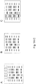
- Figs. 1A to H show simplistically, starting from A, the essential steps of the new method.
- the proteins present in a complex biological sample are denatured according to a customary protocol.
- the proteins are separated according to molecular weight by means of gel electrophoresis on an SDS gel ( Fig. 1A ).
- This band pattern is then - as in the case of a Western blot - transferred onto a protein-binding membrane ( Fig. 1 B) .
- the proteins now bound on the membrane are modified by the introduction of biotin ( Fig. 1 C) .
- the protein bands are now present in multiple columns next to one another. One of these columns is now mechanically cut out of the membrane and cut into many narrow strips ( Fig. 1D ).
- the strips are so short that one protein band can split up over several adjacent strips.
- the protein is now eluted into an aqueous solution, and so 20 to 200 fractions are generated from one membrane strip ( Fig. 1 E) .
- each fraction is now added a distinguishably colour-coded microsphere population, with each microsphere having a streptavidin coating, and so the protein is immobilized on the microspheres ( Fig. 1 F) .
- This generates 20 to 200 microsphere populations which are loaded with protein from different strips and distinguishable via their colour coding.
- microsphere populations are then firstly mixed together, whereupon the mixture is then divided up into up to 500 aliquots. Each aliquot contains at least 100 microspheres from each fraction. To each aliquot is now added a specific antibody which specifically binds to one of the proteins to be detected in the complex sample ( Fig. 1 G) .
- These antibodies are, for example, fluorescently labelled, and so a particular protein can be identified and quantified via the combination of the measurement values for the colour signal (identification of the strip) and the fluorescent signal (protein recognized) and the summing of the fluorescent signals for each microsphere population.
- the fluorescent summed signal is displayed against the fractions identified via the microsphere populations, i.e. against molecular weight, then what are specific (shaded in Fig. 1 H) can be distinguished from what are unspecific (pale in Fig. 1 H) and quantified.
- the biomolecules in several or all fractions can be subjected to affinity enrichment for a certain posttranslational modification.
- affinity enrichment for a certain posttranslational modification.
- one fraction having enriched all biomolecules showing the posttranslational modification is produced from each so processed fraction.
- a second fraction may be obtained wherein biomolecules carrying the modification are depleted.
- This enrichment can be performed by incubating the initial fraction with an immobilised binding reagent (e. g. a generic modification specific antibody) and thereby the initial fraction is depleted from biomolecules carrying this modification. By an elution step modified biomolecules get released and are found to be enriched in a new fraction. The process can be repeated sequentially for different posttranslational modifications.
- an immobilised binding reagent e. g. a generic modification specific antibody
- Each set of fractions is further processed, as detailed above, by adding microspheres, pooling the fractions into a master mix and dividing the master mix into various aliquots.
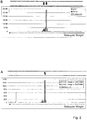
- a first antibody which recognizes the proteins ERK1 and ERK2 was used in a first analysis (A).
- a second antibody which recognizes phosphorylated ERK1 and ERK2 was used in a second analysis (B).
- Fig. 2 shows two bar charts featuring the measured signal intensities and the photo of a blot strip which had been developed in a standard Western blot.
- the two proteins can be distinguished and quantified.
- Example 3 Quantification of ERK1 in human liver cell lines
- the four individual lanes were each subsequently cut into 60 strips and the proteins were dissolved from the surface of the strips; the eluates generated from the individual strips were immobilized on 60 distinguishable microsphere populations and pooled to form a master mix.
- the bar chart in Fig. 3A shows signals for the HepG2 sample containing 4 ng of GST-ERK1 fusion protein; peaks at molecular weight 65 kDa, 42 kDa and 39 kDa correspond to GST-ERK1, ERK2 and ERK1.
- Fig. 3B shows the relative fluorescence intensity of the intrinsic ERK1 in the 4 lanes.
- Example 4 Analysis of tumour material present in a limited amount
- Frozen tumour tissue was cut and histologically assessed. In the sections examined, it was possible to identify two histologically distinguishable tumour regions.
- Fig. 1 For both tumour regions, the method according to Fig. 1 was used to separate the proteins by gel electrophoresis according to size and 96 protein fractions were generated in each case. The fractions for the two tumour regions were immobilized on 192 distinguishable bead populations. These were combined for the measurement. Thus, it was possible for both samples to be tested comparatively in one assay for differences in protein expression.
- the major advantage of the new method is the number of analyses now possible. Using the 20 000 cells available, up to 200 assays can be carried out. Using a customary Western blot, only 1-8 assays can be carried out with this material.
- Fig. 4 It can be seen from Fig. 4 that it is possible to detect the expression of the total amount of ERK1 (hatched bars) and ERK2 (grey bars) using an ERK1/2-specific antibody. Both proteins are detectable; differences in expression between the kinases were not found.
Landscapes
- Health & Medical Sciences (AREA)
- Life Sciences & Earth Sciences (AREA)
- Engineering & Computer Science (AREA)
- Immunology (AREA)
- Molecular Biology (AREA)
- Chemical & Material Sciences (AREA)
- Hematology (AREA)
- Urology & Nephrology (AREA)
- Biomedical Technology (AREA)
- Physics & Mathematics (AREA)
- Medicinal Chemistry (AREA)
- Pathology (AREA)
- Biotechnology (AREA)
- Cell Biology (AREA)
- Microbiology (AREA)
- General Physics & Mathematics (AREA)
- General Health & Medical Sciences (AREA)
- Food Science & Technology (AREA)
- Biochemistry (AREA)
- Analytical Chemistry (AREA)
- Bioinformatics & Cheminformatics (AREA)
- Bioinformatics & Computational Biology (AREA)
- Biophysics (AREA)
- Proteomics, Peptides & Aminoacids (AREA)
- Chemical Kinetics & Catalysis (AREA)
- Investigating Or Analysing Biological Materials (AREA)
- Measuring Or Testing Involving Enzymes Or Micro-Organisms (AREA)
Description
- The present invention relates to a method which makes it possible to detect in biological material biomolecules, more particularly proteins and/or peptides or proteins digested to form peptides.
- Methods of this type are widely known from the prior art.
- The specific detection of biomolecules, more particularly of proteins and/or peptides from small amounts of biological material is an important requirement for research and medical diagnostics.
- Therefore, the aim of new developments is to increase the sensitivity of known detection methods, more particularly of protein detection methods. More particularly, the aim is to be able to detect and quantify a larger number of different proteins or protein variants at the same time in one sample.
- Technological developments which have brought significant advances in the detection of proteins are in particular mass spectrometry and microarray based approaches.
- Mass spectrometric methods are capable of detecting a large number of different proteins in an unambiguous manner from a complex sample. Sensitivity is a critical issue with this technology, despite major advances in separation and detection techniques, since the sensitive detection of proteins from small sample amounts is frequently not possible owing to system limitations.
- A second critical issue is the costs associated with mass spectrometric analysis. To allow specific protein detection, complicated and cost-intensive sample preparation and protein separation techniques are required, and the costs for the equipment required for this purpose, i.e. the actual mass spectrometer, are very high.
- The result has been that mass spectrometric analyses are carried out in a few specialized laboratories and that only a small proportion of the diagnostic detection methods available are based on this technique.
- Immunoassays, such as immunohistochemistry, direct immunoassay and sandwich immunoassay, are therefore the vastly more important analytical methods in research laboratories and also in clinical analyses. They generally allow the specific detection of proteins in complex samples without complicated sample preparation.
- The achievable sensitivity can reach as low as the femtomolar concentration range, and the specificity of the measurement signal can be very high.
- In recent years, great efforts have been made to bring the immunoassay from the determination of individual parameters into a multiplex format, so that it is thus possible to detect a larger number of different proteins at the same time.
- For example, protein microarrays, which in the form of solid-phase assays on planar supports or on microspheres represent a significant miniaturization of immunoassays, have been developed which allow the detection and quantification of dozens to hundreds of different proteins from a few microlitres of sample volume.
- In the case of these antibody-based approaches, a problem is firstly the lack of binding affinity molecules, i.e. of antibodies, which are the prerequisite for a sensitive and specific immunoassay. A second problem is the cross-reactivity of the antibodies used with one another and the cross-reactivity with different analyte proteins.
- These limitations frequently result in the sensitivity of the detection method being distinctly lowered when more than about 20 proteins are simultaneously detected.
- Immunoblots, also known as Western blots, are also known, which comprise firstly using gel electrophoresis in a support matrix to separate proteins according to their size, charge or other physical properties into bands and then transferring the bands onto a membrane, where the proteins are then accessible for antibody binding events.
- This method is hampered by the same disadvantages as the above-described immunoassays.
- An active assay for detecting biological analyse is known from
WO 2004/027379 A2 . According to the method, probe molecules are immobilized on a surface. The analytes are then placed in fluidic connection with the probe molecules on the surface. A force is then applied to the analytes to move them toward the surface to facilitate contact and possibly binding of the analytes to the probe molecules. Analytes that remain bounded to the surface are then detected. Detection of bound analytes can be accomplished with rolling beads bearing antibodies specific for the analytes. - A method for detecting proteins of interest in a biological sample is known from
WO 2010/094300 A1 . The method is based on the use of binding molecules having epitopes which are the result of an in silico determination process. - It is further known that electrophoresis on agarose gels containing volatile buffers can be used for isolation of native proteins in pure form (L. Alvarez and A. Nieto: "Electrophoresis of Native Proteins on Agarose Gels Containing Volatile Buffer, Analytical Letters, (1989) 22, 2737-2745).
- A condensing method for providing a concentrated biolmolecule condensate is known from
WO 2010/057318 A1 . According to the method, a fluid sample is separated into a filtered liquid and a retentate biomolecule condensate containing target biomolecules. The target biomolecules are then attached to magnetic beads coated with antibodies of the target biomolecules, thereby obtaining a beaded biomolecule condensate. The beaded biomolecule condensate is then processed to extract constituents of the target biomolecules therefrom, thereby obtaining a concentrated biomolecule condensate. - In view of the above, it is an object of the present invention to provide a simple and inexpensively implementable method of the type mentioned at the outset, with which it is possible to specifically and sensitively detect from small amounts of biological material a multiplicity of biomolecules, more particularly proteins or peptides, or proteins digested to form peptides.
- According to the invention, this object is achieved by a method of the type mentioned at the outset, comprising the steps of:
- a) separating the biological sample into various fractions according to at least one physical property of the biomolecules; and
- b) specifically detecting biomolecules present in the fractions using at least one solid-phase-based, immunological detection method, which involves immobilizing the biomolecules from the individual fractions on microsphere populations which are specific for each fraction and distinguishable from one another.
- The object underlying the invention is completely achieved in this way.
- In the context of the present invention, a "complex biological sample" is understood to mean a sample containing a mixture of various biomolecules which can be of natural origin or which have been prepared synthetically/by genetic engineering. The sample can also consist of a small amount of tissue, for example biopsy material, or of cultured cells.
- In the context of the present invention, biomolecules are understood to mean molecules which originate from a biological sample in the above sense, and are accessible to the method according to the invention. More particularly, the biomolecules defined in this way include proteins, peptides and polysaccharides.
- The separation in step a) is achieved, for example, by column chromatography and the specific detection in step b) is achieved using microspheres.
- Therefore, according to the invention, fractionation of the complex biological sample is followed by use of a microsphere-based immunoassay, i.e. a multiplex, miniaturized system, in order to detect hundreds of proteins from a small sample amount in one immunoassay.
- The biomolecule separation according to physical property obtained in step a), i.e. the collection of the biomolecules in the fractions representing different physical property values, is "frozen" by the binding of the biomolecules to the microspheres. The assignement of the individual biomolecules to the fractions can, even after mixing of the microspheres from some or all fractions, be subsequently retrieved again and/or be used for further analysis, because the microspheres used for different fractions are distinguishable from one another - for example via their colouring.
- Therefore, according to the invention, firstly a physical method is used to separate the biomolecules, and the fractions obtained are then re-immobilized on different populations of microspheres.
- Unexpectedly in this new method, immunological analysis is possible after loading the microspheres with unknown biomolecules, more particularly proteins/peptides, and so mass spectrometric analysis is not required.
- The biomolecules then present accumulated on the microspheres (beads) are detected, for example, in a multiplex immunoassay after mixing of the microspheres. For this assay, all bead populations (i.e. all fractions) can be mixed and, at the same time, incubated with one antibody.
- The microsphere populations used can, for example, be those from the xMAP® technology of Luminex Corp., Austin, Texas. This technology uses up to 500 differently colour-coded microsphere populations or sets which can be individually recognized in an appropriate reader.
- For each set of identically coded microspheres, optical signals of the substances bound to the microspheres can be measured and outputted as a summed signal.
- If differently colour-coded sets of microspheres are loaded with various substances, it is possible in one analytical operation to capture the optical signals for the different sets of identically colour-coded microspheres and to present them assigned to the respective set of microspheres.
- Therefore, according to the invention, the separation capacity of physical methods such as one-dimensional or two-dimensional gel electrophoresis or isoelectric focussing is combined with the flexibility and the detection sensitivity of a bead-based assay system.
- In one embodiment of the new method, all proteins present in the sample are denatured, separated using one-dimensional SDS gel electrophoresis, and transferred onto a support membrane and immobilized thereon using the Western blot method.
- After biotinylation of the immobilized proteins, this matrix is then cut into columns approximately 1 cm in width and 5-10 cm in length and containing all the proteins of the sample. Thereafter, the columns are cut into 0.5 mm wide strips. From each individual strip, the protein is then eluted and bound in each case to distinguishable bead populations. Sufficient protein is eluted from each strip to coat several thousand microspheres. The signal is then generated in a direct bead-based immunoassay, which requires about 100 microspheres per assay. Thus, several hundred assays can be carried out using the microspheres generated from one fraction.
- In view of the above, it is preferred if, in step a), the biological sample is separated by gel electrophoresis and the resulting band pattern is transferred preferably onto a matrix, wherein preferably the biomolecules/proteins transferred onto the matrix are modified, preferably biotinylated, for binding to a support molecule such as avidin or streptavidin, and if further preferably, in step a), the matrix is mechanically divided into small segments and the biomolecules are thereafter eluted from the individual segments into respective fractions, wherein preferably the matrix is cut into at least one column and the at least one column is cut into multiple strips, wherein the at least one column is cut into at least 10, preferably 20 to 200, strips.
- The advantage of this gel electrophoretic separation over a column-based method is that many samples can be separated in parallel at the same time. A further advantage is the simple and cost-effective implementation of the gel-based method. In addition, the biomolecules bound to the matrix can be biotinylated more effectively than the biomolecules present in fractions after a column separation.
- While it is also possible to mechanically divide the gel itself, i.e. to cut it into columns and strips, this mechanical manipulation can be carried out more easily and more reproducibly in the case of a matrix, more particularly in the case of a matrix used in a Western blot.
- As a result of the mechanical division of gel or matrix, the biomolecules/proteins/peptides separated according to physical property in the bands are distributed onto the segments/strips and spatially separated from one another.
- It is not necessary to stain the gel after the separation, since the resulting bands do not have to be visible for the further course of the new method. Checking whether the separation was successful can be carried out at the level of the microspheres.
- The invention therefore provides a method which is capable of detecting different biomolecules present in a complex sample using a multiplex immunoassay.
- It is preferred if, in step b), the biomolecule-coated microspheres from at least several fractions are mixed together to form a master mix and preferably this master mix is then divided up into aliquots, and if further preferably, in step b), a specific binding molecule, preferably an antibody, is added to at least one aliquot, wherein the binding molecule is designed to emit a preferably optically detectable signal, wherein further preferably, in step b), the intensity of the detectable signal is determined for the biomolecules from an aliquot and separately according to the microsphere populations.
- From the master mix, many aliquots can be taken, which are each tested against an antibody.
- The proteins can, in step a), be denatured prior to the electrophoretic separation, wherein the biological sample, in step a), can be pre-treated prior to the electrophoretic separation to degrade the proteins into peptide fragments, and so, in step b), the peptide fragments are detected.
- According to one improvement, the biolomecules from some or all fractions are further processed in step a), such that each processed fraction gives at least rise to one fraction in a first set of fractions and preferably one further fraction in a second set of fractions, the biomolecules in the first and the second set of fractions differing according to posttranslational protein modification, preferably the processing includes the enrichment of biomolecules showing one type of posttranslational modification in the fractions of the first set and further preferably a depletion of biomolecules showing said one type of posttranslational modification in the fractions of the second set, further preferably, in step b), the biomolecule-coated microspheres from some or all fractions of one set are mixed together to form a master mix.
- This has to be seen against the background of posttranslational modifications of proteins that frequently determine the function of a protein. Therefore, it is one object for new methods for detection of biomolecules to be able to distinguish between modified and unmodified proteins.
- However, for many posttranslational modifications, there are no specific binding molecules available that selectively either bind the modified or the unmodified protein. But, what is available are binding agents that generically bind to many different proteins carrying a specific modification.
- Posttranslational modifications include among others acetylation, phosphorylation, methylation, prenylation, ubiquitinylation, sumoylation, glycosylation, o-N-acetylglcosaminylation. A more complete list of modifications can be found on http://en.wikipedia.org/wiki/Posttranslational modification.
- Agents binding generically to different proteins having the same modification include antibodies against such structures (e.g. anti-acetyllysine antibodies for recognizing acetylated proteins), binding proteins that recognize specific sugars (e. g. lectins), immobilized ions to enrich phosphorylated proteins (e. g. gallium oxide-coated magnetic microspheres).
- The biomolecules, after having been fractionated in step a), can be further fractionated according to their type of posttranslational modifications, such that a first set of fractions is enriched with biomolecules having a specific posttranslational modification. Preferably a second set is depleted from biomoelcules having said modification. To this end, several or all fractions obtained in step a) can be further processed by affinity enrichment methods using agents that generically bind biomolecules having a specific type of posttranslational modification but do not distinguish between different proteins having the same modification.
- After that further processing, the one or two sets of fractions are further proceeded as described above. The fractions from each set are loaded on microspheres specific for each fraction, pooled into a master mix, and each master mix is divided up into aliquots.
- When now a protein-specific antibody is added to the aliquots of each set of fractions, one can discern from comparing the optical read-out of both sets the amount of modified and unmodified protein in the original sample.
- This has the advantage that selected proteins having a specific posttranslational modification can be analyzed although no binding molecules are available that specifically bind to such selected proteins that carry such modification.
- Since with the new method a great number of different protein-specific antibodies can be used with fractions obtained from one gel electrophoresis run, from one small biological sample many such proteins can be analyzed for posttranslational modification although no antibodies are available that specifically bind to proteins having the modification.
- The new method has various advantages.
- It was found that, surprisingly, the fluorescent signal obtained in each case for the different physical property values, i.e. for the individual fractions, is higher when the strip has been cut out more narrowly from the matrix. Therefore, the resolution of the mechanical division affects the sensitivity of the new method.
- Using a small amount of sample material, it is possible to carry out a large number of measurements. Using 10 µg of protein, as is typically used for the detection of a protein in a Western blot, it is possible to create a master mix with which hundreds of measurements can be carried out.
- From 20 000 to 50 000 cells or 5 to 20 µg of cellular protein, it is possible according to initial experiments by the inventor to detect a few dozen to several hundred different proteins.
- It is possible to bind, for example, up to 96 different antibodies to the material from a Western blot lane after aliquoting, and so up to a thousand measurements are possible. This allows a comprehensive characterization of the material studied, and so even the study of cellular signal transduction such as the phosphorylation cascades is possible.
- The new method also exhibits a very good resolution. A Western blot lane can, for example, be divided up into up to 96 fractions. Thus, proteins present in various isoforms of different molecular weight, processed proteins and modified proteins can be distinguished and can be detected using an antibody.
- Because of the fractionation of the proteins using gel electrophoresis, increased use of nonspecific antibodies is also now possible, and so the problems mentioned at the outset can be avoided.
- Furthermore, the system has a good dynamic range, because specific signals are summed from positive fractions. Experiments with different antibodies revealed that a dynamic range of over 10 000 with good linearity is possible.
- Spike-in experiments with recombinant fusion proteins showed that the quantification of an intrinsic protein with this internal standard is possible.
- By mixing samples immobilized on different bead populations and measuring the mixed samples in one reaction, a comparative measurement of different samples and an internal standardization are possible. This is especially relevant because in this way tumour tissue and normal tissue, or tumour tissue before and after a treatment, can be compared in a single measurement.
- The use of the new method is especially interesting in the case of limiting sample amounts, for example in the case of clinical tumour material or in the case of tumour cell populations obtained following laser capture microdissection. Thus, tumour markers can be detected and an analysis of signal transduction can be carried out.
- In addition, the new method can be used in the analysis of modified proteins. For example, in the context of phosphoproteomics, phosphorylation can be detected. In addition, epigenetic changes such as histone modifications can be detected.
- Especially surprising in this combination of two detection methods is that the signal-to-noise ratio in the analysis of the loaded microspheres is better when the cut-out bands are narrower, since the narrower the cut-out band, the "purer" the eluted protein.
- The biotinylation of the proteins bound on the membrane allows even small amounts of protein to be immobilized on the microspheres. The result of the extremely high affinity of the biotin-streptavidin interaction is that streptavidin beads can be loaded even with little material and a good coating density is achieved.
- Further advantages are apparent from the description and the attached drawing.
- It will be appreciated that the features mentioned above and those to be explained below are usable not only in the combination indicated in each case but also in other combinations or alone, without departing from the scope of the present invention.
- The invention is described below by means of examples, which are explained by means of the figures, in which
- Figs. 1A to H
- show the individual method steps of the new method;
- Figs. 2A and B
- show two bar charts concerning the detection of ERK1/ERK2 from a human liver cell line;
- Figs. 3A and B
- show two bar charts and a regression curve concerning the quantification of ERK1 in a human liver cell line;
- Figs. 4A and B
- show two bar charts concerning the detection of ERK1/ERK2 in tumour tissue; and
- Figs. 5A and B
- each show two bar charts concerning the detection of the activation of a regulator in two different tumour tissues.
-
Figs. 1A to H show simplistically, starting from A, the essential steps of the new method. - Firstly, the proteins present in a complex biological sample are denatured according to a customary protocol.
- Thereafter, the proteins are separated according to molecular weight by means of gel electrophoresis on an SDS gel (
Fig. 1A ). This band pattern is then - as in the case of a Western blot - transferred onto a protein-binding membrane (Fig. 1 B) . - The proteins now bound on the membrane are modified by the introduction of biotin (
Fig. 1 C) . The protein bands are now present in multiple columns next to one another. One of these columns is now mechanically cut out of the membrane and cut into many narrow strips (Fig. 1D ). - Seen in the longitudinal direction of the columns, the strips are so short that one protein band can split up over several adjacent strips.
- From each strip, the protein is now eluted into an aqueous solution, and so 20 to 200 fractions are generated from one membrane strip (
Fig. 1 E) . - To each fraction is now added a distinguishably colour-coded microsphere population, with each microsphere having a streptavidin coating, and so the protein is immobilized on the microspheres (
Fig. 1 F) . This generates 20 to 200 microsphere populations which are loaded with protein from different strips and distinguishable via their colour coding. - These microsphere populations are then firstly mixed together, whereupon the mixture is then divided up into up to 500 aliquots. Each aliquot contains at least 100 microspheres from each fraction. To each aliquot is now added a specific antibody which specifically binds to one of the proteins to be detected in the complex sample (
Fig. 1 G) . - These antibodies are, for example, fluorescently labelled, and so a particular protein can be identified and quantified via the combination of the measurement values for the colour signal (identification of the strip) and the fluorescent signal (protein recognized) and the summing of the fluorescent signals for each microsphere population.
- If the fluorescent summed signal is displayed against the fractions identified via the microsphere populations, i.e. against molecular weight, then what are specific (shaded in
Fig. 1 H) can be distinguished from what are unspecific (pale inFig. 1 H) and quantified. - Between the steps of
Figs. 1 E and F , the biomolecules in several or all fractions can be subjected to affinity enrichment for a certain posttranslational modification. By this, from each so processed fraction there is produced one fraction having enriched all biomolecules showing the posttranslational modification. Further, a second fraction may be obtained wherein biomolecules carrying the modification are depleted. - This enrichment can be performed by incubating the initial fraction with an immobilised binding reagent (e. g. a generic modification specific antibody) and thereby the initial fraction is depleted from biomolecules carrying this modification. By an elution step modified biomolecules get released and are found to be enriched in a new fraction. The process can be repeated sequentially for different posttranslational modifications.
- This produces a first set of fractions with biomolecules carrying the modification being enriched, and preferably a second set, wherein the biomolecules carrying the modification are depleted.
- Each set of fractions is further processed, as detailed above, by adding microspheres, pooling the fractions into a master mix and dividing the master mix into various aliquots.
- By adding protein-specific antibodies to each aliquot, one can now analyze from the first set of fractions the proteins carry the modification. Further, by comparing the related fractions from the first and second set, one can at least semi-quantitatively determine the ration of modified proteins.
- 15 µg of protein extract from a human liver cell line (HepG2) were tested using the method shown in
Fig. 1 for the presence of the proteins ERK1 and ERK2 following SDS gel electrophoresis in the Western blot. - A first antibody which recognizes the proteins ERK1 and ERK2 was used in a first analysis (A). A second antibody which recognizes phosphorylated ERK1 and ERK2 was used in a second analysis (B).
- What was measured were the fluorescent summed signals for 96 bead populations, which had been coated in each case with the proteins eluted from the membrane strips. For the measurement, all 96 bead populations were mixed and 100 000 beads, i.e. about 1000 beads per population, were incubated with antibodies.
-
Fig. 2 shows two bar charts featuring the measured signal intensities and the photo of a blot strip which had been developed in a standard Western blot. - The two proteins can be distinguished and quantified.
- 20 µg of cell lysate from the human liver cell line HepG2 were applied in each case in 4 lanes on an SDS polyacrylamide gel.
- To the first sample were added 20 ng of purified GST-ERK1 fusion protein, to the
next sample 4 ng, then 0.8 ng, then 0.16 ng. Following electrophoretic separation, the proteins were transferred onto a membrane and modified using biotin. - The four individual lanes were each subsequently cut into 60 strips and the proteins were dissolved from the surface of the strips; the eluates generated from the individual strips were immobilized on 60 distinguishable microsphere populations and pooled to form a master mix.
- Thus, in this experiment, there were 4 batches which contained equal amounts of HepG2 cell lysate (and thus also contained equal amounts of intrinsic ERK1 and ERK2) but different, defined amounts of GST-ERK1 fusion protein.
- As a result of incubation of the 4 samples with a specific anti-ERK antibody, it was possible to generate measurement signals (relative fluorescence intensity) for ERK1, ERK2 and the GST-ERK1 fusion protein. For the measurement, 100 000 beads - which corresponds to about 1000 beads per population - were incubated with the ERK1/2-specific antibody. The signals for the known amounts of the fusion protein were used to create a standard curve, and this curve was used to calculate the absolute amount of ERK1.
- The bar chart in
Fig. 3A shows signals for the HepG2 sample containing 4 ng of GST-ERK1 fusion protein; peaks at molecular weight 65 kDa, 42 kDa and 39 kDa correspond to GST-ERK1, ERK2 and ERK1. - The right-hand side of
Fig. 3B shows the relative fluorescence intensity of the intrinsic ERK1 in the 4 lanes. - Frozen tumour tissue was cut and histologically assessed. In the sections examined, it was possible to identify two histologically distinguishable tumour regions.
- Using laser microdissection, approximately 20 000 cells were taken from both regions. The contained proteins were solubilized in detergent-containing lysis buffer.
- For both tumour regions, the method according to
Fig. 1 was used to separate the proteins by gel electrophoresis according to size and 96 protein fractions were generated in each case. The fractions for the two tumour regions were immobilized on 192 distinguishable bead populations. These were combined for the measurement. Thus, it was possible for both samples to be tested comparatively in one assay for differences in protein expression. - The major advantage of the new method is the number of analyses now possible. Using the 20 000 cells available, up to 200 assays can be carried out. Using a customary Western blot, only 1-8 assays can be carried out with this material.
- The expression of 54 proteins was studied in a comparative analysis. For the majority of the analytes studied (45/54), no noticeable changes in expression between the samples were detected; see
Fig. 4 , where again the fluorescent summed signals are shown against molecular weight. - It can be seen from
Fig. 4 that it is possible to detect the expression of the total amount of ERK1 (hatched bars) and ERK2 (grey bars) using an ERK1/2-specific antibody. Both proteins are detectable; differences in expression between the kinases were not found. - Significant changes were detected for a minority of the analytes. Of particular significance is the detection of the activation of a central regulator of cellular growth.
- Using a specific antibody, the expression of the total amount of the regulator from tumour A and tumour B was determined; see
Fig. 5A . - Using an activation-specific antibody, the amount of activated regulator was further determined; see
Fig. 5B . This revealed a ratio of 83:1 for tumour B : tumour A.
Claims (15)
- Method for detecting biomolecules present in a complex biological sample, comprising the steps ofa) separating the biological sample into various fractions according to at least one physical property of the biomolecules; andb) specifically detecting biomolecules present in the fractions using at least one solid-phase-based, immunological detection method, which involves immobilizing the biomolecules from the individual fractions on microsphere populations which are specific for each fraction and distinguishable from one another.
- Method according to Claim 1, characterized in that, in step a), the biological sample is separated by gel electrophoresis and preferably the resulting band pattern is transferred onto a matrix.
- Method according to Claim 2, characterized in that the separated biomolecules are modified, preferably biotinylated, for binding to a support molecule such as avidin or streptavidin.
- Method according to Claim 2 or 3, characterized in that, in step a), the matrix is mechanically divided into small segments and the biomolecules are thereafter eluted from the individual segments into respective fractions.
- Method according to Claim 4, characterized in that the matrix is cut into at least one column and the at least one column is cut into multiple strips, in particular into at least 10, preferably 20 to 200, strips and the biomolecules are thereafter eluted from the individual strips into respective fractions.
- Method according to anyone of Claims 1 to 5, characterized in that in step a) the biomolecules from some or all fractions are further processed, such that each processed fraction gives at least rise to one fraction in a first set of fractions and preferably one fraction in a second set of fractions, the biomolecules in the first and the second set of fractions differing according to posttranslational protein modification.
- Method according to Claim 6, characterized in that the processing includes the enrichment of biomolecules showing one type of posttranslational modification in the fractions of the first set and preferably a depletion of biomolecules showing said one type of posttranslational modification in the fractions of the second set.
- Method according to Claim 6 or 7, characterized in that, in step b), the biomolecule-coated microspheres from some or all fractions of one set are mixed together to form a master mix.
- Method according to anyone of Claims 1 to 5, characterized in that, in step b), the biomolecule-coated microspheres from some or all fractions are mixed together to form a master mix.
- Method according to Claim 8 or 9, characterized in that, in step b), the master mix is divided up into aliquots.
- Method according to Claim 10, characterized in that, in step b), a specific binding molecule such as an antibody is added to at least one aliquot, wherein the binding molecule is designed to emit a preferably optically detectable signal.
- Method according to Claim 11, characterized in that, in step b), the intensity of the detectable signal is determined for the biomolecules from an aliquot and separately according to the microsphere populations.
- Method according to anyone of Claims 1 to 12, characterized in that the biomolecules, in step a), are denatured prior to the electrophoretic separation.
- Method according to anyone of Claims 1 to 13, characterized in that the biomolecules are proteins.
- Method according to Claim 14, characterized in that the biological sample, in step a), is pretreated prior to the separation to degrade the proteins into peptide fragments and that, in step b), the peptide fragments are detected.
Applications Claiming Priority (2)
| Application Number | Priority Date | Filing Date | Title |
|---|---|---|---|
| DE102011109063A DE102011109063B3 (en) | 2011-07-28 | 2011-07-28 | Method for the detection of biomolecules |
| PCT/EP2012/062403 WO2013013922A1 (en) | 2011-07-28 | 2012-06-27 | Method for detecting biomolecules |
Publications (2)
| Publication Number | Publication Date |
|---|---|
| EP2737319A1 EP2737319A1 (en) | 2014-06-04 |
| EP2737319B1 true EP2737319B1 (en) | 2016-12-21 |
Family
ID=46456549
Family Applications (1)
| Application Number | Title | Priority Date | Filing Date |
|---|---|---|---|
| EP12731399.7A Active EP2737319B1 (en) | 2011-07-28 | 2012-06-27 | Method for detecting biomolecules |
Country Status (6)
| Country | Link |
|---|---|
| US (1) | US10274485B2 (en) |
| EP (1) | EP2737319B1 (en) |
| CN (1) | CN103917873B (en) |
| CA (1) | CA2842995C (en) |
| DE (1) | DE102011109063B3 (en) |
| WO (1) | WO2013013922A1 (en) |
Families Citing this family (3)
| Publication number | Priority date | Publication date | Assignee | Title |
|---|---|---|---|---|
| GB201616313D0 (en) | 2016-09-26 | 2016-11-09 | Oslo Universitetssykehus Hf And Universitetet I Oslo | Method |
| CN111323595B (en) * | 2020-03-11 | 2023-12-01 | 江苏省肿瘤医院 | A multi-visualized pathological biopsy method based on colloidal crystal microspheres |
| WO2024145145A1 (en) | 2022-12-30 | 2024-07-04 | Pierce Biotechnology, Inc. | A method for fractionation of biomolecules |
Family Cites Families (18)
| Publication number | Priority date | Publication date | Assignee | Title |
|---|---|---|---|---|
| US5948627A (en) | 1997-05-30 | 1999-09-07 | One Lambda | Immunobead flow cytometric detection of anti-HLA panel-reactive antibody |
| DE10054055A1 (en) * | 2000-10-31 | 2002-05-23 | Nmi Univ Tuebingen | Methods for analyzing proteins |
| US20030044843A1 (en) * | 2001-01-09 | 2003-03-06 | Mitsubishi Pharma Corporation | Novel proteome analysis method and devices therefor |
| AUPR378001A0 (en) | 2001-03-16 | 2001-04-12 | Proteome Systems Ltd | Protein chip |
| WO2002095356A2 (en) * | 2001-05-21 | 2002-11-28 | Aclara Biosciences, Inc. | Methods and compositions for analyzing proteins |
| CA2480646C (en) * | 2002-03-28 | 2010-08-03 | Marligen Biosciences, Inc. | Detection of dna-binding proteins |
| AU2003267255A1 (en) * | 2002-09-20 | 2004-04-08 | Novus Molecular, Inc. | Methods and devices for active bioassay |
| ATE517340T1 (en) | 2002-10-15 | 2011-08-15 | Abmetrix Inc | SETS OF DIGITAL ANTIBODIES DIRECTED AGAINST SHORT EPITOPES AND METHODS USING THEM |
| WO2004074452A2 (en) * | 2003-02-19 | 2004-09-02 | The Regents Of The University Of California | Multiplex mapping of protein interactions |
| CN1247795C (en) * | 2004-04-14 | 2006-03-29 | 厦门大学 | Bolting and identifying method for microbial cross antigen |
| NO20053373D0 (en) | 2005-07-11 | 2005-07-11 | Rikshospitalet Radiumhospitale | Multicolored Particles. |
| DE102006015001A1 (en) * | 2006-03-31 | 2007-10-11 | NMI Naturwissenschaftliches und Medizinisches Institut an der Universität Tübingen | Method for detecting and / or enriching analyte proteins and / or analyte peptides from a complex protein mixture |
| CN101550441B (en) * | 2008-04-01 | 2011-11-23 | 博尔诚(北京)科技有限公司 | Method for high-flux analysis of specimen containing biological marker and sample bank preparation |
| CN101392172B (en) | 2008-11-01 | 2012-04-18 | 厦门大学 | Carboxylic fluorescent encoding microsphere and synthetic method thereof |
| US20110223583A1 (en) | 2008-11-24 | 2011-09-15 | Early Warning Inc. | Devices and methods for providing concentrated biomolecule condensates to biosensing devices |
| WO2010094300A1 (en) * | 2009-02-20 | 2010-08-26 | NMI Naturwissenschaftliches und Medizinisches Institut an der Universität Tübingen | A method for determining in silico- a set of selected target epitopes |
| CN101550447B (en) | 2009-04-30 | 2011-04-20 | 张丽红 | Method for detecting sperm DNA fragments by using terminal transferase mediated biotin labeling dispersed chromatin |
| CN101846672A (en) | 2010-05-07 | 2010-09-29 | 天津大学 | Encoding detection method based on polymer microsphere change |
-
2011
- 2011-07-28 DE DE102011109063A patent/DE102011109063B3/en not_active Expired - Fee Related
-
2012
- 2012-06-27 US US14/235,598 patent/US10274485B2/en active Active
- 2012-06-27 EP EP12731399.7A patent/EP2737319B1/en active Active
- 2012-06-27 CN CN201280047533.4A patent/CN103917873B/en not_active Expired - Fee Related
- 2012-06-27 WO PCT/EP2012/062403 patent/WO2013013922A1/en not_active Ceased
- 2012-06-27 CA CA2842995A patent/CA2842995C/en active Active
Also Published As
| Publication number | Publication date |
|---|---|
| US10274485B2 (en) | 2019-04-30 |
| US20140248715A1 (en) | 2014-09-04 |
| CA2842995A1 (en) | 2013-01-31 |
| DE102011109063B3 (en) | 2012-12-20 |
| EP2737319A1 (en) | 2014-06-04 |
| WO2013013922A1 (en) | 2013-01-31 |
| CN103917873A (en) | 2014-07-09 |
| CA2842995C (en) | 2019-09-24 |
| CN103917873B (en) | 2019-04-16 |
Similar Documents
| Publication | Publication Date | Title |
|---|---|---|
| Wasinger et al. | Current status and advances in quantitative proteomic mass spectrometry | |
| Poetz et al. | Protein microarrays: catching the proteome | |
| Veenstra | Global and targeted quantitative proteomics for biomarker discovery | |
| Tambor et al. | Application of proteomics in biomarker discovery: a primer for the clinician. | |
| Stoll et al. | Microarray technology: an increasing variety of screening tools for proteomic research | |
| CN113567687A (en) | Application of reagent for detecting expression levels of two biomarkers in sample in preparation of kit for detecting preeclampsia | |
| EP2545370B1 (en) | Method for recognition and quantification of multiple analytes in a single analysis | |
| Chaerkady et al. | Quantitative proteomics for identification of cancer biomarkers | |
| US10274485B2 (en) | Method for detecting biomolecules | |
| US7879573B2 (en) | Isolation of analytes | |
| Haab et al. | Using antibody arrays to measure protein abundance and glycosylation: considerations for optimal performance | |
| WO2010094300A1 (en) | A method for determining in silico- a set of selected target epitopes | |
| Balog et al. | Novel automated biomarker discovery work flow for urinary peptidomics | |
| Sroka-Bartnicka et al. | Particle-based N-linked glycan analysis of selected proteins from biological samples using nonglycosylated binders | |
| EP3529596B1 (en) | Compositions and methods for diagnosing and differentiating systemic juvenile idiopathic arthritis and kawasaki disease | |
| Boccardi et al. | An automated plasma protein fractionation design: high-throughput perspectives for proteomic analysis | |
| EP1941269B1 (en) | A method of screening a biological target for weak interactions using weak affinity chromatography | |
| US20100267581A1 (en) | High throughput integrated microfluidic system and device | |
| US12629681B2 (en) | Apparatuses and methods for performing multiple omics analysis and processing analyte mixtures | |
| Díez et al. | Genomics and proteomics for biomarker validation | |
| Stoll et al. | Protein microarrays: technologies and applications | |
| WO2004097368A2 (en) | Improved immunoassays | |
| WO2023055790A1 (en) | Apparatus and methods for performing multiple omics analysis and processing analyte mixtures | |
| Hanash et al. | Application of proteomics to disease diagnostics | |
| Shoshan et al. | Proteomics in clinical laboratory diagnosis |
Legal Events
| Date | Code | Title | Description |
|---|---|---|---|
| PUAI | Public reference made under article 153(3) epc to a published international application that has entered the european phase |
Free format text: ORIGINAL CODE: 0009012 |
|
| 17P | Request for examination filed |
Effective date: 20140220 |
|
| AK | Designated contracting states |
Kind code of ref document: A1 Designated state(s): AL AT BE BG CH CY CZ DE DK EE ES FI FR GB GR HR HU IE IS IT LI LT LU LV MC MK MT NL NO PL PT RO RS SE SI SK SM TR |
|
| DAX | Request for extension of the european patent (deleted) | ||
| 17Q | First examination report despatched |
Effective date: 20150126 |
|
| GRAJ | Information related to disapproval of communication of intention to grant by the applicant or resumption of examination proceedings by the epo deleted |
Free format text: ORIGINAL CODE: EPIDOSDIGR1 |
|
| GRAP | Despatch of communication of intention to grant a patent |
Free format text: ORIGINAL CODE: EPIDOSNIGR1 |
|
| GRAP | Despatch of communication of intention to grant a patent |
Free format text: ORIGINAL CODE: EPIDOSNIGR1 |
|
| RAP1 | Party data changed (applicant data changed or rights of an application transferred) |
Owner name: NMI NATURWISSENSCHAFTLICHES UND MEDIZINISCHES INST |
|
| INTG | Intention to grant announced |
Effective date: 20160629 |
|
| GRAS | Grant fee paid |
Free format text: ORIGINAL CODE: EPIDOSNIGR3 |
|
| GRAA | (expected) grant |
Free format text: ORIGINAL CODE: 0009210 |
|
| AK | Designated contracting states |
Kind code of ref document: B1 Designated state(s): AL AT BE BG CH CY CZ DE DK EE ES FI FR GB GR HR HU IE IS IT LI LT LU LV MC MK MT NL NO PL PT RO RS SE SI SK SM TR |
|
| REG | Reference to a national code |
Ref country code: GB Ref legal event code: FG4D |
|
| REG | Reference to a national code |
Ref country code: CH Ref legal event code: EP |
|
| REG | Reference to a national code |
Ref country code: IE Ref legal event code: FG4D |
|
| REG | Reference to a national code |
Ref country code: AT Ref legal event code: REF Ref document number: 855934 Country of ref document: AT Kind code of ref document: T Effective date: 20170115 |
|
| REG | Reference to a national code |
Ref country code: NL Ref legal event code: FP |
|
| REG | Reference to a national code |
Ref country code: DE Ref legal event code: R096 Ref document number: 602012026832 Country of ref document: DE |
|
| PG25 | Lapsed in a contracting state [announced via postgrant information from national office to epo] |
Ref country code: LV Free format text: LAPSE BECAUSE OF FAILURE TO SUBMIT A TRANSLATION OF THE DESCRIPTION OR TO PAY THE FEE WITHIN THE PRESCRIBED TIME-LIMIT Effective date: 20161221 |
|
| REG | Reference to a national code |
Ref country code: CH Ref legal event code: NV Representative=s name: TROESCH SCHEIDEGGER WERNER AG, CH |
|
| REG | Reference to a national code |
Ref country code: SE Ref legal event code: TRGR |
|
| REG | Reference to a national code |
Ref country code: LT Ref legal event code: MG4D |
|
| PG25 | Lapsed in a contracting state [announced via postgrant information from national office to epo] |
Ref country code: NO Free format text: LAPSE BECAUSE OF FAILURE TO SUBMIT A TRANSLATION OF THE DESCRIPTION OR TO PAY THE FEE WITHIN THE PRESCRIBED TIME-LIMIT Effective date: 20170321 Ref country code: GR Free format text: LAPSE BECAUSE OF FAILURE TO SUBMIT A TRANSLATION OF THE DESCRIPTION OR TO PAY THE FEE WITHIN THE PRESCRIBED TIME-LIMIT Effective date: 20170322 Ref country code: LT Free format text: LAPSE BECAUSE OF FAILURE TO SUBMIT A TRANSLATION OF THE DESCRIPTION OR TO PAY THE FEE WITHIN THE PRESCRIBED TIME-LIMIT Effective date: 20161221 |
|
| REG | Reference to a national code |
Ref country code: AT Ref legal event code: MK05 Ref document number: 855934 Country of ref document: AT Kind code of ref document: T Effective date: 20161221 |
|
| PG25 | Lapsed in a contracting state [announced via postgrant information from national office to epo] |
Ref country code: HR Free format text: LAPSE BECAUSE OF FAILURE TO SUBMIT A TRANSLATION OF THE DESCRIPTION OR TO PAY THE FEE WITHIN THE PRESCRIBED TIME-LIMIT Effective date: 20161221 Ref country code: RS Free format text: LAPSE BECAUSE OF FAILURE TO SUBMIT A TRANSLATION OF THE DESCRIPTION OR TO PAY THE FEE WITHIN THE PRESCRIBED TIME-LIMIT Effective date: 20161221 Ref country code: FI Free format text: LAPSE BECAUSE OF FAILURE TO SUBMIT A TRANSLATION OF THE DESCRIPTION OR TO PAY THE FEE WITHIN THE PRESCRIBED TIME-LIMIT Effective date: 20161221 |
|
| REG | Reference to a national code |
Ref country code: FR Ref legal event code: PLFP Year of fee payment: 6 |
|
| PG25 | Lapsed in a contracting state [announced via postgrant information from national office to epo] |
Ref country code: RO Free format text: LAPSE BECAUSE OF FAILURE TO SUBMIT A TRANSLATION OF THE DESCRIPTION OR TO PAY THE FEE WITHIN THE PRESCRIBED TIME-LIMIT Effective date: 20161221 Ref country code: CZ Free format text: LAPSE BECAUSE OF FAILURE TO SUBMIT A TRANSLATION OF THE DESCRIPTION OR TO PAY THE FEE WITHIN THE PRESCRIBED TIME-LIMIT Effective date: 20161221 Ref country code: IS Free format text: LAPSE BECAUSE OF FAILURE TO SUBMIT A TRANSLATION OF THE DESCRIPTION OR TO PAY THE FEE WITHIN THE PRESCRIBED TIME-LIMIT Effective date: 20170421 Ref country code: EE Free format text: LAPSE BECAUSE OF FAILURE TO SUBMIT A TRANSLATION OF THE DESCRIPTION OR TO PAY THE FEE WITHIN THE PRESCRIBED TIME-LIMIT Effective date: 20161221 Ref country code: SK Free format text: LAPSE BECAUSE OF FAILURE TO SUBMIT A TRANSLATION OF THE DESCRIPTION OR TO PAY THE FEE WITHIN THE PRESCRIBED TIME-LIMIT Effective date: 20161221 |
|
| PG25 | Lapsed in a contracting state [announced via postgrant information from national office to epo] |
Ref country code: ES Free format text: LAPSE BECAUSE OF FAILURE TO SUBMIT A TRANSLATION OF THE DESCRIPTION OR TO PAY THE FEE WITHIN THE PRESCRIBED TIME-LIMIT Effective date: 20161221 Ref country code: PL Free format text: LAPSE BECAUSE OF FAILURE TO SUBMIT A TRANSLATION OF THE DESCRIPTION OR TO PAY THE FEE WITHIN THE PRESCRIBED TIME-LIMIT Effective date: 20161221 Ref country code: SM Free format text: LAPSE BECAUSE OF FAILURE TO SUBMIT A TRANSLATION OF THE DESCRIPTION OR TO PAY THE FEE WITHIN THE PRESCRIBED TIME-LIMIT Effective date: 20161221 Ref country code: IT Free format text: LAPSE BECAUSE OF FAILURE TO SUBMIT A TRANSLATION OF THE DESCRIPTION OR TO PAY THE FEE WITHIN THE PRESCRIBED TIME-LIMIT Effective date: 20161221 Ref country code: PT Free format text: LAPSE BECAUSE OF FAILURE TO SUBMIT A TRANSLATION OF THE DESCRIPTION OR TO PAY THE FEE WITHIN THE PRESCRIBED TIME-LIMIT Effective date: 20170421 Ref country code: AT Free format text: LAPSE BECAUSE OF FAILURE TO SUBMIT A TRANSLATION OF THE DESCRIPTION OR TO PAY THE FEE WITHIN THE PRESCRIBED TIME-LIMIT Effective date: 20161221 Ref country code: BG Free format text: LAPSE BECAUSE OF FAILURE TO SUBMIT A TRANSLATION OF THE DESCRIPTION OR TO PAY THE FEE WITHIN THE PRESCRIBED TIME-LIMIT Effective date: 20170321 |
|
| REG | Reference to a national code |
Ref country code: DE Ref legal event code: R097 Ref document number: 602012026832 Country of ref document: DE |
|
| PLBE | No opposition filed within time limit |
Free format text: ORIGINAL CODE: 0009261 |
|
| STAA | Information on the status of an ep patent application or granted ep patent |
Free format text: STATUS: NO OPPOSITION FILED WITHIN TIME LIMIT |
|
| 26N | No opposition filed |
Effective date: 20170922 |
|
| PG25 | Lapsed in a contracting state [announced via postgrant information from national office to epo] |
Ref country code: DK Free format text: LAPSE BECAUSE OF FAILURE TO SUBMIT A TRANSLATION OF THE DESCRIPTION OR TO PAY THE FEE WITHIN THE PRESCRIBED TIME-LIMIT Effective date: 20161221 |
|
| PG25 | Lapsed in a contracting state [announced via postgrant information from national office to epo] |
Ref country code: MC Free format text: LAPSE BECAUSE OF FAILURE TO SUBMIT A TRANSLATION OF THE DESCRIPTION OR TO PAY THE FEE WITHIN THE PRESCRIBED TIME-LIMIT Effective date: 20161221 |
|
| PG25 | Lapsed in a contracting state [announced via postgrant information from national office to epo] |
Ref country code: SI Free format text: LAPSE BECAUSE OF FAILURE TO SUBMIT A TRANSLATION OF THE DESCRIPTION OR TO PAY THE FEE WITHIN THE PRESCRIBED TIME-LIMIT Effective date: 20161221 |
|
| REG | Reference to a national code |
Ref country code: IE Ref legal event code: MM4A |
|
| PG25 | Lapsed in a contracting state [announced via postgrant information from national office to epo] |
Ref country code: LU Free format text: LAPSE BECAUSE OF NON-PAYMENT OF DUE FEES Effective date: 20170627 Ref country code: IE Free format text: LAPSE BECAUSE OF NON-PAYMENT OF DUE FEES Effective date: 20170627 |
|
| REG | Reference to a national code |
Ref country code: FR Ref legal event code: PLFP Year of fee payment: 7 |
|
| PG25 | Lapsed in a contracting state [announced via postgrant information from national office to epo] |
Ref country code: MT Free format text: LAPSE BECAUSE OF NON-PAYMENT OF DUE FEES Effective date: 20170627 |
|
| PG25 | Lapsed in a contracting state [announced via postgrant information from national office to epo] |
Ref country code: HU Free format text: LAPSE BECAUSE OF FAILURE TO SUBMIT A TRANSLATION OF THE DESCRIPTION OR TO PAY THE FEE WITHIN THE PRESCRIBED TIME-LIMIT; INVALID AB INITIO Effective date: 20120627 |
|
| PG25 | Lapsed in a contracting state [announced via postgrant information from national office to epo] |
Ref country code: CY Free format text: LAPSE BECAUSE OF NON-PAYMENT OF DUE FEES Effective date: 20161221 |
|
| PG25 | Lapsed in a contracting state [announced via postgrant information from national office to epo] |
Ref country code: MK Free format text: LAPSE BECAUSE OF FAILURE TO SUBMIT A TRANSLATION OF THE DESCRIPTION OR TO PAY THE FEE WITHIN THE PRESCRIBED TIME-LIMIT Effective date: 20161221 |
|
| PG25 | Lapsed in a contracting state [announced via postgrant information from national office to epo] |
Ref country code: TR Free format text: LAPSE BECAUSE OF FAILURE TO SUBMIT A TRANSLATION OF THE DESCRIPTION OR TO PAY THE FEE WITHIN THE PRESCRIBED TIME-LIMIT Effective date: 20161221 |
|
| PG25 | Lapsed in a contracting state [announced via postgrant information from national office to epo] |
Ref country code: AL Free format text: LAPSE BECAUSE OF FAILURE TO SUBMIT A TRANSLATION OF THE DESCRIPTION OR TO PAY THE FEE WITHIN THE PRESCRIBED TIME-LIMIT Effective date: 20161221 |
|
| P01 | Opt-out of the competence of the unified patent court (upc) registered |
Effective date: 20230504 |
|
| PGFP | Annual fee paid to national office [announced via postgrant information from national office to epo] |
Ref country code: NL Payment date: 20230620 Year of fee payment: 12 |
|
| PGFP | Annual fee paid to national office [announced via postgrant information from national office to epo] |
Ref country code: SE Payment date: 20230620 Year of fee payment: 12 |
|
| PGFP | Annual fee paid to national office [announced via postgrant information from national office to epo] |
Ref country code: BE Payment date: 20230620 Year of fee payment: 12 |
|
| REG | Reference to a national code |
Ref country code: SE Ref legal event code: EUG |
|
| REG | Reference to a national code |
Ref country code: NL Ref legal event code: MM Effective date: 20240701 |
|
| PG25 | Lapsed in a contracting state [announced via postgrant information from national office to epo] |
Ref country code: NL Free format text: LAPSE BECAUSE OF NON-PAYMENT OF DUE FEES Effective date: 20240701 |
|
| PG25 | Lapsed in a contracting state [announced via postgrant information from national office to epo] |
Ref country code: NL Free format text: LAPSE BECAUSE OF NON-PAYMENT OF DUE FEES Effective date: 20240701 |
|
| PG25 | Lapsed in a contracting state [announced via postgrant information from national office to epo] |
Ref country code: BE Free format text: LAPSE BECAUSE OF NON-PAYMENT OF DUE FEES Effective date: 20240630 |
|
| REG | Reference to a national code |
Ref country code: BE Ref legal event code: MM Effective date: 20240630 |
|
| PGFP | Annual fee paid to national office [announced via postgrant information from national office to epo] |
Ref country code: GB Payment date: 20250618 Year of fee payment: 14 |
|
| PGFP | Annual fee paid to national office [announced via postgrant information from national office to epo] |
Ref country code: FR Payment date: 20250624 Year of fee payment: 14 |
|
| PGFP | Annual fee paid to national office [announced via postgrant information from national office to epo] |
Ref country code: DE Payment date: 20250730 Year of fee payment: 14 |
|
| PG25 | Lapsed in a contracting state [announced via postgrant information from national office to epo] |
Ref country code: SE Free format text: LAPSE BECAUSE OF NON-PAYMENT OF DUE FEES Effective date: 20240628 |
|
| PGFP | Annual fee paid to national office [announced via postgrant information from national office to epo] |
Ref country code: CH Payment date: 20250701 Year of fee payment: 14 |