EP2735272A2 - Ultrasound system and method for providing biometric information of fetus - Google Patents
Ultrasound system and method for providing biometric information of fetus Download PDFInfo
- Publication number
- EP2735272A2 EP2735272A2 EP13193996.9A EP13193996A EP2735272A2 EP 2735272 A2 EP2735272 A2 EP 2735272A2 EP 13193996 A EP13193996 A EP 13193996A EP 2735272 A2 EP2735272 A2 EP 2735272A2
- Authority
- EP
- European Patent Office
- Prior art keywords
- measurement
- information
- biometric
- percent
- storage unit
- Prior art date
- Legal status (The legal status is an assumption and is not a legal conclusion. Google has not performed a legal analysis and makes no representation as to the accuracy of the status listed.)
- Withdrawn
Links
Images





Classifications
-
- A—HUMAN NECESSITIES
- A61—MEDICAL OR VETERINARY SCIENCE; HYGIENE
- A61B—DIAGNOSIS; SURGERY; IDENTIFICATION
- A61B8/00—Diagnosis using ultrasonic, sonic or infrasonic waves
-
- A—HUMAN NECESSITIES
- A61—MEDICAL OR VETERINARY SCIENCE; HYGIENE
- A61B—DIAGNOSIS; SURGERY; IDENTIFICATION
- A61B8/00—Diagnosis using ultrasonic, sonic or infrasonic waves
- A61B8/08—Clinical applications
- A61B8/0866—Clinical applications involving foetal diagnosis; pre-natal or peri-natal diagnosis of the baby
-
- A—HUMAN NECESSITIES
- A61—MEDICAL OR VETERINARY SCIENCE; HYGIENE
- A61B—DIAGNOSIS; SURGERY; IDENTIFICATION
- A61B8/00—Diagnosis using ultrasonic, sonic or infrasonic waves
- A61B8/46—Ultrasonic, sonic or infrasonic diagnostic devices with special arrangements for interfacing with the operator or the patient
- A61B8/461—Displaying means of special interest
- A61B8/463—Displaying means of special interest characterised by displaying multiple images or images and diagnostic data on one display
-
- A—HUMAN NECESSITIES
- A61—MEDICAL OR VETERINARY SCIENCE; HYGIENE
- A61B—DIAGNOSIS; SURGERY; IDENTIFICATION
- A61B8/00—Diagnosis using ultrasonic, sonic or infrasonic waves
- A61B8/52—Devices using data or image processing specially adapted for diagnosis using ultrasonic, sonic or infrasonic waves
- A61B8/5215—Devices using data or image processing specially adapted for diagnosis using ultrasonic, sonic or infrasonic waves involving processing of medical diagnostic data
- A61B8/5223—Devices using data or image processing specially adapted for diagnosis using ultrasonic, sonic or infrasonic waves involving processing of medical diagnostic data for extracting a diagnostic or physiological parameter from medical diagnostic data
-
- G—PHYSICS
- G16—INFORMATION AND COMMUNICATION TECHNOLOGY [ICT] SPECIALLY ADAPTED FOR SPECIFIC APPLICATION FIELDS
- G16H—HEALTHCARE INFORMATICS, i.e. INFORMATION AND COMMUNICATION TECHNOLOGY [ICT] SPECIALLY ADAPTED FOR THE HANDLING OR PROCESSING OF MEDICAL OR HEALTHCARE DATA
- G16H50/00—ICT specially adapted for medical diagnosis, medical simulation or medical data mining; ICT specially adapted for detecting, monitoring or modelling epidemics or pandemics
- G16H50/30—ICT specially adapted for medical diagnosis, medical simulation or medical data mining; ICT specially adapted for detecting, monitoring or modelling epidemics or pandemics for calculating health indices; for individual health risk assessment
-
- A—HUMAN NECESSITIES
- A61—MEDICAL OR VETERINARY SCIENCE; HYGIENE
- A61B—DIAGNOSIS; SURGERY; IDENTIFICATION
- A61B8/00—Diagnosis using ultrasonic, sonic or infrasonic waves
- A61B8/08—Clinical applications
- A61B8/0808—Clinical applications for diagnosis of the brain
-
- A—HUMAN NECESSITIES
- A61—MEDICAL OR VETERINARY SCIENCE; HYGIENE
- A61B—DIAGNOSIS; SURGERY; IDENTIFICATION
- A61B8/00—Diagnosis using ultrasonic, sonic or infrasonic waves
- A61B8/08—Clinical applications
- A61B8/0833—Clinical applications involving detecting or locating foreign bodies or organic structures
- A61B8/085—Clinical applications involving detecting or locating foreign bodies or organic structures for locating body or organic structures, e.g. tumours, calculi, blood vessels, nodules
-
- A—HUMAN NECESSITIES
- A61—MEDICAL OR VETERINARY SCIENCE; HYGIENE
- A61B—DIAGNOSIS; SURGERY; IDENTIFICATION
- A61B8/00—Diagnosis using ultrasonic, sonic or infrasonic waves
- A61B8/08—Clinical applications
- A61B8/0875—Clinical applications for diagnosis of bone
Definitions
- One or more embodiments of the present invention relate to an ultrasound system, and more particularly, to an ultrasound system and method for providing biometric information of a fetus, i.e., graphical representation of fetal biometric information.
- an ultrasound system Due to its non-invasive and non-destructive nature, an ultrasound system has been widely used in the medical field that requires information about the inside of living bodies.
- the ultrasound system also plays a critical role in the medical profession since it can provide real-time, high-resolution images of tissue of a living body to a doctor without the need for a surgical procedure that directly incises the living body for observation.
- the ultrasound system transmits ultrasound signals to a living body including an object (e.g., a fetus) and receives ultrasound echo signals reflected from the living body to create an ultrasound image of the living body.
- the ultrasound system performs fetal biometric measurements by using an ultrasound image to create biometric information.
- the biometric information includes measurement information such as biparietal diameter, head circumference, abdominal circumference, femur length, arm length, leg length, and nuchal translucency (NT).
- fetal measurement information is expressed in a report as a percentile for a given week of pregnancy or as a position (i.e., point) on a graph corresponding to a measurement value.
- a user may determine the result of measurement based on various measurement values.
- the conventional method may require time to understand measurement results by seeing measured values, thereby causing inconvenience to the user.
- One or more embodiments of the present invention include an ultrasound system and method for providing a graph representing biometric measurement information of a fetus by a percentile rank or percent, wherein the biometric measurement information is created by performing biometric measurement on an ultrasound image.
- an ultrasound system includes: an ultrasound data acquisition unit that acquires ultrasound data of a living body including a fetus; a storage unit for storing reference information corresponding to each of a plurality of measurement parameters; and a processor that generates an ultrasound image by using the ultrasound data, performs biometric measurement based on the ultrasound image, produces biometric measurement information including biometric measurement parameters of the fetus and their corresponding measurement values, calculates the measurement values as a percentile rank or percent based on the reference information, and creates a graph that represents the biometric measurement information by using the percentile rank or percent.
- a method of providing biometric information includes: acquiring ultrasound data of a living body including a fetus; generating an ultrasound image by using the ultrasound data; performing biometric measurement based on the ultrasound image to produce biometric measurement information including biometric measurement parameters of the fetus and their corresponding measurement values; calculating the measurement values as a percentile rank or percent based on reference information stored in a storage unit for storing the reference information corresponding to each of a plurality of measurement parameters; and creating a graph that represents the biometric measurement information of the fetus by using the percentile rank or percent.
- the ultrasound system and method for providing biometric information of a fetus allows graphic representation of measurement values for a plurality of fetal measurement parameters, thereby providing convenience for a user to identify the overall status of the fetus at first sight.
- the level of risk for each of the fetal measurement parameters may also be represented by colors, thereby enabling a user to quickly understand the status of the fetus.
- percentile ranks or percents of previous measurement values for an expected week of pregnancy which are within the margin of error, are displayed simultaneously, thereby enabling a user to conveniently identify the status of the fetus.
- previous biometric measurement information may be expressed as a percentile rank or percent by using recent biometric measurement information as reference, thereby allowing visualization of a fetal growth for a plurality of parameters.
- FIG. 1 is a block diagram showing a configuration of an ultrasound system 100 according to an exemplary embodiment of the present invention.
- the ultrasound system 100 includes an ultrasound data acquisition unit 110, a storage unit 120, a user input unit 130, a processor 140, and a display unit 150.
- the ultrasound data acquisition unit 110 acquires ultrasound data corresponding to an ultrasound image of a living body including an object such as a fetus.
- the ultrasound image includes a brightness (B) mode image, but is not limited thereto.
- the ultrasound data includes radio frequency (RF) data, but is not limited thereto.
- FIG. 2 is a block diagram showing a configuration of the ultrasound data acquisition unit 110.
- the ultrasound data acquisition unit 110 includes an ultrasound probe 210, a transmitter 220, a receiver 230, and an ultrasound data forming section 240.
- the ultrasound probe 210 includes a plurality of transducer elements (not shown) that convert electrical signals into ultrasound signals, and vice versa.
- the ultrasound probe 210 is configured to transmit an ultrasound signal to a living body and receive an ultrasound echo signal reflected from the living body to generate an electrical signal (hereinafter referred to as a "reception signal").
- the reception signal is an analog signal.
- the ultrasound probe 210 includes a convex probe, a linear probe, and a three-dimensional (3D) probe.
- the transmitter 220 controls the transmission of an ultrasound signal.
- the transmitter 220 also produces an electrical signal (hereinafter referred to as a "transmission signal") that is used to obtain an ultrasound image in consideration of the transducer elements and a focal point.
- transmission signal an electrical signal
- the ultrasound probe 210 converts the transmission signal into an ultrasound signal, transmits the ultrasound signal to a living body, and creates a reception signal based on an ultrasound echo signal reflected from the living body.
- the receiver 230 performs analog-to-digital conversion on the reception signal provided by the ultrasound probe 210 to produce a digital signal.
- the receiver 230 also performs reception beamforming on the digital signal in consideration of the transducer elements and a focal point to create a focused reception signal. Since the reception beamforming may be performed by using various known methods, a detailed description thereof is omitted here.
- the ultrasound data forming section 240 creates ultrasound data corresponding to an ultrasound image by using the focused reception signal provided by the receiver 230.
- the ultrasound data forming section 240 may also perform various signal processings, such as gain control, needed to form ultrasound data, on the focused reception signal.
- the ultrasound data acquisition unit 110 may acquire ultrasound data from an external or internal device (not shown) connected to the ultrasound system 100 in a wired or wireless manner.
- the storage unit 120 stores ultrasound data acquired by the ultrasound data acquisition unit 110 as well as reference information corresponding to a plurality of measurement parameters.
- the measurement parameters are used for measuring an object (i.e., a fetus) and includes biparietal diameter (BPD), head circumference (HC), femur length (FL), abdominal circumference (AC), expected fetal weight (EFW), Crown-Rump Length (CRL), a heart rate, and a ratio between two of the measurement parameters.
- the measurement parameters may also include measurement parameters of a fetal brain, such as OccipitoFrontal Diameter (OFD), Anterior Cerebral Ventricule Diameter (Va), Posterior Cerebral Ventricle Diameter (Vp), Transverse Cerebellar Diameter (TCD), and Cisterna Magna (CM).
- the measurement parameters may further include cardiac biometric measurement parameters such as Cardiac Circumference, Thoracic Circumference, Left Ventricular Diameter, Right Ventricular Diameter, Aortic Valve Annulus Diameter, and Pulmonary Valve Annulus Diameter.
- the reference information includes reference values needed for converting measurement values for measurement parameters to percentile ranks or percents. Since various known types of information such as a reference table may be used as the reference information, a detailed description thereof is omitted here.
- the storage unit 120 may store a mapping table for providing reference information corresponding to each of the plurality of measurement parameters. Alternatively, the storage unit 120 may store a mapping table for providing a plurality of pieces of reference information corresponding to each of the measurement parameters. For another example, the storage unit 120 may store a mapping table for providing reference information corresponding to each of the measurement parameters for each week of pregnancy. Alternatively, the storage unit 120 may store a mapping table for providing a plurality of pieces of reference information corresponding to each of the measurement parameters.
- the storage unit 120 also stores normal range information including a normal range of a percentile rank or percent for each week of pregnancy.
- the user input unit 130 receives user input information.
- the user input information includes first input information needed for selecting a measurement parameter to be represented by a graph among the plurality of measurement parameters.
- the user input information also includes second input information needed for selecting reference information from the selected measurement parameter.
- the user input information further includes third input information needed for selecting the number of weeks of pregnancy.
- the user input unit 130 includes a control panel, a track ball, a touch screen, a keyboard, and a mouse.
- the processor 140 is connected to the ultrasound data acquisition unit 110, the storage unit 120, and the user input unit 130.
- the processor 140 includes a central processing unit (CPU), a microprocessor, and a graphic processing unit (GPU).
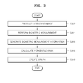
- FIG. 3 is a flowchart of a process of providing biometric information of a fetus as a graph, according to an exemplary embodiment of the present invention.
- the processor 140 produces an ultrasound image by using ultrasound data provided by the ultrasound data acquisition unit 110 (S302).
- the processor 140 performs biometric measurement on the ultrasound image (S304) and creates biometric measurement information including fetal biometric measurement parameters and their corresponding measurement values (S306). Since fetal biometric parameters may be measured by using various known methods, a detailed description thereof is omitted here.
- the biometric measurement information may be stored in the storage unit 120.
- the processor 140 calculates a measurement value in the biometric measurement information as a percentile rank or percent, based on reference information stored in the storage unit 120 (S308). In the present embodiment, the processor 140 calculates a percentile rank or percent of the measurement value contained in the biometric measurement information by using a reference value in the reference information. Since the percentile rank or percent may be calculated by using various known methods, a detailed description thereof is omitted here.
- the processor 140 may selectively inquire the storage unit 120, select a measurement parameter corresponding to first input information provided by the user input unit 130, and extract reference information about the selected measurement parameter from the storage unit 120. That is, the processor 140 may select a measurement parameter corresponding to the first input information from the biometric measurement information and extract a measurement value corresponding to the selected measurement parameter from biometric measurement information. The processor 140 may calculate a percentile rank or percent of the measurement value by using the extracted reference information.
- the processor 140 may selectively inquire the storage unit 120 and extract reference information that satisfy first input information and second input information provided by the user input unit 130 from the storage unit 120. The processor 140 may then extract a measurement value that satisfies the first input information and the second input information from the biometric measurement information. The processor 140 may calculate a percentile rank or percent of the measurement value by using the extracted reference information.
- the processor 140 may selectively inquire the storage unit 120, select the number of weeks of pregnancy corresponding to the third input information provided by the user input unit 130, and extract a measurement parameter and reference information corresponding to the selected number of weeks of pregnancy from the storage unit 120. The processor 140 may then calculate a percentile rank or percent of the measurement value by using the extracted measurement parameter and reference information.
- the processor 140 may selectively inquire the storage unit 120 and extract reference information that satisfy third input information and first input information provided by the user input unit 130 from the storage unit 120. The processor 140 may then select a measurement parameter corresponding to the first input information from the biometric measurement information and extract a measurement value corresponding to the selected measurement parameter therefrom. The processor 140 may calculate a percentile rank or percent of the measurement value by using the extracted reference information.
- the processor 140 may selectively inquire the storage unit 120 and extract from the storage unit 120 reference information that satisfy third input information, first input information, and second input information provided by the user input unit 130. The processor 140 may then extract a measurement value that satisfies the first input information and the second input information from the biometric measurement information. The processor 140 may calculate a percentile rank or percent of the measurement value by using the extracted reference information.
- the processor 140 creates a graph that represents the biometric measurement information of a fetus by using the calculated percentile rank or percent (S310). For example, as shown in FIG. 4 , the processor 140 may create a graph representing biometric measurement information of a fetus by using a percentile rank or percent for each of the measurement parameters (BPD, EFW, AC, HC, and FL).
- the processor 140 may selectively create risk level information that represents a risk level for a percentile rank or percent by colors.
- the processor 140 may selectively create first normal range information that represents a normal range for a given week of pregnancy by a line based on normal range information stored in the storage unit 120.
- a normal range may be an interval between dashed lines indicating 30W up and 30W down.
- the processor 140 may selectively create second normal range information that represents a normal range for a given week of pregnancy by dots based on normal range information stored in the storage unit 120.
- a normal range may be an interval between dots indicating 30W up and 30W down.
- the processor 140 may selectively create a graph that represents a measurement value in other biometric measurement information as a percentile rank or percent by using biometric measurement information for a particular week of pregnancy as reference.
- the processor 140 may set the last biometric measurement information 38W as reference information and create a graph that represents measurement values in pieces of biometric measurement information 30W and 25W for previous weeks of pregnancy as percentile ranks or percents simultaneously with those of the last biometric measurement information 38W.
- the processor 140 may set biometric measurement information obtained immediately prior to delivery as reference information and create a graph that represents a measurement value in biometric measurement information 30W as a percentile rank or percent by using the reference information.
- the processor 140 may set biometric measurement information obtained immediately prior to delivery as reference information and create a graph that represents measurement values in pieces of previous biometric measurement information 38W, 30W, and 25W as percentile ranks or percents by using the reference information.
- the biometric measurement information obtained immediately prior to the delivery may include expected measurement values.
- the processor 140 creates a radial graph by biometric measurement information
- the present invention is not limited thereto.
- the processor 140 may create a longitudinal or transverse bar graph, a graph of broken line, a pie chart, an area chart, a box chart, or a surface chart.
- the display unit 150 displays the ultrasound image as well as the graph generated by the processor 140.
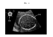
- FIG. 11 illustrates an example in which a graph 320 is displayed on an ultrasound system 100 according to an exemplary embodiment of the present invention.
- the ultrasound system 100 is configured to display an ultrasound image 310 of a cross-section of a fetal brain on a display unit 150 and perform a biometric measurement on a fetus based on the displayed ultrasound image 310.
- the ultrasound system 100 may provide biometric measurement information of the fetus as the graph 320, and biometric measurement parameters may include BPD, FL, HC, AC, and EFW.
Landscapes
- Health & Medical Sciences (AREA)
- Life Sciences & Earth Sciences (AREA)
- Engineering & Computer Science (AREA)
- Public Health (AREA)
- Medical Informatics (AREA)
- General Health & Medical Sciences (AREA)
- Biomedical Technology (AREA)
- Pathology (AREA)
- Nuclear Medicine, Radiotherapy & Molecular Imaging (AREA)
- Radiology & Medical Imaging (AREA)
- Biophysics (AREA)
- Heart & Thoracic Surgery (AREA)
- Physics & Mathematics (AREA)
- Molecular Biology (AREA)
- Surgery (AREA)
- Animal Behavior & Ethology (AREA)
- Veterinary Medicine (AREA)
- Gynecology & Obstetrics (AREA)
- Pregnancy & Childbirth (AREA)
- Physiology (AREA)
- Computer Vision & Pattern Recognition (AREA)
- Data Mining & Analysis (AREA)
- Databases & Information Systems (AREA)
- Epidemiology (AREA)
- Primary Health Care (AREA)
- Ultra Sonic Daignosis Equipment (AREA)
Abstract
Description
- This application claims the benefit of
Korean Patent Application No. 10-2012-0133988, filed on November 23, 2012 - One or more embodiments of the present invention relate to an ultrasound system, and more particularly, to an ultrasound system and method for providing biometric information of a fetus, i.e., graphical representation of fetal biometric information.
- Due to its non-invasive and non-destructive nature, an ultrasound system has been widely used in the medical field that requires information about the inside of living bodies. The ultrasound system also plays a critical role in the medical profession since it can provide real-time, high-resolution images of tissue of a living body to a doctor without the need for a surgical procedure that directly incises the living body for observation.
- The ultrasound system transmits ultrasound signals to a living body including an object (e.g., a fetus) and receives ultrasound echo signals reflected from the living body to create an ultrasound image of the living body. The ultrasound system performs fetal biometric measurements by using an ultrasound image to create biometric information. The biometric information includes measurement information such as biparietal diameter, head circumference, abdominal circumference, femur length, arm length, leg length, and nuchal translucency (NT).
- According to a conventional method, fetal measurement information is expressed in a report as a percentile for a given week of pregnancy or as a position (i.e., point) on a graph corresponding to a measurement value. In this case, a user may determine the result of measurement based on various measurement values. Thus, the conventional method may require time to understand measurement results by seeing measured values, thereby causing inconvenience to the user.
- One or more embodiments of the present invention include an ultrasound system and method for providing a graph representing biometric measurement information of a fetus by a percentile rank or percent, wherein the biometric measurement information is created by performing biometric measurement on an ultrasound image.
- Additional aspects will be set forth in part in the description which follows and, in part, will be apparent from the description, or may be learned by practice of the presented embodiments.
- According to one or more embodiments of the present invention, an ultrasound system includes: an ultrasound data acquisition unit that acquires ultrasound data of a living body including a fetus; a storage unit for storing reference information corresponding to each of a plurality of measurement parameters; and a processor that generates an ultrasound image by using the ultrasound data, performs biometric measurement based on the ultrasound image, produces biometric measurement information including biometric measurement parameters of the fetus and their corresponding measurement values, calculates the measurement values as a percentile rank or percent based on the reference information, and creates a graph that represents the biometric measurement information by using the percentile rank or percent..
- According to one or more embodiments of the present invention, a method of providing biometric information includes: acquiring ultrasound data of a living body including a fetus; generating an ultrasound image by using the ultrasound data; performing biometric measurement based on the ultrasound image to produce biometric measurement information including biometric measurement parameters of the fetus and their corresponding measurement values; calculating the measurement values as a percentile rank or percent based on reference information stored in a storage unit for storing the reference information corresponding to each of a plurality of measurement parameters; and creating a graph that represents the biometric measurement information of the fetus by using the percentile rank or percent.
- The ultrasound system and method for providing biometric information of a fetus according to embodiments of the present invention allows graphic representation of measurement values for a plurality of fetal measurement parameters, thereby providing convenience for a user to identify the overall status of the fetus at first sight.
- The level of risk for each of the fetal measurement parameters may also be represented by colors, thereby enabling a user to quickly understand the status of the fetus.
- Furthermore, percentile ranks or percents of previous measurement values for an expected week of pregnancy, which are within the margin of error, are displayed simultaneously, thereby enabling a user to conveniently identify the status of the fetus.
- In addition, previous biometric measurement information may be expressed as a percentile rank or percent by using recent biometric measurement information as reference, thereby allowing visualization of a fetal growth for a plurality of parameters.
- These and/or other aspects will become apparent and more readily appreciated from the following description of the embodiments, taken in conjunction with the accompanying drawings of which:
-
FIG. 1 is a block diagram showing a configuration of an ultrasound system according to an exemplary embodiment of the present invention; -
FIG. 2 is a block diagram showing a configuration of an ultrasound data acquisition unit in the ultrasound system ofFIG. 1 ; -
FIG. 3 is a flowchart of a process of providing biometric information of a fetus as a graph according to an exemplary embodiment of the present invention; -
FIG. 4 is an exemplary diagram of graphical representation according to an exemplary embodiment of the present invention; -
FIG. 5 is an exemplary diagram illustrating information about risk levels for a percentile rank or percent according to an exemplary embodiment of the present invention; -
FIGS. 6 and7 are exemplary diagrams illustrating a normal range for a given week of pregnancy according to an exemplary embodiment of the present invention; -
FIGS. 8 through 10 are exemplary diagrams of graphs that represent measurement values in other biometric measurement information as percentile ranks or percents, by using biometric measurement information for a particular week of pregnancy as reference; and -
FIG. 11 illustrates an example in which a graph is displayed on an ultrasound system according to an exemplary embodiment of the present invention. - Reference will now be made in detail to embodiments, examples of which are illustrated in the accompanying drawings, wherein like reference numerals refer to the like elements throughout. In this regard, the present embodiments may have different forms and should not be construed as being limited to the descriptions set forth herein. Accordingly, the embodiments are merely described below, by referring to the figures, to explain aspects of the present description. Expressions such as "at least one of," when preceding a list of elements, modify the entire list of elements and do not modify the individual elements of the list.
- Hereinafter, exemplary embodiments of the present invention will be described with reference to the accompanying drawings.
-
FIG. 1 is a block diagram showing a configuration of anultrasound system 100 according to an exemplary embodiment of the present invention. Referring toFIG. 1 , theultrasound system 100 according to the present embodiment includes an ultrasounddata acquisition unit 110, astorage unit 120, auser input unit 130, aprocessor 140, and adisplay unit 150. - The ultrasound
data acquisition unit 110 acquires ultrasound data corresponding to an ultrasound image of a living body including an object such as a fetus. The ultrasound image includes a brightness (B) mode image, but is not limited thereto. The ultrasound data includes radio frequency (RF) data, but is not limited thereto. -
FIG. 2 is a block diagram showing a configuration of the ultrasounddata acquisition unit 110. Referring toFIG. 2 , the ultrasounddata acquisition unit 110 includes anultrasound probe 210, atransmitter 220, areceiver 230, and an ultrasounddata forming section 240. - The
ultrasound probe 210 includes a plurality of transducer elements (not shown) that convert electrical signals into ultrasound signals, and vice versa. Theultrasound probe 210 is configured to transmit an ultrasound signal to a living body and receive an ultrasound echo signal reflected from the living body to generate an electrical signal (hereinafter referred to as a "reception signal"). The reception signal is an analog signal. Theultrasound probe 210 includes a convex probe, a linear probe, and a three-dimensional (3D) probe. - The
transmitter 220 controls the transmission of an ultrasound signal. Thetransmitter 220 also produces an electrical signal (hereinafter referred to as a "transmission signal") that is used to obtain an ultrasound image in consideration of the transducer elements and a focal point. Thus, upon receipt of the transmission signal from thetransmitter 220, theultrasound probe 210 converts the transmission signal into an ultrasound signal, transmits the ultrasound signal to a living body, and creates a reception signal based on an ultrasound echo signal reflected from the living body. - The
receiver 230 performs analog-to-digital conversion on the reception signal provided by theultrasound probe 210 to produce a digital signal. Thereceiver 230 also performs reception beamforming on the digital signal in consideration of the transducer elements and a focal point to create a focused reception signal. Since the reception beamforming may be performed by using various known methods, a detailed description thereof is omitted here. - The ultrasound
data forming section 240 creates ultrasound data corresponding to an ultrasound image by using the focused reception signal provided by thereceiver 230. The ultrasounddata forming section 240 may also perform various signal processings, such as gain control, needed to form ultrasound data, on the focused reception signal. - Although the ultrasound
data acquisition unit 110 has been described to transmit an ultrasound signal to a living body and receives an ultrasound echo signal reflected from the living body to acquire ultrasound data corresponding to an ultrasound image, the ultrasounddata acquisition unit 110 may acquire ultrasound data from an external or internal device (not shown) connected to theultrasound system 100 in a wired or wireless manner. - Referring back to
FIG. 1 , thestorage unit 120 stores ultrasound data acquired by the ultrasounddata acquisition unit 110 as well as reference information corresponding to a plurality of measurement parameters. In the present embodiment, the measurement parameters are used for measuring an object (i.e., a fetus) and includes biparietal diameter (BPD), head circumference (HC), femur length (FL), abdominal circumference (AC), expected fetal weight (EFW), Crown-Rump Length (CRL), a heart rate, and a ratio between two of the measurement parameters. The measurement parameters may also include measurement parameters of a fetal brain, such as OccipitoFrontal Diameter (OFD), Anterior Cerebral Ventricule Diameter (Va), Posterior Cerebral Ventricle Diameter (Vp), Transverse Cerebellar Diameter (TCD), and Cisterna Magna (CM). The measurement parameters may further include cardiac biometric measurement parameters such as Cardiac Circumference, Thoracic Circumference, Left Ventricular Diameter, Right Ventricular Diameter, Aortic Valve Annulus Diameter, and Pulmonary Valve Annulus Diameter. However, the measurement parameters are not limited thereto. The reference information includes reference values needed for converting measurement values for measurement parameters to percentile ranks or percents. Since various known types of information such as a reference table may be used as the reference information, a detailed description thereof is omitted here. - For one example, the
storage unit 120 may store a mapping table for providing reference information corresponding to each of the plurality of measurement parameters. Alternatively, thestorage unit 120 may store a mapping table for providing a plurality of pieces of reference information corresponding to each of the measurement parameters. For another example, thestorage unit 120 may store a mapping table for providing reference information corresponding to each of the measurement parameters for each week of pregnancy. Alternatively, thestorage unit 120 may store a mapping table for providing a plurality of pieces of reference information corresponding to each of the measurement parameters. - The
storage unit 120 also stores normal range information including a normal range of a percentile rank or percent for each week of pregnancy. - The
user input unit 130 receives user input information. In the present embodiment, the user input information includes first input information needed for selecting a measurement parameter to be represented by a graph among the plurality of measurement parameters. The user input information also includes second input information needed for selecting reference information from the selected measurement parameter. The user input information further includes third input information needed for selecting the number of weeks of pregnancy. Theuser input unit 130 includes a control panel, a track ball, a touch screen, a keyboard, and a mouse. - The
processor 140 is connected to the ultrasounddata acquisition unit 110, thestorage unit 120, and theuser input unit 130. Theprocessor 140 includes a central processing unit (CPU), a microprocessor, and a graphic processing unit (GPU). -
FIG. 3 is a flowchart of a process of providing biometric information of a fetus as a graph, according to an exemplary embodiment of the present invention. Referring toFIG. 3 , theprocessor 140 produces an ultrasound image by using ultrasound data provided by the ultrasound data acquisition unit 110 (S302). - The
processor 140 performs biometric measurement on the ultrasound image (S304) and creates biometric measurement information including fetal biometric measurement parameters and their corresponding measurement values (S306). Since fetal biometric parameters may be measured by using various known methods, a detailed description thereof is omitted here. The biometric measurement information may be stored in thestorage unit 120. - The
processor 140 calculates a measurement value in the biometric measurement information as a percentile rank or percent, based on reference information stored in the storage unit 120 (S308). In the present embodiment, theprocessor 140 calculates a percentile rank or percent of the measurement value contained in the biometric measurement information by using a reference value in the reference information. Since the percentile rank or percent may be calculated by using various known methods, a detailed description thereof is omitted here. - The
processor 140 may selectively inquire thestorage unit 120, select a measurement parameter corresponding to first input information provided by theuser input unit 130, and extract reference information about the selected measurement parameter from thestorage unit 120. That is, theprocessor 140 may select a measurement parameter corresponding to the first input information from the biometric measurement information and extract a measurement value corresponding to the selected measurement parameter from biometric measurement information. Theprocessor 140 may calculate a percentile rank or percent of the measurement value by using the extracted reference information. - The
processor 140 may selectively inquire thestorage unit 120 and extract reference information that satisfy first input information and second input information provided by theuser input unit 130 from thestorage unit 120. Theprocessor 140 may then extract a measurement value that satisfies the first input information and the second input information from the biometric measurement information. Theprocessor 140 may calculate a percentile rank or percent of the measurement value by using the extracted reference information. - The
processor 140 may selectively inquire thestorage unit 120, select the number of weeks of pregnancy corresponding to the third input information provided by theuser input unit 130, and extract a measurement parameter and reference information corresponding to the selected number of weeks of pregnancy from thestorage unit 120. Theprocessor 140 may then calculate a percentile rank or percent of the measurement value by using the extracted measurement parameter and reference information. - The
processor 140 may selectively inquire thestorage unit 120 and extract reference information that satisfy third input information and first input information provided by theuser input unit 130 from thestorage unit 120. Theprocessor 140 may then select a measurement parameter corresponding to the first input information from the biometric measurement information and extract a measurement value corresponding to the selected measurement parameter therefrom. Theprocessor 140 may calculate a percentile rank or percent of the measurement value by using the extracted reference information. - The
processor 140 may selectively inquire thestorage unit 120 and extract from thestorage unit 120 reference information that satisfy third input information, first input information, and second input information provided by theuser input unit 130. Theprocessor 140 may then extract a measurement value that satisfies the first input information and the second input information from the biometric measurement information. Theprocessor 140 may calculate a percentile rank or percent of the measurement value by using the extracted reference information. - The
processor 140 creates a graph that represents the biometric measurement information of a fetus by using the calculated percentile rank or percent (S310). For example, as shown inFIG. 4 , theprocessor 140 may create a graph representing biometric measurement information of a fetus by using a percentile rank or percent for each of the measurement parameters (BPD, EFW, AC, HC, and FL). - As shown in
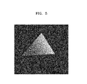
FIG. 5 , theprocessor 140 may selectively create risk level information that represents a risk level for a percentile rank or percent by colors. - In addition, as shown in
FIG. 6 , theprocessor 140 may selectively create first normal range information that represents a normal range for a given week of pregnancy by a line based on normal range information stored in thestorage unit 120. For example, for a 30 week old fetus, a normal range may be an interval between dashed lines indicating 30W up and 30W down. - Furthermore, as shown in
FIG. 7 , theprocessor 140 may selectively create second normal range information that represents a normal range for a given week of pregnancy by dots based on normal range information stored in thestorage unit 120. For example, for a 30 week old fetus, a normal range may be an interval between dots indicating 30W up and 30W down. - The
processor 140 may selectively create a graph that represents a measurement value in other biometric measurement information as a percentile rank or percent by using biometric measurement information for a particular week of pregnancy as reference. - For one example, as shown in
FIG. 8 , theprocessor 140 may set the lastbiometric measurement information 38W as reference information and create a graph that represents measurement values in pieces ofbiometric measurement information biometric measurement information 38W. - For another example, as shown in
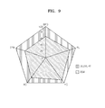
FIG. 9 , theprocessor 140 may set biometric measurement information obtained immediately prior to delivery as reference information and create a graph that represents a measurement value inbiometric measurement information 30W as a percentile rank or percent by using the reference information. - Alternatively, as shown in
FIG. 10 , theprocessor 140 may set biometric measurement information obtained immediately prior to delivery as reference information and create a graph that represents measurement values in pieces of previousbiometric measurement information - While the
processor 140 creates a radial graph by biometric measurement information, the present invention is not limited thereto. For example, theprocessor 140 may create a longitudinal or transverse bar graph, a graph of broken line, a pie chart, an area chart, a box chart, or a surface chart. - Referring back to
FIG. 1 , thedisplay unit 150 displays the ultrasound image as well as the graph generated by theprocessor 140. -
FIG. 11 illustrates an example in which a graph 320 is displayed on anultrasound system 100 according to an exemplary embodiment of the present invention. Referring toFIG. 11 , theultrasound system 100 is configured to display anultrasound image 310 of a cross-section of a fetal brain on adisplay unit 150 and perform a biometric measurement on a fetus based on the displayedultrasound image 310. As illustrated inFIG. 11 , theultrasound system 100 may provide biometric measurement information of the fetus as the graph 320, and biometric measurement parameters may include BPD, FL, HC, AC, and EFW. - While one or more embodiments of the present invention have been described with reference to the figures, it will be understood by those of ordinary skill in the art that various changes in form and details may be made therein without departing from the spirit and scope of the present invention as defined by the following claims.
Claims (15)
- An ultrasound system comprising
an ultrasound data acquisition unit that acquires ultrasound data of a living body including a fetus;
a storage unit for storing reference information corresponding to each of a plurality of measurement parameters; and
a processor that generates an ultrasound image by using the ultrasound data, performs biometric measurement based on the ultrasound image, produces biometric measurement information including biometric measurement parameters of the fetus and their corresponding measurement values, calculates the measurement values as a percentile rank or percent based on the reference information, and creates a graph that represents the biometric measurement information by using the percentile rank or percent. - The ultrasound system of claim 1, further comprising a user input unit for receiving at least one of first input information needed for selecting at least one of the plurality of measurement parameters and second input information needed for selecting reference information about the at least one measurement parameter selected in response to the first input information,
wherein the processor creates a graph that represents measurement information corresponding to the at least one measurement parameter. - The ultrasound system of claim 2, wherein the processor inquires the storage unit to select a measurement parameter corresponding to the first input information, extracts reference information corresponding to the selected measurement parameter from the storage unit, extracts a measurement value corresponding to the selected measurement parameter from the biometric measurement information, and calculates the percentile rank or percent of the measurement value by using the extracted reference information.
- The ultrasound system of claim 2, wherein the processor inquires the storage unit to extract reference information corresponding to the second input information from the storage unit, extracts a measurement value corresponding to the selected measurement parameter from the biometric measurement information, and calculates the percentile rank or percent of the measurement value by using the extracted reference information.
- The ultrasound system of claim 1, wherein the storage unit further stores reference information corresponding to each of the plurality of measurement parameters of the fetus for each week of pregnancy.
- The ultrasound system of claim 5, further comprising a user input unit for receiving at least one of first input information needed for selecting at least one of the plurality of measurement parameters and third input information needed for selecting the number of weeks of pregnancy for the selected measurement parameter.
- The ultrasound system of claim 6, wherein the processor inquires the storage unit to extract reference information corresponding to the third input information, extracts a measurement value corresponding to the selected measurement parameter from the biometric measurement information, and calculates the percentile rank or percent of the measurement value by using the extracted reference information.
- The ultrasound system of claim 1, wherein the storage unit further stores normal range information including a normal range of the percentile rank or percent for each week of pregnancy.
- A method of providing biometric information, the method comprising:acquiring ultrasound data of a living body including a fetus;generating an ultrasound image by using the ultrasound data,performing biometric measurement based on the ultrasound image to produce biometric measurement information including biometric measurement parameters of the fetus and their corresponding measurement values;calculating the measurement values as a percentile rank or percent based on reference information stored in a storage unit for storing the reference information corresponding to each a plurality of measurement parameters; andcreating a graph that represents the biometric measurement information of the fetus by using the percentile rank or percent.
- The method of claim 9, prior to the calculating of the measurement values as a percentile rank or percent, further comprising receiving at least one of first input information needed for selecting at least one of the plurality of measurement parameters and second input information needed for selecting reference information about the at least one measurement parameter selected in response to the first input information,
wherein in the creating of the graph, the graph is created so as to represent measurement information corresponding to the at least one measurement parameter - The method of claim 10, wherein the calculating of the measurement values as a percentile rank or percent comprises:inquiring the storage unit to select a measurement parameter corresponding to the first input information;extracting reference information corresponding to the selected measurement parameter from the storage unit;extracting a measurement value corresponding to the selected measurement parameter from the biometric measurement information; andcalculating the percentile rank or percent of the measurement value by using the extracted reference information.
- The method of claim 10, wherein the calculating of the measurement values as a percentile rank or percent comprises:inquiring the storage unit to extract reference information corresponding to the second input information from the storage unit;extracting a measurement value corresponding to the selected measurement parameter from the biometric measurement information; andcalculating the percentile rank or percent of the measurement value by using the extracted reference information.
- The method of claim 9, wherein the storage unit further stores reference information corresponding to each of the plurality of measurement parameters of the fetus for each week of pregnancy.
- The method of claim 13, prior to the calculating of the measurement values as a percentile rank or percent, further comprising receiving at least one of first input information needed for selecting at least one of the plurality of measurement parameters and third input information needed for selecting the number of weeks of pregnancy for the selected measurement parameter.
- The method of claim 14, wherein the calculating of the measurement value as a percentile rank or percent comprises:Inquiring the storage unit to extract reference information corresponding to the third input information;extracting a measurement value corresponding to the selected measurement parameter from the biometric measurement information; andcalculating the percentile rank or percent of the measurement value by using the extracted reference information.
Applications Claiming Priority (1)
| Application Number | Priority Date | Filing Date | Title |
|---|---|---|---|
| KR1020120133988A KR20140066573A (en) | 2012-11-23 | 2012-11-23 | Ultrasound system and method for providing biometry information of fetus |
Publications (2)
| Publication Number | Publication Date |
|---|---|
| EP2735272A2 true EP2735272A2 (en) | 2014-05-28 |
| EP2735272A3 EP2735272A3 (en) | 2017-08-02 |
Family
ID=49709462
Family Applications (1)
| Application Number | Title | Priority Date | Filing Date |
|---|---|---|---|
| EP13193996.9A Withdrawn EP2735272A3 (en) | 2012-11-23 | 2013-11-22 | Ultrasound system and method for providing biometric information of fetus |
Country Status (3)
| Country | Link |
|---|---|
| US (1) | US20140148696A1 (en) |
| EP (1) | EP2735272A3 (en) |
| KR (1) | KR20140066573A (en) |
Cited By (3)
| Publication number | Priority date | Publication date | Assignee | Title |
|---|---|---|---|---|
| CN109640831A (en) * | 2016-09-01 | 2019-04-16 | 皇家飞利浦有限公司 | Supersonic diagnostic appts |
| WO2021165037A1 (en) * | 2020-02-20 | 2021-08-26 | Koninklijke Philips N.V. | Methods and systems for fetal heart assessment |
| EP3888558A1 (en) * | 2020-04-02 | 2021-10-06 | Koninklijke Philips N.V. | Methods and systems for fetal heart assessment |
Families Citing this family (3)
| Publication number | Priority date | Publication date | Assignee | Title |
|---|---|---|---|---|
| KR101630071B1 (en) * | 2014-10-24 | 2016-06-14 | 한국표준과학연구원 | Method and system for evaluating fetal growth state using Fetal biometric data |
| WO2018109114A1 (en) * | 2016-12-15 | 2018-06-21 | Koninklijke Philips N.V. | Prenatal ultrasound imaging |
| CN112155603B (en) * | 2020-09-24 | 2023-06-09 | 广州爱孕记信息科技有限公司 | Weight value determining method and device for fetal structural characteristics |
Family Cites Families (4)
| Publication number | Priority date | Publication date | Assignee | Title |
|---|---|---|---|---|
| US20060247527A1 (en) * | 2005-04-28 | 2006-11-02 | Kabushiki Kaisha Toshiba | Ultrasonic diagnostic apparatus, measuring result display apparatus and method of displaying measuring result |
| EP1743581B8 (en) * | 2005-07-11 | 2016-09-21 | Esaote S.p.A. | Method and system for foetal weight evaluation |
| JP5481108B2 (en) * | 2009-06-26 | 2014-04-23 | 株式会社東芝 | Ultrasonic diagnostic apparatus and automatic diagnosis support apparatus |
| KR20120049777A (en) * | 2010-11-09 | 2012-05-17 | 삼성메디슨 주식회사 | Ultrasound system and method for providing biometry information of fetus |
-
2012
- 2012-11-23 KR KR1020120133988A patent/KR20140066573A/en not_active Ceased
-
2013
- 2013-11-22 US US14/088,219 patent/US20140148696A1/en not_active Abandoned
- 2013-11-22 EP EP13193996.9A patent/EP2735272A3/en not_active Withdrawn
Non-Patent Citations (1)
| Title |
|---|
| None |
Cited By (5)
| Publication number | Priority date | Publication date | Assignee | Title |
|---|---|---|---|---|
| CN109640831A (en) * | 2016-09-01 | 2019-04-16 | 皇家飞利浦有限公司 | Supersonic diagnostic appts |
| CN109640831B (en) * | 2016-09-01 | 2022-06-28 | 皇家飞利浦有限公司 | Ultrasound diagnostic equipment |
| WO2021165037A1 (en) * | 2020-02-20 | 2021-08-26 | Koninklijke Philips N.V. | Methods and systems for fetal heart assessment |
| CN115151193A (en) * | 2020-02-20 | 2022-10-04 | 皇家飞利浦有限公司 | Method and system for fetal cardiac assessment |
| EP3888558A1 (en) * | 2020-04-02 | 2021-10-06 | Koninklijke Philips N.V. | Methods and systems for fetal heart assessment |
Also Published As
| Publication number | Publication date |
|---|---|
| EP2735272A3 (en) | 2017-08-02 |
| US20140148696A1 (en) | 2014-05-29 |
| KR20140066573A (en) | 2014-06-02 |
Similar Documents
| Publication | Publication Date | Title |
|---|---|---|
| KR102452998B1 (en) | Ultrasonic Diagnostic Apparatus | |
| EP2783635B1 (en) | Ultrasound system and method of providing direction information of object | |
| JP5730196B2 (en) | Ultrasonic diagnostic apparatus, ultrasonic image processing apparatus, and ultrasonic image generation method | |
| CN101380239B (en) | Ultrasonic image processing apparatus | |
| US20140148696A1 (en) | Ultrasound system and method for providing biometric information of fetus | |
| CN102415902B (en) | Ultrasonic diagnostic apparatus and ultrasonic image processng apparatus | |
| US10368841B2 (en) | Ultrasound diagnostic apparatus | |
| JP6118021B2 (en) | Ultrasonic diagnostic apparatus and image display method | |
| KR20030078760A (en) | Image processing unit and ultrasonic diagnostic device | |
| CN102090902A (en) | Medical image processing apparatus, a medical image processing method, and ultrasonic diagnosis apparatus | |
| EP2989987B1 (en) | Ultrasound diagnosis apparatus and method and computer readable storage medium | |
| JP7075854B2 (en) | Ultrasonic diagnostic equipment and display method | |
| JP4667394B2 (en) | Ultrasonic diagnostic equipment | |
| CN104055539A (en) | Apparatus and method for providing elasticity information | |
| KR20140131808A (en) | Ultrasound imaging apparatus and control method for the same | |
| CN107198546A (en) | Diagnostic ultrasound equipment, image processing apparatus and image processing method | |
| WO2012161040A1 (en) | Ultrasonic diagnosis device and method for presenting items for inspection | |
| US20220313214A1 (en) | Ultrasonic diagnostic apparatus, image processing apparatus, and image processing method | |
| JP2019154654A (en) | Ultrasonic imaging device and ultrasonic image processing system | |
| CN111317508B (en) | Ultrasonic diagnostic equipment, medical information processing equipment, computer program products | |
| US10761198B2 (en) | Method and apparatus for acquiring image using ultrasound | |
| KR101868019B1 (en) | Ultrasound Imaging Apparatus and Method for processing a ultrasound image thereof | |
| KR101014559B1 (en) | Ultrasound System and Method for Providing 3D Ultrasound Image | |
| JP2010022418A (en) | Ultrasonic image diagnostic apparatus | |
| JP6828581B2 (en) | Medical image display device, measurement method and program |
Legal Events
| Date | Code | Title | Description |
|---|---|---|---|
| PUAI | Public reference made under article 153(3) epc to a published international application that has entered the european phase |
Free format text: ORIGINAL CODE: 0009012 |
|
| 17P | Request for examination filed |
Effective date: 20131122 |
|
| AK | Designated contracting states |
Kind code of ref document: A2 Designated state(s): AL AT BE BG CH CY CZ DE DK EE ES FI FR GB GR HR HU IE IS IT LI LT LU LV MC MK MT NL NO PL PT RO RS SE SI SK SM TR |
|
| AX | Request for extension of the european patent |
Extension state: BA ME |
|
| PUAL | Search report despatched |
Free format text: ORIGINAL CODE: 0009013 |
|
| AK | Designated contracting states |
Kind code of ref document: A3 Designated state(s): AL AT BE BG CH CY CZ DE DK EE ES FI FR GB GR HR HU IE IS IT LI LT LU LV MC MK MT NL NO PL PT RO RS SE SI SK SM TR |
|
| AX | Request for extension of the european patent |
Extension state: BA ME |
|
| RIC1 | Information provided on ipc code assigned before grant |
Ipc: A61B 8/00 20060101ALI20170628BHEP Ipc: A61B 8/08 20060101AFI20170628BHEP |
|
| STAA | Information on the status of an ep patent application or granted ep patent |
Free format text: STATUS: THE APPLICATION IS DEEMED TO BE WITHDRAWN |
|
| 18D | Application deemed to be withdrawn |
Effective date: 20180203 |