EP2734548B1 - Tnf-alpha-antigen-binding proteins against with increased fcrn binding for use in therapy - Google Patents
Tnf-alpha-antigen-binding proteins against with increased fcrn binding for use in therapy Download PDFInfo
- Publication number
- EP2734548B1 EP2734548B1 EP12738101.0A EP12738101A EP2734548B1 EP 2734548 B1 EP2734548 B1 EP 2734548B1 EP 12738101 A EP12738101 A EP 12738101A EP 2734548 B1 EP2734548 B1 EP 2734548B1
- Authority
- EP
- European Patent Office
- Prior art keywords
- seq
- sequence
- antigen binding
- binding protein
- tnf
- Prior art date
- Legal status (The legal status is an assumption and is not a legal conclusion. Google has not performed a legal analysis and makes no representation as to the accuracy of the status listed.)
- Active
Links
Images


Classifications
-
- C—CHEMISTRY; METALLURGY
- C07—ORGANIC CHEMISTRY
- C07K—PEPTIDES
- C07K16/00—Immunoglobulins [IG], e.g. monoclonal or polyclonal antibodies
- C07K16/18—Immunoglobulins [IG], e.g. monoclonal or polyclonal antibodies against material from animals or humans
- C07K16/24—Immunoglobulins [IG], e.g. monoclonal or polyclonal antibodies against material from animals or humans against cytokines, lymphokines or interferons
- C07K16/241—Tumor Necrosis Factors
-
- A—HUMAN NECESSITIES
- A61—MEDICAL OR VETERINARY SCIENCE; HYGIENE
- A61K—PREPARATIONS FOR MEDICAL, DENTAL OR TOILETRY PURPOSES
- A61K39/00—Medicinal preparations containing antigens or antibodies
- A61K39/395—Antibodies; Immunoglobulins; Immune serum, e.g. antilymphocytic serum
- A61K39/39591—Stabilisation, fragmentation
-
- A—HUMAN NECESSITIES
- A61—MEDICAL OR VETERINARY SCIENCE; HYGIENE
- A61P—SPECIFIC THERAPEUTIC ACTIVITY OF CHEMICAL COMPOUNDS OR MEDICINAL PREPARATIONS
- A61P19/00—Drugs for skeletal disorders
- A61P19/02—Drugs for skeletal disorders for joint disorders, e.g. arthritis, arthrosis
-
- A—HUMAN NECESSITIES
- A61—MEDICAL OR VETERINARY SCIENCE; HYGIENE
- A61P—SPECIFIC THERAPEUTIC ACTIVITY OF CHEMICAL COMPOUNDS OR MEDICINAL PREPARATIONS
- A61P29/00—Non-central analgesic, antipyretic or antiinflammatory agents, e.g. antirheumatic agents; Non-steroidal antiinflammatory drugs [NSAID]
-
- A—HUMAN NECESSITIES
- A61—MEDICAL OR VETERINARY SCIENCE; HYGIENE
- A61P—SPECIFIC THERAPEUTIC ACTIVITY OF CHEMICAL COMPOUNDS OR MEDICINAL PREPARATIONS
- A61P37/00—Drugs for immunological or allergic disorders
-
- A—HUMAN NECESSITIES
- A61—MEDICAL OR VETERINARY SCIENCE; HYGIENE
- A61P—SPECIFIC THERAPEUTIC ACTIVITY OF CHEMICAL COMPOUNDS OR MEDICINAL PREPARATIONS
- A61P43/00—Drugs for specific purposes, not provided for in groups A61P1/00-A61P41/00
-
- C—CHEMISTRY; METALLURGY
- C07—ORGANIC CHEMISTRY
- C07K—PEPTIDES
- C07K2317/00—Immunoglobulins specific features
- C07K2317/20—Immunoglobulins specific features characterized by taxonomic origin
- C07K2317/21—Immunoglobulins specific features characterized by taxonomic origin from primates, e.g. man
-
- C—CHEMISTRY; METALLURGY
- C07—ORGANIC CHEMISTRY
- C07K—PEPTIDES
- C07K2317/00—Immunoglobulins specific features
- C07K2317/40—Immunoglobulins specific features characterized by post-translational modification
- C07K2317/41—Glycosylation, sialylation, or fucosylation
-
- C—CHEMISTRY; METALLURGY
- C07—ORGANIC CHEMISTRY
- C07K—PEPTIDES
- C07K2317/00—Immunoglobulins specific features
- C07K2317/50—Immunoglobulins specific features characterized by immunoglobulin fragments
- C07K2317/56—Immunoglobulins specific features characterized by immunoglobulin fragments variable (Fv) region, i.e. VH and/or VL
- C07K2317/565—Complementarity determining region [CDR]
-
- C—CHEMISTRY; METALLURGY
- C07—ORGANIC CHEMISTRY
- C07K—PEPTIDES
- C07K2317/00—Immunoglobulins specific features
- C07K2317/70—Immunoglobulins specific features characterized by effect upon binding to a cell or to an antigen
- C07K2317/71—Decreased effector function due to an Fc-modification
-
- C—CHEMISTRY; METALLURGY
- C07—ORGANIC CHEMISTRY
- C07K—PEPTIDES
- C07K2317/00—Immunoglobulins specific features
- C07K2317/70—Immunoglobulins specific features characterized by effect upon binding to a cell or to an antigen
- C07K2317/72—Increased effector function due to an Fc-modification
-
- C—CHEMISTRY; METALLURGY
- C07—ORGANIC CHEMISTRY
- C07K—PEPTIDES
- C07K2317/00—Immunoglobulins specific features
- C07K2317/70—Immunoglobulins specific features characterized by effect upon binding to a cell or to an antigen
- C07K2317/73—Inducing cell death, e.g. apoptosis, necrosis or inhibition of cell proliferation
-
- C—CHEMISTRY; METALLURGY
- C07—ORGANIC CHEMISTRY
- C07K—PEPTIDES
- C07K2317/00—Immunoglobulins specific features
- C07K2317/70—Immunoglobulins specific features characterized by effect upon binding to a cell or to an antigen
- C07K2317/76—Antagonist effect on antigen, e.g. neutralization or inhibition of binding
-
- C—CHEMISTRY; METALLURGY
- C07—ORGANIC CHEMISTRY
- C07K—PEPTIDES
- C07K2317/00—Immunoglobulins specific features
- C07K2317/90—Immunoglobulins specific features characterized by (pharmaco)kinetic aspects or by stability of the immunoglobulin
-
- C—CHEMISTRY; METALLURGY
- C07—ORGANIC CHEMISTRY
- C07K—PEPTIDES
- C07K2317/00—Immunoglobulins specific features
- C07K2317/90—Immunoglobulins specific features characterized by (pharmaco)kinetic aspects or by stability of the immunoglobulin
- C07K2317/92—Affinity (KD), association rate (Ka), dissociation rate (Kd) or EC50 value
-
- C—CHEMISTRY; METALLURGY
- C07—ORGANIC CHEMISTRY
- C07K—PEPTIDES
- C07K2317/00—Immunoglobulins specific features
- C07K2317/90—Immunoglobulins specific features characterized by (pharmaco)kinetic aspects or by stability of the immunoglobulin
- C07K2317/94—Stability, e.g. half-life, pH, temperature or enzyme-resistance
Definitions
- the invention relates to novel variants of anti-TNF antibodies with increased half-life for use in a method of treating a disease.
- Antibodies are heteromultimeric glycoproteins comprising at least two heavy and two light chains. Aside from IgM, intact antibodies are usually heterotetrameric glycoproteins of approximately 150Kda, composed of two identical light (L) chains and two identical heavy (H) chains. Each heavy chain has at one end a variable domain (VH) followed by a number of constant regions. Each light chain has a variable domain (VL) and a constant region at its other end; the constant region of the light chain is aligned with the first constant region of the heavy chain and the light chain variable domain is aligned with the variable domain of the heavy chain.
- VH variable domain
- VL variable domain
- human antibodies can be assigned to five different classes, IgA, IgD, IgE, IgG and IgM.
- IgG and IgA can be further subdivided into subclasses, IgG1, IgG2, IgG3 and IgG4; and IgA1 and IgA2.
- the variable domain of the antibody confers binding specificity upon the antibody with certain regions displaying particular variability called complementarity determining regions (CDRs).
- CDRs complementarity determining regions
- the more conserved portions of the variable region are called Framework regions (FR).
- the variable domains of intact heavy and light chains each comprise four FR connected by three CDRs.
- the constant regions are not directly involved in the binding of the antibody to the antigen but exhibit various effector functions such as participation in antibody dependent cell-mediated cytotoxicity (ADCC), phagocytosis via binding to Fc ⁇ receptor, half-life/clearance rate via neonatal Fc receptor (FcRn) and complement dependent cytotoxicity via the C1q component of the complement cascade.
- ADCC antibody dependent cell-mediated cytotoxicity
- FcRn neonatal Fc receptor
- complement dependent cytotoxicity via the C1q component of the complement cascade.
- the nature of the structure of an IgG antibody is such that there are two antigen-binding sites, both of which are specific for the same epitope. They are therefore, monospecific.
- FcRn also known as the neonatal Fc receptor
- IgG molecules are endocytosed by endothelial cells, and if they bind to FcRn, are recycled out into circulation.
- IgG molecules that do not bind to FcRn enter the cells and are targeted to the lysosomal pathway where they are degraded.
- the neonatal FcRn receptor is believed to be involved in both antibody clearance and the transcytosis across tissues (see Junghans R.P (1997) Immunol.Res 16. 29-57 and Ghetie et al (2000) Annu.Rev.lmmunol. 18, 739-766 ).
- WO 9734631 discloses a composition comprising a mutant IgG molecule having increased serum half-life and at least one amino acid substitution in the Fc-hinge region. Amino acid substitution at one or more of the amino acids selected from number 252, 254, 256, 309, 311 or 315 in the CH2 domain or 433 or 434 in the CH3 domain is disclosed.
- WO 00/42072 discloses a polypeptide comprising a variant Fc region with altered FcRn binding affinity, which polypeptide comprises an amino acid modification at any one or more of amino acid positions 238, 252, 253, 254, 255, 256, 265, 272, 286, 288, 303, 305, 307, 309, 311, 312, 317, 340, 356, 360, 362, 376, 378, 380, 386,388, 400, 413, 415, 424,433, 434,435, 436, 439, and 447 of the Fc region.
- WO 02/060919 discloses a modified IgG comprising an IgG constant domain comprising amino acid modifications at one or more of positions 251, 253, 255, 285-290, 308-314, 385-389, and 428-435.
- WO 2004035752 discloses a modified antibody of class IgG wherein at least one amino acid residue from the heavy chain constant region selected from the group consisting of amino acid residues 250, 314, and 428 is different from that present in an unmodified class IgG antibody.
- Dall'Acqua et al. (2002, J Immunol.;169:5171-80 ) described random mutagenesis and screening of human IgG1 hinge-Fc fragment phage display libraries against mouse FcRn. They disclosed random mutagenesis of positions 251, 252, 254-256, 308, 309, 311, 312, 314, 385-387, 389, 428, 433, 434, and 436.
- WO2006130834 discloses modified IgG comprising an IgG comprising an IgG constant domain comprising amino acid modifications at one or more positions of 252, 254, 256, 433, 434 and 436. Therefore, modification of Fc domains of IgG antibodies has been discussed as a means of increasing the serum half- life of therapeutic antibodies. However, numerous such modifications have been suggested with varying and sometimes contradictory results in different antibodies. For example, US 2009/0163699 relates to Fc variants that have altered binding to FcRn and/or improved half life. One such modification disclosed relates to the mutations herein referred to as the 'YTE' mutations.
- antigen binding proteins as therapeutics requires injections with a prescribed frequency relating to the clearance and half-life characteristics of the protein.
- Adalimumab is a monoclonal antibody against TNF-alpha which is used for treatment of rheumatoid arthritis, psoriatic arthritis, ankylosing spondylitis, and Crohn's disease. It is produced by recombinant DNA technology using a mammalian cell expression system. It consists of 330 amino acids and has a molecular weight of approximately 148 kilodaltons. See United States Patent 6090382 .
- clearance for adalimumab is said to range from 11 to 15 ml/hour, the distribution volume (V ss ) ranges from 5 to 6 litres and the mean terminal phase half-life was approximately two weeks (Summary of Product Characteristics available from www.medicines.org.uk). These half life and clearance properties mean that currently adalimumab needs to be administered once every two weeks. In some patients depending on disease it may be necessary to administer a loading dose such as for example in psoriasis patients. This dosage may differ from the maintenance dose.
- the invention relates to an antigen binding protein which specifically binds to TNF-alpha comprising:
- the antigen binding protein has a human IgG1 constant domain of SEQ ID No. 13 before amino acid substitutions are introduced.
- the antigen binding protein is administered to patients subcutaneously as a single 40 mg dose no more than once every four weeks or no more than once every eight weeks.
- the half-life of the antigen binding protein is increased 2 fold, 3 fold, 4 fold or 5 fold as compared to the native IgG.
- the administration of the antigen binding protein no more than once every four weeks in patients achieves the mean steady-state trough concentration in the patient population of between 4 ⁇ g/ml to 7 ⁇ g/ml or for example between 5 ⁇ g/ml to 6 ⁇ g/ml.
- the clearance of the antigen binding protein is 2ml/ hr to 4ml/hr.
- the antigen binding protein comprises a constant domain as shown in SEQ ID No: 7
- the antigen binding protein is an antibody.
- the antigen binding protein is to be administered with methotrexate, preferably wherein the antigen binding protein is administered for the treatment of rheumatoid arthritis.
- the antigen binding protein comprises the heavy chain sequence as shown in SEQ ID No 5 optionally with a light chain sequence as shown in SEQ ID No: 2.
- the antigen binding protein is for use in the treatment of rheumatoid arthritis, polyarticular juvenile idiopathic arthritis, psoriatic arthritis, ankylosing spondylitis, Crohn's disease or Psoriasis.
- human IgG1 constant domain encompasses all allotypes and variants thereof known to a person skilled in the art.
- the invention relates to novel antigen binding proteins binding specifically to TNF-alpha comprising:
- Adalimumab is an IgG monoclonal antibody comprising the light chain sequence of SEQ ID No. 2 and heavy chain sequence of SEQ ID No.12.
- adalimumab as described herein show particular improvements in FcRn binding as shown in the examples below.
- Affinity matured variants of adalimumab also show improvement in anti-TNF-alpha binding and/or neutralisation activity.
- novel antigen binding proteins have an increased binding to the FcRn receptor and increased half life and/ or increased Mean Residence Time and/ or decreased Clearance. It is considered that binding to FcRn results in longer serum retention in vivo. In order to increase the retention of the Fc proteins in vivo, the increase in binding affinity is observed around pH 6. In one aspect, there is described an antigen binding protein with optimised binding to FcRn.
- the half-life of the antigen binding protein is increased 2 to 6 fold, such as 2 fold, 3 fold, 4 fold, 5 fold or 6 fold as compared to an IgG comprising the light chain sequence of SEQ ID No. 2 and heavy chain sequence of SEQ ID No.12.
- the half-life of the antigen binding protein is increased 3 fold, 4 fold, or more compared to an IgG comprising the light chain sequence of SEQ ID No. 2 and heavy chain sequence of SEQ ID No.12.
- the IgG is adalimumab having a half life of 10 days or in the range of 10 to 20 days then in one aspect an antigen binding protein shows a half life of about 40 to 80 days.
- an antigen binding protein comprising a heavy chain sequence of SEQ ID NO:5..
- the antigen binding protein of the invention administered no more than once every four weeks in patients, achieves mean steady-state trough concentrations between 2 ⁇ g/ml to 7 ⁇ g/ml.
- the mean steady-state trough concentrations are between 4 ⁇ g/ml to 7 ⁇ g/ml and more preferably between 5 ⁇ g/ml to 6 ⁇ g/ml.
- the antigen binding protein administered no more than once every 28 days in patients achieves mean steady-state trough concentrations between 2 ⁇ g/ml to 7 ⁇ g/ml.
- the mean steady-state trough concentrations are between 4 ⁇ g/ml to 7 ⁇ g/ml and more preferably between 5 ⁇ g/ml to 6 ⁇ g/ml.
- the antigen binding protein can be administered once every 4, 5, 6, 7 or 8 weeks to achieve comparable mean steady-state trough concentrations as those achieved by adalimumab, when administered once every two weeks at the same dose, for example the antigen binding protein can be administered once every 7 or 8 weeks.
- the antigen binding protein can be administered once every 25-80 days for example once every 40-60 days, or for example once every 28, 35, 42, 49 or 56 days to achieve comparable mean steady-state trough concentrations as those achieved by adalimumab, when administered once every 14 days at the same dose for example every 49 to 60 days, for example every 56 days.
- the antigen binding protein has a 2 fold, or 4 fold, or 6 fold, or 8 fold or greater affinity for human FcRn at pH 6 as assessed by ProteOn XPR36 protein interaction array system at 25°C wherein the antibodies are immobilised on the chip.
- the antigen binding protein has an affinity for human FcRn between about 100 to about 500 KD(nM), such as between about 130 to about 360 KD(nM) or between about 140 to about 250KD(nM) or between about 140 to about 210KD(nM).
- the clearance of the antigen binding protein is 2 to 10 ml/hr, preferably 2 to 5ml/hr or 2 to 4ml/hr or 2 to 3ml/hr, such as 2, 2.5, 3, 4 or 5 ml/hr.
- the antigen binding protein shows a clearance rate which is 2 fold, 3 fold, 4 fold or 5 fold lower than adalimumab.
- clearance for an antigen binding protein is in the ranges specified above or 2 fold, 3 fold, 4 fold or 5 fold lower than adalimumab at a human dose of about 40 mg.
- antigen binding protein is a variant of adalimumab (IgG comprising the light chain sequence of SEQ ID No. 2 and the heavy chain sequence of SEQ ID No.12), the variant comprising one or more substitutions in the FcRn binding portion of the IgG constant domain to increase the half-life of the variant compared with adalimumab, wherein when the variant is administered to patients at a single dose of 40 mg at a four to eight weekly interval, preferably eight weekly interval, the mean steady-state trough antibody concentration in the patient population does not fall below 5 ⁇ g/ml for example, the mean steady-state trough antibody concentration in the patient population does not fall below 6 ⁇ g /ml, between dosing intervals.
- adalimumab IgG comprising the light chain sequence of SEQ ID No. 2 and the heavy chain sequence of SEQ ID No.12
- the variant comprising one or more substitutions in the FcRn binding portion of the IgG constant domain to increase the half
- the antigen binding protein comprises amino acid modifications in the Fc region of said antigen binding protein, wherein said modification is at positions 252, 254 and 256 of the Fc region as compared to same position in the adalimumab sequence, wherein the numbering of the amino acids in the Fc region is that of the EU index in Kabat.
- the wild type human IgG1 has amino acid residues Val-Leu-His-Gln-Asp-Trp-Leu at positions 308-314, amino acid residues Leu-Met- Ile-Ser-Arg-Thr at positions 251-256, amino acid residues Met-His-Glu-Ala-Leu-His-Asn-HisTyr at positions 428-436, and amino acid residues Gly-Gln-Pro-Glu-Asn at positions 385-389. Residue numbering may differ for IgG2-4.
- the antigen binding protein comprises one or more amino acid substitution relative to the human IgG1 constant domain comprising the sequence of SEQ ID No. 13.
- the one or more amino acid substitution in the FcRn binding portion of the human IgG1 heavy chain constant domain is at amino acid residues 252, 254 and 256 numbered according to EU index of Kabat and the aa substitution at residue 252 is a substitution of met with tyr, the aa substitution at residue 254 is a substitution of ser with thr; and the aa substitution at residue 256 is a substitution of glu.
- the IgG1 constant domain is as shown in SEQ ID No: 7.
- the antigen binding protein is an antibody.
- the antigen binding protein comprises the heavy chain sequence as shown in SEQ ID No 5 optionally with a light chain sequence as shown in SEQ ID No: 2.
- an antigen binding protein as disclosed herein for use in the treatment of disease in a human.
- the disease to be treated by the antigen binding protein is rheumatoid arthritis, polyarticular juvenile idiopathic arthritis, psoriatic arthritis, ankylosing spondylitis, Ulcerative colitis, spondyloarthropathy, Crohn's disease or Psoriasis.
- the antigen binding protein is to be administered with methotrexate.
- the methotrexate can be delivered before, after or at the same time, or substantially the same time, as the antigen binding protein.
- the antigen binding protein is to be administered with methotrexate to a patient suffering from rheumatoid arthritis.
- methotrexate is administered to patients receiving an antigen binding protein as described herein to reduce the immunogenic effect of the antigen binding protein.
- the antigen binding protein is administered to patients already receiving methotrexate.
- Methotrexate may be substituted by another acceptable compound which reduced the immune response to the antigen binding protein, for example corticosteroids.
- the antigen binding protein is administered to the patient as a single 20, 30, 35, 40, 45, 50, 55, 60, 65, 70, 75 or 80 mg dose no more than once every four weeks, preferably once every 5, 6, 7, or 8 weeks and most preferably once every 8 weeks.
- the dose is 40 to 80 mg, for example 40mg.
- an antigen binding protein with enhanced FcRn binding having one or more additional substitutions, deletions or insertions that modulate another property of the effector function.
- the antigen binding protein is then examined for in vitro activity by use of an appropriate assay.
- Presently conventional ELISA and Biacore assay formats are employed to assess qualitative and quantitative binding of the antigen binding construct to its target. Additionally, other in vitro assays may also be used to verify neutralizing efficacy prior to subsequent human clinical studies performed to evaluate the persistence of the antigen binding protein in the body despite the usual clearance mechanisms.
- the dose and duration of treatment relates to the relative duration of the molecules of the present invention in the human circulation, and can be adjusted by one of skill in the art depending upon the condition being treated and the general health of the patient based on the information provided herein. It is envisaged that repeated dosing (e.g. once every 4 weeks, 5 weeks, 6 weeks, 7 weeks or 8 weeks) over an extended time period (e.g. four to six months) maybe required to achieve maximal therapeutic efficacy.
- the mode of administration of the therapeutic agent as herein described may be any suitable route which delivers the agent to the host.
- the antigen binding proteins are particularly useful for parenteral administration, i.e., subcutaneously (s.c.), intrathecally, intraperitoneally, intramuscularly (i.m.), intravenously (i.v.), or intranasally.
- the antigen binding proteins are administered via a subcutaneous auto injector pen or a subcutaneous pre-filled syringe.
- Antigen binding proteins as herein described may be prepared as pharmaceutical compositions containing an effective amount of the antigen binding protein as an active ingredient in a pharmaceutically acceptable carrier.
- a pharmaceutically acceptable carrier preferably an aqueous carrier.
- a variety of aqueous carriers may be employed, e.g., 0.9% saline, 0.3% glycine, and the like. These solutions may be made sterile and generally free of particulate matter.
- compositions may contain pharmaceutically acceptable auxiliary substances as required to approximate physiological conditions such as pH adjusting and buffering agents, etc.
- concentration of the antigen binding protein in such pharmaceutical formulation can vary widely, i.e., from less than about 0.5%, usually at or at least about 1% to as much as 15 or 20% by weight and will be selected primarily based on fluid volumes, viscosities, etc., according to the particular mode of administration selected.
- the TNF-alpha antigen binding proteins described herein may be formulated in the concentration range of 20 to 300 mg/mL.
- the antigen binding protein is present in a concentration of 20-200 mg/mL or 50-100 mg/mL; or about 40 mg/mL or about 45 mg/mL or about 50 mg/mL or about 55 mg/mL or about 60 mg/mL or about 70 mg/mL or about 80 mg/mL or about 90 mg/mL, or about 100mg/mL.
- the TNF-alpha antigen binding protein is at a concentration of about 50 mg/mL.
- the antigen binding protein is provided or administered at a dose of about 40 mg.
- the antigen binding protein is suitable for subcutaneous delivery and is delivered subcutaneously.
- Other dosing or administration routes may also be used, as disclosed herein.
- the antigen binding proteins shows increased Mean Residence Time as compared to an IgG comprising the light chain sequence of SEQ ID No. 2 and heavy chain sequence of SEQ ID No.12.
- modified IgGs and molecules comprising an IgG constant domain or FcRn binding portion thereof can be characterized by various in vitro assays.
- PCT publication WO 97/34631 by Ward discloses various methods in detail.
- the modified IgG or fragments thereof and the wild type IgG can be radio-labeled and reacted with FcRn-expressing cells in vitro. The radioactivity of the cell-bound fractions can be then counted and compared.
- the cells expressing FcRn to be used for this assay are may be endothelial cell lines including mouse pulmonary capillary endothelial cells (B10, D2.PCE) derived from lungs of B10.DBA/2 mice and SV40 transformed endothelial cells (SVEC) ( Kim et al., J Immunol., 40: 457-465,1994 ) derived from C3H/HeJ mice.
- B10, D2.PCE mouse pulmonary capillary endothelial cells
- SVEC SV40 transformed endothelial cells
- other types of cells which express sufficient number of FcRn including mammalian cells which express recombinant FcRn of a species of choice, can be also used.
- the bound molecules can be then extracted with the detergent, and the percent release per unit number of cells can be calculated and compared.
- Affinity of antigen binding proteins for FcRn can be measured by surface plasmon resonance (SPR) measurement using, for example, a BIAcore 2000 (BIAcore Inc.) as described previously ( Popov et al., Mol. Immunol., 33: 493-502,1996 ; Karlsson et al., J Immunol. Methods, 145: 229-240,1991 ).
- SPR surface plasmon resonance
- CM5 chip by Pharmacia g., CM5 chip by Pharmacia
- the binding of modified IgG to the immobilized FcRn is measured at a certain flow rate to obtain sensorgrams using BIA evaluation 2.1 software, based on which on-and off-rates of the modified IgG, constant domains, or fragments thereof, to FcRn can be calculated.
- Relative affinities of antigen binding proteins and unmodified IgG for FcRn can be also measured by a simple competition binding assay.
- affinities of modified IgGs or fragments thereof, and the wild type IgG for FcRn can be also measured by a saturation study and the Scatchard analysis.
- Transfer of modified IgG or fragments thereof across the cell by FcRn can be measured by in vitro transfer assay using radiolabeled IgG or fragments thereof and FcRn- expressing cells and comparing the radioactivity of the one side of the cell monolayer with that of the other side.
- transfer can be measured in vivo by feeding 10-to 14-day old suckling mice with radiolabeled, modified IgG and periodically counting the radioactivity in blood samples which indicates the transfer of the IgG through the intestine to the circulation (or any other target tissue, e. g., the lungs).
- the half-life of antigen binding proteins can be measured by pharmacokinetic studies according to the method described by Kim et al. (Eur. J. of Immuno. 24: 542,1994). According to this method, radiolabeled antigen binding protein is injected intravenously into mice and its plasma concentration is periodically measured as a function of time, for example, at 3 minutes to 72 hours after the injection.
- the clearance curve thus obtained should be biphasic.
- the clearance rate in ⁇ -phase is calculated and compared with that of the unmodified IgG.
- Antigen binding proteins may be assayed for the ability to immunospecifically bind to an antigen.
- Such an assay may be performed in solution (e. g., Houghten, BiolTechniques, 13: 412-421,1992 ), on beads ( Lam, Nature, 354: 82-84,1991 , on chips ( Fodor, Nature, 364: 555-556,1993 ), on bacteria ( U. S. Patent No. 5,223,409 ), on spores ( U. S. Patent Nos. 5,571,698 ; 5,403,484 ; and 5,223,409 ), on plasmids ( Cull et al., Proc. Natl. Acad. Sci.
- the antigen binding proteins may be assayed for immunospecific binding to an antigen and cross-reactivity with other antigens by any method known in the art.
- Immunoassays which can be used to analyze immunospecific binding and cross-reactivity include, but are not limited to, competitive and non-competitive assay systems using techniques such as western blots, radioimmunoassays, ELISA (enzyme linked immunosorbent assay),"sandwich” immunoassays, immunoprecipitation assays, precipitin reactions, gel diffusion precipitin reactions, immunodiffusion assays, agglutination assays, complement-fixation assays, immunoradiometric assays, fluorescent immunoassays, protein A immunoassays, to name but a few.
- BIAcore kinetic analysis is used to determine the binding on and off rates of antibodies to an antigen.
- BIAcore kinetic analysis comprises analyzing the binding and dissociation of an antigen from chips with immobilized antibodies on their surface.
- Antigen binding protein includes reference to antibodies, antibody fragments and other protein constructs, which are capable of binding to TNF-alpha.
- Antibody is used herein in the broadest sense and includes reference to molecules with an immunoglobulin-like domain and includes monoclonal, recombinant, polyclonal, chimeric, humanised, bispecific and heteroconjugate antibodies.
- Human IgG1 heavy chain constant domain refers to human amino acid sequence for the IgG1 heavy chain constant domain that is found in nature, including allelic variations.
- Hyf-life (t1/2) refers to the time required for the concentration of the antigen binding polypeptide to reach half of its original value.
- the serum half-life of proteins can be measured by pharmacokinetic studies according to the method described by Kim et al. (Eur. J. of Immuno. 24: 542, 1994 ). According to this method, radiolabeled protein is injected intravenously into mice and its plasma concentration is periodically measured as a function of time, for example, at about 3 minutes to about 72 hours after the injection. Other methods for pharmacokinetic analysis and determination of the half-life of a molecule will be familiar to those skilled in the art.
- MRT Mobile Residence Time
- Stepss is the concentration reached when the drug elimination rate becomes equal to drug administration rate as a result of continued drug administration. Css fluctuates between peak and trough levels and is measured in microgram/ml. "Mean steady-state trough concentration” refers to the mean of the trough level across the patient population at a given time.
- Comparable mean steady-state trough concentration refers to mean steady-state trough concentration which is the same or within about 10% to 30% of the stated value. Comparable mean steady-state trough concentration for the antigen binding polypeptides may be considered to be those mean steady-state trough concentrations that are 0.8 to 1.25 times the mean steady-state trough concentration achieved with an IgG comprising the light chain sequence of SEQ ID No. 2 and the heavy chain sequence of SEQ ID No. 12.
- Half lives and AUC can be determined from a curve of serum concentration of drug (for example the antigen binding polypeptide) against time.
- Half life may be determined through compartmental or non-compartmental analysis.
- the WINNONLINTM analysis package (available from Pharsight Corp., Mountain View, CA94040, USA) can be used, for example, to model the curve.
- "half life" refers to the terminal half life.
- the term "specifically binds" as used throughout the present specification in relation to antigen binding proteins means that the antigen binding protein binds to TNF-alpha with no or insignificant binding to other unrelated proteins. The term however does not exclude the fact that the antigen binding proteins may also be cross-reactive with closely related molecules.
- the antigen binding proteins described herein may bind to TNF-alpha with at least 2, at least 5, at least 10, at least 50, at least 100, or at least 1000 fold greater affinity than they bind to closely related molecules.
- % identity of variants indicates the degree of identity between two nucleic acid or two amino acid sequences when optimally aligned and compared with appropriate insertions or deletions.
- the variants described herein may have 90, 91, 92, 93, 94, 95, 96, 97, 98, or 99% identity to the native CDR or variable domain sequences at the amino acid level.
- the words “comprising” (and any form of comprising, such as “comprise” and “comprises”), “having” (and any form of having, such as “have” and “has”), “including” (and any form of including, such as “includes” and “include”) or “containing” (and any form of containing, such as “contains” and “contain”) are inclusive or open-ended and do not exclude additional, unrecited elements or method steps.
- such open ended terms also comprise within their scope a restricted or closed definition, for example such as “consisting essentially of", or “consisting of”.
- compositions and/or methods disclosed and claimed herein can be made and executed without undue experimentation in light of the present disclosure. While the compositions and methods of this invention have been described in terms of preferred embodiments, it will be apparent to those of skill in the art that variations may be applied to the compositions and/or methods and in the steps or in the sequence of steps of the method described herein without departing from the concept, spirit and scope of the invention. All such similar substitutes and modifications apparent to those skilled in the art are deemed to be within the spirit, scope and concept of the invention as defined by the appended claims.
- variable heavy (VH) and variable light (VL) domains of an anti-TNF ⁇ antibody were previously prepared de novo and included restriction sites for cloning into mammalian expression vectors. Both heavy and light chain variable domain sequences were sequence optimised for expression in mammalian cells (for methodology see WO2009024567 and Kotsopoulou et al, J Biotechnol (2010) 146: 186-193 ). Information describing the heavy and light chain variable region sequences can be found in US patent US6090382 . To generate the constructs used in this study, the variable heavy domain (VH) sequences were amplified using PCR.
- the PCR primers contained HindIII and SpeI restriction sites to frame the VH domain containing the signal sequence for cloning into a pTT mammalian expression vectors containing the human ⁇ 1 constant region.
- the VL domain sequence was amplified by PCR using primers containing HindIII and BsiWI restriction sites to facilitate cloning into a pTT mammalian expression vector containing the human kappa constant region.
- the heavy chain expression plasmid was given the code SJC322 and the light chain expression plasmid was given the plasmid code SJC321.
- DNA expression constructs encoding alternative variable heavy and light chain regions of anti-TNF ⁇ antibodies with modifications in the CDR regions were prepared de novo by build up of overlapping oligonucleotides and similar molecular biology techniques to those described above.
- the resulting plasmids encoding the heavy and light chains of variants cb1-3, cb2-6 and cb2-44 are described in Table 1.
- a PCR fragment encoding the VH domain of an anti-TNF ⁇ antibody was generated using a previously constructed, codon optimised vector as a template. The resulting fragment was cloned using HindIII and SpeI into a pTT expression vector containing the modified human ⁇ 1 constant region described in the preceding paragraph.
- the plasmid encoding the heavy chain of the anti-TNF ⁇ antibody with the M252Y/S254T/T256E modification was designated SJC324.
- the plasmid encoding the heavy chain with the T250Q/M428L modification was designated SJC323.
- Plasmid SJC326 encodes the anti-TNF ⁇ heavy chain containing the M428L/N434S modification in the human ⁇ 1 constant region.
- Plasmid SJC328 encodes the anti-TNF ⁇ heavy chain containing the V308F modification in the human ⁇ 1 constant region.
- Example 3 Expression of antibodies in HEK2936E cells using pTT5 episomal vectors
- Expression plasmids encoding the heavy and light chains described above were transiently co-transfected into HEK 293 6E cells. Expressed antibody was purified from the supernatant by affinity chromatography using a 1ml HiTrap Protein A column (GE Healthcare). Table 1 below shows the list of antibodies produced.
- Example 4 Binding of antibodies to tumour necrosis factor alpha in a direct binding ELISA
- a binding ELISA was carried out to test the binding of the expressed antibodies purified using protein A to recombinant tumour necrosis factor alpha (TNF ⁇ ).
- ELISA plates were coated with recombinant human TNF ⁇ at 0.1 ⁇ g/ml and blocked with blocking solution (4% BSA.
- Various dilutions of the purified antibody were added (diluted in 4% BSA in T Tris-buffered saline at pH8.0 containing 0.05% Tween 20) and the plate was incubated for 1 hour at room temperature before washing in deionised water. Binding was detected by the addition of a peroxidase labelled anti human kappa light chain antibody (Sigma A7164) in blocking solution.
- the plate was incubated for 1 hour at room temperature before washing in deionised water.
- the plate was developed by addition of OPD substrate (Sigma P9187) and colour development stopped by addition of 2M HCl.
- Absorbance was measured at 490nm with a plate reader and the mean absorbance plotted against concentration. The results are shown in Figure 1 and confirm that all the antibodies have a similar profile.
- This assay was used to test the neutralising ability of the antibodies to neutralise TNF- ⁇ and inhibit cell death. Briefly, L929 cells were seeded in a 96-well flat-bottomed plate at 10,000/well in 100 ⁇ l RPMI 1640 (w/o phenol red) and incubated overnight at 37°C, 5% CO 2 . Cells were sensitised with 1.25 ⁇ g/ml actinomycin D for 1 hour. For the neutralising study, 0.001-60 ⁇ g/ml (0.0067- 400 nM) anti-TNF- ⁇ mAb was pre-incubated with approx. 2ng/ml (approximately 0.05nM) TNF- ⁇ in a 1:1 ratio for 1 hour at room temperature.
- RPMI phosphatidylcholine
- 20 ⁇ l of antibody-antigen complex was added per well.
- 10ul media alone was added to wells as a negative control. Plates were incubated at 18 hour at 37°C, 5% CO 2 .
- cell viability was determined by a cell titer-Glo Luminescent assay kit according to manufacturer's instructions (Promega, Madison USA).
- L929 assay the percentage cell viability of the unknowns was expressed as a percentage of the untreated group (taken as a 100%) and IC50 values were determined by Graphpad prism.
- Table 3 shows the IC50 values derived from the experiment. The results indicate that the improved anti-TNF ⁇ antibodies (BPC1499, BPC1500, BPC1501) show increased potency in this assay compared to BPC1492 and adalimumab. Table 3: IC 50 values for improved anti-TNF ⁇ antibodies in an L929 neutralisation assay Antibody IC 50 value ( ⁇ g/ml) BPC1492 1.19 ⁇ 0.1 BPC1499 0.21 ⁇ 0.04 BPC1500 0.13 ⁇ 0.02 BPC1501 0.21 ⁇ 0.03 Adalimumab 1.09 ⁇ 0.07
- the neutralising ability of antibodies was determined by measuring their effect on inhibiting TNF- ⁇ mediated IL-6 release from whole blood cells. Briefly, 130 ⁇ L of whole blood was added to each well and plates were incubated at 37°C in a humidified 5% CO 2 incubator for 1 hour. For the neutralising study, 0.001-30 ⁇ g/ml (0.0067- 200 nM) TNF- ⁇ mAb was pre-incubated with 10ng/ml (approx. 0.4 nM) TNF-alpha in a 1:1 ratio for 1 hour at 4°C. For control group, RPMI was used in place of the antibody.
- the MSD signal for each sample was read using a MSD SECTOR® Imager 2400 and IL-6 release from the cells was quantified using a standard data analysis package in PRISM 4.00 software (GraphPad. San Diego, USA).
- the percentage of IL-6 inhibition by each antibody was expressed as a percentage of the TNF- ⁇ alone treated group.
- dose response curves were obtained for each antibody and IC50 values were determined.
- Table 4 shows the IC50 values derived from these data. These results suggest that there is no significant difference in potency between the antibodies tested. Table 4: IC 50 values for various anti-TNF antibodies in a TNF ⁇ -induced IL-6 release assay Antibody IC 50 value (nM) BPC1492 0.72 ⁇ 0.32 BPC1494 0.62 ⁇ 0.11 BPC1496 0.64 ⁇ 0.13 Adalimumab 0.47 ⁇ 0.09
- IC50 values are shown in Table 5. The results indicate that the improved anti-TNF ⁇ antibodies (BPC1499, BPC1500, BPC1501) show increased potency in this assay.
- Table 5 IC 50 values for various improved anti-TNF antibodies in a TNF ⁇ -induced IL-6 release assay
- Antibody IC 50 value (nM) BPC1492 0.72 ⁇ 0.32 BPC1494 0.62 ⁇ 0.12 BPC1499 0.14 ⁇ 0.02
- BPC1500 0.11 ⁇ 0.05
- BPC1501 0.15 ⁇ 0.03
- antibodies to be tested were quantified on a spectrophotometer at OD280nm and diluted to 1.1mg ml in PBS (pH7.4). An aliquot was removed and 10%v/v of 500mM sodium acetate was added to give a final concentration of1mg/ml at pH5.5 and the sample inspected for precipitation. The remaining sample in PBS had 10% PBS v/v added to a final concentration of 1mg/ml at pH7.4 and an aliquot of this sample was removed to provide a baseline aggregation level (as monitored by size exclusion chromatography).
- the samples were then incubated at 37°C for two weeks in an incubator, after which the samples were re-quantified on a spectrophotometer at OD280nm and assessed (by size exclusion chromatography) for aggregation.
- the samples were tested for human TNF ⁇ binding in a direct binding ELISA. The results are shown in Figure 2 and confirm that the binding activity of all antibodies tested is comparable following the accelerated stressor study.
- antibodies to be tested were quantified on a spectrophotometer at OD280nm and diluted to 1.25 mg/ml in PBS (pH7.4). An aliquot was removed and 25%v/v of human serum was added to give a final concentration of 1mg/ml. The remaining sample in PBS had 25% PBS v/v added to a final concentration of 1mg/ml and an aliquot of this sample was removed to provide a baseline level. The samples were then incubated at 37°C for two weeks in an incubator, after which the samples were tested for human TNF ⁇ binding in a direct binding ELISA. The results are shown in Figure 3 and confirm that the binding activity of all antibodies tested is comparable following incubation in 25% human serum for two weeks.
- Antibody samples were diluted to 1mg/ml in a buffer containing 50mM Acetate and 150mM NaCl (pH6.0), snap-frozen in dry ice and then thawed at 4°C overnight. Binding of the antibodies to human TNF ⁇ was tested in comparison to an antibody which had not been snap-frozen.
- ELISA plates were coated with recombinant human TNF ⁇ at 1 ⁇ g/ml and blocked with blocking solution (4% BSA in Tris buffered saline). Various concentrations were added to the coated plates and incubated for 1 hour at room temperature before washing in deionised water.
- Binding was detected by the addition of a peroxidase labelled anti human kappa light chain antibody (Sigma A7164) in blocking solution. The plate was incubated for 1 hour at room temperature before washing in deionised water. The plate was developed by addition of OPD substrate (Sigma P9187) and colour development stopped by addition of 2M HCL. Absorbance was measured at 490nm with a plate reader and the mean absorbance plotted against concentration. The results are shown in Figure 4 and confirm that the binding activity of all antibodies tested is comparable following freeze-thaw.
- ELISA plates were coated with recombinant human Fc ⁇ RIIIa (V158 and F158 variants) at 1 ⁇ g/ml and blocked with blocking solution (4% BSA in Tris buffered saline). Various concentrations were added to the coated plates and incubated for 1 hour at room temperature before washing in deionised water. Binding was detected by the addition of a peroxidase labelled anti human kappa light chain antibody (Sigma A7164) in blocking solution. The plate was incubated for 1 hour at room temperature before washing in deionised water. The plate was developed by addition of OPD substrate (Sigma P9187) and colour development stopped by addition of 2M HCl.
- OPD substrate Sigma P9187
- Antibodies for testing were immobilised to similar levels on a GLC biosensor chip (BioRad 176-5011) by primary amine coupling. Recombinant human and cynomolgus FcRn were used as analytes at 2048nM, 512nM, 128nM, 32nM, and 8nM, an injection of buffer alone (i.e. 0nM) was used to double reference the binding curves. Regeneration of the antibody surface following FcRn injection used HBS-N at pH9.0, the assay was run on the ProteOn XPR36 Protein Interaction Array System at 25°C and run in HBS-N pH7.4 and HBS-N pH6.0 with the FcRn diluted in appropriate buffer.
- Example 12 PK studies in human FcRn transgenic mice.
- BPC1494 and BPC1492 were administered intravenously (IV) at 1 mg/kg to two different strains of FcRn humanised mice and one strain deficient in FcRn ( Petkova et al. Int. Immunol (2010) 18(12): 1759-1769 ). Plasma samples were analyzed for BPC1494 or BPC1492, as appropriate, using a validated Gyrolab fluorescent immunoassay.
- the methods used biotinylated human TNF alpha as the capture antigen and an Alexa labelled anti-human IgG (Fc specific) antibody as the detection antibody.
- LLQ lower limit of quantification
- HLQ higher limit of quantification
- Plasma concentrations below the lowest standards were considered to be not quantifiable.
- PK analysis was performed by non-compartmental pharmacokinetic analysis using WinNonLin, version 6.1. All computations utilised the nominal blood sampling times.
- the systemic exposure to BPC1494 and BPC1492 was determined by calculating the area under the plasma concentration time curve (AUC) from the start of dosing until the last quantifiable time point (AUC 0-t ) using the linear log trapezoidal calculation method. Further PK parameters could not be derived from the data due discrepancies in sample labelling.
- the DNA encoding the expression cassettes for the heavy and light chains were excised from the vectors described in Example 3 using HindIII and EcoRI and cloned into pEF vectors, where expression occurs from the hEF1a promoter, using standard molecular biology techniques (for description of vectors see Kotsopoulou et al J. Biotechnol (2010) 146: 186-193 ).
- Table 8 BPC code Fc modification Heavy chain expression vectors Light chain expression vector Heavy chain SEQ ID No. Light chain SED ID No.
- Example 14 Expression of antibodies in CHO cells using pEF expression vectors
- Expression plasmids encoding heavy and light chains were co-transfected into CHO DG44 cells and expressed at scale to produce antibody.
- BPC1492 plasmids SJC329 and SJC330 were used.
- BPC1494 plasmids SJC329 and SJC331 were used.
- BPC1496 plasmids SJC329 and SJC332 were used.
- the contents of the three cuvettes (300 ⁇ l) of electroporated cells were added to 10ml of warmed MR14 medium (including nucleosides and BSA) and incubated in a T75 flask for 48 hours. Following this period, the medium was changed to nucleoside-free-MR14 (MR14 containing only BSA)). Every 3-4 days, conditioned medium was removed and replaced with fresh selection medium. Once cells had undergone recovery, the medium was substituted to 2X MR14 and IgG expression was confirmed by nephlometry. 2L shake-flasks were seeded with 1L of the IgG-expressing cells at 0.6X10 6 /ml and grown for 7 days.
- Cells were separated from supernatant by centrifugation and the supernatant was used for protein purification.
- 1 litre cell culture supernatants were purified using a 2-step automated process on an AKTA Xpress system.
- the antibody was captured on a 5ml MabSelectSure column and then washed prior to elution.
- the eluted antibody was then loaded onto a 440ml Superdex 200 gel filtration column and 2ml fractions collected in a 96-well block. Fractions of purified antibody were pooled and 0.2 ⁇ m filtered and then concentrated to ⁇ 5mg/ml using Amicon spin concentrators. The final material was again 0.2 ⁇ m filtered and then dispensed into sterile tubes for delivery.
- the final material was subject to analytical SEC to determine aggregation, an endotoxin assay, LC-MS for accurate mass determination (included PNGaseF and untreated material to determine glycosylation), SDS PAGE electrophoresis, PMF for sequence confirmation and A280 for concentration determination.
- Example 15 Alternative method for expression of antibodies in CHO cells using pEF expression vectors
- DHFR-null CHO DG44 cells were obtained from Dr. Chasin of Columbia University. These cells were subsequently adapted to a chemically defined medium. These adapted host cells were designated DG44-c and are cultured in proprietary chemically defined medium supplemented with Glutamax and HT-supplement.
- DG44-c cells were transfected with plasmids encoding the heavy and light chains and DHFR and neoR respectively by electroporation (using the Amaxa nucleofector system).
- electroporation using the Amaxa nucleofector system.
- selection was initiated by addition of G418 (at a final concentration of 400 ⁇ g/ml) and removal of HT.
- methotrexate methotrexate (MTX) was added at a final concentration of 5nM.
- Antibodies were purified at room temperature using a two step chromatographic procedure: Initial capture was performed using a 50ml MabSelect SuRe column (GE Healthcare) followed by Size Exclusion Chromatography (SEC) with a 1.5L Superdex 200 pg SEC (GE Healthcare). The conditioned media was loaded onto a pre-equilibrated MabSelect SuRe column at a flow rate of 9cm/h. Following washing to base line with equilibration buffer (50mm Tris pH 8.0, 2M NaCl) the column was washed with a low salt buffer buffer (50mM NaCl Tris pH 8.0, 150mM NaCl) until conductivity was stable.
- equilibration buffer 50mm Tris pH 8.0, 2M NaCl
- the column was then eluted with elution buffer (25mM Citrate pH 2.5). Fractions corresponding to peak protein elution were immediately neutralized with 1/10 vol. 1.0M Tris pH 8.0 which were then pooled and filtered through a 0.2 ⁇ m bottletop filter. The recovered sample was loaded at 21cm/h onto the SEC column pre-equilibrated with SEC buffer (50mM Na Acetate, 150mM NaCl). The fractions containing the main (monomeric) protein peak were pooled and filter sterilized.
- elution buffer 25mM Citrate pH 2.5
- Fractions corresponding to peak protein elution were immediately neutralized with 1/10 vol. 1.0M Tris pH 8.0 which were then pooled and filtered through a 0.2 ⁇ m bottletop filter.
- SEC buffer 50mM Na Acetate, 150mM NaCl
- Antibodies prepared by this method were used for analytical comparability studies summarised in the following example.
- Size exclusion chromatography was carried out to determine the aggregation levels of the protein.
- the optimised method involved injection of the sample onto a TOSOH TSK G3000SWXL column which had been equilibrated in 100 mM sodium phosphate, 400 mM NaCl, pH 6.8. Absorbance was measured at both 280nm and 214nm. Reverse-phase HPLC separates proteins and their isoforms based on hydrophobicity. Protein was injected onto a PLRP-S 1000 °A 8 ⁇ m column and eluted using a gradient produced by 50%Formic acid, and 95% Acetonitrile. Absorbance was measured at 280nm.
- the purity of the molecule is reported as a percentage of the main peak area relative to the total peak area.
- Different isoforms of the mAb were separated on the basis of their pl values using capillary isoelectric focussing (clEF). IEF separation was performed on a 10cm, UV280 transparent cartridge capillary.
- the optimised method involved a solution containing 5% pH 3-10 ampholytes, 10mM NaOH, protein of interest and internal pl markers (7.05 and 9.5) which was loaded into the capillary by pressure injection.
- the specific activity of antibodies was determined using MSD.
- 96-well plates were coated with 50 ⁇ L per well TNF ⁇ diluted to 1 ⁇ g/mL in PBS. The plate was incubated on the bench top at ambient temperature without shaking for 2 hours. The coating solution was removed and the plate was blocked with 50 ⁇ L per well of 1% BSA in PBS, with 0.05% Polysorbate 20. The plate was incubated for 1 hour at 24°C with shaking at 400 rpm and then washed 4 times with wash buffer. The antibodies were diluted in 0.1 % BSA in PBS with 0.05% Polysorbate 20 and 30 ⁇ l of each sample was added to the plate.
- the plate was incubated for 1 hour at 24°C with shaking at 400 rpm. The plate was then washed 4 times with wash buffer.
- Anti-human IgG sulfotag was diluted 1 in 5000 in assay buffer. 30 ⁇ L was added to each well of the plate and then incubated for 1.5 hour at 24°C, with shaking at 400 rpm. The plate was then washed 4 times with wash buffer.
- the 4x MSD Read Buffer concentrate was diluted to 1x using deionised water. 100 ⁇ L was then added per well of the plate. The plate was then read using the MSD Sector Imager instrument. From the signals obtained from the assay, specific activities of the molecules were calculated.
- Deamidation is a common post-translational modification that can occur to asparagine and glutamine residues, but is most commonly observed with asparagine residues, particularly when adjacent to a glycine residue.
- adalimumab, BPC1494 and BPC1496 were exposed to a stress study. The stress was carried out by incubation in 1% ammonium bicarbonate at pH 9.0, for 48 hrs, conditions which have previously been shown to cause deamidation.
- the stressed samples were incubated alongside a control (in PBS) and were compared to this as well as an unstressed reference and analysed using c-IEF, SEC and Binding ELISA. Forced deamidation was also done on all samples in the presence and absence of EDTA. It has been shown previously that forced deamidation conditions cause fragmentation in addition to deamidation. EDTA prevents and or minimizes the fragmentation.
- Oxidation of various residues can occur throughout the processing and storage of proteins; however the most commonly oxidised residue is methionine, which was the focus of this screen. Oxidation susceptibility of these residues was examined through exposure to stress conditions by incubation in 5mM and 50 mM H 2 O 2 for 30minutes and evaluated using RP-HPLC, SEC and ELISA.
- Both BPC1494 and BPC1496 behave very favourably compared to adalimumab as shown by analytical comparability on both stressed and control samples. For all antibodies tested, no significant degradation was observed under forced oxidation conditions as shown by all analytical techniques employed. Significant deamidation as measured by c-IEF was observed at pH 9.0 as expected for all antibodies tested. In addition we saw significant fragmentation for all antibodies tested as shown by SEC at pH 9.0 in samples without EDTA, this is also as expected. There is a reduction in the pl value, (approximately 0.2) of BPC1494 when compared to adalimumab. This is attributed to the presence of an additional glutamic acid residue in the heavy chain sequence of the BPC1494 thus making it more acidic. Forced deamidation and oxidation had minimal impact on binding and this was observed for BPC1494, BPC1496 and adalimumab.
- Example 17 Analysis of binding of improved antibodies by ELISA
- Antibodies BPC1499, 1500 and 1501 were assessed for binding activity by ELISA as described in Example 4. Using two different antigen coating concentrations (0.1 and 1.0 ⁇ g/ml), the antibodies did not show any difference in their binding profile when compared with BPC1492. Under the conditions tested, it appears that the ELISA does not discriminate between antibodies with different reported binding activities. The same antibodies were assessed using methodologies described in Examples 18, 5 and 6 which are considered more sensitive assays. In these assays, antibodies BPC1499, 1500 and 1501 show improved binding affinity and improved potency when compared with BPC1492.
- Protein A and anti-human IgG were coupled on separate flow cells on a CM3 biosensor chip. These surfaces were used to capture the antibodies for binding analysis. Recombinant human and cynomolgus TNF alpha were used as analytes at 64nM, 21.33nM, 7.11nM, 2.37nM, 0.79nM, an injection of buffer alone (i.e. 0nM) used to double reference the binding curves. Regeneration of the capture surface was carried out using 100mM phosphoric acid and 3M MgCl 2 . The run was carried out on the Biacore T100 machine at 37°C using HBS-EP as running buffer.
- Biotinylated TNF alpha was mixed with biotinylated BSA at a 1:49 ratio, at a final total protein concentration of 20 ⁇ g/ml (i.e. 0.4 ⁇ g biotinylated TNF alpha and 19.6 ⁇ g biotinylated BSA).
- This mixture was captured on a NLC biosensor chip (a single flowcell) (Biorad 176-5021).
- the chip surface was conditioned with 10mM glycine pH3.0 till a stable signal was achieved.
- the antibodies to be tested were used as analytes at 256nM, 64nM, 16nM, 4nM and 1nM and 0nM.
- the binding curves were referenced against a flowcell coated with biotinylated BSA alone.
- This data is one set of two experiments which were carried out (second set not shown).
- the KD ranking of the data is representative of both data sets.
- Example 20 Construction of alternative antibodies which bind to human TNF ⁇
- the DNA expression constructs encoding additional variable heavy regions with modifications in the CDR regions were prepared de novo by build up of overlapping oligonucleotides and similar molecular biology techniques to those described in Example 1. Examples of DNA sequences encoding the variable heavy domains of these variant antibodies are given in SED IQ NO: 81, 83, 85, 87, 89, 91, 93 and 95.
- the DNA expression constructs encoding additional variable light domain regions with modifications in the CDR regions (as described in Rajpal et al.
- Example 21 Construction of expression vectors for BPC2604 (Pascolizumab-YTE)
- the pTT-based DNA expression constructs encoding the heavy chain of pascolizumab was engineered to include the following changes M252Y/S254T/T256E (EU index numbering) using the Quikchange protocol (Promega).
- Expression plasmids encoding the heavy and light chains of BPC2604 were transiently co-transfected into HEK 293 6E cells. Expressed antibody was purified from the bulk supernatant using a two step purification carried out by affinity chromatography and SEC using a 5ml MabSelectSure column and Superdex 200 column on an AKTA Xpress.
- Example 23 BIAcore analysis of Pasco vs. Pasco YTE for FcRn binding
- Antibodies were immobilised on a GLM chip (20 ⁇ g/ml in acetate pH4.5) by primary amine coupling.
- Human, cynomolgus, rat and mouse FcRn receptors used at 2048, 512, 128, 32 and 8nM. 0nM used for double referencing.
- Assay were carried out in HBS-EP pH7.4 and HBS-EP pH6.0 (FcRn receptor diluted in appropriate running buffer for each pH. The surface was regenerated for FcRn binding with 200mM Tris pH9.0. Data was fitted to an equilibrium model, with R-max set to highest R-max obtained of any construct.
- FIG 6 shows the average dose normalised plasma concentrations of pascolizumab-YTE (BPC2604) ) in female cynomolgus monkeys and pascolizumab in male cynomolgus monkeys following a single intravenous (1 hr infusion) administration at a target dose of 1 mg/kg.
- BPC2604 and pascolizumab were generated in separate studies.
- Plasma antibody concentrations for pascolizumab and BPC2604 were assessed by chemi-luminescence ELISA using IL-4 as the capture reagent and anti-human IgG (Fc specific)-HRP conjugate as the detection reagent.
- the validated range for the assay was 50-5000 ng/mL.
- the results are shown in Figure 6 . Both compounds had similar Cmax but BPC2604 had a 3-fold lower plasma clearance resulting in 3-fold increase in AUC and 2-fold increase in half-life (T1 ⁇ 2).
- Formulation 'A' (citrate-phosphate buffer) is the marketed adalimumab formulation made up of 6.16 mg/ml Sodium chloride + 0.30 mg/ml Sodium citrate monobasic + 1.30 mg/mL Citric acid monohydrate + 12 mg/ml Mannitol + 0.86mg/mL Monobasic sodium phosphate dihydrate + 1.53mg/mL Dibasic sodium phosphate dihydrate + 1.0 mg/ml PS80 at pH 5.2.
- Formulation 'B' (acetate buffer) is composed of 6.81 mg/mL (50mM) Sodium Acetate trihydrate + 10mg/mL (1% w/v) Arginine + 0.0186mg/mL (0.05mM) EDTA + 2.98mg/mL (51mM) Sodium Chloride + 0.2mg/mL (0.02% w/v) Polysorbate 80, adjusted to pH 5.5 using HCIlor NaOH.
- TNF-alpha variant BPC1494 material used in this study was made in a Chinese Hamster Ovary (CHO DG44) cell line and purified using a two step process involving mAb Select Sure followed by Superdex column 200pg.
- Adalimumab (Product code NDC 0074-3799-02, Lot number 91073LX40) manufactured by Abbott Laboratories was used.
- Adalimumab was re-formulated into Formulation 'B' by overnight dialysis at 5°C using a 10KDa Slide - A - Lyzer cassette (Product Number 66830, Lot Number LJ150514); produced by Thermo Scientific (Rockford, IL ;USA). This experiment was carried out at a different time point to the other three formulations.Both Adalimumab in Formulations 'A' and 'B' were diluted to 5mg/mL using their respective formulation buffers. The TNF-alpha variant BPC1494 molecule was also formulated in Formulations A and B at ⁇ 5mg/mL.
- Protein concentration was measured using a nanodrop spectrometer, which is indicative of protein stability.
- the extinction coefficient for adalimumab is 1.46 and for TNF-alpha variant BPC1494 is 1.48. There was no significant difference in the results after 14 weeks storage at 5°C, 25°C and 40°C.
- This method separates soluble protein molecules in the solution based on size and not molecular weight.
- small molecules will penetrate every small pore of the stationary phase and hence will elute later.
- the chromatogram obtained enables the determination of percentage area of aggregates, monomer and low molecular weight (MW) fragments.
- MW molecular weight
- Increased stability corresponds to a high percentage of monomeric species (Mono) together with a low percentage of Total Aggregates (TA) and Total Low Molecular weight Fragments (TLMWF).
- TNF-alpha variant BPC1494 was relatively more stable in formulation 'B' compared to formulation A after storage at 25°C and 40°C for 14 weeks. Furthermore, TNF-alpha variant BPC1494 was relatively more stable, or at least as stable as adalimumab in formulation A. The results for adalimumab in formulation B are all within 5% TA and/or TLMWF. Therefore, formulation B has advantages over formulation A for both TNF-alpha variant BPC1494 and adalimumab.
- Table 12 shows that after storage at 25°C for 8 weeks, TNF-alpha variant BPC1494 in formulation A has 2.3% TLMWF while formulation B produced only 1.5%. Furthermore, TNF-alpha variant BPC1494 in formulation B was relatively more stable than adalimumab in formulation A (1.8% TLMWF). Similarly, at the 14 week time point at 25°C, 3.15% TLMWF was observed for TNF-alpha variant BPC1494 in formulation 'A' compared to 2.3% TLMWF in formulation 'B'. Furthermore, TNF-alpha variant BPC1494 in 'B' was relatively more stable than adalimumab in formulation A (3.4% TLMWF).
- results for Total Aggregate show that at 14 weeks at 25°C, the TNF-alpha variant BPC1494 was relatively more stable in 'B' (0.3%) than in 'A' (0.5%); and relatively more stable than adalimumab in formulation A (0.4%).
- %AMI % area of main isoform
- adalimumab is relatively more stable in formulation 'B' than in formulation 'A' (see Week 4 data). For example, increased changes in charge heterogeneity (i.e. increase in number of peaks) were observed for adalimumab incubated for up to 4 weeks at 40°C in formulation 'A' compared to formulation 'B' (8 peaks and 6 peaks respectively). TNF-alpha variant BPC1494 showed a more consistent charge heterogeneity of 5 peaks at all timepoints and temperatures.
- the binding activity of adalimumab and TNF-alpha variant BPC1494 in the two formulations was assessed by Biacore. Over a 14 week period of storage at 5°C, 25°C and 40°C, the samples showed similar %binding within assay variability.
- formulation 'B' can serve as an alternative to formulation 'A' in a clinical setting without compromising the stability of the protein and potentially eliminating the pain associated with the marketed adalimumab formulation (A).
- this data shows that not only does the acetate formulation (B) improve the stability of the TNF-alpha variant BPC1494 compared to the citrate-phosphate formulation (A); but the acetate formulation is comparable or slightly better than the citrate-phosphate formulation when stabilising adalimumab.
- adalimumab and TNF-alpha variant BPC1494 at 5mg/mL in formulation 'B' can serve as an alternative to formulation 'A'.
- This example is focused on comparing the stability of adalimumab in its marketed formulation 'A' compared to formulation 'B' and other TNF-alpha variants at 50mg/ml.
- TNF-alpha variant BPC1494 Two samples of TNF-alpha variant BPC1494 were analysed, one expressed in CHO DG44 cells and one expressed in CHOK1 cells. A second TNF-alpha variant BPC1496 was made in a CHO-DG44 cell line. All three samples were expressed and purified using mAb Select Sure. In contrast to Example 25, no Superdex column step was carried out. Adalimumab (Product code N 00515-01, Lot number 02136XH12) manufactured by Abbott Laboratories, as in Example 25.
- Adalimumab was formulated in formulations 'A' (as purchased) and 'B' (by buffer exchange) as described above in Example 25, and the TNF-alpha variants (BPC1494 and 1496) were formulated in 'B', all at ⁇ 50mg/mL (total of 5 samples).
- the samples were filtered with MillexGV 0.22um filter under clean laminar flow conditions before being transferred into labelled pre-sterilized glass vials and incubated at 5°C and 40°C for up to 9 weeks. At selected time-points, samples were taken and analysed using SEC-HPLC (Table 14), clEF (Table 15). Other assays as described below were also carried out.
- Protein concentration was measured using a nanodrop spectrometer, which is indicative of protein stability. There was no significant difference in the results after 9 weeks storage at 5°C and 40°C.
- the results include a % change column at the right hand side to compare the changes from Initial to Week 9 at 40°C.
- table 13 shows that after 9 week storage, the percentage change in total low molecular weight fragment (TLMWF) in formulation 'B' was between 3.82-4.96% compared to 6.08% in formulation 'A'.
- the monomer percentage change in formulation 'A' was greater for adalimumab than for 'B' (7.54 and 4.52% respectively).
- TNF-alpha variants in 'B' were all relatively at least as stable or more stable as adalimumab in formulation 'A' (% change at Week 9).
- the results at week 4 for all samples are within the 5% TA and/or TLMWF allowance for a commercial product. Therefore, 'B' has advantages over 'A' for both TNF-alpha variants and adalimumab at 50 mg/ml.
- the TNF-alpha variant BCP1496 showed a low TLMWF value of 3.86 at Week 9 at 40°C.
- Formulation B shows a reduced % change of %AMI at week 9 for adalimumab as compared to Formulation A (23.53 and 27.57 respectively).
- the TNF-alpha variants in 'B' are more stable in terms of charge heterogeneity (i.e. increase in number of peaks) than adalimumab (in both 'A' and 'B'). For example, at Week 9 there were 5 and 6 peaks for each of the variants; and 6 and 9 peaks for adalimumab, at 5°C and 40°C respectively.
- the relatively higher % change in %AMI at week 9 for the TNF-alpha variant BCP1496 (CHO DG44) of 38.13 may be due to the relatively high initial %AMI of 75.03.
- the biological activity of adalimumab and the TNF-alpha variants in the two formulations was assessed by Biacore. Over the 9 week period of storage at 5°C and 40°C, the samples showed the same %binding within assay variability.
- formulation 'B' can serve as an alternative to formulation 'A' in a clinical setting without compromising the stability of the antibody at 50mg/mL dosage strength.
- Table 14 SEC-HPLC of adalimumab and TNF-alpha variants BPC1494 and 1496 in Formulations 'A' and 'B' at 5°C, 25°C and 40°C.
- TLMWF Total Low Molecular Weight Fragment; Mono - Monomer; TA- Total Aggregate.
- N 2 Condition °C Initial Week 1 Week 2 Week 4 Week 9 % Change at Week 9 TA Mono TLMWF TA Mono TLMWF TA Mono TLMWF TA Mono TLMWF adalimumab in formulation 'A' 5°C 0.30 99.62 0.08 0.30 99.54 0.16 0.32 99.53 0.15 0.29 99.51 0.21 0.34 99.49 0.17 40°C 0.39 99.13 0.48 0.51 99.03 0.46 0.54 98.77 0.69 1.75 92.08 6.16 1.45 7.54 6.08 adalimumab in formulation 'B' 5°C 0.42 99.45 0.13 0.34 99.49 0.17 0.38 99.45 0.16 0.35 99.42 0.23 0.38 99.44 0.18 40°C 0.41 99.36 0.23 0.48 99.23 0.29 0.54 98.85 0.61 0.88 94.93 4.19 0.46 4.52 4.06 TNF-alpha variant BPC14
- Example 27 Plasma concentrations of BPC1494 following subcutaneous administration in the male cynomolgus monkey
- BPC1494 was administered sub-cutaneously weekly or biweekly for 4 weeks at 30 or 100 mg/kg to male cynomolgus monkeys.
- group 2 the animals were administered 2x 30mg/kg doses on day 1 (approximately 1 hour apart) followed by a single 30 mg/kg dose on days 8, 15 and 22.
- group 3 the animals were administered with 2x 30mg/kg doses on day 1 (approximately 1 hour apart) followed by a single 30 mg/kg dose on day 15.
- mice were administered with 2x 100mg/kg doses on day 1 (approximately 1 hour apart) followed by a single 100 mg/kg dose on day 15. Plasma samples were taken at intervals throughout the dosing and recovery phases of the study.
- the method used 10 ⁇ g/ml biotinylated recombinant human TNF-alpha as the capture antigen and a 1:100 dilution of AlexaFluor 647-labelled anti-human IgG (Fc specific) antibody as the detection antibody (G18-145).
- Gyrolab Workstation Version 5.2.0 Gyrolab Companion version 1.0
- SMS2000 version 2.3 PK analysis was performed by non-compartmental pharmacokinetic analysis using WinNonlin Enterprise Pheonix version 6.1.
- Pharmacokinetic data is presented in Table 16 with parameters determined from last dose received on Week 4 to the time point (t) 840 hours post dosing for 30 mg/kg/week dose group (2) and last dose received on Week 3 to the time point (t)1008 hours post dosing for 30 & 100 mg/kg/biweekly dose groups (3 and 4).
- Parameter estimates have been calculated from i) using AUC0-168 or 336, ii) extrapolation of data from week 1 based on half-life and iii) using total dose over the defined sampling with AUC0-inf d) Animal 274 and 277 excluded from mean pharmacokinetic calculations based on scientific judgment that these animals are likely to be exhibiting an anti-drug antibody response. Mean data shown in parentheses are inclusive of these animals.
- Example 28 SPR binding analysis of FcRn to Protein L captured anti-TNF ⁇ mAbs
- Protein L was immobilised on a GLM chip (BioRad, Cat No: 176-5012) by primary amine coupling. This surface was then used to capture the humanised antibodies, human and cyno FcRn (both in-house materials) was then used as analytes at 2048nM, 512nM, 128nM, 32nM, and 8nM, an injection of buffer alone (i.e. 0nM) used to double reference the binding curves. Regeneration of the protein L surface was carried out using Glycine-HCl pH1.5.
- the assay was run at 25°C and run in HBS-EP pH7.4 and HBS-EP pH6.0 with human or cynomolgus FcRn diluted in appropriate buffer. Affinities were calculated using the Equilibrium model, inherent to the ProteOn analysis software, using a "Global R-max" for binding at pH6.0 and the R-max from binding at pH6.0 for affinity calculation at pH7.4. Since the binding curves did not reach saturation at pH7.4, the values obtained are unlikely to be true affinities however were used to rank the binding of the antibodies tested.
Landscapes
- Health & Medical Sciences (AREA)
- Chemical & Material Sciences (AREA)
- Medicinal Chemistry (AREA)
- Life Sciences & Earth Sciences (AREA)
- Organic Chemistry (AREA)
- General Health & Medical Sciences (AREA)
- Immunology (AREA)
- Pharmacology & Pharmacy (AREA)
- Animal Behavior & Ethology (AREA)
- Public Health (AREA)
- Veterinary Medicine (AREA)
- Bioinformatics & Cheminformatics (AREA)
- Engineering & Computer Science (AREA)
- General Chemical & Material Sciences (AREA)
- Nuclear Medicine, Radiotherapy & Molecular Imaging (AREA)
- Chemical Kinetics & Catalysis (AREA)
- Proteomics, Peptides & Aminoacids (AREA)
- Biophysics (AREA)
- Molecular Biology (AREA)
- Genetics & Genomics (AREA)
- Biochemistry (AREA)
- Microbiology (AREA)
- Rheumatology (AREA)
- Mycology (AREA)
- Epidemiology (AREA)
- Physical Education & Sports Medicine (AREA)
- Orthopedic Medicine & Surgery (AREA)
- Pain & Pain Management (AREA)
- Peptides Or Proteins (AREA)
- Medicines That Contain Protein Lipid Enzymes And Other Medicines (AREA)
- Medicines Containing Antibodies Or Antigens For Use As Internal Diagnostic Agents (AREA)
- Pharmaceuticals Containing Other Organic And Inorganic Compounds (AREA)
- Micro-Organisms Or Cultivation Processes Thereof (AREA)
- Medicinal Preparation (AREA)
Description
- The invention relates to novel variants of anti-TNF antibodies with increased half-life for use in a method of treating a disease.
- Antibodies are heteromultimeric glycoproteins comprising at least two heavy and two light chains. Aside from IgM, intact antibodies are usually heterotetrameric glycoproteins of approximately 150Kda, composed of two identical light (L) chains and two identical heavy (H) chains. Each heavy chain has at one end a variable domain (VH) followed by a number of constant regions. Each light chain has a variable domain (VL) and a constant region at its other end; the constant region of the light chain is aligned with the first constant region of the heavy chain and the light chain variable domain is aligned with the variable domain of the heavy chain. Depending on the amino acid sequence of the constant region of their heavy chains, human antibodies can be assigned to five different classes, IgA, IgD, IgE, IgG and IgM. IgG and IgA can be further subdivided into subclasses, IgG1, IgG2, IgG3 and IgG4; and IgA1 and IgA2. The variable domain of the antibody confers binding specificity upon the antibody with certain regions displaying particular variability called complementarity determining regions (CDRs). The more conserved portions of the variable region are called Framework regions (FR). The variable domains of intact heavy and light chains each comprise four FR connected by three CDRs. The constant regions are not directly involved in the binding of the antibody to the antigen but exhibit various effector functions such as participation in antibody dependent cell-mediated cytotoxicity (ADCC), phagocytosis via binding to Fcγ receptor, half-life/clearance rate via neonatal Fc receptor (FcRn) and complement dependent cytotoxicity via the C1q component of the complement cascade. The nature of the structure of an IgG antibody is such that there are two antigen-binding sites, both of which are specific for the same epitope. They are therefore, monospecific.
- In adult mammals, FcRn, also known as the neonatal Fc receptor, plays a key role in maintaining serum antibody levels by acting as a protective receptor that binds and salvages antibodies of the IgG isotype from degradation. IgG molecules are endocytosed by endothelial cells, and if they bind to FcRn, are recycled out into circulation. In contrast, IgG molecules that do not bind to FcRn enter the cells and are targeted to the lysosomal pathway where they are degraded.
- The neonatal FcRn receptor is believed to be involved in both antibody clearance and the transcytosis across tissues (see Junghans R.P (1997) Immunol.Res 16. 29-57 and Ghetie et al (2000) Annu.Rev.lmmunol. 18, 739-766).
-
WO 9734631 -
WO 00/42072 -
WO 02/060919 WO 2004035752 discloses a modified antibody of class IgG wherein at least one amino acid residue from the heavy chain constant region selected from the group consisting of amino acid residues 250, 314, and 428 is different from that present in an unmodified class IgG antibody. - Shields et al. (2001, J Biol Chem ; 276:6591-604) used alanine scanning mutagenesis to alter residues in the Fc region of a human IgG1 antibody and then assessed the binding to human FcRn. Positions that effectively abrogated binding to FcRn when changed to alanine include I253, S254, H435, and Y436. Other positions showed a less pronounced reduction in binding as follows: E233-G236, R255, K288, L309, S415, and H433. Several amino acid positions exhibited an improvement in FcRn binding when changed to alanine.
- Dall'Acqua et al. (2002, J Immunol.;169:5171-80) described random mutagenesis and screening of human IgG1 hinge-Fc fragment phage display libraries against mouse FcRn. They disclosed random mutagenesis of positions 251, 252, 254-256, 308, 309, 311, 312, 314, 385-387, 389, 428, 433, 434, and 436.
-
WO2006130834 discloses modified IgG comprising an IgG comprising an IgG constant domain comprising amino acid modifications at one or more positions of 252, 254, 256, 433, 434 and 436. Therefore, modification of Fc domains of IgG antibodies has been discussed as a means of increasing the serum half- life of therapeutic antibodies. However, numerous such modifications have been suggested with varying and sometimes contradictory results in different antibodies. For example,US 2009/0163699 relates to Fc variants that have altered binding to FcRn and/or improved half life. One such modification disclosed relates to the mutations herein referred to as the 'YTE' mutations. In addition an RSV antibody was modified using mutations to enhance half life ( Dall'Acqua et al Protein Structure and Folding: Properties of Human IgG1s Engineered for Enhanced Binding to the Neonatal Fc Receptor (FcRn) June 21, 2006 J. Biol. Chem. 2006, 281:23514-23524. - The administration of antigen binding proteins as therapeutics requires injections with a prescribed frequency relating to the clearance and half-life characteristics of the protein.
- Adalimumab is a monoclonal antibody against TNF-alpha which is used for treatment of rheumatoid arthritis, psoriatic arthritis, ankylosing spondylitis, and Crohn's disease. It is produced by recombinant DNA technology using a mammalian cell expression system. It consists of 330 amino acids and has a molecular weight of approximately 148 kilodaltons. See
United States Patent 6090382 . At doses of 0.5 mg/kg (-40 mg), clearance for adalimumab is said to range from 11 to 15 ml/hour, the distribution volume (Vss) ranges from 5 to 6 litres and the mean terminal phase half-life was approximately two weeks (Summary of Product Characteristics available from www.medicines.org.uk). These half life and clearance properties mean that currently adalimumab needs to be administered once every two weeks. In some patients depending on disease it may be necessary to administer a loading dose such as for example in psoriasis patients. This dosage may differ from the maintenance dose. - In one aspect, the invention relates to an antigen binding protein which specifically binds to TNF-alpha comprising:
- (i) CDRH1 (SEQ ID NO: 27), CDRH2 (SEQ ID NO: 28), CDRH3 (SEQ ID No: 29), CDRL1 (SEQ ID NO: 30), CDRL2 (SEQ ID NO: 31), and CDRL3 (SEQ ID NO: 32); and
- (ii) a neonatal Fc receptor (FcRn) binding portion of a human IgG1 constant domain comprising amino acid substitutions relative to the human IgG1 constant domain at amino acid residues 252, 254 and 256 numbered according to EU index of Kabat and wherein the substitution at residue 252 is a substitution of met with tyr; the substitution at residue 254 is a substitution of ser with thr and the substitution at residue 256 is a substitution of thr with glu;
- In a further aspect the antigen binding protein has a human IgG1 constant domain of SEQ ID No. 13 before amino acid substitutions are introduced.
- In a further aspect the antigen binding protein is administered to patients subcutaneously as a single 40 mg dose no more than once every four weeks or no more than once every eight weeks.
- In a further aspect the half-life of the antigen binding protein is increased 2 fold, 3 fold, 4 fold or 5 fold as compared to the native IgG.
- In a further aspect the administration of the antigen binding protein no more than once every four weeks in patients achieves the mean steady-state trough concentration in the patient population of between 4 µg/ml to 7 µg/ml or for example between 5 µg/ml to 6 µg/ml.
- In a further aspect the clearance of the antigen binding protein is 2ml/ hr to 4ml/hr.
- In a further aspect the antigen binding protein comprises a constant domain as shown in SEQ ID No: 7
- In a further aspect the antigen binding protein is an antibody.
- In yet a further aspect the antigen binding protein is to be administered with methotrexate, preferably wherein the antigen binding protein is administered for the treatment of rheumatoid arthritis.
- In a further aspect the antigen binding protein comprises the heavy chain sequence as shown in SEQ ID No 5 optionally with a light chain sequence as shown in SEQ ID No: 2.
- In a further aspect the antigen binding protein is for use in the treatment of rheumatoid arthritis, polyarticular juvenile idiopathic arthritis, psoriatic arthritis, ankylosing spondylitis, Crohn's disease or Psoriasis.
- Throughout the specification the term "human IgG1 constant domain" encompasses all allotypes and variants thereof known to a person skilled in the art.
-
-
Figure 1 - Binding of anti-TNFα antibodies to human TNFα -
Figure 2 - Analysis of binding activity of anti-TNFα antibodies to human TNFα following an accelerated stressor study -
Figure 3 - Binding of anti-TNFα antibodies to human TNFα following incubation in 25% human serum for 2 weeks -
Figure 4 - Binding of anti-TNFα antibodies to human TNFα following freeze-thaw -
Figure 5 - Analysis of anti-TNFα antibodies to FcγRIIIa receptors (a) Binding to human FcγRIIIa (valine 158 variant) (b) Binding to human FcγRIIIA (phenylalanine 158 variant) -
Figure 6 - Average dose normalised plasma concentrations of BPC2604 in female cynomolgus monkeys and pascolizumab in male cynomolgus monkeys following a single intravenous (1 hr infusion) - The invention relates to novel antigen binding proteins binding specifically to TNF-alpha comprising:
- (i) CDRH1 (SEQ ID NO: 27), CDRH2 (SEQ ID NO: 28), CDRH3 (SEQ ID No: 29), CDRL1 (SEQ ID NO: 30), CDRL2 (SEQ ID NO: 31), and CDRL3 (SEQ ID NO: 32); and
- (ii) a neonatal Fc receptor (FcRn) binding portion of a human IgG1 constant domain comprising amino acid substitutions relative to the human IgG1 constant domain at amino acid residues 252, 254 and 256 numbered according to EU index of Kabat and wherein the substitution at residue 252 is a substitution of met with tyr; the substitution at residue 254 is a substitution of ser with thr and the substitution at residue 256 is a substitution of thr with glu;
- In particular, novel variants of anti-TNF antibodies such as adalimumab which show increased binding to the FcRn receptor and increased half life as compared to adalimumab. Adalimumab is an IgG monoclonal antibody comprising the light chain sequence of SEQ ID No. 2 and heavy chain sequence of SEQ ID No.12.
- Specific modifications to adalimumab as described herein show particular improvements in FcRn binding as shown in the examples below. Affinity matured variants of adalimumab also show improvement in anti-TNF-alpha binding and/or neutralisation activity.
- The novel antigen binding proteins have an increased binding to the FcRn receptor and increased half life and/ or increased Mean Residence Time and/ or decreased Clearance. It is considered that binding to FcRn results in longer serum retention in vivo. In order to increase the retention of the Fc proteins in vivo, the increase in binding affinity is observed around pH 6. In one aspect, there is described an antigen binding protein with optimised binding to FcRn.
- The half-life of the antigen binding protein is increased 2 to 6 fold, such as 2 fold, 3 fold, 4 fold, 5 fold or 6 fold as compared to an IgG comprising the light chain sequence of SEQ ID No. 2 and heavy chain sequence of SEQ ID No.12. Preferably, the half-life of the antigen binding protein is increased 3 fold, 4 fold, or more compared to an IgG comprising the light chain sequence of SEQ ID No. 2 and heavy chain sequence of SEQ ID No.12. For example, if the IgG is adalimumab having a half life of 10 days or in the range of 10 to 20 days then in one aspect an antigen binding protein shows a half life of about 40 to 80 days. For example an antigen binding protein comprising a heavy chain sequence of SEQ ID NO:5..
- The antigen binding protein of the invention administered no more than once every four weeks in patients, achieves mean steady-state trough concentrations between 2 µg/ml to 7 µg/ml. Preferably, the mean steady-state trough concentrations are between 4 µg/ml to 7 µg/ml and more preferably between 5 µg/ml to 6 µg/ml.
- The antigen binding protein administered no more than once every 28 days in patients, achieves mean steady-state trough concentrations between 2 µg/ml to 7 µg/ml. Preferably, the mean steady-state trough concentrations are between 4 µg/ml to 7 µg/ml and more preferably between 5 µg/ml to 6 µg/ml.
- The antigen binding protein can be administered once every 4, 5, 6, 7 or 8 weeks to achieve comparable mean steady-state trough concentrations as those achieved by adalimumab, when administered once every two weeks at the same dose, for example the antigen binding protein can be administered once every 7 or 8 weeks.
- The antigen binding protein can be administered once every 25-80 days for example once every 40-60 days, or for example once every 28, 35, 42, 49 or 56 days to achieve comparable mean steady-state trough concentrations as those achieved by adalimumab, when administered once every 14 days at the same dose for example every 49 to 60 days, for example every 56 days.
- The antigen binding protein has a 2 fold, or 4 fold, or 6 fold, or 8 fold or greater affinity for human FcRn at pH 6 as assessed by ProteOn XPR36 protein interaction array system at 25°C wherein the antibodies are immobilised on the chip. Preferably, the antigen binding protein has an affinity for human FcRn between about 100 to about 500 KD(nM), such as between about 130 to about 360 KD(nM) or between about 140 to about 250KD(nM) or between about 140 to about 210KD(nM).
- The clearance of the antigen binding protein is 2 to 10 ml/hr, preferably 2 to 5ml/hr or 2 to 4ml/hr or 2 to 3ml/hr, such as 2, 2.5, 3, 4 or 5 ml/hr. The antigen binding protein shows a clearance rate which is 2 fold, 3 fold, 4 fold or 5 fold lower than adalimumab. For example, clearance for an antigen binding protein is in the ranges specified above or 2 fold, 3 fold, 4 fold or 5 fold lower than adalimumab at a human dose of about 40 mg.
- As described herein,
antigen binding protein is a variant of adalimumab (IgG comprising the light chain sequence of SEQ ID No. 2 and the heavy chain sequence of SEQ ID No.12), the variant comprising one or more substitutions in the FcRn binding portion of the IgG constant domain to increase the half-life of the variant compared with adalimumab, wherein when the variant is administered to patients at a single dose of 40 mg at a four to eight weekly interval, preferably eight weekly interval, the mean steady-state trough antibody concentration in the patient population does not fall below 5 µg/ml for example, the mean steady-state trough antibody concentration in the patient population does not fall below 6 µg /ml, between dosing intervals. - Described herein the antigen binding protein comprises amino acid modifications in the Fc region of said antigen binding protein, wherein said modification is at positions 252, 254 and 256 of the Fc region as compared to same position in the adalimumab sequence, wherein the numbering of the amino acids in the Fc region is that of the EU index in Kabat.
- The wild type human IgG1 has amino acid residues Val-Leu-His-Gln-Asp-Trp-Leu at positions 308-314, amino acid residues Leu-Met- Ile-Ser-Arg-Thr at positions 251-256, amino acid residues Met-His-Glu-Ala-Leu-His-Asn-HisTyr at positions 428-436, and amino acid residues Gly-Gln-Pro-Glu-Asn at positions 385-389. Residue numbering may differ for IgG2-4.
- The antigen binding protein comprises one or more amino acid substitution relative to the human IgG1 constant domain comprising the sequence of SEQ ID No. 13.
- In one aspect the one or more amino acid substitution in the FcRn binding portion of the human IgG1 heavy chain constant domain is at amino acid residues 252, 254 and 256 numbered according to EU index of Kabat and the aa substitution at residue 252 is a substitution of met with tyr, the aa substitution at residue 254 is a substitution of ser with thr; and the aa substitution at residue 256 is a substitution of glu. Preferably, the IgG1 constant domain is as shown in SEQ ID No: 7.
- In one aspect the antigen binding protein is an antibody.
- For example, the antigen binding protein comprises the heavy chain sequence as shown in SEQ ID No 5 optionally with a light chain sequence as shown in SEQ ID No: 2.
- Also described is an antigen binding protein as disclosed herein for use in the treatment of disease in a human.
- As described herein the disease to be treated by the antigen binding protein is rheumatoid arthritis, polyarticular juvenile idiopathic arthritis, psoriatic arthritis, ankylosing spondylitis, Ulcerative colitis, spondyloarthropathy, Crohn's disease or Psoriasis.
- In one aspect, the antigen binding protein is to be administered with methotrexate. The methotrexate can be delivered before, after or at the same time, or substantially the same time, as the antigen binding protein. In a preferred aspect the antigen binding protein is to be administered with methotrexate to a patient suffering from rheumatoid arthritis. In one aspect, methotrexate is administered to patients receiving an antigen binding protein as described herein to reduce the immunogenic effect of the antigen binding protein. In one aspect, the antigen binding protein is administered to patients already receiving methotrexate. Methotrexate may be substituted by another acceptable compound which reduced the immune response to the antigen binding protein, for example corticosteroids.
- In one aspect, the antigen binding protein is administered to the patient as a single 20, 30, 35, 40, 45, 50, 55, 60, 65, 70, 75 or 80 mg dose no more than once every four weeks, preferably once every 5, 6, 7, or 8 weeks and most preferably once every 8 weeks. Preferably, the dose is 40 to 80 mg, for example 40mg.
- Also described is an antigen binding protein with enhanced FcRn binding and having one or more additional substitutions, deletions or insertions that modulate another property of the effector function. Once expressed by the desired method, the antigen binding protein is then examined for in vitro activity by use of an appropriate assay. Presently conventional ELISA and Biacore assay formats are employed to assess qualitative and quantitative binding of the antigen binding construct to its target. Additionally, other in vitro assays may also be used to verify neutralizing efficacy prior to subsequent human clinical studies performed to evaluate the persistence of the antigen binding protein in the body despite the usual clearance mechanisms.
- The dose and duration of treatment relates to the relative duration of the molecules of the present invention in the human circulation, and can be adjusted by one of skill in the art depending upon the condition being treated and the general health of the patient based on the information provided herein. It is envisaged that repeated dosing (e.g. once every 4 weeks, 5 weeks, 6 weeks, 7 weeks or 8 weeks) over an extended time period (e.g. four to six months) maybe required to achieve maximal therapeutic efficacy.
- The mode of administration of the therapeutic agent as herein described may be any suitable route which delivers the agent to the host. The antigen binding proteins are particularly useful for parenteral administration, i.e., subcutaneously (s.c.), intrathecally, intraperitoneally, intramuscularly (i.m.), intravenously (i.v.), or intranasally. In one aspect the antigen binding proteins are administered via a subcutaneous auto injector pen or a subcutaneous pre-filled syringe.
- Antigen binding proteins as herein described may be prepared as pharmaceutical compositions containing an effective amount of the antigen binding protein as an active ingredient in a pharmaceutically acceptable carrier. In the prophylactic agent, an aqueous suspension or solution containing the antigen binding construct, preferably buffered at physiological pH, in a form ready for injection is preferred. The compositions for parenteral administration will commonly comprise a solution of the antigen binding construct or a cocktail thereof dissolved in a pharmaceutically acceptable carrier, preferably an aqueous carrier. A variety of aqueous carriers may be employed, e.g., 0.9% saline, 0.3% glycine, and the like. These solutions may be made sterile and generally free of particulate matter. These solutions may be sterilized by conventional, well known sterilization techniques (e.g., filtration). The compositions may contain pharmaceutically acceptable auxiliary substances as required to approximate physiological conditions such as pH adjusting and buffering agents, etc. The concentration of the antigen binding protein in such pharmaceutical formulation can vary widely, i.e., from less than about 0.5%, usually at or at least about 1% to as much as 15 or 20% by weight and will be selected primarily based on fluid volumes, viscosities, etc., according to the particular mode of administration selected.
- The TNF-alpha antigen binding proteins described herein may be formulated in the concentration range of 20 to 300 mg/mL. For example, the antigen binding protein is present in a concentration of 20-200 mg/mL or 50-100 mg/mL; or about 40 mg/mL or about 45 mg/mL or about 50 mg/mL or about 55 mg/mL or about 60 mg/mL or about 70 mg/mL or about 80 mg/mL or about 90 mg/mL, or about 100mg/mL. In one aspect, the TNF-alpha antigen binding protein is at a concentration of about 50 mg/mL.
- In one aspect, the antigen binding protein is provided or administered at a dose of about 40 mg. Preferably the antigen binding protein is suitable for subcutaneous delivery and is delivered subcutaneously. Other dosing or administration routes may also be used, as disclosed herein.
- In one aspect the antigen binding proteins shows increased Mean Residence Time as compared to an IgG comprising the light chain sequence of SEQ ID No. 2 and heavy chain sequence of SEQ ID No.12.
- The binding ability of modified IgGs and molecules comprising an IgG constant domain or FcRn binding portion thereof can be characterized by various in vitro assays.
PCT publication WO 97/34631 by Ward - Affinity of antigen binding proteins for FcRn can be measured by surface plasmon resonance (SPR) measurement using, for example, a BIAcore 2000 (BIAcore Inc.) as described previously (Popov et al., Mol. Immunol., 33: 493-502,1996; Karlsson et al., J Immunol. Methods, 145: 229-240,1991). In this method, FcRn molecules are coupled to a BIAcore sensor chip (e. g., CM5 chip by Pharmacia) and the binding of modified IgG to the immobilized FcRn is measured at a certain flow rate to obtain sensorgrams using BIA evaluation 2.1 software, based on which on-and off-rates of the modified IgG, constant domains, or fragments thereof, to FcRn can be calculated. Relative affinities of antigen binding proteins and unmodified IgG for FcRn can be also measured by a simple competition binding assay. Furthermore, affinities of modified IgGs or fragments thereof, and the wild type IgG for FcRn can be also measured by a saturation study and the Scatchard analysis.
- Transfer of modified IgG or fragments thereof across the cell by FcRn can be measured by in vitro transfer assay using radiolabeled IgG or fragments thereof and FcRn- expressing cells and comparing the radioactivity of the one side of the cell monolayer with that of the other side. Alternatively, such transfer can be measured in vivo by feeding 10-to 14-day old suckling mice with radiolabeled, modified IgG and periodically counting the radioactivity in blood samples which indicates the transfer of the IgG through the intestine to the circulation (or any other target tissue, e. g., the lungs). To test the dose-dependent inhibition of the IgG transfer through the gut, a mixture of radiolabeled and unlabeled IgG at certain ratio is given to the mice and the radioactivity of the plasma can be periodically measured (Kim et al., Eur. R Immunol., 24: 2429-2434,1994).
- The half-life of antigen binding proteins can be measured by pharmacokinetic studies according to the method described by Kim et al. (Eur. J. of Immuno. 24: 542,1994). According to this method, radiolabeled antigen binding protein is injected intravenously into mice and its plasma concentration is periodically measured as a function of time, for example, at 3 minutes to 72 hours after the injection. The clearance curve thus obtained should be biphasic. For the determination of the in vivo half-life of the modified IgGs or fragments thereof, the clearance rate in β-phase is calculated and compared with that of the unmodified IgG.
- Antigen binding proteins may be assayed for the ability to immunospecifically bind to an antigen. Such an assay may be performed in solution (e. g., Houghten, BiolTechniques, 13: 412-421,1992), on beads (Lam, Nature, 354: 82-84,1991, on chips (Fodor, Nature, 364: 555-556,1993), on bacteria (
U. S. Patent No. 5,223,409 ), on spores (U. S. Patent Nos. 5,571,698 ;5,403,484 ; and5,223,409 ), on plasmids (Cull et al., Proc. Natl. Acad. Sci. USA, 89: 1865-1869,1992) or on phage (Scott and Smith, Science, 249: 386-390,1990; Devlin, Science, 249: 404-406,1990; CuVirla et al., Proc. Natl. Acad. Sci. USA, 87: 6378-6382, 1990; and Felici, J : Mol. Biol., 222: 301-310, 1991). Antibodies that have been identified to immunospecifically bind to an antigen or a fragment thereof can then be assayed for their specificity affinity for the antigen. - The antigen binding proteins may be assayed for immunospecific binding to an antigen and cross-reactivity with other antigens by any method known in the art. Immunoassays which can be used to analyze immunospecific binding and cross-reactivity include, but are not limited to, competitive and non-competitive assay systems using techniques such as western blots, radioimmunoassays, ELISA (enzyme linked immunosorbent assay),"sandwich" immunoassays, immunoprecipitation assays, precipitin reactions, gel diffusion precipitin reactions, immunodiffusion assays, agglutination assays, complement-fixation assays, immunoradiometric assays, fluorescent immunoassays, protein A immunoassays, to name but a few. Such assays are routine and well known in the art (see, e. g., Ausubel et al, eds, 1994, Current Protocols in Molecular Biology, Vol. 1, John Wiley & Sons, Inc., New York,).
- In one aspect, BIAcore kinetic analysis is used to determine the binding on and off rates of antibodies to an antigen. BIAcore kinetic analysis comprises analyzing the binding and dissociation of an antigen from chips with immobilized antibodies on their surface.
- Antigen binding protein: The term "antigen binding protein" as used herein includes reference to antibodies, antibody fragments and other protein constructs, which are capable of binding to TNF-alpha.
- Antibody: The term "antibody" is used herein in the broadest sense and includes reference to molecules with an immunoglobulin-like domain and includes monoclonal, recombinant, polyclonal, chimeric, humanised, bispecific and heteroconjugate antibodies.
- Human IgG1 heavy chain constant domain: refers to human amino acid sequence for the IgG1 heavy chain constant domain that is found in nature, including allelic variations.
- "Half-life (t1/2)" refers to the time required for the concentration of the antigen binding polypeptide to reach half of its original value. The serum half-life of proteins can be measured by pharmacokinetic studies according to the method described by Kim et al. (Eur. J. of Immuno. 24: 542, 1994). According to this method, radiolabeled protein is injected intravenously into mice and its plasma concentration is periodically measured as a function of time, for example, at about 3 minutes to about 72 hours after the injection. Other methods for pharmacokinetic analysis and determination of the half-life of a molecule will be familiar to those skilled in the art. Details may be found in Kenneth, A et al: Chemical Stability of Pharmaceuticals: A Handbook for Pharmacists and in Peters et al, Pharmacokinetic analysis: A Practical Approach (1996). Reference is also made to "Pharmacokinetics", M Gibaldi & D Perron, published by Marcel Dekker, 2nd Rev. ex edition (1982), which describes pharmacokinetic parameters such as t alpha and t beta half lives and area under the curve (AUC), and "Clinical Pharmacokinetics: Concepts and Applications", Rowland and Tozer, Third Edition (1995).
- "Clearance (CL)" refers to the volume of plasma irreversibly cleared of a protein per unit time. Clearance is calculated as the Dose/AUC (AUC : is the Area Under Curve or Area under the plasma drug concentration time curve). Clearance can also be calculated by the rate of drug elimination divided by the plasma concentration of the drug (rate of elimination = CL*concentration)
- "Mean Residence Time (MRT)" is the average time that the antigen binding polypeptides reside in the body before being irreversibly eliminated. Calculated as MRT= AUMC/AUC.
- "Steady state concentration" (Css) is the concentration reached when the drug elimination rate becomes equal to drug administration rate as a result of continued drug administration. Css fluctuates between peak and trough levels and is measured in microgram/ml. "Mean steady-state trough concentration" refers to the mean of the trough level across the patient population at a given time.
- "Comparable mean steady-state trough concentration" refers to mean steady-state trough concentration which is the same or within about 10% to 30% of the stated value. Comparable mean steady-state trough concentration for the antigen binding polypeptides may be considered to be those mean steady-state trough concentrations that are 0.8 to 1.25 times the mean steady-state trough concentration achieved with an IgG comprising the light chain sequence of SEQ ID No. 2 and the heavy chain sequence of SEQ ID No. 12.
- Half lives and AUC can be determined from a curve of serum concentration of drug (for example the antigen binding polypeptide) against time. Half life may be determined through compartmental or non-compartmental analysis. The WINNONLIN™ analysis package (available from Pharsight Corp., Mountain View, CA94040, USA) can be used, for example, to model the curve. In one embodiment, "half life" refers to the terminal half life.
- Specifically binds: The term "specifically binds" as used throughout the present specification in relation to antigen binding proteins means that the antigen binding protein binds to TNF-alpha with no or insignificant binding to other unrelated proteins. The term however does not exclude the fact that the antigen binding proteins may also be cross-reactive with closely related molecules. The antigen binding proteins described herein may bind to TNF-alpha with at least 2, at least 5, at least 10, at least 50, at least 100, or at least 1000 fold greater affinity than they bind to closely related molecules. CDRs:
- "CDRs" are defined as the complementarity determining region amino acid sequences of an antigen binding protein. These are the hypervariable regions of immunoglobulin heavy and light chains. There are three heavy chain and three light chain CDRs (or CDR regions) in the variable portion of an immunoglobulin. Thus, "CDRs" as used herein refers to all three heavy chain CDRs, all three light chain CDRs, all heavy and light chain CDRs, or at least two CDRs.
- Throughout this specification, amino acid residues in variable domain sequences and full length antibody sequences are numbered according to the Kabat numbering convention. Similarly, the terms "CDR", "CDRL1", "CDRL2", "CDRL3", "CDRH1", "CDRH2", "CDRH3" used in the Examples follow the Kabat numbering convention. For further information, see Kabat et al., Sequences of Proteins of Immunological Interest, 4th Ed., U.S. Department of Health and Human Services, National Institutes of Health (1987).
- % identity of variants : The term "identical" or "sequence identity" indicates the degree of identity between two nucleic acid or two amino acid sequences when optimally aligned and compared with appropriate insertions or deletions. The variants described herein may have 90, 91, 92, 93, 94, 95, 96, 97, 98, or 99% identity to the native CDR or variable domain sequences at the amino acid level.
- The use of the word "a" or "an" when used in conjunction with the term "comprising" in the claims and/or the specification may mean "one," but it is also consistent with the meaning of "one or more," "at least one," and "one or more than one." The use of the term "or" in the claims is used to mean "and/or" unless explicitly indicated to refer to alternatives only or the alternatives are mutually exclusive, although the disclosure supports a definition that refers to only alternatives and "and/or." Throughout this application, the term "about" is used to indicate that a value includes the inherent variation of error for the device, the method being employed to determine the value, or the variation that exists among the study subjects.
- As used in this specification and claim(s), the words "comprising" (and any form of comprising, such as "comprise" and "comprises"), "having" (and any form of having, such as "have" and "has"), "including" (and any form of including, such as "includes" and "include") or "containing" (and any form of containing, such as "contains" and "contain") are inclusive or open-ended and do not exclude additional, unrecited elements or method steps. In one aspect such open ended terms also comprise within their scope a restricted or closed definition, for example such as "consisting essentially of", or "consisting of".
- The term "or combinations thereof" as used herein refers to all permutations and combinations of the listed items preceding the term. For example, "A, B, C, or combinations thereof is intended to include at least one of: A, B, C, AB, AC, BC, or ABC, and if order is important in a particular context, also BA, CA, CB, CBA, BCA, ACB, BAC, or CAB. Continuing with this example, expressly included are combinations that contain repeats of one or more item or term, such as BB, AAA, AB, BBC, AAABCCCC, CBBAAA, CABABB, and so forth. The skilled artisan will understand that typically there is no limit on the number of items or terms in any combination, unless otherwise apparent from the context.
- All of the compositions and/or methods disclosed and claimed herein can be made and executed without undue experimentation in light of the present disclosure. While the compositions and methods of this invention have been described in terms of preferred embodiments, it will be apparent to those of skill in the art that variations may be applied to the compositions and/or methods and in the steps or in the sequence of steps of the method described herein without departing from the concept, spirit and scope of the invention. All such similar substitutes and modifications apparent to those skilled in the art are deemed to be within the spirit, scope and concept of the invention as defined by the appended claims.
- The present invention is further described by reference to the following examples, not limiting upon the present invention.
- The DNA expression constructs encoding the variable heavy (VH) and variable light (VL) domains of an anti-TNFα antibody were previously prepared de novo and included restriction sites for cloning into mammalian expression vectors. Both heavy and light chain variable domain sequences were sequence optimised for expression in mammalian cells (for methodology see
WO2009024567 and Kotsopoulou et al, J Biotechnol (2010) 146: 186-193). Information describing the heavy and light chain variable region sequences can be found in US patentUS6090382 . To generate the constructs used in this study, the variable heavy domain (VH) sequences were amplified using PCR. The PCR primers contained HindIII and SpeI restriction sites to frame the VH domain containing the signal sequence for cloning into a pTT mammalian expression vectors containing the human γ1 constant region. Similarly the VL domain sequence was amplified by PCR using primers containing HindIII and BsiWI restriction sites to facilitate cloning into a pTT mammalian expression vector containing the human kappa constant region. The heavy chain expression plasmid was given the code SJC322 and the light chain expression plasmid was given the plasmid code SJC321. - DNA expression constructs encoding alternative variable heavy and light chain regions of anti-TNFα antibodies with modifications in the CDR regions (as described in Rajpal et al. PNAS (2005) 102(24): pg 8466-8471) were prepared de novo by build up of overlapping oligonucleotides and similar molecular biology techniques to those described above. The resulting plasmids encoding the heavy and light chains of variants cb1-3, cb2-6 and cb2-44 are described in Table 1.
- Forward and reverse priming primers were used to introduce modifications (M252Y/S254T/T256E and T250Q/M428L) into the human γ1 constant region of the plasmid encoding the heavy chain of pascolizumab (anti-IL-4 antibody) using the Quikchange protocol (Promega).
- As described in Example 1 above, a PCR fragment encoding the VH domain of an anti-TNFα antibody was generated using a previously constructed, codon optimised vector as a template. The resulting fragment was cloned using HindIII and SpeI into a pTT expression vector containing the modified human γ1 constant region described in the preceding paragraph. The plasmid encoding the heavy chain of the anti-TNFα antibody with the M252Y/S254T/T256E modification was designated SJC324. The plasmid encoding the heavy chain with the T250Q/M428L modification was designated SJC323.
- Forward and reverse priming primers were used to introduce modifications into the human γ1 constant region of anti-TNFα heavy chain expression plasmid SJC322 using the Quikchange protocol (Promega). Plasmid SJC326 encodes the anti-TNFα heavy chain containing the M428L/N434S modification in the human γ1 constant region. Plasmid SJC328 encodes the anti-TNFα heavy chain containing the V308F modification in the human γ1 constant region.
- Expression plasmids encoding the heavy and light chains described above were transiently co-transfected into HEK 293 6E cells. Expressed antibody was purified from the supernatant by affinity chromatography using a 1ml HiTrap Protein A column (GE Healthcare). Table 1 below shows the list of antibodies produced.
- Some antibodies were also expressed in CHO cells using a different set of expression vectors. See Examples 13, 14 and 15 for a description of the molecular biology, expression and purification.
Table 1: List of expressed antibodies BPC code CDR variant Fc modifications Heavy chain expression vector SEQ ID of heavy chain Light chain expression vector SEQ ID of light chain BPC1492 None Wild-type SJC322 12 SJC321 2 BPC1494 None M252Y/S254T/T256 E SJC324 5 SJC321 2 BPC1496 None M428L/N434S SJC326 9 SJC321 2 BPC1493 None T250Q/M428L SJC323 15 SJC321 2 BPC1498 None V308F SJC328 18 SJC321 2 BPC1499 cb1-3 Wild-type SJC336 150 SJC339 147 BPC1500 cb2-44 Wild-type SJC337 151 SJC340 148 BPC1501 cb2-6 Wild-type SJC336 150 SJC338 149 - A binding ELISA was carried out to test the binding of the expressed antibodies purified using protein A to recombinant tumour necrosis factor alpha (TNFα). ELISA plates were coated with recombinant human TNFα at 0.1µg/ml and blocked with blocking solution (4% BSA. Various dilutions of the purified antibody were added (diluted in 4% BSA in T Tris-buffered saline at pH8.0 containing 0.05% Tween 20) and the plate was incubated for 1 hour at room temperature before washing in deionised water. Binding was detected by the addition of a peroxidase labelled anti human kappa light chain antibody (Sigma A7164) in blocking solution. The plate was incubated for 1 hour at room temperature before washing in deionised water. The plate was developed by addition of OPD substrate (Sigma P9187) and colour development stopped by addition of 2M HCl. Absorbance was measured at 490nm with a plate reader and the mean absorbance plotted against concentration. The results are shown in
Figure 1 and confirm that all the antibodies have a similar profile. - This assay was used to test the neutralising ability of the antibodies to neutralise TNF-α and inhibit cell death. Briefly, L929 cells were seeded in a 96-well flat-bottomed plate at 10,000/well in 100µl RPMI 1640 (w/o phenol red) and incubated overnight at 37°C, 5% CO2. Cells were sensitised with 1.25µg/ml actinomycin D for 1 hour. For the neutralising study, 0.001-60µg/ml (0.0067- 400 nM) anti-TNF-α mAb was pre-incubated with approx. 2ng/ml (approximately 0.05nM) TNF-α in a 1:1 ratio for 1 hour at room temperature. For control group, RPMI was used in place of the antibody. Following the 1 h pre-incubation with actinomycin D, 20 µl of antibody-antigen complex was added per well. 10ul media alone was added to wells as a negative control. Plates were incubated at 18 hour at 37°C, 5% CO2. Following this treatment period, cell viability was determined by a cell titer-Glo Luminescent assay kit according to manufacturer's instructions (Promega, Madison USA). For L929 assay, the percentage cell viability of the unknowns was expressed as a percentage of the untreated group (taken as a 100%) and IC50 values were determined by Graphpad prism. Differences in IC50 values of antibodies was assessed by one-way ANOVA (Newman-Keuls post hoc test) and considered significant at P-values of less than 0.05. Data is represented as mean ± SEM, of n=4 experiments measured in duplicate. IC50 values for each antibody were determined and are listed in Table 2 below. The results show that the potency of all the antibodies tested are comparable.
Table 2: IC50 values for various anti-TNFα antibodies in an L929 neutralisation assay Antibody IC50 value (µg/ml) BPC1492 1.19 ± 0.10 BPC1494 1.20 ± 0.13 BPC1496 1.18 ± 0.10 Adalimumab 1.09 ± 0.07 - Table 3 shows the IC50 values derived from the experiment. The results indicate that the improved anti-TNFα antibodies (BPC1499, BPC1500, BPC1501) show increased potency in this assay compared to BPC1492 and adalimumab.
Table 3: IC50 values for improved anti-TNFα antibodies in an L929 neutralisation assay Antibody IC50 value (µg/ml) BPC1492 1.19 ± 0.1 BPC1499 0.21 ± 0.04 BPC1500 0.13 ± 0.02 BPC1501 0.21 ± 0.03 Adalimumab 1.09 ± 0.07 - The neutralising ability of antibodies was determined by measuring their effect on inhibiting TNF-α mediated IL-6 release from whole blood cells. Briefly, 130 µL of whole blood was added to each well and plates were incubated at 37°C in a humidified 5% CO2 incubator for 1 hour. For the neutralising study, 0.001-30µg/ml (0.0067- 200 nM) TNF-α mAb was pre-incubated with 10ng/ml (approx. 0.4 nM) TNF-alpha in a 1:1 ratio for 1 hour at 4°C. For control group, RPMI was used in place of the antibody. Following this pre-treatment, 20 µl of antigen-antibody complex or RPMI (negative control) was added per well and plates were incubated for 24 hour at 37°C, 5% CO2. 100 µL PBS (w/o MgCl2 or CaCl2) added to each well and placed on plate shaker for 10 mins at 500 rpm. Plates were then spun at 2000 rpm for 5 mins. 120 µL supernatant was carefully removed and transferred to fresh 96-well round bottomed plate and IL-6 release was determined using an MSD based assay kit (Meso Scale Diagnostics, Maryland USA). For the whole blood assay, the MSD signal for each sample was read using a MSD SECTOR® Imager 2400 and IL-6 release from the cells was quantified using a standard data analysis package in PRISM 4.00 software (GraphPad. San Diego, USA). The percentage of IL-6 inhibition by each antibody was expressed as a percentage of the TNF-α alone treated group. Hence, dose response curves were obtained for each antibody and IC50 values were determined. Using the log of the IC50 values, the difference in potency of the antibodies was determined by one-way ANOVA (Newman-Keuls post hoc test) and considered significant at P-values of less than 0.05 for each donor (n=3). Data is represented as mean ± SEM of three donors, measured in duplicate. Table 4 below shows the IC50 values derived from these data. These results suggest that there is no significant difference in potency between the antibodies tested.
Table 4: IC50 values for various anti-TNF antibodies in a TNFα-induced IL-6 release assay Antibody IC50 value (nM) BPC1492 0.72 ± 0.32 BPC1494 0.62 ± 0.11 BPC1496 0.64 ± 0.13 Adalimumab 0.47 ± 0.09 - The IC50 values are shown in Table 5. The results indicate that the improved anti-TNFα antibodies (BPC1499, BPC1500, BPC1501) show increased potency in this assay.
Table 5: IC50 values for various improved anti-TNF antibodies in a TNFα-induced IL-6 release assay Antibody IC50 value (nM) BPC1492 0.72 ± 0.32 BPC1494 0.62 ± 0.12 BPC1499 0.14 ± 0.02 BPC1500 0.11 ± 0.05 BPC1501 0.15 ± 0.03 Adalimumab 0.47 ± 0.09 - Prior to the study, antibodies to be tested were quantified on a spectrophotometer at OD280nm and diluted to 1.1mg ml in PBS (pH7.4). An aliquot was removed and 10%v/v of 500mM sodium acetate was added to give a final concentration of1mg/ml at pH5.5 and the sample inspected for precipitation. The remaining sample in PBS had 10% PBS v/v added to a final concentration of 1mg/ml at pH7.4 and an aliquot of this sample was removed to provide a baseline aggregation level (as monitored by size exclusion chromatography). The samples were then incubated at 37°C for two weeks in an incubator, after which the samples were re-quantified on a spectrophotometer at OD280nm and assessed (by size exclusion chromatography) for aggregation. The samples were tested for human TNFα binding in a direct binding ELISA. The results are shown in
Figure 2 and confirm that the binding activity of all antibodies tested is comparable following the accelerated stressor study. - Prior to the study, antibodies to be tested were quantified on a spectrophotometer at OD280nm and diluted to 1.25 mg/ml in PBS (pH7.4). An aliquot was removed and 25%v/v of human serum was added to give a final concentration of 1mg/ml. The remaining sample in PBS had 25% PBS v/v added to a final concentration of 1mg/ml and an aliquot of this sample was removed to provide a baseline level. The samples were then incubated at 37°C for two weeks in an incubator, after which the samples were tested for human TNFα binding in a direct binding ELISA. The results are shown in
Figure 3 and confirm that the binding activity of all antibodies tested is comparable following incubation in 25% human serum for two weeks. - Antibody samples were diluted to 1mg/ml in a buffer containing 50mM Acetate and 150mM NaCl (pH6.0), snap-frozen in dry ice and then thawed at 4°C overnight. Binding of the antibodies to human TNFα was tested in comparison to an antibody which had not been snap-frozen.To assess the binding activity following freeze-thaw, ELISA plates were coated with recombinant human TNFα at 1µg/ml and blocked with blocking solution (4% BSA in Tris buffered saline). Various concentrations were added to the coated plates and incubated for 1 hour at room temperature before washing in deionised water. Binding was detected by the addition of a peroxidase labelled anti human kappa light chain antibody (Sigma A7164) in blocking solution. The plate was incubated for 1 hour at room temperature before washing in deionised water. The plate was developed by addition of OPD substrate (Sigma P9187) and colour development stopped by addition of 2M HCL. Absorbance was measured at 490nm with a plate reader and the mean absorbance plotted against concentration. The results are shown in
Figure 4 and confirm that the binding activity of all antibodies tested is comparable following freeze-thaw. - ELISA plates were coated with recombinant human FcγRIIIa (V158 and F158 variants) at 1µg/ml and blocked with blocking solution (4% BSA in Tris buffered saline). Various concentrations were added to the coated plates and incubated for 1 hour at room temperature before washing in deionised water. Binding was detected by the addition of a peroxidase labelled anti human kappa light chain antibody (Sigma A7164) in blocking solution. The plate was incubated for 1 hour at room temperature before washing in deionised water. The plate was developed by addition of OPD substrate (Sigma P9187) and colour development stopped by addition of 2M HCl. Absorbance was measured at 490nm with a plate reader and the mean absorbance plotted against concentration. The results are shown in
Figure 5a and 5b and confirms that BPC1494 has reduced capacity to bind FcγRIIIa (V158 and F158 variants) compared to BPC1492 and BPC1496. - Antibodies for testing were immobilised to similar levels on a GLC biosensor chip (BioRad 176-5011) by primary amine coupling. Recombinant human and cynomolgus FcRn were used as analytes at 2048nM, 512nM, 128nM, 32nM, and 8nM, an injection of buffer alone (i.e. 0nM) was used to double reference the binding curves. Regeneration of the antibody surface following FcRn injection used HBS-N at pH9.0, the assay was run on the ProteOn XPR36 Protein Interaction Array System at 25°C and run in HBS-N pH7.4 and HBS-N pH6.0 with the FcRn diluted in appropriate buffer. Affinities were calculated using Equilibrium model, inherent to the ProteOn analysis software, using a "Global R-max" for binding at pH6.0 and the R-max from binding at pH6.0 for affinity calculation at pH7.4. Since the binding curves did not reach saturation at pH7.4, the values obtained are unlikely to be true affinities however they can be used to rank constructs. The results are shown in Table 6 and confirm that BPC1494 and BPC1496 have an improved affinity for human and cyno FcRn at pH6.0 when compared to BPC1492.
Table 6 Affinities of Anti-TNF alpha constructs binding to Human and Cyno FcRn BPC Number Human pH6.0 KD(nM) Human pH7.4 KD(nM) Cyno pH6.0 KD(nM) Cyno pH7.4 KD(nW) BPC1492 554 21200 579 29700 BPC1494 204 2320 239 2640 BPC1496 144 1910 154 2100 BPC1497 428 15500 464 20800 BPC1498 357 5910 402 6280 BPC1493 264 4390 295 4690 - In a single dose pharmacokinetic study BPC1494 and BPC1492, were administered intravenously (IV) at 1 mg/kg to two different strains of FcRn humanised mice and one strain deficient in FcRn (Petkova et al. Int. Immunol (2010) 18(12): 1759-1769). Plasma samples were analyzed for BPC1494 or BPC1492, as appropriate, using a validated Gyrolab fluorescent immunoassay.
- The methods used biotinylated human TNF alpha as the capture antigen and an Alexa labelled anti-human IgG (Fc specific) antibody as the detection antibody. Using an aliquot of mouse plasma diluted 1:10 with assay buffer, the lower limit of quantification (LLQ) was 100 ng/mL and the higher limit of quantification (HLQ) was100,000 ng/mL. Plasma concentrations below the lowest standards were considered to be not quantifiable. QC samples prepared at three different concentrations and stored with the study samples, were analysed with each batch of samples against separately prepared calibration standards. For the analyses to be acceptable, at least one QC at each concentration must not deviate from nominal concentration by more than 20%. The QC results from this study met these acceptance criteria.
- PK analysis was performed by non-compartmental pharmacokinetic analysis using WinNonLin, version 6.1. All computations utilised the nominal blood sampling times. The systemic exposure to BPC1494 and BPC1492 was determined by calculating the area under the plasma concentration time curve (AUC) from the start of dosing until the last quantifiable time point (AUC0-t) using the linear log trapezoidal calculation method. Further PK parameters could not be derived from the data due discrepancies in sample labelling.
Table 7 - Summary pharmacokinetic parameters for BPC1494 and BPC1492 following a single intravenous administration (bolus) at a target dose of 1 mg/kg to transgenic mice Compound Strain Cmax (ug/mL) AUC (hr*ug/mL) BPC1494 1 13.8 2240 BPC1492 14.8 1730 BPC1494 2 12.0 1320 BPC1492 13.2 1060 BPC1494 3 13.6 214 BPC1492 12.2 250 Strain 1 = mFcRn-/- hFcRn (32) Tg/Tg
Strain 2 = mFcRn-/- hFcRn (276) Tg/Tg Rag1-/-
Strain 3 = mFcRn -/-/Rag1-/-
Similar Cmax concentrations were obtained for all groups. In both human FcRn knock-in mouse strains BPC1494 had a higher exposure (AUC0-t) than BPC1492, although this difference was not notable (1.3 fold). In the absence of both human and mouse FcRn BPC1492 had a higher exposure than BPC1494. - In some cases, the DNA encoding the expression cassettes for the heavy and light chains were excised from the vectors described in Example 3 using HindIII and EcoRI and cloned into pEF vectors, where expression occurs from the hEF1a promoter, using standard molecular biology techniques (for description of vectors see Kotsopoulou et al J. Biotechnol (2010) 146: 186-193).
Table 8 BPC code Fc modification Heavy chain expression vectors Light chain expression vector Heavy chain SEQ ID No. Light chain SED ID No. BPC1492 None SJC330 SJC329 12 2 BPC1494 M252Y/S254T/T256E SJC331 SJC329 5 2 BPC1496 M428L/N434S SJC332 SJC329 9 2 - Expression plasmids encoding heavy and light chains were co-transfected into CHO DG44 cells and expressed at scale to produce antibody. For the generation of BPC1492 plasmids SJC329 and SJC330 were used. For the expression of BPC1494 plasmids SJC329 and SJC331 were used. For BPC1496 plasmids SJC329 and SJC332 were used.
- Briefly, 30µg DNA (15µg heavy chain and 15µg light chain) was linearised overnight with Not1 restriction enzyme. The resultant restricted DNA was then ethanol precipitated and re-dissolved in TE buffer. From culture, 6X10⁶ CHO DG44 cells were obtained and washed in 10ml of PBS. The cell pellet was then re-suspended in 300µl of Amaxa solution V. 100µl of the aforementioned cell suspension was then added into to each of three Amaxa cuvettes, which also contained 3µg of the linearised DNA. The cuvettes were inserted into an Amaxa nucleofector II device and electroporated with pre-set programme U-023. The contents of the three cuvettes (300µl) of electroporated cells were added to 10ml of warmed MR14 medium (including nucleosides and BSA) and incubated in a T75 flask for 48 hours. Following this period, the medium was changed to nucleoside-free-MR14 (MR14 containing only BSA)). Every 3-4 days, conditioned medium was removed and replaced with fresh selection medium. Once cells had undergone recovery, the medium was substituted to 2X MR14 and IgG expression was confirmed by nephlometry. 2L shake-flasks were seeded with 1L of the IgG-expressing cells at 0.6X10⁶/ml and grown for 7 days. Cells were separated from supernatant by centrifugation and the supernatant was used for protein purification. 1 litre cell culture supernatants were purified using a 2-step automated process on an AKTA Xpress system. The antibody was captured on a 5ml MabSelectSure column and then washed prior to elution. The eluted antibody was then loaded onto a 440ml Superdex 200 gel filtration column and 2ml fractions collected in a 96-well block. Fractions of purified antibody were pooled and 0.2µm filtered and then concentrated to ~5mg/ml using Amicon spin concentrators. The final material was again 0.2µm filtered and then dispensed into sterile tubes for delivery. The final material was subject to analytical SEC to determine aggregation, an endotoxin assay, LC-MS for accurate mass determination (included PNGaseF and untreated material to determine glycosylation), SDS PAGE electrophoresis, PMF for sequence confirmation and A280 for concentration determination.
- DHFR-null CHO DG44 cells were obtained from Dr. Chasin of Columbia University. These cells were subsequently adapted to a chemically defined medium. These adapted host cells were designated DG44-c and are cultured in proprietary chemically defined medium supplemented with Glutamax and HT-supplement.
- Generation of the polyclonal pool: For more details on protocols see
WO2009024567 and Kotsopoulou et al, J. Biotechnol (2010) 164(4): 186-193. Briefly, DG44-c cells were transfected with plasmids encoding the heavy and light chains and DHFR and neoR respectively by electroporation (using the Amaxa nucleofector system). At 48 hours post transfection, selection was initiated by addition of G418 (at a final concentration of 400µg/ml) and removal of HT. When viability and cell counts increased sufficiently (in thiscase 2 months post transfection) methotrexate (MTX) was added at a final concentration of 5nM. Cells were scaled up and production curves were initiated 9-16 days after addition of MTX. For these production curves cells were seeded at 0.6-0.8x10⁶ cells/ml in chemically defined media and were fed on days 6, 9 or 10, 12 or 13 and/or 16. Supernatant was collected when viability dropped to approximately 50% and the cells were removed by centrifugation at 4000g for 30 mins followed by filtration through a sartobran capsule. - Antibodies were purified at room temperature using a two step chromatographic procedure: Initial capture was performed using a 50ml MabSelect SuRe column (GE Healthcare) followed by Size Exclusion Chromatography (SEC) with a 1.5L Superdex 200 pg SEC (GE Healthcare). The conditioned media was loaded onto a pre-equilibrated MabSelect SuRe column at a flow rate of 9cm/h. Following washing to base line with equilibration buffer (50mm Tris pH 8.0, 2M NaCl) the column was washed with a low salt buffer buffer (50mM NaCl Tris pH 8.0, 150mM NaCl) until conductivity was stable. The column was then eluted with elution buffer (25mM Citrate pH 2.5). Fractions corresponding to peak protein elution were immediately neutralized with 1/10 vol. 1.0M Tris pH 8.0 which were then pooled and filtered through a 0.2µm bottletop filter. The recovered sample was loaded at 21cm/h onto the SEC column pre-equilibrated with SEC buffer (50mM Na Acetate, 150mM NaCl). The fractions containing the main (monomeric) protein peak were pooled and filter sterilized.
- Antibodies prepared by this method were used for analytical comparability studies summarised in the following example.
- Size exclusion chromatography was carried out to determine the aggregation levels of the protein. The optimised method involved injection of the sample onto a TOSOH TSK G3000SWXL column which had been equilibrated in 100 mM sodium phosphate, 400 mM NaCl, pH 6.8. Absorbance was measured at both 280nm and 214nm. Reverse-phase HPLC separates proteins and their isoforms based on hydrophobicity. Protein was injected onto a PLRP-
S 1000 °A 8µm column and eluted using a gradient produced by 50%Formic acid, and 95% Acetonitrile. Absorbance was measured at 280nm. The purity of the molecule is reported as a percentage of the main peak area relative to the total peak area. Different isoforms of the mAb were separated on the basis of their pl values using capillary isoelectric focussing (clEF). IEF separation was performed on a 10cm, UV280 transparent cartridge capillary. The optimised method involved a solution containing 5% pH 3-10 ampholytes, 10mM NaOH, protein of interest and internal pl markers (7.05 and 9.5) which was loaded into the capillary by pressure injection. - The specific activity of antibodies (adalimumab, BPC1494, BPC1496) was determined using MSD. In brief, 96-well plates were coated with 50µL per well TNFα diluted to 1 µg/mL in PBS. The plate was incubated on the bench top at ambient temperature without shaking for 2 hours. The coating solution was removed and the plate was blocked with 50µL per well of 1% BSA in PBS, with 0.05% Polysorbate 20. The plate was incubated for 1 hour at 24°C with shaking at 400 rpm and then washed 4 times with wash buffer. The antibodies were diluted in 0.1 % BSA in PBS with 0.05% Polysorbate 20 and 30µl of each sample was added to the plate. The plate was incubated for 1 hour at 24°C with shaking at 400 rpm. The plate was then washed 4 times with wash buffer. Anti-human IgG sulfotag was diluted 1 in 5000 in assay buffer. 30µL was added to each well of the plate and then incubated for 1.5 hour at 24°C, with shaking at 400 rpm. The plate was then washed 4 times with wash buffer. The 4x MSD Read Buffer concentrate was diluted to 1x using deionised water. 100µL was then added per well of the plate. The plate was then read using the MSD Sector Imager instrument. From the signals obtained from the assay, specific activities of the molecules were calculated.
- Deamidation is a common post-translational modification that can occur to asparagine and glutamine residues, but is most commonly observed with asparagine residues, particularly when adjacent to a glycine residue. In order to examine how susceptible these residues are and to determine the effects of deamidation on potency, adalimumab, BPC1494 and BPC1496 were exposed to a stress study. The stress was carried out by incubation in 1% ammonium bicarbonate at pH 9.0, for 48 hrs, conditions which have previously been shown to cause deamidation. The stressed samples were incubated alongside a control (in PBS) and were compared to this as well as an unstressed reference and analysed using c-IEF, SEC and Binding ELISA. Forced deamidation was also done on all samples in the presence and absence of EDTA. It has been shown previously that forced deamidation conditions cause fragmentation in addition to deamidation. EDTA prevents and or minimizes the fragmentation.
- Oxidation of various residues can occur throughout the processing and storage of proteins; however the most commonly oxidised residue is methionine, which was the focus of this screen. Oxidation susceptibility of these residues was examined through exposure to stress conditions by incubation in 5mM and 50 mM H2O2 for 30minutes and evaluated using RP-HPLC, SEC and ELISA.
- Both BPC1494 and BPC1496 behave very favourably compared to adalimumab as shown by analytical comparability on both stressed and control samples. For all antibodies tested, no significant degradation was observed under forced oxidation conditions as shown by all analytical techniques employed. Significant deamidation as measured by c-IEF was observed at pH 9.0 as expected for all antibodies tested. In addition we saw significant fragmentation for all antibodies tested as shown by SEC at pH 9.0 in samples without EDTA, this is also as expected. There is a reduction in the pl value, (approximately 0.2) of BPC1494 when compared to adalimumab. This is attributed to the presence of an additional glutamic acid residue in the heavy chain sequence of the BPC1494 thus making it more acidic. Forced deamidation and oxidation had minimal impact on binding and this was observed for BPC1494, BPC1496 and adalimumab.
- Antibodies BPC1499, 1500 and 1501 were assessed for binding activity by ELISA as described in Example 4. Using two different antigen coating concentrations (0.1 and 1.0 µg/ml), the antibodies did not show any difference in their binding profile when compared with BPC1492. Under the conditions tested, it appears that the ELISA does not discriminate between antibodies with different reported binding activities. The same antibodies were assessed using methodologies described in Examples 18, 5 and 6 which are considered more sensitive assays. In these assays, antibodies BPC1499, 1500 and 1501 show improved binding affinity and improved potency when compared with BPC1492.
- Protein A and anti-human IgG (GE Healthcare BR-1008-39) were coupled on separate flow cells on a CM3 biosensor chip. These surfaces were used to capture the antibodies for binding analysis. Recombinant human and cynomolgus TNF alpha were used as analytes at 64nM, 21.33nM, 7.11nM, 2.37nM, 0.79nM, an injection of buffer alone (i.e. 0nM) used to double reference the binding curves. Regeneration of the capture surface was carried out using 100mM phosphoric acid and 3M MgCl2. The run was carried out on the Biacore T100 machine at 37°C using HBS-EP as running buffer. The constructs BPC1494 and BPC1496 showed reduced binding to Protein A and the anti-human IgG surface making these surfaces unsuitable for generating kinetics for those molecules.
Table 9 Kinetic Analysis of Human and Cyno TNF alpha Binding to Captured Anti-TNF alpha Antibodies. Construct Analyte Capiture Surface ka(1/Ms) kd(1/s) KD(nM) BPC1492 human TNFα Protein A 2.12E+06 1.10E-04 0.05196 BPC1494 human TNFα Protein A Data not Analysable BPC1496 human TNFα Protein A Data not Analysable BPC1500 human TNFα Protein A 2.68E+06 4.19E-05 0.01561 BPC1492 human TNFα anti-human IgG 6.78E+06 1.73E-04 0.02554 BPC1494 human TNFα anti-human IgG Data not Analysable BPC1496 human TNFα anti-human IgG Data not Analysable BPC1500 human TNFα anti-human IgG 4.51E+06 7.07E-05 0.01568 BPC1492 Cyno TNFα Protein A 1.10E+06 1.11E-04 0.101 BPC1494 Cyno TNFα Protein A Data not Analysable BPC1496 Cyno TNFα Protein A Data not Analysable BPC1500 Cyno TNFα Protein A 2.34E+06 3.51E-05 0.01503 BPC1492 Cyno TNFα anti-human IgG 1.96E+06 3.75E-04 0.1911 BPC1494 Cyno TNFα anti-human IgG Data not Analysable BPC1496 Cyno TNFα anti-human IgG Data not analysable BPC1500 Cyno TNFα anti-human IgG 4.48E+06 2.09E-04 0.04667 - Biotinylated TNF alpha was mixed with biotinylated BSA at a 1:49 ratio, at a final total protein concentration of 20µg/ml (i.e. 0.4µg biotinylated TNF alpha and 19.6µg biotinylated BSA). This mixture was captured on a NLC biosensor chip (a single flowcell) (Biorad 176-5021). The chip surface was conditioned with 10mM glycine pH3.0 till a stable signal was achieved. The antibodies to be tested were used as analytes at 256nM, 64nM, 16nM, 4nM and 1nM and 0nM. The binding curves were referenced against a flowcell coated with biotinylated BSA alone. Regeneration was achieved using 10mM glycine pH3.0. Data was fitted to the 1:1 model inherent to the ProteOn analysis software.
Table 10 Apparent Kinetics of Anti-TNF alpha antibodies binding to Neutravidin Captured TNF alpha BPC Number ka (1/Ms) kd (1/s) KD (nM) BPC1499 2.27E+06 1.72E-05 0.008 BPC1500 2.06E+06 3.00E-05 0.015 BPC1501 1.17E+06 6.97E-05 0.06 BPC1496 6.33E+05 4.04E-04 0.639 BPC1494 7.23E+05 3.50E-04 0.484 BPC1492 7.89E+05 3.21 E-04 0.407 - This data is one set of two experiments which were carried out (second set not shown). The KD ranking of the data is representative of both data sets.
- The DNA expression constructs encoding additional variable heavy regions with modifications in the CDR regions (as described in Rajpal et al. PNAS (2005) 102(24): pg 8466-8471) were prepared de novo by build up of overlapping oligonucleotides and similar molecular biology techniques to those described in Example 1. Examples of DNA sequences encoding the variable heavy domains of these variant antibodies are given in SED IQ NO: 81, 83, 85, 87, 89, 91, 93 and 95. The DNA expression constructs encoding additional variable light domain regions with modifications in the CDR regions (as described in Rajpal et al. PNAS (2005) 102(24): pg 8466-8471) were prepared de novo by build up of overlapping oligonucleotides and similar molecular biology techniques to those described in Example 1. Examples of DNA sequences encoding the variable light domains of these variant antibodies are given in SED IQ NO: 97, 99, 101, 103, 105, 107, 109, 111, 113, 115, 117, 119, 121, 123, 125, 127, 129, 131, 133, 135 and 137. Once constructed, the expression plasmids encoding the heavy and light chains were transiently co-transfected into HEK 293 6E cells. Expressed antibody were purified from the supernatant and assessed for activity using the methods similar to those described in Example 6..
- The pTT-based DNA expression constructs encoding the heavy chain of pascolizumab was engineered to include the following changes M252Y/S254T/T256E (EU index numbering) using the Quikchange protocol (Promega).
- Expression plasmids encoding the heavy and light chains of BPC2604 were transiently co-transfected into HEK 293 6E cells. Expressed antibody was purified from the bulk supernatant using a two step purification carried out by affinity chromatography and SEC using a 5ml MabSelectSure column and Superdex 200 column on an AKTA Xpress.
- Antibodies were immobilised on a GLM chip (20µg/ml in acetate pH4.5) by primary amine coupling. Human, cynomolgus, rat and mouse FcRn receptors used at 2048, 512, 128, 32 and 8nM. 0nM used for double referencing. Assay were carried out in HBS-EP pH7.4 and HBS-EP pH6.0 (FcRn receptor diluted in appropriate running buffer for each pH. The surface was regenerated for FcRn binding with 200mM Tris pH9.0. Data was fitted to an equilibrium model, with R-max set to highest R-max obtained of any construct. The results are shown in Table 11 below and confirm that the YTE-modified pascolizumab (BPC2604) shows improved binding to FcRn at pH6.0 compared to pascolizumab.
Table 11: Affinities of anti-IL-4 antibody constructs for Human and Cyno FcRn (n.a.b. is no analysable binding) KD (nM) at pH6.0: R-max = 1020 KD (nM) at pH7.4: R-max = 1020 Antibody Fc modification Human FcRn Cyno FcRn Mouse FcRn Rat FcRn Human FcRn Cyno FcRn Mouse FcRn Rat FcRn BPC2604 M252Y/S254T/T256E 98 92.1 53.4 66.0 11600 11100 2160 4330 Pascolizumab None 541 505 205 228 n.a.b n.a.b n.a.b n.a.b -
Figure 6 shows the average dose normalised plasma concentrations of pascolizumab-YTE (BPC2604)) in female cynomolgus monkeys and pascolizumab in male cynomolgus monkeys following a single intravenous (1 hr infusion) administration at a target dose of 1 mg/kg. The data for BPC2604 and pascolizumab were generated in separate studies. Plasma antibody concentrations for pascolizumab and BPC2604 were assessed by chemi-luminescence ELISA using IL-4 as the capture reagent and anti-human IgG (Fc specific)-HRP conjugate as the detection reagent. The validated range for the assay was 50-5000 ng/mL. The results are shown inFigure 6 . Both compounds had similar Cmax but BPC2604 had a 3-fold lower plasma clearance resulting in 3-fold increase in AUC and 2-fold increase in half-life (T½). - The stability of adalimumab and the TNF-alpha variant BPC1494 in two formulations was compared. Formulation 'A' (citrate-phosphate buffer) is the marketed adalimumab formulation made up of 6.16 mg/ml Sodium chloride + 0.30 mg/ml Sodium citrate monobasic + 1.30 mg/mL Citric acid monohydrate + 12 mg/ml Mannitol + 0.86mg/mL Monobasic sodium phosphate dihydrate + 1.53mg/mL Dibasic sodium phosphate dihydrate + 1.0 mg/ml PS80 at pH 5.2.
- Formulation 'B' (acetate buffer) is composed of 6.81 mg/mL (50mM) Sodium Acetate trihydrate + 10mg/mL (1% w/v) Arginine + 0.0186mg/mL (0.05mM) EDTA + 2.98mg/mL (51mM) Sodium Chloride + 0.2mg/mL (0.02% w/v) Polysorbate 80, adjusted to pH 5.5 using HCIlor NaOH.
- The TNF-alpha variant BPC1494 material used in this study was made in a Chinese Hamster Ovary (CHO DG44) cell line and purified using a two step process involving mAb Select Sure followed by Superdex column 200pg. Adalimumab (Product code NDC 0074-3799-02, Lot number 91073LX40) manufactured by Abbott Laboratories was used.
- Adalimumab was re-formulated into Formulation 'B' by overnight dialysis at 5°C using a 10KDa Slide - A - Lyzer cassette (Product Number 66830, Lot Number LJ150514); produced by Thermo Scientific (Rockford, IL ;USA). This experiment was carried out at a different time point to the other three formulations.Both Adalimumab in Formulations 'A' and 'B' were diluted to 5mg/mL using their respective formulation buffers. The TNF-alpha variant BPC1494 molecule was also formulated in Formulations A and B at ~5mg/mL. A total of 4 samples were filtered through a MillexGV 0.22um filter under a clean laminar flow condition before being transferred into labelled pre-sterilized glass vials and incubated at 5°C, 25°C and 40°C for up to 14 weeks. Samples were taken at selected time points and analysed using SEC-HPLC (Table 12), clEF (Table 13). Other assays as described below were also carried out to assess the stability of the antibodies.
- Samples were inspected for clarity under daylight conditions. Both antibodies in each formulation remained unchanged (clear colourless solution) after 14 weeks storage at 5°C, 25°C and 40°C.
- Protein concentration was measured using a nanodrop spectrometer, which is indicative of protein stability. The extinction coefficient for adalimumab is 1.46 and for TNF-alpha variant BPC1494 is 1.48. There was no significant difference in the results after 14 weeks storage at 5°C, 25°C and 40°C.
- pH was measured for all samples stored under different storage conditions to determine whether any significant pH drifts had occurred. All results remained within assay variability after 14 weeks storage at 5°C, 25°C and 40°C.
- This method separates soluble protein molecules in the solution based on size and not molecular weight. In theory, small molecules will penetrate every small pore of the stationary phase and hence will elute later. The chromatogram obtained enables the determination of percentage area of aggregates, monomer and low molecular weight (MW) fragments. The presence of aggregates and/or low molecular weight species is indicative of protein degradation. Increased stability corresponds to a high percentage of monomeric species (Mono) together with a low percentage of Total Aggregates (TA) and Total Low Molecular weight Fragments (TLMWF).
- SEC-HPLC data (Table 12) shows that the TNF-alpha variant BPC1494 was relatively more stable in formulation 'B' compared to formulation A after storage at 25°C and 40°C for 14 weeks. Furthermore, TNF-alpha variant BPC1494 was relatively more stable, or at least as stable as adalimumab in formulation A. The results for adalimumab in formulation B are all within 5% TA and/or TLMWF. Therefore, formulation B has advantages over formulation A for both TNF-alpha variant BPC1494 and adalimumab.
- For example, Table 12 shows that after storage at 25°C for 8 weeks, TNF-alpha variant BPC1494 in formulation A has 2.3% TLMWF while formulation B produced only 1.5%. Furthermore, TNF-alpha variant BPC1494 in formulation B was relatively more stable than adalimumab in formulation A (1.8% TLMWF). Similarly, at the 14 week time point at 25°C, 3.15% TLMWF was observed for TNF-alpha variant BPC1494 in formulation 'A' compared to 2.3% TLMWF in formulation 'B'. Furthermore, TNF-alpha variant BPC1494 in 'B' was relatively more stable than adalimumab in formulation A (3.4% TLMWF). A similar trend for TLMWF was observed for both molecules on incubation at 40°C for 4 weeks (adalimumab in 'A': 3.6%; TNF-alpha variant BPC1494 in 'A': 4.1%; TNF-alpha variant BPC1494 in 'B': 2.6%).
- Also, results for Total Aggregate (TA) show that at 14 weeks at 25°C, the TNF-alpha variant BPC1494 was relatively more stable in 'B' (0.3%) than in 'A' (0.5%); and relatively more stable than adalimumab in formulation A (0.4%).
- This technique is used for determining the charge profile of molecules. A broad pl range reflects greater charge heterogeneity of the Product and in addition a broad pl range may be indicative of degradation. Typically the number of peaks will increase with increased degradation. The C-IEF data of Table 12 supports the SEC findings in Table 13.
- The % area of main isoform (%AMI) was comparable between adalimumab in formulation A and TNF-alpha variant BPC1494 in formulation B at Weeks 8 and 14 at 25°C (56.0-57.7 and 53.2 respectively). At these time points and temperature, formulation B shows a slight advantage over 'A' for TNF-alpha variant BPC1494.
- Similarly, adalimumab is relatively more stable in formulation 'B' than in formulation 'A' (see Week 4 data). For example, increased changes in charge heterogeneity (i.e. increase in number of peaks) were observed for adalimumab incubated for up to 4 weeks at 40°C in formulation 'A' compared to formulation 'B' (8 peaks and 6 peaks respectively). TNF-alpha variant BPC1494 showed a more consistent charge heterogeneity of 5 peaks at all timepoints and temperatures.
- The binding activity of adalimumab and TNF-alpha variant BPC1494 in the two formulations was assessed by Biacore. Over a 14 week period of storage at 5°C, 25°C and 40°C, the samples showed similar %binding within assay variability.
- Hence, it can be concluded that formulation 'B' can serve as an alternative to formulation 'A' in a clinical setting without compromising the stability of the protein and potentially eliminating the pain associated with the marketed adalimumab formulation (A).
- Importantly, this data shows that not only does the acetate formulation (B) improve the stability of the TNF-alpha variant BPC1494 compared to the citrate-phosphate formulation (A); but the acetate formulation is comparable or slightly better than the citrate-phosphate formulation when stabilising adalimumab.


- As shown in the previous example 25, adalimumab and TNF-alpha variant BPC1494 at 5mg/mL in formulation 'B' can serve as an alternative to formulation 'A'. This example is focused on comparing the stability of adalimumab in its marketed formulation 'A' compared to formulation 'B' and other TNF-alpha variants at 50mg/ml.
- Two samples of TNF-alpha variant BPC1494 were analysed, one expressed in CHO DG44 cells and one expressed in CHOK1 cells. A second TNF-alpha variant BPC1496 was made in a CHO-DG44 cell line. All three samples were expressed and purified using mAb Select Sure. In contrast to Example 25, no Superdex column step was carried out. Adalimumab (Product code N 00515-01, Lot number 02136XH12) manufactured by Abbott Laboratories, as in Example 25. Adalimumab was formulated in formulations 'A' (as purchased) and 'B' (by buffer exchange) as described above in Example 25, and the TNF-alpha variants (BPC1494 and 1496) were formulated in 'B', all at ~50mg/mL (total of 5 samples). The samples were filtered with MillexGV 0.22um filter under clean laminar flow conditions before being transferred into labelled pre-sterilized glass vials and incubated at 5°C and 40°C for up to 9 weeks. At selected time-points, samples were taken and analysed using SEC-HPLC (Table 14), clEF (Table 15). Other assays as described below were also carried out.
- Samples were observed for clarity under daylight conditions. Both antibodies in both formulations remained unchanged (clear colourless solution) after 9 weeks storage at 5°C and 40°C.
- Protein concentration was measured using a nanodrop spectrometer, which is indicative of protein stability. There was no significant difference in the results after 9 weeks storage at 5°C and 40°C.
- SEC-HPLC data (Table 13) showed that adalimumab at 50mg/ml was relatively more stable in formulation 'B' compared to formulation 'A' after storage at 40°C for 9 weeks. Also, the TNF-alpha variants (BPC1494 and 1496) were relatively as stable or more stable in 'B' as adalimumab in 'B'. No comparison between the variants in 'A' and 'B' was carried out.
- Note that the Initial TA levels for the TNF-alpha variants were relatively higher than for adalimumab. Therefore, the results include a % change column at the right hand side to compare the changes from Initial to Week 9 at 40°C. For example, table 13 shows that after 9 week storage, the percentage change in total low molecular weight fragment (TLMWF) in formulation 'B' was between 3.82-4.96% compared to 6.08% in formulation 'A'. Similarly, the monomer percentage change in formulation 'A' was greater for adalimumab than for 'B' (7.54 and 4.52% respectively). The TNF-alpha variants in 'B' were all relatively at least as stable or more stable as adalimumab in formulation 'A' (% change at Week 9). The results at week 4 for all samples are within the 5% TA and/or TLMWF allowance for a commercial product. Therefore, 'B' has advantages over 'A' for both TNF-alpha variants and adalimumab at 50 mg/ml.
- In particular, the TNF-alpha variant BCP1496 showed a low TLMWF value of 3.86 at Week 9 at 40°C.
- C-IEF data (Table 15) supports the findings in Table 14.
- Formulation B shows a reduced % change of %AMI at week 9 for adalimumab as compared to Formulation A (23.53 and 27.57 respectively).
- The TNF-alpha variants in 'B' are more stable in terms of charge heterogeneity (i.e. increase in number of peaks) than adalimumab (in both 'A' and 'B'). For example, at Week 9 there were 5 and 6 peaks for each of the variants; and 6 and 9 peaks for adalimumab, at 5°C and 40°C respectively.
- In particular, the TNF-alpha variant BCP1496 and adalimumab, both in 'B', showed a low % change in %AMI at week 9 of 25.83 and 23.53 respectively. The relatively higher % change in %AMI at week 9 for the TNF-alpha variant BCP1496 (CHO DG44) of 38.13 may be due to the relatively high initial %AMI of 75.03.
- The biological activity of adalimumab and the TNF-alpha variants in the two formulations was assessed by Biacore. Over the 9 week period of storage at 5°C and 40°C, the samples showed the same %binding within assay variability.
- Hence, it can be concluded that formulation 'B' can serve as an alternative to formulation 'A' in a clinical setting without compromising the stability of the antibody at 50mg/mL dosage strength.
Table 14: SEC-HPLC of adalimumab and TNF-alpha variants BPC1494 and 1496 in Formulations 'A' and 'B' at 5°C, 25°C and 40°C. TLMWF - Total Low Molecular Weight Fragment; Mono - Monomer; TA- Total Aggregate. N = 2 Condition °C Initial Week 1 Week 2 Week 4 Week 9 % Change at Week 9 TA Mono TLMWF TA Mono TLMWF TA Mono TLMWF TA Mono TLMWF TA Mono TLMWF TA Mono TLMWF adalimumab in formulation 'A' 5°C 0.30 99.62 0.08 0.30 99.54 0.16 0.32 99.53 0.15 0.29 99.51 0.21 0.34 99.49 0.17 40°C 0.39 99.13 0.48 0.51 99.03 0.46 0.54 98.77 0.69 1.75 92.08 6.16 1.45 7.54 6.08 adalimumab in formulation 'B' 5°C 0.42 99.45 0.13 0.34 99.49 0.17 0.38 99.45 0.16 0.35 99.42 0.23 0.38 99.44 0.18 40°C 0.41 99.36 0.23 0.48 99.23 0.29 0.54 98.85 0.61 0.88 94.93 4.19 0.46 4.52 4.06 TNF-alpha variant BPC1494 (CHO DG44) in formulation 'B' 5°C 2.76 97.13 0.11 2.65 97.25 0.09 3.29 96.45 0.25 2.66 97.18 0.16 2.83 97.05 0.12 40°C 2.96 96.74 0.29 2.98 96.70 0.32 2.96 96.36 0.68 4.08 90.85 5.07 1.32 6.28 4.96 TNF-alpha variant BPC1494 (CHOK1) in formulation 'B' 5°C 2.35 97.64 0.00 2.32 97.69 0.00 2.36 97.64 0.00 2.33 97.67 0.00 2.56 97.40 0.04 40°C 2.67 97.09 0.24 2.79 96.99 0.22 3.17 96.20 0.63 4.91 90.22 4.88 2.56 7.42 4.88 TNF-alpha variant BPC1496 in formulation 'B' 5°C 1.19 98.78 0.04 1.47 98.49 0.04 1.62 98.34 0.03 1.71 98.15 0.14 1.92 97.95 0.13 40°C 1.75 97.99 0.26 1.46 98.27 0.27 1.40 98.05 0.54 2.64 93.50 3.86 1.45 5.28 3.82 


- In a repeat dose pharmacokinetic study BPC1494 was administered sub-cutaneously weekly or biweekly for 4 weeks at 30 or 100 mg/kg to male cynomolgus monkeys. For group 2 (n=3), the animals were administered 2x 30mg/kg doses on day 1 (approximately 1 hour apart) followed by a single 30 mg/kg dose on days 8, 15 and 22. For group 3 (n=3), the animals were administered with 2x 30mg/kg doses on day 1 (approximately 1 hour apart) followed by a single 30 mg/kg dose on day 15. For group 4 (n=3), the animals were administered with 2x 100mg/kg doses on day 1 (approximately 1 hour apart) followed by a single 100 mg/kg dose on day 15. Plasma samples were taken at intervals throughout the dosing and recovery phases of the study.
- Plasma samples were analyzed for BPC1494 using a qualified analytical method based on sample dilution followed by immunoassay analysis Plasma samples were analyzed for BPC1494 or BPC1492. The method used 10 µg/ml biotinylated recombinant human TNF-alpha as the capture antigen and a 1:100 dilution of AlexaFluor 647-labelled anti-human IgG (Fc specific) antibody as the detection antibody (G18-145). The lower limit of quantification (LLQ) for BPC1494 was 1 µg/mL using a 50 µL aliquot of 100-fold diluted monkey plasma with a higher limit of quantification (HLQ) of 100 µg/mL. The computer systems that were used on this study to acquire and quantify data included Gyrolab Workstation Version 5.2.0, Gyrolab Companion version 1.0 and SMS2000 version 2.3. PK analysis was performed by non-compartmental pharmacokinetic analysis using WinNonlin Enterprise Pheonix version 6.1.
- Pharmacokinetic data is presented in Table 16 with parameters determined from last dose received on Week 4 to the time point (t) 840 hours post dosing for 30 mg/kg/week dose group (2) and last dose received on Week 3 to the time point (t)1008 hours post dosing for 30 & 100 mg/kg/biweekly dose groups (3 and 4).
Table 16: Individual and Mean Pharmacokinetic Parameters for BPC1494 in the Male Cynomolgus Monkey Following Subcutaneous Dosing of BPC1494 at 30 mg/kg/week or 30 and 100 mg/kg/biweekly over a 4-Week Investigative Study Dose (mg/kg/biweekly) Animal Number Pharmacokinetic Parameters b AUC0-t (mg.h/mL) Cmax (mg/mL) Median Tmax (h) t½ (h) MRT (h) Estimated c CL_F (mL/h/kg) Estimated c Vz_F (mL/kg) 30a P12M-272 923 1.51 168 616 367 0.125 111 P12M-273 758 1.29 168 604 368 0.141 123 P12M-274d 21.3 0.135 24 226 142 3.01 978 Mean 841 (568) 1.40 (0.977) 168 610 (482) 367 (292) 0.133 (1.09) 117 (404) 30 P12M-275 743 1.08 24 420 419 0.115 69.5 P12M-276 538 2.31 48 197 307 0.141 40.2 P12M-277d 239 1.09 24 123 189 0.217 38.6 Mean 641 (507) 1.70 (1.49) 36 (24) 309 (247) 363 (305) 0.128 (0.158) 54.9 (49.4) 100 P12M-278 2760 5.89 24 398 374 0.0998 57.3 P12M-279 2480 5.21 72 332 362 0.131 62.9 P12M-280 2080 4.10 72 331 364 0.123 58.8 Mean 2440 5.07 72 354 367 0.118 59.7 a) Group 2 animals received 30 mg/kg weekly for 4 weeks
b) Pharmacokinetic parameters determined from last dose received on Week 4 to the time point (t) 840 hours post dosing for 30 mg/kg/week and last dose received on Week 3 to the time point (t)1008 hours post dosing for 30 & 100 mg/kg/biweekly
c) Cl_F and Vz_F are estimates due to elimination phase following multiple doses and steady state not yet achieved. Parameter estimates have been calculated from i) using AUC0-168 or 336, ii) extrapolation of data fromweek 1 based on half-life and iii) using total dose over the defined sampling with AUC0-inf
d) Animal 274 and 277 excluded from mean pharmacokinetic calculations based on scientific judgment that these animals are likely to be exhibiting an anti-drug antibody response. Mean data shown in parentheses are inclusive of these animals. - The study was carried out using the ProteOnTM XPR36 (BioRadTM) biosensor machine, a surface plasmon based machine designed for label free kinetic/affinity measurements. Protein L was immobilised on a GLM chip (BioRad, Cat No: 176-5012) by primary amine coupling. This surface was then used to capture the humanised antibodies, human and cyno FcRn (both in-house materials) was then used as analytes at 2048nM, 512nM, 128nM, 32nM, and 8nM, an injection of buffer alone (i.e. 0nM) used to double reference the binding curves. Regeneration of the protein L surface was carried out using Glycine-HCl pH1.5. The assay was run at 25°C and run in HBS-EP pH7.4 and HBS-EP pH6.0 with human or cynomolgus FcRn diluted in appropriate buffer. Affinities were calculated using the Equilibrium model, inherent to the ProteOn analysis software, using a "Global R-max" for binding at pH6.0 and the R-max from binding at pH6.0 for affinity calculation at pH7.4. Since the binding curves did not reach saturation at pH7.4, the values obtained are unlikely to be true affinities however were used to rank the binding of the antibodies tested.
- The binding affinity of different batches of BPC1492, BPC1494 and BPC1496 for human FcRn was compared using antibodies captures by Protein L. Table 17shows the results from a series of experiments using this format. The data confirms that BPC1494 and BPC1496 have an improved affinity for recombinant human FcRn compared to BPC1492 at both pH6.0 and pH7.4. The fold improvement in binding affinity of BPC1494 for FcRn compared to BPC1492 differs from experiment to experiment due to changes in the Protein L activity on the capture. However, in the experiments shown in Table 17, the fold improvement in binding affinity at pH6.0 ranges between 3.5-fold and 16.3-fold. It was not possible to determine the fold improvement in binding affinity at pH7.4 due to the weak binding activity of human IgG for FcRn at neutral pH.
- The binding affinity of different batches of BPC1492, BPC1494 and BPC1496 for cynomolgus FcRn was also compared using antibodies captured with Protein L. Table 18 shows the results from the experiment using this format. The data confirms that BPC1494 has an improved affinity for recombinant cynomolgus FcRn compared to BPC1492 at both pH6.0 and pH7.4. The fold improvement in binding affinity of BPC1494 (range 41.8-46.8nM) for cynomolgus FcRn compared to BPC1492 (range 394-398nM) is approximately 9-fold at pH6. It was not possible to determine the fold improvement in binding affinity at pH7.4 due to the weak binding activity of BPC1492 for FcRn.
Table 17 Recombinant human FcRn binding affinities using the Protein L capture method Affinity KD (nM) BPC1492 BPC1494 BPC1496 Batch Batch Batch Expt. pH HEK HEK CHO HEK HEK GRITS HEK HEK GRITS 1406 1348 clinical grade 1407 1350 42954 1352 1408 42955 5 6 320. 0 325. 0 315.0 6.08* * 24.9 26.2 14.3 16.9 15.4 7.4 NAB NAB NAB 2020 ** 1260 0 11700 8980 9830 9670 4 6 50.9 54.8 55.5 1.33 4.05 4.50 2.35 3.60 2.33 7.4 NAB NAB NAB 303 5270 4740 6820 7550 7550 3 6 16.0 16.8 17.3 0.70 1 1.96 0 2.430 2.20 0 4.14 0 1.810 7.4 NAB NAB NAB 1760 1050 0 10900 7830 8050 8460 2 6 13.1 12.9 13.9 ## 0.35 9 0.979 0.97 8 2.44 0 0.546 7.4 NAB NAB NAB 2010 9190 9330 1090 0 9480 9550 1 6 ND 234 ND ND 66 ND ND 85 ND 7.4 ND NAB ND ND NAB ND ND 2010 ND ** - although data points have been reported, the values should be treated with caution because these data are not consistent with the data obtained for the other batches of the same molecule during this experiment
NAB = no analysable binding
ND = not tested in this experiment
## = high affinity binding - beyond the sensitivity of the machineTable 18 Recombinant cynomolgus FcRn binding affinities using the Protein L capture method Batch number Construct pH6 KD (nM) pH7.4 KD (nM) GRITS44463 BPC1494 46.8 14800 MCB16Marc2012 BPC1494 41.8 13300 GRITS42954 BPC1494 43.2 13700 Clinical grade BPC1492 394 No binding GRITS44348 BPC1492 398 No binding Table A Description Sequence identifier (SEQ ID NO) Polynucleotide Amino acid Anti-TNF antibody light chain 1 2 Anti-TNF antibody variable domain (VL) - 3 anti-TNF antibody heavy chain plus M252Y/S254T/T256E modification 4 5 Anti-TNF antibody heavy variable domain (VH) - 6 IgG1 constant domain plus M252Y/S254T/T256E modification - 7 Anti-TNF antibody heavy chain plus M428L/N434S modification 8 9 IgG1 constant domain plus M428L/N434S modification - 10 Anti-TNF antibody heavy chain (wild-type IgG1) 11 12 IgG1 constant domain (wild-type) - 13 Anti-TNF antibody heavy chain plus T250Q/M428L modification 14 15 IgG1 constant domain plus T250Q/M428L modification - 16 Anti-TNF antibody heavy chain plus V308F modification 17 18 IgG1 constant domain plus V308F modification - 19 Anti-TNF antibody heavy chain plus V259I modification 20 21 IgG1 constant domain plus V259I modification - 22 Anti-TNF antibody heavy chain plus P257L and N434Y variant 23 24 IgG1 constant domain plus P257L and N434Y modification - 25 Signal peptide sequence - 26 Anti-TNF antibody CDRH1 - 27 Anti-TNF antibody CDRH2 - 28 Anti-TNF antibody CDRH3 - 29 Anti-TNF antibody CDRL1 - 30 Anti-TNF antibody CDRL2 - 31 Anti-TNF antibody CDRL3 - 32 Anti-TNF antibody CDRH1 variant - 33-38 Cimzia (certolizumab) LC (VL + Ck 39 Anti-TNF antibody CDRH3 variant - 40-49 Anti-TNF antibody CDRL1 variant - 50-61 Anti-TNF antibody CDRL2 variant - 62-72 Anti-TNF antibody CDRL3 variant - 73-76 cb1-3-VH 77 78 cb2-44-VH 79 80 cb1-39-VH 81 82 cb1-31-VH 83 84 cb2-11-VH 85 86 cb2-40-VH 87 88 cb2-35-VH 89 90 cb2-28-VH 91 92 cb2-38-VH 93 94 cb2-20-VH 95 96 cb1-8-VL 97 98 cb1-43-VL 99 100 cb1-45-VL 101 102 cb1-4-VL 103 104 cb1-41-VL 105 106 cb1-37-VL 107 108 cb1-39-VL 109 110 cb1-33-VL 111 112 cb1-35-VL 113 114 cb1-31-VL 115 116 cb1-29-VL 117 118 cb1-22-VL 119 120 cb1-23-VL 121 122 cb1-12-VL 123 124 cb1-10-VL 125 126 cb2-1-VL 127 128 cb2-11-VL 129 130 cb2-40-VL 131 132 cb2-35-VL 133 134 cb2-28-VL 135 136 cb2-20-VL 137 138 cb1-3-VL 139 140 cb2-6-VL 141 142 cb2-44-VL 143 144 Anti-TNF antibody heavy chain variant cb1-3-VH plus M252Y/S254T/T256E modification - 145 Anti-TNF antibody heavy chain variant cb2-44-VH plus M252Y/S254T/T256E modification - 146 Anti-TNF antibody light chain variant cb1-3-VL 147 148 Anti-TNF antibody light chain variant cb2-6-VL 149 150 Anti-TNF antibody light chain variant cb2-44-VL 151 152 Anti-TNF antibody heavy chain variant cb1-3-VH 153 154 Anti-TNF antibody heavy chain variant cb2-44-VH 155 156 Pascolizumab heavy chain containing the M252Y/S254T/T256E modifications 157 158 Pascolizumab light chain 159 160 Pascolizumab heavy chain - 161 Alternative anti-TNF antibody heavy chain plus M428L/N434S modification 162 Alternative IgG1 constant domain plus M428L/N434S modification 163 Anti-TNF antibody heavy chain plus H433K/N434F modification 164 IgG1 constant domain plus H433K/N434F modification 165 Alternative anti-TNF antibody heavy chain plus H433K/N434F modification 166 Alternative IgG1 constant domain plus H433K/N434F modification 167 Alternative anti-TNF antibody heavy chain plus M428L/N434S modification 168 Alternative IgG1/2 constant domain plus M428L/N434S modification 169 Golimumab_VH 170 Golimumab_VL 171 Golimumab HC 172 Golimumab LC 173 Remicade VH 174 Remicade VL 175 Remicade HC 176 Remicade LC 177 Cimzia (certolizumab) VH 178 Cimzia (certolizumab) VL 179 Cimzia (certolizumab) HC (VH+CH1) 180 -
- SEQ ID NO: 1 Polynucleotide sequence of the anti-TNF antibody light chain

- SEQ ID NO: 2 Protein sequence of the anti-TNF antibody light chain

- SEQ ID NO: 3 Protein sequence of the anti-TNF antibody variable domain (VL)

- SEQ ID NO: 4 Polynucleotide sequence of the anti-TNF antibody heavy chain plus M252Y/S254T/T256E modification


- SEQ ID NO: 5 Protein sequence of the anti-TNF antibody heavy chain plus M252Y/S254T/T256E modification

- SEQ ID NO: 6 Protein sequence of the anti-TNF antibody heavy variable domain (VH)

- SEQ ID NO: 7 Protein sequence of the IgG1 constant domain plus M252Y/S254T/T256E modification

- SEQ ID NO: 8 Polynucleotide sequence of the anti-TNF antibody heavy chain plus M428L/N434S modification


- SEQ ID NO: 9 Protein sequence of the anti-TNF antibody heavy chain plus M428L/N434S modification

- SEQ ID NO: 10 Protein sequence of the IgG1 constant domain plus M428L/N434S modification

- SEQ ID NO: 11 Polynucleotide sequence of the anti-TNF antibody heavy chain (wild-type IgG1)


- SEQ ID NO: 12 Protein sequence of the anti-TNF antibody heavy chain (wild-type IgG1)

- SEQ ID NO: 13 Protein sequence of the IgG1 constant domain (wild-type)

- SEQ ID NO: 14 Polynucleotide sequence of the anti-TNF antibody heavy chain plus T250Q/M428L modification

- SEQ ID NO: 15 Protein sequence of the anti-TNF antibody heavy chain plus T250Q/M428L modification

- SEQ ID NO: 16 Protein sequence of the IgG1 constant domain plus T250Q/M428L modification


- SEQ ID NO: 17 Polynucleotide sequence of the anti-TNF antibody heavy chain plus V308F modification

- SEQ ID NO: 18 Protein sequence of the anti-TNF antibody heavy chain plus V308F modification

- SEQ ID NO: 19 Protein sequence of the IgG1 constant domains plus V308F modification

- SEQ ID NO: 20 Polynucleotide sequence of the anti-TNF antibody heavy chain plus V259I modification

- SEQ ID NO: 21 Protein sequence of the anti-TNF antibody heavy chain plus V259I modification


- SEQ ID NO: 22 Protein sequence of the IgG1 constant domains plus V259I modification

- SEQ ID NO: 23 Polynucleotide sequence of the anti-TNF antibody heavy chain plus P257L and N434Y variant

- SEQ ID NO: 24 Protein sequence of the anti-TNF antibody heavy chain plus P257L and N434Y modification

- SEQ ID NO: 25 Protein sequence of the IgG1 constant domains plus P257L and N434Y modification

- SEQ ID NO: 26 Signal peptide sequence MGWSCIILFLVATATGVHS
- SEQ ID NO: 27 anti-TNF antibody CDRH1 DYAMH
- SEQ ID NO: 28 anti-TNF antibody CDRH2 AITWNSGHIDYADSVEG
- SEQ ID NO: 29 anti-TNF antibody CDRH3 VSYLSTASSLDY
- SEQ ID NO: 30 anti-TNF antibody CDRL1 RASQGIRNYLA
- SEQ ID NO: 31 anti-TNF antibody CDRL2 AASTLQS
- SEQ ID NO: 32 anti-TNF antibody CDRL3 QRYNRAPYT
- SEQ ID NO: 33 anti-TNF antibody CDRH1 variant QYAMH
- SEQ ID NO: 34 anti-TNF antibody CDRH1 variant HYALH
- SEQ ID NO: 35 anti-TNF antibody CDRH1 variant HYAMH
- SEQ ID NO: 36 anti-TNF antibody CDRH1 variant QHALH
- SEQ ID NO: 37 anti-TNF antibody CDRH1 variant QHAMH
- SEQ ID NO: 38 anti-TNF antibody CDRH1 variant DHALH
- SEQ ID NO: 39 Cimzia (certolizumab) LC (VL + Ck)

- SEQ ID NO: 40 anti-TNF antibody CDRH3 variant VHYLSTASQLHH
- SEQ ID NO: 41 anti-TNF antibody CDRH3 variant VQYLSTASSLQS
- SEQ ID NO: 42 anti-TNF antibody CDRH3 variant VKYLSTASSLHY
- SEQ ID NO: 43 anti-TNF antibody CDRH3 variant VKYLSTASNLES
- SEQ ID NO: 44 anti-TNF antibody CDRH3 variant VHYLSTASSLDY
- SEQ ID NO: 45 anti-TNF antibody CDRH3 variant VSYLSTASSLQS
- SEQ ID NO: 46 anti-TNF antibody CDRH3 variant VRYLSTASNLQH
- SEQ ID NO: 47 anti-TNF antibody CDRH3 variant VQYLSTASQLHS
- SEQ ID NO: 48 anti-TNF antibody CDRH3 variant VRYLSTASQLDY
- SEQ ID NO: 49 anti-TNF antibody CDRH3 variant VRYLSTASSLDY
- SEQ ID NO: 50 anti-TNF antibody CDRL1 variant HASKKIRNYLA
- SEQ ID NO: 51 anti-TNF antibody CDRL1 variant HASRKLRNYLA
- SEQ ID NO: 52 anti-TNF antibody CDRL1 variant HASRRLRNYLA
- SEQ ID NO: 53 anti-TNF antibody CDRL1 variant HASKRIRNYLA
- SEQ ID NO: 54 anti-TNF antibody CDRL1 variant HASRKIRNYLA
- SEQ ID NO: 55 anti-TNF antibody CDRL1 variant HASRRIRNYLA
- SEQ ID NO: 56 anti-TNF antibody CDRL1 variant HASREIRNYLA
- SEQ ID NO: 57 anti-TNF antibody CDRL1 variant HASQGIRNYLA
- SEQ ID NO: 58 anti-TNF antibody CDRL1 variant HASQKIRNYLA
- SEQ ID NO: 59 anti-TNF antibody CDRL1 variant RASRGLRNYLA
- SEQ ID NO: 60 anti-TNF antibody CDRL1 variant HASQRIRNYLA
- SEQ ID NO: 61 anti-TNF antibody CDRL1 variant RASRRIRNYLA
- SEQ ID NO: 62 anti-TNF antibody CDRL2 variant AASSLLR
- SEQ ID NO: 63 anti-TNF antibody CDRL2 variant AASSLLK
- SEQ ID NO: 64 anti-TNF antibody CDRL2 variant AASSLLP
- SEQ ID NO: 65 anti-TNF antibody CDRL2 variant AASSLQP
- SEQ ID NO: 66 anti-TNF antibody CDRL2 variant AASSLLH
- SEQ ID NO: 67 anti-TNF antibody CDRL2 variant AASSFLP
- SEQ ID NO: 68 anti-TNF antibody CDRL2 variant AASSLLQ
- SEQ ID NO: 69 anti-TNF antibody CDRL2 variant AASSLQQ
- SEQ ID NO: 70 anti-TNF antibody CDRL2 variant AASTLLK
- SEQ ID NO: 71 anti-TNF antibody CDRL2 variant AASSLQN
- SEQ ID NO: 72 anti-TNF antibody CDRL2 variant AASSLQK
- SEQ ID NO: 73 anti-TNF antibody CDRL3 variant QRYDRPPYT
- SEQ ID NO: 74 anti-TNF antibody CDRL3 variant QRYDKPPYT
- SEQ ID NO: 75 anti-TNF antibody CDRL3 variant QRYNRPPYT
- SEQ ID NO: 76 anti-TNF antibody CDRL3 variant QRYNKPPYT
- SEQ ID NO: 77 Polynucleotide sequence of anti-TNF antibody variable heavy domain variant cb1-3-VH (aka cb2-6-VH)

- SEQ ID NO: 78 Protein sequence of anti-TNF antibody variable heavy domain variant cb1-3-VH (aka cb2-6-VH)

- SEQ ID NO: 79 Polynucleotide sequence of anti-TNF antibody variable heavy domain variant cb2-44-VH

- SEQ ID NO: 80 Protein sequence of anti-TNF antibody variable heavy domain variant cb2-44-VH

- SEQ ID NO: 81 Polynucleotide sequence of anti-TNF antibody variable heavy domain variant cb1-39-VH

- SEQ ID NO: 82 Protein sequence of anti-TNF antibody variable heavy domain variant cb1-39-VH

- SEQ ID NO: 83 Polynucleotide sequence of anti-TNF antibody variable heavy domain variant cb1-31-VH

- SEQ ID NO: 84 Protein sequence of anti-TNF antibody variable heavy domain variant cb1-31-VH

- SEQ ID NO: 85 Polynucleotide sequence of anti-TNF antibody variable heavy domain variant cb2-11-VH

- SEQ ID NO: 86 Protein sequence of anti-TNF antibody variable heavy domain variant cb2-11-VH

- SEQ ID NO: 87 Polynucleotide sequence of anti-TNF antibody variable heavy domain variant cb2-40-VH

- SEQ ID NO: 88 Protein sequence of anti-TNF antibody variable heavy domain variant cb2-40-VH

- SEQ ID NO: 89 Polynucleotide sequence of anti-TNF antibody variable heavy domain variant cb2-35-VH

- SEQ ID NO: 90 Protein sequence of anti-TNF antibody variable heavy domain variant cb2-35-VH

- SEQ ID NO: 91 Polynucleotide sequence of anti-TNF antibody variable heavy domain variant cb2-28-VH

- SEQ ID NO: 92 Protein sequence of anti-TNF antibody variable heavy domain variant cb2-28-VH

- SEQ ID NO: 93 Polynucleotide sequence of anti-TNF antibody variable heavy domain variant cb2-38-VH

- SEQ ID NO: 94 Protein sequence of anti-TNF antibody variable heavy domain variant cb2-38-VH

- SEQ ID NO: 95 Polynucleotide sequence of anti-TNF antibody variable heavy domain variant cb2-20-VH

- SEQ ID NO: 96 Protein sequence of anti-TNF antibody variable heavy domain variant cb2-20-VH

- SEQ ID NO: 97 Polynucleotide sequence of anti-TNF antibody variable light domain variant cb1-8-VL

- SEQ ID NO: 98 Protein sequence of anti-TNF antibody variable light domain variant cb1-8-VL

- SEQ ID NO: 99 Polynucleotide sequence of anti-TNF antibody variable light domain variant cb1-43-VL

- SEQ ID NO: 100 Protein sequence of anti-TNF antibody variable light domain variant cb1-43-VL

- SEQ ID NO: 101 Polynucleotide sequence of anti-TNF antibody variable light domain variant cb1-45-VL

- SEQ ID NO: 102 Protein sequence of anti-TNF antibody variable light domain variant cb1-45-VL

- SEQ ID NO: 103 Polynucleotide sequence of anti-TNF antibody variable light domain variant cb1-4-VL

- SEQ ID NO: 104 Protein sequence of anti-TNF antibody variable light domain variant cb1-4-VL

- SEQ ID NO: 105 Polynucleotide sequence of anti-TNF antibody variable light domain variant cb1-41-VL

- SEQ ID NO: 106 Protein sequence of anti-TNF antibody variable light domain variant cb1-41-VL

- SEQ ID NO: 107 Polynucleotide sequence of anti-TNF antibody variable light domain variant cb1-37-VL

- SEQ ID NO: 108 Protein sequence of anti-TNF antibody variable light domain variant cb1-37-VL

- SEQ ID NO: 109 Polynucleotide sequence of anti-TNF antibody variable light domain variant cb1-39-VL
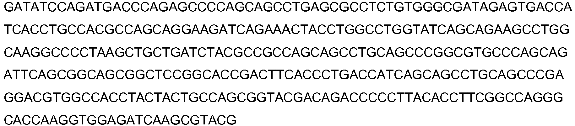
- SEQ ID NO: 110 Protein sequence of anti-TNF antibody variable light domain variant cb1-39-VL

- SEQ ID NO: 111 Polynucleotide sequence of anti-TNF antibody variable light domain variant cb1-33-VL

- SEQ ID NO: 112 Protein sequence of anti-TNF antibody variable light domain variant cb1-33-VL

- SEQ ID NO: 113 Polynucleotide sequence of anti-TNF antibody variable light domain variant cb1-35-VL

- SEQ ID NO: 114 Protein sequence of anti-TNF antibody variable light domain variant cb1-35-VL

- SEQ ID NO: 115 Polynucleotide sequence of anti-TNF antibody variable light domain variant cb1-31-VL

- SEQ ID NO: 116 Protein sequence of anti-TNF antibody variable light domain variant cb1-31-VL

- SEQ ID NO: 117 Polynucleotide sequence of anti-TNF antibody variable light domain variant cb1-29-VL
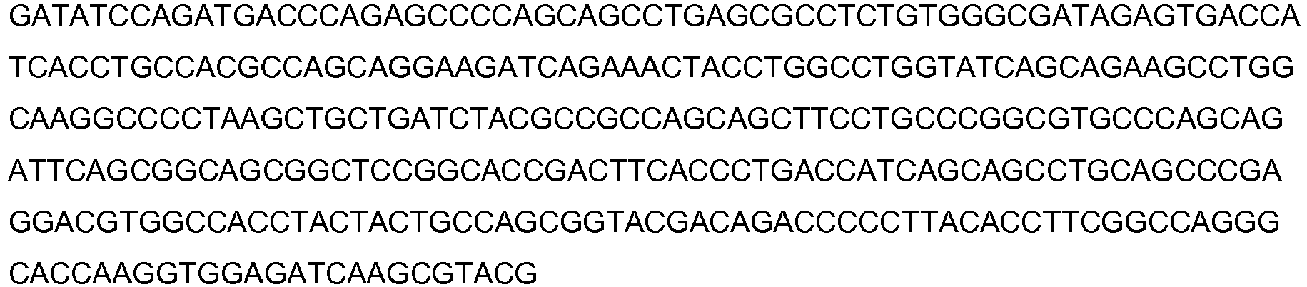
- SEQ ID NO: 118 Protein sequence of anti-TNF antibody variable light domain variant cb1-29-VL

- SEQ ID NO: 119 Polynucleotide sequence of anti-TNF antibody variable light domain variant cb1-22-VL

- SEQ ID NO: 120 Protein sequence of anti-TNF antibody variable light domain variant cb1-22-VL

- SEQ ID NO: 121 Polynucleotide sequence of anti-TNF antibody variable light domain variant cb1-23-VL

- SEQ ID NO: 122 Protein sequence of anti-TNF antibody variable light domain variant cb1-23-VL

- SEQ ID NO: 123 Polynucleotide sequence of anti-TNF antibody variable light domain variant cb1-12-VL


- SEQ ID NO: 124 Protein sequence of anti-TNF antibody variable light domain variant cb1-12-VL

- SEQ ID NO: 125 Polynucleotide sequence of anti-TNF antibody variable light domain variant cb1-10-VL

- SEQ ID NO: 126 Protein sequence of anti-TNF antibody variable light domain variant cb1-10-VL

- SEQ ID NO: 127 Polynucleotide sequence of anti-TNF antibody variable light domain variant cb2-1-VL

- SEQ ID NO: 128 Protein sequence of anti-TNF antibody variable light domain variant cb2-1-VL

- SEQ ID NO: 129 Polynucleotide sequence of anti-TNF antibody variable light domain variant cb2-11-VL


- SEQ ID NO: 130 Protein sequence of anti-TNF antibody variable light domain variant cb2-11-VL

- SEQ ID NO: 131 Polynucleotide sequence of anti-TNF antibody variable light domain variant cb2-40-VL

- SEQ ID NO: 132 Protein sequence of anti-TNF antibody variable light domain variant cb2-40-VL

- SEQ ID NO: 133 Polynucleotide sequence of anti-TNF antibody variable light domain variant cb2-35-VL

- SEQ ID NO: 134 Protein sequence of anti-TNF antibody variable light domain variant cb2-35-VL

- SEQ ID NO: 135 Polynucleotide sequence of anti-TNF antibody variable light domain variant cb2-28-VL


- SEQ ID NO: 136 Protein sequence of anti-TNF antibody variable light domain variant cb2-28-VL

- SEQ ID NO: 137 Polynucleotide sequence of anti-TNF antibody variable light domain variant cb2-20-VL

- SEQ ID NO: 138 Protein sequence of anti-TNF antibody variable light domain variant cb2-20-VL

- SEQ ID NO: 139 Polynucleotide sequence of anti-TNF antibody variable light domain variant cb1-3-VL

- SEQ ID NO: 140 Protein sequence of anti-TNF antibody variable light domain variant cb1-3-VL

- SEQ ID NO: 141 Polynucleotide sequence of anti-TNF antibody variable light domain variant cb2-6-VL

- SEQ ID NO: 142 Protein sequence of anti-TNF antibody variable light domain variant cb2-6-VL

- SEQ ID NO: 143 Polynucleotide sequence of anti-TNF antibody variable light domain variant cb2-44-VL

- SEQ ID NO: 144 Protein sequence of anti-TNF antibody variable light domain variant cb2-44-VL

- SEQ ID NO: 145 Protein sequence of anti-TNF antibody heavy chain variant cb1-3-VH plus M252Y/S254T/T256E modification

- SEQ ID NO: 146 Protein sequence of anti-TNF antibody heavy chain variant cb2-44-VH plus M252Y/S254T/T256E modification

- SEQ ID NO: 147 Polynucleotide sequence of anti-TNF antibody light chain variant cb1-3-VL

- SEQ ID NO: 148 Protein sequence of anti-TNF antibody light chain variant cb1-3-VL

- SEQ ID NO: 149 Polynucleotide sequence of anti-TNF antibody light chain variant cb2-6-VL

- SEQ ID NO: 150 Protein sequence of anti-TNF antibody light chain variant cb2-6-VL

- SEQ ID NO: 151 Polynucleotide sequence of anti-TNF antibody light chain variant cb2-44-VL


- SEQ ID NO: 152 Protein sequence of anti-TNF antibody light chain variant cb2-44-VL

- SEQ ID NO: 153 Polynucleotide sequence of anti-TNF antibody heavy chain variant cb1-3-VH

- SEQ ID NO: 154 Protein sequence of anti-TNF antibody heavy chain variant cb1-3-VH

- SEQ ID NO: 155 Polynucleotide sequence of anti-TNF antibody heavy chain variant cb2-44-VH

- SEQ ID NO: 156 Protein sequence of anti-TNF antibody heavy chain variant cb2-44-VH


- SEQ ID NO: 157 Polynucleotide sequence of pascolizumab heavy chain containing the M252Y/S254T/T256E modifications
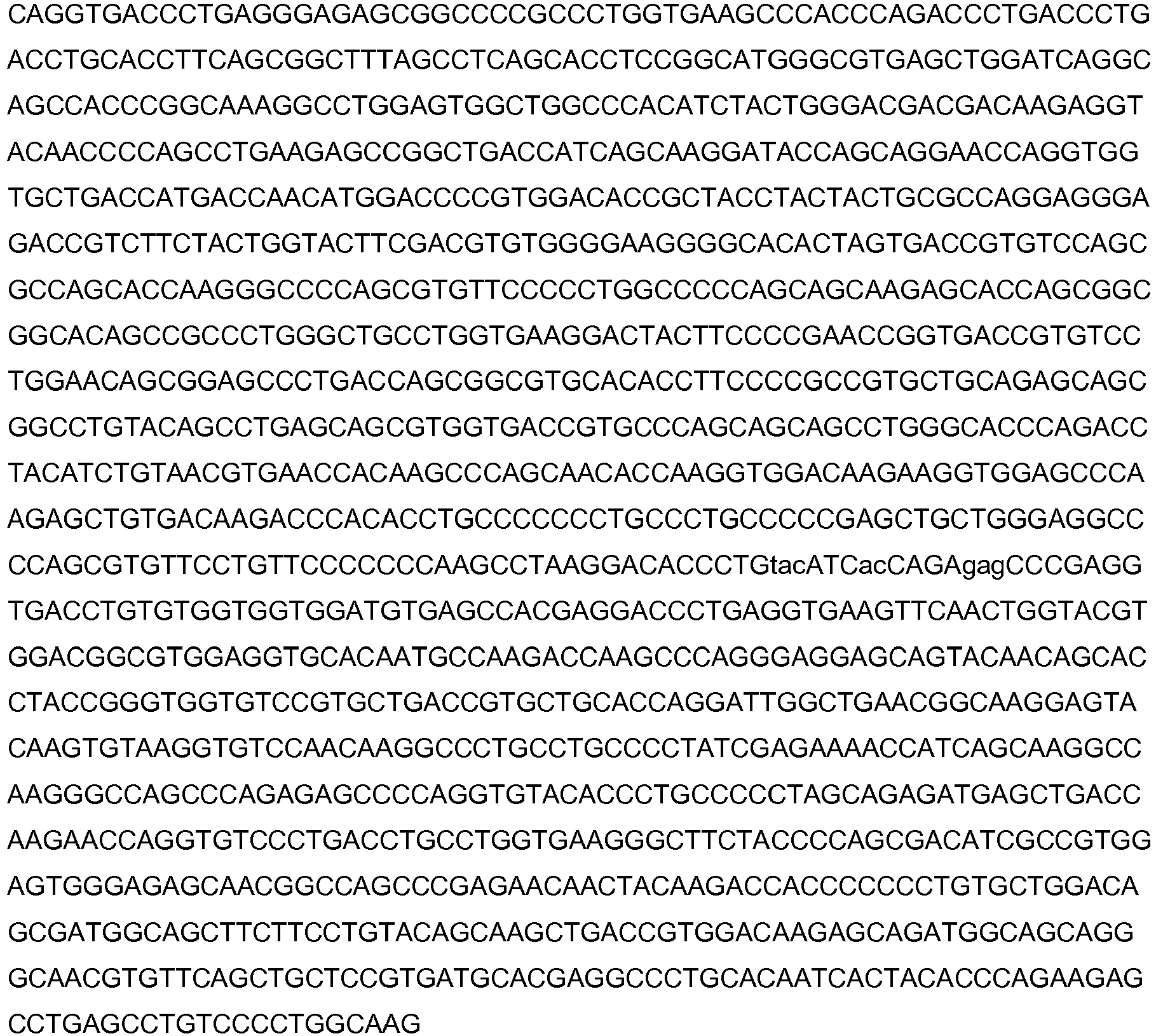
- SEQ ID NO: 158 Protein sequence of pascolizumab heavy chain containing the M252Y/S254T/T256E modifications


- SEQ ID NO: 159 Polynucleotide sequence of pascolizumab light chain

- SEQ ID NO: 160 Protein sequence of pascolizumab light chain

- SEQ ID NO: 161 Protein sequence of pascolizumab heavy chain

- SEQ ID NO: 162 Alternative protein sequence of the anti-TNF antibody heavy chain plus M428L/N434S modification
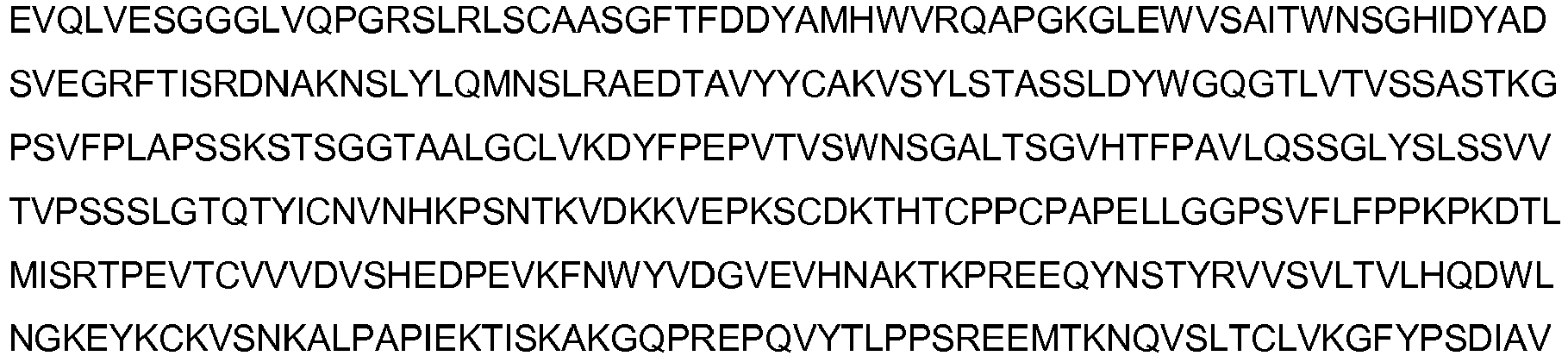

- SEQ ID NO: 163 Alternative protein sequence of the IgG1 constant domain plus M428L/N434S modification

- SEQ ID NO: 164 Protein sequence of the anti-TNF antibody heavy chain plus H433K/N434F modification

- SEQ ID NO: 165 Protein sequence of the IgG1 constant domain plus H433K/N434F modification
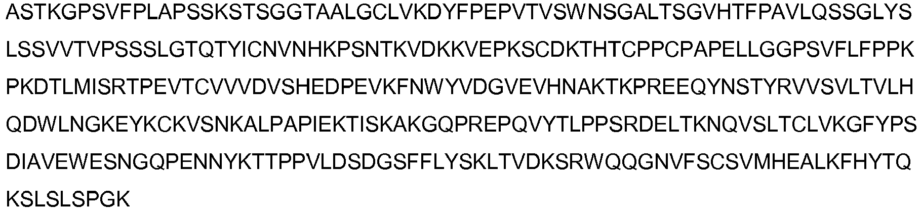
- SEQ ID NO: 166 Alternative protein sequence of the anti-TNF antibody heavy chain plus H433K/N434F modification


- SEQ ID NO: 167 Alternative protein sequence of the IgG1 constant domain plus H433K/N434F modification

- SEQ ID NO: 168 Alternative protein sequence of the anti-TNF antibody heavy chain plus M428L/N434S modification

- SEQ ID NO: 169 Alternative protein sequence of the IgG1/2 constant domain plus M428L/N434S modification

- SEQ ID NO: 170 Golimumab_VH

- SEQ ID NO: 171 Golimumab_VL

- SEQ ID NO: 172 Golimumab_HC

- SEQ ID NO: 173 Golimumab_LC

- SEQ ID NO: 174 Remicade_VH

- SEQ ID NO: 175 Remicade_VL

- SEQ ID NO: 176 Remicade_HC

- SEQ ID NO: 177 Remicade_LC

- SEQ ID NO: 178 Cimzia (certolizumab) VH

- SEQ ID NO: 179 Cimzia (certolizumab) VL

- SEQ ID NO: 180 Cimzia (certolizumab) HC (VH+CH1)

-
- <110> ELLIS, johnathan TOMLINSON, Ian SHAH, Tejash MOLLOY, Micheal YASIN, Ahmed
- <120> Antigen-binding proteins with increased
FcRn binding - <130> PB64705
- <150>
GB1112429.4
<151> 2011-07-19 - <160> 180
- <170> FastSEQ for Windows Version 4.0
- <210> 1
<211> 642
<212> DNA
<213> Artificial Sequence - <220>
<223> Humanised sequence - <400> 1

- <210> 2
<211> 214
<212> PRT
<213> Artificial Sequence - <220>
<223> Humanised sequence - <400> 2


- <210> 3
<211> 109
<212> PRT
<213> Artificial Sequence - <220>
<223> Humanised sequence - <400> 3

- <210> 4
<211> 1353
<212> DNA
<213> Artificial Sequence - <220>
<223> Humanised sequence - <400> 4


- <210> 5
<211> 451
<212> PRT
<213> Artificial Sequence - <220>
<223> Humanised sequence - <400> 5


- <210> 6
<211> 121
<212> PRT
<213> Artificial Sequence - <220>
<223> Humanised sequence - <400> 6

- <210> 7
<211> 330
<212> PRT
<213> Artificial Sequence - <220>
<223> Humanised sequence - <400> 7
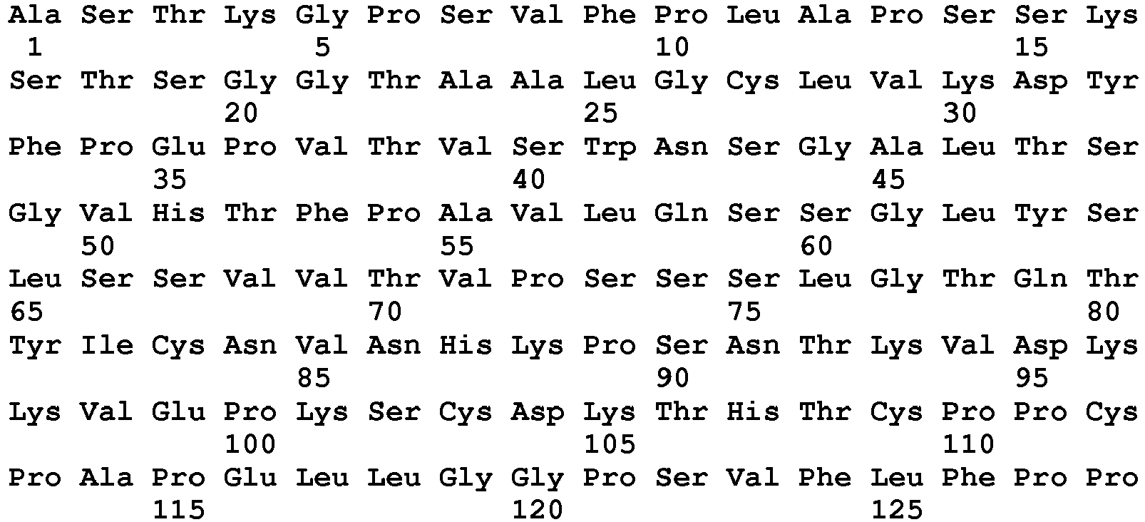

- <210> 8
<211> 1353
<212> DNA
<213> Artificial Sequence - <220>
<223> Humanised sequence - <400> 8

- <210> 9
<211> 451
<212> PRT
<213> Artificial Sequence - <220>
<223> Humanised sequence - <400> 9

- <210> 10
<211> 330
<212> PRT
<213> Artificial Sequence - <220>
<223> Humanised sequence - <400> 10

- <210> 11
<211> 1353
<212> DNA
<213> Artificial Sequence - <220>
<223> Humanised sequence - <400> 11


- <210> 12
<211> 451
<212> PRT
<213> Artificial Sequence - <220>
<223> Humanised sequence - <400> 12


- <210> 13
<211> 330
<212> PRT
<213> Artificial Sequence - <220>
<223> Humanised sequence - <400> 13

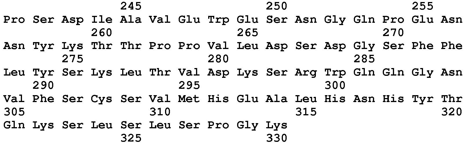
- <210> 14
<211> 1353
<212> DNA
<213> Artificial Sequence - <220>
<223> Humanised sequence - <400> 14

- <210> 15
<211> 451
<212> PRT
<213> Artificial Sequence - <220>
<223> Humanised sequence - <400> 15


- <210> 16
<211> 330
<212> PRT
<213> Artificial Sequence - <220>
<223> Humanised sequence - <400> 16


- <210> 17
<211> 1353
<212> DNA
<213> Artificial Sequence - <220>
<223> Humanised sequence - <400> 17


- <210> 18
<211> 451
<212> PRT
<213> Artificial Sequence - <220>
<223> Humanised sequence - <400> 18


- <210> 19
<211> 330
<212> PRT
<213> Artificial Sequence - <220>
<223> Humanised sequence - <400> 19

- <210> 20
<211> 1353
<212> DNA
<213> Artificial Sequence - <220>
<223> Humanised sequence - <400> 20

- <210> 21
<211> 451
<212> PRT
<213> Artificial Sequence - <220>
<223> Humanised sequence - <400> 21


- <210> 22
<211> 330
<212> PRT
<213> Artificial Sequence - <220>
<223> Humanised sequence - <400> 22


- <210> 23
<211> 1353
<212> DNA
<213> Artificial Sequence - <220>
<223> Humanised sequence - <400> 23

- <210> 24
<211> 451
<212> PRT
<213> Artificial Sequence - <220>
<223> Humanised sequence - <400> 24

- <210> 25
<211> 330
<212> PRT
<213> Artificial Sequence - <220>
<223> Humanised sequence - <400> 25
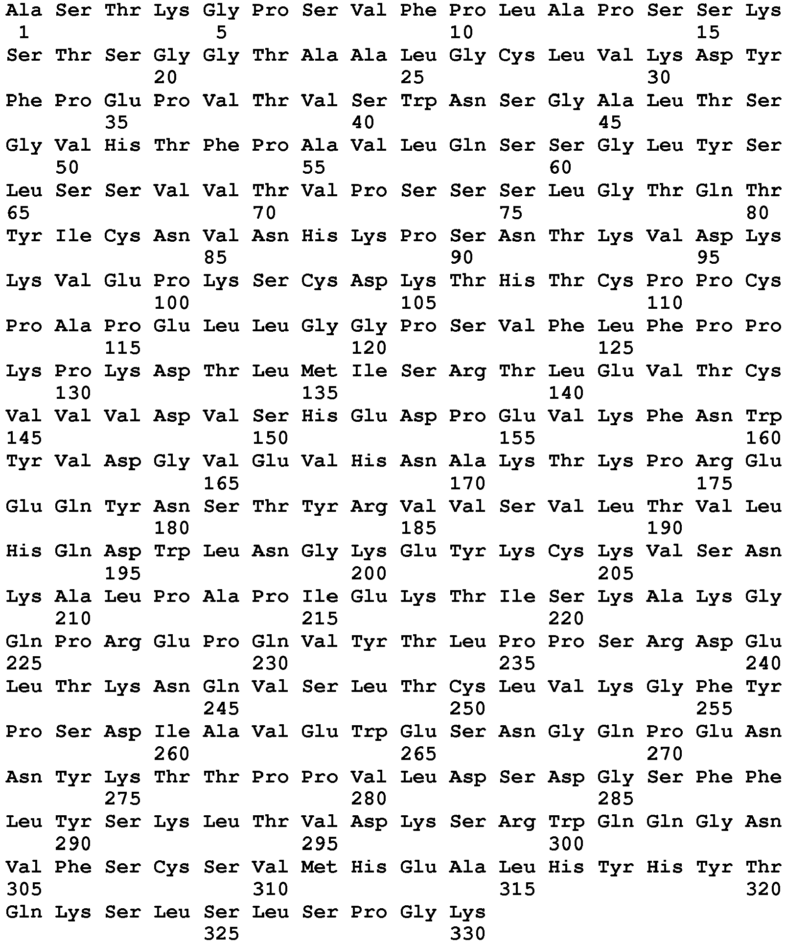
- <210> 26
<211> 19
<212> PRT
<213> Artificial Sequence - <220>
<223> Humanised sequence - <400> 26

- <210> 27
<211> 5
<212> PRT
<213> Artificial Sequence - <220>
<223> Humanised sequence - <400> 27

- <210> 28
<211> 17
<212> PRT
<213> Artificial Sequence - <220>
<223> Humanised sequence - <400> 28

- <210> 29
<211> 12
<212> PRT
<213> Artificial Sequence - <220>
<223> Humanised sequence - <400> 29

- <210> 30
<211> 11
<212> PRT
<213> Artificial Sequence - <220>
<223> Humanised sequence - <400> 30

- <210> 31
<211> 7
<212> PRT
<213> Artificial Sequence - <220>
<223> Humanised sequence - <400> 31

- <210> 32
<211> 9
<212> PRT
<213> Artificial Sequence - <220>
<223> Humanised sequence - <400> 32

- <210> 33
<211> 5
<212> PRT
<213> Artificial Sequence - <220>
<223> Humanised sequence - <400> 33

- <210> 34
<211> 5
<212> PRT
<213> Artificial Sequence - <220>
<223> Humanised sequence - <400> 34

- <210> 35
<211> 5
<212> PRT
<213> Artificial Sequence - <220>
<223> Humanised sequence - <400> 35

- <210> 36
<211> 5
<212> PRT
<213> Artificial Sequence - <220>
<223> Humanised sequence - <400> 36

- <210> 37
<211> 5
<212> PRT
<213> Artificial Sequence - <220>
<223> Humanised sequence - <400> 37

- <210> 38
<211> 5
<212> PRT
<213> Artificial Sequence - <220>
<223> Humanised sequence - <400> 38

- <210> 39
<211> 214
<212> PRT
<213> Artificial Sequence - <220>
<223> Humanised sequence - <400> 39

- <210> 40
<211> 12
<212> PRT
<213> Artificial Sequence - <220>
<223> Humanised sequence - <400> 40

- <210> 41
<211> 12
<212> PRT
<213> Artificial Sequence - <220>
<223> Humanised sequence - <400> 41

- <210> 42
<211> 12
<212> PRT
<213> Artificial Sequence - <220>
<223> Humanised sequence - <400> 42

- <210> 43
<211> 12
<212> PRT
<213> Artificial Sequence - <220>
<223> Humanised sequence - <400> 43

- <210> 44
<211> 12
<212> PRT
<213> Artificial Sequence - <220>
<223> Humanised sequence - <400> 44

- <210> 45
<211> 12
<212> PRT
<213> Artificial Sequence - <220>
<223> Humanised sequence - <400> 45

- <210> 46
<211> 12
<212> PRT
<213> Artificial Sequence - <220>
<223> Humanised sequence - <400> 46

- <210> 47
<211> 12
<212> PRT
<213> Artificial Sequence - <220>
<223> Humanised sequence - <400> 47

- <210> 48
<211> 12
<212> PRT
<213> Artificial Sequence - <220>
<223> Humanised sequence - <400> 48

- <210> 49
<211> 12
<212> PRT
<213> Artificial Sequence - <220>
<223> Humanised sequence - <400> 49

- <210> 50
<211> 11
<212> PRT
<213> Artificial Sequence - <220>
<223> Humanised sequence - <400> 50

- <210> 51
<211> 11
<212> PRT
<213> Artificial Sequence - <220>
<223> Humanised sequence - <400> 51

- <210> 52
<211> 11
<212> PRT
<213> Artificial Sequence - <220>
<223> Humanised sequence - <400> 52

- <210> 53
<211> 11
<212> PRT
<213> Artificial Sequence - <220>
<223> Humanised sequence - <400> 53

- <210> 54
<211> 11
<212> PRT
<213> Artificial Sequence - <220>
<223> Humanised sequence - <400> 54

- <210> 55
<211> 11
<212> PRT
<213> Artificial Sequence - <220>
<223> Humanised sequence - <400> 55

- <210> 56
<211> 11
<212> PRT
<213> Artificial Sequence - <220>
<223> Humanised sequence - <400> 56

- <210> 57
<211> 11
<212> PRT
<213> Artificial Sequence - <220>
<223> Humanised sequence - <400> 57

- <210> 58
<211> 11
<212> PRT
<213> Artificial Sequence - <220>
<223> Humanised sequence - <400> 58

- <210> 59
<211> 11
<212> PRT
<213> Artificial Sequence - <220>
<223> Humanised sequence - <400> 59

- <210> 60
<211> 11
<212> PRT
<213> Artificial Sequence - <220>
<223> Humanised sequence - <400> 60

- <210> 61
<211> 11
<212> PRT
<213> Artificial Sequence - <220>
<223> Humanised sequence - <400> 61

- <210> 62
<211> 7
<212> PRT
<213> Artificial Sequence - <220>
<223> Humanised sequence - <400> 62

- <210> 63
<211> 7
<212> PRT
<213> Artificial Sequence - <220>
<223> Humanised sequence - <400> 63

- <210> 64
<211> 7
<212> PRT
<213> Artificial Sequence - <220>
<223> Humanised sequence - <400> 64

- <210> 65
<211> 7
<212> PRT
<213> Artificial Sequence - <220>
<223> Humanised sequence - <400> 65

- <210> 66
<211> 7
<212> PRT
<213> Artificial Sequence - <220>
<223> Humanised sequence - <400> 66

- <210> 67
<211> 7
<212> PRT
<213> Artificial Sequence - <220>
<223> Humanised sequence - <400> 67

- <210> 68
<211> 7
<212> PRT
<213> Artificial Sequence - <220>
<223> Humanised sequence - <400> 68

- <210> 69
<211> 7
<212> PRT
<213> Artificial Sequence - <220>
<223> Humanised sequence - <400> 69

- <210> 70
<211> 7
<212> PRT
<213> Artificial Sequence - <220>
<223> Humanised sequence - <400> 70

- <210> 71
<211> 7
<212> PRT
<213> Artificial Sequence - <220>
<223> Humanised sequence - <400> 71

- <210> 72
<211> 7
<212> PRT
<213> Artificial Sequence - <220>
<223> Humanised sequence - <400> 72

- <210> 73
<211> 9
<212> PRT
<213> Artificial Sequence - <220>
<223> Humanised sequence - <400> 73

- <210> 74
<211> 9
<212> PRT
<213> Artificial Sequence - <220>
<223> Humanised sequence - <400> 74

- <210> 75
<211> 9
<212> PRT
<213> Artificial Sequence - <220>
<223> Humanised sequence - <400> 75

- <210> 76
<211> 9
<212> PRT
<213> Artificial Sequence - <220>
<223> Humanised sequence - <400> 76

- <210> 77
<211> 363
<212> DNA
<213> Artificial Sequence - <220>
<223> Humanised sequence - <400> 77

- <210> 78
<211> 121
<212> PRT
<213> Artificial Sequence - <220>
<223> Humanised sequence - <400> 78


- <210> 79
<211> 363
<212> DNA
<213> Artificial Sequence - <220>
<223> Humanised sequence - <400> 79

- <210> 80
<211> 121
<212> PRT
<213> Artificial Sequence - <220>
<223> Humanised sequence - <400> 80

- <210> 81
<211> 363
<212> DNA
<213> Artificial Sequence - <220>
<223> Humanised sequence - <400> 81

- <210> 82
<211> 121
<212> PRT
<213> Artificial Sequence - <220>
<223> Humanised sequence - <400> 82

- <210> 83
<211> 363
<212> DNA
<213> Artificial Sequence - <220>
<223> Humanised sequence - <400> 83

- <210> 84
<211> 121
<212> PRT
<213> Artificial Sequence - <220>
<223> Humanised sequence - <400> 84


- <210> 85
<211> 363
<212> DNA
<213> Artificial Sequence - <220>
<223> Humanised sequence - <400> 85

- <210> 86
<211> 121
<212> PRT
<213> Artificial Sequence - <220>
<223> Humanised sequence - <400> 86
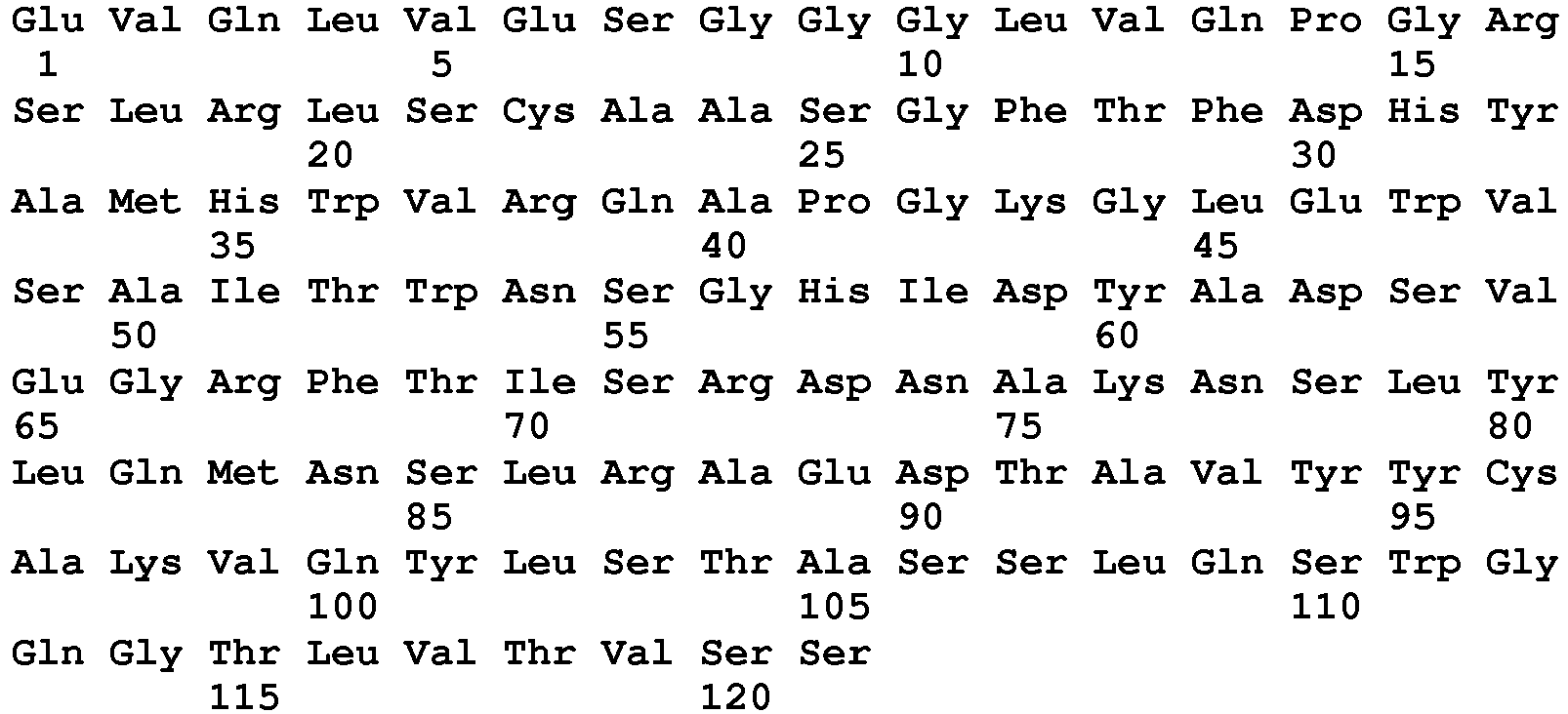
- <210> 87
<211> 363
<212> DNA
<213> Artificial Sequence - <220>
<223> Humanised sequence - <400> 87


- <210> 88
<211> 121
<212> PRT
<213> Artificial Sequence - <220>
<223> Humanised sequence - <400> 88

- <210> 89
<211> 363
<212> DNA
<213> Artificial Sequence - <220>
<223> Humanised sequence - <400> 89

- <210> 90
<211> 121
<212> PRT
<213> Artificial Sequence - <220>
<223> Humanised sequence - <400> 90


- <210> 91
<211> 363
<212> DNA
<213> Artificial Sequence - <220>
<223> Humanised sequence - <400> 91

- <210> 92
<211> 121
<212> PRT
<213> Artificial Sequence - <220>
<223> Humanised sequence - <400> 92

- <210> 93
<211> 363
<212> DNA
<213> Artificial Sequence - <220>
<223> Humanised sequence - <400> 93


- <210> 94
<211> 121
<212> PRT
<213> Artificial Sequence - <220>
<223> Humanised sequence - <400> 94

- <210> 95
<211> 363
<212> DNA
<213> Artificial Sequence - <220>
<223> Humanised sequence - <400> 95

- <210> 96
<211> 121
<212> PRT
<213> Artificial Sequence - <220>
<223> Humanised sequence - <400> 96


- <210> 97
<211> 327
<212> DNA
<213> Artificial Sequence - <220>
<223> Humanised sequence - <400> 97

- <210> 98
<211> 109
<212> PRT
<213> Artificial Sequence - <220>
<223> Humanised sequence - <400> 98

- <210> 99
<211> 327
<212> DNA
<213> Artificial Sequence - <220>
<223> Humanised sequence - <400> 99


- <210> 100
<211> 109
<212> PRT
<213> Artificial Sequence - <220>
<223> Humanised sequence - <400> 100

- <210> 101
<211> 327
<212> DNA
<213> Artificial Sequence - <220>
<223> Humanised sequence - <400> 101

- <210> 102
<211> 109
<212> PRT
<213> Artificial Sequence - <220>
<223> Humanised sequence - <400> 102


- <210> 103
<211> 327
<212> DNA
<213> Artificial Sequence - <220>
<223> Humanised sequence - <400> 103

- <210> 104
<211> 109
<212> PRT
<213> Artificial Sequence - <220>
<223> Humanised sequence - <400> 104

- <210> 105
<211> 327
<212> DNA
<213> Artificial Sequence - <220>
<223> Humanised sequence - <400> 105

- <210> 106
<211> 109
<212> PRT
<213> Artificial Sequence - <220>
<223> Humanised sequence - <400> 106

- <210> 107
<211> 327
<212> DNA
<213> Artificial Sequence - <220>
<223> Humanised sequence - <400> 107

- <210> 108
<211> 109
<212> PRT
<213> Artificial Sequence - <220>
<223> Humanised sequence - <400> 108

- <210> 109
<211> 327
<212> DNA
<213> Artificial Sequence - <220>
<223> Humanised sequence - <400> 109

- <210> 110
<211> 109
<212> PRT
<213> Artificial Sequence - <220>
<223> Humanised sequence - <400> 110

- <210> 111
<211> 327
<212> DNA
<213> Artificial Sequence - <220>
<223> Humanised sequence - <400> 111

- <210> 112
<211> 109
<212> PRT
<213> Artificial Sequence - <220>
<223> Humanised sequence - <400> 112


- <210> 113
<211> 327
<212> DNA
<213> Artificial Sequence - <220>
<223> Humanised sequence - <400> 113

- <210> 114
<211> 109
<212> PRT
<213> Artificial Sequence - <220>
<223> Humanised sequence - <400> 114

- <210> 115
<211> 327
<212> DNA
<213> Artificial Sequence - <220>
<223> Humanised sequence - <400> 115


- <210> 116
<211> 109
<212> PRT
<213> Artificial Sequence - <220>
<223> Humanised sequence - <400> 116

- <210> 117
<211> 327
<212> DNA
<213> Artificial Sequence - <220>
<223> Humanised sequence - <400> 117

- <210> 118
<211> 109
<212> PRT
<213> Artificial Sequence - <220>
<223> Humanised sequence - <400> 118


- <210> 119
<211> 327
<212> DNA
<213> Artificial Sequence - <220>
<223> Humanised sequence - <400> 119

- <210> 120
<211> 109
<212> PRT
<213> Artificial Sequence - <220>
<223> Humanised sequence - <400> 120

- <210> 121
<211> 327
<212> DNA
<213> Artificial Sequence - <220>
<223> Humanised sequence - <400> 121

- <210> 122
<211> 109
<212> PRT
<213> Artificial Sequence - <220>
<223> Humanised sequence - <400> 122

- <210> 123
<211> 327
<212> DNA
<213> Artificial Sequence - <220>
<223> Humanised sequence - <400> 123

- <210> 124
<211> 109
<212> PRT
<213> Artificial Sequence - <220>
<223> Humanised sequence - <400> 124

- <210> 125
<211> 327
<212> DNA
<213> Artificial Sequence - <220>
<223> Humanised sequence - <400> 125

- <210> 126
<211> 109
<212> PRT
<213> Artificial Sequence - <220>
<223> Humanised sequence - <400> 126

- <210> 127
<211> 327
<212> DNA
<213> Artificial Sequence - <220>
<223> Humanised sequence - <400> 127

- <210> 128
<211> 109
<212> PRT
<213> Artificial Sequence - <220>
<223> Humanised sequence - <400> 128

- <210> 129
<211> 327
<212> DNA
<213> Artificial Sequence - <220>
<223> Humanised sequence - <400> 129

- <210> 130
<211> 109
<212> PRT
<213> Artificial Sequence - <220>
<223> Humanised sequence - <400> 130

- <210> 131
<211> 327
<212> DNA
<213> Artificial Sequence - <220>
<223> Humanised sequence - <400> 131

- <210> 132
<211> 109
<212> PRT
<213> Artificial Sequence - <220>
<223> Humanised sequence - <400> 132

- <210> 133
<211> 327
<212> DNA
<213> Artificial Sequence - <220>
<223> Humanised sequence - <400> 133

- <210> 134
<211> 109
<212> PRT
<213> Artificial Sequence - <220>
<223> Humanised sequence - <400> 134

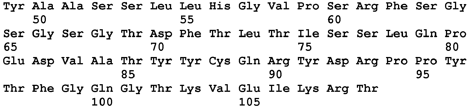
- <210> 135
<211> 327
<212> DNA
<213> Artificial Sequence - <220>
<223> Humanised sequence - <400> 135

- <210> 136
<211> 109
<212> PRT
<213> Artificial Sequence - <220>
<223> Humanised sequence - <400> 136

- <210> 137
<211> 327
<212> DNA
<213> Artificial Sequence - <220>
<223> Humanised sequence - <400> 137


- <210> 138
<211> 109
<212> PRT
<213> Artificial Sequence - <220>
<223> Humanised sequence - <400> 138

- <210> 139
<211> 327
<212> DNA
<213> Artificial Sequence - <220>
<223> Humanised sequence - <400> 139

- <210> 140
<211> 109
<212> PRT
<213> Artificial Sequence - <220>
<223> Humanised sequence - <400> 140


- <210> 141
<211> 327
<212> DNA
<213> Artificial Sequence - <220>
<223> Humanised sequence - <400> 141

- <210> 142
<211> 109
<212> PRT
<213> Artificial Sequence - <220>
<223> Humanised sequence - <400> 142
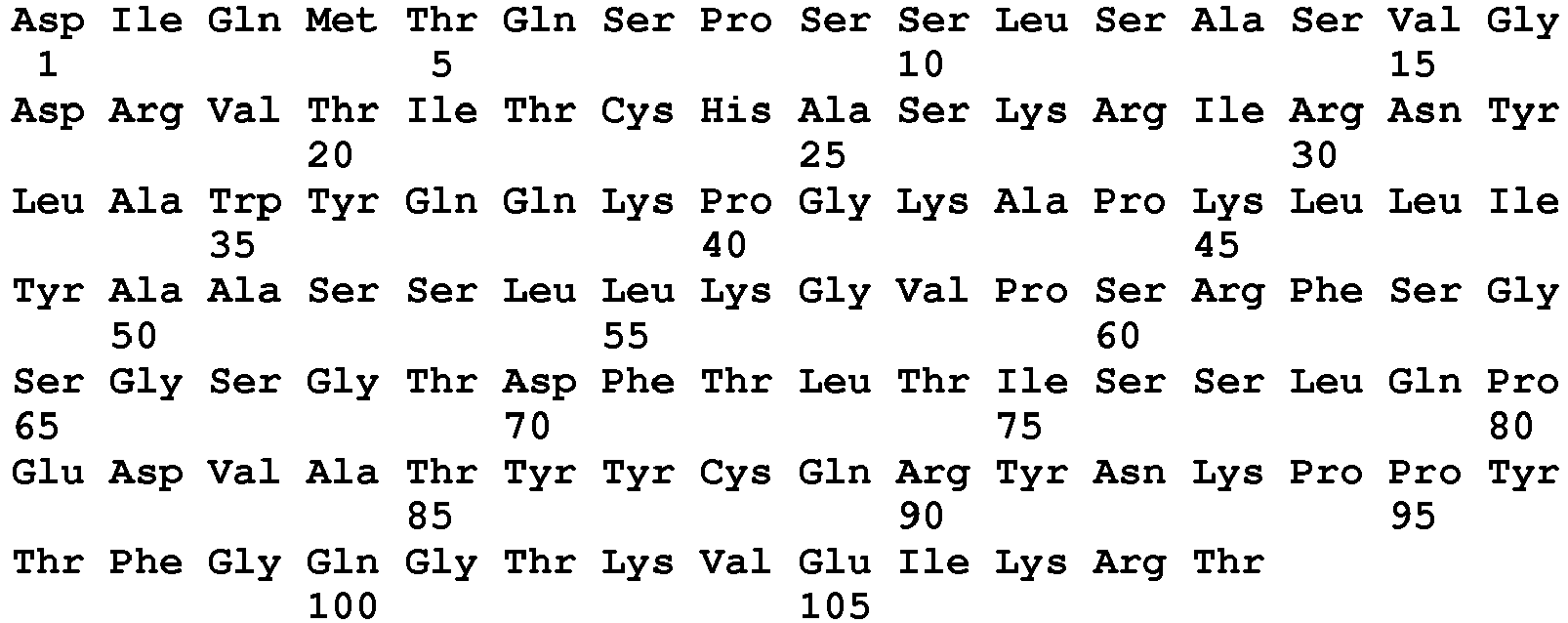
- <210> 143
<211> 327
<212> DNA
<213> Artificial Sequence - <220>
<223> Humanised sequence - <400> 143

- <210> 144
<211> 109
<212> PRT
<213> Artificial Sequence - <220>
<223> Humanised sequence - <400> 144

- <210> 145
<211> 451
<212> PRT
<213> Artificial Sequence - <220>
<223> Humanised sequence - <400> 145


- <210> 146
<211> 451
<212> PRT
<213> Artificial Sequence - <220>
<223> Humanised sequence - <400> 146

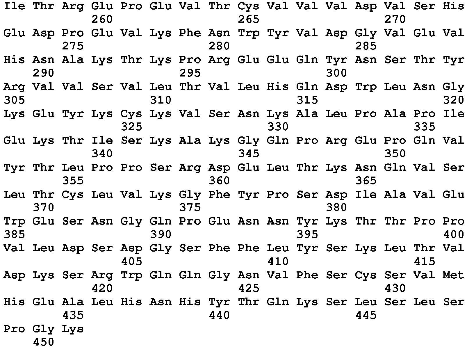
- <210> 147
<211> 642
<212> DNA
<213> Artificial Sequence - <220>
<223> Humanised sequence - <400> 147

- <210> 148
<211> 214
<212> PRT
<213> Artificial Sequence - <220>
<223> Humanised sequence - <400> 148


- <210> 149
<211> 642
<212> DNA
<213> Artificial Sequence - <220>
<223> Humanised sequence - <400> 149

- <210> 150
<211> 214
<212> PRT
<213> Artificial Sequence - <220>
<223> Humanised sequence - <400> 150


- <210> 151
<211> 642
<212> DNA
<213> Artificial Sequence - <220>
<223> Humanised sequence - <400> 151

- <210> 152
<211> 214
<212> PRT
<213> Artificial Sequence - <220>
<223> Humanised sequence - <400> 152


- <210> 153
<211> 1353
<212> DNA
<213> Artificial Sequence - <220>
<223> Humanised sequence - <400> 153

- <210> 154
<211> 451
<212> PRT
<213> Artificial Sequence - <220>
<223> Humanised sequence - <400> 154


- <210> 155
<211> 1353
<212> DNA
<213> Artificial Sequence - <220>
<223> Humanised sequence - <400> 155


- <210> 156
<211> 451
<212> PRT
<213> Artificial Sequence - <220>
<223> Humanised sequence - <400> 156


- <210> 157
<211> 1353
<212> DNA
<213> Artificial Sequence - <220>
<223> Humanised sequence - <400> 157

- <210> 158
<211> 451
<212> PRT
<213> Artificial Sequence - <220>
<223> Humanised sequence - <400> 158


- <210> 159
<211> 654
<212> DNA
<213> Artificial Sequence - <220>
<223> Humanised sequence - <400> 159

- <210> 160
<211> 218
<212> PRT
<213> Artificial Sequence - <220>
<223> Humanised sequence - <400> 160

- <210> 161
<211> 451
<212> PRT
<213> Artificial Sequence - <220>
<223> Humanised sequence - <400> 161


- <210> 162
<211> 451
<212> PRT
<213> Artificial Sequence - <220>
<223> Humanised sequence - <400> 162

- <210> 163
<211> 330
<212> PRT
<213> Artificial Sequence - <220>
<223> Humanised sequence - <400> 163

- <210> 164
<211> 451
<212> PRT
<213> Artificial Sequence - <220>
<223> Humanised sequence - <400> 164


- <210> 165
<211> 330
<212> PRT
<213> Artificial Sequence - <220>
<223> Humanised sequence - <400> 165


- <210> 166
<211> 451
<212> PRT
<213> Artificial Sequence - <220>
<223> Humanised sequence - <400> 166


- <210> 167
<211> 330
<212> PRT
<213> Artificial Sequence - <220>
<223> Humanised sequence - <400> 167


- <210> 168
<211> 450
<212> PRT
<213> Artificial Sequence - <220>
<223> Humanised sequence - <400> 168


- <210> 169
<211> 329
<212> PRT
<213> Artificial Sequence - <220>
<223> Humanised sequence - <400> 169


- <210> 170
<211> 126
<212> PRT
<213> Artificial Sequence - <220>
<223> Humanised sequence - <400> 170

- <210> 171
<211> 110
<212> PRT
<213> Artificial Sequence - <220>
<223> Humanised sequence - <400> 171


- <210> 172
<211> 456
<212> PRT
<213> Artificial Sequence - <220>
<223> Humanised sequence - <400> 172


- <210> 173
<211> 215
<212> PRT
<213> Artificial Sequence - <220>
<223> Humanised sequence - <400> 173

- <210> 174
<211> 120
<212> PRT
<213> Artificial Sequence - <220>
<223> Humanised sequence - <400> 174

- <210> 175
<211> 109
<212> PRT
<213> Artificial Sequence - <220>
<223> Humanised sequence - <400> 175

- <210> 176
<211> 450
<212> PRT
<213> Artificial Sequence - <220>
<223> Humanised sequence - <400> 176


- <210> 177
<211> 214
<212> PRT
<213> Artificial Sequence - <220>
<223> Humanised sequence - <400> 177


- <210> 178
<211> 118
<212> PRT
<213> Artificial Sequence - <220>
<223> Humanised sequence - <400> 178

- <210> 179
<211> 109
<212> PRT
<213> Artificial Sequence - <220>
<223> Humanised sequence - <400> 179


- <210> 180
<211> 229
<212> PRT
<213> Artificial Sequence - <220>
<223> Humanised sequence - <400> 180

Claims (13)
- An antigen binding protein which specifically binds to TNF-alpha comprising:(i) CDRH1 (SEQ ID NO: 27), CDRH2 (SEQ ID NO: 28), CDRH3 (SEQ ID No: 29), CDRL1 (SEQ ID NO: 30), CDRL2 (SEQ ID NO: 31), and CDRL3 (SEQ ID NO: 32); and(ii) a neonatal Fc receptor (FcRn) binding portion of a human IgG1 constant domain comprising amino acid substitutions relative to the human IgG1 constant domain at amino acid residues 252, 254 and 256 numbered according to EU index of Kabat and wherein the substitution at residue 252 is a substitution of met with tyr; the substitution at residue 254 is a substitution of ser with thr and the substitution at residue 256 is a substitution of thr with glu;wherein the antigen binding protein has an increased FcRn binding affinity at pH 6 and increased half-life as compared to an IgG comprising the light chain sequence of SEQ ID No: 2 and the heavy chain sequence of SEQ ID No:12. for use in a method of treating a disease wherein the antigen binding protein is administered to patients at a single dose between 35 to 45 mg at a four to eight weekly interval.
- An antigen binding protein for use as claimed in claim 1 wherein the human IgG1 constant domain has the sequence of SEQ ID No: 13 before amino acid substitutions are introduced.
- An antigen binding protein for use as claimed in any preceding claim wherein the antigen binding protein is administered to patients subcutaneously as a single 40 mg dose no more than once every four weeks.
- An antigen binding protein for use as claimed in any preceding claim wherein the antigen binding protein is administered to patients subcutaneously as a single 40 mg dose no more than once every eight weeks.
- An antigen binding protein for use as claimed in any preceding claim wherein the half-life of the antigen binding protein is increased 2 fold, 3 fold, 4 fold or 5 fold as compared to the native IgG.
- An antigen binding protein for use as claimed in any preceding claim wherein administration of the antigen binding protein no more than once every four weeks in patients achieves the mean steady-state trough concentration in the patient population of between 4 µg/ml to 7 µg/ml.
- An antigen binding protein for use as claimed in claim 6 wherein the mean steady-state trough concentration is between 5 µg/ml to 6 µg/ml.
- An antigen binding protein for use as claimed in any preceding claim wherein the clearance of the antigen binding protein is 2ml/ hr to 4ml/hr
- An antigen binding protein for use as claimed in any preceding claim comprising a constant domain as shown in SEQ ID No: 7
- An antigen binding protein for use as claimed in any preceding claim wherein the antigen binding protein is an antibody.
- An antigen binding protein for use as claimed in any preceding claim wherein the antigen binding protein is to be administered with methotrexate, preferably wherein the antigen binding protein is administered for the treatment of rheumatoid arthritis.
- An antigen binding protein for use as claimed in any preceding claim comprising the heavy chain sequence as shown in SEQ ID No: 5 optionally with a light chain sequence as shown in SEQ ID No: 2.
- An antigen binding protein for use as claimed in any preceding claim wherein the disease is rheumatoid arthritis, polyarticular juvenile idiopathic arthritis, psoriatic arthritis, ankylosing spondylitis, Crohn's disease or Psoriasis.
Priority Applications (1)
| Application Number | Priority Date | Filing Date | Title |
|---|---|---|---|
| EP15197750.1A EP3009450A1 (en) | 2011-07-19 | 2012-07-19 | Liquid formulation comprising adalimumab and an acetate buffer |
Applications Claiming Priority (2)
| Application Number | Priority Date | Filing Date | Title |
|---|---|---|---|
| GBGB1112429.4A GB201112429D0 (en) | 2011-07-19 | 2011-07-19 | Antigen-binding proteins with increased FcRn binding |
| PCT/EP2012/064129 WO2013011076A2 (en) | 2011-07-19 | 2012-07-19 | Antigen-binding proteins with increased fcrn binding |
Related Child Applications (2)
| Application Number | Title | Priority Date | Filing Date |
|---|---|---|---|
| EP15197750.1A Division EP3009450A1 (en) | 2011-07-19 | 2012-07-19 | Liquid formulation comprising adalimumab and an acetate buffer |
| EP15197750.1A Division-Into EP3009450A1 (en) | 2011-07-19 | 2012-07-19 | Liquid formulation comprising adalimumab and an acetate buffer |
Publications (2)
| Publication Number | Publication Date |
|---|---|
| EP2734548A2 EP2734548A2 (en) | 2014-05-28 |
| EP2734548B1 true EP2734548B1 (en) | 2016-08-17 |
Family
ID=44586854
Family Applications (2)
| Application Number | Title | Priority Date | Filing Date |
|---|---|---|---|
| EP15197750.1A Withdrawn EP3009450A1 (en) | 2011-07-19 | 2012-07-19 | Liquid formulation comprising adalimumab and an acetate buffer |
| EP12738101.0A Active EP2734548B1 (en) | 2011-07-19 | 2012-07-19 | Tnf-alpha-antigen-binding proteins against with increased fcrn binding for use in therapy |
Family Applications Before (1)
| Application Number | Title | Priority Date | Filing Date |
|---|---|---|---|
| EP15197750.1A Withdrawn EP3009450A1 (en) | 2011-07-19 | 2012-07-19 | Liquid formulation comprising adalimumab and an acetate buffer |
Country Status (22)
| Country | Link |
|---|---|
| US (1) | US20130243764A1 (en) |
| EP (2) | EP3009450A1 (en) |
| JP (1) | JP2014524748A (en) |
| KR (1) | KR20140054085A (en) |
| CN (1) | CN103748110A (en) |
| AU (1) | AU2012285786B2 (en) |
| BR (1) | BR112014000341A2 (en) |
| CA (1) | CA2841105A1 (en) |
| CL (1) | CL2014000134A1 (en) |
| CO (1) | CO6862106A2 (en) |
| CR (1) | CR20140029A (en) |
| DO (1) | DOP2014000007A (en) |
| EA (1) | EA201391789A1 (en) |
| ES (1) | ES2600854T3 (en) |
| GB (1) | GB201112429D0 (en) |
| MA (1) | MA35345B1 (en) |
| MX (1) | MX2014000739A (en) |
| PE (1) | PE20141660A1 (en) |
| PH (1) | PH12014500167A1 (en) |
| SG (1) | SG10201601154QA (en) |
| WO (1) | WO2013011076A2 (en) |
| ZA (1) | ZA201400062B (en) |
Families Citing this family (61)
| Publication number | Priority date | Publication date | Assignee | Title |
|---|---|---|---|---|
| CA2817619A1 (en) | 2001-06-08 | 2002-12-08 | Abbott Laboratories (Bermuda) Ltd. | Methods of administering anti-tnf.alpha. antibodies |
| UY33679A (en) | 2010-10-22 | 2012-03-30 | Esbatech | STABLE AND SOLUBLE ANTIBODIES |
| US9062106B2 (en) | 2011-04-27 | 2015-06-23 | Abbvie Inc. | Methods for controlling the galactosylation profile of recombinantly-expressed proteins |
| US10093728B2 (en) | 2012-03-07 | 2018-10-09 | Cadila Healthcare Limited | Pharmaceutical formulations of TNF-alpha antibodies |
| WO2013158275A1 (en) * | 2012-04-20 | 2013-10-24 | Abbvie Inc. | Cell culture methods to reduce acidic species |
| WO2013158279A1 (en) | 2012-04-20 | 2013-10-24 | Abbvie Inc. | Protein purification methods to reduce acidic species |
| WO2013158273A1 (en) | 2012-04-20 | 2013-10-24 | Abbvie Inc. | Methods to modulate c-terminal lysine variant distribution |
| US9067990B2 (en) | 2013-03-14 | 2015-06-30 | Abbvie, Inc. | Protein purification using displacement chromatography |
| US9249182B2 (en) | 2012-05-24 | 2016-02-02 | Abbvie, Inc. | Purification of antibodies using hydrophobic interaction chromatography |
| US20150150982A1 (en) * | 2012-06-12 | 2015-06-04 | Boehringer Ingelheim International Gmbh | Pharmaceutical formulation for a therapeutic antibody |
| KR20150043523A (en) | 2012-09-02 | 2015-04-22 | 애브비 인코포레이티드 | Methods to control protein heterogeneity |
| US9512214B2 (en) | 2012-09-02 | 2016-12-06 | Abbvie, Inc. | Methods to control protein heterogeneity |
| MX2015003007A (en) | 2012-09-07 | 2015-06-05 | Coherus Biosciences Inc | Stable aqueous formulations of adalimumab. |
| WO2014096672A1 (en) * | 2012-12-17 | 2014-06-26 | Laboratoire Francais Du Fractionnement Et Des Biotechnologies | Use of monoclonal antibodies for the treatment of inflammation and bacterial infections |
| FR2999431B1 (en) * | 2012-12-17 | 2016-03-18 | Lfb Biotechnologies | USE OF MONOCLONAL ANTIBODIES FOR THE TREATMENT OF INFLAMMATION AND BACTERIAL INFECTIONS |
| CN105051064A (en) * | 2013-01-24 | 2015-11-11 | 葛兰素史克知识产权开发有限公司 | Tnf-alpha antigen-binding proteins |
| SG11201507230PA (en) | 2013-03-12 | 2015-10-29 | Abbvie Inc | Human antibodies that bind human tnf-alpha and methods of preparing the same |
| KR20150129033A (en) * | 2013-03-14 | 2015-11-18 | 애브비 인코포레이티드 | Low acidic species compositions and methods for producing and using the same |
| US8921526B2 (en) | 2013-03-14 | 2014-12-30 | Abbvie, Inc. | Mutated anti-TNFα antibodies and methods of their use |
| WO2014151878A2 (en) | 2013-03-14 | 2014-09-25 | Abbvie Inc. | Methods for modulating protein glycosylation profiles of recombinant protein therapeutics using monosaccharides and oligosacharides |
| US9017687B1 (en) | 2013-10-18 | 2015-04-28 | Abbvie, Inc. | Low acidic species compositions and methods for producing and using the same using displacement chromatography |
| EP3021833B2 (en) * | 2013-07-19 | 2022-03-30 | Hexal Aktiengesellschaft | Methods and formulations which allow the modulation of immune responses related to the administration of a biopharmaceutical drug |
| EP3052640A2 (en) | 2013-10-04 | 2016-08-10 | AbbVie Inc. | Use of metal ions for modulation of protein glycosylation profiles of recombinant proteins |
| JP6526025B2 (en) * | 2013-10-16 | 2019-06-05 | オンコバイオロジクス,インコーポレイティド | Buffer solution formulation for enhancing antibody stability |
| US8946395B1 (en) | 2013-10-18 | 2015-02-03 | Abbvie Inc. | Purification of proteins using hydrophobic interaction chromatography |
| US9085618B2 (en) | 2013-10-18 | 2015-07-21 | Abbvie, Inc. | Low acidic species compositions and methods for producing and using the same |
| US9181337B2 (en) | 2013-10-18 | 2015-11-10 | Abbvie, Inc. | Modulated lysine variant species compositions and methods for producing and using the same |
| WO2015073884A2 (en) | 2013-11-15 | 2015-05-21 | Abbvie, Inc. | Glycoengineered binding protein compositions |
| EP2946767B1 (en) | 2014-05-23 | 2016-10-05 | Ares Trading S.A. | Liquid pharmaceutical composition |
| PL2946765T3 (en) * | 2014-05-23 | 2017-08-31 | Ares Trading S.A. | Liquid pharmaceutical composition |
| ES2572919T3 (en) | 2014-05-23 | 2016-06-03 | Ares Trading S.A. | Liquid pharmaceutical composition |
| AR101753A1 (en) | 2014-09-03 | 2017-01-11 | Boehringer Ingelheim Int | COMPOUND OF OBJECTIVE TO IL-23A AND TNF-A AND USE OF THE SAME |
| HUP1400510A1 (en) * | 2014-10-28 | 2016-05-30 | Richter Gedeon Nyrt | Pharmaceutical anti-tnfalpha antibody formulation |
| US10696735B2 (en) | 2015-01-21 | 2020-06-30 | Outlook Therapeutics, Inc. | Modulation of charge variants in a monoclonal antibody composition |
| AR103544A1 (en) * | 2015-01-28 | 2017-05-17 | Mabxience S A | PHARMACEUTICAL FORMULATIONS FOR ANTI-TNF-a ANTIBODIES |
| WO2016189491A1 (en) * | 2015-05-28 | 2016-12-01 | Glaxosmithkline Intellectual Property Development Limited | Novel formulation |
| US11229702B1 (en) | 2015-10-28 | 2022-01-25 | Coherus Biosciences, Inc. | High concentration formulations of adalimumab |
| GB201522394D0 (en) | 2015-12-18 | 2016-02-03 | Ucb Biopharma Sprl | Antibodies |
| WO2017110980A1 (en) | 2015-12-25 | 2017-06-29 | 中外製薬株式会社 | Antibody having enhanced activity, and method for modifying same |
| CN109563161A (en) | 2016-02-03 | 2019-04-02 | 安口生物公司 | For improving the buffer preparation of Antibody stability |
| MA43773B1 (en) | 2016-03-14 | 2026-01-30 | Universitetet I Oslo | Engineered immunoglobulins with altered FcRn binding |
| WO2017184880A1 (en) | 2016-04-20 | 2017-10-26 | Coherus Biosciences, Inc. | A method of filling a container with no headspace |
| ES3055961T3 (en) | 2016-04-28 | 2026-02-17 | Chugai Pharmaceutical Co Ltd | Antibody-containing preparation |
| CN109415444B (en) | 2016-07-29 | 2024-03-01 | 中外制药株式会社 | Bispecific antibodies showing increased functional activity of alternative FVIII cofactors |
| FI3494134T3 (en) | 2016-08-02 | 2026-02-19 | Visterra Inc | MODIFIED POLYPEPTIDES AND THEIR USES |
| TW202300515A (en) * | 2016-10-20 | 2023-01-01 | 法商賽諾菲公司 | Anti-chikv antibodies and uses thereof |
| CN110167531B (en) * | 2017-01-11 | 2025-06-03 | 赛特瑞恩股份有限公司 | Stable liquid formulations |
| EP3569615A4 (en) * | 2017-01-13 | 2020-07-29 | Hanx Biopharmaceutics, Inc | Method for improving binding affinity of igg antibody to fcrn and prolonging serum half-life period thereof |
| US11608357B2 (en) | 2018-08-28 | 2023-03-21 | Arecor Limited | Stabilized antibody protein solutions |
| EP3372242A1 (en) * | 2017-03-06 | 2018-09-12 | Ares Trading S.A. | Liquid pharmaceutical composition |
| EP3372241A1 (en) * | 2017-03-06 | 2018-09-12 | Ares Trading S.A. | Liquid pharmaceutical composition |
| EP3603670A4 (en) | 2017-03-31 | 2021-03-10 | Public University Corporation Nara Medical University | MEDICINAL COMPOSITION FOR USE FOR PREVENTING AND / OR TREATING ANOMALY OF BLOOD COAGULATION FACTOR IX, INCLUDING A MULTISPECIFIC ANTIGEN BINDING MOLECULE REPLACING THE FUNCTION OF BLOOD COAGULATION FACTOR VIII |
| JP7191087B2 (en) * | 2017-08-11 | 2022-12-16 | リサーチ ディベロップメント ファウンデーション | Engineered antibody FC variants for enhanced serum half-life |
| KR20190024572A (en) * | 2017-08-30 | 2019-03-08 | (주)셀트리온 | Subcutaneous Dose Regimen For Treating TNFα-related Disorders |
| JP6496095B1 (en) | 2017-09-29 | 2019-04-03 | 中外製薬株式会社 | Multispecific antigen-binding molecule having blood coagulation factor VIII (FVIII) cofactor function alternative activity and pharmaceutical preparation containing the molecule as an active ingredient |
| TW202600611A (en) | 2017-11-01 | 2026-01-01 | 日商中外製藥股份有限公司 | Antibody variants and isoforms with reduced biological activity |
| CA3089602A1 (en) * | 2018-01-26 | 2019-08-01 | Genzyme Corporation | Fc variants with enhanced binding to fcrn and prolonged half-life |
| EA202192430A1 (en) * | 2019-03-05 | 2022-02-11 | Те Стейт Оф Израэл, Министри Оф Эгрикалчер Энд Рурал Дивелопмент, Эгрикалчарал Рисёрч Органайзейшн (Аро) (Волкани Сентер) | BIRDS WITH GENOME-EDIT |
| CN115770291A (en) * | 2021-09-08 | 2023-03-10 | 珠海市丽珠单抗生物技术有限公司 | Novel coronavirus SARS-CoV-2 vaccine booster immunization and its application |
| CN115770292A (en) * | 2021-09-08 | 2023-03-10 | 珠海市丽珠单抗生物技术有限公司 | Synergistic immunization of novel coronavirus SARS-CoV-2 and its application |
| CN118175993A (en) * | 2021-11-30 | 2024-06-11 | 江苏恒瑞医药股份有限公司 | An anti-SOST antibody pharmaceutical composition and its use |
Family Cites Families (26)
| Publication number | Priority date | Publication date | Assignee | Title |
|---|---|---|---|---|
| US5223409A (en) | 1988-09-02 | 1993-06-29 | Protein Engineering Corp. | Directed evolution of novel binding proteins |
| US6090382A (en) | 1996-02-09 | 2000-07-18 | Basf Aktiengesellschaft | Human antibodies that bind human TNFα |
| HU230048B1 (en) * | 1996-02-09 | 2015-06-29 | Abbvie Biotechnology Ltd | Use of human tnf-alpha binding antibodies |
| ATE279947T1 (en) | 1996-03-18 | 2004-11-15 | Univ Texas | IMMUNOGLOBULIN-LIKE DOMAIN WITH INCREASED HALF-LIFE TIMES |
| KR100940380B1 (en) | 1999-01-15 | 2010-02-02 | 제넨테크, 인크. | Polypeptide Variants with Altered Effector Function |
| JP4336498B2 (en) | 2000-12-12 | 2009-09-30 | メディミューン,エルエルシー | Molecules with extended half-life and compositions and uses thereof |
| US7658921B2 (en) * | 2000-12-12 | 2010-02-09 | Medimmune, Llc | Molecules with extended half-lives, compositions and uses thereof |
| US20040033228A1 (en) | 2002-08-16 | 2004-02-19 | Hans-Juergen Krause | Formulation of human antibodies for treating TNF-alpha associated disorders |
| WO2004035752A2 (en) | 2002-10-15 | 2004-04-29 | Protein Design Labs, Inc. | ALTERATION OF FcRn BINDING AFFINITIES OR SERUM HALF-LIVES OF ANTIBODIES BY MUTAGENESIS |
| US8367805B2 (en) * | 2004-11-12 | 2013-02-05 | Xencor, Inc. | Fc variants with altered binding to FcRn |
| US8163881B2 (en) | 2005-05-31 | 2012-04-24 | The Board Of Regents Of The University Of Texas System | Immunoglobulin molecules with improved characteristics |
| NZ613356A (en) * | 2006-10-27 | 2015-02-27 | Abbvie Biotechnology Ltd | Crystalline anti-htnfalpha antibodies |
| EP2185591B1 (en) | 2007-08-20 | 2014-05-14 | Glaxo Group Limited | Production method |
| AU2008345242B2 (en) * | 2007-10-31 | 2014-02-27 | Xencor, Inc. | Fc variants with altered binding to FcRn |
| EP2222709B1 (en) * | 2007-11-30 | 2016-11-23 | Glaxo Group Limited | Antigen-binding constructs |
| KR20210049186A (en) * | 2007-11-30 | 2021-05-04 | 애브비 바이오테크놀로지 리미티드 | Protein formulations and methods of making same |
| WO2009073805A2 (en) * | 2007-12-04 | 2009-06-11 | Verdezyne, Inc. | Aglycosylated therapeutic antibodies and therapeutic antibody-encoding nucleotide sequences |
| EP2799450A1 (en) * | 2007-12-31 | 2014-11-05 | Bayer Intellectual Property GmbH | Antibodies to TNFalpha |
| FR2934599B1 (en) | 2008-07-29 | 2012-12-21 | Arkema France | MANUFACTURE OF POLYETHYLENE FROM RENEWABLE MATERIALS, POLYETHYLENE OBTAINED AND USES |
| KR101593285B1 (en) * | 2008-10-29 | 2016-02-11 | 아블린쓰 엔.브이. | Formulations of single domain antigen binding molecules |
| US20110305692A1 (en) * | 2009-02-24 | 2011-12-15 | Glaxo Group Limited | Antigen-binding contructs |
| TW201613969A (en) * | 2009-03-05 | 2016-04-16 | Abbvie Inc | IL-17 binding proteins |
| NZ595694A (en) | 2009-05-04 | 2013-09-27 | Abbvie Biotechnology Ltd | Stable high protein concentration formulations of human anti-tnf-alpha-antibodies |
| SG176202A1 (en) * | 2009-05-28 | 2011-12-29 | Glaxo Group Ltd | Combination of a tnf-alpha antagonist and a vegf antagonist for use in the treatment or prevention of diseases of the eye. |
| WO2011074965A1 (en) * | 2009-12-17 | 2011-06-23 | Stichting Sanquin Bloedvoorziening | Methods and means for counteracting an activity of an fc domain |
| CN103458926B (en) | 2010-11-11 | 2016-10-26 | 艾伯维生物技术有限公司 | Improved liquid preparation of high concentration anti-TNFα antibody |
-
2011
- 2011-07-19 GB GBGB1112429.4A patent/GB201112429D0/en not_active Ceased
-
2012
- 2012-07-19 SG SG10201601154QA patent/SG10201601154QA/en unknown
- 2012-07-19 PH PH1/2014/500167A patent/PH12014500167A1/en unknown
- 2012-07-19 AU AU2012285786A patent/AU2012285786B2/en not_active Ceased
- 2012-07-19 ES ES12738101.0T patent/ES2600854T3/en active Active
- 2012-07-19 PE PE2014000086A patent/PE20141660A1/en not_active Application Discontinuation
- 2012-07-19 BR BR112014000341A patent/BR112014000341A2/en not_active IP Right Cessation
- 2012-07-19 CN CN201280034887.5A patent/CN103748110A/en active Pending
- 2012-07-19 EP EP15197750.1A patent/EP3009450A1/en not_active Withdrawn
- 2012-07-19 KR KR1020147004161A patent/KR20140054085A/en not_active Withdrawn
- 2012-07-19 MX MX2014000739A patent/MX2014000739A/en unknown
- 2012-07-19 EA EA201391789A patent/EA201391789A1/en unknown
- 2012-07-19 JP JP2014520658A patent/JP2014524748A/en active Pending
- 2012-07-19 EP EP12738101.0A patent/EP2734548B1/en active Active
- 2012-07-19 WO PCT/EP2012/064129 patent/WO2013011076A2/en not_active Ceased
- 2012-07-19 CA CA2841105A patent/CA2841105A1/en not_active Abandoned
- 2012-07-19 US US13/988,400 patent/US20130243764A1/en not_active Abandoned
-
2014
- 2014-01-06 ZA ZA2014/00062A patent/ZA201400062B/en unknown
- 2014-01-17 DO DO2014000007A patent/DOP2014000007A/en unknown
- 2014-01-17 CL CL2014000134A patent/CL2014000134A1/en unknown
- 2014-01-20 CR CR20140029A patent/CR20140029A/en unknown
- 2014-01-21 CO CO14011137A patent/CO6862106A2/en not_active Application Discontinuation
- 2014-02-12 MA MA36748A patent/MA35345B1/en unknown
Also Published As
| Publication number | Publication date |
|---|---|
| EP3009450A1 (en) | 2016-04-20 |
| SG10201601154QA (en) | 2016-03-30 |
| BR112014000341A2 (en) | 2017-02-14 |
| PH12014500167A1 (en) | 2019-03-22 |
| CN103748110A (en) | 2014-04-23 |
| JP2014524748A (en) | 2014-09-25 |
| MA35345B1 (en) | 2014-08-01 |
| WO2013011076A3 (en) | 2013-04-04 |
| MX2014000739A (en) | 2014-02-19 |
| CR20140029A (en) | 2014-03-05 |
| PE20141660A1 (en) | 2014-11-21 |
| US20130243764A1 (en) | 2013-09-19 |
| CL2014000134A1 (en) | 2014-07-25 |
| ES2600854T3 (en) | 2017-02-13 |
| AU2012285786B2 (en) | 2016-05-12 |
| WO2013011076A2 (en) | 2013-01-24 |
| DOP2014000007A (en) | 2014-04-30 |
| CO6862106A2 (en) | 2014-02-10 |
| CA2841105A1 (en) | 2013-01-24 |
| GB201112429D0 (en) | 2011-08-31 |
| NZ618897A (en) | 2016-02-26 |
| KR20140054085A (en) | 2014-05-08 |
| ZA201400062B (en) | 2017-08-30 |
| AU2012285786A1 (en) | 2014-02-06 |
| EA201391789A1 (en) | 2014-06-30 |
| EP2734548A2 (en) | 2014-05-28 |
Similar Documents
| Publication | Publication Date | Title |
|---|---|---|
| AU2012285786B2 (en) | TNF -alpha antigen- binding proteins with increased FcRn binding | |
| AU2014209994B2 (en) | TNF-alpha antigen-binding proteins | |
| US11680096B2 (en) | Compound targeting IL-23A and TNF-alpha and uses thereof | |
| RU2587622C2 (en) | Humanised anti-il-10 antibodies for treating systemic lupus erythematosus (sle) | |
| TWI844034B (en) | Aqueous pharmaceutical composition of an anti-il17a antibody and use thereof | |
| CN108473568A (en) | Multispecific antibody molecules specific for TNF-α, IL-17A and IL-17F | |
| JP2014519517A (en) | Antibody to G-CSFR and use thereof | |
| JP2023524643A (en) | Long-acting anti-IL31 antibodies for veterinary use | |
| JP2004536605A (en) | Interleukin 1β antibody | |
| JP2025542239A (en) | Treatment of autoimmune diseases | |
| JP2025519612A (en) | IGF1R antibody | |
| EP4013785A1 (en) | Complement c2 binding proteins and uses thereof | |
| EP4631974A1 (en) | Antibody specifically binding to rsv | |
| US12630616B2 (en) | Complement C2 binding proteins and uses thereof | |
| NZ618897B2 (en) | Tnf-alpha antigen-binding proteins with increased fcrn binding | |
| WO2025260123A1 (en) | Anti-c2 antibodies and uses thereof |
Legal Events
| Date | Code | Title | Description |
|---|---|---|---|
| PUAI | Public reference made under article 153(3) epc to a published international application that has entered the european phase |
Free format text: ORIGINAL CODE: 0009012 |
|
| 17P | Request for examination filed |
Effective date: 20140115 |
|
| AK | Designated contracting states |
Kind code of ref document: A2 Designated state(s): AL AT BE BG CH CY CZ DE DK EE ES FI FR GB GR HR HU IE IS IT LI LT LU LV MC MK MT NL NO PL PT RO RS SE SI SK SM TR |
|
| AX | Request for extension of the european patent |
Extension state: BA ME |
|
| REG | Reference to a national code |
Ref country code: HK Ref legal event code: DE Ref document number: 1197751 Country of ref document: HK |
|
| 17Q | First examination report despatched |
Effective date: 20150828 |
|
| GRAP | Despatch of communication of intention to grant a patent |
Free format text: ORIGINAL CODE: EPIDOSNIGR1 |
|
| INTG | Intention to grant announced |
Effective date: 20160321 |
|
| GRAS | Grant fee paid |
Free format text: ORIGINAL CODE: EPIDOSNIGR3 |
|
| GRAA | (expected) grant |
Free format text: ORIGINAL CODE: 0009210 |
|
| AK | Designated contracting states |
Kind code of ref document: B1 Designated state(s): AL AT BE BG CH CY CZ DE DK EE ES FI FR GB GR HR HU IE IS IT LI LT LU LV MC MK MT NL NO PL PT RO RS SE SI SK SM TR |
|
| AX | Request for extension of the european patent |
Extension state: BA ME |
|
| REG | Reference to a national code |
Ref country code: GB Ref legal event code: FG4D |
|
| REG | Reference to a national code |
Ref country code: CH Ref legal event code: EP |
|
| REG | Reference to a national code |
Ref country code: IE Ref legal event code: FG4D |
|
| REG | Reference to a national code |
Ref country code: AT Ref legal event code: REF Ref document number: 821007 Country of ref document: AT Kind code of ref document: T Effective date: 20160915 |
|
| REG | Reference to a national code |
Ref country code: DE Ref legal event code: R096 Ref document number: 602012021890 Country of ref document: DE |
|
| REG | Reference to a national code |
Ref country code: NL Ref legal event code: MP Effective date: 20160817 |
|
| REG | Reference to a national code |
Ref country code: LT Ref legal event code: MG4D |
|
| REG | Reference to a national code |
Ref country code: AT Ref legal event code: MK05 Ref document number: 821007 Country of ref document: AT Kind code of ref document: T Effective date: 20160817 |
|
| PG25 | Lapsed in a contracting state [announced via postgrant information from national office to epo] |
Ref country code: HR Free format text: LAPSE BECAUSE OF FAILURE TO SUBMIT A TRANSLATION OF THE DESCRIPTION OR TO PAY THE FEE WITHIN THE PRESCRIBED TIME-LIMIT Effective date: 20160817 Ref country code: LT Free format text: LAPSE BECAUSE OF FAILURE TO SUBMIT A TRANSLATION OF THE DESCRIPTION OR TO PAY THE FEE WITHIN THE PRESCRIBED TIME-LIMIT Effective date: 20160817 Ref country code: FI Free format text: LAPSE BECAUSE OF FAILURE TO SUBMIT A TRANSLATION OF THE DESCRIPTION OR TO PAY THE FEE WITHIN THE PRESCRIBED TIME-LIMIT Effective date: 20160817 Ref country code: RS Free format text: LAPSE BECAUSE OF FAILURE TO SUBMIT A TRANSLATION OF THE DESCRIPTION OR TO PAY THE FEE WITHIN THE PRESCRIBED TIME-LIMIT Effective date: 20160817 Ref country code: NO Free format text: LAPSE BECAUSE OF FAILURE TO SUBMIT A TRANSLATION OF THE DESCRIPTION OR TO PAY THE FEE WITHIN THE PRESCRIBED TIME-LIMIT Effective date: 20161117 Ref country code: NL Free format text: LAPSE BECAUSE OF FAILURE TO SUBMIT A TRANSLATION OF THE DESCRIPTION OR TO PAY THE FEE WITHIN THE PRESCRIBED TIME-LIMIT Effective date: 20160817 |
|
| REG | Reference to a national code |
Ref country code: ES Ref legal event code: FG2A Ref document number: 2600854 Country of ref document: ES Kind code of ref document: T3 Effective date: 20170213 |
|
| PG25 | Lapsed in a contracting state [announced via postgrant information from national office to epo] |
Ref country code: AT Free format text: LAPSE BECAUSE OF FAILURE TO SUBMIT A TRANSLATION OF THE DESCRIPTION OR TO PAY THE FEE WITHIN THE PRESCRIBED TIME-LIMIT Effective date: 20160817 Ref country code: LV Free format text: LAPSE BECAUSE OF FAILURE TO SUBMIT A TRANSLATION OF THE DESCRIPTION OR TO PAY THE FEE WITHIN THE PRESCRIBED TIME-LIMIT Effective date: 20160817 Ref country code: GR Free format text: LAPSE BECAUSE OF FAILURE TO SUBMIT A TRANSLATION OF THE DESCRIPTION OR TO PAY THE FEE WITHIN THE PRESCRIBED TIME-LIMIT Effective date: 20161118 Ref country code: PT Free format text: LAPSE BECAUSE OF FAILURE TO SUBMIT A TRANSLATION OF THE DESCRIPTION OR TO PAY THE FEE WITHIN THE PRESCRIBED TIME-LIMIT Effective date: 20161219 Ref country code: PL Free format text: LAPSE BECAUSE OF FAILURE TO SUBMIT A TRANSLATION OF THE DESCRIPTION OR TO PAY THE FEE WITHIN THE PRESCRIBED TIME-LIMIT Effective date: 20160817 Ref country code: SE Free format text: LAPSE BECAUSE OF FAILURE TO SUBMIT A TRANSLATION OF THE DESCRIPTION OR TO PAY THE FEE WITHIN THE PRESCRIBED TIME-LIMIT Effective date: 20160817 |
|
| PG25 | Lapsed in a contracting state [announced via postgrant information from national office to epo] |
Ref country code: RO Free format text: LAPSE BECAUSE OF FAILURE TO SUBMIT A TRANSLATION OF THE DESCRIPTION OR TO PAY THE FEE WITHIN THE PRESCRIBED TIME-LIMIT Effective date: 20160817 Ref country code: EE Free format text: LAPSE BECAUSE OF FAILURE TO SUBMIT A TRANSLATION OF THE DESCRIPTION OR TO PAY THE FEE WITHIN THE PRESCRIBED TIME-LIMIT Effective date: 20160817 |
|
| REG | Reference to a national code |
Ref country code: DE Ref legal event code: R097 Ref document number: 602012021890 Country of ref document: DE |
|
| PG25 | Lapsed in a contracting state [announced via postgrant information from national office to epo] |
Ref country code: BE Free format text: LAPSE BECAUSE OF FAILURE TO SUBMIT A TRANSLATION OF THE DESCRIPTION OR TO PAY THE FEE WITHIN THE PRESCRIBED TIME-LIMIT Effective date: 20160817 Ref country code: SM Free format text: LAPSE BECAUSE OF FAILURE TO SUBMIT A TRANSLATION OF THE DESCRIPTION OR TO PAY THE FEE WITHIN THE PRESCRIBED TIME-LIMIT Effective date: 20160817 Ref country code: SK Free format text: LAPSE BECAUSE OF FAILURE TO SUBMIT A TRANSLATION OF THE DESCRIPTION OR TO PAY THE FEE WITHIN THE PRESCRIBED TIME-LIMIT Effective date: 20160817 Ref country code: BG Free format text: LAPSE BECAUSE OF FAILURE TO SUBMIT A TRANSLATION OF THE DESCRIPTION OR TO PAY THE FEE WITHIN THE PRESCRIBED TIME-LIMIT Effective date: 20161117 Ref country code: CZ Free format text: LAPSE BECAUSE OF FAILURE TO SUBMIT A TRANSLATION OF THE DESCRIPTION OR TO PAY THE FEE WITHIN THE PRESCRIBED TIME-LIMIT Effective date: 20160817 Ref country code: DK Free format text: LAPSE BECAUSE OF FAILURE TO SUBMIT A TRANSLATION OF THE DESCRIPTION OR TO PAY THE FEE WITHIN THE PRESCRIBED TIME-LIMIT Effective date: 20160817 |
|
| PLBE | No opposition filed within time limit |
Free format text: ORIGINAL CODE: 0009261 |
|
| STAA | Information on the status of an ep patent application or granted ep patent |
Free format text: STATUS: NO OPPOSITION FILED WITHIN TIME LIMIT |
|
| 26N | No opposition filed |
Effective date: 20170518 |
|
| PG25 | Lapsed in a contracting state [announced via postgrant information from national office to epo] |
Ref country code: SI Free format text: LAPSE BECAUSE OF FAILURE TO SUBMIT A TRANSLATION OF THE DESCRIPTION OR TO PAY THE FEE WITHIN THE PRESCRIBED TIME-LIMIT Effective date: 20160817 |
|
| REG | Reference to a national code |
Ref country code: DE Ref legal event code: R119 Ref document number: 602012021890 Country of ref document: DE |
|
| REG | Reference to a national code |
Ref country code: CH Ref legal event code: PL |
|
| GBPC | Gb: european patent ceased through non-payment of renewal fee |
Effective date: 20170719 |
|
| REG | Reference to a national code |
Ref country code: IE Ref legal event code: MM4A |
|
| REG | Reference to a national code |
Ref country code: FR Ref legal event code: ST Effective date: 20180330 |
|
| PG25 | Lapsed in a contracting state [announced via postgrant information from national office to epo] |
Ref country code: DE Free format text: LAPSE BECAUSE OF NON-PAYMENT OF DUE FEES Effective date: 20180201 Ref country code: CH Free format text: LAPSE BECAUSE OF NON-PAYMENT OF DUE FEES Effective date: 20170731 Ref country code: IE Free format text: LAPSE BECAUSE OF NON-PAYMENT OF DUE FEES Effective date: 20170719 Ref country code: GB Free format text: LAPSE BECAUSE OF NON-PAYMENT OF DUE FEES Effective date: 20170719 Ref country code: LI Free format text: LAPSE BECAUSE OF NON-PAYMENT OF DUE FEES Effective date: 20170731 |
|
| PG25 | Lapsed in a contracting state [announced via postgrant information from national office to epo] |
Ref country code: FR Free format text: LAPSE BECAUSE OF NON-PAYMENT OF DUE FEES Effective date: 20170731 |
|
| PG25 | Lapsed in a contracting state [announced via postgrant information from national office to epo] |
Ref country code: LU Free format text: LAPSE BECAUSE OF NON-PAYMENT OF DUE FEES Effective date: 20170719 |
|
| PG25 | Lapsed in a contracting state [announced via postgrant information from national office to epo] |
Ref country code: IT Free format text: LAPSE BECAUSE OF NON-PAYMENT OF DUE FEES Effective date: 20170719 |
|
| PG25 | Lapsed in a contracting state [announced via postgrant information from national office to epo] |
Ref country code: MT Free format text: LAPSE BECAUSE OF NON-PAYMENT OF DUE FEES Effective date: 20170719 |
|
| REG | Reference to a national code |
Ref country code: ES Ref legal event code: FD2A Effective date: 20181024 |
|
| PG25 | Lapsed in a contracting state [announced via postgrant information from national office to epo] |
Ref country code: AL Free format text: LAPSE BECAUSE OF FAILURE TO SUBMIT A TRANSLATION OF THE DESCRIPTION OR TO PAY THE FEE WITHIN THE PRESCRIBED TIME-LIMIT Effective date: 20160817 |
|
| PG25 | Lapsed in a contracting state [announced via postgrant information from national office to epo] |
Ref country code: ES Free format text: LAPSE BECAUSE OF NON-PAYMENT OF DUE FEES Effective date: 20170720 |
|
| PG25 | Lapsed in a contracting state [announced via postgrant information from national office to epo] |
Ref country code: HU Free format text: LAPSE BECAUSE OF FAILURE TO SUBMIT A TRANSLATION OF THE DESCRIPTION OR TO PAY THE FEE WITHIN THE PRESCRIBED TIME-LIMIT; INVALID AB INITIO Effective date: 20120719 Ref country code: MC Free format text: LAPSE BECAUSE OF FAILURE TO SUBMIT A TRANSLATION OF THE DESCRIPTION OR TO PAY THE FEE WITHIN THE PRESCRIBED TIME-LIMIT Effective date: 20160817 |
|
| PG25 | Lapsed in a contracting state [announced via postgrant information from national office to epo] |
Ref country code: CY Free format text: LAPSE BECAUSE OF NON-PAYMENT OF DUE FEES Effective date: 20160817 |
|
| PG25 | Lapsed in a contracting state [announced via postgrant information from national office to epo] |
Ref country code: MK Free format text: LAPSE BECAUSE OF FAILURE TO SUBMIT A TRANSLATION OF THE DESCRIPTION OR TO PAY THE FEE WITHIN THE PRESCRIBED TIME-LIMIT Effective date: 20160817 |
|
| PG25 | Lapsed in a contracting state [announced via postgrant information from national office to epo] |
Ref country code: TR Free format text: LAPSE BECAUSE OF FAILURE TO SUBMIT A TRANSLATION OF THE DESCRIPTION OR TO PAY THE FEE WITHIN THE PRESCRIBED TIME-LIMIT Effective date: 20160817 |
|
| PG25 | Lapsed in a contracting state [announced via postgrant information from national office to epo] |
Ref country code: IS Free format text: LAPSE BECAUSE OF FAILURE TO SUBMIT A TRANSLATION OF THE DESCRIPTION OR TO PAY THE FEE WITHIN THE PRESCRIBED TIME-LIMIT Effective date: 20161217 |
|
| REG | Reference to a national code |
Ref country code: HK Ref legal event code: WD Ref document number: 1197751 Country of ref document: HK |